Embed Size (px)
Citation preview
Celiac artery compression syndrome managed bylaparoscopyPaolo Baccari, MD,a Efrem Civilini, MD,b Laura Dordoni, MD,b Germano Melissano, MD,b
Roberto Nicoletti, MD,c and Roberto Chiesa, MD,b Milan, Italy
Objective: Celiac artery compression syndrome (CACS) is an unusual condition caused by abnormally low insertion of themedian fibrous arcuate ligament and muscular diaphragmatic fiber resulting in luminal narrowing of the celiac trunk.Surgical treatment is the release of the extrinsic compression by division of the median arcuate ligament overlying theceliac axis and skeletonization of the aorta and celiac trunk. The laparoscopic approach has been recently reported forsingle cases. Percutaneous transluminal angioplasty (PTA) and stenting of the CA alone, before or after the surgical reliefof external compression to the celiac axis, has also been used. We report our 7-year experience with the laparoscopicmanagement of CACS caused by the median arcuate ligament.Methods: Between July 2001 and May 2008, 16 patients (5 men; mean age, 52 years) were treated. Diagnosis was made byduplex ultrasound scan and angiogram (computed tomography [CT] or magnetic resonance). The mean body mass indexof the patients was 21.2 kg/m2. One patient underwent laparoscopic surgery after failure of PTA and stenting of the CA,and two patients after a stenting attempt failed.Results: All procedural steps were laparoscopically completed, and the celiac trunk was skeletonized. The laparoscopicprocedures lasted a mean of 90 minutes. Two cases were converted to open surgery for bleeding at the end of the operationwhen high energies were used. The postoperative course was uneventful. Mean postoperative hospital stay was 3 days. Onfollow-up, 14 patients remained asymptomatic, with postoperative CT angiogram showing no residual stenosis of theceliac trunk. One patient had restenosis and underwent aortoceliac artery bypass grafting after 3 months. Another patienthad PTA and stenting 2 months after laparoscopic operation. All patients reported complete resolution of symptoms ata mean follow-up of 28.3 months.Conclusions: The laparoscopic approach to CACS appears to be feasible, safe, and successful, if performed by experiencedlaparoscopic surgeons. PTA and stenting resulted in a valid complementary procedure only when performed after therelease of the extrinsic compression on the CA. Additional patients with longer follow-up are needed. (J Vasc Surg 2009;
50:134-9.)Celiac artery compression syndrome (CACS) is an un-usual disease associated with chronic postprandial abdom-inal pain, epigastric bruit, and weight loss. The syndromeoccurs when the median arcuate ligament (MAL) of thediaphragm, a fibrous arch crossing the aorta cephalad to theproximal portion of the celiac artery that has a relatively lowinsertion, causes extrinsic compression and narrowing ofthe CA. Controversies still exist about several aspects of thesyndrome, including the pathophysiologic origin of thesymptoms, diagnosis, and the optimal treatment and man-agement.
Traditional surgical therapy is the division, through amidline laparotomy, of the anomalous fibrous diaphrag-matic bands overlying the CA, along with the celiac plexusand lymphatic tissues. Arterial bypass surgery is anothertreatment option. Percutaneous transluminal angioplasty(PTA) and stenting of the CA alone, before or after thesurgical relief of external compression to the celiac axis, hasalso been used.1
From the Department of General Surgery,a Vascular Surgery,b and Radi-ology,c Scientific Institute San Raffaele University Hospital.
Competition of interest: none.Reprint requests: Paolo Baccari, MD, Scientific Institute San Raffaele Uni-
versity Hospital, Via Olgettina 60, 20132 Milan, Italy (e-mail: [email protected]).
0741-5214/$36.00Copyright © 2009 by the Society for Vascular Surgery.
doi:10.1016/j.jvs.2008.11.124134
In recent years, a successful laparoscopic approach tothe treatment of this disorder by laparoscopic division ofthe MAL has been reported in single cases. Here we de-scribe the clinical manifestations and minimally invasivetreatment of 16 consecutive patients with CACS operatedon by laparoscopic technique at our hospital. To ourknowledge, this is the first report of a group of consecutivepatients with CACS treated with a laparoscopic approach.
PATIENTS AND METHODS
From 2002 to 2008, 53 patients with a suspectedchronic mesenteric ischemia came up to our attention; ofwhom 16 (11 women, 5 men) were diagnosed with CACSand were prospectively assigned to laparoscopic treatment.These patients had a mean age of 54 years (range, 29-75years) and had mean body mass index of 21.1 kg/m2
(range, 17.8-27.0 kg/m2). Detailed clinical characteristicsof the patients are summarized in the Table. All the patientshad a history of symptoms, including abdominal pain,weight loss, bloating, and vomiting, variously combinedand sometimes associated with an abdominal bruit. Theaverage duration of complaints before diagnosis was 26months (range, 3-120 months).
As part of the search for more common causes of upperabdominal pain, investigations such as upper gastrointesti-nal series, upper endoscopy, or abdominal ultrasound (US)
imaging were performed in most patients, without conclu-
virus;
JOURNAL OF VASCULAR SURGERYVolume 50, Number 1 Baccari et al 135
sive results. All patients underwent a diagnostic work-upthat included basal and dynamic tests (inspiratory andexpiratory) with duplex US (DUS)2 scanning associatedwith computed tomography angiography (CTA). At thebeginning of our experience, three patients were also stud-ied with angiography together with magnetic resonanceangiography (MRA).
Diagnosis of CACS and indication to treatmentwas made in the presence of two or more of the above-mentioned abdominal symptoms and signs, together withradiologic findings of an eccentric stenosis of the celiactrunk at basal scan, varying with respiration acts. A closerelation of the celiac trunk with the diaphragmatic crux wasalso evident at a sagittal view in all cases.
A predischarge evaluation with dynamic DUS imagingwas performed in all patients. Our follow-up protocolincluded clinical evaluation together with inspiration andexpiration abdominal DUS scanning and CTA at 2 monthsafter the procedure, then DUS imaging alone at 6 months
Table. Characteristics of patients with celiac artery compr
Patient SexAge(y) Symptoms
Complaintsduration
(mon)AV
1 F 45 Postprandial painExercise-induced painWeight loss (10 kg)
120 No
2 M 30 Postprandial painWeight loss (3 kg)Vomiting
12 CroVis
3 M 64 Postprandial pain 12 Aor4 F 54 Epigastric pain
Epigastric bruit24 Gas
5 M 75 Postprandial pain 24 Car6 F 61 Postprandial pain
Weight loss (7 kg)24 Anx
MV7 M 63 Postprandial pain
Weight loss (14 kg)20 Ch
HyCer
8 F 45 Postprandial painWeight loss (10 kg)Epigastric bruit
24 Sus
9 F 29 Postprandial painWeight loss (4 kg)
18 No
10 F 64 Epigastric painWeight loss (4 kg)Epigastric bruit
3 Hy
11 F 51 Postprandial painBloating
12 AnxGE
12 M 54 Postprandial pain 1013 F 49 Postprandial pain
Weight loss (12 kg)Epigastric bruit
60 Anx
14 F 70 NauseaWeight loss (5 kg)Epigastric bruit
24 HyOstHy
15 F 49 Postprandial pain 2416 F 60 Postprandial pain
Weight loss (7 kg)12 Anx
Hy
CA, Celiac artery; FU, follow-up; GE, gastroesophageal; HCV, hepatitis C
and yearly thereafter.
Operative technique. All the laparoscopic procedureswere performed by the same author (P. B.), and a vascularteam was always readily available. The patients were placedunder general anesthesia in the supine position, in steepreverse Trendelenburg with the legs apart. A nasogastrictube was placed. For 11 operations, four ports were re-quired (Fig 1); in five procedures, a fifth port was inserted.
The first step of the operation was a thorough exam-ination of the peritoneal cavity, always excluding otherintra-abdominal diseases. The avascular region of thegastrohepatic omentum was then divided, and the rightcrus was isolated inferiorly to the cardia. A fifth port canbe used for a grasper to retract to the left the stomach togain better exposure of the aortoceliac region. In sixpatients, the left gastric artery was looped and retractedinferiorly to the left.
The dissection was conducted posteriorly to the esoph-agus, and the crural decussation was identified and isolated.The muscular fibers of the crural decussation were divided,
n syndrome caused by the median arcuate ligament
ted pathologiesar anomalies BMI
FU(mon)
Conversion(open surg)
Associatedprocedures
18.5 83 No No
iseaseart aneurysm
20.2 50 No No
herosclerosis 26.3 47 No No19.4 45 No No
scular history 26.4 43 No Pre-op CA stentsyndromefficiency
20.3 38 No No
HCV hepatitissionaneurysm
27.0 28 No Pre-op CA stentattempt
iac disease 19.2 25 No No
18.5 25 Yes No
tomy 18.0 19 No Post-op CAstent
syndromex
21.2 15 Yes No
23.2 14 No Nosyndrome 17.8 12 No No
sionrosistomy
19.6 4 No Pre-op CA stentattempt
20.1 3 No Nosyndrometomy
22.6 2 No No
MV, mitral valve.
essio
ssociaascul
hn dceral
tic attritis
diovaiousinsu
ronicpertenebralp. cel
sterec
iousreflu
ious
perteneoposterec
ioussterec
and the anterior surface of the aorta was exposed. The CA

JOURNAL OF VASCULAR SURGERYJuly 2009136 Baccari et al
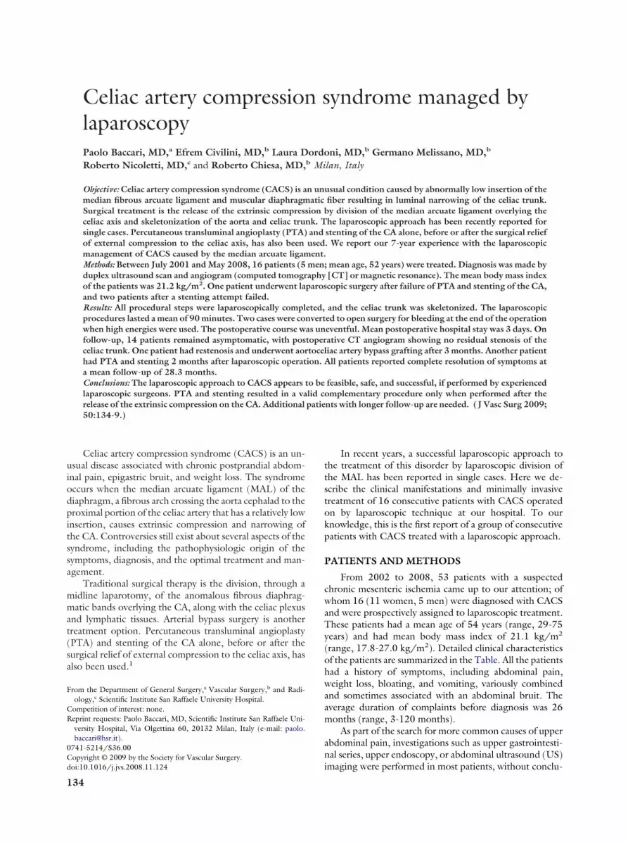
was compressed on its superior aspect at its origin from theaorta by a band of fibers forming the MAL. The celiactrifurcation was always clearly seen because of its postste-notic dilatation and pulsations. The CA was completelyisolated. The left gastric, common hepatic, and splenicartery were identified. Gentle inferior traction of this trifur-cation was of great help to expose the MAL. The ligamentwas cut with the coagulating hook along with the nervousceliac plexus and any fibrous and lymphatic tissue (Fig 2).
The procedure ended when the origin of the CA wascompletely exposed and freed by any external stricture. Adrain was then placed near the CA for the first 24 hours,and the wounds were closed in a routine fashion. Allpatients were extubated at the end of the procedure anddischarged to the surgical ward.
RESULTS
Mesenteric DUS imaging revealed a stenosis of the CA�70% during deep expiration in all patients, and a stenosis�70% was also present during inspiration in 12 patients. Inall patients the anatomy of the superior and inferior mes-enteric artery was normal at the preoperative diagnosticworkup.
The mean length of the laparoscopic procedures was 90minutes (range, 35-180 minutes). The mean intraoperativeblood loss was 50 mL (range, 0-300 mL). The averagepostoperative hospital length of stay was 3 days (range, 2-5days).
The laparoscopic operations in two patients were con-verted to an open approach, at the last step of the proce-dure, to obtain hemostasis for bleeding at the proximal partof the CA. At this level, the arterial wall was damaged bythe Harmonic scalpel (Ethicon Endo-Surgery, Cincinnati,Ohio) in the first patient and by the coagulating hook in the
Fig 1. Position of the surgeon during the laparoscopic proce-dure. The camera is inserted through an access above the umbili-cus. Inset, The positions of the four ports are shown: A, camera; B,operating ports; C, retractor. When a fifth port was needed, it wasinserted in the left flank.
second. In both patients, the damaged artery was readily
controlled by the vascular surgeon through a xiphoumbili-cal midline incision. After laparotomy, the lesser omentumand the diaphragmatic crus were already divided, and thebleeding from CA was promptly controlled with 6-0pledgeted suture, without major bleeding. No bleeding-related hypotension was recorded, and no patient requireda blood transfusion. The postoperative course was unevent-ful in all the patients, without complications.
In two patients the laparoscopic surgical division of theMAL was not followed by a complete and persistent reliefof the clinical symptoms of CACS. DUS imaging and CTAshowed a recurrent, fixed stenosis of the CA in both pa-tients, even in the absence of extrinsic compression. Patient5 had undergone laparoscopic division of the MAL forrestenosis after PTA and stenting of the CA. The minimallyinvasive procedure apparently resulted in the release of theextrinsic CA compression as diagnosed by postoperativeDUS imaging, but was followed by an early restenosis 3months later. Complete relief of symptoms was achievedwith aortoceliac artery bypass grafting. In patient 10, earlyrestenosis occurred 2 months after the laparoscopic proce-dure. The complete resolution of the clinical syndrome wasobtained by PTA and stenting.
At a mean follow-up of 28.3 months (range, 2-83months), symptoms were completely resolved in all 16patients. Postoperative imaging studies demonstrated noresidual CA stenosis.
DISCUSSION
Chronic abdominal symptoms caused by respiration-varying eccentric stenoses of the CA represent the classicmanifestation of CACS: a rare entity first described byHarjola3 in 1963 and by Dunbar et al4 in 1965. Otherreported symptoms are weight loss, nausea, vomiting, andbloating associated with a midepigastric bruit. CACS hasbeen attributed to compression of the CA by the MAL as aresult of the too-high emergence of the CA from the aortaor of the too-low insertion of the crus.
Controversies exist regarding the clinical features,pathophysiology, diagnosis, and treatment of CACS, andthe observation of CA compression in asymptomatic pa-tients even leads to questions about the real existence of thesyndrome.5 Some authors5-8 suggested that pain is causedby ischemia secondary to the reduction of blood flowthrough the stenotic CA. Others9,10 claimed that paincomes from direct compression of celiac ganglias. Bothmechanisms may be involved.
We found that symptoms are variable. The classic triadof postprandial abdominal pain, weight loss, and midepi-gastric bruit was found in only three patients, the mostfrequent symptom being abdominal epigastric or epi-mesogastric pain, mainly related to meals. Weight loss wasthe second most frequent symptom, with patients report-ing a mean weight loss of 7 kg (range, 3-14 kg).
In three patients if our series, CACS was not recog-nized because of negative results after abdominal investiga-tions; a psychosomatic disease was initially diagnosed, and
antidepressive therapy was started. After a mean of 32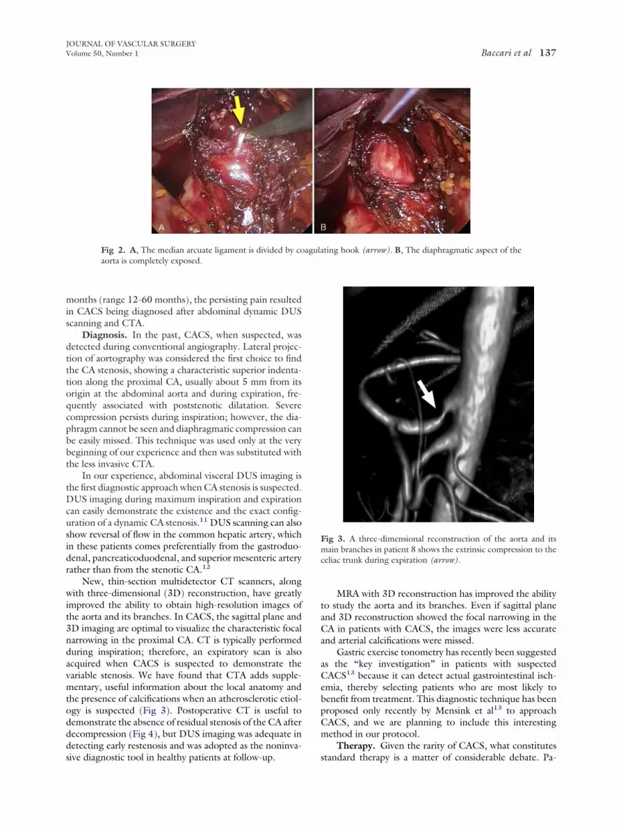
JOURNAL OF VASCULAR SURGERYVolume 50, Number 1 Baccari et al 137
months (range 12-60 months), the persisting pain resultedin CACS being diagnosed after abdominal dynamic DUSscanning and CTA.
Diagnosis. In the past, CACS, when suspected, wasdetected during conventional angiography. Lateral projec-tion of aortography was considered the first choice to findthe CA stenosis, showing a characteristic superior indenta-tion along the proximal CA, usually about 5 mm from itsorigin at the abdominal aorta and during expiration, fre-quently associated with poststenotic dilatation. Severecompression persists during inspiration; however, the dia-phragm cannot be seen and diaphragmatic compression canbe easily missed. This technique was used only at the verybeginning of our experience and then was substituted withthe less invasive CTA.
In our experience, abdominal visceral DUS imaging isthe first diagnostic approach when CA stenosis is suspected.DUS imaging during maximum inspiration and expirationcan easily demonstrate the existence and the exact config-uration of a dynamic CA stenosis.11 DUS scanning can alsoshow reversal of flow in the common hepatic artery, whichin these patients comes preferentially from the gastroduo-denal, pancreaticoduodenal, and superior mesenteric arteryrather than from the stenotic CA.12
New, thin-section multidetector CT scanners, alongwith three-dimensional (3D) reconstruction, have greatlyimproved the ability to obtain high-resolution images ofthe aorta and its branches. In CACS, the sagittal plane and3D imaging are optimal to visualize the characteristic focalnarrowing in the proximal CA. CT is typically performedduring inspiration; therefore, an expiratory scan is alsoacquired when CACS is suspected to demonstrate thevariable stenosis. We have found that CTA adds supple-mentary, useful information about the local anatomy andthe presence of calcifications when an atherosclerotic etiol-ogy is suspected (Fig 3). Postoperative CT is useful todemonstrate the absence of residual stenosis of the CA afterdecompression (Fig 4), but DUS imaging was adequate indetecting early restenosis and was adopted as the noninva-
Fig 2. A, The median arcuate ligament is divided by coaorta is completely exposed.
sive diagnostic tool in healthy patients at follow-up.
MRA with 3D reconstruction has improved the abilityto study the aorta and its branches. Even if sagittal planeand 3D reconstruction showed the focal narrowing in theCA in patients with CACS, the images were less accurateand arterial calcifications were missed.
Gastric exercise tonometry has recently been suggestedas the “key investigation” in patients with suspectedCACS13 because it can detect actual gastrointestinal isch-emia, thereby selecting patients who are most likely tobenefit from treatment. This diagnostic technique has beenproposed only recently by Mensink et al13 to approachCACS, and we are planning to include this interestingmethod in our protocol.
Therapy. Given the rarity of CACS, what constitutes
Fig 3. A three-dimensional reconstruction of the aorta and itsmain branches in patient 8 shows the extrinsic compression to theceliac trunk during expiration (arrow).
ting hook (arrow). B, The diaphragmatic aspect of the
agulastandard therapy is a matter of considerable debate. Pa-

JOURNAL OF VASCULAR SURGERYJuly 2009138 Baccari et al
tients who exhibit symptoms should undergo operativetreatment. Some patients are asymptomatic or have suffi-ciently minimal symptoms that they don’t warrant treat-ment. Surgical division of the MAL and diaphragmaticpillars by open surgery has been so far the mainstay treat-ment, which can remove the extrinsic compression on theCA. Takach et al14 in 1996 reported patients asymptomaticafter 15 years with the open surgical approach.
After laparoscopic surgery became more widespread, asmall number of patients with CACS have been reportedtreated with the minimally invasive approach. Roayaie etal15 in 2000 described a patient in whom laparoscopyprovided a less invasive but equally effective method fordecompressing the CA; moreover, laparoscopic intraoper-ative US scanning demonstrated markedly improved flowin the CA. Other single cases of laparoscopic managementof CACS were subsequently reported by Carbonell et al,16
Baldassarre at al,17 and by our group.18 Laparoscopy al-lowed surgeons to skeletonize the CA without a laparot-omy; however, adequate laparoscopic experience is re-quired because this is not a simple procedure.
No postoperative complications occurred in our series.Despite a spacious release of the CA (Fig 2) from theanomalous MAL, often involving the diaphragmatic crus,at follow-up we recorded no symptoms of postoperativegastric reflux or evidence of hiatal hernia. Less blood lossand postoperative pain, faster return to normal diet, shorterhospital stay, and better cosmetic effect were the obtainedbenefits.
After the first successful approach, we decided to use
Fig 4. A three-dimensional aortic reconstruction of patient 8(Fig 3), after laparoscopic release of the median arcuate liga-ment, shows good patency of the celiac trunk (arrow) at the1-month follow-up.
laparoscopy as the intervention for the other CACS patients
subsequently observed at our institution or transferredfrom other centers. To our knowledge, this is the firstreport of a series of consecutive patients with CACS treatedby a minimally invasive approach. Of the 16 patients treatedby us by laparoscopy, 14 had uneventful operations andpostoperative courses. Patients 9 and 11 of the series wereconverted to open surgery because of bleeding from thetakeoff of the CA from the aorta. This was a small lesion tothe proximal part of a CA with a thin wall, caused byHarmonic Scalpel in one patient and by the coagulatinghook in the other patient. Both lesions occurred at the endof the procedure during an attempt to improve the aorto-celiac skeletonization by removing the abundant ganglionictissue encasing the CA. We believe that, during the laststeps of the procedure, avoiding high energies and reducingthe coagulating force can be advisable.
A successful robotic-assisted laparoscopic approachto CACS using the da Vinci Surgical System (IntuitiveSurgical, Sunnyvale, Calif) was recently reported.19 Ro-botic assistance is a significant technologic advancement inminimally invasive surgery. Benefits of robotic surgerycould be the superior stereoscopic view of the surgical fieldand the improved surgical precision of movement due tothe wrist-like articulations of the instruments. Disadvan-tages are the higher cost and longer time requirements forthe procedure and for equipment setup. Operative time was168 minutes in the reported case, in contrast with a mean of90 minutes of our series. In the future, however, roboticscould have a role in reconstructive surgery of visceral arter-ies.
Endovascular treatment. PTA and stenting in thetreatment of CACS is also controversial. The successfulendovascular treatment with balloon angioplasty and stent-ing for celiac and mesenteric atherosclerotic stenoses20
suggested the extension of PTA also to patients withCACS. However, the endovascular procedure is not con-sidered adequate as the sole mode of treatment before thesurgical release of the extrinsic compression on the CAbecause the compression on the CA often does not allowadequate expansion of the stent,21 as confirmed by ourseries. Patients 7 and 14 had undergone a previous attemptof CA stenting that failed as a result of technical difficulties,and no stent was implanted.
A recent report pointed at the partial, short-lived reliefof symptoms and early CA restenosis after repeat PTA andstenting in the presence of high-grade CA compression dueto CACS.22 The patient in that report recovered after opensurgical division of the ligament and reconstruction of theCA, which exactly matches with our experience. In patient5, early restenosis of CA occurred after the endovascularprocedure before undergoing laparoscopic surgical divisionof the ligament and release of the extrinsic compression onthe CA. The laparoscopic procedure, however, was alsofollowed by only temporary relief of symptoms and earlyCA restenosis. Three months later, this patient thereforeunderwent open surgical reconstruction of the CA with aside-to-end aortoceliac interposition graft, followed by re-
lief of the symptoms linked to CACS. We speculated that
JOURNAL OF VASCULAR SURGERYVolume 50, Number 1 Baccari et al 139
failures of stenting in the presence of CACS are usually dueto crushing of the stent, which later would not be reversedby extrinsic decompression alone.
In the presence of CACS, we therefore strongly adviseagainst using endovascular treatment of the lesion; thatshould be limited to the management of residual fixeddisease after extrinsic decompression either by laparoscopicor open means. As detailed above, patient 10 from ourseries may summarize this statement.
CONCLUSION
Laparoscopic release of CA compression by CACS istechnically feasible and an appealing option to an openapproach in centers with large experience in vascular sur-gery and in major laparoscopic operations. Given its lessinvasive nature, it can provide an important approach (ther-apeutic or even diagnostic) in younger patients where therisk of surgery and cosmetic impairment are significant.
PTA and stenting in the context of CACS should beavoided. Balloon dilatation alone may be used as a tempo-rizing modality but is likely to fail quickly. The role ofendovascular therapy should be limited to the managementof residual fixed disease after extrinsic decompression.
The laparoscopic technique for treatment of CACS is apromising alternative to open surgery, but additional caseswith longer follow-up are needed to confirm the validity ofthe minimally invasive approach.
AUTHOR CONTRIBUTIONS
Conception and design: PB, RCAnalysis and interpretation: PB, LD, GMData collection: LDWriting the article: PB, EC, RNCritical revision of the article: PB, EC, GMFinal approval of the article: PBStatistical analysis: Not applicableObtained funding: Not applicableOverall responsibility: RC
REFERENCES
1. Cina CS, Safar H. Successful treatment of recurrent celiac axis compres-sion syndrome. A case report. Panminerva Med 2002;44:69-72.
2. Mitchell EL, Moneta GL. Mesenteric duplex scanning. Perspect VascSurg Endovasc Ther 2006;18:175-83.
3. Harjola PT. A rare obstruction of the coeliac artery; report of a case. Ann
Chir Gyn Fenn 1963;52:547-50.4. Dunbar DJ, Molnar N, Beman FF. Compression of the celiac artery andabdominal angina. Am J Roentgenol Therapeut Nucl Med 1965;95:731-44.
5. Stoney RJ, Ehrenfeld WK, Wylie EJ. Revascularization methods inchronic visceral ischemia caused by atherosclerosis. Ann Surg 1977;184:468-76.
6. Lindner HH, Kemprud E. A clinicoanatomic study of the arcuateligament of the diaphragm. Arch Surg 1971;103:600-5.
7. Williams S, Gillespie P, Little JM. Celiac axis compression syndrome:factors predicting a favourable outcome. Surgery 1985;98:879-87.
8. Jamieson CW. Coeliac axis compression syndrome. BMJ 1986;293:159-60.
9. Snyder MA, Mahoney EB, Rob CG. Symptomatic celiac stenosis due toconstriction by the neurofibrous tissue of the celiac ganglion. Surgery1967;61:372-6.
10. Marable SA, Molnar W, Beman FM. Abdominal pain secondary toceliac axis compression. Am J Surg 1974;76:867-71.
11. Erden A, Yurdakul M, Cumhur T. Marked increase in flow velocitiesduring deep expiration: a duplex Doppler sign of celiac artery compres-sion sydrome. Cardiovasc Intervent Radiol 1999;22:331-2.
12. Lampert VD, Holz D, Jager K, Landmann J. New therapeutic approachin the celiac trunk compression syndrome. Helv Chir Acta 1991;58:169-72.
13. Mensink PBF, van Petersen AS, Kolkman J, Otte JA, Huisman AB,Geelkerken RH. Gastric exercise tonometry: the key investigation inpatients with suspected celiac artery compression syndrome. J Vasc Surg2006;44:277-81.
14. Takach TJ, Livesay JJ, Reul GJ, Cooley DA. Celiac compression syn-drome: tailored therapy based on intraoperative findings. J Am CollSurg 1996;183:606-10.
15. Roayaie S, Jossart G, Gitlitz D, Lamparello P, Hollier L, Gagner M.Laparoscopic release of celiac artery compression syndrome facilitatedby laparoscopic ultrasound scanning to confirm restoration of flow. JVasc Surg 2000;32:814-7.
16. Carbonell AM, Kercher KW, Heniford BT, Matthews BD. Laparo-scopic management of median arcuate ligament syndrome. Surg En-dosc 2005;19:729.
17. Baldassarre E, Torino G, Siani A, Barone M, Valenti G. The laparo-scopic approach in the median arcuate ligament syndrome. A casereport. Swiss Med Wkly 2007;137:353-4.
18. Dordoni L, Tshomba Y, Giacomelli M, Jannello AM, Chiesa R. Celiacartery compression syndrome: successful laparoscopic treatment. A casereport. Vasc Endovasc Surg 2002;36:317-21.
19. Jaik NP, Stawicki SP, Weger NS, Lukaszczyk JJ. Celiac artery compres-sion syndrome: successful utilization of robotic-assisted laparoscopicapproach. J Gastrointest Liver Dis 2007;16:93-6.
20. Sharafuddin MJ, Olson CH, Sun S, Kresowik TF, Corson JD. Endo-vascular treatment of celiac and mesenteric arteries stenoses: applica-tions and results. J Vasc Surg 2003;38:692-8.
21. Allen RC, Martin GH, Rees CR, Rivera FJ, Talkington CM, GarrettWV. Mesenteric angioplasty in the treatment of chronic intestinalischemia. J Vasc Surg 1996;24:415-21.
22. Delis KT, Gloviczki P, Altuwaijri M, McKusick MA. Median arcuateligament syndrome: open celiac artery reconstruction and ligamentdivision after endovascular failure. J Vasc Surg 2007;46:799-802.
Submitted Sep 10, 2008; accepted Nov 17, 2008.


![[Immunology of celiac disease]](https://img.dokumen.tips/doc/110x75/635861503cd558f04e054612/immunology-of-celiac-disease.jpg)


