Embed Size (px)
Citation preview
CD11c1/CD11b1 Cells Are Critical for OrganicDust–Elicited Murine Lung Inflammation
Jill A. Poole1, Angela M. Gleason1, Christopher Bauer1, William W. West2, Neil Alexis3,Nico van Rooijen4, Stephen J. Reynolds5, Debra J. Romberger1,6, and Tammy L. Kielian2
1Pulmonary, Critical Care, Sleep, and Allergy Division, Department of Medicine, and 2Department of Pathology and Microbiology, University of
Nebraska Medical Center, Omaha, Nebraska; 3Center for Environmental Medicine, Asthma, and Lung Biology, University of North Carolina School
of Medicine, Chapel Hill, North Carolina; 4Department of Molecular Cell Biology, Vrije University, Amsterdam, The Netherlands; 5High PlainsIntermountain Center for Agricultural Health and Safety, Department of Environmental and Radiological Health Sciences, Colorado State University,
Fort Collins, Colorado; and 6Veterans Administration–Nebraska Western Iowa Healthcare System, Omaha, Nebraska
Organic dust exposure in the agricultural industry results in signif-icant lung disease.Macrophage infiltrates are increased in the lungsafter organic dust exposures, yet the phenotype and functionalimportance of these cells remain unclear. Using an establishedintranasal inhalation murine model of dust-induced lung inflamma-tion, animals were treated once or daily for 3 weeks with swineconfinement organic dust extract (DE). Repetitive DE treatmentfor 3weeks resulted in significant increases inCD11c1/CD11b1mac-rophages in whole lung–associated tissue. These cells displayed in-creased costimulatory molecule (CD80 and CD86) expression,enhanced phagocytic ability, and an increased production of IL-6,CXCL1, and CXCL2. Similar findings were observed with theCD11c1/CD11b1 macrophage infiltrate after repetitive exposureto peptidoglycan, a major DE component. To determine the func-tional importance of macrophages in mediating DE-induced airwayinflammation, lung macrophages were selectively depleted usinga well-established intranasal clodronate liposome depletion/suicidestrategy. First, macrophage depletion by clodronate liposomesresulted in significant reductions in airway neutrophil influx andTNF-a and IL-6 production after a single exposure to DE. In contrast,after repetitive 3-week exposure to DE, airway lavage fluid andlung tissue neutrophils were significantly increased in clodronateliposome–treatedmice comparedwith controlmice. A histologicalexamination of lung tissue demonstrated striking increases in alveolarandbronchiolar inflammation, aswell as in the size anddistributionof cellular aggregates in clodronate–liposome versus saline–liposomegroups repetitively exposed toDE. These studies demonstrate thatDE elicits activated CD11c1/CD11b1 macrophages in the lung,which play a critical role in regulating the outcome of DE-inducedairway inflammation.
Keywords: macrophage; neutrophil; airway inflammation; peptidogly-can; organic dust
The chronic inhalation of organic dusts or bioaerosols can resultin several respiratory diseases including chronic bronchitis,asthma, and obstructive lung disease (1, 2). Agricultural workers
are routinely exposed to organic dust environments, with thehighest prevalence of airway disease associated with animalfarming exposures, particularly swine confinement operations(2). Initial exposure to organic dust induces an intense proin-flammatory response marked by fevers, bronchial hyperrespon-siveness, and increases in bronchoalveolar lavage fluid (BALF)inflammatory cells and cytokines/chemokines that wane overtime. However, persons chronically exposed to these environ-ments are at increased risk of lung function decline, airwayinflammation, and progressive respiratory impairment (1, 3–5).Although this paradigm, termed “chronic adaptation inflamma-tory response” (1), has been replicated and described in animalmodels by our group (6) and others (7), the critical cellularmechanisms underlying this response remain unclear.
Lung mononuclear phagocytes and macrophages are impor-tant innate immune lung effectors that serve a critical role in hostdefense against inhaled environmental insults and toxins (8–10).These cells can orchestrate inflammatory responses through therelease of mediators that promote inflammation, activate epi-thelial cells, and elicit neutrophil recruitment (11, 12). Macro-phages produce several mediators after in vitro exposure toorganic dust extract (DE) that are associated with agriculturallyinduced disease, such as TNF-a, IL-6, and CXCL8 (1, 13). In thelung, alveolar macrophages are well recognized to represent thelargest mononuclear phagocyte population (14). These cells dif-fer from other tissue macrophages based on their high expres-sion levels of CD11c, which is a molecule typically not presenton other tissue macrophages, and is generally considered a den-dritic cell marker (15–20). Furthermore, CD11b expression,which is high in other tissue macrophage populations, is quitelow in alveolar macrophages, unless these cells are activated(16, 17). Lifetime nonsmoking swine farmers demonstrate signsof bronchial inflammation with neutrophils, alveolar macro-phages, and lymphocytes present in lavage fluid (21). Further-more, evidence suggests that the alveolar macrophages in thesefarmers may be activated, based on observations of increased
(Received in original form March 7, 2012 and in final form July 10, 2012)
This work was supported by grants R01 ES019325, K08 ES015522-01, and the
American Recovery & Reinvestment Act (ARRA) ES015522-03S1 from the National
Institute of Environmental Health Sciences (J.A.P.), grants 1R01OH008539-01
(D.J.R.) and 5U50 OH0080856 (S.J.R.) from the National Institute of Occupational
Safety Health, and grant 2R01NS055385 from the National Institute of Neurolog-
ical Disorders and Stroke (T.L.K.).
Correspondence and requests for reprints should be addressed to Jill A. Poole,
M.D., Pulmonary, Critical Care, Sleep, and Allergy Division, Department of Med-
icine, University of Nebraska Medical Center, 985300 The Nebraska Medical
Center, Omaha, NE 68198-5300. E-mail: [email protected]
This article has an online supplement, which is accessible from this issue’s table of
contents at www.atsjournals.org
Am J Respir Cell Mol Biol Vol 47, Iss. 5, pp 652–659, Nov 2012
Copyright ª 2012 by the American Thoracic Society
Originally Published in Press as DOI: 10.1165/rcmb.2012-0095OC on July 19, 2012
Internet address: www.atsjournals.org
CLINICAL RELEVANCE
Inhalation exposure to organic dust extract from swineconfinement facilities induces an influx of activated CD11c1/CD11b1 macrophages in the lung. Our findings establishedlung macrophages as key to the acute response to dust ex-tract, and furthermore, lung macrophages are critical indown-regulating inflammatory responses after prolonged,repetitive dust extract exposures. This information may beimportant because potential therapies such as macrolidesand vitamin D were shown to enhance macrophage phago-cytic ability or autophagy.

macrophage chemotactic activity and ex vivo oxygen radicalformation (21). Airway and lung parenchymal macrophage infil-trates are also increased in mice after organic dust exposure (6,7). However, the phenotype and functional importance of thesecells have not been well described. Therefore, to address thefunctional role of alveolar macrophages in the context of repet-itive DE-induced lung inflammation, we analyzed CD11c1 lungmacrophages to determine their activation phenotype. In addi-tion, we compared these responses with those of peptidoglycan(PGN), a major component of organic DE previously shown todrive DE-elicited inflammatory responses in macrophages (13).Finally, we selectively depleted lung macrophages using a well-established intranasal clodronate liposomemacrophage depletion/suicide strategy to determine the functional importance of thesecells in the context of both single and repetitive DE exposures(17, 22, 23).
Our studies demonstrate that both DE and PGN exposureelicited increased numbers of activated CD11c1/CD11b1 lungmacrophages that were critical for regulating the extent of in-flammation. Namely, airway inflammatory responses were at-tenuated after a one-time exposure to DE. However, afterrepetitive DE exposures, lung inflammatory and pathologic out-comes, primarily marked by neutrophil influx, were significantlyworse when macrophages were depleted. Collectively, thesestudies demonstrate a critical role for lung macrophages in con-trolling DE-induced lung inflammation.
MATERIALS AND METHODS
Organic DE
DE was prepared as previously described (24, 25), with details providedin the online supplement.
Animal Model
Using an established intranasal instillation murine model of organicdust–induced and PGN-induced airway inflammation (6), C57BL/6mice (6–8 wk old; Jackson Laboratory, Bar Harbor, ME) were treatedonce (acute/single exposure) or daily for 3 weeks (repetitive exposure)with 12.5% DE, PGN (100 mg), or sterile PBS (diluent). The Staphy-lococcus aureus PGN (Sigma, St. Louis, MO) concentration comprisesapproximately half the protein in 12.5% DE, and elicits airway inflam-matory responses similar to those of DE (26). Whole lung cells wereisolated, as described previously (27) and in the online supplement. Allanimal experimental procedures were approved by the InstitutionalAnimal Care and Use Committee of the University of Nebraska Med-ical Center, according to National Institutes of Health guidelines forthe use of rodents.
Macrophage Depletion
The intranasal delivery of encapsulated clodronate liposomes is a well-established method to deplete lung macrophages selectively (17, 18, 28).Liposomes (30 ml) were administered 48 hours before the first DEtreatment, and every 3–4 days throughout the 3-week repetitive DEperiod to maintain macrophage depletion.
BALF
BALF was collected as previously described (6). The total cell numberof pooled lavages (3 3 1 ml lavages) was enumerated, and differentialcell counts were determined on cytospin-prepared slides (CytoproCytocentrifuge; Wescor, Inc., Logan, UT) stained with Diff-Quik(Dade Behring, Newark, DE). Cell-free supernatants for cytokine anal-ysis from the first lavage were collected and stored at 2208C.
Lung Histology
Whole lungs were inflated to 10 cm H2O pressure with 10% formalin(Sigma) to preserve pulmonary architecture. Lungs were processed and
embedded in paraffin, and sections (4–5 mm) were cut and stained withhematoxylin and eosin. Slides were semiquantitatively assessed, usinga previously described lung inflammatory scoring system (6, 26) bya pathologist (W.W.W.) blinded to the treatment conditions.
Flow Cytometry
Whole lung cells from each animal were incubated with anti-CD16/32(Fc Block; BD Biosciences, San Jose, CA), and then stained withmonoclonal antibodies directed against CD11c, CD11b, IA-b,CD80, CD86, Ly-6G, and appropriate isotype control samples (BDBiosciences). The CD11c1 macrophage gating strategy is depictedin Figure E1 in the online supplement. The CD11c1 macrophagenumber was calculated by multiplying the percentage of gated cellsmeasured by flow cytometry by the original hemocytometer count oftotal cells recovered for each animal.
Phagocytosis Assay
The phagocytic capacity of lung-associated CD11c1 cells was assessedby flow cytometry, modified from methods described previously (10,13) and in the online supplement.
Cytokine Assays
CD11c1 lung cells isolated by FACS from mice repetitively exposed tosaline and DE were analyzed for ex vivo cytokine/chemokine (e.g.,TNF-a, IL-6, KC/CXCL1, or MIP-2/CXCL2) responsiveness to subse-quent DE restimulation by ELISA. Details are provided in the onlinesupplement.
Statistical Methods
Data are presented as means 6 SEM. Statistical analyses were per-formed using a two-tailed, nonpaired t test and one-way ANOVA withthe Tukey multicomparison post hoc test. We used GraphPad (version5.01; GraphPad Software, La Jolla, CA) software. In all analyses, Pvalues less than 0.05 were considered statistically significant.
RESULTS
Organic Dust Exposure Induces the Accumulation
of Activated CD11c1/CD11b1 Lung Macrophages
The total number of lung-associated cells had increased after3-week repetitive DE exposure compared with saline controlsamples, concomitant with significant elevations in the absolutenumber of CD11c1 cells (Figure 1A). The expression of co-stimulatory molecules (CD80 and CD86) and of the adhesionmolecule CD11b was significantly up-regulated in lung-associatedCD11c1 cells after DE challenge, compared with saline-treatedcontrol samples (Figure 1B). No change was observed in MHCClass II expression.
A significant increase was evident in the phagocytic abilityof lung CD11c1 macrophages after repetitive DE challenge,compared with saline-treated mice (Figures 2A and 2B).In addition, IL-6 and CXCL1 production were significantlyincreased in ex vivo stimulated CD11c1 macrophages obtainedfrom DE-treated animals compared with saline-treated mice,whereas TNF-a was significantly decreased (Figure 2C). In-terestingly, CD11c1 cells from DE-treated mice produced ele-vated concentrations of IL-6, CXCL1, and CXCL2 at baseline,compared with saline-treated mice. Collectively, these datademonstrate that repetitive intranasal inhalation challenge withDE evokes a significant influx of CD11c1 lung cells, demon-strating an activated phenotype as revealed by increasedCD11b and costimulatory molecule expression, phagocytic abil-ity, and selected cytokine responsiveness to subsequent DEexposure.
Poole, Gleason, Bauer, et al.: CD11c/CD11b in Organic Dust–Induced Lung Inflammation 653

Repetitive Intranasal Challenge with PGN Induces Activated
CD11c1/CD11b1 Macrophages
Because emerging evidence suggests that bacterial PGNs in in-dustrial animal farming environments are highly prevalent andmay be responsible for mediating airway disease (29, 30), andbecause our previous work demonstrated that PGN exposure-induced outcomes closely resemble those elicited by DE expo-sure (13, 26), we investigated the effects of intranasal PGNinhalation in separate experiments. Similar to what was ob-served among DE-treated mice, PGN induced an expansion intotal lung cell infiltrates, with an increase in total number ofCD11c1 macrophages (Figure 3A). Repetitive PGN exposureresulted in increased costimulatory molecule expression (CD80and CD86) and increased adhesion molecule CD11b expression(Figure 3B) in CD11c1 lung macrophages, consistent with theobservations already described for DE. However, unlike treat-ment with DE, PGN treatment significantly augmented MHCClass II expression in CD11c1 cells (Figure 3B). The phagocyticability of lung CD11c1 cells from PGN-challenged mice was alsosignificantly enhanced compared with saline-treated mice (Figure3B). Collectively, these results show that repetitive PGN
treatment results in a significant influx of CD11c1 lung cells,demonstrating an activated phenotype, similar to findings ob-served after DE exposure.
Effects of Lung Macrophage Depletion in Acute
DE-Induced Airway Inflammation
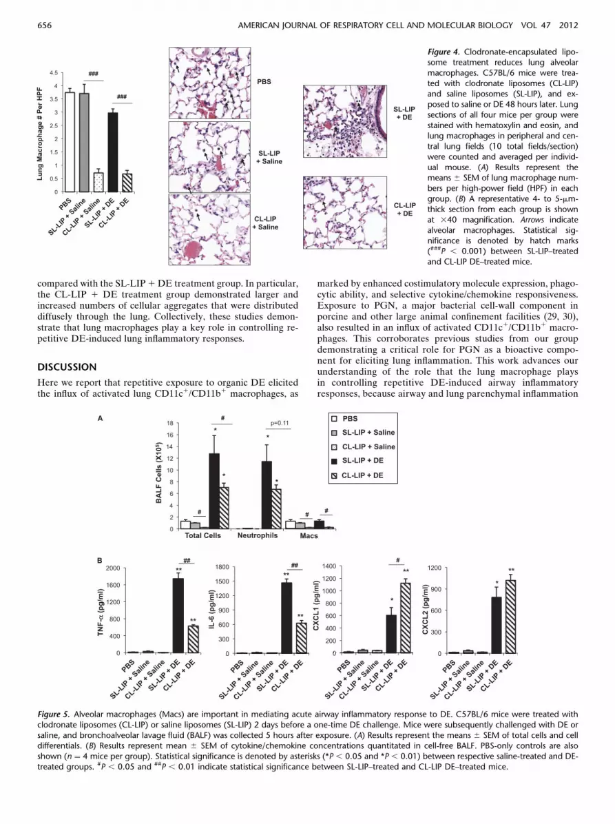
Next, we sought to assess the functional importance of lung mac-rophages in mediating DE-induced airway inflammation by se-lectively depleting these cells via the intranasal delivery ofclodronate-encapsulated liposomes. To establish the efficacyand safety of this approach, we first investigated airway inflam-matory outcomes in an acute DE-induced airway model (6).Clodronate liposomes (CL-LIP) and saline liposomes (SL-LIP) were administered 2 days before a one-time (acute) expo-sure to DE or saline control, whereupon mice were killed5 hours after DE exposure. Macrophage reduction was con-firmed by a microscopic review of the hematoxylin and eosinstaining of lung sections. Decreases in macrophage infiltrates(. 80%) were observed in CL-LIP–treated mice, comparedwith SL-LIP–treated animals (Figure 4). Significant reductionsin macrophage recovery (. 80%) from BALF in CL-LIP 1saline and CL-LIP 1 DE animals were evident, compared withrespective SL-LIP control mice (Figure 5A).
Macrophage reduction also produced changes in acute DE-induced airway inflammation. DE-induced airway neutrophilinflux was nonsignificantly (P ¼ 0.11) reduced in CL-LIP 1DE–treated mice compared with SL-LIP 1 DE mice (Figure5A). Significant decreases in TNF-a and IL-6 concentrationsin CL-LIP 1 DE–treated animals were evident, comparedwith SL-LIP1DE–treated animals (Figure 5B). Interestingly,the expression of neutrophil chemokines was differently af-fected by macrophage infiltrates, because CXCL1 was signif-icantly elevated in CL-LIP 1DE–treated mice compared withSL-LIP 1 DE–treated mice, whereas CXCL2 concentrationswere similar (Figure 5B). These studies demonstrate that macro-phages play a selective role in regulating acute (single exposure)DE-induced airway inflammatory responses.
Effects of Alveolar Macrophage Depletion in Repetitive
DE-Induced Airway Inflammation
Next, we investigated the role of macrophages in the repetitiveexposuremodel to determine the functional importance ofmacro-phages in mediating the chronic inflammatory adaptation–likeresponse to DE. The administration of CL-LIP throughout theduration of repetitive DE exposure treatment (CL-LIP 1 DE)resulted in increased neutrophil numbers recovered from BALF,compared with SL-LIP 1 DE–treated animals (Figure 6A). Asexpected, macrophage recovery in BALF and whole-lung tissuewas significantly reduced in CL-LIP 1 DE–treated mice com-pared with SL-LIP 1 DE–treated animals, confirming the effi-cacy of the clodronate liposome–mediated depletion approach(Figures 6A and 6B). CL-LIP did not alter the activation profilesof the few CD11c1 macrophages that were detected in the lungsafter repetitive DE exposure, compared with SL-LIP–treatedmice. Namely, CD11b, CD80, and CD86 expression had all in-creased (data not shown). The percentage and numbers of lungneutrophils from whole-lung tissue were significantly increased inanimals treated with CL-LIP 1 DE, compared with SL-LIP 1DE (Figure 6B). After 3 weeks of repetitive exposure, BALFcytokines and chemokines were at the lower limit of assay detec-tion, consistent with the chronic inflammatory adaptation re-sponse (6, 7), and concentrations were not different betweenSL-LIP 1 DE and CL-LIP 1 DE (data not shown).
The histological examination of lung tissue demonstrated strik-ing differences in lung parenchymal inflammation between the
Figure 1. Repetitive organic dust extract (DE) exposure leads to the
activation of CD11c1 lung macrophages. Lung-associated cells were
collected from C57BL/6 mice after 3 weeks of repetitive intranasal ex-posure to DE or saline control, and stained for FACS. (A) Results repre-
sent total lung cells and absolute CD11c1 cells. #, number of. (B) Mean
fluorescence intensity (MFI) of surface marker expression on gated
CD11c1 cells is shown. Results represent the mean 6 SEM (n ¼6 mice/group), with statistical significance denoted by asterisks
(*P , 0.05, **P , 0.01, and ***P , 0.001).
654 AMERICAN JOURNAL OF RESPIRATORY CELL AND MOLECULAR BIOLOGY VOL 47 2012

CL-LIP 1 DE and SL-LIP 1 DE treatment groups (Figures 7Aand 7B). Semiquantitative analysis of these DE-induced histopa-thologic changes were assessed, using an established lung pathology
scoring system (inflammatory score range of 0–3) (6). Signifi-cant increases were observed in the inflammatory scores forall parameters assessed in the CL-LIP 1 DE treatment group
Figure 2. DE treatment increases
phagocytosis and cytokineproduction by CD11c1 lung
cells. C57BL/6 mice were
repetitively exposed to DE or
saline for 3 weeks, and lung-associated cells were exposed ex
vivo to FITC-labeled Saccharomy-
ces cerevisiae zymosan-A biopar-
ticles at 0 and 60 minutes todetermine the phagocytic ability
of gated CD11c1 lung macro-
phages. (A) A representative dot
plot depicts particle uptake in gatedCD11c1Ly-6G2 macrophages from
DE-treated mice and saline con-
trol–treated mice as a rightwardshift in fluorescence. (B) The
phagocytic ability of macro-
phages is shown as fold change
in mean MFI (6 SEM) of the pro-portion of cells in the zymosan-
exposed gated population at 60
minutes, compared with cells ex-
posed for 0 minutes from DE andsaline control mice (n ¼ 6 mice/
group). (C) CD11c1 macrophages
isolated by FACS from DE-treatedmice and saline-exposed mice were
restimulated ex vivo with DE (1%)
or saline (0% DE) for 24 hours, and cytokine/chemokine concentrations were measured in cell-free supernatant by ELISA. Results represent the mean 6SEM (n ¼ 6–9 mice/group), with statistical significance between saline-and DE-treated denoted by asterisks (*P , 0.05 and ***P , 0.001).
Figure 3. Repetitive intranasal challenge
with peptidoglycan (PGN) increases acti-vated CD11c1 lung macrophages.
C57BL/6 mice were repetitively exposed
to PGN (100 mg) or saline for 3 weeks,
and lung-associated cells were enumer-ated and stained for FACS. (A) Results
represent the means 6 SEM (n ¼ 6
mice/group) of total lung cells and
CD11c1 lung macrophages. (B) Themean fluorescence intensity (MFI) of sur-
face marker expression on gated
CD11c1 cells is shown. Next, lung-associated cells were exposed ex vivo to
FITC-labeled Saccharomyces cerevisiae
zymosan-A bioparticles at 0 and 60
minutes, to determine the phagocyticability of gated CD11c1 lung macro-
phages. (C) Results represent fold
changes in MFI (proportion of cells in
the gated population at 60 min, com-pared with cells exposed for 0 min),
expressed as means 6 SEM (n ¼6 mice/group). Statistical significance
is denoted by asterisks (*P , 0.05 and***P , 0.001).
Poole, Gleason, Bauer, et al.: CD11c/CD11b in Organic Dust–Induced Lung Inflammation 655
compared with the SL-LIP1DE treatment group. In particular,the CL-LIP 1 DE treatment group demonstrated larger andincreased numbers of cellular aggregates that were distributeddiffusely through the lung. Collectively, these studies demon-strate that lung macrophages play a key role in controlling re-petitive DE-induced lung inflammatory responses.
DISCUSSION
Here we report that repetitive exposure to organic DE elicitedthe influx of activated lung CD11c1/CD11b1 macrophages, as
marked by enhanced costimulatory molecule expression, phago-cytic ability, and selective cytokine/chemokine responsiveness.Exposure to PGN, a major bacterial cell-wall component inporcine and other large animal confinement facilities (29, 30),also resulted in an influx of activated CD11c1/CD11b1 macro-phages. This corroborates previous studies from our groupdemonstrating a critical role for PGN as a bioactive compo-nent for eliciting lung inflammation. This work advances ourunderstanding of the role that the lung macrophage playsin controlling repetitive DE-induced airway inflammatoryresponses, because airway and lung parenchymal inflammation
Figure 4. Clodronate-encapsulated lipo-some treatment reduces lung alveolar
macrophages. C57BL/6 mice were trea-
ted with clodronate liposomes (CL-LIP)and saline liposomes (SL-LIP), and ex-
posed to saline or DE 48 hours later. Lung
sections of all four mice per group were
stained with hematoxylin and eosin, andlung macrophages in peripheral and cen-
tral lung fields (10 total fields/section)
were counted and averaged per individ-
ual mouse. (A) Results represent themeans 6 SEM of lung macrophage num-
bers per high-power field (HPF) in each
group. (B) A representative 4- to 5-mm-
thick section from each group is shownat 340 magnification. Arrows indicate
alveolar macrophages. Statistical sig-
nificance is denoted by hatch marks(###P , 0.001) between SL-LIP–treated
and CL-LIP DE–treated mice.
Figure 5. Alveolar macrophages (Macs) are important in mediating acute airway inflammatory response to DE. C57BL/6 mice were treated with
clodronate liposomes (CL-LIP) or saline liposomes (SL-LIP) 2 days before a one-time DE challenge. Mice were subsequently challenged with DE or
saline, and bronchoalveolar lavage fluid (BALF) was collected 5 hours after exposure. (A) Results represent the means 6 SEM of total cells and celldifferentials. (B) Results represent mean 6 SEM of cytokine/chemokine concentrations quantitated in cell-free BALF. PBS-only controls are also
shown (n ¼ 4 mice per group). Statistical significance is denoted by asterisks (*P , 0.05 and *P , 0.01) between respective saline-treated and DE-
treated groups. #P , 0.05 and ##P , 0.01 indicate statistical significance between SL-LIP–treated and CL-LIP DE–treated mice.
656 AMERICAN JOURNAL OF RESPIRATORY CELL AND MOLECULAR BIOLOGY VOL 47 2012

marked by neutrophil influx became worse after macrophagedepletion by clodronate liposomes. These experiments estab-lish that DE-activated CD11c1/CD11b1 macrophages are, inpart, responsible for down-regulating the chronic inflamma-tory lung response to organic dust exposures.
The chronic inflammatory adaptation response to organicdust environments is well-recognized (3), but the important un-derlying cellular mechanisms have not been clear. Alveolarmacrophages are key innate immune cells that are rapidly acti-vated and orchestrate immune responses after exposure to in-haled environmental toxins, such as organic dust, bacterialproducts, particulate air pollution, and ozone (9, 13, 31). Macro-phages can produce inflammatory mediators and regulate andresolve chronic inflammatory responses by clearing bacteria,debris, and apoptotic cells (32). Activated CD11c1 lung macro-phages are a common feature of inflammatory responses afterexposure to various viral or bacterial pathogens (12, 16, 20, 33,34). Our findings support that repetitive exposure to DE indu-ces an analogous activated CD11c1 macrophage phenotype,with findings of high CD11b1 and costimulatory molecule ex-pression and phagocytic ability (Figures 1 and 2). DE treatmentdid not augment MHC Class II expression, which is a feature
that was also observed in other infectious models (22). This mayrepresent a regulatory signal or differences in the recycling ofthe MHC Class II complex (35). Although lung infections arenot a common characteristic of lung disease in farmers, animalfacilities are associated with a large diversity of microbialagents, and particularly Gram-positive bacteria (. 80–95%)(29, 30). Indeed, repetitive exposure to PGN, a major cell-wall component of Gram-positive and, to a lesser degree, Gram-negative bacteria, also resulted in similar increases of CD11c1
macrophage activation.We found evidence of a “priming effect,” as revealed by en-
hanced IL-6 and CXCL1 after the stimulation of CD11c1 cellsfrom DE-challenged mice (Figure 2C). Furthermore, the basalrelease of IL-6, CXCL1, and CXCL2 was significantly increasedin CD11c1 cells isolated from DE-treated mice. This would indi-cate cellular responsiveness triggered by DE in vivo, because noadditional stimulus was used. However, this “primed” or activatedstatus was not global because TNF-a responsiveness was damp-ened, consistent with the so-called tolerant response/ adapted re-sponse (1, 36).However, a hyperresponsiveness in cytokine releasewith ex vivo, endotoxin-stimulated whole blood from farmerscomparedwith healthy control subjects has been reported (37, 38).
Figure 6. Repetitive DE-induced
neutrophilic influx is increasedwhen lung macrophages are
depleted. C57BL/6 mice were
treated with clodronate lipo-
somes (CL-LIP) or saline lipo-somes (SL-LIP) beginning 2
days before the first DE chal-
lenge, and then every 3 to 4
days during the daily 3-weekrepetitive DE or saline expo-
sure. (A) Results represent the
means 6 SEM (n ¼ 4 mice/
group) of the total cells andcell differentials recovered from
the BALF of mice. Next, lung-
associated cells were collectedfrommice, and total cells were enumerated and stained by FACS. (B) Results represent means6 SEM (n¼ 4 mice/group) of CD11c1 lung macrophages
and neutrophils (percentage of cell type3 total lung cell count). Statistical significance is denoted by asterisks (*P, 0.05, *P, 0.01, and ***P, 0.001)
between respective saline-treated and DE-treated groups. Hatch marks (#P , 0.05, ##P , 0.01, and ###P , 0.001) indicate statistical differences
between SL-LIP 1 DE–treated and CL-LIP 1 DE–treated mice.
Figure 7. Lung macrophages
are important in mediating re-petitive DE-induced lung in-
flammation. C57BL/6 mice
were treated with clodronate
liposomes (CL-LIP) or salineliposomes (SL-LIP) beginning
2 days before the first DE chal-
lenge, and then every 3 to 4
days during the daily 3-weekrepetitive DE or saline expo-
sure. (A) Semiquantitative in-
flammatory scores (means 6SEM; n ¼ 4 mice per group)
of the degree and distribution
of cellular aggregates, and of
alveolar and bronchiolar lung inflammation, are shown. PBS-only control scores are also shown. (B) A representative 4- to 5-mm-thick, hematoxylin-and-eosin–stained section of one of four mice per treatment group is shown at 310 magnification. All lung specimens were inflated to 10 cm H2O
pressure during fixation to avoid atelectasis artifacts. Statistical significance is denoted by asterisks (*P, 0.05, *P, 0.01, and ***P, 0.001) between
respective saline-treated and DE-treated groups. #P , 0.05 and ##P , 0.01 indicate statistical difference between SL-LIP 1 DE–treated and CL-LIP 1DE–treated mice.
Poole, Gleason, Bauer, et al.: CD11c/CD11b in Organic Dust–Induced Lung Inflammation 657
The finding that CD11b expression was dramatically in-creased in DE-exposed CD11c1 cells is also important. Al-though CD11b is well recognized for its role in leukocyteadhesion and activation, phagocytosis, and transmigration(39), CD11b expression on mononuclear phagocytes withinthe respiratory tract has been correlated with airway neutro-philia (9, 40), and the correlation with airway neutrophilia issupported by our studies. Activated macrophages may contrib-ute to DE-induced airway neutrophilia. However, CD11c1/CD11b1 macrophages are more likely responding to the inflam-matory insult in an attempt to control and resolve disease. Thispossibility is supported by our demonstration that without anadequate lung macrophage population, enhanced inflammatoryconsequences follow repetitive DE exposure (Figures 6 and 7).Previous work established that the mononuclear cellular aggre-gates induced by repetitive organic dust exposure comprise anadmixture of T and B lymphocytes and phagocytes (6). Fur-thermore, DE-induced cellular aggregates consist of live cells(. 98.5%), as opposed to clumps of apoptotic cells based onthe common method for detecting DNA fragmentation, termi-nal deoxynucleotidyl transferase dUTP nick end labeling(TUNEL) staining of tissue sections (data not shown).
In acute DE exposure studies within the setting of lung mac-rophage depletion, findings suggest that the alveolar macrophageis likely a major source for TNF-a and IL-6 production afteracute DE exposure (Figure 5). In contrast, the macrophage doesnot appear to be an important source of neutrophil chemoat-tractants during DE-induced airway inflammation (Figure 5).This implies that other cell types, such as airway epithelium,may be important sources of DE-induced neutrophil chemoat-tractants. Furthermore, TNF-a expression was demonstrated inairway epithelial cells in all groups by an immunohistochemicalstaining procedure (Figure E3). Isolated airway epithelial cellsproduce substantial amounts of neutrophil chemoattractants af-ter organic dust exposures (25, 41, 42). Without macrophages tophagocytize and remove the organic dust burden effectively, theairway epithelium may hyperrespond to an increased DE load.
Trends toward a reduction in DE-induced acute neutrophilcounts were observed in the setting of macrophage depletion,despite the lack of reduction in murine neutrophil chemoattrac-tants.With repetitive DE exposures, an adaptation-like responseoccurs, marked by decreased CXCL1 and CXCL2 expression,although neutrophil influx remains elevated (6, 7). Likewise,in our study, the cytokine/chemokine response diminished anddid not differ between groups (data not shown), yet neutrophilinflux remained elevated after repetitive DE exposures, and thiselevation was further enhanced when macrophages were de-leted (Figures 6 and 7). These observations highlight that thecontrol and regulation of neutrophil recruitment after lung in-jury are not limited to chemokines, but may include a number ofcomplex factors and networks (e.g., integrins, selectins, pro-teases, and reactive oxygen species) (43). Moreover, extractsof organic dusts from agricultural environments exhibit directchemotactic activity in vitro, and this response has been shownto be independent of endotoxin (44, 45). This response may alsobe driven by N-formyl–methionyl peptides, motifs of microbialproteins, which can also trigger neutrophil recruitment via theformylated peptide receptor (46). Future lines of study shouldexplore other mechanisms of neutrophil influx, and investigateand characterize subpopulations of DE-induced lung macro-phages (e.g., M1, M2a, M2b, and M2c) (47).
Although we found no evidence of distress or mortality, micereceiving repetitive clodronate liposome treatment, regardless ofsubsequent DE or saline exposure, failed to exhibit equivalentweight gain, compared with their respective control mice. Al-though the reason for this observation is not entirely clear,
repeated clodronate liposome treatment (i.e., 2–3 repeatedchallenges) was reported to cause subtle immunologic changes(18, 48). By extension, the host may elicit a compensatory mech-anism to respond to the reduction in macrophages. Thus, wecannot completely eliminate the possibility of a nonspecificclodronate liposome effect in our repetitive delivery studies.
In conclusion, inhalation exposure to organic DE fromswine confinement facilities induces an influx of activatedCD11c1/CD11b1 macrophages in the lung. Moreover, PGN,a component of organic dust, elicited a similar lung response,suggesting that PGN may be an important player in complexorganic dust–induced pathogenic lung inflammation. Our find-ings establish that lung macrophages are key in the acuteresponse to DE, and furthermore, lung macrophages are crit-ical in down-regulating inflammatory responses after pro-longed, repetitive DE exposures. This information may beimportant because potential therapies such as macrolidesand vitamin D were shown to enhance macrophage phagocyticability or autophagy (49–52). Future studies are warranted toinvestigate whether promoting macrophage function mightultimately lead to novel approaches to reduce chronic organicdust–induced airway inflammatory consequences in exposedagricultural workers.
Author disclosures are available with the text of this article at www.atsjournals.org.
Acknowledgments: The authors thank Megan Michalak, Victoria Smith, andCharles Kuzinski, Ph.D., of the Cell Analysis Facility at the University of NebraskaMedical Center for assistance with flow cytometric measurements, Greg Dooley,Ph.D., for assistance with the mass spectrometry analysis of dust samples, andGeoffrey Talmon, M.D., David Muirhead, and David Wert of the Tissue ScienceFacility at the University of Nebraska Medical Center and Thomas Jerrells,Ph.D., for assistance with digital microscopy images prepared for the manuscript.The authors also thank Lisa Chudomelka for assistance with preparation of themanuscript.
References
1. Von Essen S, Romberger D. The respiratory inflammatory response to
the swine confinement building environment: the adaptation to re-
spiratory exposures in the chronically exposed worker. J Agric Saf
Health 2003;9:185–196.
2. Eduard W, Pearce N, Douwes J. Chronic bronchitis, COPD, and lung
function in farmers: the role of biological agents. Chest 2009;136:
716–725.
3. Sundblad BM, von Scheele I, Palmberg L, Olsson M, Larsson K. Re-
peated exposure to organic material alters inflammatory and physio-
logical airway responses. Eur Respir J 2009;34:80–88.
4. Palmberg L, Larssson BM, Malmberg P, Larsson K. Airway responses of
healthy farmers and nonfarmers to exposure in a swine confinement
building. Scand J Work Environ Health 2002;28:256–263.
5. Schwartz DA, Donham KJ, Olenchock SA, Popendorf WJ, Van Fossen
DS, Burmeister LF, Merchant JA. Determinants of longitudinal
changes in spirometric function among swine confinement operators
and farmers. Am J Respir Crit Care Med 1995;151:47–53.
6. Poole JA, Wyatt TA, Oldenburg PJ, Elliott MK, West WW, Sisson JH,
Von Essen SG, Romberger DJ. Intranasal organic dust exposure–
induced airway adaptation response marked by persistent lung in-
flammation and pathology in mice. Am J Physiol Lung Cell Mol
Physiol 2009;296:L1085–L1095.
7. Charavaryamath C, Janardhan KS, Townsend HG, Willson P, Singh B.
Multiple exposures to swine barn air induce lung inflammation and
airway hyper-responsiveness. Respir Res 2005;6:50.
8. Arjomandi M, Witten A, Abbritti E, Reintjes K, Schmidlin I, Zhai W,
Solomon C, Balmes J. Repeated exposure to ozone increases alveolar
macrophage recruitment into asthmatic airways. Am J Respir Crit
Care Med 2005;172:427–432.
9. Alexis NE, Lay JC, Zeman K, Bennett WE, Peden DB, Soukup JM,
Devlin RB, Becker S. Biological material on inhaled coarse fraction
particulate matter activates airway phagocytes in vivo in healthy
volunteers. J Allergy Clin Immunol 2006;117:1396–1403.
658 AMERICAN JOURNAL OF RESPIRATORY CELL AND MOLECULAR BIOLOGY VOL 47 2012

10. Lay JC, Alexis NE, Kleeberger SR, Roubey RA, Harris BD, Bromberg
PA, Hazucha MG, Devlin RB, Peden DB. Ozone enhances markers
of innate immunity and antigen presentation on airway monocytes in
healthy individuals. J Allergy Clin Immunol 2007;120:719–722.
11. Fels AO, Cohn ZA. The alveolar macrophage. J Appl Physiol 1986;60:
353–369.
12. Thepen T, Kraal G, Holt PG. The role of alveolar macrophages in
regulation of lung inflammation. Ann N Y Acad Sci 1994;725:200–206.
13. Poole JA, Alexis NE, Parks C, MacInnes AK, Gentry-Nielsen MJ, Fey
PD, Larsson L, Allen-Gipson D, Von Essen SG, Romberger DJ.
Repetitive organic dust exposure in vitro impairs macrophage dif-
ferentiation and function. J Allergy Clin Immunol 2008;122:375–382.
14. Martin TR, Frevert CW. Innate immunity in the lungs. Proc Am Thorac
Soc 2005;2:403–411.
15. Gonzalez-Juarrero M, Shim TS, Kipnis A, Junqueira-Kipnis AP, Orme
IM. Dynamics of macrophage cell populations during murine pul-
monary tuberculosis. J Immunol 2003;171:3128–3135.
16. Guth AM, Janssen WJ, Bosio CM, Crouch EC, Henson PM, Dow SW.
Lung environment determines unique phenotype of alveolar macro-
phages. Am J Physiol Lung Cell Mol Physiol 2009;296:L936–946.
17. Lai JF, Zindl CL, Duffy LB, Atkinson TP, Jung YW, van Rooijen N,
Waites KB, Krause DC, Chaplin DD. Critical role of macrophages
and their activation via MyD88-NFkappaB signaling in lung innate
immunity to mycoplasma pneumoniae. PLoS ONE 2010;5:e14417.
18. Tate MD, Pickett DL, van Rooijen N, Brooks AG, Reading PC. Critical
role of airway macrophages in modulating disease severity during
influenza virus infection of mice. J Virol 2010;84:7569–7580.
19. Landsman L, Varol C, Jung S. Distinct differentiation potential of blood
monocyte subsets in the lung. J Immunol 2007;178:2000–2007.
20. Matthews KE, Karabeg A, Roberts JM, Saeland S, Dekan G, Epstein MM,
Ronchese F. Long-term deposition of inhaled antigen in lung resident
CD11b2CD11c1 cells. Am J Respir Cell Mol Biol 2007;36:435–441.
21. Pedersen B, Iversen M, Bundgaard Larsen B, Dahl R. Pig farmers have
signs of bronchial inflammation and increased numbers of lympho-
cytes and neutrophils in BAL fluid. Eur Respir J 1996;9:524–530.
22. Archambaud C, Salcedo SP, Lelouard H, Devilard E, de Bovis B, Van
Rooijen N, Gorvel JP, Malissen B. Contrasting roles of macrophages
and dendritic cells in controlling initial pulmonary Brucella infection.
Eur J Immunol 2010;40:3458–3471.
23. Narasaraju T, Yang E, Samy RP, Ng HH, Poh WP, Liew AA, Phoon
MC, Van Rooijen N, Chow VT. Excessive neutrophils and neutrophil
extracellular traps contribute to acute lung injury of influenza pneu-
monitis. Am J Pathol 2011;179:199–210.
24. Poole JA, Thiele GM, Alexis NE, Burrell AM, Parks C, Romberger DJ.
Organic dust exposure alters monocyte-derived dendritic cell differ-
entiation and maturation. Am J Physiol Lung Cell Mol Physiol 2009;
296:L767–L776.
25. Romberger DJ, Bodlak V, Von Essen SG, Mathisen T, Wyatt TA. Hog
barn dust extract stimulates IL-8 and IL-6 release in human bronchial
epithelial cells via PKC activation. J Appl Physiol 2002;93:289–296.
26. Poole JA,Wyatt TA, Kielian T, Oldenburg P, Gleason AM, Bauer A, Sisson
JH, Romberger DJ. Toll-like receptor 2 regulates organic dust–induced
airway inflammation. Am J Respir Cell Mol Biol 2011;45:711–719.
27. Poole JA, Kielian T, Wyatt TA, Gleason AM, Stone J, Palm K, et al.
Organic dust augments nucleotide-binding oligomerization domain
(NOD2) expression via an NF-{kappa}B pathway to negatively reg-
ulate inflammatory responses. Am J Physiol Lung Cell Mol Physiol
2011;301:L296–L306.
28. Thepen T, Van Rooijen N, Kraal G. Alveolar macrophage elimination
in vivo is associated with an increase in pulmonary immune response
in mice. J Exp Med 1989;170:499–509.
29. Nehme B, Letourneau V, Forster RJ, Veillette M, Duchaine C. Culture-
independent approach of the bacterial bioaerosol diversity in the
standard swine confinement buildings, and assessment of the seasonal
effect. Environ Microbiol 2008;10:665–675.
30. Poole JA, Dooley GP, Saito R, Burrell AM, Bailey KL, Romberger DJ,
Mehaffy J, Reynolds SJ. Muramic acid, endotoxin, 3-hydroxy fatty
acids, and ergosterol content explain monocyte and epithelial cell
inflammatory responses to agricultural dusts. J Toxicol Environ
Health A 2010;73:684–700.
31. Borish LC, Steinke JW. 2. cytokines and chemokines. J Allergy Clin
Immunol 2003;111: S460–S475.
32. Marriott HM, Dockrell DH. The role of the macrophage in lung disease
mediated by bacteria. Exp Lung Res 2007;33:493–505.
33. Osterholzer JJ, Chen GH, Olszewski MA, Curtis JL, Huffnagle GB,
Toews GB. Accumulation of CD11b1 lung dendritic cells in response
to fungal infection results from the CCR2-mediated recruitment and
differentiation of Ly-6Chigh monocytes. J Immunol 2009;183:8044–
8053.
34. Fei M, Bhatia S, Oriss TB, Yarlagadda M, Khare A, Akira S, Saijo S,
Iwakura Y, Fallert Junecko BA, Reinhart TA, et al. TNF-alpha from
inflammatory dendritic cells (DCs) regulates lung IL-17A/IL-5 levels
and neutrophilia versus eosinophilia during persistent fungal infection.
Proc Natl Acad Sci USA 2011;108:5360–5365.
35. von Delwig A, Musson JA, Shim HK, Lee JJ, Walker N, Harding CV,
Williamson ED, Robinson JH. Distribution of productive antigen-
processing activity for MHC Class II presentation in macrophages.
Scand J Immunol 2005;62:243–250.
36. Hoffmann HJ, Iversen M, Sigsgaard T, Omland O, Takai H, Bonefeld-
Jorgensen E, Seedorf J, Dahl R. A single exposure to organic dust of
non-naive non-exposed volunteers induces long-lasting symptoms of
endotoxin tolerance. Int Arch Allergy Immunol 2005;138:121–126.
37. Lambert GP, Spurzem JR,RombergerDJ,Wyatt TA, LydenE, Stromquist
AM, Merchant JA, Von Essen S. Tumor necrosis factor–alpha hyper-
responsiveness to endotoxin in whole blood is associated with chronic
bronchitis in farmers. J Agromed 2005;10:39–44.
38. Dosman JA, Fukushima Y, Senthilselvan A, Kirychuk SP, Lawson JA,
Pahwa P, Cormier Y, Hurst T, Barber EM, Rhodes CS. Respiratory
response to endotoxin and dust predicts evidence of inflammatory
response in volunteers in a swine barn.Am J IndMed 2006;49:761–766.
39. Ehlers MR. CR3: a general purpose adhesion-recognition receptor es-
sential for innate immunity. Microbes Infect 2000;2:289–294.
40. Alexis NE, Eldridge MW, Peden DB. Effect of inhaled endotoxin on
airway and circulating inflammatory cell phagocytosis and CD11b
expression in atopic asthmatic subjects. J Allergy Clin Immunol 2003;
112:353–361.
41. Wyatt TA, Slager RE, Heires AJ, Devasure JM, Vonessen SG, Poole
JA, Romberger DJ. Sequential activation of protein kinase C iso-
forms by organic dust is mediated by tumor necrosis factor. Am J
Respir Cell Mol Biol 2010;42:706–715.
42. Wyatt TA, Slager RE, Devasure J, Auvermann BW, Mulhern ML, Von
Essen S, Mathisen T, Floreani AA, Romberger DJ. Feedlot dust
stimulation of interleukin-6 and -8 requires protein kinase C{varep-
silon} in human bronchial epithelial cells.Am J Physiol Lung Cell Mol
Physiol 2007;293:L1163–L1170.
43. Grommes J, Soehnlein O. Contribution of neutrophils to acute lung
injury. Mol Med 2011;17:293–307.
44. Buck MG, Schachter EN, Fick RB, Merrill WW, Cooper JA, Jr, Keirns
JJ, Oliver J, Wall JH. Biologic activity of purified cotton bract extracts
in man and guinea pig. Environ Health Perspect 1986;66:37–44.
45. Von Essen SG, O’Neill DP, Robbins RA, Rennard SI. Neutrophil
chemotaxis to extracts of grain plant components. Am J Ind Med
1994;25:85–88.
46. Dalpiaz A, Spisani S, Biondi C, Fabbri E, Nalli M, Ferretti ME. Studies
on human neutrophil biological functions by means of formyl–peptide
receptor agonists and antagonists. Curr Drug Targets Immune Endocr
Metabol Disord 2003;3:33–42.
47. Benoit M, Desnues B, Mege JL. Macrophage polarization in bacterial
infections. J Immunol 2008;181:3733–3739.
48. Zhao J, Zhao J, Van Rooijen N, Perlman S. Evasion by stealth: ineffi-
cient immune activation underlies poor T cell response and severe
disease in SARS-CoV–infected mice. PLoS Pathog 2009;5:e1000636.
49. Hodge S, Hodge G, Jersmann H, Matthews G, Ahern J, Holmes M,
Reynolds PN. Azithromycin improves macrophage phagocytic func-
tion and expression of mannose receptor in chronic obstructive pul-
monary disease. Am J Respir Crit Care Med 2008;178:139–148.
50. Hodge S, Hodge G, Brozyna S, Jersmann H, Holmes M, Reynolds PN.
Azithromycin increases phagocytosis of apoptotic bronchial epithelial
cells by alveolar macrophages. Eur Respir J 2006;28:486–495.
51. Wu S, Sun J. Vitamin D, vitamin D receptor, and macroautophagy in
inflammation and infection. Discov Med 2011;11:325–335.
52. Hoyer-Hansen M, Nordbrandt SP, Jaattela M. Autophagy as a basis for
the health-promoting effects of vitamin D. Trends Mol Med 2010;16:
295–302.
Poole, Gleason, Bauer, et al.: CD11c/CD11b in Organic Dust–Induced Lung Inflammation 659


