Embed Size (px)
Citation preview
REVIEW ARTICLE
Cardiovascular function and the veteran athlete
M. Wilson • R. O’Hanlon • S. Basavarajaiah • K. George • D. Green • P. Ainslie •
S. Sharma • S. Prasad • C. Murrell • D. Thijssen • A. Nevill • G. Whyte
Accepted: 31 May 2010
� Springer-Verlag 2010
Abstract The cardiovascular benefits of exercise are well
known. In contrast, the impact of lifelong endurance exer-
cise is less well understood. Long-term high-intensity
endurance exercise is associated with changes in cardiac
morphology together with electrocardiographic alterations
that are believed to be physiologic in nature. Recent data
however has suggested a number of deleterious adaptive
changes in cardiac structure, function and electrical activity,
together with peripheral and cerebral vascular structure and
function. This review serves to detail knowledge in relation
to; (1) Cardiac structure and function in veteran endurance
athletes focusing on the differentiation of physiological and
pathological changes in cardiac remodelling; (2) Cardiac
electrical activity and the veteran endurance athlete with
attention to arrhythmias, the substrate for arrhythmia
generation and the clinical significance of such arrhythmias;
(3) Peripheral and cerebral vascular structure and function
in ageing and endurance-trained individuals; and (4)
directions for future research.
Keywords Veteran athlete � Endurance �Cardiac remodelling � Arrhythmia � Fibrosis
Introduction
The central and peripheral cardiovascular benefits of
regular physical exercise have been well documented
(Paffenbarger et al. 1997). Overwhelming evidence from
epidemiological and intervention studies, suggest that
cardiovascular disease is largely a disease associated with
physical inactivity and that exercise plays a beneficial role
Communicated by Nigel Taylor.
M. Wilson (&)
ASPETAR, Qatar Orthopaedic and Sports Medicine Hospital,
PO Box 29222 Doha, Qatar
e-mail: [email protected]
M. Wilson � K. George � S. Prasad � D. Thijssen � G. Whyte
Research Institute for Sport and Exercise Science,
Liverpool John Moores University, Liverpool, UK
R. O’Hanlon
Department of Cardiac Magnetic Resonance Imaging,
Royal Brompton and Harefield NHS Trust, London, UK
S. Basavarajaiah � S. Sharma
Department of Heart Muscle Disorders, Kings College London,
London, UK
D. Green
Department of Sport and Exercise Health,
University of Western Australia, Perth, Australia
P. Ainslie
Faculty of Health and Social Development,
University of British Columbia, Okanagan, BC, Canada
C. Murrell
Department of Physiology, University of Otago,
Dunedin, New Zealand
A. Nevill
Research Centre of Sport and Exercise Performance,
University of Wolverhampton, Walsall, UK
G. Whyte
Centre for Sports Cardiology, Centre for Health and Human
Performance, 76 Harley Street, London, UK
123
Eur J Appl Physiol
DOI 10.1007/s00421-010-1534-3

in prevention and treatment (Kokkinos 2008; Leung et al.
2008; Loomba and Arora 2008; Singer 2008). Much of this
work has focussed on endurance exercise of moderate
intensity, duration and frequency. In contrast, there is a
burgeoning debate surrounding the cardiovascular benefits
of endurance and ultra-endurance exercise (Whyte 2008).
With a growing population of veteran endurance athletes
regularly participating in endurance training and competi-
tion there is an emerging requirement to establish the
impact of such exercise on the cardiovascular system. For
the purpose of this review, a veteran athlete is defined as
any individual greater than 50 years of age competing in
endurance events.
Long-term high-intensity endurance exercise is associ-
ated with changes in cardiac morphology together with
electrocardiographic (ECG) alterations that are believed to
be physiologic in nature (Pluim et al. 2000; Pelliccia et al.
2002a, b; Whyte et al. 2004a, b; Pelliccia et al. 2008).
Recent data however has documented an increased preva-
lence of supraventricular, complex ventricular, and pro-
found bradyarrhythmias in endurance-trained athletes,
predominantly occurring in veteran athletes (Jensen-Urstad
et al. 1998; Ector et al. 2007; Whyte et al. 2007; Whyte
2008; Mont et al. 2009). Furthermore, several forms of
idiopathic ventricular arrhythmia have been identified in
athletes, which, by definition, originate in hearts without
structural abnormalities (Anselme 2003). The clinical sig-
nificance of these arrhythmias remains to be fully eluci-
dated. In support of the potential pathological changes in
the cardiac electrical activity recent studies have reported
an incomplete reversal of left ventricular hypertrophy in
retired elite athletes suggesting, in part, a pathological
remodelling process (Pelliccia et al. 2002a, b; Naylor et al.
2005; Baldesberger et al. 2008). Debate continues as to
whether changes in cardiac morphology and function,
together with electrocardiographic changes persist in vete-
ran endurance athletes, even after detraining.
Studies of the vasculature indicate that ageing is asso-
ciated with a progressive decline in conduit, resistance and
microvascular endothelial function, along with remodelling
in larger vessels (Green et al. 2004a, b). Endothelial dys-
function is a strong independent prognostic index in
patients with established cardiovascular disease and also
asymptomatic apparently healthy individuals, so a decline
in endothelial health with age may reflect the impact of
other risk factors, or alternatively represent a causative link
between ageing and cardiovascular risk. The mechanisms
associated with endothelial deterioration with age are
incompletely understood, but there is some evidence that
they may be linked with elevated inflammation or oxidative
stress (Green et al. 2004a, b). Exercise training appears to
ameliorate the detrimental impacts of ageing on the vas-
culature and is associated with enhanced endothelial
function at all levels of the vasculature (Buijs et al. 1998;
Ainslie et al. 2008a, b; Demirkaya et al. 2008a, b). The
impact of chronic endurance exercise on vascular structure
and function is less well understood.
A gradual decline in global cerebral blood flow (CBF)
occurs between 20 and 80 years of age (Fazekas et al. 1952;
Pantano et al. 1984; Grolimund and Seiler 1988; Buijs et al.
1998; Krejza et al. 1999; Scheel et al. 2000; Stoquart-
Elsankari et al. 2007; Demirkaya et al. 2008a, b). The reason
for this age-related decline in CBF is not well understood;
however, a reduced cerebral metabolism both at a cellular
and global level due to cerebral atrophy, as well as vascular
alterations (e.g. atherosclerosis) have been implicated (Kety
1956; Jernigan et al. 2001; Ainslie et al. 2008a, b). Regular
physical activity is associated with an elevated CBF, thus
individuals with a higher cardiorespiratory fitness have
higher cerebral blood flows than age-matched sedentary
individuals (Ainslie et al. 2008a, b). A higher CBF, as
observed in physically active individuals may be protective
against cerebrovascular disease (Hooker et al. 2008).
Accordingly, ageing is associated with deleterious
changes in both the structure and function of the cardio-
vascular system. Using sedentary, age and gender matched
controls, previous studies have described a positive effect of
exercise in slowing the progressive decline with age. Care is
warranted in the interpretation of such results given the
multiple genetic and lifestyle factors that could affect the
health of the ageing cardiovascular system particularly
when employing cross-sectional designs. With this in mind,
the present paper aims to provide a systematic review of the
cardiac and vascular structure and function of the veteran
athlete and examine whether the aetiology of these exer-
cise-induced changes are physiologic or pathologic in nat-
ure. The values presented in this review are obtained under
resting conditions unless specifically stated. Of note, this
review will examine the link between mechanisms relevant
to the limited evidence reporting an increased prevalence of
supraventricular, complex ventricular and profound brad-
yarrhythmias in endurance-trained veteran athletes. The
review will conclude with potential areas for future inves-
tigations to increase our knowledge of the impact of
endurance exercise on the aged cardiovascular system.
Cardiac structure and function in veteran endurance
athletes
Cardiac output ð _QÞ
The decrease in peak exercise _Q in older individuals is in
part due to a decrease in maximal heart rate (HR). Less
well-known is the decline in peak exercise stroke volume
(SV) in the older individuals (Kohrt et al. 1991). Ogawa
Eur J Appl Physiol
123
et al. (1992) demonstrated that the decline in _VO2max with
age in veteran athletes is related primarily to a lower
maximal _Q. Although a lower maximal HR accounts for a
portion of this effect, a smaller SV is also of importance. It
is thought that compromised early LV filling is compen-
sated partially by an increased contribution during late
diastole (i.e., a higher A wave and a lower E/A) (Peterson
et al. 2003).
Nussbacher et al. (1999) stated that in addition to a
reduction in maximal HR with ageing, there is also an
increase in LV afterload and preload. There are four main
causes for this increase in LV afterload: increased systemic
vascular resistance; reduced arterial compliance; increased
inertance due to a larger volume of blood requiring
acceleration at the onset of ejection associated with aortic
dilatation and; increased pulse wave velocity (Lakatta
1993a, b; Fleg et al. 1995).
Cardiac systolic and diastolic function
Most data would suggest that global resting LV systolic
function (normally represented by EF or fractional short-
ening) is largely unaltered in the ‘‘normal’’ ageing process
(Goldspink 2005; Lumens et al. 2006). Conversely, it is
well known that there is a ‘‘normal’’ age-related decrease in
resting LV diastolic function that may reflect altered active
relaxation, chamber compliance as well as changes in left
atrial function (Oxenham and Sharpe 2003; Oxenham et al.
2003; Hees et al. 2004). Whether years of endurance
training in the veteran athlete may offset an age-related
decline in resting LV diastolic function has been the sub-
ject of some debate. Furthermore, whether such years of
high-level training could augment LV diastolic and/or
systolic function during exercise, when the LV faces sig-
nificant hemodynamic loading, is also of interest. Indeed,
the assessment of LV diastolic and systolic function in
veteran endurance athletes effectively serves two func-
tions: (1) to help determine if any structural changes in the
heart have an impact on function and potentially, therefore,
health, and; (2) to help determine if cardiac function in the
older athlete is important in explaining the ability to attain
an age-related supra-normal cardiorespiratory or endurance
capacity (often assessed as _VO2 max). Whilst _VO2 max
declines with age in a similar manner regardless of activity
status, the higher absolute cardio-respiratory capacities in
veteran endurance athletes compared to age-matched con-
trols is well documented (Hawkins et al. 2001).
A reduction in LV diastolic function commonly occurs
in normal ageing, regardless of lifelong physical activity
(Nottin et al. 2004). Impairment of active and passive
diastolic properties of the myocardium involves both
muscular and interstitial components. Impaired Ca2?
uptake by the sarcoplasmic reticulum of the
cardiomyocytes leads to slow and incomplete active ven-
tricular relaxation (Nikitin et al. 2005), while expansion of
the interstitium and alterations in collagen metabolism
adversely affected the passive elastic properties of the
myocardium (Villari et al. 1997). LV early diastolic filling
rate progressively slows after the age of 20 years (Benja-
min et al. 1992; Schulman et al. 1992; Swinne et al. 1992),
so that by 80 years the rate is reduced, on average, up to
50%. Fibrotic changes within the LV myocardium or
residual myofilament Ca2? activation from the preceding
systole are presumed mechanisms for a reduced early
diastolic LV filling rate. More filling occurs in late diastole,
ultimately producing an exaggerated A wave (Lakatta and
Levy 2003). This is thought to be due to a general increase
in LV stiffness with ageing (Aronow 2001). During vigo-
rous exercise, despite a reduction in the LV early diastolic
filling rate (Schulman et al. 1992), the LV at end diastole in
healthy older persons is not reduced, but rather is greater in
older than in younger men. Whether the capacity for fur-
ther acute dilation of the LV of veteran athletes is com-
promised is yet to be determined. Bouvier et al. (2001)
demonstrated significantly better diastolic function (E/A
ratio, isovolumetric relaxation time, and deceleration time)
in healthy male veteran athletes ([70 years) at rest, com-
pared with age-matched control sedentary participants.
LV EF, the most commonly used clinical measure of LV
systolic performance, is usually preserved during healthy
ageing (Lakatta and Levy 2003; Nikitin et al. 2005). The
average value of EF is approximately 65%, and very few
healthy, sedentary older individuals examined to exclude
clinical and occult coronary disease have an EF \50%
(Fleg et al. 1995), which is a value indicative of impaired
LV systolic function (Vasan and Levy 2000). Nikitin et al.
(2005) demonstrated global LV systolic function does not
deteriorate at rest with normal ageing in healthy adults
between 40 ± 13 and 73 ± 8 years of age. During exer-
cise, EF has been shown to be significantly higher in vet-
eran athletes ([70 years) than age-matched control
participants (Bouvier et al. 2001).
Studies of LV function at rest in veteran endurance
athletes have been generally limited to cross-sectional
comparisons with age-matched sedentary groups and/or
younger healthy subjects. An early non-invasive study of
LV function in veteran athletes documented normal sys-
tolic function (fractional shortening) in 9 male endurance
and 13 male sprint veteran athletes (Child et al. 1984).
However, the sample sizes were small, of heterogeneous
age (mean 54 and 46 years, respectively), no control group
was studied and imaging technologies were limited as no
Doppler assessment of LV diastolic function was per-
formed. In a later study, Forman et al. (1992) employed
flow Doppler assessment of early (E) and late (A) diastolic
filling of the LV and reported that veteran endurance
Eur J Appl Physiol
123
athletes had E filling velocities similar to younger controls
but were significantly greater than older sedentary subjects
suggesting some preservation of LV diastolic function due
to exercise training into older age. As non-invasive echo-
cardiography techniques have developed further insight has
questioned such simple interpretation. Nottin et al. (2004)
employed both flow Doppler and myocardial tissue
Doppler velocity analyses of early and late LV diastolic
function. Whilst early flow velocities were increased in
older athletes compared to older controls they were still
blunted compared to young sedentary controls. Myocardial
tissue Doppler velocities, a less load-dependent measure of
LV diastolic function, were not different in both older
groups and significantly depressed compared to young
sedentary controls. Nottin et al. (2004) concluded that
long-term training did not reduce the age-related decline in
LV relaxation properties and those factors such as lower
HR and higher blood volumes may explain the higher E in
veteran athletes. In partial support of Nottin et al. Prasad
et al. (2007) reported that maintenance of physical fitness
with age did not prevent an age-related decrease in the rate
of LV relaxation occurring before the opening of the mitral
valve or in the propagation of flow into the LV. In contrast
diastolic function, reflecting myocardial relaxation after
aortic valve closure and during early mitral inflow was
higher in trained versus untrained older individuals. This
was ascribed, at least in part, to the more compliant ven-
tricles of fit older individuals (Arbab-Zadeh et al. 2004). A
more recent study by D’Andrea et al. (2007) also assessed
RV function and suggested that early RV diastolic function
may be enhanced in veteran athletes and that this may be
an independent determinant of cardiac performance during
physical effort.
The study of Arbab-Zadeh et al. (2004) was notable
because it adopted invasive assessments of LV diastolic
and systolic function in a protocol that included assessment
during multiple changes in hemodynamic load on the LV.
Loading was altered by rapid saline infusion and lower-
body negative pressure which altered LV filling pressure
and was correlated to simultaneous changes in stroke vol-
ume (SV). The increase in LV filling pressure with rapid
saline infusion is partially akin to hemodynamic changes
on the LV during exercise. In old endurance-trained indi-
viduals the filling pressure-SV curve was shifted upwards
and to the left, relative to age-matched controls. In short,
for the same filling pressure veteran athletes had greater SV
and it was concluded that endurance training improved
ventricular compliance.
Taken together, these studies suggest that exercise
training may be associated with enhanced LV diastolic
function that is most obvious when the LV is placed under
some load. Veteran endurance athletes have normal
intrinsic global LV systolic function. Exercise training into
old age may maintain ventricular compliance that, during
exercise, could explain an augmented SV and thus elevated_VO2 max compared to age-matched sedentary subjects. Data
gleaned from cross-sectional analysis of LV function at rest
are more conflicting but suffer from self-selection biases
and other confounders. For example, resting heart rate is
inversely proportional to the E/A ratio (Galderisi et al.
1993), as a slower heart rate may reduce the atrial com-
ponent of LV filling by lengthening the diastole filling
period (Johannessen et al. 1991). Whether an intrinsic
increase in relaxation or LV compliance occurs at rest is,
therefore, difficult to interpret from cross-sectional athlete-
control comparisons. Longitudinal studies may remove
some of these technical issues but of course are highly
problematic given a design that requires virtually lifelong
exercise training. In shorter aerobic training studies (6–12
months) in older individuals, it is interesting to note that EF
at rest was unaltered (Ehsani et al. 1991; Stratton et al.
1994; Spina et al. 1996; Jungblut et al. 2000). Endurance
training may offset some of the biochemical consequences
of ageing that increase stiffness and reduce compliance
(Lakatta and Yin 1982). Another possible mechanism may
reflect a training induced improvement in calcium re-uptake
by the sarcoplasmic reticulum that has been reported to
decrease with healthy ageing (Capasso et al. 1983).
In summary, veteran athletes demonstrate maintenance
in LV systolic function and may be able to partially offset
the reduction in diastolic filling with regular and intensive
endurance exercise. This is most obvious when the LV is
placed under some physical load. Whilst some interesting
data exist comparing LV function between veteran endur-
ance athletes and age-matched controls further research is
required. Studies with larger cohorts including women, e.g.
(Hagmar et al. 2005) or a resistance-training stimulus, e.g.
(Haykowsky et al. 2000a, b) should be combined with
newer imaging techniques (e.g. strain analysis) and dif-
ferent cardiac chamber assessment (e.g. RV, left atria). An
extension of work with veteran endurance athletes could
further our understanding of age and exercise interactions
on major artery structure and function (e.g. stiffness,
compliance) as well as cardiac autonomic control.
Cardiac remodelling with age
Cross-sectional studies of participants without hypertension
or clinically apparent cardiovascular disease indicate that
LV wall thickness increases progressively with age in both
sexes (Lakatta and Levy 2003). At the sub-cellular level,
ageing is associated with changes in excitation–contraction
coupling mechanisms and diminished b-adrenergic con-
tractile response (Lakatta 1993a, b). At the cellular level,
cardiac ageing is characterised by a significant reduction of
cardiomyocyte number with hypertrophy of remaining cells
Eur J Appl Physiol
123
and an increase in interstitial tissue (Olivetti et al. 1991).
Olivetti et al. (1995) reported cardiac myocyte enlargement,
together with a decrease in the estimated myocyte number
that was greater in males than in females.
Long-term consequences of cardiac remodelling
Henschen et al. (1889) first described the athlete’s heart
over a century ago using young cross-country skiers.
Heschen noted that ‘‘skiing causes an enlargement of the
heart, and that this enlarged heart can perform more work
than the normal heart. There is therefore, a physiologic
enlargement of the heart, due to athletic activity’’ (Rost
1997). In the mid-1930s, Kirch (1935, 1936) presented data
on 35 athletes who had died suddenly, reporting that car-
diac hypertrophy was the result of ‘physical exercise’.
Further work by notable Scandinavian scientists, such as
Kjellberg et al. (1949) and Reindell et al. (1957) on the
athlete’s heart using radiological techniques developed the
understanding between heart size and performance. How-
ever, it was the development of echocardiography and
computed tomography scanning in the 1970s, which
accelerated our understanding of the athlete’s heart.
Establishment of upper normal limits of physiological
hypertrophy in response to physical training is important
in the differentiation of physiological and pathological LV
hypertrophy. Whyte et al. (2004a, b) examined the hearts
of 306 young, international British male athletes identi-
fying 11 (2.5%, mean age: 24.4 ± 5.9 years) with a wall
thickness [13 mm, commensurate with a diagnosis of
hypertrophic cardiomyopathy. Furthermore, 18 (5.8%)
presented with a LV internal diameter during diastole
[60 mm, with an upper limit of 65 mm. This British
experience is in line with previous Italian data (Pelliccia
et al. 1999; Pelliccia et al. 1999, 2002a, b) promoting
concern for individuals with extreme LV remodelling. It
is worth noting that the majority of evidence for cardiac
remodelling with prolonged and intensive exercise comes
from endurance-based populations. However, the 11 ath-
letes identified in Whyte et al. paper with a wall thickness
[13 mm, competed in a range of sports including judo,
skiing, cycling, triathlon, rugby and tennis. Thus, different
long-term training methods, such as resistance exercise
(either strength and/or explosive power) may have
different remodelling effects, both structurally and func-
tionally, and on components such as arterial stiffness
(Scharhag et al. 2009).
Incomplete reversal of extreme LV cavity dilatation
with deconditioning has been documented with longitudi-
nal echocardiographic examinations. Pelliccia et al.
(2002a, b) reported that substantial chamber enlargement
persisted in 20% of retired and deconditioned former elite
athletes after 5 years. Miki et al. (1994) echocardiographic
examination of nine veteran cyclists 2 years post retire-
ment demonstrated a significant reduction in LV dimension
(p \ 0.001) but with no change in LV wall thickness or
fractional shortening. The authors also noted a significant
increase in E:A ratio (p \ 0.05), postulating that the
abnormal increase in E:A ratio observed within retired
veteran cyclists, may by induced by lifelong high-intensity
exercise, resulting in LV diastolic dysfunction. Although
observed in young athletes (mean 20 years), Naylor et al.
(2005) documented that following a 6-week detraining
period, athletes exhibited a significantly higher LV mass
with a significant reduction in diastolic function compared
to controls. Noteworthy was the normalisation of diastolic
function following return to training raising the possibility
that diastolic function may be normal in athletes who
exhibit LV hypertrophy in the presence of a training
stimulus, whereas the absence of an ongoing training
stimulus may be associated with decreased diastolic func-
tion in subjects who exhibit LV hypertrophy.
Conclusions in this area are difficult to draw however;
Pelliccia et al. (2010) recently provided new insights into
the risk/benefit relationship of long-term exercise by
reporting the results of a longitudinal cardiovascular eva-
luation in 114 Olympic endurance athletes (mean age
22 ± 4 years) over a 4–17-year period. Global LV systolic
function was unchanged whilst wall motion abnormalities
were absent. In addition, LV volumes and LV mass index
were unchanged, and LV filling patterns remained within
normal limits, although left atrial dimension showed a mild
increase. The authors concluded that intensive endurance
conditioning over many years in Olympic athletes was not
associated with inappropriate LV remodelling or dysfunc-
tion or with adverse clinical events, onset of symptoms, or
new diagnosis of cardiomyopathies. Importantly, Bhella
and Levine (2010) point out, that 2 of these 114 athletes did
have significant ventricular arrhythmias that required
medical intervention. Whilst the Pelliccia et al. (2010)
paper significantly contributes to our understanding of the
long-term consequences of cardiac remodelling in trained
athletes in the short-term (\17 years), the impact of life-
long endurance exercise noted in veteran athlete’s
([50 years) remains unclear. The application of modern
imaging techniques, such as strain and cardiac magnetic
resonance (to be discussed), longitudinally in both young
and veteran athletes, may help resolve this debate.
Ultra-endurance exercise and cardiac structure
and function
Our group, and others, have demonstrated that acute bouts
of ultra-endurance exercise result in a depression in indices
of global LV diastolic function and the unrelated appear-
ance of elevations in humoral markers of cardiac myocyte
Eur J Appl Physiol
123
damage above acute myocardial infarction cut-off levels,
most notably reflected by an elevation in cardiac troponin I
or T (George et al. 2005; Neilan et al. 2006; Shave et al.
2007, 2008; Middleton et al. 2008). Although the presence
of cardiac troponins is pathognomonic of cardiac damage,
the rapid return of cardiac troponins to baseline (\24 h)
has led to the suggestion that this phenomenon is physio-
logical and not pathological in nature.
The impact of multiple episodes of prolonged exercise,
as experienced by highly trained veteran endurance athlete
however is not fully understood. Whyte et al. (2007) pro-
posed that in the absence of any other cause, lifelong,
repetitive bouts of arduous endurance exercise may result
in fibrous replacement of the myocardium, resulting in a
pathological substrate for the propagation of arrhythmias.
This proposed mechanism is supported by studies in non-
ischaemic cardiomyopathy where myocardial damage
leading to fibrosis has been implicated in myocardial re-
entry leading to ventricular arrhythmias (Hsia and March-
linski 2002). Furthermore, previous studies have supported
the view that conduction system abnormalities and
arrhythmias in athletes may be associated with myocardial
damage (Bjornstad et al. 1993).
Evidence for Whyte et al. (2007) proposed theory that,
lifelong repetitive endurance exercise results in fibrous
replacement of the myocardium, comes from the same
research group who recently observed idiopathic interstitial
myocardial fibrosis at post-mortem in the heart of an ath-
lete that died suddenly during marathon running (Whyte
et al. 2008). The deceased had been running for 20 years,
having completed multiple marathons, with a personal best
time of 2 h 30 min. At autopsy, the weight of the heart was
480 g (above that expected for a 75-kg male, upper limit of
431 g), with widespread replacement fibrosis particularly
in the lateral and posterior ventricular walls as well as
interstitial fibrosis in the inner layer of the myocardium.
Pre-mortem, the athlete was healthy and free from car-
diovascular disease, and there was no documented evi-
dence of diseases associated with widespread myocardial
fibrosis. The cardiac pathologic findings were consistent
with a LV hypertrophy of indeterminate causation (also
known as ‘‘idiopathic left ventricular hypertrophy’’) in the
presence of idiopathic interstitial fibrosis (Whyte 2008).
The presence of idiopathic interstitial fibrosis could act
as a pathological substrate in the development of fatal
arrhythmias. Limited evidence reporting idiopathic fibrosis
exists in the literature, likely due to the absent histological
examination of the hearts of veteran athletes at post-mor-
tem. Focal fibrosis of the papillary muscle in a highly
trained endurance athlete has been reported previously
(Rowe 1993) and lends support to this observation. Fur-
thermore, idiopathic left ventricular hypertrophy has been
previously documented in athletes at post-mortem and is
associated with sudden cardiac death (Sharma et al. 1997;
Seto 2003).
Changes to the myocardium with ageing are difficult to
separate with diseases associated with ageing, namely,
hypertension (Lakhan and Harle 2008). An autopsy study
of 230 non-cardiac patients demonstrated increased fibrosis
and fat within the cardiac conduction system of elderly
patients (Song et al. 1999), together with an age-related
increase in right atrial fibrosis and a decrease in nerve
plexus population (Burkauskiene et al. 2006). The causes
of interstitial fibrosis are not well understood, however
variable and dense interstitial fibrosis are observed in
dilated cardiomyopathy (Marijianowski et al. 1995), non-
infarcted myocardium from hearts with ischaemic scars
(Volders et al. 1993), dilated non-ischaemic myocardium
(Brooks et al. 2003) and systemic hypertension (Pardo
Mindan and Panizo 1993). An increased collagen content
following sirius red FB3 staining of the myocardium is also
observed in the presence of inflammatory and amyloid
cells, and as a result of myocarditis. Wilson et al. (2002)
suggested that myocardial ischaemia secondary to intra-
myocardial small-vessel coronary artery disease and the
increased oxygen requirements of a hypertrophied myo-
cardium, may contribute to the development of myocardial
fibrosis, LV dysfunction and atrial and ventricular
arrhythmias. However, from a biochemical-mechanical
standpoint, Lakhan and Harle (2008) noted that myocardial
fibrosis that occurs with normal ageing should not be
dependent upon the renin-angiotensin-aldosterone system
or inflammatory mediators, as neither of these systems are
activated in the healthy elderly patient. Even in the absence
of overt hypertension, arterial vascular walls loose com-
pliance with age, resulting in some degree of pressure
overload with normal ageing. Whether this age-related
pressure overload is severe enough to cause cardiac
ischaemia and fibrosis is unknown.
Gadolinium-enhanced CMR provides a sensitive tool for
detection of myocardial fibrosis, which is distinguished by
bright late-enhancement regions where the contrast lingers
in the extracellular spaces of scarred myocardium
(McCrohon et al. 2003). This technique relies on the dif-
ference in wash-in and wash-out kinetics and volume of
distribution of gadolinium in oedematous/fibrotic myocar-
dium, and with increasing image resolution shows promise
in other causes of myocardial fibrosis including sarcoid,
systemic sclerosis, hypertrophic cardiomyopathy and dila-
ted cardiomyopathy (McCrohon et al. 2003; Moon et al.
2004). Recently, several research groups have employed
CMR to address the cardiac structure and function of the
veteran endurance athlete (Breuckmann et al. 2009; Yared
and Wood 2009). Breuckmann et al. (2009) examined 102
healthy asymptomatic veteran male marathon runners and
reported an unexpectedly high prevalence of late
Eur J Appl Physiol
123
gadolinium enhancement (12%), although not significantly
different from control participants (4%, p = 0.07). How-
ever, Breuckmann et al. (2009) differentiated sub-endo-
cardial regions of late gadolinium enhancement, typical of
myocardial infarction (CAD) pattern (n = 5) from regions
of a predominately, mid-myocardial patchy pattern of late
gadolinium enhancement (non-CAD) pattern (n = 7). A
limitation of these investigations was firstly, the use of
veteran participants, who whilst endurance trained, were
not truly lifelong ultra-endurance athletes and secondly,
their ultra-endurance history was too weak to draw
significant conclusions from.
In conclusion, the cause(s) and consequence(s) of the
myocardial fibrosis are currently unknown. Whether myo-
cardial fibrosis reflects ageing, lifelong intense endurance
training, subclinical cardiovascular disease or the interac-
tion of these factors in some individuals cannot be deter-
mined from available data. The case for a direct effect of
exercise in promoting myocardial fibrosis is limited but has
(re)gained some popularity in recent years. Case studies
such as the report of focal fibrosis in the papillary muscle
of a highly trained endurance athlete (Rowe 1993) have
been widely reported. However, experimental data is still
limited and does not reflect cause and effect. Many studies
lack an age-matched sedentary control population, whilst
lifelong female veteran athletes (both endurance and
resistance); are often neglected. Future studies employing
large cohorts of lifelong veteran male and female athletes
are warranted to enhance our understanding of the impact
of long-term endurance exercise on cardiac structure and
function; particularly in those veteran athletes currently
experiencing cardiac arrhythmia.
Electrocardiography and the veteran endurance athlete
Cardiac autonomic function
Physical exercise has a beneficial effect upon cardiac
autonomic activity. Regardless of age, endurance athletes
demonstrate a higher parasympathetic modulation and have
a particularly high global heart rate variability compared
with sedentary individuals, indicating that endurance
activity may have a favourable effect on the cardiac
autonomic profile (Sztajzel et al. 2008). Veteran athletes
demonstrate a decreased heart rate (HR) variability in both
time and frequency domains suggesting an increased
parasympathetic withdrawal during the autonomic control
of post-exercise tachycardia (Brown and Brown 2007).
Pollock et al. (1997) conducted a 20-year review of
veteran athletes documenting a linear decrease in maximal
HR of 5–7 beat min-1 decade-1 that was independent of
continued high-intensity training. Early data suggested the
decrease in maximal exercise HR with age was mainly due
to the withdrawal of cardiac parasympathetic modulation
and diminished b-adrenergic responsiveness. This weak-
ened b-adrenergic responsiveness in older sedentary indi-
viduals appeared to contribute to an attenuated LV
contractile response to exercise, regardless of a larger b-
adrenergic stimulation (Schulman et al. 1992; Seals et al.
1994a, b). Although, recent experiments with atropine
administration demonstrate that there is no change in peak
HR, suggesting that the reduction in peak HR was not due
to parasympathetic withdrawal (Uusitalo et al. 1998; Stein
et al. 2002). At present, the degree to which b-adrenergic
responsiveness diminishes in veteran athletes has yet to be
documented. However, the reduction in HR response to
exercise is the reason why the maximum acute cardiac
output reserve in healthy individuals decreases, on average,
by about 30% between ages 20 and 85 years (Lakatta and
Levy 2003).
Electrocardiographic changes
ECG changes are common in elite athletes with up to 40%
demonstrating minor changes (Pelliccia et al. 2000)
including, most commonly, repolarisation abnormalities
and increased R- or S-wave voltage suggestive of LV
hypertrophy (Bjornstad et al. 1991; Pelliccia et al. 2002a,
b; Pelliccia et al. 2008). In the majority of cases, these ECG
alterations are considered an innocent and benign conse-
quence of athletic training (Sharma et al. 1999). However,
data from the Italian National pre-participation screening
programme (Pelliccia et al. 2000), identified a small but
important number of athletes (5% of 1,005 athletes) dem-
onstrating a particularly abnormal or bizarre ECG pattern,
but with no evidence of structural cardiovascular abnor-
malities or an increase in cardiac dimensions. The long-
term clinical outcome of this cohort as they progress
through the ageing process, to veteran athletes, remains
largely unknown.
Recently, from a database of 12,550 athletes, Pelliccia
et al. (2008) reported on 81 athletes with diffusely dis-
tributed and deeply inverted T waves (C2 mm in at least
three leads) who had no apparent cardiac disease and who
had undergone serial clinical, ECG, and echocardiographic
studies for a mean (SD) of 9 ± 7 years (range 1–27). From
the 81 athletes, 63 with an abnormal repolarisation pattern
(78%) were still engaged in regular competition and
training. During serial follow-up, ECG alterations
remained essentially unchanged (or showed deeper T-wave
inversion) in 54 athletes (67%). In the remaining 27 ath-
letes, ECG patterns either normalised completely (in 12) or
became less abnormal (in 15) by showing reduced T-wave
inversion. No changes in LV dimensions were observed in
the 81 athletes during the follow-up period regardless of
Eur J Appl Physiol
123
change in ECG patterns. Importantly, a diagnosis of car-
diomyopathy was made in 5 (6%) of the 81 athletes who
had no previous evidence of cardiac disease. Pelliccia
et al., suggest that these abnormal ECG’s may represent the
initial expression of genetic cardiac disease, preceding by
many years phenotypic expression and adverse clinical
outcomes (Corrado et al. 2006).
Supraventricular, complex ventricular and profound
bradyarrhythmias
Together with a high vagal tone, lifelong endurance ath-
letes are also known to incur ‘apparent’ innocent arrhyth-
mias and conduction alterations, such as sinus
bradyarrhythmia, junctional rhythm, and first degree AV
block. Recent data has documented an increased preva-
lence of substantial ectopy with frequent premature beats
and complex ventricular tachyarrhythmias (including cou-
plets and bursts of non-sustained ventricular tachycardia),
predominantly occurring within endurance-trained veteran
athletes (Jensen-Urstad et al. 1998; Biffi et al. 2002, 2004;
Ector et al. 2007; Whyte et al. 2007; Whyte 2008; Mont
et al. 2009). Biffi et al. (2008) reported that intensive
endurance training may shift cardiovascular autonomic
modulation from parasympathetic toward sympathetic
dominance, thereby enhancing cardiovascular performance
at peak training (Iellamo et al. 2002). However, the
increased ventricular irritability, caused by predominance
of sympathetic modulation, might explain the clinical
occurrence of ventricular arrhythmias in some veteran
athletes (Chen et al. 2007).
Paroxysmal and lone atrial fibrillation
Atrial fibrillation (AF) is characterised by rapid and chaotic
electrical impulses (300–600 per min) circulating within
the atria and resulting in dysfunctional atrial activity and an
irregular heart rate. AF is the most common sustained
cardiac arrhythmia, which affects approximately 1.0–1.5%
of the general population, and has a projected incidence
that is markedly increasing (Miyasaka et al. 2006).
Although many comorbidities and risk factors are known
(prevalence of AF doubles approximately every 10 years
after age 50) (Kannel et al. 1998), the ultimate underlying
cause(s) remain unknown. Obel and Davidson (2005)
reported that studies using prolonged rapid atrial pacing as
a method of inducing sustained AF in animal models, long
periods of intense physical activity may result in a pro-
pensity to atrial tachyarrhythmia. Studies examining the
effects of prolonged rapid atrial pacing on the electroan-
atomic remodelling of the atria have shown that sympa-
thetic hyperactivity occurs, which has a powerful influence
on the maintenance of AF under such conditions.
Interestingly, the authors provide evidence of a 53-year-old
male endurance runner with symptomatic cardiac arrhyth-
mias, including atrial ectopy and AF, but otherwise heal-
thy. After 3 months of detraining, the patient’s symptoms
were ameliorated, atrial ectopy all but vanished, as did
AF—these changes were sustained at a 6-month follow-up.
Whilst there are over 16,000 AF articles indexed on
Medline (Swanson 2006), few articles exist documenting
the impact of lifelong endurance exercise upon prevalence
rates of AF in veteran athletes (Zeppilli et al. 1994).
Although not considered veteran athletes for this review,
Mont et al. (2002) reported 32 men out of 51 (63%) with
lone AF (mean age 44) had been engaged in long-term
physical activity (av. 22 years) at least 3 h per week.
Athletes started their episodes of AF at a younger age, they
had a lower incidence of mild hypertension and their epi-
sodes of AF were predominantly vagal in contrast to the
sedentary patients. When compared to healthy controls,
and not sedentary participants, the athletes had greater
atrial and ventricular dimensions and a higher ventricular
mass. Karjalainen et al. (1998) postulated that enhanced
vagal modulation, atrial enlargement and LV hypertrophy,
all characteristic of many endurance veteran athletes, may
predispose normal hearts to AF.
Baldesberger et al. (2008) examined 62 former Swiss
professional cyclists (66 ± 7 years), who completed the
Tour de Suisse at least once during the years 1955–1975, in
comparison with 62 male golfers who had never engaged
with high-intensity endurance activity and were age,
weight, hypertension, and cardiac medication matched.
Former cyclists demonstrated a lower HR and a higher
incidence of AF or atrial flutter (10 vs. 0%, p \ 0.028) and
non-sustained ventricular tachycardia. Mont et al. (2009)
noted that the higher proportion of AF and flutter when
compared with the study by Karjalainen et al. (1998) is
probably explained because the former cyclists were older,
suggesting that incidence of AF and flutter further increa-
ses with ageing in veteran athletes, as with any kind of AF.
Elosua et al. (2006) assessed former and current sport
practice and the number of lifetime hours of sport practice
in 51 men with lone AF (20 with vagal characteristics) in
comparison to 109 general population control participants.
Two important, yet cautious findings were reported: (1) the
proportion of patients with lone AF who reported current
sport practice was higher than in controls (31 vs. 14%), and
(2) current practice of sport was associated with a higher
prevalence of lone AF, with the practice of more than 1,500
lifetime hours of sport appearing to be the threshold for the
observed association. Baldesberger et al. (2008) examina-
tion 62 former Swiss professional cyclists (noted above)
corroborated these observations by reporting that former
athletes with a very high number of previous bicycle years
had a higher LV mass, larger atria, and a significant higher
Eur J Appl Physiol
123
occurrence of AF or flutter, indicating that there might be a
threshold (volume) above which irreversible cardiac
changes occur as another cause for AF or flutter.
Potential arrhythmic substrate(s) for AF
In patients with hypertension or structural heart disease, AF
may be the consequence of structural changes in the atria,
dilatation and/or fibrosis, secondary to chronic volume and
pressure overload (Mont et al. 2009). It would seem logi-
cal, that lifelong endurance exercise may induce structural
changes in the atrium (enlargement and/or fibrosis) that
may create a favourable substrate for AF. Interestingly,
Frustaci et al. (1997) found structural changes in a series of
12 patients with paroxysmal, recurrent, drug refractory
lone AF. Inflammatory lymphonomonuclear infiltrates,
compatible with myocarditis, were documented in 66% of
patients; a non-inflammatory cardiomyopathic process in
17%; and patchy fibrosis in the remaining 17%. Whilst
numerous authors have examined myocarditis and its role
within sudden cardiac death, few have examined the link
between myocarditis, fibrotic infiltrate and lifelong endur-
ance exercise in veteran athletes (Andersson et al. 2001;
Durakovic et al. 2005; Chimenti et al. 2006; Basso et al.
2007; Durakovic et al. 2008).
Recently, our group investigated a 68-year-old male
veteran runner who had accurately recorded running a total
distance of 148,561 miles over a 42 year period, but was
recently experiencing symptoms of sustained tachycardia,
chest discomfort, dyspnoea and loss of competitive running
performance (Wilson et al. 2009). On questioning, the
patient reported several periods of sustained intensive
exercise whilst suffering with flu-like symptoms to main-
tain his World Record attempt. On examination, resting
12-lead electrocardiography, maximal cardiopulmonary
exercise stress testing and echocardiography were all
entirely normal. Cardiovascular magnetic resonance imag-
ing demonstrated no regional wall motion abnormality
together with normal RV and LV wall thickness. However,
a pattern of late gadolinium enhancement which indicated
myocardial scarring in the basal, lateral wall as a result of
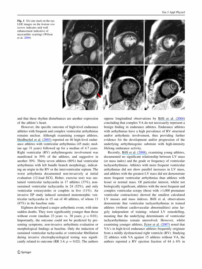
previous myocarditis was observed (Fig. 1).
Acute myocarditis is typically a viral or post-viral pro-
cess, which may result in the acute onset of LV systolic
dysfunction. It can range from mild and clinically unde-
tectable to fulfilment and fatal over a short time course.
Clinically, patients with acute viral myocarditis present
with tachycardia, hypotension and shortness of breath. The
clinical course of myocarditis is highly variable with
complete or near complete resolution occurring in a few
weeks. The majority will experience some degree of
recovery of function but are often left with a degree of left
ventricular dysfunction. Myocarditis should be suspected
in athletes with unexplained cardiac arrhythmias and dys-
function, especially if preceded by a flu-like syndrome. An
early diagnosis is desirable in order to avoid the risk of
fatal consequences, since physical activity can enhance the
inflammatory process (Chimenti et al. 2006). In patients
with acute or chronic myocarditis, arrhythmia may be the
only clinical symptom in the natural course of the disease.
The potentially malignant tachyarrhythmias and bradyar-
rhythmias caused by myocarditis are of particular concern.
Acutely, inflammatory processes in the cardiac myocytes
and interstitium can lead directly to fluctuations in mem-
brane potential, hence arrythmogenesis (Babu-Narayan
et al. 2007).
Sustained lifelong endurance activity, provides ample
opportunity for viral or post-viral infection to occur.
Treatment is often difficult for highly competitive athletes
to comprehend, as initial treatment for athletes with myo-
carditis should be complete absence from all physical
activity for at least 6 months. Athletes should only resume
training when ventricular function and cardiac dimensions
return to normal and the clinically relevant arrhythmias
disappear. Adherence to such guidelines should be strongly
advocated to reduce the potential of life-threatening
arrhythmias or rapidly progressive cardiac dysfunction and
the introduction of antiviral or an immunosuppressive
treatment (Chimenti et al. 2006).
In summary, veteran athletes are at increased risk of
developing supraventricular arrhythmias. Likely reasons
include changes in autonomic drive including increased
parasympathetic modulation at rest and increased sympa-
thetic modulation during exercise, increased atrial size and
increased inflammation (Sorokin et al. 2009).
Ventricular arrhythmias
Recent data has documented an increase in complex ven-
tricular arrhythmias (VA), including couplets and bursts of
non-sustained ventricular tachycardia, predominantly
occurring within veteran athletes (Jensen-Urstad et al.
1998; Biffi et al. 2002; Biffi et al. 2004; Ector et al. 2007;
Baldesberger et al. 2008; Mont et al. 2009). Debate con-
tinues to the clinical nature of VA and the specific outcome
of high-level endurance athletes with frequent and complex
ventricular arrhythmias (Heidbuchel et al. 2003). Indeed,
VA’s are sensitive to deconditioning in athletes with and
without structural heart disease. Biffi et al. (2004) reported
an 80% decrease in frequency and complexity of ventri-
cular arrhythmias (from 10,611 ± 10,078 to 2,165 ±
4,877), as well as a 90% decrease in the occurrence of non-
sustained ventricular tachycardia’s with deconditioning.
Using longitudinal data, the authors conclude that frequent
and complex ventricular tachyarrhythmias are not ominous
in trained athletes without cardiovascular abnormalities,
Eur J Appl Physiol
123
and that these rhythm disturbances are another expression
of the athlete’s heart.
However, the specific outcome of high-level endurance
athletes with frequent and complex ventricular arrhythmias
remains unclear. Although examining younger athletes,
Heidbuchel et al. (2003) reported on 46 high-level endur-
ance athletes with ventricular arrhythmias (45 male; med-
ian age 31 years) followed up for a median of 4.7 years.
Right ventricular (RV) arrhythmogenic involvement was
manifested in 59% of the athletes, and suggestive in
another 30%. Thirty-seven athletes (80%) had ventricular
arrhythmias with left bundle branch morphology, indicat-
ing an origin in the RV or the interventricular septum. The
worst arrhythmia documented non-invasively at initial
evaluation (12-lead ECG, Holter, exercise test) was sus-
tained ventricular tachycardia in 17 athletes (37%), non-
sustained ventricular tachycardia in 24 (52%), and only
ventricular extrasystoles or couplets in five (11%). An
invasive EP study induced sustained monomorphic ven-
tricular tachycardia in 15 out of 40 athletes, of whom 13
(87%) in the baseline state.
Eighteen developed a major arrhythmic event, with nine
sudden deaths. They were significantly younger than those
without event (median 23 years vs. 38 years; p = 0.01).
Importantly, the outcome could not be predicted by pre-
senting symptoms, non-invasive arrhythmia evaluation or
morphological findings at baseline. Only the induction of
sustained ventricular tachycardia or ventricular fibrillation
during invasive electrophysiological testing was signifi-
cantly related to outcome (RR 3.4; p = 0.02). The authors
oppose longitudinal observations by Biffi et al. (2004)
concluding that complex VA do not necessarily represent a
benign finding in endurance athletes. Endurance athletes
with arrhythmias have a high prevalence of RV structural
and/or arrhythmic involvement, thus providing further
evidence for the development and/or progression of the
underlying arrhythmogenic substrate with high-intensity
lifelong endurance activity.
Recently, Biffi et al. (2008), examining young athletes,
documented no significant relationship between LV mass
(or mass index) and the grade or frequency of ventricular
tachyarrhythmias. Athletes with more frequent ventricular
arrhythmias did not show parallel increases in LV mass,
and athletes with the greatest LV mass did not demonstrate
more frequent ventricular arrhythmias than athletes with
lesser or normal mass. Of particular interest, whilst not
biologically significant, athletes with the most frequent and
complex ventricular ectopy (those with [1,000 premature
ventricular contractions) showed the smallest calculated
LV masses and mass indexes. Biffi et al. observations
demonstrate that ventricular tachyarrhythmias in trained
athletes (without cardiovascular abnormalities) arise lar-
gely independent of training- related LV remodelling,
meaning that the underlying determinants of ventricular
tachyarrhythmias remain unresolved. However, whilst
examining younger athletes, Ector et al. (2007) noted that
VA’s in high-level endurance athletes frequently originate
from a mildly dysfunctional right ventricle (RV). Studying
22 athletes with VA against 15 athletes without VA, the
authors reported a RV ejection fraction of 64 ± 6% in
Fig. 1 SA cine stack on the top.
LGE images on the bottom row(arrow indicates mid wall
enhancement indicative of
myocarditic scarring) (Wilson
et al. 2009)
Eur J Appl Physiol
123
athletes without VA and a reduced RV ejection fraction
(49 ± 10%) in athletes with VA. Since both groups of
athletes were engaged in the same intensity and frequency
of exercise, this finding cannot be attributed to the presence
of athlete’s heart per se. The authors suggest that endur-
ance exercise and volume overload subject the thin-walled
RV to a greater increase in workload than the thick-walled
LV with subsequent different remodelling. Indeed, animal
models have demonstrated that chronic volume overload
causes a greater mass increase in the RV than in the LV,
resulting in an increased collagen deposition and selective
growth factor activation restricted to the RV (Modesti et al.
2004).
In summary, the relationship between ventricular
arrhythmias and sudden cardiac death in athletes without
evident heart disease is controversial. Furthermore, the
origins and clinical significance of complex ventricular
arrhythmias within veteran athletes remain to be fully
elucidated and requires a robust methodological and lon-
gitudinal examination of a large number of lifelong veteran
athletes, especially those currently experiencing
arrhythmia.
Vascular structure and function in veteran endurance
athletes
The ageing process leads to various changes in the artery
wall which impact upon function and structure. For
example, a progressive decrease occurs in the compliance
of large elastic arteries and peripheral arterial compliance
(Avolio et al. 1983; Tanaka et al. 2000; Miyachi et al.
2003; Moreau et al. 2003). In women, this effect may be
predominant in central rather than peripheral arteries
(Tanaka et al. 1998). This suggests that sex and site-
specific changes in arterial compliance occur with ageing
in the large, elastic vessels. In larger conduit arteries,
ageing is associated with impaired endothelial function
(Celermajer et al. 1994; Eskurza et al. 2004; Eskurza et al.
2005; Eskurza et al. 2006; Black et al. 2009), which begins
to decline at the age of 40 in men. A steeper decline occurs
in women around the time of the menopause (Celermajer
et al. 1994). Resistance artery endothelial function is also
impaired in healthy older humans (Taddei et al. 1995;
DeSouza et al. 2000a, b). In parallel to conduit and resis-
tance vessels, physiological ageing is associated with
impairment in the skin microvascular function (Minson
et al. 2002; Holowatz et al. 2007). Indeed, microvascular
responses to both physiological (local heating) and phar-
macological stimuli are impaired with age (Black et al.
2008).
Impact of ageing on vascular structure
Ageing leads to a gradual thickening of the artery wall,
predominantly demonstrated by the age-dependent increa-
ses in carotid and femoral artery wall thickness (Dinenno
et al. 2000; Moreau et al. 2002; Galetta et al. 2006). Most
studies have observed no difference in baseline arterial
diameter between young and middle-aged or older indi-
viduals (i.e. 50-60 years) (Parker et al. 2006; Nishiyama
et al. 2008; Thijssen et al. 2008). However, the oldest
cohort of men ([70 years), but not women, show an
increase in baseline diameter (Parker et al. 2008; Gonzales
et al. 2009). Nonetheless, the wall (thickness)-to-lumen
ratio demonstrates an age-dependent increase in men as
well as women. It has been suggested that the increase in
wall thickness with ageing is related to elevation in sym-
pathetic outflow in the older individuals (Dinenno et al.
2000). Peak reactive hyperemic blood flow, an index of
remodelling in resistance vessels in humans, is typically
smaller in older individuals and independent of gender
(Takeshita and Mark 1980; Sinoway et al. 1986; Silber and
Sinoway 1990; Silber et al. 1991; Sarabi et al. 1999). This
suggests an age-dependent decrease in collective cross-
sectional area of peripheral resistance artery vascular bed
and may infer the existence of some concentric arterial
remodelling at this level of the vasculature.
Impact of ageing on the vasculature: mechanisms
Evidence supports an important role for the NO-pathway in
age-related impairment in endothelial function (Taddei
et al. 1995). Animal studies have observed lower eNOS
gene and protein expression and activation in older rats
(Spier et al. 2004) (Durrant et al. 2009). Elevated levels of
oxidative stress may contribute to age-related endothelial
dysfunction (Taddei et al. 2000; Eskurza et al. 2004). This
is supported by the finding that vitamin C supplementation,
a strong anti-oxidant, can restore the impaired NO-medi-
ated endothelial function in sedentary older men (Galetta
et al. 2006). Another mechanism linked with the NO-
pathway relates to tetrahydrobiopterin (BH4), an essential
co-factor for eNOS-regulated production of NO, which
decreases with age in animals (Delp et al. 2008), while
BH4-supplementation improves endothelial function in
sedentary older men (Eskurza et al. 2005). Recent studies
also found elevated endothelin(ET)-mediated vascular
tone in older men, predominantly through ETA-receptors
(Thijssen et al. 2007a, b; Van Guilder et al. 2007). These
findings suggest that vasodilator (NO) as well as constrictor
(ET) pathways may contribute to the age-related endothe-
lial dysfunction.
Eur J Appl Physiol
123
Impact of exercise training on vascular function
Exercise training reduces cardiovascular events. However,
the effects of exercise on traditional risk factors do not
fully account for the magnitude of risk reduction (Mora
et al. 2007). The direct effects of exercise on the vascu-
lature provide a plausible explanation for the reduction in
cardiac events associated with exercise training, inde-
pendent of change in traditional risk factors (Green et al.
2008). Regular aerobic exercise is associated with
enhanced arterial compliance in ageing larger arteries,
whilst short-term exercise training in previously sedentary
older men can also improve arterial compliance (Tanaka
et al. 2000). Older endurance-trained men demonstrate
enhanced arterial compliance compared to their sedentary
peers, but compliance is nonetheless lower than that in
young men (Monahan et al. 2001). In contrast, postmen-
opausal athletic women demonstrate a similar carotid
artery compliance compared with their younger peers
(Tanaka et al. 1998). These cross-sectional data suggest a
sex-specific effect of exercise training on the age-related
decrease in arterial compliance. However, exercise train-
ing studies in sedentary older men and women indicate
that exercise training in previously sedentary subjects
improves vascular compliance (Moreau et al. 2003;
Thijssen et al. 2007a, b).
Regular endurance exercise in middle-aged and veteran
athletes prevents the attenuation of the age-related endo-
thelial dysfunction in conduit and resistance arteries
(Rywik et al. 1999; DeSouza et al. 2000a, b; Rinder et al.
2000; Taddei et al. 2000; Hagmar et al. 2006; Black et al.
2009). The beneficial impact of exercise training is rein-
forced by various studies which have observed improved
endothelial function in previously sedentary older humans
after exercise training. Black et al. (2009) recently iden-
tified a gender-specific effect of exercise training on
conduit artery endothelial function. Improvement in bra-
chial artery endothelial function was observed after
exercise training in women, but not men (who had a
smaller age-related decline in endothelial function). This
suggests that exercise training is an effective method to
improve conduit artery endothelial function, especially in
those with a larger age-related attenuation in vascular
function.
Microvascular functional impairment in older humans
can be prevented by maintaining a high level of physical
fitness, whilst exercise training in previously sedentary
older men partly reverses the age-related impairment in
microvascular function (Black et al. 2008). This indicates
that maintaining fitness and taking up exercise prevents the
age-related decline in microvascular NO-mediated vaso-
dilator function.
Impact of exercise training on vascular structure
Exercise training represents a potent stimulus to structural
remodelling in conduit arteries. Endurance-trained older
male and female athletes demonstrate a smaller wall
thickness in the carotid and femoral arteries compared with
their inactive peers (Moreau et al. 2002; Galetta et al.
2006), while a larger femoral diameter is found in older
athletes (Dinenno et al. 2001). Also, 3–6 months exercise
training in older men induces remodelling of conduit
arteries, leading to decreased wall thickness and outward
remodelling of conduit arteries, with consequent decrease
in the wall-to-lumen ratio (Green et al. 2010).
The effects of exercise training on wall thickness may
be time-specific, since 8 weeks of training is insufficient
to induce significant changes in conduit artery wall
thickness (Thijssen et al. 2007a, b). Exercise-induced
effects on intima-media thickness may also be site-spe-
cific. It has been suggested that endurance exercise
training has a larger effect on wall thickness in ‘muscular’
arteries (e.g. femoral) than in more ‘elastic’ arteries (e.g.
carotid) (Moreau et al. 2002), possibly due to the larger
number of smooth muscle cells and plasticity of muscular
arteries. Indeed, exercise-induced improvements in wall
thickness have also been reported in the brachial and
popliteal artery, supporting the idea that exercise has a
systemic effect on muscular artery wall thickness in older
humans.
In resistance arteries, exercise partly prevents the age-
related decline in the peak blood flow response; i.e. the
surrogate marker of resistance artery structural remodel-
ling. Several studies have demonstrated that a period of
exercise training, varying from 8 weeks up to 6 months, in
previously sedentary older men increases the peak blood
flow (Martin et al. 1987; Martin et al. 1990; Thijssen et al.
2007a, b). Evidence from healthy individuals indicates that
the improvement in peak blood flow may be a systemic
response in resistance arteries, rather than a localised
response limited to the exercised limbs only (Silber et al.
1991). Accordingly, these changes in resistance arteries
may contribute to a decrease in arterial blood pressure and
cardiac afterload, typically observed after training.
Regarding the mechanisms of vascular adaptation to
exercise training, a number of animal studies have dem-
onstrated a key role for shear rate in inducing endothelium-
dependent changes in vascular size and wall thickness
(Langille and O’Donnell 1986; Tuttle et al. 2001; Brown
2003). Indeed, a recent series of experiments in humans
examined the role for shear rate in exercise-induced
changes in both vascular function and remodelling (Tinken
et al. 2009; Tinken et al. 2010). Eight weeks handgrip
exercise induced a time-dependent change in vascular
Eur J Appl Physiol
123
function and structure. However, when shear rate was
experimentally normalised during exercise, no vascular
adaptations were observed. These experiments support
previous experiments which indicate that shear rate plays a
key role in mediating vascular adaptations to exercise
training in humans (Hambrecht et al. 2003).
In summary, the data above indicate that ageing impairs
vascular function and distensibility and is associated with
detrimental and pro-atherogenic changes in arterial wall
thickness. Such effects are evident at all major functional
levels of the arterial tree. Recent evidence indicates that
exercise training and physical fitness can retard or even
reverse these effects. Exercise training, in this context,
represents a direct and particularly efficacious form of
vascular medicine.
Cerebral vascular blood flow and syncope
As previously stated, global CBF undergoes a gradual
continuous decline of 25–30% between 20 and 80 years of
age, or 5% per decade (Fazekas et al. 1952; Kety 1956;
Buijs et al. 1998; Krejza et al. 1999; Scheel et al. 2000;
Stoquart-Elsankari et al. 2007; Demirkaya et al. 2008a, b).
The reason for this age-related decline in CBF is not well
understood; however a reduced cerebral metabolism both
at a cellular and global level due to cerebral atrophy, as
well as vascular alterations (e.g. atherosclerosis) have been
postulated to cause this decline in CBF with increasing age.
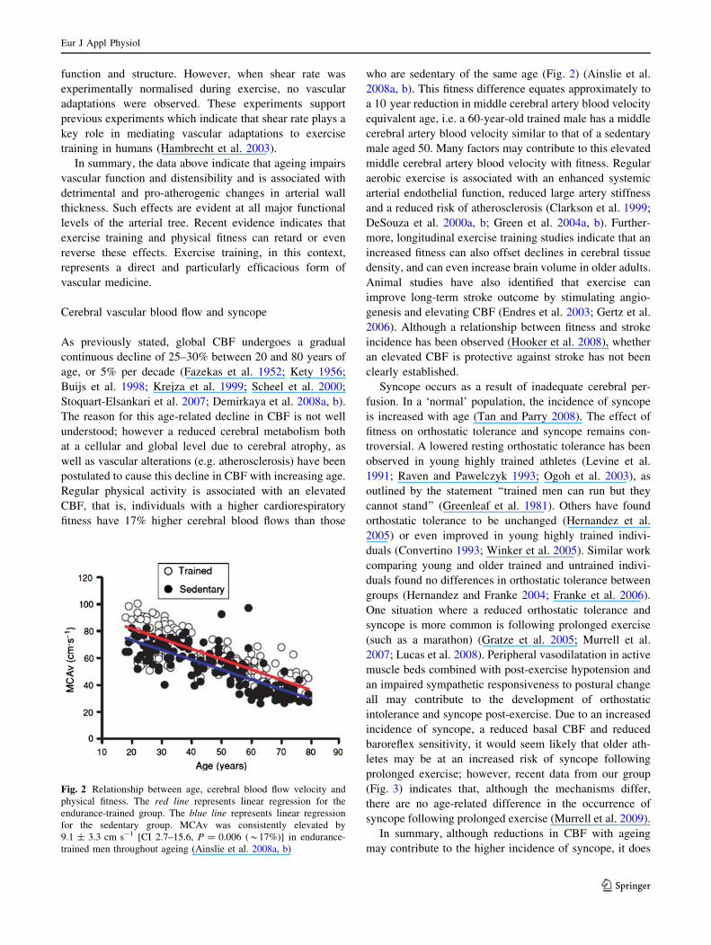
Regular physical activity is associated with an elevated
CBF, that is, individuals with a higher cardiorespiratory
fitness have 17% higher cerebral blood flows than those
who are sedentary of the same age (Fig. 2) (Ainslie et al.
2008a, b). This fitness difference equates approximately to
a 10 year reduction in middle cerebral artery blood velocity
equivalent age, i.e. a 60-year-old trained male has a middle
cerebral artery blood velocity similar to that of a sedentary
male aged 50. Many factors may contribute to this elevated
middle cerebral artery blood velocity with fitness. Regular
aerobic exercise is associated with an enhanced systemic
arterial endothelial function, reduced large artery stiffness
and a reduced risk of atherosclerosis (Clarkson et al. 1999;
DeSouza et al. 2000a, b; Green et al. 2004a, b). Further-
more, longitudinal exercise training studies indicate that an
increased fitness can also offset declines in cerebral tissue
density, and can even increase brain volume in older adults.
Animal studies have also identified that exercise can
improve long-term stroke outcome by stimulating angio-
genesis and elevating CBF (Endres et al. 2003; Gertz et al.
2006). Although a relationship between fitness and stroke
incidence has been observed (Hooker et al. 2008), whether
an elevated CBF is protective against stroke has not been
clearly established.
Syncope occurs as a result of inadequate cerebral per-
fusion. In a ‘normal’ population, the incidence of syncope
is increased with age (Tan and Parry 2008). The effect of
fitness on orthostatic tolerance and syncope remains con-
troversial. A lowered resting orthostatic tolerance has been
observed in young highly trained athletes (Levine et al.
1991; Raven and Pawelczyk 1993; Ogoh et al. 2003), as
outlined by the statement ‘‘trained men can run but they
cannot stand’’ (Greenleaf et al. 1981). Others have found
orthostatic tolerance to be unchanged (Hernandez et al.
2005) or even improved in young highly trained indivi-
duals (Convertino 1993; Winker et al. 2005). Similar work
comparing young and older trained and untrained indivi-
duals found no differences in orthostatic tolerance between
groups (Hernandez and Franke 2004; Franke et al. 2006).
One situation where a reduced orthostatic tolerance and
syncope is more common is following prolonged exercise
(such as a marathon) (Gratze et al. 2005; Murrell et al.
2007; Lucas et al. 2008). Peripheral vasodilatation in active
muscle beds combined with post-exercise hypotension and
an impaired sympathetic responsiveness to postural change
all may contribute to the development of orthostatic
intolerance and syncope post-exercise. Due to an increased
incidence of syncope, a reduced basal CBF and reduced
baroreflex sensitivity, it would seem likely that older ath-
letes may be at an increased risk of syncope following
prolonged exercise; however, recent data from our group
(Fig. 3) indicates that, although the mechanisms differ,
there are no age-related difference in the occurrence of
syncope following prolonged exercise (Murrell et al. 2009).
In summary, although reductions in CBF with ageing
may contribute to the higher incidence of syncope, it does
Fig. 2 Relationship between age, cerebral blood flow velocity and
physical fitness. The red line represents linear regression for the
endurance-trained group. The blue line represents linear regression
for the sedentary group. MCAv was consistently elevated by
9.1 ± 3.3 cm s-1 [CI 2.7–15.6, P = 0.006 (*17%)] in endurance-
trained men throughout ageing (Ainslie et al. 2008a, b)
Eur J Appl Physiol
123
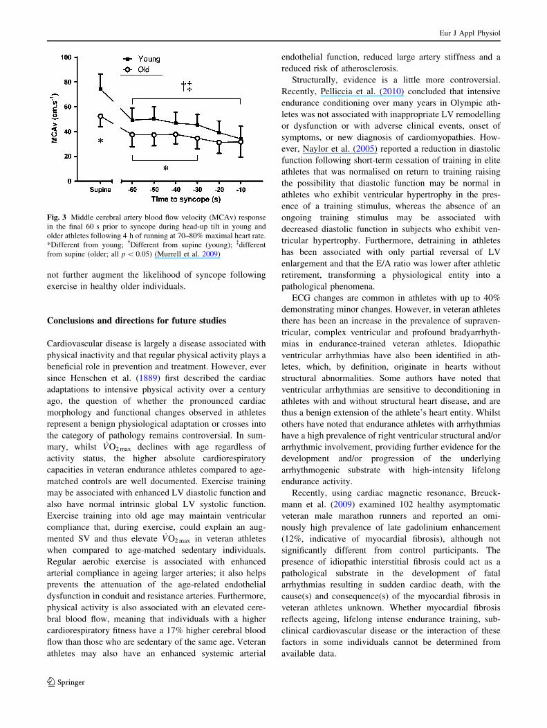
not further augment the likelihood of syncope following
exercise in healthy older individuals.
Conclusions and directions for future studies
Cardiovascular disease is largely a disease associated with
physical inactivity and that regular physical activity plays a
beneficial role in prevention and treatment. However, ever
since Henschen et al. (1889) first described the cardiac
adaptations to intensive physical activity over a century
ago, the question of whether the pronounced cardiac
morphology and functional changes observed in athletes
represent a benign physiological adaptation or crosses into
the category of pathology remains controversial. In sum-
mary, whilst _VO2 max declines with age regardless of
activity status, the higher absolute cardiorespiratory
capacities in veteran endurance athletes compared to age-
matched controls are well documented. Exercise training
may be associated with enhanced LV diastolic function and
also have normal intrinsic global LV systolic function.
Exercise training into old age may maintain ventricular
compliance that, during exercise, could explain an aug-
mented SV and thus elevate _VO2 max in veteran athletes
when compared to age-matched sedentary individuals.
Regular aerobic exercise is associated with enhanced
arterial compliance in ageing larger arteries; it also helps
prevents the attenuation of the age-related endothelial
dysfunction in conduit and resistance arteries. Furthermore,
physical activity is also associated with an elevated cere-
bral blood flow, meaning that individuals with a higher
cardiorespiratory fitness have a 17% higher cerebral blood
flow than those who are sedentary of the same age. Veteran
athletes may also have an enhanced systemic arterial
endothelial function, reduced large artery stiffness and a
reduced risk of atherosclerosis.
Structurally, evidence is a little more controversial.
Recently, Pelliccia et al. (2010) concluded that intensive
endurance conditioning over many years in Olympic ath-
letes was not associated with inappropriate LV remodelling
or dysfunction or with adverse clinical events, onset of
symptoms, or new diagnosis of cardiomyopathies. How-
ever, Naylor et al. (2005) reported a reduction in diastolic
function following short-term cessation of training in elite
athletes that was normalised on return to training raising
the possibility that diastolic function may be normal in
athletes who exhibit ventricular hypertrophy in the pres-
ence of a training stimulus, whereas the absence of an
ongoing training stimulus may be associated with
decreased diastolic function in subjects who exhibit ven-
tricular hypertrophy. Furthermore, detraining in athletes
has been associated with only partial reversal of LV
enlargement and that the E/A ratio was lower after athletic
retirement, transforming a physiological entity into a
pathological phenomena.
ECG changes are common in athletes with up to 40%
demonstrating minor changes. However, in veteran athletes
there has been an increase in the prevalence of supraven-
tricular, complex ventricular and profound bradyarrhyth-
mias in endurance-trained veteran athletes. Idiopathic
ventricular arrhythmias have also been identified in ath-
letes, which, by definition, originate in hearts without
structural abnormalities. Some authors have noted that
ventricular arrhythmias are sensitive to deconditioning in
athletes with and without structural heart disease, and are
thus a benign extension of the athlete’s heart entity. Whilst
others have noted that endurance athletes with arrhythmias
have a high prevalence of right ventricular structural and/or
arrhythmic involvement, providing further evidence for the
development and/or progression of the underlying
arrhythmogenic substrate with high-intensity lifelong
endurance activity.
Recently, using cardiac magnetic resonance, Breuck-
mann et al. (2009) examined 102 healthy asymptomatic
veteran male marathon runners and reported an omi-
nously high prevalence of late gadolinium enhancement
(12%, indicative of myocardial fibrosis), although not
significantly different from control participants. The
presence of idiopathic interstitial fibrosis could act as a
pathological substrate in the development of fatal
arrhythmias resulting in sudden cardiac death, with the
cause(s) and consequence(s) of the myocardial fibrosis in
veteran athletes unknown. Whether myocardial fibrosis
reflects ageing, lifelong intense endurance training, sub-
clinical cardiovascular disease or the interaction of these
factors in some individuals cannot be determined from
available data.
Fig. 3 Middle cerebral artery blood flow velocity (MCAv) response
in the final 60 s prior to syncope during head-up tilt in young and
older athletes following 4 h of running at 70–80% maximal heart rate.
*Different from young; �Different from supine (young); �different
from supine (older; all p \ 0.05) (Murrell et al. 2009)
Eur J Appl Physiol
123
In conclusion, the veteran athlete may not be as healthy
as believed with many established areas lacking conclusive
evidence to support the benefits of a lifelong career in high
intensive endurance exercise. Future studies employing
large cohorts of veteran athletes, employing modern tech-
niques such are cardiac magnetic resonance, are warranted
to enhance our understanding of the impact of long-term
endurance exercise on cardiac, peripheral vascular and
cerebrovascular structure and function. Importantly, the
systematic and longitudinal follow-up of veteran athletes
(both male and female) who are currently experiencing
arrhythmia is paramount. These studies will add to the
relatively small body of knowledge providing important
information regarding the differentiation of physiologic
and pathologic cardiovascular remodelling and in the
identification and management of cardiovascular pathology
in veteran endurance athletes.
Acknowledgments DG received funding support from the National
Heart Foundation of Australia. GW has received funding from Car-
diac Risk in the Young (CRY, UK) and KG and GW have received
technical and equipment support from General Electric (UK, South
Africa, Australia and New Zealand). PA and CM received funding
from Sport and Recreation New Zealand (SPARC).
References
Ainslie PN, Cotter JD, George KP et al (2008a) Elevation in cerebral
blood flow velocity with aerobic fitness throughout healthy
human ageing. J Physiol 586(16):4005–4010
Ainslie PN, Cotter JD, George KP et al (2008b) Elevation in cerebral
blood flow velocity with aerobic fitness throughout healthy
human ageing. J Physiol 586(16):4005–4010
Andersson LG, Henriksen E, Damm S et al (2001) Thallium-201
myocardial imaging at rest in male orienteers and other
endurance athletes. Ups J Med Sci 106(1):59–66
Anselme F (2003) Association of idiopathic RVOT VT and AVNRT:
anything else than chance? Europace 5(3):221–223
Arbab-Zadeh A, Dijk E, Prasad A et al (2004) Effect of aging and
physical activity on left ventricular compliance. Circulation
110:1799–1805
Aronow WS (2001) Left ventricular diastolic heart failure with
normal left ventricular systolic function in older persons. J Lab
Clin Med 137(5):316–323
Avolio AP, Chen SG, Wang RP et al (1983) Effects of aging on
changing arterial compliance and left ventricular load in a
northern Chinese urban community. Circulation 68(1):50–58
Babu-Narayan SV, McCarthy KP, Ho SY et al (2007) Images in
cardiovascular medicine. Myocarditis and sudden cardiac death
in the young: extensive fibrosis suggested by cardiovascular
magnetic resonance in vivo and confirmed post mortem.
Circulation 116(6):e122–e125
Baldesberger S, Bauersfeld U, Candinas R et al (2008) Sinus node
disease and arrhythmias in the long-term follow-up of former
professional cyclists. Eur Heart J 29(1):71–78
Basso C, Carturan E, Corrado D et al (2007) Myocarditis and dilated
cardiomyopathy in athletes: diagnosis, management, and recom-
mendations for sport activity. Cardiol Clin 25(3):423–429 vi
Benjamin EJ, Levy D, Anderson KM et al (1992) Determinants of
Doppler indexes of left ventricular diastolic function in normal
subjects (the Framingham Heart Study). Am J Cardiol
70(4):508–515
Bhella PS, Levine BD (2010) The heart of a champion. J Am Coll
Cardiol 5(15):1626–1628
Biffi A, Pelliccia A, Verdile L et al (2002) Long-term clinical
significance of frequent and complex ventricular tachyarrhyth-
mias in trained athletes. J Am Coll Cardiol 40(3):446–452
Biffi A, Maron BJ, Verdile L et al (2004) Impact of physical
deconditioning on ventricular tachyarrhythmias in trained ath-
letes. J Am Coll Cardiol 44(5):1053–1058
Biffi A, Maron BJ, Di Giacinto B et al (2008) Relation between
training-induced left ventricular hypertrophy and risk for ven-
tricular tachyarrhythmias in elite athletes. Am J Cardiol
101(12):1792–1795
Bjornstad H, Storstein L, Meen HD et al (1991) Electrocardiographic
findings in athletic students and sedentary controls. Cardiology
79(4):290–305
Bjornstad H, Storstein L, Meen HD et al (1993) Electrocardiographic
findings of left, right and septal hypertrophy in athletic students
and sedentary controls. Cardiology 82(1):56–65
Black MA, Green DJ, Cable NT (2008) Exercise prevents age-related
decline in nitric-oxide-mediated vasodilator function in cutane-
ous microvessels. J Physiol 586(14):3511–3524
Black MA, Cable NT, Thijssen DH et al (2009) Impact of age, sex
and exercise on brachial artery flow-mediated dilatation. Am J
Physiol Heart Circ Physiol 297(3):H1109–H1116
Bouvier F, Saltin B, Nejat M et al (2001) Left ventricular function and
perfusion in elderly endurance athletes. Med Sci Sports Exerc
33(5):735–740
Breuckmann F, Mohlenkamp S, Nassenstein K et al (2009) Myocar-
dial late gadolinium enhancement: prevalence, pattern, and
prognostic relevance in marathon runners. Radiology 251(1):50–
57
Brooks A, Schinde V, Bateman AC et al (2003) Interstitial fibrosis in
the dilated non-ischaemic myocardium. Heart 89(10):1255–1256
Brown MD (2003) Exercise and coronary vascular remodelling in the
healthy heart. Exp Biol Med 88:645–658
Brown SJ, Brown JA (2007) Resting and postexercise cardiac
autonomic control in trained master athletes. J Physiol Sci
57(1):23–29
Buijs PC, Krabbe-Hartkamp MJ, Bakker CJ et al (1998) Effect of age
on cerebral blood flow: measurement with ungated two-dimen-
sional phase-contrast MR angiography in 250 adults. Radiology
209(3):667–674
Burkauskiene A, Mackiewicz Z, Virtanen I et al (2006) Age-related
changes in myocardial nerve and collagen networks of the
auricle of the right atrium. Acta Cardiol 61(5):513–518
Capasso JM, Malhotra A, Remily RM et al (1983) Effects of age on
mechanical and electrical performance of rat myocardium. Am J
Physiol 245(1):H72–H81
Celermajer DS, Sorensen KE, Spiegelhalter DJ et al (1994) Aging is
associated with endothelial dysfunction in healthy men years
before the age-related decline in women. J Am Coll Cardiol
24(2):471–476
Chen LS, Zhou S, Fishbein MC et al (2007) New perspectives on the
role of autonomic nervous system in the genesis of arrhythmias.
J Cardiovasc Electrophysiol 18(1):123–127
Child JS, Barnard RJ, Taw RL (1984) Cardiac hypertrophy and
function in master endurance runners and sprinters. J Appl
Physiol 57(1):176–181
Chimenti C, Pieroni M, Frustaci A (2006) Myocarditis: when to
suspect and how to diagnose it in athletes. J Cardiovasc Med
(Hagerstown) 7(4):301–306
Clarkson P, Montgomery HE, Mullen MJ et al (1999) Exercise
training enhances endothelial function in young men. J Am Coll
Cardiol 33(5):1379–1385
Eur J Appl Physiol
123
Convertino VA (1993) Endurance exercise training: conditions of
enhanced hemodynamic responses and tolerance to LBNP. Med
Sci Sports Exerc 25(6):705–712
Corrado D, Basso C, Schiavon M et al (2006) Does sports activity
enhance the risk of sudden cardiac death? J Cardiovasc Med
(Hagerstown) 7(4):228–233
D’Andrea A, Caso P, Scarafile R et al (2007) Biventricular
myocardial adaptation to different training protocols in compet-
itive master athletes. Int J Cardiol 115(3):342–349
Delp MD, Behnke BJ, Spier SA et al (2008) Ageing diminishes
endothelium-dependent vasodilatation and tetrahydrobiopterin
content in rat skeletal muscle arterioles. J Physiol 586(4):1161–
1168
Demirkaya S, Uluc K, Bek S et al (2008a) Normal blood flow
velocities of basal cerebral arteries decrease with advancing age:
a transcranial Doppler sonography study. Tohoku J Exp Med
214(2):145–149
Demirkaya S, Uluc K, Bek S et al (2008b) Normal blood flow
velocities of basal cerebral arteries decrease with advancing age:
a transcranial Doppler sonography study. Tohoku J Exp Med
214(2):145–149
DeSouza CA, Shapiro LF, Clevenger CM et al (2000a) Regular
aerobic exercise prevents and restores age-related declines in
endothelium-dependent vasodilation in healthy men. Circulation
102(12):1351–1357
DeSouza CA, Shapiro LF, Clevenger CM et al (2000b) Regular
aerobic exercise prevents and restores age-related declines in
endothelium-dependent vasodilation in healthy men. Circulation
102(12):1351–1357
Dinenno FA, Jones PP, Seals DR et al (2000) Age-associated arterial
wall thickening is related to elevations in sympathetic activity in
healthy humans. Am J Physiol Heart Circ Physiol
278(4):H1205–H1210
Dinenno FA, Tanaka H, Monahan KD et al (2001) Regular endurance
exercise induces expansive arterial remodelling in the trained
limbs of healthy men. J Physiol 534(Pt 1):287–295
Durakovic Z, Misigoj-Durakovic M, Vuori I et al (2005) Sudden
cardiac death due to physical exercise in male competitive
athletes. A report of six cases. J Sports Med Phys Fitness
45(4):532–536
Durakovic Z, Misigoj Durakovic M, Skavic J et al (2008) Myoper-
icarditis and sudden cardiac death due to physical exercise in
male athletes. Coll Antropol 32(2):399–401
Durrant JR, Seals DR, Connell ML et al (2009) Voluntary wheel
running restores endothelial function in conduit arteries of old
mice: direct evidence for reduced oxidative stress, increased
superoxide dismutase activity and down-regulation of NADPH
oxidase. J Physiol 587(Pt 13):3271–3285
Ector J, Ganame J, van der Merwe N et al (2007) Reduced right
ventricular ejection fraction in endurance athletes presenting
with ventricular arrhythmias: a quantitative angiographic assess-
ment. Eur Heart J 28(3):345–353
Ehsani AA, Ogawa T, Miller TR et al (1991) Exercise training
improves left ventricular systolic function in older men.
Circulation 83(1):96–103
Elosua R, Arquer A, Mont L et al (2006) Sport practice and the risk of
lone atrial fibrillation: a case–control study. Int J Cardiol
108(3):332–337
Endres M, Gertz K, Lindauer U et al (2003) Mechanisms of stroke
protection by physical activity. Ann Neurol 54(5):582–590
Eskurza I, Monahan KD, Robinson JA et al (2004) Effect of acute and
chronic ascorbic acid on flow-mediated dilatation with sedentary
and physically active human ageing. J Physiol 556(Pt 1):315–324
Eskurza I, Myerburgh LA, Kahn ZD et al (2005) Tetrahydrobiopterin
augments endothelium-dependent dilatation in sedentary but not
in habitually exercising older adults. J Physiol 568(Pt 3):1057–
1065
Eskurza I, Kahn ZD, Seals DR (2006) Xanthine oxidase does not
contribute to impaired peripheral conduit artery endothelium-
dependent dilatation with ageing. J Physiol 571(Pt 3):661–668
Fazekas JF, Alman RW, Bessman AN (1952) Cerebral physiology of
the aged. Am J Med Sci 223(3):245–257
Fleg JL, O’Connor F, Gerstenblith G et al (1995) Impact of age on the
cardiovascular response to dynamic upright exercise in healthy
men and women. J Appl Physiol 78(3):890–900
Forman DE, Manning WJ, Hauser R et al (1992) Enhanced left
ventricular diastolic filling associated with long-term endurance
training. J Gerontol 47(2):M56–M58
Franke WD, Allbee KA, Spencer SE (2006) Cerebral blood flow
responses to severe orthostatic stress in fit and unfit young and
older adults. Gerontology 52(5):282–289
Frustaci A, Chimenti C, Bellocci F et al (1997) Histological substrate
of atrial biopsies in patients with lone atrial fibrillation.
Circulation 96(4):1180–1184
Galderisi M, Benjamin E, Evans J et al (1993) Impact of heart rate
and PP interval on Doppler indexed of left ventricular diastolic
filling in an elderly cohort (the Framingham heart study). Am J
Cardiol 72:1183–1187
Galetta F, Franzoni F, Virdis A et al (2006) Endothelium-dependent
vasodilation and carotid artery wall remodeling in athletes and
sedentary subjects. Atherosclerosis 186(1):184–192
George K, Oxborough D, Forster J et al (2005) Mitral annular
myocardial velocity assessment of segmental left ventricular
diastolic function after prolonged exercise in humans. J Physiol
569(Pt 1):305–313
Gertz K, Priller J, Kronenberg G et al (2006) Physical activity
improves long-term stroke outcome via endothelial nitric oxide
synthase-dependent augmentation of neovascularization and
cerebral blood flow. Circ Res 99(10):1132–1140
Goldspink DF (2005) Ageing and activity: their effects on the
functional reserve capacities of the heart and vascular smooth
and skeletal muscles. Ergonomics 48(11–14):1334–1351
Gonzales JU, Parker BA, Ridout SJ et al (2009) Femoral shear rate
response to knee extensor exercise: an age and sex comparison.
Biorheology 46(2):145–154
Gratze G, Rudnicki R, Urban W et al (2005) Hemodynamic and
autonomic changes induced by Ironman: prediction of compe-
tition time by blood pressure variability. J Appl Physiol
99(5):1728–1735
Green DJ, Maiorana A, O’Driscoll G et al (2004a) Effect of exercise
training on endothelium-derived nitric oxide function in humans.
J Physiol 561(Pt 1):1–25
Green DJ, Maiorana AJ, O’Driscoll G et al (2004b) Effects of
exercise training on vascular endothelial nitric oxide function in
humans. J Physiol 561:1–25
Green DJ, O’Driscoll G, Joyner MJ et al (2008) Exercise and
cardiovascular risk reduction: time to update the rationale for
exercise? J Appl Physiol 105(2):766–768
Green DJ, Swart A, Exterkate A et al (2010) Impact of age, sex and
exercise on brachial and popliteal artery remodelling in humans.
Atherosclerosis 210:525–530
Greenleaf JE, Sciaraffa D, Shvartz E et al (1981) Exercise training
hypotension: implications for plasma volume, renin, and vaso-
pressin. J Appl Physiol 51(2):298–305
Grolimund P, Seiler RW (1988) Age dependence of the flow velocity
in the basal cerebral arteries—a transcranial Doppler ultrasound
study. Ultrasound Med Biol 14(3):191–198
Hagmar M, Hirschberg AL, Lindholm C et al (2005) Athlete’s heart
in postmenopausal former elite endurance female athletes. Clin J
Sport Med 15(4):257–262
Eur J Appl Physiol
123
Hagmar M, Eriksson MJ, Lindholm C et al (2006) Endothelial
function in post-menopausal former elite athletes. Clin J Sport
Med 16(3):247–252
Hambrecht R, Adams V, Erbs S et al (2003) Regular physical activity
improves endothelial function in patients with coronary artery
disease by increasing phosphorylation of endothelial nitric oxide
synthase. Circulation 107(25):3152–3158
Hawkins SA, Marcell TJ, Victoria Jaque S et al (2001) A longitudinal
assessment of change in VO2max and maximal heart rate in
master athletes. Med Sci Sports Exerc 33(10):1744–1750
Haykowsky MJ, Quinney HA, Gillis R et al (2000a) Left ventricular
morphology in junior and master resistance trained athletes. Med
Sci Sports Exerc 32(2):349–352
Haykowsky MJ, Teo KK, Quinney AH et al (2000b) Effects of long
term resistance training on left ventricular morphology. Can J
Cardiol 16(1):35–38
Hees PS, Fleg JL, Dong SJ et al (2004) MRI and echocardiographic
assessment of the diastolic dysfunction of normal aging: altered
LV pressure decline or load? Am J Physiol Heart Circ Physiol
286(2):H782–H788
Heidbuchel H, Hoogsteen J, Fagard R et al (2003) High prevalence of
right ventricular involvement in endurance athletes with ven-
tricular arrhythmias. Role of an electrophysiologic study in risk
stratification. Eur Heart J 24(16):1473–1480
Henschen S, Skilanglauf and Skiwettlauf (1889) Eine medizinische
Sporte Studie. Mitt Med Klin Uppsala
Hernandez JP, Franke WD (2004) Age- and fitness-related differences
in limb venous compliance do not affect tolerance to maximal
lower body negative pressure in men and women. J Appl Physiol
97(3):925–929
Hernandez JP, Karandikar A, Franke WD (2005) Effects of age and
fitness on tolerance to lower body negative pressure. J Gerontol
A Biol Sci Med Sci 60(6):782–786
Holowatz LA, Thompson-Torgerson CS, Kenney WL (2007) Altered
mechanisms of vasodilation in aged human skin. Exerc Sport Sci
Rev 35(3):119–125
Hooker SP, Sui X, Colabianchi N et al (2008) Cardiorespiratory
fitness as a predictor of fatal and nonfatal stroke in asymptomatic
women and men. Stroke 39(11):2950–2957
Hsia HH, Marchlinski FE (2002) Characterization of the electroan-
atomic substrate for monomorphic ventricular tachycardia in
patients with nonischemic cardiomyopathy. Pacing Clin Elec-
trophysiol 25(7):1114–1127
Iellamo F, Legramante JM, Pigozzi F et al (2002) Conversion from
vagal to sympathetic predominance with strenuous training in
high-performance world class athletes. Circulation 105(23):
2719–2724
Jensen-Urstad K, Bouvier F, Saltin B et al (1998) High prevalence of
arrhythmias in elderly male athletes with a lifelong history of
regular strenuous exercise. Heart 79(2):161–164
Jernigan TL, Archibald SL, Fennema-Notestine C et al (2001) Effects
of age on tissues and regions of the cerebrum and cerebellum.
Neurobiol Aging 22(4):581–594
Johannessen KA, Cerqueira M, Veith RC et al (1991) Influence of
sympathetic stimuluation and parasympathetic withdrawal on
Doppler echocardiographic left ventricular diastolic filling
velocities in young normal subjects. Am J Cardiol 67:520–526
Jungblut PR, Osborne JA, Quigg RJ et al (2000) Echocardiographic
Doppler evaluation of left ventricular diastolic filling in older,
highly trained male endurance athletes. Echocardiography
17(1):7–16
Kannel WB, Wolf PA, Benjamin EJ et al (1998) Prevalence,
incidence, prognosis, and predisposing conditions for atrial
fibrillation: population-based estimates. Am J Cardiol 82(8A):
2N–9N
Karjalainen J, Kujala UM, Kaprio J et al (1998) Lone atrial fibrillation
in vigorously exercising middle aged men: case–control study.
Bmj 316(7147):1784–1785
Kety SS (1956) Human cerebral blood flow and oxygen consumption
as related to aging. J Chronic Dis 3(5):478–486
Kirch E (1935) Anatomische Grundlagen des Sportherzens. Verh
Dtsch Ges Inn Med 47:73
Kirch E (1936) Herzkraftigung und echte Herzhypertrophie durch
Sport. Z Kreislaufforsch 28:893
Kjellberg S, Ruhde U, Sjostrand T (1949) The relation of the cardiac
volume to the weight and surface area of the body, the blood
volume and the physical capacity for work. Acta Radiol
31(2):113–122
Kohrt WM, Malley MT, Coggan AR et al (1991) Effects of gender,
age, and fitness level on response of VO2max to training in 60–
71 yr olds. J Appl Physiol 71(5):2004–2011
Kokkinos P (2008) Physical activity and cardiovascular disease
prevention: current recommendations. Angiology 59(2 Sup-
pl):26S–29S
Krejza J, Mariak Z, Walecki J et al (1999) Transcranial color Doppler
sonography of basal cerebral arteries in 182 healthy subjects: age
and sex variability and normal reference values for blood flow
parameters. AJR Am J Roentgenol 172(1):213–218
Lakatta EG (1993a) Cardiovascular regulatory mechanisms in
advanced age. Physiol Rev 73(2):413–467
Lakatta EG (1993b) Myocardial adaptations in advanced age. Basic
Res Cardiol 88(Suppl 2):125–133
Lakatta EG, Levy D (2003) Arterial and cardiac aging: major
shareholders in cardiovascular disease enterprises. Part II: the
aging heart in health: links to heart disease. Circulation
107(2):346–354
Lakatta EG, Yin FC (1982) Myocardial aging: functional alterations
and related cellular mechanisms. Am J Physiol 242(6):H927–
H941
Lakhan SE, Harle L (2008) Cardiac fibrosis in the elderly, normo-
tensive athlete: case report and review of the literature. Diagn
Pathol 3:12
Langille BL, O’Donnell F (1986) Reductions in arterial diameter
produced by chronic decreases in blood flow are endothelium-
dependent. Science 231(4736):405–407
Leung FP, Yung LM, Laher I et al (2008) Exercise, vascular wall and
cardiovascular diseases: an update (Part 1). Sports Med
38(12):1009–1024
Levine BD, Buckey JC, Fritsch JM et al (1991) Physical fitness and
cardiovascular regulation: mechanisms of orthostatic intoler-
ance. J Appl Physiol 70(1):112–122
Loomba RS, Arora R (2008) Prevention of coronary heart disease in
women. Ther Adv Cardiovasc Dis 2(5):321–327
Lucas SJ, Cotter JD, Murrell C et al (2008) Mechanisms of orthostatic
intolerance following very prolonged exercise. J Appl Physiol
105(1):213–225
Lumens J, Delhaas T, Arts T et al (2006) Impaired subendocardial
contractile myofiber function in asymptomatic aged humans, as
detected using MRI. Am J Physiol Heart Circ Physiol
291(4):H1573–H1579
Marijianowski MM, Teeling P, Mann J et al (1995) Dilated
cardiomyopathy is associated with an increase in the type I/
type III collagen ratio: a quantitative assessment. J Am Coll
Cardiol 25(6):1263–1272
Martin WH III, Montgomery J, Snell PG et al (1987) Cardiovascular
adaptations to intense swim training in sedentary middle-aged
men and women. Circulation 75(2):323–330
Martin WH III, Kohrt WM, Malley MT et al (1990) Exercise training
enhances leg vasodilatory capacity of 65-yr-old men and women.
J Appl Physiol 69(5):1804–1809
Eur J Appl Physiol
123
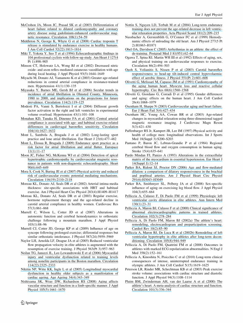
McCrohon JA, Moon JC, Prasad SK et al (2003) Differentiation of
heart failure related to dilated cardiomyopathy and coronary
artery disease using gadolinium-enhanced cardiovascular mag-
netic resonance. Circulation 108(1):54–59
Middleton N, George K, Whyte G et al (2008) Cardiac troponin T
release is stimulated by endurance exercise in healthy humans.
J Am Coll Cardiol 52(22):1813–1814
Miki T, Yokota Y, Seo T et al (1994) Echocardiographic findings in
104 professional cyclists with follow-up study. Am Heart J 127(4
Pt 1):898–905
Minson CT, Holowatz LA, Wong BJ et al (2002) Decreased nitric
oxide- and axon reflex-mediated cutaneous vasodilation with age
during local heating. J Appl Physiol 93(5):1644–1649
Miyachi M, Donato AJ, Yamamoto K et al (2003) Greater age-related
reductions in central arterial compliance in resistance-trained
men. Hypertension 41(1):130–135
Miyasaka Y, Barnes ME, Gersh BJ et al (2006) Secular trends in
incidence of atrial fibrillation in Olmsted County, Minnesota,
1980 to 2000, and implications on the projections for future
prevalence. Circulation 114(2):119–125
Modesti PA, Vanni S, Bertolozzi I et al (2004) Different growth
factor activation in the right and left ventricles in experimental
volume overload. Hypertension 43(1):101–108
Monahan KD, Tanaka H, Dinenno FA et al (2001) Central arterial
compliance is associated with age- and habitual exercise-related
differences in cardiovagal baroreflex sensitivity. Circulation
104(14):1627–1632
Mont L, Sambola A, Brugada J et al (2002) Long-lasting sport
practice and lone atrial fibrillation. Eur Heart J 23(6):477–482
Mont L, Elosua R, Brugada J (2009) Endurance sport practice as a
risk factor for atrial fibrillation and atrial flutter. Europace
11(1):11–17
Moon JC, Fisher NG, McKenna WJ et al (2004) Detection of apical
hypertrophic cardiomyopathy by cardiovascular magnetic reso-
nance in patients with non-diagnostic echocardiography. Heart
90(6):645–649
Mora S, Cook N, Buring JE et al (2007) Physical activity and reduced
risk of cardiovascular events: potential mediating mechanisms.
Circulation 116(19):2110–2118
Moreau KL, Donato AJ, Seals DR et al (2002) Arterial intima-media
thickness: site-specific associations with HRT and habitual
exercise. Am J Physiol Heart Circ Physiol 283(4):H1409–H1417
Moreau KL, Donato AJ, Seals DR et al (2003) Regular exercise,
hormone replacement therapy and the age-related decline in
carotid arterial compliance in healthy women. Cardiovasc Res
57(3):861–868
Murrell C, Wilson L, Cotter JD et al (2007) Alterations in
autonomic function and cerebral hemodynamics to orthostatic
challenge following a mountain marathon. J Appl Physiol
103(1):88–96
Murrell CJ, Cotter JD, George KP et al (2009) Influence of age on
syncope following prolonged exercise; differential responses but
similar orthostatic intolerance. J Physiol 587(24):5959–5969
Naylor LH, Arnolda LF, Deague JA et al (2005) Reduced ventricular
flow propagation velocity in elite athletes is augmented with the
resumption of exercise training. J Physiol 563(Pt 3):957–963
Neilan TG, Januzzi JL, Lee-Lewandrowski E et al (2006) Myocardial
injury and ventricular dysfunction related to training levels
among nonelite participants in the Boston marathon. Circulation
114(22):2325–2333
Nikitin NP, Witte KK, Ingle L et al (2005) Longitudinal myocardial
dysfunction in healthy older subjects as a manifestation of
cardiac ageing. Age Ageing 34(4):343–349
Nishiyama SK, Wray DW, Richardson RS (2008) Aging affects
vascular structure and function in a limb-specific manner. J Appl
Physiol 105(5):1661–1670
Nottin S, Nguyen LD, Terbah M et al (2004) Long-term endurance
training does not prevent the age-related decrease in left ventric-
ular relaxation properties. Acta Physiol Scand 181(2):209–215
Nussbacher A, Gerstenblith G, O’Connor FC et al (1999) Hemody-
namic effects of unloading the old heart. Am J Physiol 277(5 Pt
2):H1863–H1871
Obel OA, Davidson C (2005) Arrhythmias in an athlete: the effect of
de-training. Postgrad Med J 81(951):62–64
Ogawa T, Spina RJ, Martin WH III et al (1992) Effects of aging, sex,
and physical training on cardiovascular responses to exercise.
Circulation 86(2):494–503
Ogoh S, Volianitis S, Nissen P et al (2003) Carotid baroreflex
responsiveness to head-up tilt-induced central hypovolaemia:
effect of aerobic fitness. J Physiol 551(Pt 2):601–608
Olivetti G, Melissari M, Capasso JM et al (1991) Cardiomyopathy of
the aging human heart. Myocyte loss and reactive cellular
hypertrophy. Circ Res 68(6):1560–1568
Olivetti G, Giordano G, Corradi D et al (1995) Gender differences
and aging: effects on the human heart. J Am Coll Cardiol
26(4):1068–1079
Oxenham H, Sharpe N (2003) Cardiovascular aging and heart failure.
Eur J Heart Fail 5(4):427–434
Oxenham HC, Young AA, Cowan BR et al (2003) Age-related
changes in myocardial relaxation using three-dimensional tagged
magnetic resonance imaging. J Cardiovasc Magn Reson
5(3):421–430
Paffenbarger RS Jr, Kampert JB, Lee IM (1997) Physical activity and
health of college men: longitudinal observations. Int J Sports
Med 18(Suppl 3):S200–S203
Pantano P, Baron JC, Lebrun-Grandie P et al (1984) Regional
cerebral blood flow and oxygen consumption in human aging.
Stroke 15(4):635–641
Pardo Mindan FJ, Panizo A (1993) Alterations in the extracellular
matrix of the myocardium in essential hypertension. Eur Heart J
14(Suppl J):12–14
Parker BA, Ridout SJ, Proctor DN (2006) Age and flow-mediated
dilation: a comparison of dilatory responsiveness in the brachial
and popliteal arteries. Am J Physiol Heart Circ Physiol
291(6):H3043–H3049
Parker BA, Smithmyer SL, Pelberg JA et al (2008) Sex-specific
influence of aging on exercising leg blood flow. J Appl Physiol
104(3):655–664
Pelliccia A, Culasso F, Di Paolo FM et al (1999) Physiologic left
ventricular cavity dilatation in elite athletes. Ann Intern Med
130(1):23–31
Pelliccia A, Maron BJ, Culasso F et al (2000) Clinical significance of
abnormal electrocardiographic patterns in trained athletes.
Circulation 102(3):278–284
Pelliccia A, Di Paolo FM, Maron BJ (2002a) The athlete’s heart:
remodeling, electrocardiogram and preparticipation screening.
Cardiol Rev 10(2):85–90
Pelliccia A, Maron BJ, De Luca R et al (2002b) Remodeling of left
ventricular hypertrophy in elite athletes after long-term decon-
ditioning. Circulation 105(8):944–949
Pelliccia A, Di Paolo FM, Quattrini FM et al (2008) Outcomes in
athletes with marked ECG repolarization abnormalities. N Engl JMed 358(2):152–161
Pelliccia A, Kinoshita N, Pisicchio C et al (2010) Long-term clinical
consequences of intense, uninterrupted endurance training in
olympic athletes. J Am Coll Cardiol 5(15):1619–1625
Peterson LR, Rinder MR, Schechtman KB et al (2003) Peak exercise
stroke volume: associations with cardiac structure and diastolic
function. J Appl Physiol 94(3):1108–1114
Pluim BM, Zwinderman AH, van der Laarse A et al (2000) The
athlete’s heart. A meta-analysis of cardiac structure and function.
Circulation 101(3):336–344
Eur J Appl Physiol
123
Pollock ML, Mengelkoch LJ, Graves JE et al (1997) Twenty-year
follow-up of aerobic power and body composition of older track
athletes. J Appl Physiol 82(5):1508–1516
Prasad A, Popovic ZB, Arbad-Zadeh A et al (2007) The effects of
aging and physical activity on Doppler measures of diastolic
function. Am J Cardiol 99:1629–1636
Raven PB, Pawelczyk JA (1993) Chronic endurance exercise training:
a condition of inadequate blood pressure regulation and reduced
tolerance to LBNP. Med Sci Sports Exerc 25(6):713–721
Reindell H, Klepzig H, Musshoff K et al (1957) Results of recent
studies on the relationship between size and efficiency of the
normal human heart, especially athletic heart. Dtsch Med
Wochenschr 82(23):613–619
Rinder MR, Spina RJ, Ehsani AA (2000) Enhanced endothelium-
dependent vasodilation in older endurance-trained men. J Appl
Physiol 88(2):761–766
Rost R (1997) The athlete’s heart. Historical perspectives—solved
and unsolved problems. Cardiol Clin 15(3):493–512
Rowe WJ (1993) Endurance exercise and injury to the heart. Sports
Med 16(2):73–79
Rywik TM, Blackman MR, Yataco AR et al (1999) Enhanced
endothelial vasoreactivity in endurance-trained older men.
J Appl Physiol 87(6):2136–2142
Sarabi M, Millgard J, Lind L (1999) Effects of age, gender and
metabolic factors on endothelium-dependent vasodilation: a
population-based study. J Intern Med 246(3):265–274
Scharhag J, Schneider G, Kurschner R et al (2009) Right and left
ventricular mass and function in strength athletes determined by
MRI. Med Sci Sports Exerc 41(5):158–159
Scheel P, Ruge C, Petruch UR et al (2000) Color duplex measurement
of cerebral blood flow volume in healthy adults. Stroke
31(1):147–150
Schulman SP, Lakatta EG, Fleg JL et al (1992) Age-related decline in
left ventricular filling at rest and exercise. Am J Physiol 263(6 Pt
2):H1932–H1938
Seals DR, Hagberg JM, Spina RJ et al (1994a) Enhanced left
ventricular performance in endurance trained older men. Circu-
lation 89(1):198–205
Seals DR, Taylor JA, Ng AV et al (1994b) Exercise and aging:
autonomic control of the circulation. Med Sci Sports Exerc
26(5):568–576
Seto CK (2003) Preparticipation cardiovascular screening. Clin Sports
Med 22(1):23–35
Sharma S, Whyte G, McKenna WJ (1997) Sudden death from
cardiovascular disease in young athletes: fact or fiction? Br
J Sports Med 31(4):269–276
Sharma S, Whyte G, Elliott P et al (1999) Electrocardiographic
changes in 1000 highly trained junior elite athletes. Br J Sports
Med 33(5):319–324
Shave R, George KP, Atkinson G et al (2007) Exercise-induced
cardiac troponin T release: a meta-analysis. Med Sci Sports
Exerc 39(12):2099–2106
Shave R, George K, Whyte G et al (2008) Postexercise changes in left
ventricular function: the evidence so far. Med Sci Sports Exerc
40(8):1393–1399
Silber DH, Sinoway LI (1990) Reversible impairment of forearm
vasodilation after forearm casting. J Appl Physiol 68(5):1945–
1949
Silber D, McLaughlin D, Sinoway L (1991) Leg exercise conditioning
increases peak forearm blood flow. J Appl Physiol 71(4):1568–
1573
Singer RB (2008) Mortality in older adults in relation to daily activity
energy expenditure. J Insur Med 40(1):38–43
Sinoway LI, Musch TI, Minotti JR et al (1986) Enhanced maximal
metabolic vasodilatation in the dominant forearms of tennis
players. J Appl Physiol 61(2):673–678
Song Y, Yao Q, Zhu J et al (1999) Age-related variation in the
interstitial tissues of the cardiac conduction system; and autopsy
study of 230 Han Chinese. Forensic Sci Int 104(2–3):133–142
Sorokin AV, Araujo CGS, Zweibel A et al (2009) Atrial fibrillation in
endurance-trained athletes. Br J Sports Med. doi:10.1136/
bjsm.2009.057885
Spier SA, Delp MD, Meininger CJ et al (2004) Effects of ageing and
exercise training on endothelium-dependent vasodilatation and
structure of rat skeletal muscle arterioles. J Physiol 556(Pt
3):947–958
Spina RJ, Miller TR, Bogenhagen WH et al (1996) Gender-related
differences in left ventricular filling dynamics in older subjects
after endurance exercise training. J Gerontol A Biol Sci Med Sci
51(3):B232–B237
Stein R, Medeiros CM, Rosito GA et al (2002) Intrinsic sinus and
atrioventricular node electrophysiologic adaptations in endur-
ance athletes. J Am Coll Cardiol 39(6):1033–1038
Stoquart-Elsankari S, Baledent O, Gondry-Jouet C et al (2007) Aging
effects on cerebral blood and cerebrospinal fluid flows. J Cereb
Blood Flow Metab 27(9):1563–1572
Stratton JR, Levy WC, Cerqueira MD et al (1994) Cardiovascular
responses to exercise. Effects of aging and exercise training in
healthy men. Circulation 89(4):1648–1655
Swanson DR (2006) Atrial fibrillation in athletes: implicit literature-
based connections suggest that overtraining and subsequent
inflammation may be a contributory mechanism. Med Hypoth-
eses 66(6):1085–1092
Swinne CJ, Shapiro EP, Lima SD et al (1992) Age-associated changes
in left ventricular diastolic performance during isometric exer-
cise in normal subjects. Am J Cardiol 69(8):823–826
Sztajzel J, Jung M, Sievert K et al (2008) Cardiac autonomic profile in
different sports disciplines during all-day activity. J Sports Med
Phys Fitness 48(4):495–501
Taddei S, Virdis A, Mattei P et al (1995) Aging and endothelial
function in normotensive subjects and patients with essential
hypertension. Circulation 91(7):1981–1987
Taddei S, Galetta F, Virdis A et al (2000) Physical activity prevents
age-related impairment in nitric oxide availability in elderly
athletes. Circulation 101(25):2896–2901
Takeshita A, Mark AL (1980) Decreased vasodilator capacity of
forearm resistance vessels in borderline hypertension. Hyper-
tension 2(5):610–616
Tan MP, Parry SW (2008) Vasovagal syncope in the older patient.
J Am Coll Cardiol 51(6):599–606
Tanaka H, DeSouza CA, Seals DR (1998) Absence of age-related
increase in central arterial stiffness in physically active women.
Arterioscler Thromb Vasc Biol 18(1):127–132
Tanaka H, Dinenno FA, Monahan KD et al (2000) Aging, habitual
exercise, and dynamic arterial compliance. Circulation
102(11):1270–1275
Thijssen DH, de Groot PC, Smits P et al (2007) Vascular adaptations
to 8-week cycling training in older men. Acta Physiol (Oxf)
190(3):221–228
Thijssen DH, Rongen GA, van Dijk A et al (2007b) Enhanced
endothelin-1-mediated leg vascular tone in healthy older
subjects. J Appl Physiol 103(3):852–857
Thijssen DH, van Bemmel M, Bullens L et al (2008) The impact of
baseline diameter on flow mediated dilation (FMD) differs in
young and older humans. Am J Physiol Heart Circ Physiol
295(4):H1594–H1598
Tinken TM, Thijssen DH, Hopkins N et al (2009) Impact of shear rate
modulation on vascular function in humans. Hypertension
54(2):278–285
Tinken TM, Thijssen DH, Dawson EA et al (2010) Shear stress
modulates vascular adaptations to exercise training in humans.
Hypertension 55(2):312–318
Eur J Appl Physiol
123
Tuttle JL, Nachreiner RD, Bhuller AS et al (2001) Shear level
influences resistance artery remodeling: wall dimensions, cell
density, and eNOS expression. Am J Physiol Heart Circ Physiol
281(3):H1380–H1389
Uusitalo AL, Uusitalo AJ, Rusko HK (1998) Exhaustive endurance
training for 6–9 weeks did not induce changes in intrinsic heart
rate and cardiac autonomic modulation in female athletes. Int J
Sports Med 19(8):532–540
Van Guilder GP, Westby CM, Greiner JJ et al (2007) Endothelin-1
vasoconstrictor tone increases with age in healthy men but can be
reduced by regular aerobic exercise. Hypertension 50(2):403–409
Vasan RS, Levy D (2000) Defining diastolic heart failure: a call for
standardized diagnostic criteria. Circulation 101(17):2118–2121
Villari B, Vassalli G, Schneider J et al (1997) Age dependency of left
ventricular diastolic function in pressure overload hypertrophy.
J Am Coll Cardiol 29(1):181–186
Volders PG, Willems IE, Cleutjens JP et al (1993) Interstitial collagen
is increased in the non-infarcted human myocardium after
myocardial infarction. J Mol Cell Cardiol 25(11):1317–1323
Whyte GP (2008) Clinical significance of cardiac damage and
changes in function after exercise. Med Sci Sports Exerc
40(8):1416–1423
Whyte GP, George K, Nevill A et al (2004a) Left ventricular
morphology and function in female athletes: a meta-analysis. Int
J Sports Med 25(5):380–383
Whyte GP, George K, Sharma S et al (2004b) The upper limit of
physiological cardiac hypertrophy in elite male and female
athletes: the British experience. Eur J Appl Physiol 92(4–5):592–
597
Whyte GP, Sheppard M, George KP et al (2007) Arrhythmias and the
athlete: mechanisms and clinical significance. Eur Heart J
28(11):1399–1401 author reply 1401
Whyte G, Sheppard M, George K et al (2008) Post-mortem evidence
of idiopathic left ventricular hypertrophy and idiopathic inter-
stitial myocardial fibrosis: is exercise the cause. Br J Sports Med
42(4):304–305
Wilson JM, Villareal RP, Hariharan R et al (2002) Magnetic
resonance imaging of myocardial fibrosis in hypertrophic
cardiomyopathy. Tex Heart Inst J 29(3):176–180
Wilson MG, O’Hanlon R, Prasad S et al (2009) Myocardial fibrosis in
an veteran endurance athlete. BMJ Case Report. doi:10.1136/
bcr.12.2008.1345
Winker R, Barth A, Bidmon D et al (2005) Endurance exercise
training in orthostatic intolerance: a randomized, controlled trial.
Hypertension 45(3):391–398
Yared K, Wood MJ (2009) Is marathon running hazardous to your
cardiovascular health? The jury is still out. Radiology 251(1):3–5
Zeppilli P, Santini C, Palmieri V et al (1994) Role of myocarditis in
athletes with minor arrhythmias and/or echocardiographic
abnormalities. Chest 106(2):373–380
Eur J Appl Physiol
123


