Embed Size (px)
Citation preview

Cancer cells are selectively eliminated through UPRdriven apoptosis during treatment with Aquilegianivalis extractsNazia Hilal
University of Kashmir, SrinagarOzaira Qadri
University of Kashmir, SrinagarIrshad A Nawchoo
University of Kashmir, SrinagarSeema Akbar
Regional Research Institute of Unani Medicine, University of Kashmir CampusKhalid Majid Fazili ( [email protected] )
University of Kashmir, Srinagar
Research Article
Keywords: Aquilegia nivalis, UPR, IRE1α, XBP1, eIF2α, ATF4, Apoptosis
Posted Date: March 10th, 2021
DOI: https://doi.org/10.21203/rs.3.rs-234542/v1
License: This work is licensed under a Creative Commons Attribution 4.0 International License. Read Full License
1
Cancer cells are selectively eliminated through UPR driven apoptosis during 1
treatment with Aquilegia nivalis extracts 2
Nazia Hilal1#, Ozaira Qadri1, Irshad A Nawchoo2, Seema Akbar3 and Khalid Majid 3
Fazili1* 4
5
1Department of Biotechnology, University of Kashmir, Srinagar-190006, Jammu and 6
Kashmir 7
2Department of Botany, University of Kashmir, Srinagar- 190006, Jammu and Kashmir 8
3Regional Research Institute of Unani Medicine, University of Kashmir, Srinagar- 9
190006 , Jammu and Kashmir 10
11
12
13
14
15
# Current address 16
Department of Biochemistry and Molecular Genetics 17
University of Illinois, Chicago 18
19
20
*Correspondence: 21
Dr. Khalid Majid Fazili, PhD 22
Professor, Department of Biotechnology 23
University of Kashmir, Srinagar-190006, Jammu and Kashmir 24
Tel: +91-9419003881 25
Email: [email protected] 26
2
ABSTRACT: 27
Background 28
Aquilegia nivalis Flax Jackson, also called Aquilegia vulgaris sub sp. nivalis (Bak.) Brühl 29
or columbine, locally known as “Zoe-neel”, is a wild edible plant traditionally used as an 30
anti-inflammatory medicine by the local nomadic tribes inhabiting the Himalayas of 31
Jammu and Kashmir. The plant has been used as herbal medicine since middle ages in 32
treating ailments that include chronic rhinitis and various infectious diseases. The extracts 33
from the plant possess antioxidant properties and have been reported to be 34
hepatoprotective in rats. Our preliminary studies, however pointed to hitherto unexplored 35
anti-apoptotic potential of the plant which lead us to carry the in-depth study using breast 36
cancer cell lines to validate its anti-cancerous properties and explore the affected 37
pathways. 38
Methods 39
MTT assay was used to draw the dose response curve and evaluate the effect of 40
increasing concentrations of the extract on cell lines to determine the appropriate dosage 41
to be used for further experimentation. DNA fragmentation analysis was followed 42
through gel electrophoresis and DAPI staining was pursued by phase contrast microscopy 43
to study apoptosis. Quantitative PCR was used to study the expression of UPR signaling 44
and RIDD markers at the level of mRNA. Western blot analysis was used in studying the 45
expression of the various markers of the signaling pathways. The cell cycle analysis was 46
carried out using flow cytometry. 47
Results 48
MTT assay revealed that the methanolic extract of the plant (ANME) was selectively 49
cytotoxic to various cancer cell lines as revealed by lower IC50 values relative to normal 50
cell lines. The results of cell cycle analysis were similar as ANME caused Sub G1 arrest 51
of the cell cycle. DNA fragmentation analysis, DAPI staining and western blot analysis 52

3
for PARP and caspases revealed that the extract selectively induced apoptosis in 53
cancerous cell lines. UPR markers p-Ire1α and Xbp1 splicing were consistently alleviated 54
in a dose dependent manner, the rate of phosphorylation of eIF2a and ATF4 also 55
decreased with increasing concentration of ANME. The RT PCR results of the RIDD 56
marker, Blos1S1 revealed a similar dose dependent association. The methanolic extract 57
was especially chosen for it could be easily internalized by the cells and any resultant 58
potential bioactive compounds could gain access to the cells because of their hydrophobic 59
nature. 60
Conclusion 61
Our results suggest that ANME causes deactivation of UPR signaling pathway 62
facilitating apoptosis selectively in cancerous cells, paving the way forward for a novel 63
approach in cancer therapeutics. 64
Keywords: Aquilegia nivalis, UPR, IRE1α, XBP1, eIF2α, ATF4, Apoptosis 65
Abbreviations: 66
UPR , Unfolded protein response: ATF4, Activating transcription factor 4: ATF6, Activating 67
transcription factor 6: GADD34, Growth arrest and DNA damage 3: XBP1, X-box binding 68
protein 1: ANME, Aquilegia nivalis methanolic extract : RIDD, Regulated IRE1-dependent 69
decay of mRNA 70
Introduction 71
The exploration of natural products offers great opportunity to researchers in medical 72
sciences to identify and explore nature friendly therapeutic agents relevant to different 73
diseases. Aquilegia nivalis is a traditional indigenous herb found in higher altitudes of 74
Himalayas1. The plant has been traditionally used in treatment of Asthma and as an anti-75
inflammatory agent. It is also used as a remedy for many ailments including chronic 76
rhinitis and various infections2. The extracts of the plant [plant extract] have shown 77
hepatoprotective effects in mice.3 Several studies have shown the anti-oxidant role of the 78
4
plant.4,5. Despite of its well-known anti-inflammatory and anti-oxidative properties, and 79
the fact that the plant has been used for a long time as a traditional therapeutic agent in 80
treating several disorders like bronchitis and bowel disorders, the plant has not been 81
explored to its full potential. No study has been carried out to find its molecular targets 82
and the mode of action. The plant is being extensively used by the nomadic tribes 83
inhabiting the Himalayan ranges in Jammu and Kashmir. Some studies have suggested 84
the antiproliferative and pro-apoptotic activities of the plant6. Our preliminary data 85
indicated that the plant extracts were selectively toxic to cancerous cells, so the current 86
study was carried out to validate the antiproliferative potential of the plant and explore its 87
molecular targets in the cell. The study revealed that the methanolic extract of the plant 88
affected UPR signaling and RIDD pathways. 89
The unfolded protein response is a defensive mechanism that gets activated under 90
stressful conditions to restore homeostasis, during prolonged stress however, when 91
restoration becomes difficult, it promotes apoptosis7. The synthesis and trafficking of 92
nearly one-third of the total protein in eukaryotic cells is synchronized by Endoplasmic 93
Reticulum8,9,10. The ER is responsible for tuning up protein homeostasis11 and ensures the 94
release and bartering of properly folded proteins after post-transcriptional and post-95
translational modifications through various mechanisms existing within the organelle. 96
Despite this, a colossal amount of proteins transiting via ER are not properly folded and 97
may be degraded by ER-associated degradation (ERAD) system11. The perturbations in 98
influx to efflux ratios of proteins within the ER compartment coupled with accumulation 99
of misfolded proteins causes ER stress that is combated through a defensive mechanism 100
known as Unfolded Protein Response12,13. UPR determines the cellular fate either 101
towards survival or death depending upon the severity of stress and primarily operates via 102
three axis namely; Ire1-α/Xbp-1, PERK/eIF2α and ATF6 7,14,15 103
5
Tumor cells experience increase metabolic activities and high proliferation rates, which 104
in turn demands boisterous ER and secretory mechanisms. This boosted demand for 105
secretory functions is likely to trigger a mid-course correction of ER homeostasis and 106
consequently ends up in basal or constitutive UPR induction. Ire1α mutations have been 107
implicated in many human cancers on the basis of genome wide screening with roles in 108
tumor growth, metastasis and chemo-resistance16,17,18. A compromising tumor growth and 109
survival is observed when Xbp1 expression is thwarted, inhibiting Ire1/Xbp1 axis 110
impinge the coherence of the secretory tissues 19-23. PERK pathway in tumor cells either 111
facilitates their survival or suppresses their progression. PERK branch confers oncogenic 112
transformation by promoting myc induced autophagic pathways24,25. CHOP (an apoptosis 113
mediator) suppresses the tumor progression thus chop deletion has tumor progression 114
phenotype in lung cancer.26,27 115
Many pathological complications are associated with the disturbance in the protein 116
folding machinery, aggregation and concurrent ER stress7,28. Various therapeutic drugs 117
induce ER stress and destine the cells to apoptosis29. It is possible that an optimum level 118
of ER stress and UPR signaling is induced under tumor microenvironment is necessary 119
for cancer survival and progression, but exacerbating the stress levels beyond the 120
handling capacity can drive cancer cells toward death. UPR signaling can be targeted in 121
cancer treatment in both ways, that is, either by inducing a high level of stress in the pre-122
existing stress microenvironment of tumor or by preventing activation of UPR that is 123
beneficial for cancer progression and drug resistance, making them sensitive to other 124
chemotherapeutic drugs. 125
In the above context, our study becomes important as it reflects the potential effects of 126
the plant extract to selectively induce apoptosis in cancerous cells coupled with 127
deactivation of UPR and RIDD signaling pathways. 128
129
6
MATERIALS AND METHODS: 130
Chemicals 131
Tunicamycin was purchased from EMD Millipore (Darmstadt, Germany).Dulbecco's 132
modified Eagles medium (DMEM) and Fetal bovine Serum (FBS) were purchased from 133
GIBCO (St. Louis, Mo, USA). Bicinchoninic acid (BCA) protein assay kit was purchased 134
from pierce (Rockford, USA). and 3-(4,5-dimethylthiazole-2-yl)-2,5, diphenyltetrazolium 135
bromide (MTT) were purchased from Sigma-Aldrich (St. Louis, Mo, USA). 136
Preparation of methanolic fraction of Aquilegia nivalis 137
Aquilegia nivalis was collected between July and August 2016, from several regions of 138
Kashmiri Himalayas. The plant was identified and deposited in the KASH Herbarium 139
under voucher specimen No.2716-(KASH). Whole Plant parts were washed with distilled 140
water to remove residues and shade dried. The dead parts were removed, and healthy 141
ones were grounded to powder, preserved for extraction. ANME (Methanolic fraction of 142
Aquilegia nivalis) was prepared from dried and coarse powder using methanol as a 143
solvent. Hot extraction was carried out in a Soxhlet apparatus taking Plant powder and 144
solvent in ratio of 1:6. The extract was filtered and evaporated to dryness and the residues 145
were weighed. The extract solutions obtained were combined and evaporated to dryness 146
by a rotary evaporator under vacuum at 65 °C. The yield was calculated by dividing the 147
mass of recovered dry extract (mr) by the initial mass of powder (mi). 148
Yield(%)= mr/mi × 100% 149
This residue was then dissolved in 50% DMSO to make the concentration of 100 mg/mL 150
working stock and stored at 4°C. The working concentration of DMSO used was 2% for 151
experiments. 152
153
154
155
7
Cell lines 156
Breast cancer cell lines, MDAMB-231 and MCF7 and neuronal cell line U87MG 157
glioblastoma, and the normal human embryonic kidney cells, Hek293T were purchased 158
from National Centre for Cell Science (NCCS, Pune). 159
Antibodies and Western blot analysis 160
SDS-Polyacrylamide gel electrophoresis and Western blotting were performed by usual 161
procedures . All the four cells were seeded at 1 × 10⁶ cells/well in a 100 mm culture dish 162
and incubated for 24 h. The incubated cells were treated with varying concentrations of 163
ANME with or without 6μM of tunicamycin for 24 h. The cells were harvested in 164
phosphate buffered saline (PBS) containing 0.1% protease inhibitor cocktail. The primary 165
and secondary antibodies used were as follows. Rabbit polyclonal antibodies against 166
Phosphorylated version of Inositol requiring enzyme (Ire1α), X-box binding protein 167
spliced (sXbp1), Activating transcription factor 4 (ATF4), Phosphorylated version of 168
alpha subunit of eukaryotic initiation factor (p-eIF2α) and GAPDH were obtained from 169
Cell Signaling Technology (Danvers, MA). Alkaline Phosphatase linked secondary anti-170
Rabbit antibody was purchased from Sigma-Aldrich (St. Louis, Mo, USA). BCIP (5-171
Bromo-4-chloro-3-indolylphosphate) and NBT (Nitro blue tetrazolium) were purchased 172
from Sigma-Aldrich (St. Louis, Mo, USA). 173
Real-time quantitative PCR for analysis 174
The cells were seeded at 1 × 10⁶ cells/well in a 100 mm culture dish and incubated for 24 175
h. The incubated cells were treated with varying concentrations of ANME with or 176
without 6μM of tunicamycin for 24 h. RNA was extracted from the harvested cells using 177
an RNeasy kit (Qiagen, Hilden, Germany). RNA was reverse-transcribed to synthesize 178
cDNA using a Revert Aid First strand cDNA synthesis kit (Thermo scientific) as per 179
manufacturer’s protocol., and used in duplicate for quantitative real-time PCR analysis 180
using the SYBR Green reagent system Applied Biosystems 7500 Fast Real-Time PCR 181

8
System (Applied Biosystems, Foster City, CA). Relative quantities of amplified cDNAs 182
were then determined using SDS software (Applied Biosystems) and normalized to β-183
actin mRNA. 184
The following primers were used: 185
ATF4 186
5/-TTC CTG AGC AGC GAG GTG TTG -3/(sense) 187
5/-TCCAATCTGTCCCGGAGAAGG-3 (antisense) 188
CHOP 189
5/-CTTGGCTGACTGAGGAGGAG-3/(sense) 190
5/-TCACCATTCGGTCAATCAGA-3/(antisense) 191
Spliced XBP1 192
5 /-CTGAGTCCGAATCAGGTGCAG-3/(sense) 193
5/-ATCCATGGGGAGATGTTCTGG-3/(antisense) 194
β-actin, 195
5/-TCATCACCATTGGCAATGAG-3/ (sense) 196
5/-CACTGTGTTGGCGTACAGGT-3/ (antisense) 197
Quantification of western blot 198
The intensity (area x optical density) of the individual bands on Western blots was 199
measured by using ImageJ and normalized either to GAPDH or, in the case of a 200
phosphoprotein, to its total protein as mentioned. 201
Cell viability assays 202
Cell viability measurements for ANME were done using MTT assay. Cell were seeded in 203
96-well plate at 104 cells per well. Different concentrations of extract (1-2000μg/mL) 204
were added to each well incubated for 24 h. Next day 10 μL of 5 mg/mL MTT were 205
added to each well and incubated for 4 h. The formazan crystals were dissolved by 206
adding 100 μL of DMSO to each well. Absorbance was measured at 560 nm with 207
9
Universal Microplate Reader (Bio-Tek Instruments, USA). The reference wavelength 208
was set at 650 nm. The cell viability of untreated cells was considered 100%. 209
Cell cycle analysis by flow cytometry 210
The cells were seeded at 1 × 10⁶ cells/well in a 100-mm culture dish and incubated for 24 211
h. The incubated cells were treated with ANME (50 μg/mL for MDAMB231 100 μg/mL 212
for MCF7 and 25 μg/mL for U87MG), with or without 6μM of Tm for 24 h.. Media was 213
collected in 15ml tubes and cells were washed with PBS containing 0.1% EDTA. 214
Washing solution was also collected. PBS-EDTA was added to the plates followed by 215
incubation at 37 °C for 5-10 minutes. Cells were collected, pipetted up and down and 216
collected in same tubes. Tubes were centrifuged at 1000 g for 5 minutes followed by 217
washing in PBS-Serum (1% serum) and centrifuged again at 1000g. Afterwards, cells 218
were resuspended in 0.5 ml PBS. For fixing, 5 ml ethanol was added drop wise while 219
vertexing the cells. Cells were then stored in deep freezer for FACS analysis. For FACS 220
analysis, fixed samples were centrifuged at 1000g for 5 minutes and washed with PBS-221
serum followed by resuspension in Propidium Iodide-RNAse solution (50 μg/ml 222
propidium iodide, 10mM Tris pH 7.5, 5 mM MgCl2 and 20 μg/ml RNase A). Finally, 223
samples were acquired and analyzed by using BD FACS machine and BD FACS Suite 224
software. The experiments were done in triplicates. For analysis outliers were discarded. 225
Statistical analysis 226
The GraphPad Prism®6 software (GraphPad software Inc.) was used for statistical 227
analysis. All experiments were repeated independently three times. IC50 values were also 228
calculated using non-linear regression analysis with GraphPad Prism®6 software. 229
Results 230
ANME display cytotoxicity against cancer cell lines in vitro 231
MTT viability assay on HEK293T human embryonic kidney cells, MDAMD-231 breast 232
cancer,MCF7 breast cancer and U87 glioblastoma cells were done separately with a 233
10
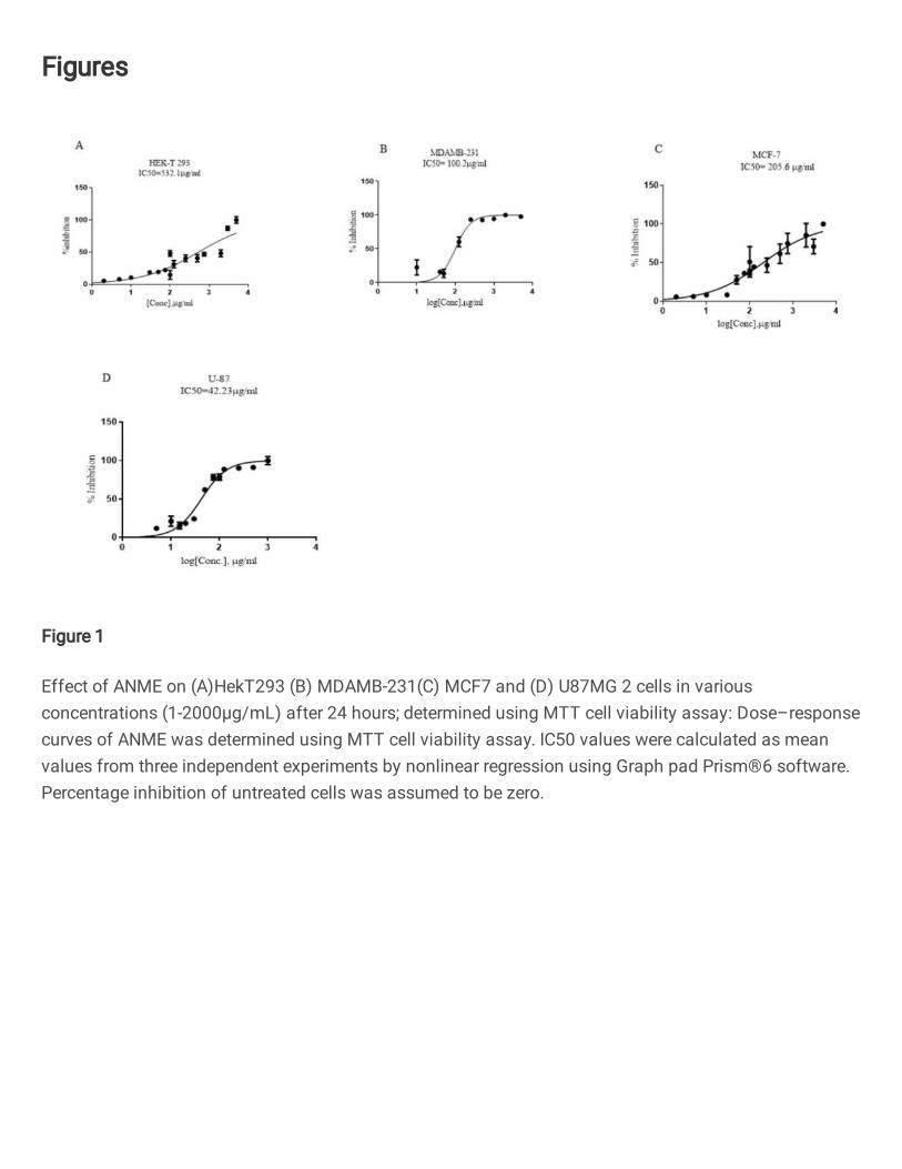
range of concentrations of ANME between 1 to 2000 µg/ml. ANME alleviated cell 234
viability in a dose dependent manner. All the experiments were performed in triplicate. 235
IC50 values were calculated by non-linear regression analysis using Graph Pad Prism 236
software as a mean of three reactions that inhibited 50% of the positive control. The IC50 237
values of ANME in HEK293T human embryonic kidney cells, MDAMD-231 breast 238
cancer, MCF7 breast cancer and U87 glioblastoma were 532.1μg/mL, 100.2μg/mL, 239
205.6μg/mL and 42.23μg/mL, respectively (Fig.1A-D). Higher concentrations of the 240
extract presented with vivid morphological changes under phase contrast microscope. 241
The most marked changes were cell shrinkage and extensive cell detachment from cell 242
culture substratum. 243
ANME displayed different cell cycle responses 244
The propidium iodide based cell cycle analysis by flow cytometry revealed that the cell 245
lines responded differently to the treatment with some cells undergoing significant cell 246
death, as indicated by an increase in SubG1 peak, whereas others not showing any 247
appreciable change in the cell cycle profile. HEK 293T (human embryonic kidney) cell 248
line which is a normal cell line didn’t show an appreciable increase in SubG1 peak (Fig. 249
2 A). U87MG (human glioblastoma) cell line showed moderate response to ANME 250
treatment (Fig. 2 B). MDAMB231 and MCF7 (Breast cancer) cell lines, however, 251
showed a remarkable increase in SubG1 (Fig. 2 C-D). This indicated that the breast 252
cancer cell lines were highly sensitive to ANME treatment. These results show that 253
different cell types respond differently to ANME, whereas different cancer cell lines are 254
sensitive to a varying degree, the response ranging from low to high cell death, the 255
normal cell lines did not show any significant effect. 256
ANME inhibits tumor cell growth in vitro by induction of apoptosis 257
The antiproliferative potential of ANME was evaluated in HEK293T human embryonic 258
kidney cells, MDAMD-231 breast cancer, MCF7 breast cancer and U87 glioblastoma. As 259
11
shown in Fig 1A IC50 values of HEK293T human embryonic kidney cells, MDAMD-260
231 breast cancer, MCF7 breast cancer and U87 glioblastoma for ANME treatment 261
were 532.1μg/mL, 100.2μg/mL, 205.6μg/mL and 42.23μg/mL, respectively revealing a 262
relatively higher sensitivity of cancer cell lines. Also there was a general SubG1 arrest in 263
the cells. The Ladder assay revealed a time-dependent increase in DNA fragmentation on 264
treatment with ANME as observed on DNA-gel electrophoresis. DNA extracted from 265
cells treated with ANME showed an increased generation of apoptotic DNA fragments as 266
compared with solvent-treated control cells, it also displays a higher sensitivity of the 267
cancer cell lines relative to normal cell lines.(Fig. 3A) 268
DAPI assay revealed apoptosis-specific features of ANME-treated cells from 0 Hours to 269
48 Hours. U87 MG, MDAMB231 and MCF7 on treatment with ANME showed various 270
nuclear changes such as chromatin condensation, nuclei condensation, and nuclear 271
degradation, however Hek293T cells did not show any signs of significant apoptotic 272
activity. (Fig. 3B) 273
UPR mediated onset of Apoptosis 274
Since the cancerous cells are in a state of constant stress because of their high metabolic 275
profile, controlled UPR is known to be active in these cells. So next we decided to look at 276
the UPR signaling pathway. Figures 4-7 show the results of Western blots of various 277
UPR signaling markers using specific antibodies. In order for us to check the pathway 278
involved through which ANME caused the apoptosis, we checked for the global impact 279
on proteostasis markers. The perturbation of cellular proteostasis networks by inhibition 280
of the proteasome results in induction of an unfolded protein response30,31. Our results 281
suggested that ANME averted UPR response in both Cancer and 293T cells, as judged 282
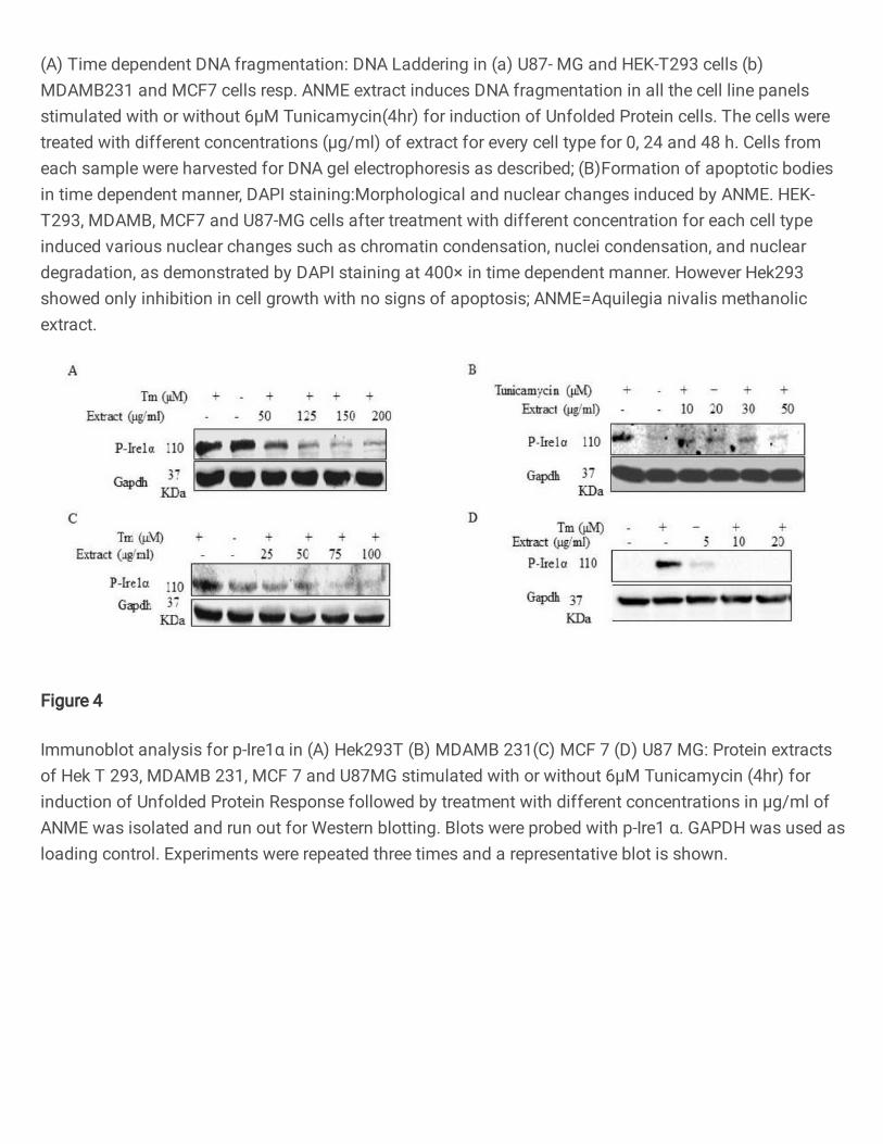
by inhibition of p-Ire1α. Figure 4 shows the effect of tunicamycin and increasing 283
concentrations of ANME on the expression of phosphorylated Ire1α. The 284
phosphorylation of Ire1α causes activation of UPR and serves as one of the first markers 285
12
of UPR activation. As is shown in figure, ANME exposure is associated with decreasing 286
concentration of P-Ire1α in all the four cell types (Fig. 4 A-D) reflecting deactivation of 287
the kinase associated IRE1 signaling of UPR. Xbp1s is another downstream marker of 288
the same pathway. Figure 5 shows the effect of tunicamycin and increasing 289
concentrations of ANME on Xbp1 splicing. ANME inhibits the splicing reaction in all 290
the cell types as indicated by reduced expression of XBp1-s.(Fig.5 A-D), the total 291
phosphorylation level of Ire1α protein decreased, with kinetics matching the inhibition of 292
Xbp1s, consistent with a previous report32. 293
Next, we evaluated the effect on the PERK arm of UPR. Figure 6 shows the effect of 294
tunicamycin and increasing concentrations of ANME on the expression levels of p-eIF2α 295
and ATF4, the downstream effector molecules of the PERK arm of UPR The expression 296
levels of both the effector molecules decreased in a concentration dependent manner on 297
treatment with ANME (Figure 6 A-D) 298
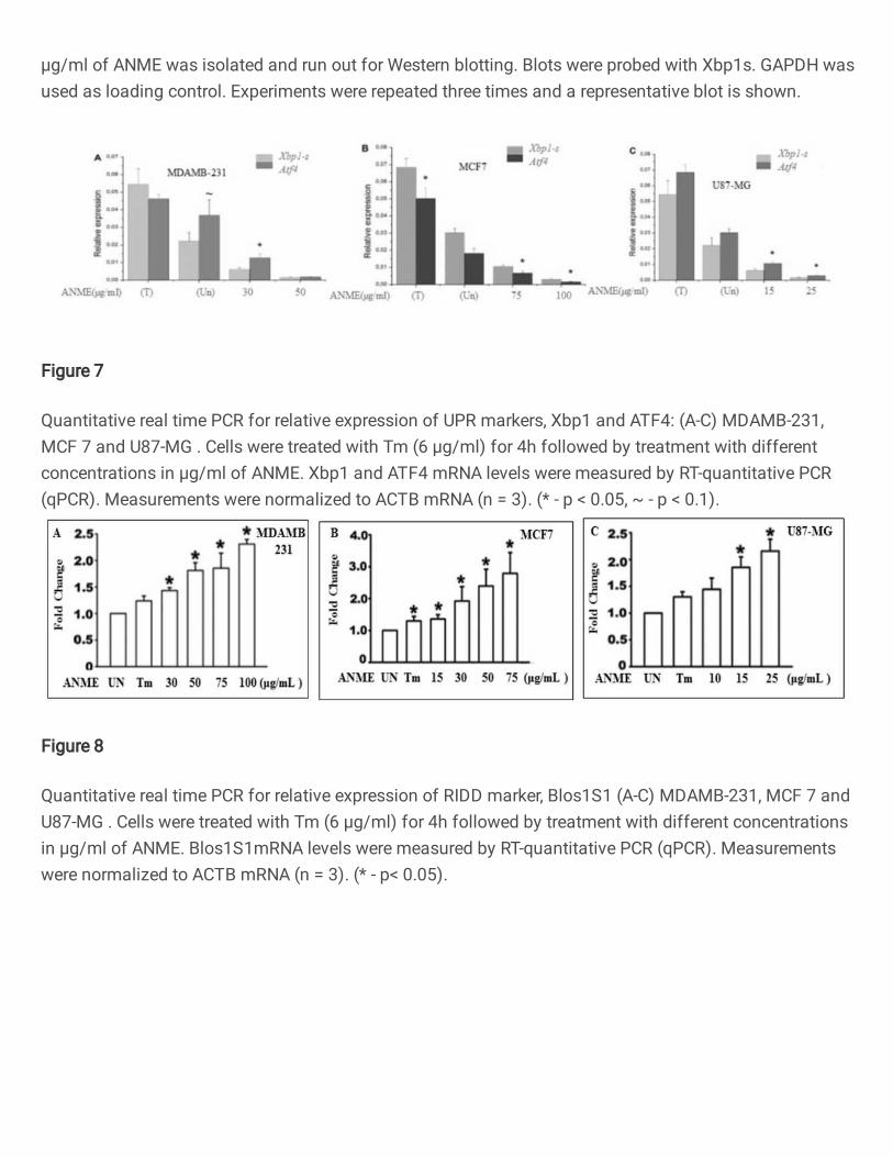
Further we tested the effect of ANME treatment on mRNA expression levels of Xbp1 and 299
ATF4 in different cell lines. ANME significantly repressed the expression of Xbp1, a 300
downstream target of Ire1 arm, and ATF4, a downstream target of PERK arm of UPR in 301
a concentration dependent manner (Figure 7 A-C) 302
Next we checked effect of the treatment on RIDD activity. Figure 8 shows the qRT PCR 303
based mRNA expression of BLOC1S1 (BLOS1) which is used as a marker for RIDD 304
activity. Treatment with increasing concentrations of the extract shows a 2-3-fold 305
increase in the expression levels in all the cell lines tested (Figure 8 A-C) indicating that 306
the RIDD arm of UPR is also decreased. To check whether ANME directly inhibits Ire1α 307
activity, we tested ANME inhibition on RIDD marker BLOC1S1 (BLOS1) which are 308
considered as standard markers for testing RIDD activity.33,34 We found that ANME 309
inhibit Ire1α activity in vitro for RIDD (Fig 8 A-C), indicating the role of ANME on in 310
apoptosis is through the RNAse activity of Ire1α in a dose-dependent manner. 311
13
ANME-induced apoptosis of tumor cells is associated with dual inhibition of 312
Ire1/xbp1 and PERK/ATF4 and activation of Caspases 313
Following DNA ladder assay and cell cycle analysis, we probed the effect of ANME on 314
caspase and PARP. Western blots with PARP and caspase antibodies were used to probe 315
the effect of the treatment with different cell lines. The results are shown in Figure 9. 316
HEK293T cell lines did not show any apoptotic signals as revealed by the absence of 317
caspase3 and PARP (Fig.9A), however, other cell lines U87MG, MDAMB231 and 318
MCF7 clearly show the onset of apoptosis in a concentration dependent manner (Figure 9 319
B,C). This was consistent with the results obtained in DNA fragmentation assay and cell 320
cycle analysis. This can be attributed to the fact that in HEK293T cells the Unfolded 321
Protein Response is rescued due to ANME treatment resulting in restoration of the in 322
milieu proteostasis. However, the apoptotic death in other cell lines that are cancerous in 323
nature is ascribed to uncompromised ER stress associated with activation of the RIDD 324
pathway. These results suggested the pro-apoptotic potential of ANME is attributed to the 325
dual inhibition of Ire1/xbp1 and PERK/ATF4 axis that renders the cancer cells vulnerable 326
to apoptotic death via activation of caspases. 327
Discussion 328
Aquilegia nivalis traditionally used in treatment of asthma and ailments like chronic 329
rhinitis and infections with documented hepatoprotective properties offers great promise 330
in disease therapeutics. The plant though has not been explored to its full potential. The 331
extracts of the Aquilegia species have been shown to display hepatoprotective effects in 332
mice.3, 35,36 Several studies have shown the anti-oxidant activities of the plant. The plant 333
has been used as a herbal medicine since ages with proven efficacy in diseases like 334
chronic rhinitis, and as anti-inflammatory in infections.4,5,36 However detailed studies 335
validating the therapeutic potential of the plant and about the mode of its action are 336
lacking. This study was conducted to validate its antiproliferative effects and evaluate its 337
14
target signaling pathways as anti-inflammatory activities in certain plants are reported to 338
be coupled with anti-proliferative pro-apoptotic activities6. Our preliminary investigations 339
on the extracts of the plant showed that the methanolic extract of the plant selectively 340
acted on cancer cells. Based on this we carried in-depth study on several cancer cell lines 341
with promising results. This study established that the extract from the plant selectively 342
induced the apoptotic pathways leading to cell death in cancerous cells. The methanolic 343
extract from the plant was particularly chosen for it displayed higher activity and for the 344
fact that it could easily be internalized by the cells. Any potential bioactive compounds 345
could also be easily accessible to the cells because of their hydrophobic nature. In the 346
ladder assay, DNA fragmentation followed by gel electrophoresis revealed generation of 347
apoptotic fragments selectively in cancerous cells and also an alleviation of apoptosis is 348
observed on treatment with the methanolic extract of Aquilegia nivalis in a concentration 349
dependent manner. Cell cycle analysis with flow cytometry displayed a differential 350
response of the cells, whereas subG1 arrest was observed in all the cancerous cells but 351
with variations in the subG1 peak, the normal cells behaved differently with no subG1 352
peak observed. DNA fragmentation and cell cycle arrest have been considered a hall 353
mark of apoptosis 33. Other pro-apoptotic markers that were tested include Caspase-3 and 354
PARP which showed comparable results with different cancerous cell lines experiencing 355
apoptosis to variable degrees, though no significant amount of apoptosis is seen in 356
normal cell lines. Several recent studies have demonstrated the proapoptotic and anti-357
apoptotic potential of plant derived extracts34,37,38. The methanolic extracts 358
of Andrographis nallamalayana Ellis have been shown to induce apoptosis in various 359
melanoma cell lines 6. Similarly extracts derived from Menyanthes trifoliata L. are 360
reported to induce apoptosis in human cancer cells affecting Caspase 3, PARP and other 361
pro apoptotic markers in a similar fashion39. Next we tried to understand the affected 362
pathways. Cancer cells are under constant stress due to their high metabolic load, and the 363
15
link between the cellular stress response and apoptosis is relatively well established. 364
Many studies have linked modulation of unfolded protein response to apoptosis40,41. 365
Under hypoxia, cells have been found to show upregulation of hypoxia induced proteins 366
and UPR signaling cascade that differ in normal and cancerous cell lines, however, the 367
exact mechanism deciding about the cell fate is not clearly understood42. Next we 368
checked the expression of downstream molecules of the Ire1 and PERK arms of the UPR 369
signaling pathway as well as BLOS1, a marker for RIDD activity. 370
It was found that treatment of cells with the methanolic extract of the plant 371
downregulated the phosphorylation of Ire1 alpha and Xbp1 splicing in a concentration 372
dependent manner. It also resulted in reduced protein expression levels of 373
phosphorylation levels of p-eIF2α and protein levels of ATF4 in all four cell lines viz 374
HEK-T293, U87-MG , MCF7 and MDAMB 231. The complexity of Ire1 biology was 375
demonstrated through a recent study suggesting that modulation of Ire1 RNase activity 376
was possible through an allosteric mechanism using ATP-competitive kinase inhibitors 377
APY29 and sunitinib43. Furthermore, a peptide derived from the Ire1 kinase domain was 378
shown to stimulate Ire1 oligomerization while inhibiting the JNK activation and RIDD 379
activity of Ire144. Ire1 modulators specific for either Xbp1 splicing or RIDD activity may 380
be clinically useful depending on the therapeutic intent. Protein kinase R (PKR)-like 381
endoplasmic reticulum kinase (PERK) mediates the translational control arm of the UPR 382
by enhancing phosphorylation of alpha subunit of eukaryotic Initiation Factor 2 (eIF2α) 383
that kickbacks to a variety of endoplasmic reticulum stresses associated with numerous 384
diseased states. Evidences relating PERK with tumorigenesis and cancer cell survival 385
commoved our search for small molecule inhibitors. 386
The expression of Xbp1s and ATF4 was also found to be inhibited in a similar manner at 387
mRNA level. The mRNA expression of BLOC1S1, a marker for RIDD activity was 388
found to be increased on treatment with the extracts, owing to the fact that the extract was 389
16
downregulating RIDD activities as well. This in in line with the studies carried by These 390
results were consistent with the protein markers of UPR signaling and RIDD activity and 391
reflected a dual modification of the signaling by ANME treatment45. 392
The unfolded protein response (UPR) serves as an adaptive mechanism to restore 393
homeostasis. When the stress prolongs and resolution becomes difficult, the UPR 394
commits the cell to apoptotic death. Here we show that in cancerous cells MDAMB-231 , 395
Breast cancer cell lines, MCF7, neuronal cell line and U87MG glioblastoma, apoptotic 396
pathways are activated via modulation of Unfolded protein response that includes 397
inhibition of the Ire and PERK signaling as well as activation of RIDD signaling. These 398
results point towards the cross-regulation between the apoptotic cascade and the adaptive 399
UPR signaling in cancerous cells. Similar observations have recently been recorded with 400
studies on Ire1 mutants 46. 401
An important finding of the study is that in Hek293T cells the cell death via apoptosis 402
was not evident. This may be attributed to the fact that in these cells the UPR has being 403
rescued due to ANME and the in mileu proteostasis was restored as depicted from 404
restoration of UPR markers to the basal level, and that no Caspase 3 or PARP cleavage 405
is seen in these cells. The apoptotic death in other cell lines cancerous in nature can be 406
ascribed to the chronic stress conditions developing as a result of inhibition of the arms 407
after drug treatment that in turn increases ER stress intensity to its threshold level 408
initiating apoptosis through repression of antiapoptotic pre-miRNAs 47,48 or alternatively 409
through upregulation of Casp249. In Hek293T cells the UPR has been rescued due to 410
ANME and the in mileu proteostasis was restored as depicted from restoration of UPR 411
markers to the basal level and also no Caspase 3 or PARP cleavage was seen in these 412
cells. A close association of Ire1α activity and cell fate determination has been proposed 413
that provide evidences of Ire1α being molecular switch that facilitates apoptosis during 414
prolonged ER stress. The caspase activation due to inhibition of PERK arm is in line with 415
17
earlier studies wherein deletion of PERK in MEF cells subjected to prolonged hypoxia 416
has been reported to produce partial restoration of protein synthesis and enhanced 417
activation of caspases, leading to elevated levels of cell death50. Taken together our 418
results suggest that apoptosis is selectively induced in cancer cells MDAMB-231, MCF7 419
and neuronal cell line U87MG glioblastoma during treatment with Aquilegia nivalis 420
extract (ANME) coupled with modulation of the Unfolded Protein Response signaling 421
pathways with inhibition of Ire1 and PERK signaling cascades. Interestingly however the 422
treatment also resulted in deactivation of the regulated IRE1-dependent decay of mRNA 423
(RIDD). Further studies are required to understand the mechanistic basis of these effects 424
and explore the therapeutic potential of the constituent bioactive molecules. 425
Conclusion 426
The methanolic extracts of Aquilegia nivalis selectively target cross-regulation between 427
apoptosis and adaptive UPR signaling in cancerous cells may contain potential 428
therapeutic molecules selectively targeting cancerous cells. 429
Declarations 430
Ethics approval and consent to participate 431
Not applicable 432
Consent for publication 433
All authors agree to submission of the manuscript for publication to BMC 434
complementary medicine and therapies and give their consent for its publication, if 435
accepted. 436
Availability of data and materials 437
The datasets used and/or analyzed during the current study are available from the 438
corresponding author on reasonable request. 439
440
441
18
Competing interests 442
The authors declare that they have no competing interests as defined by BMC , or other 443
interests that might be perceived to influence the results and / or discussion reported in 444
this paper. 445
Funding 446
The study was funded by research grants from the Ministry of Ayuerveda, Yoga nad 447
Naturopathy, Unani, Siddha and Homoaepathy (AYUSH) to KMF, Grant No. 448
Z.28015/09/2016-HPC (EMR)-AYUSH-C. NH and OQ have been the recipients of 449
Senior Research Fellowship and Junior Research Fellowship respectively from the 450
Ministry of AYUSH funded project. 451
Author contributions 452
Nazia Hilal, Seema Akbar and Khalid Majid Fazili were involved in the concept and 453
design of the study. The acquisition and analysis of the data was done by Nazia Hilal and 454
Ozaira Qadri. Nazia Hilal wrote the main manuscript and prepared figures, KMF revised 455
the manuscript. Irshad A Nawchoo and Seema Akbar identified and the plant and were 456
involved in extraction. All the authors reviewed and approved the manuscript . The final 457
approval of the manuscript was by Khalid Majid Fazili and Nazia Hilal 458
Acknowledgements 459
We acknowledge the financial support provided by grants from the Ministry of AYUSH, 460
New Delhi (No.Z.28015/09/2016- HPC (EMR)-AYUSH-C) to Khalid M Fazili. Nazia 461
Hilal and Ozaira Qadri received fellowships from the Ministry of AYUSH during this 462
project. We would also like to acknowledge Department of Biotechnology (DBT), New 463
Delhi to provide grant support (No. BT/PR7240/MED/30/915/2012) to Khalid Majid 464
Fazili. We would also like to acknowledge the FIST support (No. SR/FST/LSI-384/2008) 465
from Department of Science and Technology and the facilities extended by University of 466
Kashmir, Srinagar-190006, India 467

19
Authors' information 468
NH has been a Ph D student and recipient of Senior Research Fellowship from Ministry of 469
AYUSH funded project TO KMF. She is currently a Post Doctoral Fellow at University of 470
Illinois, Chicago. NH is recipient of MSCA and DFG Fellowship. Ozaira Qadri is a PhD 471
student and has been the recipient of Junior Research Fellowship from Ministry of AYUSH 472
funded project sanctioned to KMF. OQ is recipient of the Senior Research Fellowship from 473
Indian Council of Medical Research. Irshad A Nawchoo is Professor of Botany and Dean, 474
School of Biological Sciences at University of Kashmir. Seema Akbar is a Scientist and 475
Director In-charge of Regional Research Institute of Unani Medicine, University of Kashmir 476
campus, Srinagar. Khalid Majid Fazili is Professor and Head of Biotechnology Department 477
and Dean School of Unani and Ayuervedic Medicine at University of Kashmir. 478
479
480
481
482
483
484
485
486
487
488
489
490
491
492
493
20
FIGURES: 494
495
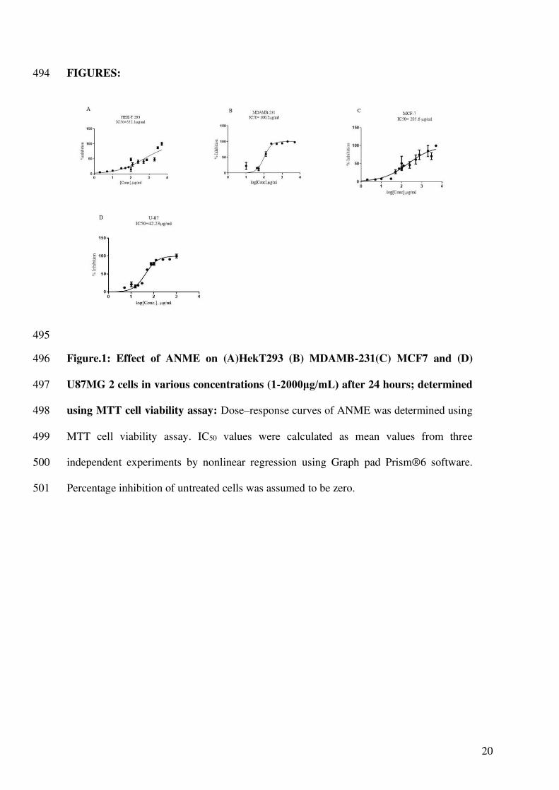
Figure.1: Effect of ANME on (A)HekT293 (B) MDAMB-231(C) MCF7 and (D) 496
U87MG 2 cells in various concentrations (1-2000μg/mL) after 24 hours; determined 497
using MTT cell viability assay: Dose–response curves of ANME was determined using 498
MTT cell viability assay. IC50 values were calculated as mean values from three 499
independent experiments by nonlinear regression using Graph pad Prism®6 software. 500
Percentage inhibition of untreated cells was assumed to be zero. 501
21
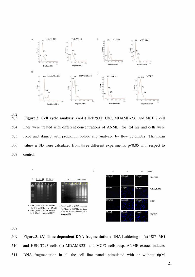
502 Figure.2: Cell cycle analysis: (A-D) Hek293T, U87, MDAMB-231 and MCF 7 cell 503
lines were treated with different concentrations of ANME for 24 hrs and cells were 504
fixed and stained with propidium iodide and analyzed by flow cytometry. The mean 505
values ± SD were calculated from three different experiments. p<0.05 with respect to 506
control. 507
508
Figure.3: (A) Time dependent DNA fragmentation: DNA Laddering in (a) U87- MG 509
and HEK-T293 cells (b) MDAMB231 and MCF7 cells resp. ANME extract induces 510
DNA fragmentation in all the cell line panels stimulated with or without 6µM 511
22
Tunicamycin(4hr) for induction of Unfolded Protein cells. The cells were treated with 512
different concentrations (µg/ml) of extract for every cell type for 0, 24 and 48 h. Cells 513
from each sample were harvested for DNA gel electrophoresis as described; 514
(B) Formation of apoptotic bodies in time dependent manner, DAPI staining: 515
Morphological and nuclear changes induced by ANME. HEK- T293, MDAMB, MCF7 516
and U87-MG cells after treatment with different concentration for each cell type induced 517
various nuclear changes such as chromatin condensation, nuclei condensation, and 518
nuclear degradation, as demonstrated by DAPI staining at 400× in time dependent 519
manner. However Hek293 showed only inhibition in cell growth with no signs of 520
apoptosis; ANME=Aquilegia nivalis methanolic extract. 521
522
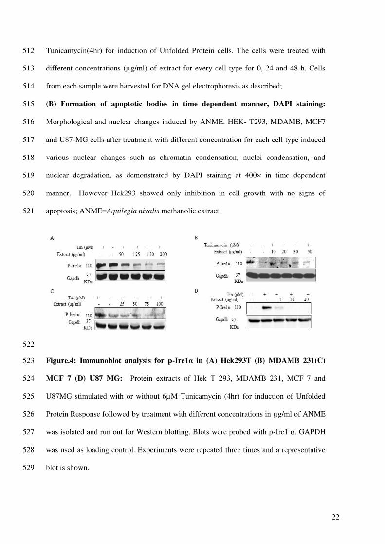
Figure.4: Immunoblot analysis for p-Ire1α in (A) Hek293T (B) MDAMB 231(C) 523
MCF 7 (D) U87 MG: Protein extracts of Hek T 293, MDAMB 231, MCF 7 and 524
U87MG stimulated with or without 6µM Tunicamycin (4hr) for induction of Unfolded 525
Protein Response followed by treatment with different concentrations in µg/ml of ANME 526
was isolated and run out for Western blotting. Blots were probed with p-Ire1 α. GAPDH 527
was used as loading control. Experiments were repeated three times and a representative 528
blot is shown. 529
23
530
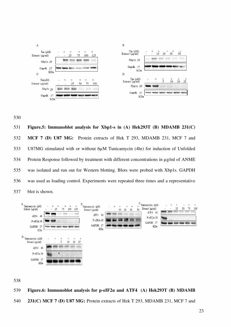
Figure.5: Immunoblot analysis for Xbp1-s in (A) Hek293T (B) MDAMB 231(C) 531
MCF 7 (D) U87 MG: Protein extracts of Hek T 293, MDAMB 231, MCF 7 and 532
U87MG stimulated with or without 6µM Tunicamycin (4hr) for induction of Unfolded 533
Protein Response followed by treatment with different concentrations in µg/ml of ANME 534
was isolated and run out for Western blotting. Blots were probed with Xbp1s. GAPDH 535
was used as loading control. Experiments were repeated three times and a representative 536
blot is shown. 537
538
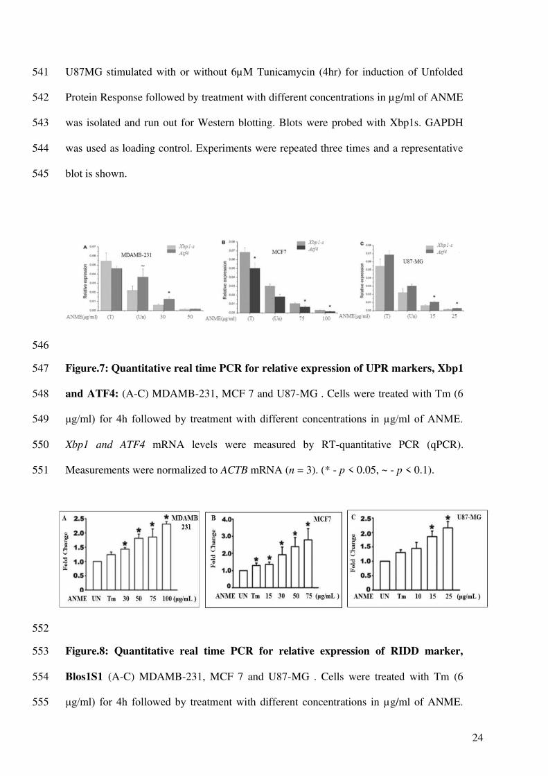
Figure.6: Immunoblot analysis for p-eIF2α and ATF4 (A) Hek293T (B) MDAMB 539
231(C) MCF 7 (D) U87 MG: Protein extracts of Hek T 293, MDAMB 231, MCF 7 and 540
24
U87MG stimulated with or without 6µM Tunicamycin (4hr) for induction of Unfolded 541
Protein Response followed by treatment with different concentrations in µg/ml of ANME 542
was isolated and run out for Western blotting. Blots were probed with Xbp1s. GAPDH 543
was used as loading control. Experiments were repeated three times and a representative 544
blot is shown. 545
546
Figure.7: Quantitative real time PCR for relative expression of UPR markers, Xbp1 547
and ATF4: (A-C) MDAMB-231, MCF 7 and U87-MG . Cells were treated with Tm (6 548
μg/ml) for 4h followed by treatment with different concentrations in µg/ml of ANME. 549
Xbp1 and ATF4 mRNA levels were measured by RT-quantitative PCR (qPCR). 550
Measurements were normalized to ACTB mRNA (n = 3). (* - p < 0.05, ~ - p < 0.1). 551
552
Figure.8: Quantitative real time PCR for relative expression of RIDD marker, 553
Blos1S1 (A-C) MDAMB-231, MCF 7 and U87-MG . Cells were treated with Tm (6 554
μg/ml) for 4h followed by treatment with different concentrations in µg/ml of ANME. 555

25
Blos1S1mRNA levels were measured by RT-quantitative PCR (qPCR). Measurements 556
were normalized to ACTB mRNA (n = 3). (* - p< 0.05). 557
558
559
Figure.9: Activation of caspases by ANME: Protein expression of anti- Caspase 3 and 560
anti- PARP using ANME in (A) HEK- 293T (B) U87 MG (C) MDAMB231 and MCF7 561
cells, resp. Protein extracts of HEK T 293, U87 MG, MDAMB 231 and MCF7 stimulated 562
with or without 6µM Tunicamycin for induction of Unfolded Protein Response followed 563
by treatment with different concentrations of ANME were isolated and run out for 564
Western blotting. Blots were probed with anti- Caspase 3 and anti- PARP. GAPDH was 565
used as loading control. Experiments were repeated three times and a representative blot 566
is shown. 567
References: 568
1. Mudasar Ahmad, Zahoor A. Kaloo1, Bashir A. Ganai, Ubaid Yaqoob , Hilal A. Ganaie 569
(2017). Phytochemical Screening of Aquilegia nivalis Flax Jackson: An Important 570
Medicinal Plant of Kashmir Himalaya: A Perspective, Adv. Biomed. Pharma. 4:1, 6-12 571
2. Chandra Prakash Kala (2006) Medicinal plants of the high altitude cold desert in India: 572
Diversity, distribution and traditional uses, The International Journal of Biodiversity 573
Science and Management, 2:1, 43-56 574
26
3. Adamska T, Młynarczyk M, Jodynis‐Liebert J, Bylka W, Matławska I, 2003. 575
Hepatoprotective effect of the extract and isocytisoside from Aquilegia vulgaris. 576
Phytotherapy research 17(6), 691-696. 577
4. Jadwiga Jodynis-Liebert, Teresa Adamska, Małgorzata Ewertowska, Wiesława Bylka, 578
Irena Matławska, 2009. Aquilegia vulgaris extract attenuates carbon tetrachloride-579
induced liver fibrosis in rats, Experimental and Toxicologic Pathology, 61(5) 443-580
451. 581
5. Sekena H. A., Aziza A. E., Ibrahim A. B., Mohamed I. M. and Mosaad A. W., 2011. 582
Aquilegia vulgaris extract protects against the oxidative stress and the mutagenic 583
effects of cadmium in Balb/c mice, Experimental and Toxicologic Pathology, 63(4), 584
337-344. 585
6. Purushotham, G., Padma, Y., Nabiha, Y. et al. 2016.. In vitro evaluation of anti-586
proliferative, anti-inflammatory and pro-apoptotic activities of the methanolic 587
extracts of Andrographis nallamalayana Ellis on A375 and B16F10 melanoma cell 588
lines. 3 Biotech (6) 212. 589
7. Younis Mohammad Hazari, Arif Bashir, Ehtisham ul Haq, Khalid Majid Fazili., 2016. 590
Emerging tale of UPR and cancer: an essentiality for malignancy. Tumor Biology, 37, 591
11, 14381-14390 592
8. Chevet, E., Hetz, C., Samali, A., 2015. Endoplasmic Reticulum Stress-Activated 593
Cell Reprogramming in Oncogenesis. Cancer Discov. 5, 586-597 594
9. Dejeans, N., Barroso, K., Fernandez-Zapico, M.E., Samali, A., Chevet, E., 2015. 595
Novel roles of the unfolded protein response in the control of tumor development 596
and aggressiveness. Semin Cancer Biol. 33, 67-73. 597
10. Hetz, C., Chevet, E., Oakes, S.A., 2015. Proteostasis control by the unfolded 598
protein response. Nat Cell Biol. 17, 829-38. 599
27
11. Balch, W.E., Morimoto, R.I., Dillin, A., Kelly, J.W., 2008. Adapting proteostasis for 600
disease intervention. Science. 319, 916-9. 601
12. Bartoszewska, S., Collawn, J.F., 2020. Unfolded protein response (UPR) integrated 602
signaling networks determine cell fate during hypoxia. Cell Mol Biol Lett 25, 18 603
13. Schubert, U., Anton, L.C., Gibbs, J., Norbury, C.C., Yewdell, J.W., Bennink, J.R., 604
2000. Rapid degradation of a large fraction of newly synthesized proteins by 605
proteasomes. Nature. 404, 770-4. 606
14. Claudio Hetz and Feroz R. Papa, 2018., The Unfolded Protein Response and Cell Fate 607
Control., Molecular cell, 69, 2, 169-181, 608
15. Samirul Bashir, Mariam Banday, Ozaira Qadri, Arif Bashir, Nazia Hilal, Nida-i-Fatima, 609
Stephen Rader, Khalid Majid Fazili, 2021., The molecular mechanism and functional 610
diversity of UPR signaling sensor IRE1, Life Sciences,, Volume 265,, 2021, 118740 611
16. Greenman C, Stephens P, Smith R, Dalgliesh GL et al. 2007. Patterns of somatic 612
mutation in human cancer genomes. Nature. 8; 446(7132),153-8. 613
17. Guichard C, Amaddeo G, Imbeaud S, Ladeiro Y, Pelletier L, Maad IB, Calderaro 614
J, Bioulac-Sage P, Letexier M, Degos F, Clément B, Balabaud C, Chevet E, Laurent 615
A, Couchy G, Letouzé E, Calvo F, Zucman-Rossi J. 2012. Integrated analysis of 616
somatic mutations and focal copy-number changes identifies key genes and 617
pathways in hepatocellular carcinoma. Nat Genet. 44(6), 694-8. 618
18. Parsons DW, Jones S, Zhang X, Lin JC et al. 2008. An integrated genomic analysis 619
of human glioblastoma multiforme. Science. 321(5897),1807-12. 620
19. Ortiz-Urda S, Garcia J, Green CL, Chen L, Lin Q, Veitch DP, Sakai LY, Lee 621
H, Marinkovich MP, Khavari PA. 2005. Type VII collagen is required for Ras-622
driven human epidermal tumorigenesis. Science. 307(5716), 1773-6. 623
20. Snuderl M, Fazlollahi L, Le LP, Nitta M, Zhelyazkova BH, Davidson 624
CJ, Akhavanfard S, Cahill DP, Aldape KD, Betensky RA, Louis DN, Iafrate AJ. 2011. 625
28
Mosaic amplification of multiple receptor tyrosine kinase genes in glioblastoma. 626
Cancer Cell. 20(6), 810-7. 627
21. Hanahan D, Weinberg RA. 2011. Hallmarks of cancer: the next generation. 628
Cell. 144(5), 646-74. 629
22. Venditti R, Scanu T, Santoro M, Di Tullio G, Spaar A, Gaibisso R, Beznoussenko 630
GV, Mironov AA, Mironov A Jr, Zelante L, Piemontese MR, Notarangelo 631
A, Malhotra V, Vertel BM, Wilson C, De Matteis MA. 2012. Sedlin controls the ER 632
export of procollagen by regulating the Sar1 cycle. Science. 337(6102), 1668-72. 633
23. Levental KR, Yu H, Kass L, Lakins JN, Egeblad M, Erler JT, Fong SF, Csiszar 634
K, Giaccia A, Weninger W, Yamauchi M, Gasser DL, Weaver VM. 2009. Matrix 635
crosslinking forces tumor progression by enhancing integrin signaling. 636
Cell. 139(5),891-906. 637
24. Shi, Z., Yu, X., Yuan, M. et al., 2019., Activation of the PERK-ATF4 pathway 638
promotes chemo-resistance in colon cancer cells. Sci Rep 9, 3210 639
25. Hart LS, Cunningham JT, Datta T, Dey S, Tameire F, Lehman SL, Qiu B, Zhang H, 640
Cerniglia G, Bi M, Li Y, Gao Y, Liu H, Li C, Maity A, Thomas-Tikhonenko A, Perl 641
AE, Koong A, Fuchs SY, Diehl JA, Mills IG, Ruggero D, Koumenis C. 2012, ER 642
stress-mediated autophagy promotes Myc-dependent transformation and tumor 643
growth. J Clin Invest.,122, 12, 4621-34. 644
26. Rozpedek W, Pytel D, Mucha B, Leszczynska H, Diehl JA, Majsterek I. The Role of 645
the PERK/eIF2α/ATF4/CHOP Signaling Pathway in Tumor Progression During 646
Endoplasmic Reticulum Stress. Curr Mol Med. 2016;16(6):533-44. 647
27. Huber AL, Lebeau J, Guillaumot P, Pétrilli V, Malek M, Chilloux J, Fauvet F, Payen 648
L, Kfoury A, Renno T, Chevet E, Manié SN.,2013, p58(IPK)-mediated attenuation 649
of the proapoptotic PERK-CHOP pathway allows malignant progression upon 650
low glucose. Mol Cell, 49, 6, 1049-59. 651
29
28. Arif Bashir, Younis Hazari, Debnath Pal, Dibyajyoti Maity, Samirul Bashir, Laishram 652
Rajendrakumar Singh, Naveed Nazir Shah, Khalid Majid Fazil, 2020., Aggregation of 653
M3 (E376D) variant of alpha1- antitrypsin. Sci Rep 10, 8290 654
29. Anderson D. J. et al. 2015. Targeting the AAA ATPase p97 as an approach to treat 655
cancer through disruption of protein homeostasis. Cancer Cell (28), 653–665. 656
30. Chou TF. 2011. Reversible inhibitor of p97, DBeQ, impairs both ubiquitin-657
dependent and autophagic protein clearance pathways. Proc. Natl. Acad. Sci. USA 658
(108), 4834–4839. 659
31. Benosman S. 2013 Interleukin-1 receptor-associated kinase-2 (IRAK2) is a critical 660
mediator of endoplasmic reticulum (ER) stress signaling. PloS one 8; e64256. 661
32. Tam AB. Koong AC. & Niwa M. 2014. Ire1 has distinct catalytic mechanisms for 662
XBP1/HAC1 splicing and RIDD. Cell reports (9, 850–858. 663
33. Hamid Bakshi , Smitha Sam, Roya Rozati, Phalisteen Sultan, Tajamul Islam, Babita 664
Rathore, Zahoor Lone, Manik Sharma, Jagrati Triphati, Ramesh Chand Saxena, 2010. 665
DNA fragmentation and cell cycle arrest: a hallmark of apoptosis induced by crocin 666
from kashmiri saffron in a human pancreatic cancer cell line; Asian Pac J Cancer 667
Prev. 2010;11(3):675-9. 668
34. Albrahim T & Alnasser M & Al-Anazi, Mashael & Alkahtani, Muneera & Alkahtani, 669
Saad & Al-Qahtani, Ahmed. 2020. Potential anti-inflammatory and anti-apoptotic 670
effect of Coccinia grandis plant extract in LPS stimulated-THP-1 cells. 671
Environmental Science and Pollution Research. 27. 10.1007/s11356-020-08445-5. 672
35. Ashok Singh , Manohar Lal & S. S. Samant (2009) Diversity, indigenous uses and 673
conservation prioritization of medicinal plants in Lahaul valley, proposed Cold 674
Desert Biosphere Reserve, India, International Journal of Biodiversity Science & 675
Management, 5:3, 132-154 676
30
36. Ewertowska M, Jodynis-Liebert J, Kujawska M, Adamska T, Matławska I, Szaufer-677
Hajdrych M., 2009, Effect of Aquilegia vulgaris (L.) ethyl ether extract on liver 678
antioxidant defense system in rats. Int J Occup Med Environ Health, 22(2):115-23. 679
37. Marina Potest, Antonella Minutolo, Angelo Gismondi, Lorena Canuti, Maurice 680
Kenzo, Valentina Roglia, Federico Macchi, Sandro Grelli, Antonella Canini, Vittorio 681
Colizzi, Carla Montesano. 2019. Cytotoxic and apoptotic effects of different 682
extracts of Moringa oleifera Lam on lymphoid and monocytoid cells, 683
Experimental and Therapeutic Medicine, 5-17 684
38. Li, S., Pasquin, S., Eid, H. M., Gauchat, J. F., Saleem, A., & Haddad, P. S. 2018. Anti-685
apoptotic potential of several antidiabetic medicinal plants of the eastern James Bay 686
Cree pharmacopeia in cultured kidney cells. BMC complementary and alternative 687
medicine, 18(1), 37. 688
39. Kowalczyk, T., Sitarek, P., Skała, E., Toma, M., Wielanek, M., Pytel, D., 689
Wieczfińska, J., Szemraj, J., & Śliwiński, T. 2019. Induction of apoptosis by in vitro 690
and in vivo plant extracts derived from Menyanthes trifoliata L. in human cancer 691
cells. Cytotechnology, 71(1), 165–180. 692
40. Fribley, A., Zhang, K., & Kaufman, R. J. (2009). Regulation of apoptosis by the 693
unfolded protein response. Methods in molecular biology (Clifton, N.J.), 559, 191–694
204. https://doi.org/10.1007/978-1-60327-017-5_14 695
41. Raffaella Iurlaro and Cristina Munoz-Pinedo.2016. Cell death induced by 696
endoplasmic reticulum stress, FEBS Journal 283, 2640–2652 697
42. Bartoszewska, S., Collawn, J.F. 2020. Unfolded protein response (UPR) integrated 698
signaling networks determine cell fate during hypoxia. Cell Mol Biol Lett (25), 18 699
43. Wang L, Perera BG, Hari SB, Bhhatarai B, Backes BJ, Seeliger MA. 2012. Divergent 700
allosteric control of the IRE1alpha endoribonuclease using kinase inhibitors. Nat 701
Chem Biol. (8), 982–9. 702

31
44. Bouchecareilh M, Higa A, Fribourg S, Moenner M, Chevet E. 2011. Peptides derived 703
from the bifunctional kinase/RNase enzyme IRE1alpha modulate IRE1alpha 704
activity and protect cells from endoplasmic reticulum stress. FASEB J. (25),3115–705
29. 706
45. Arvin B. Tam, Lindsay S. Roberts, Vivek Chandra, Io Guane Rivera, Daniel K. 707
Nomura, Douglass J. Forbes, Maho Niwa 2018, The UPR Activator ATF6 Responds 708
to Proteotoxic and Lipotoxic Stress by Distinct Mechanisms . Developmental Cell 709
46, 327–343 710
46. Anna Shemorry ,Jonathan M Harnoss, Ofer Guttman, Scot A Marsters, László G 711
Kőműves, David A Lawrence, Avi Ashkenazi (2019) Caspase-mediated cleavage of 712
IRE1 controls apoptotic cell commitment during endoplasmic reticulum 713
stress eLife 2019;8:e47084 714
47. Anna Walczak, Kinga Gradzik, Jacek Kabzinski, Karolina Przybylowska-Sygut, Ireneusz 715
Majsterek, 2019, The Role of the ER-Induced UPR Pathway and the Efficacy of Its 716
Inhibitors and Inducers in the Inhibition of Tumor Progression, Oxidative Medicine 717
and Cellular Longevity, 2019, 5729710, 1-15 718
48. Emma Madden , Susan E. Logue , Sandra J. Healy , Serge Manie , Afshin Samali, 2019. 719
The role of the unfolded protein response in cancer progression: From oncogenesis to 720
chemoresistance, Biology of the Cell, 111,1,1-17. 721
49. Upton JP, Wang L, Han D, Wang ES, Huskey NE, Lim L, Truitt M, McManus 722
MT, Ruggero D, Goga A, Papa FR, Oakes SA. 2012. IRE1α cleaves select 723
microRNAs during ER stress to derepress translation of proapoptotic Caspase-2. 724
Science. 338, 6108, 818-22. 725
50. Bi M, Naczki C, Koritzinsky M, Fels D, Blais J, Hu N, Harding H, Novoa I, Varia M, 726
Raleigh J, Scheuner D, Kaufman RJ, Bell J, Ron D, Wouters BG, Koumenis C.,2005. ER 727

32
stress-regulated translation increases tolerance to extreme hypoxia and promotes 728
tumor growth. EMBO J. 24, 19 729
730
Figures
Figure 1
Effect of ANME on (A)HekT293 (B) MDAMB-231(C) MCF7 and (D) U87MG 2 cells in variousconcentrations (1-2000μg/mL) after 24 hours; determined using MTT cell viability assay: Dose–responsecurves of ANME was determined using MTT cell viability assay. IC50 values were calculated as meanvalues from three independent experiments by nonlinear regression using Graph pad Prism®6 software.Percentage inhibition of untreated cells was assumed to be zero.
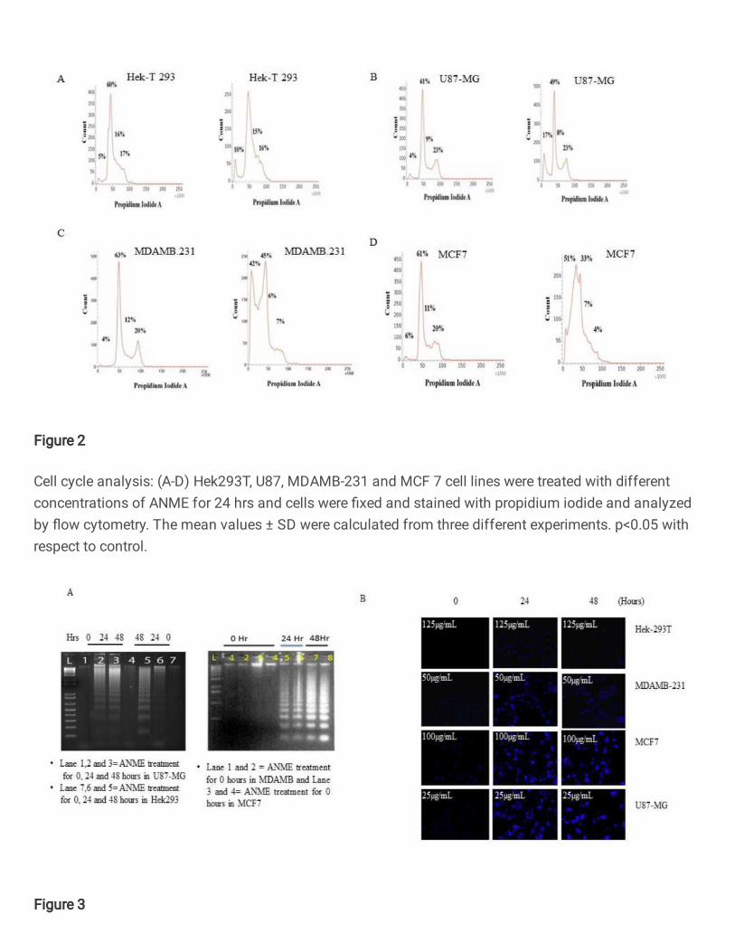
Figure 2
Cell cycle analysis: (A-D) Hek293T, U87, MDAMB-231 and MCF 7 cell lines were treated with differentconcentrations of ANME for 24 hrs and cells were �xed and stained with propidium iodide and analyzedby �ow cytometry. The mean values ± SD were calculated from three different experiments. p<0.05 withrespect to control.
Figure 3
(A) Time dependent DNA fragmentation: DNA Laddering in (a) U87- MG and HEK-T293 cells (b)MDAMB231 and MCF7 cells resp. ANME extract induces DNA fragmentation in all the cell line panelsstimulated with or without 6μM Tunicamycin(4hr) for induction of Unfolded Protein cells. The cells weretreated with different concentrations (μg/ml) of extract for every cell type for 0, 24 and 48 h. Cells fromeach sample were harvested for DNA gel electrophoresis as described; (B)Formation of apoptotic bodiesin time dependent manner, DAPI staining:Morphological and nuclear changes induced by ANME. HEK-T293, MDAMB, MCF7 and U87-MG cells after treatment with different concentration for each cell typeinduced various nuclear changes such as chromatin condensation, nuclei condensation, and nucleardegradation, as demonstrated by DAPI staining at 400× in time dependent manner. However Hek293showed only inhibition in cell growth with no signs of apoptosis; ANME=Aquilegia nivalis methanolicextract.
Figure 4
Immunoblot analysis for p-Ire1α in (A) Hek293T (B) MDAMB 231(C) MCF 7 (D) U87 MG: Protein extractsof Hek T 293, MDAMB 231, MCF 7 and U87MG stimulated with or without 6μM Tunicamycin (4hr) forinduction of Unfolded Protein Response followed by treatment with different concentrations in μg/ml ofANME was isolated and run out for Western blotting. Blots were probed with p-Ire1 α. GAPDH was used asloading control. Experiments were repeated three times and a representative blot is shown.
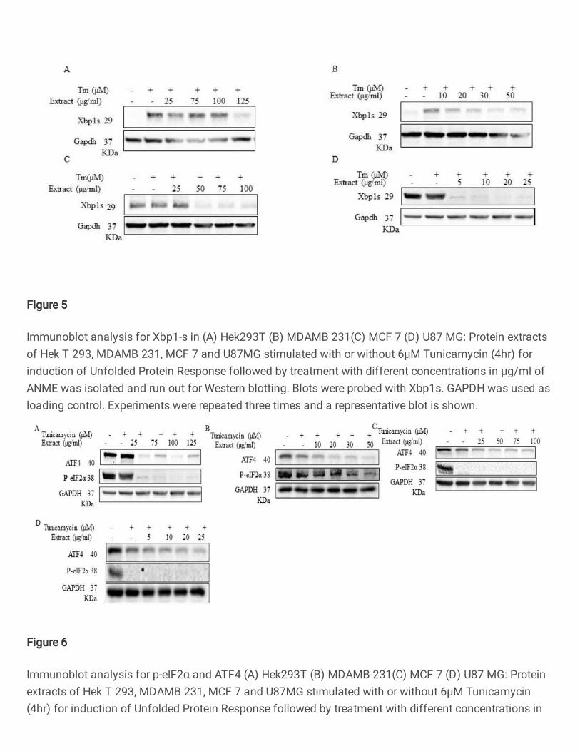
Figure 5
Immunoblot analysis for Xbp1-s in (A) Hek293T (B) MDAMB 231(C) MCF 7 (D) U87 MG: Protein extractsof Hek T 293, MDAMB 231, MCF 7 and U87MG stimulated with or without 6μM Tunicamycin (4hr) forinduction of Unfolded Protein Response followed by treatment with different concentrations in μg/ml ofANME was isolated and run out for Western blotting. Blots were probed with Xbp1s. GAPDH was used asloading control. Experiments were repeated three times and a representative blot is shown.
Figure 6
Immunoblot analysis for p-eIF2α and ATF4 (A) Hek293T (B) MDAMB 231(C) MCF 7 (D) U87 MG: Proteinextracts of Hek T 293, MDAMB 231, MCF 7 and U87MG stimulated with or without 6μM Tunicamycin(4hr) for induction of Unfolded Protein Response followed by treatment with different concentrations in
μg/ml of ANME was isolated and run out for Western blotting. Blots were probed with Xbp1s. GAPDH wasused as loading control. Experiments were repeated three times and a representative blot is shown.
Figure 7
Quantitative real time PCR for relative expression of UPR markers, Xbp1 and ATF4: (A-C) MDAMB-231,MCF 7 and U87-MG . Cells were treated with Tm (6 μg/ml) for 4h followed by treatment with differentconcentrations in μg/ml of ANME. Xbp1 and ATF4 mRNA levels were measured by RT-quantitative PCR(qPCR). Measurements were normalized to ACTB mRNA (n = 3). (* - p < 0.05, ~ - p < 0.1).
Figure 8
Quantitative real time PCR for relative expression of RIDD marker, Blos1S1 (A-C) MDAMB-231, MCF 7 andU87-MG . Cells were treated with Tm (6 μg/ml) for 4h followed by treatment with different concentrationsin μg/ml of ANME. Blos1S1mRNA levels were measured by RT-quantitative PCR (qPCR). Measurementswere normalized to ACTB mRNA (n = 3). (* - p< 0.05).
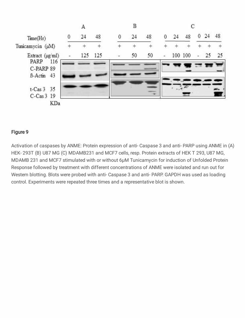
Figure 9
Activation of caspases by ANME: Protein expression of anti- Caspase 3 and anti- PARP using ANME in (A)HEK- 293T (B) U87 MG (C) MDAMB231 and MCF7 cells, resp. Protein extracts of HEK T 293, U87 MG,MDAMB 231 and MCF7 stimulated with or without 6μM Tunicamycin for induction of Unfolded ProteinResponse followed by treatment with different concentrations of ANME were isolated and run out forWestern blotting. Blots were probed with anti- Caspase 3 and anti- PARP. GAPDH was used as loadingcontrol. Experiments were repeated three times and a representative blot is shown.


