Embed Size (px)
Citation preview

Preliminary Report
Calf Augmentation and Reshapingwith Autologous Fat Grafting
Gerhard S. Mundinger, MD; and James E. Vogel, MD, FACS
AbstractBackground: Despite multiple advantages of fat grafting for calf augmentation and re-shaping over traditional silicone calf implants, few reports havebeen published.Objectives: To report our technique and results with autologous fat grafting for calf augmentation and reshaping.Methods: A retrospective review of the senior author’s ( JEV) experience with autologous fat grafting for calf augmentation was performed. Medial andlateral calf augmentation was accomplished with injection of prepared autologous lipoaspirate intramuscularly and subcutaneously.Results: Over a 5-year period, 13 patients underwent calf augmentation and reshaping with the described technique. Ten cases were bilateral (77%),and 3 cases (23%) were performed for congenital leg discrepancies. Mean 157 cc of prepared lipoaspirate was transferred per leg, with roughly 60% and40% transferred into the medial and lateral calf, respectively. Four patients (31%) underwent a second round of autologous fat injection for further calfaugmentation because they desired more volume. At mean 19.6 month follow-up, durable augmentation and improvement in calf contour was documentedby comparison of standardized preoperative and postoperative photographs.Conclusions: Autologous calf fat grafting is a viable alternative to traditional implant-based calf augmentation for congenital calf discrepancies and theaesthetic pseudo-varus deformity. This technique provides results comparable to those obtainable with traditional methods.
Level of Evidence: 4
TherapeuticAccepted for publication July 16, 2015.
Advancements in plastic surgery are often made throughthe application of existing principles and techniques to aproblem or area of the body where they had not previouslybeen considered. The use of autologous fat grafting for calfaugmentation and reshaping is one such application.
The slender, unaesthetic calf, which we frequently con-sider contributes to a “pseudo-varus” lower leg appearance(Figure 1), can be a source of unhappiness for men andwomen alike. Correction of slender unaesthetic lower legs,either for purely cosmetic purposes, or for discrepancies inappearance between legs due to congenital deformities, in-fections, or trauma, is an increasingly expressed patientdesire.1 Silicone calf implants have most frequently beenused for this purpose, but carry risks of well-documentedand frequent complications, including hyperpigmentation,
seromas, extrusion, infection, capsular contracture, removalfor cosmetic dissatisfaction, and compartment syndrome.2-5
Fat grafting has revolutionized the conceptualization andtreatment of contour and soft-tissue volume issues, andoffers safe, durable outcomes with diminished scar burdencompared to traditional filling techniques.6,7 Fat grafting forcorrection of unaesthetic calves would seem to be an idealalternative to traditional calf aesthetic procedures, especially
From the Department of Plastic Surgery, The Johns Hopkins Hospitaland School of Medicine, Baltimore, MD, USA.
Corresponding Author:Dr James E. Vogel, 4 Park Center Court, Owings Mills, MD 21117, USA.E-mail: [email protected]
Body Contouring
Aesthetic Surgery Journal2015, 1–10© 2015 The American Society forAesthetic Plastic Surgery, Inc.Reprints and permission:[email protected]: 10.1093/asj/sjv166www.aestheticsurgeryjournal.com
Aesthetic Surgery Journal Advance Access published September 1, 2015 by guest on January 5, 2016
http://asj.oxfordjournals.org/D
ownloaded from

considering the risks of silicone calf implantation, yet sur-prisingly few reports on the use of autologous fat injectionfor correction of unaesthetic calves have been published.8-11
The purpose of this paper is to report the senior author’s( JEV) experience with autologous fat grafting for augmen-tation and reshaping of unaesthetic calves. Key points foradequate assessment and correction with this techniqueare highlighted.
METHODS
Records of the senior author ( JEV) were reviewed andpatients undergoing calf augmentation with fat graftingwere identified. Between April 2010 and February 2015, 13consecutive patients underwent calf augmentation and
reshaping with fat grafting. Patient records, including pre-operative and postoperative photographs, were evaluatedat last follow-up and compared to preoperative imaging.Informed consent was acquired from all patients. Institutionalresearch board approval for this study was not obtained, asthese patients were seen in the senior author’s private clinic.
Fat harvest was performed as follows: fat harvest siteswere infiltrated with liposuction tumescent fluid consisting of1 L of lactated ringer’s, 30 mL of 1% lidocaine, and 1 ampuleof epinephrine at a concentration of 1:1000 to turgidity of theharvest site (tumescent liposuction). Three and 4 mm accel-erator tip cannulas were used for fat harvest. Fat was variablyharvested from the abdomen, lateral thigh, medial thigh,waistline, flanks, axilla, upper back, and hips. Irrespective offat harvest site, liposuction was also performed at the medial
Figure 1. The pseudo-varus deformity in a 52-year-old woman. (A) Frontal, (B) left anterior oblique, (C) lateral, and (D) posteriorviews. Despite subjectively normal knee joint angles and lack of tibial bone growth discrepancies, the patient’s legs appear to be ina varus (bow-legged) relationship. This is due to a relative excess of medial knee subcutantous tissue, relative lack of calf volume(with more medial deficiency than lateral deficiency, though both lack relative volume), and poorly defined lateral calf contourbelow the popliteal crease.
2 Aesthetic Surgery Journal
by guest on January 5, 2016http://asj.oxfordjournals.org/
Dow
nloaded from
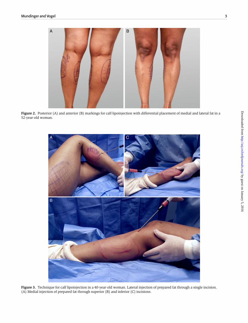
Figure 3. Technique for calf lipoinjection in a 40-year-old woman. Lateral injection of prepared fat through a single incision.(A) Medial injection of prepared fat through superior (B) and inferior (C) incisions.
Figure 2. Posterior (A) and anterior (B) markings for calf lipoinjection with differential placement of medial and lateral fat in a52-year-old woman.
Mundinger and Vogel 3
by guest on January 5, 2016http://asj.oxfordjournals.org/
Dow
nloaded from
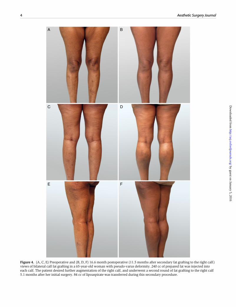
Figure 4. (A, C, E) Preoperative and (B, D, F) 16.6 month postoperative (11.5 months after secondary fat grafting to the right calf )views of bilateral calf fat grafting in a 65-year-old woman with pseudo-varus deformity. 240 cc of prepared fat was injected intoeach calf. The patient desired further augmentation of the right calf, and underwent a second round of fat grafting to the right calf5.1 months after her initial surgery. 84 cc of lipoaspirate was transferred during this secondary procedure.
4 Aesthetic Surgery Journal
by guest on January 5, 2016http://asj.oxfordjournals.org/
Dow
nloaded from

knee to improve calf contour. Fat was collected into a sterileglass container, and sterilely transferred to a strainer, whereexcess tumescent solution and blood were separated fromthe fat.
Separated fat was then transferred to 10 cc syringes for in-jection into the calves. Incision sites were 1 to 2 mm in lengthand created with 14 gauge Nokor needles (Becton, Dickinson,and Company, East Rutherford, NJ). Lipoinjection was per-formed with a 21 cm, 2 mm fat transfer needle attached tothe 10 cc fat-containing syringe. Typically two calf inci-sions (superior and inferior) were utilized for lipoinjection(Figures 2 and 3). Injections were first performed directlyinto the calf muscles and then into the subcutaneous calftissue. Fat volume was judged to be sufficient when thecalf was minimally firm but not tense. Volume was addedto the lateral and anterior compartments when judged tobe deficient and clinically indicated.
All procedures were performed under intravenous seda-tion with local anesthesia. All patients were positionedsupine or in a decubitus position only. No patients were po-sitioned prone as this approach is not used during intrave-nous sedation in our practice. Incisions were closed with asingle absorbable 6-0 fast suture. Marcaine 0.25% was pre-injected into lateral and medial gastrocnemius muscles toprovide pain relief postoperatively and to encourage earlyambulation. The local anesthesia was injected prior to fattransfer to utilize the smallest amount of effective anestheticvolume and to precisely place it into the muscle. Additionally,this sequence may result in a less sedation and more rapidpostoperative recovery.
Compression garments were not used in the areas of fatgrafting in the immediate postoperative period, nor werecompression boots. No patients were placed on anti-plateletmedication. All were well hydrated intraoperatively. All
Figure 5. (A, C) Preoperative and (B, D) 46.4 month postoperative views of bilateral fat grafting in a 40-year-old woman. 168 and180 cc of prepared lipoaspiratre were injected into the right and left calf, respectively.
Mundinger and Vogel 5
by guest on January 5, 2016http://asj.oxfordjournals.org/
Dow
nloaded from
patients were encouraged to ambulate the day of surgeryand daily thereafter. Starting on postoperative day 5, pa-tients were asked to wear support stockings to providerelief from leg swelling. They were asked to continue thispractice for three months after surgery. Sports and exercisewere permitted as tolerated. Patients were evaluated in-itially at 2 weeks following surgery and then at regularintervals.
RESULTS
Mean patient age was 45 years (range, 29 to 65 years). Tenpatients (77%) were women. Seven patients (54%) wereAfrican-American. Medical comorbidities included hyper-tension in 3 patients (23%) and atrial fibrillation in onepatient (8%). Ten patients (77%) underwent bilateral calfaugmentation. Three cases (23%) were performed for calf
asymmetries associated with congenital anomalies (spinabifida, club foot, and tethered spinal cord). No patient hadprior cosmetic procedures involving the calves.
Mean operating time was 106 minutes (range, 75 to 165minutes). Fat was harvested variably from the lateral thigh,medial thigh, medial knee, flanks, upper and lower abdomen,upper back, waistline, and/or hips. Mean lipoaspirate volumeharvested per procedure was 1307 cc (range, 84 to 4500 cc),and mean fat volume transferred per leg was 157 cc (range,78 to 330 cc). On average, 58% of transferred fat was placedinto the medial calf, and 42% was placed into the lateral calf.Four patients (31%) desired further calf volume and under-went a second round of fat grafting. A mean of 155 cc (range,84 to 200 cc) of fat was transferred to each leg during thesesecondary procedures.
Mean follow-up was 19.6 months (range, 3.5 to 53.2months), and median follow-up was 13.7 months. Eight
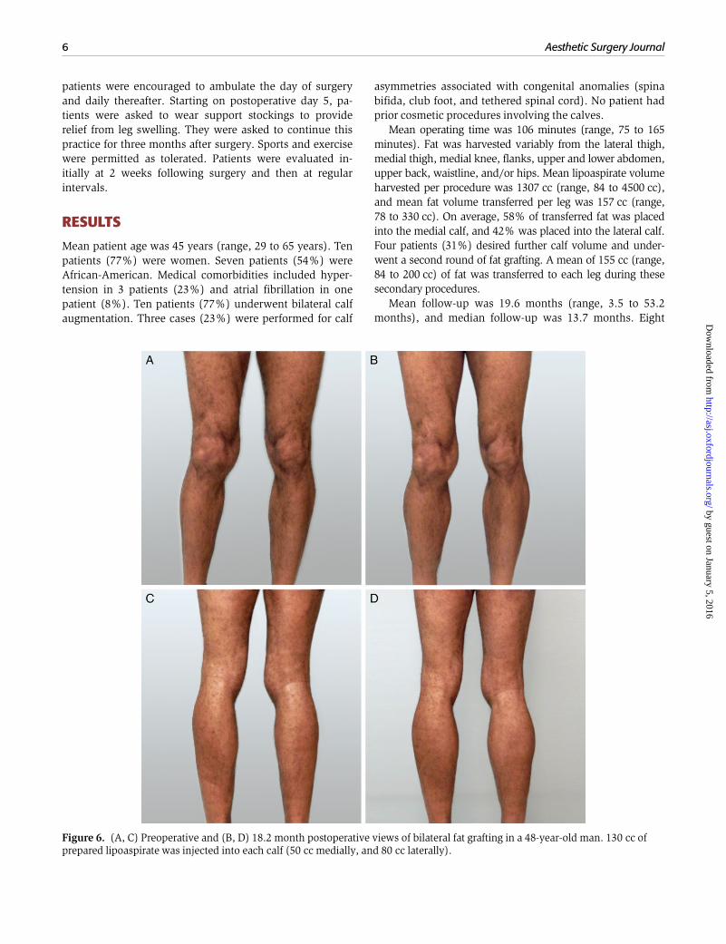
Figure 6. (A, C) Preoperative and (B, D) 18.2 month postoperative views of bilateral fat grafting in a 48-year-old man. 130 cc ofprepared lipoaspirate was injected into each calf (50 cc medially, and 80 cc laterally).
6 Aesthetic Surgery Journal
by guest on January 5, 2016http://asj.oxfordjournals.org/
Dow
nloaded from

patients had follow-up greater than 12 months, and 4 pa-tients had follow-up greater than 24 months. All patientsachieved durable improvement in calf volume, shape, andcontour, and agreed that fat take was evident (Figures 4-6and Supplemental Figures S1 and S2). There were no infec-tions. Three patients (23%) with dark skin tone complainedof hyperpigmentation of the injection sites but at follow-upbeyond 8 months, 2 of the 3 patients indicated these areas ofhyperpigmentation were no longer a concern. Patient demo-graphics and results are presented in Table 1.
DISCUSSION
As our series illustrates, both men and women, mayrequest calf augmentation for a number of reasons. Wehave found these reasons generally stem from leg discrep-ancies resulting from congenital disease or trauma, or anoverall dissatisfaction with the shape and appearance oftheir legs in the absence of underlying disease.9,10 In theformer, patients are most concerned with augmentation ofone calf, while they generally request augmentation of bothcalves in the latter, though this is not always the case.
For patients desiring calf augmentation, silicone calfimplants have been the most widely applied modality.
Although high rates of patient satisfaction have been notedby specific authors, implants carry well described risks, in-cluding those related to long-term implantation of foreignbodies.2-5 Autologous fat augmentation offers a number ofadvantages over calf implants, including liposuction in ad-jacent areas to improve calf contour, smaller incisions, ad-ditional augmentation through subsequent fat grafting,durable results, lack of foreign body reaction, and precisepatient-specific adjustments not possible with off-theshelf implants.
Despite the marked increase in autologous fat injectionprocedures for other areas of the body, little has beenwritten regarding calf augmentation with fat grafting.8-11
Additional articles have cursorily mentioned fat graftingwithout description of technique, and have failed toprovide adequate preoperative photographs for critical eval-uation of results.5,12 The first and largest published seriesdescribing a technique for autologous calf augmentationwith fat injection8 has furthermore been criticized for un-persuasive results.3 Similar results with combination endo-scopic fascial release, calf implantation, and fat grafting areequally underwhelming.13
The plastic surgery literature is replete with conceptuali-zations of the “ideal” leg, especially for women, and
Table 1. Individual Patient Demographic, Procedural, and Follow-up Information
PatientN0.
Age(years)
Gender Ethnicity Comorbidities Etiology ofCalf Deformity
Laterality ofAugmentation
Follow-up(months)
Complications Second Roundof
Augmentation
1 41 Male Caucasian None Tetheredspinal cord
Right 53.2 None Yes
2 52 Female African-american Hyptertension,atrialfibrillation
Cosmetic Bilateral 30 None No
3 40 Female African-american None Cosmetic Bilateral 46.4 Incisionalhyperpigmentation
Yes
4 29 Female Caucasian None Cosmetic Bilateral 4.1 None No
5 29 Female African-american None Right club foot Right 12.4 Incisionalhyperpigmentation
No
6 41 Female Arabic None Cosmetic Bilateral 34 None No
7 65 Female African-american Hyptertension Cosmetic Bilateral 16.6 None Yes
8 56 Female African-american None Cosmetic Bilateral 10.2 None No
9 48 Male African-american None Cosmetic Bilateral 18.2 None Yes
10 46 Male African-american Hyptertension Spina bifida Right 3.5 None No
11 35 Female Arabic None Cosmetic Bilateral 13.7 None No
12 43 Female Caucasian None Cosmetic Bilateral 4.6 None No
13 33 Male Hispanic None Cosmetic Bilateral 8.4 Incisionalhyperpigmentation
No
Mundinger and Vogel 7
by guest on January 5, 2016http://asj.oxfordjournals.org/
Dow
nloaded from

multiple methods for classification of calf deficits havebeen published. (Figures 7 and 8).1,14,15 These evaluationsbroadly consider the degree of soleus, gastrocnemius, pero-neus longus, and flexor digitorum longus muscle under-development, which contributes to medial, lateral, and/orposterior hypoplasia.14 Multiple ratios of leg length to calflength, ideal calf circumference, and ideal location ofmaximal calf projection have been published.1,11,14,15
We feel that these classification systems are cumbersomein clinical practice, and find that most patients without con-genital issues present with the pseudo-varus deformity(Figure 1). These patients appear to be bow-legged despitesubjectively normal knee and tibial alignment without ahistory of leg growth discrepancies. We feel that augmenta-tion of both the medial and lateral compartment with fat iscritical to success, as both areas lack appropriate volume,definition, and aesthetically pleasing contour. Previously
described techniques that augment the lateral aspect of thecalf9,10 have superior results, in our opinion, to those whereonly medial augmentation was performed.8,11,13 In clinicalpractice, patient satisfaction is a crucial determinant of pro-cedural success. In our experience, most patients were satis-fied with one round of calf fat grafting, although 29% (4patients) desired further augmentation. We feel that excel-lent results can be obtained in one procedure. This appearsto be congruous with practice patterns of those authorswho augment the lateral calf,9,10 as opposed to those whodo not.8,13
Critical to patient satisfaction is an anatomic discussionoutlining the location of the calf musculature and whereaugmentation is recommended. Often patients requestaugmentation of not only the gastrocnemius muscle butalso the lower leg and even upper ankle region. It has notbeen our practice to inject fat primarily into these areas
Figure 7. The “ideal”male calf as demonstrated by at 59-year-old subject. (A) Anterior, (B) anterior contracting, (C) posterior,and (D) posterior contracting views. The medial calf (gastrocnemius muscle) has a lower bulge or prominence than the lateral calf.The gastrocnemius muscles themselves end at the mid-tibia and thus augmentation ought to also end or taper off at this point.Anteriorly the lateral calf bulge begins at the lower margin of the patellar tendon insertion and the medial calf bulge begins about 1to 2 cm lower than the lateral bulge. The medial gastrocnemius is generally, but not always, larger and fat grafting should be doneto provide balance with the rest of the leg musculature and in accordance with the missing muscle mass as individualized for eachpatient.
8 Aesthetic Surgery Journal
by guest on January 5, 2016http://asj.oxfordjournals.org/
Dow
nloaded from

because of the unforgiving nature and perceived low likeli-hood of fat “take” in these lower leg tissues, although somefat grafting in these immediately surrounding tissues isperformed to provide contour blending. Additionally, andespecially in women, it is important to consider the shapeof the entire upper and lower leg to create a balanced andharmonious leg/calf contour. Specifically, liposuction ofthe medial and lateral thigh as well as the inner knee areaalone improve the pseudo-varus appearance of the lowerleg. When directed calf augmentation is added to thismore global contour approach, the result is objectivelyand well received by the patient (Figures 4 and 5).
It has been our practice to over-augment by approxi-mately 10%-15%. This is an arbitrary estimate of the over-fill volume and perhaps a more aggressive fill, such as the
20%-30% over-augmentation described by Hoppmannet al,11 would have eliminated the need for a second proce-dure in those patients that requested additional surgery. Onthe other hand, the calf becomes quite tense with a surpris-ingly small volume of fat injection. Concern for increasedpain and excessive pressure impairing the fat take are otherfactors we consider with the judgment of final utilizedvolumes. The pre-grafting injection of Marcaine (10 to 15cc/muscle, 0.25% with epi 1:200,000) has been an excel-lent method to reduce postoperative pain and permit ambu-lation upon discharge.
Limitations of this study include its retrospectivenature and lack of objective calf measurements. Patientsatisfaction was also not objectively assessed. This tech-nique may be less applicable for younger patients with
Figure 8. The “ideal” female calf as demonstrated by a 32-year-old subject. (A) Anterior, (B) anterior contracting, (C) posterior,and (D) posterior contracting views. Similar to conceptualizations of feminization of other body parts, the calf also reflects thisconcept of balance and harmony with the rest of the body. Male calf muscles are ideally more bulky or defined and prominent com-pared to the softer curves of the female calf.
Mundinger and Vogel 9
by guest on January 5, 2016http://asj.oxfordjournals.org/
Dow
nloaded from
more taught tissues, and its use has not been explored inbodybuilders. Four months was considered a stable resultand this was verified by the long-term follow-up reportedin this article.
CONCLUSIONS
Autologous calf fat grafting is a viable alternative to tradi-tional implant-based calf augmentation for congenital calfdiscrepancies and the pseudo-varus deformity. This tech-nique provides durable results that minimize patient risk.Lateral lipoinjection is a key component of effective correc-tion and may provide results superior to those obtainablewith traditional methods.
Supplementary Material
This article contains supplementary material located online atwww.aestheticsurgeryjournal.com.
DisclosuresThe authors declared no potential conflicts of interest withrespect to the research, authorship, and publication of thisarticle.
FundingThe authors received no financial support for the research,authorship, and publication of this article.
REFERENCES1. Howard PS. Calf augmentation and correction of contour
deformities. Clin Plast Surg. 1991;18(3):601-613.2. Niechajev I. Calf augmentation and restoration. Plast
Reconstr Surg. 2005;116(1):295-305.
3. Niechajev I. Calf augmentation with autologous tissue in-jection. Plast Reconstr Surg. 2009;123(6):1891-1892; authorreply 1892-3.
4. Aiache A. Leg contouring with calf implants. Clin PlastSurg. 1996;23(4):737-749.
5. Fraccalvieri M, Contessa L, Salomone M, Zingarelli EM,Bruschi S. Preoperative color duplex echographicalvenous mapping before autologous fat graft for calf aug-mentation: a case report of superficial vein thrombosisand prevalence of intersaphenic anastomosis. Ann PlastSurg. 2014;73(2):137-140.
6. Toledo LS, Mauad R. Fat injection: a 20-year revision.Clin Plast Surg. 2006;33(1):47-53. vi.
7. Coleman SR. Structural fat grafts: the ideal filler? ClinPlast Surg. 2001;28(1):111-119.
8. Erol OO, Gürlek A, Agaoglu G. Calf augmentation withautologous tissue injection. Plast Reconstr Surg. 2008;121(6):2127-2133.
9. Veber M Jr, Mojallal A. Calf augmentation with autolo-gous tissue injection. Plast Reconstr Surg. 2010;125(1):423-424; author reply 424-5.
10. Mojallal A, Veber M, Shipkov C, Ghetu N, Foyatier JL, BrayeF. Analysis of a series of autologous fat tissue transfer forlower limb atrophies. Ann Plast Surg. 2008;61(5):537-543.
11. Hoppmann R, Meruane M, González D, Wisnia P,Hasbún A, Villalobos B. Calf lipo-reshaping. J PlastReconstr Aesthet Surg. 2013;66(7):956-961.
12. Matsudo PK, Toledo LS. Experience of injected fat graft-ing. Aesthetic Plast Surg. 1988;12(1):35-38.
13. Karacaoglu E, Zienowicz RJ, Balan I. Calf contouringwith endoscopic fascial release, calf implant, andstructural fat grafting. Plast Reconstr Surg Glob Open.2013;1(5):e35.
14. Cuenca-Guerra R, Daza-Flores JL, Saade-Saade AJ. Calfimplants. Aesthetic Plast Surg. 2009;33(4):505-513.
15. Tsai CC, Lin SD, Lai CS, Lin TM. Aesthetic analysis ofthe ideal female leg. Aesthetic Plast Surg. 2000;24(4):303-305.
10 Aesthetic Surgery Journal
by guest on January 5, 2016http://asj.oxfordjournals.org/
Dow
nloaded from