Embed Size (px)
Citation preview
Biomarkers to track the abandonment of a Roman wastewater course 149Archaeometry 45, 1 (2003) 149–161. Printed in Great Britain
THE APPLICATION OF STEROIDAL BIOMARKERS TOTRACK THE ABANDONMENT OF A ROMAN WASTEWATER
COURSE AT THE AGORA (ATHENS, GREECE)*
I. D. BULL, M. M. ELHMMALI, D. J. ROBERTS and R. P. EVERSHED†
Organic Geochemistry Unit, Biogeochemistry Research Centre, School of Chemistry,University of Bristol, Cantock’s Close, Bristol BS8 1TS, UK
A subterranean, stone-built, sediment-filled culvert discovered during excavations at theAgora (Athens, Greece) was dated to the Roman period on the basis of its characteristicconstruction and associated finds, including coins. The location of the culvert relative toother adjacent watercourses and an ancient river bed suggested that the structure was asewer. This was confirmed through a multi-molecular biomarker approach based on analysisof the bile acids associated with sediment filling the sewer, using gas chromatography (GC)and gas chromatography/mass spectrometry (GC/MS). The acid fraction contained predomin-antly deoxycholic and lithocholic acids, while the neutral steroid fraction comprised a com-plex mixture of ∆5 sterol and 5β- and 5α-stanols, dominated by coprostanol, suggesting thepresence of faecal matter of predominantly human origin. The concentrations of neutral andacidic faecal biomarkers were observed to vary in tandem, with the highest concentrationsbeing found in the sediment at the base of the fill in the culvert. A reduction in concentrationoccurred with decreasing depth of the fill, with concentrations in the uppermost samplesbeing little different from control samples of sediment taken beyond the confines of theculvert. The enhanced concentration of bile acids relative to 5β-stanols compared with freshhuman faeces must reflect the enhanced diagenetic stability of the former, thereby makingbile acids the possibly preferred biomarker for this type of study. The quantitative dataobtained suggest that the culvert fell rapidly out of use, possibly coinciding with the Slavicincursion in AD 582–3.
KEYWORDS: 5β-STANOLS, AGORA, BILE ACIDS, BIOMARKER, GC, GC/MS,FAECAL, LIPID, SEDIMENT, SEWAGE, STEROID
INTRODUCTION
One of the most significant technological achievements of the Romans was their ability toconstruct major civil engineering works—that is, aqueducts and watercourses—to supply freshwater to optimal locations within towns and cities; for example, public baths. Equally import-ant, but perhaps less visually striking, was their ability to deal with the large quantities of wastewater contaminated with human excrement produced by major towns and cities. Such projectsinvolved the construction of complex networks of subterranean sewers capable of long-termuse, in some cases over several centuries (Carcopino 1956), delivering waste from point loca-tions within domestic, public and administrative centres to discharge points in adjacent rivers,where the noxious wastes could be swept to the sea, usually by natural river flow.
* Received 16 January 2001; accepted 10 July 2002.† To whom correspondence should be addressed.© University of Oxford, 2003
150 I. D. Bull et al.
The sewers themselves range from huge structures of the type, many metres in height anddiameter, that are still in use in parts of Rome to the smaller arterial drains designed to carrylesser amounts of waste from more remote parts of towns; these peripheral culverts oftenformed a complex network that finally adjoined a major discharge or river course (White 1984;Humphrey et al. 1998).
Work by this laboratory has shown bile acids to be a useful marker of faecal pollution inthe natural environment, with a stability surpassing that of related faecal biomarkers such as5β-stanols (Elhmmali et al. 1997; Bull et al. 2002). More recent studies have exploited thehighly diagnostic nature of bile acids to determine the source of manure inputs to ancient,anthropogenic deep topsoil formations in West Mainland Orkney, UK (Simpson et al. 1999),and to differentiate between agricultural and urban faecal inputs to sediment and particulatesin the River Avon, UK (Elhmmali et al. 2000). In this study, a biomolecular analysis ofsediment taken from in and around a suspected late Roman street drain was made. Thedrain was substantial, built with a large U-shaped tile channel at the bottom, side walls of tilesset in mortar and a cover of shallow, curved terracotta tiles. Analysis of lamps recoveredfrom the silt accreted in the channel revealed that it had been abandoned during the late sixthto early seventh century ad, which most likely coincides with the mass abandonment ofthe Agora during the late sixth century, presumably due to the Slavic incursion in ad 582–3(Camp 1996). This investigation tests a twofold hypothesis: (a) that the structure was indeedused in the transport of sewage effluent; and (b) that bile acids are more recalcitrant (and hencemore useful as an indicator of faecal pollution) in archaeological soils and sediments than 5β-stanols, which have hitherto received far more interest as markers of faecal deposition inantiquity.
MATERIALS AND METHODS
Sample collection
Seven sediment/soil samples were taken from in and around the suspected Roman drain. Fivesamples were excised from the sediment accreted within the culvert. The first of these wascollected from the surface top layer of the drain, and then successive samples were taken atincreasing depths, starting from 15 cm below the first sample. The fifth sample was taken fromthe very bottom. Two control samples were taken 5 m east and west from the suspected drainand were denoted as controls 1 and 2, respectively. All samples were air dried and stored atroom temperature.
Extraction and isolation of bile acids and sterols
Soils were ground to a fine powder with a ceramic pestle and mortar and then sieved througha 250 µm sieve. Powdered soil was inoculated with 20–120 µg of two internal standards(hyocholic acid and 5β-pregnan-3α-ol) and solvent extracted for 24 h in a standard Soxhletapparatus, utilizing a dichloromethane/methanol (2:1 v/v) solvent system: solvent was removedfrom the total lipid extract (TLE) under reduced pressure. Samples were then saponified bythe addition of 5 ml of 5 M potassium hydroxide in 90% methanol to the TLE, followedby heating at 120°C for 1 h. After cooling, a further 10–15 ml of double distilled water wasadded to the organic extract, which was then acidified to pH 3–4 with 6 M hydrochloric acid,monitored using a pH meter. Saponified organics were then extracted into chloroform (3 ×
Biomarkers to track the abandonment of a Roman wastewater course 151
10 ml), combined and the chloroform removed under reduced pressure. The residue wasthen re-dissolved in dichloromethane/methanol (2:1 v/v) and passed through a short glasscolumn filled with anhydrous sodium sulphate to remove any residual water that might affectthe efficiency of ensuing separation processes; solvent was removed under a gentle stream ofnitrogen.
Residues were applied to a pre-eluted (hexane, 6 ml) solid-phase extraction column with anaminopropyl bonded phase as solutions in dichloromethane/isopropanol (2:1 v/v). A neutralfraction containing predominantly n-alkyl alcohols and sterols was eluted with a further 5 ml ofdichloromethane/isopropanol (2:1 v/v), and a second more polar fraction containing carboxylicand hydroxylated carboxylic acids was eluted with 12 ml of 5% acetic acid in diethylether(v/v). 5β-stanols were isolated from the neutral fraction following procedures reported previ-ously by Bull and others in 1999.
The polar fraction, which contained—amongst other components—bile acids, was thenevaporated under a gentle stream of nitrogen, re-dissolved in 1 ml of methanol and methylatedby adding 10 ml of freshly prepared diazomethane in diethylether, and left at room temper-ature for 12 h. Diethylether and excess diazomethane were removed under a gentle stream ofnitrogen, and the residue dissolved in dichloromethane/hexane (2:1 v/v) and further frac-tionated on an activated (120°C, 24 h) silica gel flash column. After eluting with a further 5 mlaliquot of dichloromethane/hexane (2:1 v/v) the fraction containing bile acids was eluted with5 ml dichloromethane/methanol (2:1 v/v); solvent was removed under a gentle stream ofnitrogen.
Derivatization
The bile acid and sterol fractions were converted to their trimethylsilyl (TMS) derivativesby adding 100 µl of TMS reagent (dry pyridine/hexamethyldisilazine/trimethylchlorosilane9:3:1 v/v/v), leaving to stand under nitrogen at 70°C for 1 h. The excess derivatizing agentswere removed under a gentle stream of nitrogen. Derivatized samples were then diluted withan appropriate volume of hexane prior to instrumental analysis.
Instrumental analysis
Aliquots (0.5–1.0 µl) of all samples were injected manually into a Hewlett-Packard 5890Series II GC using on-column injection. The analytes were separated using a ChrompackCPSil-5CB fused silica capillary column (50 m × 0.32 mm i.d. × 0.12 µm film thickness). Theoven was held for 1 min at 40°C following injection, then temperature programmed from 40°Cto 230°C at 20°C min−1, then to 300°C at 2°C min−1 holding at that temperature for 20 min.Hydrogen was used as the carrier gas (10 psi column head pressure), and a flame ionizationdetector (FID) was used to monitor the column effluent during GC analyses. GC/MS analyseswere carried out using a Carlo Erba HRGC 5160 Mega series GC, comprising an on-columninjector coupled to a single-stage quadrupole MS (Finnigan MAT 4500). The GC conditionswere similar to those used for the GC analyses. The temperature of the interface between theGC and MS was held at 300°C. The MS operating conditions were as follows: ion source170°C, filament current 0.25 mA, electron voltage 70 eV, scanning m/z 50–650, at one scanper second, helium was employed as carrier gas. Data acquisition and processing were carriedout using an INCOS data system. Peak assignments were made by comparison of literaturemass spectra and comparisons of retention times with those of authentic standard compounds,
152 I. D. Bull et al.
followed by co-injection. Quantification was based on GC–FID peak areas with reference to aninternal standard.
RESULTS AND DISCUSSION
Bile acids
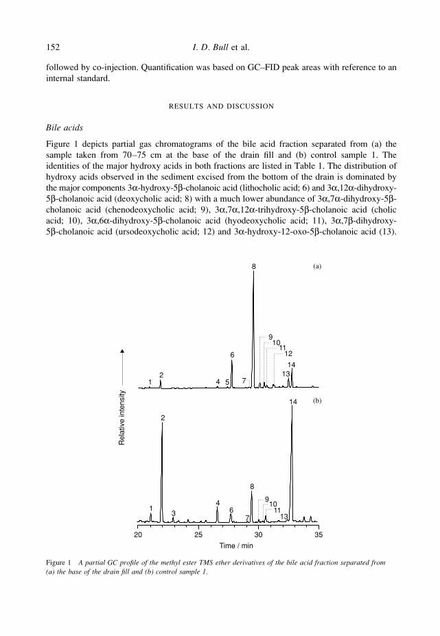
Figure 1 depicts partial gas chromatograms of the bile acid fraction separated from (a) thesample taken from 70–75 cm at the base of the drain fill and (b) control sample 1. Theidentities of the major hydroxy acids in both fractions are listed in Table 1. The distribution ofhydroxy acids observed in the sediment excised from the bottom of the drain is dominated bythe major components 3α-hydroxy-5β-cholanoic acid (lithocholic acid; 6) and 3α,12α-dihydroxy-5β-cholanoic acid (deoxycholic acid; 8) with a much lower abundance of 3α,7α-dihydroxy-5β-cholanoic acid (chenodeoxycholic acid; 9), 3α,7α,12α-trihydroxy-5β-cholanoic acid (cholicacid; 10), 3α,6α-dihydroxy-5β-cholanoic acid (hyodeoxycholic acid; 11), 3α,7β-dihydroxy-5β-cholanoic acid (ursodeoxycholic acid; 12) and 3α-hydroxy-12-oxo-5β-cholanoic acid (13).
Figure 1 A partial GC profile of the methyl ester TMS ether derivatives of the bile acid fraction separated from(a) the base of the drain fill and (b) control sample 1.
13
4 1011
12
4 5
6
7
8
910
1112
1314
67
8
13
14
Time / min
2
9
20 25 30 35
Rel
ativ
e in
tens
ity
(a)
(b)
Biomarkers to track the abandonment of a Roman wastewater course 153
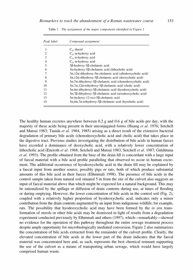
Table 1 The assignment of the major components identified in Figure 1
Peak label Compound assignment
1 C22 diacid2 C22 ω-hydroxy acid3 C24 α-hydroxy acid4 C24 ω-hydroxy acid5 3β-hydroxy-5β-cholanoic acid6 3α-hydroxy-5β-cholanoic acid (lithocholic acid)7 3α,12α-dihydroxy-5α-cholanoic acid (allodeoxycholic acid)8 3α,12α-dihydroxy-5β-cholanoic acid (deoxycholic acid)9 3α,7α-dihydroxy-5β-cholanoic acid (chenodeoxycholic acid)
10 3α,7α,12α-trihydroxy-5β-cholanoic acid (cholic acid)11 3α,6α-dihydroxy-5β-cholanoic acid (hyodeoxycholic acid)12 3α,7β-dihydroxy-5β-cholanoic acid (ursodeoxycholic acid)13 3α-hydroxy-12-oxo-5β-cholanoic acid14 3α,6α,7α-trihydroxy-5β-cholanoic acid (hyocholic acid)
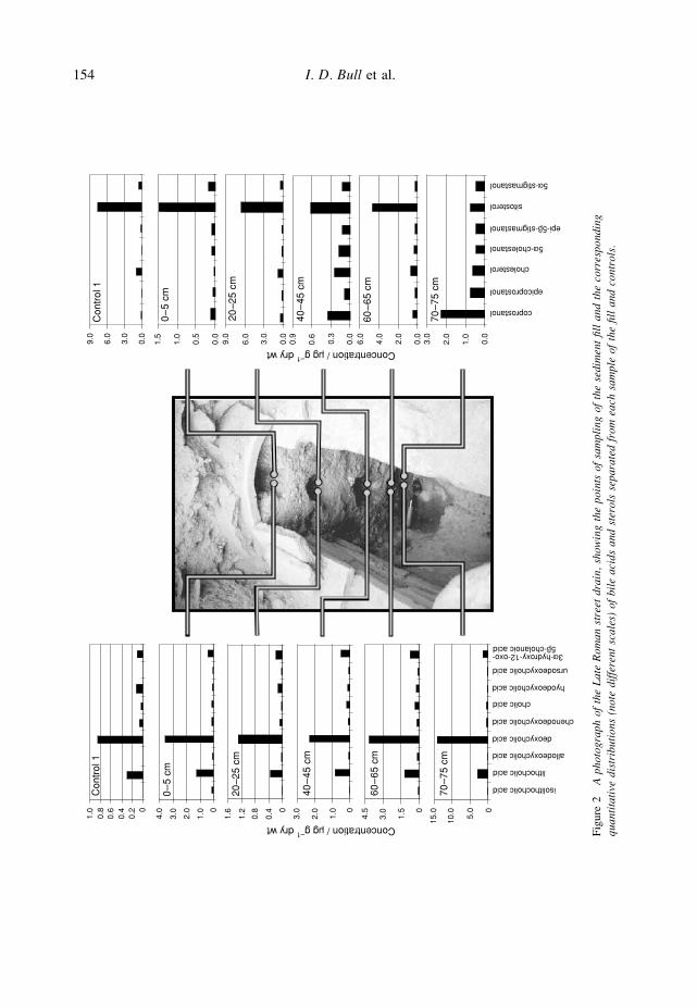
The healthy human excretes anywhere between 0.2 g and 0.6 g of bile acids per day, with themajority of these acids being present in their unconjugated forms (Huang et al. 1976; Setchelland Matsui 1983; Tanida et al. 1984, 1985) arising as a direct result of the extensive bacterialdegradation of primary bile acids (chenodeoxycholic acid and cholic acid) that takes place inthe digestive tract. Previous studies investigating the distribution of bile acids in human faeceshave recorded a dominance of deoxycholic acid, with a relatively lower concentration oflithocholic acid (Eneroth et al. 1968; Setchell and Matsui 1983, Setchell et al. 1987; Güldütunaet al. 1993). The profile obtained from the base of the drain fill is concordant with a large inputof faecal material with a bile acid profile paralleling that observed to occur in human excre-ment. The additional occurrence of hyodeoxycholic acid in the drain fill may be explained bya faecal input from another source, possibly pigs or rats, both of which produce substantialamounts of this bile acid in their faeces (Elhmmali 1998). The presence of bile acids in thecontrol sample taken from natural soil situated 5 m from the site of the culvert also suggests aninput of faecal material above that which might be expected for a natural background. This maybe rationalized by the spillage or diffusion of drain contents during use, at times of floodingor during emptying. However, the lower concentration of bile acids in the control soil (Fig. 2),coupled with a relatively higher proportion of hyodeoxycholic acid, indicates only a minorcontribution from the drain contents augmented by an input from indigenous wildlife; for example,rats. The possibility that hyodeoxycholic acid may have been formed by the in situ trans-formation of sterols or other bile acids may be dismissed in light of results from a degradationexperiment conducted previously by Elhmmali and others (1997), which—remarkably—showedno evidence for the operation of this pathway throughout the entire sewage treatment processdespite ample opportunity for microbiologically mediated conversion. Figure 2 also summarizesthe concentration of bile acids extracted from the remainder of the culvert profile. Clearly, theelevated concentration of bile acids in the lower part of the drain indicates that the faecalmaterial was concentrated here and, as such, represents the best chemical remnant supportingthe use of the culvert as a means of transporting urban sewage, which would have largelycomprised human waste.
154 I. D. Bull et al.
Figu
re 2
A p
hoto
grap
h of
the
Lat
e R
oman
str
eet
drai
n, s
how
ing
the
poin
ts o
f sa
mpl
ing
of t
he s
edim
ent
fill
and
the
corr
espo
ndin
gqu
anti
tati
ve d
istr
ibut
ions
(no
te d
iffe
rent
sca
les)
of
bile
aci
ds a
nd s
tero
ls s
epar
ated
fro
m e
ach
sam
ple
of t
he fi
ll a
nd c
ontr
ols.
1.0
2.0
3.0 0
4.0 0
5.0
10.0
15.00
1.0
2.0
3.0 0
1.5
3.0
4.50
0.4
0.8
1.2
1.6
Concentration / mg g–1
dry wt
Concentration / mg g–1
dry wt
00.
20.
40.
60.
81.
0C
ontr
ol 1
0–
5 cm
20–2
5 cm
40–
45 c
m
60–
65 c
m
70–7
5 cm
Con
trol
1
0–
5 cm
20–2
5 cm
40–
45 c
m
60–
65 c
m
70–7
5 cm
0.0
1.0
2.0
3.0
0.0
2.0
4.0
6.0
0.0
0.3
0.6
0.9
0.0
3.0
6.0
9.0
0.0
0.5
1.0
1.5
0.0
3.0
6.0
9.0
isolithocholic acid
lithocholic acid
allodeoxycholic acid
deoxycholic acid
chenodeoxycholic acid
cholic acid
hyodeoxycholic acid
ursodeoxycholic acid
5b-cholanoic acid3a-hydroxy-12-oxo-
coprostanol
epicoprostanol
cholesterol
5a-cholestanol
epi-5b-stigmastanol
sitosterol
5a-stigmastanol
Biomarkers to track the abandonment of a Roman wastewater course 155
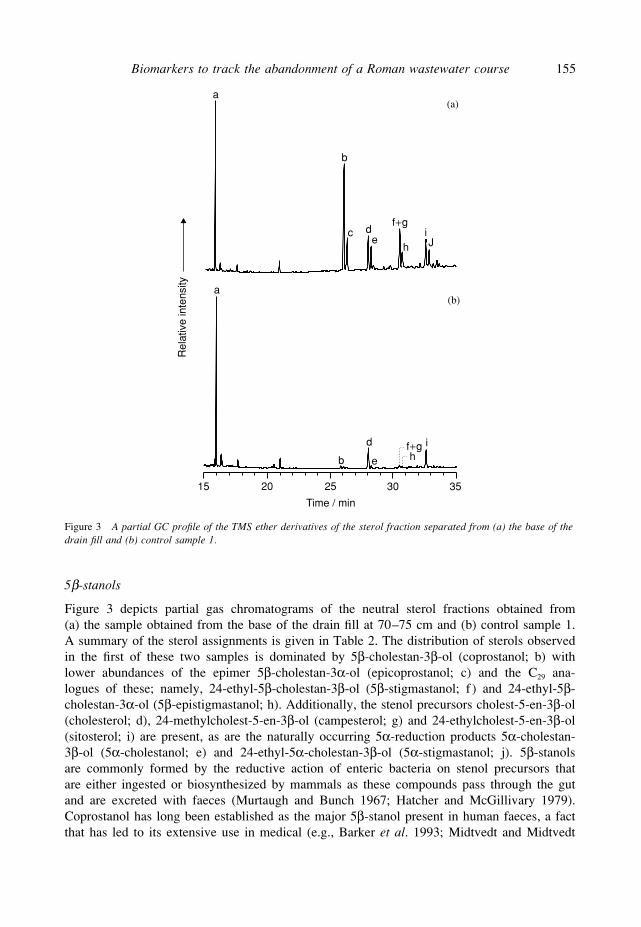
Figure 3 A partial GC profile of the TMS ether derivatives of the sterol fraction separated from (a) the base of thedrain fill and (b) control sample 1.
Time / min
Rel
ativ
e in
tens
ity a
b
d
ef+gh
i
15 20 25 30 35
a
b
c de
f+g
hiJ
(a)
(b)
5β-stanols
Figure 3 depicts partial gas chromatograms of the neutral sterol fractions obtained from(a) the sample obtained from the base of the drain fill at 70–75 cm and (b) control sample 1.A summary of the sterol assignments is given in Table 2. The distribution of sterols observedin the first of these two samples is dominated by 5β-cholestan-3β-ol (coprostanol; b) withlower abundances of the epimer 5β-cholestan-3α-ol (epicoprostanol; c) and the C29 ana-logues of these; namely, 24-ethyl-5β-cholestan-3β-ol (5β-stigmastanol; f ) and 24-ethyl-5β-cholestan-3α-ol (5β-epistigmastanol; h). Additionally, the stenol precursors cholest-5-en-3β-ol(cholesterol; d), 24-methylcholest-5-en-3β-ol (campesterol; g) and 24-ethylcholest-5-en-3β-ol(sitosterol; i) are present, as are the naturally occurring 5α-reduction products 5α-cholestan-3β-ol (5α-cholestanol; e) and 24-ethyl-5α-cholestan-3β-ol (5α-stigmastanol; j). 5β-stanolsare commonly formed by the reductive action of enteric bacteria on stenol precursors thatare either ingested or biosynthesized by mammals as these compounds pass through the gutand are excreted with faeces (Murtaugh and Bunch 1967; Hatcher and McGillivary 1979).Coprostanol has long been established as the major 5β-stanol present in human faeces, a factthat has led to its extensive use in medical (e.g., Barker et al. 1993; Midtvedt and Midtvedt
156 I. D. Bull et al.
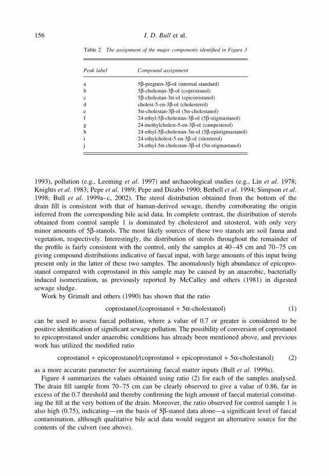
Table 2 The assignment of the major components identified in Figure 3
Peak label Compound assignment
a 5β-pregnen-3β-ol (internal standard)b 5β-cholestan-3β-ol (coprostanol)c 5β-cholestan-3α-ol (epicorostanol)d cholest-5-en-3β-ol (cholesterol)e 5α-cholestan-3β-ol (5α-cholestanol)f 24-ethyl-5β-cholestan-3β-ol (5β-stigmastanol)g 24-methylcholest-5-en-3β-ol (campesterol)h 24-ethyl-5β-cholestan-3α-ol (5β-epistigmastanol)i 24-ethylcholest-5-en-3β-ol (sitosterol)j 24-ethyl-5α-cholestan-3β-ol (5α-stigmastanol)
1993), pollution (e.g., Leeming et al. 1997) and archaeological studies (e.g., Lin et al. 1978;Knights et al. 1983; Pepe et al. 1989; Pepe and Dizabo 1990; Bethell et al. 1994; Simpson et al.1998; Bull et al. 1999a–c, 2002). The sterol distribution obtained from the bottom of thedrain fill is consistent with that of human-derived sewage, thereby corroborating the origininferred from the corresponding bile acid data. In complete contrast, the distribution of sterolsobtained from control sample 1 is dominated by cholesterol and sitosterol, with only veryminor amounts of 5β-stanols. The most likely sources of these two stanols are soil fauna andvegetation, respectively. Interestingly, the distribution of sterols throughout the remainder ofthe profile is fairly consistent with the control, only the samples at 40–45 cm and 70–75 cmgiving compound distributions indicative of faecal input, with large amounts of this input beingpresent only in the latter of these two samples. The anomalously high abundance of epicopro-stanol compared with coprostanol in this sample may be caused by an anaerobic, bacteriallyinduced isomerization, as previously reported by McCalley and others (1981) in digestedsewage sludge.
Work by Grimalt and others (1990) has shown that the ratio
coprostanol/(coprostanol + 5α-cholestanol) (1)
can be used to assess faecal pollution, where a value of 0.7 or greater is considered to bepositive identification of significant sewage pollution. The possibility of conversion of coprostanolto epicoprostanol under anaerobic conditions has already been mentioned above, and previouswork has utilized the modified ratio
coprostanol + epicoprostanol/(coprostanol + epicoprostanol + 5α-cholestanol) (2)
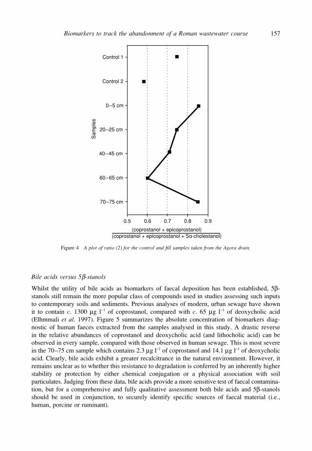
as a more accurate parameter for ascertaining faecal matter inputs (Bull et al. 1999a).Figure 4 summarizes the values obtained using ratio (2) for each of the samples analysed.
The drain fill sample from 70–75 cm can be clearly observed to give a value of 0.86, far inexcess of the 0.7 threshold and thereby confirming the high amount of faecal material constitut-ing the fill at the very bottom of the drain. Moreover, the ratio observed for control sample 1 isalso high (0.75), indicating—on the basis of 5β-stanol data alone—a significant level of faecalcontamination, although qualitative bile acid data would suggest an alternative source for thecontents of the culvert (see above).
Biomarkers to track the abandonment of a Roman wastewater course 157
Figure 4 A plot of ratio (2) for the control and fill samples taken from the Agora drain.
0.5 0.6 0.7 0.8 0.9
(coprostanol + epicoprostanol)(coprostanol + epicoprostanol + 5a-cholestanol)
Control 1
Control 2
0–5 cm
20–25 cm
40–45 cm
60–65 cm
70–75 cm
Sam
ples
Bile acids versus 5β-stanols
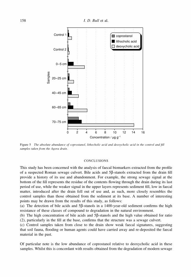
Whilst the utility of bile acids as biomarkers of faecal deposition has been established, 5β-stanols still remain the more popular class of compounds used in studies assessing such inputsto contemporary soils and sediments. Previous analyses of modern, urban sewage have shownit to contain c. 1300 µg l−1 of coprostanol, compared with c. 65 µg l−1 of deoxycholic acid(Elhmmali et al. 1997). Figure 5 summarizes the absolute concentration of biomarkers diag-nostic of human faeces extracted from the samples analysed in this study. A drastic reversein the relative abundances of coprostanol and deoxycholic acid (and lithocholic acid) can beobserved in every sample, compared with those observed in human sewage. This is most severein the 70–75 cm sample which contains 2.3 µg l−1 of coprostanol and 14.1 µg l−1 of deoxycholicacid. Clearly, bile acids exhibit a greater recalcitrance in the natural environment. However, itremains unclear as to whether this resistance to degradation is conferred by an inherently higherstability or protection by either chemical conjugation or a physical association with soilparticulates. Judging from these data, bile acids provide a more sensitive test of faecal contamina-tion, but for a comprehensive and fully qualitative assessment both bile acids and 5β-stanolsshould be used in conjunction, to securely identify specific sources of faecal material (i.e.,human, porcine or ruminant).
158 I. D. Bull et al.
Control 1
Control 2
0–5 cm
20–25 cm
40–45 cm
60–65 cm
70–75 cm
Sam
ples
0 2 4 6 8 10 12 14 16
Concentration / mg g–1
coprostanol
lithocholic acid
deoxycholic acid
Figure 5 The absolute abundance of coprostanol, lithocholic acid and deoxycholic acid in the control and fillsamples taken from the Agora drain.
CONCLUSIONS
This study has been concerned with the analysis of faecal biomarkers extracted from the profileof a suspected Roman sewage culvert. Bile acids and 5β-stanols extracted from the drain fillprovide a history of its use and abandonment. For example, the strong sewage signal at thebottom of the fill represents the residue of the contents flowing through the drain during its lastperiod of use, while the weaker signal in the upper layers represents sediment fill, low in faecalmatter, introduced after the drain fell out of use and, as such, more closely resembles thecontrol samples than those obtained from the sediment at its base. A number of interestingpoints may be drawn from the results of this study, as follows:(a) The detection of bile acids and 5β-stanols in a 1400-year-old sediment confirms the highresistance of these classes of compound to degradation in the natural environment.(b) The high concentration of bile acids and 5β-stanols and the high value obtained for ratio(2), particularly in the fill at the base, confirms that the structure was a sewage culvert.(c) Control samples taken from close to the drain show weak faecal signatures, suggestingthat soil fauna, flooding or human agents could have carried away and re-deposited the faecalmaterial in the past.
Of particular note is the low abundance of coprostanol relative to deoxycholic acid in thesesamples. Whilst this is concordant with results obtained from the degradation of modern sewage
Biomarkers to track the abandonment of a Roman wastewater course 159
material (Elhmmali et al. 1997), this study clearly demonstrates the inherently greater resist-ance of bile acids to degradation over an archaeological timescale (1400 years) and therefore,given the well-defined source of the analysed sediment, supports the use of such biomarkers inthe determination of faecal inputs of more ambiguously sourced samples in antiquity.
ACKNOWLEDGEMENTS
This work was undertaken within the Organic Geochemistry Unit (OGU), a subdivision of theBiogeochemistry Research Centre at the University of Bristol. The authors would like to thankProfessor J. Camp for advice on sample collection, Mr Jim Carter for his technical assistanceand the NERC for funding the Organic Mass Spectrometry Facility at the University of Bristol(Contract Grant F14/6/13).
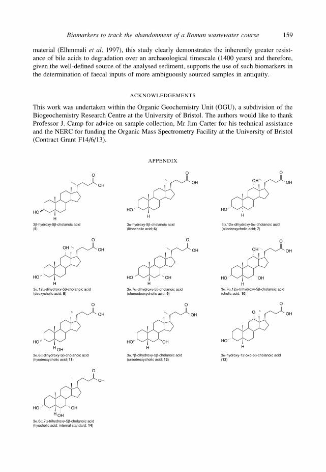
APPENDIX
HO
H
OH
O
3α-hydroxy-5β-cholanoic acid(lithocholic acid; 6)
HOH
OH
O
O
3α-hydroxy-12-oxo-5β-cholanoic acid(13)
H
HO
OH
O
OH
3α,12α-dihydroxy-5β-cholanoic acid(deoxycholic acid; 8)
HHO
OH
O
OH
3α,12α-dihydroxy-5α-cholanoic acid(allodeoxycholic acid; 7)
HHO
OH
O
OH
3α,7α-dihydroxy-5β-cholanoic acid(chenodeoxycholic acid; 9)
HHO
OH
O
OH
OH
3α,7α,12α-trihydroxy-5β-cholanoic acid(cholic acid; 10)
HHO
OH
O
OH3α,6α-dihydroxy-5β-cholanoic acid(hyodeoxycholic acid; 11)
HHO
OH
O
OH
3α,7β-dihydroxy-5β-cholanoic acid(ursodeoxycholic acid; 12)
HHO
OH
O
OH
OH
3α,6α,7α-trihydroxy-5β-cholanoic acid(hyocholic acid; internal standard; 14)
HO
H
OH
O
3β-hydroxy-5β-cholanoic acid(5)
160 I. D. Bull et al.
HOHH
HOH
HO
H
HO
HOH
HOH
HO
HO
H
HO
HO
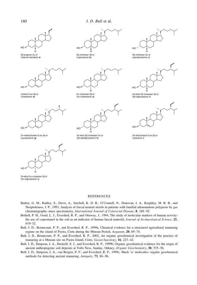
5β-pregnan-3α-ol(internal standard; a)
5β-cholestan-3β-ol(coprostanol; b)
24-ethyl-5β-cholestan-3β-ol(5β-stigmastanol; f)
24-ethyl-5α-cholestan-3β-ol(5α-stigmastanol; j)
24-ethyl-5β-cholestan-3α-ol(5β-epistigmastanol; h)
5β-cholestan-3α-ol(epicoprostanol; c)
cholest-5-en-3β-ol(cholesterol; d)
5α-cholestan-3β-ol(5α-cholestanol; e)
24-methylcholest-5-en-3β-ol(campesterol; g)
24-ethylcholest-5-en-3β-ol(sitosterol; i)
REFERENCES
Barker, G. M., Radley, S., Davis, A., Setchell, K. D. R., O’Connell, N., Donovan, I. A., Keighley, M. R. B., andNeoptolemos, J. P., 1993, Analysis of faecal neutral sterols in patients with familial adenomatous polyposis by gaschromatography–mass spectrometry, International Journal of Colorectal Disease, 8, 188–92.
Bethell, P. H., Goad, L. J., Evershed, R. P., and Ottaway, J., 1994, The study of molecular markers of human activity:the use of coprostanol in the soil as an indicator of human faecal material, Journal of Archaeological Science, 21,619–32.
Bull, I. D., Betancourt, P. P., and Evershed, R. P., 1999a, Chemical evidence for a structured agricultural manuringregime on the island of Pseira, Crete during the Minoan Period, Aegaeum, 20, 69–74.
Bull, I. D., Betancourt, P. P., and Evershed, R. P., 2002, An organic geochemical investigation of the practice ofmanuring at a Minoan site on Pseira Island, Crete, Geoarchaeology, 16, 223–42.
Bull, I. D., Simpson, I. A., Dockrill, S. J., and Evershed, R. P., 1999b, Organic geochemical evidence for the origin ofancient anthropogenic soil deposits at Tofts Ness, Sanday, Orkney, Organic Geochemistry, 30, 535–56.
Bull, I. D., Simpson, I. A., van Bergen, P. F., and Evershed, R. P., 1999c, Muck ‘n’ molecules: organic geochemicalmethods for detecting ancient manuring, Antiquity, 73, 86–96.
Biomarkers to track the abandonment of a Roman wastewater course 161
Bull, I. D., Lockheart, M. J., Elhmmali, M. M., Roberts, D. J., and Evershed, R. P., 2002, The origin of faeces bymeans of biomarker detection, Environment International, 27, 647–54.
Camp, J. M., 1996, Excavations of the Athenian Agora: 1994 and 1995, Hesperia, 65(3), 231–61.Carcopino, J., 1956, Daily life in ancient Rome, Penguin, Harmondsworth, UK.Elhmmali, M. M., 1998, Complementary use of bile acids and sterols as sewage pollution indicators, Ph.D. disserta-
tion, University of Bristol.Elhmmali, M. M., Roberts, D. J., and Evershed, R. P., 1997, Bile acids as a new class of sewage indicator, Environmental
Science and Technology, 31, 3663–8.Elhmmali, M. M., Roberts, D. J., and Evershed, R. P., 2000, Combined analysis of bile acids and sterols/stanols from
riverine particulates to assess sewage discharges and other faecal sources, Environmental Science and Technology,34, 39–46.
Eneroth, P., Hellström, K., and Sjövall, J., 1968, A method for quantitative determination of bile acids in human feces,Acta Chemica Scandinavica, 22, 1729–44.
Grimalt, J. O., Fernández, P., Bayona, J. M., and Albaigés, J., 1990, Assessment of fecal sterols and ketones asindicators of urban sewage inputs to coastal waters, Environmental Science and Technology, 24, 357–63.
Güldütuna, S., You, T., Kurts, W., and Leuschner, U., 1993, High performance liquid chromatographic determinationof free and conjugated bile acids in serum, liver biopsies, bile, gastric juice and feces by fluorescence labeling,Clinica Chimica Acta, 214, 195–207.
Hatcher, P. G., and McGillivary, P. A., 1979, Sewage contamination in the New York Bight. Coprostanol as anindicator, Environmental Science and Technology, 13, 1225–9.
Humphrey, J. W., Oleson, J. P., and Sherwood, A. N., 1998, Greek and Roman technology: a sourcebook, Routledge,London.
Huang, C. T. L., Rodriguez, J. T., Woodward, W. E., and Nichols, B. L., 1976, Comparison of patterns of fecal bileacid and neutral sterol between children and adults, American Journal of Clinical Nutrition, 29, 1196–203.
Knights, B. A., Dickson, C. A., Dickson, J. H., and Breeze D. J., 1983, Evidence concerning the Roman military dietat Bearsden, Scotland, in the 2nd century A.D., Journal of Archaeological Science, 10, 139–52.
Leeming, R., Latham, V., Rayner, M., and Nichols, P., 1997, Detecting and distinguishing sources of sewage pollutionin Australian inland and coastal waters and sediments, ACS Symposium Series, 671, 306–19.
Lin, D. S., Connor, W. E., Napton, L. K., and Heizer, R. F., 1978, The steroids of 2000-year-old human coprolites,Journal of Lipid Research, 19, 215–21.
McCalley, D. V., Cooke, M., and Nickless, G., 1981, Effect of sewage treatment on faecal sterols, Water Research, 15,1019–25.
Midtvedt, A. C., and Midtvedt, T, 1993, Conversion of cholesterol to coprostanol by the intestinal microflora during thefirst two years of human life, Journal of Pediatric Gastroenterology and Nutrition, 17, 161–9.
Murtaugh, J. J., and Bunch, R. L., 1967, Sterols as a measure of fecal pollution, Journal of Water Pollution Control, 39,404–9.
Pepe, C., and Dizabo, P., 1990, Étude d’une fosse du 13ème siecle par les marquers biogéochimiques: chantierarchéologique du Louvre (Paris), Revue d’Archéométrie, 14, 23–8.
Pepe, C., Dizabo, P., Scibe, P., Dagaux, J., Fillaux, J., and Saliot, A., 1989, Les marquers biogéochimiques: applicationa l’archéologie, Revue d’Archéométrie, 13, 1–11.
Setchell, K. D. R., and Matsui, A., 1983, Serum bile acid analysis, Clinica Chimica Acta, 127, 1–17.Setchell, K. D. R., Ives, J. A., Cashmore, G. C., and Lawson, A. M., 1987, On the homogeneity of stools with respect
to bile acid composition and normal day to day variations: a detailed qualitative and quantitative study usingcapillary column gas chromatography – mass spectrometry, Clinica Chimica Acta, 162, 257–75.
Simpson, I. A., Bull, I. D., Dockrill, S. J., and Evershed, R. P., 1998, Early anthropogenic soil formation at Tofts Ness,Sanday, Orkney, Journal of Archaeological Science, 25, 729–46.
Simpson, I. A., van Bergen, P. F., Perret, V., Elhmmali, M. M., Roberts, D. J., and Evershed, R. P., 1999, Lipidbiomarkers of manuring practice in relict anthropogenic soils, The Holocene, 9, 223–9.
Tanida, H., Hikasa, Y., Shimoyama, T., and Setchell, K. D. R., 1984, Comparison of the faecal bile acid profilesbetween patients with adenomatous polyps of the large bowel and healthy subjects in Japan, Gut, 25, 824–32.
Tanida, H., Hikasa, Y., Shimoyama, T., and Setchell, K. D. R., 1985, Faecal bile acid profiles of Japanese patients withadenomatous polyps of the large bowel: special reference to distribution multiplicity, size and degree of dysplasiaof the polyps, Japanese Journal of Cancer Research, 76, 104–12.
White, K. D., 1984, Greek and Roman technology, Cornell University Press, Ithaca, New York.


