Embed Size (px)
Citation preview
314 Biochimica et Biophysica Acta 925 (1987) 314-324 Elsevier
BBA 22802
B i o l o g i c a l c h a r a c t e r i z a t i o n o f p u r i f i e d n a t i v e 2 0 - k D a h u m a n g r o w t h h o r m o n e
J a c k L. K o s t y o a, A n n a S k o t t n e r b, P e t e r B r o s t e d t c, P a u l R o o s c,
C h r i s t o p h e r M. C a m e r o n ~, A n d e r s F o r s m a n b, L i n d a F r y k l u n d b
N a a A. A d a m a f i o a a n d Bo S k o o g b
Department o/Physiology, The University of Michigan Medical School, Ann Arbor, MI (U.S.A.), h KabiVitrum AB, Stockholm and c Department of Biochemistry, University of Uppsala, Uppsala (Sweden)
(Received 20 January 1987)
Key words: Growth hormone; Growth-promoting activity; Diabetogenic activity; Insulin-like activity; IGF-1 level; ( ob/ob mouse)
Because of the propensity of the 20-kDa variant of human growth hormone (GH) to aggregate with itself and with 22-kDa human GH, it has been difficult to prepare monomeric 20-kDa GH in highly purified form. This has been a major complicating factor in determining whether 20-kDa GH has a biological activity profile distinct from that of 22-kDa GH. In the present study, native 20-kI)a GH was isolated from a human GH dimer concentrate and purified by a procedure that included column electrophoresis in agarose suspension as a final separation step. This procedure yielded highly purified monomeric 20-kDa GH, which was contaminated to an extent of less than 1% with 22-kDa GH, and which exhibited only a small degree of dimerization upon storage. The native 20-kDa GH was quite active in stimulating growth in hypophysecto- mized rats, when growth was assessed by body weight gain, longitudinal bone growth, the stimulation of sulfation of cartilage, and the elevation of serum IGF-1 level. However, in all of these growth assays, the 20-kDa GH was somewhat less active than the native 22-kDa GH to which it was compared; e.g., in the body weight gain and longitudinal bone growth assays, it had an estimated potency of 0.6 relative to the 22-kDa GH. The 20-kDa GH exhibited substantial diabetogenic activity when tested for the ability to raise fasting blood glucose concentration and to impair glucose tolerance in o h ~ o h mice. Also, the native 20-kDa GH had significant in vitro insulin-like activity, although its potency was approximately 20% that of the native 22-kDa GH to which it was compared. Thus, the biological activity profile of native 20-kDa GH differs from that of 22-kDa GH primarily in that insulin-like activity is markedly attenuated.
Introduction
A variety of structurally modified forms of growth hormone (GH) have been isolated from the human pituitary gland. The predominant
Abbreviation: GH, growth hormone.
Correspondence: J.L. Kostyo, Department of Physiology, The University of Michigan Medical School, Ann Arbor, MI 48109, U.S.A.
monomeric form of the hormone present in the gland consists of a single peptide chain of 191 amino acid residues, having a molecular size of approx. 22 kDa. About 10% of the total G H in the human pituitary is a 20-kDa variant lacking re- sidues 32-46 of the amino acid sequence [1-5]. This variant occurs because the nucleotides encod- ing sequence 32-46 are occasionally deleted dur- ing the conversion of G H pre-mRNA to GH mRNA [6]. Because of its propensity to aggregate with itself and with 22-kDa GH, 20-kDa G H
0304-4165/87/$03.50 ~ 1987 Elsevier Science Publishers B.V. (Biomedical Division)
exists in human pituitary extracts primarily in various dimeric forms [2,5]. As a consequence, it has been quite difficult to prepare monomeric 20-kDa GH in highly purified form. This has been a maj.or complicating factor in determining whether 20-kDa G H has a biological activity pro- file distinct from that of 22-kDa GH.
It has been reported previously [2,7] that native 20-kDa GH is equipotent with 22-kDa GH in stimulating growth in the hypophysectomized rat, and that it has lactogenic activity in the pigeon crop sac assay equivalent to that of 22-kDa GH [2]' However, it has been claimed [7,8] that native 20-kDa GH lacks significant diabetogenic and insulin-like activities, which are intrinsic proper- ties of 22-kDa GH. This conclusion was based on the findings that a native 20-kDa GH preparation (a) failed to cause hyperglycemia and glucose in- tolerance in the dog when given 10 h prior to a glucose tolerance test [7], (b) did not produce hypoglycemia or decrease plasma free fatty acids when administered acutely to hypophysectomized rats [8], and (c) failed to increase glucose oxida- tion by isolated rat adipose tissue [8]. This conclu- sion has been challenged in recent reports demon- strating that bacterially derived biosynthetic hu- man 20-kDa methionyl-GH had both diabetogenic [9] and insulin-like activities [9,10], although the latter was approximately 20% that of biosynthetic human 22-kDa methionyl-GH [9].
The present study describes the biological char- acterization of a native 20-kDa GH preparation that was isolated by a procedure similar to that used by Singh and Lewis [4], but which included a final electrophoretic step. This procedure yielded a highly purified preparation of native 20-kDa GH having less than 1% contamination with 22-kDa GH.
Materials and Methods
Preparation of 20-kDa GH The starting material for the isolation of 20-kDa
G H monomer was a human GH dimer con- centrate obtained as a side fraction during the preparation of 22-kDa GH (Crescormon ®) at KabiVitrum AB, according to the method of Roos et al. [11]. About 2.5 g of dimer concentrate in a volume of 2 1, which had been prepared from 4000
315
human pituitary glands, were utilized. As a first step, the concentrate was dialyzed against 0.005 M potassium phosphate buffer (pH 7.0) and then applied to a 5 x 22 cm column of SP-Sephadex C-50 (Pharmacia, Uppsala, Sweden) to remove human thyrotropic hormone. The column was run at a flow rate of 40 ml /h , and 17-ml fractions were collected. The column was eluted by increas- ing buffer concentration stepwise, while maintain- ing constant pH. The thyrotropic hormone was adsorbed on the SP-Sephadex and was later re- covered from the column by elution with 0.06 M potassium phosphate buffer [12]. The GH dimer material passed through the column unretarded and was transferred directly to a second column (2 x 56 cm) packed with DEAE-Sepharose CL-6B (Pharmacia), which was coupled to the SP-Seph- adex column by plastic tubing. The DEAE-Seph- arose column had been equilibrated with 0.005 M potassium phosphate buffer (pH 7.0). The col- umns were then disconnected, and the dimer material was eluted from the DEAE-Sepharose column with 0.5 M potassium phosphate buffer (pH 7.0). This step concentrated the dimer frac- tion 25-fold, making it possible to proceed to molecular sieve chromatography directly.
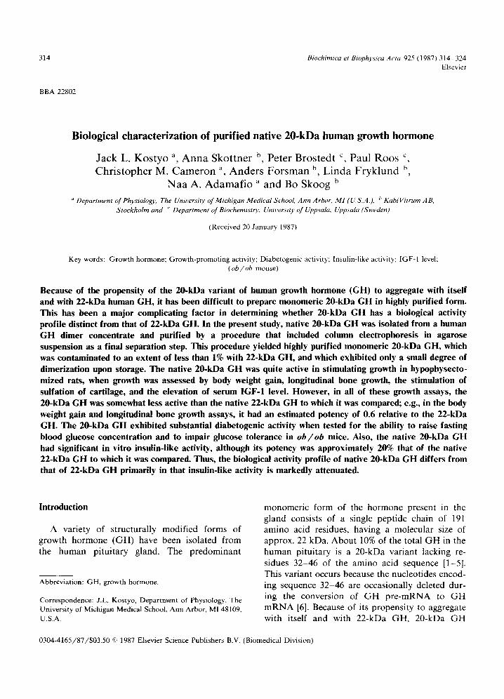
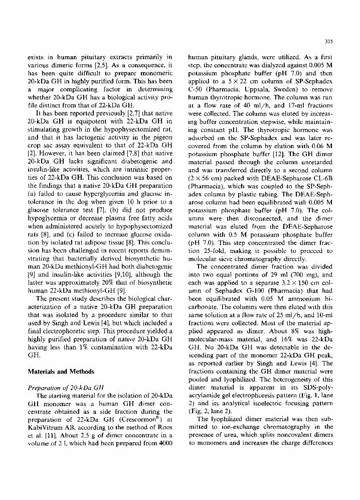
The concentrated dimer fraction was divided into two equal portions of 29 ml (700 mg), and each was applied to a separate 3.2 x 150 cm col- umn of Sephadex G-100 (Pharmacia) that had been equilibrated with 0.05 M ammonium bi- carbonate. The columns were then eluted with this same solution at a flow rate of 25 ml/h , and 10-ml fractions were collected. Most of the material ap- plied appeared as dimer. About 8% was high- molecular-mass material, and 16% was 22-kDa GH. No 20-kDa G H was detectable in the de- scending part of the monomer 22-kDa GH peak, as reported earlier by Singh and Lewis [4]. The fractions containing the G H dimer material were pooled and lyophilized. The heterogeneity of this dimer material is apparent in its SDS-poly- acrylamide gel electrophoresis pattern (Fig. 1, lane 2) and its analytical isoelectric focusing pattern (Fig. 2, lane 2).
The lyophilized dimer material was then sub- mitted to ion-exchange chromatography in the presence of urea, which splits noncovalent dimers to monomers and increases the charge differences
316
® 1 2 3 /,
~!! 11 ~
I i~ !
®
Fig. 1. Analytical SDS-polyacrylamide gel electrophoresis of (1) 22-kDa GH, (2) GH dimer from Sephadex G-100 chro- matography, (3) pooled material (peak 3) from chromatogra- phy on DEAE-Sepharose CL-6B in 6M urea, and (4) the final 20-kDa GH pool from zone electrophoresis in agarose suspen-
sion.
between 20-kDa GH and 22-kDa GH. The material was dissolved in 6 M urea (analytical grade, Merck, Darmstadt, F.R.G.), and the con- ductivity of the solution was adjusted to 1.1 mmho with solid ammonium bicarbonate. The sample (700 mg) was then applied to a 2 X 23 cm column packed with DEAE-Sepharose CL-6B that had been equilibrated with 6 M urea-ammonium bi- carbonate buffer (1.1 mmho). All urea-ammonium bicarbonate solutions used in this procedure were passed through 0.2-~m filters and were used im- mediately. The column was washed with the above urea-ammonium bicarbonate buffer, and then the material bound to the column was eluted by in- creasing the conductivity of the elution buffer in a steep, stepwise fashion (from 1.1 to 5.0 mmho) by the addition of solid ammonium bicarbonate. The
Fig. 2. Analytical isoelectric focusing in agarose gel (pH 4.0-6.5) of (1) 22-kDa GH, (2) GH dimer from Sephadex G-100 chromatography, (3) pooled material (peak 3) from chromatography on DEAE-Sepharose CL-6B in 6M urea, and (4) the final 20-kDa GH pool from zone electrophoresis in
agarose suspension.
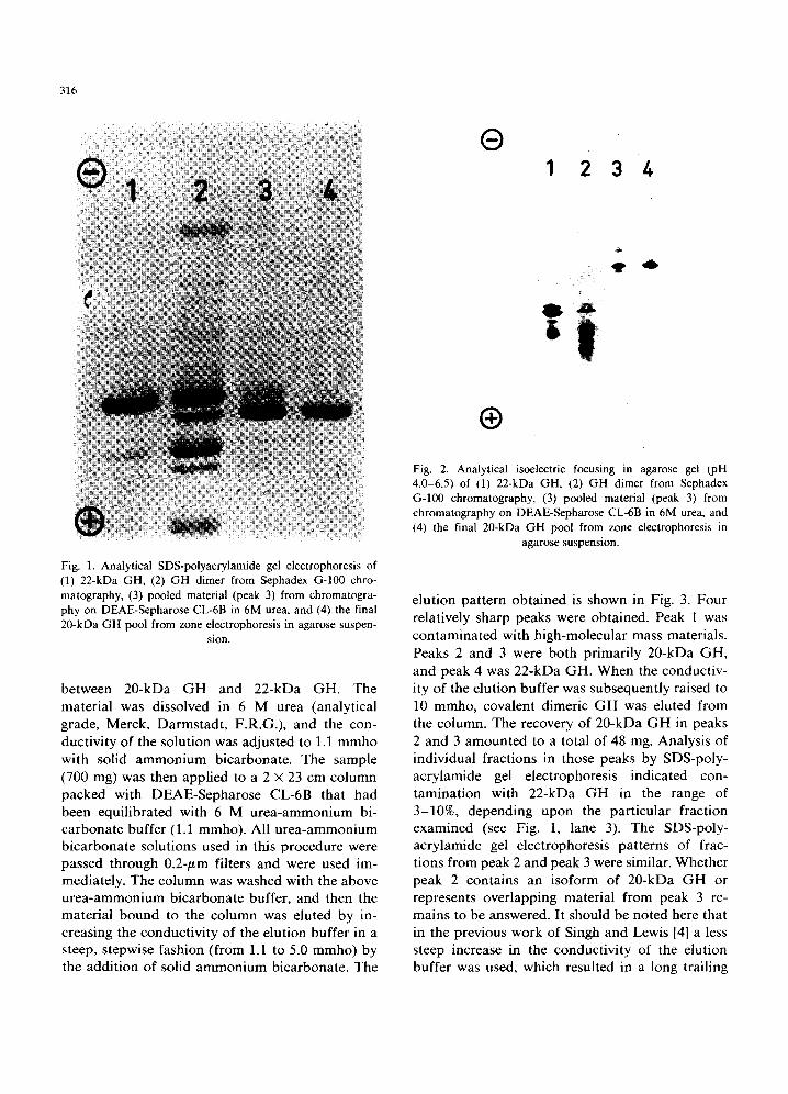
elution pattern obtained is shown in Fig. 3. Four relatively sharp peaks were obtained. Peak 1 was contaminated with high-molecular mass materials. Peaks 2 and 3 were both primarily 20-kDa GH, and peak 4 was 22-kDa GH. When the conductiv- ity of the elution buffer was subsequently raised to 10 mmho, covalent dimeric GH was eluted from the column. The recovery of 20-kDa GH in peaks 2 and 3 amounted to a total of 48 mg. Analysis of individual fractions in those peaks by SDS-poly- acrylamide gel electrophoresis indicated con- tamination with 22-kDa GH in the range of 3-10%, depending upon the particular fraction examined (see Fig. 1, lane 3). The SDS-poly- acrylamide gel electrophoresis patterns of frac- tions from peak 2 and peak 3 were similar. Whether peak 2 contains an isoform of 20-kDa GH or represents overlapping material from peak 3 re- mains to be answered. It should be noted here that in the previous work of Singh and Lewis [4] a less steep increase in the conductivity of the elution buffer was used, which resulted in a long trailing
/
20
~i /I-
30 /.0 50 60 70 80 Fraction number
0s T 0.4
E
0 O0
~ 0 . 3 U
Ca .£1 t - O
~ 0 . 2
0.1 ~
0.05
0 10
Fig. 3. Chromatography on DEAE-Sepharose CL-6B of GH dimer from molecular sieve chromatography. The buffer was 6 M urea adjusted to a conductivity of 5.0 mmho with solid ammonium bicarbonate. Flow rate was 40 ml/h, and fraction size was 10 ml. The hatched area (peak 3) represents the 20-kDa GH-containing fractions that were utilized for further
purification.
peak, having less than 5% contamination with 22-kDa GH, as judged by SDS-polyacrylamide gel electrophoresis.
In a final separation step, the pooled and ultra- filtered fractions corresponding to peak 3 in the DEAE-Sepharose chromatogram (Fig. 3) were subjected to column electrophoresis in agarose suspension according to the method of Hjert~n [13]. The 22-kDa GH contamination of this pool was about 7%. The electrophoresis was performed in 0.15 M glycine-sodium hydroxide buffer (pH 8.8) which also contained 6 M urea. Because of the presence of urea, the content of agarose (Agarose C, Pharmacia) had to be increased to 0.30% (w/v), which is approximately twice that normally used. The solid urea was added to the boiled agarose suspension after it had cooled to 50 ° C. The agarose suspension was then left over- night to cool and stabilize. The gel which formed was broken by gentle stirring and poured into the electrophoresis tube (1.0 x 65 cm). The con- centrated sample (11 mg in 0.7 ml), which was
317
stabilized by being mixed with the pellet from a 1.05-ml aliquot of the agarose suspension that had been centrifuged, was applied close to the cathode. Electrophoresis was carried out for 30000 V-h. The column was then emptied from the top by sucking out fractions of approx. 0.8 ml each with a thin plastic tube. The distribution of protein among the fractions was determined by measuring the difference in their absorbance at 280 and 310 nm [14]. The agarose was removed from the frac- tions by centrifugation at 40000 x g for 1 h at 4 ° C. The 20-kDa GH-containing fractions, as de- termined by SDS-polyacrylamide gel electrophore- sis, were then pooled. SDS-polyacrylamide gel electrophoresis of this 20-kDa GH pool prior to lyophilization is shown in Fig. 1 (lane 4). It is clear that the zone gel electrophoresis step gave a very effective separation of 20-kDa GH from 22- kDa GH. When this material was submitted to polyacrylamide gel electrophoresis in the absence of SDS or 2-mercaptoethanol, the 20-kDa GH migrated as monomer, indicating a complete and lasting conversion of the substance to monomeric form.
The 20-kDa GH pool was dialyzed against 0.005 M ammonium bicarbonate and lyophilized. To eliminate salt completely, the lyophilized material was suspended in 0.001 M ammonium bicarbonate and relyophilized. Analysis of the lyophilized 20-kDa GH preparation indicated that it was contaminated to an extent of less than 1% with 22-kDa GH. Supporting evidence for the homogeneity of this 20-kDa GH preparation was obtained by submitting it to analytical isoelectric focusing (see Fig. 2, lane 4). The lyophilized pre- paration was submitted to polyacrylamide gel electrophoresis in the absence of SDS and 2- mercaptoethanol after storage for 2 months at 4°C in the dry form and subsequently for 6 months at - 20 °C as a frozen solution (1 mg/ml in 0.001 M ammonium bicarbonate). Scanning the stained gels at 560 nm revealed the presence of only about 15% dimeric material. This suggests that this preparation of 20-kDa GH is more stable upon storage than that made earlier [4] by some- what different methods.
Polyacrylamide gel electrophoresis in the pres- ence or absence of SDS and 2-mercaptoethanol was carried out according to the methods of Ne-
318
ville [15]. The composition of the gel was T = 18.5% and C = 1% (the notation of Hjert6n [16]), and the buffers used were chosen to obtain a pH of 10.50. The gels were stained with Coomassie brilliant blue in a solution of 50% methanol /7% acetic acid and destained in a solution of 10% methanol /7% acetic acid. Isoelectric focusing in agarose gel was performed with equipment manu- factured by Pharmacia according to methods pro- vided by the supplier. Pharmalyte ® (pH 4.0-6.5) was used, and approx. 2-#g samples were applied. After termination of isoelectric focusing, the pro- teins were visualized by silver staining [17].
Protein concentration was estimated throughout by measuring absorbance at 280 nm in a 1 cm cell. 1 unit of absorbance was assumed to correspond to a protein concentration of 1 mg/ml.
The purification procedure was guided in part by radioimmunoassays performed with a double antibody technique.
Assays for growth-promoting activity Two comprehensive growth studies (experi-
ments 1 and 2) were conducted in the following manner. Hypophysectomized male rats (80-100 g body weight) of the Sprague-Dawley strain were purchased from Mollegaard Breeding Labs, Den- mark, and were used for experiments 2 weeks after the operation. On the first day of the experiment, each animal received an intraperitoneal injection of 0.8 mg of tetracycline (Terramycin ®, Pfizer) in 0.5 ml saline for the subsequent measurement of longitudinal bone growth [18-20]. They were also given a subcutaneous injection of 40 /~g cortisone acetate (Upjohn) in 0.5 ml saline. On this day, each animal was also lightly anesthetized with diethyl ether, and a blood sample was taken from the retroorbital plexus to obtain serum, which was frozen for subsequent analysis of IGF-1 level. On days 2 and 3, the injections of cortisone acetate were repeated. On days 4 and 5, each rat received an intraperitoneal injection of 10 /~g of L-thyrox- ine (Sigma) in 0.5 ml saline that had been made alkaline with NaOH. On days 6-15 of the experi- ment, the animals received two daily subcutaneous injections (morning and late afternoon) of 0.5 ml of either vehicle, native 22-kDa GH or native 20-kDa G H (6 or 60 /~g/day). On day 6, each animal was again anesthetized with diethyl ether,
and another blood sample was taken for the mea- surement of IGF-1 [21]. On day 15, all animals received an intraperitoneal injection of [35S]sulfate for the measurement of sulfate incorporation into cartilage, as described previously by Skottner et al. [22]. On the following day, a final blood sample was obtained for the measurement of IGF-1 level, and the animals were then decapitated, and vari- ous tissues were removed for analysis.
The native 22-kDa GH used in the growth-pro- moting assays was Crescormon ® (KabiVitrum AB) (3 IU/mg) , which had been desalted on PD-10 columns (Pharmacia) that were equilibrated with 0.1 M ammonium bicarbonate. The protein con- centration of the desalted hormone solution was estimated by measuring its absorbance at 280 nm, assuming an absorption coefficient (A280, 0.1% (w/v) solution, 1 cm) for 22-kDa GH of 0.76. (This absorption coefficient for 22-kDa GH was empirically determined. Corrected ultraviolet spectra of desalted sample solutions were com- pared to their protein contents estimated from the amino acid composition of lyophilized, hydrolysed samples.) The hormone solution was then diluted with saline, and aliquots of the diluted solution were frozen. Fresh aliquots of frozen hormone were thawed and used immediately for each injec- tion. The 20-kDa G H used for the growth-promo- ting assays was an aliquot of the final 20-kDa GH pool. For the first growth study (experiment 1), a sample of the final 20-kDa GH pool was frozen and maintained at - 7 0 °C until it was diluted for use in experiments. The protein concentration of this solution was estimated by measuring its ab- sorbance at 280 nm, assuming an absorption coef- ficient of 0.70 (as determined for 22-kDa GH). The somewhat lower absorption coefficient is con- sistent with the loss of two tyrosine residues in the deletion peptide 32-46, and is in accord with the previous observation of Chapman et al. [5] show- ing about a 10% difference in the absorption coef- ficients of 22-kDa GH and 20-kDa GH. The 20-kDa GH solution was diluted with saline, and aliquots were frozen. Fresh aliquots of frozen 20- kDa G H were thawed and used immediately for each injection. For experiment 2, a sample of the final 20-kDa GH pool was divided into aliquots, and these were then lyophilized individually. Each morning, a lyophilized aliquot of the 20-kDa G H
was dissolved in saline and used for injections immediately. The remainder of the aliquot was refrozen and stored at - 70 o C until the afternoon, when it was thawed and used for the second series of injections.
The potency of the 20-kDa GH relative to that of 22-kDa GH in stimulating weight gain and longitudinal bone growth was estimated using a parallel line bioassay program [23].
Assay for diabetogenic activity The diabetogenic activity of the native 20-kDa
GH was assessed in female ob/ob mice as previ- ously described in detail [24,25]. The final re- lyophilized preparation of the 20-kDa GH was used for these experiments (see above), and the doses of hormone given were based on actual weight of the dried hormone. A fresh solution of the hormone was used each day for the injections. The hormone was dissolved in distilled water made slightly alkaline with NaOH and administered in a volume of 0.1 ml immediately after being dis- solved. Statistical comparisons were made using the paired t-test, and effects were considered to be significant at the P < 0.05 level. Statistically sig- nificant diabetogenic effects are routinely ob- tained in this assay with 10-25/~g of highly puri- fied native 22-kDa GH [25-27].
Assay of insulin-like activity The 20-kDa GH was tested for in vitro insulin-
like activity by assessing its ability to stimulate [14C]glucose oxidation to 14CO2 by isolated epidi- dymal adipose tissue from hypophysectomized rats, as previously described in detail [28]. The activity of the 20-kDa GH was compared to that of purified native human GH monomer (1.1 IU/mg) isolated and purified by ion-exchange chromatography as previously described [29]. Stat- istical comparisons were made with Dunnet's test, and a difference was considered significant if P < 0.05.
Results
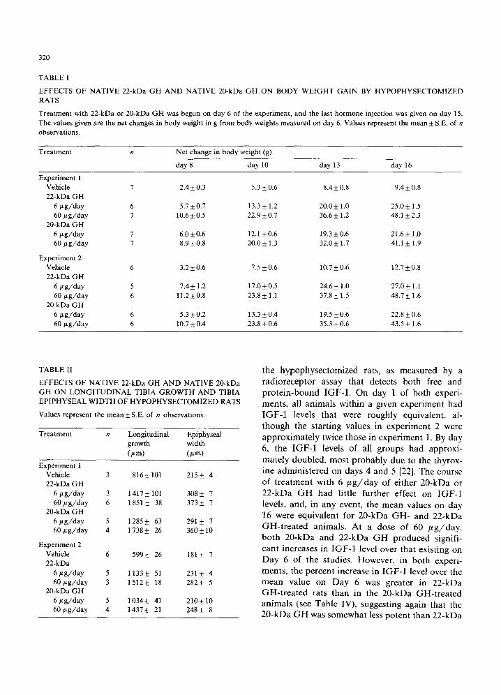
The ability of the purified native 20-kDa GH to stimulate body weight gain in hypophysectomized rats compared to that of 22-kDa GH is shown in Table I. It can be seen that, like 22-kDa GH,
319
20-kDa GH stimulated body weight gain in a dose-dependent manner during the course of treatment in both experiments 1 and 2. In both experiments, however, 20-kDa GH generally pro- duced smaller mean net changes in body weight than did 22-kDa GH at both the 6 and 60 ~tg daily doses used. When the data were analysed using a parallel line bioassay program, the potency of the 20-kDa GH relative to that of the 22-kDa GH in experiment 1 was 0.54 (0.35-0.80, 95% confidence limits) and in experiment 2 was 0.60 (0.44-0.81).
The effects of 20-kDa GH on longitudinal bone growth and tibial epiphyseal width are compared with those of 22-kDa GH in Table II. The 20-kDa GH behaved like 22-kDa GH in stimulating longi- tudinal bone growth and increasing tibial epi- physeal width in a dose-dependent fashion. Again, in both experiments, 20-kDa GH appeared to be somewhat less effective than 22-kDa GH in stimulating both longitudinal bone growth and tibial epiphyseal width. When the data for longitu- dinal bone growth were analysed using the parallel line bioassay program, the potency of the 20-kDa GH relative to that of the 22-kDa GH in experi- ment 1 was 0.50 (0.23-0.92) and in experiment 2 was 0.62 (0.35-1.04). Thus, the estimated potency of 20-kDa GH in this assay was quite similar to that found in the body weight gain studies de- scribed above.
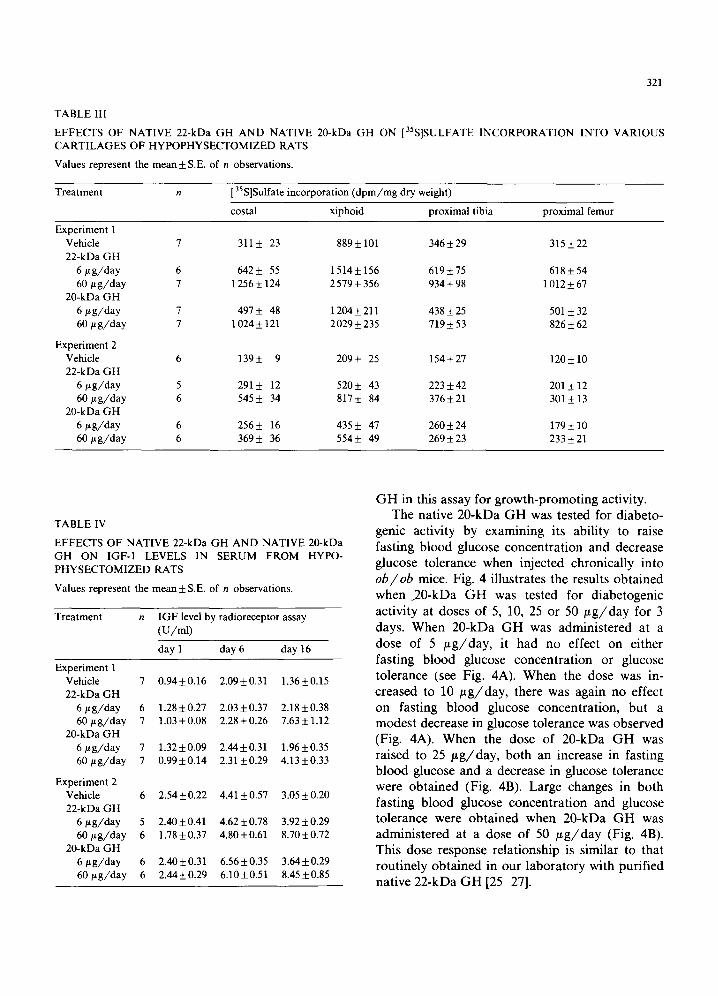
Table III summarizes the results obtained with 20-kDa GH and 22-kDa GH on [35S]sulfate incor- poration into various cartilages of the hypo- physectomized rat. In the case of all four carti- lages examined (costal, xiphoid, proximal tibia and proximal femur), both hormones stimulated [35S]sulfate incorporation in a dose-dependent manner, and, again, the 20-kDa GH appeared to be somewhat less effective in stimulating sulfation than was 22-kDa GH.
Various organs, including the liver, lungs, kid- neys, heart and testes, were removed from the 20-kDa GH- and 22-kDa GH-treated rats on the final day of the experiment, and their wet weights were compared. Both hormones increased the wet weight of all of these organs over those of the vehicle-treated controls (data not shown).
Last, Table IV summarizes the effects of the course of treatment with 20-kDa GH and 22-kDa GH on the concentration of IGF-1 in the serum of
320
TABLE I
EFFECTS OF NATIVE 22-kDa GH AND NATIVE 20-kDa GH ON BODY WEIGHT GAIN BY HYPOPHYSECTOMIZED RATS
Treatment with 22-kDa or 20-kDa GH was begun on day 6 of the experiment, and the last hormone injection was given on day 15. The values given are the net changes in body weight in g from body weights measured on day 6. Values represent the mean +_ S.E. of n observations.
Treatment n Net change in body weight (g)
day 8 day 10 day 13 day 16
Experiment 1 Vehicle 7 2.4 _+ 0.3 5.3 + 0.6 8.4 -+ 0.8 9.4 _+ 0.8 22-kDa GH
6 #g /day 6 5.7 _+ 0.7 13.3 _+ 1.2 20.0 _+ 1.0 25.0 -+ 1.5 60 bt g /day 7 10.6 + 0.5 22.9 + 0.7 36.6 _+ 1.2 48.1 _+ 2.3
20-kDa GH 6 #g /day 7 6.0-+0.6 12.1 +0.6 19.3-+0.6 21.6-+ 1.0 60 #g /day 7 8.9-+0.8 20.0_+ 1.3 32.0_+ 1.7 41.1 _+ 1.9
Experiment 2 Vehicle 6 3.2 _+ 0.6 7.5 _+ 0.6 10.7 _+ 0.6 ! 2.7 _+ 0.8 22-kDa GH
6 #g /day 5 7.4 _+ 1.2 17.0 _+ 0.5 24.6 _+ 1.0 27.0 _+ 1.1 60 # g / d a y 6 11.2 +_ 0.8 23.8 _+ 1.1 37.8 _+ 1.5 48.7 _+ 1.6
20 kDa GH 6 # g /day 6 5.3 + 0.2 13.3 _+ 0.4 19.5 _+ 0.6 22.8 _+ 0.6 60 # g / d a y 6 10.7 -+ 0.4 23.8 _+ 0.6 35.3 _+ 0.6 43.5 _+ 1.6
TABLE I1
EFFECTS OF NATIVE 22-kDa GH AND NATIVE 20-kDa GH ON LONGITUDINAL TIBIA GROWTH AND TIBIA EPIPHYSEAL WIDTH OF HYPOPHYSECTOMIZED RATS
Values represent the mean-+ S.E. of n observations.
Treatment n Longitudinal Epiphyseal growth width (#m) (#m)
Experiment 1 Vehicle 3 816 _+ 101 215 -t- 4 22-kDa GH
6 #g /day 3 1417_+101 308_+ 7 60 # g / d a y 6 1851 _+ 38 373_+ 7
20-kDa GH 6 # g / d a y 5 1285_+ 63 291_+ 7 60 # g / d a y 4 1 738_+ 26 360_+ 10
Experiment 2 Vehicle 6 599-+ 26 181-+ 7 22-kDa
6 # g / d a y 5 1133-+ 51 231_+ 4 6 0 # g / d a y 3 1512-+ 18 282_+ 5
20-kDa GH 6 #g /day 5 1034-+ 41 210_+10 6 0 # g / d a y 4 1437_+ 21 248_+ 8
the hypophysectomized rats, as measured by a radioreceptor assay that detects both free and protein-bound IGF-1. On day 1 of both experi- ments, all animals within a given experiment had IGF-1 levels that were roughly equivalent, al- though the starting values in experiment 2 were approximately twice those in experiment 1. By day 6, the IGF-1 levels of all groups had approxi- mately doubled, most probably due to the thyrox- ine administered on days 4 and 5 [22]. The course of treatment with 6 /~g/day of either 20-kDa or 22-kDa G H had little further effect on IGF-1 levels, and, in any event, the mean values on day 16 were equivalent for 20-kDa GH- and 22-kDa GH-trea ted animals. At a dose of 60 ~ g / d a y , both 20-kDa and 22-kDa G H produced signifi- cant increases in IGF-1 level over that existing on Day 6 of the studies. However, in both experi- ments, the percent increase in IGF-1 level over the mean value on Day 6 was greater in 22-kDa GH-treated rats than in the 20-kDa GH-treated animals (see Table IV), suggesting again that the 20-kDa G H was somewhat less potent than 22-kDa
321
TABLE III
EFFECTS OF NATIVE 22-kDa GH AND NATIVE 20-kDa GH ON [35S]SULFATE INCORPORATION INTO VARIOUS CARTILAGES OF HYPOPHYSECTOMIZED RATS
Values represent the mean +_ S.E. of n observations.
Treatment n 35 [ S]Sulfate incorporation (dpm/mg dry weight)
costal xiphoid proximal tibia proximal femur
Experiment 1 Vehicle 7 311 _+ 23 889 +_ 101 346_+ 29 315 +_ 22 22-kDa GH
6 #g/day 6 642 _+ 55 1514_+ 156 619 +_ 75 618 _+ 54 60 bt g/day 7 1 256 +_ 124 2 579 _+ 356 934 _+ 98 1 012 _+ 67
20-kDa GH 6/L g/day 7 497 +- 48 1204 _+ 211 438 _+ 25 501 _+ 32 60/~ g/day 7 1024 _+ 121 2029 _+ 235 719 _+ 53 826 _+ 62
Experiment 2 Vehicle 6 139+- 9 209_+ 25 154_+27 120_+10 22-kDa GH
6 t~g/day 5 291 +_ 12 520_+ 43 223 +_ 42 201 +_ 12 60 t~g/day 6 545 _+ 34 817 _+ 84 376_+ 21 301 _+ 13
20-kDa GH 6 t~g/day 6 256_+ 16 435 _+ 47 260_+ 24 179_+ 10 60/tg/day 6 369_+ 36 554_+ 49 269_+23 233+21
TABLE IV
EFFECTS OF NATIVE 22-kDa GH AND NATIVE 20-kDa GH ON IGF-1 LEVELS IN SERUM FROM HYPO- PHYSECTOMIZED RATS
Values represent the mean_+ S.E. of n observations.
Treatment n IGFlevel by radioreceptor assay (U/ml)
day 1 day 6 day 16
Experiment 1 Vehicle 7 0.94+0.16 2.09+_0.31 1.36_+0.15 22-kDa GH
6 #g/day 6 1.28_+0.27 2.03+0.37 2.18+_0.38 60 btg/day 7 1.03+_0.08 2.28+_0.26 7.63+_1.12
20-kDa GH 6 btg/day 7 1.32+_0.09 2.44+_0.31 1.96_+0.35 60t~g/day 7 0.99+_0.14 2.31 _+0.29 4.13 _+0.33
Experiment 2 Vehicle 6 2.54_+0.22 4.41_+0.57 3.05_+0.20 22-kDa GH
6 ~g/day 5 2.40+_0.41 4.62_+0.78 3.92+_0.29 60~tg/day 6 1.78+_0.37 4.80+_0.61 8.70+0.72
20-kDa GH 6/~g/day 6 2.40_+0.31 6.56+_0.35 3.64_+0.29 60t~g/day 6 2.44_+0.29 6.10+_0.51 8.45+0.85
G H in this assay for growth-promot ing activity. The native 20-kDa G H was tested for diabeto-
genic activity by examining its abili ty to raise
fasting blood glucose concent ra t ion and decrease glucose tolerance when injected chronical ly into
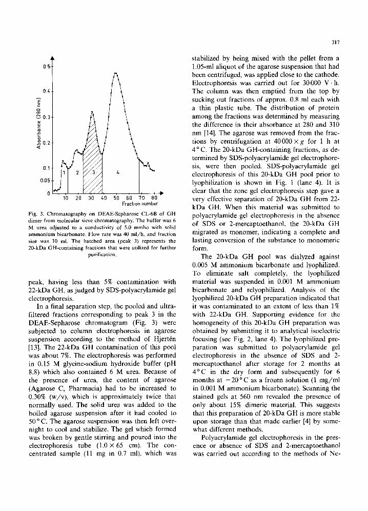
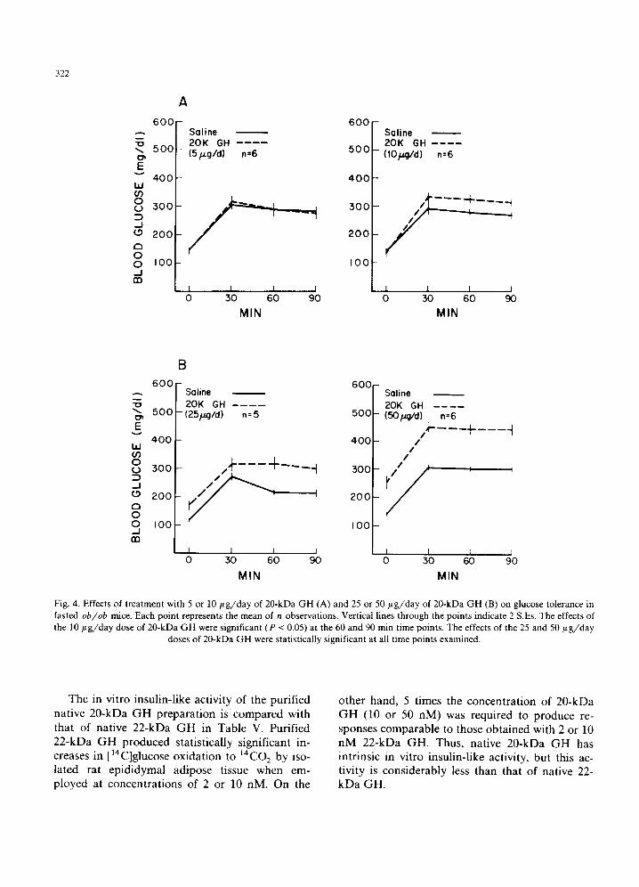
ob/ob mice. Fig. 4 il lustrates the results ob ta ined when ~20-kDa G H was tested for diabetogenic
activity at doses of 5, 10, 25 or 5 0 / ~ g / d a y for 3 days. When 20-kDa G H was adminis tered at a
dose of 5 /~g/day, it had no effect on either fasting b lood glucose concent ra t ion or glucose tolerance (see Fig. 4A). When the dose was in- creased to 10 / t g / d a y , there was again no effect on fasting blood glucose concentra t ion, bu t a modest decrease in glucose tolerance was observed (Fig. 4A). When the dose of 20-kDa G H was raised to 25 /~g/day, both an increase in fasting b lood glucose and a decrease in glucose tolerance were obta ined (Fig. 4B). Large changes in both fasting blood glucose concent ra t ion and glucose tolerance were ob ta ined when 20-kDa G H was adminis tered at a dose of 50 ~ tg /day (Fig. 4B). This dose response relat ionship is similar to that rout inely obta ined in our laboratory with purif ied nat ive 22-kDa G H [25-27].
322
A
E
w if) 0 (.9
--I 0 a 0 0 -J
A
6 0 0 - Sa l ine 2 0 K GH
5 0 0 - (Sp.g/d) n=6
4 0 0 -
5 0 0 ~
2OO
I00
1 I I I 0 30 60 90
MIN
6 0 0
5 0 0
4 0 0
3 0 0
2 0 0
I 0 0
Sal ine 2 0 K GH . . . . ( tO /4g /d ) n=6
I I I I 0 30 6O 9O
M I N
6 0 0
500
E
400 w or) 0
500
J o 200 a o 0 I 0 0 __l
B
Saline 2 0 K GH
' (25/a.g/d) n = 5
60C
500
4 0 0
500
2 0 0
I 0 0
Saline 20K GH (50/.raid) n=6 ,,+----+----~
/ /
/ ' ,
I I I I [ I I 0 50 60 90 0 50 60 90
MIN MIN
Fig. 4. Effects of treatment with 5 or 10 #g/day of 20-kDa GH (A) and 25 or 50/~g/day of 20-kDa GH (B) on glucose tolerance in fasted ob/ob mice. Each point represents the mean of n observations. Vertical lines through the points indicate 2 S.Es. The effects of the 10/~g/day dose of 20-kDa GH were significant (P < 0.05) at the 60 and 90 min time points. The effects of the 25 and 50/~g/day
doses of 20-kDa GH were statistically significant at all time points examined.
The in vitro insulin-l ike activity of the purified native 20-kDa G H prepara t ion is compared with that of nat ive 22-kDa G H in Table V. Purified 22-kDa G H produced statistically significant in- creases in [14C]glucose oxidat ion to 14C02 by iso- lated rat epididymal adipose tissue when em- ployed at concent ra t ions of 2 or 10 nM. On the
other hand, 5 times the concent ra t ion of 20-kDa G H (10 or 50 nM) was required to produce re- sponses comparable to those obtained with 2 or 10 n M 22-kDa GH. Thus, native 20-kDa G H has intr insic in vitro insulin-l ike activity, but this ac- tivity is considerably less than that of native 22- kD a GH.
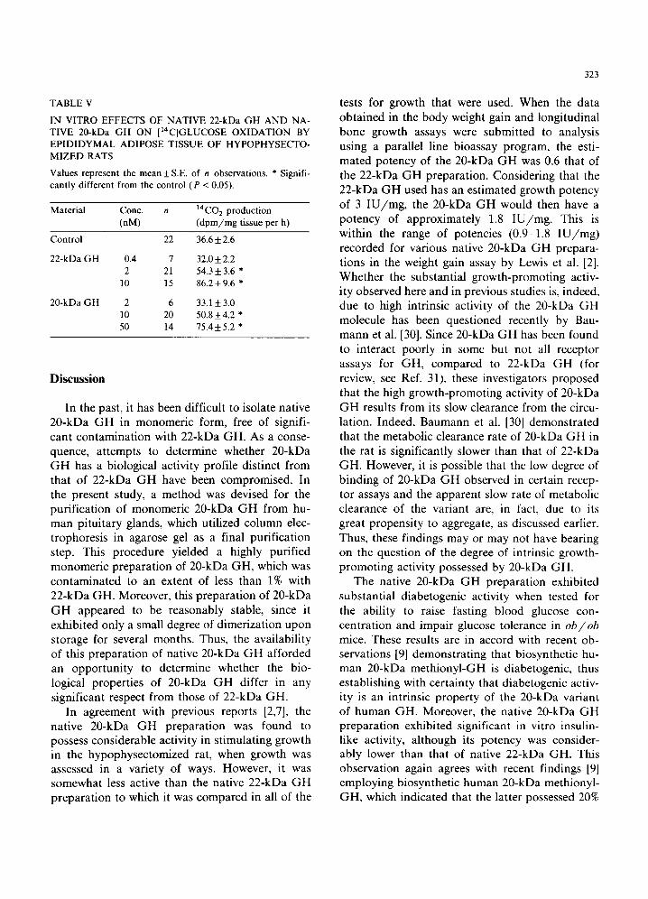
TABLE V
IN VITRO EFFECTS OF NATIVE 22-kDa GH AND NA- TIVE 20-kDa GH ON [14C]GLUCOSE OXIDATION BY EPIDIDYMAL ADIPOSE TISSUE OF HYPOPHYSECTO- MIZED RATS
Values represent the mean_+ S.E. of n observations. * Signifi- cantly different from the control (P < 0.05).
Material Conc. t/ 14C02 production (nM) (dpm/mg tissue per h)
Control
22-kDa GH
20-kDa GH
22 36.6 + 2.6
0.4 7 32.0 + 2.2 2 21 54.3 + 3.6 *
10 15 86.2+9.6 *
2 6 33.1 + 3.0 10 20 50.8+4.2 * 50 14 75.4_+5.2 *
Discussion
In the past, it has been difficult to isolate native 20-kDa G H in monomeric form, free of signifi- cant contamination with 22-kDa GH. As a conse- quence, at tempts to determine whether 20-kDa G H has a biological activity profile distinct from that of 22-kDa G H have been compromised. In the present study, a method was devised for the purification of monomeric 20-kDa G H from hu- man pituitary glands, which utilized column elec- trophoresis in agarose gel as a final purification step. This procedure yielded a highly purified monomeric preparation of 20-kDa GH, which was contaminated to an extent of less than 1% with 22-kDa GH. Moreover, this preparation of 20-kDa G H appeared to be reasonably stable, since it exhibited only a small degree of dimerization upon storage for several months. Thus, the availability of this preparation of native 20-kDa G H afforded an opportunity to determine whether the bio- logical properties of 20-kDa G H differ in any significant respect from those of 22-kDa GH.
In agreement with previous reports [2,7], the native 20-kDa G H preparation was found to possess considerable activity in stimulating growth in the hypophysectomized rat, when growth was assessed in a variety of ways. However, it was somewhat less active than the native 22-kDa G H preparation to which it was compared in all of the
323
tests for growth that were used. When the data obtained in the body weight gain and longitudinal bone growth assays were submitted to analysis using a parallel line bioassay program, the esti- mated potency of the 20-kDa G H was 0.6 that of the 22-kDa G H preparation. Considering that the 22-kDa G H used has an estimated growth potency of 3 I U / m g , the 20-kDa G H would then have a potency of approximately 1.8 I U / m g . This is within the range of potencies (0.9-1.8 I U / m g ) recorded for various native 20-kDa G H prepara- tions in the weight gain assay by Lewis et al. [2]. Whether the substantial growth-promoting activ- ity observed here and in previous studies is, indeed, due to high intrinsic activity of the 20-kDa G H molecule has been questioned recently by Bau- mann et al. [30]. Since 20-kDa G H has been found to interact poorly in some but not all receptor assays for GH, compared to 22-kDa G H (for review, see Ref. 31), these investigators proposed that the high growth-promoting activity of 20-kDa G H results from its slow clearance from the circu- lation. Indeed, Baumann et al. [30] demonstrated that the metabolic clearance rate of 20-kDa G H in the rat is significantly slower than that of 22-kDa GH. However, it is possible that the low degree of binding of 20-kDa G H observed in certain recep- tor assays and the apparent slow rate of metabolic clearance of the variant are, in fact, due to its great propensity to aggregate, as discussed earlier. Thus, these findings may or may not have bearing on the question of the degree of intrinsic growth- promoting activity possessed by 20-kDa GH.
The native 20-kDa G H preparation exhibited substantial diabetogenic activity when tested for the ability to raise fasting blood glucose con- centration and impair glucose tolerance in ob/ob mice. These results are in accord with recent ob- servations [9] demonstrating that biosynthetic hu- man 20-kDa methionyl-GH is diabetogenic, thus establishing with certainty that diabetogenic activ- ity is an intrinsic property of the 20-kDa variant of human GH. Moreover, the native 20-kDa G H preparat ion exhibited significant in vitro insulin- like activity, although its potency was consider- ably lower than that of native 22-kDa GH. This observation again agrees with recent findings [9] employing biosynthetic human 20-kDa methionyl- GH, which indicated that the latter possessed 20%
324
of the in vitro insul in- l ike activity of biosynthet ic h u m a n 22-kDa meth iony l -GH. Thus, insul in-l ike
activity is also an intr insic proper ty of 20-kDa GH. Its a t tenuated insul in- l ike activity may reflect
either that the deleted sequence (residues 32-46)
is par t of the active site for this activity of G H or that the altered conformat ion of 20-kDa G H may
interfere with the expression of its insul in-l ike activity, as has recently been proposed [9].
Acknowledgements
Financ ia l support from the Swedish Medical Research Counci l (Project No. 5921) is gratefully
acknowledged. C.M.C. was a recipient of a Na- t ional Research Service Award (AM-07268).
N.A.A. was the recipient of a fellowship from the Amer ican Diabetes Association, Michigan Af-
filiate.
References
1 Singh, R.N.P., Seavey, B.K. and Lewis, U.J. (1974) Endo- crinol. Res. Commun. 1,449-464
2 Lewis, U.J., Dunn, J.T., Bonewald, L.F., Seavey, B.K. and VanderLaan, W.P. (1978) J. Biol. Chem. 253, 2679-2687
3 Lewis, U.J., Bonewald, L.F. and Lewis, L.J. (1980) Bio- chem. Biophys. Res. Commun. 92, 511-516
4 Singh, R.N.P. and Lewis, U.J. (1981) Prep. Biochem. 11, 559-570
5 Chapman, G.E., Rogers, K.M., Brittain, T., Bradshaw, R.A., Bates, O.J., Turner, C., Cary, P.D. and Crane-Robinson, C. (1981) J. Biol. Chem. 256, 2395-2401
6 DeNoto, F.M., Moore, D.D. and Goodman, H.M. (1981) Nucleic Acids Res. 9, 3719-3730
7 Lewis, U.J., Singh, R.N.P. and Tutwiler, G.F. (1981) Endo- crinol. Res. Commun. 8, 155-164
8 Frigeri, L.G., Peterson, S.M. and Lewis, U.J. (1979) Bio- chem. Biophys. Res. Commun. 91,778-782
9 Kostyo, J.L, Cameron, C.M., Olson, K.C., Jones, A.J.S. and Pai, R.-C. (1985) Proc. Natl. Acad. Sci. USA 82, 4250-4253
10 Tinsley, F.C., Grinna, E.L., Baker, S.H., Powell, J.G., Be- mis, K.G. and Shaar, C.J. (1986) Biochem. Biophys. Res. Commun. 138, 342-348
11 Roos, P., Fevold, H.R. and Gemzell, C.A. (1963) Biochim. Biophys. Acta 74, 525-531
12 Roos, P., Jacobsson, G. and Wide, L. (1975) Biochim. Biophys. Acta 379, 247-261
13 Hjert~n, S, (1963) J. Chromatogr. 12, 510-526 14 Hjertrn, S., Hoglund, S. and Ruttkay-Nedecky, G. (1970)
Acta Virol. 14, 89-101 15 Neville, D.M. (1971) J. Biol. Chem. 246, 6328-6334 16 Hjertrn, S. (1962) Arch. Biochem. Biophys., Suppl. 1,
147-151 17 Vesterberg, O. and Gramstrup-Christensen, B. (1984) Elec-
trophoresis 5, 282-285 18 Hansson, L.F., Menander-Sellman, K., Stenstrom, A. and
Thorngren, K.-G. (1972) Calcif. Tissue Res. 10, 238-251 19 Thorngren, K.-G., Hansson, L.I. and Sundin, G. (1980) J.
Endocrinol. 84, 199-204 20 Thorngren, K.-G. and Hansson, L.I. (1974) Acta Endo-
crinol. 76, 35-52 21 Hall, R., Takano, K. and Fryklund, L. (1974) J. Clin.
Endocrinol. Metab 39, 973-976 22 Skottner, A., Forsman, A., Lofberg, E. and Thorngren,
K.-G. (1984) Acta Endocrinol. 107, 192-198 23 Armitage, P. (1977) Statistical Methods in Medical Re-
search, Ch. 17, Wiley, New York 24 Reagan, C.R. (1978) Diabetes 27, 883-888 25 Kostyo, J.L., Gennick, S.E. and Sauder, S.E. (1984) Am. J.
Physiol. 246, E356-E360 26 Cameron, C.M., Kostyo, J.L. and Papkoff, H. (1985) Endo-
crinology 116, 1501-1505 27 Cameron, C.M., Kostyo, J.L., Kumar, V. and Gennick, S.E.
(1985) Biochim. Biophys. Acta 841, 254-260 28 Mills, J.B., Kostyo, J.L., Reagan, C.R., Wagner, S.A.,
Moseley, M.H. and Wilhelmi, A.E. (1980) Endocrinology 107, 391-399
29 Mills, J.B., Ashworth, R.B., Wilhelmi, A.E. and Hartree, A.S. (1969) J. Clin. Endocrinol. Metab. 29, 1456-1459
30 Baumann, G., Stolar, M.W. and Buchanan, T.A. (1985) Endocrinology 117, 1309-1313
31 Smal, J., Closset, J., Hennen, G. and DeMeyts, P. (1986) Biochem. Biophys. Res. Commun. 134, 159-165


