Embed Size (px)
Citation preview

J. Agric. Res., Kafrelsheikh Univ., 34(3): 567 – 587.
J. Agric. Res., Kafrelsheikh Univ., 34(3): 567 – 587. 1
BIODEGRADATION OF WASTEPAPER BY
TRICHODERMA VIRIDE AND USING THE BIOPROCESSED
MATERIALS IN BIOCONTROL OF DAMPING-OFF OF PEA
CAUSED BY PYTHIUM DEBARYANUM
BY
E. B. Belal
Agricultural Microbiology, Dept. of Agric. Botany, Fac. of Agric.,
Kafrelsheikh Univ.,
ABSTRACT
The biodegradation of wastepaper materials such as filter paper,
foolscap, cardboard, tissue paper and newspapers was investigated. By
using a method based on clear zone formation on agar plates, a total of
100 microbial strains (comprising fungi and bacteria) could be isolated
from different microbial sources using carboxymethyl cellulose as
substrate for cellulase. Sixteen cellulylotic microorganisms from the 100
isolated (comprising fungi and bacteria) as potential cellulase producers
were selected. One fungal strain of 16 cellulylotic microorganisms
exhibited wider clear zone than the other strains. This strain was
identified as Trichoderma viride. The optimal pH and temperature for
growth T. viride and its cellulase production were 6.5 and 25°C,
respectively.
It was shown that the cellulase was induced in submerged culture
with presence of the carboxymethyl cellulose and wastepaper materials in
MSL, while the presence of additional carbon sources such as glucose or
a complex media (Potato Dextrose) suppressed enzyme production.
All wastepaper materials exhibited different susceptibilities towards
cellulase to their conversion to reducing sugars. The present study
showed also that, the general trend of bioconversion of different
wastepaper materials with cellulase was more than the general trend of
bioconversion of different wastepaper materials by T. viride.
Additionally to the qualitative clear zone tests, the degradation
potential of T. viride was characterized via weight loss measurements of
wastepaper materials on agar plates. T. viride exhibited also good
degradability for mixture from the tested wastepaper materials in solid
state fermentation without newspapers leads to produce biomass
(bioprocessed materials).
Results of this study suggest that soil treatment with the
bioprocessed materials was an effective in controlling damping-off of pea
and could be considered as promising alternative to existing chemical
products, where the effects were similar in more cases to those of maxim
fungicide. The application of these results may be help in reducing the

J. Agric. Res., Kafrelsheikh Univ., 34(3): 567 – 587.
J. Agric. Res., Kafrelsheikh Univ., 34(3): 567 – 587. 2
effect of environmental pollution which caused by wastepaper materials
or chemical fungicides.
Keywords: Biodegradation, Trichoderma viride, cellulase, wastepaper,
Pythium debarayanum, soil treatment, biological control
INTRODUCTION
Various wastepaper materials are a major component of solid waste
all over the world. Cellulolytic enzymes play an important role in natural
biodegradation process in which plant lignocellulosic materials are
efficiently degraded by cellulolytic fungi and bacteria. In industry, these
enzymes have found novel applications in the production of fermentable
sugars and ethanol (Olson and Hahn-Hagerdahl, 1997; Levy et al., 2002;
Van Wyk and Mohulatsi, 2003), organic acids (Luo et al., 1997),
detergents, and other chemicals (Oksanan and Peeabilaniana 1998). They
have been used in the pulp and paper industry, e. g., in deinking of fiber
surfaces and in improving pulp drainage (Oksanan et al., 2000;
Suurnakki et al., (2004)), in the textile industry (Cavaco-Paulo and
Gübitz (2003), Nierstrasz, and Warmoeskerken, 2003; Miettinen-
Oinonen et al., 2004), animal feed (Ishikuro, 2000), and even in the food
industry(Penttila et al., (2004); Urlaub, (2002), for the processing of
paper and cellophane, as well as for biotransformation of wastepaper to
fermentable sugars (Van Wyk and Mohulatsi, 2003). As lytic enzymes,
they are of prime importance is the protoplast production (Davis, 1985;
Mandels, 1974; Bhat, 2000). Fungal cellulases are inducible enzymes
that are usually excreted into the environment (Bhat and Bhat, 1997) and
depend on cellulose type (amorphous or crystalline) acting on the
organism (Ortega et al., 2001). The role of the fungi Acremonium spp.,
Chaetomium spp., Trichoderma reesei, Trichoderma viride, Penicillium
pinophilum, Phanerochaete chrysosporium (Sporotrichum
pulverulentum), Fusarium solani, Talaromyces emersonii, Trichoderma
koningii, Fusarium oxysporum, Aspegillus niger and Rhizopus oryzae in
the cellulose degradation process in various environments has been well
documented (Kuzmanova et al., (1991); Teerei and Koivala, (1995); Bhat
and Bhat, (1997); Schülein, 1997; Murashima et al., 2002; and Mach,
Zeilinger, 2003).
Many investigators reported earlier the use of Trichoderma
hamatum or T. harzianum for the control of Pythium seed rot and
Rhizoctonia root rot in different crops such as pea (Harman et al., 1980,
Nelson et al., 1988 and Belal et al., 1996).
Therefore, the present investigation is an attempt to study the
biodegradation of wastepaper, bioconversion of the tested wastepaper to

J. Agric. Res., Kafrelsheikh Univ., 34(3): 567 – 587.
J. Agric. Res., Kafrelsheikh Univ., 34(3): 567 – 587. 3
fermentable sugars as well as using the bioprocessed materials in
controlling of damping –off of pea caused by Pythium debaraynum.
MATERIALS AND METHODS
Media Minimal Medium as mineral salt medium (MSL) was used through
this study as described by (Drews 1968) and Luria Bertani Medium (LB),
TSB as well as Potato Dextrose Agar (PDA) were used also as complex
media in the present study.
Sampling and Cellulolytic microorganisms isolation
Samples of the different substrates–raw materials (wastepaper),
mushroom fruits, soil, mature compost, primary effluent mud,
wastewater from the sedimentation reservoir were collected from the
paper factories located in different Governorates in Egypt.
In laboratory, 1 g of each substrate (some of which needed to be
milled) was added to the conical flask containing 99ml of MSL medium
and mixed for 30 min on a rotary shaker (150 rpm) at room temperature.
Samples of sludge and wastewater were added as 1 ml to 99 ml of MSL
medium. The supernatants were separated by centrifugation at 5000rpm
for 20min. Ten-fold dilutions were prepared and then 100μl of each
dilution were spread on plates containing MSA + carboxymethyl
cellulose (10 g/L as a sole source of carbon) using drigalisky triangle.
The plates were sealed in polyethylene bags and were incubated at 25◦C
for 7days monitored for appearance of colonies. Cellulolytic strains were
selected on the basis of the diameter of the hydrolysis zone surrounding
the colonies as described by (Teather, Wood, 1982; Bradner et al., 1999
and Peciulyte 2007). Single colonies growing on the dilution plates were
isolated by picking the colonies using sterile inoculation needle and were
further purified by the standard spatial streaking for bacterial isolates on
complex agar media (TSB or nutrient agar) and or using acidic complex
medium or addition of ampicilline 800mg/L to complex medium for
fungal isolates (PDA for fungal isolates).
Identification
The selected wastepaper degrading bacterial strains were identified
as described by Bergy’s manual of systematic bacteriology (1984). Also,
the selected wastepaper degrading fungal strains were identified
according to Rifai, M. A. (1969), Domsch et al., 1980, and Burgess et al.,
(1994).
Degradation of different wastepapers materials by the
microorganisms via measuring of clear zone
The isolated colonies were then tested for their ability to grow
and degrade different wastepaper materials (foolscap, filter paper

J. Agric. Res., Kafrelsheikh Univ., 34(3): 567 – 587.
J. Agric. Res., Kafrelsheikh Univ., 34(3): 567 – 587. 4
(whatman paper No.1), cardboard (packing materials), tissue paper,
newspaper) in MSL medium. All wastepaper were milled. 100ml MSL
medium containing 10g/L from each material was inoculated by 3 ml
from fungal suspension at 106 cfu/ml or bacterial cell suspension at 10
7
cfu/ml, respectively. One treatment contained the medium and
carboxymethyl cellulose and the other contained the medium without
carboxymethyl cellulose and the isolate (control).
The cultures were shaken at 150rpm and 25◦C for 14 days. All
assays were carried out from cultures supernatant as extracellular
cellulase source after removing the growth by using sterile membrane
filter (0.2μm). 50μl of culture supernatant was added in wells (5mm in
diameter) of MSA (containing carboxymethyl cellulose 10 g/L as
substrate). The plates were treated and the clear zone was measured
according to the method described by Teather, Wood, (1982); Bradner et
al., (1999) and Peciulyte (2007).
Effect of pH and temperature on growth of Trichoderma viride and its
cellulase (CMCase) production
One hundred ml MS-medium supplemented with carboxymethyl
cellulose (10 g/L) as a sole source of carbon were used to determine the
effect of pH and temperature on growth of T. viride and its cellulase
production. The medium was inoculated by 3ml (106 cfu/ml) of culture of
Trichoderma viride strain. The experiments were carried out at pH 4, 4.5,
5, 5.5, 6, 6.5, 7, 7.5 and 8 and the culture was incubated at 25oC with
shaking (150 rpm) for 14 days. To determine the optimum temperature,
MSL medium at pH6.5 was incubated at 20, 25, 30, 35 and 40oC with
shaking (150 rpm) for 14 days. The activity of Trichoderma viride
cellulase was determined by measuring of clear zone as described above.
The growth was determined as mycelial dry weight of biomass (g) after
14 days as described by Belal (2003).
Effect of different carbon source on growth Trichoderma viride and
enzyme induction
One hundred ml MSL in conical flasks (250ml) containing 10g/L (all
wastepaper materials were milled) from each material or glucose was
inoculated by 3 ml from fungal suspension at 106 cfu/ml (one-week-old
colonies of fungi grown at 25 °C on PDA plates). Potato dextrose (PD)
was used as complex medium and it was carried at the same conditions.
Cultures were incubated in shaker incubator for 14 days at 25°C and 150
rpm. After 14 days of cultivation, culture aliquots were centrifuged at
5000 rpm to remove solids. The supernatants were assayed for their
enzymatic activity by measuring of clear zone as described above. The

J. Agric. Res., Kafrelsheikh Univ., 34(3): 567 – 587.
J. Agric. Res., Kafrelsheikh Univ., 34(3): 567 – 587. 5
growth was determined as mycelial dry weight of biomass (g) after 14
days as described by Belal (2003).
Enzyme assay and saccharification of wastepapers materials by the
Trichoderma viride cellulase
Cellulase activity was determined by incubating 0.5 ml of the
supernatant (at a concentration of 250 μg/ml, while the enzyme
concentration was determined according to Lowry et al., 1951) with 0.5
ml of an amount 10g/L of each material carboxymethyl cellulose or
milled wastepaper materials in 0.05 M citrate buffer (pH4.8) at 50oC for
30min. After incubation, the reaction was terminated by adding 3 ml of
1% 3,5-dinitrosalicylic acid (DNS) reagent to 1 ml of the reaction
mixture and heated for 10min. In these tests, reducing sugars were
estimated calorimetrically after Miller (1959), using glucose as standards.
One unit of cellulase activity is defined as the amount of enzyme that
releases 1 µmol reducing sugars (measured as glucose) per ml per min.
Biodegradation of different wastepaper materials by Trichoderma
viride Weight loss determination of wastepaper films on agar plates
The degradation test was carried out with wastepaper films (30 –
60mg) on MSA as a sole source of carbon at the optimal growth
conditions. Three preweighted, sterile circular films (25 mm diameter,
surface area assessable for degradation (4.91 cm²) of the wastepaper were
placed on a MSA- plate and inoculated with 80μl from fungal suspension
at 106 cfu/ml on surface of the wastepaper film. The degradation times on
the agar plates was 3 weeks at the optimal growth conditions. Sterile
controls incubated over the same period of time were performed and
showed no weight loss due to a biotic hydrolysis of the paper samples
according to the described methods of Belal (2003) with replacing plastic
films with wastepaper films.
Biodegradation of mixture of wastepaper materials by
Trichoderma viride by using solid state fermentation and using
the produced biomass (bioprocessed materials) in suppression of
Pythium debarayanum
Different wastepaper materials (filter paper(whatman paper
No.1), foolscap, , cardboard (packing materials), and tissue paper were
prepared in pieces of 3cm × 3cm and mixed well with ratio of 1:1:1:1 and
placed in glass box (width 40cm × 30cm height). The mixed wastepapers
were moistened (till 65% ) with MSL medium without addition any
carbon source, the substrate moistened when needed. The mixed
wastepaper materials were treated with spore suspension of Trichoderma
viride (106 cfu/gm) from wastepaper substrate and after that was covered

J. Agric. Res., Kafrelsheikh Univ., 34(3): 567 – 587.
J. Agric. Res., Kafrelsheikh Univ., 34(3): 567 – 587. 6
by polyethylene and incubated for 42 days at the optimal growth
conditions. The produced biomass (bioprocessed materials) was used for
soil treatment (at rate of 2%) for suppression of damping off of pea plants
caused by Pythium debarayanum (soil was inoculated with the
phytopathogenic fungi as described by Belal et al., 1996).
Statistical analysis:
The obtained data were subjected to the proper statistical
procedures for analysis of variance according to Gomez and Gomez,
(1984).
RESULTS AND DISCUSSION
Seven different sources (mushroom fruits, wastepaper, soil, mature
compost, primary effluent mud, waste water) were used to isolate the
cellulolytic microorganisms in the present study. One hundred
microorganism were isolated on MSA by using clear zone formation on
agar plates. Sixteen cellulylotic microorganisms from the 100 isolated
(comprising fungi and bacteria) as potential cellulase producers were
selected (Table 1). A preliminary classification based on the morphology
of the isolates revealed that the wastepaper – degrading microorganisms
belong to the group of fungi as well as to the group of bacteria. Thirteen
fungal isolates of 16 were isolated and identified as Trichoderma viride, T.
harzianum, T. reesei, Penicillium sp., Rhizopus sp., Mucor sp., Aspergillus
niger, Fusarium solani, Acremonium strictum, Cladosporium herbarum,
Pleurotus sp., Agaricus sp. and Myrothecium sp. Fungi are well-known
agents of decomposition of organic matter in general and cellulose
substrates in particular (Lynd et al., 2002).
Three of 16 wastepapers – degrading organisms were bacteria.
Two of 3 were gram- positive, non-motile, filamentous and identified as
Streptomyces sp. and Microbispora sp.
One of 3 bacterial isolates was gram -positive, motile, rods, and
spore former and identified as Bacillus sp.. Obviously, fungi play an
outstanding role in degrading of the tested wastepapers materials, since
the majority of strains belong to this group. It is known that many genera
of fungi play an important role in degradation of anthropogenic
substrates. Due to the paucity of growth which was generally observed
on MSA + carboxymethyl cellulose. The bacterial isolates were also
routinely streaked onto plates of TSB or nutrient agar for bacterial strains
but the fungal strains were further purified by using acidic complex
medium (PDA) or addition of ampicilline 800mg/l to complex medium
(PDA).
Results in Table 1 showed that the strains were tested for their
growth ability on MSL supplemented with wastepaper materials

J. Agric. Res., Kafrelsheikh Univ., 34(3): 567 – 587.
J. Agric. Res., Kafrelsheikh Univ., 34(3): 567 – 587. 7
(foolscap, filter paper (whatman paper No.1), cardboard (packing
materials), tissue paper and newspaper) as a sole source of carbon. The
general trend of biodegradability with all strains of carboxymethyl
cellulose was > filter paper > foolscap > cardboard > tissue paper >
newspapers. carboxymethyl cellulose, filter paper and foolscap exhibited
the highest degree of bioconversion followed by the other materials
because the diameter of clear zone value was wider than the other
materials. Carboxymethyl cellulose and filter paper were a more
favourable carbon source for screening the cellulolytic microorganisms.
On the other hand, newspaper exhibited the lowest degree of
bioconversion by the cellulolytic microorganisms and this may be depend
on cellulose type (amorphous or crystalline) acting on the organism
(Ortega et al., 2001). The obtained results show also that the fungal
strains exhibited the highest biodegradability for the wastepaper than the
bacterial strains.
Among 16 isolated strains, one strain was identified as Trichoderma
viride exhibited relative high clearing of plates which was supplemented
with carboxymethyl cellulose as substrate for cellulase. This indicates
that this strain is the highest degradability for the tested wastepaper
materials than the other strains. The obtained results were compared with
the growth of the isolates in MSL (no wastepaper materials).
Trichoderma is known as a very good producer of cellulases, perhaps due
to the different adaptability of fungi to the anthropogenic substrates and
different resistance to the factors affecting fungal populations during the
recycling procedures. Our results are in agreement with previous findings
reported by Teather and Wood, (1982); Bradner et al., (1999) and
Peciulyte (2007).
Therefore, Trichoderma viride as efficient for productivity of
extracellular cellulase was selected for the further studies.
Table (1) Degradation of different wastepaper materials by the
microorganisms via measuring clear zone. Microorganisms Diameter of clear zone (mm)
Carboxymethyl
cellulose
Filter
paper
Foolscap Cardboard Tissue
paper
Newspapers
Trichoderma
viride 40 36 29 19 13 10
T. harzianum 32 26 20 16 10 8 T. reesei 33 27 21 15 11 7 Penicillium sp. 27 23 22 13 10 7 Rhizopus sp. 23 20 17 12 10 7 Mucor sp. 29 24 16 11 9 9 Aspergillus 28 23 18 12 8 8

J. Agric. Res., Kafrelsheikh Univ., 34(3): 567 – 587.
J. Agric. Res., Kafrelsheikh Univ., 34(3): 567 – 587. 8
niger
Fusarium
solani 27 21 16 11 9 7
Acremonium
strictum 26 20 18 11 9 7
Cladosporium
herbarum 23 18 12 11 10 7
Pleurotus sp. 37 28 23 18 12 9 Agaricus sp. 36 29 22 17 11 8 Myrothecium
sp. 23 16 13 11 9 8
Streptomyces
sp. 22 17 12 10 9 7
Microbispora
sp. 21 13 12 11 8 7
Bacillus sp. 18 12 11 9 7 7
Effect of pH and temperature on growth of Trichoderma viride and
its cellulase (CMCase) production Environmental factors do not only influence the wastepapers to
be degraded, they also have a crucial influence on the microbial
population and on the activity of the different microorganisms
themselves and also the amount of the enzyme production depends on the
biomass. Factors such as temperature and pH, have important effects on
the microbial degradation of wastepapers and so these conditions must be
considered when the biodegradability of wastepapers is tested. Karpouzas
and Walker (2000) reported that the degradation of ethoprophos by
Pseudomonas putida strains epI and II affected by pH and temperature.
The question is now, what are the optimal conditions (pH and
temperature) for the growth of Trichoderma viride and its cellulase
(CMCase) production?. To determine the optimal growth conditions,
carboxymethyl cellulose was used as a sole source of carbon.
Optimum pH:
The influence of pH on biomass yield of Trichoderma viride and its
cellulase (CMCase) production is shown in Fig. 1. Generally, the
optimum pH was 6.5 for Trichoderma viride. The maximum mycelial dry
weight for Trichoderma viride and its cellulase (CMCase) production
were recorded at pH6.5. Trichoderma viride grew at quite wide pH range
(from 4 to 8). This variation is very useful to use these isolates in
degradation test in different environments at different pH. Therefore, it
can expect that these isolates can tolerate the pH change during the
degradation process thereby increase the degradation potential for these
isolates.

J. Agric. Res., Kafrelsheikh Univ., 34(3): 567 – 587.
J. Agric. Res., Kafrelsheikh Univ., 34(3): 567 – 587. 9
3 4 5 6 7 8 9
-5
0
5
10
15
20
25
30
35
40
pH
Dia
met
er o
f cl
ear
zone
(mm
) Diameter of cear zone (mm)
0.05
0.10
0.15
0.20
0.25
0.30
0.35
0.40
0.45
0.50
My
celi
al d
ry w
eight
(g)
Mycelial dry weight (g)
Fig.(1 ): Effect of pH on growth of Trichoderma viride and its cellulase
(CMCase) production by measuring clear zone.
Optimum temperature:
The effect of different temperatures on growth of Trichoderma viride
and its cellulase (CMCase) production is shown in Fig.2, respectively. A
temperature 25◦C appears to be the optimum for growth of Trichoderma
viride and its cellulase (CMCase) production. Trichoderma viride and its
cellulase (CMCase) production exhibited growth and cellulase (CMCase)
production at different temperatures but Trichoderma viride did not grow
at 40◦C. Therefore, these strain was used for further studies under the
optimum growth conditions with the aim of the effect of different carbon
source on growth Trichoderma viride and enzyme induction as well as
determination of the degradation potential for the wastepaper material
under solid state fermentation.

J. Agric. Res., Kafrelsheikh Univ., 34(3): 567 – 587.
J. Agric. Res., Kafrelsheikh Univ., 34(3): 567 – 587. 10
18 20 22 24 26 28 30 32 34 36 38 40 42
0
10
20
30
40
50
Temperature
Dia
met
er o
f cl
ear
zone
(mm
)
Diameter of clear zone (mm)
0.00
0.05
0.10
0.15
0.20
0.25
0.30
0.35
0.40
0.45
My
celi
al d
r w
eight
(g)
Mycelial dry weight(g)
Fig.(2) : Effect of temperature (◦
C) on growth of Trichoderma viride and
its cellulase (CMCase) production by measuring clear zone.
Regulation of enzyme production (constitutive or inductive enzyme)
According to Schlegel (1992) most enzymes systems involved in
substrate degradation are inductive enzymes. Therefore it is of interest to
know, if the wastepaper degrading enzyme system is constitutively
secreted or induced by the presence of wastepaper or other carbon source.
Furthermore it is of interest, if the enzyme is inducible, what are the
substances inducing the enzyme activity?
Fig. 3. shows that the fungal growth (determined as mycelial dry weight)
was high on PD, followed by MSL+glucose, MSL + carboxymethyl
cellulose, MSL + filter paper, MSL + foolscap, MSL + cardboard, MSL +
tissue paper, MSL + newspaper and latter on MSL without carbon source.
Fig. 3.and Fig.4. indicate that the extracellular cellulase was produced
only during growth of Trichoderma viride on all wastepaper materials in
MSL medium as carbon sources. The results demonstrated that a
maximum cellulase activity was obtained when carboxymethyl cellulose
followed by filter paper, foolscap, cardboard, tissue paper were used as
substrate. The low enzyme activities measured with newspapers. On the
other hand in PD as a complex medium or in MSL+glucose, enzyme
secretion is not induced despite the media generated a good cell growth.
This results are in agreement with my previous findings and other
investigators while secretion of PCL-hydrolase was only induced in the
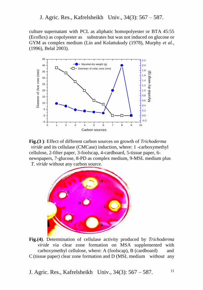
J. Agric. Res., Kafrelsheikh Univ., 34(3): 567 – 587.
J. Agric. Res., Kafrelsheikh Univ., 34(3): 567 – 587. 11
culture supernatant with PCL as aliphatic homopolyester or BTA 45:55
(Ecoflex) as copolyester as substrates but was not induced on glucose or
GYM as complex medium (Lin and Kolattukudy (1978), Murphy et al.,
(1996), Belal 2003).
0 1 2 3 4 5 6 7 8 9 10
-5
0
5
10
15
20
25
30
35
40
45
Carbon sources
Dia
met
er o
f cl
ear
zone
(mm
) Diameter of celar zone (mm)
-0.2
0.0
0.2
0.4
0.6
0.8
1.0
1.2
1.4
1.6
1.8
2.0
2.2
Myce
lial d
ry w
eig
t (g
)
Mycelial dry weight (g)
Fig.(3 ): Effect of different carbon sources on growth of Trichoderma
viride and its cellulase (CMCase) induction, where: 1 -carboxymethyl
cellulose, 2-filter paper, 3-foolscap, 4-cardboard, 5-tissue paper, 6-
newspapers, 7-glucose, 8-PD as complex medium, 9-MSL medium plus
T. viride without any carbon source.
Fig.(4). Determination of cellulase activity produced by Trichoderma
viride via clear zone formation on MSA supplemented with
carboxymethyl cellulose, where: A (foolscap), B (cardboard) and
C (tissue paper) clear zone formation and D (MSL medium without any
A
C
B
D
E
F

J. Agric. Res., Kafrelsheikh Univ., 34(3): 567 – 587.
J. Agric. Res., Kafrelsheikh Univ., 34(3): 567 – 587. 12
carbon source plus T. viride), E (MSL medium with glucose) and F
(PD medium) no clear zone formation.
Saccharification of wastepapers materials by the Trichoderma viride
cellulase
Aside from the traditional methods of waste management, biowaste
has been used in the production of clean energy where it replaces coal,
oil or natural gases to generate electricity through combustion. The
conversion process of wastes to energy has been proved to be safe,
environmental friendly and reduces the incoming volume of waste to a
great extent. An alternative to the combustion of biowaste could be
through the fermentation of saccharified waste cellulose into bioproducts.
An initial increasing trend of sugar formation was observed when
more of each wastepaper substrate was degraded with a fixed enzyme
concentration (Table 2). Carboxymethyl cellulose and filter paper
showed more bioconversion than the other paper materials. The trend of
biodegradadability with T. viride cellulase of carboxymethyl cellulose
was > filter paper > foolscap > cardboard > tissue paper > newspapers.
Due to the structural composition of wastepaper material it can be
biodegraded into fermentable sugars. The variation in wastepaper
bioconversion by T. viride cellulase could be due to the composition of
the enzyme system as well as the structure of cellulose, this consists of a
crystalline section, which is difficult to hydrolyze, and an amorphous
section that is more susceptible to cellulase attack (Van Wyk and
Mohulatsi, (2003). The present study showed also that, the trend of
biodegradadability of different wastepaper materials with cellulase was
more than the trend of biodegradadability of different wastepaper
materials by T. viride because after14days of cultivation T. viride, the
production of reducing sugar was almost (enzymatic activity measured-
by the production of reducing sugars end group, which is taken to be an
indication of cleavage of cellulose molecules) equal to which produced
with cellulase after the incubation period (at 50oC for 30min).
It is of interest that isolation and purification as well as
characterization of T. viride cellulase in the next study and using it in
different industrial purposes as well as in bioconversion other cellulolytic
materials such as agricultural wastes.
In most investigations, members of the fungal genus Trichoderma
have been extensively studied due to their ability to secrete cellulose-
degrading enzymes. Most of the works have been carried out on T.
aureoviride Rifai, T. viride Pers., T. reesei E. G. Simmons, T. harzianum
Rifai strains and their mutants evaluating their ability to produce
extracellular cellulolytic enzymes (endoglucanases, exoglucanases and

J. Agric. Res., Kafrelsheikh Univ., 34(3): 567 – 587.
J. Agric. Res., Kafrelsheikh Univ., 34(3): 567 – 587. 13
cellobiase) which act synergistically in the conversion of cellulose to
glucose. The cellulases secreted by Trichoderma have received
widespread industrial interest leading to commercial applications (Olson
and Hahn-Hagerdahl (1997); Oksanan et al., (2000); Mach and Zeilinger
(2003); Cavaco-Paulo and Gübitz, (2003), Nierstrasz and
Warmoeskerken, (2003); Van Wyk and Mohulatsi, (2003); Penttila et al.,
(2004).
Table (2) Degradation of different wastepaper materials by Trichoderma
viride cellulase Wastepaper materials Activity of cellulase
(U/ml/min) after the
incubation period (at
50oC for 30min)
Activity of T. viride
cellulase (U/ml/min)
after the incubation
period (14 days at 25°C
and 150 rpm)
Carboxymethyl
cellulose 3.3 3
Filter paper 2.2 2.1 Foolscap 1.7 1.6 Cardboard 1.2 1.1 Tissue paper 0.7 0.6 Newspapers 0.4 0.3
Biodegradation of different wastepaper materials by Trichoderma
viride Weight loss determination of wastepaper on agar plates
The degradation potential of Trichoderma viride was quantified
(expressed as weight loss (mg), % degradation, and degradation rate per
surface area [mg/(week cm²)] for wastepaper films on agar plates after 3
weeks incubation at 25°C (Table 3). Trichoderma viride grew on mineral
salts agar plates containing wastepaper films (filter paper, foolscap,
cardboard, tissue paper and newspapers) as a sole source of carbon (Fig.5).
The obtained results shows that, filter paper and foolscap films
were degraded much faster than the cardboard, tissue paper and
newspapers films which were a more favourable carbon source for
Trichoderma viride. It was also observed that the incubation time was
longer than the previous experiment and this due to the surface area of
the wastepaper materials.
Table (3) Degradation rate [mg/(week cm²)] of different wastepaper
materials by Trichoderma viride on agar plates
Wastepaper
materials
% Degradation Degradation
rate[mg/(week cm²)]

J. Agric. Res., Kafrelsheikh Univ., 34(3): 567 – 587.
J. Agric. Res., Kafrelsheikh Univ., 34(3): 567 – 587. 14
Filter paper 100 1.7
Foolscap 100 1.4
Cardboard 75 1.02
Tissue paper 45.7 0.54
Newspapers 25 0.34
Fig.(5). Degradation of filter paper films by Trichoderma viride on agar
plates where: A: Control, and B: inoculated with Trichoderma
viride
Biodegradation of mixture of wastepaper materials by
Trichoderma viride by using solid state fermentation and using
the bioprocessed materials in suppression of damping-off caused by
Pythium debarayanum It is of interest to mix wastepaper materials together with removing
of newspapers(because it containing lead) and degradation of the
wastepaper mixture by Trichoderma viride in solid state fermentation. A
mixture of wastepaper materials exhibited completely degradation after 6
weeks and color of mixture of wastepaper materials convert to
Trichoderma viride color (green color) and this produced biomass was
named as (bioprocessed materials). The produced biomass (bioprocessed
materials) contained high numbers of from Trichoderma viride which
contained (105cfu/ml).
It was of a particular interest to use the produced biomass
(bioprocessed materials) as biocontrolling agent for suppression of
damping-off of pea caused by Pythium debarayanum, since, different
species of Trichoderma were previously used in controlling of Pythium
seed rot and Rhizoctonia root rot in different crops such as pea (Harman
et al., 1980, Nelson et al., 1988 and Belal et al., 1996).
Under greenhouse conditions, application of the bioprocessed
materials to soil infested artificially with Pythium debarayanum (Table 4)
A B

J. Agric. Res., Kafrelsheikh Univ., 34(3): 567 – 587.
J. Agric. Res., Kafrelsheikh Univ., 34(3): 567 – 587. 15
exhibited their efficacy to control damping-off of pea and increased
survival plants. The effects were similar in most cases to those of maxim
fungicide. Also, Attempts had been successfully carried out using
antagonists to control soil – borne fungal pathogens on pea (Harman et
al., 1991, Belal et al., 1996, Mao et al., 1997, Xue, 2001 and 2003).
Table (4) Biocontrol of damping-off of pea by the bioprocessed
materials. Treatments %
pre-emergence
damping-off
% post-emergence
damping-off
% survival
plants
Control
(un-inoculated)
0a 0a 100a
Pythium
debarayanum
60b 20b 20b
T. viride + Pythium
debarayanum
10c 5c 85c
Maxim+ Pythium
debarayanum
5c 5c 90c
Values having the same alphabetical letter within column are not significantly different
(P < 0.05).
The obtained results indicate the necessity of biodegradation of
wastepaper and use of the bioprocessed materials as alternative for
fungicides to reduce the environmental pollution which caused by
wastepaper materials or chemical fungicides.
REFERENCES
Belal, E. B., Sh. El-Gremi, M. Gabr and M. E. K. Ibrahim. (1996). Using
of peat-based inocula of selected antagonists against certain soil-
borne pathogens of pea in the presence of Rhizobium
leguminosarum. J. Agric. Res. Tanta Univ., 22 (4): 444-450.
Belal, E. B., M. Gabr, Sh. El-Gremi and M. E.K. Ibrahim. (1996).
Interaction between Antagonistic Microorganisms and certain
soil-borne pathogens of soybean in relation to Bradyrhizobium
japonicum. J. Agric. Res. Tanta Univ., 22 (4): 451-459.
Belal, E. B. A. (2003). Investigation on the biodegradation of polyesters
by isolated mesophilic microbes. Dissertation, Technical
University Braunschweig, Germany.
Bergy,s manual of systematic bacteriology. (1984). Williams and
Wilkins, Baltimore, USA. Vol. 1. Krieg, N. R. *(ed). Ordinary
gram negative bacteria. Vol. 2. Sneath, P. h. A (ed.) Ordinary
gram positive bacteria.
Bhat M. K. 2000.Cellulases and related enzymes in biotechnology.
Biotechnology Advances. Vol. 18. P. 355–383.

J. Agric. Res., Kafrelsheikh Univ., 34(3): 567 – 587.
J. Agric. Res., Kafrelsheikh Univ., 34(3): 567 – 587. 16
Bhat M., Bhat S. 1997. Cellulose degrading enzymes and their potential
industrial applications. Biotechnol. Adv. Vol. 15. P. 583, 620.
Bradner J. R., Gillings M., Nevalainen K. M. H. (1999). Qualitative
assessment of hydrolytic activities in Antarctic microfungi
grown at different temperatures on solid media. World J.
Microbiol. Biotechnol. Vol. 15. P. 131–132.
Burgess, L. W., Summerell, B. A., Bullocks, S. G. and Backhouse, K. P.
D. (1994). Laboratory Manual for Fusarium Research. 3rd
ed.
University of Sydney. 133p.
Cavaco-Paulo A., Gübitz G. (2003). Catalysis and processing. In:
Cavaco-Paulo A., Gübitz G. (ed.). Textile Processing with
Enzymes. England, Woodhead PublishingLtd. P. 86, 119.
Davis B. (1985). Factors influencing protoplast isolation. In: Peberdy
J. F., Ferenizy L. (eds.). Fungal Protoplasts: Applications in
Biochemistry and Genetics. Marcel Dekker, New York. 356 p.
Domsch, K. H., Gams, W., Andersson T-H. (1980) In: Compendium of
soil fungi, 1. London: Academic Press.
Drews, G. (1968). Mikrobilogishes Praktikum fuer Natuwissenschaftler,
Springer, Verlag, Berlin-Heidelberg – New York, in Alef, K.
(1991). Methodenhandbuch Bodenmikrobiologie Bayreuth,
Deutschland.
Gomez, K. A. and A. A. Gomez (1984): Statistical procedures for
Agricultural research. 2nd
ed. John Wiley and Sons, pp. 229-308.
Harman, G. E., I. Chet and R. Baker . (1980). Trichoderma hamatum
effect on seed and seedling diseases induced in radish and pea
by Pythium spp. or Rhizoctonia solani. Phytopathology, 70: 1167 – 1172.
Harman, G. E. (1991). Seed treatments for biological control of plant
disease. Crop. Prot. 10:166 - 171.
Ishikuro E. (1993). Feed additives. Modern Media. Vol. 46. P. 289–296.
Karpouzas, D. G. and Walker, A. (2000). Factors influencing the
ability of Pseudomonas putida strain epl and epll to
degrade the organophosphate ethoprophos. J. Appl.
Microbiol. 89, 40 – 48.
Kuzmanova S., Vandeska E., Dimitrovski A. (1991). Production of
mycelial protein and cellulolytic enzymes from food waste. Journal
of Industrial Microbiology and Biotechnology. Vol. 7. N 4. P. 257–261.
Levy I., Shani Z., Shoseyov O. 2002. Modification of polysaccharides
and plant cell wall by endo-1,4-b-glucanase and cellulose- binding
domains. Biomolecular Engineering. Vol. 19. P. 17–30.

J. Agric. Res., Kafrelsheikh Univ., 34(3): 567 – 587.
J. Agric. Res., Kafrelsheikh Univ., 34(3): 567 – 587. 17
Luo J., Xia L. M., Lin J. P., Cen P. L. 1997. Kinetics of simultaneous
saccharification and lactic acid fermentation processes. Biotechnol.
Progr. Vol. 1. P. 762–767.
Lin, T. S. and Kolattukudy, P. E. (1978). Induction of a biopolyester
hydrolase (cutinase) by low levels of cutin monomers in
Fusarium solani f. sp. Pisi. J. Bacteriol. 133 (2), 942-951.
Lowry, O.H, Rsebrough, N. J., Farr, A. L. and Rundal, R. L. (1951).
protein measurements with the folin phenol reagent. J. Biol.
Chem. 193, 265-275.
Lynd L. R., Weimer P. J., van Zyl W. H., Pretorius I. S. (2002).
Microbial Cellulose Utilization: Fundamentals and Biotechnology.
Microbiology and Molecular Biology Reviews. Vol. 66. N 3. P. 506–
577.
Mach R., Zeilinger S. (2003). Regulation of gene expression in
industrial fungi: Trichoderma. Appl. Microbiol. Biotechnol. Vol. 60.
P. 515–522.
Mandels M. L., Hontz L., Nistrom J. 1974. Enzymatic hydrolysis of
waste cellulose. Biotechnology and Bioengineering. Vol. 16. P.
471–493.
Mao, W., J. A. Lewis, P. K. Hebber and R. D. Lumsden. (1997). Seed
treatment with a fungal or a bacterial antagonist for reducing
corn damping – off caused by species of Pythium and Fusarium.
Pant Dis. 81:450 – 454.
Miller G. L. (1959). Use of dinitrosalicylic acid reagent for
determination of reducing sugar. Annl. Chem. Vol. 31. P. 426–
428.
Miettinen-Oinonen A., Londesborough J., Joutsjoki V., Lantto R.,
Vehmaanpera J. 2004.Three cellulases from Melanocarpus
albomyces with applications in the textile industry. Enzyme and
Microbial Technology. Vol. 34. P. 332–341.
Murashima K., Nishimura T., Nakamura Y., Koga J., Moriya T.,
Sumida N., Yaguchi T., Kono T. 2002. Purification and
characterization of new endo-1,4-?-D-glucanases from
Rhizopus oryzae. Enzyme Microb. Technol. Vol. 30. P.
319–326.
Murphy, C. A., Cameron, J. A., Huang, S. J., and Vinopal, R. T.
(1996). Fusarium polycaprolactone depolymerase is
cutinase. Appl. Environ. Microbiol. 62:(2) 456-460.
Nelson, E. B., Harman, G. E., and Nash, G. T. (1988). Enhancement of
Trichoderma-induced biological control of Pythium seed rot and

J. Agric. Res., Kafrelsheikh Univ., 34(3): 567 – 587.
J. Agric. Res., Kafrelsheikh Univ., 34(3): 567 – 587. 18
pre-emergence damping-off of peas. Soil Bio. Biochem. 20:145-
150.
Nierstrasz V., Warmoeskerken M. (2003). Process engineering and
industrial enzyme applications. In: Cavaco-Pau-lo A., Gübitz G.
(eds.). Textile Processing with Enzymes. England, Woodhead
Publishing Ltd. P. 120–157.
Oda, Y., Asari, H. , Urakami, T., and Tonomura, K. (1995). Microbial
Degradation of Poly(3- Hydroxybutyrate) and Polycaprolactone
by Filamentous Fungi. J. Ferm. Bioeng. 80(3),265-269.
Oksanan T. and Peeabilaniana J. (1998). Alkaline detergent enzymes
from alkaliphilic enzymatic properties, genetics and structures.
Extremophiles. Vol. 2. N 3. P. 185–190.
Oksanan T., Pere J., Paavilainen I., Büchert J., Viikari L. (2000).
Treatment of recycled crat pulps with Trichoderma reesei
hemicellulases and cellulases. J. Biotechnol. Vol. 78. P. 39–48.
Olson L., Hahn-Hagerdahl B. (1997). Fermentation of lingocellulose
hydrolisates for ethanol production. Enzyme Microb. Technol.
Vol. 18. P. 312–331.
Ortega N., Busto M. D., Perez-Mateos M. 2001. Kinetics of cellulose
saccharification by Trichoderma reesei cellulases. International
Biodeterioration and Biodegradation. Vol. 47. P. 7–14.
Peciulyte, D. (2007). Isolation of cellulolytic fungi from wastepaper
gradual recycling materials. Ekologija, 53 :(4)11-18.
Penttila M., Limon C., Nevalainen H. (2004). Molecular biology of
Trichoderma and biotechnological applications. In: Arora D.
(ed.). Handbook of fungal biotechnology. Marcel Dekker, Inc. P.
413–427.
Rifai, M. A. (1969). A revision of the genus Trichoderma. Common
Wealth Mycol., Inst. Myco. Papers No. 116, 56pp.
Schlegel, H. G. (ed.). (1992). Allgemeine Mikrobiolgie. 7th
edition.
Georg Thieme Verlag, Stuttgart.
Schülein M. 1997. Enzymatic properties of cellulases from Humicola
insolens. J. Biotechnol. Vol. 57. P. 71–81.
Suurnakki A., Niku-Paavola M-L., Buchert J., Viikari L. 2004.
Enzymes in pulp and paper processing. In: Aehle W. (ed.).
Enzymes in Industry. Weinheim, Wiley-VCH. P. 232–244, 437–
439.-
Teather R. M., Wood P. J. (1982). Use of congo red-polysacharide
interactions in enumeration and characterization of cellulolytic
bacteria in the bovine rumen. Appl. Environm. Microbiol. Vol. 43.
N 4. P. 777–780.

J. Agric. Res., Kafrelsheikh Univ., 34(3): 567 – 587.
J. Agric. Res., Kafrelsheikh Univ., 34(3): 567 – 587. 19
Teeri T., Koivula A. 1995. Cellulose degradation by native and
engineered fundal cellulases. Carbohydr. Eur. Vol. 12.
P. 28–33.
Urlaub R. 2002. Enzymes in fruit and vegetable juice extraction. In:
Whitehurst R., Law B. (eds.) Enzymes in food technology.
Sheffield, AcademicPress,CRCPress.P.145–183.
Van Wyk J. P. H., Mohulatsi M. (2003). Biodegradation of waste-
paper by cellulose from Trichoderma viride. Bioresource
Technology. Vol. 86. P. 21–23.
Xue, A. G. (2001). Biological control of root rots in field pea. Can. J.
Plant Pathol. 23:209.
Xue, A. G. (2003). Biological control of pathogens causing root rot
complex in field pea using Clonostachys rosea strain ACM941.
Phytopath. 93 (3) 329 – 335.
الولخص العربي
Trichoderma viride باستخذام التكسير الحيوي للوخلفات الورقية
الحيوية وقاوهةالفي (bioprocessed materials)واستخذام الوواد التي تن تذويرها
Pythium debarayanumتساقط البادرات في البسلة الوتسبب عن وــرض ل
السيذ بالل عبذ الونطلب بالل
انشخعبيعخ كفش -كهخ انضساعخ -انضساعقسى انجبد - عب صساعخكشثني
خ يضم سق انزششي انفهسيكبة انكشري سق هخهفبد انسقنرى دساسخ انزكسش انح
سيلنخ 011عيضل ييب قيشة يي ريى طشقيخ انبنيخ انشيفببخ ثباطجيبق انبدم سق انغشائذ. ثبسزعبل
ثبسيزعبل ييبدح كشثكسي يضيم نهكشثيبد)شبيهخ انفطشبد انجكزشب( ي يصبدس يخزهفيخ يكشثخ
)شييبيل ييي انكشثييبد انحههييخ نهسييههنص 01رييى رحذييذ . سييههنص كييبدح رفبعييم اييضى انسييههنض
.ى انسههنضإضانز كبذ أكضش قذسح عه أزبط عضنب انز رى 011ي انفطشبد انجكزشب(
أعطيذ أكجيش بنيخ انزي انسيلنخ انحههيخ نهسيههنص 01اخزيبس سيلنخ بطشيخ ا يذح يي رى
قيذ أحيحذ .Trichoderma virideأب عه انسلنخ زرى رعشف ع انسلالد األخش. شفببخ
Trichoderma بطيشيئخ ب انضهي ني ˚52أضب دسعخ انحشاسح 6.5انزبئظ إ سقى انححخ
viride .أضب إزبعخ إضى انسههنض
إييييضى انسييييههنض انييييزظ ييييي أ أظييييشد انزييييبئظ انزحصييييم عهييييب ييييي ييييز انذساسييييخ
Trichoderma viride يخهفيبد حفض ب يضسعيخ اني بي عيد ييبدح كشثكسي يضيم سيههنض
بيي انجئييخ انزشكجييخ ) سق انبدييم سق انغشائييذ )سق انزششيي انفهسييكبة انكشريي انييسق
(MSL نكيي بيي بنييخ عييد يصييذس نخييش نهكشثيي يضييم انغهكييص أ انجئييخ انكبيهييخ يضييم ثطييبط )
.إضى انسههنض( رضجظ إزبط Potato Dextrose (PD) (انذكسزشص
يضى انسيههنض زب ايذ قبثه أظشد يخهفبد انسقأ كم أظشد انزبئظ انزحصم عهب
بطيشأعهي يي انقيذسح انزحههيخ يضى انسيههنضكبذ انقذسح انزحههخ انزحهب إن سكشبد يخزضنخ.
Trichoderma viride.

J. Agric. Res., Kafrelsheikh Univ., 34(3): 567 – 587.
J. Agric. Res., Kafrelsheikh Univ., 34(3): 567 – 587. 20
Trichoderma قيذسح رحههيخ نفطيشإحببخ الخزجبس طشقخ انبنخ انشفببخ ثباطجيبق ريى رقيى ان
viride أضييب أ بطييش أظييشد انذساسييخثزقييذش انفقييذ بيي انييص. ييش نيي ر يخهفييبد انييسقنكييم
Trichoderma viride سيياك كبييذ عهيي ثئييخ أعييبس ) يخهفييبد انييسقنكييم قييذسح رحههييخ أظييش
(MSA يي يخهفيبد انيسق نخهيظ قيذسح رحههيخأضيب أظشن ثزقذش انفقذ ب انص. كب رب أطجبق
يسيييزخذيب ركييي رخيييش انييياد انصيييهجخ يتديييب نزكييي انكزهيييخ ثعضيييب ) يييي اسيييزجعبد سق انغشائيييذ(
.(bioprocessed materials)انحخ
كبيذ بعبنيخ ييتصشح bioprocessed materials)) ة انزشثخيعبيهخ أث أظشد انزبئظكب
رنيي يقبسييخ Pythium debarayanumرسييبقظ ثييبدساد انجسييهخ انزسييجت عيي بيي يقبيييخ يييش
انبكسيى انجيذ انفطيشريأصش إني كجيش زأصش يشبث إني يذانكب انغش يعبيهخ )كزشل(. ثبنعبيلد
عه انسجت انش ش كبذ سجخ انجبربد انجبقخ غش يخزهفخ يعب ب انحبنز.
bioprocessed)انياد انزي ريى ريذشب اسيزخذاو إيكبيخ زبئظ يز انذساسيخسزخهص ي
materials) جييذم نهيياد انكبييخك ييخ أضييبخهفييبد انسقاننيي نزقهييم انزهييس انجئيي انييبعى عيي ر
Pythium debarayanumرسبقظ ثبدساد انجسهخ انزسججخ ع يقبيخ ب انسزعهخ


