Embed Size (px)
Citation preview
Phytochemistry 95 (2013) 298–307
Contents lists available at SciVerse ScienceDirect
Phytochemistry
journal homepage: www.elsevier .com/locate /phytochem
Bioactive steroidal saponins from Agave offoyana flowers
0031-9422/$ - see front matter � 2013 Elsevier Ltd. All rights reserved.http://dx.doi.org/10.1016/j.phytochem.2013.06.020
⇑ Corresponding author. Tel.: +34 956 012770; fax: +34 956 016193.E-mail address: [email protected] (F.A. Macías).
Andy J. Pérez a,c, Juan M. Calle a, Ana M. Simonet a, José O. Guerra b, Anna Stochmal c,Francisco A. Macías a,⇑a Grupo de Alelopatía, Departamento de Química Orgánica, Facultad de Ciencias, Universidad de Cádiz, C/República Saharaui, s/n, 11510 Puerto Real, Cádiz, Spainb Departamento de Licenciatura en Química, Facultad de Química y Farmacia, Universidad Central ‘‘Marta Abreu’’ de Las Villas, Carretera a Camajuaní km 5.5, 54830 Santa Clara, Cubac Department of Biochemistry, Institute of Soil Science and Plant Cultivation, State Research Institute, ul. Czartoryskich 8, 24-100 Puławy, Poland
a r t i c l e i n f o a b s t r a c t
Article history:Received 15 March 2013Received in revised form 14 June 2013Available online 13 July 2013
Keywords:Agave offoyanaSteroidal saponinsStructural elucidationPhytotoxic activityStructure–activity relationship
Bioguided studies of flowers of Agave offoyana allowed the isolation of five steroidal saponinsnever described previously, Magueyosides A–E (1–5), along with six known steroidal saponins(6–11). The structures of compounds were determined as (25R)-spirost-5-en-2a,3b-diol-12-one3-O-{b-D-xylopyranosyl-(1?3)-O-b-D-glucopyranosyl-(1?2)-O-[b-D-xylopyranosyl-(1?3)]-O-b-D-gluco-pyranosyl-(1?4)-O-b-D-galactopyranoside} (1), (25R)-spirost-5-en-2a,3b-diol-12-one 3-O-{b-D-gluco-pyranosyl-(1?2)-O-[b-D-xylopyranosyl-(1?3)]-O-b-D-glucopyranosyl-(1?4)-O-b-D-galactopyranoside}(2), (25R)-spirost-5-en-2a,3b,12b-triol 3-O-{b-D-glucopyranosyl-(1?2)-O-[b-D-xylopyranosyl-(1?3)]-O-b-D-glucopyranosyl-(1?4)-O-b-D-galactopyranoside} (3), (25R)-5a-spirostan-2a,3b-diol-12-one3-O-{b-D-xylopyranosyl-(1?3)-O-b-D-glucopyranosyl-(1?2)-O-[b-D-xylopyranosyl-(1?3)]-O-b-D-gluco-pyranosyl-(1?4)-O-b-D-galactopyranoside} (4), and (25R)-5a-spirostan-2a,3b-diol-9(11)-en-12-one3-O-{b-D-xylopyranosyl-(1?3)-O-b-D-glucopyranosyl-(1?2)-O-[b-D-xylopyranosyl-(1?3)]-O-b-D-gluco-pyranosyl-(1?4)-O-b-D-galactopyranoside} (5), by comprehensive spectroscopic analysis, includingone- and two-dimensional NMR techniques, mass spectrometry and chemical methods. The bioactivitiesof the isolated compounds on the standard target species Lactuca sativa were evaluated. A dose-dependent phytotoxicity and low dose stimulation were observed.
� 2013 Elsevier Ltd. All rights reserved.
1. Introduction
The Agavaceae family includes more than 480 species that oc-cur natively in America, which are distributed among UnitedStates, Central America and Antilles, with larger diversification inMexico. Those plants are present in Cuba with several genera, inthe case of Agave genus, with around 16 species with an evendistribution throughout Cuba, which are popularly known as‘‘Maguey’’. One of them is Agave offoyana, which grows as endemicplant in the central region (Álvarez de Zayas, 1996).
Many species from Agave genus have been extensively investi-gated in chemical constituents, such as A. ghiesbrechtii (Blundenet al., 1974), A. cantala (Varshney et al., 1981, 1982; Sharma andSati, 1982; Pant et al., 1986; Sati et al., 1985, 1987; Jain, 1987;Uniyal et al., 1990, 1991a,b), A. sisalana (Blunden et al., 1986; Dinget al., 1989, 1993; Zou et al., 2006; Chen et al., 2009, 2011),A. utahensis (Yokosuka and Mimaki, 2007, 2009), A. shrevei (Pereirada Silva et al., 2006a), A. americana (Jin et al., 2002a,b, 2003b, 2004;Jin and Yang, 2003a; Yokosuka et al., 2000; Tinto et al., 2005),A. fourcroydes (Ohtsuki et al., 2004), A. attenuata Salm-Dyck
(Paz Mendes et al., 2004; Pereira da Silva et al., 2002), A. macroa-cantha Zucc (Eskander et al., 2010), A. dicipiens Baker (Abdel Gawadet al., 1999), A. brittoniana Trel. Spp Brachypus (Macías et al., 2007,2010), A. barbadensis (Tinto et al., 2005) and A. tequilana Weber(Morales Serna et al., 2010). However, phytochemical studies ofA. offoyana have not been reported so far.
The main secondary metabolites isolated from leaves, roots andflowers are steroidal saponins, to which various biological proper-ties (Sparg et al., 2004), such as hemolytic (Paz Mendes et al., 2004;Pereira da Silva et al., 2006b), molluscicide (Pant et al., 1986; AbdelGawad et al., 1999), anti-inflammatory (Pereira da Silva et al.,2002), antifungal (Yang et al., 2006; Zhang et al., 2008), anti-bacte-rial (Killeen et al., 1998; Qin et al., 2012); antiplatelet aggregation(Li et al., 2010; Kang et al., 2012) and cytotoxic (Itabashi et al.,2000; Yokosuka and Mimaki, 2009; Ohtsuki et al., 2004; Mimakiet al., 1998, 2000) have been attributed. However, few studiesreporting phytotoxicity of these metabolites have been described,concerning usually the triterpenoid saponins.
A bioguided isolation of the phytotoxic constituents of flowersof A. offoyana was performed. The isolation and structural elucida-tion of five steroidal saponins, named Magueyosides A–E (1–5),along with six known steroidal saponins, YS-IX (6) (Nakano et al.,1991), Agabrittonoside A (7) (Macías et al., 2007), Karatavioside

A.J. Pérez et al. / Phytochemistry 95 (2013) 298–307 299
A (8) (Vollermer et al., 1978), 3-O-[b-D-glucopyranosyl]-6-O-[b-D-glucopyranosyl] chlorogenin (9) (Sharma and Sati, 1982),Hecogenin 3-O-{b-D-xylopyranosyl-(1?3)-O-b-D-glucopyranosyl-(1?2)-O-[b-D-xylopyranosyl-(1?3)]-O-b-D-glucopyranosyl-(1?4)-O-b-D-galactopyranoside} (10) (Mimaki et al., 2000) and Hecogenin3-O-{b-D-glucopyranosyl-(1?2)-O-[b-D-xylopyranosyl-(1?3)]-O-b-D-glucopyranosyl-(1?4)-O-b-D-galactopyranoside} (11) (Mimakiet al., 1995) is presented in this work.
This is the first time that Agabrittonoside A as a pure compoundis obtained; its optical rotation and HRMSFAB are described. Phyto-toxic activity of the isolated compounds against Lactuca sativa L.was evaluated.
2. Results and discussion
2.1. Characterization of the compounds
Dried flowers of A. offoyana were extracted exhaustively withEtOH–H2O (7:3). This extract was partitioned (n-BuOH–water),and the n-BuOH-soluble portion was subjected to a bioassay-guided fractionation by VLC using RP-18 as stationary phase to givefive fractions. Obtained extracts and fractions were assayed on eti-olated wheat coleoptiles (Macías et al., 2000) at 800, 400, 200 ppm(Fig. 1). A high inhibitory activity of the extracts was observed,which after its fractionation appeared in D and E fractions.
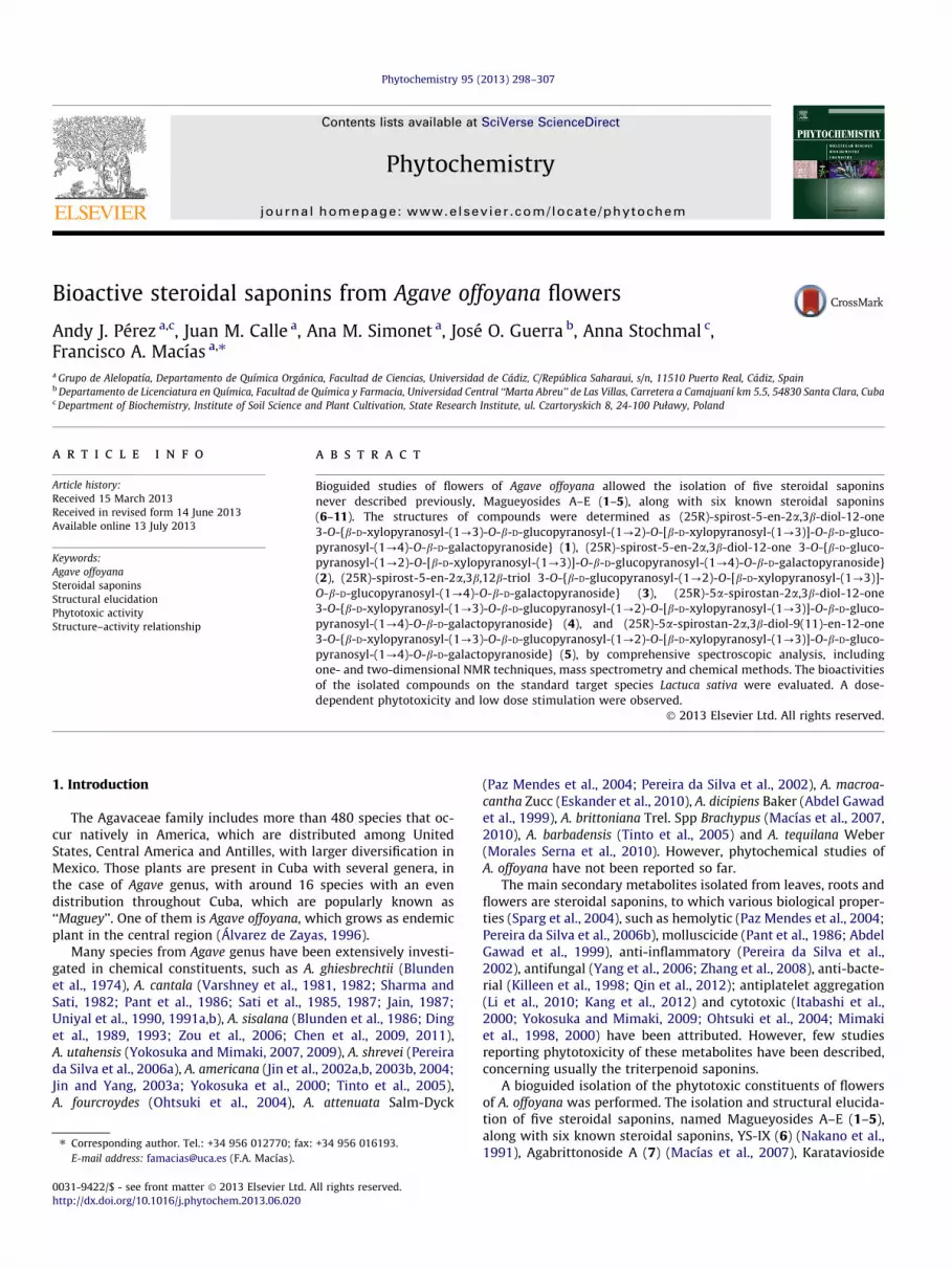
These fractions (D and E) were selected for the phytotoxic eval-uation (Fig. 2). The concentrations tested were identical to those forthe previous bioassay. Standard target species (STS) were L. sativa L.(lettuce), Lycopersicum esculentum Will. (tomato), Lepidium sativumL. (cress), and Allium cepa L. (onion). The tested fractions did notcause significant effects on the germination of the evaluated seeds,unlike root growth of the seeds, which was highly inhibited. Higherthan 60% of inhibitory effect on roots growth of L. sativa, L. sativumand A. cepa, at 800 ppm of fraction D was shown. These activity val-ues were greater than those of the herbicide Logran� on lettuce andcress. The shoots growth was slightly affected, only a 40% of inhibi-tion on L. sativa, L. sativum and A. cepa for the higher concentrationof fraction D was observed as the best result.
After multiple separation processes by normal and reversephase medium-pressure liquid chromatography (MPLC) and HPLCof D and E fractions, eleven saponins 1–11 were obtained (Fig. 3).Their structures were elucidated on the basis of spectroscopy datausing 1H and 13C NMR, 2D (HSQC, HMBC, DQF-COSY, TOCSY,ROESY, HSQC-TOCSY), selective excitation 1D-ROESY and 1D-TOC-SY acquired ‘‘in array’’ experiments, as well as HRFABMS and acidhydrolysis.
Compound 1 (Magueyoside A), a white amorphous solid, exhib-ited in the HR-FAB-MS (positive ion mode) a quasi-molecular ion
Fig. 1. Effect of extracts and fractions of Agave offoyana flowers on the etiolatedwheat coleoptile elongation. Values are expressed as percentage from the control.
peak at m/z 1217.5201 [M+Na]+ (calcd. 1217.5203), which was con-sistent with the molecular formula of C55H86O28. NMR data of com-pound 1 (Tables 1 and 2) showed similar characteristics as those inAgabrittonoside A (7), a previously described saponin from thesame genus (Macías et al., 2007), which was also isolated in thiswork in pure form. 1H and 13C chemical shifts of sugars portionshowed a great similitude with those reported for compound 7,as well as in the connectivity between sugar units determined byHMBC and ROESY correlations between linkage points. This sug-gested the same sugar chain. Absolute configurations of sugar unitswere determined using the method reported by Tanaka et al., 2007with slight modification. Compound 1 was hydrolyzed and con-verted into the thiazolizine derivatives to arylthiocarbamate usingL-cystein methyl ester and o-tolylisothiocyanate. Then, the reactionmixture was directly analyzed by C-18 HPLC and the retentionstime (Rt) were compared with values obtained for derivatives ofeach D- or L- authentic sugars. The peaks at 12.59, 14.63 and17.60 min coincided with derivative of D-galactose, D-glucose andD-xylose (Rt of D-galactose: 12.63, Rt of D-glucose: 14.57, Rt ofD-xylose: 17.69), respectively.
On the other hand, significant differences in the NMR data of Cand D rings for the aglycone moieties were observed. A signal atd 212.4 in the 13C NMR spectrum suggested the presence of acarbonyl group, which showed HMBC correlations with protonsat d 2.52 and 2.37 (2H-11), 2.77 (H-17) and 1.06 (H-18), consistentwith its location in C-12. This was confirmed by the analysis of thecoupling constants of 2H-11 signals; d 2.52 (dd, J = 14.8, 12.7 Hz)and d 2.37 (dd, J = 14.8, 5.8 Hz), which shown only one vicinalcoupling (3J) with H-9.
Finally, the correlations in 2D-ROESY were studied and the agly-cone of 1 was elucidated as (25R)-spirost-5-en-2a,3b-diol-12-one,which has been reported previously as Kammogenin (Marker et al.,1943).
The connection between sugars chain and aglycone was estab-lished to be at C-3, by means of HMBC cross-peaks between H-1gal
(d 4.89) and C-3 (d 84.2) of Kammogenin, as well as between H-3 (d3.78) of Kammogenin and C-1gal (d 103.2). This was confirmed by aNOE correlation of H-1gal with H-3 of the aglycone. Therefore, thestructure of 1 was established as Kammogenin-3-O-{b-D-xylopyr-anosyl-(1?3)-O-b-D-glucopyranosyl-(1?2)-O-[b-D-xylopyranosyl-(1?3)]-O-b-D-glucopyranosyl-(1?4)-O-b-D-galactopyranoside}.This compound has not been previously described and wepropose its name as Magueyoside A.
Compound 2 (Magueyoside B) was isolated as a white amor-phous solid, with molecular formula C50H78O24 deduced by HR-FAB-MS, which showed in the positive mode a quasi-molecularion peak at m/z 1085.4830 [M+Na]+, calculated for C50H78O24Na,1085.4781. 13C and 1H NMR characteristics of the aglycone of com-pound 2 (Table 1) were similar to those of compound 1. However,differences in sugars portion were observed. The 1H NMR spectrumshowed only four signals of anomeric protons at d 4.89, 5.18, 5.56and 5.22, which showed HSQC correlations with d 103.2, 104.6,104.7 and 104.9 respectively. Differences of chemical shifts be-tween compounds 1 and 2, mainly for the second glucose unit(Glc0), were observed (Table 2). The signal of C-3Glc0 in compound2 was assigned at d 78.0, while this in compound 1 is at d 87.0,which suggest the lack of a glycosidic bond in this carbon of com-pound 2. Therefore, the same sugars chain of compound 1, butwithout a xylose unit was suggested. The absolute configurationsof sugar units were determined in the same way employed in com-pound 1. The peaks at 12.59, 14.63 and 17.60 min coincided withderivatives of D-galactose, D-glucose and D-xylose (Rt of D-galactose:12.63, Rt of D-glucose: 14.57, Rt of D-xylose: 17.69), respectively.Thus, the compound 2 was elucidated as Kammogenin-3-O-{b-D-glucopyranosyl-(1?2)-O-[b-D-xylopyranosyl-(1?3)]-O-b-D-glucopyranosyl-(1?4)-O-b-D-galactopyranoside}. This compound
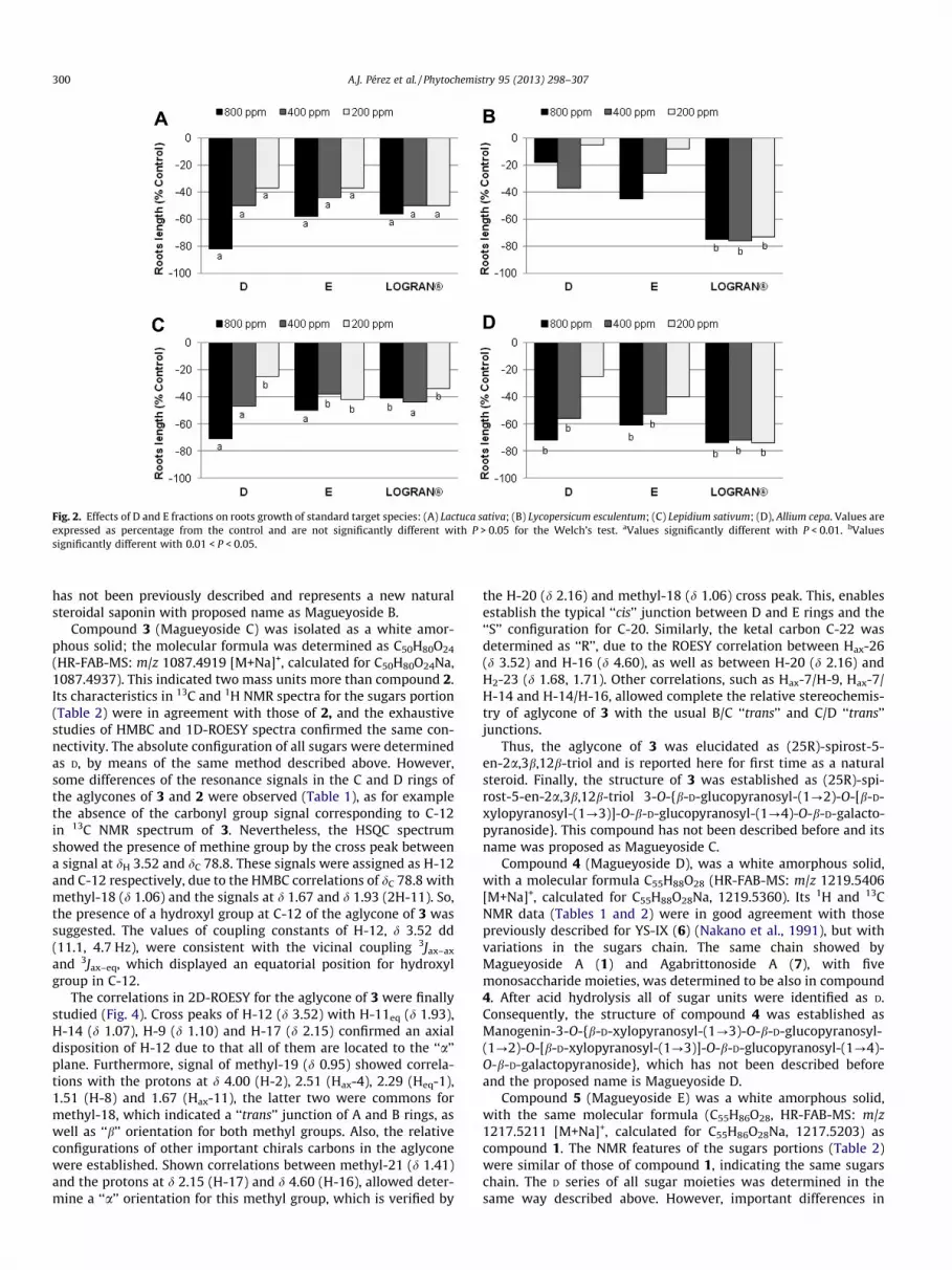
Fig. 2. Effects of D and E fractions on roots growth of standard target species: (A) Lactuca sativa; (B) Lycopersicum esculentum; (C) Lepidium sativum; (D), Allium cepa. Values areexpressed as percentage from the control and are not significantly different with P > 0.05 for the Welch’s test. aValues significantly different with P < 0.01. bValuessignificantly different with 0.01 < P < 0.05.
300 A.J. Pérez et al. / Phytochemistry 95 (2013) 298–307
has not been previously described and represents a new naturalsteroidal saponin with proposed name as Magueyoside B.
Compound 3 (Magueyoside C) was isolated as a white amor-phous solid; the molecular formula was determined as C50H80O24
(HR-FAB-MS: m/z 1087.4919 [M+Na]+, calculated for C50H80O24Na,1087.4937). This indicated two mass units more than compound 2.Its characteristics in 13C and 1H NMR spectra for the sugars portion(Table 2) were in agreement with those of 2, and the exhaustivestudies of HMBC and 1D-ROESY spectra confirmed the same con-nectivity. The absolute configuration of all sugars were determinedas D, by means of the same method described above. However,some differences of the resonance signals in the C and D rings ofthe aglycones of 3 and 2 were observed (Table 1), as for examplethe absence of the carbonyl group signal corresponding to C-12in 13C NMR spectrum of 3. Nevertheless, the HSQC spectrumshowed the presence of methine group by the cross peak betweena signal at dH 3.52 and dC 78.8. These signals were assigned as H-12and C-12 respectively, due to the HMBC correlations of dC 78.8 withmethyl-18 (d 1.06) and the signals at d 1.67 and d 1.93 (2H-11). So,the presence of a hydroxyl group at C-12 of the aglycone of 3 wassuggested. The values of coupling constants of H-12, d 3.52 dd(11.1, 4.7 Hz), were consistent with the vicinal coupling 3Jax–ax
and 3Jax–eq, which displayed an equatorial position for hydroxylgroup in C-12.
The correlations in 2D-ROESY for the aglycone of 3 were finallystudied (Fig. 4). Cross peaks of H-12 (d 3.52) with H-11eq (d 1.93),H-14 (d 1.07), H-9 (d 1.10) and H-17 (d 2.15) confirmed an axialdisposition of H-12 due to that all of them are located to the ‘‘a’’plane. Furthermore, signal of methyl-19 (d 0.95) showed correla-tions with the protons at d 4.00 (H-2), 2.51 (Hax-4), 2.29 (Heq-1),1.51 (H-8) and 1.67 (Hax-11), the latter two were commons formethyl-18, which indicated a ‘‘trans’’ junction of A and B rings, aswell as ‘‘b’’ orientation for both methyl groups. Also, the relativeconfigurations of other important chirals carbons in the aglyconewere established. Shown correlations between methyl-21 (d 1.41)and the protons at d 2.15 (H-17) and d 4.60 (H-16), allowed deter-mine a ‘‘a’’ orientation for this methyl group, which is verified by
the H-20 (d 2.16) and methyl-18 (d 1.06) cross peak. This, enablesestablish the typical ‘‘cis’’ junction between D and E rings and the‘‘S’’ configuration for C-20. Similarly, the ketal carbon C-22 wasdetermined as ‘‘R’’, due to the ROESY correlation between Hax-26(d 3.52) and H-16 (d 4.60), as well as between H-20 (d 2.16) andH2-23 (d 1.68, 1.71). Other correlations, such as Hax-7/H-9, Hax-7/H-14 and H-14/H-16, allowed complete the relative stereochemis-try of aglycone of 3 with the usual B/C ‘‘trans’’ and C/D ‘‘trans’’junctions.
Thus, the aglycone of 3 was elucidated as (25R)-spirost-5-en-2a,3b,12b-triol and is reported here for first time as a naturalsteroid. Finally, the structure of 3 was established as (25R)-spi-rost-5-en-2a,3b,12b-triol 3-O-{b-D-glucopyranosyl-(1?2)-O-[b-D-xylopyranosyl-(1?3)]-O-b-D-glucopyranosyl-(1?4)-O-b-D-galacto-pyranoside}. This compound has not been described before and itsname was proposed as Magueyoside C.
Compound 4 (Magueyoside D), was a white amorphous solid,with a molecular formula C55H88O28 (HR-FAB-MS: m/z 1219.5406[M+Na]+, calculated for C55H88O28Na, 1219.5360). Its 1H and 13CNMR data (Tables 1 and 2) were in good agreement with thosepreviously described for YS-IX (6) (Nakano et al., 1991), but withvariations in the sugars chain. The same chain showed byMagueyoside A (1) and Agabrittonoside A (7), with fivemonosaccharide moieties, was determined to be also in compound4. After acid hydrolysis all of sugar units were identified as D.Consequently, the structure of compound 4 was established asManogenin-3-O-{b-D-xylopyranosyl-(1?3)-O-b-D-glucopyranosyl-(1?2)-O-[b-D-xylopyranosyl-(1?3)]-O-b-D-glucopyranosyl-(1?4)-O-b-D-galactopyranoside}, which has not been described beforeand the proposed name is Magueyoside D.
Compound 5 (Magueyoside E) was a white amorphous solid,with the same molecular formula (C55H86O28, HR-FAB-MS: m/z1217.5211 [M+Na]+, calculated for C55H86O28Na, 1217.5203) ascompound 1. The NMR features of the sugars portions (Table 2)were similar of those of compound 1, indicating the same sugarschain. The D series of all sugar moieties was determined in thesame way described above. However, important differences in
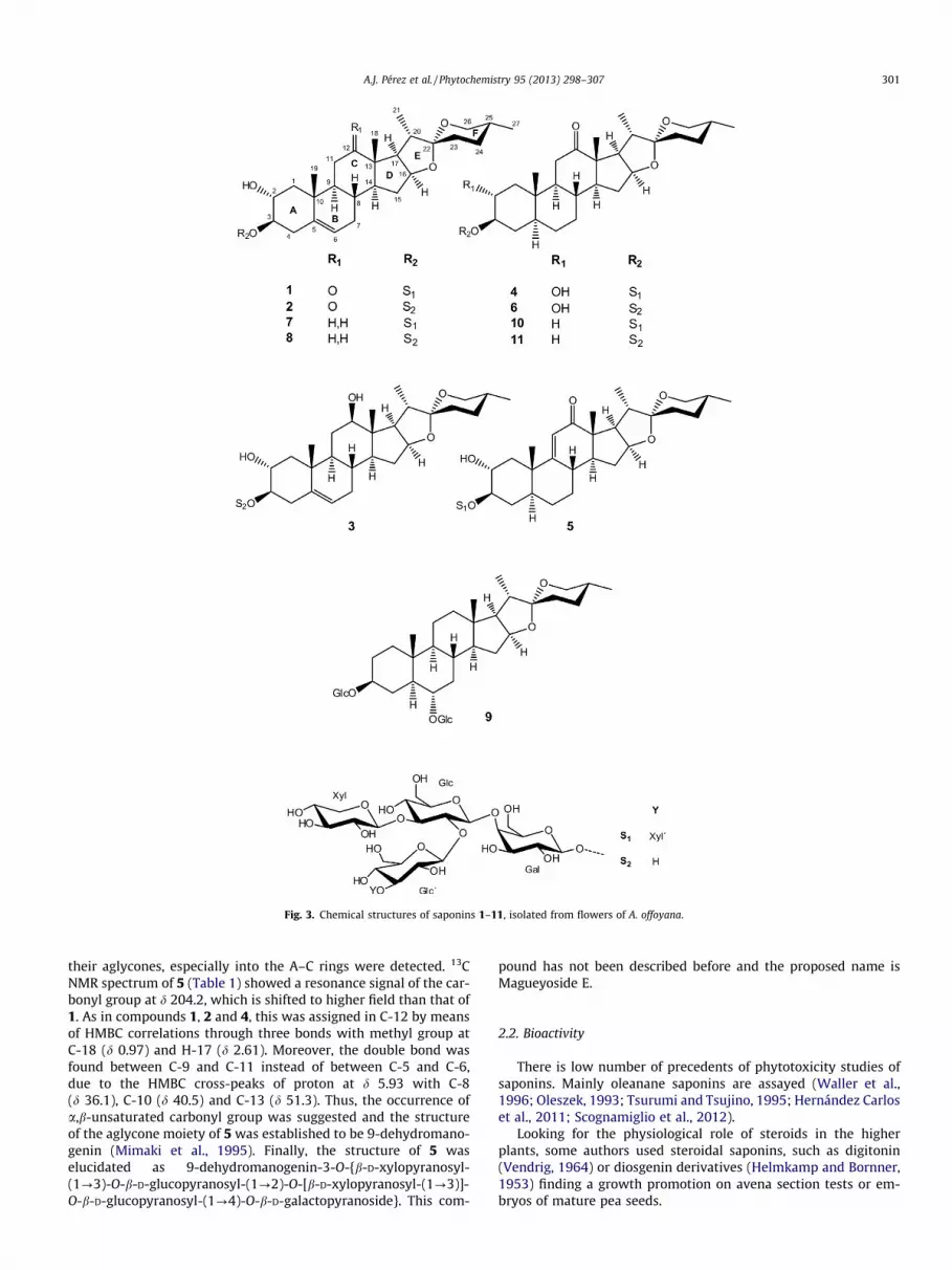
Fig. 3. Chemical structures of saponins 1–11, isolated from flowers of A. offoyana.
A.J. Pérez et al. / Phytochemistry 95 (2013) 298–307 301
their aglycones, especially into the A–C rings were detected. 13CNMR spectrum of 5 (Table 1) showed a resonance signal of the car-bonyl group at d 204.2, which is shifted to higher field than that of1. As in compounds 1, 2 and 4, this was assigned in C-12 by meansof HMBC correlations through three bonds with methyl group atC-18 (d 0.97) and H-17 (d 2.61). Moreover, the double bond wasfound between C-9 and C-11 instead of between C-5 and C-6,due to the HMBC cross-peaks of proton at d 5.93 with C-8(d 36.1), C-10 (d 40.5) and C-13 (d 51.3). Thus, the occurrence ofa,b-unsaturated carbonyl group was suggested and the structureof the aglycone moiety of 5 was established to be 9-dehydromano-genin (Mimaki et al., 1995). Finally, the structure of 5 waselucidated as 9-dehydromanogenin-3-O-{b-D-xylopyranosyl-(1?3)-O-b-D-glucopyranosyl-(1?2)-O-[b-D-xylopyranosyl-(1?3)]-O-b-D-glucopyranosyl-(1?4)-O-b-D-galactopyranoside}. This com-
pound has not been described before and the proposed name isMagueyoside E.
2.2. Bioactivity
There is low number of precedents of phytotoxicity studies ofsaponins. Mainly oleanane saponins are assayed (Waller et al.,1996; Oleszek, 1993; Tsurumi and Tsujino, 1995; Hernández Carloset al., 2011; Scognamiglio et al., 2012).
Looking for the physiological role of steroids in the higherplants, some authors used steroidal saponins, such as digitonin(Vendrig, 1964) or diosgenin derivatives (Helmkamp and Bornner,1953) finding a growth promotion on avena section tests or em-bryos of mature pea seeds.
Table 113C and 1H NMR data (J in Hz) of the aglycone moieties of compounds 1–5 (pyridine-d5, 600 MHz).a
Magueyoside A (1) Magueyoside B (2) Magueyoside C (3) Magueyoside D (4) Magueyoside E (5)
dC dH dC dH dC dH dC dH dC dH
1ax 45.2 1.22 dd (12.4, 12.9) 45.1 1.21 dd (12.3, 12.8) 45.7 1.28 dd (12.2, 12.9) 45.0 1.09 m 43.4 1.55 dd (12.7, 10.9)1eq 2.14 dd (12.9, 4.5) 2.13 dd (12.8, 4.5) 2.29 dd (12.9, 4.7) 2.00 dd (12.6, 4.7) 2.21 dd (12.7, 4.8)2 69.7 3.99 m 69.7 3.99 m 69.9 4.00 m 70.1 3.89 m 70.2 4.00 m3 84.2 3.78 ddd (11.4, 8.9, 5.2) 84.0 3.78 ddd (11.9, 9.1, 5.1) 84.5 3.79 ddd (11.5, 8.9, 5.2) 83.9 3.82 ddd (10.6, 8.9, 4.9) 83.4 3.83 ddd (11.0, 8.9, 4.9)4ax 37.4 2.51 dd (14.0, 11.4) 37.4 2.49 dd (14.0, 11.9) 37.5 2.51 dd (13.9, 11.5) 33.8 1.44 ddd (13.2, 11.9, 10.6) 33.6 1.49 ddd (13.2, 12.8, 11.0)4eq 2.71 dd (14.0. 5.2) 2.70 dd (14.0, 5.1) 2.69 dd (13.9, 5.3) 1.84 ddd (13.2, 4.9, 2.8) 1.91 ddd (13.2, 5.3, 3.0)5 139.8 – 139.8 – 140.0 – 44.3 0.93 m 42.4 1.15 tdd (12.8, 13.0, 3.0, 3.3)6ax 121.6 5.25 dd (5.2, 2.3) 121.6 5.25 dd (5.1, 2.4) 122.0 5.28 dd (5.7, 2.7) 27.7 1.03 m 27.1 1.23 dq (13.0, 3.7)6eq 1.12 m 1.16 m7ax 31.6 1.43 m 31.6 1.42 m 32.0 1.43 m 31.5 0.72 dq (12.7, 4.7) 32.4 0.87 m7eq 1.84 m 1.83 m 1.83 ddd (12.1, 4.8, 2.7) 1.50 m 1.73 m8 30.3 1.78 m 30.3 1.78 m 30.3 1.51 dq (4.8, 10.8, 10.7, 10.7) 33.6 1.70 dq (12.7, 4.3) 36.1 2.36 tdd (10.7, 10.7, 5.7, 2.1)9 52.0 1.36 m 52.0 1.34 m 49.9 1.10 ddd (10.8, 10.7, 4.7) 55.3 0.96 m 170.4 –10 38.3 – 38.3 – 38.0 – 38.0 – 40.5 –11ax 37.4 2.52 dd (14.8, 12.7) 37.4 2.51 dd (14.6, 12.9) 31.5 1.67 m 37.2 2.38 dd (14.3, 13.6) 120.1 5.93 d (2.1)11eq 2.37 dd (14.8, 5.8) 2.35 dd (14.6, 5.8) 1.93 ddd (13.1, 4.7, 4.7) 2.32 dd (14.3, 5.2)12 212.4 – 212.4 – 78.8 3.52 dd (11.1, 4.7) 212.5 – 204.2 –13 54.9 – 54.9 – 46.2 – 55.3 – 51.3 –14 55.7 1.40 m 55.7 1.37 m 55.2 1.07 m 55.7 1.33 m 52.6 1.69 m15ax 31.5 1.58 m 31.5 1.57 m 31.9 1.56 m 31.4 1.55 m 31.8 1.62 m15eq 2.07 ddd (12.1, 6.8, 5.3) 2.06 ddd (12.1, 7.2, 5.9) 2.05 ddd (11.7, 6.7, 5.3) 2.07 ddd (11.6, 6.7, 5.7) 2.16 ddd (11.4, 7.1, 4.8)16 79.7 4.45 m 79.6 4.44 m 81.1 4.60 m 79.6 4.45 m 80.2 4.50 m17 53.9 2.77 dd (8.9, 6.6) 53.9 2.76 dd (8.7, 6.8) 62.7 2.15 m 54.2 2.73 dd (8.6, 6.7) 54.5 2.61 dd (8.8, 7.1)18 15.8 1.06 s 15.8 1.05 s 11.0 1.06 s 16.0 1.04 s 15.2 0.97 s19 19.8 0.95 s 19.8 0.94 s 20.4 0.95 s 12.8 0.70 s 19.3 0.86 s20 42.6 1.89 dq (6.9, 6.6) 42.6 1.88 dq (6.9, 6.8) 43.0 2.16 dq (6.5, 6.5) 42.6 1.89 dq (6.7, 6.9) 42.9 1.98 dq (7.1, 6.9)21 13.8 1.32 d (6.9) 13.8 1.31 d (6.9) 14.3 1.41 d (6.5) 13.9 1.32 d (6.9) 13.7 1.38 d (6.9)22 109.3 – 109.3 – 109.5 – 109.3 – 109.4 –23 31.7 1.60 m 31.7 1.60 m 31.7 1.71 m 31.7 1.60 m 31.7 1.70 (2H) m
1.65 m 1.66 m 1.68 m 1.66 m24 29.1 1.54 (2H) m 29.1 1.54 (2H) m 29.3 1.56 (2H) m 29.2 1.53 (2H) m 29.2 1.54 (2H) m25 30.5 1.54 m 30.5 1.54 m 30.6 1.56 m 30.5 1.54 m 30.5 1.55 m26ax 66.9 3.45 dd (10.5, 11.0) 66.9 3.45 dd (10.9, 10.7) 66.8 3.52 dd (10.4, 10.4) 66.9 3.46 dd (11.1, 9.7) 66.9 3.46 dd (11.4, 9.8)26eq 3.56 dd (11.0, 3.9) 3.56 dd (10.7, 3.4) 3.58 dd (10.4, 3.8) 3.57 dd (11.1, 3.7) 3.57 dd (11.4, 3.7)27 17.3 0.67 d (5.9) 17.2 0.66 d (6.0) 17.3 0.68 d (5.6) 17.3 0.66 d (6.0) 17.3 0.67 d (5.9)
a Assignments were confirmed by DQF-COSY, 2D-TOCSY, HSQC, HSQC-TOCSY and HMBC experiments.
302A
.J.Pérezet
al./Phytochemistry
95(2013)
298–307
Table 213C and 1H NMR data (J in Hz) of the sugar portions of compounds 1–5 (pyridine-d5, 600 MHz).a
Magueyoside A (1) Magueyoside B (2) Magueyoside C (3) Magueyoside D (4) Magueyoside E (5)b-D-Gal b-D-Gal b-D-Gal b-D-Gal b-D-Gal
1 103.2 4.89 d (7.8) 103.2 4.89 d (7.8) 103.3 4.89 d (7.8) 103.2 4.88 d (7.8) 103.2 4.89 d (7.8)2 72.6 4.47 dd (7.8, 9.6) 72.6 4.50 dd (7.8, 9.5) 72.6 4.50 dd (7.8, 9.5) 72.5 4.47 dd (7.8, 9.3) 72.5 4.48 dd (7.8, 9.4)3 75.3 4.11 dd (9.6, 3.0) 75.4 4.10 dd (9.5, 3.2) 75.5 4.11 dd (9.5, 3.9) 75.4 4.11 dd (9.3, 3.3) 75.4 4.12 dd (9.4, 4.0)4 79.1 4.56 dd (3.0, 1.5) 79.2 4.57 dd (3.2, 1.2) 79.2 4.57 dd (3.9, 1.8) 79.2 4.57 dd (3.3, 1.2) 79.2 4.57 dd (4.0, 1.4)5 75.6 3.99 ddd (8.9, 6.0, 1.5) 75.6 3.99 ddd (7.6, 6.3, 1.2) 75.6 3.99 ddd (8.5, 5.7, 1.8) 75.7 4.02 ddd (8.5, 6.0, 1.2) 75.7 4.02 ddd (8.5, 6.1, 1.4)6 60.5 4.15 m 60.5 4.15 m 60.5 4.14 m 60.6 4.17 m 60.6 4.19 m
4.55 m 4.57 m 4.57 m 4.57 m 4.59 m
b-D-Glc b-D-Glc b-D-Glc b-D-Glc b-D-Glc1 104.4 5.19 d (7.9) 104.6 5.18 d (7.9) 104.6 5.20 d (7.9) 104.4 5.19 d (7.9) 104.5 5.19 d (7.9)2 80.5 4.31 dd (7.9, 8.8) 81.1 4.33 dd (7.9, 8.8) 81.2 4.33 dd (7.9, 8.8) 80.6 4.32 dd (7.9, 8.9) 80.6 4.33 dd (7.9, 8.7)3 86.9 4.09 dd (8.8, 8.6) 86.9 4.12 dd (8.8, 8.8) 86.9 4.12 dd (8.8, 8.7) 86.9 4.09 dd (8.9, 8.8) 87.0 4.10 dd (8.7, 8.7)4 70.3 3.78 dd (8.6, 9.3) 70.3 3.79 dd (8.8, 9.3) 70.4 3.80 dd (8.7, 9.2) 70.3 3.77 dd (8.8, 9.5) 70.3 3.79 dd (8.7, 9.5)5 77.5 3.82 ddd (9.3, 7.4, 2.2) 77.5 3.82 ddd (9.3, 7.7, 2.5) 77.6 3.83 ddd (9.2, 7.3, 2.2) 77.5 3.82 ddd (9.5, 7.3, 2.7) 77.6 3.82 ddd (9.5, 7.5, 2.6)6 62.8 4.04 dd (10.9, 7.4) 62.8 4.03 dd (11.0, 7.7) 62.9 4.04 dd (10.3, 7.3) 62.8 4.03 dd (10.2, 7.3) 62.9 4.04 dd (10.5, 7.5)
4.47 dd (10.9, 2.2) 4.48 dd (11.0, 2.5) 4.48 dd (10.3, 2.2) 4.47 dd (10.2, 2.7) 4.47 dd (10.5, 2.6)
b-D-Glc0 b-D-Glc0 b-D-Glc0 b-D-Glc0 b-D-Glc0
1 103.9 5.59 d (7.4) 104.7 5.56 d (7.9) 104.8 5.55 d (7.9) 103.9 5.60 d (7.5) 103.9 5.61 d (7.6)2 75.0 4.04 dd (7.4, 9.2) 76.0 4.02 dd (7.9, 9.5) 76.0 4.03 dd (7.9, 9.3) 75.0 4.04 dd (7.5, 9.9) 75.0 4.04 dd (7.6, 9.0)3 87.0 4.07 dd (9.2, 8.9) 78.0 4.12 dd (9.5, 9.3) 78.1 4.12 dd (9.3, 8.2) 87.0 4.08 dd (9.9, 9.0) 87.0 4.08 dd (9.0, 9.0)4 69.3 3.93 dd (8.9, 9.3) 71.3 4.07 dd (9.3, 8.7) 71.3 4.07 dd (8.2, 9.3) 69.4 3.92 dd (9.0, 8.8) 69.4 3.93 dd (9.0, 9.1)5 77.9 3.85 ddd (9.3, 6.0, 2.4) 78.4 3.89 ddd (8.7, 5.5, 2.3) 78.4 3.88 ddd (9.3, 5.5, 2.2) 77.9 3.85 ddd (8.8, 5.7, 1.9) 77.9 3.85 ddd (9.1, 5.0, 2.5)6 62.3 4.29 dd (11.7, 5.0) 62.6 4.38 dd (12.0, 5.5) 62.7 4.37 dd (12.5, 5.5) 62.3 4.30 dd (12.0, 5.7) 62.3 4.30 dd (11.9, 5.0)
4.46 dd (11.7, 3.1) 4.54 dd (12.0, 2.3) 4.53 dd (12.5, 2.2) 4.47 dd (12.0, 1.9) 4.47 dd (11.9, 2.5)
b-D-Xyl b-D-Xyl b-D-Xyl b-D-Xyl b-D-Xyl1 104.8 5.15 d (7.8) 104.9 5.22 d (7.8) 104.9 5.23 d (7.8) 104.8 5.15 d (7.8) 104.8 5.16 d (7.7)2 75.1 3.93 dd (7.8, 8.5) 75.1 3.93 dd (7.8, 8.4) 75.1 3.93 dd (7.8, 8.4) 75.1 3.93 dd (7.8, 8.6) 75.1 3.93 dd (7.7, 8.3)3 78.4 4.04 dd (8.5, 8.9) 78.6 4.06 dd (8.4, 8.9) 78.7 4.06 dd (8.4, 8.8) 78.4 4.04 dd (8.6, 8.9) 78.4 4.04 dd (8.3, 9.0)4 70.7 4.08 m 70.7 4.10 m 70.7 4.09 m 70.7 4.08 m 70.7 4.07 m5ax 67.2 3.62 dd (11.2, 10.4) 67.3 3.64 dd (11.2, 10.2) 67.3 3.64 dd (11.2, 10.3) 67.2 3.61 dd (11.4, 9.9) 67.2 3.62 t (11.2)5eq 4.18 dd (11.2, 4.9) 4.19 dd (11.2, 4.9) 4.20 dd (11.2, 4.9) 4.18 dd (11.4, 4.9) 4.18 dd (11.2, 4.9)
b-D-Xyl0 b-D-Xyl0 b-D-Xyl0
1 106.1 5.06 d (7.4) 106.1 5.06 d (7.4) 106.1 5.06 d (7.4)2 75.3 3.91 dd (7.4, 8.6) 75.4 3.91 dd (7.4, 8.5) 75.4 3.91 dd (7.4, 8.1)3 77.7 4.01 dd (8.6, 8.6) 77.7 4.01 dd (8.5, 8.6) 77.7 4.01 dd (8.1, 8.6)4 70.7 4.08 m 70.7 4.08 m 70.7 4.08 m5ax 67.0 3.46 dd (11.1, 10.6) 67.0 3.46 dd (11.6, 9.9) 67.1 3.46 t (11.4)5eq 4.16 dd (11.1, 5.3) 4.16 dd (11.6, 5.2) 4.16 dd (11.4, 5.1)
a Assignments were confirmed by DQF-COSY, 1D-TOCSY, HSQC, HSQC-TOCSY and HMBC experiments.
A.J.Pérez
etal./Phytochem
istry95
(2013)298–
307303

Fig. 4. Main correlations observed in 2D-ROESY spectrum for aglycone moiety of 3.
Table 3Phytotoxicity of compounds 1, 2 and 4–9 on roots of Lactuca sativa L.
Compounds IC50 (lM) r2
1 88.4 0.96892 104.3 0.97144 131.2 0.97065 101.6 0.97296 66.7 0.98027 160.2 0.99648 700.1* 0.98069 375.9* 0.9510LOGRAN� 523.7* 0.9863
* The data were not adjusted to the dose–response curve.
304 A.J. Pérez et al. / Phytochemistry 95 (2013) 298–307
The fractions bioassay (Fig. 2) showed a roots growth inhibitionas general behavior, being L. sativa the most affected species. Basedon the available quantities of the saponins, lettuce was chosen as amodel to know their activities, testing at 333; 100; 33; 10; 3.3 and1 lM. Magueyoside C (3) and compounds 10 and 11 could not beassayed.
The results of the bioassay are shown in Fig. 5, where data arepresented as percentage from the control. Positive values indicatestimulation and negative values represent an inhibition of thestudied parameters. Due to the fact that effects on germinationand shoot development were not significant, the discussion wasperformed on the base of roots growth effects.
Inhibitory effects at concentrations higher than 100 lM andstimulatory effects at concentration lower than 33 lM of testedcompounds were observed. Compounds 1, 2 and 4–7 presentedbetter inhibition profiling than shown by the commercial herbicideLOGRAN�. YS-IX (6) showed the lowest value of IC50 66.7 lM (Ta-ble 3). In addition, the growth inhibition is more prominent in 1, 2and 4–6, showing an average IC50 of 100 lM, which could be pos-sible correlate with the presence of the carbonyl group at C-12 oftheir aglycones.
On the other hand, it is not completely possible to find a corre-lation between 12-keto-saponins bioactivity and units number insugar moiety, nonetheless the comparison of the profiling of 7and 8 allowed to observe a better inhibition profile (IC50
160.2 lM) for this with five sugar units, Agabrittonoside A (7), thanthat with four sugar units, Karatavioside A (8) (IC50 700.1 lM).
Likewise, stimulatory effects at low concentrations were ob-served. Saponins 1 and 9 showed the best profiles with maximumof 16% and 19% (P < 0.01) of stimulatory response at 10 lM.
Biphasic dose–response relationships, which are characterizedby a stimulatory response in the measured parameter at low dosesof active compound and inhibition at higher doses, have beenrecognized in plants, whether effects of synthetic herbicides or
Fig. 5. Effects of compounds 1, 2 and 4–9 on roots growth of Lactuca sativa L. Valuesare expressed as percentage from the control and are not significantly different withP > 0.05 for the Welch’s test. aValues significantly different with P < 0.01. bValuessignificantly different with 0.01 < P < 0.05.
natural phytotoxins are concerned (Duke et al., 2006). This dose–response phenomenon is known as hormesis, which has been de-scribed for oleanane saponins, in special for those isolated frommungbeans (Waller et al., 1996).
In conclusion, we can state that A. offoyana is an interestingsource of active compounds. The 12-ketosaponins presented thehighest levels of roots growth inhibition better than shown bythe commercial herbicide Logran
�. On the other hand, low concen-
trations of saponins enhance the root growth.
3. Experimental
3.1. General experimental procedure
Optical rotations were carried out using a Perkin-Elmer 241polarimeter (589 nm, 20 �C). 1D and 2D NMR spectra were re-corded on a Varian INOVA-600 spectrometer equipped with5 mm 1H {15N–31P} PFG high-field inverse detection z-gradientprobe. 1H (599.78 MHz) and 13C (150.83 MHz) NMR spectra wererecorded in pyridine-d5 at 25 �C. Chemical shift are given on thed scale and were referenced to residual pyridine, dH 8.70, 7.55,7.18 and dC 149.84, 135.50, 123.48. Varian pulse sequence usinggradient were applied and all 2D spectra, except for HMBC, wererecorded in the phase-sensitive mode. HR-FAB-MS data were de-tected on a Waters AUTOSPEC mass spectrometer. HPLC in isocraticmode was performed by a Merck Hitachi apparatus equipped withLaChrom (L-2490) refractive index detector and analytical Phe-nomenex Gemini C18 column (4.6 � 250 mm, i.d).
3.2. Plant material
Flowers of A. offoyana were collected on January 2008 by bota-nist Dr. Alfredo Noa in Palenque, Remedios city, north of Villa Claraprovince, Cuba. A voucher specimen was deposited in the Herbar-ium Dr. Alberto Alonso Triana of the Universidad Central ‘MartaAbreu’ de Las Villas, Cuba (number HPVC 3017).
A.J. Pérez et al. / Phytochemistry 95 (2013) 298–307 305
3.3. Extraction and isolation
Dried and powdered flowers (0.5 kg) were extracted three timeswith ethanol–water (7:3) during 48 h by maceration at room tem-perature. The solvent was removed under reduced pressure andthe syrupy extract (11.4%) was suspended in distilled water, defat-ted with n-hexane, and then extracted with water-saturated n-BuOH. After removing the solvent, 10 g of n-BuOH extract (30.8%,of ethanolic extract) was purified by VLC over LiChrospher RP-18and eluted with a MeOH–H2O (4:6, 5:5, 6:4, 7:3, 8:2, 10:0, each250 ml), to give five fractions (Fr A: 2.13 g, Fr B: 0.87 g, Fr C:0.29 g, Fr D: 0.69 g, and Fr E: 6.0 g).
Fr D (0.69 g) was subjected to MPLC (1 ml/min) on a Büchi 861apparatus onto, 40–63 lm LiChrospher RP-18 column, using Ace-tone-H2O (1:1) as mobile phase. Six milliliter fractions were col-lected and checked by TLC on RP-18 F254S, developed withMeOH-H2O (8:2) and sprayed with Oleum reagent and heated at150 �C. Fractions showing similar profiling were combined to give6 fractions, from Fr D-3 to Fr D-5, each comprising two to foursaponins.
Fractionation of Fr D-3 by HPLC on analytical C18 column andMeOH–H2O (7:3) as mobile phase yielded two further fractions,which were purified by HPLC using ACN–H2O (3.5:6.5) and thesame column to afford compounds 3 (2.7 mg) and 5 (3.3 mg).
In the same conditions described above Fr D-4 was fractionatedby HPLC to obtain 3 additional fractions. The first one gave com-pound 1 (9.4 mg) after further purification by means of HPLC ana-lytical C18 column and ACN–H2O (3.5:6.5) as mobile phase. Usingthe same HPLC column but MeOH–H2O (7:3) as mobile phase al-lowed obtain compound 6 (3.9 mg) from the third fraction and apreparative TLC silica gel EtOAc–HOAc–H2O (7:2:2), for the secondone gave compounds 2 (4.1 mg) and 4 (3.5 mg).
From Fr D-5, the compounds 9 (4.5 mg), 10 (1.2 mg) and 11(1.4 mg) were purified by HPLC, analytical C18 column andMeOH–H2O (8:2) as mobile phase.
Fr E (6.0 g) was chromatographed by CC silica gel CHCl3–MeOH–H2O (30:10:1 to 7:4:1) and purified by HPLC on analyticalC18 column (1 ml/min) with MeOH–H2O (7:3) as mobile phaseto afford compounds 7 (10.5 mg) and 8 (8 mg).
3.4. Compound 1: [a]D20 �43.5 (MeOH, c 0.1)
HRFABMS, m/z 1217.5201 [M+Na]+ calculated for C55H86O28Na,1217.5203. 1H and 13C NMR reported in Tables 1 and 2.
3.5. Compound 2: [a]D20 �33.5 (MeOH, c 0.1)
HRFABMS, m/z 1085.4830 [M+Na]+ calculated for C50H78O24Na,1085.4781. 1H and 13C NMR reported in Tables 1 and 2.
3.6. Compound 3: [a]D20 �79.2 (MeOH, c 0.1)
HRFABMS, m/z 1087.4919 [M+Na]+ calculated for C50H80O24Na,1087.4937. 1H and 13C NMR reported in Tables 1 and 2.
3.7. Compound 4: [a]D20 �18,2 (MeOH, c 0.1)
HRFABMS, m/z 1219.5406 [M+Na]+ calculated for C55H88O28Na,1219.5360. 1H and 13C NMR reported in Tables 1 and 2.
3.8. Compound 5: [a]D20 �44.7 (MeOH, c 0.1)
HRFABMS, m/z 1217.5211 [M+Na]+ calculated for C55H86O28Na,1217.5203. 1H and 13C NMR reported in Tables 1 and 2.
3.9. Compound 7: [a]D20 �50.5 (MeOH, c 0.1)
HRFABMS, m/z 1203.5411 [M+Na]+ calculated for C55H88O27Na,1203.5411.
3.10. Acid Hydrolysis of saponins
Compounds 1–5 and 7 (each 2 mg) were treated with 1 N HCl(1 ml) at 80 �C for 3 h. After cooling, the solvent was eliminatedwith a stream of N2, dry residue was suspended in water, and agly-cones were extracted with ethyl acetate (3 � 2 ml). The aqueouslayer, containing sugars, was neutralized with Amberlite IRA-400(OH- form). For each samples, aqueous solution were dried underN2 and stored for the subsequent analysis.
3.11. Determination of absolute configurations of sugars
To determine the absolute configuration of monosaccharideconstituents of isolated compounds 1–5 and 7, the methodreported by (Tanaka et al., 2007) was used with slight modifica-tions. Sugars of each sample were dissolved in pyridine (0.5 mL)containing L-cysteine methyl ester hydrochloride (2 mg) andheated at 60 �C for 1 h; o-tolyl isothiocyanate (2 ll) was thenadded and the mixture was heated at 60 �C for 1 h. Each reactionmixture was directly analyzed by reversed-phase HPLC using amodel 616 pump, a 996 photodiode array detector and a 717-plusautosampler (Waters, Milford, MA). An Eurospher-100 C18,250 mm � 4 mm i.d., 5 lm (Knauer, Berlin, Germany) HPLC col-umn was used; mobile phase: MeCN–H2O (25:75, v/v) containing50 mM H3PO4; detection: UV (250 nm); flow rate: 0.8 ml/min; col-umn temperature: 35 �C. The HPLC column was washed withMeOH after each injection.
The derivatives of monosaccharides of D-xylose, D-glucose andD-galactose, in the analyzed saponins were identified by compari-son of their retention times with those of authentic samples(Sigma–Aldrich, Steinheim, Germany) treated in the same way asdescribed above (Rt: D-xylose 17.69 min, L-xylose 16.27 min, D-glu-cose 14.57 min, L-glucose 13.43 min, D-galactose 12.63 min, L-gal-actose 13.13 min).
3.12. Coleoptiles bioassay
Wheat seeds (T. aestivum L. cv. Duro) were sown in 15 cm diam-eter Petri dishes moistened with water and grown in the dark at22 ± 1 �C for 3 days (Hancock et al., 1964). The roots and caryopsiswere removed from the shoots. Then the shoots were placed in aVan der Weij guillotine and the apical 2 mm were cut off anddiscarded.
The next 4 mm of the coleoptiles were removed and used forthe bioassay. All manipulations were performed under a greensafelight (Nitsch and Nitsch, 1956). Products of interest were pre-viously dissolved in DMSO and diluted to the final bioassay con-centration with a maximum of 0.5% DMSO. Parallel controls werealso run.
Crude extracts and fractions to be assayed for biological activitywere added to test tubes. The assays were carried out in duplicate.Phosphate-citrate buffer (2 ml) containing 2% sucrose (Nitsch andNitsch, 1956) at pH 5.6 was added to each test tube. Five coleop-tiles were placed in each test tube (three tubes per dilution) andthe tubes were rotated at 6 rpm in a roller tube apparatus for24 h at 22 �C in the dark. The coleoptiles were measured by digita-lization of their images. Data were statistically analyzed usingWelch’s test (Martín Andrés and Luna del Castillo, 1990). Dataare presented as percentage differences from control, where zerorepresents the control, positive values represent stimulation ofthe studied parameter, and negative values represent inhibition.
306 A.J. Pérez et al. / Phytochemistry 95 (2013) 298–307
3.13. Phytotoxicity bioassay
Selection of target plants was based on an optimization processdeveloped by us in our search for a standard phytotoxicity bioassay(Macías et al., 2000). Several Standard Target Species (STS) wereproposed, including monocot A. cepa L. (onion) and dicots L. escu-lentum Will. (tomato), L. sativum L. (cress) and L. sativa L. (lettuce),which were assayed for this study.
Bioassays were conducted using Petri dishes (50 mm diameter),with one sheet of Whatman No.1 filter paper as support. Germina-tion and growth were conducted in aqueous solutions at controlledpH using 10�2 M 2-[N-morpholino]ethanesulfonic acid (MES) and1 M NaOH (pH 5.6). Fractions and compounds to be assayed weredissolved in DMSO and these solutions were diluted with buffer(with a maximum of 0.5% DMSO), so that test concentrations forfractions (800, 400 and 200 ppm) and each compound (333, 100,33, 10, 3,3, 1 lM) were reached. This procedure facilitated the sol-ubility of the assayed compounds. 20 seeds were used to each Petridish and four replicates were made (80 seeds in total). Treatment,control or internal reference solution (1 ml) was added to eachPetri dish.
After adding seeds and aqueous solutions, Petri dishes weresealed with Parafilm to ensure closed-system models. Seeds werefurther incubated at 25 �C in a Memmert ICE 700 controlled envi-ronment growth chamber in the dark. Bioassays took 4 days forcress, 5 days for lettuce and tomato, and 7 days for onion. Aftergrowth, plants were frozen at �10 �C for 24 h to avoid subsequentgrowth during the measurement process.
The commercial herbicide Logran�, a combination of N-(1,1-dimethylethyl)-N0-ethyl-6-(methylthio)-1,3,5-triazine-2,4-diamine(Terbutryn, 59.4%) and 2-(2-chloroethoxy)-N-{[(4-methoxy-6-methyl-1,3,5-triazin-2yl)amino]carbonyl}benzene-sulfonamide(Triasulfuron, 0.6%), was used as an internal reference accordingthe comparison study reported previously (Macías et al., 2000).This was tested in the same way as described above for thesamples.
Control samples (buffered aqueous solutions with DMSO andwithout any test compound) were used for all of the plant speciesassayed.
Evaluated parameters (germination rate, root length and shootlength) were recorded using a Fitomed� system (Castellano et al.,2001), which allowed automatic data acquisition and statisticalanalysis using its associated software. Data were analyzed statisti-cally using Welch’s test, with significance fixed at 0.01 and 0.05.
Results are presented as percentage differences from the con-trol. Zero represents control, positive values represent stimulation,and negative values represent inhibition. IC50 values to inhibitioneffects were obtained after adjusting phytotoxicity data to concen-tration (logarithmic scale), to a sigmoidal dose–response curve, de-fined by equation:
Y ¼ Ymin þYmin � Ymin
1þ 10log gEC0�x
where X indicates the logarithm of concentration, Y indicates the re-sponse (phytotoxicity) and Ymax and Ymin are the maximum andminimum values of the response, respectively. Goodness of fit is de-scribed by the determination coefficient (r2). The adjustment andthe r2 were obtained using GraphPad Prism 5.0� software.
Acknowledgements
This research was supported by the Ministerio de Educación yCiencia (Project No. AGL2009-08864 (AGR)) and Junta de Andalucía(PAI-III, AGR-5822), Seville, Spain. A fellowship from the Agencia
Española de Cooperación Internacional para el Desarrollo, AECID(AJP) is also gratefully acknowledged.
Appendix A. Supplementary data
Supplementary data associated with this article include NMRspectra (1H and 13C NMR, HSQC, HMBC, ROESY and DQF-COSY)for the new compounds (1–5), tables with complete spectroscopydata of known compounds 6, 9–11 which have not been reportedso far, graphics of the effects of compounds 1, 2 and 4–9 on germi-nation and shoots growth of Lactuca sativa and a table with thephytotoxicity values of these compounds. Supplementary dataassociated with this article can be found, in the online version, athttp://dx.doi.org/10.1016/j.phytochem.2013.06.020.
References
Abdel Gawad, M.M., El Sayed, M.M., Abdel Hameed, E.S., 1999. Molluscicidalsteroidal saponins and lipid content of Agave decipiens. Fitoterapia 70, 371–381.
Álvarez de Zayas, A., 1996. Los agaves de Cuba central. Fontqueria 44, 117–128.Blunden, G., Yi, Y., Jewers, K., Burbage, M.B., 1974. Agave ghiesbrechtii, a new source
of gloriogenin. Phytochemistry 13, 2025–2027.Blunden, G., Patel, A.V., Crabb, T.A., 1986. Barbourgenin, a new steroidal sapogenin
from Agave sisalana leaves. J. Nat. Prod. 49, 687–689.Castellano, D., Macías, F.A., Castellano, M., Cambronero, R., 2001. Sistema
automatizado para la adquisición simultánea y gestión informatizada demedidas de longitud variable. Spanish Patent No. P9901565.
Chen, P.Y., Chen, C.H., Kuo, C.C., Lee, T.H., Kuo, Y.H., Lee, C.K., 2011. Cytotoxicsteroidal saponins from Agave sisalana. Planta Med. 77, 929–933.
Chen, P.Y., Kuo, Y.C., Chen, C.H., Kuo, Y.H., Lee, C.K., 2009. Isolation andimmunomodulatory effect of homoisoflavones and flavones from Agavesisalana Perrine ex Engelm. Molecules 14, 1789–1795.
Ding, Y., Chen, Y.Y., Wang, D.Z., Yang, C.R., 1989. Steroidal saponins from acultivated form of Agave sisalana. Phytochemistry 28, 2787–2791.
Ding, Y., Tian, R.H., Yang, C.R., Chen, Y.Y., Nohara, T., 1993. Two new steroidalsaponins from dried fermented residues of leaf-juices of Agave sisalana formaDong No. 1. Chem. Pharm. Bull. 41, 557–560.
Duke, S.O., Cedergreen, N., Velini, E.D., Belz, R.G., 2006. Hormesis: is it an importantfactor in herbicide use and allelopathy? Outlooks Pest Manag. 17, 29–33.
Eskander, J., Lavaud, C., Harakat, D., 2010. Steroidal saponins from the leaves ofAgave macroacantha. Fitoterapia 81, 371–374.
Hancock, C.R., Barlow, H.W., Lacey, H.J., 1964. The east malling coleoptile straightgrowth test method. J. Exp. Bot. 15, 166–176.
Helmkamp, G.K., Bornner, J., 1953. Some relationships of sterols to plant growth.Plant Physiol. 28, 428–435.
Hernández Carlos, B., González Coloma, A., Orozco Valencia, A.U., Ramírez Mares,M.V., Andrés Yeves, M.F., Joseph Nathan, P., 2011. Bioactive saponins fromMicrosechium helleri and Sicyos bulbosus. Phytochemistry 72, 743–751.
Itabashi, M., Segawa, K., Ikeda, Y., Kondo, S., Naganawa, H., Koyano, T., Umezawa, K.,2000. A new bioactive steroidal saponin, furcreastatin, from the plant Furcraeafoetida. Carbohydr. Res. 323, 57–62.
Jain, D.C., 1987. Gintogenin-3-O-b-D-laminaribioside from the aerial part of Agavecantala. Phytochemistry 26, 1789–1790.
Jin, J.M., Liu, X.K., Teng, R.W., Yang, C.R., 2002a. Two new steroidal glycosides fromfermented leaves of Agave americana. Chin. Chem. Lett. 13, 629–632.
Jin, J.M., Liu, X.K., Yang, C.R., 2002b. A new pregnane glycoside from fermentedleaves of Agave americana. Chin. Chem. Lett. 13, 1189–1192.
Jin, J.M., Yang, C.R., 2003a. Two new spirostanol steroidal sapogenins fromfermented leaves of Agave americana. Chin. Chem. Lett. 14, 491–494.
Jin, J.M., Liu, X.K., Yang, C.R., 2003b. Three new hecogenin glycosides fromfermented leaves of Agave americana. J. Asian Nat. Prod. Res. 5, 95–103.
Jin, J.M., Zhang, Y.J., Yang, C.R., 2004. Four new steroid constituents from the wasteresidue of fibre separation from Agave americana leaves. Chem. Pharm. Bull. 52,654–658.
Kang, L.P., Zhang, J., Cong, Y., Li, B., Xiong, C.Q., Zhao, Y., Tan, D.W., Yu, H.S., Yu, Z.Y.,Cong, Y.W., Liu, C., Ma, B.P., 2012. Steroidal glycosides from the rhizomes ofAnemarrhena asphodeloides and their antiplatelet aggregation activity. PlantaMed. 78, 611–616.
Killeen, G.F., Madigan, C.A., Connolly, C.R., Walsh, G.A., Clark, C., Hynes, M.J.,Timmins, B.F., James, P., Headon, D.R., Power, R.F., 1998. Antimicrobial saponinsof Yucca schidigera and the implications of their in vitro properties for theirin vivo impact. J. Agric. Food Chem. 46, 3179–3186.
Li, H., Huang, W., Wen, Y., Gong, G., Zhao, Q., Yu, G., 2010. Anti-thrombotic activityand chemical characterization of steroidal saponins from Dioscorea zingiberensisC.H. Wright. Fitoterapia 8, 1147–1156.
Macías, F.A., Castellano, D., Molinillo, J.M.G., 2000. Search for a standard phytotoxicbioassay for allelochemicals. Selection of standard target species. J. Agric. FoodChem. 48, 2512–2521.
Macías, F.A., Guerra, J.O., Simonet, A.M., Nogueiras, C.M., 2007. Characterization ofthe fraction components using 1D TOCSY and 1D ROESY experiments. Four new

A.J. Pérez et al. / Phytochemistry 95 (2013) 298–307 307
spirostane saponins from Agave brittoniana Trel. spp. Brachypus. Magn. Reson.Chem. 45, 615–620.
Macías, F.A., Guerra, J.O., Simonet, A.M., Pérez, A.J., Nogueiras, C.M., 2010.Characterization of three saponins from a fraction using 1D DOSY as a solventsignal suppression tool. Agabrittonosides E–F. Furostane Saponins from Agavebrittoniana Trel. spp. Brachypus. Magn. Reson. Chem. 48, 350–355.
Marker, R.E., Wagner, R.B., Ulshafer, P.R., Wittbecker, E.L., Goldsmith, D.P.J., Ruof,C.H., 1943. Sterols. CLVII. Sapogenins. LXM’. Isolation and structures of thirteennew steroidal sapogenins. New sources for known sapogenins. J. Am. Chem. Soc.65, 1199–1209.
Martín Andrés, A., Luna el Castillo, J.De.D., 1990. Bioestadística Para Las Ciencias dela Salud, third ed. Norma-Capitel, Madrid.
Mimaki, Y., Kanmoto, T., Kuroda, M., Sashida, Y., Nishino, A., Satomi, Y., Nishino, H.,1995. Steroidal saponins from the underground parts of Hosta longipes and theirinhibitory activity on tumor promoter-induced phospholipid metabolism.Chem. Pharm. Bull. 43, 1190–1196.
Mimaki, Y., Kuroda, M., Kameyama, A., Yokosuka, A., Sashida, Y., 1998. Steroidalsaponins from the rhizomes of Hosta sieboldii and their citostatic activity on HL-60 cells. Phytochemistry 48, 1361–1369.
Mimaki, Y., Yokosuka, A., Sashida, Y., 2000. Steroidal glycosides from the aerial partsof Polianthes tuberosa. J. Nat. Prod. 63, 1519–1523.
Morales Serna, J.A., Jiménez, A., Estrada Reyes, R., Márquez, C., Cárdenas, J., Salmón,M., 2010. Homoisoflavanones from Agave tequilana Weber. Molecules 15, 3295–3301.
Nakano, K., Midzuta, Y., Hara, Y., Murakami, K., Takaishi, Y., Tomimatsu, T., 1991. 12-Keto steroidal glycosides from the caudex of Yucca gloriosa. Phytochemistry 30,633–636.
Nitsch, J.P., Nitsch, C., 1956. Studies on the growth of coleoptile and first internodesections. A new sensitive, straight-growth test for auxins. Plant Physiol. 31, 94–111.
Ohtsuki, T., Koyano, T., Kowithayakorn, T., Sakai, S., Kawahara, N., Goda, Y.,Yamaguchi, N., Ishibashi, M., 2004. New chlorogenin hexasaccharide isolatedfrom Agave fourcroydes with cytotoxic and cell cycle inhibitory activities. Bioorg.Med. Chem. 12, 3841–3845.
Oleszek, W., 1993. Allelopathic potential of alfalfa (Medicago sativa) saponins: theirrelation to antifungal and haemolytic activities. J. Chem. Ecol. 19, 1063–1074.
Pant, G., Sati, O.P., Miyahara, K., Kawasaki, T., 1986. Spirostanol glycosides fromAgave cantala. Phytochemistry 25, 1491–1494.
Paz Mendes, T., De Medeiros Silva, G., Pereira da Silva, B., Paz Parente, J., 2004. Anew steroidal saponin from Agave attenuata. Nat. Prod. Lett. 18, 183–188.
Pereira da Silva, B., Valente, A.P., Paz Parente, J., 2006a. A new steroidal saponinfrom Agave shrevei. Nat. Prod. Lett. 20, 385–390.
Pereira da Silva, B., Oliveira Campos, P., Paz Parente, J., 2006b. Chemical structureand biological activity of steroidal saponins from Furcraea gigantea. Chem. Nat.Comp. 42, 316–321.
Pereira da Silva, B., De Sousa, A.C., Medeiros Silva, G., Paz Mendes, T., Paz Parente, J.,2002. A new bioactive steroidal saponin from Agave attenuata. Z. Naturforsch.57, 423–428.
Qin, X.J., Sun, D.J., Ni, W., Chen, C.X., Hua, Y., He, L., Liu, H.Y., 2012. Steroidal saponinswith antimicrobial activity from stems and leaves of Paris polyphylla var.yunnanensis. Steroids 77, 1242–1248.
Sati, O.P., Pant, G., Miyahara, K., Kawasaki, T., 1985. Cantalasaponin-1, a novelspirostanol bisdesmoside from Agave cantala. J. Nat. Prod. 48, 395–399.
Sati, O.P., Rana, U., Chaukiyal, D.C., Sholichin, M., 1987. A new spirostanol glycosidefrom Agave cantala. J. Nat. Prod. 50, 263–265.
Scognamiglio, M., D’Abrosca, B., Fiumano, V., Chambery, A., Severino, V., Tsafantakis,N., Pacifico, S., Esposito, A., Fiorentino, A., 2012. Oleanane saponins form Bellissylvestris Cyr. and evaluatin of their phytotoxicity on Aegilops geniculata Roth.Phytochemistry 84, 125–134.
Sharma, S.C., Sati, O.P., 1982. A spirostanol glycoside from Agave cantala.Phytochemistry 21, 1820–1821.
Sparg, S.G., Light, M.E., van Staden, J., 2004. Biological activities and distribution ofplant saponins. J. Ethnopharmacol. 94, 219–243.
Tanaka, T., Nakashima, T., Ueda, T., Tomii, K., Kouno, I., 2007. Facile discrimination ofaldose enantiomers by reversed-phase HPLC. Chem. Pharm. Bull. 55, 899–901.
Tinto, W.F., Simmons Boyce, J.L., McLean, S., Reynolds, W.F., 2005. Constituents ofAgave americana and Agave barbadensis. Fitoterapia 76, 594–597.
Tsurumi, S., Tsujino, Y., 1995. Chromosaponin I stimulates the growth of lettuceroots. Physiol. Plant. 93, 785–789.
Uniyal, G.C., Agrawal, P.K., Thakur, R.S., Sati, O.P., 1990. Steroidal glycosides fromAgave cantala. Phytochemistry 29, 937–940.
Uniyal, G.C., Agrawal, P.K., Sati, O.P., Thakur, R.S., 1991a. A spirostane hexaglycosidefrom Agave cantala fruits. Phytochemistry 30, 4187–4189.
Uniyal, G.C., Agrawal, P.K., Sati, O.P., Thakur, R.S., 1991b. Agaveside C, a steroidalglycoside from Agave cantala. Phytochemistry 30, 1336–1339.
Varshney, I.P., Jain, D.C., Srivastava, H.C., 1981. Cantalanin-A. A new saponin fromthe leaves of Agave cantala. J. Nat. Prod. 44, 662–663.
Varshney, I.P., Jain, D.C., Srivastava, H.C., 1982. A triglucoside of hecogenin fromfruits of Agave cantala. Phytochemistry 21, 239–240.
Vendrig, J.C., 1964. Growth regulating activity of some saponins. Nature 203, 1301–1302.
Vollermer, Y.S., Gorovits, M.B., Gorovits, T.T., Abubakirov, N.K., 1978. Steroidsaponins and sapogenins of Allium. XIV. Structure of karatavioside A. Khim. Prir.Soedin. 6, 740–746.
Waller, G.R., Yang, C.F., Chen, L.F., Su, C.H., Liou, R.M., Wu, S.C., Young, C.C., Lee, M.R.,Lee, J.S., Chou, C.H., Kim, D., 1996. Can soyasaponin I and mono- and bi-desmosides isolated from mungbeans serve as growth enhancers in mungbeansand lettuce? Adv. Exp. Med. Biol. 405, 123–139.
Yang, C.R., Zhang, Y., Jacob, M.R., Khan, S.I., Zhang, Y.J., Li, X.C., 2006. Antifungalactivity of C-27 steroidal saponins. Antimicrob. Agents Chemother. 50, 1710–1714.
Yokosuka, A., Mimaki, Y., 2007. Steroidal glycosides from Agave utahensis. Chem.Pharm. Bull. 55, 145–149.
Yokosuka, A., Mimaki, Y., 2009. Steroidal saponins from the whole plants of Agaveutahensis and their cytotoxic activity. Phytochemistry 70, 807–815.
Yokosuka, A., Mimaki, Y., Kuroda, M., Sashida, Y., 2000. A new steroidal saponinfrom the leaves of Agave americana. Planta Med. 66, 393–395.
Zhang, Y., Zhang, Y.J., Jacob, M.R., Li, X.C., Yang, C.R., 2008. Steroidal saponins fromthe stem of Yucca elephantipes. Phytochemistry 69, 264–270.
Zou, P., Fu, J., Yu, H.S., Zhang, J., Kang, L.P., Ma, B.P., Yan, X.Z., 2006. The NMR studieson two new furostanol saponins from Agave sisalana leaves. Magn. Reson. Chem.44, 1090–1095.


