Embed Size (px)
Citation preview
Molecular and Cellular Endocrinology 382 (2014) 452–459
Contents lists available at ScienceDirect
Molecular and Cellular Endocrinology
journal homepage: www.elsevier .com/locate /mce
Review
Biased signalling in follicle stimulating hormone action
0303-7207/$ - see front matter � 2013 Elsevier Ireland Ltd. All rights reserved.http://dx.doi.org/10.1016/j.mce.2013.09.035
⇑ Corresponding author at: CNRS, UMR7247, F-37380 Nouzilly, France. Tel.: +33 247 42 77 83; fax: +33 2 47 42 77 43.
E-mail address: [email protected] (E. Reiter).
Flavie Landomiel, Nathalie Gallay, Gwenhael Jégot, Thibaud Tranchant, Guillaume Durand,Thomas Bourquard, Pascale Crépieux, Anne Poupon, Eric Reiter ⇑BIOS group, INRA, UMR85, Unité Physiologie de la Reproduction et des Comportements, F-37380 Nouzilly, FranceCNRS, UMR7247, F-37380 Nouzilly, FranceUniversité François Rabelais, F-37041 Tours, France
a r t i c l e i n f o a b s t r a c t
Article history:Available online 11 October 2013
Keywords:GonadotropinFSHFSHRGPCRSignalling bias
Follicle-stimulating hormone (FSH) plays a crucial role in the control of reproduction by specifically bind-ing to and activating a membrane receptor (FSHR) that belongs to the G protein-coupled receptor (GPCR)family. Similar to all GPCRs, FSHR activation mechanisms have generally been viewed as a two-state pro-cess connecting a unique FSH-bound active receptor to the Gs/cAMP pathway. Over the last decade, par-alleling the breakthroughs that were made in the GPCR field, our understanding of FSH actions at themolecular level has dramatically changed. There are numerous facts indicating that the active FSHR isconnected to a complex signalling network rather than the sole Gs/cAMP pathway. Consistently, the FSHRprobably exists in equilibrium between multiple conformers, a subset of them being stabilized uponligand binding. Importantly, the nature of the stabilized conformers of the receptor directly dependson the chemical structure of the ligand bound. This implies that it is possible to selectively control theintracellular signalling pathways activated by using biased ligands. Such biased ligands can be of differentnature: small chemical molecules, glycosylation variants of the hormone or antibody/hormone com-plexes. Likewise, mutations or polymorphisms affecting the FSHR can also lead to stabilization of prefer-ential conformers, hence to selective modulation of signalling pathways. These emerging notions offer anew conceptual framework that could potentially lead to the development of more specific drugs whilealso improving the way FSHR mutants/variants are functionally characterized.
� 2013 Elsevier Ireland Ltd. All rights reserved.
Contents
1. Introduction . . . . . . . . . . . . . . . . . . . . . . . . . . . . . . . . . . . . . . . . . . . . . . . . . . . . . . . . . . . . . . . . . . . . . . . . . . . . . . . . . . . . . . . . . . . . . . . . . . . . . . . . . 4522. Complexity of FSH-induced intracellular signalling networks. . . . . . . . . . . . . . . . . . . . . . . . . . . . . . . . . . . . . . . . . . . . . . . . . . . . . . . . . . . . . . . . . . 4533. Mechanism of receptor activation. . . . . . . . . . . . . . . . . . . . . . . . . . . . . . . . . . . . . . . . . . . . . . . . . . . . . . . . . . . . . . . . . . . . . . . . . . . . . . . . . . . . . . . . 4544. Biasing the signal . . . . . . . . . . . . . . . . . . . . . . . . . . . . . . . . . . . . . . . . . . . . . . . . . . . . . . . . . . . . . . . . . . . . . . . . . . . . . . . . . . . . . . . . . . . . . . . . . . . . . 4545. Ligand bias . . . . . . . . . . . . . . . . . . . . . . . . . . . . . . . . . . . . . . . . . . . . . . . . . . . . . . . . . . . . . . . . . . . . . . . . . . . . . . . . . . . . . . . . . . . . . . . . . . . . . . . . . . 4556. Receptor bias . . . . . . . . . . . . . . . . . . . . . . . . . . . . . . . . . . . . . . . . . . . . . . . . . . . . . . . . . . . . . . . . . . . . . . . . . . . . . . . . . . . . . . . . . . . . . . . . . . . . . . . . 4567. Quantification of signalling bias . . . . . . . . . . . . . . . . . . . . . . . . . . . . . . . . . . . . . . . . . . . . . . . . . . . . . . . . . . . . . . . . . . . . . . . . . . . . . . . . . . . . . . . . . 4568. Conclusions. . . . . . . . . . . . . . . . . . . . . . . . . . . . . . . . . . . . . . . . . . . . . . . . . . . . . . . . . . . . . . . . . . . . . . . . . . . . . . . . . . . . . . . . . . . . . . . . . . . . . . . . . . 457
Acknowledgments . . . . . . . . . . . . . . . . . . . . . . . . . . . . . . . . . . . . . . . . . . . . . . . . . . . . . . . . . . . . . . . . . . . . . . . . . . . . . . . . . . . . . . . . . . . . . . . . . . . . 457References . . . . . . . . . . . . . . . . . . . . . . . . . . . . . . . . . . . . . . . . . . . . . . . . . . . . . . . . . . . . . . . . . . . . . . . . . . . . . . . . . . . . . . . . . . . . . . . . . . . . . . . . . . 457
1. Introduction
Follicle-stimulating hormone (FSH) belongs to the pituitaryglycoprotein hormone family which also comprises luteinizing
hormone, chorionic gonadotropin and thyroid-stimulating hor-mone. These hormones share a common a subunit which associ-ates to a specific b subunit to form a functional heterodimer(Pierce and Parsons, 1981). Being hydrophilic, FSH binds to andactivates a plasma membrane receptor belonging to the G pro-tein-coupled receptor (GPCR) superfamily. The FSH receptor(FSHR) displays a high degree of tissue specificity, targeting Sertoliand granulosa cells located in the male and female gonads

F. Landomiel et al. / Molecular and Cellular Endocrinology 382 (2014) 452–459 453
respectively. FSH is required for growth and maturation of ovarianfollicles in female and for normal spermatogenesis in male (Simoniet al., 1997). Gene knock-out in mice as well as inactivating muta-tions in men and women lead to profound alterations of fertility,substantiating the key role played by FSH and its receptor in repro-duction (Abel et al., 2003; Aittomaki et al., 1995; Dierich et al.,1998; Kumar et al., 1997; Tapanainen et al., 1997; Themmen andHuhtaniemi, 2000). As a consequence, FSH has been extensivelyused in assisted reproduction technologies (Macklon et al., 2006;Ulloa-Aguirre et al., 2011).
Over the last few years, an accumulation of structural, biophys-ical, biological and pharmacological evidences has transformed theclassical paradigm of GPCR activation. Not that long ago, it wasgenerally assumed that any agonist binding at a GPCR would beequivalently efficacious on all the intracellular effectors linked tothis receptor, the agonists being classified according to their over-all strengths compared to a reference ligand (i.e., anywhere fromsuper agonist to partial agonist). This view was consistent withthe thought that a unique active conformation of the receptorwas stabilized upon agonist binding. In this paradigm, the strengthof an agonist was dictated by the proportion of active versus inac-tive receptor conformation (Galandrin et al., 2007; Kenakin, 2005;Violin and Lefkowitz, 2007). As direct evidences that multiple ac-tive and inactive receptor conformations co-exist have been accu-mulating, the concept of pharmacological efficacy has beenrevisited and is now widely considered as being multi-dimen-sional. This means that different subsets of conformations can bestabilized by different agonists at a given GPCR and that the spec-ificity of signal transduction therefore directly depends on thestructure of the ligand (Galandrin et al., 2007; Kenakin, 2005;Kobilka, 2011; Nygaard et al., 2013; Reiter et al., 2012; Violinand Lefkowitz, 2007; Wacker et al., 2013). Echoing these structuraland pharmacological advances, a wide array of evidence has firmlyestablished that GPCRs are capable of initiating multiple intracellu-lar signalling pathways in parallel and/or sequentially through dif-ferent transduction mechanisms. Beyond the fundamentalimportance of the emerging complexity of GPCR signalling, thesefindings offer a novel conceptual framework for the developmentof new types of drugs.
Fig. 1. Schematic representation of the main mechanisms reported to contribute to tpreviously published models of FSH signalling and references herein (Gloaguen et al., 2second messenger-dependent effectors are in orange and the downstream signalling pa
In the present paper, we will review the data supporting thatmulti-dimensional efficacy also applies to the FSHR. We will alsosurvey the different levels at which biases of FSH action have beenencountered. The available methods for quantifying the differentbiases will be examined and their limits discussed. Finally, implica-tions of these new concepts for FSH and FSHR biology will beconsidered.
2. Complexity of FSH-induced intracellular signalling networks
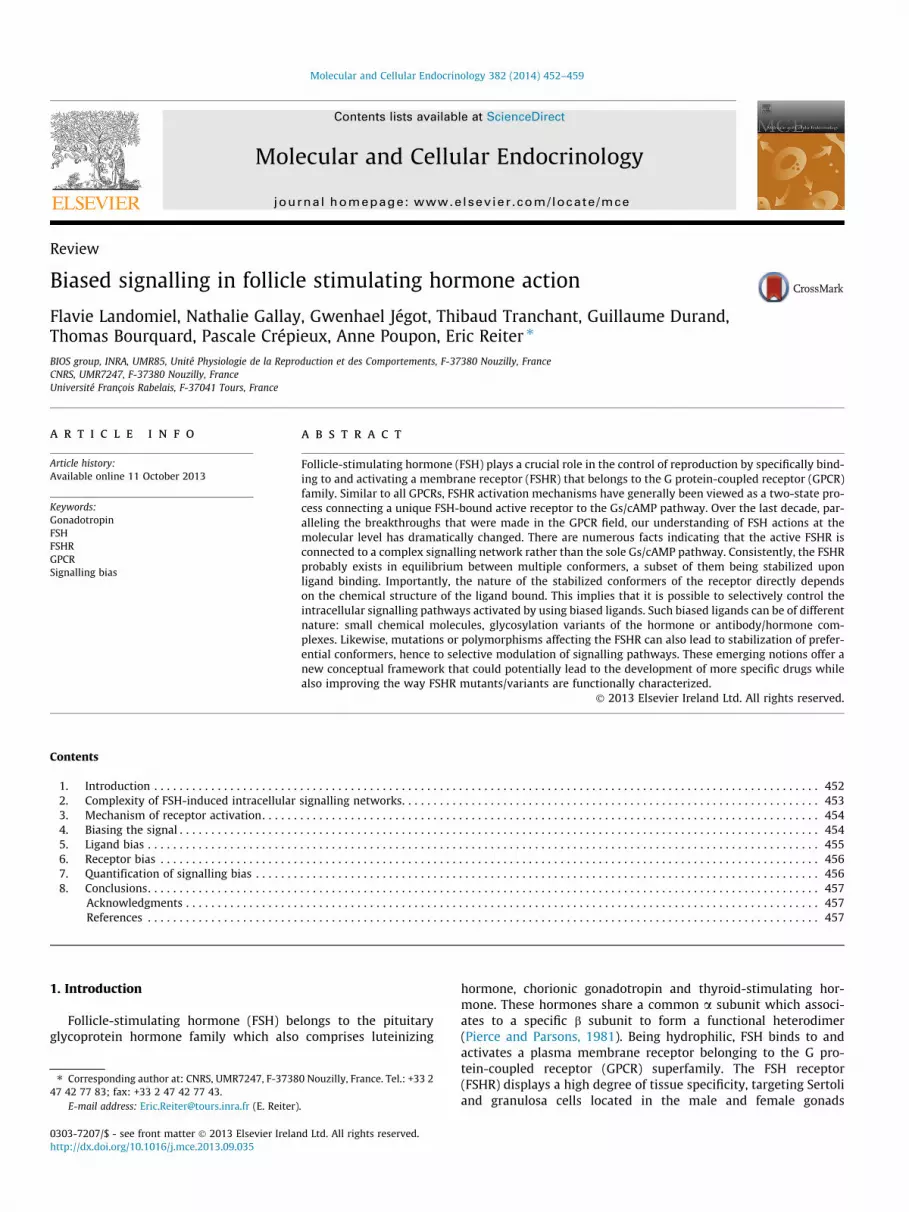
For many years, FSH effects were assumed to be essentially re-layed through the Gas/cAMP/protein kinase A (PKA) signalling cas-cade (Dattatreyamurty et al., 1987; Means et al., 1974). Thiscascade, by subsequently activating CREB, modulates gene tran-scription at the nuclear level (McLean et al., 2002; Meachemet al., 2005; Sadate-Ngatchou et al., 2004). Over the last decadehowever, the activated FSHR has been reported to couple to multi-ple other transduction mechanisms, triggering the activation of acomplex intracellular network (Gloaguen et al., 2011; Ulloa-Aguirreet al., 2011) (Fig. 1). Notably, this signalling network, in addition totrigger gene transcription, has been demonstrated to regulateprotein translation (Musnier et al., 2010, 2012), hence controllingthe fate of FSH-targeted cells (Loss et al., 2007; Richards andPangas, 2010).
A number of Gas-independent transduction mechanisms havebeen reported (Escamilla-Hernandez et al., 2008a; Gloaguenet al., 2011; Zeleznik et al., 2003). They involve coupling to Gai(Arey et al., 1997; Crepieux et al., 2001), Gaq (Escamilla-Hernandezet al., 2008b), b-arrestins (Kara et al., 2006; Tranchant et al., 2011;Wehbi et al., 2010b), epithelial growth factor receptor (EGFR)(Andric and Ascoli, 2006; Cottom et al., 2003; Wayne et al., 2007;Yang and Roy, 2006) and Adapter protein containing Pleckstrinhomology domain, Phosphotyrosine binding domain and Leucinezipper motif (APPL1) (Nechamen et al., 2004, 2007; Thomaset al., 2011). In addition, FSH has also been reported to promotephosphatidylinositol 3-kinase (PI3K) (Zeleznik et al., 2003) andphosphatase and tensin homolog (PTEN) (Dupont et al., 2010)activation but the transduction mechanism involved has not beenclearly identified yet. More specifically, cAMP accumulation
he FSH-induced signalling network. This model has been built according to our011). The different factors involved in FSHR coupling are represented in blue. Thethways are in red.
454 F. Landomiel et al. / Molecular and Cellular Endocrinology 382 (2014) 452–459
activates PKA (Escamilla-Hernandez et al., 2008a; Zeleznik et al.,2003) and exchange protein directly activated by cAMP (EPAC)(Gonzalez-Robayna et al., 2000; Wayne et al., 2007). Once acti-vated, PKA triggers p38 mitogen-activated protein kinases (p38)(Maizels et al., 1998), extracellular signal-regulated kinases 1, 2(ERK) (Cottom et al., 2003) and p70S6 kinase (p70S6K) (Lecureuilet al., 2005) in addition of cAMP response element-binding protein(CREB). On the other hand, EPAC activation has been associatedwith p38 and protein kinase B/Akt (Akt) phosphorylation (Choiet al., 2009; Wayne et al., 2007). Gai-coupling counteracts cAMPaccumulation but can also lead to ERK1,2 activation (Crepieuxet al., 2001).
In addition, in line with many other GPCRs, FSHR has beenshown to specifically interact with G protein-coupled receptor ki-nases (GRKs) and b-arrestins (Reiter and Lefkowitz, 2006). Initially,these proteins steered a lot of attention because of their role in thecontrol of desensitization, internalization and recycling of thereceptor. However, over the last few years, their role has expandedas the GRK/b-arrestin system has been shown to operate as signaltransducer at the FSHR, leading to G protein-independent activa-tion of ERK1,2 and rpS6 (Kara et al., 2006; Tranchant et al., 2011;Wehbi et al., 2010b). Moreover, based on data obtained with otherGPCRs, one can speculate that b-arrestins are involved in the acti-vation of numerous other signalling pathways at the FSHR. Indeed,it has been clearly established that they interact with many proteinpartners (Xiao et al., 2007) and facilitate the phosphorylation of awide array of intracellular targets (Xiao et al., 2010).
The complexity of signalling pathways activated downstream ofthe FSHR is now strongly established and, while not providing di-rect evidence, is generally consistent with the concept of multi-dimensional efficacy.
3. Mechanism of receptor activation
Structural and biophysical approaches represent the gold stan-dard in order to collect direct evidence that multiple active confor-mations of the receptor can be stabilized as a function of thestructure of ligands. Such data have recently accumulated for sev-eral other GPCRs and they support the view that the unligandedreceptor exists in a continuum of conformations. Moleculardynamics simulation of the b2 adrenergic receptor (b2AR) suggeststhat binding of a ligand shifts the conformational equilibrium, en-riches one or a subset of the possible conformations and therebytriggers a specific cellular response (Dror et al., 2011). Nuclearmagnetic resonance (NMR) spectroscopy data have recently con-firmed this model (Nygaard et al., 2013). More generally, the con-cept of ligand-specific conformations has recently been confirmedusing different approaches such as fluorescence spectroscopy (Yaoet al., 2006), quantitative mass spectrometry (Kahsai et al., 2011)or intra-molecular FRET (Granier et al., 2007; Zurn et al., 2009).
Regarding the FSHR, the available structural data sheds light onthe extracellular domain and its binding with FSH, the role of thehinge region in hormone binding and transactivation mechanismhas been explored in much detail, as a novel crystal structurewas solved (Jiang et al., 2012). Data supporting the view that thehinge region acts as a tethered inverse agonist of the transmem-brane domain and plays a very critical role in the hormonal activa-tion of FSHR have been reported (Agrawal and Dighe, 2009;Majumdar et al., 2012).
Models attempting to explain the mechanism of FSHR activa-tion have been proposed and are reviewed in Jiang’s review paperpublished in the present issue. Most of the attention has been fo-cused on the role played by the extracellular domain and the hingeregion in the activation process. However, it can be assumed thatultimately, the activation mechanism triggers a reorganization of
the transmembrane helices TM5 and TM6 to accommodate Gasubunit recruitment, as has been shown for b2AR (Rasmussenet al., 2011a,b) and/or of TM3 and TM7 to recruit b-arrestin as re-ported in the case of the serotonin 5HT1B and 5HT2B receptors(Wacker et al., 2013). As discussed above, considering that thesestructures are likely snapshot representations of highly flexibleproteins oscillating between multiple conformations defined bythe receptor structure, one can come up with a general schemefor FSHR activation and biased signalling as presented in Fig. 2.
Once ligand-bound, the receptor adopts an active conformationand becomes a target for G protein-coupled receptor kinase (GRK)recruitment and phosphorylation. Interestingly, it has recentlybeen demonstrated that the C-terminus of GPCR can be differen-tially phosphorylated by different GRK subtypes, following a ‘‘barcode’’ that regulates the nature of b-arrestin intracellular function(Nobles et al., 2011). Specifically, GRK5 and 6 have been reportedto compete with GRK2 and 3 for the phosphorylation of differentreceptors, with GRK5 and 6 promoting b-arrestin-dependent ERKactivation whereas GRK2 and GRK3 inhibit the same pathway(Heitzler et al., 2012; Kim et al., 2005; Ren et al., 2005). Recentstudies have provided direct evidence of the phosphorylation barcode for different GPCRs (Busillo et al., 2010; Butcher et al.,2011; Lau et al., 2011; Nobles et al., 2011). In particular, isoprote-renol, a full agonist at the b2AR, was compared with carvedilol, aweak b-arrestin-biased ligand, in control cells or in cells depletedof either GRK6 or GRK2 (Nobles et al., 2011). Interestingly, carvedi-lol was found to lead to phosphorylation of the GRK6-dependentsites only, whereas isoproterenol promoted phosphorylation onboth GRK6 and GRK2 sites in the b2AR C-terminus. These findingssupport a model in which ligand efficacy, occurring from the stabil-ization of distinct receptor conformations, subsequently leads tospecific phosphorylation patterns involving distinct GRKs. Thephosphorylation bar code then directs b-arrestin conformation,thereby controlling its interaction partners and subsequent biolog-ical functions (Reiter et al., 2012).
Even though direct evidence has not been obtained yet for theFSHR, it has been reported that GRK2 and 3 compete with GRK5and 6 for receptor phosphorylation (Kara et al., 2006). Moreover,GRK5 and 6 have been shown to promote b-arrestin-dependentERK activation at the FSHR whereas GRK2 and GRK3 are inhibitoryof the same pathway (Heitzler et al., 2012; Kara et al., 2006).
4. Biasing the signal
As already explained, in the classical two-state model, one inac-tive conformation of a receptor was thought to be in equilibriumwith a single ligand-bound active conformation. The functionalimplications being that, any ligand binding at a given receptorwould displace the equilibrium towards one or the other form.According to this two-state model, all the biological activities elic-ited by a ligand were correlated, meaning that all functional read-outs were predicted to lead to the same pharmacological profile.Obviously, this concept has had profound practical consequencesin terms of how drug screening has been approached over the lastdecades.
Now that it is generally accepted that GPCRs adopt multipleinactive and active conformations that are connected to distincttransduction mechanisms, the notion of signalling bias is comingto the fore. In this paradigm, a given ligand or receptor mutationcan modify the stabilized conformation of the receptor–ligandcomplex, as compared to the wild-type receptor–reference-ligandcomplex (Galandrin et al., 2007; Kenakin, 2005; Kobilka, 2011;Nygaard et al., 2013; Reiter et al., 2012; Violin and Lefkowitz,2007; Wacker et al., 2013). Practically, it means that it is possibleto selectively control pathway activation with biased ligands. Such

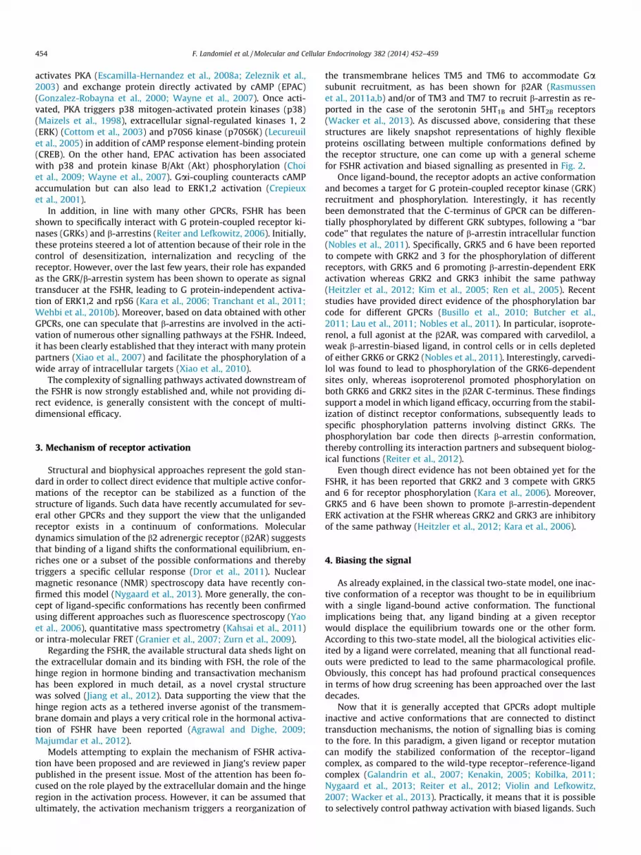
Fig. 2. Model of the structural rearrangements occurring in FSHR transmembrane domain upon activation. The structures of FSHR transmembrane domain in its inactive, Gs-active and b-arrestin-active conformations have been modelled by homology from the b2AR in inactive (PDB 4GBR (Zou et al., 2012), grey) and active (PDB 3SN6 (Rasmussenet al., 2011b), orange) conformations and 5HT2BR binding the b-arrestin-biased ligand ERG (PDB 4IB4 (Wacker et al., 2013), blue), using the software Modeller (Eswar et al.,2006). Major structural rearrangements upon receptor activation are situated in two regions. In first region (right zoom), at the C-terminus, a displacement of helix VIItowards helix VI is observed, in both G-active and b-arrestin-active structures. This movement is thought to be the major structural rearrangement allowing the triggering ofsignalling through b-arrestin. The second rearrangement (left zoom) is the outward rotation of helix VI, which opens the G-protein binding site. Although with differentamplitudes, this rearrangement has been observed for many GPCRs, and is the structural change allowing signalling through G proteins.
F. Landomiel et al. / Molecular and Cellular Endocrinology 382 (2014) 452–459 455
ligands, by stabilizing a subset of the receptor conformations,potentially lead to drugs that are associated with fewer side effects(Whalen et al., 2011). Similarly, a mutation or a polymorphism inthe receptor can potentially modify the balance between the differ-ent pathways it normally triggers. As a consequence, the way func-tional impact of mutations and polymorphisms is usuallyinvestigated (i.e., tracking loss or gain of function) has to be refinedto explore the multiple dimensions of receptor activity.
An important distinction has to be made with another phenom-enon that can lead to apparent biases but which is not directly dueto the preferential stabilization of receptor–ligand conformations.Indeed, it has been demonstrated that the cellular context in whicha receptor is expressed can impact its relative efficacies with
Fig. 3. Schematic representation of the three main categories of signalling biases.Biases can be consecutive to the stabilization of a subset of FSHR conformersdifferent from the conformations favoured in the reference FSH/FSHR complex. Thistype of preferential stabilization can be due to the chemical structure of the ligand(in green) or to modification of the structure of the receptor itself (in blue). Thethird source of signalling bias is due to modifications in the cellular context (inbrown). Since it is independent from the active conformations of the FSH/FSHRcomplex, conditional bias cannot occur when all assays are made in the same cells.
respect to different ligands. This phenomenon, which has beencalled ‘‘conditional efficacy’’, is due to differences in the expressionlevels of receptor-interacting partners, or of molecules down-stream in the signalling pathways, such as receptor activity-modi-fying proteins (RAMPs), in different cell types (Christopoulos et al.,2003; Kenakin, 2002). However, in contrast to the two other causesthat can lead to signalling bias (i.e., ligands and mutations/poly-morphisms), conditional efficacy is revealed only when comparinga single GPCR in different cellular systems (Fig. 3).
5. Ligand bias
The identification of small molecules that could promote orantagonize gonadotropin receptor activation has long been in thepharmaceutical industry research and development pipeline assuch compounds could lead to oral forms of ovarian stimulatorsas well as non-steroidal contraceptives. Over the years, these ef-forts have led to the discovery of several families of small mole-cules acting as agonists or antagonists at the FSHR (Arey et al.,2002, 2008; Dias et al., 2011; Guo et al., 2004a,b; Maclean et al.,2004; van Straten et al., 2005; Wrobel et al., 2002; Yanofskyet al., 2006). Some of these compounds have exhibited very goodpotency and in vivo efficacy in animal models (Arey, 2008; vanKoppen et al., 2013).
Even though these compounds have generally been identifiedusing reporters of Gs-coupling as read-outs, they raise intriguingpossibilities for the development of biased agonists at the FSHR.Interestingly, only one of the identified series of antagonist mole-cules competes with radio-labelled FSH for binding at the receptor,thus suggesting that only this family of antagonists may have anorthosteric site of action located in the extracellular domain ofthe FSHR (Arey et al., 2002). All the other available families of mol-ecules did not compete with FSH in binding assays, consistent withan allosteric mode of action. Recently, a FSHR modulator has evenbeen reported to enhance FSH affinity for the receptor (Dias et al.,2011). The allosteric nature of thiazolidinone derivatives has beendirectly confirmed using FSHR/TSHR chimeras (Yanofsky et al.,2006). Consistent with this view, it has been proposed that mostsmall molecule ligands bind to the transmembrane region of theFSHR (Arey, 2008). By doing so, these small molecule ligands canpotentially stabilize different conformation subsets thereby lead-ing to biased signalling.

456 F. Landomiel et al. / Molecular and Cellular Endocrinology 382 (2014) 452–459
Supporting this hypothesis, biased pharmacological responseshave been observed in several instances with subtle modificationsmade on a conserved core structure (Arey, 2008; Arey et al., 2008;Dias et al., 2011). For example, agonists and antagonists of the Gs/cAMP pathway have been found to share the same core structure(Arey et al., 2008; van Straten et al., 2005). Interestingly, Areyand colleagues found that discrete modifications in the chemicalstructure of a thiazolidinone agonist lead to a switch of FSHR cou-pling from Gs to Gi, directly supporting the concept of biased agon-ism (Arey and Lopez, 2011; Arey et al., 2008). Other authors haveidentified, within the same chemical series, a biased negative allo-steric modulator which presents partial antagonism for cAMP re-sponse, full antagonism for FSH-induced progesterone productionand neutral effects on oestrogen production whereas, in contrast,other compounds behave as balanced negative allosteric modula-tors (Dias et al., 2011). Recently, a dihydropyridine small moleculespecifically binding at the FSHR has been reported (van Koppenet al., 2013). This molecule displays potent agonist effects on Gs/cAMP, FSHR internalization and oestrogen production. However,it also acts as a positive allosteric modulator, strongly enhancingFSH binding and signalling.
A radically different way to explore efficacy at the FSH receptorcomes from antibodies that, by specifically interacting with FSH,are able to transform the pharmacological properties of the nativehormone. Enhancing effect of a polyclonal antibody directedagainst the b-subunit of ovine FSH was observed in vivo in Snelldwarf mice (Ferasin et al., 1997). Similar effects were reported witha bovine FSH monoclonal antibody (Glencross et al., 1993). Anenhancing effect at the FSHR was also found with anti-equine cho-rionic gonadotropin (eCG) antibodies. Importantly, the presence ofthese antibodies was correlated with high fertility and prolificityafter artificial insemination in goats (Herve et al., 2004). A monova-lent fragment (Fab) displayed a similar enhancing effect as the cor-responding bivalent anti-eCG antibody in cellular assays. Morerecently, Wehbi and collaborators reported that various eCG/anti-eCG antibody complexes differentially impacted on the balance be-tween the Gas/cAMP/PKA and the b-arrestin pathways activated atthe FSHR (Wehbi et al., 2010a). From these data, it is tempting tospeculate that the interaction of the hormone with certain antibod-ies can impact on the active conformers of the FSHR.
A third and very intriguing category of bias at the FSHR has beenassociated with the glycosylation of the hormone itself (Arey andLopez, 2011; Ulloa-Aguirre et al., 2011). FSH isoforms presentinglow acidic/sialylated glycan chains induce bell-shaped cAMPdose–response curves consistent with a switch in the FSHR cou-pling from Gs to Gi/Go (Timossi et al., 2000, 1998; Ulloa-Aguirreet al., 2003). Similar bell-shaped cAMP dose–response has been ob-served with human FSH expressed in insect cells (Arey et al., 1997).Recently, a preparation of equine LH made of D (121–149) LHb-subunit and LHa-subunit deglycosylated at Asn56 (eLHdg) has re-vealed very interesting pharmacological properties. This eLHdgwas initially classified as an antagonist at the FSHR because itbound the FSH receptor without eliciting any steroidogenic activity(Butnev et al., 2002). It was later found that, in HEK-293 cells sta-bly expressing the FSHR, at doses up to 1 nM, eLHdg was unable toexhibit detectable agonistic activities on a number of Gas-depen-dent read outs, including cAMP production, protein kinase A activ-ity and cAMP-responsive element-dependent transcriptionalactivity. However, eLHdg induced b-arrestin 1 and 2 recruitment,ERK and ribosomal protein S6 phosphorylation through a PKA-independent but b-arrestin-dependent mechanism (Wehbi et al.,2010b). Collectively, these data support the notion that the micro-heterogeneity of glycosylation that is naturally encountered in vivomight be a natural source of signalling bias, adding a new level ofregulation to the hypothalamo-pituitary-gonadal axis (Arey andLopez, 2011; Ulloa-Aguirre et al., 2011).
6. Receptor bias
So far, biased ligands have concentrated most of the attention inthe field of GPCR pharmacology as they represent potential leadsfor the development of new drugs. However, the whole conceptof bias equally applies to modifications occurring at the receptorlevel. Such events play a crucial role in medicine as they can mate-rialize in patients under the form of mutations or polymorphisms.Understanding the functional consequences associated with suchmodifications of the receptor structure is an important issue inmolecular pathology and, in the future, might be valuable in theestablishment of personalized medicine. In this particular context,the standards that are generally applied to the molecular charac-terization of mutations and/or variants should be expanded to ex-plore the possibility that the modified receptor’s transductionmight be biased compared with the wild-type receptor.
Examples of mutation-induced bias have been well docu-mented for many GPCRs. For instance, the angiotensin type 2receptor DRY-AAY (Wei et al., 2003) and the b2AR TYY (Shenoyet al., 2006) mutants are both biased towards b-arrestin-dependentsignalling. Over the years, a large number of FSHR mutations andvariants have been examined. Some of them have led to paradox-ical results. For instance, the N680S polymorphism might poten-tially impact on the phosphorylation bar code as it substitutes anasparagine for a serine in the C-terminal tail of the FSHR (Simoniet al., 1999). This polymorphism has clearly been associated withsignificant clinical (Daelemans et al., 2004) and physiological ef-fects, women bearing the serine variant being less responsive toFSH than those bearing the asparagine variant (Greb et al., 2005).However, in vitro characterization failed to reveal any differencewhen exploring the Gs/cAMP pathway (Simoni et al., 1999; Sudoet al., 2002). It would really be worthwhile to investigate whetherother pathways are affected by this polymorphism.
Another well-studied genetic alteration identified in the FSHR isthe A189V mutation (Aittomaki et al., 1995). This mutation hasbeen classified as a loss of function because of a lack of plasmamembrane expression. However, the phenotype in man was notconsistent with a complete lack of function. A recent study hasdemonstrated that the A189V FSHR is expressed at very low levelat the plasma membrane but still functional. Indeed, when thewild-type receptor is expressed at a comparable level, it does notcouple to the Gs/cAMP pathway. However, A189V and wild-typeFSHR, both expressed at low levels, are able to induce ERK phos-phorylation via the b-arrestin-dependent pathway (Tranchantet al., 2011). These observations illustrate nicely the concept ofconditional efficacy presented earlier as, when receptor expressiondecreases, the stoichiometry of its modulating partners (b-arres-tins in this case) changes and consequently the signalling outcome.
Recently, the N431I mutation has been found in a man withundetectable circulating FSH but normal spermatogenesis (Casas-Gonzalez et al., 2012). Interestingly, this mutation leads to amarked decrease in FSH-induced desensitization and internaliza-tion. This is another illustration of how complex molecular charac-terization of FSHR mutants/variants is. Obviously, a lot of workremains in order to re-evaluate the structure–activity relationshipsof the FSHR using this new conceptual framework.
7. Quantification of signalling bias
Even though the concept of bias is now widely accepted and isthe object of intense research in the GPCR field, the definition of anappropriate methodology to unambiguously quantifying pharma-cological biases is not trivial and remains a subject of debate. Thisis a very important issue as raw data are often misleading, makingrobust identification of biased effects a challenge. One of the main

F. Landomiel et al. / Molecular and Cellular Endocrinology 382 (2014) 452–459 457
problems is the difficulty to compare signalling events that dra-matically differ in their degree of amplification (e.g., second mes-senger versus b-arrestin recruitment), thereby differentiallyimpacting on the amount of spare receptors. Several methods havebeen proposed recently, each presenting pluses and minuses(Kenakin and Christopoulos, 2013; Rajagopal et al., 2011). Of note,none of the methods proposed so far explicitly accounts for the dif-ferences of receptor expression levels that generally occur whencomparing different mutants or variants.
Probably the most popular method to date is directly derivedfrom the operational model proposed by Black and Leff (1983).To estimate the bias of a ligand as compared to a reference ligand,it is proposed to compute the so-called s value from the EC50:
EC50 ¼KA
1þ swith s ¼ R½ �
KEð1Þ
where [R] is the receptor concentration, KA the affinity of the ligandfor the receptor and KE is the concentration of the complex ligand–receptor for which half the maximal response is obtained. Whenconsidering two different ligands and a single receptor, [R] remainsconstant (granted that a unique cellular system is used), and com-paring the s values is equivalent to comparing the KE values.
However, when considering a ligand’s effects on differentreceptors, for instance mutants or variants, it is very challengingto achieve the exact same receptor expression levels. More gener-ally, the bias should be defined for a test receptor–ligand pair ascompared to a reference receptor–ligand pair. To this aim, it is moreinformative to directly compare the KE values, which can also beobtained from the EC50 value:
KE ¼ R½ �: EC50
KA � EC50ð2Þ
The bias btest of a test ligand/receptor pair as compared to a ref-erence pair for two pathways (path1 and path2) is then given by:
btest ¼rpath1
test � rpath2test
ffiffiffi
2p with rpath
test ¼spath
test
spathref
ð3Þ
Which is equivalent to:
btest ¼R½ �test
R½ �ref:
1ffiffiffi
2p :
Kpath1Eref
Kpath1Etest
�Kpath2
Eref
Kpath2Etest
ð4Þ
We then define:
b�test ¼1ffiffiffi
2p :
Kpath1Eref
Kpath1Etest
�Kpath2
Eref
Kpath2Etest
ð5Þ
The b�test defined in Eq. (5) is independent of the receptor con-centration, and we have:
btest ¼R½ �test
R½ �ref:b�test ð6Þ
From (6) we can see that when comparing a test ligand with a ref-erence ligand, the receptor concentrations are equal and conse-quently b�test ¼ btest . When comparing two different receptors, b�test
can be seen as the bias value that would be obtained if both recep-tors where at the same concentration.
Using this method, it then becomes possible to take the actualexpression level of each receptor mutant/variant into accountand to accurately quantify the degree of bias engendered by thereceptor modification. Classical statistical tests can subsequentlybe used to assess the significance of the different b factors. We
have recently proposed a matrix representation allowing to handlehigh dimensional comparison of b factors (Reiter et al., 2012).
8. Conclusions
Now that the proof of concept has been achieved that biased sig-nalling exists for FSHR, a number of intriguing prospects are emerg-ing. New families of small molecule ligands active at the FSHR havebeen primarily identified and then functionally characterizedaccording to the classical two-state paradigm. Multiplexed assayscould be extremely interesting to further characterize the existingfamilies of compounds as well as to set up new high throughputscreenings. For instance, selective agonists that would trigger ovula-tion while lowering the risk of ovarian hyper stimulation syndromewould be extremely valuable in medically-assisted reproduction.That naturally occurring variations in LH and FSH glycan structurecould impact on receptor activated states and play a physiologicalrole by selectively activating intracellular signalling pathways is aquestion that deserves to be further explored. Studies with highlypurified natural isoforms of known glycan structure, coupled tomultiplexed functional assays, will likely help progressing in thisdirection. Molecular characterization of FSHR mutants and variantsalso needs to shift gear since multi-dimensional evaluation ofreceptor coupling and signalling becomes a necessity.
In order to tackle most of the questions raised in the previousparagraphs, a prerequisite is the development of reliable andquantitative assays for the different coupling mechanisms knownfor the FSHR. Specifically, sensors for Gi-coupling, Gq-coupling,b-arrestin recruitment, b-arrestin-dependent internalization,b-arrestin conformation, b-arrestin-dependent signalling, EGFRtransactivation, APPL1 activation would be highly relevant in thecase of FSHR. Label-free techniques such as impedance measure-ments might also prove valuable in that context (Stallaert et al.,2012). Of course, the Holy Grail is to isolate and quantitate thedifferent arms of FSHR efficacy independently and to calculatethe biases between all the pairs of read-outs. This is the price topay to explore and make the most of the multiple dimensions ofFSHR biological activities.
Acknowledgments
F.L. is recipient of a doctoral fellowship Region Centre. Thiswork was supported by Region Centre research grants, AE INRIA/INRA Regate, ANR and LabEx MabImprove.
References
Abel, M.H., Huhtaniemi, I., Pakarinen, P., Kumar, T.R., Charlton, H.M., 2003.Age-related uterine and ovarian hypertrophy in FSH receptor knockout andFSHbeta subunit knockout mice. Reproduction 125, 165–173.
Agrawal, G., Dighe, R.R., 2009. Critical involvement of the hinge region of thefollicle-stimulating hormone receptor in the activation of the receptor. J. Biol.Chem. 284, 2636–2647.
Aittomaki, K., Lucena, J.L., Pakarinen, P., Sistonen, P., Tapanainen, J., Gromoll, J.,Kaskikari, R., Sankila, E.M., Lehvaslaiho, H., Engel, A.R., Nieschlag, E.,Huhtaniemi, I., de la Chapelle, A., 1995. Mutation in the follicle-stimulatinghormone receptor gene causes hereditary hypergonadotropic ovarian failure.Cell 82, 959–968.
Andric, N., Ascoli, M., 2006. A delayed gonadotropin-dependent and growth factor-mediated activation of the extracellular signal-regulated kinase 1/2 cascadenegatively regulates aromatase expression in granulosa cells. Mol. Endocrinol.20, 3308–3320.
Arey, B.J., 2008. Allosteric modulators of glycoprotein hormone receptors: discoveryand therapeutic potential. Endocrine 34, 1–10.
Arey, B.J., Deecher, D.C., Shen, E.S., Stevis, P.E., Meade Jr., E.H., Wrobel, J., Frail, D.E.,Lopez, F.J., 2002. Identification and characterization of a selective, nonpeptidefollicle-stimulating hormone receptor antagonist. Endocrinology 143, 3822–3829.
Arey, B.J., Lopez, F.J., 2011. Are circulating gonadotropin isoforms naturallyoccurring biased agonists? Basic and therapeutic implications. Rev. Endocr.Metab. Disord. 12, 275–288.

458 F. Landomiel et al. / Molecular and Cellular Endocrinology 382 (2014) 452–459
Arey, B.J., Stevis, P.E., Deecher, D.C., Shen, E.S., Frail, D.E., Negro-Vilar, A., Lopez, F.J.,1997. Induction of promiscuous G protein coupling of the follicle-stimulatinghormone (FSH) receptor: a novel mechanism for transducing pleiotropic actionsof FSH isoforms. Mol. Endocrinol. 11, 517–526.
Arey, B.J., Yanofsky, S.D., Claudia Perez, M., Holmes, C.P., Wrobel, J., Gopalsamy, A.,Stevis, P.E., Lopez, F.J., Winneker, R.C., 2008. Differing pharmacological activitiesof thiazolidinone analogs at the FSH receptor. Biochem. Biophys. Res. Commun.368, 723–728.
Black, J.W., Leff, P., 1983. Operational models of pharmacological agonism. Proc.Roy. Soc. Lond. B Biol. Sci. 220, 141–162.
Busillo, J.M., Armando, S., Sengupta, R., Meucci, O., Bouvier, M., Benovic, J.L., 2010.Site-specific phosphorylation of CXCR4 is dynamically regulated by multiplekinases and results in differential modulation of CXCR4 signaling. J. Biol. Chem.285, 7805–7817.
Butcher, A.J., Prihandoko, R., Kong, K.C., McWilliams, P., Edwards, J.M., Bottrill, A.,Mistry, S., Tobin, A.B., 2011. Differential G-protein-coupled receptorphosphorylation provides evidence for a signaling bar code. J. Biol. Chem.286, 11506–11518.
Butnev, V.Y., Singh, V., Nguyen, V.T., Bousfield, G.R., 2002. Truncated equine LH betaand asparagine(56)-deglycosylated equine LH alpha combine to produce apotent FSH antagonist. J. Endocrinol. 172, 545–555.
Casas-Gonzalez, P., Scaglia, H.E., Perez-Solis, M.A., Durand, G., Scaglia, J., Zarinan, T.,Dias, J.A., Reiter, E., Ulloa-Aguirre, A., 2012. Normal testicular function withoutdetectable follicle-stimulating hormone. A novel mutation in the follicle-stimulating hormone receptor gene leading to apparent constitutive activityand impaired agonist-induced desensitization and internalization. Mol. Cell.Endocrinol. 364, 71–82.
Choi, J.H., Chen, C.L., Poon, S.L., Wang, H.S., Leung, P.C., 2009. Gonadotropin-stimulated epidermal growth factor receptor expression in human ovariansurface epithelial cells: involvement of cyclic AMP-dependent exchange proteinactivated by cAMP pathway. Endocr. Relat. Cancer 16, 179–188.
Christopoulos, A., Christopoulos, G., Morfis, M., Udawela, M., Laburthe, M.,Couvineau, A., Kuwasako, K., Tilakaratne, N., Sexton, P.M., 2003. Novelreceptor partners and function of receptor activity-modifying proteins. J. Biol.Chem. 278, 3293–3297.
Cottom, J., Salvador, L.M., Maizels, E.T., Reierstad, S., Park, Y., Carr, D.W., Davare,M.A., Hell, J.W., Palmer, S.S., Dent, P., Kawakatsu, H., Ogata, M., Hunzicker-Dunn,M., 2003. Follicle-stimulating hormone activates extracellular signal-regulatedkinase but not extracellular signal-regulated kinase kinase through a 100-kDaphosphotyrosine phosphatase. J. Biol. Chem. 278, 7167–7179.
Crepieux, P., Marion, S., Martinat, N., Fafeur, V., Vern, Y.L., Kerboeuf, D., Guillou, F.,Reiter, E., 2001. The ERK-dependent signalling is stage-specifically modulatedby FSH, during primary Sertoli cell maturation. Oncogene 20, 4696–4709.
Daelemans, C., Smits, G., de Maertelaer, V., Costagliola, S., Englert, Y., Vassart, G.,Delbaere, A., 2004. Prediction of severity of symptoms in iatrogenic ovarianhyperstimulation syndrome by follicle-stimulating hormone receptorSer680Asn polymorphism. J. Clin. Endocrinol. Metab. 89, 6310–6315.
Dattatreyamurty, B., Figgs, L.W., Reichert Jr., L.E., 1987. Physical and functionalassociation of follitropin receptors with cholera toxin-sensitive guaninenucleotide-binding protein. J. Biol. Chem. 262, 11737–11745.
Dias, J.A., Bonnet, B., Weaver, B.A., Watts, J., Kluetzman, K., Thomas, R.M., Poli, S.,Mutel, V., Campo, B., 2011. A negative allosteric modulator demonstrates biasedantagonism of the follicle stimulating hormone receptor. Mol. Cell. Endocrinol..
Dierich, A., Sairam, M.R., Monaco, L., Fimia, G.M., Gansmuller, A., LeMeur, M.,Sassone-Corsi, P., 1998. Impairing follicle-stimulating hormone (FSH) signalingin vivo: targeted disruption of the FSH receptor leads to aberrant gametogenesisand hormonal imbalance. Proc. Natl. Acad. Sci. USA 95, 13612–13617.
Dror, R.O., Arlow, D.H., Maragakis, P., Mildorf, T.J., Pan, A.C., Xu, H., Borhani, D.W.,Shaw, D.E., 2011. Activation mechanism of the beta2-adrenergic receptor. Proc.Natl. Acad. Sci. USA 108, 18684–18689.
Dupont, J., Musnier, A., Decourtye, J., Boulo, T., Lecureuil, C., Guillou, H., Valet, S.,Fouchecourt, S., Pitetti, J.L., Nef, S., Reiter, E., Crepieux, P., 2010. FSH-stimulatedPTEN activity accounts for the lack of FSH mitogenic effect in prepubertal ratSertoli cells. Mol. Cell. Endocrinol. 315, 271–276.
Escamilla-Hernandez, R., Little-Ihrig, L., Orwig, K.E., Yue, J., Chandran, U., Zeleznik,A.J., 2008a. Constitutively active protein kinase A qualitatively mimics theeffects of follicle-stimulating hormone on granulosa cell differentiation. Mol.Endocrinol. 22, 1842–1852.
Escamilla-Hernandez, R., Little-Ihrig, L., Zeleznik, A.J., 2008b. Inhibition ofrat granulosa cell differentiation by overexpression of Galphaq. Endocrine 33,21–31.
Eswar, N., Webb, B., Marti-Renom, M.A., Madhusudhan, M.S., Eramian, D., Shen,M.Y., Pieper, U., Sali, A., 2006. Comparative protein structure modeling usingmodeller. Curr. Protoc. Bioinform. (Chapter 5, Unit 5.6).
Ferasin, L., Gabai, G., Beattie, J., Bono, G., Holder, A.T., 1997. Enhancement of FSHbioactivity in vivo using site-specific antisera. J. Endocrinol. 152, 355–363.
Galandrin, S., Oligny-Longpre, G., Bouvier, M., 2007. The evasive nature of drugefficacy: implications for drug discovery. Trends Pharmacol. Sci. 28, 423–430.
Glencross, R.G., Lovell, R.D., Holder, A.T., 1993. Monoclonal antibody enhancementof FSH-induced uterine growth in snell dwarf mice. J. Endocrinol. 136, R5–R7.
Gloaguen, P., Crepieux, P., Heitzler, D., Poupon, A., Reiter, E., 2011. Mapping thefollicle-stimulating hormone-induced signaling networks. Front. Endocrinol.(Lausanne) 2, 45.
Gonzalez-Robayna, I.J., Falender, A.E., Ochsner, S., Firestone, G.L., Richards, J.S., 2000.Follicle-stimulating hormone (FSH) stimulates phosphorylation and activationof protein kinase B (PKB/Akt) and serum and glucocorticoid-induced kinase
(Sgk): evidence for A kinase-independent signaling by FSH in granulosa cells.Mol. Endocrinol. 14, 1283–1300.
Granier, S., Kim, S., Shafer, A.M., Ratnala, V.R., Fung, J.J., Zare, R.N., Kobilka, B., 2007.Structure and conformational changes in the C-terminal domain of the beta2-adrenoceptor: insights from fluorescence resonance energy transfer studies. J.Biol. Chem. 282, 13895–13905.
Greb, R.R., Grieshaber, K., Gromoll, J., Sonntag, B., Nieschlag, E., Kiesel, L., Simoni, M.,2005. A common single nucleotide polymorphism in exon 10 of the humanfollicle stimulating hormone receptor is a major determinant of length andhormonal dynamics of the menstrual cycle. J. Clin. Endocrinol. Metab. 90, 4866–4872.
Guo, T., Adang, A.E., Dolle, R.E., Dong, G., Fitzpatrick, D., Geng, P., Ho, K.K., Kultgen,S.G., Liu, R., McDonald, E., McGuinness, B.F., Saionz, K.W., Valenzano, K.J., vanStraten, N.C., Xie, D., Webb, M.L., 2004a. Small molecule biaryl FSH receptoragonists. Part 1: lead discovery via encoded combinatorial synthesis. Bioorg.Med. Chem. Lett. 14, 1713–1716.
Guo, T., Adang, A.E., Dong, G., Fitzpatrick, D., Geng, P., Ho, K.K., Jibilian, C.H., Kultgen,S.G., Liu, R., McDonald, E., Saionz, K.W., Valenzano, K.J., van Straten, N.C., Xie, D.,Webb, M.L., 2004b. Small molecule biaryl FSH receptor agonists. Part 2: leadoptimization via parallel synthesis. Bioorg. Med. Chem. Lett. 14, 1717–1720.
Heitzler, D., Durand, G., Gallay, N., Rizk, A., Ahn, S., Kim, J., Violin, J.D., Dupuy, L.,Gauthier, C., Piketty, V., Crepieux, P., Poupon, A., Clement, F., Fages, F., Lefkowitz,R.J., Reiter, E., 2012. Competing G protein-coupled receptor kinases balance Gprotein and beta-arrestin signaling. Mol. Syst. Biol. 8, 590.
Herve, V., Roy, F., Bertin, J., Guillou, F., Maurel, M.C., 2004. Antiequine chorionicgonadotropin (eCG) antibodies generated in goats treated with eCG for theinduction of ovulation modulate the luteinizing hormone and follicle-stimulating hormone bioactivities of eCG differently. Endocrinology 145, 294–303.
Jiang, X., Liu, H., Chen, X., Chen, P.H., Fischer, D., Sriraman, V., Yu, H.N., Arkinstall, S.,He, X., 2012. Structure of follicle-stimulating hormone in complex with theentire ectodomain of its receptor. Proc. Natl. Acad. Sci. USA 109, 12491–12496.
Kahsai, A.W., Xiao, K., Rajagopal, S., Ahn, S., Shukla, A.K., Sun, J., Oas, T.G., Lefkowitz,R.J., 2011. Multiple ligand-specific conformations of the beta2-adrenergicreceptor. Nat. Chem. Biol. 7, 692–700.
Kara, E., Crepieux, P., Gauthier, C., Martinat, N., Piketty, V., Guillou, F., Reiter, E.,2006. A phosphorylation cluster of five serine and threonine residues in the C-terminus of the follicle-stimulating hormone receptor is important fordesensitization but not for beta-arrestin-mediated ERK activation. Mol.Endocrinol. 20, 3014–3026.
Kenakin, T., 2002. Efficacy at G-protein-coupled receptors. Nat. Rev. Drug Discov. 1,103–110.
Kenakin, T., 2005. New concepts in drug discovery: collateral efficacy andpermissive antagonism. Nat. Rev. Drug Discov. 4, 919–927.
Kenakin, T., Christopoulos, A., 2013. Signalling bias in new drug discovery:detection, quantification and therapeutic impact. Nat. Rev. Drug Discov. 12,205–216.
Kim, J., Ahn, S., Ren, X.R., Whalen, E.J., Reiter, E., Wei, H., Lefkowitz, R.J., 2005.Functional antagonism of different G protein-coupled receptor kinases for beta-arrestin-mediated angiotensin II receptor signaling. Proc. Natl. Acad. Sci. USA102, 1442–1447.
Kobilka, B.K., 2011. Structural insights into adrenergic receptor function andpharmacology. Trends Pharmacol. Sci. 32, 213–218.
Kumar, T.R., Wang, Y., Lu, N., Matzuk, M.M., 1997. Follicle stimulating hormone isrequired for ovarian follicle maturation but not male fertility. Nat. Genet. 15,201–204.
Lau, E.K., Trester-Zedlitz, M., Trinidad, J.C., Kotowski, S.J., Krutchinsky, A.N.,Burlingame, A.L., von Zastrow, M., 2011. Quantitative encoding of the effect ofa partial agonist on individual opioid receptors by multisite phosphorylationand threshold detection. Sci. Signal. 4, ra52.
Lecureuil, C., Tesseraud, S., Kara, E., Martinat, N., Sow, A., Fontaine, I., Gauthier, C.,Reiter, E., Guillou, F., Crepieux, P., 2005. Follicle-stimulating hormone activatesp70 ribosomal protein S6 kinase by protein kinase A-mediateddephosphorylation of Thr 421/Ser 424 in primary Sertoli cells. Mol.Endocrinol. 19, 1812–1820.
Loss, E.S., Jacobus, A.P., Wassermann, G.F., 2007. Diverse FSH and testosteronesignaling pathways in the Sertoli cell. Horm. Metab. Res. 39, 806–812.
Macklon, N.S., Stouffer, R.L., Giudice, L.C., Fauser, B.C., 2006. The science behind25 years of ovarian stimulation for in vitro fertilization. Endocr. Rev. 27, 170–207.
Maclean, D., Holden, F., Davis, A.M., Scheuerman, R.A., Yanofsky, S., Holmes, C.P.,Fitch, W.L., Tsutsui, K., Barrett, R.W., Gallop, M.A., 2004. Agonists of the folliclestimulating hormone receptor from an encoded thiazolidinone library. J. Comb.Chem. 6, 196–206.
Maizels, E.T., Cottom, J., Jones, J.C., Hunzicker-Dunn, M., 1998. Follicle stimulatinghormone (FSH) activates the p38 mitogen-activated protein kinase pathway,inducing small heat shock protein phosphorylation and cell rounding inimmature rat ovarian granulosa cells. Endocrinology 139, 3353–3356.
Majumdar, R., Railkar, R., Dighe, R.R., 2012. The antibodies against thecomputationally designed mimic of the glycoprotein hormone receptortransmembrane domain provide insights into receptor activation andsuppress the constitutively activated receptor mutants. J. Biol. Chem. 287,34514–34532.
McLean, D.J., Friel, P.J., Pouchnik, D., Griswold, M.D., 2002. Oligonucleotidemicroarray analysis of gene expression in follicle-stimulating hormone-treated rat Sertoli cells. Mol. Endocrinol. 16, 2780–2792.

F. Landomiel et al. / Molecular and Cellular Endocrinology 382 (2014) 452–459 459
Meachem, S.J., Ruwanpura, S.M., Ziolkowski, J., Ague, J.M., Skinner, M.K., Loveland,K.L., 2005. Developmentally distinct in vivo effects of FSH on proliferation andapoptosis during testis maturation. J. Endocrinol. 186, 429–446.
Means, A.R., MacDougall, E., Soderling, T.R., Corbin, J.D., 1974. Testicular adenosine30:50-monophosphate-dependent protein kinase. Regulation by follicle-stimulating hormone. J. Biol. Chem. 249, 1231–1238.
Musnier, A., Blanchot, B., Reiter, E., Crepieux, P., 2010. GPCR signalling to thetranslation machinery. Cell. Signal. 22, 707–716.
Musnier, A., Leon, K., Morales, J., Reiter, E., Boulo, T., Costache, V., Vourc’h, P.,Heitzler, D., Oulhen, N., Poupon, A., Boulben, S., Cormier, P., Crepieux, P., 2012.MRNA-selective translation induced by FSH in primary Sertoli cells. Mol.Endocrinol. 26, 669–680.
Nechamen, C.A., Thomas, R.M., Cohen, B.D., Acevedo, G., Poulikakos, P.I., Testa, J.R.,Dias, J.A., 2004. Human follicle-stimulating hormone (FSH) receptor interactswith the adaptor protein APPL1 in HEK 293 cells: potential involvement of thePI3K pathway in FSH signaling. Biol. Reprod. 71, 629–636.
Nechamen, C.A., Thomas, R.M., Dias, J.A., 2007. APPL1, APPL2, Akt2 and FOXO1ainteract with FSHR in a potential signaling complex. Mol. Cell. Endocrinol. 260–262, 93–99.
Nobles, K.N., Xiao, K., Ahn, S., Shukla, A.K., Lam, C.M., Rajagopal, S., Strachan, R.T.,Huang, T.Y., Bressler, E.A., Hara, M.R., Shenoy, S.K., Gygi, S.P., Lefkowitz, R.J., 2011.Distinct phosphorylation sites on the beta(2)-adrenergic receptor establish abarcode that encodes differential functions of beta-arrestin. Sci. Signal. 4, ra51.
Nygaard, R., Zou, Y., Dror, R.O., Mildorf, T.J., Arlow, D.H., Manglik, A., Pan, A.C., Liu,C.W., Fung, J.J., Bokoch, M.P., Thian, F.S., Kobilka, T.S., Shaw, D.E., Mueller, L.,Prosser, R.S., Kobilka, B.K., 2013. The dynamic process of beta(2)-adrenergicreceptor activation. Cell 152, 532–542.
Pierce, J.G., Parsons, T.F., 1981. Glycoprotein hormones: structure and function.Annu. Rev. Biochem. 50, 465–495.
Rajagopal, S., Ahn, S., Rominger, D.H., Gowen-MacDonald, W., Lam, C.M., Dewire,S.M., Violin, J.D., Lefkowitz, R.J., 2011. Quantifying ligand bias at seven-transmembrane receptors. Mol. Pharmacol. 80, 367–377.
Rasmussen, S.G., Choi, H.J., Fung, J.J., Pardon, E., Casarosa, P., Chae, P.S., Devree, B.T.,Rosenbaum, D.M., Thian, F.S., Kobilka, T.S., Schnapp, A., Konetzki, I., Sunahara,R.K., Gellman, S.H., Pautsch, A., Steyaert, J., Weis, W.I., Kobilka, B.K., 2011a.Structure of a nanobody-stabilized active state of the beta(2) adrenoceptor.Nature 469, 175–180.
Rasmussen, S.G., DeVree, B.T., Zou, Y., Kruse, A.C., Chung, K.Y., Kobilka, T.S., Thian,F.S., Chae, P.S., Pardon, E., Calinski, D., Mathiesen, J.M., Shah, S.T., Lyons, J.A.,Caffrey, M., Gellman, S.H., Steyaert, J., Skiniotis, G., Weis, W.I., Sunahara, R.K.,Kobilka, B.K., 2011b. Crystal structure of the beta2 adrenergic receptor-Gsprotein complex. Nature 477, 549–555.
Reiter, E., Ahn, S., Shukla, A.K., Lefkowitz, R.J., 2012. Molecular mechanism of beta-arrestin-biased agonism at seven-transmembrane receptors. Annu. Rev.Pharmacol. Toxicol. 52, 179–197.
Reiter, E., Lefkowitz, R.J., 2006. GRKs and beta-arrestins: roles in receptor silencing,trafficking and signaling. Trends Endocrinol. Metab. 17, 159–165.
Ren, X.R., Reiter, E., Ahn, S., Kim, J., Chen, W., Lefkowitz, R.J., 2005. Different Gprotein-coupled receptor kinases govern G protein and beta-arrestin-mediatedsignaling of V2 vasopressin receptor. Proc. Natl. Acad. Sci. USA 102, 1448–1453.
Richards, J.S., Pangas, S.A., 2010. The ovary: basic biology and clinical implications. J.Clin. Invest. 120, 963–972.
Sadate-Ngatchou, P.I., Pouchnik, D.J., Griswold, M.D., 2004. Follicle-stimulatinghormone induced changes in gene expression of murine testis. Mol. Endocrinol.18, 2805–2816.
Shenoy, S.K., Drake, M.T., Nelson, C.D., Houtz, D.A., Xiao, K., Madabushi, S., Reiter, E.,Premont, R.T., Lichtarge, O., Lefkowitz, R.J., 2006. Beta-arrestin-dependent, Gprotein-independent ERK1/2 activation by the beta2 adrenergic receptor. J. Biol.Chem. 281, 1261–1273.
Simoni, M., Gromoll, J., Hoppner, W., Kamischke, A., Krafft, T., Stahle, D., Nieschlag,E., 1999. Mutational analysis of the follicle-stimulating hormone (FSH) receptorin normal and infertile men: identification and characterization of two discreteFSH receptor isoforms. J. Clin. Endocrinol. Metab. 84, 751–755.
Simoni, M., Gromoll, J., Nieschlag, E., 1997. The follicle-stimulating hormonereceptor: biochemistry, molecular biology, physiology, and pathophysiology.Endocr. Rev. 18, 739–773.
Stallaert, W., Dorn, J.F., van der Westhuizen, E., Audet, M., Bouvier, M., 2012. Impedanceresponses reveal beta(2)-adrenergic receptor signaling pluridimensionality andallow classification of ligands with distinct signaling profiles. PLoS ONE 7, e29420.
Sudo, S., Kudo, M., Wada, S., Sato, O., Hsueh, A.J., Fujimoto, S., 2002. Genetic andfunctional analyses of polymorphisms in the human FSH receptor gene. Mol.Hum. Reprod. 8, 893–899.
Tapanainen, J.S., Aittomaki, K., Min, J., Vaskivuo, T., Huhtaniemi, I.T., 1997. Menhomozygous for an inactivating mutation of the follicle-stimulating hormone(FSH) receptor gene present variable suppression of spermatogenesis andfertility. Nat. Genet. 15, 205–206.
Themmen, A.P.N., Huhtaniemi, I.T., 2000. Mutations of gonadotropins andgonadotropin receptors: elucidating the physiology and pathophysiology ofpituitary-gonadal function. Endocr. Rev. 21, 551–583.
Thomas, R.M., Nechamen, C.A., Mazurkiewicz, J.E., Ulloa-Aguirre, A., Dias, J.A., 2011.The adapter protein APPL1 links FSH receptor to inositol 1,4,5-trisphosphateproduction and is implicated in intracellular Ca(2+) mobilization.Endocrinology 152, 1691–1701.
Timossi, C.M., Barrios-de-Tomasi, J., Gonzalez-Suarez, R., Arranz, M.C.,Padmanabhan, V., Conn, P.M., Ulloa-Aguirre, A., 2000. Differential effects of
the charge variants of human follicle-stimulating hormone. J. Endocrinol. 165,193–205.
Timossi, C.M., Barrios de Tomasi, J., Zambrano, E., Gonzalez, R., Ulloa-Aguirre, A.,1998. A naturally occurring basically charged human follicle-stimulatinghormone (FSH) variant inhibits FSH-induced androgen aromatization andtissue-type plasminogen activator enzyme activity in vitro.Neuroendocrinology 67, 153–163.
Tranchant, T., Durand, G., Gauthier, C., Crepieux, P., Ulloa-Aguirre, A., Royere, D.,Reiter, E., 2011. Preferential beta-arrestin signalling at low receptor densityrevealed by functional characterization of the human FSH receptor A189 Vmutation. Mol. Cell. Endocrinol. 331, 109–118.
Ulloa-Aguirre, A., Crepieux, P., Poupon, A., Maurel, M.C., Reiter, E., 2011. Novelpathways in gonadotropin receptor signaling and biased agonism. Rev. Endocr.Metab. Disord. 12, 259–274.
Ulloa-Aguirre, A., Timossi, C., Barrios-de-Tomasi, J., Maldonado, A., Nayudu, P., 2003.Impact of carbohydrate heterogeneity in function of follicle-stimulatinghormone: studies derived from in vitro and in vivo models. Biol. Reprod. 69,379–389.
van Koppen, C.J., Verbost, P.M., van de Lagemaat, R., Karstens, W.J., Loozen, H.J., vanAchterberg, T.A., van Amstel, M.G., Brands, J.H., van Doornmalen, E.J., Wat, J.,Mulder, S.J., Raafs, B.C., Verkaik, S., Hanssen, R.G., Timmers, C.M., 2013. Signalingof an allosteric, nanomolar potent, low molecular weight agonist for the follicle-stimulating hormone receptor. Biochem. Pharmacol. 85, 1162–1170.
van Straten, N.C., van Berkel, T.H., Demont, D.R., Karstens, W.J., Merkx, R., Oosterom,J., Schulz, J., van Someren, R.G., Timmers, C.M., van Zandvoort, P.M., 2005.Identification of substituted 6-amino-4-phenyltetrahydroquinoline derivatives:potent antagonists for the follicle-stimulating hormone receptor. J. Med. Chem.48, 1697–1700.
Violin, J.D., Lefkowitz, R.J., 2007. Beta-arrestin-biased ligands at seven-transmembrane receptors. Trends Pharmacol. Sci. 28, 416–422.
Wacker, D., Wang, C., Katritch, V., Han, G.W., Huang, X.P., Vardy, E., McCorvy, J.D.,Jiang, Y., Chu, M., Siu, F.Y., Liu, W., Xu, H.E., Cherezov, V., Roth, B.L., Stevens, R.C.,2013. Structural features for functional selectivity at serotonin receptors.Science 340, 615–619.
Wayne, C.M., Fan, H.Y., Cheng, X., Richards, J.S., 2007. Follicle-stimulating hormoneinduces multiple signaling cascades: evidence that activation of Rous sarcomaoncogene, RAS, and the epidermal growth factor receptor are critical forgranulosa cell differentiation. Mol. Endocrinol. 21, 1940–1957.
Wehbi, V., Decourtye, J., Piketty, V., Durand, G., Reiter, E., Maurel, M.C., 2010a.Selective modulation of follicle-stimulating hormone signaling pathways withenhancing equine chorionic gonadotropin/antibody immune complexes.Endocrinology 151, 2788–2799.
Wehbi, V., Tranchant, T., Durand, G., Musnier, A., Decourtye, J., Piketty, V., Butnev,V.Y., Bousfield, G.R., Crepieux, P., Maurel, M.C., Reiter, E., 2010b. Partiallydeglycosylated equine LH preferentially activates beta-arrestin-dependentsignaling at the follicle-stimulating hormone receptor. Mol. Endocrinol. 24,561–573.
Wei, H., Ahn, S., Shenoy, S.K., Karnik, S.S., Hunyady, L., Luttrell, L.M., Lefkowitz, R.J.,2003. Independent beta-arrestin 2 and G protein-mediated pathways forangiotensin II activation of extracellular signal-regulated kinases 1 and 2.Proc. Natl. Acad. Sci. USA 100, 10782–10787.
Whalen, E.J., Rajagopal, S., Lefkowitz, R.J., 2011. Therapeutic potential of beta-arrestin- and G protein-biased agonists. Trends Mol. Med. 17, 126–139.
Wrobel, J., Green, D., Jetter, J., Kao, W., Rogers, J., Perez, M.C., Hardenburg, J.,Deecher, D.C., Lopez, F.J., Arey, B.J., Shen, E.S., 2002. Synthesis of (bis)sulfonicacid, (bis)benzamides as follicle-stimulating hormone (FSH) antagonists.Bioorg. Med. Chem. 10, 639–656.
Xiao, K., McClatchy, D.B., Shukla, A.K., Zhao, Y., Chen, M., Shenoy, S.K., Yates 3rd, J.R.,Lefkowitz, R.J., 2007. Functional specialization of beta-arrestin interactionsrevealed by proteomic analysis. Proc. Natl. Acad. Sci. USA 104, 12011–12016.
Xiao, K., Sun, J., Kim, J., Rajagopal, S., Zhai, B., Villen, J., Haas, W., Kovacs, J.J., Shukla,A.K., Hara, M.R., Hernandez, M., Lachmann, A., Zhao, S., Lin, Y., Cheng, Y.,Mizuno, K., Ma’ayan, A., Gygi, S.P., Lefkowitz, R.J., 2010. Global phosphorylationanalysis of beta-arrestin-mediated signaling downstream of a seventransmembrane receptor (7TMR). Proc. Natl. Acad. Sci. USA 107, 15299–15304.
Yang, P., Roy, S.K., 2006. A novel mechanism of FSH regulation of DNA synthesis in thegranulosa cells of hamster preantral follicles: involvement of a protein kinase C-mediated MAP kinase 3/1 self-activation loop. Biol. Reprod. 75, 149–157.
Yanofsky, S.D., Shen, E.S., Holden, F., Whitehorn, E., Aguilar, B., Tate, E., Holmes, C.P.,Scheuerman, R., MacLean, D., Wu, M.M., Frail, D.E., Lopez, F.J., Winneker, R.,Arey, B.J., Barrett, R.W., 2006. Allosteric activation of the follicle-stimulatinghormone (FSH) receptor by selective, nonpeptide agonists. J. Biol. Chem. 281,13226–13233.
Yao, X., Parnot, C., Deupi, X., Ratnala, V.R., Swaminath, G., Farrens, D., Kobilka, B.,2006. Coupling ligand structure to specific conformational switches in thebeta2-adrenoceptor. Nat. Chem. Biol. 2, 417–422.
Zeleznik, A.J., Saxena, D., Little-Ihrig, L., 2003. Protein kinase B is obligatory forfollicle-stimulating hormone-induced granulosa cell differentiation.Endocrinology 144, 3985–3994.
Zou, Y., Weis, W.I., Kobilka, B.K., 2012. N-terminal T4 lysozyme fusion facilitatescrystallization of a G protein coupled receptor. PLoS ONE 7, e46039.
Zurn, A., Zabel, U., Vilardaga, J.P., Schindelin, H., Lohse, M.J., Hoffmann, C., 2009.Fluorescence resonance energy transfer analysis of alpha 2a-adrenergicreceptor activation reveals distinct agonist-specific conformational changes.Mol. Pharmacol. 75, 534–541.


