Embed Size (px)
Citation preview

Vision Research 98 (2014) 26–34
Contents lists available at ScienceDirect
Vision Research
journal homepage: www.elsevier .com/locate /v isres
Bi-directional corneal accommodation in alert chicks withexperimentally-induced astigmatism q
http://dx.doi.org/10.1016/j.visres.2014.03.0020042-6989/� 2014 Elsevier Ltd. All rights reserved.
q Grant support: RGC General Research Fund #561209; The Centre of MyopiaResearch and The Niches Areas-Myopia Research Fund (J-BB7P) from The HongKong Polytechnic University.⇑ Corresponding author. Fax: +852 2764 6051.
E-mail address: [email protected] (C.-s. Kee).
Chin-hung Geoffrey Chu a, Yongjin Zhou b, Yongping Zheng c, Chea-su Kee a,⇑a School of Optometry, The Hong Kong Polytechnic University, Hung Hom, Kowloon, Hong Kong Special Administrative Regionb School of Medicine, Shenzhen University, Guangdong, Chinac Interdisciplinary Division of Biomedical Engineering, The Hong Kong Polytechnic University, Kowloon, Hong Kong Special Administrative Region
a r t i c l e i n f o
Article history:Received 3 October 2013Received in revised form 4 March 2014Available online 14 March 2014
Keywords:Corneal accommodationCornea curvatureCorneal videokeratography systemAstigmatismChickens
a b s t r a c t
This study aimed to characterize corneal accommodation in alert chicks with and without experimen-tally-induced astigmatism. Refraction and corneal biometry were measured in 16 chicks with experimen-tally-induced astigmatism (>1.00 D) and 6 age-matched control chicks (astigmatism 61.00 D). Cornealaccommodation was detected using a Placido-ring based videokeratography system, by measuringchanges in corneal curvature from a series of consecutive images acquired from alert chicks. The corre-lation between the magnitudes of corneal accommodation and astigmatism was analyzed by includingdata from all 22 chicks. Data from all eyes showed obvious bi-directional changes in corneal accommo-dation. There was no significant difference in corneal accommodative changes between the fellow eyes ofthe treated birds, and the right and left eyes of control birds. However, positive accommodation (PA) andmaximum magnitude of PA (MPA) were significantly higher in the astigmatic vs. the fellow eyes of trea-ted chicks (mean ± SE: PA = +2.24 ± 0.44 D vs. +1.26 ± 0.20 D; MPA = +7.53 ± 0.81 D vs. +4.38 ± 0.53 D,both p < 0.05). This was not the case for negative accommodation (NA) or maximum magnitude of NA(MNA) (NA = �0.46 ± 0.15 D vs. �0.33 ± 0.04 D; MNA = �0.92 ± 0.23 D vs. �0.73 ± 0.12 D, respectively,p > 0.05). Furthermore, higher PA and MPA were found to be correlated with higher refractive astigma-tism (both r = 0.34, p < 0.05). These results suggest that the presence of astigmatism may interfere withaccommodative function in chicks.
� 2014 Elsevier Ltd. All rights reserved.
1. Introduction
The extent to which the cornea, the major refractive componentof the eye, plays a role in accommodation is controversial. Althoughprevious studies found 0.40–0.72 D of corneal accommodation inhumans aged between 20 and 40 years old (Pierscionek, Popiolek-Masajada, & Kasprzak, 2001; Yasuda & Yamaguchi, 2005; Yasuda,Yamaguchi, & Ohkoshi, 2003), negative results have also been re-ported (Bannon, 1946; Buehren, Collins, & Carney, 2003; He et al.,2003; Read, Buehren, & Collins, 2007; Rosenfield & Gilmartin,1987). These inconsistent results may be due to methodological dif-ferences or difficulties in detecting subtle changes in corneal curva-ture. In contrast to the findings in humans, there is strongerevidence for corneal accommodation in several avian species,
including the chicken, which has been proposed as a good modelfor studying corneal accommodation, because of its prominentamplitude of corneal accommodation (Glasser, Troilo, & Howland,1994; Troilo & Wallman, 1985). Previous studies showed significantcorneal steepening accompanied with lenticular accommodation(Glasser, Troilo, & Howland, 1994; Murphy, Glasser, & Howland,1995; Ostrin et al., 2011; Schaeffel & Howland, 1987; Troilo &Wallman, 1987) and the total accommodation (i.e., lenticular pluscorneal accommodations) can be over 25.00 D (Glasser, Troilo, &Howland, 1994; Schaeffel, Glasser, & Howland, 1988). Indeed, cor-neal deformation has been estimated to contribute 40.0–50.0%(about 6.00–9.00 D) of the ocular accommodation (Glasser, Troilo,& Howland, 1994; Schaeffel & Howland, 1987; Troilo & Wallman,1987). Nevertheless, some studies could not detect any cornealaccommodation in chicks (Beer, 1892; Sivak et al., 1986).
Corneal accommodation in chicks has been reported to occurdue to the contraction of a longitudinal Crampton’s muscle (Walls,1942). This muscle is the anterior portion of the striated ciliarymuscle which originates at the sclera, with the scleral occiscleacting as a supporting base (Glasser, Troilo, & Howland, 1994;

Chin-hung Geoffrey Chu et al. / Vision Research 98 (2014) 26–34 27
Murphy, Glasser, & Howland, 1995). A direct connection of themuscle to the corneal inner lamella creates a circumferential ten-sion that alters corneal curvature upon muscle contraction. Inempirical studies, changes in chick corneal curvature have beenmeasured either by an infrared photokeratometer (García de laCera et al., 2007; Schaeffel & Howland, 1987; Troilo & Judge,1993) or by a modified keratometer (Irving, Sivak, & Callender,1992; Troilo & Wallman, 1987). Ocular accommodation was in-duced either pharmacologically by treatment with nicotine (Glas-ser, Troilo, & Howland, 1994; Schmid & Wildsoet, 1997; Troilo &Wallman, 1987), or electrophysiologically by stimulation of theEdinger–Westphal nucleus (Glasser, Troilo, & Howland, 1994; Troi-lo & Wallman, 1987). However, the extent to which experimentalmanipulations to stimulate corneal accommodation mimic the nat-ural action of the system is still unclear.
Astigmatism is a refractive error frequently associated withmyopia (or ‘‘nearsightedness’’) and hyperopia (or ‘‘farsightedness’’)in humans (Read, Collins, & Carney, 2007) and animal models(monkeys: Kee et al., 2005; chicks: Kee & Deng, 2008). It has beenhypothesized that the presence of astigmatism may facilitate theaccuracy of accommodative response by utilizing the contrast cuesassociated with the two principal refractive meridians (Howland,1982); thus the significant astigmatism found in infants couldpotentially interfere with the eye’s focusing strategy and signalingpathway during early eye growth. However, despite the high prev-alence of astigmatism found across different nations (see a sum-mary figure in Kee, 2013), the functional role, if any, ofastigmatism during normal and abnormal refractive developmentremains unclear (Kee, 2013). The present investigation had twokey aims. First, we investigated whether we could detect cornealaccommodation in chicks under natural viewing conditions: thatis with no artificial stimulation, anesthesia, nor the use of lidretractors. Second, we sought to test the hypothesis that cornealaccommodation in chicks is influenced by the level of either refrac-tive or corneal astigmatism.
2. Methods
2.1. Animal subjects
Twenty-two White Leghorn chicks (Gallus gallus domesticus)were hatched and raised in a temperature- and light-controlledanimal room at The Hong Kong Polytechnic University. The light/dark cycle was 12 h/12 h (7:00 am to 7:00 pm) and the illumina-tion level was about 100 lux at the chicks’ eye level. Food andwater were provided ad libitum. Care and use of the animals werein compliance with the ARVO Statement for the Use of Animals inOphthalmic and Vision Research and the protocol was reviewedand approved by the Animal Subjects Ethics Sub-committee ofthe university.
2.2. Manipulations
Sixteen chicks treated by optical manipulations (see below) thatdeveloped >1.00 D of corneal astigmatism were included in thisstudy. Six age-matched untreated chicks served as controls. To in-duce astigmatism, the right eyes of the treated birds were covered,from day 5 to day 12 post-hatching, with a crossed-cylinderlens (+4.00 DS/�8.00 DCx45, n = 3; +4.00 DS/�8.00 DCx90, n = 3;+2.00 DS/�4.00 DCx180, n = 3), a slit aperture (0.5 mmwidth � 10 mm height; horizontal slit, n = 3; vertical slit, n = 2), ora spherical spectacle lens (+15 D, n = 1; �15 D, n = 1). The felloweyes were left untreated (we refer to these eyes as, ‘‘untreated felloweyes’’). Each lens or slit aperture was first glued to a Velcro ring withNorland Optical Adhesive (Norland Products Inc., New Brunswick,
NJ, USA) and later attached to the Velcro ring’s adhesive mate, whichwas glued (Pattex leather contact adhesive, Dusseldorf, Germany) tothe feathers around the right eye. During the treatment period, thedevices were cleaned every morning. All measurements were per-formed at 12 days of age.
2.3. Measurements
Refractive status was measured under anesthesia with a modi-fied Hartinger refractometer as described previously (Chu, Deng, &Kee, 2012). After refractometry, corneal parameters were mea-sured in alert chicks using a custom-made videokeratography sys-tem under dim illumination without using lid retractors. To avoidthe potential influence of diurnal effects (Campbell et al., 2008;Johnson et al., 2004), the refractions and corneal curvature mea-surements were performed between 9:00 am to 11:00 am and1:00 pm to 5:00 pm, respectively. The components of refractive er-rors (i.e., M, spherical equivalent; MMM, most myopic meridian;MHM, most hyperopic meridian; RA, refractive astigmatism; R-J0and R-J45, the two astigmatic components of RA) and corneal cur-vature parameters (i.e., MK, mean corneal curvature; FK, flattestcorneal curvature; SK, steepest corneal curvature; CA, cornealastigmatism; C-J0 and C-J45, the two astigmatic components ofCA) were decomposed using power vector analysis (Thibos,Wheeler, & Horner, 1997).
2.3.1. Videokeratography system (VKS)A Placido-ring videokeratography system (VKS) was custom-
built for chick eyes. The instrument comprised of a dome(80 mm in radius) with an inner aperture of 12 mm diameter tohouse a telecentric imaging system (CCD camera: Guppy AVT F-046, Edmund Optics, NJ, USA). The dome surface has 16 concentricbright rings around the inner aperture (see Fig. 1A). Unlike a previ-ous version (Xu et al., 2009), the current system used a series ofwhite LEDs (illumination LEDs), instead of a circular fluorescentlight, to provide even illumination for the Placido-rings (seeFig. 1A). To align the center of Placido-rings with the subject’s pupilcenter, four infrared LEDs were installed at the outer perimeter ofthe dome to illuminate the pupil (Fig. 1A, ‘‘iris LED’’). These LEDscan be switched off independently after a good alignment wasachieved (Fig. 1 B and C). Another four red LEDs were installed nearthe inner aperture to serve as fixation targets to attract chick’sattention (Fig. 1A, ‘‘Fixation LED’’). Once the image was alignedand focused at a working distance of 80 mm, the iris LEDs wereswitched off and a series of images were captured in multiple-shotmode (frame rate = 49.4 frame per second) using the software (AVTFire Package version 3.0) provided by the CCD camera.
To derive the common corneal biometric parameters, images ofgood quality (sharply focused with good alignment) were selectedand analyzed via a user interface written in MatLab software (seeAppendix A for details). All corneal parameters were calculatedfrom the central 2.8 mm diameter because the instrumental noisewas the lowest (0.18 D) when compared to smaller diameters (seeAppendix A for details).
2.3.2. Corneal accommodationWhen the chick’s attention was directed to the fixation LEDs,
only the iris LEDs were switched off (i.e., the fixation LEDs werestill switched on) and a series of continuous frames were capturedusing the multiple-shot mode as described above (500–1500frames, 10.1 and 30.3 s duration, respectively). The fixation LEDs,located at 80 mm working distance (i.e., 12.5 D), were the onlystimuli for positive accommodation; no stimulus was used to stim-ulate the negative accommodation. This procedure was performedon each eye consecutively for all birds. After excluding all distortedor disrupted images from the 500–1500 frames acquired from each
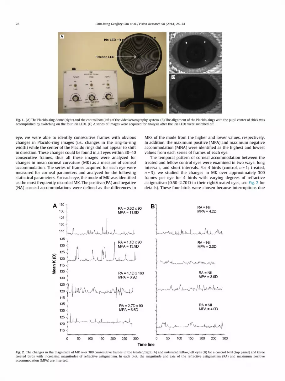
Fig. 1. (A) The Placido-ring dome (right) and the control box (left) of the videokeratography system. (B) The alignment of the Placido-rings with the pupil center of chick wasaccomplished by switching on the four iris LEDs. (C) A series of images were acquired for analysis after the iris LEDs were switched off.
28 Chin-hung Geoffrey Chu et al. / Vision Research 98 (2014) 26–34
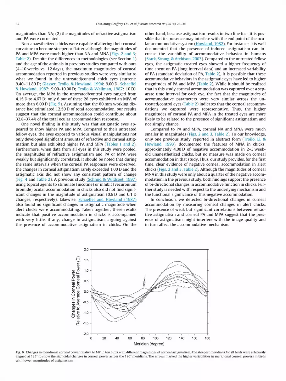
eye, we were able to identify consecutive frames with obviouschanges in Placido-ring images (i.e., changes in the ring-to-ringwidth) while the center of the Placido rings did not appear to shiftin direction. These changes could be found in all eyes within 30–40consecutive frames, thus all these images were analyzed forchanges in mean corneal curvature (MK) as a measure of cornealaccommodation. The series of frames acquired for each eye weremeasured for corneal parameters and analyzed for the followingstatistical parameters. For each eye, the mode of MK was identifiedas the most frequently recorded MK. The positive (PA) and negative(NA) corneal accommodations were defined as the differences in
Fig. 2. The changes in the magnitude of MK over 300 consecutive frames in the treatedtreated birds with increasing magnitudes of refractive astigmatism. In each plot, theaccommodation (MPA) are inserted.
MKs of the mode from the higher and lower values, respectively.In addition, the maximum positive (MPA) and maximum negativeaccommodation (MNA) were identified as the highest and lowestvalues from each series of frames of each eye.
The temporal pattern of corneal accommodation between thetreated and fellow control eyes were examined in two ways: longintervals, and short intervals. For 4 birds (control, n = 1; treated,n = 3), we studied the changes in MK over approximately 300frames per eye for 4 birds with varying degrees of refractiveastigmatism (0.50–2.70 D in their right/treated eyes, see Fig. 2 fordetails). These four birds were chosen because interruptions due
/right (A) and untreated fellow/left eyes (B) for a control bird (top panel) and threemagnitude and axis of the refractive astigmatism (RA) and maximum positive

Table 1Refractive errors measured after 1 week of treatment or at equivalent age (P12). Data are presented as mean ± SE, the range is presented in parentheses. Statistical significancebetween treated and fellow eyes is marked with asterisk �p < 0.05, and ���p < 0.001. M, spherical equivalent; MMM, most myopic meridian; RA, refractive astigmatism; R-J0,refractive J0; CA, corneal astigmatism.
Treated group (n = 16) Control group (n = 6)
Treated eye Fellow eye Right eye Left eye
M (D) �1.95 ± 1.55 +0.47 ± 0.19 +0.67 ± 0.32 +0.93 ± 0.36(�12.20 to +13.21) (�0.38 to +1.54) (�0.38 to +1.71) (�0.38 to +2.06)
MMM (D) �3.90 ± 1.58� +0.33 ± 0.19 �0.43 ± 0.32 +0.76 ± 0.40(�13.57 to +12.68) (�0.90 to +1.19) (�0.38 to +1.54) (�0.90 to +1.86)
RA (D) 3.14 ± 0.39��� 0.28 ± 0.14 0.47 ± 0.14 0.35 ± 0.18(1.05–6.58) (0.00–1.74) (0.00–1.05) (0.00–1.05)
R-J0 (D) �0.94 ± 0.35� �0.14 ± 0.07 �0.23 ± 0.07 �0.17 ± 0.09(�3.29 to 1.38) (�0.87 to 0.00) (�0.52 to 0.00) (�0.52 to 0.00)
CA (D) 1.53 ± 0.19��� 0.59 ± 0.08 0.57 ± 0.20 0.75 ± 0.19(0.47–3.09) (0.21–1.25) (0.19–1.48) (0.34–1.66)
Fig. 3. The frequency distributions of MK in the treated/right (dark bars) anduntreated fellow/left eyes (gray bars) for the four birds in Fig. 2. The modes ofaccommodation are marked with arrow heads.
Chin-hung Geoffrey Chu et al. / Vision Research 98 (2014) 26–34 29
to poor image quality, eye movement, and/or lid closure were min-imal over a long interval of consecutive frames. For another 18birds, data from shorter intervals (30–40 frames) were analyzed.
2.4. Data analysis
Statistical analysis was performed using SPSS 16.0 (SPSS Inc.,Illinois, USA). One-way ANOVAs were used to determine if therefractive and corneal parameters are significantly different acrossthe untreated fellow eyes of the treated birds, the right and the lefteyes of control birds. One-way ANOVA was also used to determineif there were significant differences in individual parameters acrossthe treated eyes of the treatment groups (i.e., crossed-cylinderlenses, spherical lenses, and slit apertures). Paired t-test was usedto determine the differences between the treated and untreatedfellow eyes in the treated birds. Pearson’s correlation analysesare used to determine if the magnitudes of corneal accommodationin the fellow eyes (i.e., right and left eyes) are correlated, as well aswhether the magnitudes of corneal accommodation and astigma-tism were correlated (i.e., right and left eyes of all birds). In alltests, significance level was set at 95% level of confidence. Unlessotherwise indicated, data are presented as mean and standard er-ror (mean ± SE).
3. Results
3.1. Effects of visual manipulations on refractive errors and cornealcurvature
Neither the refractive (M, MMM, MHM, RA, R-J0, and R-J45) northe corneal (MK, FK, SK, CA, C-J0, and C-J45) parameters were sig-nificantly different across the untreated fellow eyes of the treatedbirds, and the right and left eyes of the control birds (one-way AN-OVA, all p P 0.16). As summarized in Table 1, the treated eyes, as agroup, exhibited significantly higher MMM, RA, CA, and R-J0 whencompared to their fellow untreated eyes (paired t-tests, allp < 0.05); all other refractive (M, MHM, R-J45) and corneal (MK,FK, SK, C-J0, C-J45) parameters were not significantly different be-tween the treated and fellow untreated eyes in the treatmentgroups. The magnitudes of refractive and corneal astigmatism forall eyes as a group were significantly correlated (r = 0.69,p < 0.001). With respect to the refractive and corneal parametersin the treated eyes, only MK, FK, and SK showed significant treat-ment effects (one-way ANOVAs, all p < 0.02), with the eyes treatedwith spherical lenses (MK: 116.70 ± 2.60 D, FK: 115.95 ± 2.45 D,and SK: 117.50 ± 2.80 D) showing significantly flatter corneal cur-vature (Tukey’s pairwise tests, all p < 0.05) than those treated withcrossed-cylinder lenses (MK: 121.21 ± 0.64 D, FK: 120.52 ± 0.63 K,and SK: 122.03 ± 0.64 D) or slit apertures (MK: 121.92 ± 0.72 D,
FK: 121.480.84 D, and SK: 122.62 ± 0.76 D). However, note thatthere were only two birds treated with spherical lenses, a flattercorneal curvature was found in the +15 D treated eye (MK:114.1,FK:113.5, and SK:114.7) compared to the �15 D treated eye(MK:119.3, FK:118.4, and SK:120.3); thus the flatter corneal

Fig. 4. The frequency distributions of the changes in corneal astigmatic magnitude (A) and axis (B) for the four birds in Fig. 2. The astigmatic magnitude and axis for eachimage were subtracted by the modes of corresponding parameters.
30 Chin-hung Geoffrey Chu et al. / Vision Research 98 (2014) 26–34
curvature in this treatment group was mainly due to the +15 Dtreated eye.
3.2. Corneal accommodation
3.2.1. Longer interval (n = 4)Fig. 2 shows the temporal changes in MK over 300 consecutive
frames of the right (A) and left eyes (B) for a control bird (toprow) and three treated birds (bottom three rows, the right eyeswere the treated eyes). The sequence of birds was arranged fromtop to bottom according to the magnitude of refractive astigma-tism. As can be observed from this figure, the MK was frequentlymaintained at a particular level for all eyes, but both the treatedand fellow eyes clearly showed bi-directional changes in MK fromthis level. In general, the changes in MK usually took a longer dura-tion for positive (PA, about 200 ms) than negative accommodation(NA, about 100 ms), and the magnitudes of PA showed morevariability between fellow eyes (control: RE = +1.26 ± 0.20 D vs.LE = +1.20 ± 0.29 D; treated: RE = +2.24 ± 0.44 D vs. LE = +1.20 ±0.29 D) when compared to the NA of fellow eyes (control: RE =�0.33 ± 0.15 D vs. LE = �0.44 ± 0.18 D; treated: RE = �0.46 ± 0.5 D
vs. LE = �0.39 ± 0.11 D). On the other hand, although the MPA inthe four treated/right eyes (Fig. 2A) were all higher than those inthe untreated/left eyes (Fig. 2B), there were no correlations be-tween the magnitudes of MPA with RA or CA in these four birds.Fig. 3 compares the frequency distributions of MK between the fel-low eyes of the four birds; the sequence of birds followed that ofFig. 2. For all eight eyes, the modes of MK occupied 45 ± 4.6% (range:32.0–65.0%) of the time, and the deviations from the mode of MK(i.e., excluding the mode) were within 1.00 D in 25.2 ± 3.3% (range:12.0–36.0%) and 12.1 ± 3.2% (range: 4.7–28.0%) of the time for PAand NA, respectively.
Fig. 4 shows the frequency distributions of the changes in cor-neal astigmatic magnitude (A) and axis (B) for the four birds inthe same sequence as Figs. 2 and 3. These changes were calculatedby subtracting the modes of each parameter from the correspond-ing values. On average, the changes in corneal astigmatism duringthese intervals were within ±1.00 D for 99.1 ± 0.4% of the time(ranges: control/untreated fellow eyes: 99.0–100.0%; treated eyes:97.2–98.9%), indicating that under most circumstances the cornealastigmatism contributed to at most 0.50 D of changes in MK (since1.00 D cylindrical power = 0.50 D spherical-equivalent power). On

Table 2Corneal accommodation measured after 1 week of treatment or at equivalent age (P12). Data are presented as mean ± SE, the range is presented in parentheses. Statisticalsignificance between treated and untreated fellow eyes is marked with asterisk �p < 0.05, ��p < 0.01. PA, positive accommodation; PA S.D., standard deviation of positiveaccommodation; MPA, maximum positive accommodation; MNA, maximum negative accommodation; DCA, change in the magnitude of corneal astigmatism; DAxis of CA,change in the axis of corneal astigmatism; NA, negative accommodation; NA S.D., standard deviation of negative accommodation.
Treated group (n = 16) Control group (n = 6)
Treated eye Fellow eye Right eye Left eye
Positive accommodationPA (D) +2.24 ± 0.44� +1.26 ± 0.20 +1.19 ± 0.16 +1.20 ± 0.29
(0.46–7.88) (0.26–3.38) (0.66–1.81) (0.37–2.11)PA S.D. (D) 0.39 ± 0.07�� 0.23 ± 0.05 0.20 ± 0.04 0.26 ± 0.07
(0.07–1.18) (0.04–0.69) (0.10–0.34) (0.08–0.55)MPA (D) +7.53 ± 0.81�� +4.38 ± 0.53 +4.67 ± 1.47 +4.15 ± 1.16
(3.00–15.70) (1.70–9.40) (1.80–11.8) (1.90–9.70)DCA (D) 0.02 ± 0.16 �0.09 ± 0.07 �0.14 ± 005 �0.09 ± 0.14
(�1.37 to 1.20) (�0.82 to 0.40) (�0.28 to �0.02) (�0.69 to 0.13)DAxis of CA (�) 3.21 ± 3.49 �4.50 ± 5.79 �12.17 ± 10.89 �26.78 ± 9.48
(�14.40 to 36.00) (�53.00 to 38.00) (�61.00 to 4.00) (�59.00 to �1.70)
Negative accommodationNA (D) �0.46 ± 0.15 �0.33 ± 0.04 �0.39 ± 0.11 �0.44 ± 0.18
(�2.53 to �0.12) (�0.73 to �0.16) (�0.86 to �0.21) (�1.20 to �0.10)NA S.D. (D) 0.07 ± 0.02 0.06 ± 0.01 0.05 ± 0.02 0.06 ± 0.02
(0.00–0.24) (0.00–0.17) (0.02–0.09) (0.00–0.16)MNA (D) �0.92 ± 0.23 �0.73 ± 0.12 �0.73 ± 0.19 �0.87 ± 0.27
(�3.90 to �0.20) (�2.30 to �0.30) (�1.30 to �0.40) (�1.70 to �0.30)DCA (D) 0.09 ± 0.17 �0.14 ± 0.08 �0.09 ± 0.17 �0.15 + 0.18
(�0.75 to 2.12) (�1.02 to 0.33) (�0.75 to 0.27) (�0.36 to 0.31)DAxis of CA (�) 0.03 ± 2.64 �3.02 ± 4.58 �1.83 ± 12.78 7.67 ± 10.49
(�15.00 to 26.00) (�41.00 to 15.00) (�47.00 to 27.00) (�27.00 to 50.00)
Fig. 5. The maximum positive corneal accommodation is plotted as a function ofrefractive astigmatism for the treated (filled symbols) and untreated fellow/controleyes (open symbols). Low but significant correlation was found when all data werepooled. }, Crossed-cylinder lens; h, Spherical lens;4, Slit aperture; �, Control righteye; s, Control left eye. The four birds with data measured from longer intervals arelabeled with arrow heads ( ).
Chin-hung Geoffrey Chu et al. / Vision Research 98 (2014) 26–34 31
the other hand, the astigmatic axis changed by less than ±20� for75.2 ± 9.1% of the time, with more variation in the control/un-treated eyes than treated eyes (ranges: control/untreated felloweyes: 22.6–90.9%; treated eyes: 72.7–97.9%), probably due to thehigher instrumental noise when measuring eyes with low cornealastigmatism (see Appendix A and Fig. 6). Although significant cor-relations were found between the changes in MK and astigmaticaxis within the three right/treated eyes (i.e., the top three righteyes in Fig. 4B), the correlations were generally low and varied insign (Pearson’s r = �0.24, +0.24, �0.36, all p < 0.001), indicatingthat corneal accommodation was not correlated with a consistentpattern of change in the direction of the astigmatic axis.
3.2.2. Shorter interval (n = 22)Table 2 summarizes the magnitudes of corneal accommodative
changes as well as the corresponding changes (relative to the cor-responding modes) in astigmatic magnitude and axis. Except theNA in the fellow eyes of the treated group (r = 0.64, p < 0.01), nosignificant correlations between the fellow eyes were found in allother parameters for the treated and control groups (r = 0.08–0.69, all p P 0.10). Similar to the refractive status (Table 1), no sig-nificant difference in any of the corneal parameters was foundacross the untreated fellow eyes of the treated birds, and the rightand left eyes of the control birds (one-way ANOVAs, all p P 0.11).However, the PA (+2.24 D vs. 1.26 D, paired t-test, p < 0.05), stan-dard deviation of PA (0.39 D vs. 0.23 D, paired t-test, p < 0.01),and MPA (+7.53 D vs. +4.38 D, paired t-test, p < 0.01) were all sig-nificantly higher in the treated eyes when compared to their un-treated fellow eyes. In contrast, the NA, standard deviation of NA,and MNA were not significant different between the treated anduntreated fellow eyes (paired t-tests, all p P 0.29). One-way ANO-VAs showed that there was no treatment effect on any of the cor-neal accommodative changes (all p P 0.38). Interestingly, whendata from all eyes were pooled, both the PA and MPA were signif-icantly correlated with refractive (PA vs. RA: r = 0.34; MPA vs. RA:r = 0.34, both p < 0.05), but not corneal astigmatism (PA vs. CA:r = 0.13; MPA vs. CA: r = 0.10, both p P 0.41). Fig. 5 illustrates thelow but significant correlation between the MPA and refractiveastigmatism. On the other hand, PA was significantly correlatedwith NA (r = �0.67, p < 0.001), but there was no correlation be-tween MPA vs. MNA (r = �0.06, p = 0.71), MPA vs. M (MPA vs. M:r = �0.22, p = 0.16) or MNA vs. M (r = 0.08, p = 0.59), nor betweenthe maximum level of accommodation and the change in astig-matic axis (MPA vs. DAxis: r = �0.08, p = 0.60; MNA vs. DAxis:r = �0.03, p = 0.86; Table 2).
4. Discussion
The key findings of this study are: (1) both the control and trea-ted eyes in alert chicks demonstrated frequent increases (PA) anddecreases (NA) in corneal curvature, with PA showing much higher
32 Chin-hung Geoffrey Chu et al. / Vision Research 98 (2014) 26–34
magnitudes than NA; (2) the magnitudes of refractive astigmatismand PA were correlated.
Non-anaesthetized chicks were capable of altering their cornealcurvature to become steeper or flatter, although the magnitudes ofPA and MPA were much higher than NA and MNA (Figs. 2 and 3;Table 2). Despite the differences in methodologies (see Section 1)and the age of the animals in previous studies compared with ours(4–10 weeks vs. 12 days), the maximum magnitudes of cornealaccommodation reported in previous studies were very similar towhat we found in the untreated/control chick eyes (current:9.40–11.80 D; Glasser, Troilo, & Howland, 1994: 9.00 D; Schaeffel& Howland, 1987: 9.00–10.00 D; Troilo & Wallman, 1987: 10 D).On average, the MPA in the untreated/control eyes ranged from4.15 D to 4.67 D; only 6 out of these 28 eyes exhibited an MPA ofmore than 6.00 D (Fig. 5). Assuming that the 80 mm working dis-tance had stimulated 12.50 D of total accommodation, our resultssuggest that the corneal accommodation could contribute about32.8–37.4% of the total ocular accommodation response.
One novel finding in this study was that astigmatic eyes ap-peared to show higher PA and MPA. Compared to their untreatedfellow eyes, the eyes exposed to various visual manipulations notonly developed significant amounts of refractive and corneal astig-matism but also exhibited higher PA and MPA (Tables 1 and 2).Furthermore, when data from all eyes in this study were pooled,the magnitudes of refractive astigmatism and PA or MPA wereweakly but significantly correlated. It should be noted that duringthe same intervals when the corneal PA responses were observed,the changes in corneal astigmatism rarely exceeded 1.00 D and theastigmatic axis did not show any consistent pattern of change(Fig. 4 and Table 2). A previous study (Schmid & Wildsoet, 1997)using topical agents to stimulate (nicotine) or inhibit (vecuroniumbromide) ocular accommodation in chicks also did not find signif-icant changes in the magnitude of astigmatism (0.6 D and 0.1 Dchanges, respectively). Likewise, Schaeffel and Howland (1987)also found no significant changes in astigmatic magnitude whenalert chicks were accommodating. Taken together, these resultsindicate that positive accommodation in chicks is accompaniedwith very little, if any, change in astigmatism, arguing againstthe presence of accommodative astigmatism in chicks. On the
Fig. 6. Changes in meridional corneal power relative to MK in ten birds with different maaligned at 135� to show the sigmoidal changes in corneal power across the 180� meridianwith lower magnitudes of astigmatism.
other hand, because astigmatism results in two line foci, it is pos-sible that its presence may interfere with the end point of the ocu-lar accommodative system (Howland, 1982). For instance, it is welldocumented that the presence of induced astigmatism can in-crease the variability of accommodative behavior in humans(Stark, Strang, & Atchison, 2003). Compared to the untreated felloweyes, the astigmatic treated eyes showed a higher frequency oftime spent on PA (long interval data) and an increased variabilityof PA (standard deviation of PA, Table 2), it is possible that theseaccommodative behaviors in the astigmatic eyes have led to highermagnitudes of PA and MPA (Table 2). While it should be realizedthat in this study corneal accommodation was captured over a sep-arate time interval for each eye, the fact that the magnitudes ofaccommodative parameters were very similar across the un-treated/control eyes (Table 2) indicates that the corneal accommo-dations we captured were representative. Thus, the highermagnitudes of corneal PA and MPA in the treated eyes are morelikely to be related to the presence of significant astigmatism andnot simply chance.
Compared to PA and MPA, corneal NA and MNA were muchsmaller in magnitudes (Figs. 2 and 3, Table 2). To our knowledge,only one previous study, reported in abstract form (Troilo, Li, &Howland, 1993), documented the features of MNA in chicks;approximately 4.00 D of negative accommodation in 2–3 week-old unanesthetized chicks, but no measure was made on cornealaccommodation in that study. Thus, our study provides, for the firsttime, clear evidence of negative corneal accommodation in alertchicks (Figs. 2 and 3, Table 2). Although the magnitudes of cornealMNA in this study were only about a quarter of the negative accom-modation in the previous study, both findings support the presenceof bi-directional changes in accommodative function in chicks. Fur-ther study is needed with respect to the underlying mechanism andthe functional significance of this negative accommodation.
In conclusion, we detected bi-directional changes in cornealaccommodation by measuring corneal changes in alert chicks.The presence of weak but significant correlations between refrac-tive astigmatism and corneal PA and MPA suggest that the pres-ence of astigmatism might interfere with the image quality andin turn affect the accommodative mechanism.
gnitudes of corneal astigmatism. The steepest meridians for all birds were arbitrarilys. The arrows marked the higher variabilities in meridional corneal powers in birds

Fig. 7. Short-term repeatability of MK, FK, SK, CA, C-J0 and C-J45 for videokeratography in chicks. The averages and differences of the two sets of consecutive readings areplotted on the abscissa and ordinate, respectively. Solid line: mean difference; dashed lines: 95% limits of agreement.
Chin-hung Geoffrey Chu et al. / Vision Research 98 (2014) 26–34 33
Acknowledgments
Our sincere thanks to Mr. Chris Kuether from University ofHouston College of Optometry for designing and building theVKS system. We are also indebted to Dr. Jeremy Guggenheim(School of Optometry, The Hong Kong Polytechnic University) fora careful reading of early and final drafts.
Appendix A
A.1. Calibration of VKS
The radius of curvature was calibrated with five rustproof chro-mium steel balls (Grade 25, AISI 52100, USA) of known diametersthat cover a range of corneal radii in young chicks (5/3200
(3.97 mm), 3/1600 (4.76 mm), 7/3200 (5.56 mm), 1/400 (6.35 mm),and 9/3200 (7.14 mm)). A steel ball was fixed on a platform withits surface cleaned with alcohol before measurements. Five topo-graphic images of the steel balls were taken for each ball withre-focusing between measurements. Using a calibration curve(r2 = 0.99) compiled from the results of all steel balls, the cornealradius of curvature (r, measured in mm) was converted into thecorneal power (K, i.e., corneal curvature) using the formulaK = (n � 1)/r; where n = 1.369 is the corneal refractive index ofchicks (Mandelman & Sivak, 1983). To be able to analyze astig-matic cornea, we further derived six biometric parameters: SK,the meridian with steepest curvature; FK, the meridian with theflattest meridian; MK, the average value of SK and FK; cornealastigmatism (CA), the dioptric difference between SK and FK;C-J0 and C-J45, the power vectors calculated from the cornealastigmatic magnitude and axis (Thibos, Wheeler, & Horner, 1997).
Fig. 6 plots the changes in meridional corneal power with re-spect to MK of ten chicks who exhibited a range of corneal astig-matisms. As shown, the meridional corneal powers changedsmoothly through the 180� meridians, with the SK and FK sepa-rated by 90�, indicating that the corneal astigmatism found inchicks was due to a regular change in meridional corneal shape(i.e., regular astigmatism). Compared to birds with higher magni-tudes of astigmatism, those with lower magnitudes exhibited
slightly more variability in meridional corneal powers, probablydue to the relatively higher instrumental noise when measuringlower magnitudes of change.
Images were analyzed using a algorithm written in MatLab soft-ware. Specifically, each image was first processed to enhance therings’ regions using a Gabor filtering with an adaptive thresholdingstrategy. After these processed rings were identified in a coarse-to-fine fashion and labeled digitally, the radial distance of each ringfrom the origin was detected using the Hough transform (Bryan,2000; Duda & Hart, 1972). The radial distance was then smoothedusing a median filter and converted to radii using the method pro-posed by Carvalho et al. (2002). The radii within three pre-selectedareas, 1.50 mm, 2.10 mm and 2.80 mm diameters of the centralcornea, were segmented into 360 semi-meridians, summed, andaveraged for the conventional 180 meridians according to clinicalnotation.
The accuracy of the instrument for measuring the three centralcorneal areas (1.50 mm, 2.10 mm, and 2.80 mm diameter) weredetermined by calculating the difference of the measured valuesfrom the real values of three steel balls (2.78 mm, 3.18 mm,3.57 mm). Five images, separated by re-alignment and re-focusing,were acquired consecutively from each ball. The data of the fiveimages were averaged using power vector analysis (Thibos, Wheel-er, & Horner, 1997) and subtracted from the real values.
A.2. Reliability and repeatability
A.2.1. Steel ballsRepeated measures of the three steel balls showed that the
accuracy of the instrument (measured value minus real value)was 0.18 D for the largest tested areas (maximum differences:1.50 mm: 0.45 D, 2.1 mm: 0.32 D, and 2.80 mm: 0.18 D) in all sixcorneal parameters. There were no significant differences acrossthe three tested areas in MK, FK, and C-J0 astigmatic components.Although significant differences across the three tested areas werefound for corneal astigmatism, SK and C-J45 astigmatic compo-nents (one-way ANOVAs, all p < 0.001), Tukey’s post hoc tests (allp < 0.001) showed that the maximum differences between thetwo tested areas (1.50 mm vs. 2.80 mm) for astigmatism, SK and

34 Chin-hung Geoffrey Chu et al. / Vision Research 98 (2014) 26–34
C-J45 were only, respectively, 0.33 D, �0.32 D, and �0.17 D. Mea-surements of the astigmatic components showed a greater instru-mental noise for smaller tested area (maximum differences fromreal value among the three steel balls: 1.50 mm vs. 2.80 mm:CA = �0.45 D vs. �0.18 D; C-J0 = �0.02 D vs. �0.01 D; andC-J45 = �0.22 D vs. �0.09 D). Because of the higher accuracy andlower instrumental noise with wider tested area, only data of the2.80 mm diameter central cornea were used for the analyses in thisstudy.
A.2.2. Alert chicks eyesSix sets of images (50–100 images per set) were collected from
each of the treated eye for 12 birds from a separate experiment.These birds were treated monocularly with crossed-cylinder lensesand developed different degrees of corneal astigmatism (see CA re-sults in Fig. 7). Each set of images was separated by a re-alignmentwhich often took less than 2 min. From each set of data, one imagewith good quality was manually selected, i.e., there were siximages from each of the twelve eyes. To see if different numbersof images would affect the outcome measures, the mean valuesof 5 and 3 randomly selected images from each bird were com-pared. Because no significant differences were found between themeans of 5 vs. 3 images for all six corneal parameters (i.e., SK,FK, MK, CA, C-J0 & C-J45; all p P 0.78), the repeatability of theinstrument was tested by comparing the means from the firstand second sets of 3 images.
The Bland–Altman plots in Fig. 7 illustrate the repeatability ofthe six corneal parameters for these 12 treated birds. As reflectedfrom the distributions of the six parameters, the crossed-cylinderlens treatment produced a wide range of corneal curvature andastigmatic components. Despite this significant treatment effect,the mean differences and 95% limits of agreement (in parentheses)for the six parameters were small: MK, �0.02 D (+0.25, �0.25); SK,�0.03 D (+0.26, �0.26); FK, �0.01 D (+0.25, �0.25); CA, 0.02 D(+0.21, �0.21); C-J0, 0.00 D (+0.24, �0.24); and C-J45, 0.01 D(+0.29, �0.29). In addition, there were no systematic changesacross the dioptric ranges measured in all six parameters, and83% of the repeated measurements differed by less than 0.25 D.
References
Bannon, R. E. (1946). A study of astigmatism at the near point with special referenceto astigmatic accommodation. American Journal of Optometry and Archives ofAmerican Academy of Optometry, 23, 53–75.
Beer, T. (1892). Studien über die accommodation des vogelauges. Pflügers ArchivEuropean Journal of Physiology, 53, 175–237.
Bryan, S. M. (2000). Lecture 15: Segmentation. Lecture notes. Brigham YoungUniversity.
Buehren, T., Collins, M. J., & Carney, L. (2003). Corneal aberrations and reading.Optometry & Vision Science, 80, 159–166.
Campbell, M. C., Bunghardt, K., Kisilak, M., & Irving, E. L. (2008). Diurnal rhythms ofrefractive error components in normal chick. Journal of Vision, 8 [Article 48]http://www.journalofvision.org/content/8/17/48.abstract.
Carvalho, L. A., Romão, A. C., Tonissi, S., Yasuoka, F., Castro, J. C., Schor, P., et al.(2002). Videokeratograph (VKS) for monitoring corneal curvature duringsurgery. Arquivos Brasileiros De Oftalmologia, 65, 37–41.
Chu, C. H., Deng, L., & Kee, C. S. (2012). Effects of hemiretinal form deprivation oncentral refractive development and posterior eye shape in chicks. VisionResearch, 55, 24–31.
Duda, R. O., & Hart, P. E. (1972). Use of the Hough transformation to detect lines andcurves in pictures. Communications of the ACM, 15, 11–15.
García de la Cera, E., Rodríguez, G., de Castro, A., Merayo, J., & Marcos, S. (2007).Emmetropization and optical aberrations in a myopic corneal refractive surgerychick model. Vision Research, 47, 2465–2472.
Glasser, A., Troilo, D., & Howland, H. C. (1994). The mechanism of cornealaccommodation in chicks. Vision Research, 34, 1549–1566.
He, J. C., Gwiazda, J. E., Thorn, F., Held, R., & Huang, W. (2003). Change in cornealshape and corneal wave-front aberrations with accommodation. Journal ofVision, 3, 456–463.
Howland, H. C. (1982). Infant eyes: Optics and accommodation. Current EyeResearch, 2, 217–224.
Irving, E. L., Sivak, J. G., & Callender, M. G. (1992). Refractive plasticity of thedeveloping chick eye. Ophthalmic and Physiological Optics, 12, 448–456.
Johnson, C. A., Lytle, G., Troilo, D., & Nickla, D. L. (2004). Chick eyes show a diurnalrhythm in refractive error. ARVO Meeting Abstracts, 45, 4295.
Kee, C. S. (2013). Astigmatism and its role in emmetropization. Experimental EyeResearch, 114, 89–95.
Kee, C. S., & Deng, L. (2008). Astigmatism associated with experimentally inducedmyopia or hyperopia in chickens. Investigative Ophthalmology & Visual Science,49, 858–867.
Kee, C. S., Hung, L. F., Qiao, G. Y., Ramamirtham, R., & Smith, I. E. L. (2005).Astigmatism in monkeys with experimentally induced myopia or hyperopia.Optometry & Vision Science, 82, 248–260.
Mandelman, T., & Sivak, J. G. (1983). Longitudinal chromatic aberration of thevertebrate eye. Vision Research, 23, 1555–1559.
Murphy, C. J., Glasser, A., & Howland, H. C. (1995). The anatomy of the ciliary regionof the chicken eye. Investigative Ophthalmology & Visual Science, 36, 889–896.
Ostrin, L. A., Liu, Y., Choh, V., & Wildsoet, C. F. (2011). The role of the iris in chickaccommodation. Investigative Ophthalmology & Visual Science, 52, 4710–4716.
Pierscionek, B. K., Popiolek-Masajada, A., & Kasprzak, H. (2001). Corneal shapechange during accommodation. Eye (London, England), 15, 766–769.
Read, S. A., Buehren, T., & Collins, M. J. (2007). Influence of accommodation on theanterior and posterior cornea. Journal of Cataract & Refractive Surgery, 33,1877–1885.
Read, S. A., Collins, M. J., & Carney, L. G. (2007). A review of astigmatism and itspossible genesis. Clinical and Experimental Optometry, 90, 5–19.
Rosenfield, M., & Gilmartin, B. (1987). Beta-adrenergic receptor antagonism inmyopia. Ophthalmic and Physiological Optics, 7, 359–364.
Schaeffel, F., Glasser, A., & Howland, H. C. (1988). Accommodation, refractive errorand eye growth in chickens. Vision Research, 28, 639–657.
Schaeffel, F., & Howland, H. C. (1987). Corneal accommodation in chick and pigeon.Journal of Comparative Physiology A: Neuroethology, Sensory, Neural, andBehavioral Physiology, 160, 375–384.
Schmid, K. L., & Wildsoet, C. F. (1997). Natural and imposed astigmatism and theirrelation to emmetropization in the chick. Experimental Eye Research, 64,837–847.
Sivak, J. G., Hildebrand, T. E., Lebert, C. G., Myshak, L. M., & Ryall, L. A. (1986). Ocularaccommodation in chickens: Corneal vs lenticular accommodation and effect ofage. Vision Research, 26, 1865–1872.
Stark, L. R., Strang, N. C., & Atchison, D. A. (2003). Dynamic accommodationresponse in the presence of astigmatism. Journal of the Optical Society of AmericaA, 20, 2228–2236.
Thibos, L. N., Wheeler, W., & Horner, D. (1997). Power vectors: An application ofFourier analysis to the description and statistical analysis of refractive error.Optometry & Vision Science, 74, 367–375.
Troilo, D., & Judge, S. J. (1993). Ocular development and visual deprivation myopiain the common marmoset (Callithrix jacchus). Vision Research, 33, 1311–1324.
Troilo, D., Li, T., & Howland, H. C. (1993). Negative accommodation occurs in thechick and may be mediated by sympathetic input. Investigative Ophthalmology &Visual Science, 34, 1310 [ARVO abstract No. 2990].
Troilo, D., & Wallman, J. (1985). Mechanisms of accommodation in the chicken. In15th Annual meeting. soc. neurosci. (Vol. 2, p. 1042).
Troilo, D., & Wallman, J. (1987). Changes in corneal curvature duringaccommodation in chicks. Vision Research, 27, 241–247.
Walls, G. L. (1942). The vertebrate eye and its adaptive radiation. Bloomfield Hills,Michigan: The Cranbrook Press.
Xu, N., Kee, C. S., Zhou, Y., Zheng, Y., & Liu, L. (2009). Repeatability of a videokeratography system specially designed for measuring corneal astigmatism inanimals with small eyes. Sheng Wu Yi Xue Gong Cheng Xue Za Zhi, 26(5),978–988.
Yasuda, A., & Yamaguchi, T. (2005). Steepening of corneal curvature withcontraction of the ciliary muscle. Journal of Cataract & Refractive Surgery, 31,1177–1181.
Yasuda, A., Yamaguchi, T., & Ohkoshi, K. (2003). Changes in corneal curvature inaccommodation. Journal of Cataract & Refractive Surgery, 29, 1297–1301.


