Embed Size (px)
Citation preview
BI 102 General Biology
Collection Editor:Lena Green

BI 102 General Biology
Collection Editor:Lena Green
Authors:OpenStax
Lena Green
Online:< http://cnx.org/content/col26253/1.2/ >

This selection and arrangement of content as a collection is copyrighted by Lena Green. It is licensed under the
Creative Commons Attribution License 4.0 (http://creativecommons.org/licenses/by/4.0/).
Collection structure revised: January 5, 2019
PDF generated: January 16, 2022
For copyright and attribution information for the modules contained in this collection, see p. 396.
Table of Contents
1 Plant Tissues1.1 The Plant Body . . . . . . . . . . . . . . . . . . . . . . . . . . . . . . . . . . . . . . . . . . . . . . . . . . . . . . . . . . . . . . . . . . . . . . . . . . . . . 11.2 Stems . . . . . . . . . . . . . . . . . . . . . . . . . . . . . . . . . . . . . . . . . . . . . . . . . . . . . . . . . . . . . . . . . . . . . . . . . . . . . . . . . . . . . . . 51.3 Roots . . . . . . . . . . . . . . . . . . . . . . . . . . . . . . . . . . . . . . . . . . . . . . . . . . . . . . . . . . . . . . . . . . . . . . . . . . . . . . . . . . . . . . 191.4 Leaves . . . . . . . . . . . . . . . . . . . . . . . . . . . . . . . . . . . . . . . . . . . . . . . . . . . . . . . . . . . . . . . . . . . . . . . . . . . . . . . . . . . . . 26Solutions . . . . . . . . . . . . . . . . . . . . . . . . . . . . . . . . . . . . . . . . . . . . . . . . . . . . . . . . . . . . . . . . . . . . . . . . . . . . . . . . . . . . . . . . 38
2 Plant Transport
2.1 The Soil and Plant Nutrition . . . . . . . . . . . . . . . . . . . . . . . . . . . . . . . . . . . . . . . . . . . . . . . . . . . . . . . . . . . . . . . 412.2 Transport of Water and Solutes in Plants . . . . . . . . . . . . . . . . . . . . . . . . . . . . . . . . . . . . . . . . . . . . . . . . . . . 48Solutions . . . . . . . . . . . . . . . . . . . . . . . . . . . . . . . . . . . . . . . . . . . . . . . . . . . . . . . . . . . . . . . . . . . . . . . . . . . . . . . . . . . . . . . . 61
3 Animal Organization
3.1 Animal Form and Function . . . . . . . . . . . . . . . . . . . . . . . . . . . . . . . . . . . . . . . . . . . . . . . . . . . . . . . . . . . . . . . . . 633.2 Animal Primary Tissues . . . . . . . . . . . . . . . . . . . . . . . . . . . . . . . . . . . . . . . . . . . . . . . . . . . . . . . . . . . . . . . . . . . . 713.3 Homeostasis . . . . . . . . . . . . . . . . . . . . . . . . . . . . . . . . . . . . . . . . . . . . . . . . . . . . . . . . . . . . . . . . . . . . . . . . . . . . . . . . 88Solutions . . . . . . . . . . . . . . . . . . . . . . . . . . . . . . . . . . . . . . . . . . . . . . . . . . . . . . . . . . . . . . . . . . . . . . . . . . . . . . . . . . . . . . . . 96
4 Nervous System
4.1 Nervous System . . . . . . . . . . . . . . . . . . . . . . . . . . . . . . . . . . . . . . . . . . . . . . . . . . . . . . . . . . . . . . . . . . . . . . . . . . . . 99Solutions . . . . . . . . . . . . . . . . . . . . . . . . . . . . . . . . . . . . . . . . . . . . . . . . . . . . . . . . . . . . . . . . . . . . . . . . . . . . . . . . . . . . . . . 112
5 Sensory Perception
5.1 Sensory Processes . . . . . . . . . . . . . . . . . . . . . . . . . . . . . . . . . . . . . . . . . . . . . . . . . . . . . . . . . . . . . . . . . . . . . . . . . 1135.2 Somatosensation . . . . . . . . . . . . . . . . . . . . . . . . . . . . . . . . . . . . . . . . . . . . . . . . . . . . . . . . . . . . . . . . . . . . . . . . . . 1205.3 Vision . . . . . . . . . . . . . . . . . . . . . . . . . . . . . . . . . . . . . . . . . . . . . . . . . . . . . . . . . . . . . . . . . . . . . . . . . . . . . . . . . . . . . 1265.4 Hearing and Vestibular Sensation . . . . . . . . . . . . . . . . . . . . . . . . . . . . . . . . . . . . . . . . . . . . . . . . . . . . . . . . . . 1365.5 Taste and Smell . . . . . . . . . . . . . . . . . . . . . . . . . . . . . . . . . . . . . . . . . . . . . . . . . . . . . . . . . . . . . . . . . . . . . . . . . . . 144Solutions . . . . . . . . . . . . . . . . . . . . . . . . . . . . . . . . . . . . . . . . . . . . . . . . . . . . . . . . . . . . . . . . . . . . . . . . . . . . . . . . . . . . . . . 151
6 Structural Support and Movement
6.1 Types of Skeletal Systems . . . . . . . . . . . . . . . . . . . . . . . . . . . . . . . . . . . . . . . . . . . . . . . . . . . . . . . . . . . . . . . . . 1536.2 Bone . . . . . . . . . . . . . . . . . . . . . . . . . . . . . . . . . . . . . . . . . . . . . . . . . . . . . . . . . . . . . . . . . . . . . . . . . . . . . . . . . . . . . . 1606.3 Muscle Contraction and Locomotion . . . . . . . . . . . . . . . . . . . . . . . . . . . . . . . . . . . . . . . . . . . . . . . . . . . . . . . 171Solutions . . . . . . . . . . . . . . . . . . . . . . . . . . . . . . . . . . . . . . . . . . . . . . . . . . . . . . . . . . . . . . . . . . . . . . . . . . . . . . . . . . . . . . . 182
7 Digestive System
7.1 Digestive Systems . . . . . . . . . . . . . . . . . . . . . . . . . . . . . . . . . . . . . . . . . . . . . . . . . . . . . . . . . . . . . . . . . . . . . . . . . 1857.2 Nutrition and Energy Production . . . . . . . . . . . . . . . . . . . . . . . . . . . . . . . . . . . . . . . . . . . . . . . . . . . . . . . . . . 2027.3 Digestive System Processes . . . . . . . . . . . . . . . . . . . . . . . . . . . . . . . . . . . . . . . . . . . . . . . . . . . . . . . . . . . . . . . . 212Solutions . . . . . . . . . . . . . . . . . . . . . . . . . . . . . . . . . . . . . . . . . . . . . . . . . . . . . . . . . . . . . . . . . . . . . . . . . . . . . . . . . . . . . . . 221
8 Circulatory System
8.1 Overview of the Circulatory System . . . . . . . . . . . . . . . . . . . . . . . . . . . . . . . . . . . . . . . . . . . . . . . . . . . . . . . 2238.2 Components of the Blood . . . . . . . . . . . . . . . . . . . . . . . . . . . . . . . . . . . . . . . . . . . . . . . . . . . . . . . . . . . . . . . . . 2288.3 Mammalian Heart and Blood Vessels . . . . . . . . . . . . . . . . . . . . . . . . . . . . . . . . . . . . . . . . . . . . . . . . . . . . . . 2358.4 Blood Flow and Blood Pressure Regulation . . . . . . . . . . . . . . . . . . . . . . . . . . . . . . . . . . . . . . . . . . . . . . . . 246Solutions . . . . . . . . . . . . . . . . . . . . . . . . . . . . . . . . . . . . . . . . . . . . . . . . . . . . . . . . . . . . . . . . . . . . . . . . . . . . . . . . . . . . . . . 251
9 Respiration
9.1 Systems of Gas Exchange . . . . . . . . . . . . . . . . . . . . . . . . . . . . . . . . . . . . . . . . . . . . . . . . . . . . . .. . . . . . . . . . . . 2539.2 Gas Exchange across Respiratory Surfaces . . . . . . . . . . . . . . . . . . . . . . . . . . . . . . . . . . . . . . . . . . . . . . . . . 265
iv
9.3 Breathing . . . . . . . . . . . . . . . . . . . . . . . . . . . . . . . . . . . . . . . . . . . . . . . . . . . . . . . . . . . . . . . . . . . . . . . . . . . . . . . . . 2729.4 Transport of Gases in Human Bodily Fluids . . . . . . . . . . . . . . . . . . . . . . . . . . . . . . . . . . . . . . . . . . . . . . . 278Solutions . . . . . . . . . . . . . . . . . . . . . . . . . . . . . . . . . . . . . . . . . . . . . . . . . . . . . . . . . . . . . . . . . . . . . . . . . . . . . . . . . . . . . . . 285
10 Immunity
10.1 Innate Immune Response . . . . . . . . . . . . . . . . . . . . . . . . . . . . . . . . . . . . . . . . . . . . . . . . . . . . .. . . . . . . . . . . . 28710.2 Adaptive Immune Response . . . . . . . . . . . . . . . . . . . . . . . . . . . . . . . . . . . . . . . . . . . . . . . . . . . . . . . . . . . . . . 29610.3 Disruptions in the Immune System . . . . . . . . . . . . . . . . . . . . . . . . . . . . . . . . . . . . . . . . . . . . . . . . . . . . . . . 315Solutions . . . . . . . . . . . . . . . . . . . . . . . . . . . . . . . . . . . . . . . . . . . . . . . . . . . . . . . . . . . . . . . . . . . . . . . . . . . . . . . . . . . . . . . 320
11 Reproduction and Development
11.1 How Animals Reproduce . . . . . . . . . . . . . . . . . . . . . . . . . . . . . . . . . . . . . . . . . . . . . . . . . . . . . . . . . . . . . . . . . 32311.2 Development and Organogenesis . . . . . . . . . . . . . . . . . . . . . . . . . . . . . . . . . . . . . . . . . . . . . . . . . . . . . . . . . 33011.3 Human Reproduction . . . . . . . . . . . . . . . . . . . . . . . . . . . . . . . . . . . . . . . . . . . . . . . . . . . . . . . . . . . . . . . . . . . . 334Solutions . . . . . . . . . . . . . . . . . . . . . . . . . . . . . . . . . . . . . . . . . . . . . . . . . . . . . . . . . . . . . . . . . . . . . . . . . . . . . . . . . . . . . . . 349
Glossary . . . . . . . . . . . . . . . . . . . . . . . . . . . . . . . . . . . . . . . . . . . . . . . . . . . . . . . . . . . . . . . . . . . . . . . . . . . . . . . . . . . . . . . . . . . . 351Index . . . . . . . . . . . . . . . . . . . . . . . . . . . . . . . . . . . . . . . . . . . . . . . . . . . . . . . . . . . . . . . . . . . . . . . . . . . . . . . . . . . . . . . . . . . . . . . 385Attributions . . . . . . . . . . . . . . . . . . . . . . . . . . . . . . . . . . . . . . . . . . . . . . . . . . . . . . . . . . . . . . . . . . . . . . . . . . . . . . . . . . . . . . . .396
Available for free at Connexions <http://cnx.org/content/col26253/1.2>
Chapter 1
Plant Tissues
1.1 The Plant Body1
Like animals, plants contain cells with organelles in which speci�c metabolic activities take place. Unlikeanimals, however, plants use energy from sunlight to form sugars during photosynthesis. In addition, plantcells have cell walls, plastids, and a large central vacuole: structures that are not found in animal cells. Eachof these cellular structures plays a speci�c role in plant structure and function.
: Watch BotanyWithout Borders2 , a video produced by the BotanicalSociety of America about the importance of plants.
1.1.1 Plant Organ Systems
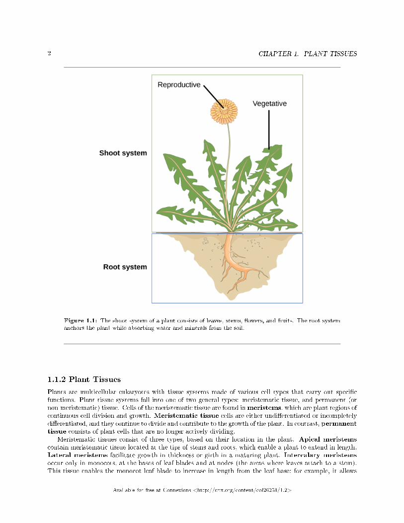
In plants, just as in animals, similar cells working together form a tissue. When di�erent types of tissues worktogether to perform a unique function, they form an organ; organs working together form organ systems.Vascular plants have two distinct organ systems: a shoot system, and a root system. The shoot systemconsists of two portions: the vegetative (non-reproductive) parts of the plant, such as the leaves and thestems, and the reproductive parts of the plant, which include �owers and fruits. The shoot system generallygrows above ground, where it absorbs the light needed for photosynthesis. The root system, which supportsthe plants and absorbs water and minerals, is usually underground. Figure 1.1 shows the organ systems ofa typical plant.
1This content is available online at <http://cnx.org/content/m44700/1.9/>.2http://openstaxcollege.org/l/botany_wo_bord
Available for free at Connexions <http://cnx.org/content/col26253/1.2>
1
2 CHAPTER 1. PLANT TISSUES
Figure 1.1: The shoot system of a plant consists of leaves, stems, �owers, and fruits. The root systemanchors the plant while absorbing water and minerals from the soil.
1.1.2 Plant Tissues
Plants are multicellular eukaryotes with tissue systems made of various cell types that carry out speci�cfunctions. Plant tissue systems fall into one of two general types: meristematic tissue, and permanent (ornon-meristematic) tissue. Cells of the meristematic tissue are found inmeristems, which are plant regions ofcontinuous cell division and growth. Meristematic tissue cells are either undi�erentiated or incompletelydi�erentiated, and they continue to divide and contribute to the growth of the plant. In contrast, permanenttissue consists of plant cells that are no longer actively dividing.
Meristematic tissues consist of three types, based on their location in the plant. Apical meristemscontain meristematic tissue located at the tips of stems and roots, which enable a plant to extend in length.Lateral meristems facilitate growth in thickness or girth in a maturing plant. Intercalary meristemsoccur only in monocots, at the bases of leaf blades and at nodes (the areas where leaves attach to a stem).This tissue enables the monocot leaf blade to increase in length from the leaf base; for example, it allows
Available for free at Connexions <http://cnx.org/content/col26253/1.2>
3
lawn grass leaves to elongate even after repeated mowing.Meristems produce cells that quickly di�erentiate, or specialize, and become permanent tissue. Such
cells take on speci�c roles and lose their ability to divide further. They di�erentiate into three main types:dermal, vascular, and ground tissue. Dermal tissue covers and protects the plant, and vascular tissuetransports water, minerals, and sugars to di�erent parts of the plant. Ground tissue serves as a site forphotosynthesis, provides a supporting matrix for the vascular tissue, and helps to store water and sugars.
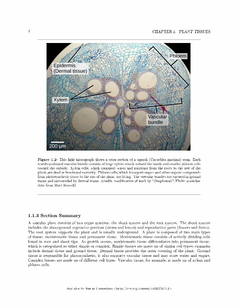
Secondary tissues are either simple (composed of similar cell types) or complex (composed of di�erent celltypes). Dermal tissue, for example, is a simple tissue that covers the outer surface of the plant and controlsgas exchange. Vascular tissue is an example of a complex tissue, and is made of two specialized conductingtissues: xylem and phloem. Xylem tissue transports water and nutrients from the roots to di�erent parts ofthe plant, and includes three di�erent cell types: vessel elements and tracheids (both of which conduct water),and xylem parenchyma. Phloem tissue, which transports organic compounds from the site of photosynthesisto other parts of the plant, consists of four di�erent cell types: sieve cells (which conduct photosynthates),companion cells, phloem parenchyma, and phloem �bers. Unlike xylem conducting cells, phloem conductingcells are alive at maturity. The xylem and phloem always lie adjacent to each other (Figure 1.2). In stems,the xylem and the phloem form a structure called a vascular bundle; in roots, this is termed the vascularstele or vascular cylinder.
Available for free at Connexions <http://cnx.org/content/col26253/1.2>
4 CHAPTER 1. PLANT TISSUES
Figure 1.2: This light micrograph shows a cross section of a squash (Cucurbita maxima) stem. Eachteardrop-shaped vascular bundle consists of large xylem vessels toward the inside and smaller phloem cellstoward the outside. Xylem cells, which transport water and nutrients from the roots to the rest of theplant, are dead at functional maturity. Phloem cells, which transport sugars and other organic compoundsfrom photosynthetic tissue to the rest of the plant, are living. The vascular bundles are encased in groundtissue and surrounded by dermal tissue. (credit: modi�cation of work by "(biophotos)"/Flickr; scale-bardata from Matt Russell)
1.1.3 Section Summary
A vascular plant consists of two organ systems: the shoot system and the root system. The shoot systemincludes the aboveground vegetative portions (stems and leaves) and reproductive parts (�owers and fruits).The root system supports the plant and is usually underground. A plant is composed of two main typesof tissue: meristematic tissue and permanent tissue. Meristematic tissue consists of actively dividing cellsfound in root and shoot tips. As growth occurs, meristematic tissue di�erentiates into permanent tissue,which is categorized as either simple or complex. Simple tissues are made up of similar cell types; examplesinclude dermal tissue and ground tissue. Dermal tissue provides the outer covering of the plant. Groundtissue is responsible for photosynthesis; it also supports vascular tissue and may store water and sugars.Complex tissues are made up of di�erent cell types. Vascular tissue, for example, is made up of xylem andphloem cells.
Available for free at Connexions <http://cnx.org/content/col26253/1.2>
5
1.1.4 Review Questions
Exercise 1.1.1 (Solution on p. 38.)
Plant regions of continuous growth are made up of ________.
a. dermal tissueb. vascular tissuec. meristematic tissued. permanent tissue
Exercise 1.1.2 (Solution on p. 38.)
Which of the following is the major site of photosynthesis?
a. apical meristemb. ground tissuec. xylem cellsd. phloem cells
1.1.5 Free Response
Exercise 1.1.3 (Solution on p. 38.)
What type of meristem is found only in monocots, such as lawn grasses? Explain how this type ofmeristematic tissue is bene�cial in lawn grasses that are mowed each week.
Exercise 1.1.4 (Solution on p. 38.)
Which plant part is responsible for transporting water, minerals, and sugars to di�erent parts ofthe plant? Name the two types of tissue that make up this overall tissue, and explain the role ofeach.
1.2 Stems3
Stems are a part of the shoot system of a plant. They may range in length from a few millimeters to hundredsof meters, and also vary in diameter, depending on the plant type. Stems are usually above ground, althoughthe stems of some plants, such as the potato, also grow underground. Stems may be herbaceous (soft) orwoody in nature. Their main function is to provide support to the plant, holding leaves, �owers and buds;in some cases, stems also store food for the plant. A stem may be unbranched, like that of a palm tree, or itmay be highly branched, like that of a magnolia tree. The stem of the plant connects the roots to the leaves,helping to transport absorbed water and minerals to di�erent parts of the plant. It also helps to transportthe products of photosynthesis, namely sugars, from the leaves to the rest of the plant.
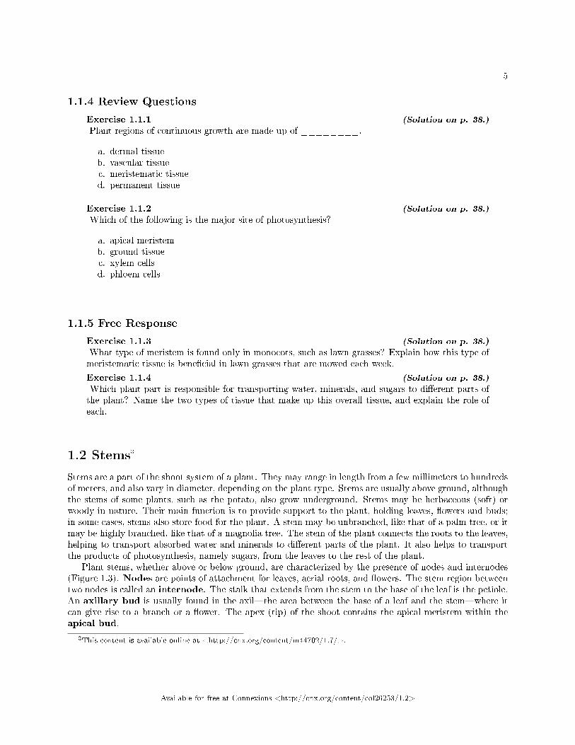
Plant stems, whether above or below ground, are characterized by the presence of nodes and internodes(Figure 1.3). Nodes are points of attachment for leaves, aerial roots, and �owers. The stem region betweentwo nodes is called an internode. The stalk that extends from the stem to the base of the leaf is the petiole.An axillary bud is usually found in the axil�the area between the base of a leaf and the stem�where itcan give rise to a branch or a �ower. The apex (tip) of the shoot contains the apical meristem within theapical bud.
3This content is available online at <http://cnx.org/content/m44702/1.7/>.
Available for free at Connexions <http://cnx.org/content/col26253/1.2>
6 CHAPTER 1. PLANT TISSUES
Figure 1.3: Leaves are attached to the plant stem at areas called nodes. An internode is the stemregion between two nodes. The petiole is the stalk connecting the leaf to the stem. The leaves just abovethe nodes arose from axillary buds.
1.2.1 Stem Anatomy
The stem and other plant organs arise from the ground tissue, and are primarily made up of simple tissuesformed from three types of cells: parenchyma, collenchyma, and sclerenchyma cells.
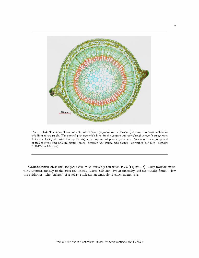
Parenchyma cells are the most common plant cells (Figure 1.4). They are found in the stem, the root,the inside of the leaf, and the pulp of the fruit. Parenchyma cells are responsible for metabolic functions,such as photosynthesis, and they help repair and heal wounds. Some parenchyma cells also store starch.
Available for free at Connexions <http://cnx.org/content/col26253/1.2>
7
Figure 1.4: The stem of common St John's Wort (Hypericum perforatum) is shown in cross section inthis light micrograph. The central pith (greenish-blue, in the center) and peripheral cortex (narrow zone3�5 cells thick just inside the epidermis) are composed of parenchyma cells. Vascular tissue composedof xylem (red) and phloem tissue (green, between the xylem and cortex) surrounds the pith. (credit:Rolf-Dieter Mueller)
Collenchyma cells are elongated cells with unevenly thickened walls (Figure 1.5). They provide struc-tural support, mainly to the stem and leaves. These cells are alive at maturity and are usually found belowthe epidermis. The �strings� of a celery stalk are an example of collenchyma cells.
Available for free at Connexions <http://cnx.org/content/col26253/1.2>

8 CHAPTER 1. PLANT TISSUES
Figure 1.5: Collenchyma cell walls are uneven in thickness, as seen in this light micrograph. Theyprovide support to plant structures. (credit: modi�cation of work by Carl Szczerski; scale-bar data fromMatt Russell)
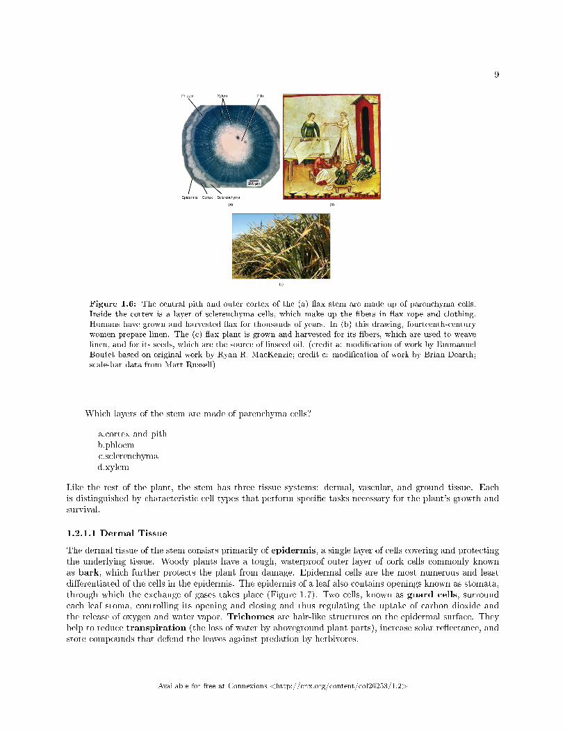
Sclerenchyma cells also provide support to the plant, but unlike collenchyma cells, many of them aredead at maturity. There are two types of sclerenchyma cells: �bers and sclereids. Both types have secondarycell walls that are thickened with deposits of lignin, an organic compound that is a key component of wood.Fibers are long, slender cells; sclereids are smaller-sized. Sclereids give pears their gritty texture. Humansuse sclerenchyma �bers to make linen and rope (Figure 1.6).
:
Available for free at Connexions <http://cnx.org/content/col26253/1.2>
9
Figure 1.6: The central pith and outer cortex of the (a) �ax stem are made up of parenchyma cells.Inside the cortex is a layer of sclerenchyma cells, which make up the �bers in �ax rope and clothing.Humans have grown and harvested �ax for thousands of years. In (b) this drawing, fourteenth-centurywomen prepare linen. The (c) �ax plant is grown and harvested for its �bers, which are used to weavelinen, and for its seeds, which are the source of linseed oil. (credit a: modi�cation of work by EmmanuelBoutet based on original work by Ryan R. MacKenzie; credit c: modi�cation of work by Brian Dearth;scale-bar data from Matt Russell)
Which layers of the stem are made of parenchyma cells?
a.cortex and pithb.phloemc.sclerenchymad.xylem
Like the rest of the plant, the stem has three tissue systems: dermal, vascular, and ground tissue. Eachis distinguished by characteristic cell types that perform speci�c tasks necessary for the plant's growth andsurvival.
1.2.1.1 Dermal Tissue
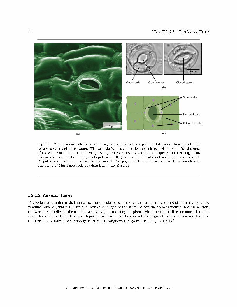
The dermal tissue of the stem consists primarily of epidermis, a single layer of cells covering and protectingthe underlying tissue. Woody plants have a tough, waterproof outer layer of cork cells commonly knownas bark, which further protects the plant from damage. Epidermal cells are the most numerous and leastdi�erentiated of the cells in the epidermis. The epidermis of a leaf also contains openings known as stomata,through which the exchange of gases takes place (Figure 1.7). Two cells, known as guard cells, surroundeach leaf stoma, controlling its opening and closing and thus regulating the uptake of carbon dioxide andthe release of oxygen and water vapor. Trichomes are hair-like structures on the epidermal surface. Theyhelp to reduce transpiration (the loss of water by aboveground plant parts), increase solar re�ectance, andstore compounds that defend the leaves against predation by herbivores.
Available for free at Connexions <http://cnx.org/content/col26253/1.2>
10 CHAPTER 1. PLANT TISSUES
Figure 1.7: Openings called stomata (singular: stoma) allow a plant to take up carbon dioxide andrelease oxygen and water vapor. The (a) colorized scanning-electron micrograph shows a closed stomaof a dicot. Each stoma is �anked by two guard cells that regulate its (b) opening and closing. The(c) guard cells sit within the layer of epidermal cells (credit a: modi�cation of work by Louisa Howard,Rippel Electron Microscope Facility, Dartmouth College; credit b: modi�cation of work by June Kwak,University of Maryland; scale-bar data from Matt Russell)
1.2.1.2 Vascular Tissue
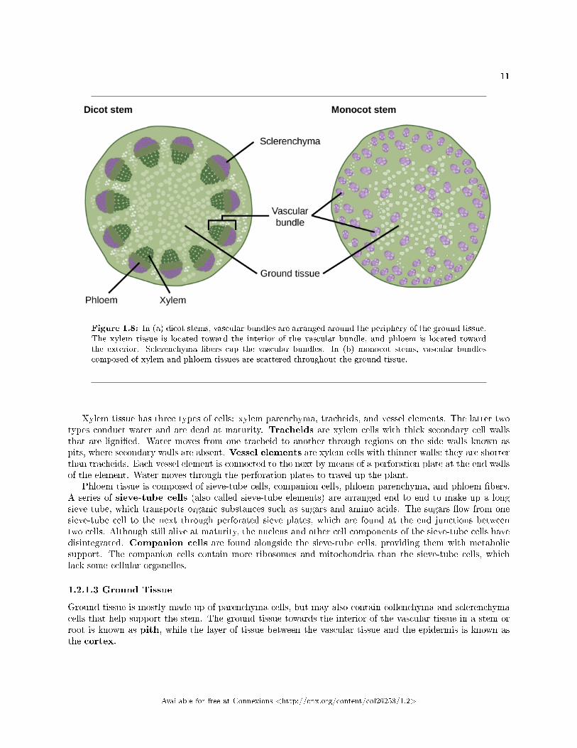
The xylem and phloem that make up the vascular tissue of the stem are arranged in distinct strands calledvascular bundles, which run up and down the length of the stem. When the stem is viewed in cross section,the vascular bundles of dicot stems are arranged in a ring. In plants with stems that live for more than oneyear, the individual bundles grow together and produce the characteristic growth rings. In monocot stems,the vascular bundles are randomly scattered throughout the ground tissue (Figure 1.8).
Available for free at Connexions <http://cnx.org/content/col26253/1.2>
11
Figure 1.8: In (a) dicot stems, vascular bundles are arranged around the periphery of the ground tissue.The xylem tissue is located toward the interior of the vascular bundle, and phloem is located towardthe exterior. Sclerenchyma �bers cap the vascular bundles. In (b) monocot stems, vascular bundlescomposed of xylem and phloem tissues are scattered throughout the ground tissue.
Xylem tissue has three types of cells: xylem parenchyma, tracheids, and vessel elements. The latter twotypes conduct water and are dead at maturity. Tracheids are xylem cells with thick secondary cell wallsthat are ligni�ed. Water moves from one tracheid to another through regions on the side walls known aspits, where secondary walls are absent. Vessel elements are xylem cells with thinner walls; they are shorterthan tracheids. Each vessel element is connected to the next by means of a perforation plate at the end wallsof the element. Water moves through the perforation plates to travel up the plant.
Phloem tissue is composed of sieve-tube cells, companion cells, phloem parenchyma, and phloem �bers.A series of sieve-tube cells (also called sieve-tube elements) are arranged end to end to make up a longsieve tube, which transports organic substances such as sugars and amino acids. The sugars �ow from onesieve-tube cell to the next through perforated sieve plates, which are found at the end junctions betweentwo cells. Although still alive at maturity, the nucleus and other cell components of the sieve-tube cells havedisintegrated. Companion cells are found alongside the sieve-tube cells, providing them with metabolicsupport. The companion cells contain more ribosomes and mitochondria than the sieve-tube cells, whichlack some cellular organelles.
1.2.1.3 Ground Tissue
Ground tissue is mostly made up of parenchyma cells, but may also contain collenchyma and sclerenchymacells that help support the stem. The ground tissue towards the interior of the vascular tissue in a stem orroot is known as pith, while the layer of tissue between the vascular tissue and the epidermis is known asthe cortex.
Available for free at Connexions <http://cnx.org/content/col26253/1.2>
12 CHAPTER 1. PLANT TISSUES
1.2.2 Growth in Stems
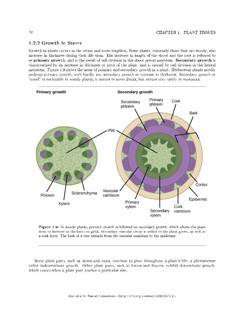
Growth in plants occurs as the stems and roots lengthen. Some plants, especially those that are woody, alsoincrease in thickness during their life span. The increase in length of the shoot and the root is referred toas primary growth, and is the result of cell division in the shoot apical meristem. Secondary growth ischaracterized by an increase in thickness or girth of the plant, and is caused by cell division in the lateralmeristem. Figure 1.9 shows the areas of primary and secondary growth in a plant. Herbaceous plants mostlyundergo primary growth, with hardly any secondary growth or increase in thickness. Secondary growth or�wood� is noticeable in woody plants; it occurs in some dicots, but occurs very rarely in monocots.
Figure 1.9: In woody plants, primary growth is followed by secondary growth, which allows the plantstem to increase in thickness or girth. Secondary vascular tissue is added as the plant grows, as well asa cork layer. The bark of a tree extends from the vascular cambium to the epidermis.
Some plant parts, such as stems and roots, continue to grow throughout a plant's life: a phenomenoncalled indeterminate growth. Other plant parts, such as leaves and �owers, exhibit determinate growth,which ceases when a plant part reaches a particular size.
Available for free at Connexions <http://cnx.org/content/col26253/1.2>
13
1.2.2.1 Primary Growth
Most primary growth occurs at the apices, or tips, of stems and roots. Primary growth is a result of rapidlydividing cells in the apical meristems at the shoot tip and root tip. Subsequent cell elongation also contributesto primary growth. The growth of shoots and roots during primary growth enables plants to continuouslyseek water (roots) or sunlight (shoots).
The in�uence of the apical bud on overall plant growth is known as apical dominance, which diminishesthe growth of axillary buds that form along the sides of branches and stems. Most coniferous trees exhibitstrong apical dominance, thus producing the typical conical Christmas tree shape. If the apical bud isremoved, then the axillary buds will start forming lateral branches. Gardeners make use of this fact whenthey prune plants by cutting o� the tops of branches, thus encouraging the axillary buds to grow out, givingthe plant a bushy shape.
: Watch this BBC Nature video4 showing how time-lapse photographycaptures plant growth at high speed.
1.2.2.2 Secondary Growth
The increase in stem thickness that results from secondary growth is due to the activity of the lateralmeristems, which are lacking in herbaceous plants. Lateral meristems include the vascular cambium and,in woody plants, the cork cambium (see Figure 1.9). The vascular cambium is located just outside theprimary xylem and to the interior of the primary phloem. The cells of the vascular cambium divide andform secondary xylem (tracheids and vessel elements) to the inside, and secondary phloem (sieve elementsand companion cells) to the outside. The thickening of the stem that occurs in secondary growth is due tothe formation of secondary phloem and secondary xylem by the vascular cambium, plus the action of corkcambium, which forms the tough outermost layer of the stem. The cells of the secondary xylem containlignin, which provides hardiness and strength.
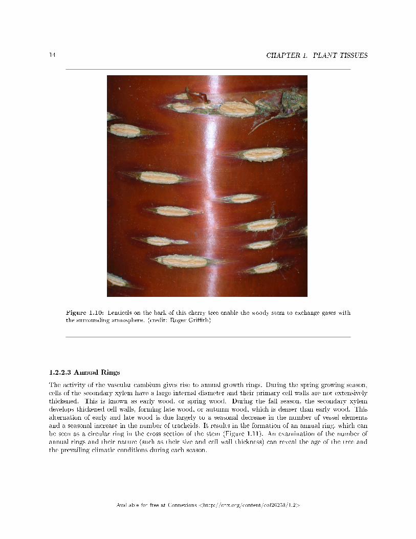
In woody plants, cork cambium is the outermost lateral meristem. It produces cork cells (bark) containinga waxy substance known as suberin that can repel water. The bark protects the plant against physical damageand helps reduce water loss. The cork cambium also produces a layer of cells known as phelloderm, whichgrows inward from the cambium. The cork cambium, cork cells, and phelloderm are collectively termedthe periderm. The periderm substitutes for the epidermis in mature plants. In some plants, the peridermhas many openings, known as lenticels, which allow the interior cells to exchange gases with the outsideatmosphere (Figure 1.10). This supplies oxygen to the living and metabolically active cells of the cortex,xylem and phloem.
4http://openstaxcollege.org/l/motion_plants
Available for free at Connexions <http://cnx.org/content/col26253/1.2>
14 CHAPTER 1. PLANT TISSUES
Figure 1.10: Lenticels on the bark of this cherry tree enable the woody stem to exchange gases withthe surrounding atmosphere. (credit: Roger Gri�th)
1.2.2.3 Annual Rings
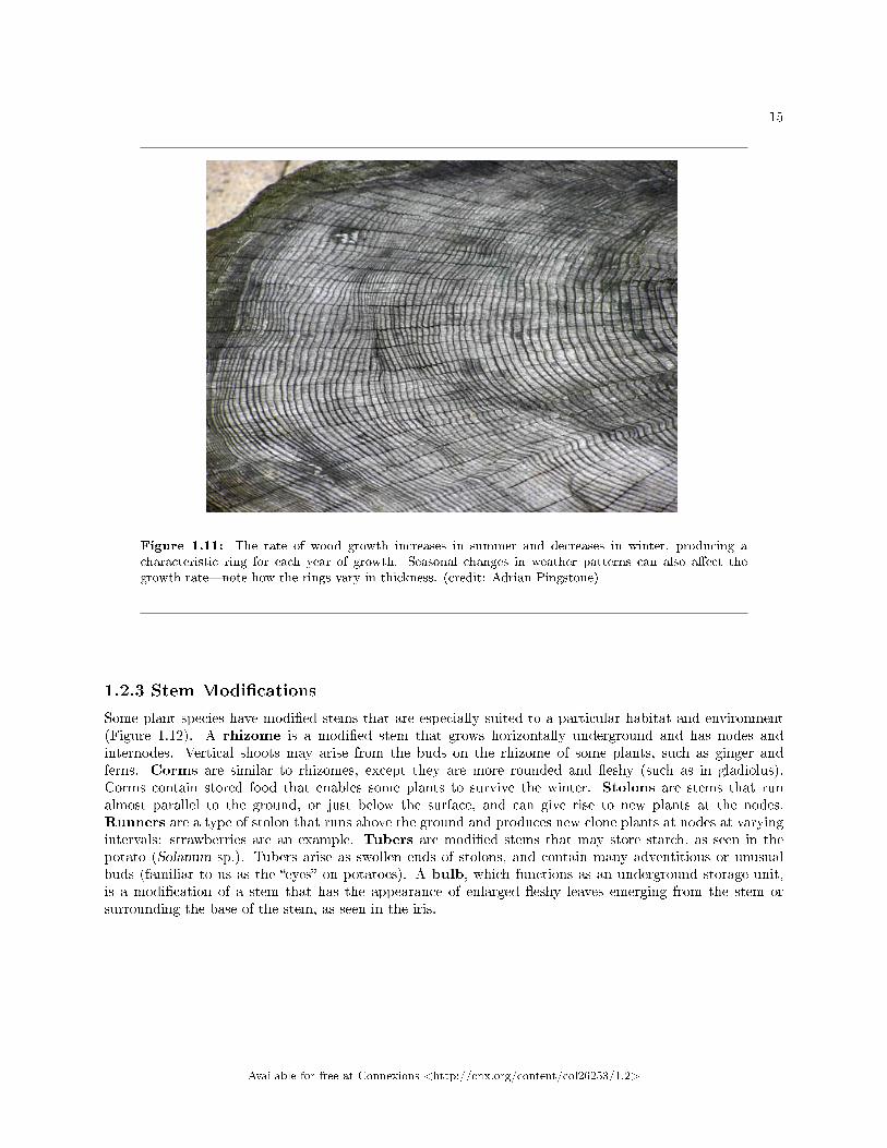
The activity of the vascular cambium gives rise to annual growth rings. During the spring growing season,cells of the secondary xylem have a large internal diameter and their primary cell walls are not extensivelythickened. This is known as early wood, or spring wood. During the fall season, the secondary xylemdevelops thickened cell walls, forming late wood, or autumn wood, which is denser than early wood. Thisalternation of early and late wood is due largely to a seasonal decrease in the number of vessel elementsand a seasonal increase in the number of tracheids. It results in the formation of an annual ring, which canbe seen as a circular ring in the cross section of the stem (Figure 1.11). An examination of the number ofannual rings and their nature (such as their size and cell wall thickness) can reveal the age of the tree andthe prevailing climatic conditions during each season.
Available for free at Connexions <http://cnx.org/content/col26253/1.2>
15
Figure 1.11: The rate of wood growth increases in summer and decreases in winter, producing acharacteristic ring for each year of growth. Seasonal changes in weather patterns can also a�ect thegrowth rate�note how the rings vary in thickness. (credit: Adrian Pingstone)
1.2.3 Stem Modi�cations
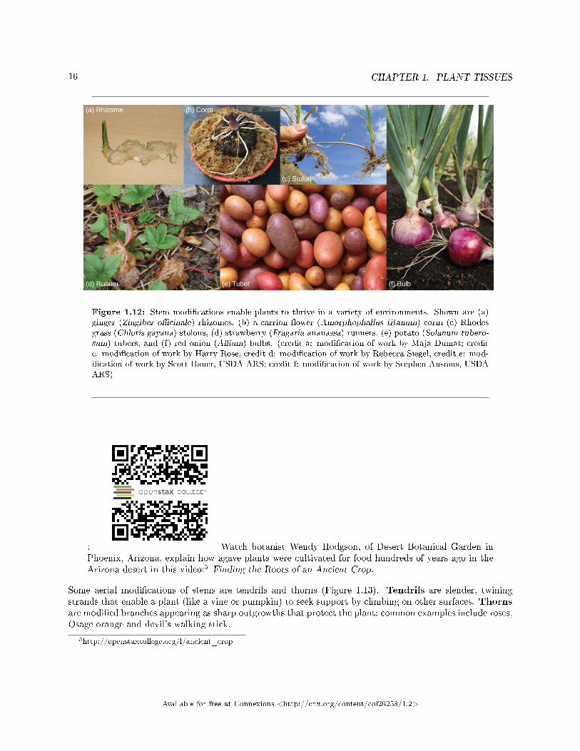
Some plant species have modi�ed stems that are especially suited to a particular habitat and environment(Figure 1.12). A rhizome is a modi�ed stem that grows horizontally underground and has nodes andinternodes. Vertical shoots may arise from the buds on the rhizome of some plants, such as ginger andferns. Corms are similar to rhizomes, except they are more rounded and �eshy (such as in gladiolus).Corms contain stored food that enables some plants to survive the winter. Stolons are stems that runalmost parallel to the ground, or just below the surface, and can give rise to new plants at the nodes.Runners are a type of stolon that runs above the ground and produces new clone plants at nodes at varyingintervals: strawberries are an example. Tubers are modi�ed stems that may store starch, as seen in thepotato (Solanum sp.). Tubers arise as swollen ends of stolons, and contain many adventitious or unusualbuds (familiar to us as the �eyes� on potatoes). A bulb, which functions as an underground storage unit,is a modi�cation of a stem that has the appearance of enlarged �eshy leaves emerging from the stem orsurrounding the base of the stem, as seen in the iris.
Available for free at Connexions <http://cnx.org/content/col26253/1.2>
16 CHAPTER 1. PLANT TISSUES
Figure 1.12: Stem modi�cations enable plants to thrive in a variety of environments. Shown are (a)ginger (Zingiber o�cinale) rhizomes, (b) a carrion �ower (Amorphophallus titanum) corm (c) Rhodesgrass (Chloris gayana) stolons, (d) strawberry (Fragaria ananassa) runners, (e) potato (Solanum tubero-sum) tubers, and (f) red onion (Allium) bulbs. (credit a: modi�cation of work by Maja Dumat; creditc: modi�cation of work by Harry Rose; credit d: modi�cation of work by Rebecca Siegel; credit e: mod-i�cation of work by Scott Bauer, USDA ARS; credit f: modi�cation of work by Stephen Ausmus, USDAARS)
: Watch botanist Wendy Hodgson, of Desert Botanical Garden inPhoenix, Arizona, explain how agave plants were cultivated for food hundreds of years ago in theArizona desert in this video:5 Finding the Roots of an Ancient Crop.
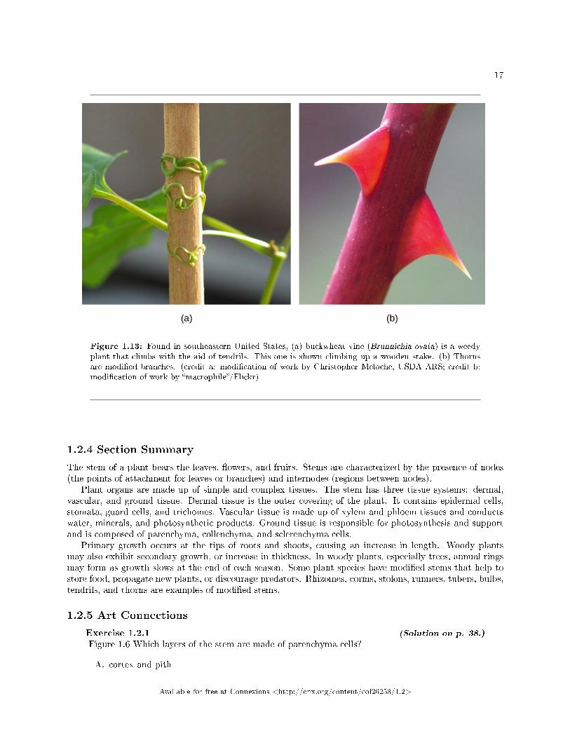
Some aerial modi�cations of stems are tendrils and thorns (Figure 1.13). Tendrils are slender, twiningstrands that enable a plant (like a vine or pumpkin) to seek support by climbing on other surfaces. Thornsare modi�ed branches appearing as sharp outgrowths that protect the plant; common examples include roses,Osage orange and devil's walking stick.
5http://openstaxcollege.org/l/ancient_crop
Available for free at Connexions <http://cnx.org/content/col26253/1.2>
17
Figure 1.13: Found in southeastern United States, (a) buckwheat vine (Brunnichia ovata) is a weedyplant that climbs with the aid of tendrils. This one is shown climbing up a wooden stake. (b) Thornsare modi�ed branches. (credit a: modi�cation of work by Christopher Meloche, USDA ARS; credit b:modi�cation of work by �macrophile�/Flickr)
1.2.4 Section Summary
The stem of a plant bears the leaves, �owers, and fruits. Stems are characterized by the presence of nodes(the points of attachment for leaves or branches) and internodes (regions between nodes).
Plant organs are made up of simple and complex tissues. The stem has three tissue systems: dermal,vascular, and ground tissue. Dermal tissue is the outer covering of the plant. It contains epidermal cells,stomata, guard cells, and trichomes. Vascular tissue is made up of xylem and phloem tissues and conductswater, minerals, and photosynthetic products. Ground tissue is responsible for photosynthesis and supportand is composed of parenchyma, collenchyma, and sclerenchyma cells.
Primary growth occurs at the tips of roots and shoots, causing an increase in length. Woody plantsmay also exhibit secondary growth, or increase in thickness. In woody plants, especially trees, annual ringsmay form as growth slows at the end of each season. Some plant species have modi�ed stems that help tostore food, propagate new plants, or discourage predators. Rhizomes, corms, stolons, runners, tubers, bulbs,tendrils, and thorns are examples of modi�ed stems.
1.2.5 Art Connections
Exercise 1.2.1 (Solution on p. 38.)
Figure 1.6 Which layers of the stem are made of parenchyma cells?
A. cortex and pith
Available for free at Connexions <http://cnx.org/content/col26253/1.2>
18 CHAPTER 1. PLANT TISSUES
B. epidermisC. sclerenchymaD. epidermis and cortex.
1.2.6 Review Questions
Exercise 1.2.2 (Solution on p. 38.)
Stem regions at which leaves are attached are called ________.
a. trichomesb. lenticelsc. nodesd. internodes
Exercise 1.2.3 (Solution on p. 38.)
Which of the following cell types forms most of the inside of a plant?
a. meristem cellsb. collenchyma cellsc. sclerenchyma cellsd. parenchyma cells
Exercise 1.2.4 (Solution on p. 38.)
Tracheids, vessel elements, sieve-tube cells, and companion cells are components of ________.
a. vascular tissueb. meristematic tissuec. ground tissued. dermal tissue
Exercise 1.2.5 (Solution on p. 38.)
The primary growth of a plant is due to the action of the ________.
a. lateral meristemb. vascular cambiumc. apical meristemd. cork cambium
Exercise 1.2.6 (Solution on p. 38.)
Which of the following is an example of secondary growth?
a. increase in lengthb. increase in thickness or girthc. increase in root hairsd. increase in leaf number
Exercise 1.2.7 (Solution on p. 38.)
Secondary growth in stems is usually seen in ________.
a. monocotsb. dicotsc. both monocots and dicotsd. neither monocots nor dicots
Available for free at Connexions <http://cnx.org/content/col26253/1.2>
19
1.2.7 Free Response
Exercise 1.2.8 (Solution on p. 38.)
Describe the roles played by stomata and guard cells. What would happen to a plant if these cellsdid not function correctly?
Exercise 1.2.9 (Solution on p. 38.)
Compare the structure and function of xylem to that of phloem.
Exercise 1.2.10 (Solution on p. 38.)
Explain the role of the cork cambium in woody plants.
Exercise 1.2.11 (Solution on p. 38.)
What is the function of lenticels?
Exercise 1.2.12 (Solution on p. 38.)
Besides the age of a tree, what additional information can annual rings reveal?
Exercise 1.2.13 (Solution on p. 38.)
Give two examples of modi�ed stems and explain how each example bene�ts the plant.
1.3 Roots6
The roots of seed plants have three major functions: anchoring the plant to the soil, absorbing waterand minerals and transporting them upwards, and storing the products of photosynthesis. Some roots aremodi�ed to absorb moisture and exchange gases. Most roots are underground. Some plants, however, alsohave adventitious roots, which emerge above the ground from the shoot.
1.3.1 Types of Root Systems
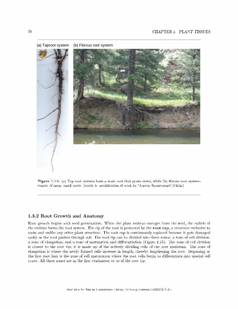
Root systems are mainly of two types (Figure 1.14). Dicots have a tap root system, while monocots have a�brous root system. A tap root system has a main root that grows down vertically, and from which manysmaller lateral roots arise. Dandelions are a good example; their tap roots usually break o� when trying topull these weeds, and they can regrow another shoot from the remaining root. A tap root system penetratesdeep into the soil. In contrast, a �brous root system is located closer to the soil surface, and forms adense network of roots that also helps prevent soil erosion (lawn grasses are a good example, as are wheat,rice, and corn). Some plants have a combination of tap roots and �brous roots. Plants that grow in dryareas often have deep root systems, whereas plants growing in areas with abundant water are likely to haveshallower root systems.
6This content is available online at <http://cnx.org/content/m66598/1.12/>.
Available for free at Connexions <http://cnx.org/content/col26253/1.2>
20 CHAPTER 1. PLANT TISSUES
Figure 1.14: (a) Tap root systems have a main root that grows down, while (b) �brous root systemsconsist of many small roots. (credit b: modi�cation of work by �Austen Squarepants�/Flickr)
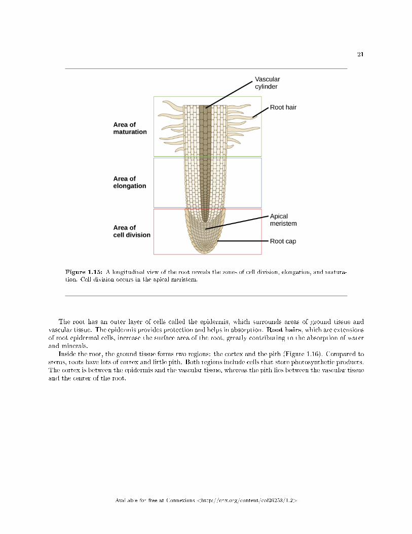
1.3.2 Root Growth and Anatomy
Root growth begins with seed germination. When the plant embryo emerges from the seed, the radicle ofthe embryo forms the root system. The tip of the root is protected by the root cap, a structure exclusive toroots and unlike any other plant structure. The root cap is continuously replaced because it gets damagedeasily as the root pushes through soil. The root tip can be divided into three zones: a zone of cell division,a zone of elongation, and a zone of maturation and di�erentiation (Figure 1.15). The zone of cell divisionis closest to the root tip; it is made up of the actively dividing cells of the root meristem. The zone ofelongation is where the newly formed cells increase in length, thereby lengthening the root. Beginning atthe �rst root hair is the zone of cell maturation where the root cells begin to di�erentiate into special celltypes. All three zones are in the �rst centimeter or so of the root tip.
Available for free at Connexions <http://cnx.org/content/col26253/1.2>
21
Figure 1.15: A longitudinal view of the root reveals the zones of cell division, elongation, and matura-tion. Cell division occurs in the apical meristem.
The root has an outer layer of cells called the epidermis, which surrounds areas of ground tissue andvascular tissue. The epidermis provides protection and helps in absorption. Root hairs, which are extensionsof root epidermal cells, increase the surface area of the root, greatly contributing to the absorption of waterand minerals.
Inside the root, the ground tissue forms two regions: the cortex and the pith (Figure 1.16). Compared tostems, roots have lots of cortex and little pith. Both regions include cells that store photosynthetic products.The cortex is between the epidermis and the vascular tissue, whereas the pith lies between the vascular tissueand the center of the root.
Available for free at Connexions <http://cnx.org/content/col26253/1.2>
22 CHAPTER 1. PLANT TISSUES
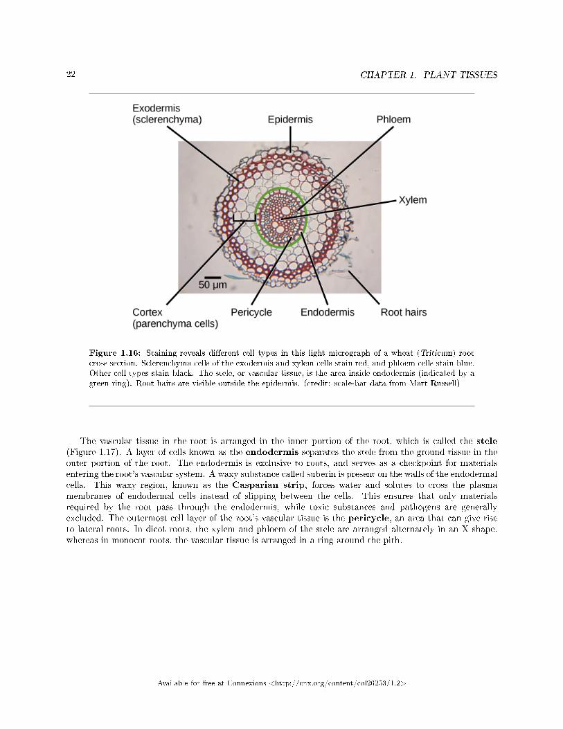
Figure 1.16: Staining reveals di�erent cell types in this light micrograph of a wheat (Triticum) rootcross section. Sclerenchyma cells of the exodermis and xylem cells stain red, and phloem cells stain blue.Other cell types stain black. The stele, or vascular tissue, is the area inside endodermis (indicated by agreen ring). Root hairs are visible outside the epidermis. (credit: scale-bar data from Matt Russell)
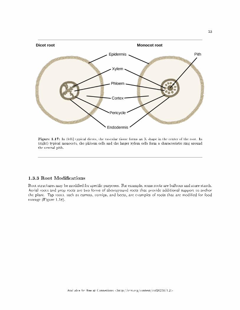
The vascular tissue in the root is arranged in the inner portion of the root, which is called the stele(Figure 1.17). A layer of cells known as the endodermis separates the stele from the ground tissue in theouter portion of the root. The endodermis is exclusive to roots, and serves as a checkpoint for materialsentering the root's vascular system. A waxy substance called suberin is present on the walls of the endodermalcells. This waxy region, known as the Casparian strip, forces water and solutes to cross the plasmamembranes of endodermal cells instead of slipping between the cells. This ensures that only materialsrequired by the root pass through the endodermis, while toxic substances and pathogens are generallyexcluded. The outermost cell layer of the root's vascular tissue is the pericycle, an area that can give riseto lateral roots. In dicot roots, the xylem and phloem of the stele are arranged alternately in an X shape,whereas in monocot roots, the vascular tissue is arranged in a ring around the pith.
Available for free at Connexions <http://cnx.org/content/col26253/1.2>
23
Figure 1.17: In (left) typical dicots, the vascular tissue forms an X shape in the center of the root. In(right) typical monocots, the phloem cells and the larger xylem cells form a characteristic ring aroundthe central pith.
1.3.3 Root Modi�cations
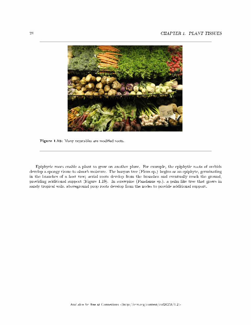
Root structures may be modi�ed for speci�c purposes. For example, some roots are bulbous and store starch.Aerial roots and prop roots are two forms of aboveground roots that provide additional support to anchorthe plant. Tap roots, such as carrots, turnips, and beets, are examples of roots that are modi�ed for foodstorage (Figure 1.18).
Available for free at Connexions <http://cnx.org/content/col26253/1.2>
24 CHAPTER 1. PLANT TISSUES
Figure 1.18: Many vegetables are modi�ed roots.
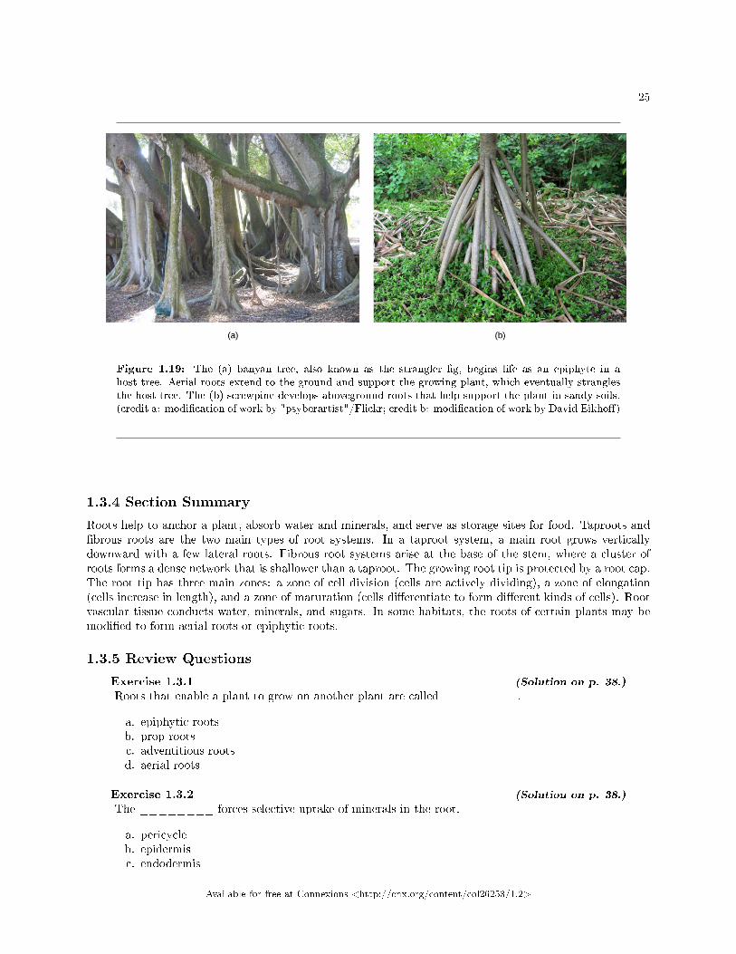
Epiphytic roots enable a plant to grow on another plant. For example, the epiphytic roots of orchidsdevelop a spongy tissue to absorb moisture. The banyan tree (Ficus sp.) begins as an epiphyte, germinatingin the branches of a host tree; aerial roots develop from the branches and eventually reach the ground,providing additional support (Figure 1.19). In screwpine (Pandanus sp.), a palm-like tree that grows insandy tropical soils, aboveground prop roots develop from the nodes to provide additional support.
Available for free at Connexions <http://cnx.org/content/col26253/1.2>
25
Figure 1.19: The (a) banyan tree, also known as the strangler �g, begins life as an epiphyte in ahost tree. Aerial roots extend to the ground and support the growing plant, which eventually stranglesthe host tree. The (b) screwpine develops aboveground roots that help support the plant in sandy soils.(credit a: modi�cation of work by "psyberartist"/Flickr; credit b: modi�cation of work by David Eikho�)
1.3.4 Section Summary
Roots help to anchor a plant, absorb water and minerals, and serve as storage sites for food. Taproots and�brous roots are the two main types of root systems. In a taproot system, a main root grows verticallydownward with a few lateral roots. Fibrous root systems arise at the base of the stem, where a cluster ofroots forms a dense network that is shallower than a taproot. The growing root tip is protected by a root cap.The root tip has three main zones: a zone of cell division (cells are actively dividing), a zone of elongation(cells increase in length), and a zone of maturation (cells di�erentiate to form di�erent kinds of cells). Rootvascular tissue conducts water, minerals, and sugars. In some habitats, the roots of certain plants may bemodi�ed to form aerial roots or epiphytic roots.
1.3.5 Review Questions
Exercise 1.3.1 (Solution on p. 38.)
Roots that enable a plant to grow on another plant are called ________.
a. epiphytic rootsb. prop rootsc. adventitious rootsd. aerial roots
Exercise 1.3.2 (Solution on p. 38.)
The ________ forces selective uptake of minerals in the root.
a. pericycleb. epidermisc. endodermis
Available for free at Connexions <http://cnx.org/content/col26253/1.2>
26 CHAPTER 1. PLANT TISSUES
d. root cap
Exercise 1.3.3 (Solution on p. 38.)
Newly-formed root cells begin to form di�erent cell types in the ________.
a. zone of elongationb. zone of maturationc. root meristemd. zone of cell division
1.3.6 Critical Thinking Questions
Exercise 1.3.4 (Solution on p. 39.)
Compare a tap root system with a �brous root system. For each type, name a plant that providesa food in the human diet. Which type of root system is found in monocots? Which type of rootsystem is found in dicots?
Exercise 1.3.5 (Solution on p. 39.)
What might happen to a root if the pericycle disappeared?
1.4 Leaves7
Leaves are the main sites for photosynthesis: the process by which plants synthesize food. Most leaves areusually green, due to the presence of chlorophyll in the leaf cells. However, some leaves may have di�erentcolors, caused by other plant pigments that mask the green chlorophyll.
The thickness, shape, and size of leaves are adapted to the environment. Each variation helps a plantspecies maximize its chances of survival in a particular habitat. Usually, the leaves of plants growing intropical rainforests have larger surface areas than those of plants growing in deserts or very cold conditions,which are likely to have a smaller surface area to minimize water loss.
1.4.1 Structure of a Typical Leaf
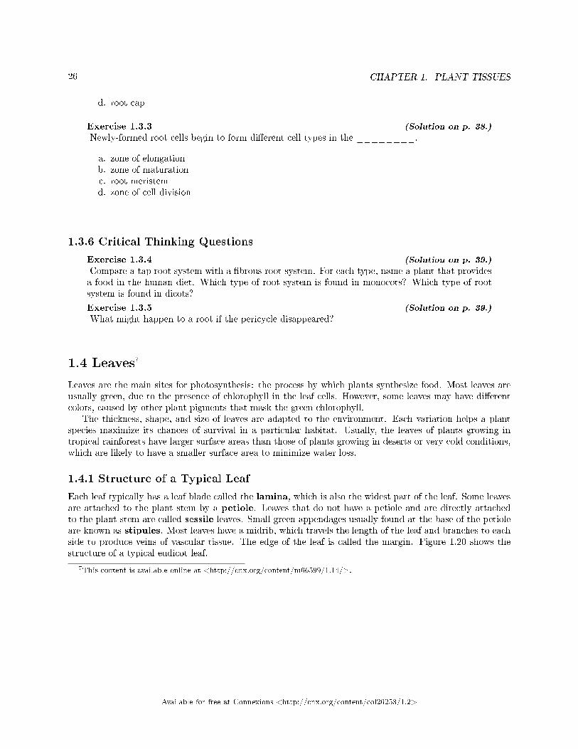
Each leaf typically has a leaf blade called the lamina, which is also the widest part of the leaf. Some leavesare attached to the plant stem by a petiole. Leaves that do not have a petiole and are directly attachedto the plant stem are called sessile leaves. Small green appendages usually found at the base of the petioleare known as stipules. Most leaves have a midrib, which travels the length of the leaf and branches to eachside to produce veins of vascular tissue. The edge of the leaf is called the margin. Figure 1.20 shows thestructure of a typical eudicot leaf.
7This content is available online at <http://cnx.org/content/m66599/1.14/>.
Available for free at Connexions <http://cnx.org/content/col26253/1.2>
27
Figure 1.20: Deceptively simple in appearance, a leaf is a highly e�cient structure.
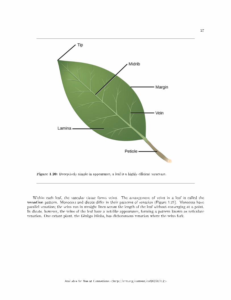
Within each leaf, the vascular tissue forms veins. The arrangement of veins in a leaf is called thevenation pattern. Monocots and dicots di�er in their patterns of venation (Figure 1.21). Monocots haveparallel venation; the veins run in straight lines across the length of the leaf without converging at a point.In dicots, however, the veins of the leaf have a net-like appearance, forming a pattern known as reticulatevenation. One extant plant, the Ginkgo biloba, has dichotomous venation where the veins fork.
Available for free at Connexions <http://cnx.org/content/col26253/1.2>
28 CHAPTER 1. PLANT TISSUES
Figure 1.21: (a) Tulip (Tulipa), a monocot, has leaves with parallel venation. The netlike venation inthis (b) linden (Tilia cordata) leaf distinguishes it as a dicot. The (c) Ginkgo biloba tree has dichotomousvenation. (credit a photo: modi�cation of work by �Drewboy64�/Wikimedia Commons; credit b photo:modi�cation of work by Roger Gri�th; credit c photo: modi�cation of work by "geishaboy500"/Flickr;credit abc illustrations: modi�cation of work by Agnieszka Kwiecie«)
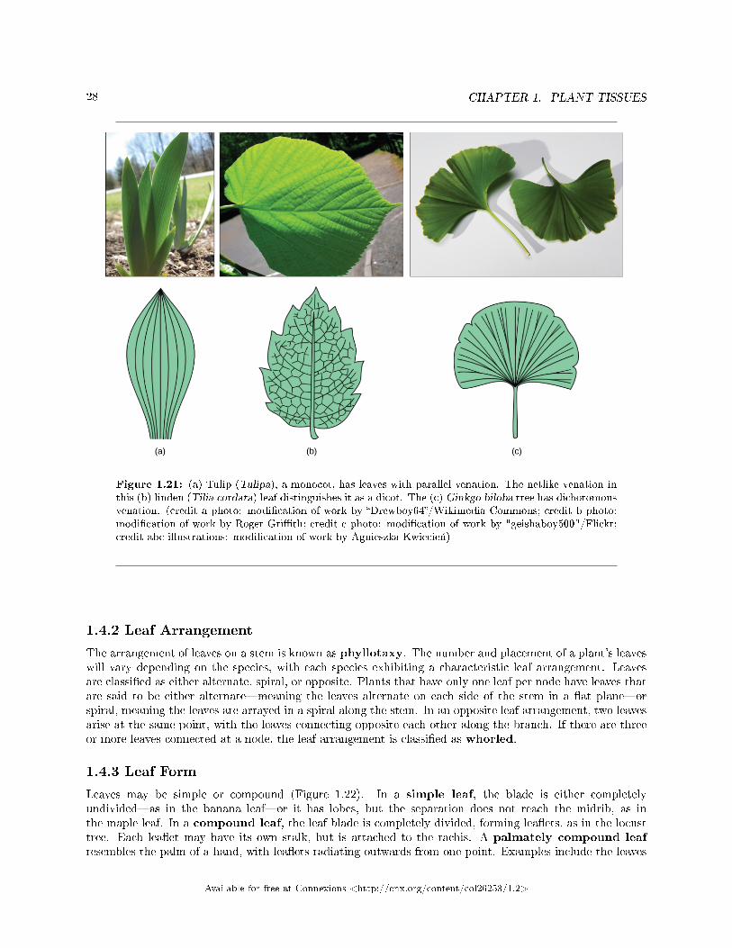
1.4.2 Leaf Arrangement
The arrangement of leaves on a stem is known as phyllotaxy. The number and placement of a plant's leaveswill vary depending on the species, with each species exhibiting a characteristic leaf arrangement. Leavesare classi�ed as either alternate, spiral, or opposite. Plants that have only one leaf per node have leaves thatare said to be either alternate�meaning the leaves alternate on each side of the stem in a �at plane�orspiral, meaning the leaves are arrayed in a spiral along the stem. In an opposite leaf arrangement, two leavesarise at the same point, with the leaves connecting opposite each other along the branch. If there are threeor more leaves connected at a node, the leaf arrangement is classi�ed as whorled.
1.4.3 Leaf Form
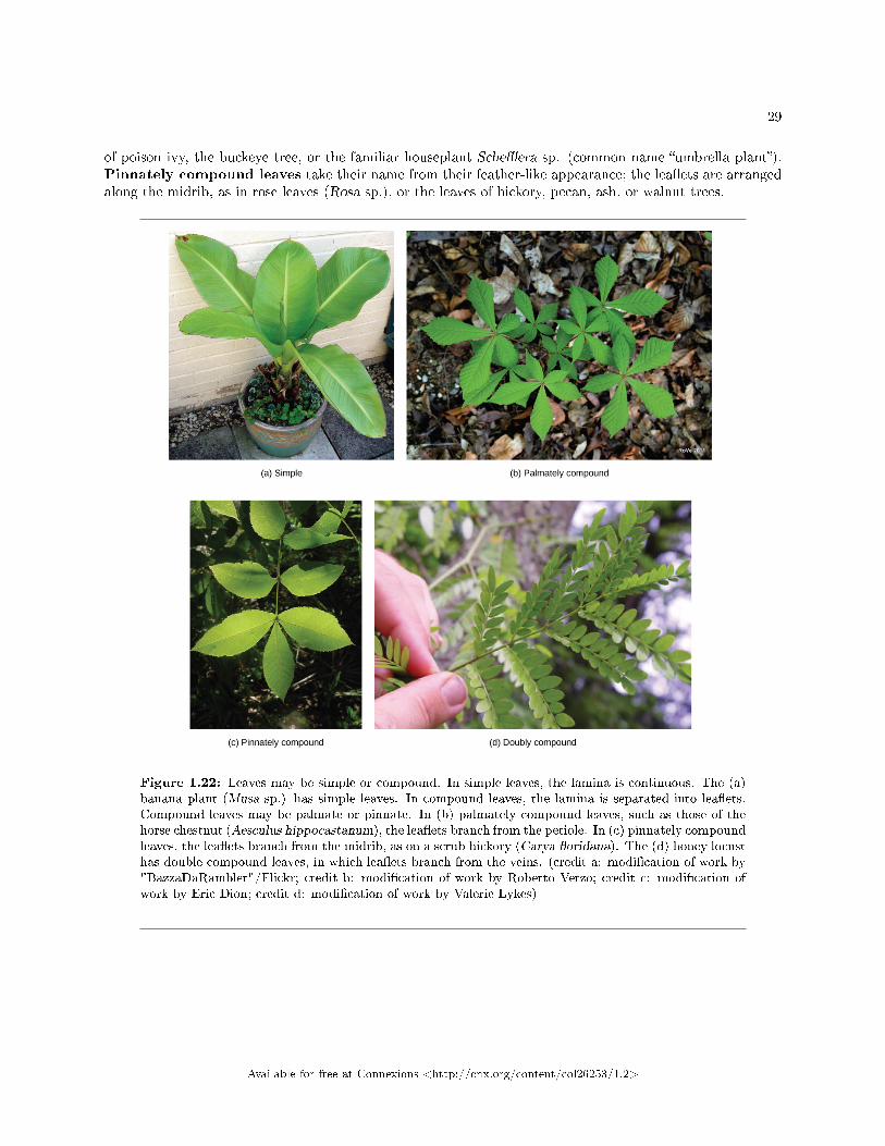
Leaves may be simple or compound (Figure 1.22). In a simple leaf, the blade is either completelyundivided�as in the banana leaf�or it has lobes, but the separation does not reach the midrib, as inthe maple leaf. In a compound leaf, the leaf blade is completely divided, forming lea�ets, as in the locusttree. Each lea�et may have its own stalk, but is attached to the rachis. A palmately compound leafresembles the palm of a hand, with lea�ets radiating outwards from one point. Examples include the leaves
Available for free at Connexions <http://cnx.org/content/col26253/1.2>
29
of poison ivy, the buckeye tree, or the familiar houseplant Sche�era sp. (common name �umbrella plant�).Pinnately compound leaves take their name from their feather-like appearance; the lea�ets are arrangedalong the midrib, as in rose leaves (Rosa sp.), or the leaves of hickory, pecan, ash, or walnut trees.
Figure 1.22: Leaves may be simple or compound. In simple leaves, the lamina is continuous. The (a)banana plant (Musa sp.) has simple leaves. In compound leaves, the lamina is separated into lea�ets.Compound leaves may be palmate or pinnate. In (b) palmately compound leaves, such as those of thehorse chestnut (Aesculus hippocastanum), the lea�ets branch from the petiole. In (c) pinnately compoundleaves, the lea�ets branch from the midrib, as on a scrub hickory (Carya �oridana). The (d) honey locusthas double compound leaves, in which lea�ets branch from the veins. (credit a: modi�cation of work by"BazzaDaRambler"/Flickr; credit b: modi�cation of work by Roberto Verzo; credit c: modi�cation ofwork by Eric Dion; credit d: modi�cation of work by Valerie Lykes)
Available for free at Connexions <http://cnx.org/content/col26253/1.2>
30 CHAPTER 1. PLANT TISSUES
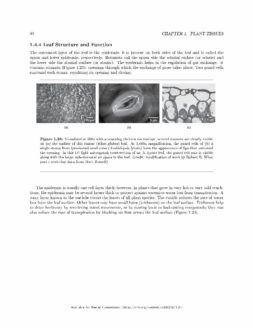
1.4.4 Leaf Structure and Function
The outermost layer of the leaf is the epidermis; it is present on both sides of the leaf and is called theupper and lower epidermis, respectively. Botanists call the upper side the adaxial surface (or adaxis) andthe lower side the abaxial surface (or abaxis). The epidermis helps in the regulation of gas exchange. Itcontains stomata (Figure 1.23): openings through which the exchange of gases takes place. Two guard cellssurround each stoma, regulating its opening and closing.
Figure 1.23: Visualized at 500x with a scanning electron microscope, several stomata are clearly visibleon (a) the surface of this sumac (Rhus glabra) leaf. At 5,000x magni�cation, the guard cells of (b) asingle stoma from lyre-leaved sand cress (Arabidopsis lyrata) have the appearance of lips that surroundthe opening. In this (c) light micrograph cross-section of an A. lyrata leaf, the guard cell pair is visiblealong with the large, sub-stomatal air space in the leaf. (credit: modi�cation of work by Robert R. Wise;part c scale-bar data from Matt Russell)
The epidermis is usually one cell layer thick; however, in plants that grow in very hot or very cold condi-tions, the epidermis may be several layers thick to protect against excessive water loss from transpiration. Awaxy layer known as the cuticle covers the leaves of all plant species. The cuticle reduces the rate of waterloss from the leaf surface. Other leaves may have small hairs (trichomes) on the leaf surface. Trichomes helpto deter herbivory by restricting insect movements, or by storing toxic or bad-tasting compounds; they canalso reduce the rate of transpiration by blocking air �ow across the leaf surface (Figure 1.24).
Available for free at Connexions <http://cnx.org/content/col26253/1.2>

31
Figure 1.24: Trichomes give leaves a fuzzy appearance as in this (a) sundew (Drosera sp.). Leaf tri-chomes include (b) branched trichomes on the leaf of Arabidopsis lyrata and (c) multibranched trichomeson a mature Quercus marilandica leaf. (credit a: John Freeland; credit b, c: modi�cation of work byRobert R. Wise; scale-bar data from Matt Russell)
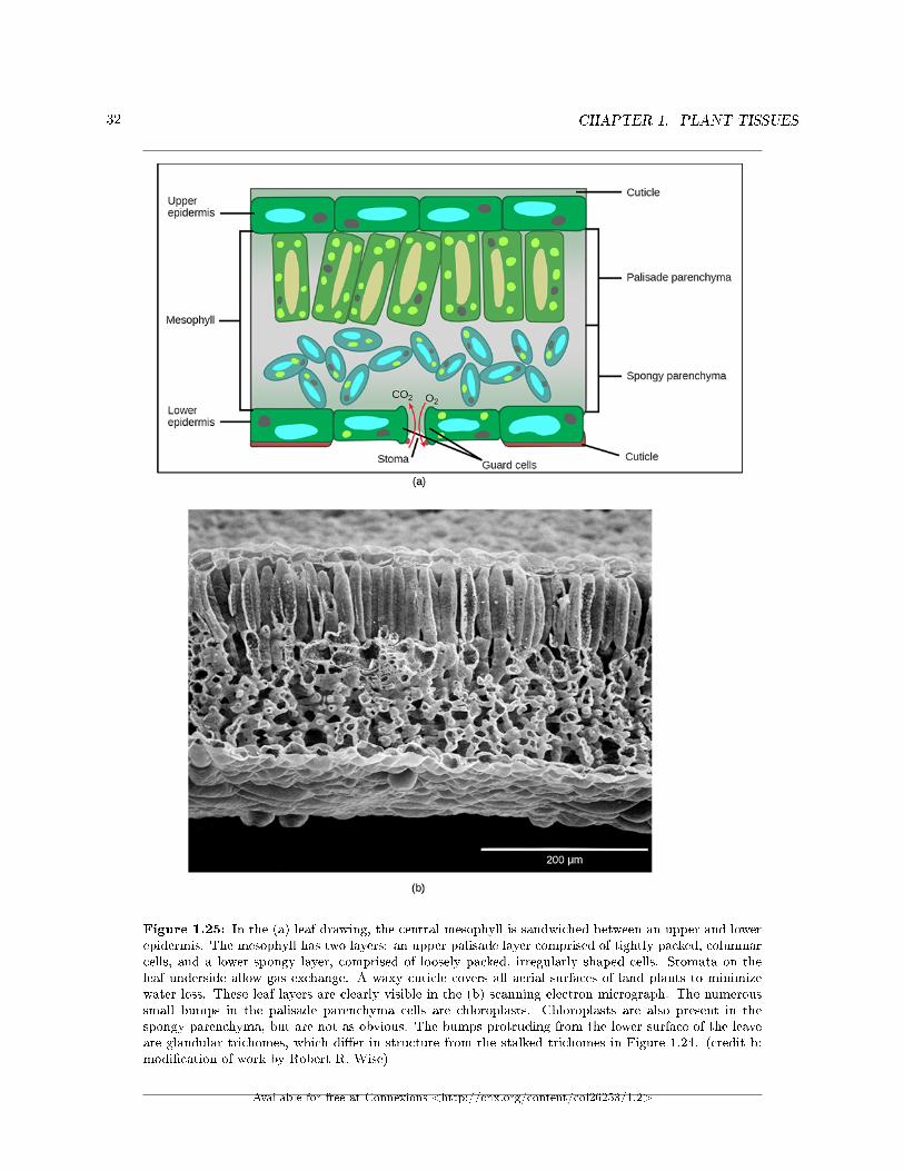
Below the epidermis of dicot leaves are layers of cells known as the mesophyll, or �middle leaf.� Themesophyll of most leaves typically contains two arrangements of parenchyma cells: the palisade parenchymaand spongy parenchyma (Figure 1.25). The palisade parenchyma (also called the palisade mesophyll) hascolumn-shaped, tightly packed cells, and may be present in one, two, or three layers. Below the palisadeparenchyma are loosely arranged cells of an irregular shape. These are the cells of the spongy parenchyma(or spongy mesophyll). The air space found between the spongy parenchyma cells allows gaseous exchangebetween the leaf and the outside atmosphere through the stomata. In aquatic plants, the intercellular spacesin the spongy parenchyma help the leaf �oat. Both layers of the mesophyll contain many chloroplasts. Guardcells are the only epidermal cells to contain chloroplasts.
Available for free at Connexions <http://cnx.org/content/col26253/1.2>
32 CHAPTER 1. PLANT TISSUES
Figure 1.25: In the (a) leaf drawing, the central mesophyll is sandwiched between an upper and lowerepidermis. The mesophyll has two layers: an upper palisade layer comprised of tightly packed, columnarcells, and a lower spongy layer, comprised of loosely packed, irregularly shaped cells. Stomata on theleaf underside allow gas exchange. A waxy cuticle covers all aerial surfaces of land plants to minimizewater loss. These leaf layers are clearly visible in the (b) scanning electron micrograph. The numeroussmall bumps in the palisade parenchyma cells are chloroplasts. Chloroplasts are also present in thespongy parenchyma, but are not as obvious. The bumps protruding from the lower surface of the leaveare glandular trichomes, which di�er in structure from the stalked trichomes in Figure 1.24. (credit b:modi�cation of work by Robert R. Wise)
Available for free at Connexions <http://cnx.org/content/col26253/1.2>
33
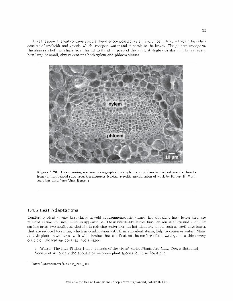
Like the stem, the leaf contains vascular bundles composed of xylem and phloem (Figure 1.26). The xylemconsists of tracheids and vessels, which transport water and minerals to the leaves. The phloem transportsthe photosynthetic products from the leaf to the other parts of the plant. A single vascular bundle, no matterhow large or small, always contains both xylem and phloem tissues.
Figure 1.26: This scanning electron micrograph shows xylem and phloem in the leaf vascular bundlefrom the lyre-leaved sand cress (Arabidopsis lyrata). (credit: modi�cation of work by Robert R. Wise;scale-bar data from Matt Russell)
1.4.5 Leaf Adaptations
Coniferous plant species that thrive in cold environments, like spruce, �r, and pine, have leaves that arereduced in size and needle-like in appearance. These needle-like leaves have sunken stomata and a smallersurface area: two attributes that aid in reducing water loss. In hot climates, plants such as cacti have leavesthat are reduced to spines, which in combination with their succulent stems, help to conserve water. Manyaquatic plants have leaves with wide lamina that can �oat on the surface of the water, and a thick waxycuticle on the leaf surface that repels water.
: Watch �The Pale Pitcher Plant� episode of the video8 series Plants Are Cool, Too, a BotanicalSociety of America video about a carnivorous plant species found in Louisiana.
8http://openstax.org/l/plants_cool_too
Available for free at Connexions <http://cnx.org/content/col26253/1.2>
34 CHAPTER 1. PLANT TISSUES
: Plant Adaptations in Resource-De�cient Environments
Roots, stems, and leaves are structured to ensure that a plant can obtain the required sunlight,water, soil nutrients, and oxygen resources. Some remarkable adaptations have evolved to enableplant species to thrive in less than ideal habitats, where one or more of these resources is in shortsupply.
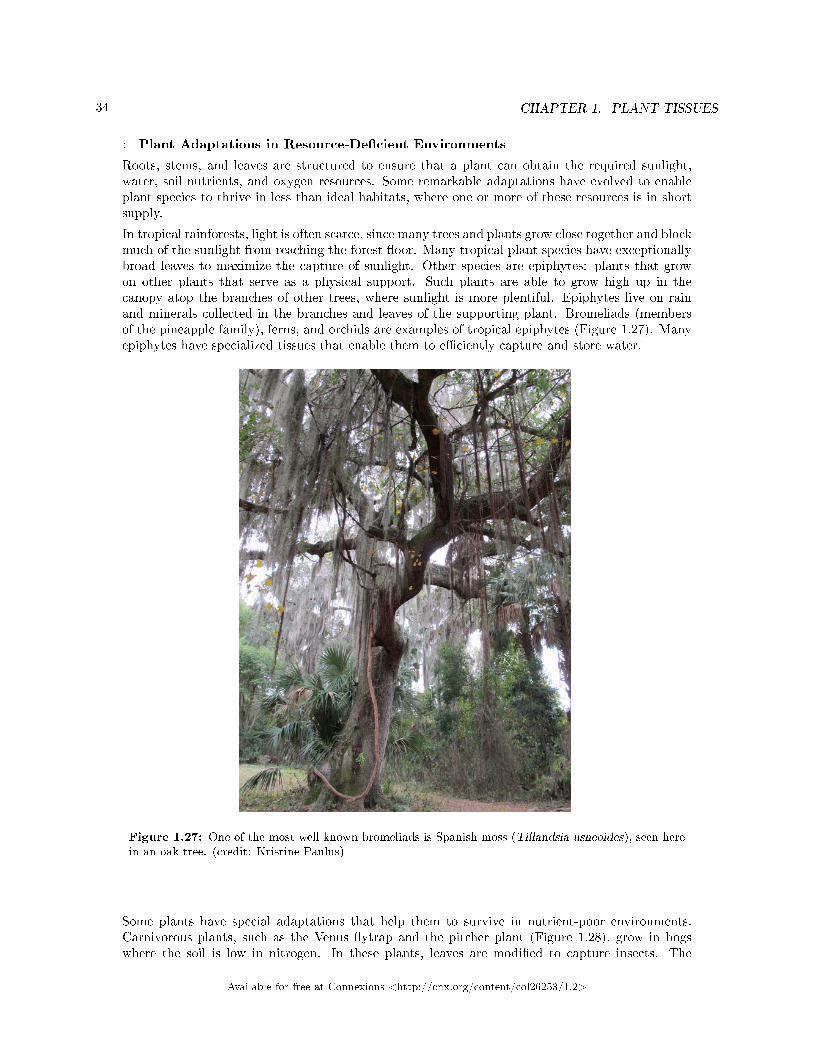
In tropical rainforests, light is often scarce, since many trees and plants grow close together and blockmuch of the sunlight from reaching the forest �oor. Many tropical plant species have exceptionallybroad leaves to maximize the capture of sunlight. Other species are epiphytes: plants that growon other plants that serve as a physical support. Such plants are able to grow high up in thecanopy atop the branches of other trees, where sunlight is more plentiful. Epiphytes live on rainand minerals collected in the branches and leaves of the supporting plant. Bromeliads (membersof the pineapple family), ferns, and orchids are examples of tropical epiphytes (Figure 1.27). Manyepiphytes have specialized tissues that enable them to e�ciently capture and store water.
Figure 1.27: One of the most well known bromeliads is Spanish moss (Tillandsia usneoides), seen herein an oak tree. (credit: Kristine Paulus)
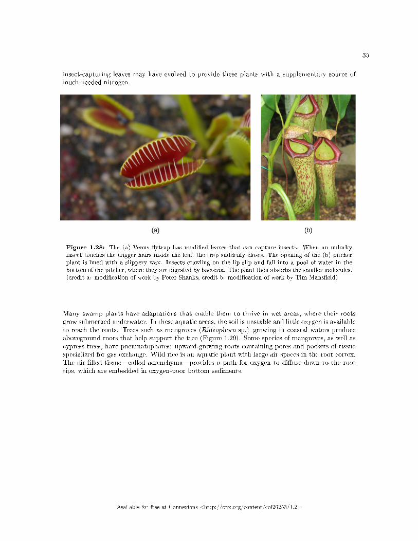
Some plants have special adaptations that help them to survive in nutrient-poor environments.Carnivorous plants, such as the Venus �ytrap and the pitcher plant (Figure 1.28), grow in bogswhere the soil is low in nitrogen. In these plants, leaves are modi�ed to capture insects. The
Available for free at Connexions <http://cnx.org/content/col26253/1.2>
35
insect-capturing leaves may have evolved to provide these plants with a supplementary source ofmuch-needed nitrogen.
Figure 1.28: The (a) Venus �ytrap has modi�ed leaves that can capture insects. When an unluckyinsect touches the trigger hairs inside the leaf, the trap suddenly closes. The opening of the (b) pitcherplant is lined with a slippery wax. Insects crawling on the lip slip and fall into a pool of water in thebottom of the pitcher, where they are digested by bacteria. The plant then absorbs the smaller molecules.(credit a: modi�cation of work by Peter Shanks; credit b: modi�cation of work by Tim Mans�eld)
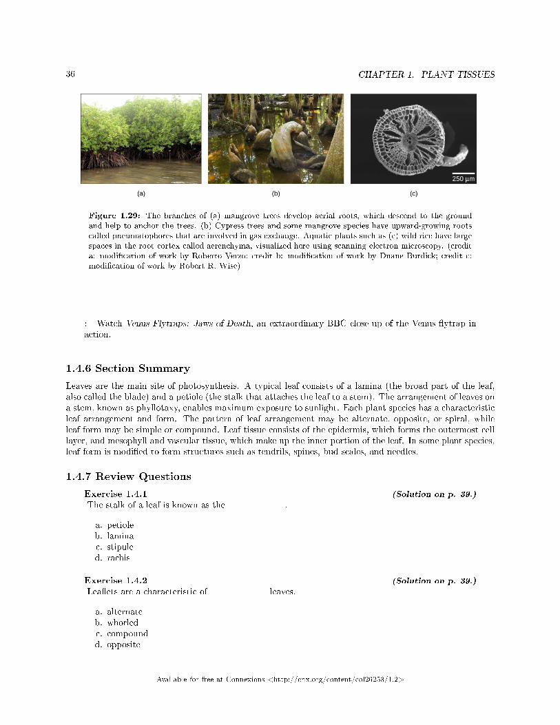
Many swamp plants have adaptations that enable them to thrive in wet areas, where their rootsgrow submerged underwater. In these aquatic areas, the soil is unstable and little oxygen is availableto reach the roots. Trees such as mangroves (Rhizophora sp.) growing in coastal waters produceaboveground roots that help support the tree (Figure 1.29). Some species of mangroves, as well ascypress trees, have pneumatophores: upward-growing roots containing pores and pockets of tissuespecialized for gas exchange. Wild rice is an aquatic plant with large air spaces in the root cortex.The air-�lled tissue�called aerenchyma�provides a path for oxygen to di�use down to the roottips, which are embedded in oxygen-poor bottom sediments.
Available for free at Connexions <http://cnx.org/content/col26253/1.2>
36 CHAPTER 1. PLANT TISSUES
Figure 1.29: The branches of (a) mangrove trees develop aerial roots, which descend to the groundand help to anchor the trees. (b) Cypress trees and some mangrove species have upward-growing rootscalled pneumatophores that are involved in gas exchange. Aquatic plants such as (c) wild rice have largespaces in the root cortex called aerenchyma, visualized here using scanning electron microscopy. (credita: modi�cation of work by Roberto Verzo; credit b: modi�cation of work by Duane Burdick; credit c:modi�cation of work by Robert R. Wise)
: Watch Venus Flytraps: Jaws of Death, an extraordinary BBC close-up of the Venus �ytrap inaction.
1.4.6 Section Summary
Leaves are the main site of photosynthesis. A typical leaf consists of a lamina (the broad part of the leaf,also called the blade) and a petiole (the stalk that attaches the leaf to a stem). The arrangement of leaves ona stem, known as phyllotaxy, enables maximum exposure to sunlight. Each plant species has a characteristicleaf arrangement and form. The pattern of leaf arrangement may be alternate, opposite, or spiral, whileleaf form may be simple or compound. Leaf tissue consists of the epidermis, which forms the outermost celllayer, and mesophyll and vascular tissue, which make up the inner portion of the leaf. In some plant species,leaf form is modi�ed to form structures such as tendrils, spines, bud scales, and needles.
1.4.7 Review Questions
Exercise 1.4.1 (Solution on p. 39.)
The stalk of a leaf is known as the ________.
a. petioleb. laminac. stipuled. rachis
Exercise 1.4.2 (Solution on p. 39.)
Lea�ets are a characteristic of ________ leaves.
a. alternateb. whorledc. compoundd. opposite
Available for free at Connexions <http://cnx.org/content/col26253/1.2>
37
Exercise 1.4.3 (Solution on p. 39.)
Cells of the ________ contain chloroplasts.
a. epidermisb. vascular tissuec. peridermd. mesophyll
Exercise 1.4.4 (Solution on p. 39.)
Which of the following is most likely to be found in a desert environment?
a. broad leaves to capture sunlightb. spines instead of leavesc. needle-like leavesd. wide, �at leaves that can �oat
1.4.8 Critical Thinking Questions
Exercise 1.4.5 (Solution on p. 39.)
How do dicots di�er from monocots in terms of leaf structure?
Exercise 1.4.6 (Solution on p. 39.)
Describe an example of a plant with leaves that are adapted to cold temperatures.
Available for free at Connexions <http://cnx.org/content/col26253/1.2>
38 CHAPTER 1. PLANT TISSUES
Solutions to Exercises in Chapter 1
to Exercise 1.1.1 (p. 5)Cto Exercise 1.1.2 (p. 5)Bto Exercise 1.1.3 (p. 5)Lawn grasses and other monocots have an intercalary meristem, which is a region of meristematic tissue atthe base of the leaf blade. This is bene�cial to the plant because it can continue to grow even when the tipof the plant is removed by grazing or mowing.to Exercise 1.1.4 (p. 5)Vascular tissue transports water, minerals, and sugars throughout the plant. Vascular tissue is made upof xylem tissue and phloem tissue. Xylem tissue transports water and nutrients from the roots upward.Phloem tissue carries sugars from the sites of photosynthesis to the rest of the plant.to Exercise 1.2.1 (p. 17)Figure 1.6 A and B. The cortex, pith, and epidermis are made of parenchyma cells.to Exercise 1.2.2 (p. 18)Cto Exercise 1.2.3 (p. 18)Dto Exercise 1.2.4 (p. 18)Ato Exercise 1.2.5 (p. 18)Cto Exercise 1.2.6 (p. 18)Bto Exercise 1.2.7 (p. 18)Bto Exercise 1.2.8 (p. 19)Stomata allow gases to enter and exit the plant. Guard cells regulate the opening and closing of stomata.If these cells did not function correctly, a plant could not get the carbon dioxide needed for photosynthesis,nor could it release the oxygen produced by photosynthesis.to Exercise 1.2.9 (p. 19)Xylem is made up tracheids and vessel elements, which are cells that transport water and dissolved mineralsand that are dead at maturity. Phloem is made up of sieve-tube cells and companion cells, which transportcarbohydrates and are alive at maturity.to Exercise 1.2.10 (p. 19)In woody plants, the cork cambium is the outermost lateral meristem; it produces new cells towards theinterior, which enables the plant to increase in girth. The cork cambium also produces cork cells towardsthe exterior, which protect the plant from physical damage while reducing water loss.to Exercise 1.2.11 (p. 19)In woody stems, lenticels allow internal cells to exchange gases with the outside atmosphere.to Exercise 1.2.12 (p. 19)Annual rings can also indicate the climate conditions that prevailed during each growing season.to Exercise 1.2.13 (p. 19)Answers will vary. Rhizomes, stolons, and runners can give rise to new plants. Corms, tubers, and bulbscan also produce new plants and can store food. Tendrils help a plant to climb, while thorns discourageherbivores.to Exercise 1.3.1 (p. 25)Ato Exercise 1.3.2 (p. 25)C
Available for free at Connexions <http://cnx.org/content/col26253/1.2>
39
to Exercise 1.3.3 (p. 26)Bto Exercise 1.3.4 (p. 26)A tap root system has a single main root that grows down. A �brous root system forms a dense network ofroots that is closer to the soil surface. An example of a tap root system is a carrot. Grasses such as wheat,rice, and corn are examples of �brous root systems. Fibrous root systems are found in monocots; tap rootsystems are found in dicots.to Exercise 1.3.5 (p. 26)The root would not be able to produce lateral roots.to Exercise 1.4.1 (p. 36)Ato Exercise 1.4.2 (p. 36)Cto Exercise 1.4.3 (p. 37)Dto Exercise 1.4.4 (p. 37)Bto Exercise 1.4.5 (p. 37)Monocots have leaves with parallel venation, and dicots have leaves with reticulate, net-like venation.to Exercise 1.4.6 (p. 37)Conifers such as spruce, �r, and pine have needle-shaped leaves with sunken stomata, helping to reducewater loss.
Available for free at Connexions <http://cnx.org/content/col26253/1.2>
40 CHAPTER 1. PLANT TISSUES
Available for free at Connexions <http://cnx.org/content/col26253/1.2>
Chapter 2
Plant Transport
2.1 The Soil and Plant Nutrition1
Plants obtain inorganic elements from the soil, which serves as a natural medium for land plants. Soil is theouter loose layer that covers the surface of Earth. Soil quality is a major determinant, along with climate, ofplant distribution and growth. Soil quality depends not only on the chemical composition of the soil, but alsothe topography (regional surface features) and the presence of living organisms. In agriculture, the historyof the soil, such as the cultivating practices and previous crops, modify the characteristics and fertility ofthat soil.
Soil develops very slowly over long periods of time, and its formation results from natural and environ-mental forces acting on mineral, rock, and organic compounds. Soils can be divided into two groups: organicsoils are those that are formed from sedimentation and primarily composed of organic matter, while thosethat are formed from the weathering of rocks and are primarily composed of inorganic material are calledmineral soils. Mineral soils are predominant in terrestrial ecosystems, where soils may be covered by waterfor part of the year or exposed to the atmosphere.
2.1.1 Soil Composition
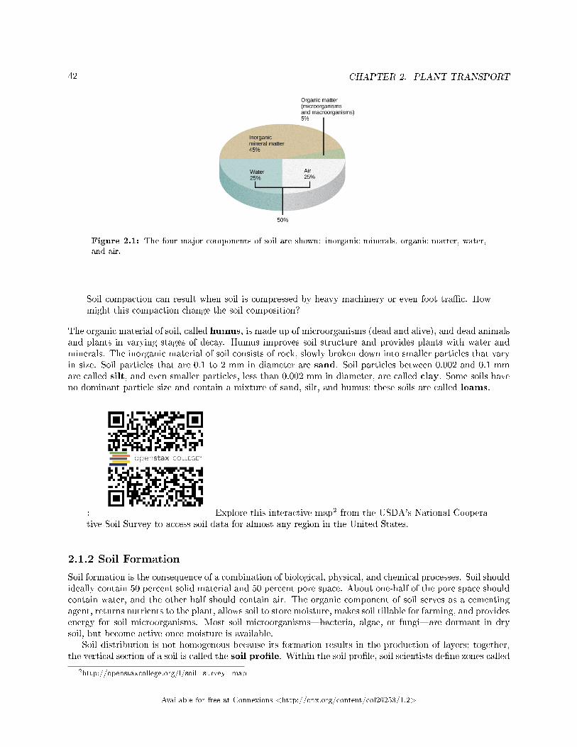
Soil consists of these major components (Figure 2.1):
• inorganic mineral matter, about 40 to 45 percent of the soil volume• organic matter, about 5 percent of the soil volume• water and air, about 50 percent of the soil volume
The amount of each of the four major components of soil depends on the amount of vegetation, soil com-paction, and water present in the soil. A good healthy soil has su�cient air, water, minerals, and organicmaterial to promote and sustain plant life.
:
1This content is available online at <http://cnx.org/content/m44715/1.7/>.
Available for free at Connexions <http://cnx.org/content/col26253/1.2>
41
42 CHAPTER 2. PLANT TRANSPORT
Figure 2.1: The four major components of soil are shown: inorganic minerals, organic matter, water,and air.
Soil compaction can result when soil is compressed by heavy machinery or even foot tra�c. Howmight this compaction change the soil composition?
The organic material of soil, called humus, is made up of microorganisms (dead and alive), and dead animalsand plants in varying stages of decay. Humus improves soil structure and provides plants with water andminerals. The inorganic material of soil consists of rock, slowly broken down into smaller particles that varyin size. Soil particles that are 0.1 to 2 mm in diameter are sand. Soil particles between 0.002 and 0.1 mmare called silt, and even smaller particles, less than 0.002 mm in diameter, are called clay. Some soils haveno dominant particle size and contain a mixture of sand, silt, and humus; these soils are called loams.
: Explore this interactive map2 from the USDA's National Coopera-tive Soil Survey to access soil data for almost any region in the United States.
2.1.2 Soil Formation
Soil formation is the consequence of a combination of biological, physical, and chemical processes. Soil shouldideally contain 50 percent solid material and 50 percent pore space. About one-half of the pore space shouldcontain water, and the other half should contain air. The organic component of soil serves as a cementingagent, returns nutrients to the plant, allows soil to store moisture, makes soil tillable for farming, and providesenergy for soil microorganisms. Most soil microorganisms�bacteria, algae, or fungi�are dormant in drysoil, but become active once moisture is available.
Soil distribution is not homogenous because its formation results in the production of layers; together,the vertical section of a soil is called the soil pro�le. Within the soil pro�le, soil scientists de�ne zones called
2http://openstaxcollege.org/l/soil_survey_map
Available for free at Connexions <http://cnx.org/content/col26253/1.2>
43
horizons. A horizon is a soil layer with distinct physical and chemical properties that di�er from those ofother layers. Five factors account for soil formation: parent material, climate, topography, biological factors,and time.
2.1.2.1 Parent Material
The organic and inorganic material in which soils form is the parent material. Mineral soils form directlyfrom the weathering of bedrock, the solid rock that lies beneath the soil, and therefore, they have a similarcomposition to the original rock. Other soils form in materials that came from elsewhere, such as sand andglacial drift. Materials located in the depth of the soil are relatively unchanged compared with the depositedmaterial. Sediments in rivers may have di�erent characteristics, depending on whether the stream movesquickly or slowly. A fast-moving river could have sediments of rocks and sand, whereas a slow-moving rivercould have �ne-textured material, such as clay.
2.1.2.2 Climate
Temperature, moisture, and wind cause di�erent patterns of weathering and therefore a�ect soil character-istics. The presence of moisture and nutrients from weathering will also promote biological activity: a keycomponent of a quality soil.
2.1.2.3 Topography
Regional surface features (familiarly called �the lay of the land�) can have a major in�uence on the char-acteristics and fertility of a soil. Topography a�ects water runo�, which strips away parent material anda�ects plant growth. Steeps soils are more prone to erosion and may be thinner than soils that are relatively�at or level.
2.1.2.4 Biological factors
The presence of living organisms greatly a�ects soil formation and structure. Animals and microorganismscan produce pores and crevices, and plant roots can penetrate into crevices to produce more fragmentation.Plant secretions promote the development of microorganisms around the root, in an area known as therhizosphere. Additionally, leaves and other material that fall from plants decompose and contribute to soilcomposition.
2.1.2.5 Time
Time is an important factor in soil formation because soils develop over long periods. Soil formation is adynamic process. Materials are deposited over time, decompose, and transform into other materials thatcan be used by living organisms or deposited onto the surface of the soil.
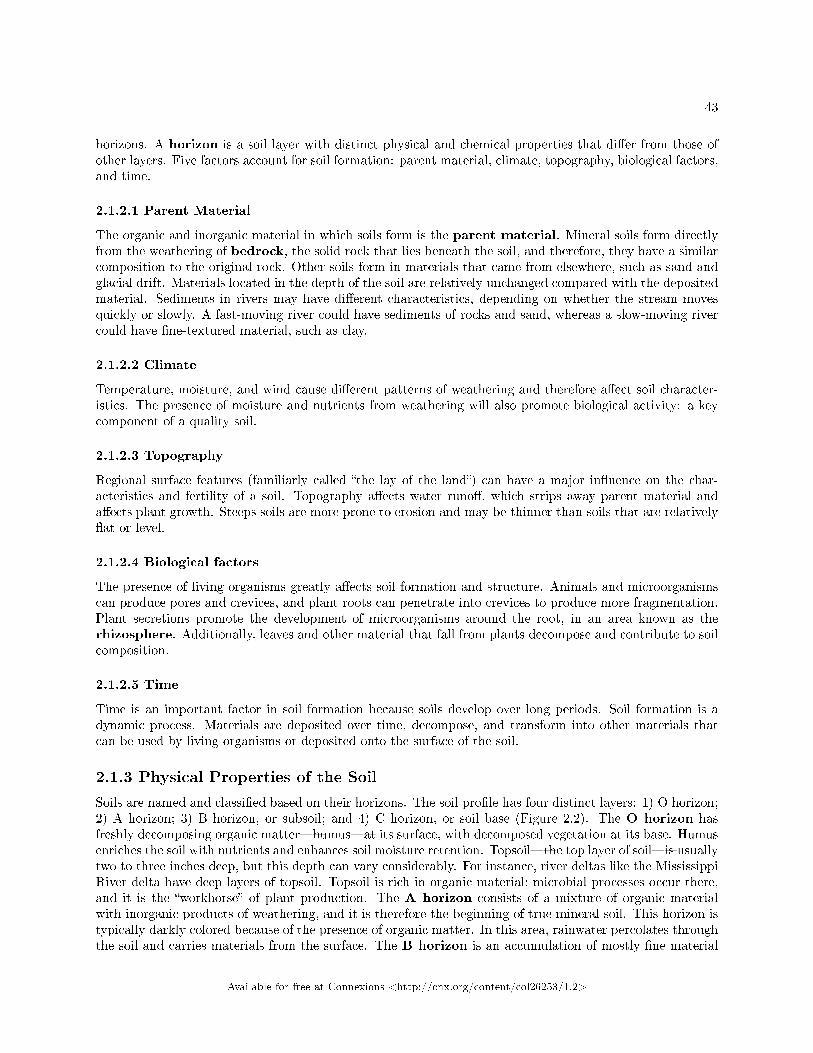
2.1.3 Physical Properties of the Soil
Soils are named and classi�ed based on their horizons. The soil pro�le has four distinct layers: 1) O horizon;2) A horizon; 3) B horizon, or subsoil; and 4) C horizon, or soil base (Figure 2.2). The O horizon hasfreshly decomposing organic matter�humus�at its surface, with decomposed vegetation at its base. Humusenriches the soil with nutrients and enhances soil moisture retention. Topsoil�the top layer of soil�is usuallytwo to three inches deep, but this depth can vary considerably. For instance, river deltas like the MississippiRiver delta have deep layers of topsoil. Topsoil is rich in organic material; microbial processes occur there,and it is the �workhorse� of plant production. The A horizon consists of a mixture of organic materialwith inorganic products of weathering, and it is therefore the beginning of true mineral soil. This horizon istypically darkly colored because of the presence of organic matter. In this area, rainwater percolates throughthe soil and carries materials from the surface. The B horizon is an accumulation of mostly �ne material
Available for free at Connexions <http://cnx.org/content/col26253/1.2>
44 CHAPTER 2. PLANT TRANSPORT
that has moved downward, resulting in a dense layer in the soil. In some soils, the B horizon contains nodulesor a layer of calcium carbonate. The C horizon, or soil base, includes the parent material, plus the organicand inorganic material that is broken down to form soil. The parent material may be either created in itsnatural place, or transported from elsewhere to its present location. Beneath the C horizon lies bedrock.
:
Figure 2.2: This soil pro�le shows the di�erent soil layers (O horizon, A horizon, B horizon, and Chorizon) found in typical soils. (credit: modi�cation of work by USDA)
Which horizon is considered the topsoil, and which is considered the subsoil?
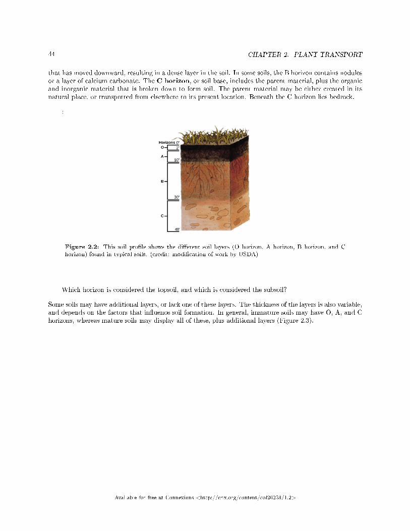
Some soils may have additional layers, or lack one of these layers. The thickness of the layers is also variable,and depends on the factors that in�uence soil formation. In general, immature soils may have O, A, and Chorizons, whereas mature soils may display all of these, plus additional layers (Figure 2.3).
Available for free at Connexions <http://cnx.org/content/col26253/1.2>
45
Figure 2.3: The San Joaquin soil pro�le has an O horizon, A horizon, B horizon, and C horizon. (credit:modi�cation of work by USDA)
: Soil Scientist
A soil scientist studies the biological components, physical and chemical properties, distribution,formation, and morphology of soils. Soil scientists need to have a strong background in physicaland life sciences, plus a foundation in mathematics. They may work for federal or state agencies,academia, or the private sector. Their work may involve collecting data, carrying out research,interpreting results, inspecting soils, conducting soil surveys, and recommending soil managementprograms.
Available for free at Connexions <http://cnx.org/content/col26253/1.2>
46 CHAPTER 2. PLANT TRANSPORT
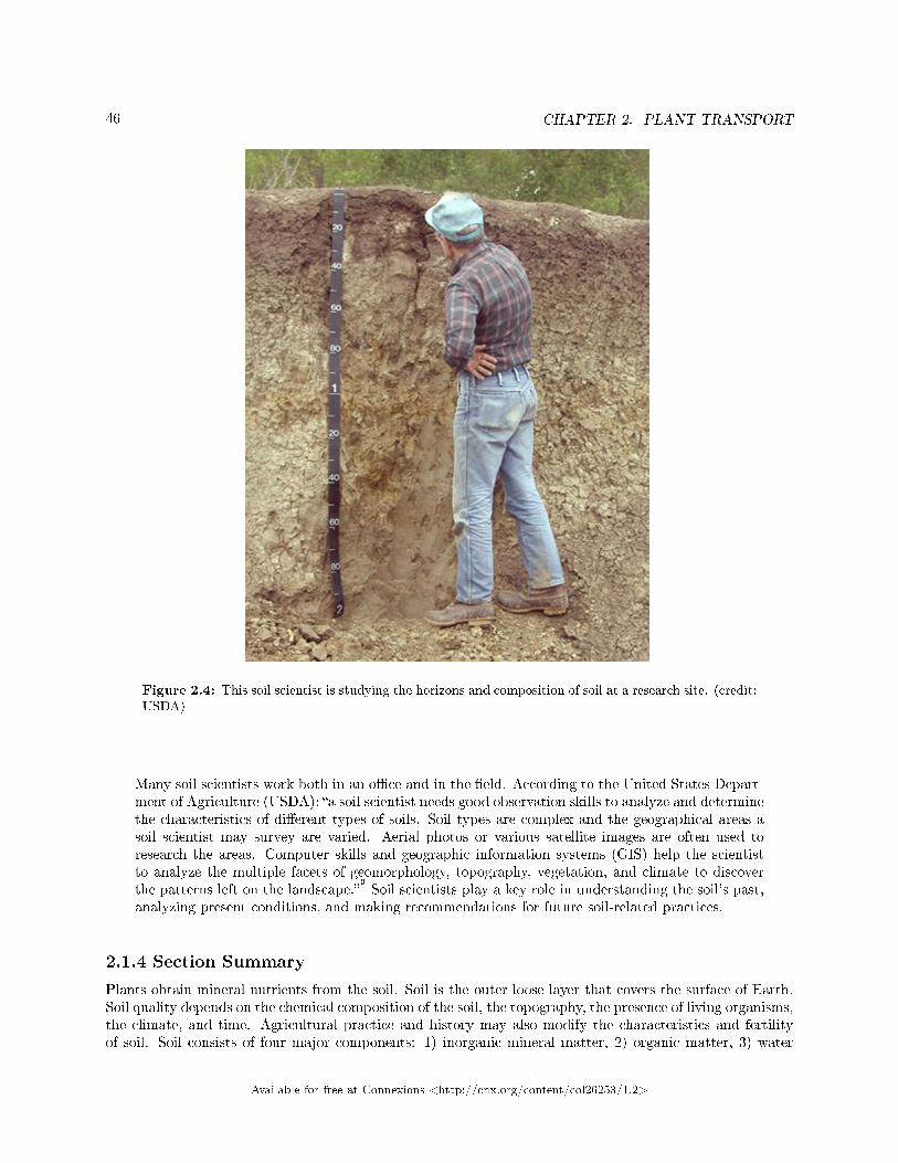
Figure 2.4: This soil scientist is studying the horizons and composition of soil at a research site. (credit:USDA)
Many soil scientists work both in an o�ce and in the �eld. According to the United States Depart-ment of Agriculture (USDA): �a soil scientist needs good observation skills to analyze and determinethe characteristics of di�erent types of soils. Soil types are complex and the geographical areas asoil scientist may survey are varied. Aerial photos or various satellite images are often used toresearch the areas. Computer skills and geographic information systems (GIS) help the scientistto analyze the multiple facets of geomorphology, topography, vegetation, and climate to discoverthe patterns left on the landscape.�
3
Soil scientists play a key role in understanding the soil's past,analyzing present conditions, and making recommendations for future soil-related practices.
2.1.4 Section Summary
Plants obtain mineral nutrients from the soil. Soil is the outer loose layer that covers the surface of Earth.Soil quality depends on the chemical composition of the soil, the topography, the presence of living organisms,the climate, and time. Agricultural practice and history may also modify the characteristics and fertilityof soil. Soil consists of four major components: 1) inorganic mineral matter, 2) organic matter, 3) water
Available for free at Connexions <http://cnx.org/content/col26253/1.2>
47
and air, and 4) living matter. The organic material of soil is made of humus, which improves soil structureand provides water and minerals. Soil inorganic material consists of rock slowly broken down into smallerparticles that vary in size, such as sand, silt, and loam.
Soil formation results from a combination of biological, physical, and chemical processes. Soil is nothomogenous because its formation results in the production of layers called a soil pro�le. Factors that a�ectsoil formation include: parent material, climate, topography, biological factors, and time. Soils are classi�edbased on their horizons, soil particle size, and proportions. Most soils have four distinct horizons: O, A, B,and C.
2.1.5 Art Connections
Exercise 2.1.1 (Solution on p. 61.)
Figure 2.1 Soil compaction can result when soil is compressed by heavy machinery or even foottra�c. How might this compaction change the soil composition?
Exercise 2.1.2 (Solution on p. 61.)
Figure 2.2 Which horizon is considered the topsoil, and which is considered the subsoil?
2.1.6 Review Questions
Exercise 2.1.3 (Solution on p. 61.)
Which factors a�ect soil quality?
a. chemical compositionb. history of the soilc. presence of living organisms and topographyd. all of the above
Exercise 2.1.4 (Solution on p. 61.)
Soil particles that are 0.1 to 2 mm in diameter are called ________.
a. sandb. siltc. clayd. loam
Exercise 2.1.5 (Solution on p. 61.)
A soil consists of layers called ________ that taken together are called a ________.
a. soil pro�les : horizonb. horizons : soil pro�lec. horizons : humusd. humus : soil pro�le
Exercise 2.1.6 (Solution on p. 61.)
What is the term used to describe the solid rock that lies beneath the soil?
a. sandb. bedrockc. clayd. loam
Available for free at Connexions <http://cnx.org/content/col26253/1.2>
48 CHAPTER 2. PLANT TRANSPORT
2.1.7
Exercise 2.1.7 (Solution on p. 61.)
Describe the main di�erences between a mineral soil and an organic soil.
Exercise 2.1.8 (Solution on p. 61.)
Name and brie�y explain the factors that a�ect soil formation.
Exercise 2.1.9 (Solution on p. 61.)
Describe how topography in�uences the characteristics and fertility of a soil.
2.2 Transport of Water and Solutes in Plants4
The structure of plant roots, stems, and leaves facilitates the transport of water, nutrients, and photosyn-thates throughout the plant. The phloem and xylem are the main tissues responsible for this movement.Water potential, evapotranspiration, and stomatal regulation in�uence how water and nutrients are trans-ported in plants. To understand how these processes work, we must �rst understand the energetics of waterpotential.
2.2.1 Water Potential
Plants are phenomenal hydraulic engineers. Using only the basic laws of physics and the simple manipulationof potential energy, plants can move water to the top of a 116-meter-tall tree (Figure 2.5a). Plants can alsouse hydraulics to generate enough force to split rocks and buckle sidewalks (Figure 2.5b). Plants achievethis because of water potential.
4This content is available online at <http://cnx.org/content/m66600/1.19/>.
Available for free at Connexions <http://cnx.org/content/col26253/1.2>
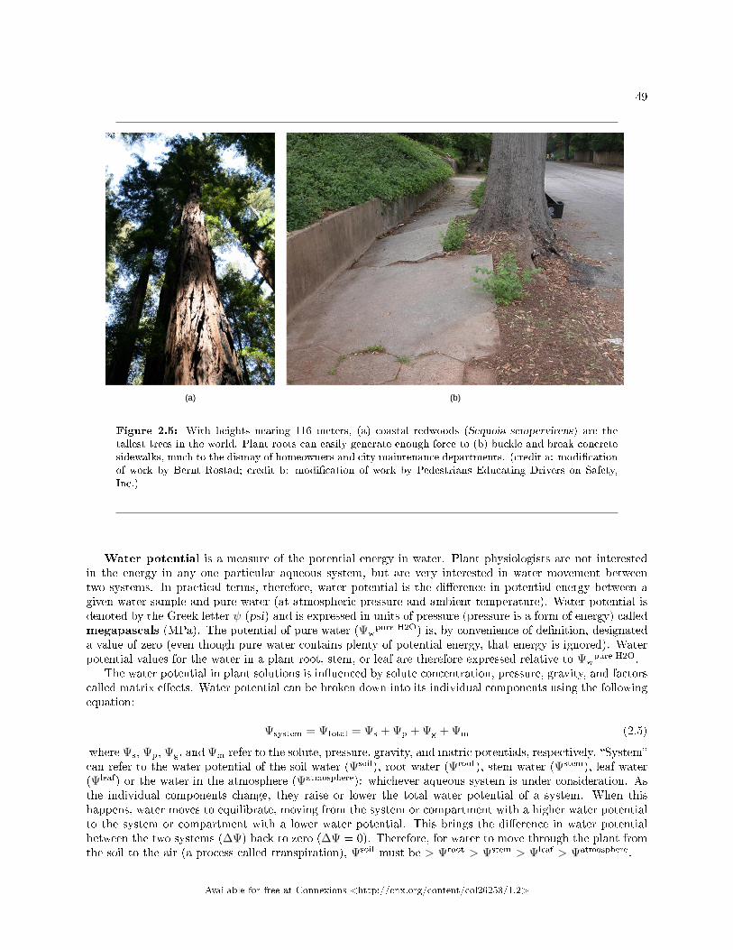
49
Figure 2.5: With heights nearing 116 meters, (a) coastal redwoods (Sequoia sempervirens) are thetallest trees in the world. Plant roots can easily generate enough force to (b) buckle and break concretesidewalks, much to the dismay of homeowners and city maintenance departments. (credit a: modi�cationof work by Bernt Rostad; credit b: modi�cation of work by Pedestrians Educating Drivers on Safety,Inc.)
Water potential is a measure of the potential energy in water. Plant physiologists are not interestedin the energy in any one particular aqueous system, but are very interested in water movement betweentwo systems. In practical terms, therefore, water potential is the di�erence in potential energy between agiven water sample and pure water (at atmospheric pressure and ambient temperature). Water potential isdenoted by the Greek letter ψ (psi) and is expressed in units of pressure (pressure is a form of energy) calledmegapascals (MPa). The potential of pure water (Ψw
pure H2O) is, by convenience of de�nition, designateda value of zero (even though pure water contains plenty of potential energy, that energy is ignored). Waterpotential values for the water in a plant root, stem, or leaf are therefore expressed relative to Ψw
pure H2O.The water potential in plant solutions is in�uenced by solute concentration, pressure, gravity, and factors
called matrix e�ects. Water potential can be broken down into its individual components using the followingequation:
Ψsystem = Ψtotal = Ψs + Ψp + Ψg + Ψm (2.5)
where Ψs, Ψp, Ψg, and Ψm refer to the solute, pressure, gravity, and matric potentials, respectively. �System�can refer to the water potential of the soil water (Ψsoil), root water (Ψroot), stem water (Ψstem), leaf water(Ψleaf) or the water in the atmosphere (Ψatmosphere): whichever aqueous system is under consideration. Asthe individual components change, they raise or lower the total water potential of a system. When thishappens, water moves to equilibrate, moving from the system or compartment with a higher water potentialto the system or compartment with a lower water potential. This brings the di�erence in water potentialbetween the two systems (∆Ψ) back to zero (∆Ψ = 0). Therefore, for water to move through the plant fromthe soil to the air (a process called transpiration), Ψsoil must be > Ψroot > Ψstem > Ψleaf > Ψatmosphere.
Available for free at Connexions <http://cnx.org/content/col26253/1.2>
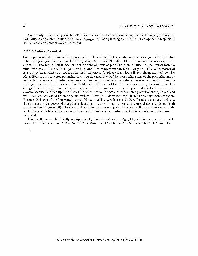
50 CHAPTER 2. PLANT TRANSPORT
Water only moves in response to ∆Ψ, not in response to the individual components. However, because theindividual components in�uence the total Ψsystem, by manipulating the individual components (especiallyΨs), a plant can control water movement.
2.2.1.1 Solute Potential
Solute potential (Ψs), also called osmotic potential, is related to the solute concentration (in molarity). Thatrelationship is given by the van 't Ho� equation: Ψs= �Mi RT; where M is the molar concentration of thesolute, i is the van 't Ho� factor (the ratio of the amount of particles in the solution to amount of formulaunits dissolved), R is the ideal gas constant, and T is temperature in Kelvin degrees. The solute potentialis negative in a plant cell and zero in distilled water. Typical values for cell cytoplasm are �0.5 to �1.0MPa. Solutes reduce water potential (resulting in a negative Ψw) by consuming some of the potential energyavailable in the water. Solute molecules can dissolve in water because water molecules can bind to them viahydrogen bonds; a hydrophobic molecule like oil, which cannot bind to water, cannot go into solution. Theenergy in the hydrogen bonds between solute molecules and water is no longer available to do work in thesystem because it is tied up in the bond. In other words, the amount of available potential energy is reducedwhen solutes are added to an aqueous system. Thus, Ψ s decreases with increasing solute concentration.Because Ψs is one of the four components of Ψsystem or Ψtotal, a decrease in Ψs will cause a decrease in Ψtotal.The internal water potential of a plant cell is more negative than pure water because of the cytoplasm's highsolute content (Figure 2.6). Because of this di�erence in water potential water will move from the soil intoa plant's root cells via the process of osmosis. This is why solute potential is sometimes called osmoticpotential.
Plant cells can metabolically manipulate Ψs (and by extension, Ψtotal) by adding or removing solutemolecules. Therefore, plants have control over Ψtotal via their ability to exert metabolic control over Ψs.
:
Available for free at Connexions <http://cnx.org/content/col26253/1.2>
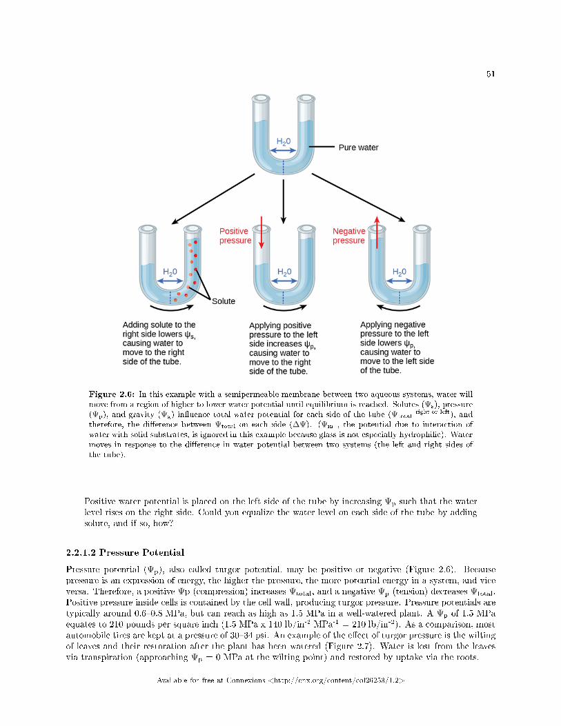
51
Figure 2.6: In this example with a semipermeable membrane between two aqueous systems, water willmove from a region of higher to lower water potential until equilibrium is reached. Solutes (Ψs), pressure(Ψp), and gravity (Ψg) in�uence total water potential for each side of the tube (Ψtotal
right or left), andtherefore, the di�erence between Ψtotal on each side (∆Ψ). (Ψm , the potential due to interaction ofwater with solid substrates, is ignored in this example because glass is not especially hydrophilic). Watermoves in response to the di�erence in water potential between two systems (the left and right sides ofthe tube).
Positive water potential is placed on the left side of the tube by increasing Ψp such that the waterlevel rises on the right side. Could you equalize the water level on each side of the tube by addingsolute, and if so, how?
2.2.1.2 Pressure Potential
Pressure potential (Ψp), also called turgor potential, may be positive or negative (Figure 2.6). Becausepressure is an expression of energy, the higher the pressure, the more potential energy in a system, and viceversa. Therefore, a positive Ψp (compression) increases Ψtotal, and a negative Ψp (tension) decreases Ψtotal.Positive pressure inside cells is contained by the cell wall, producing turgor pressure. Pressure potentials aretypically around 0.6�0.8 MPa, but can reach as high as 1.5 MPa in a well-watered plant. A Ψp of 1.5 MPaequates to 210 pounds per square inch (1.5 MPa x 140 lb/in-2 MPa-1 = 210 lb/in-2). As a comparison, mostautomobile tires are kept at a pressure of 30�34 psi. An example of the e�ect of turgor pressure is the wiltingof leaves and their restoration after the plant has been watered (Figure 2.7). Water is lost from the leavesvia transpiration (approaching Ψp = 0 MPa at the wilting point) and restored by uptake via the roots.
Available for free at Connexions <http://cnx.org/content/col26253/1.2>
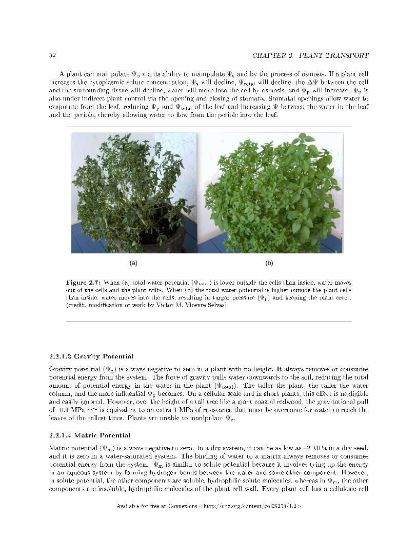
52 CHAPTER 2. PLANT TRANSPORT
A plant can manipulate Ψp via its ability to manipulate Ψs and by the process of osmosis. If a plant cellincreases the cytoplasmic solute concentration, Ψs will decline, Ψtotal will decline, the ∆Ψ between the celland the surrounding tissue will decline, water will move into the cell by osmosis, and Ψp will increase. Ψp isalso under indirect plant control via the opening and closing of stomata. Stomatal openings allow water toevaporate from the leaf, reducing Ψp and Ψtotal of the leaf and increasing Ψ between the water in the leafand the petiole, thereby allowing water to �ow from the petiole into the leaf.
Figure 2.7: When (a) total water potential (Ψtotal) is lower outside the cells than inside, water movesout of the cells and the plant wilts. When (b) the total water potential is higher outside the plant cellsthan inside, water moves into the cells, resulting in turgor pressure (Ψp) and keeping the plant erect.(credit: modi�cation of work by Victor M. Vicente Selvas)
2.2.1.3 Gravity Potential
Gravity potential (Ψg) is always negative to zero in a plant with no height. It always removes or consumespotential energy from the system. The force of gravity pulls water downwards to the soil, reducing the totalamount of potential energy in the water in the plant (Ψtotal). The taller the plant, the taller the watercolumn, and the more in�uential Ψg becomes. On a cellular scale and in short plants, this e�ect is negligibleand easily ignored. However, over the height of a tall tree like a giant coastal redwood, the gravitational pullof �0.1 MPa m-1 is equivalent to an extra 1 MPa of resistance that must be overcome for water to reach theleaves of the tallest trees. Plants are unable to manipulate Ψg.
2.2.1.4 Matric Potential
Matric potential (Ψm) is always negative to zero. In a dry system, it can be as low as �2 MPa in a dry seed,and it is zero in a water-saturated system. The binding of water to a matrix always removes or consumespotential energy from the system. Ψm is similar to solute potential because it involves tying up the energyin an aqueous system by forming hydrogen bonds between the water and some other component. However,in solute potential, the other components are soluble, hydrophilic solute molecules, whereas in Ψm, the othercomponents are insoluble, hydrophilic molecules of the plant cell wall. Every plant cell has a cellulosic cell
Available for free at Connexions <http://cnx.org/content/col26253/1.2>
53
wall and the cellulose in the cell walls is hydrophilic, producing a matrix for adhesion of water: hence thename matric potential. Ψm is very large (negative) in dry tissues such as seeds or drought-a�ected soils.However, it quickly goes to zero as the seed takes up water or the soil hydrates. Ψm cannot be manipulatedby the plant and is typically ignored in well-watered roots, stems, and leaves.
2.2.2 Movement of Water and Minerals in the Xylem
Solutes, pressure, gravity, and matric potential are all important for the transport of water in plants. Watermoves from an area of higher total water potential (higher Gibbs free energy) to an area of lower total waterpotential. Gibbs free energy is the energy associated with a chemical reaction that can be used to do work.This is expressed as ∆Ψ.
Transpiration is the loss of water from the plant through evaporation at the leaf surface. It is themain driver of water movement in the xylem. Transpiration is caused by the evaporation of water at theleaf�atmosphere interface; it creates negative pressure (tension) equivalent to �2 MPa at the leaf surface.This value varies greatly depending on the vapor pressure de�cit, which can be negligible at high relativehumidity (RH) and substantial at low RH. Water from the roots is pulled up by this tension. At night, whenstomata shut and transpiration stops, the water is held in the stem and leaf by the adhesion of water tothe cell walls of the xylem vessels and tracheids, and the cohesion of water molecules to each other. This iscalled the cohesion�tension theory of sap ascent.
Inside the leaf at the cellular level, water on the surface of mesophyll cells saturates the cellulose micro�b-rils of the primary cell wall. The leaf contains many large intercellular air spaces for the exchange of oxygenfor carbon dioxide, which is required for photosynthesis. The wet cell wall is exposed to this leaf internal airspace, and the water on the surface of the cells evaporates into the air spaces, decreasing the thin �lm onthe surface of the mesophyll cells. This decrease creates a greater tension on the water in the mesophyll cells(Figure 2.8), thereby increasing the pull on the water in the xylem vessels. The xylem vessels and tracheidsare structurally adapted to cope with large changes in pressure. Rings in the vessels maintain their tubularshape, much like the rings on a vacuum cleaner hose keep the hose open while it is under pressure. Smallperforations between vessel elements reduce the number and size of gas bubbles that can form via a processcalled cavitation. The formation of gas bubbles in xylem interrupts the continuous stream of water from thebase to the top of the plant, causing a break termed an embolism in the �ow of xylem sap. The taller thetree, the greater the tension forces needed to pull water, and the more cavitation events. In larger trees, theresulting embolisms can plug xylem vessels, making them nonfunctional.
:
Available for free at Connexions <http://cnx.org/content/col26253/1.2>
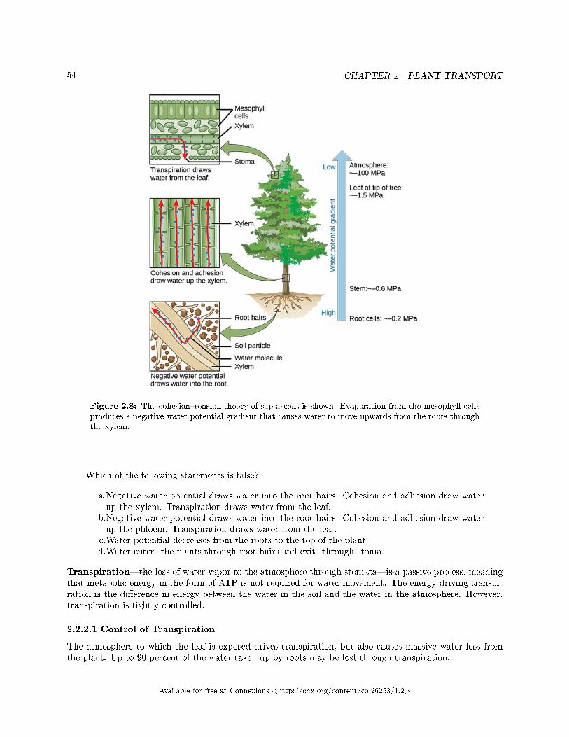
54 CHAPTER 2. PLANT TRANSPORT
Figure 2.8: The cohesion�tension theory of sap ascent is shown. Evaporation from the mesophyll cellsproduces a negative water potential gradient that causes water to move upwards from the roots throughthe xylem.
Which of the following statements is false?
a.Negative water potential draws water into the root hairs. Cohesion and adhesion draw waterup the xylem. Transpiration draws water from the leaf.
b.Negative water potential draws water into the root hairs. Cohesion and adhesion draw waterup the phloem. Transpiration draws water from the leaf.
c.Water potential decreases from the roots to the top of the plant.d.Water enters the plants through root hairs and exits through stoma.
Transpiration�the loss of water vapor to the atmosphere through stomata�is a passive process, meaningthat metabolic energy in the form of ATP is not required for water movement. The energy driving transpi-ration is the di�erence in energy between the water in the soil and the water in the atmosphere. However,transpiration is tightly controlled.
2.2.2.1 Control of Transpiration
The atmosphere to which the leaf is exposed drives transpiration, but also causes massive water loss fromthe plant. Up to 90 percent of the water taken up by roots may be lost through transpiration.
Available for free at Connexions <http://cnx.org/content/col26253/1.2>
55
Leaves are covered by a waxy cuticle on the outer surface that prevents the loss of water. Regulationof transpiration, therefore, is achieved primarily through the opening and closing of stomata on the leafsurface. Stomata are surrounded by two specialized cells called guard cells, which open and close in responseto environmental cues such as light intensity and quality, leaf water status, and carbon dioxide concentra-tions. Stomata must open to allow air containing carbon dioxide and oxygen to di�use into the leaf forphotosynthesis and respiration. When stomata are open, however, water vapor is lost to the external envi-ronment, increasing the rate of transpiration. Therefore, plants must maintain a balance between e�cientphotosynthesis and water loss.
Plants have evolved over time to adapt to their local environment and reduce transpiration (Figure 2.9).Desert plant (xerophytes) and plants that grow on other plants (epiphytes) have limited access to water. Suchplants usually have a much thicker waxy cuticle than those growing in more moderate, well-watered environ-ments (mesophytes). Aquatic plants (hydrophytes) also have their own set of anatomical and morphologicalleaf adaptations.
Available for free at Connexions <http://cnx.org/content/col26253/1.2>
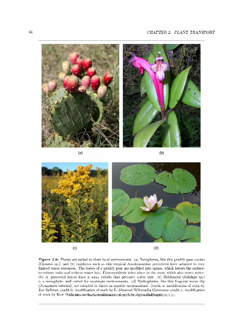
56 CHAPTER 2. PLANT TRANSPORT
Figure 2.9: Plants are suited to their local environment. (a) Xerophytes, like this prickly pear cactus(Opuntia sp.) and (b) epiphytes such as this tropical Aeschynanthus perrottetii have adapted to verylimited water resources. The leaves of a prickly pear are modi�ed into spines, which lowers the surface-to-volume ratio and reduces water loss. Photosynthesis takes place in the stem, which also stores water.(b) A. perottetii leaves have a waxy cuticle that prevents water loss. (c) Goldenrod (Solidago sp.)is a mesophyte, well suited for moderate environments. (d) Hydrophytes, like this fragrant water lily(Nymphaea odorata), are adapted to thrive in aquatic environments. (credit a: modi�cation of work byJon Sullivan; credit b: modi�cation of work by L. Shyamal/Wikimedia Commons; credit c: modi�cationof work by Huw Williams; credit d: modi�cation of work by Jason Hollinger)Available for free at Connexions <http://cnx.org/content/col26253/1.2>
57
Xerophytes and epiphytes often have a thick covering of trichomes or of stomata that are sunken belowthe leaf's surface. Trichomes are specialized hair-like epidermal cells that secrete oils and substances. Theseadaptations impede air �ow across the stomatal pore and reduce transpiration. Multiple epidermal layersare also commonly found in these types of plants.
2.2.3 Transportation of Photosynthates in the Phloem
Plants need an energy source to grow. In seeds and bulbs, food is stored in polymers (such as starch) thatare converted by metabolic processes into sucrose for newly developing plants. Once green shoots and leavesare growing, plants are able to produce their own food by photosynthesizing. The products of photosynthesisare called photosynthates, which are usually in the form of simple sugars such as sucrose.
Structures that produce photosynthates for the growing plant are referred to as sources. Sugars producedin sources, such as leaves, need to be delivered to growing parts of the plant via the phloem in a processcalled translocation. The points of sugar delivery, such as roots, young shoots, and developing seeds, arecalled sinks. Seeds, tubers, and bulbs can be either a source or a sink, depending on the plant's stage ofdevelopment and the season.
The products from the source are usually translocated to the nearest sink through the phloem. Forexample, the highest leaves will send photosynthates upward to the growing shoot tip, whereas lower leaveswill direct photosynthates downward to the roots. Intermediate leaves will send products in both directions,unlike the �ow in the xylem, which is always unidirectional (soil to leaf to atmosphere). The pattern ofphotosynthate �ow changes as the plant grows and develops. Photosynthates are directed primarily to theroots early on, to shoots and leaves during vegetative growth, and to seeds and fruits during reproductivedevelopment. They are also directed to tubers for storage.
2.2.3.1 Translocation: Transport from Source to Sink
Photosynthates, such as sucrose, are produced in the mesophyll cells of photosynthesizing leaves. From therethey are translocated through the phloem to where they are used or stored. Mesophyll cells are connected bycytoplasmic channels called plasmodesmata. Photosynthates move through these channels to reach phloemsieve-tube elements (STEs) in the vascular bundles. From the mesophyll cells, the photosynthates are loadedinto the phloem STEs. The sucrose is actively transported against its concentration gradient (a processrequiring ATP) into the phloem cells using the electrochemical potential of the proton gradient. This iscoupled to the uptake of sucrose with a carrier protein called the sucrose-H+ symporter.
Phloem STEs have reduced cytoplasmic contents, and are connected by a sieve plate with pores thatallow for pressure-driven bulk �ow, or translocation, of phloem sap. Companion cells are associated withSTEs. They assist with metabolic activities and produce energy for the STEs (Figure 2.10).
Available for free at Connexions <http://cnx.org/content/col26253/1.2>
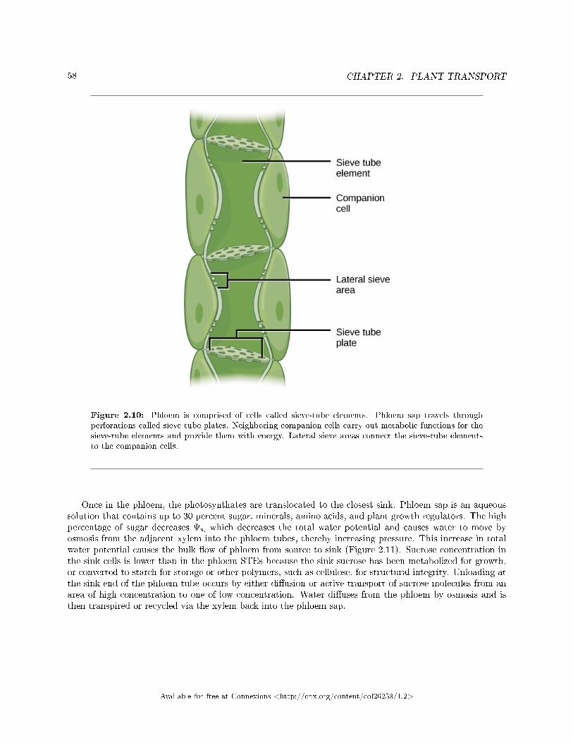
58 CHAPTER 2. PLANT TRANSPORT
Figure 2.10: Phloem is comprised of cells called sieve-tube elements. Phloem sap travels throughperforations called sieve tube plates. Neighboring companion cells carry out metabolic functions for thesieve-tube elements and provide them with energy. Lateral sieve areas connect the sieve-tube elementsto the companion cells.
Once in the phloem, the photosynthates are translocated to the closest sink. Phloem sap is an aqueoussolution that contains up to 30 percent sugar, minerals, amino acids, and plant growth regulators. The highpercentage of sugar decreases Ψs, which decreases the total water potential and causes water to move byosmosis from the adjacent xylem into the phloem tubes, thereby increasing pressure. This increase in totalwater potential causes the bulk �ow of phloem from source to sink (Figure 2.11). Sucrose concentration inthe sink cells is lower than in the phloem STEs because the sink sucrose has been metabolized for growth,or converted to starch for storage or other polymers, such as cellulose, for structural integrity. Unloading atthe sink end of the phloem tube occurs by either di�usion or active transport of sucrose molecules from anarea of high concentration to one of low concentration. Water di�uses from the phloem by osmosis and isthen transpired or recycled via the xylem back into the phloem sap.
Available for free at Connexions <http://cnx.org/content/col26253/1.2>
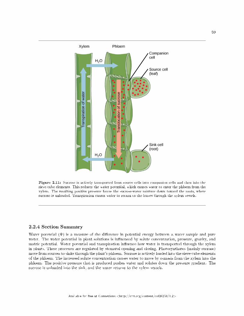
59
Figure 2.11: Sucrose is actively transported from source cells into companion cells and then into thesieve-tube elements. This reduces the water potential, which causes water to enter the phloem from thexylem. The resulting positive pressure forces the sucrose-water mixture down toward the roots, wheresucrose is unloaded. Transpiration causes water to return to the leaves through the xylem vessels.
2.2.4 Section Summary
Water potential (Ψ) is a measure of the di�erence in potential energy between a water sample and purewater. The water potential in plant solutions is in�uenced by solute concentration, pressure, gravity, andmatric potential. Water potential and transpiration in�uence how water is transported through the xylemin plants. These processes are regulated by stomatal opening and closing. Photosynthates (mainly sucrose)move from sources to sinks through the plant's phloem. Sucrose is actively loaded into the sieve-tube elementsof the phloem. The increased solute concentration causes water to move by osmosis from the xylem into thephloem. The positive pressure that is produced pushes water and solutes down the pressure gradient. Thesucrose is unloaded into the sink, and the water returns to the xylem vessels.
Available for free at Connexions <http://cnx.org/content/col26253/1.2>
60 CHAPTER 2. PLANT TRANSPORT
2.2.5 Visual Connection Questions
Exercise 2.2.1 (Solution on p. 61.)
Figure 2.6 Positive water potential is placed on the left side of the tube by increasing Ψp such thatthe water level rises on the right side. Could you equalize the water level on each side of the tubeby adding solute, and if so, how?
Exercise 2.2.2 (Solution on p. 61.)
Figure 2.8 Which of the following statements is false?
a. Negative water potential draws water into the root hairs. Cohesion and adhesion draw waterup the xylem. Transpiration draws water from the leaf.
b. Negative water potential draws water into the root hairs. Cohesion and adhesion draw waterup the phloem. Transpiration draws water from the leaf.
c. Water potential decreases from the roots to the top of the plant.d. Water enters the plants through root hairs and exits through stoma.
2.2.6 Review Questions
Exercise 2.2.3 (Solution on p. 61.)
When stomata open, what occurs?
a. Water vapor is lost to the external environment, increasing the rate of transpiration.b. Water vapor is lost to the external environment, decreasing the rate of transpiration.c. Water vapor enters the spaces in the mesophyll, increasing the rate of transpiration.d. Water vapor enters the spaces in the mesophyll, decreasing the rate of transpiration.
Exercise 2.2.4 (Solution on p. 61.)
Which cells are responsible for the movement of photosynthates through a plant?
a. tracheids, vessel elementsb. tracheids, companion cellsc. vessel elements, companion cellsd. sieve-tube elements, companion cells
2.2.7 Critical Thinking Questions
Exercise 2.2.5 (Solution on p. 61.)
The process of bulk �ow transports �uids in a plant. Describe the two main bulk �ow processes.
Available for free at Connexions <http://cnx.org/content/col26253/1.2>
61
Solutions to Exercises in Chapter 2
to Exercise 2.1.1 (p. 47)Figure 2.1 The air content of the soil decreases.to Exercise 2.1.2 (p. 47)Figure 2.2 The A horizon is the topsoil, and the B horizon is subsoil.to Exercise 2.1.3 (p. 47)Dto Exercise 2.1.4 (p. 47)Ato Exercise 2.1.5 (p. 47)Bto Exercise 2.1.6 (p. 47)Bto Exercise 2.1.7 (p. 48)A mineral soil forms from the weathering of rocks; it is inorganic material. An organic soil is formed fromsedimentation; it mostly consists of humus.to Exercise 2.1.8 (p. 48)Parent material, climate, topography, biological factors, and time a�ect soil formation. Parent material isthe material in which soils form. Climate describes how temperature, moisture, and wind cause di�erentpatterns of weathering, in�uencing the characteristics of the soil. Topography a�ects the characteristics andfertility of a soil. Biological factors include the presence of living organisms that greatly a�ect soil formation.Processes such as freezing and thawing may produce cracks in rocks; plant roots can penetrate these crevicesand produce more fragmentation. Time a�ects soil because soil develops over long periods.to Exercise 2.1.9 (p. 48)Topography a�ects water runo�, which strips away parent material and a�ects plant growth. Steeps soilsare more prone to erosion and may be thinner than soils that are on level surfaces.to Exercise 2.2.1 (p. 60)Figure 2.6 Yes, you can equalize the water level by adding the solute to the left side of the tube such thatwater moves toward the left until the water levels are equal.to Exercise 2.2.2 (p. 60)Figure 2.8 B.to Exercise 2.2.3 (p. 60)Ato Exercise 2.2.4 (p. 60)Dto Exercise 2.2.5 (p. 60)The process of bulk �ow moves water up the xylem and moves photosynthates (solutes) up and down thephloem.
Available for free at Connexions <http://cnx.org/content/col26253/1.2>
62 CHAPTER 2. PLANT TRANSPORT
Available for free at Connexions <http://cnx.org/content/col26253/1.2>
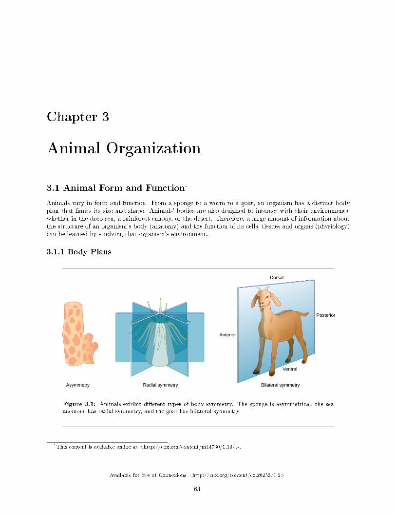
Chapter 3
Animal Organization
3.1 Animal Form and Function1
Animals vary in form and function. From a sponge to a worm to a goat, an organism has a distinct bodyplan that limits its size and shape. Animals' bodies are also designed to interact with their environments,whether in the deep sea, a rainforest canopy, or the desert. Therefore, a large amount of information aboutthe structure of an organism's body (anatomy) and the function of its cells, tissues and organs (physiology)can be learned by studying that organism's environment.
3.1.1 Body Plans
Figure 3.1: Animals exhibit di�erent types of body symmetry. The sponge is asymmetrical, the seaanemone has radial symmetry, and the goat has bilateral symmetry.
1This content is available online at <http://cnx.org/content/m44730/1.10/>.
Available for free at Connexions <http://cnx.org/content/col26253/1.2>
63
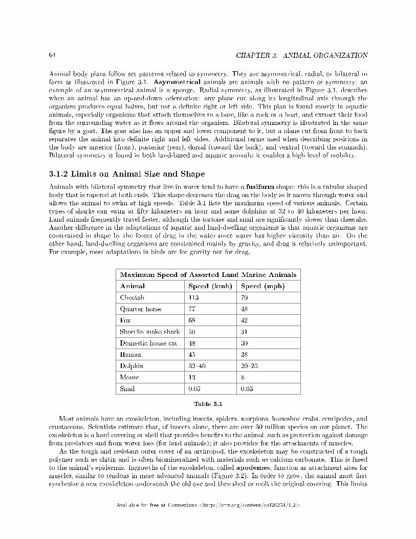
64 CHAPTER 3. ANIMAL ORGANIZATION
Animal body plans follow set patterns related to symmetry. They are asymmetrical, radial, or bilateral inform as illustrated in Figure 3.1. Asymmetrical animals are animals with no pattern or symmetry; anexample of an asymmetrical animal is a sponge. Radial symmetry, as illustrated in Figure 3.1, describeswhen an animal has an up-and-down orientation: any plane cut along its longitudinal axis through theorganism produces equal halves, but not a de�nite right or left side. This plan is found mostly in aquaticanimals, especially organisms that attach themselves to a base, like a rock or a boat, and extract their foodfrom the surrounding water as it �ows around the organism. Bilateral symmetry is illustrated in the same�gure by a goat. The goat also has an upper and lower component to it, but a plane cut from front to backseparates the animal into de�nite right and left sides. Additional terms used when describing positions inthe body are anterior (front), posterior (rear), dorsal (toward the back), and ventral (toward the stomach).Bilateral symmetry is found in both land-based and aquatic animals; it enables a high level of mobility.
3.1.2 Limits on Animal Size and Shape
Animals with bilateral symmetry that live in water tend to have a fusiform shape: this is a tubular shapedbody that is tapered at both ends. This shape decreases the drag on the body as it moves through water andallows the animal to swim at high speeds. Table 3.1 lists the maximum speed of various animals. Certaintypes of sharks can swim at �fty kilometers an hour and some dolphins at 32 to 40 kilometers per hour.Land animals frequently travel faster, although the tortoise and snail are signi�cantly slower than cheetahs.Another di�erence in the adaptations of aquatic and land-dwelling organisms is that aquatic organisms areconstrained in shape by the forces of drag in the water since water has higher viscosity than air. On theother hand, land-dwelling organisms are constrained mainly by gravity, and drag is relatively unimportant.For example, most adaptations in birds are for gravity not for drag.
Maximum Speed of Assorted Land Marine Animals
Animal Speed (kmh) Speed (mph)
Cheetah 113 70
Quarter horse 77 48
Fox 68 42
Short�n mako shark 50 31
Domestic house cat 48 30
Human 45 28
Dolphin 32�40 20�25
Mouse 13 8
Snail 0.05 0.03
Table 3.1
Most animals have an exoskeleton, including insects, spiders, scorpions, horseshoe crabs, centipedes, andcrustaceans. Scientists estimate that, of insects alone, there are over 30 million species on our planet. Theexoskeleton is a hard covering or shell that provides bene�ts to the animal, such as protection against damagefrom predators and from water loss (for land animals); it also provides for the attachments of muscles.
As the tough and resistant outer cover of an arthropod, the exoskeleton may be constructed of a toughpolymer such as chitin and is often biomineralized with materials such as calcium carbonate. This is fusedto the animal's epidermis. Ingrowths of the exoskeleton, called apodemes, function as attachment sites formuscles, similar to tendons in more advanced animals (Figure 3.2). In order to grow, the animal must �rstsynthesize a new exoskeleton underneath the old one and then shed or molt the original covering. This limits
Available for free at Connexions <http://cnx.org/content/col26253/1.2>
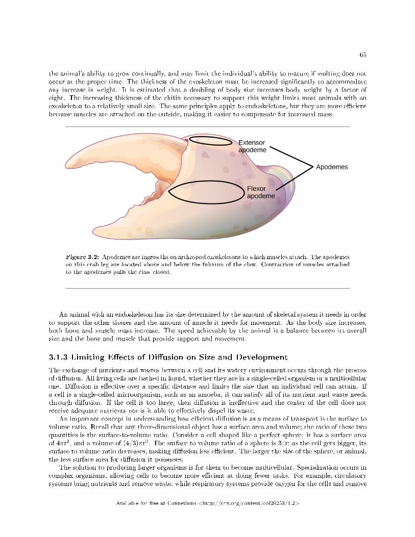
65
the animal's ability to grow continually, and may limit the individual's ability to mature if molting does notoccur at the proper time. The thickness of the exoskeleton must be increased signi�cantly to accommodateany increase in weight. It is estimated that a doubling of body size increases body weight by a factor ofeight. The increasing thickness of the chitin necessary to support this weight limits most animals with anexoskeleton to a relatively small size. The same principles apply to endoskeletons, but they are more e�cientbecause muscles are attached on the outside, making it easier to compensate for increased mass.
Figure 3.2: Apodemes are ingrowths on arthropod exoskeletons to which muscles attach. The apodemeson this crab leg are located above and below the fulcrum of the claw. Contraction of muscles attachedto the apodemes pulls the claw closed.
An animal with an endoskeleton has its size determined by the amount of skeletal system it needs in orderto support the other tissues and the amount of muscle it needs for movement. As the body size increases,both bone and muscle mass increase. The speed achievable by the animal is a balance between its overallsize and the bone and muscle that provide support and movement.
3.1.3 Limiting E�ects of Di�usion on Size and Development
The exchange of nutrients and wastes between a cell and its watery environment occurs through the processof di�usion. All living cells are bathed in liquid, whether they are in a single-celled organism or a multicellularone. Di�usion is e�ective over a speci�c distance and limits the size that an individual cell can attain. Ifa cell is a single-celled microorganism, such as an amoeba, it can satisfy all of its nutrient and waste needsthrough di�usion. If the cell is too large, then di�usion is ine�ective and the center of the cell does notreceive adequate nutrients nor is it able to e�ectively dispel its waste.
An important concept in understanding how e�cient di�usion is as a means of transport is the surface tovolume ratio. Recall that any three-dimensional object has a surface area and volume; the ratio of these twoquantities is the surface-to-volume ratio. Consider a cell shaped like a perfect sphere: it has a surface areaof 4πr2, and a volume of (4/3)πr3. The surface-to-volume ratio of a sphere is 3/r; as the cell gets bigger, itssurface to volume ratio decreases, making di�usion less e�cient. The larger the size of the sphere, or animal,the less surface area for di�usion it possesses.
The solution to producing larger organisms is for them to become multicellular. Specialization occurs incomplex organisms, allowing cells to become more e�cient at doing fewer tasks. For example, circulatorysystems bring nutrients and remove waste, while respiratory systems provide oxygen for the cells and remove
Available for free at Connexions <http://cnx.org/content/col26253/1.2>
66 CHAPTER 3. ANIMAL ORGANIZATION
carbon dioxide from them. Other organ systems have developed further specialization of cells and tissuesand e�ciently control body functions. Moreover, surface-to-volume ratio applies to other areas of animaldevelopment, such as the relationship between muscle mass and cross-sectional surface area in supportingskeletons, and in the relationship between muscle mass and the generation of dissipation of heat.
: Visit this interactive site2 to see an entire animal (a zebra�shembryo) at the cellular and sub-cellular level. Use the zoom and navigation functions for a virtualnanoscopy exploration.
3.1.4 Animal Bioenergetics
All animals must obtain their energy from food they ingest or absorb. These nutrients are converted toadenosine triphosphate (ATP) for short-term storage and use by all cells. Some animals store energy forslightly longer times as glycogen, and others store energy for much longer times in the form of triglycerideshoused in specialized adipose tissues. No energy system is one hundred percent e�cient, and an animal'smetabolism produces waste energy in the form of heat. If an animal can conserve that heat and maintain arelatively constant body temperature, it is classi�ed as a warm-blooded animal and called an endotherm.The insulation used to conserve the body heat comes in the forms of fur, fat, or feathers. The absence ofinsulation in ectothermic animals increases their dependence on the environment for body heat.
The amount of energy expended by an animal over a speci�c time is called its metabolic rate. The rateis measured variously in joules, calories, or kilocalories (1000 calories). Carbohydrates and proteins containabout 4.5 to 5 kcal/g, and fat contains about 9 kcal/g. Metabolic rate is estimated as the basal metabolicrate (BMR) in endothermic animals at rest and as the standard metabolic rate (SMR) in ectotherms.Human males have a BMR of 1600 to 1800 kcal/day, and human females have a BMR of 1300 to 1500kcal/day. Even with insulation, endothermal animals require extensive amounts of energy to maintain aconstant body temperature. An ectotherm such as an alligator has an SMR of 60 kcal/day.
3.1.4.1 Energy Requirements Related to Body Size
Smaller endothermic animals have a greater surface area for their mass than larger ones (Figure 3.3). There-fore, smaller animals lose heat at a faster rate than larger animals and require more energy to maintain aconstant internal temperature. This results in a smaller endothermic animal having a higher BMR, per bodyweight, than a larger endothermic animal.
2http://openstaxcollege.org/l/nanoscopy
Available for free at Connexions <http://cnx.org/content/col26253/1.2>
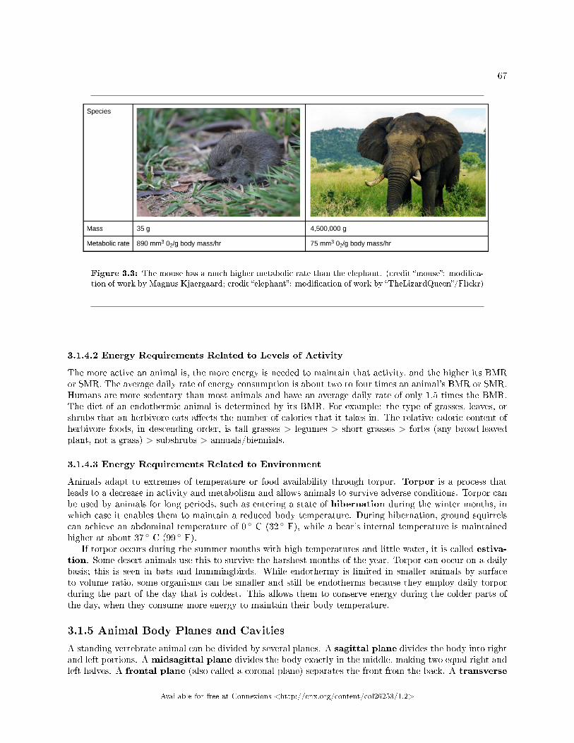
67
Figure 3.3: The mouse has a much higher metabolic rate than the elephant. (credit �mouse�: modi�ca-tion of work by Magnus Kjaergaard; credit �elephant�: modi�cation of work by �TheLizardQueen�/Flickr)
3.1.4.2 Energy Requirements Related to Levels of Activity
The more active an animal is, the more energy is needed to maintain that activity, and the higher its BMRor SMR. The average daily rate of energy consumption is about two to four times an animal's BMR or SMR.Humans are more sedentary than most animals and have an average daily rate of only 1.5 times the BMR.The diet of an endothermic animal is determined by its BMR. For example: the type of grasses, leaves, orshrubs that an herbivore eats a�ects the number of calories that it takes in. The relative caloric content ofherbivore foods, in descending order, is tall grasses > legumes > short grasses > forbs (any broad-leavedplant, not a grass) > subshrubs > annuals/biennials.
3.1.4.3 Energy Requirements Related to Environment
Animals adapt to extremes of temperature or food availability through torpor. Torpor is a process thatleads to a decrease in activity and metabolism and allows animals to survive adverse conditions. Torpor canbe used by animals for long periods, such as entering a state of hibernation during the winter months, inwhich case it enables them to maintain a reduced body temperature. During hibernation, ground squirrelscan achieve an abdominal temperature of 0 ◦ C (32 ◦ F), while a bear's internal temperature is maintainedhigher at about 37 ◦ C (99 ◦ F).
If torpor occurs during the summer months with high temperatures and little water, it is called estiva-tion. Some desert animals use this to survive the harshest months of the year. Torpor can occur on a dailybasis; this is seen in bats and hummingbirds. While endothermy is limited in smaller animals by surfaceto volume ratio, some organisms can be smaller and still be endotherms because they employ daily torporduring the part of the day that is coldest. This allows them to conserve energy during the colder parts ofthe day, when they consume more energy to maintain their body temperature.
3.1.5 Animal Body Planes and Cavities
A standing vertebrate animal can be divided by several planes. A sagittal plane divides the body into rightand left portions. A midsagittal plane divides the body exactly in the middle, making two equal right andleft halves. A frontal plane (also called a coronal plane) separates the front from the back. A transverse
Available for free at Connexions <http://cnx.org/content/col26253/1.2>
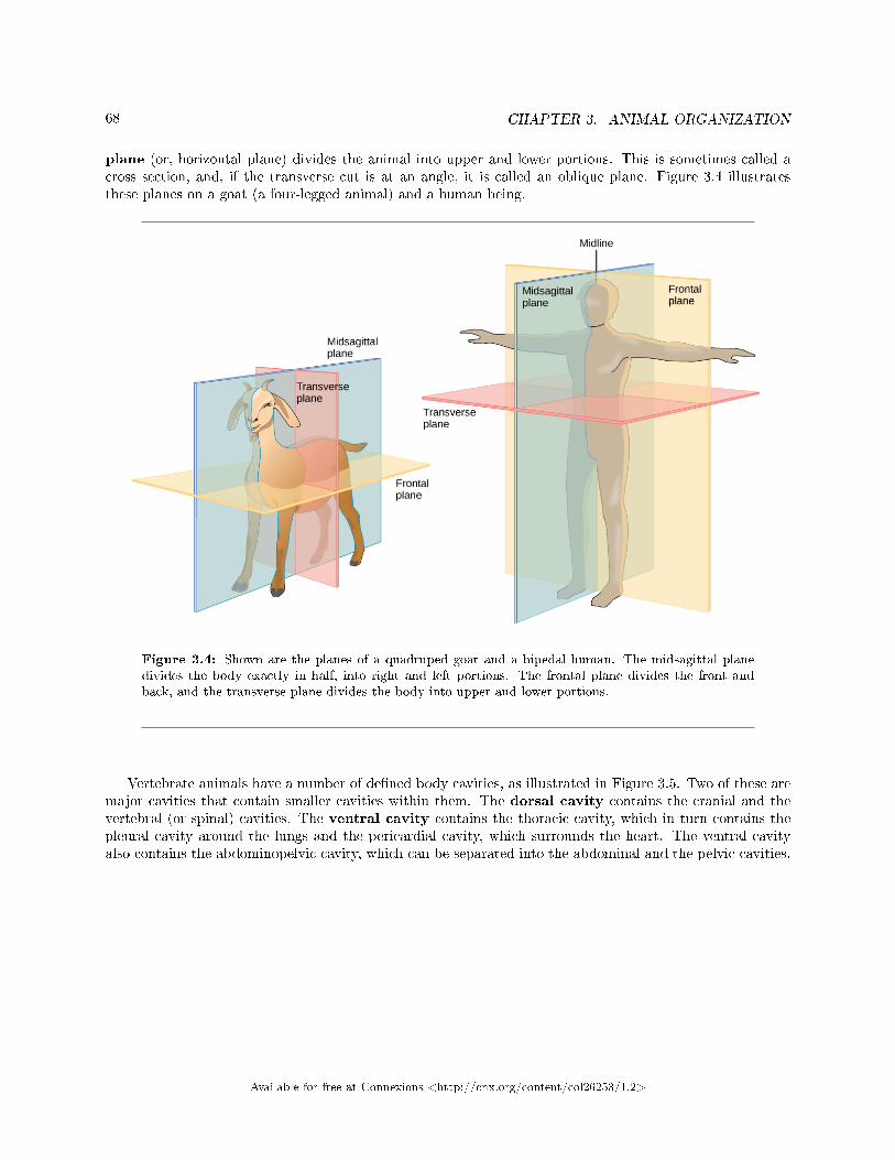
68 CHAPTER 3. ANIMAL ORGANIZATION
plane (or, horizontal plane) divides the animal into upper and lower portions. This is sometimes called across section, and, if the transverse cut is at an angle, it is called an oblique plane. Figure 3.4 illustratesthese planes on a goat (a four-legged animal) and a human being.
Figure 3.4: Shown are the planes of a quadruped goat and a bipedal human. The midsagittal planedivides the body exactly in half, into right and left portions. The frontal plane divides the front andback, and the transverse plane divides the body into upper and lower portions.
Vertebrate animals have a number of de�ned body cavities, as illustrated in Figure 3.5. Two of these aremajor cavities that contain smaller cavities within them. The dorsal cavity contains the cranial and thevertebral (or spinal) cavities. The ventral cavity contains the thoracic cavity, which in turn contains thepleural cavity around the lungs and the pericardial cavity, which surrounds the heart. The ventral cavityalso contains the abdominopelvic cavity, which can be separated into the abdominal and the pelvic cavities.
Available for free at Connexions <http://cnx.org/content/col26253/1.2>
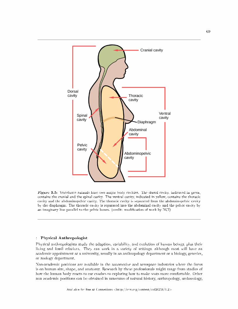
69
Figure 3.5: Vertebrate animals have two major body cavities. The dorsal cavity, indicated in green,contains the cranial and the spinal cavity. The ventral cavity, indicated in yellow, contains the thoraciccavity and the abdominopelvic cavity. The thoracic cavity is separated from the abdominopelvic cavityby the diaphragm. The thoracic cavity is separated into the abdominal cavity and the pelvic cavity byan imaginary line parallel to the pelvis bones. (credit: modi�cation of work by NCI)
: Physical Anthropologist
Physical anthropologists study the adaption, variability, and evolution of human beings, plus theirliving and fossil relatives. They can work in a variety of settings, although most will have anacademic appointment at a university, usually in an anthropology department or a biology, genetics,or zoology department.
Non-academic positions are available in the automotive and aerospace industries where the focusis on human size, shape, and anatomy. Research by these professionals might range from studies ofhow the human body reacts to car crashes to exploring how to make seats more comfortable. Othernon-academic positions can be obtained in museums of natural history, anthropology, archaeology,
Available for free at Connexions <http://cnx.org/content/col26253/1.2>
70 CHAPTER 3. ANIMAL ORGANIZATION
or science and technology. These positions involve educating students from grade school throughgraduate school. Physical anthropologists serve as education coordinators, collection managers,writers for museum publications, and as administrators. Zoos employ these professionals, especiallyif they have an expertise in primate biology; they work in collection management and captivebreeding programs for endangered species. Forensic science utilizes physical anthropology expertisein identifying human and animal remains, assisting in determining the cause of death, and forexpert testimony in trials.
3.1.6 Section Summary
Animal bodies come in a variety of sizes and shapes. Limits on animal size and shape include impacts totheir movement. Di�usion a�ects their size and development. Bioenergetics describes how animals use andobtain energy in relation to their body size, activity level, and environment.
3.1.7 Review Questions
Exercise 3.1.1 (Solution on p. 96.)
Which type of animal maintains a constant internal body temperature?
a. endothermb. ectothermc. coelomated. mesoderm
Exercise 3.1.2 (Solution on p. 96.)
The symmetry found in animals that move swiftly is ________.
a. radialb. bilateralc. sequentiald. interrupted
Exercise 3.1.3 (Solution on p. 96.)
What term describes the condition of a desert mouse that lowers its metabolic rate and �sleeps�during the hot day?
a. turgidb. hibernationc. estivationd. normal sleep pattern
Exercise 3.1.4 (Solution on p. 96.)
A plane that divides an animal into equal right and left portions is ________.
a. diagonalb. midsagittalc. coronald. transverse
Exercise 3.1.5 (Solution on p. 96.)
A plane that divides an animal into dorsal and ventral portions is ________.
Available for free at Connexions <http://cnx.org/content/col26253/1.2>
71
a. sagittalb. midsagittalc. coronald. transverse
Exercise 3.1.6 (Solution on p. 96.)
The pleural cavity is a part of which cavity?
a. dorsal cavityb. thoracic cavityc. abdominal cavityd. pericardial cavity
3.1.8 Free Response
Exercise 3.1.7 (Solution on p. 96.)
How does di�usion limit the size of an organism? How is this counteracted?
Exercise 3.1.8 (Solution on p. 96.)
What is the relationship between BMR and body size? Why?
3.2 Animal Primary Tissues3
The tissues of multicellular, complex animals are four primary types: epithelial, connective, muscle, andnervous. Recall that tissues are groups of similar cells (cells carrying out related functions). These tissuescombine to form organs�like the skin or kidney�that have speci�c, specialized functions within the body.Organs are organized into organ systems to perform functions; examples include the circulatory system, whichconsists of the heart and blood vessels, and the digestive system, consisting of several organs, including thestomach, intestines, liver, and pancreas. Organ systems come together to create an entire organism.
3.2.1 Epithelial Tissues
Epithelial tissues cover the outside of organs and structures in the body and line the lumens of organs in asingle layer or multiple layers of cells. The types of epithelia are classi�ed by the shapes of cells present andthe number of layers of cells. Epithelia composed of a single layer of cells is called simple epithelia; epithelialtissue composed of multiple layers is called strati�ed epithelia. Table 3.2 summarizes the di�erent typesof epithelial tissues.
Di�erent Types of Epithelial Tissues
Cell shape Description Location
continued on next page
3This content is available online at <http://cnx.org/content/m66612/1.15/>.
Available for free at Connexions <http://cnx.org/content/col26253/1.2>
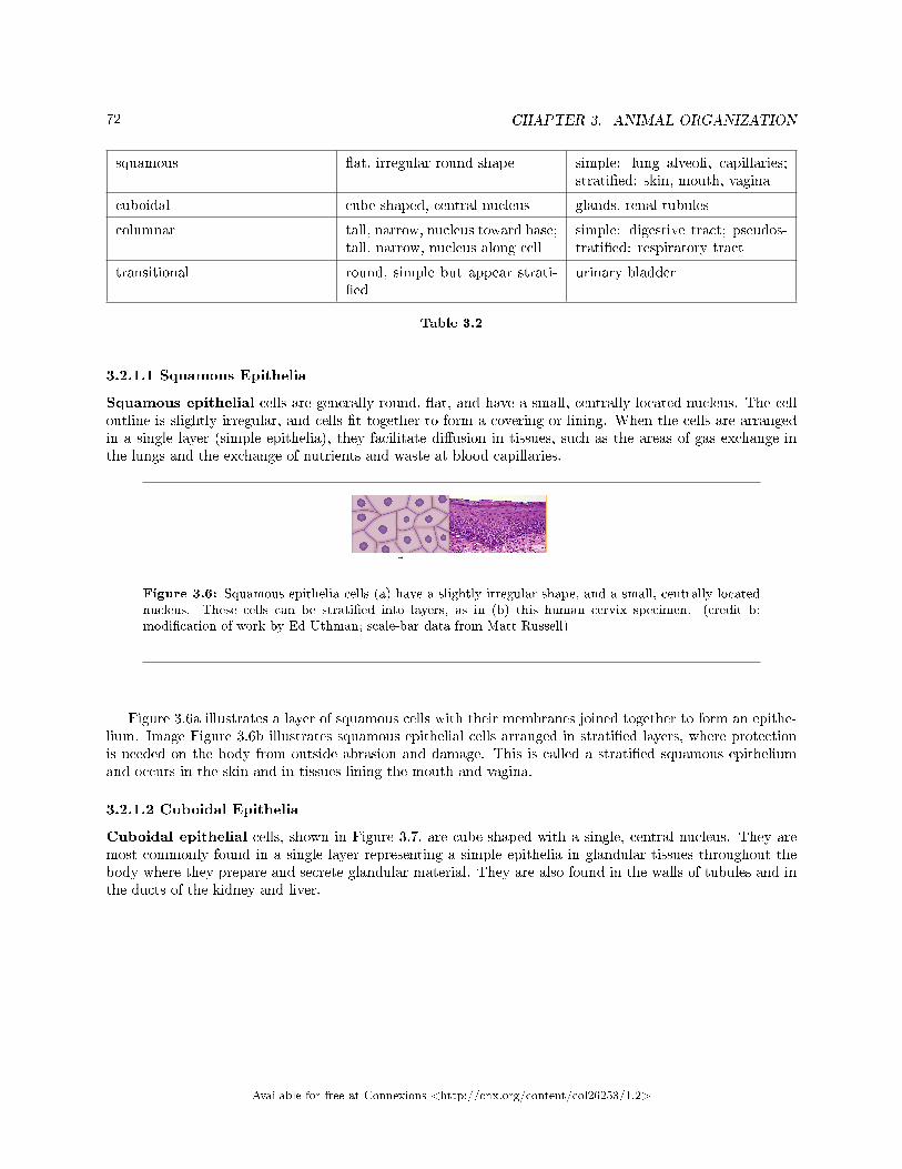
72 CHAPTER 3. ANIMAL ORGANIZATION
squamous �at, irregular round shape simple: lung alveoli, capillaries;strati�ed: skin, mouth, vagina
cuboidal cube shaped, central nucleus glands, renal tubules
columnar tall, narrow, nucleus toward base;tall, narrow, nucleus along cell
simple: digestive tract; pseudos-trati�ed: respiratory tract
transitional round, simple but appear strati-�ed
urinary bladder
Table 3.2
3.2.1.1 Squamous Epithelia
Squamous epithelial cells are generally round, �at, and have a small, centrally located nucleus. The celloutline is slightly irregular, and cells �t together to form a covering or lining. When the cells are arrangedin a single layer (simple epithelia), they facilitate di�usion in tissues, such as the areas of gas exchange inthe lungs and the exchange of nutrients and waste at blood capillaries.
Figure 3.6: Squamous epithelia cells (a) have a slightly irregular shape, and a small, centrally locatednucleus. These cells can be strati�ed into layers, as in (b) this human cervix specimen. (credit b:modi�cation of work by Ed Uthman; scale-bar data from Matt Russell)
Figure 3.6a illustrates a layer of squamous cells with their membranes joined together to form an epithe-lium. Image Figure 3.6b illustrates squamous epithelial cells arranged in strati�ed layers, where protectionis needed on the body from outside abrasion and damage. This is called a strati�ed squamous epitheliumand occurs in the skin and in tissues lining the mouth and vagina.
3.2.1.2 Cuboidal Epithelia
Cuboidal epithelial cells, shown in Figure 3.7, are cube-shaped with a single, central nucleus. They aremost commonly found in a single layer representing a simple epithelia in glandular tissues throughout thebody where they prepare and secrete glandular material. They are also found in the walls of tubules and inthe ducts of the kidney and liver.
Available for free at Connexions <http://cnx.org/content/col26253/1.2>
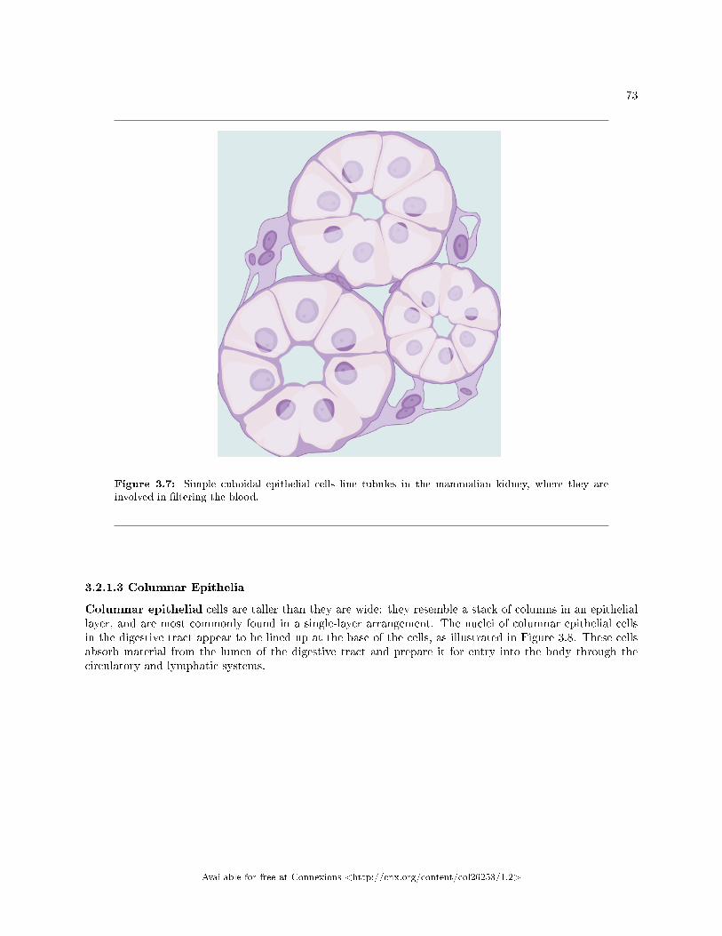
73
Figure 3.7: Simple cuboidal epithelial cells line tubules in the mammalian kidney, where they areinvolved in �ltering the blood.
3.2.1.3 Columnar Epithelia
Columnar epithelial cells are taller than they are wide: they resemble a stack of columns in an epitheliallayer, and are most commonly found in a single-layer arrangement. The nuclei of columnar epithelial cellsin the digestive tract appear to be lined up at the base of the cells, as illustrated in Figure 3.8. These cellsabsorb material from the lumen of the digestive tract and prepare it for entry into the body through thecirculatory and lymphatic systems.
Available for free at Connexions <http://cnx.org/content/col26253/1.2>
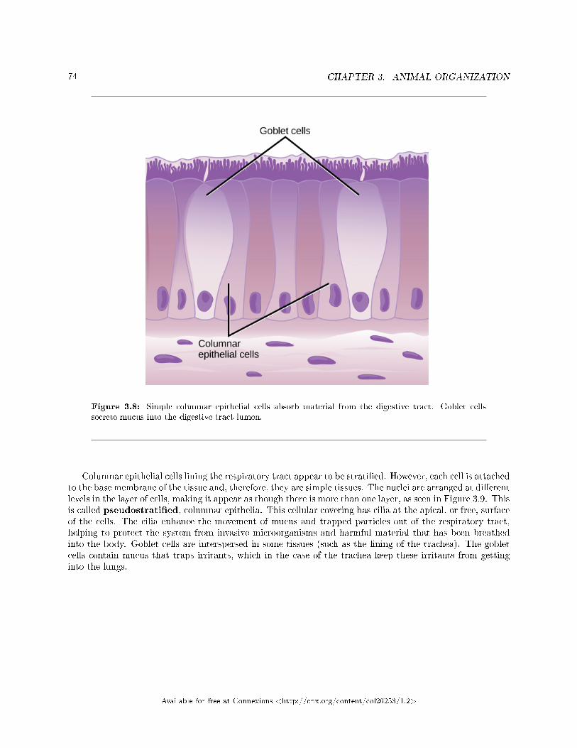
74 CHAPTER 3. ANIMAL ORGANIZATION
Figure 3.8: Simple columnar epithelial cells absorb material from the digestive tract. Goblet cellssecrete mucus into the digestive tract lumen.
Columnar epithelial cells lining the respiratory tract appear to be strati�ed. However, each cell is attachedto the base membrane of the tissue and, therefore, they are simple tissues. The nuclei are arranged at di�erentlevels in the layer of cells, making it appear as though there is more than one layer, as seen in Figure 3.9. Thisis called pseudostrati�ed, columnar epithelia. This cellular covering has cilia at the apical, or free, surfaceof the cells. The cilia enhance the movement of mucus and trapped particles out of the respiratory tract,helping to protect the system from invasive microorganisms and harmful material that has been breathedinto the body. Goblet cells are interspersed in some tissues (such as the lining of the trachea). The gobletcells contain mucus that traps irritants, which in the case of the trachea keep these irritants from gettinginto the lungs.
Available for free at Connexions <http://cnx.org/content/col26253/1.2>
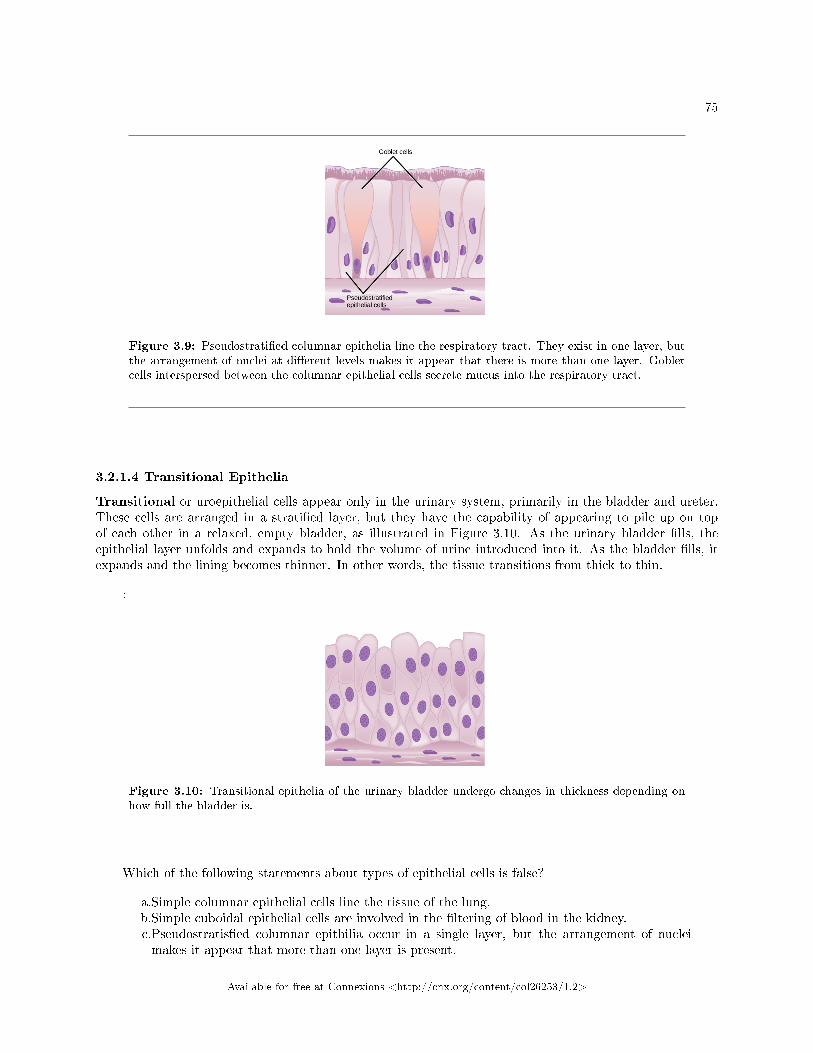
75
Figure 3.9: Pseudostrati�ed columnar epithelia line the respiratory tract. They exist in one layer, butthe arrangement of nuclei at di�erent levels makes it appear that there is more than one layer. Gobletcells interspersed between the columnar epithelial cells secrete mucus into the respiratory tract.
3.2.1.4 Transitional Epithelia
Transitional or uroepithelial cells appear only in the urinary system, primarily in the bladder and ureter.These cells are arranged in a strati�ed layer, but they have the capability of appearing to pile up on topof each other in a relaxed, empty bladder, as illustrated in Figure 3.10. As the urinary bladder �lls, theepithelial layer unfolds and expands to hold the volume of urine introduced into it. As the bladder �lls, itexpands and the lining becomes thinner. In other words, the tissue transitions from thick to thin.
:
Figure 3.10: Transitional epithelia of the urinary bladder undergo changes in thickness depending onhow full the bladder is.
Which of the following statements about types of epithelial cells is false?
a.Simple columnar epithelial cells line the tissue of the lung.b.Simple cuboidal epithelial cells are involved in the �ltering of blood in the kidney.c.Pseudostratis�ed columnar epithilia occur in a single layer, but the arrangement of nucleimakes it appear that more than one layer is present.
Available for free at Connexions <http://cnx.org/content/col26253/1.2>
76 CHAPTER 3. ANIMAL ORGANIZATION
d.Transitional epithelia change in thickness depending on how full the bladder is.
3.2.2 Connective Tissues
Connective tissues are made up of a matrix consisting of living cells and a nonliving substance, calledthe ground substance. The ground substance is made of an organic substance (usually a protein) and aninorganic substance (usually a mineral or water). The principal cell of connective tissues is the �broblast.This cell makes the �bers found in nearly all of the connective tissues. Fibroblasts are motile, able to carryout mitosis, and can synthesize whichever connective tissue is needed. Macrophages, lymphocytes, and,occasionally, leukocytes can be found in some of the tissues. Some tissues have specialized cells that are notfound in the others. The matrix in connective tissues gives the tissue its density. When a connective tissuehas a high concentration of cells or �bers, it has proportionally a less dense matrix.
The organic portion or protein �bers found in connective tissues are either collagen, elastic, or reticular�bers. Collagen �bers provide strength to the tissue, preventing it from being torn or separated from thesurrounding tissues. Elastic �bers are made of the protein elastin; this �ber can stretch to one and one half ofits length and return to its original size and shape. Elastic �bers provide �exibility to the tissues. Reticular�bers are the third type of protein �ber found in connective tissues. This �ber consists of thin strands ofcollagen that form a network of �bers to support the tissue and other organs to which it is connected. Thevarious types of connective tissues, the types of cells and �bers they are made of, and sample locations ofthe tissues is summarized in Table 3.3.
Connective Tissues
Tissue Cells Fibers Location
loose/areolar �broblasts,macrophages, somelymphocytes, someneutrophils
few: collagen, elastic,reticular
around blood vessels;anchors epithelia
dense, �brous connec-tive tissue
�broblasts,macrophages
mostly collagen irregular: skin; regular:tendons, ligaments
cartilage chondrocytes, chon-droblasts
hyaline: few: colla-gen �brocartilage: largeamount of collagen
shark skeleton, fetalbones, human ears,intervertebral discs
bone osteoblasts, osteocytes,osteoclasts
some: collagen, elastic vertebrate skeletons
adipose adipocytes few adipose (fat)
blood red blood cells, whiteblood cells
none blood
Table 3.3
3.2.2.1 Loose/Areolar Connective Tissue
Loose connective tissue, also called areolar connective tissue, has a sampling of all of the components ofa connective tissue. As illustrated in Figure 3.11, loose connective tissue has some �broblasts; macrophagesare present as well. Collagen �bers are relatively wide and stain a light pink, while elastic �bers are thin andstain dark blue to black. The space between the formed elements of the tissue is �lled with the matrix. Thematerial in the connective tissue gives it a loose consistency similar to a cotton ball that has been pulled
Available for free at Connexions <http://cnx.org/content/col26253/1.2>
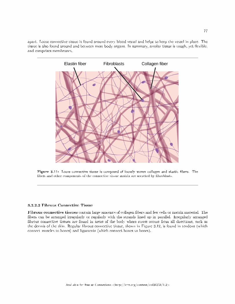
77
apart. Loose connective tissue is found around every blood vessel and helps to keep the vessel in place. Thetissue is also found around and between most body organs. In summary, areolar tissue is tough, yet �exible,and comprises membranes.
Figure 3.11: Loose connective tissue is composed of loosely woven collagen and elastic �bers. The�bers and other components of the connective tissue matrix are secreted by �broblasts.
3.2.2.2 Fibrous Connective Tissue
Fibrous connective tissues contain large amounts of collagen �bers and few cells or matrix material. The�bers can be arranged irregularly or regularly with the strands lined up in parallel. Irregularly arranged�brous connective tissues are found in areas of the body where stress occurs from all directions, such asthe dermis of the skin. Regular �brous connective tissue, shown in Figure 3.12, is found in tendons (whichconnect muscles to bones) and ligaments (which connect bones to bones).
Available for free at Connexions <http://cnx.org/content/col26253/1.2>
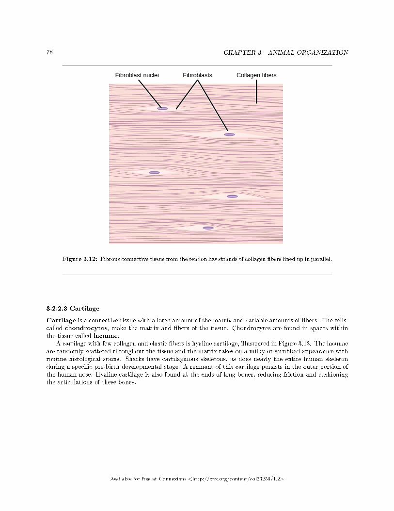
78 CHAPTER 3. ANIMAL ORGANIZATION
Figure 3.12: Fibrous connective tissue from the tendon has strands of collagen �bers lined up in parallel.
3.2.2.3 Cartilage
Cartilage is a connective tissue with a large amount of the matrix and variable amounts of �bers. The cells,called chondrocytes, make the matrix and �bers of the tissue. Chondrocytes are found in spaces withinthe tissue called lacunae.
A cartilage with few collagen and elastic �bers is hyaline cartilage, illustrated in Figure 3.13. The lacunaeare randomly scattered throughout the tissue and the matrix takes on a milky or scrubbed appearance withroutine histological stains. Sharks have cartilaginous skeletons, as does nearly the entire human skeletonduring a speci�c pre-birth developmental stage. A remnant of this cartilage persists in the outer portion ofthe human nose. Hyaline cartilage is also found at the ends of long bones, reducing friction and cushioningthe articulations of these bones.
Available for free at Connexions <http://cnx.org/content/col26253/1.2>
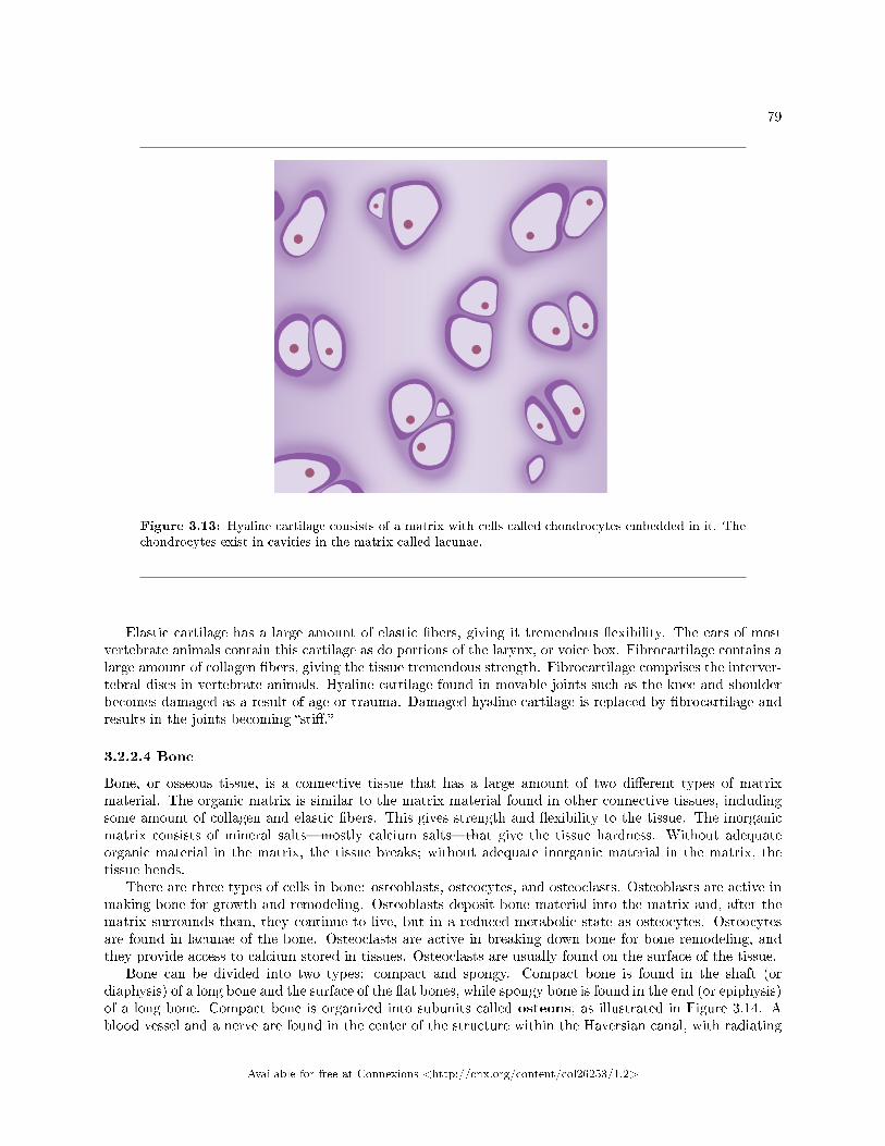
79
Figure 3.13: Hyaline cartilage consists of a matrix with cells called chondrocytes embedded in it. Thechondrocytes exist in cavities in the matrix called lacunae.
Elastic cartilage has a large amount of elastic �bers, giving it tremendous �exibility. The ears of mostvertebrate animals contain this cartilage as do portions of the larynx, or voice box. Fibrocartilage contains alarge amount of collagen �bers, giving the tissue tremendous strength. Fibrocartilage comprises the interver-tebral discs in vertebrate animals. Hyaline cartilage found in movable joints such as the knee and shoulderbecomes damaged as a result of age or trauma. Damaged hyaline cartilage is replaced by �brocartilage andresults in the joints becoming �sti�.�
3.2.2.4 Bone
Bone, or osseous tissue, is a connective tissue that has a large amount of two di�erent types of matrixmaterial. The organic matrix is similar to the matrix material found in other connective tissues, includingsome amount of collagen and elastic �bers. This gives strength and �exibility to the tissue. The inorganicmatrix consists of mineral salts�mostly calcium salts�that give the tissue hardness. Without adequateorganic material in the matrix, the tissue breaks; without adequate inorganic material in the matrix, thetissue bends.
There are three types of cells in bone: osteoblasts, osteocytes, and osteoclasts. Osteoblasts are active inmaking bone for growth and remodeling. Osteoblasts deposit bone material into the matrix and, after thematrix surrounds them, they continue to live, but in a reduced metabolic state as osteocytes. Osteocytesare found in lacunae of the bone. Osteoclasts are active in breaking down bone for bone remodeling, andthey provide access to calcium stored in tissues. Osteoclasts are usually found on the surface of the tissue.
Bone can be divided into two types: compact and spongy. Compact bone is found in the shaft (ordiaphysis) of a long bone and the surface of the �at bones, while spongy bone is found in the end (or epiphysis)of a long bone. Compact bone is organized into subunits called osteons, as illustrated in Figure 3.14. Ablood vessel and a nerve are found in the center of the structure within the Haversian canal, with radiating
Available for free at Connexions <http://cnx.org/content/col26253/1.2>
80 CHAPTER 3. ANIMAL ORGANIZATION
circles of lacunae around it known as lamellae. The wavy lines seen between the lacunae are microchannelscalled canaliculi; they connect the lacunae to aid di�usion between the cells. Spongy bone is made oftiny plates called trabeculae; these plates serve as struts to give the spongy bone strength. Over time,these plates can break causing the bone to become less resilient. Bone tissue forms the internal skeleton ofvertebrate animals, providing structure to the animal and points of attachment for tendons.
Available for free at Connexions <http://cnx.org/content/col26253/1.2>
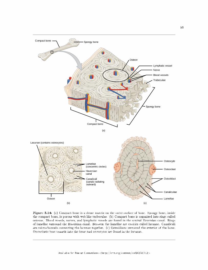
81
Figure 3.14: (a) Compact bone is a dense matrix on the outer surface of bone. Spongy bone, insidethe compact bone, is porous with web-like trabeculae. (b) Compact bone is organized into rings calledosteons. Blood vessels, nerves, and lymphatic vessels are found in the central Haversian canal. Ringsof lamellae surround the Haversian canal. Between the lamellae are cavities called lacunae. Canaliculiare microchannels connecting the lacunae together. (c) Osteoblasts surround the exterior of the bone.Osteoclasts bore tunnels into the bone and osteocytes are found in the lacunae.
Available for free at Connexions <http://cnx.org/content/col26253/1.2>
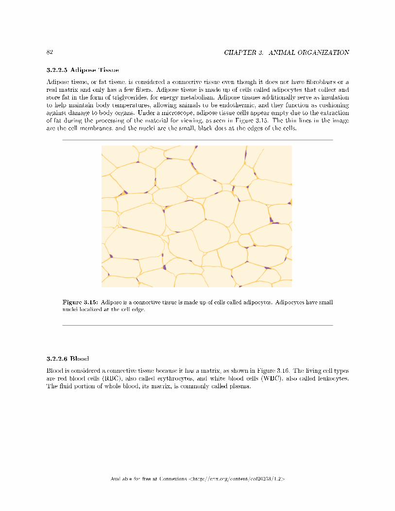
82 CHAPTER 3. ANIMAL ORGANIZATION
3.2.2.5 Adipose Tissue
Adipose tissue, or fat tissue, is considered a connective tissue even though it does not have �broblasts or areal matrix and only has a few �bers. Adipose tissue is made up of cells called adipocytes that collect andstore fat in the form of triglycerides, for energy metabolism. Adipose tissues additionally serve as insulationto help maintain body temperatures, allowing animals to be endothermic, and they function as cushioningagainst damage to body organs. Under a microscope, adipose tissue cells appear empty due to the extractionof fat during the processing of the material for viewing, as seen in Figure 3.15. The thin lines in the imageare the cell membranes, and the nuclei are the small, black dots at the edges of the cells.
Figure 3.15: Adipose is a connective tissue is made up of cells called adipocytes. Adipocytes have smallnuclei localized at the cell edge.
3.2.2.6 Blood
Blood is considered a connective tissue because it has a matrix, as shown in Figure 3.16. The living cell typesare red blood cells (RBC), also called erythrocytes, and white blood cells (WBC), also called leukocytes.The �uid portion of whole blood, its matrix, is commonly called plasma.
Available for free at Connexions <http://cnx.org/content/col26253/1.2>
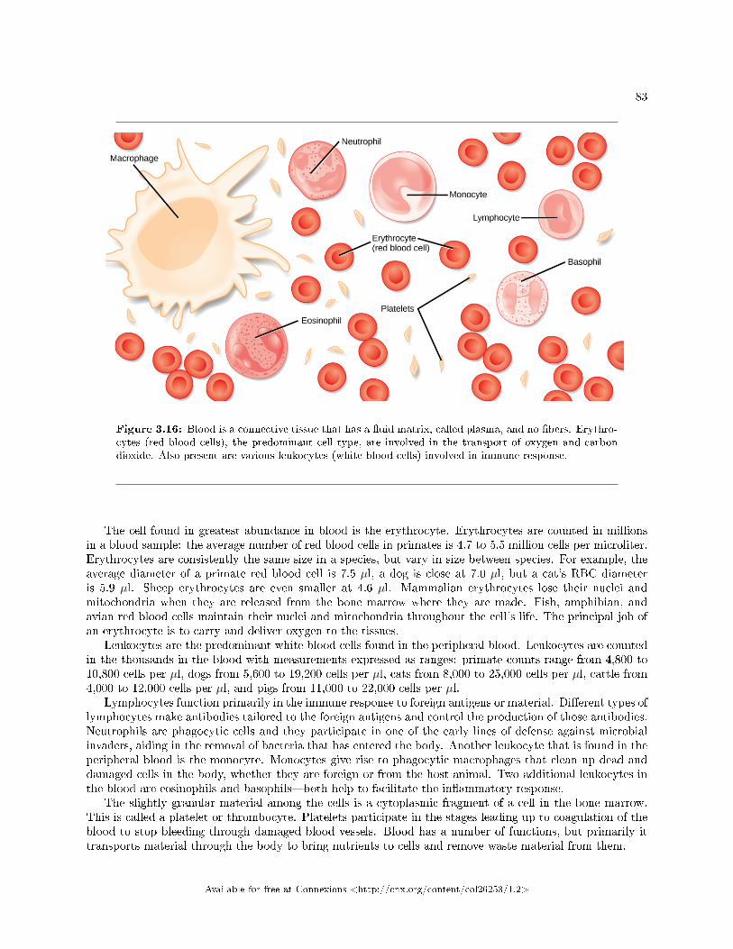
83
Figure 3.16: Blood is a connective tissue that has a �uid matrix, called plasma, and no �bers. Erythro-cytes (red blood cells), the predominant cell type, are involved in the transport of oxygen and carbondioxide. Also present are various leukocytes (white blood cells) involved in immune response.
The cell found in greatest abundance in blood is the erythrocyte. Erythrocytes are counted in millionsin a blood sample: the average number of red blood cells in primates is 4.7 to 5.5 million cells per microliter.Erythrocytes are consistently the same size in a species, but vary in size between species. For example, theaverage diameter of a primate red blood cell is 7.5 µl, a dog is close at 7.0 µl, but a cat's RBC diameteris 5.9 µl. Sheep erythrocytes are even smaller at 4.6 µl. Mammalian erythrocytes lose their nuclei andmitochondria when they are released from the bone marrow where they are made. Fish, amphibian, andavian red blood cells maintain their nuclei and mitochondria throughout the cell's life. The principal job ofan erythrocyte is to carry and deliver oxygen to the tissues.
Leukocytes are the predominant white blood cells found in the peripheral blood. Leukocytes are countedin the thousands in the blood with measurements expressed as ranges: primate counts range from 4,800 to10,800 cells per µl, dogs from 5,600 to 19,200 cells per µl, cats from 8,000 to 25,000 cells per µl, cattle from4,000 to 12,000 cells per µl, and pigs from 11,000 to 22,000 cells per µl.
Lymphocytes function primarily in the immune response to foreign antigens or material. Di�erent types oflymphocytes make antibodies tailored to the foreign antigens and control the production of those antibodies.Neutrophils are phagocytic cells and they participate in one of the early lines of defense against microbialinvaders, aiding in the removal of bacteria that has entered the body. Another leukocyte that is found in theperipheral blood is the monocyte. Monocytes give rise to phagocytic macrophages that clean up dead anddamaged cells in the body, whether they are foreign or from the host animal. Two additional leukocytes inthe blood are eosinophils and basophils�both help to facilitate the in�ammatory response.
The slightly granular material among the cells is a cytoplasmic fragment of a cell in the bone marrow.This is called a platelet or thrombocyte. Platelets participate in the stages leading up to coagulation of theblood to stop bleeding through damaged blood vessels. Blood has a number of functions, but primarily ittransports material through the body to bring nutrients to cells and remove waste material from them.
Available for free at Connexions <http://cnx.org/content/col26253/1.2>
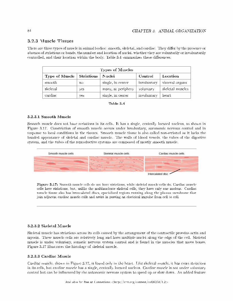
84 CHAPTER 3. ANIMAL ORGANIZATION
3.2.3 Muscle Tissues
There are three types of muscle in animal bodies: smooth, skeletal, and cardiac. They di�er by the presence orabsence of striations or bands, the number and location of nuclei, whether they are voluntarily or involuntarilycontrolled, and their location within the body. Table 3.4 summarizes these di�erences.
Types of Muscles
Type of Muscle Striations Nuclei Control Location
smooth no single, in center involuntary visceral organs
skeletal yes many, at periphery voluntary skeletal muscles
cardiac yes single, in center involuntary heart
Table 3.4
3.2.3.1 Smooth Muscle
Smooth muscle does not have striations in its cells. It has a single, centrally located nucleus, as shown inFigure 3.17. Constriction of smooth muscle occurs under involuntary, autonomic nervous control and inresponse to local conditions in the tissues. Smooth muscle tissue is also called non-striated as it lacks thebanded appearance of skeletal and cardiac muscle. The walls of blood vessels, the tubes of the digestivesystem, and the tubes of the reproductive systems are composed of mostly smooth muscle.
Figure 3.17: Smooth muscle cells do not have striations, while skeletal muscle cells do. Cardiac musclecells have striations, but, unlike the multinucleate skeletal cells, they have only one nucleus. Cardiacmuscle tissue also has intercalated discs, specialized regions running along the plasma membrane thatjoin adjacent cardiac muscle cells and assist in passing an electrical impulse from cell to cell.
3.2.3.2 Skeletal Muscle
Skeletal muscle has striations across its cells caused by the arrangement of the contractile proteins actin andmyosin. These muscle cells are relatively long and have multiple nuclei along the edge of the cell. Skeletalmuscle is under voluntary, somatic nervous system control and is found in the muscles that move bones.Figure 3.17 illustrates the histology of skeletal muscle.
3.2.3.3 Cardiac Muscle
Cardiac muscle, shown in Figure 3.17, is found only in the heart. Like skeletal muscle, it has cross striationsin its cells, but cardiac muscle has a single, centrally located nucleus. Cardiac muscle is not under voluntarycontrol but can be in�uenced by the autonomic nervous system to speed up or slow down. An added feature
Available for free at Connexions <http://cnx.org/content/col26253/1.2>
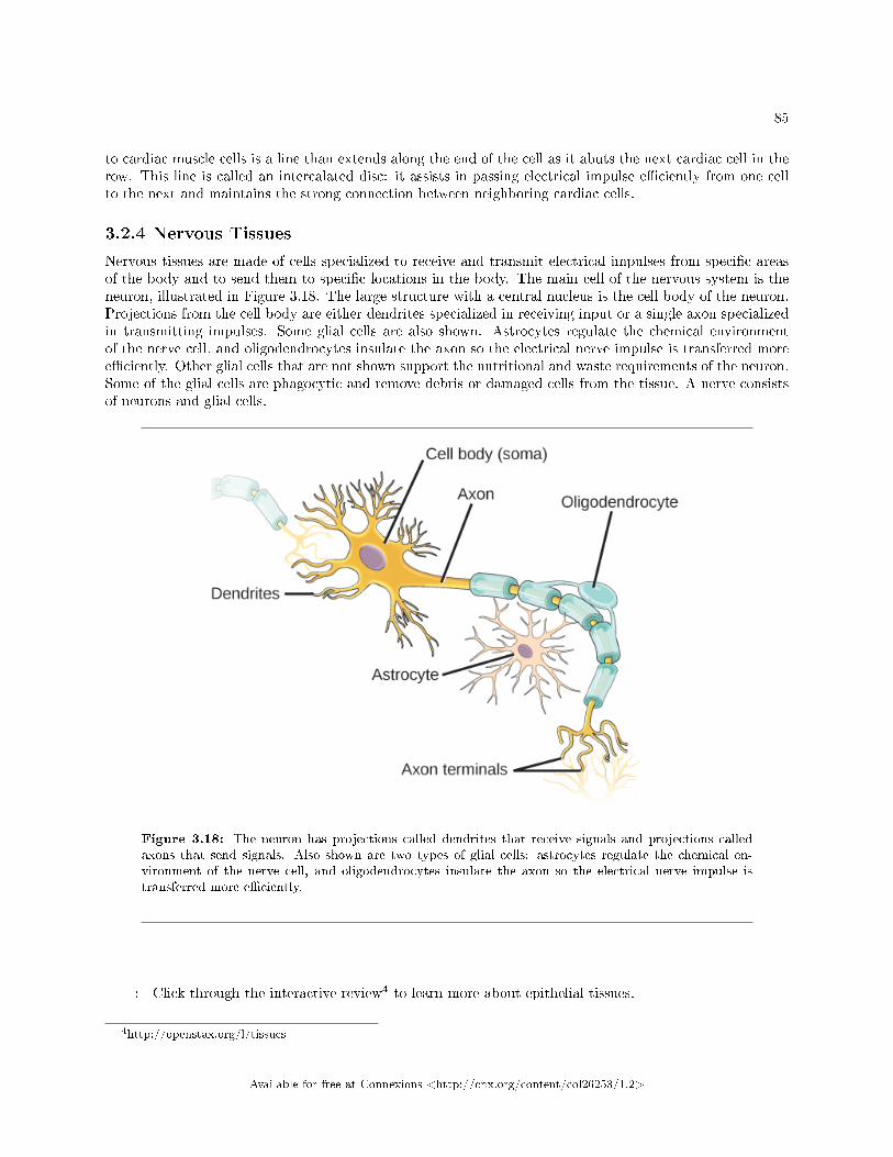
85
to cardiac muscle cells is a line than extends along the end of the cell as it abuts the next cardiac cell in therow. This line is called an intercalated disc: it assists in passing electrical impulse e�ciently from one cellto the next and maintains the strong connection between neighboring cardiac cells.
3.2.4 Nervous Tissues
Nervous tissues are made of cells specialized to receive and transmit electrical impulses from speci�c areasof the body and to send them to speci�c locations in the body. The main cell of the nervous system is theneuron, illustrated in Figure 3.18. The large structure with a central nucleus is the cell body of the neuron.Projections from the cell body are either dendrites specialized in receiving input or a single axon specializedin transmitting impulses. Some glial cells are also shown. Astrocytes regulate the chemical environmentof the nerve cell, and oligodendrocytes insulate the axon so the electrical nerve impulse is transferred moree�ciently. Other glial cells that are not shown support the nutritional and waste requirements of the neuron.Some of the glial cells are phagocytic and remove debris or damaged cells from the tissue. A nerve consistsof neurons and glial cells.
Figure 3.18: The neuron has projections called dendrites that receive signals and projections calledaxons that send signals. Also shown are two types of glial cells: astrocytes regulate the chemical en-vironment of the nerve cell, and oligodendrocytes insulate the axon so the electrical nerve impulse istransferred more e�ciently.
: Click through the interactive review4 to learn more about epithelial tissues.
4http://openstax.org/l/tissues
Available for free at Connexions <http://cnx.org/content/col26253/1.2>
86 CHAPTER 3. ANIMAL ORGANIZATION
: Pathologist
A pathologist is a medical doctor or veterinarian who has specialized in the laboratory detection ofdisease in animals, including humans. These professionals complete medical school education andfollow it with an extensive post-graduate residency at a medical center. A pathologist may overseeclinical laboratories for the evaluation of body tissue and blood samples for the detection of diseaseor infection. They examine tissue specimens through a microscope to identify cancers and otherdiseases. Some pathologists perform autopsies to determine the cause of death and the progressionof disease.
3.2.5 Section Summary
The basic building blocks of complex animals are four primary tissues. These are combined to form organs,which have a speci�c, specialized function within the body, such as the skin or kidney. Organs are organizedtogether to perform common functions in the form of systems. The four primary tissues are epithelia,connective tissues, muscle tissues, and nervous tissues.
3.2.6 Visual Connection Questions
Exercise 3.2.1 (Solution on p. 96.)
Figure 3.10 Which of the following statements about types of epithelial cells is false?
a. Simple columnar epithelial cells line the tissue of the lung.b. Simple cuboidal epithelial cells are involved in the �ltering of blood in the kidney.c. Pseudostratis�ed columnar epithilia occur in a single layer, but the arrangement of nuclei
makes it appear that more than one layer is present.d. Transitional epithelia change in thickness depending on how full the bladder is.
3.2.7 Review Questions
Exercise 3.2.2 (Solution on p. 96.)
Which type of epithelial cell is best adapted to aid di�usion?
a. squamousb. cuboidalc. columnard. transitional
Exercise 3.2.3 (Solution on p. 96.)
Which type of epithelial cell is found in glands?
a. squamousb. cuboidalc. columnard. transitional
Exercise 3.2.4 (Solution on p. 96.)
Which type of epithelial cell is found in the urinary bladder?
a. squamousb. cuboidal
Available for free at Connexions <http://cnx.org/content/col26253/1.2>
87
c. columnard. transitional
Exercise 3.2.5 (Solution on p. 96.)
Which type of connective tissue has the most �bers?
a. loose connective tissueb. �brous connective tissuec. cartilaged. bone
Exercise 3.2.6 (Solution on p. 96.)
Which type of connective tissue has a mineralized di�erent matrix?
a. loose connective tissueb. �brous connective tissuec. cartilaged. bone
Exercise 3.2.7 (Solution on p. 96.)
The cell found in bone that breaks it down is called an ________.
a. osteoblastb. osteocytec. osteoclastd. osteon
Exercise 3.2.8 (Solution on p. 96.)
The cell found in bone that makes the bone is called an ________.
a. osteoblastb. osteocytec. osteoclastd. osteon
Exercise 3.2.9 (Solution on p. 96.)
Plasma is the ________.
a. �bers in bloodb. matrix of bloodc. cell that phagocytizes bacteriad. cell fragment found in the tissue
Exercise 3.2.10 (Solution on p. 96.)
The type of muscle cell under voluntary control is the ________.
a. smooth muscleb. skeletal musclec. cardiac muscled. visceral muscle
Exercise 3.2.11 (Solution on p. 96.)
The part of a neuron that contains the nucleus is the
Available for free at Connexions <http://cnx.org/content/col26253/1.2>
88 CHAPTER 3. ANIMAL ORGANIZATION
a. cell bodyb. dendritec. axond. glial
Exercise 3.2.12 (Solution on p. 96.)
Why are intercalated discs essential to the function of cardiac muscle?
a. The discs maintain the barriers between the cells.b. The discs pass nutrients between cells.c. The discs ensure that all the cardiac muscle cells beat as a single unit.d. The discs control the heart rate.
3.2.8 Critical Thinking Questions
Exercise 3.2.13 (Solution on p. 96.)
How can squamous epithelia both facilitate di�usion and prevent damage from abrasion?
Exercise 3.2.14 (Solution on p. 97.)
What are the similarities between cartilage and bone?
Exercise 3.2.15 (Solution on p. 97.)
Multiple sclerosis is a debilitating autoimmune disease that results in the loss of the insulationaround neuron axons. What cell type is the immune system attacking, and how does this disruptthe transfer of messages by the nervous system?
Exercise 3.2.16 (Solution on p. 97.)
When a person leads a sedentary life his skeletal muscles atrophy, but his smooth muscles do not.Why?
3.3 Homeostasis5
Animal organs and organ systems constantly adjust to internal and external changes through a process calledhomeostasis (�steady state�). These changes might be in the level of glucose or calcium in blood or in externaltemperatures. Homeostasis means to maintain dynamic equilibrium in the body. It is dynamic becauseit is constantly adjusting to the changes that the body's systems encounter. It is equilibrium because bodyfunctions are kept within speci�c ranges. Even an animal that is apparently inactive is maintaining thishomeostatic equilibrium.
3.3.1 Homeostatic Process
The goal of homeostasis is the maintenance of equilibrium around a point or value called a set point. Whilethere are normal �uctuations from the set point, the body's systems will usually attempt to go back to thispoint. A change in the internal or external environment is called a stimulus and is detected by a receptor;the response of the system is to adjust the deviation parameter toward the set point. For instance, if thebody becomes too warm, adjustments are made to cool the animal. If the blood's glucose rises after a meal,adjustments are made to lower the blood glucose level by getting the nutrient into tissues that need it or tostore it for later use.
5This content is available online at <http://cnx.org/content/m44733/1.11/>.
Available for free at Connexions <http://cnx.org/content/col26253/1.2>
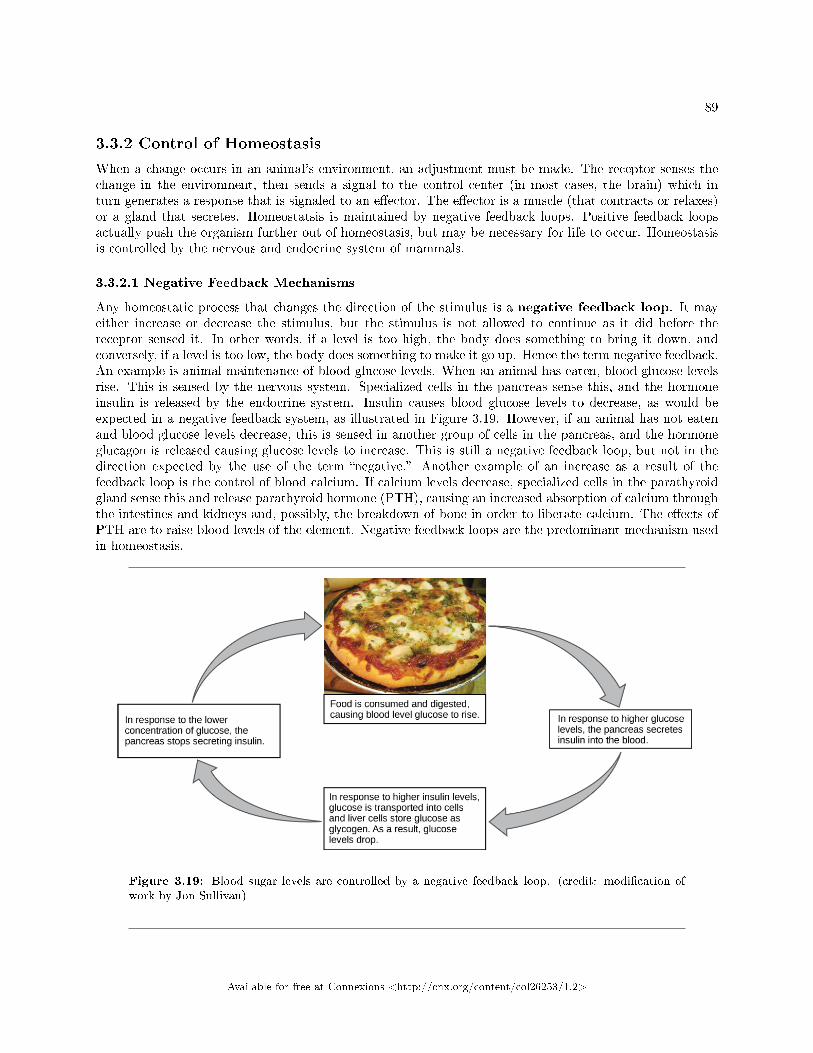
89
3.3.2 Control of Homeostasis
When a change occurs in an animal's environment, an adjustment must be made. The receptor senses thechange in the environment, then sends a signal to the control center (in most cases, the brain) which inturn generates a response that is signaled to an e�ector. The e�ector is a muscle (that contracts or relaxes)or a gland that secretes. Homeostatsis is maintained by negative feedback loops. Positive feedback loopsactually push the organism further out of homeostasis, but may be necessary for life to occur. Homeostasisis controlled by the nervous and endocrine system of mammals.
3.3.2.1 Negative Feedback Mechanisms
Any homeostatic process that changes the direction of the stimulus is a negative feedback loop. It mayeither increase or decrease the stimulus, but the stimulus is not allowed to continue as it did before thereceptor sensed it. In other words, if a level is too high, the body does something to bring it down, andconversely, if a level is too low, the body does something to make it go up. Hence the term negative feedback.An example is animal maintenance of blood glucose levels. When an animal has eaten, blood glucose levelsrise. This is sensed by the nervous system. Specialized cells in the pancreas sense this, and the hormoneinsulin is released by the endocrine system. Insulin causes blood glucose levels to decrease, as would beexpected in a negative feedback system, as illustrated in Figure 3.19. However, if an animal has not eatenand blood glucose levels decrease, this is sensed in another group of cells in the pancreas, and the hormoneglucagon is released causing glucose levels to increase. This is still a negative feedback loop, but not in thedirection expected by the use of the term �negative.� Another example of an increase as a result of thefeedback loop is the control of blood calcium. If calcium levels decrease, specialized cells in the parathyroidgland sense this and release parathyroid hormone (PTH), causing an increased absorption of calcium throughthe intestines and kidneys and, possibly, the breakdown of bone in order to liberate calcium. The e�ects ofPTH are to raise blood levels of the element. Negative feedback loops are the predominant mechanism usedin homeostasis.
Figure 3.19: Blood sugar levels are controlled by a negative feedback loop. (credit: modi�cation ofwork by Jon Sullivan)
Available for free at Connexions <http://cnx.org/content/col26253/1.2>
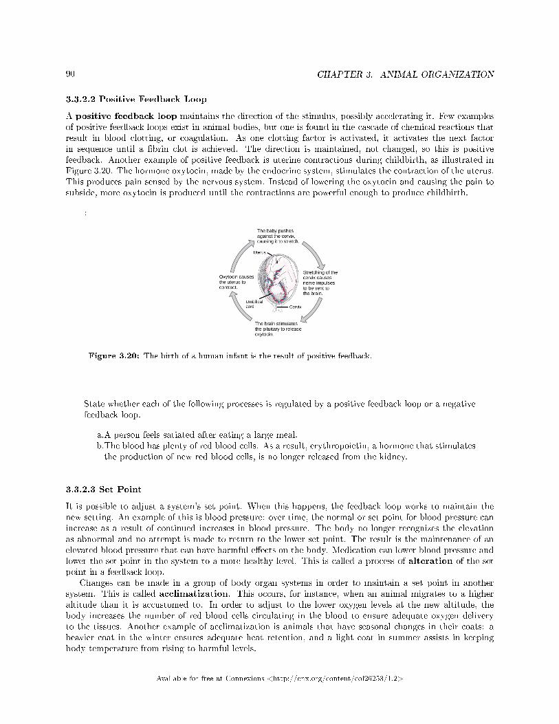
90 CHAPTER 3. ANIMAL ORGANIZATION
3.3.2.2 Positive Feedback Loop
A positive feedback loop maintains the direction of the stimulus, possibly accelerating it. Few examplesof positive feedback loops exist in animal bodies, but one is found in the cascade of chemical reactions thatresult in blood clotting, or coagulation. As one clotting factor is activated, it activates the next factorin sequence until a �brin clot is achieved. The direction is maintained, not changed, so this is positivefeedback. Another example of positive feedback is uterine contractions during childbirth, as illustrated inFigure 3.20. The hormone oxytocin, made by the endocrine system, stimulates the contraction of the uterus.This produces pain sensed by the nervous system. Instead of lowering the oxytocin and causing the pain tosubside, more oxytocin is produced until the contractions are powerful enough to produce childbirth.
:
Figure 3.20: The birth of a human infant is the result of positive feedback.
State whether each of the following processes is regulated by a positive feedback loop or a negativefeedback loop.
a.A person feels satiated after eating a large meal.b.The blood has plenty of red blood cells. As a result, erythropoietin, a hormone that stimulatesthe production of new red blood cells, is no longer released from the kidney.
3.3.2.3 Set Point
It is possible to adjust a system's set point. When this happens, the feedback loop works to maintain thenew setting. An example of this is blood pressure: over time, the normal or set point for blood pressure canincrease as a result of continued increases in blood pressure. The body no longer recognizes the elevationas abnormal and no attempt is made to return to the lower set point. The result is the maintenance of anelevated blood pressure that can have harmful e�ects on the body. Medication can lower blood pressure andlower the set point in the system to a more healthy level. This is called a process of alteration of the setpoint in a feedback loop.
Changes can be made in a group of body organ systems in order to maintain a set point in anothersystem. This is called acclimatization. This occurs, for instance, when an animal migrates to a higheraltitude than it is accustomed to. In order to adjust to the lower oxygen levels at the new altitude, thebody increases the number of red blood cells circulating in the blood to ensure adequate oxygen deliveryto the tissues. Another example of acclimatization is animals that have seasonal changes in their coats: aheavier coat in the winter ensures adequate heat retention, and a light coat in summer assists in keepingbody temperature from rising to harmful levels.
Available for free at Connexions <http://cnx.org/content/col26253/1.2>
91
: Feedback mechanisms can be understood in terms of driving a racecar along a track: watch a short video lesson on positive and negative feedback loops.
3.3.3 Homeostasis: Thermoregulation
Body temperature a�ects body activities. Generally, as body temperature rises, enzyme activity rises as well.For every ten degree centigrade rise in temperature, enzyme activity doubles, up to a point. Body proteins,including enzymes, begin to denature and lose their function with high heat (around 50oC for mammals).Enzyme activity will decrease by half for every ten degree centigrade drop in temperature, to the point offreezing, with a few exceptions. Some �sh can withstand freezing solid and return to normal with thawing.
: Watch this Discovery Channel video on thermoregulation to seeillustrations of this process in a variety of animals.
3.3.4 Endotherms and Ectotherms
Animals can be divided into two groups: some maintain a constant body temperature in the face of di�eringenvironmental temperatures, while others have a body temperature that is the same as their environmentand thus varies with the environment. Animals that do not control their body temperature are ectotherms.This group has been called cold-blooded, but the term may not apply to an animal in the desert with avery warm body temperature. In contrast to ectotherms, which rely on external temperatures to set theirbody temperatures, poikilotherms are animals with constantly varying internal temperatures. An animalthat maintains a constant body temperature in the face of environmental changes is called a homeotherm.Endotherms are animals that rely on internal sources for body temperature but which can exhibit extremesin temperature. These animals are able to maintain a level of activity at cooler temperature, which anectotherm cannot due to di�ering enzyme levels of activity.
Heat can be exchanged between an animal and its environment through four mechanisms: radiation,evaporation, convection, and conduction (Figure 3.21). Radiation is the emission of electromagnetic �heat�waves. Heat comes from the sun in this manner and radiates from dry skin the same way. Heat can beremoved with liquid from a surface during evaporation. This occurs when a mammal sweats. Convectioncurrents of air remove heat from the surface of dry skin as the air passes over it. Heat will be conducted
Available for free at Connexions <http://cnx.org/content/col26253/1.2>
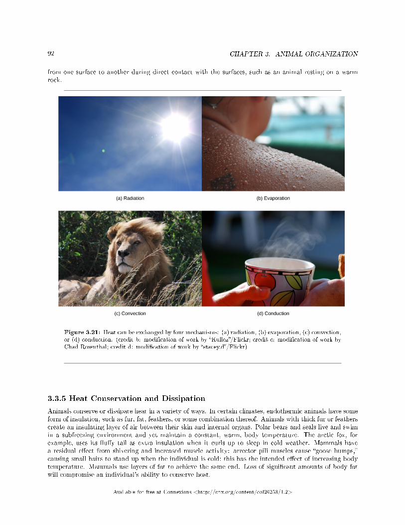
92 CHAPTER 3. ANIMAL ORGANIZATION
from one surface to another during direct contact with the surfaces, such as an animal resting on a warmrock.
Figure 3.21: Heat can be exchanged by four mechanisms: (a) radiation, (b) evaporation, (c) convection,or (d) conduction. (credit b: modi�cation of work by �Kullez�/Flickr; credit c: modi�cation of work byChad Rosenthal; credit d: modi�cation of work by �stacey.d�/Flickr)
3.3.5 Heat Conservation and Dissipation
Animals conserve or dissipate heat in a variety of ways. In certain climates, endothermic animals have someform of insulation, such as fur, fat, feathers, or some combination thereof. Animals with thick fur or featherscreate an insulating layer of air between their skin and internal organs. Polar bears and seals live and swimin a subfreezing environment and yet maintain a constant, warm, body temperature. The arctic fox, forexample, uses its �u�y tail as extra insulation when it curls up to sleep in cold weather. Mammals havea residual e�ect from shivering and increased muscle activity: arrector pili muscles cause �goose bumps,�causing small hairs to stand up when the individual is cold; this has the intended e�ect of increasing bodytemperature. Mammals use layers of fat to achieve the same end. Loss of signi�cant amounts of body fatwill compromise an individual's ability to conserve heat.
Available for free at Connexions <http://cnx.org/content/col26253/1.2>
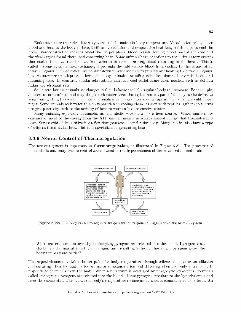
93
Endotherms use their circulatory systems to help maintain body temperature. Vasodilation brings moreblood and heat to the body surface, facilitating radiation and evaporative heat loss, which helps to cool thebody. Vasoconstriction reduces blood �ow in peripheral blood vessels, forcing blood toward the core andthe vital organs found there, and conserving heat. Some animals have adaptions to their circulatory systemthat enable them to transfer heat from arteries to veins, warming blood returning to the heart. This iscalled a countercurrent heat exchange; it prevents the cold venous blood from cooling the heart and otherinternal organs. This adaption can be shut down in some animals to prevent overheating the internal organs.The countercurrent adaption is found in many animals, including dolphins, sharks, bony �sh, bees, andhummingbirds. In contrast, similar adaptations can help cool endotherms when needed, such as dolphin�ukes and elephant ears.
Some ectothermic animals use changes in their behavior to help regulate body temperature. For example,a desert ectothermic animal may simply seek cooler areas during the hottest part of the day in the desert tokeep from getting too warm. The same animals may climb onto rocks to capture heat during a cold desertnight. Some animals seek water to aid evaporation in cooling them, as seen with reptiles. Other ectothermsuse group activity such as the activity of bees to warm a hive to survive winter.
Many animals, especially mammals, use metabolic waste heat as a heat source. When muscles arecontracted, most of the energy from the ATP used in muscle actions is wasted energy that translates intoheat. Severe cold elicits a shivering re�ex that generates heat for the body. Many species also have a typeof adipose tissue called brown fat that specializes in generating heat.
3.3.6 Neural Control of Thermoregulation
The nervous system is important to thermoregulation, as illustrated in Figure 3.21. The processes ofhomeostasis and temperature control are centered in the hypothalamus of the advanced animal brain.
:
Figure 3.22: The body is able to regulate temperature in response to signals from the nervous system.
When bacteria are destroyed by leuckocytes, pyrogens are released into the blood. Pyrogens resetthe body's thermostat to a higher temperature, resulting in fever. How might pyrogens cause thebody temperature to rise?
The hypothalamus maintains the set point for body temperature through re�exes that cause vasodilationand sweating when the body is too warm, or vasoconstriction and shivering when the body is too cold. Itresponds to chemicals from the body. When a bacterium is destroyed by phagocytic leukocytes, chemicalscalled endogenous pyrogens are released into the blood. These pyrogens circulate to the hypothalamus andreset the thermostat. This allows the body's temperature to increase in what is commonly called a fever. An
Available for free at Connexions <http://cnx.org/content/col26253/1.2>
94 CHAPTER 3. ANIMAL ORGANIZATION
increase in body temperature causes iron to be conserved, which reduces a nutrient needed by bacteria. Anincrease in body heat also increases the activity of the animal's enzymes and protective cells while inhibitingthe enzymes and activity of the invading microorganisms. Finally, heat itself may also kill the pathogen. Afever that was once thought to be a complication of an infection is now understood to be a normal defensemechanism.
3.3.7 Section Summary
Homeostasis is a dynamic equilibrium that is maintained in body tissues and organs. It is dynamic becauseit is constantly adjusting to the changes that the systems encounter. It is in equilibrium because bodyfunctions are kept within a normal range, with some �uctuations around a set point for the processes.
3.3.8 Art Connections
Exercise 3.3.1 (Solution on p. 97.)
Figure 3.20 State whether each of the following processes are regulated by a positive feedback loopor a negative feedback loop.
a. A person feels satiated after eating a large meal.b. The blood has plenty of red blood cells. As a result, erythropoietin, a hormone that stimulates
the production of new red blood cells, is no longer released from the kidney.
Exercise 3.3.2 (Solution on p. 97.)
Figure 3.22 When bacteria are destroyed by leuckocytes, pyrogens are released into the blood.Pyrogens reset the body's thermostat to a higher temperature, resulting in fever. How mightpyrogens cause the body temperature to rise?
3.3.9 Review Questions
Exercise 3.3.3 (Solution on p. 97.)
When faced with a sudden drop in environmental temperature, an endothermic animal will:
a. experience a drop in its body temperatureb. wait to see if it goes lowerc. increase muscle activity to generate heatd. add fur or fat to increase insulation
Exercise 3.3.4 (Solution on p. 97.)
Which is an example of negative feedback?
a. lowering of blood glucose after a mealb. blood clotting after an injuryc. lactation during nursingd. uterine contractions during labor
Exercise 3.3.5 (Solution on p. 97.)
Which method of heat exchange occurs during direct contact between the source and animal?
a. radiationb. evaporationc. convection
Available for free at Connexions <http://cnx.org/content/col26253/1.2>
95
d. conduction
Exercise 3.3.6 (Solution on p. 97.)
The body's thermostat is located in the ________.
a. homeostatic receptorb. hypothalamusc. medullad. vasodilation center
3.3.10 Free Response
Exercise 3.3.7 (Solution on p. 97.)
Why are negative feedback loops used to control body homeostasis?
Exercise 3.3.8 (Solution on p. 97.)
Why is a fever a �good thing� during a bacterial infection?
Exercise 3.3.9 (Solution on p. 97.)
How is a condition such as diabetes a good example of the failure of a set point in humans?
Available for free at Connexions <http://cnx.org/content/col26253/1.2>
96 CHAPTER 3. ANIMAL ORGANIZATION
Solutions to Exercises in Chapter 3
to Exercise 3.1.1 (p. 70)Ato Exercise 3.1.2 (p. 70)Bto Exercise 3.1.3 (p. 70)Cto Exercise 3.1.4 (p. 70)Bto Exercise 3.1.5 (p. 70)Dto Exercise 3.1.6 (p. 71)Bto Exercise 3.1.7 (p. 71)Di�usion is e�ective over a very short distance. If a cell exceeds this distance in its size, the center of thecell cannot get adequate nutrients nor can it expel enough waste to survive. To compensate for this, cells canloosely adhere to each other in a liquid medium, or develop into multi-celled organisms that use circulatoryand respiratory systems to deliver nutrients and remove wastes.to Exercise 3.1.8 (p. 71)Basal Metabolic Rate is an expression of the metabolic processes that occur to maintain an individual'sfunctioning and body temperature. Smaller bodied animals have a relatively large surface area comparedto a much larger animal. The small animal's large surface area leads to increased heat loss that the animalmust compensate for, resulting in a higher BMR. A large animal, having less relative surface area, does notlose as much heat and has a correspondingly lower BMR.to Exercise 3.2.1 (p. 86)Figure 3.10 Ato Exercise 3.2.2 (p. 86)Cto Exercise 3.2.3 (p. 86)Bto Exercise 3.2.4 (p. 86)Dto Exercise 3.2.5 (p. 87)Bto Exercise 3.2.6 (p. 87)Dto Exercise 3.2.7 (p. 87)Cto Exercise 3.2.8 (p. 87)Ato Exercise 3.2.9 (p. 87)Bto Exercise 3.2.10 (p. 87)Bto Exercise 3.2.11 (p. 87)Ato Exercise 3.2.12 (p. 88)C
Available for free at Connexions <http://cnx.org/content/col26253/1.2>
97
to Exercise 3.2.13 (p. 88)Squamous epithelia can be either simple or strati�ed. As a single layer of cells, it presents a very thinepithelia that minimally inhibits di�usion. As a strati�ed epithelia, the surface cells can be sloughed o� andthe cells in deeper layers protect the underlying tissues from damage.to Exercise 3.2.14 (p. 88)Both contain cells other than the traditional �broblast. Both have cells that lodge in spaces within the tissuecalled lacunae. Both collagen and elastic �bers are found in bone and cartilage. Both tissues participate invertebrate skeletal development and formation.to Exercise 3.2.15 (p. 88)In multiple sclerosis, the immune system attacks the oligodendrocytes. The death of oligodendrocytes resultsin the loss of the insulating sheath around the axon of the neurons. When the sheath is gone, the electricalimpulses travel much more slowly down the length of the axon.to Exercise 3.2.16 (p. 88)Skeletal muscles are involved in voluntary motion, so the person has to make the choice to work thosemuscles through exercise or movement. Smooth muscles are involved in involuntary activities of the body(ex. blood vessel expansion and contraction, intestinal peristalsis) so they are active even when a person issedentary.to Exercise 3.3.1 (p. 94)Figure 3.20 Both processes are the result of negative feedback loops. Negative feedback loops, which tendto keep a system at equilibrium, are more common than positive feedback loops.to Exercise 3.3.2 (p. 94)Figure 3.22 Pyrogens increase body temperature by causing the blood vessels to constrict, inducing shivering,and stopping sweat glands from secreting �uid.to Exercise 3.3.3 (p. 94)Cto Exercise 3.3.4 (p. 94)Ato Exercise 3.3.5 (p. 94)Dto Exercise 3.3.6 (p. 95)Bto Exercise 3.3.7 (p. 95)An adjustment to a change in the internal or external environment requires a change in the direction ofthe stimulus. A negative feedback loop accomplishes this, while a positive feedback loop would continue thestimulus and result in harm to the animal.to Exercise 3.3.8 (p. 95)Mammalian enzymes increase activity to the point of denaturation, increasing the chemical activity of thecells involved. Bacterial enzymes have a speci�c temperature for their most e�cient activity and are inhibitedat either higher or lower temperatures. Fever results in an increase in the destruction of the invading bacteriaby increasing the e�ectiveness of body defenses and an inhibiting bacterial metabolism.to Exercise 3.3.9 (p. 95)Diabetes is often associated with a lack in production of insulin. Without insulin, blood glucose levels goup after a meal, but never go back down to normal levels.
Available for free at Connexions <http://cnx.org/content/col26253/1.2>

98 CHAPTER 3. ANIMAL ORGANIZATION
Available for free at Connexions <http://cnx.org/content/col26253/1.2>
Chapter 4
Nervous System
4.1 Nervous System1
As you read this, your nervous system is performing several functions simultaneously. The visual system isprocessing what is seen on the page; the motor system controls your eye movements and the turn of the pages(or click of the mouse); the prefrontal cortex maintains attention. Even fundamental functions, like breathingand regulation of body temperature, are controlled by the nervous system. The nervous system is one of twosystems that exert control over all the organ systems of the body; the other is the endocrine system. Thenervous system's control is much more speci�c and rapid than the hormonal system. It communicates signalsthrough cells and the tiny gaps between them rather than through the circulatory system as in the endocrinesystem. It uses a combination of chemical and electrochemical signals, rather than purely chemical signalsused by the endocrine system to cover long distances quickly. The nervous system acquires informationfrom sensory organs, processes it and then may initiate a response either through motor function, leading tomovement, or in a change in the organism's physiological state.
Nervous systems throughout the animal kingdom vary in structure and complexity. Some organisms, likesea sponges, lack a true nervous system. Others, like jelly�sh, lack a true brain and instead have a systemof separate but connected nerve cells (neurons) called a �nerve net.� Flatworms have both a central nervoussystem (CNS), made up of a ganglion (clusters of connected neurons) and two nerve cords, and a peripheralnervous system (PNS) containing a system of nerves that extend throughout the body. The insect nervoussystem is more complex but also fairly decentralized. It contains a brain, ventral nerve cord, and ganglia.These ganglia can control movements and behaviors without input from the brain.
Compared to invertebrates, vertebrate nervous systems are more complex, centralized, and specialized.While there is great diversity among di�erent vertebrate nervous systems, they all share a basic structure: aCNS that contains a brain and spinal cord and a PNS made up of peripheral sensory and motor nerves. Oneinteresting di�erence between the nervous systems of invertebrates and vertebrates is that the nerve cordsof many invertebrates are located ventrally (toward the stomach) whereas the vertebrate spinal cords arelocated dorsally (toward the back). There is debate among evolutionary biologists as to whether these dif-ferent nervous system plans evolved separately or whether the invertebrate body plan arrangement somehow��ipped� during the evolution of vertebrates.
The nervous system is made up of neurons, specialized cells that can receive and transmit chemical orelectrical signals, and glia, cells that provide support functions for the neurons. There is great diversity inthe types of neurons and glia that are present in di�erent parts of the nervous system.
1This content is available online at <http://cnx.org/content/m45539/1.8/>.
Available for free at Connexions <http://cnx.org/content/col26253/1.2>
99
100 CHAPTER 4. NERVOUS SYSTEM
4.1.1 Neurons and Glial Cells
The nervous system of the common laboratory �y, Drosophila melanogaster, contains around 100,000 neu-rons, the same number as a lobster. This number compares to 75 million in the mouse and 300 million in theoctopus. A human brain contains around 86 billion neurons. Despite these very di�erent numbers, the ner-vous systems of these animals control many of the same behaviors�from basic re�exes to more complicatedbehaviors like �nding food and courting mates. The ability of neurons to communicate with each other aswell as with other types of cells underlies all of these behaviors.
Most neurons share the same cellular components. But neurons are also highly specialized�di�erenttypes of neurons have di�erent sizes and shapes that relate to their functional roles.
Like other cells, each neuron has a cell body (or soma) that contains a nucleus, smooth and roughendoplasmic reticulum, Golgi apparatus, mitochondria, and other cellular components. Neurons also containunique structures for receiving and sending the electrical signals that make communication between neuronspossible (Figure 4.1). Dendrites are tree-like structures that extend away from the cell body to receivemessages from other neurons at specialized junctions called synapses. Although some neurons do not haveany dendrites, most have one or many dendrites.
The bilayer lipid membrane that surrounds a neuron is impermeable to ions. To enter or exit the neuron,ions must pass through ion channels that span the membrane. Some ion channels need to be activated toopen and allow ions to pass into or out of the cell. These ion channels are sensitive to the environment andcan change their shape accordingly. Ion channels that change their structure in response to voltage changesare called voltage-gated ion channels. The di�erence in total charge between the inside and outside of thecell is called the membrane potential.
A neuron at rest is negatively charged: the inside of a cell is approximately 70 millivolts more negativethan the outside (�70 mV). This voltage is called the resting membrane potential; it is caused by di�erencesin the concentrations of ions inside and outside the cell and the selective permeability created by ion channels.Sodium-potassium pumps in the membrane produce the di�erent ion concentrations inside and outside ofthe cell by bringing in two K+ ions and removing three Na+ ions. The actions of this pump are costly: onemolecule of ATP is used up for each turn. Up to 50 percent of a neuron's ATP is used in maintaining itsmembrane resting potential. Potassium ions (K+), which are higher inside the cell, move fairly freely outof the neuron through potassium channels; this loss of positive charge produces a net negative charge insidethe cell. Sodium ions (Na+), which are low inside, have a driving force to enter but move less freely. Theirchannels are voltage dependent and will open when a slight change in the membrane potential triggers them.
A neuron can receive input from other neurons and, if this input is strong enough, send the signal todownstream neurons. Transmission of a signal between neurons is generally carried by a chemical, calleda neurotransmitter, which di�uses from the axon of one neuron to the dendrite of a second neuron. Whenneurotransmitter molecules bind to receptors located on a neuron's dendrites, the neurotransmitter opension channels in the dendrite's plasma membrane. This opening allows sodium ions to enter the neuron andresults in depolarization of the membrane�a decrease in the voltage across the neuron membrane. Oncea signal is received by the dendrite, it then travels passively to the cell body. A large enough signal fromneurotransmitters will reach the axon. If it is strong enough (that is, if the threshold of excitation, adepolarization to around �60mV is reached), then depolarization creates a positive feedback loop: as moreNa+ ions enter the cell, the axon becomes further depolarized, opening even more sodium channels at furtherdistances from the cell body. This will cause voltage dependent Na+ channels further down the axon to openand more positive ions to enter the cell. In the axon, this �signal� will become a self-propagating brief reversalof the resting membrane potential called an action potential.
An action potential is an all-or-nothing event; it either happens or it does not. The threshold of excitationmust be reached for the neuron to ��re� an action potential. As sodium ions rush into the cell, depolarizationactually reverses the charge across the membrane form -70mv to +30mV. This change in the membranepotential causes voltage-gated K+ channels to open, and K+ begins to leave the cell, repolarizing it. At thesame time, Na+ channels inactivate so no more Na+ enters the cell. K+ ions continue to leave the cell andthe membrane potential returns to the resting potential. At the resting potential, the K+ channels close andNa+ channels reset. The depolarization of the membrane proceeds in a wave down the length of the axon.
Available for free at Connexions <http://cnx.org/content/col26253/1.2>
101
It travels in only one direction because the sodium channels have been inactivated and unavailable until themembrane potential is near the resting potential again; at this point they are reset to closed and can beopened again.
An axon is a tube-like structure that propagates the signal from the cell body to specialized endingscalled axon terminals. These terminals in turn then synapse with other neurons, muscle, or target organs.When the action potential reaches the axon terminal, this causes the release of neurotransmitter onto thedendrite of another neuron. Neurotransmitters released at axon terminals allow signals to be communicatedto these other cells, and the process begins again. Neurons usually have one or two axons, but some neuronsdo not contain any axons.
Some axons are covered with a special structure called a myelin sheath, which acts as an insulator tokeep the electrical signal from dissipating as it travels down the axon. This insulation is important, as theaxon from a human motor neuron can be as long as a meter (3.2 ft)�from the base of the spine to the toes.The myelin sheath is produced by glial cells. Along the axon there are periodic gaps in the myelin sheath.These gaps are called nodes of Ranvier and are sites where the signal is �recharged� as it travels along theaxon.
It is important to note that a single neuron does not act alone�neuronal communication depends on theconnections that neurons make with one another (as well as with other cells, like muscle cells). Dendritesfrom a single neuron may receive synaptic contact from many other neurons. For example, dendrites from aPurkinje cell in the cerebellum are thought to receive contact from as many as 200,000 other neurons.
Figure 4.1: Neurons contain organelles common to other cells, such as a nucleus and mitochondria.They also have more specialized structures, including dendrites and axons.
Available for free at Connexions <http://cnx.org/content/col26253/1.2>
102 CHAPTER 4. NERVOUS SYSTEM
: Neurogenesis
At one time, scientists believed that people were born with all the neurons they would everhave. Research performed during the last few decades indicates that neurogenesis, the birth of newneurons, continues into adulthood. Neurogenesis was �rst discovered in songbirds that produce newneurons while learning songs. For mammals, new neurons also play an important role in learning:about 1,000 new neurons develop in the hippocampus (a brain structure involved in learning andmemory) each day. While most of the new neurons will die, researchers found that an increase in thenumber of surviving new neurons in the hippocampus correlated with how well rats learned a newtask. Interestingly, both exercise and some antidepressant medications also promote neurogenesisin the hippocampus. Stress has the opposite e�ect. While neurogenesis is quite limited comparedto regeneration in other tissues, research in this area may lead to new treatments for disorders suchas Alzheimer's, stroke, and epilepsy.
How do scientists identify new neurons? A researcher can inject a compound called bromodeoxyuri-dine (BrdU) into the brain of an animal. While all cells will be exposed to BrdU, BrdU will onlybe incorporated into the DNA of newly generated cells that are in S phase. A technique calledimmunohistochemistry can be used to attach a �uorescent label to the incorporated BrdU, and aresearcher can use �uorescent microscopy to visualize the presence of BrdU, and thus new neurons,in brain tissue (Figure 4.2).
Figure 4.2: This image shows new neurons in a rat hippocampus. New neurons tagged with BrdU glowred in this micrograph. (credit: modi�cation of work by Dr. Maryam Faiz, University of Barcelona)
Available for free at Connexions <http://cnx.org/content/col26253/1.2>
103
: Visit this link interactive lab2 to see more information about neurogenesis, including an interac-tive laboratory simulation and a video that explains how BrdU labels new cells.
While glial cells are often thought of as the supporting cast of the nervous system, the number of glialcells in the brain actually outnumbers the number of neurons by a factor of 10. Neurons would be unableto function without the vital roles that are ful�lled by these glial cells. Glia guide developing neurons totheir destinations, bu�er ions and chemicals that would otherwise harm neurons, and provide myelin sheathsaround axons. When glia do not function properly, the result can be disastrous�most brain tumors arecaused by mutations in glia.
4.1.2 How Neurons Communicate
All functions performed by the nervous system�from a simple motor re�ex to more advanced functions likemaking a memory or a decision�require neurons to communicate with one another. Neurons communicatebetween the axon of one neuron and the dendrites, and sometimes the cell body, of another neuron acrossthe gap between them, known as the synaptic cleft. When an action potential reaches the end of an axonit stimulates the release of neurotransmitter molecules into the synaptic cleft between the synaptic knob ofthe axon and the post-synaptic membrane of the dendrite or soma of the next cell. The neurotransmitteris released through exocytosis of vesicles containing the neurotransmitter molecules. The neurotransmitterdi�uses across the synaptic cleft and binds to receptors in the post-synaptic membrane. These receptormolecules are chemically regulated ion channels and will open, allowing sodium to enter the cell. If su�cientneurotransmitter has been released an action potential may be initiated in the next cell, but this is notguaranteed. If insu�cient neurotransmitter is released the nerve signal will die at this point. There are anumber of di�erent neurotransmitters that are speci�c to neuron types that have speci�c functions.
4.1.3 The Central Nervous System
The central nervous system (CNS) is made up of the brain and spinal cord and is covered with three layersof protective coverings called meninges (�meninges� is derived from the Greek and means �membranes�)(Figure 4.3). The outermost layer is the dura mater, the middle layer is the web-like arachnoid mater, andthe inner layer is the pia mater, which directly contacts and covers the brain and spinal cord. The spacebetween the arachnoid and pia maters is �lled with cerebrospinal �uid (CSF). The brain �oats in CSF,which acts as a cushion and shock absorber.
2http://openstax.org/l/neurogenesis2
Available for free at Connexions <http://cnx.org/content/col26253/1.2>
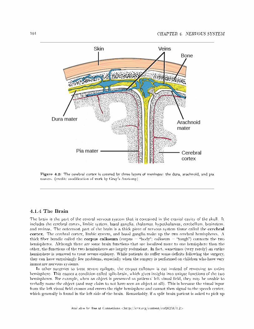
104 CHAPTER 4. NERVOUS SYSTEM
Figure 4.3: The cerebral cortex is covered by three layers of meninges: the dura, arachnoid, and piamaters. (credit: modi�cation of work by Gray's Anatomy)
4.1.4 The Brain
The brain is the part of the central nervous system that is contained in the cranial cavity of the skull. Itincludes the cerebral cortex, limbic system, basal ganglia, thalamus, hypothalamus, cerebellum, brainstem,and retinas. The outermost part of the brain is a thick piece of nervous system tissue called the cerebralcortex. The cerebral cortex, limbic system, and basal ganglia make up the two cerebral hemispheres. Athick �ber bundle called the corpus callosum (corpus = �body�; callosum = �tough�) connects the twohemispheres. Although there are some brain functions that are localized more to one hemisphere than theother, the functions of the two hemispheres are largely redundant. In fact, sometimes (very rarely) an entirehemisphere is removed to treat severe epilepsy. While patients do su�er some de�cits following the surgery,they can have surprisingly few problems, especially when the surgery is performed on children who have veryimmature nervous systems.
In other surgeries to treat severe epilepsy, the corpus callosum is cut instead of removing an entirehemisphere. This causes a condition called split-brain, which gives insights into unique functions of the twohemispheres. For example, when an object is presented to patients' left visual �eld, they may be unable toverbally name the object (and may claim to not have seen an object at all). This is because the visual inputfrom the left visual �eld crosses and enters the right hemisphere and cannot then signal to the speech center,which generally is found in the left side of the brain. Remarkably, if a split-brain patient is asked to pick up
Available for free at Connexions <http://cnx.org/content/col26253/1.2>
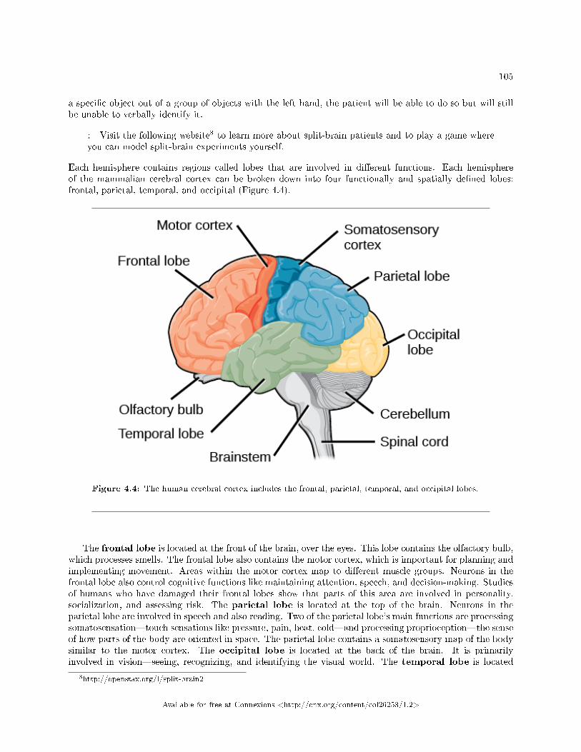
105
a speci�c object out of a group of objects with the left hand, the patient will be able to do so but will stillbe unable to verbally identify it.
: Visit the following website3 to learn more about split-brain patients and to play a game whereyou can model split-brain experiments yourself.
Each hemisphere contains regions called lobes that are involved in di�erent functions. Each hemisphereof the mammalian cerebral cortex can be broken down into four functionally and spatially de�ned lobes:frontal, parietal, temporal, and occipital (Figure 4.4).
Figure 4.4: The human cerebral cortex includes the frontal, parietal, temporal, and occipital lobes.
The frontal lobe is located at the front of the brain, over the eyes. This lobe contains the olfactory bulb,which processes smells. The frontal lobe also contains the motor cortex, which is important for planning andimplementing movement. Areas within the motor cortex map to di�erent muscle groups. Neurons in thefrontal lobe also control cognitive functions like maintaining attention, speech, and decision-making. Studiesof humans who have damaged their frontal lobes show that parts of this area are involved in personality,socialization, and assessing risk. The parietal lobe is located at the top of the brain. Neurons in theparietal lobe are involved in speech and also reading. Two of the parietal lobe's main functions are processingsomatosensation�touch sensations like pressure, pain, heat, cold�and processing proprioception�the senseof how parts of the body are oriented in space. The parietal lobe contains a somatosensory map of the bodysimilar to the motor cortex. The occipital lobe is located at the back of the brain. It is primarilyinvolved in vision�seeing, recognizing, and identifying the visual world. The temporal lobe is located
3http://openstax.org/l/split-brain2
Available for free at Connexions <http://cnx.org/content/col26253/1.2>
106 CHAPTER 4. NERVOUS SYSTEM
at the base of the brain and is primarily involved in processing and interpreting sounds. It also containsthe hippocampus (named from the Greek for �seahorse,� which it resembles in shape) a structure thatprocesses memory formation. The role of the hippocampus in memory was partially determined by studyingone famous epileptic patient, HM, who had both sides of his hippocampus removed in an attempt to cure hisepilepsy. His seizures went away, but he could no longer form new memories (although he could remembersome facts from before his surgery and could learn new motor tasks).
Interconnected brain areas called the basal ganglia play important roles in movement control andposture. The basal ganglia also regulate motivation.
The thalamus acts as a gateway to and from the cortex. It receives sensory and motor inputs fromthe body and also receives feedback from the cortex. This feedback mechanism can modulate consciousawareness of sensory and motor inputs depending on the attention and arousal state of the animal. Thethalamus helps regulate consciousness, arousal, and sleep states.
Below the thalamus is the hypothalamus. The hypothalamus controls the endocrine system by sendingsignals to the pituitary gland. Among other functions, the hypothalamus is the body's thermostat�it makessure the body temperature is kept at appropriate levels. Neurons within the hypothalamus also regulatecircadian rhythms, sometimes called sleep cycles.
The limbic system is a connected set of structures that regulates emotion, as well as behaviors relatedto fear and motivation. It plays a role in memory formation and includes parts of the thalamus and hypotha-lamus as well as the hippocampus. One important structure within the limbic system is a temporal lobestructure called the amygdala. The two amygdala (one on each side) are important both for the sensationof fear and for recognizing fearful faces.
The cerebellum (cerebellum = �little brain�) sits at the base of the brain on top of the brainstem.The cerebellum controls balance and aids in coordinating movement and learning new motor tasks. Thecerebellum of birds is large compared to other vertebrates because of the coordination required by �ight.
The brainstem connects the rest of the brain with the spinal cord and regulates some of the mostimportant and basic functions of the nervous system including breathing, swallowing, digestion, sleeping,walking, and sensory and motor information integration.
4.1.5 Spinal cord
Connecting to the brainstem and extending down the body through the spinal column is the spinal cord.The spinal cord is a thick bundle of nerve tissue that carries information about the body to the brain andfrom the brain to the body. The spinal cord is contained within the meninges and the bones of the vertebralcolumn but is able to communicate signals to and from the body through its connections with spinal nerves(part of the peripheral nervous system). A cross-section of the spinal cord looks like a white oval containinga gray butter�y-shape (Figure 4.5). Axons make up the �white matter� and neuron and glia cell bodies (andinterneurons) make up the �gray matter.� Axons and cell bodies in the dorsa spinal cord convey mostlysensory information from the body to the brain. Axons and cell bodies in the spinal cord primarily transmitsignals controlling movement from the brain to the body.
The spinal cord also controls motor re�exes. These re�exes are quick, unconscious movements�likeautomatically removing a hand from a hot object. Re�exes are so fast because they involve local synapticconnections. For example, the knee re�ex that a doctor tests during a routine physical is controlled by a singlesynapse between a sensory neuron and a motor neuron. While a re�ex may only require the involvement ofone or two synapses, synapses with interneurons in the spinal column transmit information to the brain toconvey what happened (the knee jerked, or the hand was hot).
Available for free at Connexions <http://cnx.org/content/col26253/1.2>
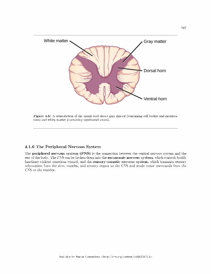
107
Figure 4.5: A cross-section of the spinal cord shows gray matter (containing cell bodies and interneu-rons) and white matter (containing myelinated axons).
4.1.6 The Peripheral Nervous System
The peripheral nervous system (PNS) is the connection between the central nervous system and therest of the body. The PNS can be broken down into the autonomic nervous system, which controls bodilyfunctions without conscious control, and the sensory-somatic nervous system, which transmits sensoryinformation from the skin, muscles, and sensory organs to the CNS and sends motor commands from theCNS to the muscles.
Available for free at Connexions <http://cnx.org/content/col26253/1.2>
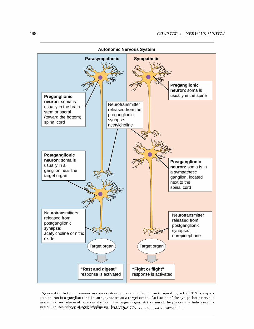
108 CHAPTER 4. NERVOUS SYSTEM
Figure 4.6: In the autonomic nervous system, a preganglionic neuron (originating in the CNS) synapsesto a neuron in a ganglion that, in turn, synapses on a target organ. Activation of the sympathetic nervoussystem causes release of norepinephrine on the target organ. Activation of the parasympathetic nervoussystem causes release of acetylcholine on the target organ.Available for free at Connexions <http://cnx.org/content/col26253/1.2>
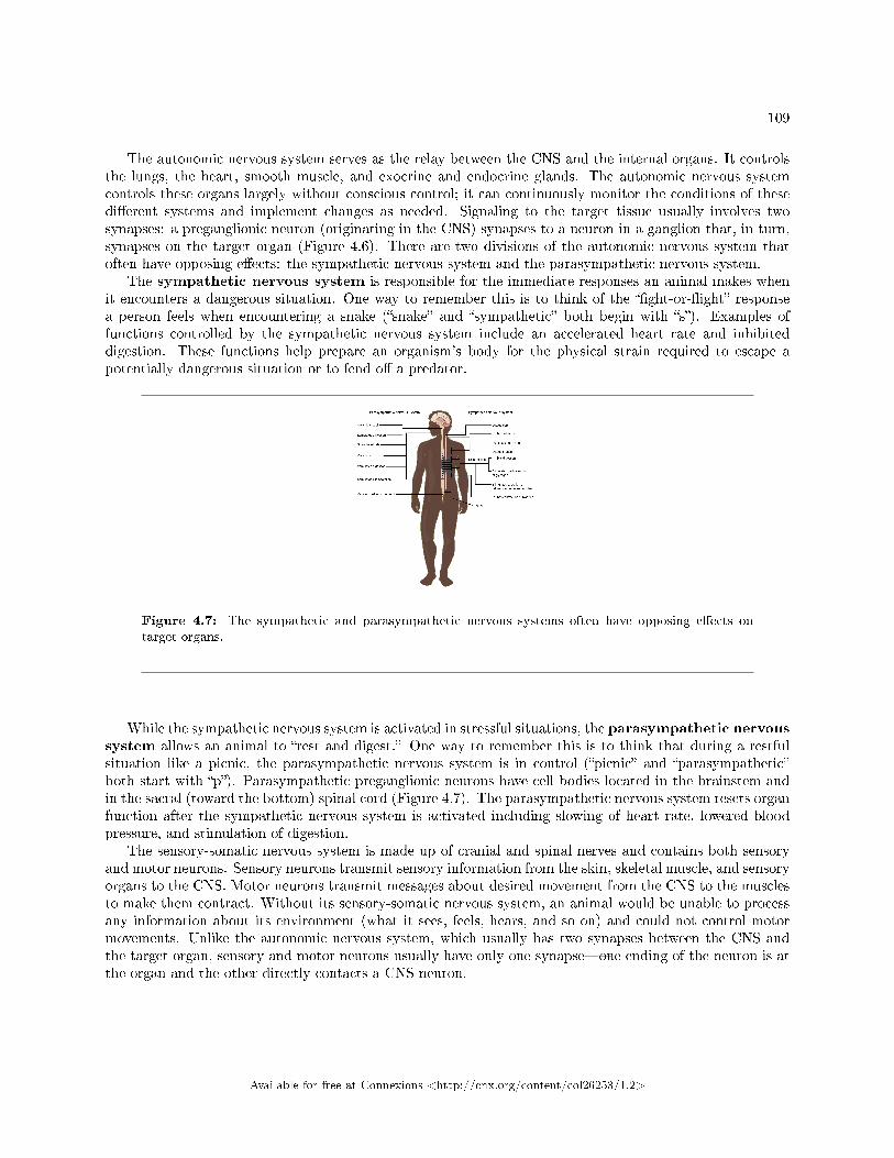
109
The autonomic nervous system serves as the relay between the CNS and the internal organs. It controlsthe lungs, the heart, smooth muscle, and exocrine and endocrine glands. The autonomic nervous systemcontrols these organs largely without conscious control; it can continuously monitor the conditions of thesedi�erent systems and implement changes as needed. Signaling to the target tissue usually involves twosynapses: a preganglionic neuron (originating in the CNS) synapses to a neuron in a ganglion that, in turn,synapses on the target organ (Figure 4.6). There are two divisions of the autonomic nervous system thatoften have opposing e�ects: the sympathetic nervous system and the parasympathetic nervous system.
The sympathetic nervous system is responsible for the immediate responses an animal makes whenit encounters a dangerous situation. One way to remember this is to think of the ��ght-or-�ight� responsea person feels when encountering a snake (�snake� and �sympathetic� both begin with �s�). Examples offunctions controlled by the sympathetic nervous system include an accelerated heart rate and inhibiteddigestion. These functions help prepare an organism's body for the physical strain required to escape apotentially dangerous situation or to fend o� a predator.
Figure 4.7: The sympathetic and parasympathetic nervous systems often have opposing e�ects ontarget organs.
While the sympathetic nervous system is activated in stressful situations, the parasympathetic nervoussystem allows an animal to �rest and digest.� One way to remember this is to think that during a restfulsituation like a picnic, the parasympathetic nervous system is in control (�picnic� and �parasympathetic�both start with �p�). Parasympathetic preganglionic neurons have cell bodies located in the brainstem andin the sacral (toward the bottom) spinal cord (Figure 4.7). The parasympathetic nervous system resets organfunction after the sympathetic nervous system is activated including slowing of heart rate, lowered bloodpressure, and stimulation of digestion.
The sensory-somatic nervous system is made up of cranial and spinal nerves and contains both sensoryand motor neurons. Sensory neurons transmit sensory information from the skin, skeletal muscle, and sensoryorgans to the CNS. Motor neurons transmit messages about desired movement from the CNS to the musclesto make them contract. Without its sensory-somatic nervous system, an animal would be unable to processany information about its environment (what it sees, feels, hears, and so on) and could not control motormovements. Unlike the autonomic nervous system, which usually has two synapses between the CNS andthe target organ, sensory and motor neurons usually have only one synapse�one ending of the neuron is atthe organ and the other directly contacts a CNS neuron.
Available for free at Connexions <http://cnx.org/content/col26253/1.2>
110 CHAPTER 4. NERVOUS SYSTEM
4.1.7 Section Summary
The nervous system is made up of neurons and glia. Neurons are specialized cells that are capable of sendingelectrical as well as chemical signals. Most neurons contain dendrites, which receive these signals, and axonsthat send signals to other neurons or tissues. Glia are non-neuronal cells in the nervous system that supportneuronal development and signaling. There are several types of glia that serve di�erent functions.
Neurons have a resting potential across their membranes and when they are stimulated by a strongenough signal from another neuron an action potential may carry an electrochemical signal along the neuronto a synapse with another neuron. Neurotransmitters carry signals across synapses to initiate a response inanother neuron.
The vertebrate central nervous system contains the brain and the spinal cord, which are covered andprotected by three meninges. The brain contains structurally and functionally de�ned regions. In mammals,these include the cortex (which can be broken down into four primary functional lobes: frontal, temporal,occipital, and parietal), basal ganglia, thalamus, hypothalamus, limbic system, cerebellum, and brainstem�although structures in some of these designations overlap. While functions may be primarily localized toone structure in the brain, most complex functions, like language and sleep, involve neurons in multiplebrain regions. The spinal cord is the information superhighway that connects the brain with the rest of thebody through its connections with peripheral nerves. It transmits sensory and motor input and also controlsmotor re�exes.
The peripheral nervous system contains both the autonomic and sensory-somatic nervous systems. Theautonomic nervous system provides unconscious control over visceral functions and has two divisions: thesympathetic and parasympathetic nervous systems. The sympathetic nervous system is activated in stressfulsituations to prepare the animal for a ��ght-or-�ight� response. The parasympathetic nervous system is activeduring restful periods. The sensory-somatic nervous system is made of cranial and spinal nerves that transmitsensory information from skin and muscle to the CNS and motor commands from the CNS to the muscles.
4.1.8 Review Questions
Exercise 4.1.1 (Solution on p. 112.)
Neurons contain _________, which can receive signals from other neurons.
a. axonsb. mitochondriac. dendritesd. Golgi bodies
Exercise 4.1.2 (Solution on p. 112.)
The part of the brain that is responsible for coordination during movement is the ______.
a. limbic systemb. thalamusc. cerebellumd. parietal lobe
Exercise 4.1.3 (Solution on p. 112.)
Which part of the nervous system directly controls the digestive system?
a. parasympathetic nervous systemb. central nervous systemc. spinal cordd. sensory-somatic nervous system
Available for free at Connexions <http://cnx.org/content/col26253/1.2>
111
4.1.9 Free Response
Exercise 4.1.4 (Solution on p. 112.)
How are neurons similar to other cells? How are they unique?
Exercise 4.1.5 (Solution on p. 112.)
What are the main functions of the spinal cord?
Exercise 4.1.6 (Solution on p. 112.)
What are the main di�erences between the sympathetic and parasympathetic branches of theautonomic nervous system?
Exercise 4.1.7 (Solution on p. 112.)
What are the main functions of the sensory-somatic nervous system?
Available for free at Connexions <http://cnx.org/content/col26253/1.2>
112 CHAPTER 4. NERVOUS SYSTEM
Solutions to Exercises in Chapter 4
to Exercise 4.1.1 (p. 110)Cto Exercise 4.1.2 (p. 110)Cto Exercise 4.1.3 (p. 110)Ato Exercise 4.1.4 (p. 111)Neurons contain organelles common to all cells, such as a nucleus and mitochondria. They are uniquebecause they contain dendrites, which can receive signals from other neurons, and axons that can send thesesignals to other cells.to Exercise 4.1.5 (p. 111)The spinal cord transmits sensory information from the body to the brain and motor commands from thebrain to the body through its connections with peripheral nerves. It also controls motor re�exes.to Exercise 4.1.6 (p. 111)The sympathetic nervous system prepares the body for ��ght or �ight,� whereas the parasympathetic nervoussystem allows the body to �rest and digest.� Sympathetic neurons release norepinephrine onto target organs;parasympathetic neurons release acetylcholine. Sympathetic neuron cell bodies are located in sympatheticganglia. Parasympathetic neuron cell bodies are located in the brainstem and sacral spinal cord. Activationof the sympathetic nervous system increases heart rate and blood pressure and decreases digestion and blood�ow to the skin. Activation of the parasympathetic nervous system decreases heart rate and blood pressureand increases digestion and blood �ow to the skin.to Exercise 4.1.7 (p. 111)The sensory-somatic nervous system transmits sensory information from the skin, muscles, and sensoryorgans to the CNS. It also sends motor commands from the CNS to the muscles, causing them to contract.
Available for free at Connexions <http://cnx.org/content/col26253/1.2>
Chapter 5
Sensory Perception
5.1 Sensory Processes1
Senses provide information about the body and its environment. Humans have �ve special senses: olfaction(smell), gustation (taste), equilibrium (balance and body position), vision, and hearing. Additionally, wepossess general senses, also called somatosensation, which respond to stimuli like temperature, pain, pressure,and vibration. Vestibular sensation, which is an organism's sense of spatial orientation and balance,proprioception (position of bones, joints, and muscles), and the sense of limb position that is used to trackkinesthesia (limb movement) are part of somatosensation. Although the sensory systems associated withthese senses are very di�erent, they all share a common function: to convert a stimulus (such as light, orsound, or the position of the body) into an electrical signal in the nervous system. This process is calledsensory transduction.
There are two broad types of cellular systems that perform sensory transduction. In one, a neuronworks with a sensory receptor, a cell, or cell process that is specialized to engage with and detect aspeci�c stimulus. Stimulation of the sensory receptor activates the associated a�erent neuron, which carriesinformation about the stimulus to the central nervous system. In the second type of sensory transduction, asensory nerve ending responds to a stimulus in the internal or external environment: this neuron constitutesthe sensory receptor. Free nerve endings can be stimulated by several di�erent stimuli, thus showing littlereceptor speci�city. For example, pain receptors in your gums and teeth may be stimulated by temperaturechanges, chemical stimulation, or pressure.
5.1.1 Reception
The �rst step in sensation is reception, which is the activation of sensory receptors by stimuli such asmechanical stimuli (being bent or squished, for example), chemicals, or temperature. The receptor can thenrespond to the stimuli. The region in space in which a given sensory receptor can respond to a stimulus,be it far away or in contact with the body, is that receptor's receptive �eld. Think for a moment aboutthe di�erences in receptive �elds for the di�erent senses. For the sense of touch, a stimulus must come intocontact with the body. For the sense of hearing, a stimulus can be a moderate distance away (some baleenwhale sounds can propagate for many kilometers). For vision, a stimulus can be very far away; for example,the visual system perceives light from stars at enormous distances.
5.1.2 Transduction
The most fundamental function of a sensory system is the translation of a sensory signal to an electrical signalin the nervous system. This takes place at the sensory receptor, and the change in electrical potential that is
1This content is available online at <http://cnx.org/content/m66402/1.13/>.
Available for free at Connexions <http://cnx.org/content/col26253/1.2>
113
114 CHAPTER 5. SENSORY PERCEPTION
produced is called the receptor potential. How is sensory input, such as pressure on the skin, changed to areceptor potential? In this example, a type of receptor called a mechanoreceptor (as shown in Figure 5.1)possesses specialized membranes that respond to pressure. Disturbance of these dendrites by compressingthem or bending them opens gated ion channels in the plasma membrane of the sensory neuron, changingits electrical potential. Recall that in the nervous system, a positive change of a neuron's electrical potential(also called the membrane potential), depolarizes the neuron. Receptor potentials are graded potentials: themagnitude of these graded (receptor) potentials varies with the strength of the stimulus. If the magnitudeof depolarization is su�cient (that is, if membrane potential reaches a threshold), the neuron will �re anaction potential. In most cases, the correct stimulus impinging on a sensory receptor will drive membranepotential in a positive direction, although for some receptors, such as those in the visual system, this is notalways the case.
Available for free at Connexions <http://cnx.org/content/col26253/1.2>
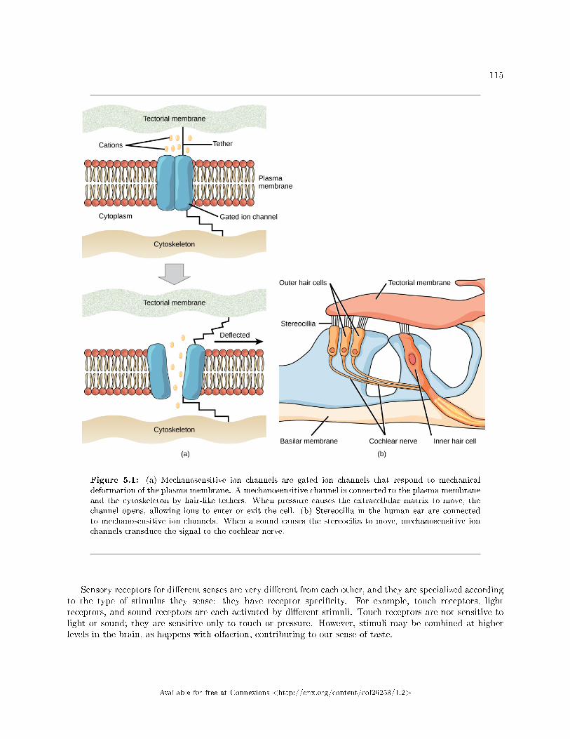
115
Figure 5.1: (a) Mechanosensitive ion channels are gated ion channels that respond to mechanicaldeformation of the plasma membrane. A mechanosensitive channel is connected to the plasma membraneand the cytoskeleton by hair-like tethers. When pressure causes the extracellular matrix to move, thechannel opens, allowing ions to enter or exit the cell. (b) Stereocilia in the human ear are connectedto mechanosensitive ion channels. When a sound causes the stereocilia to move, mechanosensitive ionchannels transduce the signal to the cochlear nerve.
Sensory receptors for di�erent senses are very di�erent from each other, and they are specialized accordingto the type of stimulus they sense: they have receptor speci�city. For example, touch receptors, lightreceptors, and sound receptors are each activated by di�erent stimuli. Touch receptors are not sensitive tolight or sound; they are sensitive only to touch or pressure. However, stimuli may be combined at higherlevels in the brain, as happens with olfaction, contributing to our sense of taste.
Available for free at Connexions <http://cnx.org/content/col26253/1.2>
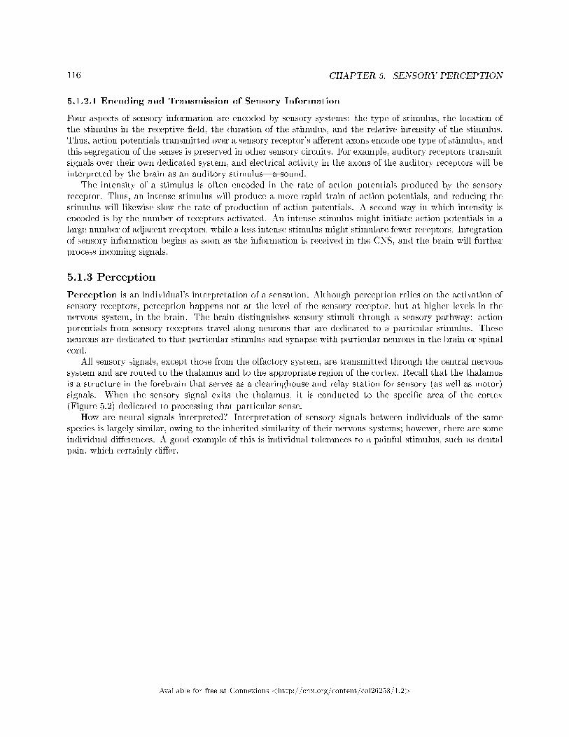
116 CHAPTER 5. SENSORY PERCEPTION
5.1.2.1 Encoding and Transmission of Sensory Information
Four aspects of sensory information are encoded by sensory systems: the type of stimulus, the location ofthe stimulus in the receptive �eld, the duration of the stimulus, and the relative intensity of the stimulus.Thus, action potentials transmitted over a sensory receptor's a�erent axons encode one type of stimulus, andthis segregation of the senses is preserved in other sensory circuits. For example, auditory receptors transmitsignals over their own dedicated system, and electrical activity in the axons of the auditory receptors will beinterpreted by the brain as an auditory stimulus�a sound.
The intensity of a stimulus is often encoded in the rate of action potentials produced by the sensoryreceptor. Thus, an intense stimulus will produce a more rapid train of action potentials, and reducing thestimulus will likewise slow the rate of production of action potentials. A second way in which intensity isencoded is by the number of receptors activated. An intense stimulus might initiate action potentials in alarge number of adjacent receptors, while a less intense stimulus might stimulate fewer receptors. Integrationof sensory information begins as soon as the information is received in the CNS, and the brain will furtherprocess incoming signals.
5.1.3 Perception
Perception is an individual's interpretation of a sensation. Although perception relies on the activation ofsensory receptors, perception happens not at the level of the sensory receptor, but at higher levels in thenervous system, in the brain. The brain distinguishes sensory stimuli through a sensory pathway: actionpotentials from sensory receptors travel along neurons that are dedicated to a particular stimulus. Theseneurons are dedicated to that particular stimulus and synapse with particular neurons in the brain or spinalcord.
All sensory signals, except those from the olfactory system, are transmitted through the central nervoussystem and are routed to the thalamus and to the appropriate region of the cortex. Recall that the thalamusis a structure in the forebrain that serves as a clearinghouse and relay station for sensory (as well as motor)signals. When the sensory signal exits the thalamus, it is conducted to the speci�c area of the cortex(Figure 5.2) dedicated to processing that particular sense.
How are neural signals interpreted? Interpretation of sensory signals between individuals of the samespecies is largely similar, owing to the inherited similarity of their nervous systems; however, there are someindividual di�erences. A good example of this is individual tolerances to a painful stimulus, such as dentalpain, which certainly di�er.
Available for free at Connexions <http://cnx.org/content/col26253/1.2>
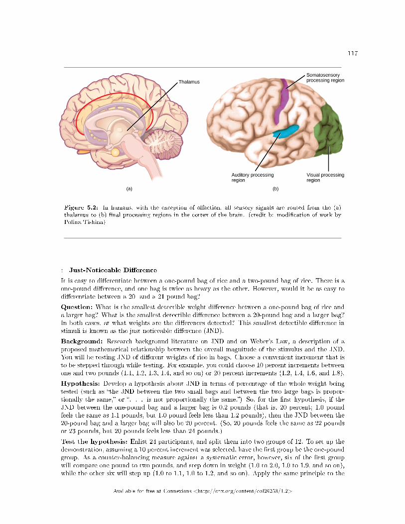
117
Figure 5.2: In humans, with the exception of olfaction, all sensory signals are routed from the (a)thalamus to (b) �nal processing regions in the cortex of the brain. (credit b: modi�cation of work byPolina Tishina)
: Just-Noticeable Di�erence
It is easy to di�erentiate between a one-pound bag of rice and a two-pound bag of rice. There is aone-pound di�erence, and one bag is twice as heavy as the other. However, would it be as easy todi�erentiate between a 20- and a 21-pound bag?
Question: What is the smallest detectible weight di�erence between a one-pound bag of rice anda larger bag? What is the smallest detectible di�erence between a 20-pound bag and a larger bag?In both cases, at what weights are the di�erences detected? This smallest detectible di�erence instimuli is known as the just-noticeable di�erence (JND).
Background: Research background literature on JND and on Weber's Law, a description of aproposed mathematical relationship between the overall magnitude of the stimulus and the JND.You will be testing JND of di�erent weights of rice in bags. Choose a convenient increment that isto be stepped through while testing. For example, you could choose 10 percent increments betweenone and two pounds (1.1, 1.2, 1.3, 1.4, and so on) or 20 percent increments (1.2, 1.4, 1.6, and 1.8).
Hypothesis: Develop a hypothesis about JND in terms of percentage of the whole weight beingtested (such as �the JND between the two small bags and between the two large bags is propor-tionally the same,� or �. . . is not proportionally the same.�) So, for the �rst hypothesis, if theJND between the one-pound bag and a larger bag is 0.2 pounds (that is, 20 percent; 1.0 poundfeels the same as 1.1 pounds, but 1.0 pound feels less than 1.2 pounds), then the JND between the20-pound bag and a larger bag will also be 20 percent. (So, 20 pounds feels the same as 22 poundsor 23 pounds, but 20 pounds feels less than 24 pounds.)
Test the hypothesis: Enlist 24 participants, and split them into two groups of 12. To set up thedemonstration, assuming a 10 percent increment was selected, have the �rst group be the one-poundgroup. As a counter-balancing measure against a systematic error, however, six of the �rst groupwill compare one pound to two pounds, and step down in weight (1.0 to 2.0, 1.0 to 1.9, and so on),while the other six will step up (1.0 to 1.1, 1.0 to 1.2, and so on). Apply the same principle to the
Available for free at Connexions <http://cnx.org/content/col26253/1.2>
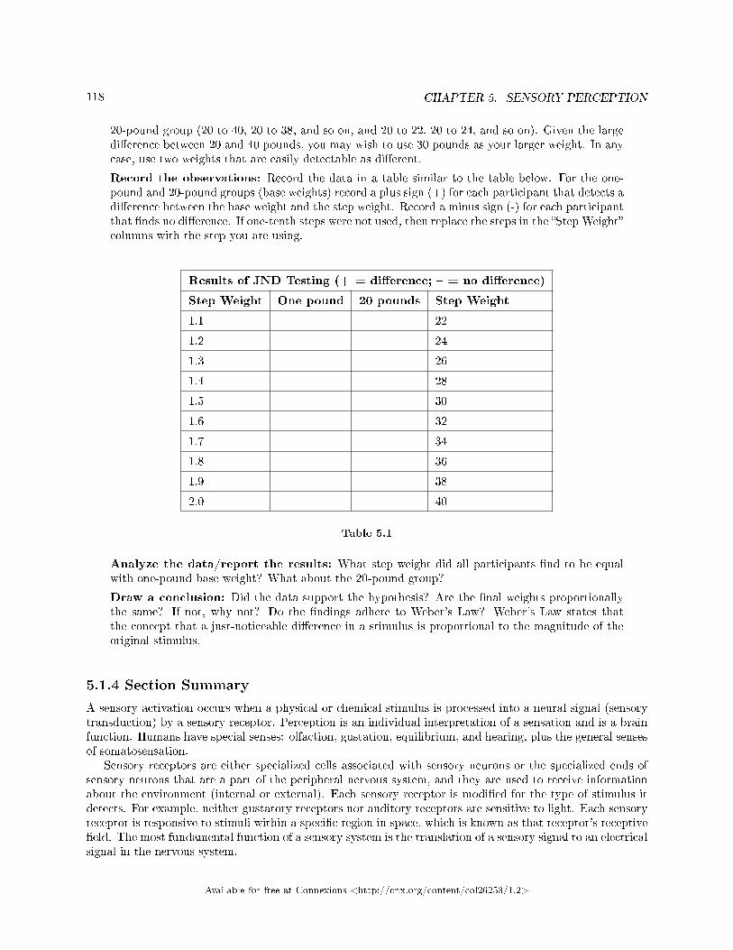
118 CHAPTER 5. SENSORY PERCEPTION
20-pound group (20 to 40, 20 to 38, and so on, and 20 to 22, 20 to 24, and so on). Given the largedi�erence between 20 and 40 pounds, you may wish to use 30 pounds as your larger weight. In anycase, use two weights that are easily detectable as di�erent.
Record the observations: Record the data in a table similar to the table below. For the one-pound and 20-pound groups (base weights) record a plus sign (+) for each participant that detects adi�erence between the base weight and the step weight. Record a minus sign (-) for each participantthat �nds no di�erence. If one-tenth steps were not used, then replace the steps in the �Step Weight�columns with the step you are using.
Results of JND Testing (+ = di�erence; � = no di�erence)
Step Weight One pound 20 pounds Step Weight
1.1 22
1.2 24
1.3 26
1.4 28
1.5 30
1.6 32
1.7 34
1.8 36
1.9 38
2.0 40
Table 5.1
Analyze the data/report the results: What step weight did all participants �nd to be equalwith one-pound base weight? What about the 20-pound group?
Draw a conclusion: Did the data support the hypothesis? Are the �nal weights proportionallythe same? If not, why not? Do the �ndings adhere to Weber's Law? Weber's Law states thatthe concept that a just-noticeable di�erence in a stimulus is proportional to the magnitude of theoriginal stimulus.
5.1.4 Section Summary
A sensory activation occurs when a physical or chemical stimulus is processed into a neural signal (sensorytransduction) by a sensory receptor. Perception is an individual interpretation of a sensation and is a brainfunction. Humans have special senses: olfaction, gustation, equilibrium, and hearing, plus the general sensesof somatosensation.
Sensory receptors are either specialized cells associated with sensory neurons or the specialized ends ofsensory neurons that are a part of the peripheral nervous system, and they are used to receive informationabout the environment (internal or external). Each sensory receptor is modi�ed for the type of stimulus itdetects. For example, neither gustatory receptors nor auditory receptors are sensitive to light. Each sensoryreceptor is responsive to stimuli within a speci�c region in space, which is known as that receptor's receptive�eld. The most fundamental function of a sensory system is the translation of a sensory signal to an electricalsignal in the nervous system.
Available for free at Connexions <http://cnx.org/content/col26253/1.2>
119
All sensory signals, except those from the olfactory system, enter the central nervous system and arerouted to the thalamus. When the sensory signal exits the thalamus, it is conducted to the speci�c area ofthe cortex dedicated to processing that particular sense.
5.1.5 Review Questions
Exercise 5.1.1 (Solution on p. 151.)
Where does perception occur?
a. spinal cordb. cerebral cortexc. receptorsd. thalamus
Exercise 5.1.2 (Solution on p. 151.)
If a person's cold receptors no longer convert cold stimuli into sensory signals, that person has aproblem with the process of ________.
a. receptionb. transmissionc. perceptiond. transduction
Exercise 5.1.3 (Solution on p. 151.)
After somatosensory transduction, the sensory signal travels through the brain as a(n) _____signal.
a. electricalb. pressurec. opticald. thermal
Exercise 5.1.4 (Solution on p. 151.)
Many people experience motion sickness while traveling in a car. This sensation results fromcontradictory inputs arising from which senses?
a. Proprioception and Kinesthesiab. Somatosensation and Equilibriumc. Gustation and Vibrationd. Vision and Vestibular System
5.1.6 Critical Thinking Questions
Exercise 5.1.5 (Solution on p. 151.)
If a person sustains damage to axons leading from sensory receptors to the central nervous system,which step or steps of sensory perception will be a�ected?
Exercise 5.1.6 (Solution on p. 151.)
In what way does the overall magnitude of a stimulus a�ect the just-noticeable di�erence in theperception of that stimulus?
Exercise 5.1.7 (Solution on p. 151.)
Describe the di�erence in the localization of the sensory receptors for general and special senses inhumans.
Available for free at Connexions <http://cnx.org/content/col26253/1.2>
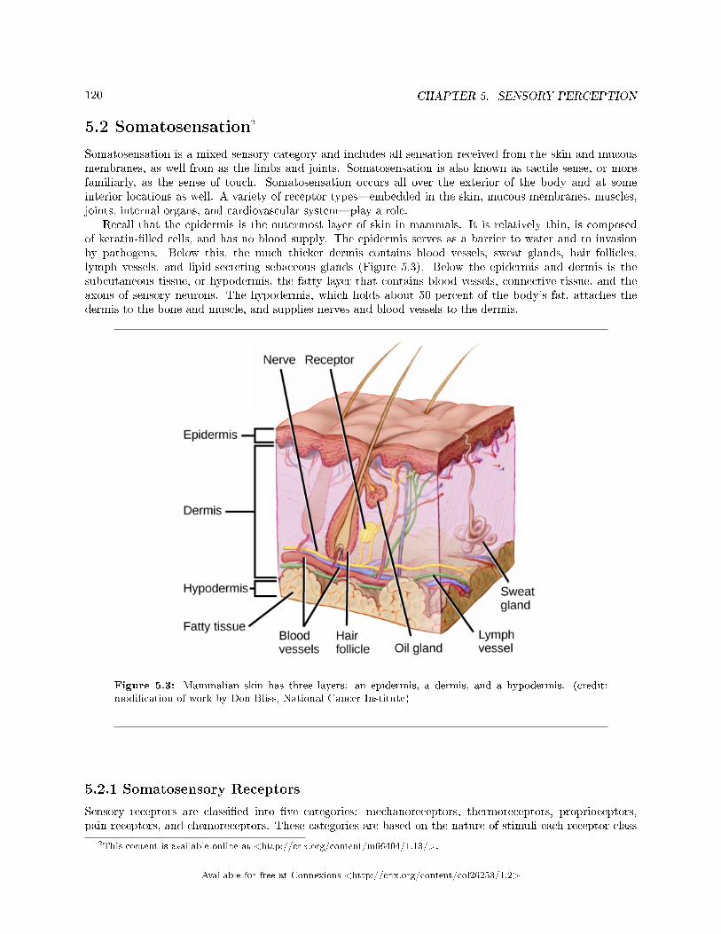
120 CHAPTER 5. SENSORY PERCEPTION
5.2 Somatosensation2
Somatosensation is a mixed sensory category and includes all sensation received from the skin and mucousmembranes, as well from as the limbs and joints. Somatosensation is also known as tactile sense, or morefamiliarly, as the sense of touch. Somatosensation occurs all over the exterior of the body and at someinterior locations as well. A variety of receptor types�embedded in the skin, mucous membranes, muscles,joints, internal organs, and cardiovascular system�play a role.
Recall that the epidermis is the outermost layer of skin in mammals. It is relatively thin, is composedof keratin-�lled cells, and has no blood supply. The epidermis serves as a barrier to water and to invasionby pathogens. Below this, the much thicker dermis contains blood vessels, sweat glands, hair follicles,lymph vessels, and lipid-secreting sebaceous glands (Figure 5.3). Below the epidermis and dermis is thesubcutaneous tissue, or hypodermis, the fatty layer that contains blood vessels, connective tissue, and theaxons of sensory neurons. The hypodermis, which holds about 50 percent of the body's fat, attaches thedermis to the bone and muscle, and supplies nerves and blood vessels to the dermis.
Figure 5.3: Mammalian skin has three layers: an epidermis, a dermis, and a hypodermis. (credit:modi�cation of work by Don Bliss, National Cancer Institute)
5.2.1 Somatosensory Receptors
Sensory receptors are classi�ed into �ve categories: mechanoreceptors, thermoreceptors, proprioceptors,pain receptors, and chemoreceptors. These categories are based on the nature of stimuli each receptor class
2This content is available online at <http://cnx.org/content/m66404/1.13/>.
Available for free at Connexions <http://cnx.org/content/col26253/1.2>
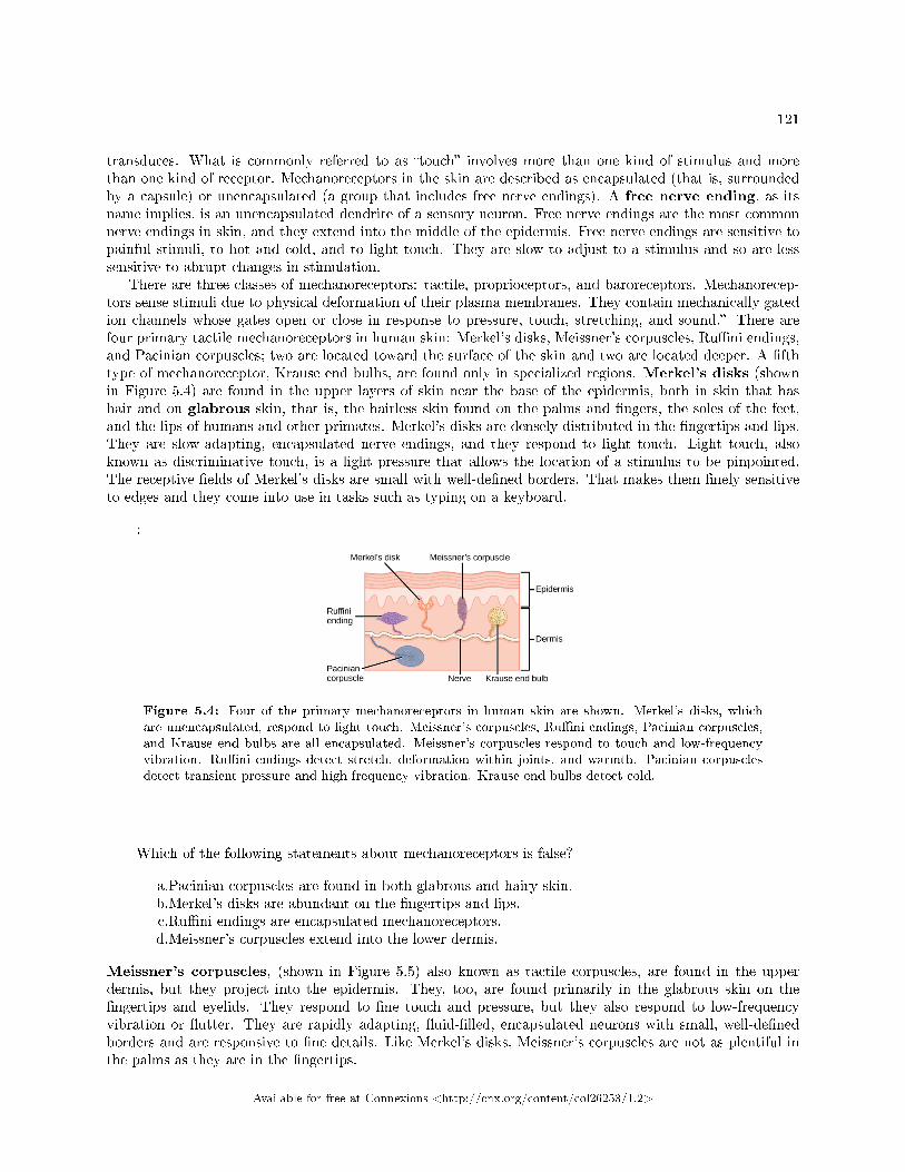
121
transduces. What is commonly referred to as �touch� involves more than one kind of stimulus and morethan one kind of receptor. Mechanoreceptors in the skin are described as encapsulated (that is, surroundedby a capsule) or unencapsulated (a group that includes free nerve endings). A free nerve ending, as itsname implies, is an unencapsulated dendrite of a sensory neuron. Free nerve endings are the most commonnerve endings in skin, and they extend into the middle of the epidermis. Free nerve endings are sensitive topainful stimuli, to hot and cold, and to light touch. They are slow to adjust to a stimulus and so are lesssensitive to abrupt changes in stimulation.
There are three classes of mechanoreceptors: tactile, proprioceptors, and baroreceptors. Mechanorecep-tors sense stimuli due to physical deformation of their plasma membranes. They contain mechanically gatedion channels whose gates open or close in response to pressure, touch, stretching, and sound.� There arefour primary tactile mechanoreceptors in human skin: Merkel's disks, Meissner's corpuscles, Ru�ni endings,and Pacinian corpuscles; two are located toward the surface of the skin and two are located deeper. A �fthtype of mechanoreceptor, Krause end bulbs, are found only in specialized regions. Merkel's disks (shownin Figure 5.4) are found in the upper layers of skin near the base of the epidermis, both in skin that hashair and on glabrous skin, that is, the hairless skin found on the palms and �ngers, the soles of the feet,and the lips of humans and other primates. Merkel's disks are densely distributed in the �ngertips and lips.They are slow-adapting, encapsulated nerve endings, and they respond to light touch. Light touch, alsoknown as discriminative touch, is a light pressure that allows the location of a stimulus to be pinpointed.The receptive �elds of Merkel's disks are small with well-de�ned borders. That makes them �nely sensitiveto edges and they come into use in tasks such as typing on a keyboard.
:
Figure 5.4: Four of the primary mechanoreceptors in human skin are shown. Merkel's disks, whichare unencapsulated, respond to light touch. Meissner's corpuscles, Ru�ni endings, Pacinian corpuscles,and Krause end bulbs are all encapsulated. Meissner's corpuscles respond to touch and low-frequencyvibration. Ru�ni endings detect stretch, deformation within joints, and warmth. Pacinian corpusclesdetect transient pressure and high-frequency vibration. Krause end bulbs detect cold.
Which of the following statements about mechanoreceptors is false?
a.Pacinian corpuscles are found in both glabrous and hairy skin.b.Merkel's disks are abundant on the �ngertips and lips.c.Ru�ni endings are encapsulated mechanoreceptors.d.Meissner's corpuscles extend into the lower dermis.
Meissner's corpuscles, (shown in Figure 5.5) also known as tactile corpuscles, are found in the upperdermis, but they project into the epidermis. They, too, are found primarily in the glabrous skin on the�ngertips and eyelids. They respond to �ne touch and pressure, but they also respond to low-frequencyvibration or �utter. They are rapidly adapting, �uid-�lled, encapsulated neurons with small, well-de�nedborders and are responsive to �ne details. Like Merkel's disks, Meissner's corpuscles are not as plentiful inthe palms as they are in the �ngertips.
Available for free at Connexions <http://cnx.org/content/col26253/1.2>
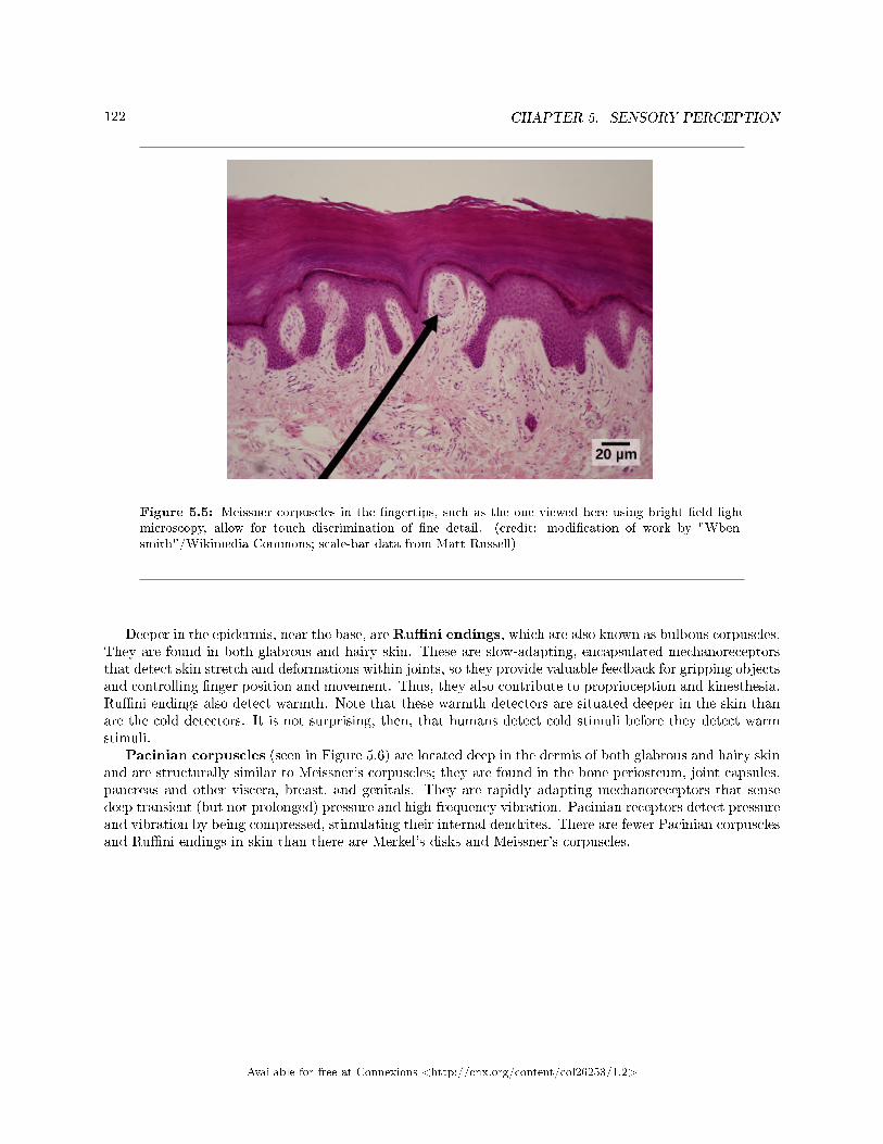
122 CHAPTER 5. SENSORY PERCEPTION
Figure 5.5: Meissner corpuscles in the �ngertips, such as the one viewed here using bright �eld lightmicroscopy, allow for touch discrimination of �ne detail. (credit: modi�cation of work by "Wben-smith"/Wikimedia Commons; scale-bar data from Matt Russell)
Deeper in the epidermis, near the base, areRu�ni endings, which are also known as bulbous corpuscles.They are found in both glabrous and hairy skin. These are slow-adapting, encapsulated mechanoreceptorsthat detect skin stretch and deformations within joints, so they provide valuable feedback for gripping objectsand controlling �nger position and movement. Thus, they also contribute to proprioception and kinesthesia.Ru�ni endings also detect warmth. Note that these warmth detectors are situated deeper in the skin thanare the cold detectors. It is not surprising, then, that humans detect cold stimuli before they detect warmstimuli.
Pacinian corpuscles (seen in Figure 5.6) are located deep in the dermis of both glabrous and hairy skinand are structurally similar to Meissner's corpuscles; they are found in the bone periosteum, joint capsules,pancreas and other viscera, breast, and genitals. They are rapidly adapting mechanoreceptors that sensedeep transient (but not prolonged) pressure and high-frequency vibration. Pacinian receptors detect pressureand vibration by being compressed, stimulating their internal dendrites. There are fewer Pacinian corpusclesand Ru�ni endings in skin than there are Merkel's disks and Meissner's corpuscles.
Available for free at Connexions <http://cnx.org/content/col26253/1.2>
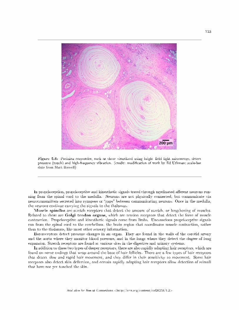
123
Figure 5.6: Pacinian corpuscles, such as these visualized using bright �eld light microscopy, detectpressure (touch) and high-frequency vibration. (credit: modi�cation of work by Ed Uthman; scale-bardata from Matt Russell)
In proprioception, proprioceptive and kinesthetic signals travel through myelinated a�erent neurons run-ning from the spinal cord to the medulla. Neurons are not physically connected, but communicate vianeurotransmitters secreted into synapses or �gaps� between communicating neurons. Once in the medulla,the neurons continue carrying the signals to the thalamus.
Muscle spindles are stretch receptors that detect the amount of stretch, or lengthening of muscles.Related to these are Golgi tendon organs, which are tension receptors that detect the force of musclecontraction. Proprioceptive and kinesthetic signals come from limbs. Unconscious proprioceptive signalsrun from the spinal cord to the cerebellum, the brain region that coordinates muscle contraction, ratherthan to the thalamus, like most other sensory information.
Baroreceptors detect pressure changes in an organ. They are found in the walls of the carotid arteryand the aorta where they monitor blood pressure, and in the lungs where they detect the degree of lungexpansion. Stretch receptors are found at various sites in the digestive and urinary systems.
In addition to these two types of deeper receptors, there are also rapidly adapting hair receptors, which arefound on nerve endings that wrap around the base of hair follicles. There are a few types of hair receptorsthat detect slow and rapid hair movement, and they di�er in their sensitivity to movement. Some hairreceptors also detect skin de�ection, and certain rapidly adapting hair receptors allow detection of stimulithat have not yet touched the skin.
Available for free at Connexions <http://cnx.org/content/col26253/1.2>
124 CHAPTER 5. SENSORY PERCEPTION
5.2.1.1 Integration of Signals from Mechanoreceptors
The con�guration of the di�erent types of receptors working in concert in human skin results in a veryre�ned sense of touch. The nociceptive receptors�those that detect pain�are located near the surface.Small, �nely calibrated mechanoreceptors�Merkel's disks and Meissner's corpuscles�are located in theupper layers and can precisely localize even gentle touch. The large mechanoreceptors�Pacinian corpusclesand Ru�ni endings�are located in the lower layers and respond to deeper touch. (Consider that the deeppressure that reaches those deeper receptors would not need to be �nely localized.) Both the upper andlower layers of the skin hold rapidly and slowly adapting receptors. Both primary somatosensory cortex andsecondary cortical areas are responsible for processing the complex picture of stimuli transmitted from theinterplay of mechanoreceptors.
5.2.1.2 Density of Mechanoreceptors
The distribution of touch receptors in human skin is not consistent over the body. In humans, touch receptorsare less dense in skin covered with any type of hair, such as the arms, legs, torso, and face. Touch receptorsare denser in glabrous skin (the type found on human �ngertips and lips, for example), which is typicallymore sensitive and is thicker than hairy skin (4 to 5 mm versus 2 to 3 mm).
How is receptor density estimated in a human subject? The relative density of pressure receptors indi�erent locations on the body can be demonstrated experimentally using a two-point discrimination test.In this demonstration, two sharp points, such as two thumbtacks, are brought into contact with the subject'sskin (though not hard enough to cause pain or break the skin). The subject reports if they feel one pointor two points. If the two points are felt as one point, it can be inferred that the two points are both inthe receptive �eld of a single sensory receptor. If two points are felt as two separate points, each is in thereceptive �eld of two separate sensory receptors. The points could then be moved closer and retested untilthe subject reports feeling only one point, and the size of the receptive �eld of a single receptor could beestimated from that distance.
5.2.2 Thermoreception
In addition to Krause end bulbs that detect cold and Ru�ni endings that detect warmth, there are di�erenttypes of cold receptors on some free nerve endings: thermoreceptors, located in the dermis, skeletal muscles,liver, and hypothalamus, that are activated by di�erent temperatures. Their pathways into the brain run fromthe spinal cord through the thalamus to the primary somatosensory cortex. Warmth and cold informationfrom the face travels through one of the cranial nerves to the brain. You know from experience that a tolerablycold or hot stimulus can quickly progress to a much more intense stimulus that is no longer tolerable. Anystimulus that is too intense can be perceived as pain because temperature sensations are conducted alongthe same pathways that carry pain sensations.
5.2.3 Pain
Pain is the name given to nociception, which is the neural processing of injurious stimuli in response totissue damage. Pain is caused by true sources of injury, such as contact with a heat source that causes athermal burn or contact with a corrosive chemical. But pain also can be caused by harmless stimuli thatmimic the action of damaging stimuli, such as contact with capsaicins, the compounds that cause peppersto taste hot and which are used in self-defense pepper sprays and certain topical medications. Peppers taste�hot� because the protein receptors that bind capsaicin open the same calcium channels that are activatedby warm receptors.
Nociception starts at the sensory receptors, but pain, inasmuch as it is the perception of nociception,does not start until it is communicated to the brain. There are several nociceptive pathways to and throughthe brain. Most axons carrying nociceptive information into the brain from the spinal cord project to thethalamus (as do other sensory neurons) and the neural signal undergoes �nal processing in the primary
Available for free at Connexions <http://cnx.org/content/col26253/1.2>
125
somatosensory cortex. Interestingly, one nociceptive pathway projects not to the thalamus but directly tothe hypothalamus in the forebrain, which modulates the cardiovascular and neuroendocrine functions of theautonomic nervous system. Recall that threatening�or painful�stimuli stimulate the sympathetic branchof the visceral sensory system, readying a �ght-or-�ight response.
: View this video3 that animates the �ve phases of nociceptive pain.
5.2.4 Section Summary
Somatosensation includes all sensation received from the skin and mucous membranes, as well as from thelimbs and joints. Somatosensation occurs all over the exterior of the body and at some interior locations aswell, and a variety of receptor types, embedded in the skin and mucous membranes, play a role.
There are several types of specialized sensory receptors. Rapidly adapting free nerve endings detectnociception, hot and cold, and light touch. Slowly adapting, encapsulated Merkel's disks are found in�ngertips and lips, and respond to light touch. Meissner's corpuscles, found in glabrous skin, are rapidlyadapting, encapsulated receptors that detect touch, low-frequency vibration, and �utter. Ru�ni endings areslowly adapting, encapsulated receptors that detect skin stretch, joint activity, and warmth. Hair receptorsare rapidly adapting nerve endings wrapped around the base of hair follicles that detect hair movementand skin de�ection. Finally, Pacinian corpuscles are encapsulated, rapidly adapting receptors that detecttransient pressure and high-frequency vibration.
5.2.5 Visual Connection Questions
Exercise 5.2.1 (Solution on p. 151.)
Figure 5.4 Which of the following statements about mechanoreceptors is false?
a. Pacini corpuscles are found in both glabrous and hairy skin.b. Merkel's disks are abundant on the �ngertips and lips.c. Ru�ni endings are encapsulated mechanoreceptors.d. Meissner's corpuscles extend into the lower dermis.
5.2.6 Review Questions
Exercise 5.2.2 (Solution on p. 151.)
_____ are found only in _____ skin, and detect skin de�ection.
a. Meissner's corpuscles; hairyb. Merkel's disks; glabrousc. hair receptors; hairyd. Krause end bulbs; hairy
Exercise 5.2.3 (Solution on p. 151.)
If you were to burn your epidermis, what receptor type would you most likely burn?
a. free nerve endingsb. Ru�ni endingsc. Pacinian corpuscled. hair receptors
3http://openstax.org/l/nociceptive
Available for free at Connexions <http://cnx.org/content/col26253/1.2>
126 CHAPTER 5. SENSORY PERCEPTION
Exercise 5.2.4 (Solution on p. 151.)
Many diabetic patients are warned by their doctors to test their glucose levels by pricking the sidesof their �ngers rather than the pads. Pricking the sides avoids stimulating which receptor?
a. Krause end bulbsb. Meissner's corpusclesc. Ru�ni endingd. Nociceptors
5.2.7 Critical Thinking Questions
Exercise 5.2.5 (Solution on p. 151.)
What can be inferred about the relative sizes of the areas of cortex that process signals from skinnot densely innervated with sensory receptors and skin that is densely innervated with sensoryreceptors?
Exercise 5.2.6 (Solution on p. 151.)
Many studies have demonstrated that females are able to tolerate the same painful stimuli forlonger than males. Why don't all people experience pain the same way?
5.3 Vision4
The visual system constructs a mental representation of the world around us (Figure 5.7). This contributesto our ability to successfully navigate through physical space and interact with important individuals andobjects in our environments. This section will provide an overview of the basic anatomy and function of thevisual system. In addition, we will explore our ability to perceive color and depth.
4This content is available online at <http://cnx.org/content/m49042/1.14/>.
Available for free at Connexions <http://cnx.org/content/col26253/1.2>
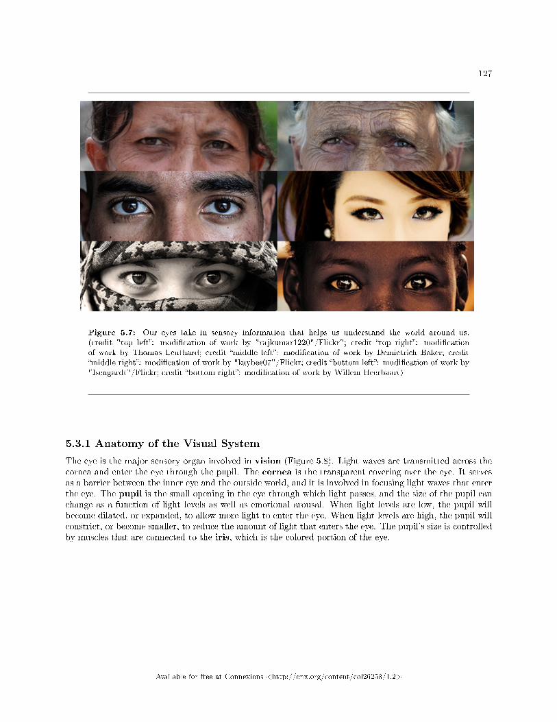
127
Figure 5.7: Our eyes take in sensory information that helps us understand the world around us.(credit "top left�: modi�cation of work by "rajkumar1220"/Flickr"; credit �top right�: modi�cationof work by Thomas Leuthard; credit �middle left�: modi�cation of work by Demietrich Baker; credit�middle right�: modi�cation of work by "kaybee07"/Flickr; credit �bottom left�: modi�cation of work by"Isengardt"/Flickr; credit �bottom right�: modi�cation of work by Willem Heerbaart)
5.3.1 Anatomy of the Visual System
The eye is the major sensory organ involved in vision (Figure 5.8). Light waves are transmitted across thecornea and enter the eye through the pupil. The cornea is the transparent covering over the eye. It servesas a barrier between the inner eye and the outside world, and it is involved in focusing light waves that enterthe eye. The pupil is the small opening in the eye through which light passes, and the size of the pupil canchange as a function of light levels as well as emotional arousal. When light levels are low, the pupil willbecome dilated, or expanded, to allow more light to enter the eye. When light levels are high, the pupil willconstrict, or become smaller, to reduce the amount of light that enters the eye. The pupil's size is controlledby muscles that are connected to the iris, which is the colored portion of the eye.
Available for free at Connexions <http://cnx.org/content/col26253/1.2>
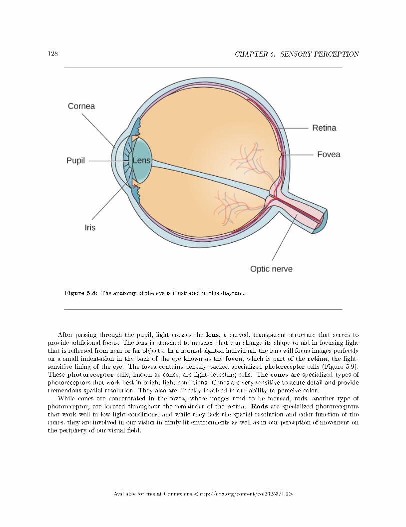
128 CHAPTER 5. SENSORY PERCEPTION
Figure 5.8: The anatomy of the eye is illustrated in this diagram.
After passing through the pupil, light crosses the lens, a curved, transparent structure that serves toprovide additional focus. The lens is attached to muscles that can change its shape to aid in focusing lightthat is re�ected from near or far objects. In a normal-sighted individual, the lens will focus images perfectlyon a small indentation in the back of the eye known as the fovea, which is part of the retina, the light-sensitive lining of the eye. The fovea contains densely packed specialized photoreceptor cells (Figure 5.9).These photoreceptor cells, known as cones, are light-detecting cells. The cones are specialized types ofphotoreceptors that work best in bright light conditions. Cones are very sensitive to acute detail and providetremendous spatial resolution. They also are directly involved in our ability to perceive color.
While cones are concentrated in the fovea, where images tend to be focused, rods, another type ofphotoreceptor, are located throughout the remainder of the retina. Rods are specialized photoreceptorsthat work well in low light conditions, and while they lack the spatial resolution and color function of thecones, they are involved in our vision in dimly lit environments as well as in our perception of movement onthe periphery of our visual �eld.
Available for free at Connexions <http://cnx.org/content/col26253/1.2>
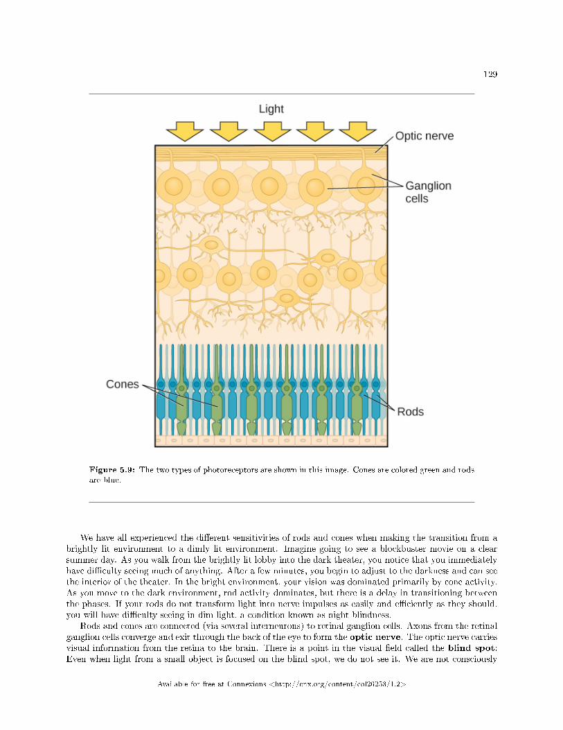
129
Figure 5.9: The two types of photoreceptors are shown in this image. Cones are colored green and rodsare blue.
We have all experienced the di�erent sensitivities of rods and cones when making the transition from abrightly lit environment to a dimly lit environment. Imagine going to see a blockbuster movie on a clearsummer day. As you walk from the brightly lit lobby into the dark theater, you notice that you immediatelyhave di�culty seeing much of anything. After a few minutes, you begin to adjust to the darkness and can seethe interior of the theater. In the bright environment, your vision was dominated primarily by cone activity.As you move to the dark environment, rod activity dominates, but there is a delay in transitioning betweenthe phases. If your rods do not transform light into nerve impulses as easily and e�ciently as they should,you will have di�culty seeing in dim light, a condition known as night blindness.
Rods and cones are connected (via several interneurons) to retinal ganglion cells. Axons from the retinalganglion cells converge and exit through the back of the eye to form the optic nerve. The optic nerve carriesvisual information from the retina to the brain. There is a point in the visual �eld called the blind spot:Even when light from a small object is focused on the blind spot, we do not see it. We are not consciously
Available for free at Connexions <http://cnx.org/content/col26253/1.2>
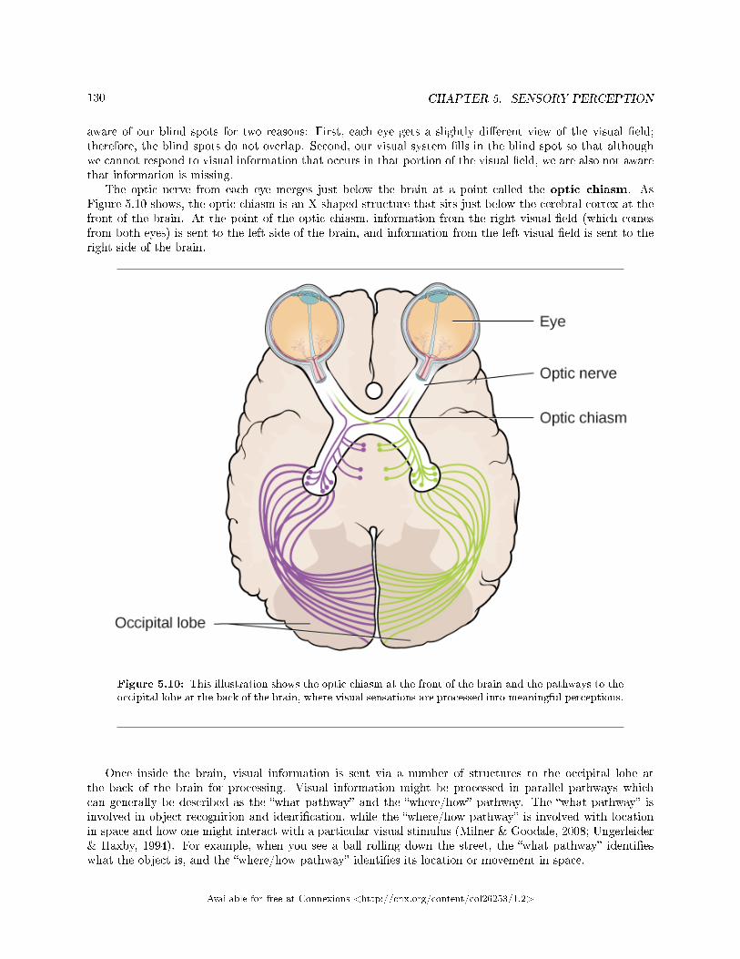
130 CHAPTER 5. SENSORY PERCEPTION
aware of our blind spots for two reasons: First, each eye gets a slightly di�erent view of the visual �eld;therefore, the blind spots do not overlap. Second, our visual system �lls in the blind spot so that althoughwe cannot respond to visual information that occurs in that portion of the visual �eld, we are also not awarethat information is missing.
The optic nerve from each eye merges just below the brain at a point called the optic chiasm. AsFigure 5.10 shows, the optic chiasm is an X-shaped structure that sits just below the cerebral cortex at thefront of the brain. At the point of the optic chiasm, information from the right visual �eld (which comesfrom both eyes) is sent to the left side of the brain, and information from the left visual �eld is sent to theright side of the brain.
Figure 5.10: This illustration shows the optic chiasm at the front of the brain and the pathways to theoccipital lobe at the back of the brain, where visual sensations are processed into meaningful perceptions.
Once inside the brain, visual information is sent via a number of structures to the occipital lobe atthe back of the brain for processing. Visual information might be processed in parallel pathways whichcan generally be described as the �what pathway� and the �where/how� pathway. The �what pathway� isinvolved in object recognition and identi�cation, while the �where/how pathway� is involved with locationin space and how one might interact with a particular visual stimulus (Milner & Goodale, 2008; Ungerleider& Haxby, 1994). For example, when you see a ball rolling down the street, the �what pathway� identi�eswhat the object is, and the �where/how pathway� identi�es its location or movement in space.
Available for free at Connexions <http://cnx.org/content/col26253/1.2>
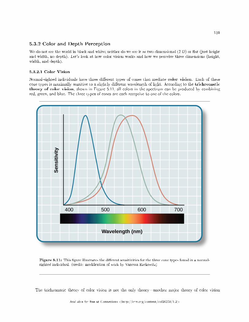
131
5.3.2 Color and Depth Perception
We do not see the world in black and white; neither do we see it as two-dimensional (2-D) or �at (just heightand width, no depth). Let's look at how color vision works and how we perceive three dimensions (height,width, and depth).
5.3.2.1 Color Vision
Normal-sighted individuals have three di�erent types of cones that mediate color vision. Each of thesecone types is maximally sensitive to a slightly di�erent wavelength of light. According to the trichromatictheory of color vision, shown in Figure 5.11, all colors in the spectrum can be produced by combiningred, green, and blue. The three types of cones are each receptive to one of the colors.
Figure 5.11: This �gure illustrates the di�erent sensitivities for the three cone types found in a normal-sighted individual. (credit: modi�cation of work by Vanessa Ezekowitz)
The trichromatic theory of color vision is not the only theory�another major theory of color vision
Available for free at Connexions <http://cnx.org/content/col26253/1.2>
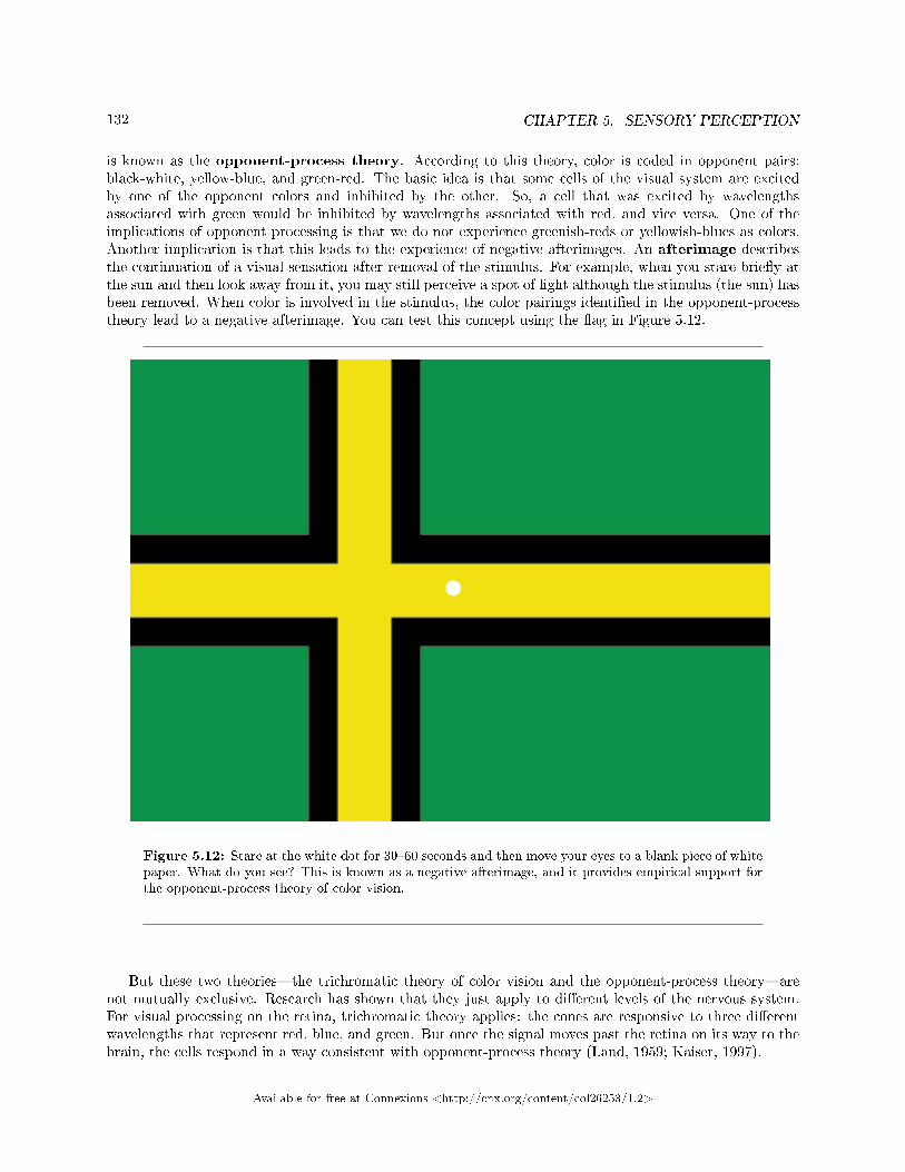
132 CHAPTER 5. SENSORY PERCEPTION
is known as the opponent-process theory. According to this theory, color is coded in opponent pairs:black-white, yellow-blue, and green-red. The basic idea is that some cells of the visual system are excitedby one of the opponent colors and inhibited by the other. So, a cell that was excited by wavelengthsassociated with green would be inhibited by wavelengths associated with red, and vice versa. One of theimplications of opponent processing is that we do not experience greenish-reds or yellowish-blues as colors.Another implication is that this leads to the experience of negative afterimages. An afterimage describesthe continuation of a visual sensation after removal of the stimulus. For example, when you stare brie�y atthe sun and then look away from it, you may still perceive a spot of light although the stimulus (the sun) hasbeen removed. When color is involved in the stimulus, the color pairings identi�ed in the opponent-processtheory lead to a negative afterimage. You can test this concept using the �ag in Figure 5.12.
Figure 5.12: Stare at the white dot for 30�60 seconds and then move your eyes to a blank piece of whitepaper. What do you see? This is known as a negative afterimage, and it provides empirical support forthe opponent-process theory of color vision.
But these two theories�the trichromatic theory of color vision and the opponent-process theory�arenot mutually exclusive. Research has shown that they just apply to di�erent levels of the nervous system.For visual processing on the retina, trichromatic theory applies: the cones are responsive to three di�erentwavelengths that represent red, blue, and green. But once the signal moves past the retina on its way to thebrain, the cells respond in a way consistent with opponent-process theory (Land, 1959; Kaiser, 1997).
Available for free at Connexions <http://cnx.org/content/col26253/1.2>
133
Link To Learning: Watch this video5 to learn about color vision in more detail.
5.3.2.2 Depth Perception
Our ability to perceive spatial relationships in three-dimensional (3-D) space is known as depth perception.With depth perception, we can describe things as being in front, behind, above, below, or to the side ofother things.
Our world is three-dimensional, so it makes sense that our mental representation of the world has three-dimensional properties. We use a variety of cues in a visual scene to establish our sense of depth. Some ofthese are binocular cues, which means that they rely on the use of both eyes. One example of a binoculardepth cue is binocular disparity, the slightly di�erent view of the world that each of our eyes receives. Toexperience this slightly di�erent view, do this simple exercise: extend your arm fully and extend one of your�ngers and focus on that �nger. Now, close your left eye without moving your head, then open your left eyeand close your right eye without moving your head. You will notice that your �nger seems to shift as youalternate between the two eyes because of the slightly di�erent view each eye has of your �nger.
A 3-D movie works on the same principle: the special glasses you wear allow the two slightly di�erentimages projected onto the screen to be seen separately by your left and your right eye. As your brainprocesses these images, you have the illusion that the leaping animal or running person is coming righttoward you.
Although we rely on binocular cues to experience depth in our 3-D world, we can also perceive depth in2-D arrays. Think about all the paintings and photographs you have seen. Generally, you pick up on depthin these images even though the visual stimulus is 2-D. When we do this, we are relying on a number ofmonocular cues, or cues that require only one eye. If you think you can't see depth with one eye, note thatyou don't bump into things when using only one eye while walking�and, in fact, we have more monocularcues than binocular cues.
An example of a monocular cue would be what is known as linear perspective. Linear perspectiverefers to the fact that we perceive depth when we see two parallel lines that seem to converge in an image(Figure 5.13). Some other monocular depth cues are interposition, the partial overlap of objects, and therelative size and closeness of images to the horizon.
5https://www.youtube.com/watch?v=1IqXyu14kpY
Available for free at Connexions <http://cnx.org/content/col26253/1.2>
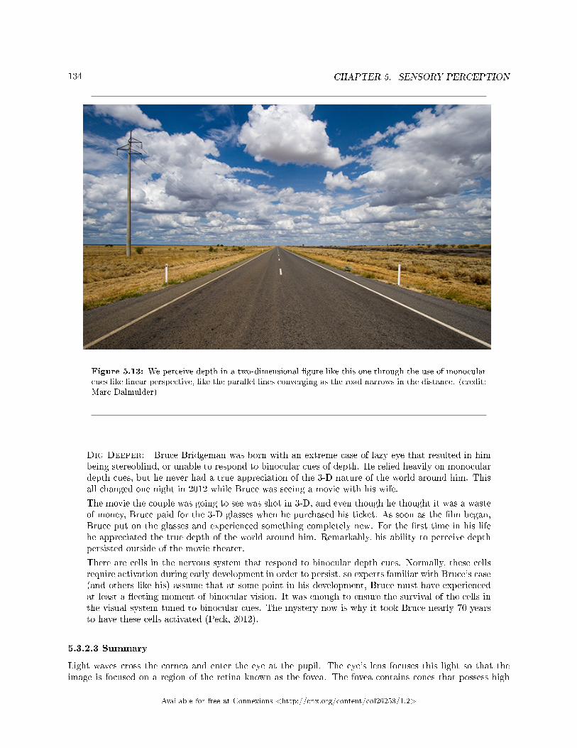
134 CHAPTER 5. SENSORY PERCEPTION
Figure 5.13: We perceive depth in a two-dimensional �gure like this one through the use of monocularcues like linear perspective, like the parallel lines converging as the road narrows in the distance. (credit:Marc Dalmulder)
Dig Deeper: Bruce Bridgeman was born with an extreme case of lazy eye that resulted in himbeing stereoblind, or unable to respond to binocular cues of depth. He relied heavily on monoculardepth cues, but he never had a true appreciation of the 3-D nature of the world around him. Thisall changed one night in 2012 while Bruce was seeing a movie with his wife.
The movie the couple was going to see was shot in 3-D, and even though he thought it was a wasteof money, Bruce paid for the 3-D glasses when he purchased his ticket. As soon as the �lm began,Bruce put on the glasses and experienced something completely new. For the �rst time in his lifehe appreciated the true depth of the world around him. Remarkably, his ability to perceive depthpersisted outside of the movie theater.
There are cells in the nervous system that respond to binocular depth cues. Normally, these cellsrequire activation during early development in order to persist, so experts familiar with Bruce's case(and others like his) assume that at some point in his development, Bruce must have experiencedat least a �eeting moment of binocular vision. It was enough to ensure the survival of the cells inthe visual system tuned to binocular cues. The mystery now is why it took Bruce nearly 70 yearsto have these cells activated (Peck, 2012).
5.3.2.3 Summary
Light waves cross the cornea and enter the eye at the pupil. The eye's lens focuses this light so that theimage is focused on a region of the retina known as the fovea. The fovea contains cones that possess high
Available for free at Connexions <http://cnx.org/content/col26253/1.2>
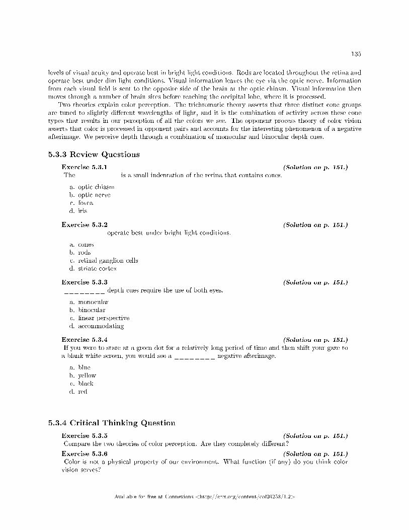
135
levels of visual acuity and operate best in bright light conditions. Rods are located throughout the retina andoperate best under dim light conditions. Visual information leaves the eye via the optic nerve. Informationfrom each visual �eld is sent to the opposite side of the brain at the optic chiasm. Visual information thenmoves through a number of brain sites before reaching the occipital lobe, where it is processed.
Two theories explain color perception. The trichromatic theory asserts that three distinct cone groupsare tuned to slightly di�erent wavelengths of light, and it is the combination of activity across these conetypes that results in our perception of all the colors we see. The opponent-process theory of color visionasserts that color is processed in opponent pairs and accounts for the interesting phenomenon of a negativeafterimage. We perceive depth through a combination of monocular and binocular depth cues.
5.3.3 Review Questions
Exercise 5.3.1 (Solution on p. 151.)
The ________ is a small indentation of the retina that contains cones.
a. optic chiasmb. optic nervec. fovead. iris
Exercise 5.3.2 (Solution on p. 151.)
________ operate best under bright light conditions.
a. conesb. rodsc. retinal ganglion cellsd. striate cortex
Exercise 5.3.3 (Solution on p. 151.)
________ depth cues require the use of both eyes.
a. monocularb. binocularc. linear perspectived. accommodating
Exercise 5.3.4 (Solution on p. 151.)
If you were to stare at a green dot for a relatively long period of time and then shift your gaze toa blank white screen, you would see a ________ negative afterimage.
a. blueb. yellowc. blackd. red
5.3.4 Critical Thinking Question
Exercise 5.3.5 (Solution on p. 151.)
Compare the two theories of color perception. Are they completely di�erent?
Exercise 5.3.6 (Solution on p. 151.)
Color is not a physical property of our environment. What function (if any) do you think colorvision serves?
Available for free at Connexions <http://cnx.org/content/col26253/1.2>
136 CHAPTER 5. SENSORY PERCEPTION
5.3.5 Personal Application Question
Exercise 5.3.7Take a look at a few of your photos or personal works of art. Can you �nd examples of linearperspective as a potential depth cue?
5.4 Hearing and Vestibular Sensation6
Audition, or hearing, is important to humans and to other animals for many di�erent interactions. Itenables an organism to detect and receive information about danger, such as an approaching predator,and to participate in communal exchanges like those concerning territories or mating. On the other hand,although it is physically linked to the auditory system, the vestibular system is not involved in hearing.Instead, an animal's vestibular system detects its own movement, both linear and angular acceleration anddeceleration, and balance.
5.4.1 Sound
Auditory stimuli are sound waves, which are mechanical, pressure waves that move through a medium, suchas air or water. There are no sound waves in a vacuum since there are no air molecules to move in waves. Thespeed of sound waves di�ers, based on altitude, temperature, and medium, but at sea level and a temperatureof 20º C (68º F), sound waves travel in the air at about 343 meters per second.
As is true for all waves, there are four main characteristics of a sound wave: frequency, wavelength, period,and amplitude. Frequency is the number of waves per unit of time, and in sound is heard as pitch. High-frequency (≥15.000Hz) sounds are higher-pitched (short wavelength) than low-frequency (long wavelengths;≤100Hz) sounds. Frequency is measured in cycles per second, and for sound, the most commonly used unit ishertz (Hz), or cycles per second. Most humans can perceive sounds with frequencies between 30 and 20,000Hz. Everyone's ability to hear high frequencies decreases with age. Dogs detect up to about 40,000 Hz; cats,60,000 Hz; bats, 100,000 Hz; and dolphins 150,000 Hz, and American shad (Alosa sapidissima), a �sh, canhear 180,000 Hz. Those frequencies above the human range are called ultrasound.
Amplitude, or the dimension of a wave from peak to trough, in sound is heard as volume and is illustratedin Figure 5.14. The sound waves of louder sounds have greater amplitude than those of softer sounds. Forsound, volume is measured in decibels (dB). The softest sound that a human can hear is the zero point.Humans speak normally at 60 decibels.
6This content is available online at <http://cnx.org/content/m66407/1.17/>.
Available for free at Connexions <http://cnx.org/content/col26253/1.2>
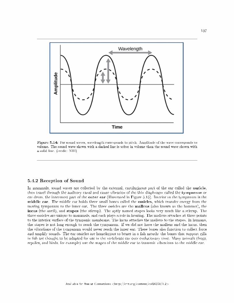
137
Figure 5.14: For sound waves, wavelength corresponds to pitch. Amplitude of the wave corresponds tovolume. The sound wave shown with a dashed line is softer in volume than the sound wave shown witha solid line. (credit: NIH)
5.4.2 Reception of Sound
In mammals, sound waves are collected by the external, cartilaginous part of the ear called the auricle,then travel through the auditory canal and cause vibration of the thin diaphragm called the tympanum orear drum, the innermost part of the outer ear (illustrated in Figure 5.15). Interior to the tympanum is themiddle ear. The middle ear holds three small bones called the ossicles, which transfer energy from themoving tympanum to the inner ear. The three ossicles are the malleus (also known as the hammer), theincus (the anvil), and stapes (the stirrup). The aptly named stapes looks very much like a stirrup. Thethree ossicles are unique to mammals, and each plays a role in hearing. The malleus attaches at three pointsto the interior surface of the tympanic membrane. The incus attaches the malleus to the stapes. In humans,the stapes is not long enough to reach the tympanum. If we did not have the malleus and the incus, thenthe vibrations of the tympanum would never reach the inner ear. These bones also function to collect forceand amplify sounds. The ear ossicles are homologous to bones in a �sh mouth: the bones that support gillsin �sh are thought to be adapted for use in the vertebrate ear over evolutionary time. Many animals (frogs,reptiles, and birds, for example) use the stapes of the middle ear to transmit vibrations to the middle ear.
Available for free at Connexions <http://cnx.org/content/col26253/1.2>
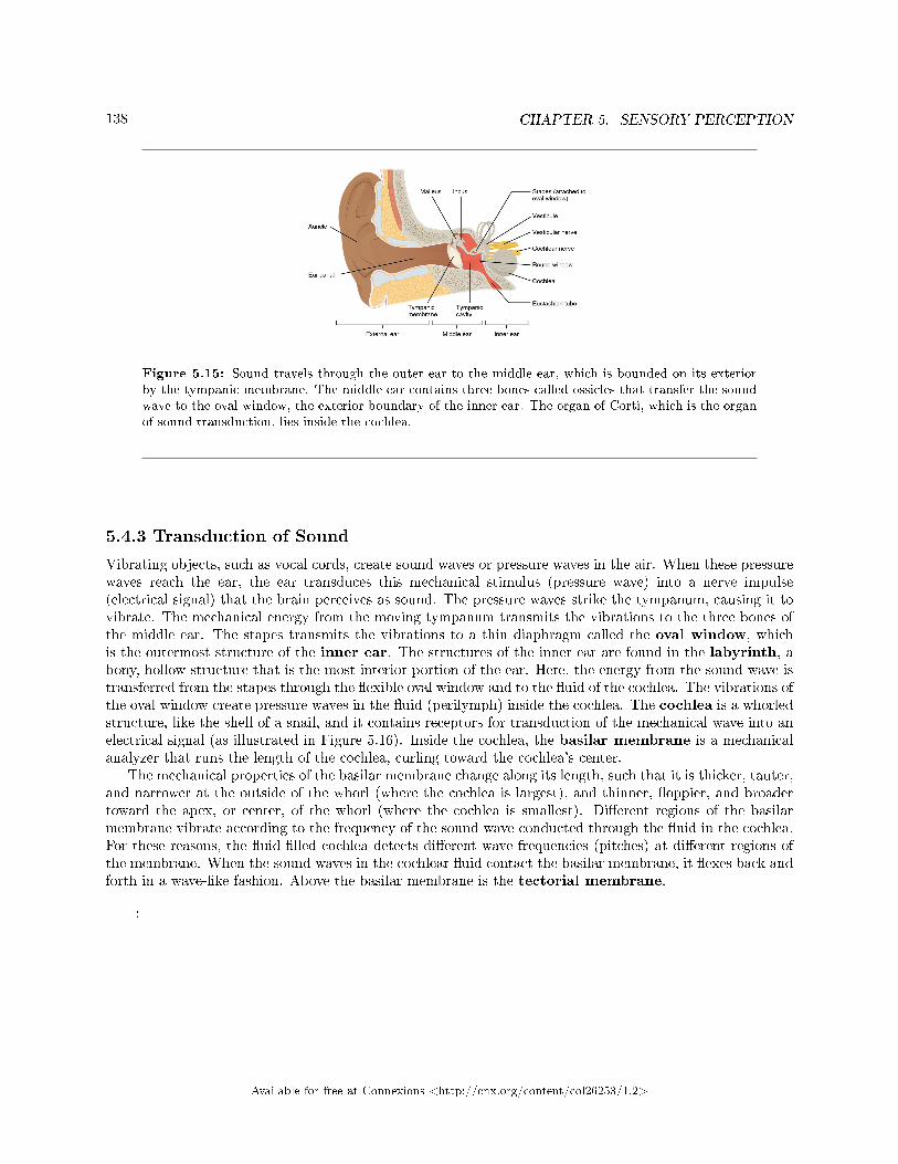
138 CHAPTER 5. SENSORY PERCEPTION
Figure 5.15: Sound travels through the outer ear to the middle ear, which is bounded on its exteriorby the tympanic membrane. The middle ear contains three bones called ossicles that transfer the soundwave to the oval window, the exterior boundary of the inner ear. The organ of Corti, which is the organof sound transduction, lies inside the cochlea.
5.4.3 Transduction of Sound
Vibrating objects, such as vocal cords, create sound waves or pressure waves in the air. When these pressurewaves reach the ear, the ear transduces this mechanical stimulus (pressure wave) into a nerve impulse(electrical signal) that the brain perceives as sound. The pressure waves strike the tympanum, causing it tovibrate. The mechanical energy from the moving tympanum transmits the vibrations to the three bones ofthe middle ear. The stapes transmits the vibrations to a thin diaphragm called the oval window, whichis the outermost structure of the inner ear. The structures of the inner ear are found in the labyrinth, abony, hollow structure that is the most interior portion of the ear. Here, the energy from the sound wave istransferred from the stapes through the �exible oval window and to the �uid of the cochlea. The vibrations ofthe oval window create pressure waves in the �uid (perilymph) inside the cochlea. The cochlea is a whorledstructure, like the shell of a snail, and it contains receptors for transduction of the mechanical wave into anelectrical signal (as illustrated in Figure 5.16). Inside the cochlea, the basilar membrane is a mechanicalanalyzer that runs the length of the cochlea, curling toward the cochlea's center.
The mechanical properties of the basilar membrane change along its length, such that it is thicker, tauter,and narrower at the outside of the whorl (where the cochlea is largest), and thinner, �oppier, and broadertoward the apex, or center, of the whorl (where the cochlea is smallest). Di�erent regions of the basilarmembrane vibrate according to the frequency of the sound wave conducted through the �uid in the cochlea.For these reasons, the �uid-�lled cochlea detects di�erent wave frequencies (pitches) at di�erent regions ofthe membrane. When the sound waves in the cochlear �uid contact the basilar membrane, it �exes back andforth in a wave-like fashion. Above the basilar membrane is the tectorial membrane.
:
Available for free at Connexions <http://cnx.org/content/col26253/1.2>
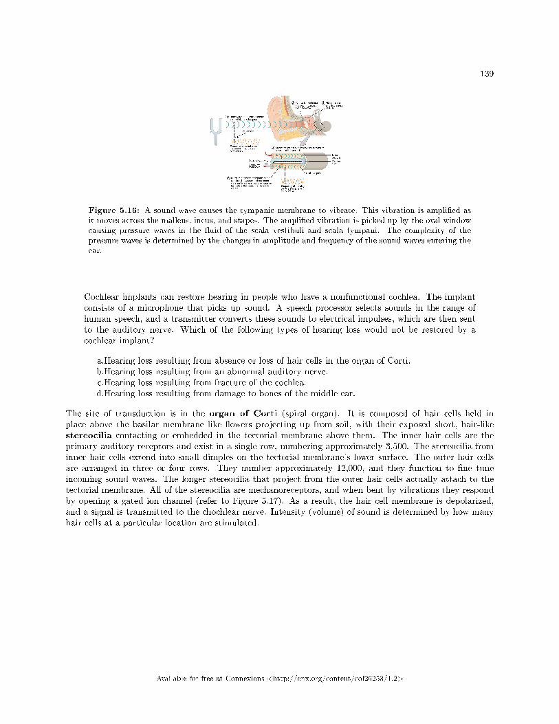
139
Figure 5.16: A sound wave causes the tympanic membrane to vibrate. This vibration is ampli�ed asit moves across the malleus, incus, and stapes. The ampli�ed vibration is picked up by the oval windowcausing pressure waves in the �uid of the scala vestibuli and scala tympani. The complexity of thepressure waves is determined by the changes in amplitude and frequency of the sound waves entering theear.
Cochlear implants can restore hearing in people who have a nonfunctional cochlea. The implantconsists of a microphone that picks up sound. A speech processor selects sounds in the range ofhuman speech, and a transmitter converts these sounds to electrical impulses, which are then sentto the auditory nerve. Which of the following types of hearing loss would not be restored by acochlear implant?
a.Hearing loss resulting from absence or loss of hair cells in the organ of Corti.b.Hearing loss resulting from an abnormal auditory nerve.c.Hearing loss resulting from fracture of the cochlea.d.Hearing loss resulting from damage to bones of the middle ear.
The site of transduction is in the organ of Corti (spiral organ). It is composed of hair cells held inplace above the basilar membrane like �owers projecting up from soil, with their exposed short, hair-likestereocilia contacting or embedded in the tectorial membrane above them. The inner hair cells are theprimary auditory receptors and exist in a single row, numbering approximately 3,500. The stereocilia frominner hair cells extend into small dimples on the tectorial membrane's lower surface. The outer hair cellsare arranged in three or four rows. They number approximately 12,000, and they function to �ne tuneincoming sound waves. The longer stereocilia that project from the outer hair cells actually attach to thetectorial membrane. All of the stereocilia are mechanoreceptors, and when bent by vibrations they respondby opening a gated ion channel (refer to Figure 5.17). As a result, the hair cell membrane is depolarized,and a signal is transmitted to the chochlear nerve. Intensity (volume) of sound is determined by how manyhair cells at a particular location are stimulated.
Available for free at Connexions <http://cnx.org/content/col26253/1.2>
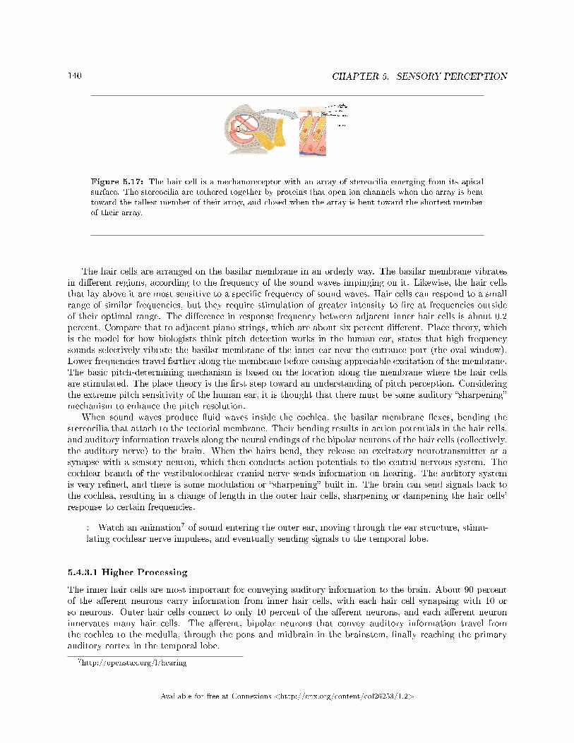
140 CHAPTER 5. SENSORY PERCEPTION
Figure 5.17: The hair cell is a mechanoreceptor with an array of stereocilia emerging from its apicalsurface. The stereocilia are tethered together by proteins that open ion channels when the array is benttoward the tallest member of their array, and closed when the array is bent toward the shortest memberof their array.
The hair cells are arranged on the basilar membrane in an orderly way. The basilar membrane vibratesin di�erent regions, according to the frequency of the sound waves impinging on it. Likewise, the hair cellsthat lay above it are most sensitive to a speci�c frequency of sound waves. Hair cells can respond to a smallrange of similar frequencies, but they require stimulation of greater intensity to �re at frequencies outsideof their optimal range. The di�erence in response frequency between adjacent inner hair cells is about 0.2percent. Compare that to adjacent piano strings, which are about six percent di�erent. Place theory, whichis the model for how biologists think pitch detection works in the human ear, states that high frequencysounds selectively vibrate the basilar membrane of the inner ear near the entrance port (the oval window).Lower frequencies travel farther along the membrane before causing appreciable excitation of the membrane.The basic pitch-determining mechanism is based on the location along the membrane where the hair cellsare stimulated. The place theory is the �rst step toward an understanding of pitch perception. Consideringthe extreme pitch sensitivity of the human ear, it is thought that there must be some auditory �sharpening�mechanism to enhance the pitch resolution.
When sound waves produce �uid waves inside the cochlea, the basilar membrane �exes, bending thestereocilia that attach to the tectorial membrane. Their bending results in action potentials in the hair cells,and auditory information travels along the neural endings of the bipolar neurons of the hair cells (collectively,the auditory nerve) to the brain. When the hairs bend, they release an excitatory neurotransmitter at asynapse with a sensory neuron, which then conducts action potentials to the central nervous system. Thecochlear branch of the vestibulocochlear cranial nerve sends information on hearing. The auditory systemis very re�ned, and there is some modulation or �sharpening� built in. The brain can send signals back tothe cochlea, resulting in a change of length in the outer hair cells, sharpening or dampening the hair cells'response to certain frequencies.
: Watch an animation7 of sound entering the outer ear, moving through the ear structure, stimu-lating cochlear nerve impulses, and eventually sending signals to the temporal lobe.
5.4.3.1 Higher Processing
The inner hair cells are most important for conveying auditory information to the brain. About 90 percentof the a�erent neurons carry information from inner hair cells, with each hair cell synapsing with 10 orso neurons. Outer hair cells connect to only 10 percent of the a�erent neurons, and each a�erent neuroninnervates many hair cells. The a�erent, bipolar neurons that convey auditory information travel fromthe cochlea to the medulla, through the pons and midbrain in the brainstem, �nally reaching the primaryauditory cortex in the temporal lobe.
7http://openstax.org/l/hearing
Available for free at Connexions <http://cnx.org/content/col26253/1.2>
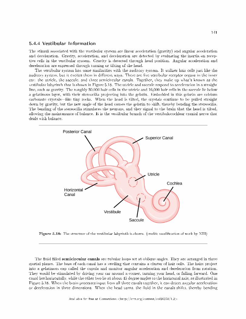
141
5.4.4 Vestibular Information
The stimuli associated with the vestibular system are linear acceleration (gravity) and angular accelerationand deceleration. Gravity, acceleration, and deceleration are detected by evaluating the inertia on recep-tive cells in the vestibular system. Gravity is detected through head position. Angular acceleration anddeceleration are expressed through turning or tilting of the head.
The vestibular system has some similarities with the auditory system. It utilizes hair cells just like theauditory system, but it excites them in di�erent ways. There are �ve vestibular receptor organs in the innerear: the utricle, the saccule, and three semicircular canals. Together, they make up what's known as thevestibular labyrinth that is shown in Figure 5.18. The utricle and saccule respond to acceleration in a straightline, such as gravity. The roughly 30,000 hair cells in the utricle and 16,000 hair cells in the saccule lie belowa gelatinous layer, with their stereocilia projecting into the gelatin. Embedded in this gelatin are calciumcarbonate crystals�like tiny rocks. When the head is tilted, the crystals continue to be pulled straightdown by gravity, but the new angle of the head causes the gelatin to shift, thereby bending the stereocilia.The bending of the stereocilia stimulates the neurons, and they signal to the brain that the head is tilted,allowing the maintenance of balance. It is the vestibular branch of the vestibulocochlear cranial nerve thatdeals with balance.
Figure 5.18: The structure of the vestibular labyrinth is shown. (credit: modi�cation of work by NIH)
The �uid-�lled semicircular canals are tubular loops set at oblique angles. They are arranged in threespatial planes. The base of each canal has a swelling that contains a cluster of hair cells. The hairs projectinto a gelatinous cap called the cupula and monitor angular acceleration and deceleration from rotation.They would be stimulated by driving your car around a corner, turning your head, or falling forward. Onecanal lies horizontally, while the other two lie at about 45 degree angles to the horizontal axis, as illustrated inFigure 5.18. When the brain processes input from all three canals together, it can detect angular accelerationor deceleration in three dimensions. When the head turns, the �uid in the canals shifts, thereby bending
Available for free at Connexions <http://cnx.org/content/col26253/1.2>
142 CHAPTER 5. SENSORY PERCEPTION
stereocilia and sending signals to the brain. Upon cessation accelerating or decelerating�or just moving�the movement of the �uid within the canals slows or stops. For example, imagine holding a glass of water.When moving forward, water may splash backwards onto the hand, and when motion has stopped, watermay splash forward onto the �ngers. While in motion, the water settles in the glass and does not splash.Note that the canals are not sensitive to velocity itself, but to changes in velocity, so moving forward at60mph with your eyes closed would not give the sensation of movement, but suddenly accelerating or brakingwould stimulate the receptors.
5.4.4.1 Higher Processing
Hair cells from the utricle, saccule, and semicircular canals also communicate through bipolar neurons tothe cochlear nucleus in the medulla. Cochlear neurons send descending projections to the spinal cord andascending projections to the pons, thalamus, and cerebellum. Connections to the cerebellum are importantfor coordinated movements. There are also projections to the temporal cortex, which account for feelingsof dizziness; projections to autonomic nervous system areas in the brainstem, which account for motionsickness; and projections to the primary somatosensory cortex, which monitors subjective measurementsof the external world and self-movement. People with lesions in the vestibular area of the somatosensorycortex see vertical objects in the world as being tilted. Finally, the vestibular signals project to certain opticmuscles to coordinate eye and head movements.
: Click through this interactive tutorial8 to review the parts of the ear and how they function toprocess sound.
5.4.5 Section Summary
Audition is important for territory defense, predation, predator defense, and communal exchanges. Thevestibular system, which is not auditory, detects linear acceleration and angular acceleration and deceleration.Both the auditory system and vestibular system use hair cells as their receptors.
Auditory stimuli are sound waves. The sound wave energy reaches the outer ear (auricle, canal, tympa-num), and vibrations of the tympanum send the energy to the middle ear. The middle ear bones shift andthe stapes transfers mechanical energy to the oval window of the �uid-�lled inner ear cochlea. Once in thecochlea, the energy causes the basilar membrane to �ex, thereby bending the stereocilia on receptor haircells. This activates the receptors, which send their auditory neural signals to the brain.
The vestibular system has �ve parts that work together to provide the sense of direction, thus helping tomaintain balance. The utricle and saccule measure head orientation: their calcium carbonate crystals shiftwhen the head is tilted, thereby activating hair cells. The semicircular canals work similarly, such that whenthe head is turned, the �uid in the canals bends stereocilia on hair cells. The vestibular hair cells also sendsignals to the thalamus and to the somatosensory cortex, but also to the cerebellum, the structure above thebrainstem that plays a large role in timing and coordination of movement.
5.4.6 Visual Connection Questions
Exercise 5.4.1 (Solution on p. 151.)
Figure 5.16 Cochlear implants can restore hearing in people who have a nonfunctional cochlea.The implant consists of a microphone that picks up sound. A speech processor selects sounds in therange of human speech, and a transmitter converts these sounds to electrical impulses, which arethen sent to the auditory nerve. Which of the following types of hearing loss would not be restoredby a cochlear implant?
a. Hearing loss resulting from absence or loss of hair cells in the organ of Corti.
8http://openstax.org/l/ear_anatomy
Available for free at Connexions <http://cnx.org/content/col26253/1.2>
143
b. Hearing loss resulting from an abnormal auditory nerve.c. Hearing loss resulting from fracture of the cochlea.d. Hearing loss resulting from damage to bones of the middle ear.
5.4.7 Review Questions
Exercise 5.4.2 (Solution on p. 152.)
In sound, pitch is measured in _____, and volume is measured in _____.
a. nanometers (nm); decibels (dB)b. decibels (dB); nanometers (nm)c. decibels (dB); hertz (Hz)d. hertz (Hz); decibels (dB)
Exercise 5.4.3 (Solution on p. 152.)
Auditory hair cells are indirectly anchored to the _____.
a. basilar membraneb. oval windowc. tectorial membraned. ossicles
Exercise 5.4.4 (Solution on p. 152.)
Which of the following are found both in the auditory system and the vestibular system?
a. basilar membraneb. hair cellsc. semicircular canalsd. ossicles
Exercise 5.4.5 (Solution on p. 152.)
Benign Paroxysmal Positional Vertigo is a disorder where some of the calcium carbonate crystals inthe utricle migrate into the semicircular canals. Why does this condition cause periods of dizziness?
a. The hair cells in the semicircular canals will be constantly activated.b. The hair cells in the semicircular canals will now be stimulated by gravity.c. The utricle will no longer recognize acceleration.d. There will be too much volume in the semicircular canals for them to detect motion.
5.4.8 Critical Thinking Questions
Exercise 5.4.6 (Solution on p. 152.)
How would a rise in altitude likely a�ect the speed of a sound transmitted through air? Why?
Exercise 5.4.7 (Solution on p. 152.)
How might being in a place with less gravity than Earth has (such as Earth's moon) a�ect vestibularsensation, and why?
Exercise 5.4.8 (Solution on p. 152.)
How does the structure of the ear allow a person to determine where a sound originates?
Available for free at Connexions <http://cnx.org/content/col26253/1.2>
144 CHAPTER 5. SENSORY PERCEPTION
5.5 Taste and Smell9
Taste, also called gustation, and smell, also called olfaction, are the most interconnected senses in thatboth involve molecules of the stimulus entering the body and bonding to receptors. Smell lets an animal sensethe presence of food or other animals�whether potential mates, predators, or prey�or other chemicals inthe environment that can impact their survival. Similarly, the sense of taste allows animals to discriminatebetween types of foods. While the value of a sense of smell is obvious, what is the value of a sense oftaste? Di�erent tasting foods have di�erent attributes, both helpful and harmful. For example, sweet-tasting substances tend to be highly caloric, which could be necessary for survival in lean times. Bitterness isassociated with toxicity, and sourness is associated with spoiled food. Salty foods are valuable in maintaininghomeostasis by helping the body retain water and by providing ions necessary for cells to function.
5.5.1 Tastes and Odors
Both taste and odor stimuli are molecules taken in from the environment. The primary tastes detectedby humans are sweet, sour, bitter, salty and umami. The �rst four tastes need little explanation. Theidenti�cation of umami as a fundamental taste occurred fairly recently�it was identi�ed in 1908 by Japanesescientist Kikunae Ikeda while he worked with seaweed broth, but it was not widely accepted as a taste thatcould be physiologically distinguished until many years later. The taste of umami, also known as savoriness,is attributable to the taste of the amino acid L-glutamate. In fact, monosodium glutamate, or MSG, is oftenused in cooking to enhance the savory taste of certain foods. What is the adaptive value of being able todistinguish umami? Savory substances tend to be high in protein.
All odors that we perceive are molecules in the air we breathe. If a substance does not release moleculesinto the air from its surface, it has no smell. And if a human or other animal does not have a receptor thatrecognizes a speci�c molecule, then that molecule has no smell. Humans have about 350 olfactory receptorsubtypes that work in various combinations to allow us to sense about 10,000 di�erent odors. Compare thatto mice, for example, which have about 1,300 olfactory receptor types, and therefore probably sense moreodors. Both odors and tastes involve molecules that stimulate speci�c chemoreceptors. Although humanscommonly distinguish taste as one sense and smell as another, they work together to create the perceptionof �avor. A person's perception of �avor is reduced if he or she has congested nasal passages.
5.5.2 Reception and Transduction
Odorants (odor molecules) enter the nose and dissolve in the olfactory epithelium, the mucosa at the backof the nasal cavity (as illustrated in Figure 5.19). The olfactory epithelium is a collection of specializedolfactory receptors in the back of the nasal cavity that spans an area about 5 cm2 in humans. Recall thatsensory cells are neurons. An olfactory receptor, which is a dendrite of a specialized neuron, responds whenit binds certain molecules inhaled from the environment by sending impulses directly to the olfactory bulbof the brain. Humans have about 12 million olfactory receptors, distributed among hundreds of di�erentreceptor types that respond to di�erent odors. Twelve million seems like a large number of receptors,but compare that to other animals: rabbits have about 100 million, most dogs have about 1 billion, andbloodhounds�dogs selectively bred for their sense of smell�have about 4 billion. The overall size of theolfactory epithelium also di�ers between species, with that of bloodhounds, for example, being many timeslarger than that of humans.
Olfactory neurons are bipolar neurons (neurons with two processes from the cell body). Each neuronhas a single dendrite buried in the olfactory epithelium, and extending from this dendrite are 5 to 20 receptor-laden, hair-like cilia that trap odorant molecules. The sensory receptors on the cilia are proteins, and it is thevariations in their amino acid chains that make the receptors sensitive to di�erent odorants. Each olfactorysensory neuron has only one type of receptor on its cilia, and the receptors are specialized to detect speci�codorants, so the bipolar neurons themselves are specialized. When an odorant binds with a receptor that
9This content is available online at <http://cnx.org/content/m44764/1.6/>.
Available for free at Connexions <http://cnx.org/content/col26253/1.2>
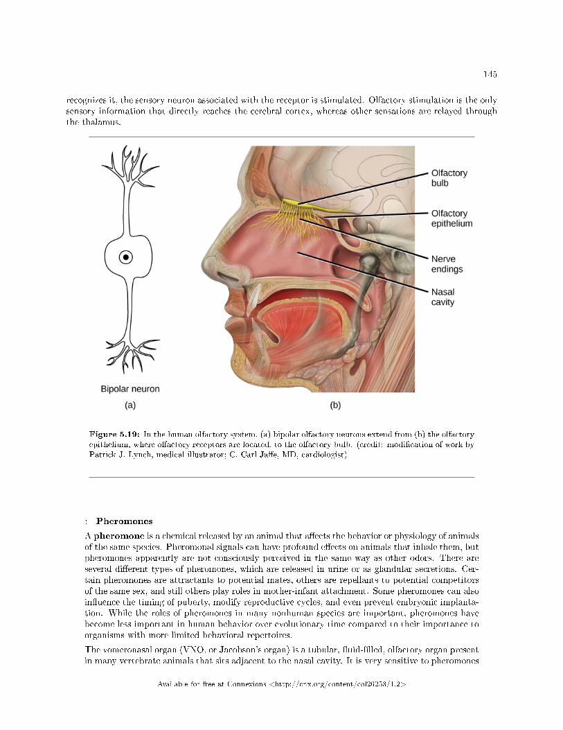
145
recognizes it, the sensory neuron associated with the receptor is stimulated. Olfactory stimulation is the onlysensory information that directly reaches the cerebral cortex, whereas other sensations are relayed throughthe thalamus.
Figure 5.19: In the human olfactory system, (a) bipolar olfactory neurons extend from (b) the olfactoryepithelium, where olfactory receptors are located, to the olfactory bulb. (credit: modi�cation of work byPatrick J. Lynch, medical illustrator; C. Carl Ja�e, MD, cardiologist)
: Pheromones
A pheromone is a chemical released by an animal that a�ects the behavior or physiology of animalsof the same species. Pheromonal signals can have profound e�ects on animals that inhale them, butpheromones apparently are not consciously perceived in the same way as other odors. There areseveral di�erent types of pheromones, which are released in urine or as glandular secretions. Cer-tain pheromones are attractants to potential mates, others are repellants to potential competitorsof the same sex, and still others play roles in mother-infant attachment. Some pheromones can alsoin�uence the timing of puberty, modify reproductive cycles, and even prevent embryonic implanta-tion. While the roles of pheromones in many nonhuman species are important, pheromones havebecome less important in human behavior over evolutionary time compared to their importance toorganisms with more limited behavioral repertoires.
The vomeronasal organ (VNO, or Jacobson's organ) is a tubular, �uid-�lled, olfactory organ presentin many vertebrate animals that sits adjacent to the nasal cavity. It is very sensitive to pheromones
Available for free at Connexions <http://cnx.org/content/col26253/1.2>
146 CHAPTER 5. SENSORY PERCEPTION
and is connected to the nasal cavity by a duct. When molecules dissolve in the mucosa of thenasal cavity, they then enter the VNO where the pheromone molecules among them bind withspecialized pheromone receptors. Upon exposure to pheromones from their own species or others,many animals, including cats, may display the �ehmen response (shown in Figure 5.20), a curlingof the upper lip that helps pheromone molecules enter the VNO.
Pheromonal signals are sent, not to the main olfactory bulb, but to a di�erent neural structure thatprojects directly to the amygdala (recall that the amygdala is a brain center important in emotionalreactions, such as fear). The pheromonal signal then continues to areas of the hypothalamus thatare key to reproductive physiology and behavior. While some scientists assert that the VNO isapparently functionally vestigial in humans, even though there is a similar structure located nearhuman nasal cavities, others are researching it as a possible functional system that may, for example,contribute to synchronization of menstrual cycles in women living in close proximity.
Figure 5.20: The �ehmen response in this tiger results in the curling of the upper lip and helps airbornepheromone molecules enter the vomeronasal organ. (credit: modi�cation of work by "chadh"/Flickr)
5.5.2.1 Taste
Detecting a taste (gustation) is fairly similar to detecting an odor (olfaction), given that both taste andsmell rely on chemical receptors being stimulated by certain molecules. The primary organ of taste is thetaste bud. A taste bud is a cluster of gustatory receptors (taste cells) that are located within the bumpson the tongue called papillae (singular: papilla) (illustrated in Figure 5.21). There are several structurallydistinct papillae. Filiform papillae, which are located across the tongue, are tactile, providing friction thathelps the tongue move substances, and contain no taste cells. In contrast, fungiform papillae, which arelocated mainly on the anterior two-thirds of the tongue, each contain one to eight taste buds and also havereceptors for pressure and temperature. The large circumvallate papillae contain up to 100 taste buds andform a V near the posterior margin of the tongue.
Available for free at Connexions <http://cnx.org/content/col26253/1.2>
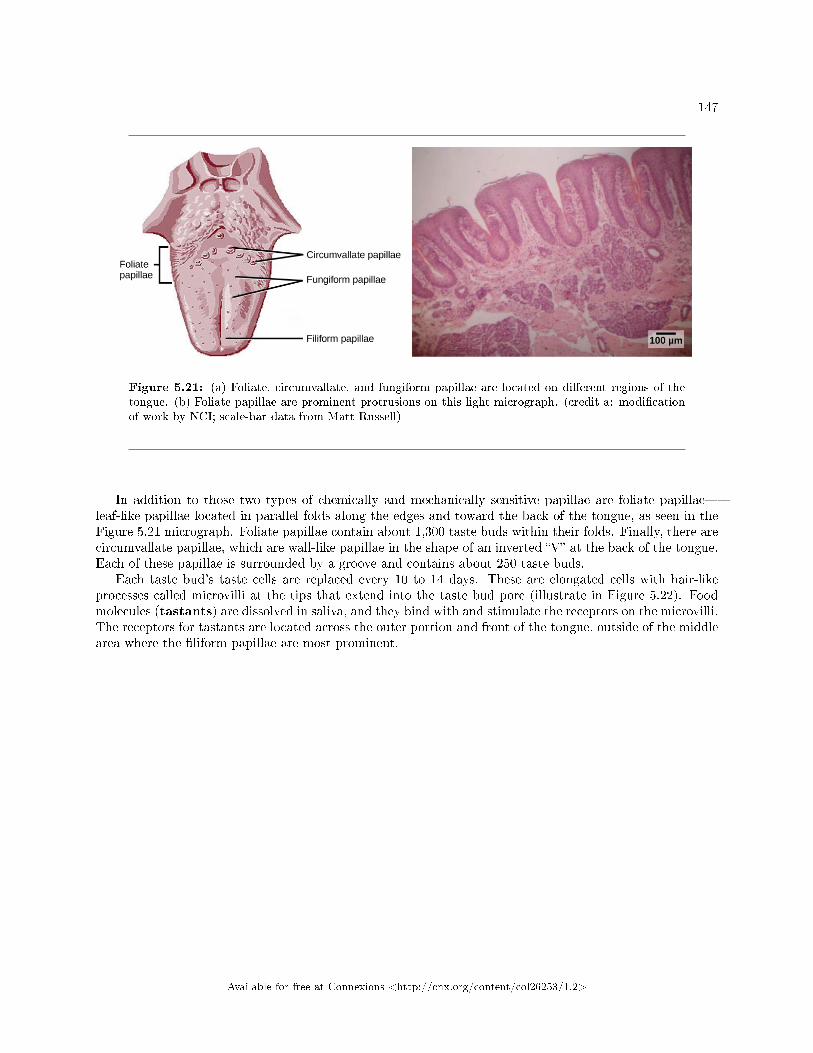
147
Figure 5.21: (a) Foliate, circumvallate, and fungiform papillae are located on di�erent regions of thetongue. (b) Foliate papillae are prominent protrusions on this light micrograph. (credit a: modi�cationof work by NCI; scale-bar data from Matt Russell)
In addition to those two types of chemically and mechanically sensitive papillae are foliate papillae�leaf-like papillae located in parallel folds along the edges and toward the back of the tongue, as seen in theFigure 5.21 micrograph. Foliate papillae contain about 1,300 taste buds within their folds. Finally, there arecircumvallate papillae, which are wall-like papillae in the shape of an inverted �V� at the back of the tongue.Each of these papillae is surrounded by a groove and contains about 250 taste buds.
Each taste bud's taste cells are replaced every 10 to 14 days. These are elongated cells with hair-likeprocesses called microvilli at the tips that extend into the taste bud pore (illustrate in Figure 5.22). Foodmolecules (tastants) are dissolved in saliva, and they bind with and stimulate the receptors on the microvilli.The receptors for tastants are located across the outer portion and front of the tongue, outside of the middlearea where the �liform papillae are most prominent.
Available for free at Connexions <http://cnx.org/content/col26253/1.2>
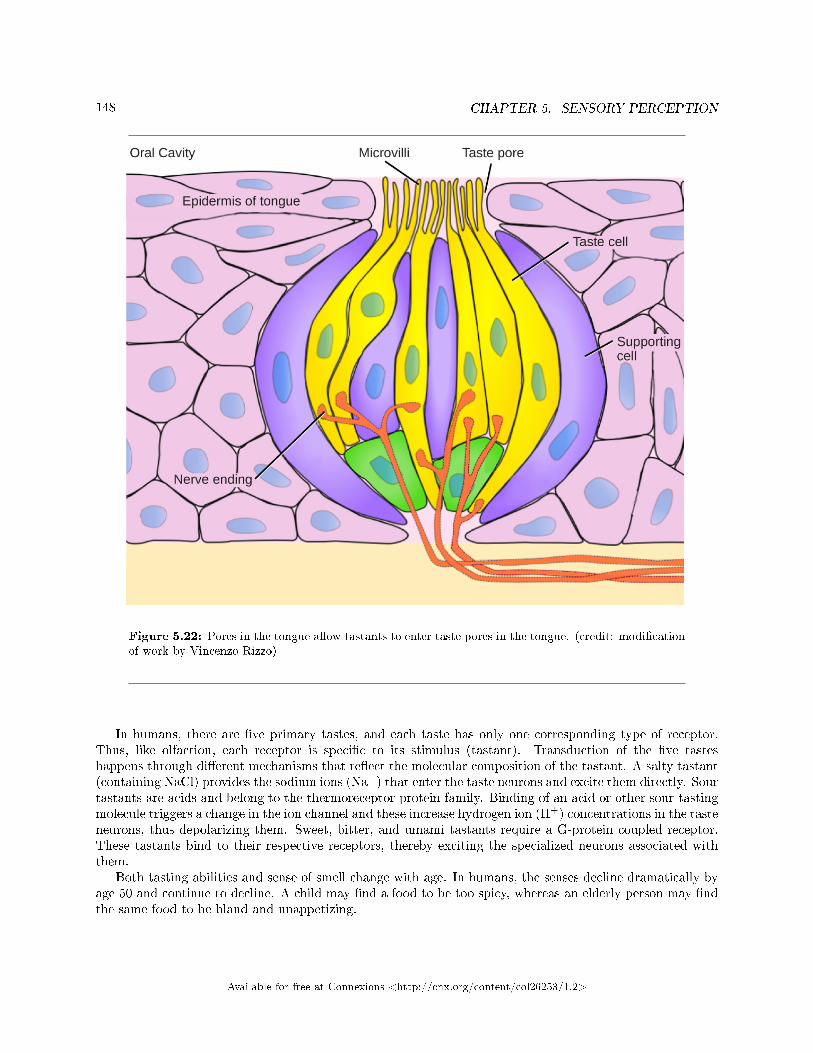
148 CHAPTER 5. SENSORY PERCEPTION
Figure 5.22: Pores in the tongue allow tastants to enter taste pores in the tongue. (credit: modi�cationof work by Vincenzo Rizzo)
In humans, there are �ve primary tastes, and each taste has only one corresponding type of receptor.Thus, like olfaction, each receptor is speci�c to its stimulus (tastant). Transduction of the �ve tasteshappens through di�erent mechanisms that re�ect the molecular composition of the tastant. A salty tastant(containing NaCl) provides the sodium ions (Na+) that enter the taste neurons and excite them directly. Sourtastants are acids and belong to the thermoreceptor protein family. Binding of an acid or other sour-tastingmolecule triggers a change in the ion channel and these increase hydrogen ion (H+) concentrations in the tasteneurons, thus depolarizing them. Sweet, bitter, and umami tastants require a G-protein coupled receptor.These tastants bind to their respective receptors, thereby exciting the specialized neurons associated withthem.
Both tasting abilities and sense of smell change with age. In humans, the senses decline dramatically byage 50 and continue to decline. A child may �nd a food to be too spicy, whereas an elderly person may �ndthe same food to be bland and unappetizing.
Available for free at Connexions <http://cnx.org/content/col26253/1.2>
149
: View this animation10 that shows how the sense of taste works.
5.5.3 Smell and Taste in the Brain
Olfactory neurons project from the olfactory epithelium to the olfactory bulb as thin, unmyelinated axons.The olfactory bulb is composed of neural clusters called glomeruli, and each glomerulus receives signalsfrom one type of olfactory receptor, so each glomerulus is speci�c to one odorant. From glomeruli, olfactorysignals travel directly to the olfactory cortex and then to the frontal cortex and the thalamus. Recall thatthis is a di�erent path from most other sensory information, which is sent directly to the thalamus beforeending up in the cortex. Olfactory signals also travel directly to the amygdala, thereafter reaching thehypothalamus, thalamus, and frontal cortex. The last structure that olfactory signals directly travel tois a cortical center in the temporal lobe structure important in spatial, autobiographical, declarative, andepisodic memories. Olfaction is �nally processed by areas of the brain that deal with memory, emotions,reproduction, and thought.
Taste neurons project from taste cells in the tongue, esophagus, and palate to the medulla, in thebrainstem. From the medulla, taste signals travel to the thalamus and then to the primary gustatory cortex.Information from di�erent regions of the tongue is segregated in the medulla, thalamus, and cortex.
5.5.4 Section Summary
There are �ve primary tastes in humans: sweet, sour, bitter, salty, and umami. Each taste has its ownreceptor type that responds only to that taste. Tastants enter the body and are dissolved in saliva. Tastecells are located within taste buds, which are found on three of the four types of papillae in the mouth.
Regarding olfaction, there are many thousands of odorants, but humans detect only about 10,000. Liketaste receptors, olfactory receptors are each responsive to only one odorant. Odorants dissolve in nasalmucosa, where they excite their corresponding olfactory sensory cells. When these cells detect an odorant,they send their signals to the main olfactory bulb and then to other locations in the brain, including theolfactory cortex.
5.5.5 Review Questions
Exercise 5.5.1 (Solution on p. 152.)
Which of the following has the fewest taste receptors?
a. fungiform papillaeb. circumvallate papillaec. foliate papillaed. �liform papillae
Exercise 5.5.2 (Solution on p. 152.)
How many di�erent taste molecules do taste cells each detect?
10http://openstaxcollege.org/l/taste
Available for free at Connexions <http://cnx.org/content/col26253/1.2>
150 CHAPTER 5. SENSORY PERCEPTION
a. oneb. �vec. tend. It depends on the spot on the tongue
Exercise 5.5.3 (Solution on p. 152.)
Salty foods activate the taste cells by _____.
a. exciting the taste cell directlyb. causing hydrogen ions to enter the cellc. causing sodium channels to closed. binding directly to the receptors
Exercise 5.5.4 (Solution on p. 152.)
All sensory signals except _____ travel to the _____ in the brain before the cerebral cortex.
a. vision; thalamusb. olfaction; thalamusc. vision; cranial nervesd. olfaction; cranial nerves
5.5.6 Free Response
Exercise 5.5.5 (Solution on p. 152.)
From the perspective of the recipient of the signal, in what ways do pheromones di�er from otherodorants?
Exercise 5.5.6 (Solution on p. 152.)
What might be the e�ect on an animal of not being able to perceive taste?
Available for free at Connexions <http://cnx.org/content/col26253/1.2>
151
Solutions to Exercises in Chapter 5
to Exercise 5.1.1 (p. 119)Bto Exercise 5.1.2 (p. 119)Dto Exercise 5.1.3 (p. 119)Ato Exercise 5.1.4 (p. 119)Dto Exercise 5.1.5 (p. 119)Transmission of sensory information from the receptor to the central nervous system will be impaired, andthus, perception of stimuli, which occurs in the brain, will be halted.to Exercise 5.1.6 (p. 119)The just-noticeable di�erence is a fraction of the overall magnitude of the stimulus and seems to be arelatively �xed proportion (such as 10 percent) whether the stimulus is large (such as a very heavy object)or small (such as a very light object).to Exercise 5.1.7 (p. 119)General sensory receptors are located throughout the body in the skin and internal organs. Conversely,special senses are all located in the head region, and require specialized organs.to Exercise 5.2.1 (p. 125)Figure 5.4 Dto Exercise 5.2.2 (p. 125)Bto Exercise 5.2.3 (p. 125)Ato Exercise 5.2.4 (p. 126)Bto Exercise 5.2.5 (p. 126)The cortical areas serving skin that is densely innervated likely are larger than those serving skin that isless densely innervated.to Exercise 5.2.6 (p. 126)Pain is a subjective sensation that relies on the brain interpreting the nociception signals received by thesensory receptors (perception). Therefore, even though two people experience identical stimuli, their brainscan perceive them as very di�erent sensory experiences.Solution to Exercise 5.3.1 (p. 135)CSolution to Exercise 5.3.2 (p. 135)ASolution to Exercise 5.3.3 (p. 135)BSolution to Exercise 5.3.4 (p. 135)DSolution to Exercise 5.3.5 (p. 135)The trichromatic theory of color vision and the opponent-process theory are not mutually exclusive. Re-search has shown they apply to di�erent levels of the nervous system. For visual processing on the retina,trichromatic theory applies: the cones are responsive to three di�erent wavelengths that represent red, blue,and green. But once the signal moves past the retina on its way to the brain, the cells respond in a wayconsistent with opponent-process theory.Solution to Exercise 5.3.6 (p. 135)Color vision probably serves multiple adaptive purposes. One popular hypothesis suggests that seeing incolor allowed our ancestors to di�erentiate ripened fruits and vegetables more easily.
Available for free at Connexions <http://cnx.org/content/col26253/1.2>

152 CHAPTER 5. SENSORY PERCEPTION
to Exercise 5.4.1 (p. 142)Figure 5.16 Bto Exercise 5.4.2 (p. 143)Dto Exercise 5.4.3 (p. 143)Ato Exercise 5.4.4 (p. 143)Bto Exercise 5.4.5 (p. 143)Bto Exercise 5.4.6 (p. 143)The sound would slow down, because it is transmitted through the particles (gas) and there are fewerparticles (lower density) at higher altitudes.to Exercise 5.4.7 (p. 143)Because vestibular sensation relies on gravity's e�ects on tiny crystals in the inner ear, a situation of reducedgravity would likely impair vestibular sensation.to Exercise 5.4.8 (p. 143)The �rst step in processing a sound in humans is the collection of sound by the auricle. When a personencounters a sound, the auricle on both sides of the head will collect the vibrations. Since the waves originatefrom a single site, the two auricles will not collect the sound at the exact same time. When the sound isprocessed by the auditory system, the brain is able to use this slight di�erence in timing to determine thelocation of the sound.to Exercise 5.5.1 (p. 149)Dto Exercise 5.5.2 (p. 149)Ato Exercise 5.5.3 (p. 150)Ato Exercise 5.5.4 (p. 150)Bto Exercise 5.5.5 (p. 150)Pheromones may not be consciously perceived, and pheromones can have direct physiological and behaviorale�ects on their recipients.to Exercise 5.5.6 (p. 150)The animal might not be able to recognize the di�erences in food sources and thus might not be able todiscriminate between spoiled food and safe food or between foods that contain necessary nutrients, such asproteins, and foods that do not.
Available for free at Connexions <http://cnx.org/content/col26253/1.2>
Chapter 6
Structural Support and Movement
6.1 Types of Skeletal Systems1
A skeletal system is necessary to support the body, protect internal organs, and allow for the movementof an organism. There are three di�erent skeleton designs that ful�ll these functions: hydrostatic skeleton,exoskeleton, and endoskeleton.
6.1.1 Hydrostatic Skeleton
A hydrostatic skeleton is a skeleton formed by a �uid-�lled compartment within the body, called thecoelom. The organs of the coelom are supported by the aqueous �uid, which also resists external compression.This compartment is under hydrostatic pressure because of the �uid and supports the other organs of theorganism. This type of skeletal system is found in soft-bodied animals such as sea anemones, earthworms,Cnidaria, and other invertebrates (Figure 6.1).
1This content is available online at <http://cnx.org/content/m71332/1.1/>.
Available for free at Connexions <http://cnx.org/content/col26253/1.2>
153
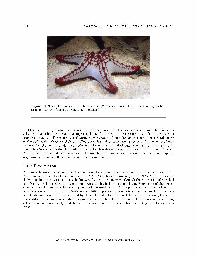
154 CHAPTER 6. STRUCTURAL SUPPORT AND MOVEMENT
Figure 6.1: The skeleton of the red-knobbed sea star (Protoreaster linckii) is an example of a hydrostaticskeleton. (credit: �Amada44�/Wikimedia Commons)
Movement in a hydrostatic skeleton is provided by muscles that surround the coelom. The muscles ina hydrostatic skeleton contract to change the shape of the coelom; the pressure of the �uid in the coelomproduces movement. For example, earthworms move by waves of muscular contractions of the skeletal muscleof the body wall hydrostatic skeleton, called peristalsis, which alternately shorten and lengthen the body.Lengthening the body extends the anterior end of the organism. Most organisms have a mechanism to �xthemselves in the substrate. Shortening the muscles then draws the posterior portion of the body forward.Although a hydrostatic skeleton is well-suited to invertebrate organisms such as earthworms and some aquaticorganisms, it is not an e�cient skeleton for terrestrial animals.
6.1.2 Exoskeleton
An exoskeleton is an external skeleton that consists of a hard encasement on the surface of an organism.For example, the shells of crabs and insects are exoskeletons (Figure 6.2). This skeleton type providesdefence against predators, supports the body, and allows for movement through the contraction of attachedmuscles. As with vertebrates, muscles must cross a joint inside the exoskeleton. Shortening of the musclechanges the relationship of the two segments of the exoskeleton. Arthropods such as crabs and lobstershave exoskeletons that consist of 30�50 percent chitin, a polysaccharide derivative of glucose that is a strongbut �exible material. Chitin is secreted by the epidermal cells. The exoskeleton is further strengthened bythe addition of calcium carbonate in organisms such as the lobster. Because the exoskeleton is acellular,arthropods must periodically shed their exoskeletons because the exoskeleton does not grow as the organismgrows.
Available for free at Connexions <http://cnx.org/content/col26253/1.2>
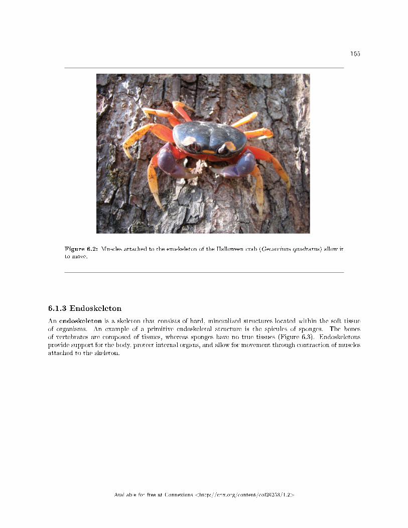
155
Figure 6.2: Muscles attached to the exoskeleton of the Halloween crab (Gecarcinus quadratus) allow itto move.
6.1.3 Endoskeleton
An endoskeleton is a skeleton that consists of hard, mineralized structures located within the soft tissueof organisms. An example of a primitive endoskeletal structure is the spicules of sponges. The bonesof vertebrates are composed of tissues, whereas sponges have no true tissues (Figure 6.3). Endoskeletonsprovide support for the body, protect internal organs, and allow for movement through contraction of musclesattached to the skeleton.
Available for free at Connexions <http://cnx.org/content/col26253/1.2>
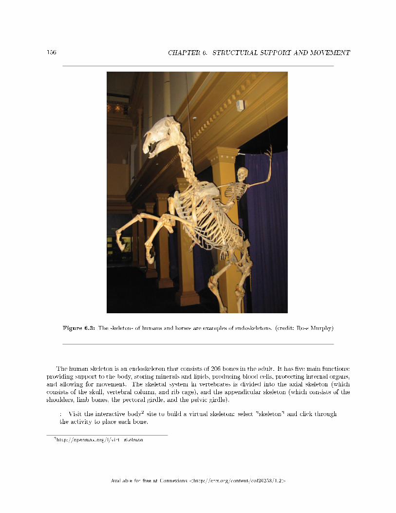
156 CHAPTER 6. STRUCTURAL SUPPORT AND MOVEMENT
Figure 6.3: The skeletons of humans and horses are examples of endoskeletons. (credit: Ross Murphy)
The human skeleton is an endoskeleton that consists of 206 bones in the adult. It has �ve main functions:providing support to the body, storing minerals and lipids, producing blood cells, protecting internal organs,and allowing for movement. The skeletal system in vertebrates is divided into the axial skeleton (whichconsists of the skull, vertebral column, and rib cage), and the appendicular skeleton (which consists of theshoulders, limb bones, the pectoral girdle, and the pelvic girdle).
: Visit the interactive body2 site to build a virtual skeleton: select "skeleton" and click throughthe activity to place each bone.
2http://openstax.org/l/virt_skeleton
Available for free at Connexions <http://cnx.org/content/col26253/1.2>
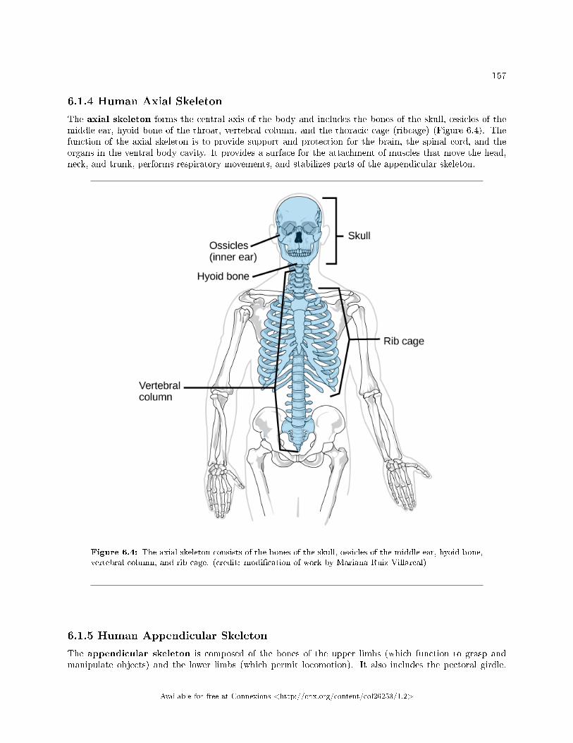
157
6.1.4 Human Axial Skeleton
The axial skeleton forms the central axis of the body and includes the bones of the skull, ossicles of themiddle ear, hyoid bone of the throat, vertebral column, and the thoracic cage (ribcage) (Figure 6.4). Thefunction of the axial skeleton is to provide support and protection for the brain, the spinal cord, and theorgans in the ventral body cavity. It provides a surface for the attachment of muscles that move the head,neck, and trunk, performs respiratory movements, and stabilizes parts of the appendicular skeleton.
Figure 6.4: The axial skeleton consists of the bones of the skull, ossicles of the middle ear, hyoid bone,vertebral column, and rib cage. (credit: modi�cation of work by Mariana Ruiz Villareal)
6.1.5 Human Appendicular Skeleton
The appendicular skeleton is composed of the bones of the upper limbs (which function to grasp andmanipulate objects) and the lower limbs (which permit locomotion). It also includes the pectoral girdle,
Available for free at Connexions <http://cnx.org/content/col26253/1.2>
158 CHAPTER 6. STRUCTURAL SUPPORT AND MOVEMENT
or shoulder girdle, that attaches the upper limbs to the body, and the pelvic girdle that attaches the lowerlimbs to the body (Figure 6.5).
Available for free at Connexions <http://cnx.org/content/col26253/1.2>
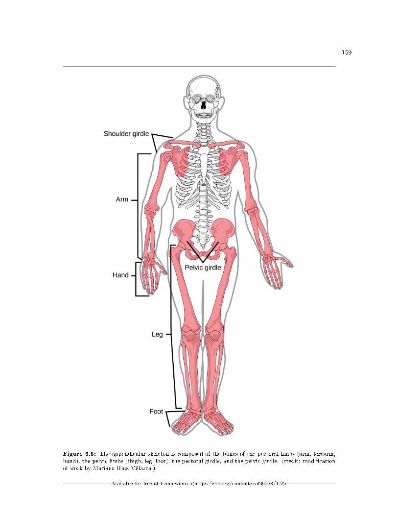
159
Figure 6.5: The appendicular skeleton is composed of the bones of the pectoral limbs (arm, forearm,hand), the pelvic limbs (thigh, leg, foot), the pectoral girdle, and the pelvic girdle. (credit: modi�cationof work by Mariana Ruiz Villareal)
Available for free at Connexions <http://cnx.org/content/col26253/1.2>
160 CHAPTER 6. STRUCTURAL SUPPORT AND MOVEMENT
6.1.6 Section Summary
The three types of skeleton designs are hydrostatic skeletons, exoskeletons, and endoskeletons. A hydrostaticskeleton is formed by a �uid-�lled compartment held under hydrostatic pressure; movement is created by themuscles producing pressure on the �uid. An exoskeleton is a hard external skeleton that protects the outersurface of an organism and enables movement through muscles attached on the inside. An endoskeletonis an internal skeleton composed of hard, mineralized tissue that also enables movement by attachment tomuscles. The human skeleton is an endoskeleton that is composed of the axial and appendicular skeleton.The axial skeleton is composed of the bones of the skull, ossicles of the ear, hyoid bone, vertebral column,and ribcage. The skull consists of eight cranial bones and 14 facial bones. Six bones make up the ossiclesof the middle ear, while the hyoid bone is located in the neck under the mandible. The vertebral columncontains 26 bones, and it surrounds and protects the spinal cord. The thoracic cage consists of the sternum,ribs, thoracic vertebrae, and costal cartilages. The appendicular skeleton is made up of the limbs of theupper and lower limbs. The pectoral girdle is composed of the clavicles and the scapulae. The upper limbcontains 30 bones in the arm, the forearm, and the hand. The pelvic girdle attaches the lower limbs to theaxial skeleton. The lower limb includes the bones of the thigh, the leg, and the foot.
6.2 Bone3
Bone, or osseous tissue, is a connective tissue that constitutes the endoskeleton. It contains specializedcells and a matrix of mineral salts and collagen �bers.
The mineral salts primarily include hydroxyapatite, a mineral formed from calcium phosphate. Calci�-cation is the process of deposition of mineral salts on the collagen �ber matrix that crystallizes and hardensthe tissue. The process of calci�cation only occurs in the presence of collagen �bers.
The bones of the human skeleton are classi�ed by their shape: long bones, short bones, �at bones, suturalbones, sesamoid bones, and irregular bones (Figure 6.6).
3This content is available online at <http://cnx.org/content/m66638/1.13/>.
Available for free at Connexions <http://cnx.org/content/col26253/1.2>
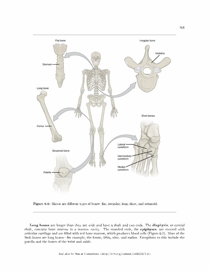
161
Figure 6.6: Shown are di�erent types of bones: �at, irregular, long, short, and sesamoid.
Long bones are longer than they are wide and have a shaft and two ends. The diaphysis, or centralshaft, contains bone marrow in a marrow cavity. The rounded ends, the epiphyses, are covered witharticular cartilage and are �lled with red bone marrow, which produces blood cells (Figure 6.7). Most of thelimb bones are long bones�for example, the femur, tibia, ulna, and radius. Exceptions to this include thepatella and the bones of the wrist and ankle.
Available for free at Connexions <http://cnx.org/content/col26253/1.2>
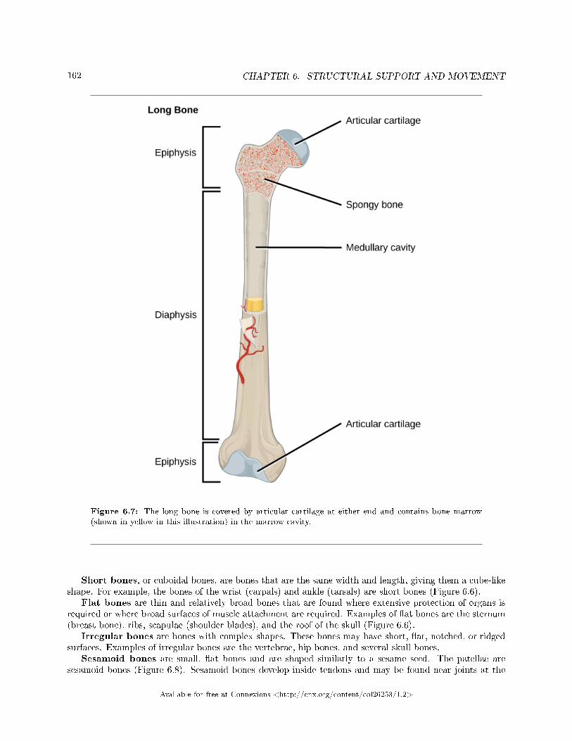
162 CHAPTER 6. STRUCTURAL SUPPORT AND MOVEMENT
Figure 6.7: The long bone is covered by articular cartilage at either end and contains bone marrow(shown in yellow in this illustration) in the marrow cavity.
Short bones, or cuboidal bones, are bones that are the same width and length, giving them a cube-likeshape. For example, the bones of the wrist (carpals) and ankle (tarsals) are short bones (Figure 6.6).
Flat bones are thin and relatively broad bones that are found where extensive protection of organs isrequired or where broad surfaces of muscle attachment are required. Examples of �at bones are the sternum(breast bone), ribs, scapulae (shoulder blades), and the roof of the skull (Figure 6.6).
Irregular bones are bones with complex shapes. These bones may have short, �at, notched, or ridgedsurfaces. Examples of irregular bones are the vertebrae, hip bones, and several skull bones.
Sesamoid bones are small, �at bones and are shaped similarly to a sesame seed. The patellae aresesamoid bones (Figure 6.8). Sesamoid bones develop inside tendons and may be found near joints at the
Available for free at Connexions <http://cnx.org/content/col26253/1.2>
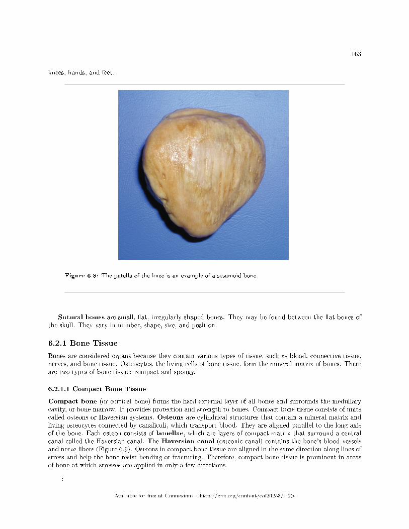
163
knees, hands, and feet.
Figure 6.8: The patella of the knee is an example of a sesamoid bone.
Sutural bones are small, �at, irregularly shaped bones. They may be found between the �at bones ofthe skull. They vary in number, shape, size, and position.
6.2.1 Bone Tissue
Bones are considered organs because they contain various types of tissue, such as blood, connective tissue,nerves, and bone tissue. Osteocytes, the living cells of bone tissue, form the mineral matrix of bones. Thereare two types of bone tissue: compact and spongy.
6.2.1.1 Compact Bone Tissue
Compact bone (or cortical bone) forms the hard external layer of all bones and surrounds the medullarycavity, or bone marrow. It provides protection and strength to bones. Compact bone tissue consists of unitscalled osteons or Haversian systems. Osteons are cylindrical structures that contain a mineral matrix andliving osteocytes connected by canaliculi, which transport blood. They are aligned parallel to the long axisof the bone. Each osteon consists of lamellae, which are layers of compact matrix that surround a centralcanal called the Haversian canal. The Haversian canal (osteonic canal) contains the bone's blood vesselsand nerve �bers (Figure 6.9). Osteons in compact bone tissue are aligned in the same direction along lines ofstress and help the bone resist bending or fracturing. Therefore, compact bone tissue is prominent in areasof bone at which stresses are applied in only a few directions.
:
Available for free at Connexions <http://cnx.org/content/col26253/1.2>
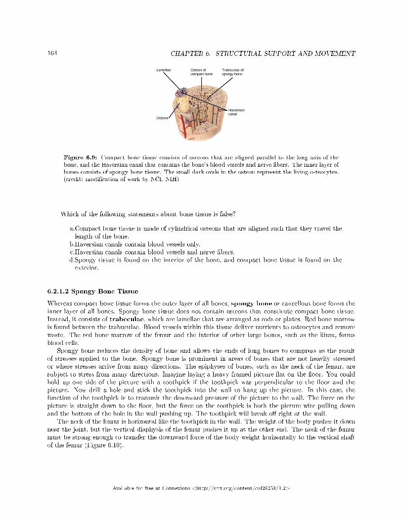
164 CHAPTER 6. STRUCTURAL SUPPORT AND MOVEMENT
Figure 6.9: Compact bone tissue consists of osteons that are aligned parallel to the long axis of thebone, and the Haversian canal that contains the bone's blood vessels and nerve �bers. The inner layer ofbones consists of spongy bone tissue. The small dark ovals in the osteon represent the living osteocytes.(credit: modi�cation of work by NCI, NIH)
Which of the following statements about bone tissue is false?
a.Compact bone tissue is made of cylindrical osteons that are aligned such that they travel thelength of the bone.
b.Haversian canals contain blood vessels only.c.Haversian canals contain blood vessels and nerve �bers.d.Spongy tissue is found on the interior of the bone, and compact bone tissue is found on theexterior.
6.2.1.2 Spongy Bone Tissue
Whereas compact bone tissue forms the outer layer of all bones, spongy bone or cancellous bone forms theinner layer of all bones. Spongy bone tissue does not contain osteons that constitute compact bone tissue.Instead, it consists of trabeculae, which are lamellae that are arranged as rods or plates. Red bone marrowis found between the trabuculae. Blood vessels within this tissue deliver nutrients to osteocytes and removewaste. The red bone marrow of the femur and the interior of other large bones, such as the ilium, formsblood cells.
Spongy bone reduces the density of bone and allows the ends of long bones to compress as the resultof stresses applied to the bone. Spongy bone is prominent in areas of bones that are not heavily stressedor where stresses arrive from many directions. The epiphyses of bones, such as the neck of the femur, aresubject to stress from many directions. Imagine laying a heavy framed picture �at on the �oor. You couldhold up one side of the picture with a toothpick if the toothpick was perpendicular to the �oor and thepicture. Now drill a hole and stick the toothpick into the wall to hang up the picture. In this case, thefunction of the toothpick is to transmit the downward pressure of the picture to the wall. The force on thepicture is straight down to the �oor, but the force on the toothpick is both the picture wire pulling downand the bottom of the hole in the wall pushing up. The toothpick will break o� right at the wall.
The neck of the femur is horizontal like the toothpick in the wall. The weight of the body pushes it downnear the joint, but the vertical diaphysis of the femur pushes it up at the other end. The neck of the femurmust be strong enough to transfer the downward force of the body weight horizontally to the vertical shaftof the femur (Figure 6.10).
Available for free at Connexions <http://cnx.org/content/col26253/1.2>
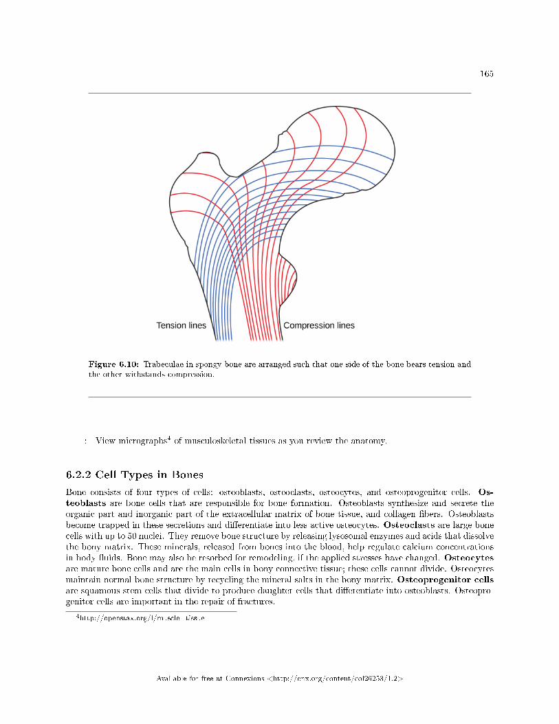
165
Figure 6.10: Trabeculae in spongy bone are arranged such that one side of the bone bears tension andthe other withstands compression.
: View micrographs4 of musculoskeletal tissues as you review the anatomy.
6.2.2 Cell Types in Bones
Bone consists of four types of cells: osteoblasts, osteoclasts, osteocytes, and osteoprogenitor cells. Os-teoblasts are bone cells that are responsible for bone formation. Osteoblasts synthesize and secrete theorganic part and inorganic part of the extracellular matrix of bone tissue, and collagen �bers. Osteoblastsbecome trapped in these secretions and di�erentiate into less active osteocytes. Osteoclasts are large bonecells with up to 50 nuclei. They remove bone structure by releasing lysosomal enzymes and acids that dissolvethe bony matrix. These minerals, released from bones into the blood, help regulate calcium concentrationsin body �uids. Bone may also be resorbed for remodeling, if the applied stresses have changed. Osteocytesare mature bone cells and are the main cells in bony connective tissue; these cells cannot divide. Osteocytesmaintain normal bone structure by recycling the mineral salts in the bony matrix. Osteoprogenitor cellsare squamous stem cells that divide to produce daughter cells that di�erentiate into osteoblasts. Osteopro-genitor cells are important in the repair of fractures.
4http://openstax.org/l/muscle_tissue
Available for free at Connexions <http://cnx.org/content/col26253/1.2>
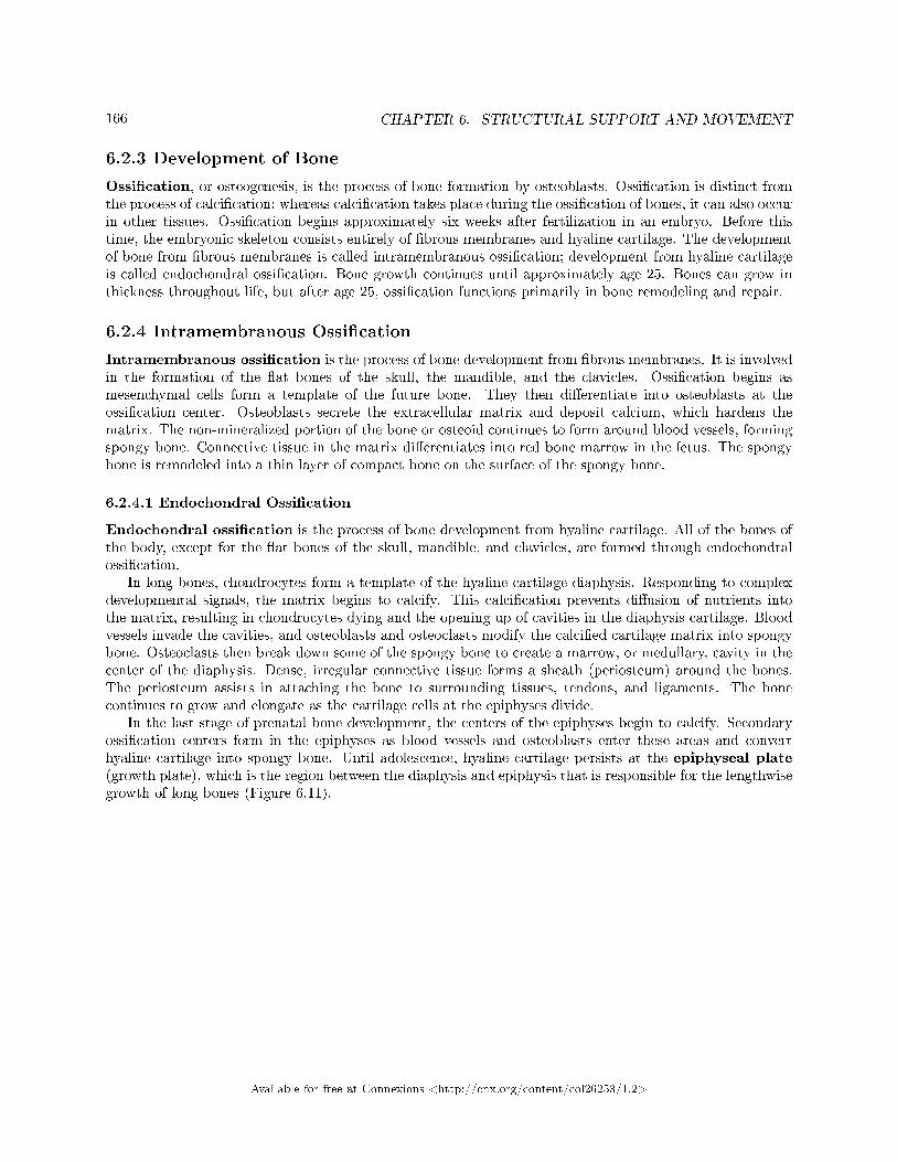
166 CHAPTER 6. STRUCTURAL SUPPORT AND MOVEMENT
6.2.3 Development of Bone
Ossi�cation, or osteogenesis, is the process of bone formation by osteoblasts. Ossi�cation is distinct fromthe process of calci�cation; whereas calci�cation takes place during the ossi�cation of bones, it can also occurin other tissues. Ossi�cation begins approximately six weeks after fertilization in an embryo. Before thistime, the embryonic skeleton consists entirely of �brous membranes and hyaline cartilage. The developmentof bone from �brous membranes is called intramembranous ossi�cation; development from hyaline cartilageis called endochondral ossi�cation. Bone growth continues until approximately age 25. Bones can grow inthickness throughout life, but after age 25, ossi�cation functions primarily in bone remodeling and repair.
6.2.4 Intramembranous Ossi�cation
Intramembranous ossi�cation is the process of bone development from �brous membranes. It is involvedin the formation of the �at bones of the skull, the mandible, and the clavicles. Ossi�cation begins asmesenchymal cells form a template of the future bone. They then di�erentiate into osteoblasts at theossi�cation center. Osteoblasts secrete the extracellular matrix and deposit calcium, which hardens thematrix. The non-mineralized portion of the bone or osteoid continues to form around blood vessels, formingspongy bone. Connective tissue in the matrix di�erentiates into red bone marrow in the fetus. The spongybone is remodeled into a thin layer of compact bone on the surface of the spongy bone.
6.2.4.1 Endochondral Ossi�cation
Endochondral ossi�cation is the process of bone development from hyaline cartilage. All of the bones ofthe body, except for the �at bones of the skull, mandible, and clavicles, are formed through endochondralossi�cation.
In long bones, chondrocytes form a template of the hyaline cartilage diaphysis. Responding to complexdevelopmental signals, the matrix begins to calcify. This calci�cation prevents di�usion of nutrients intothe matrix, resulting in chondrocytes dying and the opening up of cavities in the diaphysis cartilage. Bloodvessels invade the cavities, and osteoblasts and osteoclasts modify the calci�ed cartilage matrix into spongybone. Osteoclasts then break down some of the spongy bone to create a marrow, or medullary, cavity in thecenter of the diaphysis. Dense, irregular connective tissue forms a sheath (periosteum) around the bones.The periosteum assists in attaching the bone to surrounding tissues, tendons, and ligaments. The bonecontinues to grow and elongate as the cartilage cells at the epiphyses divide.
In the last stage of prenatal bone development, the centers of the epiphyses begin to calcify. Secondaryossi�cation centers form in the epiphyses as blood vessels and osteoblasts enter these areas and converthyaline cartilage into spongy bone. Until adolescence, hyaline cartilage persists at the epiphyseal plate(growth plate), which is the region between the diaphysis and epiphysis that is responsible for the lengthwisegrowth of long bones (Figure 6.11).
Available for free at Connexions <http://cnx.org/content/col26253/1.2>
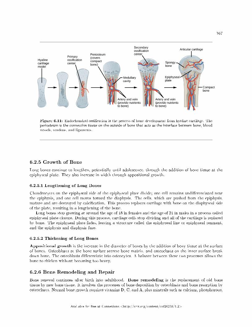
167
Figure 6.11: Endochondral ossi�cation is the process of bone development from hyaline cartilage. Theperiosteum is the connective tissue on the outside of bone that acts as the interface between bone, bloodvessels, tendons, and ligaments.
6.2.5 Growth of Bone
Long bones continue to lengthen, potentially until adolescence, through the addition of bone tissue at theepiphyseal plate. They also increase in width through appositional growth.
6.2.5.1 Lengthening of Long Bones
Chondrocytes on the epiphyseal side of the epiphyseal plate divide; one cell remains undi�erentiated nearthe epiphysis, and one cell moves toward the diaphysis. The cells, which are pushed from the epiphysis,mature and are destroyed by calci�cation. This process replaces cartilage with bone on the diaphyseal sideof the plate, resulting in a lengthening of the bone.
Long bones stop growing at around the age of 18 in females and the age of 21 in males in a process calledepiphyseal plate closure. During this process, cartilage cells stop dividing and all of the cartilage is replacedby bone. The epiphyseal plate fades, leaving a structure called the epiphyseal line or epiphyseal remnant,and the epiphysis and diaphysis fuse.
6.2.5.2 Thickening of Long Bones
Appositional growth is the increase in the diameter of bones by the addition of bony tissue at the surfaceof bones. Osteoblasts at the bone surface secrete bone matrix, and osteoclasts on the inner surface breakdown bone. The osteoblasts di�erentiate into osteocytes. A balance between these two processes allows thebone to thicken without becoming too heavy.
6.2.6 Bone Remodeling and Repair
Bone renewal continues after birth into adulthood. Bone remodeling is the replacement of old bonetissue by new bone tissue. It involves the processes of bone deposition by osteoblasts and bone resorption byosteoclasts. Normal bone growth requires vitamins D, C, and A, plus minerals such as calcium, phosphorous,
Available for free at Connexions <http://cnx.org/content/col26253/1.2>
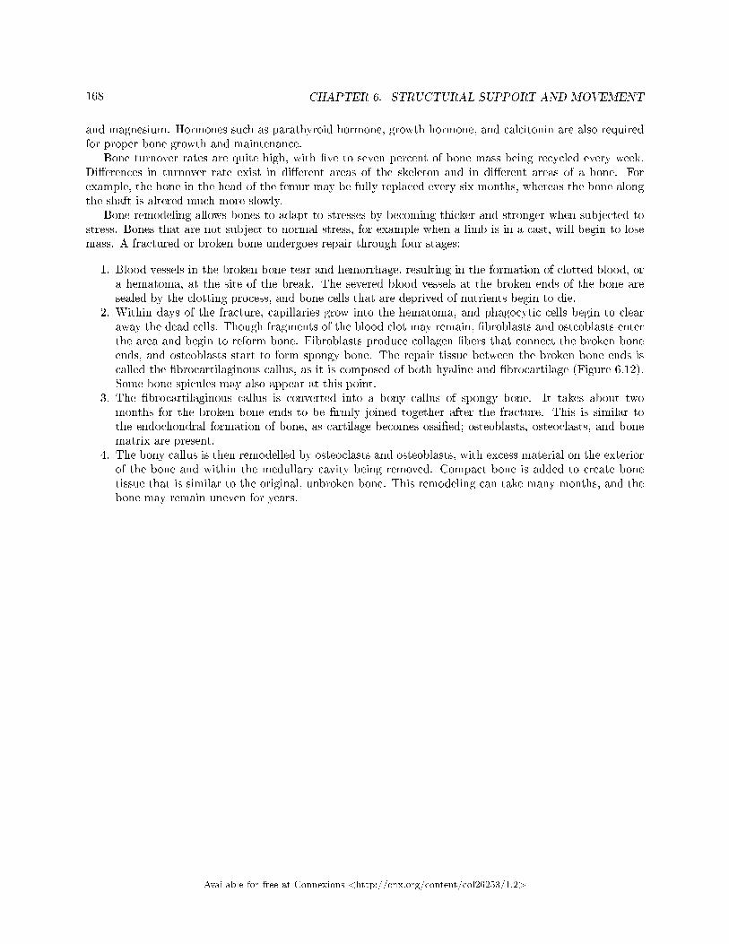
168 CHAPTER 6. STRUCTURAL SUPPORT AND MOVEMENT
and magnesium. Hormones such as parathyroid hormone, growth hormone, and calcitonin are also requiredfor proper bone growth and maintenance.
Bone turnover rates are quite high, with �ve to seven percent of bone mass being recycled every week.Di�erences in turnover rate exist in di�erent areas of the skeleton and in di�erent areas of a bone. Forexample, the bone in the head of the femur may be fully replaced every six months, whereas the bone alongthe shaft is altered much more slowly.
Bone remodeling allows bones to adapt to stresses by becoming thicker and stronger when subjected tostress. Bones that are not subject to normal stress, for example when a limb is in a cast, will begin to losemass. A fractured or broken bone undergoes repair through four stages:
1. Blood vessels in the broken bone tear and hemorrhage, resulting in the formation of clotted blood, ora hematoma, at the site of the break. The severed blood vessels at the broken ends of the bone aresealed by the clotting process, and bone cells that are deprived of nutrients begin to die.
2. Within days of the fracture, capillaries grow into the hematoma, and phagocytic cells begin to clearaway the dead cells. Though fragments of the blood clot may remain, �broblasts and osteoblasts enterthe area and begin to reform bone. Fibroblasts produce collagen �bers that connect the broken boneends, and osteoblasts start to form spongy bone. The repair tissue between the broken bone ends iscalled the �brocartilaginous callus, as it is composed of both hyaline and �brocartilage (Figure 6.12).Some bone spicules may also appear at this point.
3. The �brocartilaginous callus is converted into a bony callus of spongy bone. It takes about twomonths for the broken bone ends to be �rmly joined together after the fracture. This is similar tothe endochondral formation of bone, as cartilage becomes ossi�ed; osteoblasts, osteoclasts, and bonematrix are present.
4. The bony callus is then remodelled by osteoclasts and osteoblasts, with excess material on the exteriorof the bone and within the medullary cavity being removed. Compact bone is added to create bonetissue that is similar to the original, unbroken bone. This remodeling can take many months, and thebone may remain uneven for years.
Available for free at Connexions <http://cnx.org/content/col26253/1.2>
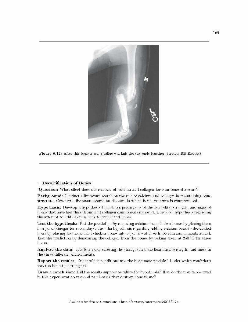
169
Figure 6.12: After this bone is set, a callus will knit the two ends together. (credit: Bill Rhodes)
: Decalci�cation of Bones
Question: What e�ect does the removal of calcium and collagen have on bone structure?
Background: Conduct a literature search on the role of calcium and collagen in maintaining bonestructure. Conduct a literature search on diseases in which bone structure is compromised.
Hypothesis: Develop a hypothesis that states predictions of the �exibility, strength, and mass ofbones that have had the calcium and collagen components removed. Develop a hypothesis regardingthe attempt to add calcium back to decalci�ed bones.
Test the hypothesis: Test the prediction by removing calcium from chicken bones by placing themin a jar of vinegar for seven days. Test the hypothesis regarding adding calcium back to decalci�edbone by placing the decalci�ed chicken bones into a jar of water with calcium supplements added.Test the prediction by denaturing the collagen from the bones by baking them at 250 ◦C for threehours.
Analyze the data: Create a table showing the changes in bone �exibility, strength, and mass inthe three di�erent environments.
Report the results: Under which conditions was the bone most �exible? Under which conditionswas the bone the strongest?
Draw a conclusion: Did the results support or refute the hypothesis? How do the results observedin this experiment correspond to diseases that destroy bone tissue?
Available for free at Connexions <http://cnx.org/content/col26253/1.2>
170 CHAPTER 6. STRUCTURAL SUPPORT AND MOVEMENT
6.2.7 Section Summary
Bone, or osseous tissue, is connective tissue that includes specialized cells, mineral salts, and collagen �bers.The human skeleton can be divided into long bones, short bones, �at bones, and irregular bones. Compactbone tissue is composed of osteons and forms the external layer of all bones. Spongy bone tissue is composedof trabeculae and forms the inner part of all bones. Four types of cells compose bony tissue: osteocytes, os-teoclasts, osteoprogenitor cells, and osteoblasts. Ossi�cation is the process of bone formation by osteoblasts.Intramembranous ossi�cation is the process of bone development from �brous membranes. Endochondralossi�cation is the process of bone development from hyaline cartilage. Long bones lengthen as chondrocytesdivide and secrete hyaline cartilage. Osteoblasts replace cartilage with bone. Appositional growth is theincrease in the diameter of bones by the addition of bone tissue at the surface of bones. Bone remodelinginvolves the processes of bone deposition by osteoblasts and bone resorption by osteoclasts. Bone repairoccurs in four stages and can take several months.
6.2.8 Art Exercise
Exercise 6.2.1 (Solution on p. 182.)
Figure 6.9 Which of the following statements about bone tissue is false?
a. Compact bone tissue is made of cylindrical osteons that are aligned such that they travel thelength of the bone.
b. Haversian canals contain blood vessels only.c. Haversian canals contain blood vessels and nerve �bers.d. Spongy tissue is found on the interior of the bone, and compact bone tissue is found on the
exterior.
6.2.9 Review Questions
Exercise 6.2.2 (Solution on p. 182.)
The Haversian canal:
a. is arranged as rods or platesb. contains the bone's blood vessels and nerve �bersc. is responsible for the lengthwise growth of long bonesd. synthesizes and secretes matrix
Exercise 6.2.3 (Solution on p. 182.)
The epiphyseal plate:
a. is arranged as rods or platesb. contains the bone's blood vessels and nerve �bersc. is responsible for the lengthwise growth of long bonesd. synthesizes and secretes bone matrix
Exercise 6.2.4 (Solution on p. 182.)
The cells responsible for bone resorption are ________.
a. osteoclastsb. osteoblastsc. �broblastsd. osteocytes
Available for free at Connexions <http://cnx.org/content/col26253/1.2>
171
Exercise 6.2.5 (Solution on p. 182.)
Compact bone is composed of ________.
a. trabeculaeb. compacted collagenc. osteonsd. calcium phosphate only
Exercise 6.2.6 (Solution on p. 182.)
Osteoporosis is a condition where bones become weak and brittle. It is caused by an imbalance inthe activity of which cells?
a. osteoclasts and osteoblastsb. osteoclasts and osteocytesc. osteoblasts and chondrocytesd. osteocytes and chondrocytes
Exercise 6.2.7 (Solution on p. 182.)
While assembling a skeleton of a new species, a scientist points to one of the bones and observesthat it looks like the most likely site of leg muscle attachment. What kind of bone did she indicate?
a. sesamoid boneb. long bonec. trabecular boned. �at bone
6.2.10 Critical Thinking Questions
Exercise 6.2.8 (Solution on p. 182.)
What are the major di�erences between spongy bone and compact bone?
Exercise 6.2.9 (Solution on p. 182.)
What are the roles of osteoblasts, osteocytes, and osteoclasts?
Exercise 6.2.10 (Solution on p. 182.)
Thalidomide was a morning sickness drug given to pregnant people; it caused babies to be bornwithout arm bones. If recent studies have shown that thalidomide prevents the formation of newblood vessels, describe the type of bone development inhibited by the drug and what stage ofossi�cation was a�ected.
6.3 Muscle Contraction and Locomotion5
Muscle cells are specialized for contraction. Muscles allow for motions such as walking, and they also facilitatebodily processes such as respiration and digestion. The body contains three types of muscle tissue: skeletalmuscle, cardiac muscle, and smooth muscle (Figure 6.13).
5This content is available online at <http://cnx.org/content/m66643/1.11/>.
Available for free at Connexions <http://cnx.org/content/col26253/1.2>
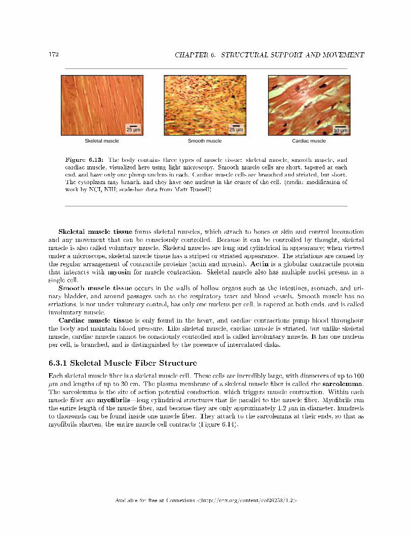
172 CHAPTER 6. STRUCTURAL SUPPORT AND MOVEMENT
Figure 6.13: The body contains three types of muscle tissue: skeletal muscle, smooth muscle, andcardiac muscle, visualized here using light microscopy. Smooth muscle cells are short, tapered at eachend, and have only one plump nucleus in each. Cardiac muscle cells are branched and striated, but short.The cytoplasm may branch, and they have one nucleus in the center of the cell. (credit: modi�cation ofwork by NCI, NIH; scale-bar data from Matt Russell)
Skeletal muscle tissue forms skeletal muscles, which attach to bones or skin and control locomotionand any movement that can be consciously controlled. Because it can be controlled by thought, skeletalmuscle is also called voluntary muscle. Skeletal muscles are long and cylindrical in appearance; when viewedunder a microscope, skeletal muscle tissue has a striped or striated appearance. The striations are caused bythe regular arrangement of contractile proteins (actin and myosin). Actin is a globular contractile proteinthat interacts with myosin for muscle contraction. Skeletal muscle also has multiple nuclei present in asingle cell.
Smooth muscle tissue occurs in the walls of hollow organs such as the intestines, stomach, and uri-nary bladder, and around passages such as the respiratory tract and blood vessels. Smooth muscle has nostriations, is not under voluntary control, has only one nucleus per cell, is tapered at both ends, and is calledinvoluntary muscle.
Cardiac muscle tissue is only found in the heart, and cardiac contractions pump blood throughoutthe body and maintain blood pressure. Like skeletal muscle, cardiac muscle is striated, but unlike skeletalmuscle, cardiac muscle cannot be consciously controlled and is called involuntary muscle. It has one nucleusper cell, is branched, and is distinguished by the presence of intercalated disks.
6.3.1 Skeletal Muscle Fiber Structure
Each skeletal muscle �ber is a skeletal muscle cell. These cells are incredibly large, with diameters of up to 100µm and lengths of up to 30 cm. The plasma membrane of a skeletal muscle �ber is called the sarcolemma.The sarcolemma is the site of action potential conduction, which triggers muscle contraction. Within eachmuscle �ber are myo�brils�long cylindrical structures that lie parallel to the muscle �ber. Myo�brils runthe entire length of the muscle �ber, and because they are only approximately 1.2 µm in diameter, hundredsto thousands can be found inside one muscle �ber. They attach to the sarcolemma at their ends, so that asmyo�brils shorten, the entire muscle cell contracts (Figure 6.14).
Available for free at Connexions <http://cnx.org/content/col26253/1.2>
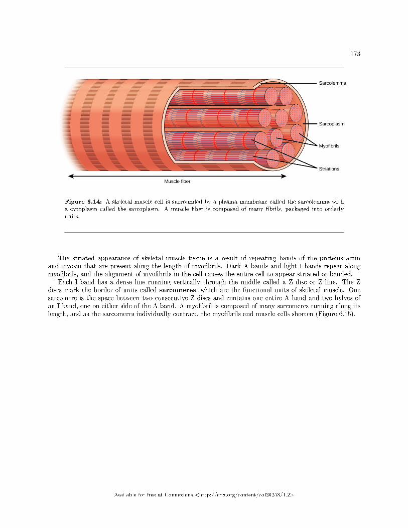
173
Figure 6.14: A skeletal muscle cell is surrounded by a plasma membrane called the sarcolemma witha cytoplasm called the sarcoplasm. A muscle �ber is composed of many �brils, packaged into orderlyunits.
The striated appearance of skeletal muscle tissue is a result of repeating bands of the proteins actinand myosin that are present along the length of myo�brils. Dark A bands and light I bands repeat alongmyo�brils, and the alignment of myo�brils in the cell causes the entire cell to appear striated or banded.
Each I band has a dense line running vertically through the middle called a Z disc or Z line. The Zdiscs mark the border of units called sarcomeres, which are the functional units of skeletal muscle. Onesarcomere is the space between two consecutive Z discs and contains one entire A band and two halves ofan I band, one on either side of the A band. A myo�bril is composed of many sarcomeres running along itslength, and as the sarcomeres individually contract, the myo�brils and muscle cells shorten (Figure 6.15).
Available for free at Connexions <http://cnx.org/content/col26253/1.2>
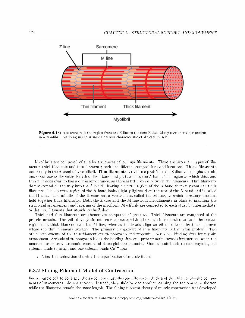
174 CHAPTER 6. STRUCTURAL SUPPORT AND MOVEMENT
Figure 6.15: A sarcomere is the region from one Z line to the next Z line. Many sarcomeres are presentin a myo�bril, resulting in the striation pattern characteristic of skeletal muscle.
Myo�brils are composed of smaller structures called myo�laments. There are two main types of �la-ments: thick �laments and thin �laments; each has di�erent compositions and locations. Thick �lamentsoccur only in the A band of a myo�bril. Thin �laments attach to a protein in the Z disc called alpha-actininand occur across the entire length of the I band and partway into the A band. The region at which thick andthin �laments overlap has a dense appearance, as there is little space between the �laments. Thin �lamentsdo not extend all the way into the A bands, leaving a central region of the A band that only contains thick�laments. This central region of the A band looks slightly lighter than the rest of the A band and is calledthe H zone. The middle of the H zone has a vertical line called the M line, at which accessory proteinshold together thick �laments. Both the Z disc and the M line hold myo�laments in place to maintain thestructural arrangement and layering of the myo�bril. Myo�brils are connected to each other by intermediate,or desmin, �laments that attach to the Z disc.
Thick and thin �laments are themselves composed of proteins. Thick �laments are composed of theprotein myosin. The tail of a myosin molecule connects with other myosin molecules to form the centralregion of a thick �lament near the M line, whereas the heads align on either side of the thick �lamentwhere the thin �laments overlap. The primary component of thin �laments is the actin protein. Twoother components of the thin �lament are tropomyosin and troponin. Actin has binding sites for myosinattachment. Strands of tropomyosin block the binding sites and prevent actin�myosin interactions when themuscles are at rest. Troponin consists of three globular subunits. One subunit binds to tropomyosin, onesubunit binds to actin, and one subunit binds Ca2+ ions.
: View this animation showing the organization of muscle �bers.
6.3.2 Sliding Filament Model of Contraction
For a muscle cell to contract, the sarcomere must shorten. However, thick and thin �laments�the compo-nents of sarcomeres�do not shorten. Instead, they slide by one another, causing the sarcomere to shortenwhile the �laments remain the same length. The sliding �lament theory of muscle contraction was developed
Available for free at Connexions <http://cnx.org/content/col26253/1.2>
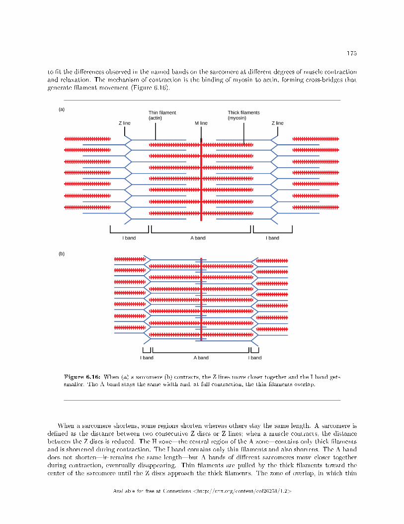
175
to �t the di�erences observed in the named bands on the sarcomere at di�erent degrees of muscle contractionand relaxation. The mechanism of contraction is the binding of myosin to actin, forming cross-bridges thatgenerate �lament movement (Figure 6.16).
Figure 6.16: When (a) a sarcomere (b) contracts, the Z lines move closer together and the I band getssmaller. The A band stays the same width and, at full contraction, the thin �laments overlap.
When a sarcomere shortens, some regions shorten whereas others stay the same length. A sarcomere isde�ned as the distance between two consecutive Z discs or Z lines; when a muscle contracts, the distancebetween the Z discs is reduced. The H zone�the central region of the A zone�contains only thick �lamentsand is shortened during contraction. The I band contains only thin �laments and also shortens. The A banddoes not shorten�it remains the same length�but A bands of di�erent sarcomeres move closer togetherduring contraction, eventually disappearing. Thin �laments are pulled by the thick �laments toward thecenter of the sarcomere until the Z discs approach the thick �laments. The zone of overlap, in which thin
Available for free at Connexions <http://cnx.org/content/col26253/1.2>
176 CHAPTER 6. STRUCTURAL SUPPORT AND MOVEMENT
�laments and thick �laments occupy the same area, increases as the thin �laments move inward.
6.3.3 ATP and Muscle Contraction
The motion of muscle shortening occurs as myosin heads bind to actin and pull the actin inwards. Thisaction requires energy, which is provided by ATP. Myosin binds to actin at a binding site on the globularactin protein. Myosin has another binding site for ATP at which enzymatic activity hydrolyzes ATP toADP, releasing an inorganic phosphate molecule and energy.
ATP binding causes myosin to release actin, allowing actin and myosin to detach from each other. Afterthis happens, the newly bound ATP is converted to ADP and inorganic phosphate, Pi. The enzyme at thebinding site on myosin is called ATPase. The energy released during ATP hydrolysis changes the angle of themyosin head into a �cocked� position. The myosin head is then in a position for further movement, possessingpotential energy, but ADP and Pi are still attached. If actin binding sites are covered and unavailable, themyosin will remain in the high energy con�guration with ATP hydrolyzed, but still attached.
If the actin binding sites are uncovered, a cross-bridge will form; that is, the myosin head spans thedistance between the actin and myosin molecules. Pi is then released, allowing myosin to expend the storedenergy as a conformational change. The myosin head moves toward the M line, pulling the actin along withit. As the actin is pulled, the �laments move approximately 10 nm toward the M line. This movement iscalled the power stroke, as it is the step at which force is produced. As the actin is pulled toward the Mline, the sarcomere shortens and the muscle contracts.
When the myosin head is �cocked,� it contains energy and is in a high-energy con�guration. This energyis expended as the myosin head moves through the power stroke; at the end of the power stroke, the myosinhead is in a low-energy position. After the power stroke, ADP is released; however, the cross-bridge formedis still in place, and actin and myosin are bound together. ATP can then attach to myosin, which allows thecross-bridge cycle to start again and further muscle contraction can occur (Figure 6.17).
: Watch this video explaining how a muscle contraction is signaled.
:
Available for free at Connexions <http://cnx.org/content/col26253/1.2>
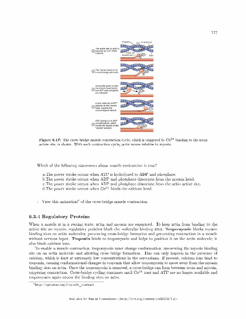
177
Figure 6.17: The cross-bridge muscle contraction cycle, which is triggered by Ca2+ binding to the actinactive site, is shown. With each contraction cycle, actin moves relative to myosin.
Which of the following statements about muscle contraction is true?
a.The power stroke occurs when ATP is hydrolyzed to ADP and phosphate.b.The power stroke occurs when ADP and phosphate dissociate from the myosin head.c.The power stroke occurs when ADP and phosphate dissociate from the actin active site.d.The power stroke occurs when Ca2+ binds the calcium head.
: View this animation6 of the cross-bridge muscle contraction.
6.3.4 Regulatory Proteins
When a muscle is in a resting state, actin and myosin are separated. To keep actin from binding to theactive site on myosin, regulatory proteins block the molecular binding sites. Tropomyosin blocks myosinbinding sites on actin molecules, preventing cross-bridge formation and preventing contraction in a musclewithout nervous input. Troponin binds to tropomyosin and helps to position it on the actin molecule; italso binds calcium ions.
To enable a muscle contraction, tropomyosin must change conformation, uncovering the myosin-bindingsite on an actin molecule and allowing cross-bridge formation. This can only happen in the presence ofcalcium, which is kept at extremely low concentrations in the sarcoplasm. If present, calcium ions bind totroponin, causing conformational changes in troponin that allow tropomyosin to move away from the myosinbinding sites on actin. Once the tropomyosin is removed, a cross-bridge can form between actin and myosin,triggering contraction. Cross-bridge cycling continues until Ca2+ ions and ATP are no longer available andtropomyosin again covers the binding sites on actin.
6http://openstax.org/l/muscle_contract
Available for free at Connexions <http://cnx.org/content/col26253/1.2>
178 CHAPTER 6. STRUCTURAL SUPPORT AND MOVEMENT
6.3.5 Excitation�Contraction Coupling
Excitation�contraction coupling is the link (transduction) between the action potential generated in thesarcolemma and the start of a muscle contraction. The trigger for calcium release from the sarcoplasmicreticulum into the sarcoplasm is a neural signal. Each skeletal muscle �ber is controlled by a motor neuron,which conducts signals from the brain or spinal cord to the muscle. The area of the sarcolemma on themuscle �ber that interacts with the neuron is called the motor end plate. The end of the neuron'saxon is called the synaptic terminal, and it does not actually contact the motor end plate. A small spacecalled the synaptic cleft separates the synaptic terminal from the motor end plate. Electrical signals travelalong the neuron's axon, which branches through the muscle and connects to individual muscle �bers at aneuromuscular junction.
The ability of cells to communicate electrically requires that the cells expend energy to create an elec-trical gradient across their cell membranes. This charge gradient is carried by ions, which are di�erentiallydistributed across the membrane. Each ion exerts an electrical in�uence and a concentration in�uence. Justas milk will eventually mix with co�ee without the need to stir, ions also distribute themselves evenly, ifthey are permitted to do so. In this case, they are not permitted to return to an evenly mixed state.
The sodium�potassium ATPase uses cellular energy to move K+ ions inside the cell and Na+ ions outside.This alone accumulates a small electrical charge, but a big concentration gradient. There is lots of K+ inthe cell and lots of Na+ outside the cell. Potassium is able to leave the cell through K+ channels that areopen 90% of the time, and it does. However, Na+ channels are rarely open, so Na+ remains outside the cell.When K+ leaves the cell, obeying its concentration gradient, that e�ectively leaves a negative charge behind.So at rest, there is a large concentration gradient for Na+ to enter the cell, and there is an accumulation ofnegative charges left behind in the cell. This is the resting membrane potential. Potential in this contextmeans a separation of electrical charge that is capable of doing work. It is measured in volts, just like abattery. However, the transmembrane potential is considerably smaller (0.07 V); therefore, the small valueis expressed as millivolts (mV) or 70 mV. Because the inside of a cell is negative compared with the outside,a minus sign signi�es the excess of negative charges inside the cell, −70 mV.
If an event changes the permeability of the membrane to Na+ ions, they will enter the cell. Thatwill change the voltage. This is an electrical event, called an action potential, that can be used as acellular signal. Communication occurs between nerves and muscles through neurotransmitters. Neuronaction potentials cause the release of neurotransmitters from the synaptic terminal into the synaptic cleft,where they can then di�use across the synaptic cleft and bind to a receptor molecule on the motor end plate.The motor end plate possesses junctional folds�folds in the sarcolemma that create a large surface area forthe neurotransmitter to bind to receptors. The receptors are actually sodium channels that open to allowthe passage of Na+ into the cell when they receive a neurotransmitter signal.
Acetylcholine (ACh) is a neurotransmitter released by motor neurons that binds to receptors in themotor end plate. Neurotransmitter release occurs when an action potential travels down the motor neuron'saxon, resulting in altered permeability of the synaptic terminal membrane and an in�ux of calcium. TheCa2+ ions allow synaptic vesicles to move to and bind with the presynaptic membrane (on the neuron), andrelease neurotransmitter from the vesicles into the synaptic cleft. Once released by the synaptic terminal,ACh di�uses across the synaptic cleft to the motor end plate, where it binds with ACh receptors. As aneurotransmitter binds, these ion channels open, and Na+ ions cross the membrane into the muscle cell.This reduces the voltage di�erence between the inside and outside of the cell, which is called depolarization.As ACh binds at the motor end plate, this depolarization is called an end-plate potential. The depolarizationthen spreads along the sarcolemma, creating an action potential as sodium channels adjacent to the initialdepolarization site sense the change in voltage and open. The action potential moves across the entire cell,creating a wave of depolarization.
ACh is broken down by the enzyme acetylcholinesterase (AChE) into acetyl and choline. AChE residesin the synaptic cleft, breaking down ACh so that it does not remain bound to ACh receptors, which wouldcause unwanted extended muscle contraction (Figure 6.18).
:
Available for free at Connexions <http://cnx.org/content/col26253/1.2>
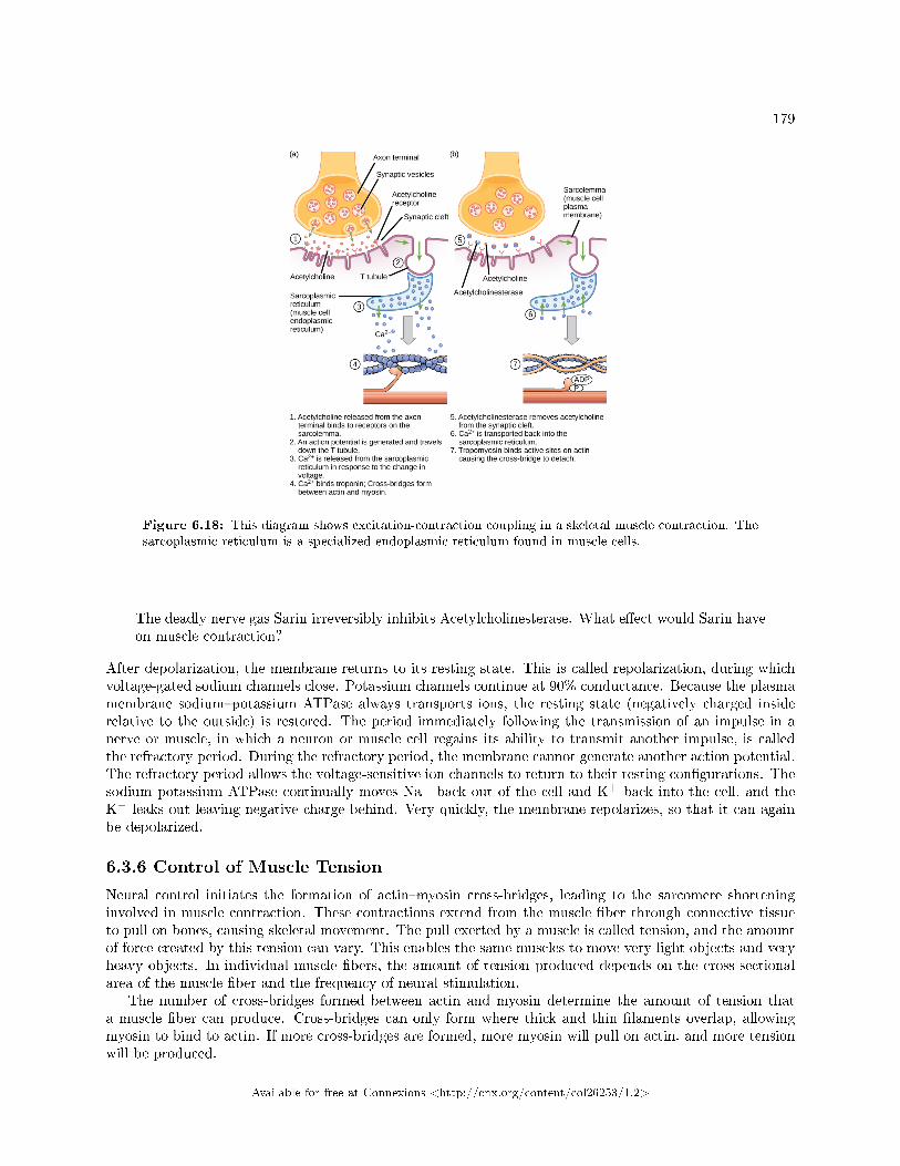
179
Figure 6.18: This diagram shows excitation-contraction coupling in a skeletal muscle contraction. Thesarcoplasmic reticulum is a specialized endoplasmic reticulum found in muscle cells.
The deadly nerve gas Sarin irreversibly inhibits Acetylcholinesterase. What e�ect would Sarin haveon muscle contraction?
After depolarization, the membrane returns to its resting state. This is called repolarization, during whichvoltage-gated sodium channels close. Potassium channels continue at 90% conductance. Because the plasmamembrane sodium�potassium ATPase always transports ions, the resting state (negatively charged insiderelative to the outside) is restored. The period immediately following the transmission of an impulse in anerve or muscle, in which a neuron or muscle cell regains its ability to transmit another impulse, is calledthe refractory period. During the refractory period, the membrane cannot generate another action potential.The refractory period allows the voltage-sensitive ion channels to return to their resting con�gurations. Thesodium potassium ATPase continually moves Na+ back out of the cell and K+ back into the cell, and theK+ leaks out leaving negative charge behind. Very quickly, the membrane repolarizes, so that it can againbe depolarized.
6.3.6 Control of Muscle Tension
Neural control initiates the formation of actin�myosin cross-bridges, leading to the sarcomere shorteninginvolved in muscle contraction. These contractions extend from the muscle �ber through connective tissueto pull on bones, causing skeletal movement. The pull exerted by a muscle is called tension, and the amountof force created by this tension can vary. This enables the same muscles to move very light objects and veryheavy objects. In individual muscle �bers, the amount of tension produced depends on the cross-sectionalarea of the muscle �ber and the frequency of neural stimulation.
The number of cross-bridges formed between actin and myosin determine the amount of tension thata muscle �ber can produce. Cross-bridges can only form where thick and thin �laments overlap, allowingmyosin to bind to actin. If more cross-bridges are formed, more myosin will pull on actin, and more tensionwill be produced.
Available for free at Connexions <http://cnx.org/content/col26253/1.2>
180 CHAPTER 6. STRUCTURAL SUPPORT AND MOVEMENT
The ideal length of a sarcomere during production of maximal tension occurs when thick and thin�laments overlap to the greatest degree. If a sarcomere at rest is stretched past an ideal resting length, thickand thin �laments do not overlap to the greatest degree, and fewer cross-bridges can form. This results infewer myosin heads pulling on actin, and less tension is produced. As a sarcomere is shortened, the zoneof overlap is reduced as the thin �laments reach the H zone, which is composed of myosin tails. Becauseit is myosin heads that form cross-bridges, actin will not bind to myosin in this zone, reducing the tensionproduced by this myo�ber. If the sarcomere is shortened even more, thin �laments begin to overlap witheach other�reducing cross-bridge formation even further, and producing even less tension. Conversely, if thesarcomere is stretched to the point at which thick and thin �laments do not overlap at all, no cross-bridgesare formed and no tension is produced. This amount of stretching does not usually occur because accessoryproteins, internal sensory nerves, and connective tissue oppose extreme stretching.
The primary variable determining force production is the number of myo�bers within the muscle thatreceive an action potential from the neuron that controls that �ber. When using the biceps to pick up apencil, the motor cortex of the brain only signals a few neurons of the biceps, and only a few myo�bersrespond. In vertebrates, each myo�ber responds fully if stimulated. When picking up a piano, the motorcortex signals all of the neurons in the biceps and every myo�ber participates. This is close to the maximumforce the muscle can produce. As mentioned above, increasing the frequency of action potentials (the numberof signals per second) can increase the force a bit more, because the tropomyosin is �ooded with calcium.
6.3.7 Section Summary
The body contains three types of muscle tissue: skeletal muscle, cardiac muscle, and smooth muscle. Skeletonmuscle tissue is composed of sarcomeres, the functional units of muscle tissue. Muscle contraction occurswhen sarcomeres shorten, as thick and thin �laments slide past each other, which is called the sliding�lament model of muscle contraction. ATP provides the energy for cross-bridge formation and �lamentsliding. Regulatory proteins, such as troponin and tropomyosin, control cross-bridge formation. Excitation�contraction coupling transduces the electrical signal of the neuron, via acetylcholine, to an electrical signal onthe muscle membrane, which initiates force production. The number of muscle �bers contracting determineshow much force the whole muscle produces.
6.3.8 Visual Connection Questions
Exercise 6.3.1 (Solution on p. 182.)
Figure 6.17 Which of the following statements about muscle contraction is true?
a. The power stroke occurs when ATP is hydrolyzed to ADP and phosphate.b. The power stroke occurs when ADP and phosphate dissociate from the myosin head.c. The power stroke occurs when ADP and phosphate dissociate from the actin active site.d. The power stroke occurs when Ca2+ binds the calcium head.
Exercise 6.3.2 (Solution on p. 182.)
Figure 6.18 The deadly nerve gas Sarin irreversibly inhibits Acetylcholinesterase. What e�ectwould Sarin have on muscle contraction?
6.3.9 Review Questions
Exercise 6.3.3 (Solution on p. 182.)
In relaxed muscle, the myosin-binding site on actin is blocked by ________.
a. titinb. troponin
Available for free at Connexions <http://cnx.org/content/col26253/1.2>
181
c. myoglobind. tropomyosin
Exercise 6.3.4 (Solution on p. 182.)
The cell membrane of a muscle �ber is called a ________.
a. myo�brilb. sarcolemmac. sarcoplasmd. myo�lament
Exercise 6.3.5 (Solution on p. 182.)
The muscle relaxes if no new nerve signal arrives. However the neurotransmitter from the previousstimulation is still present in the synapse. The activity of ________ helps to remove thisneurotransmitter.
a. myosinb. action potentialc. tropomyosind. acetylcholinesterase
Exercise 6.3.6 (Solution on p. 182.)
The ability of a muscle to generate tension immediately after stimulation is dependent on:
a. myosin interaction with the M lineb. overlap of myosin and actinc. actin attachments to the Z lined. none of the above
Exercise 6.3.7 (Solution on p. 182.)
Botulinum toxin causes �accid paralysis of the muscles, and is used for cosmetic purposes underthe name Botox. Which of the following is the most likely mechanism of action of Botox?
a. Botox decreases the production of acetylcholinesterase.b. Botox increases calcium release from the sarcoplasmic reticulum.c. Botox blocks the ATP binding site in actin.d. Botox decreases the release of acetylcholine from motor neurons.
6.3.10 Critical Thinking Questions
Exercise 6.3.8 (Solution on p. 182.)
How would muscle contractions be a�ected if ATP was completely depleted in a muscle �ber?
Exercise 6.3.9 (Solution on p. 182.)
What factors contribute to the amount of tension produced in an individual muscle �ber?
Exercise 6.3.10 (Solution on p. 183.)
What e�ect will low blood calcium have on neurons? What e�ect will low blood calcium have onskeletal muscles?
Exercise 6.3.11 (Solution on p. 183.)
Skeletal muscles can only produce a mechanical force as they are contracted, but a leg �exes andextends while walking. How can muscles perform this task?
Available for free at Connexions <http://cnx.org/content/col26253/1.2>
182 CHAPTER 6. STRUCTURAL SUPPORT AND MOVEMENT
Solutions to Exercises in Chapter 6
to Exercise 6.2.1 (p. 170)Figure 6.9Bto Exercise 6.2.2 (p. 170)Bto Exercise 6.2.3 (p. 170)Cto Exercise 6.2.4 (p. 170)Ato Exercise 6.2.5 (p. 171)Cto Exercise 6.2.6 (p. 171)Ato Exercise 6.2.7 (p. 171)Dto Exercise 6.2.8 (p. 171)Compact bone tissue forms the hard external layer of all bones and consists of osteons. Compact bonetissue is prominent in areas of bone at which stresses are applied in only a few directions. Spongy bonetissue forms the inner layer of all bones and consists of trabeculae. Spongy bone is prominent in areas ofbones that are not heavily stressed or at which stresses arrive from many directions.to Exercise 6.2.9 (p. 171)Osteocytes function in the exchange of nutrients and wastes with the blood. They also maintain normalbone structure by recycling the mineral salts in the bony matrix. Osteoclasts remove bone tissue by releasinglysosomal enzymes and acids that dissolve the bony matrix. Osteoblasts are bone cells that are responsiblefor bone formation.to Exercise 6.2.10 (p. 171)Thalidomide e�ected the development of the long bones of the arms, disrupting endochondral ossi�cation.The bones would have been able to develop into a template made of the calci�ed cartilage matrix, but newblood vessels could not be created. Since no vessels invade the template, the structure is not converted intotrabecular bone.to Exercise 6.3.1 (p. 180)Figure 6.17 Bto Exercise 6.3.2 (p. 180)Figure 6.18 In the presence of Sarin, acetycholine is not removed from the synapse, resulting in continuousstimulation of the muscle plasma membrane. At �rst, muscle activity is intense and uncontrolled, but theion gradients dissipate, so electrical signals in the T-tubules are no longer possible. The result is paralysis,leading to death by asphyxiation.to Exercise 6.3.3 (p. 180)Dto Exercise 6.3.4 (p. 181)Bto Exercise 6.3.5 (p. 181)Dto Exercise 6.3.6 (p. 181)Dto Exercise 6.3.7 (p. 181)Dto Exercise 6.3.8 (p. 181)Because ATP is required for myosin to release from actin, muscles would remain rigidly contracted untilmore ATP was available for the myosin cross-bridge release. This is why dead vertebrates undergo rigormortis.
Available for free at Connexions <http://cnx.org/content/col26253/1.2>
183
to Exercise 6.3.9 (p. 181)The cross-sectional area, the length of the muscle �ber at rest, and the frequency of neural stimulation.to Exercise 6.3.10 (p. 181)Neurons will not be able to release neurotransmitter without calcium. Skeletal muscles have calcium storedand don't need any from the outside.to Exercise 6.3.11 (p. 181)Muscles are able to drive locomotion (and other tasks involving opposing motions) because they are paired.When walking, the hamstring muscle contracts �rst, causing the leg to �ex around the knee joint. Thequadriceps muscle then contracts (while the hamstring relaxes and extends) to straighten the leg as the footreturns to the ground.
Available for free at Connexions <http://cnx.org/content/col26253/1.2>
184 CHAPTER 6. STRUCTURAL SUPPORT AND MOVEMENT
Available for free at Connexions <http://cnx.org/content/col26253/1.2>
Chapter 7
Digestive System
7.1 Digestive Systems1
Animals obtain their nutrition from the consumption of other organisms. Depending on their diet, animalscan be classi�ed into the following categories: plant eaters (herbivores), meat eaters (carnivores), and thosethat eat both plants and animals (omnivores). The nutrients and macromolecules present in food are notimmediately accessible to the cells. There are a number of processes that modify food within the animal bodyin order to make the nutrients and organic molecules accessible for cellular function. As animals evolvedin complexity of form and function, their digestive systems have also evolved to accommodate their variousdietary needs.
7.1.1 Herbivores, Omnivores, and Carnivores
Herbivores are animals whose primary food source is plant-based. Examples of herbivores, as shown inFigure 7.1 include vertebrates like deer, koalas, and some bird species, as well as invertebrates such ascrickets and caterpillars. These animals have evolved digestive systems capable of handling large amountsof plant material. Herbivores can be further classi�ed into frugivores (fruit-eaters), granivores (seed eaters),nectivores (nectar feeders), and folivores (leaf eaters).
1This content is available online at <http://cnx.org/content/m66615/1.14/>.
Available for free at Connexions <http://cnx.org/content/col26253/1.2>
185
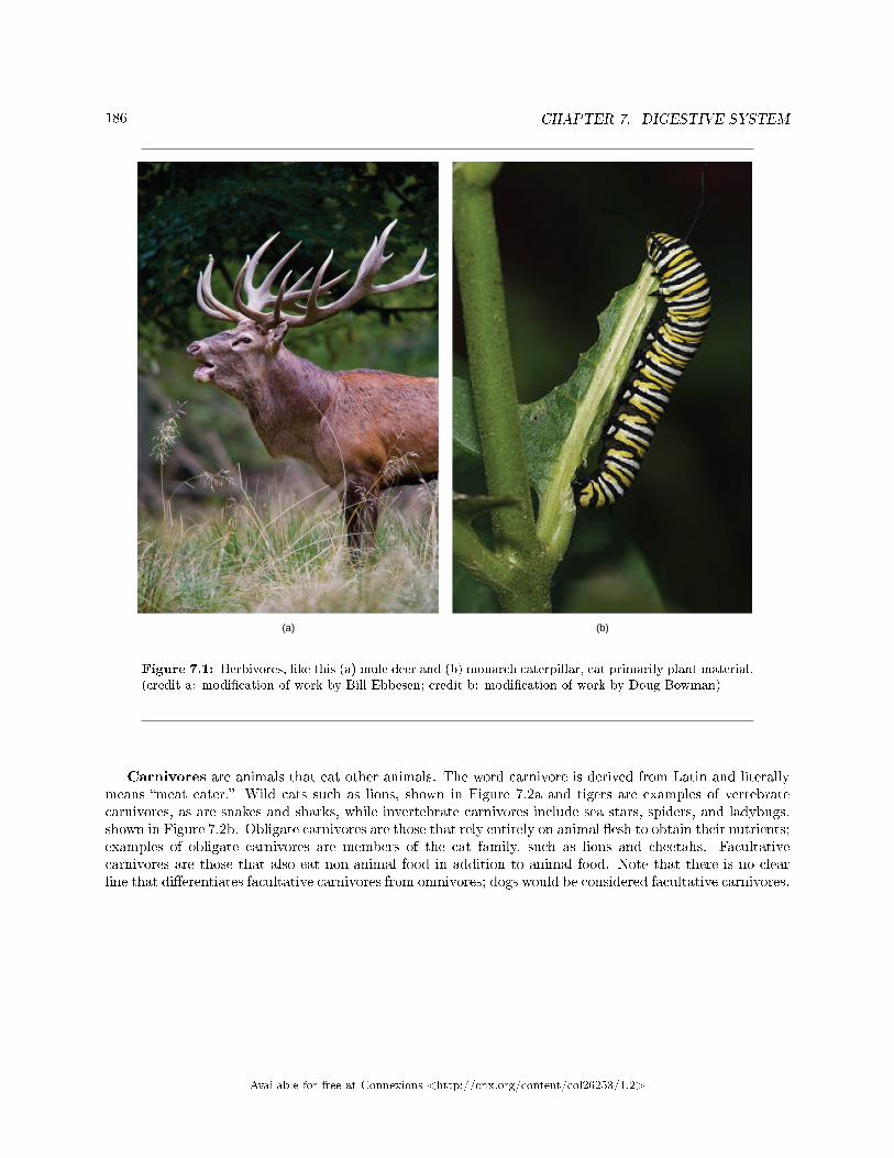
186 CHAPTER 7. DIGESTIVE SYSTEM
Figure 7.1: Herbivores, like this (a) mule deer and (b) monarch caterpillar, eat primarily plant material.(credit a: modi�cation of work by Bill Ebbesen; credit b: modi�cation of work by Doug Bowman)
Carnivores are animals that eat other animals. The word carnivore is derived from Latin and literallymeans �meat eater.� Wild cats such as lions, shown in Figure 7.2a and tigers are examples of vertebratecarnivores, as are snakes and sharks, while invertebrate carnivores include sea stars, spiders, and ladybugs,shown in Figure 7.2b. Obligate carnivores are those that rely entirely on animal �esh to obtain their nutrients;examples of obligate carnivores are members of the cat family, such as lions and cheetahs. Facultativecarnivores are those that also eat non-animal food in addition to animal food. Note that there is no clearline that di�erentiates facultative carnivores from omnivores; dogs would be considered facultative carnivores.
Available for free at Connexions <http://cnx.org/content/col26253/1.2>
187
Figure 7.2: Carnivores like the (a) lion eat primarily meat. The (b) ladybug is also a carnivorethat consumes small insects called aphids. (credit a: modi�cation of work by Kevin Pluck; credit b:modi�cation of work by Jon Sullivan)
Omnivores are animals that eat both plant- and animal-derived food. In Latin, omnivore means toeat everything. Humans, bears (shown in Figure 7.3a), and chickens are example of vertebrate omnivores;invertebrate omnivores include cockroaches and cray�sh (shown in Figure 7.3b).
Figure 7.3: Omnivores like the (a) bear and (b) cray�sh eat both plant and animal based food. (credita: modi�cation of work by Dave Menke; credit b: modi�cation of work by Jon Sullivan)
Available for free at Connexions <http://cnx.org/content/col26253/1.2>
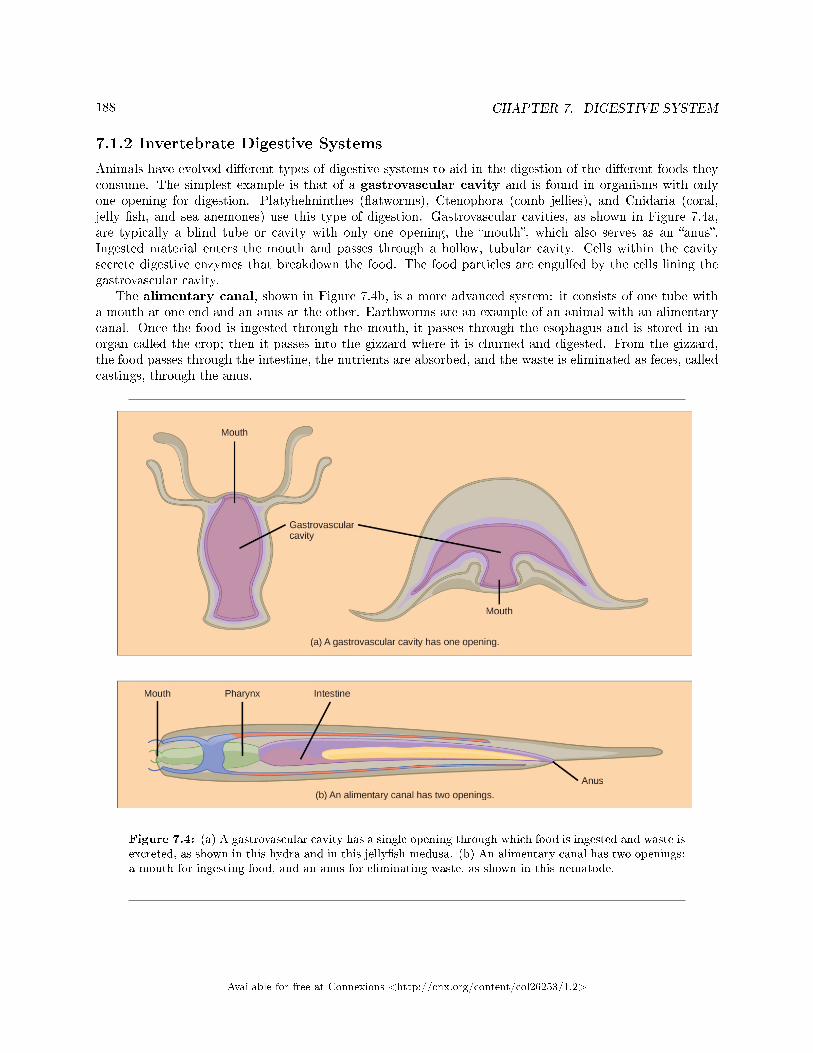
188 CHAPTER 7. DIGESTIVE SYSTEM
7.1.2 Invertebrate Digestive Systems
Animals have evolved di�erent types of digestive systems to aid in the digestion of the di�erent foods theyconsume. The simplest example is that of a gastrovascular cavity and is found in organisms with onlyone opening for digestion. Platyhelminthes (�atworms), Ctenophora (comb jellies), and Cnidaria (coral,jelly �sh, and sea anemones) use this type of digestion. Gastrovascular cavities, as shown in Figure 7.4a,are typically a blind tube or cavity with only one opening, the �mouth�, which also serves as an �anus�.Ingested material enters the mouth and passes through a hollow, tubular cavity. Cells within the cavitysecrete digestive enzymes that breakdown the food. The food particles are engulfed by the cells lining thegastrovascular cavity.
The alimentary canal, shown in Figure 7.4b, is a more advanced system: it consists of one tube witha mouth at one end and an anus at the other. Earthworms are an example of an animal with an alimentarycanal. Once the food is ingested through the mouth, it passes through the esophagus and is stored in anorgan called the crop; then it passes into the gizzard where it is churned and digested. From the gizzard,the food passes through the intestine, the nutrients are absorbed, and the waste is eliminated as feces, calledcastings, through the anus.
Figure 7.4: (a) A gastrovascular cavity has a single opening through which food is ingested and waste isexcreted, as shown in this hydra and in this jelly�sh medusa. (b) An alimentary canal has two openings:a mouth for ingesting food, and an anus for eliminating waste, as shown in this nematode.
Available for free at Connexions <http://cnx.org/content/col26253/1.2>
189
7.1.3 Vertebrate Digestive Systems
Vertebrates have evolved more complex digestive systems to adapt to their dietary needs. Some animalshave a single stomach, while others have multi-chambered stomachs. Birds have developed a digestive systemadapted to eating unmasticated food.
7.1.3.1 Monogastric: Single-chambered Stomach
As the word monogastric suggests, this type of digestive system consists of one (�mono�) stomach chamber(�gastric�). Humans and many animals have a monogastric digestive system as illustrated in Figure 7.5ab.The process of digestion begins with the mouth and the intake of food. The teeth play an important rolein masticating (chewing) or physically breaking down food into smaller particles. The enzymes present insaliva also begin to chemically breakdown food. The esophagus is a long tube that connects the mouth tothe stomach. Using peristalsis, or wave-like smooth muscle contractions, the muscles of the esophagus pushthe food towards the stomach. In order to speed up the actions of enzymes in the stomach, the stomach isan extremely acidic environment, with a pH between 1.5 and 2.5. The gastric juices, which include enzymesin the stomach, act on the food particles and continue the process of digestion. Further breakdown of foodtakes place in the small intestine where enzymes produced by the liver, the small intestine, and the pancreascontinue the process of digestion. The nutrients are absorbed into the bloodstream across the epithelial cellslining the walls of the small intestines. The waste material travels on to the large intestine where water isabsorbed and the drier waste material is compacted into feces; it is stored until it is excreted through therectum.
Available for free at Connexions <http://cnx.org/content/col26253/1.2>
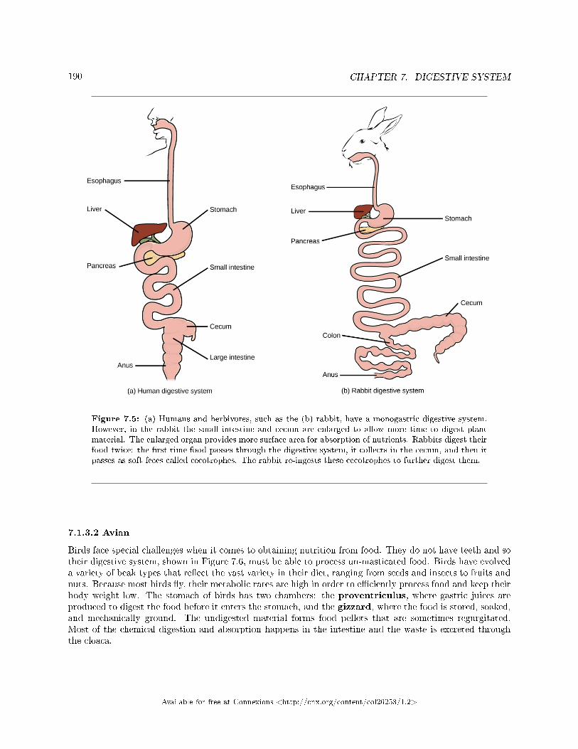
190 CHAPTER 7. DIGESTIVE SYSTEM
Figure 7.5: (a) Humans and herbivores, such as the (b) rabbit, have a monogastric digestive system.However, in the rabbit the small intestine and cecum are enlarged to allow more time to digest plantmaterial. The enlarged organ provides more surface area for absorption of nutrients. Rabbits digest theirfood twice: the �rst time food passes through the digestive system, it collects in the cecum, and then itpasses as soft feces called cecotrophes. The rabbit re-ingests these cecotrophes to further digest them.
7.1.3.2 Avian
Birds face special challenges when it comes to obtaining nutrition from food. They do not have teeth and sotheir digestive system, shown in Figure 7.6, must be able to process un-masticated food. Birds have evolveda variety of beak types that re�ect the vast variety in their diet, ranging from seeds and insects to fruits andnuts. Because most birds �y, their metabolic rates are high in order to e�ciently process food and keep theirbody weight low. The stomach of birds has two chambers: the proventriculus, where gastric juices areproduced to digest the food before it enters the stomach, and the gizzard, where the food is stored, soaked,and mechanically ground. The undigested material forms food pellets that are sometimes regurgitated.Most of the chemical digestion and absorption happens in the intestine and the waste is excreted throughthe cloaca.
Available for free at Connexions <http://cnx.org/content/col26253/1.2>
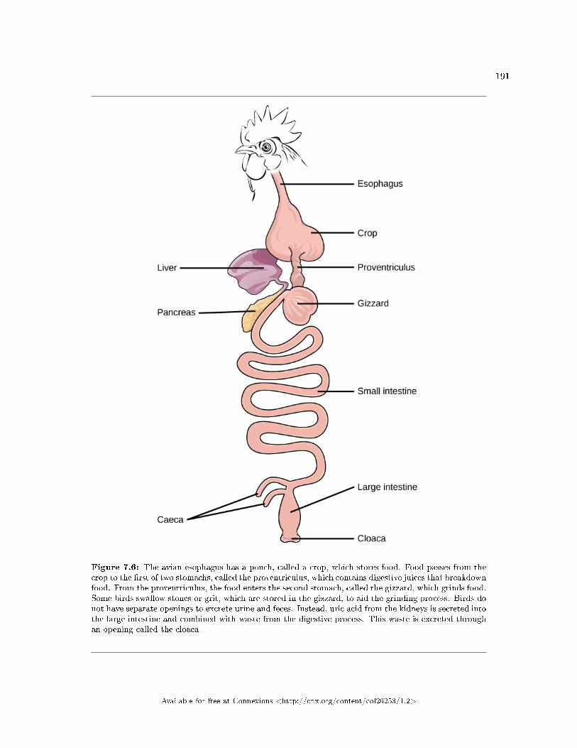
191
Figure 7.6: The avian esophagus has a pouch, called a crop, which stores food. Food passes from thecrop to the �rst of two stomachs, called the proventriculus, which contains digestive juices that breakdownfood. From the proventriculus, the food enters the second stomach, called the gizzard, which grinds food.Some birds swallow stones or grit, which are stored in the gizzard, to aid the grinding process. Birds donot have separate openings to excrete urine and feces. Instead, uric acid from the kidneys is secreted intothe large intestine and combined with waste from the digestive process. This waste is excreted throughan opening called the cloaca.
Available for free at Connexions <http://cnx.org/content/col26253/1.2>
192 CHAPTER 7. DIGESTIVE SYSTEM
: Avian Adaptations
Birds have a highly e�cient, simpli�ed digestive system. Recent fossil evidence has shown that theevolutionary divergence of birds from other land animals was characterized by streamlining andsimplifying the digestive system. Unlike many other animals, birds do not have teeth to chew theirfood. In place of lips, they have sharp pointy beaks. The horny beak, lack of jaws, and the smallertongue of the birds can be traced back to their dinosaur ancestors. The emergence of these changesseems to coincide with the inclusion of seeds in the bird diet. Seed-eating birds have beaks that areshaped for grabbing seeds and the two-compartment stomach allows for delegation of tasks. Sincebirds need to remain light in order to �y, their metabolic rates are very high, which means theydigest their food very quickly and need to eat often. Contrast this with the ruminants, where thedigestion of plant matter takes a very long time.
7.1.3.3 Ruminants
Ruminants are mainly herbivores like cows, sheep, and goats, whose entire diet consists of eating largeamounts of roughage or �ber. They have evolved digestive systems that help them digest vast amounts ofcellulose. An interesting feature of the ruminants' mouth is that they do not have upper incisor teeth. Theyuse their lower teeth, tongue and lips to tear and chew their food. From the mouth, the food travels to theesophagus and on to the stomach.
To help digest the large amount of plant material, the stomach of the ruminants is a multi-chamberedorgan, as illustrated in Figure 7.7. The four compartments of the stomach are called the rumen, reticulum,omasum, and abomasum. These chambers contain many microbes that breakdown cellulose and fermentingested food. The abomasum is the �true� stomach and is the equivalent of the monogastric stomachchamber where gastric juices are secreted. The four-compartment gastric chamber provides larger space andthe microbial support necessary to digest plant material in ruminants. The fermentation process produceslarge amounts of gas in the stomach chamber, which must be eliminated. As in other animals, the smallintestine plays an important role in nutrient absorption, and the large intestine helps in the elimination ofwaste.
Available for free at Connexions <http://cnx.org/content/col26253/1.2>
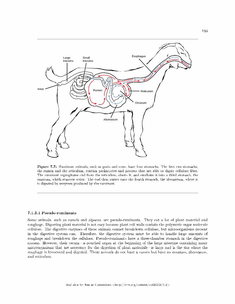
193
Figure 7.7: Ruminant animals, such as goats and cows, have four stomachs. The �rst two stomachs,the rumen and the reticulum, contain prokaryotes and protists that are able to digest cellulose �ber.The ruminant regurgitates cud from the reticulum, chews it, and swallows it into a third stomach, theomasum, which removes water. The cud then passes onto the fourth stomach, the abomasum, where itis digested by enzymes produced by the ruminant.
7.1.3.4 Pseudo-ruminants
Some animals, such as camels and alpacas, are pseudo-ruminants. They eat a lot of plant material androughage. Digesting plant material is not easy because plant cell walls contain the polymeric sugar moleculecellulose. The digestive enzymes of these animals cannot breakdown cellulose, but microorganisms presentin the digestive system can. Therefore, the digestive system must be able to handle large amounts ofroughage and breakdown the cellulose. Pseudo-ruminants have a three-chamber stomach in the digestivesystem. However, their cecum�a pouched organ at the beginning of the large intestine containing manymicroorganisms that are necessary for the digestion of plant materials�is large and is the site where theroughage is fermented and digested. These animals do not have a rumen but have an omasum, abomasum,and reticulum.
Available for free at Connexions <http://cnx.org/content/col26253/1.2>
194 CHAPTER 7. DIGESTIVE SYSTEM
7.1.4 Parts of the Digestive System
The vertebrate digestive system is designed to facilitate the transformation of food matter into the nutrientcomponents that sustain organisms.
7.1.4.1 Oral Cavity
The oral cavity, or mouth, is the point of entry of food into the digestive system, illustrated in Figure 7.8.The food consumed is broken into smaller particles by mastication, the chewing action of the teeth. Allmammals have teeth and can chew their food.
The extensive chemical process of digestion begins in the mouth. As food is being chewed, saliva,produced by the salivary glands, mixes with the food. Saliva is a watery substance produced in the mouthsof many animals. There are three major glands that secrete saliva�the parotid, the submandibular, and thesublingual. Saliva contains mucus that moistens food and bu�ers the pH of the food. Saliva also containsimmunoglobulins and lysozymes, which have antibacterial action to reduce tooth decay by inhibiting growthof some bacteria. Saliva also contains an enzyme called salivary amylase that begins the process ofconverting starches in the food into a disaccharide called maltose. Another enzyme called lipase is producedby the cells in the tongue. Lipases are a class of enzymes that can breakdown triglycerides. The linguallipase begins the breakdown of fat components in the food. The chewing and wetting action provided bythe teeth and saliva prepare the food into a mass called the bolus for swallowing. The tongue helps inswallowing�moving the bolus from the mouth into the pharynx. The pharynx opens to two passageways:the trachea, which leads to the lungs, and the esophagus, which leads to the stomach. The trachea has anopening called the glottis, which is covered by a cartilaginous �ap called the epiglottis. When swallowing,the epiglottis closes the glottis and food passes into the esophagus and not the trachea. This arrangementallows food to be kept out of the trachea.
Available for free at Connexions <http://cnx.org/content/col26253/1.2>
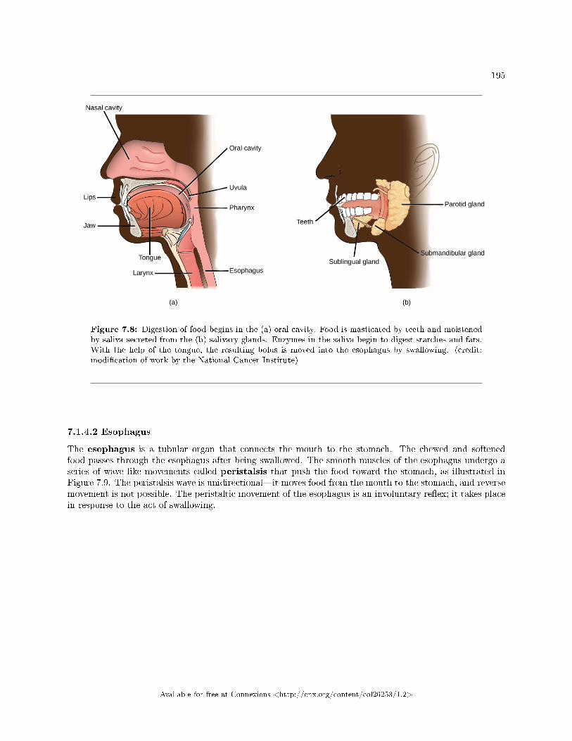
195
Figure 7.8: Digestion of food begins in the (a) oral cavity. Food is masticated by teeth and moistenedby saliva secreted from the (b) salivary glands. Enzymes in the saliva begin to digest starches and fats.With the help of the tongue, the resulting bolus is moved into the esophagus by swallowing. (credit:modi�cation of work by the National Cancer Institute)
7.1.4.2 Esophagus
The esophagus is a tubular organ that connects the mouth to the stomach. The chewed and softenedfood passes through the esophagus after being swallowed. The smooth muscles of the esophagus undergo aseries of wave like movements called peristalsis that push the food toward the stomach, as illustrated inFigure 7.9. The peristalsis wave is unidirectional�it moves food from the mouth to the stomach, and reversemovement is not possible. The peristaltic movement of the esophagus is an involuntary re�ex; it takes placein response to the act of swallowing.
Available for free at Connexions <http://cnx.org/content/col26253/1.2>
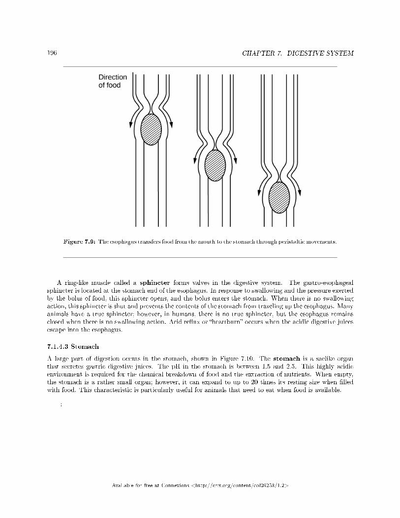
196 CHAPTER 7. DIGESTIVE SYSTEM
Figure 7.9: The esophagus transfers food from the mouth to the stomach through peristaltic movements.
A ring-like muscle called a sphincter forms valves in the digestive system. The gastro-esophagealsphincter is located at the stomach end of the esophagus. In response to swallowing and the pressure exertedby the bolus of food, this sphincter opens, and the bolus enters the stomach. When there is no swallowingaction, this sphincter is shut and prevents the contents of the stomach from traveling up the esophagus. Manyanimals have a true sphincter; however, in humans, there is no true sphincter, but the esophagus remainsclosed when there is no swallowing action. Acid re�ux or �heartburn� occurs when the acidic digestive juicesescape into the esophagus.
7.1.4.3 Stomach
A large part of digestion occurs in the stomach, shown in Figure 7.10. The stomach is a saclike organthat secretes gastric digestive juices. The pH in the stomach is between 1.5 and 2.5. This highly acidicenvironment is required for the chemical breakdown of food and the extraction of nutrients. When empty,the stomach is a rather small organ; however, it can expand to up to 20 times its resting size when �lledwith food. This characteristic is particularly useful for animals that need to eat when food is available.
:
Available for free at Connexions <http://cnx.org/content/col26253/1.2>
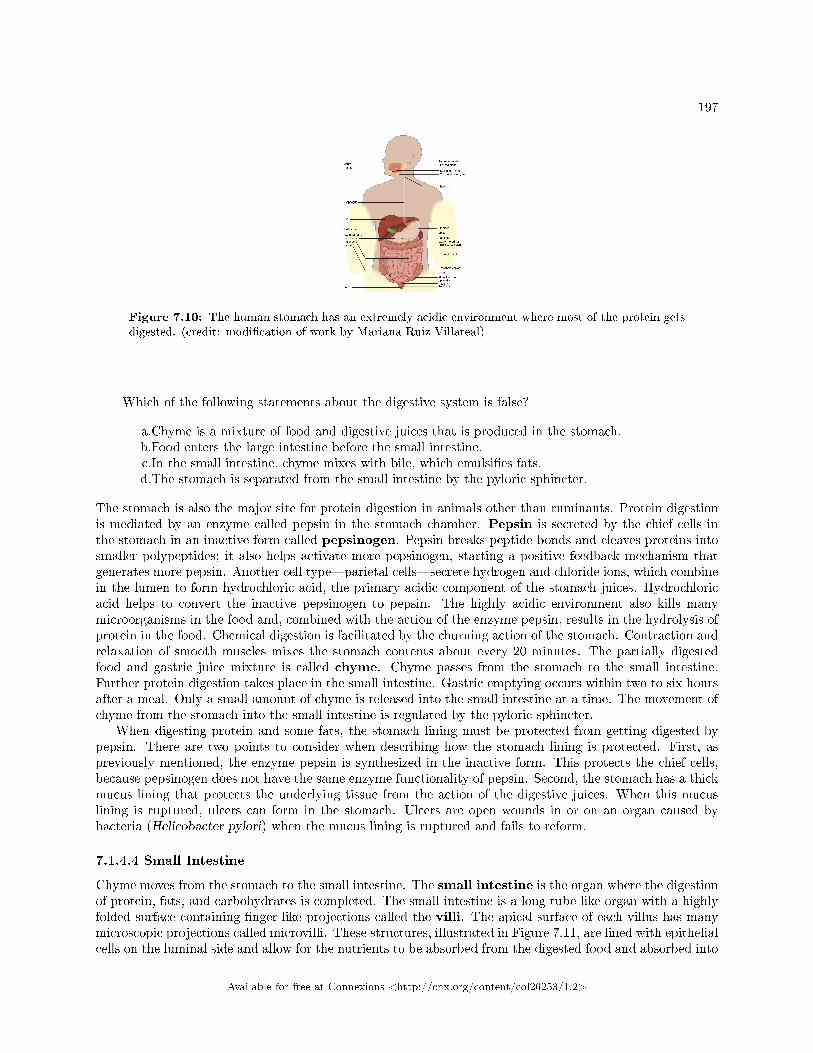
197
Figure 7.10: The human stomach has an extremely acidic environment where most of the protein getsdigested. (credit: modi�cation of work by Mariana Ruiz Villareal)
Which of the following statements about the digestive system is false?
a.Chyme is a mixture of food and digestive juices that is produced in the stomach.b.Food enters the large intestine before the small intestine.c.In the small intestine, chyme mixes with bile, which emulsi�es fats.d.The stomach is separated from the small intestine by the pyloric sphincter.
The stomach is also the major site for protein digestion in animals other than ruminants. Protein digestionis mediated by an enzyme called pepsin in the stomach chamber. Pepsin is secreted by the chief cells inthe stomach in an inactive form called pepsinogen. Pepsin breaks peptide bonds and cleaves proteins intosmaller polypeptides; it also helps activate more pepsinogen, starting a positive feedback mechanism thatgenerates more pepsin. Another cell type�parietal cells�secrete hydrogen and chloride ions, which combinein the lumen to form hydrochloric acid, the primary acidic component of the stomach juices. Hydrochloricacid helps to convert the inactive pepsinogen to pepsin. The highly acidic environment also kills manymicroorganisms in the food and, combined with the action of the enzyme pepsin, results in the hydrolysis ofprotein in the food. Chemical digestion is facilitated by the churning action of the stomach. Contraction andrelaxation of smooth muscles mixes the stomach contents about every 20 minutes. The partially digestedfood and gastric juice mixture is called chyme. Chyme passes from the stomach to the small intestine.Further protein digestion takes place in the small intestine. Gastric emptying occurs within two to six hoursafter a meal. Only a small amount of chyme is released into the small intestine at a time. The movement ofchyme from the stomach into the small intestine is regulated by the pyloric sphincter.
When digesting protein and some fats, the stomach lining must be protected from getting digested bypepsin. There are two points to consider when describing how the stomach lining is protected. First, aspreviously mentioned, the enzyme pepsin is synthesized in the inactive form. This protects the chief cells,because pepsinogen does not have the same enzyme functionality of pepsin. Second, the stomach has a thickmucus lining that protects the underlying tissue from the action of the digestive juices. When this mucuslining is ruptured, ulcers can form in the stomach. Ulcers are open wounds in or on an organ caused bybacteria (Helicobacter pylori) when the mucus lining is ruptured and fails to reform.
7.1.4.4 Small Intestine
Chyme moves from the stomach to the small intestine. The small intestine is the organ where the digestionof protein, fats, and carbohydrates is completed. The small intestine is a long tube-like organ with a highlyfolded surface containing �nger-like projections called the villi. The apical surface of each villus has manymicroscopic projections called microvilli. These structures, illustrated in Figure 7.11, are lined with epithelialcells on the luminal side and allow for the nutrients to be absorbed from the digested food and absorbed into
Available for free at Connexions <http://cnx.org/content/col26253/1.2>
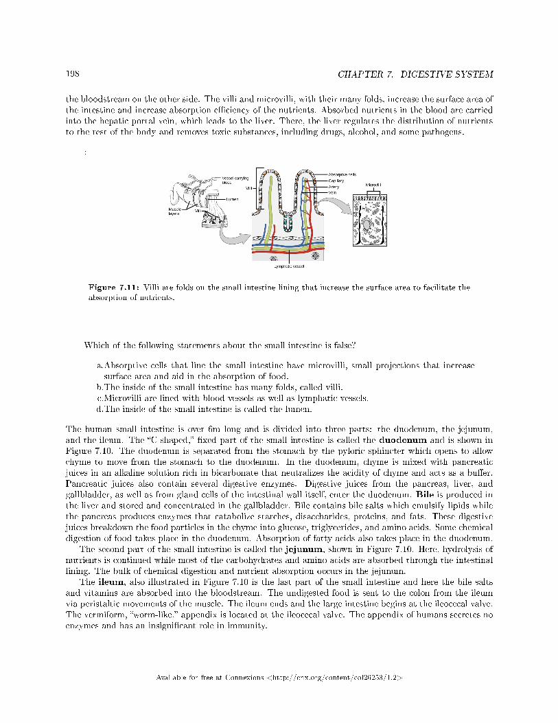
198 CHAPTER 7. DIGESTIVE SYSTEM
the bloodstream on the other side. The villi and microvilli, with their many folds, increase the surface area ofthe intestine and increase absorption e�ciency of the nutrients. Absorbed nutrients in the blood are carriedinto the hepatic portal vein, which leads to the liver. There, the liver regulates the distribution of nutrientsto the rest of the body and removes toxic substances, including drugs, alcohol, and some pathogens.
:
Figure 7.11: Villi are folds on the small intestine lining that increase the surface area to facilitate theabsorption of nutrients.
Which of the following statements about the small intestine is false?
a.Absorptive cells that line the small intestine have microvilli, small projections that increasesurface area and aid in the absorption of food.
b.The inside of the small intestine has many folds, called villi.c.Microvilli are lined with blood vessels as well as lymphatic vessels.d.The inside of the small intestine is called the lumen.
The human small intestine is over 6m long and is divided into three parts: the duodenum, the jejunum,and the ileum. The �C-shaped,� �xed part of the small intestine is called the duodenum and is shown inFigure 7.10. The duodenum is separated from the stomach by the pyloric sphincter which opens to allowchyme to move from the stomach to the duodenum. In the duodenum, chyme is mixed with pancreaticjuices in an alkaline solution rich in bicarbonate that neutralizes the acidity of chyme and acts as a bu�er.Pancreatic juices also contain several digestive enzymes. Digestive juices from the pancreas, liver, andgallbladder, as well as from gland cells of the intestinal wall itself, enter the duodenum. Bile is produced inthe liver and stored and concentrated in the gallbladder. Bile contains bile salts which emulsify lipids whilethe pancreas produces enzymes that catabolize starches, disaccharides, proteins, and fats. These digestivejuices breakdown the food particles in the chyme into glucose, triglycerides, and amino acids. Some chemicaldigestion of food takes place in the duodenum. Absorption of fatty acids also takes place in the duodenum.
The second part of the small intestine is called the jejunum, shown in Figure 7.10. Here, hydrolysis ofnutrients is continued while most of the carbohydrates and amino acids are absorbed through the intestinallining. The bulk of chemical digestion and nutrient absorption occurs in the jejunum.
The ileum, also illustrated in Figure 7.10 is the last part of the small intestine and here the bile saltsand vitamins are absorbed into the bloodstream. The undigested food is sent to the colon from the ileumvia peristaltic movements of the muscle. The ileum ends and the large intestine begins at the ileocecal valve.The vermiform, �worm-like,� appendix is located at the ileocecal valve. The appendix of humans secretes noenzymes and has an insigni�cant role in immunity.
Available for free at Connexions <http://cnx.org/content/col26253/1.2>
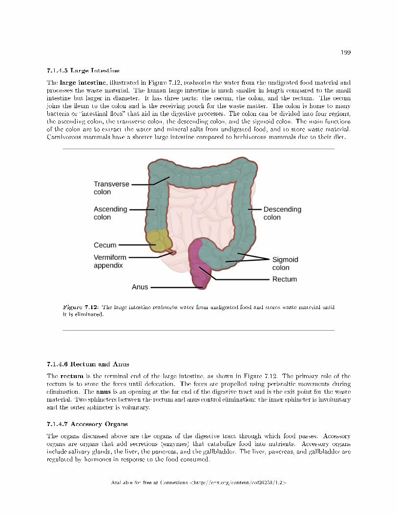
199
7.1.4.5 Large Intestine
The large intestine, illustrated in Figure 7.12, reabsorbs the water from the undigested food material andprocesses the waste material. The human large intestine is much smaller in length compared to the smallintestine but larger in diameter. It has three parts: the cecum, the colon, and the rectum. The cecumjoins the ileum to the colon and is the receiving pouch for the waste matter. The colon is home to manybacteria or �intestinal �ora� that aid in the digestive processes. The colon can be divided into four regions,the ascending colon, the transverse colon, the descending colon, and the sigmoid colon. The main functionsof the colon are to extract the water and mineral salts from undigested food, and to store waste material.Carnivorous mammals have a shorter large intestine compared to herbivorous mammals due to their diet.
Figure 7.12: The large intestine reabsorbs water from undigested food and stores waste material untilit is eliminated.
7.1.4.6 Rectum and Anus
The rectum is the terminal end of the large intestine, as shown in Figure 7.12. The primary role of therectum is to store the feces until defecation. The feces are propelled using peristaltic movements duringelimination. The anus is an opening at the far-end of the digestive tract and is the exit point for the wastematerial. Two sphincters between the rectum and anus control elimination: the inner sphincter is involuntaryand the outer sphincter is voluntary.
7.1.4.7 Accessory Organs
The organs discussed above are the organs of the digestive tract through which food passes. Accessoryorgans are organs that add secretions (enzymes) that catabolize food into nutrients. Accessory organsinclude salivary glands, the liver, the pancreas, and the gallbladder. The liver, pancreas, and gallbladder areregulated by hormones in response to the food consumed.
Available for free at Connexions <http://cnx.org/content/col26253/1.2>
200 CHAPTER 7. DIGESTIVE SYSTEM
The liver is the largest internal organ in humans and it plays a very important role in digestion of fatsand detoxifying blood. The liver produces bile, a digestive juice that is required for the breakdown of fattycomponents of the food in the duodenum. The liver also processes the vitamins and fats and synthesizesmany plasma proteins.
The pancreas is another important gland that secretes digestive juices. The chyme produced from thestomach is highly acidic in nature; the pancreatic juices contain high levels of bicarbonate, an alkali thatneutralizes the acidic chyme. Additionally, the pancreatic juices contain a large variety of enzymes that arerequired for the digestion of protein and carbohydrates.
The gallbladder is a small organ that aids the liver by storing bile and concentrating bile salts. Whenchyme containing fatty acids enters the duodenum, the bile is secreted from the gallbladder into the duode-num.
7.1.5 Section Summary
Di�erent animals have evolved di�erent types of digestive systems specialized to meet their dietary needs.Humans and many other animals have monogastric digestive systems with a single-chambered stomach.Birds have evolved a digestive system that includes a gizzard where the food is crushed into smaller pieces.This compensates for their inability to masticate. Ruminants that consume large amounts of plant materialhave a multi-chambered stomach that digests roughage. Pseudo-ruminants have similar digestive processesas ruminants but do not have the four-compartment stomach. Processing food involves ingestion (eating),digestion (mechanical and enzymatic breakdown of large molecules), absorption (cellular uptake of nutrients),and elimination (removal of undigested waste as feces).
Many organs work together to digest food and absorb nutrients. The mouth is the point of ingestionand the location where both mechanical and chemical breakdown of food begins. Saliva contains an enzymecalled amylase that breaks down carbohydrates. The food bolus travels through the esophagus by peristalticmovements to the stomach. The stomach has an extremely acidic environment. An enzyme called pepsindigests protein in the stomach. Further digestion and absorption take place in the small intestine. The largeintestine reabsorbs water from the undigested food and stores waste until elimination.
7.1.6 Visual Connection Questions
Exercise 7.1.1 (Solution on p. 221.)
Figure 7.10 Which of the following statements about the digestive system is false?
a. Chyme is a mixture of food and digestive juices that is produced in the stomach.b. Food enters the large intestine before the small intestine.c. In the small intestine, chyme mixes with bile, which emulsi�es fats.d. The stomach is separated from the small intestine by the pyloric sphincter.
Exercise 7.1.2 (Solution on p. 221.)
Figure 7.11 Which of the following statements about the small intestine is false?
a. Absorptive cells that line the small intestine have microvilli, small projections that increasesurface area and aid in the absorption of food.
b. The inside of the small intestine has many folds, called villi.c. Microvilli are lined with blood vessels as well as lymphatic vessels.d. The inside of the small intestine is called the lumen.
Available for free at Connexions <http://cnx.org/content/col26253/1.2>
201
7.1.7 Review Questions
Exercise 7.1.3 (Solution on p. 221.)
Which of the following is a pseudo-ruminant?
a. cowb. pigc. crowd. horse
Exercise 7.1.4 (Solution on p. 221.)
Which of the following statements is untrue?
a. Roughage takes a long time to digest.b. Birds eat large quantities at one time so that they can �y long distances.c. Cows do not have upper teeth.d. In pseudo-ruminants, roughage is digested in the cecum.
Exercise 7.1.5 (Solution on p. 221.)
The acidic nature of chyme is neutralized by ________.
a. potassium hydroxideb. sodium hydroxidec. bicarbonatesd. vinegar
Exercise 7.1.6 (Solution on p. 221.)
The digestive juices from the liver are delivered to the ________.
a. stomachb. liverc. duodenumd. colon
Exercise 7.1.7 (Solution on p. 221.)
A scientist dissects a new species of animal. If the animal's digestive system has a single stomachwith an extended small intestine, to which animal could the dissected specimen be closely related?
a. lionb. snowshoe harec. earthwormd. eagle
7.1.8 Critical Thinking Questions
Exercise 7.1.8 (Solution on p. 221.)
How does the polygastric digestive system aid in digesting roughage?
Exercise 7.1.9 (Solution on p. 221.)
How do birds digest their food in the absence of teeth?
Exercise 7.1.10 (Solution on p. 221.)
What is the role of the accessory organs in digestion?
Available for free at Connexions <http://cnx.org/content/col26253/1.2>
202 CHAPTER 7. DIGESTIVE SYSTEM
Exercise 7.1.11 (Solution on p. 221.)
Explain how the villi and microvilli aid in absorption.
Exercise 7.1.12 (Solution on p. 221.)
Name two components of the digestive system that perform mechanical digestion. Describe howmechanical digestion contributes to acquiring nutrients from food.
7.2 Nutrition and Energy Production2
Given the diversity of animal life on our planet, it is not surprising that the animal diet would also vary sub-stantially. The animal diet is the source of materials needed for building DNA and other complex moleculesneeded for growth, maintenance, and reproduction; collectively these processes are called biosynthesis. Thediet is also the source of materials for ATP production in the cells. The diet must be balanced to providethe minerals and vitamins that are required for cellular function.
7.2.1 Food Requirements
What are the fundamental requirements of the animal diet? The animal diet should be well balancedand provide nutrients required for bodily function and the minerals and vitamins required for maintainingstructure and regulation necessary for good health and reproductive capability. These requirements for ahuman are illustrated graphically in Figure 7.13
2This content is available online at <http://cnx.org/content/m66616/1.14/>.
Available for free at Connexions <http://cnx.org/content/col26253/1.2>
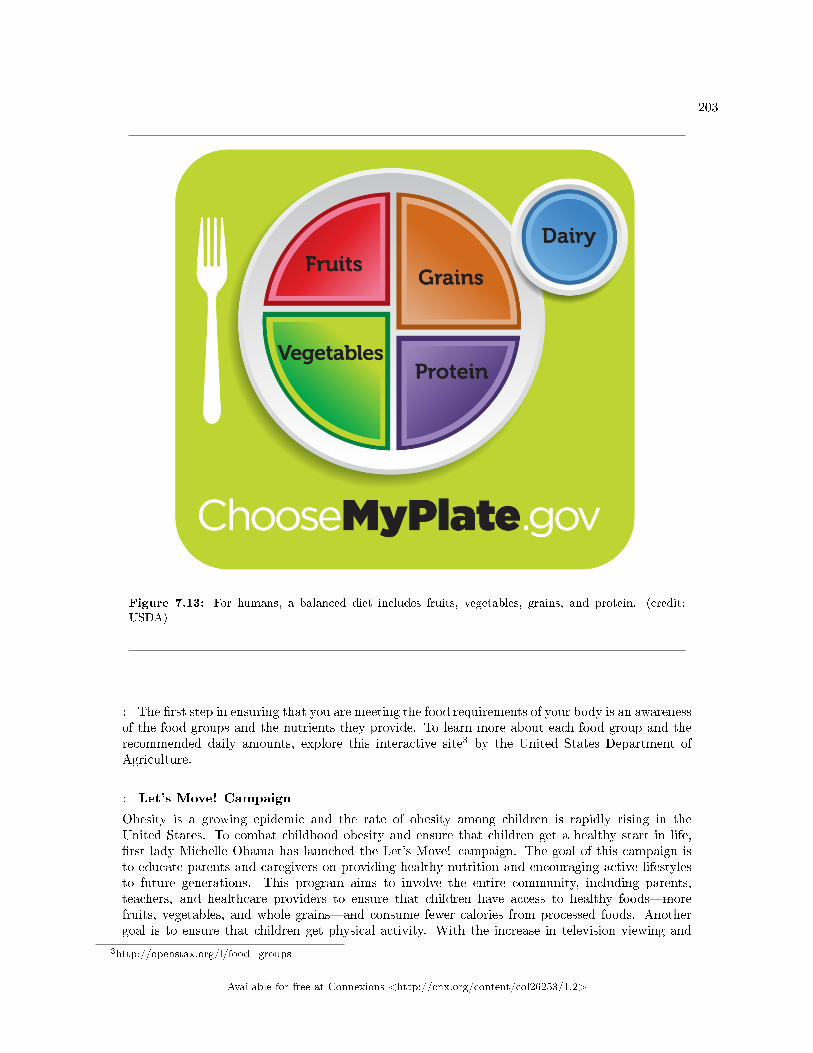
203
Figure 7.13: For humans, a balanced diet includes fruits, vegetables, grains, and protein. (credit:USDA)
: The �rst step in ensuring that you are meeting the food requirements of your body is an awarenessof the food groups and the nutrients they provide. To learn more about each food group and therecommended daily amounts, explore this interactive site3 by the United States Department ofAgriculture.
: Let's Move! Campaign
Obesity is a growing epidemic and the rate of obesity among children is rapidly rising in theUnited States. To combat childhood obesity and ensure that children get a healthy start in life,�rst lady Michelle Obama has launched the Let's Move! campaign. The goal of this campaign isto educate parents and caregivers on providing healthy nutrition and encouraging active lifestylesto future generations. This program aims to involve the entire community, including parents,teachers, and healthcare providers to ensure that children have access to healthy foods�morefruits, vegetables, and whole grains�and consume fewer calories from processed foods. Anothergoal is to ensure that children get physical activity. With the increase in television viewing and
3http://openstax.org/l/food_groups
Available for free at Connexions <http://cnx.org/content/col26253/1.2>
204 CHAPTER 7. DIGESTIVE SYSTEM
stationary pursuits such as video games, sedentary lifestyles have become the norm. Learn more athttps://letsmove.obamawhitehouse.archives.gov4 .
7.2.1.1 Organic Precursors
The organic molecules required for building cellular material and tissues must come from food. Carbohydratesor sugars are the primary source of organic carbons in the animal body. During digestion, digestible carbo-hydrates are ultimately broken down into glucose and used to provide energy through metabolic pathways.Complex carbohydrates, including polysaccharides, can be broken down into glucose through biochemicalmodi�cation; however, humans do not produce the enzyme cellulase and lack the ability to derive glucosefrom the polysaccharide cellulose. In humans, these molecules provide the �ber required for moving wastethrough the large intestine and a healthy colon. The intestinal �ora in the human gut are able to extractsome nutrition from these plant �bers. The excess sugars in the body are converted into glycogen andstored in the liver and muscles for later use. Glycogen stores are used to fuel prolonged exertions, such aslong-distance running, and to provide energy during food shortage. Excess glycogen can be converted tofats, which are stored in the lower layer of the skin of mammals for insulation and energy storage. Excessdigestible carbohydrates are stored by mammals in order to survive famine and aid in mobility.
Another important requirement is that of nitrogen. Protein catabolism provides a source of organicnitrogen. Amino acids are the building blocks of proteins and protein breakdown provides amino acids thatare used for cellular function. The carbon and nitrogen derived from these become the building block fornucleotides, nucleic acids, proteins, cells, and tissues. Excess nitrogen must be excreted as it is toxic. Fatsadd �avor to food and promote a sense of satiety or fullness. Fatty foods are also signi�cant sources ofenergy because one gram of fat contains nine calories. Fats are required in the diet to aid the absorption offat-soluble vitamins and the production of fat-soluble hormones.
7.2.1.2 Essential Nutrients
While the animal body can synthesize many of the molecules required for function from the organic precur-sors, there are some nutrients that need to be consumed from food. These nutrients are termed essentialnutrients, meaning they must be eaten, and the body cannot produce them.
The omega-3 alpha-linolenic acid and the omega-6 linoleic acid are essential fatty acids needed to makesome membrane phospholipids. Vitamins are another class of essential organic molecules that are requiredin small quantities for many enzymes to function and, for this reason, are considered to be coenzymes.Absence or low levels of vitamins can have a dramatic e�ect on health, as outlined in Table 7.1 and Table7.2. Both fat-soluble and water-soluble vitamins must be obtained from food. Minerals, listed in Table 7.3,are inorganic essential nutrients that must be obtained from food. Among their many functions, mineralshelp in structure and regulation and are considered cofactors. Certain amino acids also must be procuredfrom food and cannot be synthesized by the body. These amino acids are the �essential� amino acids. Thehuman body can synthesize only 11 of the 20 required amino acids; the rest must be obtained from food.The essential amino acids are listed in Table 7.4.
Water-soluble Essential Vitamins
Vitamin Function De�ciencies CanLead To
Sources
continued on next page
4http://openstax.org/l/Letsmove
Available for free at Connexions <http://cnx.org/content/col26253/1.2>
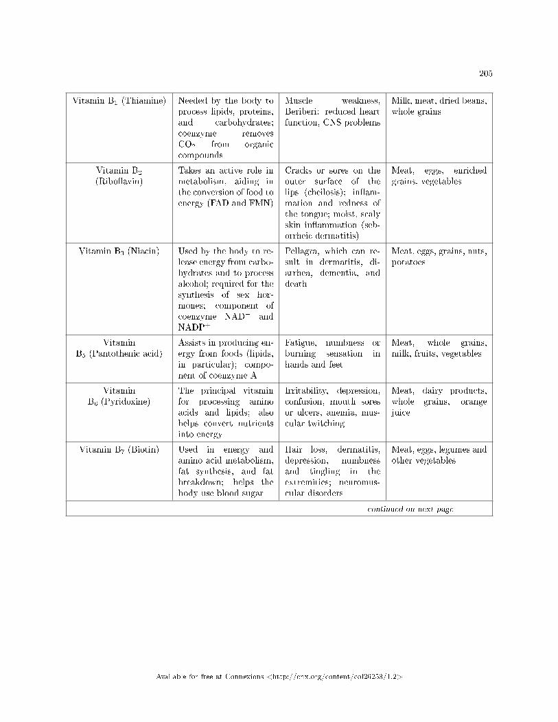
205
Vitamin B1 (Thiamine) Needed by the body toprocess lipids, proteins,and carbohydrates;coenzyme removesCO2 from organiccompounds
Muscle weakness,Beriberi: reduced heartfunction, CNS problems
Milk, meat, dried beans,whole grains
Vitamin B2
(Ribo�avin)Takes an active role inmetabolism, aiding inthe conversion of food toenergy (FAD and FMN)
Cracks or sores on theouter surface of thelips (cheilosis); in�am-mation and redness ofthe tongue; moist, scalyskin in�ammation (seb-orrheic dermatitis)
Meat, eggs, enrichedgrains, vegetables
Vitamin B3 (Niacin) Used by the body to re-lease energy from carbo-hydrates and to processalcohol; required for thesynthesis of sex hor-mones; component ofcoenzyme NAD+ andNADP+
Pellagra, which can re-sult in dermatitis, di-arrhea, dementia, anddeath
Meat, eggs, grains, nuts,potatoes
VitaminB5 (Pantothenic acid)
Assists in producing en-ergy from foods (lipids,in particular); compo-nent of coenzyme A
Fatigue, numbness orburning sensation inhands and feet
Meat, whole grains,milk, fruits, vegetables
VitaminB6 (Pyridoxine)
The principal vitaminfor processing aminoacids and lipids; alsohelps convert nutrientsinto energy
Irritability, depression,confusion, mouth soresor ulcers, anemia, mus-cular twitching
Meat, dairy products,whole grains, orangejuice
Vitamin B7 (Biotin) Used in energy andamino acid metabolism,fat synthesis, and fatbreakdown; helps thebody use blood sugar
Hair loss, dermatitis,depression, numbnessand tingling in theextremities; neuromus-cular disorders
Meat, eggs, legumes andother vegetables
continued on next page
Available for free at Connexions <http://cnx.org/content/col26253/1.2>
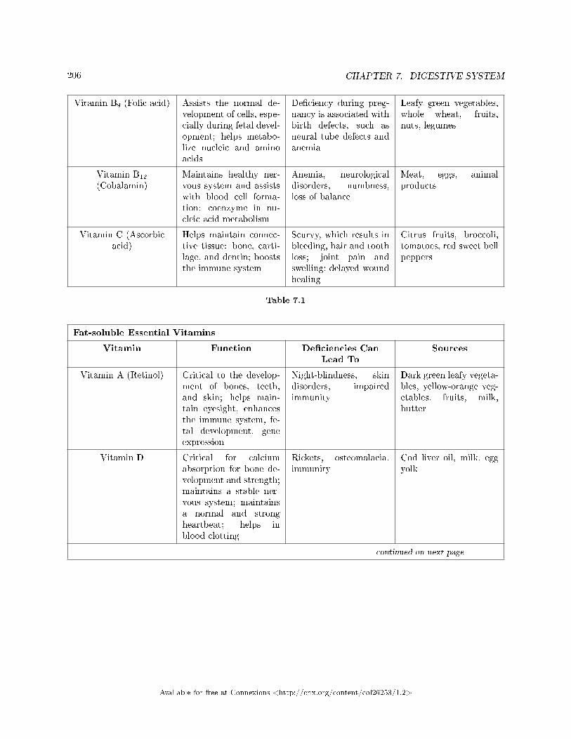
206 CHAPTER 7. DIGESTIVE SYSTEM
Vitamin B9 (Folic acid) Assists the normal de-velopment of cells, espe-cially during fetal devel-opment; helps metabo-lize nucleic and aminoacids
De�ciency during preg-nancy is associated withbirth defects, such asneural tube defects andanemia
Leafy green vegetables,whole wheat, fruits,nuts, legumes
Vitamin B12
(Cobalamin)Maintains healthy ner-vous system and assistswith blood cell forma-tion; coenzyme in nu-cleic acid metabolism
Anemia, neurologicaldisorders, numbness,loss of balance
Meat, eggs, animalproducts
Vitamin C (Ascorbicacid)
Helps maintain connec-tive tissue: bone, carti-lage, and dentin; booststhe immune system
Scurvy, which results inbleeding, hair and toothloss; joint pain andswelling; delayed woundhealing
Citrus fruits, broccoli,tomatoes, red sweet bellpeppers
Table 7.1
Fat-soluble Essential Vitamins
Vitamin Function De�ciencies CanLead To
Sources
Vitamin A (Retinol) Critical to the develop-ment of bones, teeth,and skin; helps main-tain eyesight, enhancesthe immune system, fe-tal development, geneexpression
Night-blindness, skindisorders, impairedimmunity
Dark green leafy vegeta-bles, yellow-orange veg-etables, fruits, milk,butter
Vitamin D Critical for calciumabsorption for bone de-velopment and strength;maintains a stable ner-vous system; maintainsa normal and strongheartbeat; helps inblood clotting
Rickets, osteomalacia,immunity
Cod liver oil, milk, eggyolk
continued on next page
Available for free at Connexions <http://cnx.org/content/col26253/1.2>
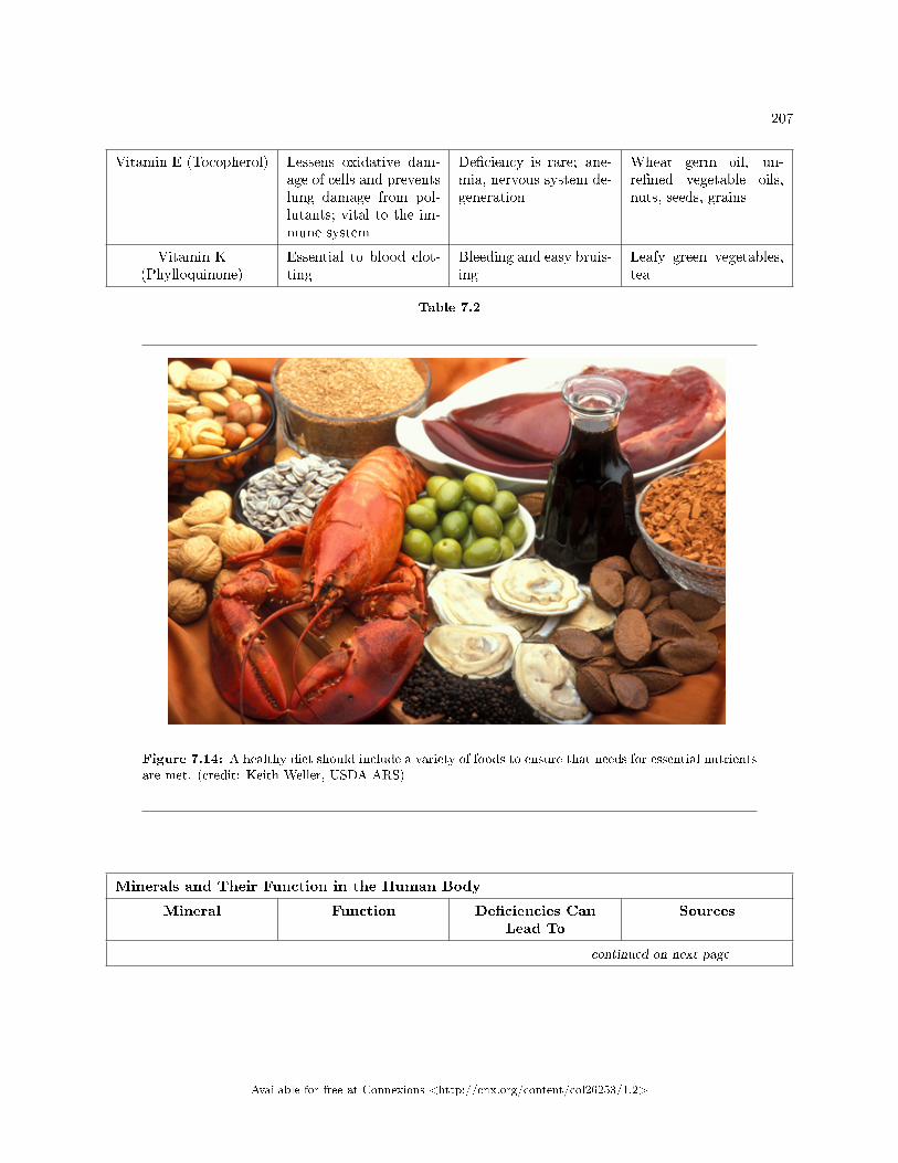
207
Vitamin E (Tocopherol) Lessens oxidative dam-age of cells and preventslung damage from pol-lutants; vital to the im-mune system
De�ciency is rare; ane-mia, nervous system de-generation
Wheat germ oil, un-re�ned vegetable oils,nuts, seeds, grains
Vitamin K(Phylloquinone)
Essential to blood clot-ting
Bleeding and easy bruis-ing
Leafy green vegetables,tea
Table 7.2
Figure 7.14: A healthy diet should include a variety of foods to ensure that needs for essential nutrientsare met. (credit: Keith Weller, USDA ARS)
Minerals and Their Function in the Human Body
Mineral Function De�ciencies CanLead To
Sources
continued on next page
Available for free at Connexions <http://cnx.org/content/col26253/1.2>
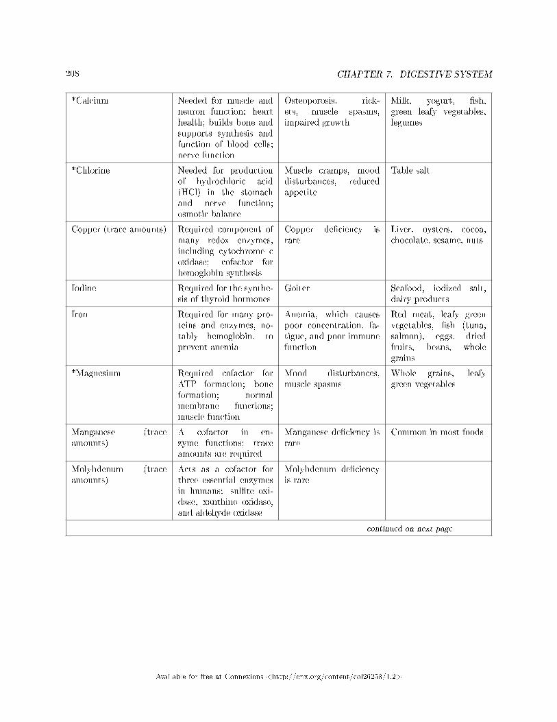
208 CHAPTER 7. DIGESTIVE SYSTEM
*Calcium Needed for muscle andneuron function; hearthealth; builds bone andsupports synthesis andfunction of blood cells;nerve function
Osteoporosis, rick-ets, muscle spasms,impaired growth
Milk, yogurt, �sh,green leafy vegetables,legumes
*Chlorine Needed for productionof hydrochloric acid(HCl) in the stomachand nerve function;osmotic balance
Muscle cramps, mooddisturbances, reducedappetite
Table salt
Copper (trace amounts) Required component ofmany redox enzymes,including cytochrome coxidase; cofactor forhemoglobin synthesis
Copper de�ciency israre
Liver, oysters, cocoa,chocolate, sesame, nuts
Iodine Required for the synthe-sis of thyroid hormones
Goiter Seafood, iodized salt,dairy products
Iron Required for many pro-teins and enzymes, no-tably hemoglobin, toprevent anemia
Anemia, which causespoor concentration, fa-tigue, and poor immunefunction
Red meat, leafy greenvegetables, �sh (tuna,salmon), eggs, driedfruits, beans, wholegrains
*Magnesium Required cofactor forATP formation; boneformation; normalmembrane functions;muscle function
Mood disturbances,muscle spasms
Whole grains, leafygreen vegetables
Manganese (traceamounts)
A cofactor in en-zyme functions; traceamounts are required
Manganese de�ciency israre
Common in most foods
Molybdenum (traceamounts)
Acts as a cofactor forthree essential enzymesin humans: sul�te oxi-dase, xanthine oxidase,and aldehyde oxidase
Molybdenum de�ciencyis rare
continued on next page
Available for free at Connexions <http://cnx.org/content/col26253/1.2>
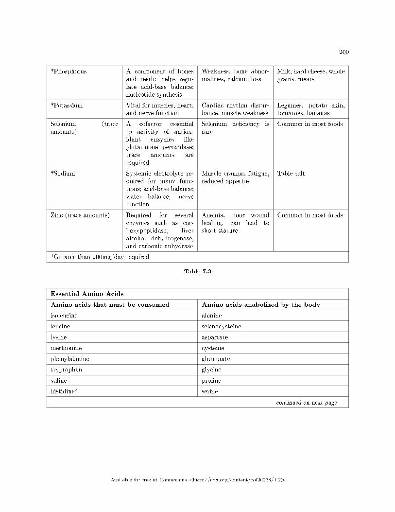
209
*Phosphorus A component of bonesand teeth; helps regu-late acid-base balance;nucleotide synthesis
Weakness, bone abnor-malities, calcium loss
Milk, hard cheese, wholegrains, meats
*Potassium Vital for muscles, heart,and nerve function
Cardiac rhythm distur-bance, muscle weakness
Legumes, potato skin,tomatoes, bananas
Selenium (traceamounts)
A cofactor essentialto activity of antiox-idant enzymes likeglutathione peroxidase;trace amounts arerequired
Selenium de�ciency israre
Common in most foods
*Sodium Systemic electrolyte re-quired for many func-tions; acid-base balance;water balance; nervefunction
Muscle cramps, fatigue,reduced appetite
Table salt
Zinc (trace amounts) Required for severalenzymes such as car-boxypeptidase, liveralcohol dehydrogenase,and carbonic anhydrase
Anemia, poor woundhealing, can lead toshort stature
Common in most foods
*Greater than 200mg/day required
Table 7.3
Essential Amino Acids
Amino acids that must be consumed Amino acids anabolized by the body
isoleucine alanine
leucine selenocysteine
lysine aspartate
methionine cysteine
phenylalanine glutamate
tryptophan glycine
valine proline
histidine* serine
continued on next page
Available for free at Connexions <http://cnx.org/content/col26253/1.2>
210 CHAPTER 7. DIGESTIVE SYSTEM
threonine tyrosine
arginine* asparagine
*The human body can synthesize histidine and arginine, but not in the quantities required, especially forgrowing children.
Table 7.4
7.2.2 Food Energy and ATP
Animals need food to obtain energy and maintain homeostasis. Homeostasis is the ability of a system tomaintain a stable internal environment even in the face of external changes to the environment. For example,the normal body temperature of humans is 37 ◦C (98.6 ◦F). Humans maintain this temperature even whenthe external temperature is hot or cold. It takes energy to maintain this body temperature, and animalsobtain this energy from food.
The primary source of energy for animals is carbohydrates, mainly glucose. Glucose is called the body'sfuel. The digestible carbohydrates in an animal's diet are converted to glucose molecules through a series ofcatabolic chemical reactions.
Adenosine triphosphate, or ATP, is the primary energy currency in cells; ATP stores energy in phosphateester bonds. ATP releases energy when the phosphodiester bonds are broken and ATP is converted to ADPand a phosphate group. ATP is produced by the oxidative reactions in the cytoplasm and mitochondrion ofthe cell, where carbohydrates, proteins, and fats undergo a series of metabolic reactions collectively calledcellular respiration. For example, glycolysis is a series of reactions in which glucose is converted to pyruvicacid and some of its chemical potential energy is transferred to NADH and ATP.
ATP is required for all cellular functions. It is used to build the organic molecules that are required forcells and tissues; it provides energy for muscle contraction and for the transmission of electrical signals inthe nervous system. When the amount of ATP is available in excess of the body's requirements, the liveruses the excess ATP and excess glucose to produce molecules called glycogen. Glycogen is a polymeric formof glucose and is stored in the liver and skeletal muscle cells. When blood sugar drops, the liver releasesglucose from stores of glycogen. Skeletal muscle converts glycogen to glucose during intense exercise. Theprocess of converting glucose and excess ATP to glycogen and the storage of excess energy is an evolutionarilyimportant step in helping animals deal with mobility, food shortages, and famine.
: Obesity
Obesity is a major health concern in the United States, and there is a growing focus on reducingobesity and the diseases it may lead to, such as type-2 diabetes, cancers of the colon and breast,and cardiovascular disease. How does the food consumed contribute to obesity?
Fatty foods are calorie-dense, meaning that they have more calories per unit mass than carbo-hydrates or proteins. One gram of carbohydrates has four calories, one gram of protein has fourcalories, and one gram of fat has nine calories. Animals tend to seek lipid-rich food for their higherenergy content.
The signals of hunger (�time to eat�) and satiety (�time to stop eating�) are controlled in thehypothalamus region of the brain. Foods that are rich in fatty acids tend to promote satiety morethan foods that are rich only in carbohydrates.
Excess carbohydrate and ATP are used by the liver to synthesize glycogen. The pyruvate producedduring glycolysis is used to synthesize fatty acids. When there is more glucose in the body thanrequired, the resulting excess pyruvate is converted into molecules that eventually result in thesynthesis of fatty acids within the body. These fatty acids are stored in adipose cells�the fat cellsin the mammalian body whose primary role is to store fat for later use.
Available for free at Connexions <http://cnx.org/content/col26253/1.2>
211
It is important to note that some animals bene�t from obesity. Polar bears and seals need body fatfor insulation and to keep them from losing body heat during Arctic winters. When food is scarce,stored body fat provides energy for maintaining homeostasis. Fats prevent famine in mammals,allowing them to access energy when food is not available on a daily basis; fats are stored when alarge kill is made or lots of food is available.
7.2.3 Section Summary
Animal diet should be balanced and meet the needs of the body. Carbohydrates, proteins, and fats arethe primary components of food. Some essential nutrients are required for cellular function but cannot beproduced by the animal body. These include vitamins, minerals, some fatty acids, and some amino acids.Food intake in more than necessary amounts is stored as glycogen in the liver and muscle cells, and in fatcells. Excess adipose storage can lead to obesity and serious health problems. ATP is the energy currencyof the cell and is obtained from the metabolic pathways. Excess carbohydrates and energy are stored asglycogen in the body.
7.2.4 Review Questions
Exercise 7.2.1 (Solution on p. 221.)
Which of the following statements is not true?
a. Essential nutrients can be synthesized by the body.b. Vitamins are required in small quantities for bodily function.c. Some amino acids can be synthesized by the body, while others need to be obtained from
diet.d. Vitamins come in two categories: fat-soluble and water-soluble.
Exercise 7.2.2 (Solution on p. 221.)
Which of the following is a water-soluble vitamin?
a. vitamin Ab. vitamin Ec. vitamin Kd. vitamin C
Exercise 7.2.3 (Solution on p. 221.)
What is the primary fuel for the body?
a. carbohydratesb. lipidsc. proteind. glycogen
Exercise 7.2.4 (Solution on p. 221.)
Excess glucose is stored as ________.
a. fatb. glucagonc. glycogend. it is not stored in the body
Available for free at Connexions <http://cnx.org/content/col26253/1.2>
212 CHAPTER 7. DIGESTIVE SYSTEM
Exercise 7.2.5 (Solution on p. 221.)
Many distance runners �carb load� the day before a big race. How does this eating strategy providean advantage to the runner?
a. The carbohydrates cause the release of insulin.b. The excess carbohydrates are converted to fats, which have a higher calorie density.c. The glucose from the carbohydrates lets the muscles make excess ATP overnight.d. The excess carbohydrates can be stored in the muscles as glycogen.
7.2.5 Critical Thinking Questions
Exercise 7.2.6 (Solution on p. 221.)
What are essential nutrients?
Exercise 7.2.7 (Solution on p. 222.)
What is the role of minerals in maintaining good health?
Exercise 7.2.8 (Solution on p. 222.)
Discuss why obesity is a growing epidemic.
Exercise 7.2.9 (Solution on p. 222.)
There are several nations where malnourishment is a common occurrence. What may be some ofthe health challenges posed by malnutrition?
Exercise 7.2.10 (Solution on p. 222.)
Generally describe how a piece of bread can power your legs as you walk up a �ight of stairs.
Exercise 7.2.11 (Solution on p. 222.)
In the 1990s fat-free foods became popular among people trying to lose weight. However, manydieticians now conclude that the fat-free trend made people less healthy and heavier. Describe howthis could occur.
7.3 Digestive System Processes5
Obtaining nutrition and energy from food is a multistep process. For true animals, the �rst step is ingestion,the act of taking in food. This is followed by digestion, absorption, and elimination. In the following sections,each of these steps will be discussed in detail.
7.3.1 Ingestion
The large molecules found in intact food cannot pass through the cell membranes. Food needs to be brokeninto smaller particles so that animals can harness the nutrients and organic molecules. The �rst step inthis process is ingestion. Ingestion is the process of taking in food through the mouth. In vertebrates, theteeth, saliva, and tongue play important roles in mastication (preparing the food into bolus). While thefood is being mechanically broken down, the enzymes in saliva begin to chemically process the food as well.The combined action of these processes modi�es the food from large particles to a soft mass that can beswallowed and can travel the length of the esophagus.
5This content is available online at <http://cnx.org/content/m66617/1.15/>.
Available for free at Connexions <http://cnx.org/content/col26253/1.2>
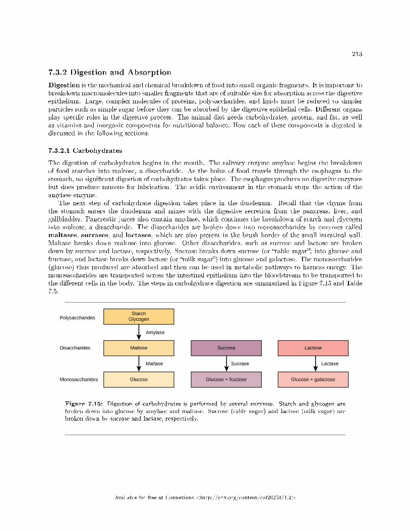
213
7.3.2 Digestion and Absorption
Digestion is the mechanical and chemical breakdown of food into small organic fragments. It is important tobreakdown macromolecules into smaller fragments that are of suitable size for absorption across the digestiveepithelium. Large, complex molecules of proteins, polysaccharides, and lipids must be reduced to simplerparticles such as simple sugar before they can be absorbed by the digestive epithelial cells. Di�erent organsplay speci�c roles in the digestive process. The animal diet needs carbohydrates, protein, and fat, as wellas vitamins and inorganic components for nutritional balance. How each of these components is digested isdiscussed in the following sections.
7.3.2.1 Carbohydrates
The digestion of carbohydrates begins in the mouth. The salivary enzyme amylase begins the breakdownof food starches into maltose, a disaccharide. As the bolus of food travels through the esophagus to thestomach, no signi�cant digestion of carbohydrates takes place. The esophagus produces no digestive enzymesbut does produce mucous for lubrication. The acidic environment in the stomach stops the action of theamylase enzyme.
The next step of carbohydrate digestion takes place in the duodenum. Recall that the chyme fromthe stomach enters the duodenum and mixes with the digestive secretion from the pancreas, liver, andgallbladder. Pancreatic juices also contain amylase, which continues the breakdown of starch and glycogeninto maltose, a disaccharide. The disaccharides are broken down into monosaccharides by enzymes calledmaltases, sucrases, and lactases, which are also present in the brush border of the small intestinal wall.Maltase breaks down maltose into glucose. Other disaccharides, such as sucrose and lactose are brokendown by sucrase and lactase, respectively. Sucrase breaks down sucrose (or �table sugar�) into glucose andfructose, and lactase breaks down lactose (or �milk sugar�) into glucose and galactose. The monosaccharides(glucose) thus produced are absorbed and then can be used in metabolic pathways to harness energy. Themonosaccharides are transported across the intestinal epithelium into the bloodstream to be transported tothe di�erent cells in the body. The steps in carbohydrate digestion are summarized in Figure 7.15 and Table7.5.
Figure 7.15: Digestion of carbohydrates is performed by several enzymes. Starch and glycogen arebroken down into glucose by amylase and maltase. Sucrose (table sugar) and lactose (milk sugar) arebroken down by sucrase and lactase, respectively.
Available for free at Connexions <http://cnx.org/content/col26253/1.2>
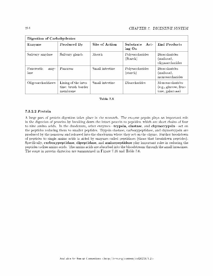
214 CHAPTER 7. DIGESTIVE SYSTEM
Digestion of Carbohydrates
Enzyme Produced By Site of Action Substrate Act-ing On
End Products
Salivary amylase Salivary glands Mouth Polysaccharides(Starch)
Disaccharides(maltose),oligosaccharides
Pancreatic amy-lase
Pancreas Small intestine Polysaccharides(starch)
Disaccharides(maltose),monosaccharides
Oligosaccharidases Lining of the intes-tine; brush bordermembrane
Small intestine Disaccharides Monosaccharides(e.g., glucose, fruc-tose, galactose)
Table 7.5
7.3.2.2 Protein
A large part of protein digestion takes place in the stomach. The enzyme pepsin plays an important rolein the digestion of proteins by breaking down the intact protein to peptides, which are short chains of fourto nine amino acids. In the duodenum, other enzymes�trypsin, elastase, and chymotrypsin�act onthe peptides reducing them to smaller peptides. Trypsin elastase, carboxypeptidase, and chymotrypsin areproduced by the pancreas and released into the duodenum where they act on the chyme. Further breakdownof peptides to single amino acids is aided by enzymes called peptidases (those that breakdown peptides).Speci�cally, carboxypeptidase, dipeptidase, and aminopeptidase play important roles in reducing thepeptides to free amino acids. The amino acids are absorbed into the bloodstream through the small intestines.The steps in protein digestion are summarized in Figure 7.16 and Table 7.6.
Available for free at Connexions <http://cnx.org/content/col26253/1.2>
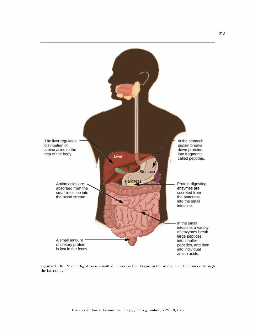
215
Figure 7.16: Protein digestion is a multistep process that begins in the stomach and continues throughthe intestines.
Available for free at Connexions <http://cnx.org/content/col26253/1.2>
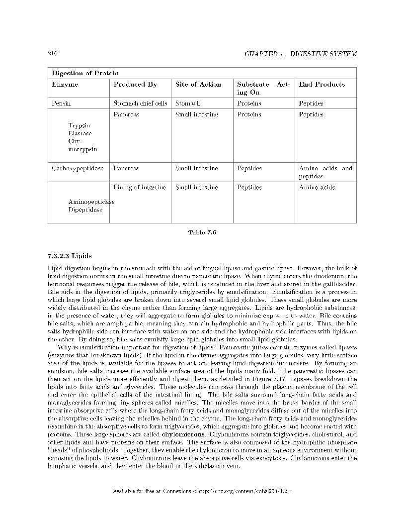
216 CHAPTER 7. DIGESTIVE SYSTEM
Digestion of Protein
Enzyme Produced By Site of Action Substrate Act-ing On
End Products
Pepsin Stomach chief cells Stomach Proteins Peptides
TrypsinElastaseChy-motrypsin
Pancreas Small intestine Proteins Peptides
Carboxypeptidase Pancreas Small intestine Peptides Amino acids andpeptides
AminopeptidaseDipeptidase
Lining of intestine Small intestine Peptides Amino acids
Table 7.6
7.3.2.3 Lipids
Lipid digestion begins in the stomach with the aid of lingual lipase and gastric lipase. However, the bulk oflipid digestion occurs in the small intestine due to pancreatic lipase. When chyme enters the duodenum, thehormonal responses trigger the release of bile, which is produced in the liver and stored in the gallbladder.Bile aids in the digestion of lipids, primarily triglycerides by emulsi�cation. Emulsi�cation is a process inwhich large lipid globules are broken down into several small lipid globules. These small globules are morewidely distributed in the chyme rather than forming large aggregates. Lipids are hydrophobic substances:in the presence of water, they will aggregate to form globules to minimize exposure to water. Bile containsbile salts, which are amphipathic, meaning they contain hydrophobic and hydrophilic parts. Thus, the bilesalts hydrophilic side can interface with water on one side and the hydrophobic side interfaces with lipids onthe other. By doing so, bile salts emulsify large lipid globules into small lipid globules.
Why is emulsi�cation important for digestion of lipids? Pancreatic juices contain enzymes called lipases(enzymes that breakdown lipids). If the lipid in the chyme aggregates into large globules, very little surfacearea of the lipids is available for the lipases to act on, leaving lipid digestion incomplete. By forming anemulsion, bile salts increase the available surface area of the lipids many fold. The pancreatic lipases canthen act on the lipids more e�ciently and digest them, as detailed in Figure 7.17. Lipases breakdown thelipids into fatty acids and glycerides. These molecules can pass through the plasma membrane of the celland enter the epithelial cells of the intestinal lining. The bile salts surround long-chain fatty acids andmonoglycerides forming tiny spheres called micelles. The micelles move into the brush border of the smallintestine absorptive cells where the long-chain fatty acids and monoglycerides di�use out of the micelles intothe absorptive cells leaving the micelles behind in the chyme. The long-chain fatty acids and monoglyceridesrecombine in the absorptive cells to form triglycerides, which aggregate into globules and become coated withproteins. These large spheres are called chylomicrons. Chylomicrons contain triglycerides, cholesterol, andother lipids and have proteins on their surface. The surface is also composed of the hydrophilic phosphate"heads" of phospholipids. Together, they enable the chylomicron to move in an aqueous environment withoutexposing the lipids to water. Chylomicrons leave the absorptive cells via exocytosis. Chylomicrons enter thelymphatic vessels, and then enter the blood in the subclavian vein.
Available for free at Connexions <http://cnx.org/content/col26253/1.2>
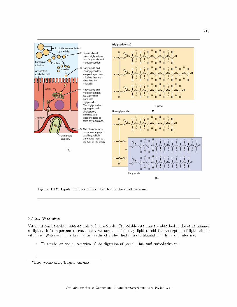
217
Figure 7.17: Lipids are digested and absorbed in the small intestine.
7.3.2.4 Vitamins
Vitamins can be either water-soluble or lipid-soluble. Fat soluble vitamins are absorbed in the same manneras lipids. It is important to consume some amount of dietary lipid to aid the absorption of lipid-solublevitamins. Water-soluble vitamins can be directly absorbed into the bloodstream from the intestine.
: This website6 has an overview of the digestion of protein, fat, and carbohydrates.
:
6http://openstax.org/l/digest_enzymes
Available for free at Connexions <http://cnx.org/content/col26253/1.2>
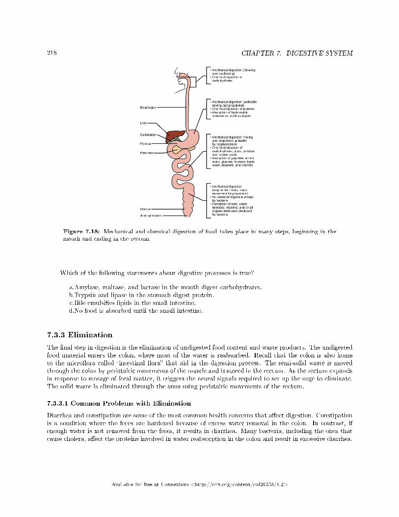
218 CHAPTER 7. DIGESTIVE SYSTEM
Figure 7.18: Mechanical and chemical digestion of food takes place in many steps, beginning in themouth and ending in the rectum.
Which of the following statements about digestive processes is true?
a.Amylase, maltase, and lactase in the mouth digest carbohydrates.b.Trypsin and lipase in the stomach digest protein.c.Bile emulsi�es lipids in the small intestine.d.No food is absorbed until the small intestine.
7.3.3 Elimination
The �nal step in digestion is the elimination of undigested food content and waste products. The undigestedfood material enters the colon, where most of the water is reabsorbed. Recall that the colon is also hometo the micro�ora called �intestinal �ora� that aid in the digestion process. The semi-solid waste is movedthrough the colon by peristaltic movements of the muscle and is stored in the rectum. As the rectum expandsin response to storage of fecal matter, it triggers the neural signals required to set up the urge to eliminate.The solid waste is eliminated through the anus using peristaltic movements of the rectum.
7.3.3.1 Common Problems with Elimination
Diarrhea and constipation are some of the most common health concerns that a�ect digestion. Constipationis a condition where the feces are hardened because of excess water removal in the colon. In contrast, ifenough water is not removed from the feces, it results in diarrhea. Many bacteria, including the ones thatcause cholera, a�ect the proteins involved in water reabsorption in the colon and result in excessive diarrhea.
Available for free at Connexions <http://cnx.org/content/col26253/1.2>
219
7.3.3.2 Emesis
Emesis, or vomiting, is elimination of food by forceful expulsion through the mouth. It is often in responseto an irritant that a�ects the digestive tract, including but not limited to viruses, bacteria, emotions, sights,and food poisoning. This forceful expulsion of the food is due to the strong contractions produced by thestomach muscles. The process of emesis is regulated by the medulla.
7.3.4 Section Summary
Digestion begins with ingestion, where the food is taken in the mouth. Digestion and absorption take placein a series of steps with special enzymes playing important roles in digesting carbohydrates, proteins, andlipids. Elimination describes removal of undigested food contents and waste products from the body. Whilemost absorption occurs in the small intestines, the large intestine is responsible for the �nal removal ofwater that remains after the absorptive process of the small intestines. The cells that line the large intestineabsorb some vitamins as well as any leftover salts and water. The large intestine (colon) is also where fecesis formed.
7.3.5 Visual Connection Questions
Exercise 7.3.1 (Solution on p. 222.)
Figure 7.18 Which of the following statements about digestive processes is true?
a. Amylase, maltase, and lactase in the mouth digest carbohydrates.b. Trypsin and lipase in the stomach digest protein.c. Bile emulsi�es lipids in the small intestine.d. No food is absorbed until the small intestine.
7.3.6 Review Questions
Exercise 7.3.2 (Solution on p. 222.)
Where does the majority of protein digestion take place?
a. stomachb. duodenumc. mouthd. jejunum
Exercise 7.3.3 (Solution on p. 222.)
Lipases are enzymes that breakdown ________.
a. disaccharidesb. lipidsc. proteinsd. cellulose
Exercise 7.3.4 (Solution on p. 222.)
Which of the following conditions is most likely to cause constipation?
a. bacterial infectionb. dehydrationc. ulcerd. excessive cellulose consumption
Available for free at Connexions <http://cnx.org/content/col26253/1.2>
220 CHAPTER 7. DIGESTIVE SYSTEM
7.3.7 Critical Thinking Questions
Exercise 7.3.5 (Solution on p. 222.)
Explain why some dietary lipid is a necessary part of a balanced diet.
Exercise 7.3.6 (Solution on p. 222.)
The gut microbiome (the bacterial colonies in the intestines) have become a popular area of studyin biomedical research. How could varying gut microbiomes impact a person's nutrition?
Exercise 7.3.7 (Solution on p. 222.)
Many mammals become ill if they drink milk as adults even though they could consume it asbabies. What causes this digestive issue?
Available for free at Connexions <http://cnx.org/content/col26253/1.2>
221
Solutions to Exercises in Chapter 7
to Exercise 7.1.1 (p. 200)Figure 7.10 Bto Exercise 7.1.2 (p. 200)Figure 7.11 Cto Exercise 7.1.3 (p. 201)Dto Exercise 7.1.4 (p. 201)Bto Exercise 7.1.5 (p. 201)Cto Exercise 7.1.6 (p. 201)Cto Exercise 7.1.7 (p. 201)Bto Exercise 7.1.8 (p. 201)Animals with a polygastric digestive system have a multi-chambered stomach. The four compartments of thestomach are called the rumen, reticulum, omasum, and abomasum. These chambers contain many microbesthat breakdown the cellulose and ferment the ingested food. The abomasum is the �true� stomach and isthe equivalent of a monogastric stomach chamber where gastric juices are secreted. The four-compartmentgastric chamber provides larger space and the microbial support necessary for ruminants to digest plantmaterial.to Exercise 7.1.9 (p. 201)Birds have a stomach chamber called a gizzard. Here, the food is stored, soaked, and ground into �ner par-ticles, often using pebbles. Once this process is complete, the digestive juices take over in the proventriculusand continue the digestive process.to Exercise 7.1.10 (p. 201)Accessory organs play an important role in producing and delivering digestive juices to the intestine duringdigestion and absorption. Speci�cally, the salivary glands, liver, pancreas, and gallbladder play importantroles. Malfunction of any of these organs can lead to disease states.to Exercise 7.1.11 (p. 202)The villi and microvilli are folds on the surface of the small intestine. These folds increase the surface areaof the intestine and provide more area for the absorption of nutrients.to Exercise 7.1.12 (p. 202)The stomach and the teeth both perform mechanical digestion, which is physically (as opposed to chemically)breaking the food into smaller components. This exposes a larger surface area for chemical digestion andrelease of nutrients. The teeth are vital to mastication, which breaks large bites of food down into smallerpieces that are easily swallowed. The stomach's muscle contractions churn the food to expose all particlesto the acid and digestive enzymes.to Exercise 7.2.1 (p. 211)Ato Exercise 7.2.2 (p. 211)Dto Exercise 7.2.3 (p. 211)Ato Exercise 7.2.4 (p. 211)Cto Exercise 7.2.5 (p. 212)D
Available for free at Connexions <http://cnx.org/content/col26253/1.2>
222 CHAPTER 7. DIGESTIVE SYSTEM
to Exercise 7.2.6 (p. 212)Essential nutrients are those nutrients that must be obtained from the diet because they cannot be producedby the body. Vitamins and minerals are examples of essential nutrients.to Exercise 7.2.7 (p. 212)Minerals�such as potassium, sodium, and calcium�are required for the functioning of many cellularprocesses, including muscle contraction and nerve conduction. While minerals are required in trace amounts,not having minerals in the diet can be potentially harmful.to Exercise 7.2.8 (p. 212)In the United States, obesity, particularly childhood obesity, is a growing concern. Some of the contrib-utors to this situation include sedentary lifestyles and consuming more processed foods and less fruits andvegetables. As a result, even young children who are obese can face health concerns.to Exercise 7.2.9 (p. 212)Malnutrition, often in the form of not getting enough calories or not enough of the essential nutrients, canhave severe consequences. Many malnourished children have vision and dental problems, and over the yearsmay develop many serious health problems.to Exercise 7.2.10 (p. 212)A piece of bread is eaten and converted into chemical energy. The bread is broken down in the mouth bymastication and salivary enzymes, then transferred to the stomach for further digestion. After digestion bythe acid and digestive enzymes in the stomach, the macromolecules that made up the bread move into thesmall intestine. In the small intestine, the carbohydrates from the bread are absorbed through the microvilliinto the bloodstream. In muscle cells in the legs, the carbohydrates can be broken down into glucose, andthen used for cellular respiration to create ATP. The muscles in the leg then use the ATP to perform themechanical work needed to climb a �ight of stairs.to Exercise 7.2.11 (p. 212)Fats are an essential component of a healthy diet, and needed by the body to function. Fats are essentialfor many processes, including the absorption of fat-soluble vitamins and production of some hormones. Fatsalso send a satiation signal to the brain that regulates hunger. Without fats in their diets many people mayhave actually consumed more calories, which would have resulted in weight gain.to Exercise 7.3.1 (p. 219)Figure 7.18 Cto Exercise 7.3.2 (p. 219)Ato Exercise 7.3.3 (p. 219)Bto Exercise 7.3.4 (p. 219)Bto Exercise 7.3.5 (p. 220)Lipids add �avor to food and promote a sense of satiety or fullness. Fatty foods are sources of highenergy; one gram of lipid contains nine calories. Lipids are also required in the diet to aid the absorption oflipid-soluble vitamins and for the production of lipid-soluble hormones.to Exercise 7.3.6 (p. 220)The gut microbiome includes all the bacteria that aid in chemical digestion in the intestines. Changing itscomposition can change the way that food is digested since not all bacteria have the same macromolecule-digesting enzymes. Additionally, changes in gut microbiome can lead to the establishment of pathogenicbacteria populations that cause in�ammation in the gut or other disease.to Exercise 7.3.7 (p. 220)As mammals wean from their mothers they stop drinking milk. Since they stop consuming the sugarlactose their bodies conserve resources by no longer making the enzyme lactase. If the animals then consumelactose at some point in the future their digestive system cannot break the lactose molecules into glucoseand galactose for absorption. When gut bacteria further along the digestive tract interact with the lactosemolecules it causes symptoms of lactose intolerance.
Available for free at Connexions <http://cnx.org/content/col26253/1.2>
Chapter 8
Circulatory System
8.1 Overview of the Circulatory System1
In all animals, except a few simple types, the circulatory system is used to transport nutrients and gasesthrough the body. Simple di�usion allows some water, nutrient, waste, and gas exchange into primitiveanimals that are only a few cell layers thick; however, bulk �ow is the only method by which the entire bodyof larger more complex organisms is accessed.
8.1.1 Circulatory System Architecture
The circulatory system is e�ectively a network of cylindrical vessels: the arteries, veins, and capillariesthat emanate from a pump, the heart. In all vertebrate organisms, as well as some invertebrates, thisis a closed-loop system, in which the blood is not free in a cavity. In a closed circulatory system,blood is contained inside blood vessels and circulates unidirectionally from the heart around the systemiccirculatory route, then returns to the heart again, as illustrated in Figure 8.1a. As opposed to a closedsystem, arthropods�including insects, crustaceans, and most mollusks�have an open circulatory system,as illustrated in Figure 8.1b. In an open circulatory system, the blood is not enclosed in the blood vesselsbut is pumped into a cavity called a hemocoel and is called hemolymph because the blood mixes withthe interstitial �uid. As the heart beats and the animal moves, the hemolymph circulates around theorgans within the body cavity and then reenters the hearts through openings called ostia. This movementallows for gas and nutrient exchange. An open circulatory system does not use as much energy as a closedsystem to operate or to maintain; however, there is a trade-o� with the amount of blood that can be moved tometabolically active organs and tissues that require high levels of oxygen. In fact, one reason that insects withwing spans of up to two feet wide (70 cm) are not around today is probably because they were outcompetedby the arrival of birds 150 million years ago. Birds, having a closed circulatory system, are thought to havemoved more agilely, allowing them to get food faster and possibly to prey on the insects.
1This content is available online at <http://cnx.org/content/m44801/1.5/>.
Available for free at Connexions <http://cnx.org/content/col26253/1.2>
223
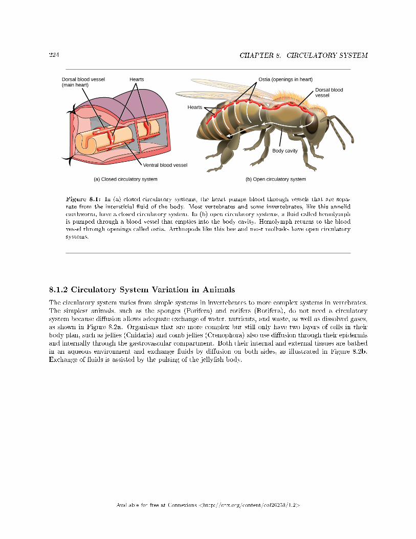
224 CHAPTER 8. CIRCULATORY SYSTEM
Figure 8.1: In (a) closed circulatory systems, the heart pumps blood through vessels that are sepa-rate from the interstitial �uid of the body. Most vertebrates and some invertebrates, like this annelidearthworm, have a closed circulatory system. In (b) open circulatory systems, a �uid called hemolymphis pumped through a blood vessel that empties into the body cavity. Hemolymph returns to the bloodvessel through openings called ostia. Arthropods like this bee and most mollusks have open circulatorysystems.
8.1.2 Circulatory System Variation in Animals
The circulatory system varies from simple systems in invertebrates to more complex systems in vertebrates.The simplest animals, such as the sponges (Porifera) and rotifers (Rotifera), do not need a circulatorysystem because di�usion allows adequate exchange of water, nutrients, and waste, as well as dissolved gases,as shown in Figure 8.2a. Organisms that are more complex but still only have two layers of cells in theirbody plan, such as jellies (Cnidaria) and comb jellies (Ctenophora) also use di�usion through their epidermisand internally through the gastrovascular compartment. Both their internal and external tissues are bathedin an aqueous environment and exchange �uids by di�usion on both sides, as illustrated in Figure 8.2b.Exchange of �uids is assisted by the pulsing of the jelly�sh body.
Available for free at Connexions <http://cnx.org/content/col26253/1.2>
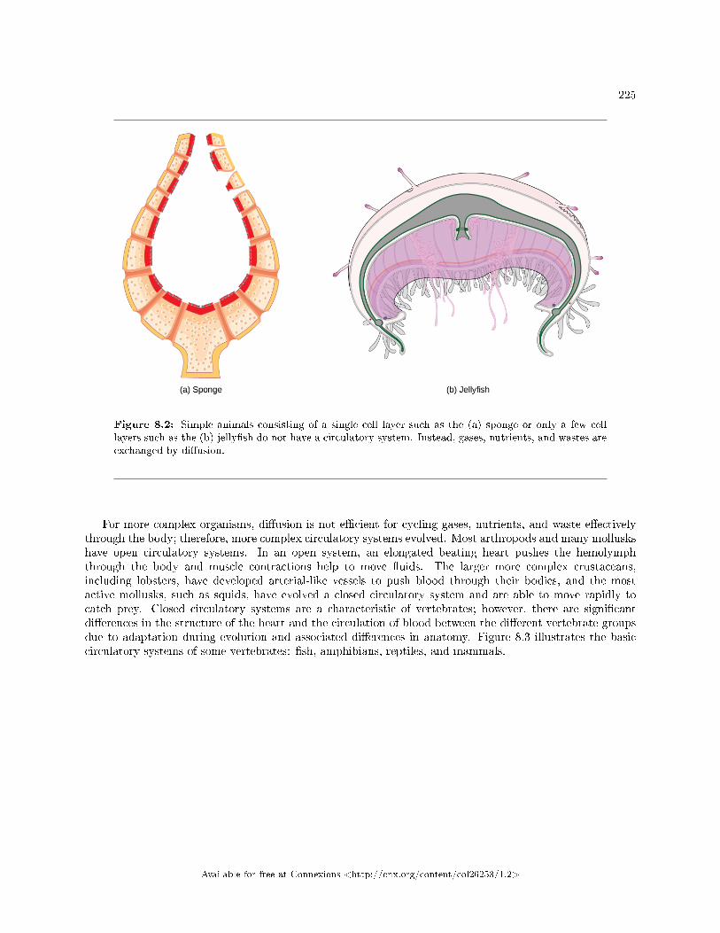
225
Figure 8.2: Simple animals consisting of a single cell layer such as the (a) sponge or only a few celllayers such as the (b) jelly�sh do not have a circulatory system. Instead, gases, nutrients, and wastes areexchanged by di�usion.
For more complex organisms, di�usion is not e�cient for cycling gases, nutrients, and waste e�ectivelythrough the body; therefore, more complex circulatory systems evolved. Most arthropods and many molluskshave open circulatory systems. In an open system, an elongated beating heart pushes the hemolymphthrough the body and muscle contractions help to move �uids. The larger more complex crustaceans,including lobsters, have developed arterial-like vessels to push blood through their bodies, and the mostactive mollusks, such as squids, have evolved a closed circulatory system and are able to move rapidly tocatch prey. Closed circulatory systems are a characteristic of vertebrates; however, there are signi�cantdi�erences in the structure of the heart and the circulation of blood between the di�erent vertebrate groupsdue to adaptation during evolution and associated di�erences in anatomy. Figure 8.3 illustrates the basiccirculatory systems of some vertebrates: �sh, amphibians, reptiles, and mammals.
Available for free at Connexions <http://cnx.org/content/col26253/1.2>
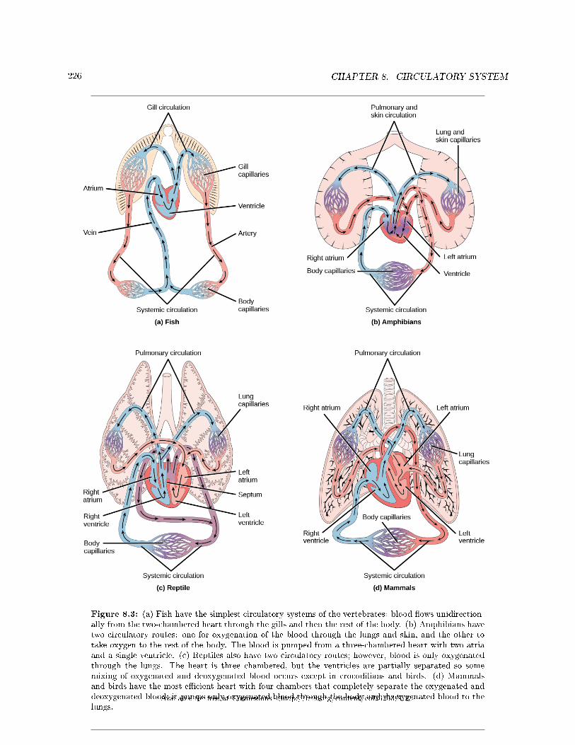
226 CHAPTER 8. CIRCULATORY SYSTEM
Figure 8.3: (a) Fish have the simplest circulatory systems of the vertebrates: blood �ows unidirection-ally from the two-chambered heart through the gills and then the rest of the body. (b) Amphibians havetwo circulatory routes: one for oxygenation of the blood through the lungs and skin, and the other totake oxygen to the rest of the body. The blood is pumped from a three-chambered heart with two atriaand a single ventricle. (c) Reptiles also have two circulatory routes; however, blood is only oxygenatedthrough the lungs. The heart is three chambered, but the ventricles are partially separated so somemixing of oxygenated and deoxygenated blood occurs except in crocodilians and birds. (d) Mammalsand birds have the most e�cient heart with four chambers that completely separate the oxygenated anddeoxygenated blood; it pumps only oxygenated blood through the body and deoxygenated blood to thelungs.
Available for free at Connexions <http://cnx.org/content/col26253/1.2>
227
As illustrated in Figure 8.3a Fish have a single circuit for blood �ow and a two-chambered heart thathas only a single atrium and a single ventricle. The atrium collects blood that has returned from the bodyand the ventricle pumps the blood to the gills where gas exchange occurs and the blood is re-oxygenated;this is called gill circulation. The blood then continues through the rest of the body before arriving backat the atrium; this is called systemic circulation. This unidirectional �ow of blood produces a gradient ofoxygenated to deoxygenated blood around the �sh's systemic circuit. The result is a limit in the amount ofoxygen that can reach some of the organs and tissues of the body, reducing the overall metabolic capacityof �sh.
In amphibians, reptiles, birds, and mammals, blood �ow is directed in two circuits: one through thelungs and back to the heart, which is called pulmonary circulation, and the other throughout the rest ofthe body and its organs including the brain (systemic circulation). In amphibians, gas exchange also occursthrough the skin during pulmonary circulation and is referred to as pulmocutaneous circulation.
As shown in Figure 8.3b, amphibians have a three-chambered heart that has two atria and one ventriclerather than the two-chambered heart of �sh. The two atria (superior heart chambers) receive blood fromthe two di�erent circuits (the lungs and the systems), and then there is some mixing of the blood in theheart's ventricle (inferior heart chamber), which reduces the e�ciency of oxygenation. The advantage to thisarrangement is that high pressure in the vessels pushes blood to the lungs and body. The mixing is mitigatedby a ridge within the ventricle that diverts oxygen-rich blood through the systemic circulatory system anddeoxygenated blood to the pulmocutaneous circuit. For this reason, amphibians are often described as havingdouble circulation.
Most reptiles also have a three-chambered heart similar to the amphibian heart that directs blood to thepulmonary and systemic circuits, as shown in Figure 8.3c. The ventricle is divided more e�ectively by apartial septum, which results in less mixing of oxygenated and deoxygenated blood. Some reptiles (alligatorsand crocodiles) are the most primitive animals to exhibit a four-chambered heart. Crocodilians have a uniquecirculatory mechanism where the heart shunts blood from the lungs toward the stomach and other organsduring long periods of submergence, for instance, while the animal waits for prey or stays underwater waitingfor prey to rot. One adaptation includes two main arteries that leave the same part of the heart: one takesblood to the lungs and the other provides an alternate route to the stomach and other parts of the body.Two other adaptations include a hole in the heart between the two ventricles, called the foramen of Panizza,which allows blood to move from one side of the heart to the other, and specialized connective tissue thatslows the blood �ow to the lungs. Together these adaptations have made crocodiles and alligators one of themost evolutionarily successful animal groups on earth.
In mammals and birds, the heart is also divided into four chambers: two atria and two ventricles, asillustrated in Figure 8.3d. The oxygenated blood is separated from the deoxygenated blood, which improvesthe e�ciency of double circulation and is probably required for the warm-blooded lifestyle of mammals andbirds. The four-chambered heart of birds and mammals evolved independently from a three-chambered heart.The independent evolution of the same or a similar biological trait is referred to as convergent evolution.
8.1.3 Section Summary
In most animals, the circulatory system is used to transport blood through the body. Some primitiveanimals use di�usion for the exchange of water, nutrients, and gases. However, complex organisms usethe circulatory system to carry gases, nutrients, and waste through the body. Circulatory systems may beopen (mixed with the interstitial �uid) or closed (separated from the interstitial �uid). Closed circulatorysystems are a characteristic of vertebrates; however, there are signi�cant di�erences in the structure of theheart and the circulation of blood between the di�erent vertebrate groups due to adaptions during evolutionand associated di�erences in anatomy. Fish have a two-chambered heart with unidirectional circulation.Amphibians have a three-chambered heart, which has some mixing of the blood, and they have doublecirculation. Most non-avian reptiles have a three-chambered heart, but have little mixing of the blood; theyhave double circulation. Mammals and birds have a four-chambered heart with no mixing of the blood anddouble circulation.
Available for free at Connexions <http://cnx.org/content/col26253/1.2>
228 CHAPTER 8. CIRCULATORY SYSTEM
8.1.4 Review Questions
Exercise 8.1.1 (Solution on p. 251.)
Why are open circulatory systems advantageous to some animals?
a. They use less metabolic energy.b. They help the animal move faster.c. They do not need a heart.d. They help large insects develop.
Exercise 8.1.2 (Solution on p. 251.)
Some animals use di�usion instead of a circulatory system. Examples include:
a. birds and jelly�shb. �atworms and arthropodsc. mollusks and jelly�shd. None of the above
Exercise 8.1.3 (Solution on p. 251.)
Blood �ow that is directed through the lungs and back to the heart is called ________.
a. unidirectional circulationb. gill circulationc. pulmonary circulationd. pulmocutaneous circulation
8.1.5 Free Response
Exercise 8.1.4 (Solution on p. 251.)
Describe a closed circulatory system.
Exercise 8.1.5 (Solution on p. 251.)
Describe systemic circulation.
8.2 Components of the Blood2
Hemoglobin is responsible for distributing oxygen, and to a lesser extent, carbon dioxide, throughout thecirculatory systems of humans, vertebrates, and many invertebrates. The blood is more than the proteins,though. Blood is actually a term used to describe the liquid that moves through the vessels and includesplasma (the liquid portion, which contains water, proteins, salts, lipids, and glucose) and the cells (red andwhite cells) and cell fragments called platelets. Blood plasma is actually the dominant component of bloodand contains the water, proteins, electrolytes, lipids, and glucose. The cells are responsible for carryingthe gases (red cells) and immune the response (white). The platelets are responsible for blood clotting.Interstitial �uid that surrounds cells is separate from the blood, but in hemolymph, they are combined.In humans, cellular components make up approximately 45 percent of the blood and the liquid plasma 55percent. Blood is 20 percent of a person's extracellular �uid and eight percent of weight.
2This content is available online at <http://cnx.org/content/m44804/1.5/>.
Available for free at Connexions <http://cnx.org/content/col26253/1.2>
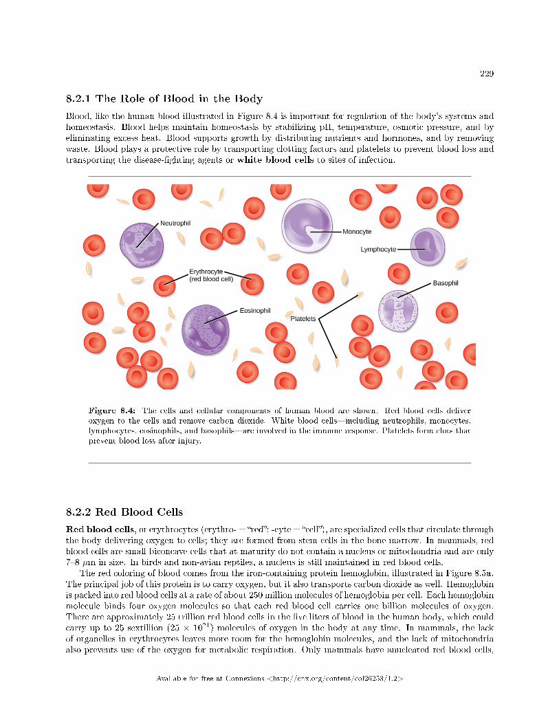
229
8.2.1 The Role of Blood in the Body
Blood, like the human blood illustrated in Figure 8.4 is important for regulation of the body's systems andhomeostasis. Blood helps maintain homeostasis by stabilizing pH, temperature, osmotic pressure, and byeliminating excess heat. Blood supports growth by distributing nutrients and hormones, and by removingwaste. Blood plays a protective role by transporting clotting factors and platelets to prevent blood loss andtransporting the disease-�ghting agents or white blood cells to sites of infection.
Figure 8.4: The cells and cellular components of human blood are shown. Red blood cells deliveroxygen to the cells and remove carbon dioxide. White blood cells�including neutrophils, monocytes,lymphocytes, eosinophils, and basophils�are involved in the immune response. Platelets form clots thatprevent blood loss after injury.
8.2.2 Red Blood Cells
Red blood cells, or erythrocytes (erythro- = �red�; -cyte = �cell�), are specialized cells that circulate throughthe body delivering oxygen to cells; they are formed from stem cells in the bone marrow. In mammals, redblood cells are small biconcave cells that at maturity do not contain a nucleus or mitochondria and are only7�8 µm in size. In birds and non-avian reptiles, a nucleus is still maintained in red blood cells.
The red coloring of blood comes from the iron-containing protein hemoglobin, illustrated in Figure 8.5a.The principal job of this protein is to carry oxygen, but it also transports carbon dioxide as well. Hemoglobinis packed into red blood cells at a rate of about 250 million molecules of hemoglobin per cell. Each hemoglobinmolecule binds four oxygen molecules so that each red blood cell carries one billion molecules of oxygen.There are approximately 25 trillion red blood cells in the �ve liters of blood in the human body, which couldcarry up to 25 sextillion (25 × 1021) molecules of oxygen in the body at any time. In mammals, the lackof organelles in erythrocytes leaves more room for the hemoglobin molecules, and the lack of mitochondriaalso prevents use of the oxygen for metabolic respiration. Only mammals have anucleated red blood cells,
Available for free at Connexions <http://cnx.org/content/col26253/1.2>
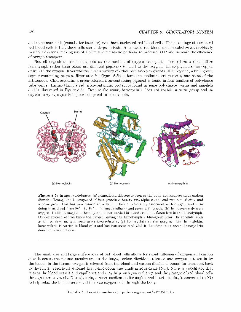
230 CHAPTER 8. CIRCULATORY SYSTEM
and some mammals (camels, for instance) even have nucleated red blood cells. The advantage of nucleatedred blood cells is that these cells can undergo mitosis. Anucleated red blood cells metabolize anaerobically(without oxygen), making use of a primitive metabolic pathway to produce ATP and increase the e�ciencyof oxygen transport.
Not all organisms use hemoglobin as the method of oxygen transport. Invertebrates that utilizehemolymph rather than blood use di�erent pigments to bind to the oxygen. These pigments use copperor iron to the oxygen. Invertebrates have a variety of other respiratory pigments. Hemocyanin, a blue-green,copper-containing protein, illustrated in Figure 8.5b is found in mollusks, crustaceans, and some of thearthropods. Chlorocruorin, a green-colored, iron-containing pigment is found in four families of polychaetetubeworms. Hemerythrin, a red, iron-containing protein is found in some polychaete worms and annelidsand is illustrated in Figure 8.5c. Despite the name, hemerythrin does not contain a heme group and itsoxygen-carrying capacity is poor compared to hemoglobin.
Figure 8.5: In most vertebrates, (a) hemoglobin delivers oxygen to the body and removes some carbondioxide. Hemoglobin is composed of four protein subunits, two alpha chains and two beta chains, anda heme group that has iron associated with it. The iron reversibly associates with oxygen, and in sodoing is oxidized from Fe2+ to Fe3+. In most mollusks and some arthropods, (b) hemocyanin deliversoxygen. Unlike hemoglobin, hemolymph is not carried in blood cells, but �oats free in the hemolymph.Copper instead of iron binds the oxygen, giving the hemolymph a blue-green color. In annelids, suchas the earthworm, and some other invertebrates, (c) hemerythrin carries oxygen. Like hemoglobin,hemerythrin is carried in blood cells and has iron associated with it, but despite its name, hemerythrindoes not contain heme.
The small size and large surface area of red blood cells allows for rapid di�usion of oxygen and carbondioxide across the plasma membrane. In the lungs, carbon dioxide is released and oxygen is taken in bythe blood. In the tissues, oxygen is released from the blood and carbon dioxide is bound for transport backto the lungs. Studies have found that hemoglobin also binds nitrous oxide (NO). NO is a vasodilator thatrelaxes the blood vessels and capillaries and may help with gas exchange and the passage of red blood cellsthrough narrow vessels. Nitroglycerin, a heart medication for angina and heart attacks, is converted to NOto help relax the blood vessels and increase oxygen �ow through the body.
Available for free at Connexions <http://cnx.org/content/col26253/1.2>
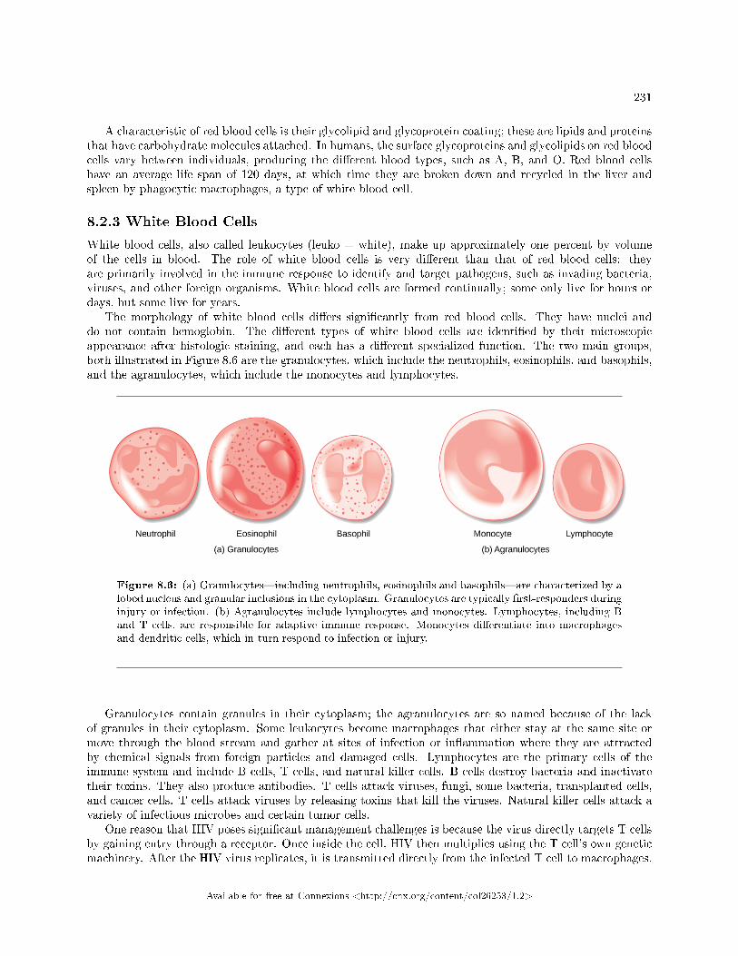
231
A characteristic of red blood cells is their glycolipid and glycoprotein coating; these are lipids and proteinsthat have carbohydrate molecules attached. In humans, the surface glycoproteins and glycolipids on red bloodcells vary between individuals, producing the di�erent blood types, such as A, B, and O. Red blood cellshave an average life span of 120 days, at which time they are broken down and recycled in the liver andspleen by phagocytic macrophages, a type of white blood cell.
8.2.3 White Blood Cells
White blood cells, also called leukocytes (leuko = white), make up approximately one percent by volumeof the cells in blood. The role of white blood cells is very di�erent than that of red blood cells: theyare primarily involved in the immune response to identify and target pathogens, such as invading bacteria,viruses, and other foreign organisms. White blood cells are formed continually; some only live for hours ordays, but some live for years.
The morphology of white blood cells di�ers signi�cantly from red blood cells. They have nuclei anddo not contain hemoglobin. The di�erent types of white blood cells are identi�ed by their microscopicappearance after histologic staining, and each has a di�erent specialized function. The two main groups,both illustrated in Figure 8.6 are the granulocytes, which include the neutrophils, eosinophils, and basophils,and the agranulocytes, which include the monocytes and lymphocytes.
Figure 8.6: (a) Granulocytes�including neutrophils, eosinophils and basophils�are characterized by alobed nucleus and granular inclusions in the cytoplasm. Granulocytes are typically �rst-responders duringinjury or infection. (b) Agranulocytes include lymphocytes and monocytes. Lymphocytes, including Band T cells, are responsible for adaptive immune response. Monocytes di�erentiate into macrophagesand dendritic cells, which in turn respond to infection or injury.
Granulocytes contain granules in their cytoplasm; the agranulocytes are so named because of the lackof granules in their cytoplasm. Some leukocytes become macrophages that either stay at the same site ormove through the blood stream and gather at sites of infection or in�ammation where they are attractedby chemical signals from foreign particles and damaged cells. Lymphocytes are the primary cells of theimmune system and include B cells, T cells, and natural killer cells. B cells destroy bacteria and inactivatetheir toxins. They also produce antibodies. T cells attack viruses, fungi, some bacteria, transplanted cells,and cancer cells. T cells attack viruses by releasing toxins that kill the viruses. Natural killer cells attack avariety of infectious microbes and certain tumor cells.
One reason that HIV poses signi�cant management challenges is because the virus directly targets T cellsby gaining entry through a receptor. Once inside the cell, HIV then multiplies using the T cell's own geneticmachinery. After the HIV virus replicates, it is transmitted directly from the infected T cell to macrophages.
Available for free at Connexions <http://cnx.org/content/col26253/1.2>
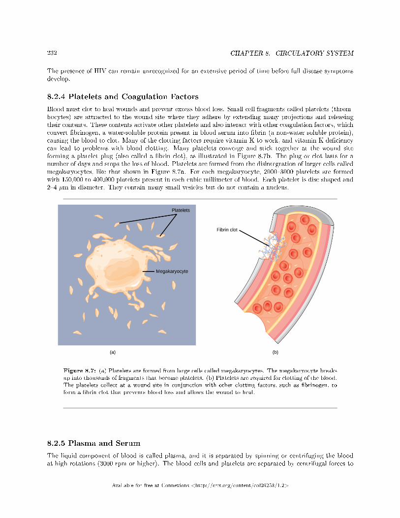
232 CHAPTER 8. CIRCULATORY SYSTEM
The presence of HIV can remain unrecognized for an extensive period of time before full disease symptomsdevelop.
8.2.4 Platelets and Coagulation Factors
Blood must clot to heal wounds and prevent excess blood loss. Small cell fragments called platelets (throm-bocytes) are attracted to the wound site where they adhere by extending many projections and releasingtheir contents. These contents activate other platelets and also interact with other coagulation factors, whichconvert �brinogen, a water-soluble protein present in blood serum into �brin (a non-water soluble protein),causing the blood to clot. Many of the clotting factors require vitamin K to work, and vitamin K de�ciencycan lead to problems with blood clotting. Many platelets converge and stick together at the wound siteforming a platelet plug (also called a �brin clot), as illustrated in Figure 8.7b. The plug or clot lasts for anumber of days and stops the loss of blood. Platelets are formed from the disintegration of larger cells calledmegakaryocytes, like that shown in Figure 8.7a. For each megakaryocyte, 2000�3000 platelets are formedwith 150,000 to 400,000 platelets present in each cubic millimeter of blood. Each platelet is disc shaped and2�4 µm in diameter. They contain many small vesicles but do not contain a nucleus.
Figure 8.7: (a) Platelets are formed from large cells called megakaryocytes. The megakaryocyte breaksup into thousands of fragments that become platelets. (b) Platelets are required for clotting of the blood.The platelets collect at a wound site in conjunction with other clotting factors, such as �brinogen, toform a �brin clot that prevents blood loss and allows the wound to heal.
8.2.5 Plasma and Serum
The liquid component of blood is called plasma, and it is separated by spinning or centrifuging the bloodat high rotations (3000 rpm or higher). The blood cells and platelets are separated by centrifugal forces to
Available for free at Connexions <http://cnx.org/content/col26253/1.2>
233
the bottom of a specimen tube. The upper liquid layer, the plasma, consists of 90 percent water along withvarious substances required for maintaining the body's pH, osmotic load, and for protecting the body. Theplasma also contains the coagulation factors and antibodies.
The plasma component of blood without the coagulation factors is called the serum. Serum is similarto interstitial �uid in which the correct composition of key ions acting as electrolytes is essential for normalfunctioning of muscles and nerves. Other components in the serum include proteins that assist with main-taining pH and osmotic balance while giving viscosity to the blood. The serum also contains antibodies,specialized proteins that are important for defense against viruses and bacteria. Lipids, including choles-terol, are also transported in the serum, along with various other substances including nutrients, hormones,metabolic waste, plus external substances, such as, drugs, viruses, and bacteria.
Human serum albumin is the most abundant protein in human blood plasma and is synthesized in theliver. Albumin, which constitutes about half of the blood serum protein, transports hormones and fattyacids, bu�ers pH, and maintains osmotic pressures. Immunoglobin is a protein antibody produced in themucosal lining and plays an important role in antibody mediated immunity.
: Blood Types Related to Proteins on the Surface of the Red Blood Cells
Red blood cells are coated in antigens made of glycolipids and glycoproteins. The composition ofthese molecules is determined by genetics, which have evolved over time. In humans, the di�erentsurface antigens are grouped into 24 di�erent blood groups with more than 100 di�erent antigenson each red blood cell. The two most well known blood groups are the ABO, shown in Figure 8.8,and Rh systems. The surface antigens in the ABO blood group are glycolipids, called antigen Aand antigen B. People with blood type A have antigen A, those with blood type B have antigen B,those with blood type AB have both antigens, and people with blood type O have neither antigen.Antibodies called agglutinougens are found in the blood plasma and react with the A or B antigens,if the two are mixed. When type A and type B blood are combined, agglutination (clumping) ofthe blood occurs because of antibodies in the plasma that bind with the opposing antigen; thiscauses clots that coagulate in the kidney causing kidney failure. Type O blood has neither A or Bantigens, and therefore, type O blood can be given to all blood types. Type O negative blood isthe universal donor. Type AB positive blood is the universal acceptor because it has both A andB antigen. The ABO blood groups were discovered in 1900 and 1901 by Karl Landsteiner at theUniversity of Vienna.
The Rh blood group was �rst discovered in Rhesus monkeys. Most people have the Rh antigen(Rh+) and do not have anti-Rh antibodies in their blood. The few people who do not have the Rhantigen and are Rh� can develop anti-Rh antibodies if exposed to Rh+ blood. This can happen aftera blood transfusion or after an Rh� woman has an Rh+ baby. The �rst exposure does not usuallycause a reaction; however, at the second exposure, enough antibodies have built up in the blood toproduce a reaction that causes agglutination and breakdown of red blood cells. An injection canprevent this reaction.
Available for free at Connexions <http://cnx.org/content/col26253/1.2>
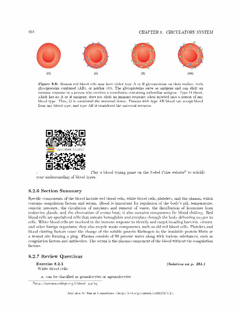
234 CHAPTER 8. CIRCULATORY SYSTEM
Figure 8.8: Human red blood cells may have either type A or B glycoproteins on their surface, bothglycoproteins combined (AB), or neither (O). The glycoproteins serve as antigens and can elicit animmune response in a person who receives a transfusion containing unfamiliar antigens. Type O blood,which has no A or B antigens, does not elicit an immune response when injected into a person of anyblood type. Thus, O is considered the universal donor. Persons with type AB blood can accept bloodfrom any blood type, and type AB is considered the universal acceptor.
: Play a blood typing game on the Nobel Prize website3 to solidifyyour understanding of blood types.
8.2.6 Section Summary
Speci�c components of the blood include red blood cells, white blood cells, platelets, and the plasma, whichcontains coagulation factors and serum. Blood is important for regulation of the body's pH, temperature,osmotic pressure, the circulation of nutrients and removal of waste, the distribution of hormones fromendocrine glands, and the elimination of excess heat; it also contains components for blood clotting. Redblood cells are specialized cells that contain hemoglobin and circulate through the body delivering oxygen tocells. White blood cells are involved in the immune response to identify and target invading bacteria, viruses,and other foreign organisms; they also recycle waste components, such as old red blood cells. Platelets andblood clotting factors cause the change of the soluble protein �brinogen to the insoluble protein �brin ata wound site forming a plug. Plasma consists of 90 percent water along with various substances, such ascoagulation factors and antibodies. The serum is the plasma component of the blood without the coagulationfactors.
8.2.7 Review Questions
Exercise 8.2.1 (Solution on p. 251.)
White blood cells:
a. can be classi�ed as granulocytes or agranulocytes
3http://openstaxcollege.org/l/blood_typing
Available for free at Connexions <http://cnx.org/content/col26253/1.2>
235
b. defend the body against bacteria and virusesc. are also called leucocytesd. All of the above
Exercise 8.2.2 (Solution on p. 251.)
Platelet plug formation occurs at which point?
a. when large megakaryocytes break up into thousands of smaller fragmentsb. when platelets are dispersed through the blood streamc. when platelets are attracted to a site of blood vessel damaged. none of the above
Exercise 8.2.3 (Solution on p. 251.)
In humans, the plasma comprises what percentage of the blood?
a. 45 percentb. 55 percentc. 25 percentd. 90 percent
Exercise 8.2.4 (Solution on p. 251.)
The red blood cells of birds di�er from mammalian red blood cells because:
a. they are white and have nucleib. they do not have nucleic. they have nucleid. they �ght disease
8.2.8 Free Response
Exercise 8.2.5 (Solution on p. 251.)
Describe the cause of di�erent blood type groups.
Exercise 8.2.6 (Solution on p. 251.)
List some of the functions of blood in the body.
Exercise 8.2.7 (Solution on p. 251.)
How does the lymphatic system work with blood �ow?
8.3 Mammalian Heart and Blood Vessels4
The heart is a complex muscle that pumps blood through the three divisions of the circulatory system: thecoronary (vessels that serve the heart), pulmonary (heart and lungs), and systemic (systems of the body),as shown in Figure 8.9. Coronary circulation intrinsic to the heart takes blood directly from the main artery(aorta) coming from the heart. For pulmonary and systemic circulation, the heart has to pump blood to thelungs or the rest of the body, respectively. In vertebrates, the lungs are relatively close to the heart in thethoracic cavity. The shorter distance to pump means that the muscle wall on the right side of the heart isnot as thick as the left side which must have enough pressure to pump blood all the way to your big toe.
4This content is available online at <http://cnx.org/content/m44805/1.8/>.
Available for free at Connexions <http://cnx.org/content/col26253/1.2>
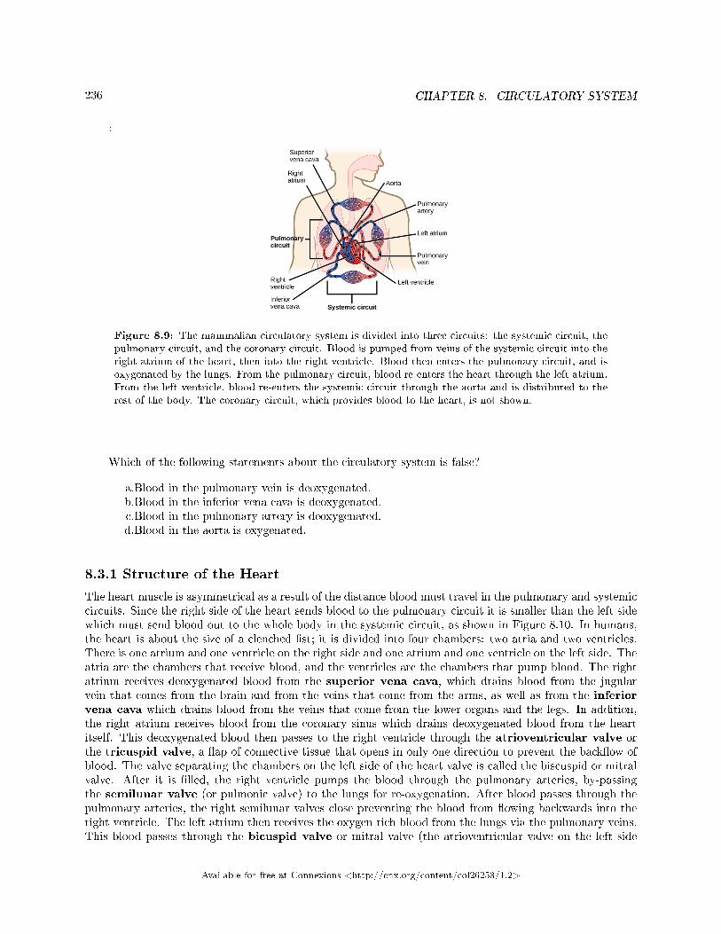
236 CHAPTER 8. CIRCULATORY SYSTEM
:
Figure 8.9: The mammalian circulatory system is divided into three circuits: the systemic circuit, thepulmonary circuit, and the coronary circuit. Blood is pumped from veins of the systemic circuit into theright atrium of the heart, then into the right ventricle. Blood then enters the pulmonary circuit, and isoxygenated by the lungs. From the pulmonary circuit, blood re-enters the heart through the left atrium.From the left ventricle, blood re-enters the systemic circuit through the aorta and is distributed to therest of the body. The coronary circuit, which provides blood to the heart, is not shown.
Which of the following statements about the circulatory system is false?
a.Blood in the pulmonary vein is deoxygenated.b.Blood in the inferior vena cava is deoxygenated.c.Blood in the pulmonary artery is deoxygenated.d.Blood in the aorta is oxygenated.
8.3.1 Structure of the Heart
The heart muscle is asymmetrical as a result of the distance blood must travel in the pulmonary and systemiccircuits. Since the right side of the heart sends blood to the pulmonary circuit it is smaller than the left sidewhich must send blood out to the whole body in the systemic circuit, as shown in Figure 8.10. In humans,the heart is about the size of a clenched �st; it is divided into four chambers: two atria and two ventricles.There is one atrium and one ventricle on the right side and one atrium and one ventricle on the left side. Theatria are the chambers that receive blood, and the ventricles are the chambers that pump blood. The rightatrium receives deoxygenated blood from the superior vena cava, which drains blood from the jugularvein that comes from the brain and from the veins that come from the arms, as well as from the inferiorvena cava which drains blood from the veins that come from the lower organs and the legs. In addition,the right atrium receives blood from the coronary sinus which drains deoxygenated blood from the heartitself. This deoxygenated blood then passes to the right ventricle through the atrioventricular valve orthe tricuspid valve, a �ap of connective tissue that opens in only one direction to prevent the back�ow ofblood. The valve separating the chambers on the left side of the heart valve is called the biscuspid or mitralvalve. After it is �lled, the right ventricle pumps the blood through the pulmonary arteries, by-passingthe semilunar valve (or pulmonic valve) to the lungs for re-oxygenation. After blood passes through thepulmonary arteries, the right semilunar valves close preventing the blood from �owing backwards into theright ventricle. The left atrium then receives the oxygen-rich blood from the lungs via the pulmonary veins.This blood passes through the bicuspid valve or mitral valve (the atrioventricular valve on the left side
Available for free at Connexions <http://cnx.org/content/col26253/1.2>
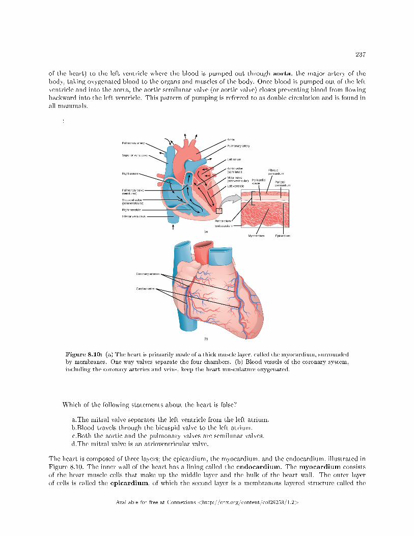
237
of the heart) to the left ventricle where the blood is pumped out through aorta, the major artery of thebody, taking oxygenated blood to the organs and muscles of the body. Once blood is pumped out of the leftventricle and into the aorta, the aortic semilunar valve (or aortic valve) closes preventing blood from �owingbackward into the left ventricle. This pattern of pumping is referred to as double circulation and is found inall mammals.
:
Figure 8.10: (a) The heart is primarily made of a thick muscle layer, called the myocardium, surroundedby membranes. One-way valves separate the four chambers. (b) Blood vessels of the coronary system,including the coronary arteries and veins, keep the heart musculature oxygenated.
Which of the following statements about the heart is false?
a.The mitral valve separates the left ventricle from the left atrium.b.Blood travels through the bicuspid valve to the left atrium.c.Both the aortic and the pulmonary valves are semilunar valves.d.The mitral valve is an atrioventricular valve.
The heart is composed of three layers; the epicardium, the myocardium, and the endocardium, illustrated inFigure 8.10. The inner wall of the heart has a lining called the endocardium. The myocardium consistsof the heart muscle cells that make up the middle layer and the bulk of the heart wall. The outer layerof cells is called the epicardium, of which the second layer is a membranous layered structure called the
Available for free at Connexions <http://cnx.org/content/col26253/1.2>
238 CHAPTER 8. CIRCULATORY SYSTEM
pericardium that surrounds and protects the heart; it allows enough room for vigorous pumping but alsokeeps the heart in place to reduce friction between the heart and other structures.
The heart has its own blood vessels that supply the heart muscle with blood. The coronary arteriesbranch from the aorta and surround the outer surface of the heart like a crown. They diverge into capillarieswhere the heart muscle is supplied with oxygen before converging again into the coronary veins to take thedeoxygenated blood back to the right atrium where the blood will be re-oxygenated through the pulmonarycircuit. The heart muscle will die without a steady supply of blood. Atherosclerosis is the blockage of anartery by the buildup of fatty plaques. Because of the size (narrow) of the coronary arteries and their functionin serving the heart itself, atherosclerosis can be deadly in these arteries. The slowdown of blood �ow andsubsequent oxygen deprivation that results from atherosclerosis causes severe pain, known as angina, andcomplete blockage of the arteries will cause myocardial infarction: the death of cardiac muscle tissue,commonly known as a heart attack.
8.3.2 The Cardiac Cycle
The main purpose of the heart is to pump blood through the body; it does so in a repeating sequencecalled the cardiac cycle. The cardiac cycle is the coordination of the �lling and emptying of the heartof blood by electrical signals that cause the heart muscles to contract and relax. The human heart beatsover 100,000 times per day. In each cardiac cycle, the heart contracts (systole), pushing out the bloodand pumping it through the body; this is followed by a relaxation phase (diastole), where the heart �llswith blood, as illustrated in Figure 8.11. The atria contract at the same time, forcing blood through theatrioventricular valves into the ventricles. Closing of the atrioventricular valves produces a monosyllabic�lup� sound. Following a brief delay, the ventricles contract at the same time forcing blood through thesemilunar valves into the aorta and the artery transporting blood to the lungs (via the pulmonary artery).Closing of the semilunar valves produces a monosyllabic �dup� sound.
Available for free at Connexions <http://cnx.org/content/col26253/1.2>
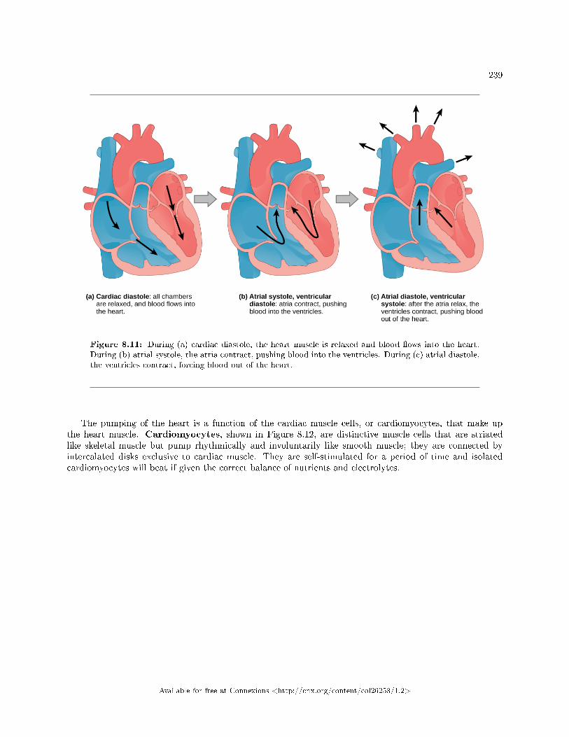
239
Figure 8.11: During (a) cardiac diastole, the heart muscle is relaxed and blood �ows into the heart.During (b) atrial systole, the atria contract, pushing blood into the ventricles. During (c) atrial diastole,the ventricles contract, forcing blood out of the heart.
The pumping of the heart is a function of the cardiac muscle cells, or cardiomyocytes, that make upthe heart muscle. Cardiomyocytes, shown in Figure 8.12, are distinctive muscle cells that are striatedlike skeletal muscle but pump rhythmically and involuntarily like smooth muscle; they are connected byintercalated disks exclusive to cardiac muscle. They are self-stimulated for a period of time and isolatedcardiomyocytes will beat if given the correct balance of nutrients and electrolytes.
Available for free at Connexions <http://cnx.org/content/col26253/1.2>
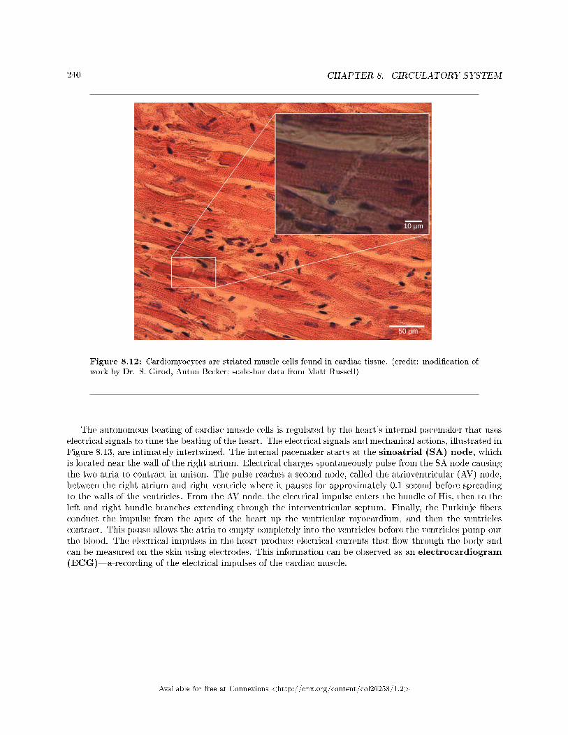
240 CHAPTER 8. CIRCULATORY SYSTEM
Figure 8.12: Cardiomyocytes are striated muscle cells found in cardiac tissue. (credit: modi�cation ofwork by Dr. S. Girod, Anton Becker; scale-bar data from Matt Russell)
The autonomous beating of cardiac muscle cells is regulated by the heart's internal pacemaker that useselectrical signals to time the beating of the heart. The electrical signals and mechanical actions, illustrated inFigure 8.13, are intimately intertwined. The internal pacemaker starts at the sinoatrial (SA) node, whichis located near the wall of the right atrium. Electrical charges spontaneously pulse from the SA node causingthe two atria to contract in unison. The pulse reaches a second node, called the atrioventricular (AV) node,between the right atrium and right ventricle where it pauses for approximately 0.1 second before spreadingto the walls of the ventricles. From the AV node, the electrical impulse enters the bundle of His, then to theleft and right bundle branches extending through the interventricular septum. Finally, the Purkinje �bersconduct the impulse from the apex of the heart up the ventricular myocardium, and then the ventriclescontract. This pause allows the atria to empty completely into the ventricles before the ventricles pump outthe blood. The electrical impulses in the heart produce electrical currents that �ow through the body andcan be measured on the skin using electrodes. This information can be observed as an electrocardiogram(ECG)�a recording of the electrical impulses of the cardiac muscle.
Available for free at Connexions <http://cnx.org/content/col26253/1.2>
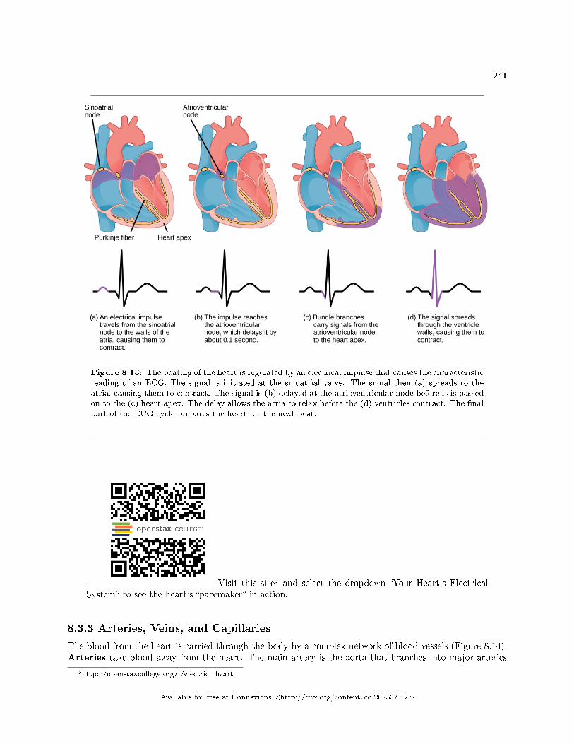
241
Figure 8.13: The beating of the heart is regulated by an electrical impulse that causes the characteristicreading of an ECG. The signal is initiated at the sinoatrial valve. The signal then (a) spreads to theatria, causing them to contract. The signal is (b) delayed at the atrioventricular node before it is passedon to the (c) heart apex. The delay allows the atria to relax before the (d) ventricles contract. The �nalpart of the ECG cycle prepares the heart for the next beat.
: Visit this site5 and select the dropdown �Your Heart's ElectricalSystem� to see the heart's �pacemaker� in action.
8.3.3 Arteries, Veins, and Capillaries
The blood from the heart is carried through the body by a complex network of blood vessels (Figure 8.14).Arteries take blood away from the heart. The main artery is the aorta that branches into major arteries
5http://openstaxcollege.org/l/electric_heart
Available for free at Connexions <http://cnx.org/content/col26253/1.2>
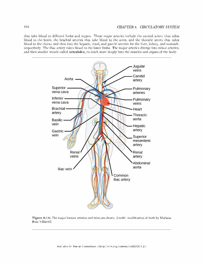
242 CHAPTER 8. CIRCULATORY SYSTEM
that take blood to di�erent limbs and organs. These major arteries include the carotid artery that takesblood to the brain, the brachial arteries that take blood to the arms, and the thoracic artery that takesblood to the thorax and then into the hepatic, renal, and gastric arteries for the liver, kidney, and stomach,respectively. The iliac artery takes blood to the lower limbs. The major arteries diverge into minor arteries,and then smaller vessels called arterioles, to reach more deeply into the muscles and organs of the body.
Figure 8.14: The major human arteries and veins are shown. (credit: modi�cation of work by MarianaRuiz Villareal)
Available for free at Connexions <http://cnx.org/content/col26253/1.2>
243
Arterioles diverge into capillary beds. Capillary beds contain a large number (10 to 100) of capillariesthat branch among the cells and tissues of the body. Capillaries are narrow-diameter tubes that can �t redblood cells through in single �le and are the sites for the exchange of nutrients, waste, and oxygen withtissues at the cellular level. Fluid also crosses into the interstitial space from the capillaries. The capillariesconverge again into venules that connect to minor veins that �nally connect to major veins that take bloodhigh in carbon dioxide back to the heart. Veins are blood vessels that bring blood back to the heart. Themajor veins drain blood from the same organs and limbs that the major arteries supply. Fluid is also broughtback to the heart via the lymphatic system.
The structure of the di�erent types of blood vessels re�ects their function or layers. There are threedistinct layers, or tunics, that form the walls of blood vessels (Figure 8.15). The �rst tunic is a smooth, innerlining of endothelial cells that are in contact with the red blood cells. The endothelial tunic is continuous withthe endocardium of the heart. In capillaries, this single layer of cells is the location of di�usion of oxygen andcarbon dioxide between the endothelial cells and red blood cells, as well as the exchange site via endocytosisand exocytosis. The movement of materials at the site of capillaries is regulated by vasoconstriction,narrowing of the blood vessels, and vasodilation, widening of the blood vessels; this is important in theoverall regulation of blood pressure.
Veins and arteries both have two further tunics that surround the endothelium: the middle tunic iscomposed of smooth muscle and the outermost layer is connective tissue (collagen and elastic �bers). Theelastic connective tissue stretches and supports the blood vessels, and the smooth muscle layer helps regulateblood �ow by altering vascular resistance through vasoconstriction and vasodilation. The arteries have thickersmooth muscle and connective tissue than the veins to accommodate the higher pressure and speed of freshlypumped blood. The veins are thinner walled as the pressure and rate of �ow are much lower. In addition,veins are structurally di�erent than arteries in that veins have valves to prevent the back�ow of blood.Because veins have to work against gravity to get blood back to the heart, contraction of skeletal muscleassists with the �ow of blood back to the heart.
Available for free at Connexions <http://cnx.org/content/col26253/1.2>
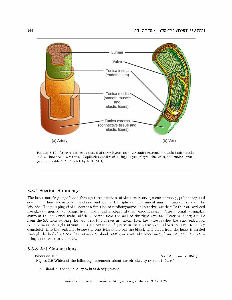
244 CHAPTER 8. CIRCULATORY SYSTEM
Figure 8.15: Arteries and veins consist of three layers: an outer tunica externa, a middle tunica media,and an inner tunica intima. Capillaries consist of a single layer of epithelial cells, the tunica intima.(credit: modi�cation of work by NCI, NIH)
8.3.4 Section Summary
The heart muscle pumps blood through three divisions of the circulatory system: coronary, pulmonary, andsystemic. There is one atrium and one ventricle on the right side and one atrium and one ventricle on theleft side. The pumping of the heart is a function of cardiomyocytes, distinctive muscle cells that are striatedlike skeletal muscle but pump rhythmically and involuntarily like smooth muscle. The internal pacemakerstarts at the sinoatrial node, which is located near the wall of the right atrium. Electrical charges pulsefrom the SA node causing the two atria to contract in unison; then the pulse reaches the atrioventricularnode between the right atrium and right ventricle. A pause in the electric signal allows the atria to emptycompletely into the ventricles before the ventricles pump out the blood. The blood from the heart is carriedthrough the body by a complex network of blood vessels; arteries take blood away from the heart, and veinsbring blood back to the heart.
8.3.5 Art Connections
Exercise 8.3.1 (Solution on p. 251.)
Figure 8.9 Which of the following statements about the circulatory system is false?
a. Blood in the pulmonary vein is deoxygenated.
Available for free at Connexions <http://cnx.org/content/col26253/1.2>
245
b. Blood in the inferior vena cava is deoxygenated.c. Blood in the pulmonary artery is deoxygenated.d. Blood in the aorta is oxygenated.
Exercise 8.3.2 (Solution on p. 251.)
Figure 8.10 Which of the following statements about the heart is false?
a. The mitral valve separates the left ventricle from the left atrium.b. Blood travels through the bicuspid valve to the left atrium.c. Both the aortic and the pulmonary valves are semilunar valves.d. The mitral valve is an atrioventricular valve.
8.3.6 Review Questions
Exercise 8.3.3 (Solution on p. 251.)
The heart's internal pacemaker beats by:
a. an internal implant that sends an electrical impulse through the heartb. the excitation of cardiac muscle cells at the sinoatrial node followed by the atrioventricular
nodec. the excitation of cardiac muscle cells at the atrioventricular node followed by the sinoatrial
noded. the action of the sinus
Exercise 8.3.4 (Solution on p. 251.)
During the systolic phase of the cardiac cycle, the heart is ________.
a. contractingb. relaxingc. contracting and relaxingd. �lling with blood
Exercise 8.3.5 (Solution on p. 251.)
Cardiomyocytes are similar to skeletal muscle because:
a. they beat involuntarilyb. they are used for weight liftingc. they pulse rhythmicallyd. they are striated
Exercise 8.3.6 (Solution on p. 251.)
How do arteries di�er from veins?
a. Arteries have thicker smooth muscle layers to accommodate the changes in pressure from theheart.
b. Arteries carry blood.c. Arteries have thinner smooth muscle layers and valves and move blood by the action of skeletal
muscle.d. Arteries are thin walled and are used for gas exchange.
Available for free at Connexions <http://cnx.org/content/col26253/1.2>
246 CHAPTER 8. CIRCULATORY SYSTEM
8.3.7 Free Response
Exercise 8.3.7 (Solution on p. 251.)
Describe the cardiac cycle.
Exercise 8.3.8 (Solution on p. 252.)
What happens in capillaries?
8.4 Blood Flow and Blood Pressure Regulation6
Blood pressure (BP) is the pressure exerted by blood on the walls of a blood vessel that helps to pushblood through the body. Systolic blood pressure measures the amount of pressure that blood exerts onvessels while the heart is beating. The optimal systolic blood pressure is 120 mmHg. Diastolic bloodpressure measures the pressure in the vessels between heartbeats. The optimal diastolic blood pressure is80 mmHg. Many factors can a�ect blood pressure, such as hormones, stress, exercise, eating, sitting, andstanding. Blood �ow through the body is regulated by the size of blood vessels, by the action of smoothmuscle, by one-way valves, and by the �uid pressure of the blood itself.
8.4.1 How Blood Flows Through the Body
Blood is pushed through the body by the action of the pumping heart. With each rhythmic pump, blood ispushed under high pressure and velocity away from the heart, initially along the main artery, the aorta. Inthe aorta, the blood travels at 30 cm/sec. As blood moves into the arteries, arterioles, and ultimately to thecapillary beds, the rate of movement slows dramatically to about 0.026 cm/sec, one-thousand times slowerthan the rate of movement in the aorta. While the diameter of each individual arteriole and capillary is farnarrower than the diameter of the aorta, and according to the law of continuity, �uid should travel fasterthrough a narrower diameter tube, the rate is actually slower due to the overall diameter of all the combinedcapillaries being far greater than the diameter of the individual aorta.
The slow rate of travel through the capillary beds, which reach almost every cell in the body, assists withgas and nutrient exchange and also promotes the di�usion of �uid into the interstitial space. After the bloodhas passed through the capillary beds to the venules, veins, and �nally to the main venae cavae, the rate of�ow increases again but is still much slower than the initial rate in the aorta. Blood primarily moves in theveins by the rhythmic movement of smooth muscle in the vessel wall and by the action of the skeletal muscleas the body moves. Because most veins must move blood against the pull of gravity, blood is prevented from�owing backward in the veins by one-way valves. Because skeletal muscle contraction aids in venous blood�ow, it is important to get up and move frequently after long periods of sitting so that blood will not poolin the extremities.
Blood �ow through the capillary beds is regulated depending on the body's needs and is directed bynerve and hormone signals. For example, after a large meal, most of the blood is diverted to the stomachby vasodilation of vessels of the digestive system and vasoconstriction of other vessels. During exercise,blood is diverted to the skeletal muscles through vasodilation while blood to the digestive system would belessened through vasoconstriction. The blood entering some capillary beds is controlled by small muscles,called precapillary sphincters, illustrated in Figure 8.16. If the sphincters are open, the blood will �ow intothe associated branches of the capillary blood. If all of the sphincters are closed, then the blood will �owdirectly from the arteriole to the venule through the thoroughfare channel (see Figure 8.16). These musclesallow the body to precisely control when capillary beds receive blood �ow. At any given moment only about5-10% of our capillary beds actually have blood �owing through them.
:
6This content is available online at <http://cnx.org/content/m44806/1.8/>.
Available for free at Connexions <http://cnx.org/content/col26253/1.2>
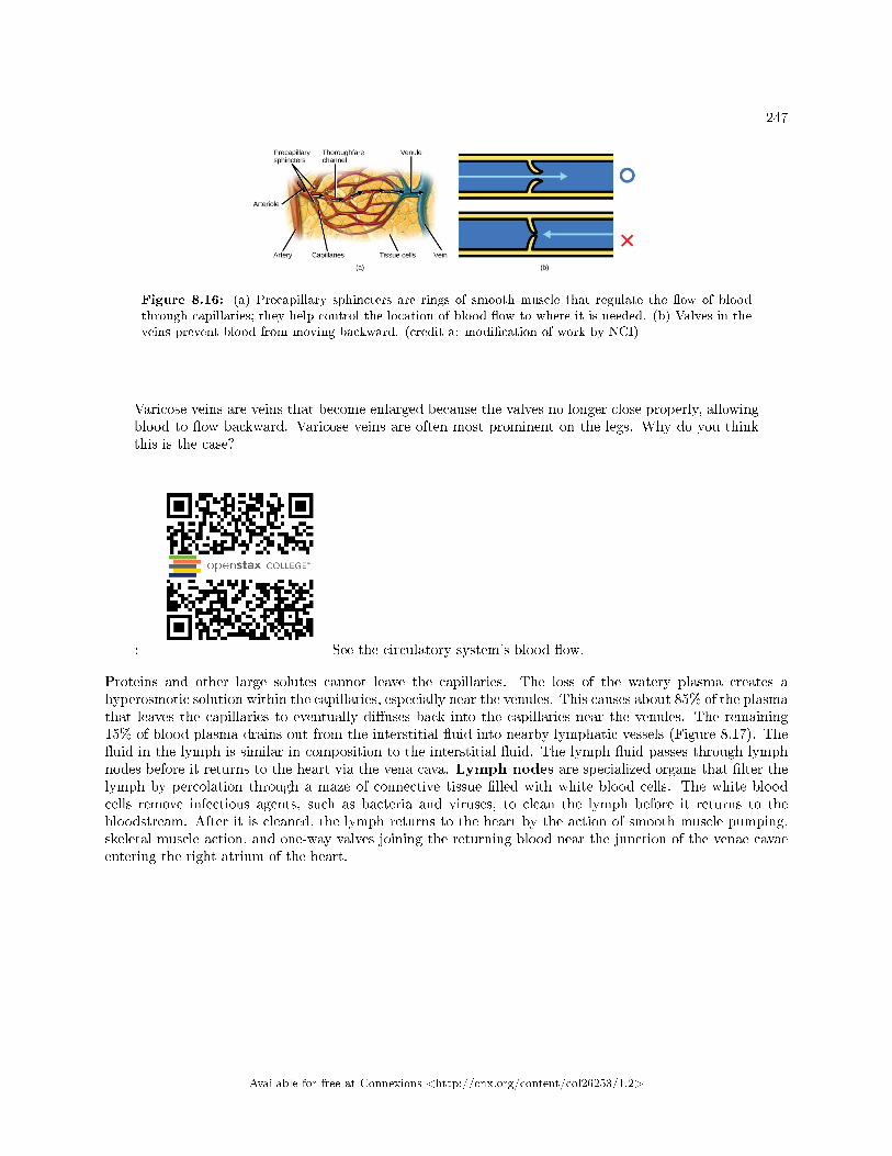
247
Figure 8.16: (a) Precapillary sphincters are rings of smooth muscle that regulate the �ow of bloodthrough capillaries; they help control the location of blood �ow to where it is needed. (b) Valves in theveins prevent blood from moving backward. (credit a: modi�cation of work by NCI)
Varicose veins are veins that become enlarged because the valves no longer close properly, allowingblood to �ow backward. Varicose veins are often most prominent on the legs. Why do you thinkthis is the case?
: See the circulatory system's blood �ow.
Proteins and other large solutes cannot leave the capillaries. The loss of the watery plasma creates ahyperosmotic solution within the capillaries, especially near the venules. This causes about 85% of the plasmathat leaves the capillaries to eventually di�uses back into the capillaries near the venules. The remaining15% of blood plasma drains out from the interstitial �uid into nearby lymphatic vessels (Figure 8.17). The�uid in the lymph is similar in composition to the interstitial �uid. The lymph �uid passes through lymphnodes before it returns to the heart via the vena cava. Lymph nodes are specialized organs that �lter thelymph by percolation through a maze of connective tissue �lled with white blood cells. The white bloodcells remove infectious agents, such as bacteria and viruses, to clean the lymph before it returns to thebloodstream. After it is cleaned, the lymph returns to the heart by the action of smooth muscle pumping,skeletal muscle action, and one-way valves joining the returning blood near the junction of the venae cavaeentering the right atrium of the heart.
Available for free at Connexions <http://cnx.org/content/col26253/1.2>
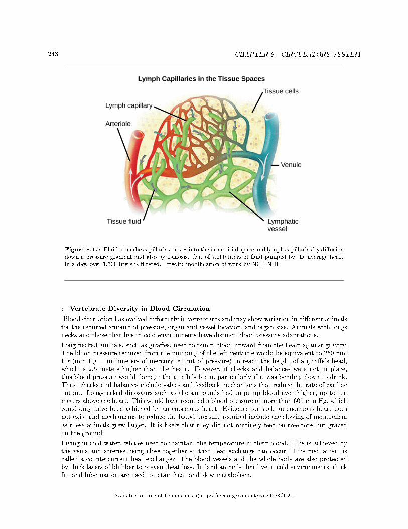
248 CHAPTER 8. CIRCULATORY SYSTEM
Figure 8.17: Fluid from the capillaries moves into the interstitial space and lymph capillaries by di�usiondown a pressure gradient and also by osmosis. Out of 7,200 liters of �uid pumped by the average heartin a day, over 1,500 liters is �ltered. (credit: modi�cation of work by NCI, NIH)
: Vertebrate Diversity in Blood Circulation
Blood circulation has evolved di�erently in vertebrates and may show variation in di�erent animalsfor the required amount of pressure, organ and vessel location, and organ size. Animals with longsnecks and those that live in cold environments have distinct blood pressure adaptations.
Long necked animals, such as gira�es, need to pump blood upward from the heart against gravity.The blood pressure required from the pumping of the left ventricle would be equivalent to 250 mmHg (mm Hg = millimeters of mercury, a unit of pressure) to reach the height of a gira�e's head,which is 2.5 meters higher than the heart. However, if checks and balances were not in place,this blood pressure would damage the gira�e's brain, particularly if it was bending down to drink.These checks and balances include valves and feedback mechanisms that reduce the rate of cardiacoutput. Long-necked dinosaurs such as the sauropods had to pump blood even higher, up to tenmeters above the heart. This would have required a blood pressure of more than 600 mm Hg, whichcould only have been achieved by an enormous heart. Evidence for such an enormous heart doesnot exist and mechanisms to reduce the blood pressure required include the slowing of metabolismas these animals grew larger. It is likely that they did not routinely feed on tree tops but grazedon the ground.
Living in cold water, whales need to maintain the temperature in their blood. This is achieved bythe veins and arteries being close together so that heat exchange can occur. This mechanism iscalled a countercurrent heat exchanger. The blood vessels and the whole body are also protectedby thick layers of blubber to prevent heat loss. In land animals that live in cold environments, thickfur and hibernation are used to retain heat and slow metabolism.
Available for free at Connexions <http://cnx.org/content/col26253/1.2>

249
8.4.2 Blood Pressure
The pressure of the blood �ow in the body is produced by the hydrostatic pressure of the �uid (blood)against the walls of the blood vessels. Fluid will move from areas of high to low hydrostatic pressures. In thearteries, the hydrostatic pressure near the heart is very high and blood �ows to the arterioles where the rateof �ow is slowed by the narrow openings of the arterioles. During systole, when new blood is entering thearteries, the artery walls stretch to accommodate the increase of pressure of the extra blood; during diastole,the walls return to normal because of their elastic properties. The blood pressure of the systole phase andthe diastole phase, graphed in Figure 8.18, gives the two pressure readings for blood pressure. For example,120/80 indicates a reading of 120 mm Hg during the systole and 80 mm Hg during diastole. Throughoutthe cardiac cycle, the blood continues to empty into the arterioles at a relatively even rate. This resistanceto blood �ow is called peripheral resistance.
Figure 8.18: Blood pressure is related to the blood velocity in the arteries and arterioles. In thecapillaries and veins, the blood pressure continues to decease but velocity increases.
8.4.3 Blood Pressure Regulation
Cardiac output is the volume of blood pumped by the heart in one minute. It is calculated by multiplying thenumber of heart contractions that occur per minute (heart rate) times the stroke volume (the volume ofblood pumped into the aorta per contraction of the left ventricle). Therefore, cardiac output can be increased
Available for free at Connexions <http://cnx.org/content/col26253/1.2>
250 CHAPTER 8. CIRCULATORY SYSTEM
by increasing heart rate, as when exercising. However, cardiac output can also be increased by increasingstroke volume, such as if the heart contracts with greater strength. Stroke volume can also be increasedby speeding blood circulation through the body so that more blood enters the heart between contractions.During heavy exertion, the blood vessels relax and increase in diameter, o�setting the increased heart rateand ensuring adequate oxygenated blood gets to the muscles. Stress triggers a decrease in the diameter ofthe blood vessels, consequently increasing blood pressure. These changes can also be caused by nerve signalsor hormones, and even standing up or lying down can have a great e�ect on blood pressure.
8.4.4 Section Summary
Blood primarily moves through the body by the rhythmic movement of smooth muscle in the vessel walland by the action of the skeletal muscle as the body moves. Blood is prevented from �owing backward inthe veins by one-way valves. Blood �ow through the capillary beds is controlled by precapillary sphinctersto increase and decrease �ow depending on the body's needs and is directed by nerve and hormone signals.Lymph vessels take �uid that has leaked out of the blood to the lymph nodes where it is cleaned beforereturning to the heart. During systole, blood enters the arteries, and the artery walls stretch to accommodatethe extra blood. During diastole, the artery walls return to normal. The blood pressure of the systole phaseand the diastole phase gives the two pressure readings for blood pressure.
8.4.5 Art Connections
Exercise 8.4.1 (Solution on p. 252.)
Figure 8.16 Varicose veins are veins that become enlarged because the valves no longer closeproperly, allowing blood to �ow backward. Varicose veins are often most prominent on the legs.Why do you think this is the case?
8.4.6 Review Questions
Exercise 8.4.2 (Solution on p. 252.)
High blood pressure would be a result of ________.
a. a high cardiac output and high peripheral resistanceb. a high cardiac output and low peripheral resistancec. a low cardiac output and high peripheral resistanced. a low cardiac output and low peripheral resistance
8.4.7 Free Response
Exercise 8.4.3 (Solution on p. 252.)
How does blood pressure change during heavy exercise?
Available for free at Connexions <http://cnx.org/content/col26253/1.2>
251
Solutions to Exercises in Chapter 8
to Exercise 8.1.1 (p. 228)Ato Exercise 8.1.2 (p. 228)Dto Exercise 8.1.3 (p. 228)Cto Exercise 8.1.4 (p. 228)A closed circulatory system is a closed-loop system, in which blood is not free in a cavity. Blood is separatefrom the bodily interstitial �uid and contained within blood vessels. In this type of system, blood circulatesunidirectionally from the heart around the systemic circulatory route, and then returns to the heart.to Exercise 8.1.5 (p. 228)Systemic circulation �ows through the systems of the body. The blood �ows away from the heart to thebrain, liver, kidneys, stomach, and other organs, the limbs, and the muscles of the body; it then returns tothe heart.to Exercise 8.2.1 (p. 234)Dto Exercise 8.2.2 (p. 235)Cto Exercise 8.2.3 (p. 235)Bto Exercise 8.2.4 (p. 235)Cto Exercise 8.2.5 (p. 235)Red blood cells are coated with proteins called antigens made of glycolipids and glycoproteins. When typeA and type B blood are mixed, the blood agglutinates because of antibodies in the plasma that bind withthe opposing antigen. Type O blood has no antigens. The Rh blood group has either the Rh antigen (Rh+)or no Rh antigen (Rh�).to Exercise 8.2.6 (p. 235)Blood is important for regulation of the body's pH, temperature, and osmotic pressure, the circulationof nutrients and removal of wastes, the distribution of hormones from endocrine glands, the elimination ofexcess heat; it also contains components for the clotting of blood to prevent blood loss. Blood also transportsclotting factors and disease-�ghting agents.to Exercise 8.2.7 (p. 235)Lymph capillaries take �uid from the blood to the lymph nodes. The lymph nodes �lter the lymph bypercolation through connective tissue �lled with white blood cells. The white blood cells remove infectiousagents, such as bacteria and viruses, to clean the lymph before it returns to the bloodstream.to Exercise 8.3.1 (p. 244)Figure 8.9 Cto Exercise 8.3.2 (p. 245)Figure 8.10 Bto Exercise 8.3.3 (p. 245)Bto Exercise 8.3.4 (p. 245)Ato Exercise 8.3.5 (p. 245)Dto Exercise 8.3.6 (p. 245)A
Available for free at Connexions <http://cnx.org/content/col26253/1.2>
252 CHAPTER 8. CIRCULATORY SYSTEM
to Exercise 8.3.7 (p. 246)The heart receives an electrical signal from the sinoatrial node triggering the cardiac muscle cells in the atriato contract. The signal pauses at the atrioventricular node before spreading to the walls of the ventricles sothe blood is pumped through the body. This is the systolic phase. The heart then relaxes in the diastoleand �lls again with blood.to Exercise 8.3.8 (p. 246)The capillaries basically exchange materials with their surroundings. Their walls are very thin and are madeof one or two layers of cells, where gases, nutrients, and waste are di�used. They are distributed as beds,complex networks that link arteries as well as veins.to Exercise 8.4.1 (p. 250)Figure 8.16 Blood in the legs is farthest away from the heart and has to �ow up to reach it.to Exercise 8.4.2 (p. 250)Ato Exercise 8.4.3 (p. 250)The heart rate increases, which increases the hydrostatic pressure against the artery walls. At the sametime, the arterioles dilate in response to the increased exercise, which reduces peripheral resistance.
Available for free at Connexions <http://cnx.org/content/col26253/1.2>
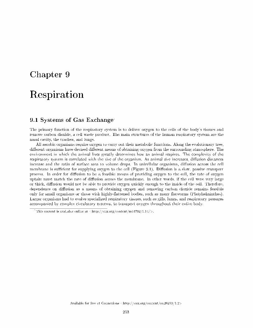
Chapter 9
Respiration
9.1 Systems of Gas Exchange1
The primary function of the respiratory system is to deliver oxygen to the cells of the body's tissues andremove carbon dioxide, a cell waste product. The main structures of the human respiratory system are thenasal cavity, the trachea, and lungs.
All aerobic organisms require oxygen to carry out their metabolic functions. Along the evolutionary tree,di�erent organisms have devised di�erent means of obtaining oxygen from the surrounding atmosphere. Theenvironment in which the animal lives greatly determines how an animal respires. The complexity of therespiratory system is correlated with the size of the organism. As animal size increases, di�usion distancesincrease and the ratio of surface area to volume drops. In unicellular organisms, di�usion across the cellmembrane is su�cient for supplying oxygen to the cell (Figure 9.1). Di�usion is a slow, passive transportprocess. In order for di�usion to be a feasible means of providing oxygen to the cell, the rate of oxygenuptake must match the rate of di�usion across the membrane. In other words, if the cell were very largeor thick, di�usion would not be able to provide oxygen quickly enough to the inside of the cell. Therefore,dependence on di�usion as a means of obtaining oxygen and removing carbon dioxide remains feasibleonly for small organisms or those with highly-�attened bodies, such as many �atworms (Platyhelminthes).Larger organisms had to evolve specialized respiratory tissues, such as gills, lungs, and respiratory passagesaccompanied by complex circulatory systems, to transport oxygen throughout their entire body.
1This content is available online at <http://cnx.org/content/m44792/1.14/>.
Available for free at Connexions <http://cnx.org/content/col26253/1.2>
253
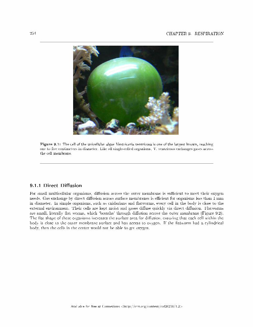
254 CHAPTER 9. RESPIRATION
Figure 9.1: The cell of the unicellular algae Ventricaria ventricosa is one of the largest known, reachingone to �ve centimeters in diameter. Like all single-celled organisms, V. ventricosa exchanges gases acrossthe cell membrane.
9.1.1 Direct Di�usion
For small multicellular organisms, di�usion across the outer membrane is su�cient to meet their oxygenneeds. Gas exchange by direct di�usion across surface membranes is e�cient for organisms less than 1 mmin diameter. In simple organisms, such as cnidarians and �atworms, every cell in the body is close to theexternal environment. Their cells are kept moist and gases di�use quickly via direct di�usion. Flatwormsare small, literally �at worms, which `breathe' through di�usion across the outer membrane (Figure 9.2).The �at shape of these organisms increases the surface area for di�usion, ensuring that each cell within thebody is close to the outer membrane surface and has access to oxygen. If the �atworm had a cylindricalbody, then the cells in the center would not be able to get oxygen.
Available for free at Connexions <http://cnx.org/content/col26253/1.2>
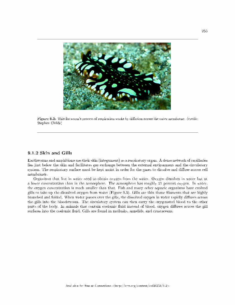
255
Figure 9.2: This �atworm's process of respiration works by di�usion across the outer membrane. (credit:Stephen Childs)
9.1.2 Skin and Gills
Earthworms and amphibians use their skin (integument) as a respiratory organ. A dense network of capillarieslies just below the skin and facilitates gas exchange between the external environment and the circulatorysystem. The respiratory surface must be kept moist in order for the gases to dissolve and di�use across cellmembranes.
Organisms that live in water need to obtain oxygen from the water. Oxygen dissolves in water but ata lower concentration than in the atmosphere. The atmosphere has roughly 21 percent oxygen. In water,the oxygen concentration is much smaller than that. Fish and many other aquatic organisms have evolvedgills to take up the dissolved oxygen from water (Figure 9.3). Gills are thin tissue �laments that are highlybranched and folded. When water passes over the gills, the dissolved oxygen in water rapidly di�uses acrossthe gills into the bloodstream. The circulatory system can then carry the oxygenated blood to the otherparts of the body. In animals that contain coelomic �uid instead of blood, oxygen di�uses across the gillsurfaces into the coelomic �uid. Gills are found in mollusks, annelids, and crustaceans.
Available for free at Connexions <http://cnx.org/content/col26253/1.2>
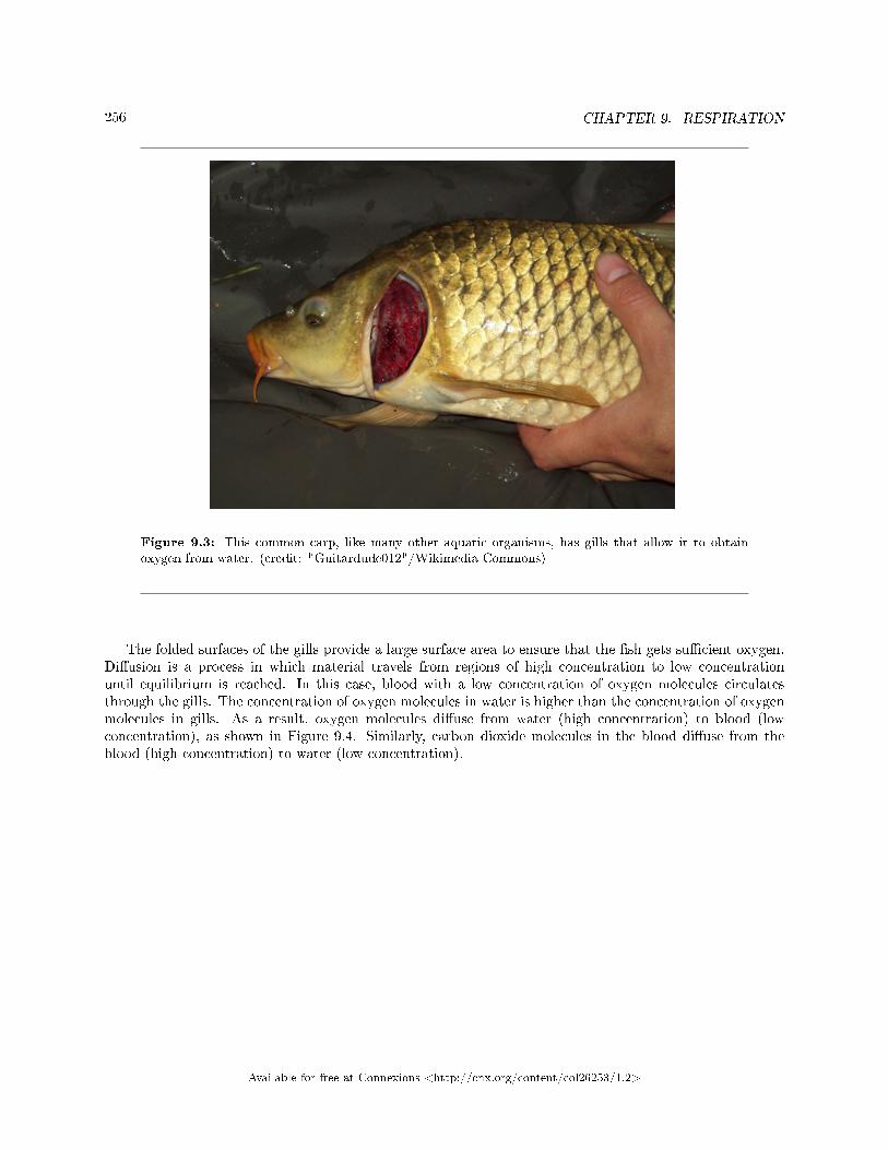
256 CHAPTER 9. RESPIRATION
Figure 9.3: This common carp, like many other aquatic organisms, has gills that allow it to obtainoxygen from water. (credit: "Guitardude012"/Wikimedia Commons)
The folded surfaces of the gills provide a large surface area to ensure that the �sh gets su�cient oxygen.Di�usion is a process in which material travels from regions of high concentration to low concentrationuntil equilibrium is reached. In this case, blood with a low concentration of oxygen molecules circulatesthrough the gills. The concentration of oxygen molecules in water is higher than the concentration of oxygenmolecules in gills. As a result, oxygen molecules di�use from water (high concentration) to blood (lowconcentration), as shown in Figure 9.4. Similarly, carbon dioxide molecules in the blood di�use from theblood (high concentration) to water (low concentration).
Available for free at Connexions <http://cnx.org/content/col26253/1.2>
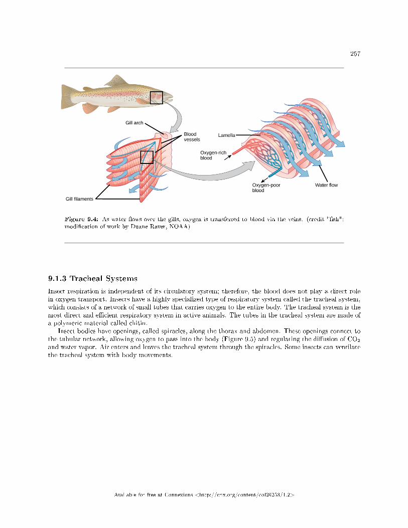
257
Figure 9.4: As water �ows over the gills, oxygen is transferred to blood via the veins. (credit "�sh":modi�cation of work by Duane Raver, NOAA)
9.1.3 Tracheal Systems
Insect respiration is independent of its circulatory system; therefore, the blood does not play a direct rolein oxygen transport. Insects have a highly specialized type of respiratory system called the tracheal system,which consists of a network of small tubes that carries oxygen to the entire body. The tracheal system is themost direct and e�cient respiratory system in active animals. The tubes in the tracheal system are made ofa polymeric material called chitin.
Insect bodies have openings, called spiracles, along the thorax and abdomen. These openings connect tothe tubular network, allowing oxygen to pass into the body (Figure 9.5) and regulating the di�usion of CO2
and water vapor. Air enters and leaves the tracheal system through the spiracles. Some insects can ventilatethe tracheal system with body movements.
Available for free at Connexions <http://cnx.org/content/col26253/1.2>
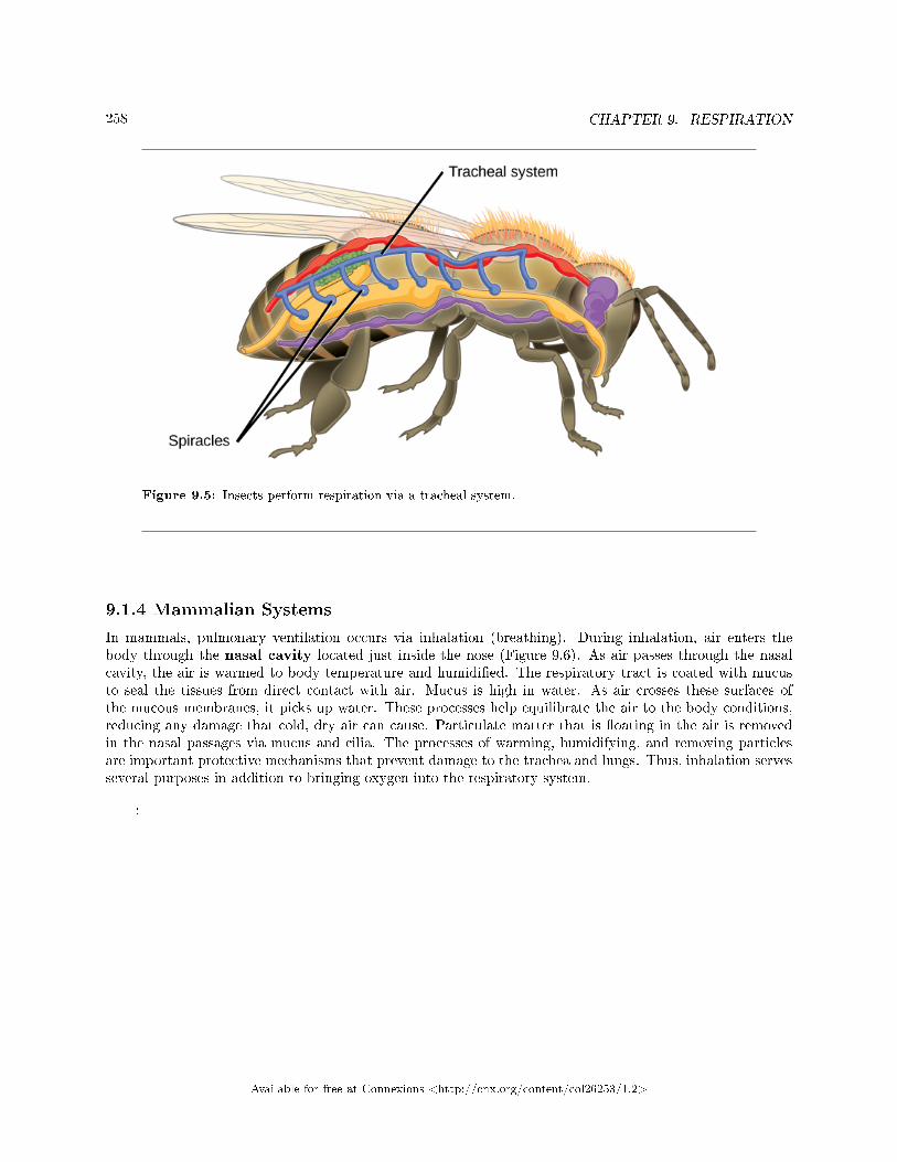
258 CHAPTER 9. RESPIRATION
Figure 9.5: Insects perform respiration via a tracheal system.
9.1.4 Mammalian Systems
In mammals, pulmonary ventilation occurs via inhalation (breathing). During inhalation, air enters thebody through the nasal cavity located just inside the nose (Figure 9.6). As air passes through the nasalcavity, the air is warmed to body temperature and humidi�ed. The respiratory tract is coated with mucusto seal the tissues from direct contact with air. Mucus is high in water. As air crosses these surfaces ofthe mucous membranes, it picks up water. These processes help equilibrate the air to the body conditions,reducing any damage that cold, dry air can cause. Particulate matter that is �oating in the air is removedin the nasal passages via mucus and cilia. The processes of warming, humidifying, and removing particlesare important protective mechanisms that prevent damage to the trachea and lungs. Thus, inhalation servesseveral purposes in addition to bringing oxygen into the respiratory system.
:
Available for free at Connexions <http://cnx.org/content/col26253/1.2>
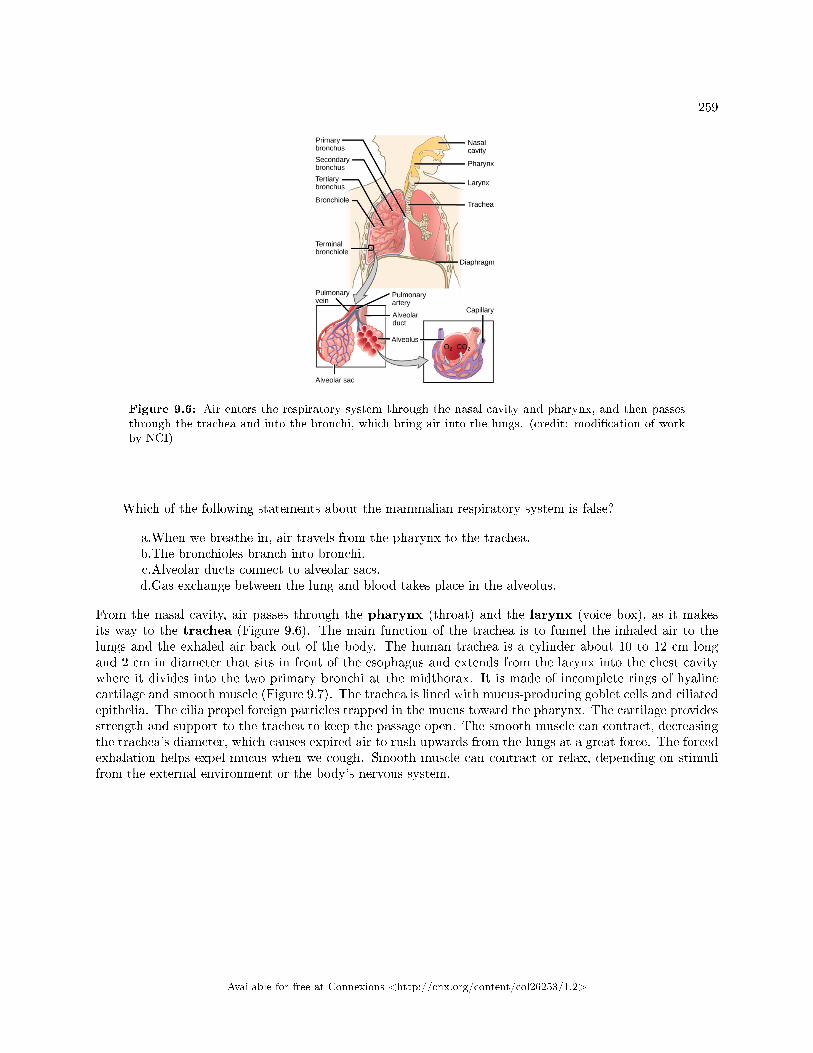
259
Figure 9.6: Air enters the respiratory system through the nasal cavity and pharynx, and then passesthrough the trachea and into the bronchi, which bring air into the lungs. (credit: modi�cation of workby NCI)
Which of the following statements about the mammalian respiratory system is false?
a.When we breathe in, air travels from the pharynx to the trachea.b.The bronchioles branch into bronchi.c.Alveolar ducts connect to alveolar sacs.d.Gas exchange between the lung and blood takes place in the alveolus.
From the nasal cavity, air passes through the pharynx (throat) and the larynx (voice box), as it makesits way to the trachea (Figure 9.6). The main function of the trachea is to funnel the inhaled air to thelungs and the exhaled air back out of the body. The human trachea is a cylinder about 10 to 12 cm longand 2 cm in diameter that sits in front of the esophagus and extends from the larynx into the chest cavitywhere it divides into the two primary bronchi at the midthorax. It is made of incomplete rings of hyalinecartilage and smooth muscle (Figure 9.7). The trachea is lined with mucus-producing goblet cells and ciliatedepithelia. The cilia propel foreign particles trapped in the mucus toward the pharynx. The cartilage providesstrength and support to the trachea to keep the passage open. The smooth muscle can contract, decreasingthe trachea's diameter, which causes expired air to rush upwards from the lungs at a great force. The forcedexhalation helps expel mucus when we cough. Smooth muscle can contract or relax, depending on stimulifrom the external environment or the body's nervous system.
Available for free at Connexions <http://cnx.org/content/col26253/1.2>
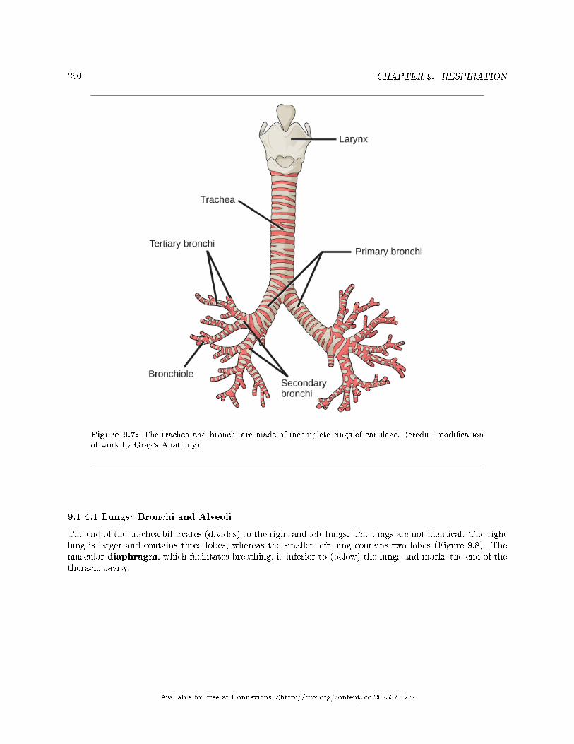
260 CHAPTER 9. RESPIRATION
Figure 9.7: The trachea and bronchi are made of incomplete rings of cartilage. (credit: modi�cationof work by Gray's Anatomy)
9.1.4.1 Lungs: Bronchi and Alveoli
The end of the trachea bifurcates (divides) to the right and left lungs. The lungs are not identical. The rightlung is larger and contains three lobes, whereas the smaller left lung contains two lobes (Figure 9.8). Themuscular diaphragm, which facilitates breathing, is inferior to (below) the lungs and marks the end of thethoracic cavity.
Available for free at Connexions <http://cnx.org/content/col26253/1.2>
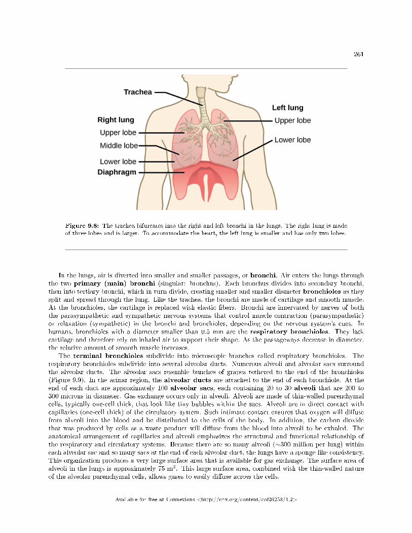
261
Figure 9.8: The trachea bifurcates into the right and left bronchi in the lungs. The right lung is madeof three lobes and is larger. To accommodate the heart, the left lung is smaller and has only two lobes.
In the lungs, air is diverted into smaller and smaller passages, or bronchi. Air enters the lungs throughthe two primary (main) bronchi (singular: bronchus). Each bronchus divides into secondary bronchi,then into tertiary bronchi, which in turn divide, creating smaller and smaller diameter bronchioles as theysplit and spread through the lung. Like the trachea, the bronchi are made of cartilage and smooth muscle.At the bronchioles, the cartilage is replaced with elastic �bers. Bronchi are innervated by nerves of boththe parasympathetic and sympathetic nervous systems that control muscle contraction (parasympathetic)or relaxation (sympathetic) in the bronchi and bronchioles, depending on the nervous system's cues. Inhumans, bronchioles with a diameter smaller than 0.5 mm are the respiratory bronchioles. They lackcartilage and therefore rely on inhaled air to support their shape. As the passageways decrease in diameter,the relative amount of smooth muscle increases.
The terminal bronchioles subdivide into microscopic branches called respiratory bronchioles. Therespiratory bronchioles subdivide into several alveolar ducts. Numerous alveoli and alveolar sacs surroundthe alveolar ducts. The alveolar sacs resemble bunches of grapes tethered to the end of the bronchioles(Figure 9.9). In the acinar region, the alveolar ducts are attached to the end of each bronchiole. At theend of each duct are approximately 100 alveolar sacs, each containing 20 to 30 alveoli that are 200 to300 microns in diameter. Gas exchange occurs only in alveoli. Alveoli are made of thin-walled parenchymalcells, typically one-cell thick, that look like tiny bubbles within the sacs. Alveoli are in direct contact withcapillaries (one-cell thick) of the circulatory system. Such intimate contact ensures that oxygen will di�usefrom alveoli into the blood and be distributed to the cells of the body. In addition, the carbon dioxidethat was produced by cells as a waste product will di�use from the blood into alveoli to be exhaled. Theanatomical arrangement of capillaries and alveoli emphasizes the structural and functional relationship ofthe respiratory and circulatory systems. Because there are so many alveoli (∼300 million per lung) withineach alveolar sac and so many sacs at the end of each alveolar duct, the lungs have a sponge-like consistency.This organization produces a very large surface area that is available for gas exchange. The surface area ofalveoli in the lungs is approximately 75 m2. This large surface area, combined with the thin-walled natureof the alveolar parenchymal cells, allows gases to easily di�use across the cells.
Available for free at Connexions <http://cnx.org/content/col26253/1.2>
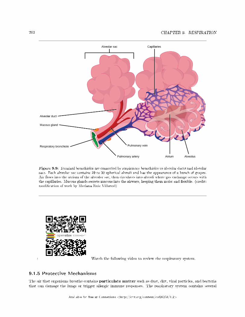
262 CHAPTER 9. RESPIRATION
Figure 9.9: Terminal bronchioles are connected by respiratory bronchioles to alveolar ducts and alveolarsacs. Each alveolar sac contains 20 to 30 spherical alveoli and has the appearance of a bunch of grapes.Air �ows into the atrium of the alveolar sac, then circulates into alveoli where gas exchange occurs withthe capillaries. Mucous glands secrete mucous into the airways, keeping them moist and �exible. (credit:modi�cation of work by Mariana Ruiz Villareal)
: Watch the following video to review the respiratory system.
9.1.5 Protective Mechanisms
The air that organisms breathe contains particulate matter such as dust, dirt, viral particles, and bacteriathat can damage the lungs or trigger allergic immune responses. The respiratory system contains several
Available for free at Connexions <http://cnx.org/content/col26253/1.2>
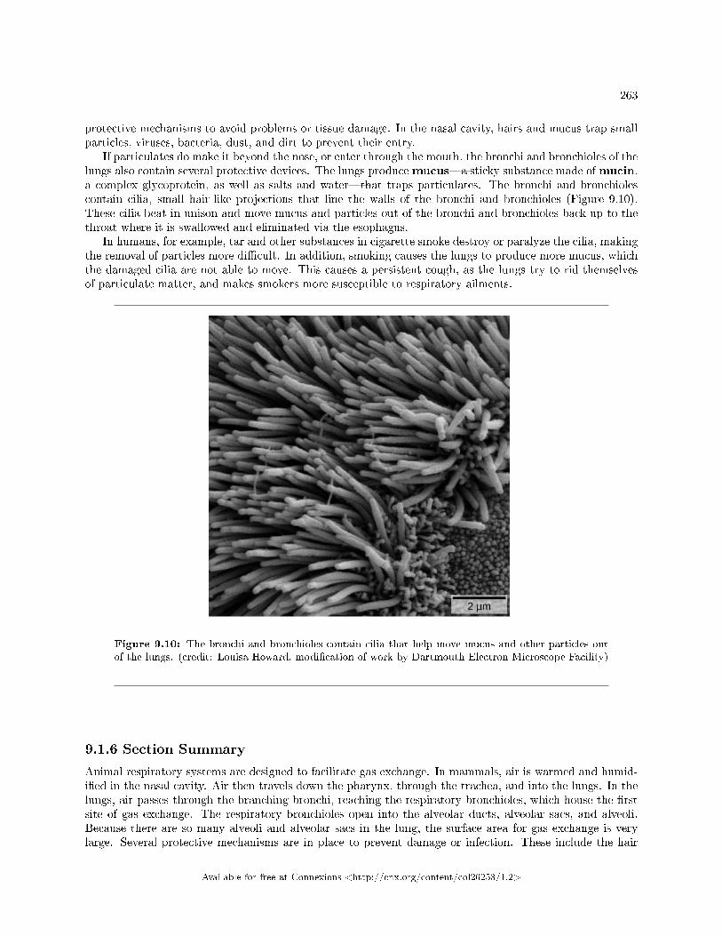
263
protective mechanisms to avoid problems or tissue damage. In the nasal cavity, hairs and mucus trap smallparticles, viruses, bacteria, dust, and dirt to prevent their entry.
If particulates do make it beyond the nose, or enter through the mouth, the bronchi and bronchioles of thelungs also contain several protective devices. The lungs producemucus�a sticky substance made ofmucin,a complex glycoprotein, as well as salts and water�that traps particulates. The bronchi and bronchiolescontain cilia, small hair-like projections that line the walls of the bronchi and bronchioles (Figure 9.10).These cilia beat in unison and move mucus and particles out of the bronchi and bronchioles back up to thethroat where it is swallowed and eliminated via the esophagus.
In humans, for example, tar and other substances in cigarette smoke destroy or paralyze the cilia, makingthe removal of particles more di�cult. In addition, smoking causes the lungs to produce more mucus, whichthe damaged cilia are not able to move. This causes a persistent cough, as the lungs try to rid themselvesof particulate matter, and makes smokers more susceptible to respiratory ailments.
Figure 9.10: The bronchi and bronchioles contain cilia that help move mucus and other particles outof the lungs. (credit: Louisa Howard, modi�cation of work by Dartmouth Electron Microscope Facility)
9.1.6 Section Summary
Animal respiratory systems are designed to facilitate gas exchange. In mammals, air is warmed and humid-i�ed in the nasal cavity. Air then travels down the pharynx, through the trachea, and into the lungs. In thelungs, air passes through the branching bronchi, reaching the respiratory bronchioles, which house the �rstsite of gas exchange. The respiratory bronchioles open into the alveolar ducts, alveolar sacs, and alveoli.Because there are so many alveoli and alveolar sacs in the lung, the surface area for gas exchange is verylarge. Several protective mechanisms are in place to prevent damage or infection. These include the hair
Available for free at Connexions <http://cnx.org/content/col26253/1.2>
264 CHAPTER 9. RESPIRATION
and mucus in the nasal cavity that trap dust, dirt, and other particulate matter before they can enter thesystem. In the lungs, particles are trapped in a mucus layer and transported via cilia up to the esophagealopening at the top of the trachea to be swallowed.
9.1.7
Exercise 9.1.1 (Solution on p. 285.)
Figure 9.6 Which of the following statements about the mammalian respiratory system is false?
a. When we breathe in, air travels from the pharynx to the trachea.b. The bronchioles branch into bronchi.c. Alveolar ducts connect to alveolar sacs.d. Gas exchange between the lung and blood takes place in the alveolus.
9.1.8 Review Questions
Exercise 9.1.2 (Solution on p. 285.)
The respiratory system ________.
a. provides body tissues with oxygenb. provides body tissues with oxygen and carbon dioxidec. establishes how many breaths are taken per minuted. provides the body with carbon dioxide
Exercise 9.1.3 (Solution on p. 285.)
Air is warmed and humidi�ed in the nasal passages. This helps to ________.
a. ward o� infectionb. decrease sensitivity during breathingc. prevent damage to the lungsd. all of the above
Exercise 9.1.4 (Solution on p. 285.)
Which is the order of air�ow during inhalation?
a. nasal cavity, trachea, larynx, bronchi, bronchioles, alveolib. nasal cavity, larynx, trachea, bronchi, bronchioles, alveolic. nasal cavity, larynx, trachea, bronchioles, bronchi, alveolid. nasal cavity, trachea, larynx, bronchi, bronchioles, alveoli
9.1.9 Free Response
Exercise 9.1.5 (Solution on p. 285.)
Describe the function of these terms and describe where they are located: main bronchus, trachea,alveoli, and acinus.
Exercise 9.1.6 (Solution on p. 285.)
How does the structure of alveoli maximize gas exchange?
Available for free at Connexions <http://cnx.org/content/col26253/1.2>
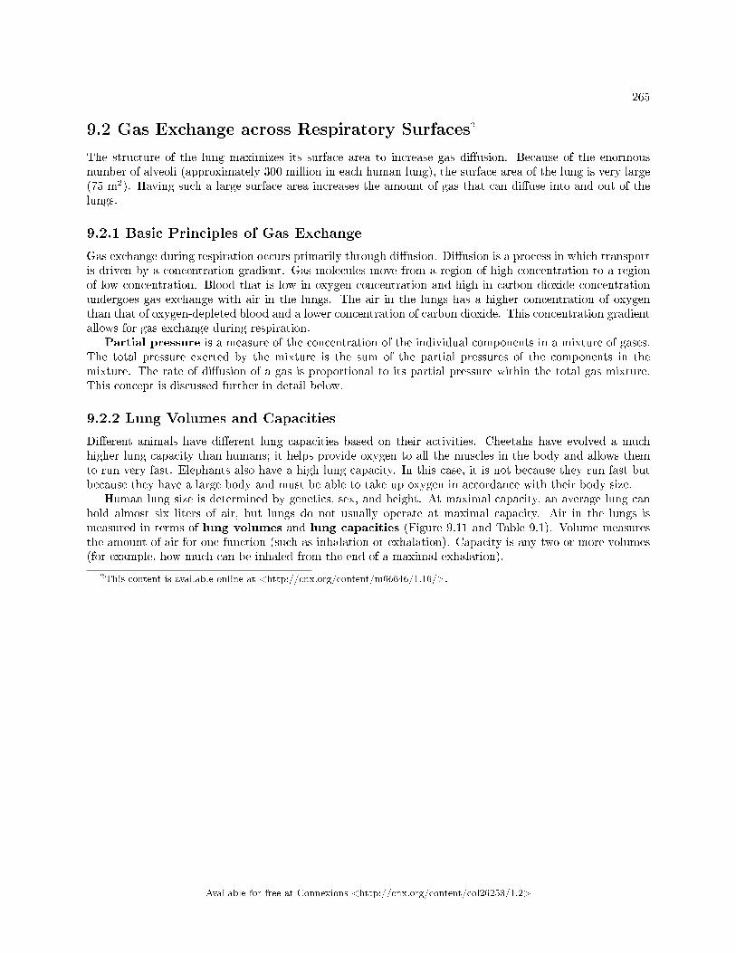
265
9.2 Gas Exchange across Respiratory Surfaces2
The structure of the lung maximizes its surface area to increase gas di�usion. Because of the enormousnumber of alveoli (approximately 300 million in each human lung), the surface area of the lung is very large(75 m2). Having such a large surface area increases the amount of gas that can di�use into and out of thelungs.
9.2.1 Basic Principles of Gas Exchange
Gas exchange during respiration occurs primarily through di�usion. Di�usion is a process in which transportis driven by a concentration gradient. Gas molecules move from a region of high concentration to a regionof low concentration. Blood that is low in oxygen concentration and high in carbon dioxide concentrationundergoes gas exchange with air in the lungs. The air in the lungs has a higher concentration of oxygenthan that of oxygen-depleted blood and a lower concentration of carbon dioxide. This concentration gradientallows for gas exchange during respiration.
Partial pressure is a measure of the concentration of the individual components in a mixture of gases.The total pressure exerted by the mixture is the sum of the partial pressures of the components in themixture. The rate of di�usion of a gas is proportional to its partial pressure within the total gas mixture.This concept is discussed further in detail below.
9.2.2 Lung Volumes and Capacities
Di�erent animals have di�erent lung capacities based on their activities. Cheetahs have evolved a muchhigher lung capacity than humans; it helps provide oxygen to all the muscles in the body and allows themto run very fast. Elephants also have a high lung capacity. In this case, it is not because they run fast butbecause they have a large body and must be able to take up oxygen in accordance with their body size.
Human lung size is determined by genetics, sex, and height. At maximal capacity, an average lung canhold almost six liters of air, but lungs do not usually operate at maximal capacity. Air in the lungs ismeasured in terms of lung volumes and lung capacities (Figure 9.11 and Table 9.1). Volume measuresthe amount of air for one function (such as inhalation or exhalation). Capacity is any two or more volumes(for example, how much can be inhaled from the end of a maximal exhalation).
2This content is available online at <http://cnx.org/content/m66646/1.16/>.
Available for free at Connexions <http://cnx.org/content/col26253/1.2>
266 CHAPTER 9. RESPIRATION
Figure 9.11: Human lung volumes and capacities are shown. The total lung capacity of the adult maleis six liters. Tidal volume is the volume of air inhaled in a single, normal breath. Inspiratory capacityis the amount of air taken in during a deep breath, and residual volume is the amount of air left in thelungs after forceful respiration.
Lung Volumes and Capacities (Avg Adult Male)
Volume/Capacity De�nition Volume (liters) Equations
Tidal volume (TV) Amount of air inhaledduring a normal breath
0.5 -
Expiratory reserve vol-ume (ERV)
Amount of air that canbe exhaled after a nor-mal exhalation
1.2 -
Inspiratory reserve vol-ume (IRV)
Amount of air that canbe further inhaled aftera normal inhalation
3.1 -
Residual volume (RV) Air left in the lungs aftera forced exhalation
1.2 -
continued on next page
Available for free at Connexions <http://cnx.org/content/col26253/1.2>
267
Vital capacity (VC) Maximum amount of airthat can be moved in orout of the lungs in a sin-gle respiratory cycle
4.8 ERV+TV+IRV
Inspiratory capacity(IC)
Volume of air that canbe inhaled in addition toa normal exhalation
3.6 TV+IRV
Functional residual ca-pacity (FRC)
Volume of air remainingafter a normal exhala-tion
2.4 ERV+RV
Total lung capacity(TLC)
Total volume of air inthe lungs after a maxi-mal inspiration
6.0 RV+ERV+TV+IRV
Forced expiratory vol-ume (FEV1)
How much air can beforced out of the lungsover a speci�c time pe-riod, usually one second
∼4.1 to 5.5 -
Table 9.1
The volume in the lung can be divided into four units: tidal volume, expiratory reserve volume, inspiratoryreserve volume, and residual volume. Tidal volume (TV) measures the amount of air that is inspired andexpired during a normal breath. On average, this volume is around one-half liter, which is a little less thanthe capacity of a 20-ounce drink bottle. The expiratory reserve volume (ERV) is the additional amountof air that can be exhaled after a normal exhalation. It is the reserve amount that can be exhaled beyondwhat is normal. Conversely, the inspiratory reserve volume (IRV) is the additional amount of air thatcan be inhaled after a normal inhalation. The residual volume (RV) is the amount of air that is left afterexpiratory reserve volume is exhaled. The lungs are never completely empty: There is always some air left inthe lungs after a maximal exhalation. If this residual volume did not exist and the lungs emptied completely,the lung tissues would stick together and the energy necessary to rein�ate the lung could be too great toovercome. Therefore, there is always some air remaining in the lungs. Residual volume is also importantfor preventing large �uctuations in respiratory gases (O2 and CO2). The residual volume is the only lungvolume that cannot be measured directly because it is impossible to completely empty the lung of air. Thisvolume can only be calculated rather than measured.
Capacities are measurements of two or more volumes. The vital capacity (VC) measures the maximumamount of air that can be inhaled or exhaled during a respiratory cycle. It is the sum of the expiratory reservevolume, tidal volume, and inspiratory reserve volume. The inspiratory capacity (IC) is the amount ofair that can be inhaled after the end of a normal expiration. It is, therefore, the sum of the tidal volumeand inspiratory reserve volume. The functional residual capacity (FRC) includes the expiratory reservevolume and the residual volume. The FRC measures the amount of additional air that can be exhaled aftera normal exhalation. Lastly, the total lung capacity (TLC) is a measurement of the total amount of airthat the lung can hold. It is the sum of the residual volume, expiratory reserve volume, tidal volume, andinspiratory reserve volume.
Lung volumes are measured by a technique called spirometry. An important measurement taken duringspirometry is the forced expiratory volume (FEV), which measures how much air can be forced out of thelung over a speci�c period, usually one second (FEV1). In addition, the forced vital capacity (FVC), whichis the total amount of air that can be forcibly exhaled, is measured. The ratio of these values (FEV1/FVCratio) is used to diagnose lung diseases including asthma, emphysema, and �brosis. If the FEV1/FVC ratiois high, the lungs are not compliant (meaning they are sti� and unable to bend properly), and the patientmost likely has lung �brosis. Patients exhale most of the lung volume very quickly. Conversely, when the
Available for free at Connexions <http://cnx.org/content/col26253/1.2>
268 CHAPTER 9. RESPIRATION
FEV1/FVC ratio is low, there is resistance in the lung that is characteristic of asthma. In this instance,it is hard for the patient to get the air out of their lungs, and it takes a long time to reach the maximalexhalation volume. In either case, breathing is di�cult and complications arise.
: Respiratory Therapist
Respiratory therapists or respiratory practitioners evaluate and treat patients with lung and car-diovascular diseases. They work as part of a medical team to develop treatment plans for patients.Respiratory therapists may treat premature babies with underdeveloped lungs, patients with chronicconditions such as asthma, or older patients su�ering from lung disease such as emphysema andchronic obstructive pulmonary disease (COPD). They may operate advanced equipment such ascompressed gas delivery systems, ventilators, blood gas analyzers, and resuscitators. Specializedprograms to become a respiratory therapist generally lead to a bachelor's degree with a respiratorytherapist specialty. Because of a growing aging population, career opportunities as a respiratorytherapist are expected to remain strong.
9.2.3 Gas Pressure and Respiration
The respiratory process can be better understood by examining the properties of gases. Gases move freely,but gas particles are constantly hitting the walls of their vessel, thereby producing gas pressure.
Air is a mixture of gases, primarily nitrogen (N2; 78.6 percent), oxygen (O2; 20.9 percent), water vapor(H2O; 0.5 percent), and carbon dioxide (CO2; 0.04 percent). Each gas component of that mixture exerts apressure. The pressure for an individual gas in the mixture is the partial pressure of that gas. Approximately21 percent of atmospheric gas is oxygen. Carbon dioxide, however, is found in relatively small amounts, 0.04percent. The partial pressure for oxygen is much greater than that of carbon dioxide. The partial pressureof any gas can be calculated by:
P = (Patm) × (percent content in mixture). (9.11)
Patm, the atmospheric pressure, is the sum of all of the partial pressures of the atmospheric gases addedtogether,
Patm = PN2+ PO2
+ PH2O + PCO2= 760 mm Hg (9.11)
× (percent content in mixture).The pressure of the atmosphere at sea level is 760 mm Hg. Therefore, the partial pressure of oxygen is:
PO2= (760 mm Hg) (0.21) = 160 mm Hg (9.11)
and for carbon dioxide:
PCO2= (760 mm Hg) (0.0004) = 0.3 mm Hg. (9.11)
At high altitudes, Patm decreases but concentration does not change; the partial pressure decrease is due tothe reduction in Patm.
When the air mixture reaches the lung, it has been humidi�ed. The pressure of the water vapor in thelung does not change the pressure of the air, but it must be included in the partial pressure equation. Forthis calculation, the water pressure (47 mm Hg) is subtracted from the atmospheric pressure:
760 mm Hg − 47 mm Hg = 713 mm Hg (9.11)
and the partial pressure of oxygen is:
(760 mm Hg − 47 mm Hg) × 0.21 = 150 mm Hg. (9.11)
Available for free at Connexions <http://cnx.org/content/col26253/1.2>
269
These pressures determine the gas exchange, or the �ow of gas, in the system. Oxygen and carbon dioxidewill �ow according to their pressure gradient from high to low. Therefore, understanding the partial pressureof each gas will aid in understanding how gases move in the respiratory system.
9.2.4 Gas Exchange across the Alveoli
In the body, oxygen is used by cells of the body's tissues and carbon dioxide is produced as a waste product.The ratio of carbon dioxide production to oxygen consumption is the respiratory quotient (RQ). RQvaries between 0.7 and 1.0. If just glucose were used to fuel the body, the RQ would equal one. One moleof carbon dioxide would be produced for every mole of oxygen consumed. Glucose, however, is not the onlyfuel for the body. Protein and fat are also used as fuels for the body. Because of this, less carbon dioxide isproduced than oxygen is consumed and the RQ is, on average, about 0.7 for fat and about 0.8 for protein.
The RQ is used to calculate the partial pressure of oxygen in the alveolar spaces within the lung, thealveolar PO2
. Above, the partial pressure of oxygen in the lungs was calculated to be 150 mm Hg. However,lungs never fully de�ate with an exhalation; therefore, the inspired air mixes with this residual air and lowersthe partial pressure of oxygen within the alveoli. This means that there is a lower concentration of oxygenin the lungs than is found in the air outside the body. Knowing the RQ, the partial pressure of oxygen inthe alveoli can be calculated:
alveolar PCO2= inspired PO2
− (alveolar PO2
RQ) (9.11)
With an RQ of 0.8 and a PCO2in the alveoli of 40 mm Hg, the alveolar PO2
is equal to:
alveolar PO2= 150 mm Hg − (
40 mm Hg
0.8) = 100 mm Hg. (9.11)
Notice that this pressure is less than the external air. Therefore, the oxygen will �ow from the inspired airin the lung (PO2
= 150 mm Hg) into the bloodstream (PO2= 100 mm Hg) (Figure 9.12).
In the lungs, oxygen di�uses out of the alveoli and into the capillaries surrounding the alveoli. Oxygen(about 98 percent) binds reversibly to the respiratory pigment hemoglobin found in red blood cells (RBCs).RBCs carry oxygen to the tissues where oxygen dissociates from the hemoglobin and di�uses into the cellsof the tissues. More speci�cally, alveolar PO2
is higher in the alveoli (PALVO2= 100 mm Hg) than blood
PO2(40 mm Hg) in the capillaries. Because this pressure gradient exists, oxygen di�uses down its pressure
gradient, moving out of the alveoli and entering the blood of the capillaries where O2 binds to hemoglobin.At the same time, alveolar PCO2
is lower PALVO2= 40 mm Hg than blood PCO2
= (45 mm Hg). CO2 di�usesdown its pressure gradient, moving out of the capillaries and entering the alveoli.
Oxygen and carbon dioxide move independently of each other; they di�use down their own pressuregradients. As blood leaves the lungs through the pulmonary veins, the venous PO2
= 100 mm Hg, whereasthe venous PCO2
= 40 mm Hg. As blood enters the systemic capillaries, the blood will lose oxygen andgain carbon dioxide because of the pressure di�erence of the tissues and blood. In systemic capillaries, PO2
=100 mm Hg, but in the tissue cells, PO2
= 40 mm Hg. This pressure gradient drives the di�usion of oxygenout of the capillaries and into the tissue cells. At the same time, blood PCO2
= 40 mm Hg and systemictissue PCO2
= 45 mm Hg. The pressure gradient drives CO2 out of tissue cells and into the capillaries. Theblood returning to the lungs through the pulmonary arteries has a venous PO2
= 40 mm Hg and a PCO2= 45
mm Hg. The blood enters the lung capillaries where the process of exchanging gases between the capillariesand alveoli begins again (Figure 9.12).
:
Available for free at Connexions <http://cnx.org/content/col26253/1.2>
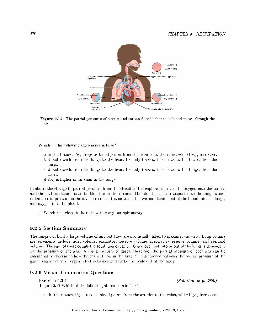
270 CHAPTER 9. RESPIRATION
Figure 9.12: The partial pressures of oxygen and carbon dioxide change as blood moves through thebody.
Which of the following statements is false?
a.In the tissues, PO2drops as blood passes from the arteries to the veins, while PCO2
increases.b.Blood travels from the lungs to the heart to body tissues, then back to the heart, then thelungs.
c.Blood travels from the lungs to the heart to body tissues, then back to the lungs, then theheart.
d.PO2is higher in air than in the lungs.
In short, the change in partial pressure from the alveoli to the capillaries drives the oxygen into the tissuesand the carbon dioxide into the blood from the tissues. The blood is then transported to the lungs wheredi�erences in pressure in the alveoli result in the movement of carbon dioxide out of the blood into the lungs,and oxygen into the blood.
: Watch this video to learn how to carry out spirometry.
9.2.5 Section Summary
The lungs can hold a large volume of air, but they are not usually �lled to maximal capacity. Lung volumemeasurements include tidal volume, expiratory reserve volume, inspiratory reserve volume, and residualvolume. The sum of these equals the total lung capacity. Gas movement into or out of the lungs is dependenton the pressure of the gas. Air is a mixture of gases; therefore, the partial pressure of each gas can becalculated to determine how the gas will �ow in the lung. The di�erence between the partial pressure of thegas in the air drives oxygen into the tissues and carbon dioxide out of the body.
9.2.6 Visual Connection Questions
Exercise 9.2.1 (Solution on p. 285.)
Figure 9.12 Which of the following statements is false?
a. In the tissues, PO2drops as blood passes from the arteries to the veins, while PCO2
increases.
Available for free at Connexions <http://cnx.org/content/col26253/1.2>
271
b. Blood travels from the lungs to the heart to body tissues, then back to the heart, then thelungs.
c. Blood travels from the lungs to the heart to body tissues, then back to the lungs, then theheart.
d. PO2is higher in air than in the lungs.
9.2.7 Review Questions
Exercise 9.2.2 (Solution on p. 285.)
The inspiratory reserve volume measures the ________.
a. amount of air remaining in the lung after a maximal exhalationb. amount of air that the lung holdsc. amount of air that can be further exhaled after a normal breathd. amount of air that can be further inhaled after a normal breath
Exercise 9.2.3 (Solution on p. 285.)
Of the following, which does not explain why the partial pressure of oxygen is lower in the lungthan in the external air?
a. Air in the lung is humidi�ed; therefore, water vapor pressure alters the pressure.b. Carbon dioxide mixes with oxygen.c. Oxygen is moved into the blood and is headed to the tissues.d. Lungs exert a pressure on the air to reduce the oxygen pressure.
Exercise 9.2.4 (Solution on p. 285.)
The total lung capacity is calculated using which of the following formulas?
a. residual volume + tidal volume + inspiratory reserve volumeb. residual volume + expiratory reserve volume + inspiratory reserve volumec. expiratory reserve volume + tidal volume + inspiratory reserve volumed. residual volume + expiratory reserve volume + tidal volume + inspiratory reserve volume
9.2.8 Critical Thinking Questions
Exercise 9.2.5 (Solution on p. 285.)
What does FEV1/FVC measure? What factors may a�ect FEV1/FVC?
Exercise 9.2.6 (Solution on p. 285.)
What is the reason for having residual volume in the lung?
Exercise 9.2.7 (Solution on p. 285.)
How can a decrease in the percent of oxygen in the air a�ect the movement of oxygen in the body?
Exercise 9.2.8 (Solution on p. 285.)
If a patient has increased resistance in their lungs, how can this be detected by a doctor? Whatdoes this mean?
Available for free at Connexions <http://cnx.org/content/col26253/1.2>
272 CHAPTER 9. RESPIRATION
9.3 Breathing3
Mammalian lungs are located in the thoracic cavity where they are surrounded and protected by the rib cage,intercostal muscles, and bound by the chest wall. The bottom of the lungs is contained by the diaphragm,a skeletal muscle that facilitates breathing. Breathing requires the coordination of the lungs, the chest wall,and most importantly, the diaphragm.
9.3.1 Types of Breathing
Amphibians have evolved multiple ways of breathing. Young amphibians, like tadpoles, use gills to breathe,and they don't leave the water. Some amphibians retain gills for life. As the tadpole grows, the gills disappearand lungs grow. These lungs are primitive and not as evolved as mammalian lungs. Adult amphibians arelacking or have a reduced diaphragm, so breathing via lungs is forced. The other means of breathing foramphibians is di�usion across the skin. To aid this di�usion, amphibian skin must remain moist.
Birds face a unique challenge with respect to breathing: They �y. Flying consumes a great amountof energy; therefore, birds require a lot of oxygen to aid their metabolic processes. Birds have evolved arespiratory system that supplies them with the oxygen needed to enable �ying. Similar to mammals, birdshave lungs, which are organs specialized for gas exchange. Oxygenated air, taken in during inhalation,di�uses across the surface of the lungs into the bloodstream, and carbon dioxide di�uses from the bloodinto the lungs and expelled during exhalation. The details of breathing between birds and mammals di�ersubstantially.
In addition to lungs, birds have air sacs inside their body. Air �ows in one direction from the posteriorair sacs to the lungs and out of the anterior air sacs. The �ow of air is in the opposite direction from blood�ow, and gas exchange takes place much more e�ciently. This type of breathing enables birds to obtainthe requisite oxygen, even at higher altitudes where the oxygen concentration is low. This directionality ofair�ow requires two cycles of air intake and exhalation to completely get the air out of the lungs.
: Avian Respiration
Birds have evolved a respiratory system that enables them to �y. Flying is a high-energy processand requires a lot of oxygen. Furthermore, many birds �y in high altitudes where the concentrationof oxygen in low. How did birds evolve a respiratory system that is so unique?
Decades of research by paleontologists have shown that birds evolved from therapods, meat-eatingdinosaurs (Figure 9.13). In fact, fossil evidence shows that meat-eating dinosaurs that lived morethan 100 million years ago had a similar �ow-through respiratory system with lungs and air sacs.Archaeopteryx and Xiaotingia, for example, were �ying dinosaurs and are believed to be earlyprecursors of birds.
3This content is available online at <http://cnx.org/content/m71333/1.1/>.
Available for free at Connexions <http://cnx.org/content/col26253/1.2>
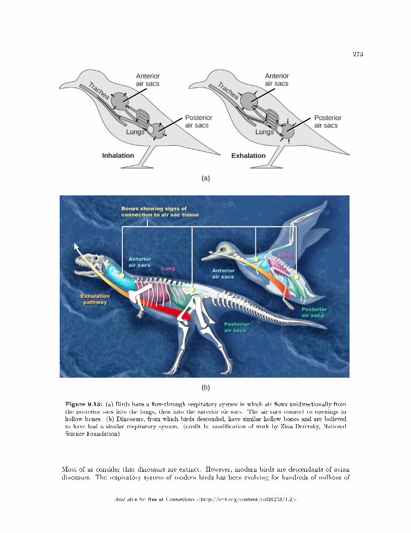
273
Figure 9.13: (a) Birds have a �ow-through respiratory system in which air �ows unidirectionally fromthe posterior sacs into the lungs, then into the anterior air sacs. The air sacs connect to openings inhollow bones. (b) Dinosaurs, from which birds descended, have similar hollow bones and are believedto have had a similar respiratory system. (credit b: modi�cation of work by Zina Deretsky, NationalScience Foundation)
Most of us consider that dinosaurs are extinct. However, modern birds are descendants of aviandinosaurs. The respiratory system of modern birds has been evolving for hundreds of millions of
Available for free at Connexions <http://cnx.org/content/col26253/1.2>
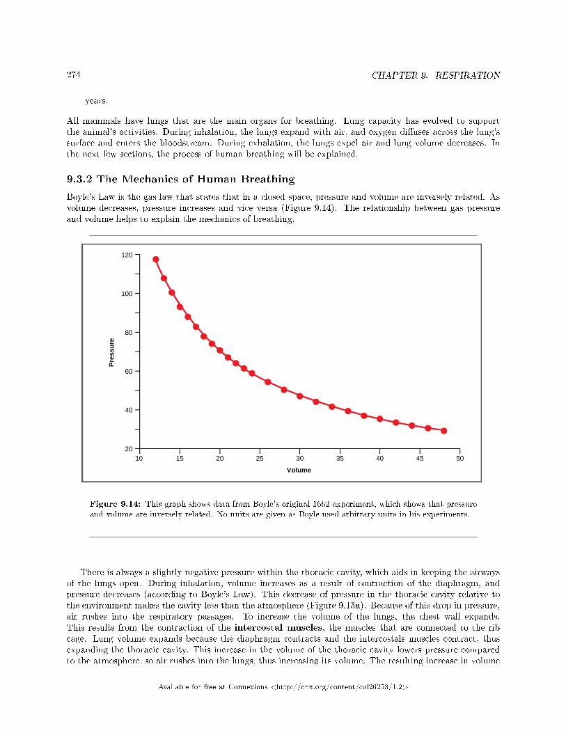
274 CHAPTER 9. RESPIRATION
years.
All mammals have lungs that are the main organs for breathing. Lung capacity has evolved to supportthe animal's activities. During inhalation, the lungs expand with air, and oxygen di�uses across the lung'ssurface and enters the bloodstream. During exhalation, the lungs expel air and lung volume decreases. Inthe next few sections, the process of human breathing will be explained.
9.3.2 The Mechanics of Human Breathing
Boyle's Law is the gas law that states that in a closed space, pressure and volume are inversely related. Asvolume decreases, pressure increases and vice versa (Figure 9.14). The relationship between gas pressureand volume helps to explain the mechanics of breathing.
Figure 9.14: This graph shows data from Boyle's original 1662 experiment, which shows that pressureand volume are inversely related. No units are given as Boyle used arbitrary units in his experiments.
There is always a slightly negative pressure within the thoracic cavity, which aids in keeping the airwaysof the lungs open. During inhalation, volume increases as a result of contraction of the diaphragm, andpressure decreases (according to Boyle's Law). This decrease of pressure in the thoracic cavity relative tothe environment makes the cavity less than the atmosphere (Figure 9.15a). Because of this drop in pressure,air rushes into the respiratory passages. To increase the volume of the lungs, the chest wall expands.This results from the contraction of the intercostal muscles, the muscles that are connected to the ribcage. Lung volume expands because the diaphragm contracts and the intercostals muscles contract, thusexpanding the thoracic cavity. This increase in the volume of the thoracic cavity lowers pressure comparedto the atmosphere, so air rushes into the lungs, thus increasing its volume. The resulting increase in volume
Available for free at Connexions <http://cnx.org/content/col26253/1.2>
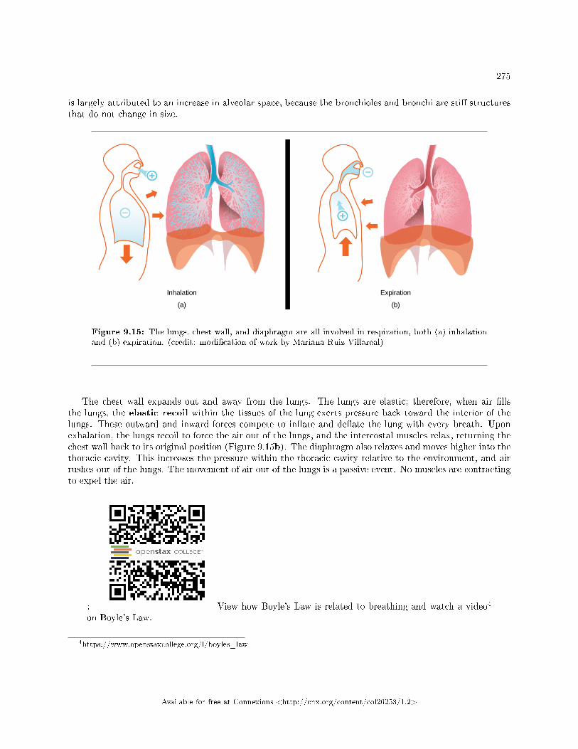
275
is largely attributed to an increase in alveolar space, because the bronchioles and bronchi are sti� structuresthat do not change in size.
Figure 9.15: The lungs, chest wall, and diaphragm are all involved in respiration, both (a) inhalationand (b) expiration. (credit: modi�cation of work by Mariana Ruiz Villareal)
The chest wall expands out and away from the lungs. The lungs are elastic; therefore, when air �llsthe lungs, the elastic recoil within the tissues of the lung exerts pressure back toward the interior of thelungs. These outward and inward forces compete to in�ate and de�ate the lung with every breath. Uponexhalation, the lungs recoil to force the air out of the lungs, and the intercostal muscles relax, returning thechest wall back to its original position (Figure 9.15b). The diaphragm also relaxes and moves higher into thethoracic cavity. This increases the pressure within the thoracic cavity relative to the environment, and airrushes out of the lungs. The movement of air out of the lungs is a passive event. No muscles are contractingto expel the air.
: View how Boyle's Law is related to breathing and watch a video4
on Boyle's Law.
4https://www.openstaxcollege.org/l/boyles_law
Available for free at Connexions <http://cnx.org/content/col26253/1.2>
276 CHAPTER 9. RESPIRATION
9.3.3 The Work of Breathing
The number of breaths per minute is the respiratory rate. On average, under non-exertion conditions,the human respiratory rate is 12�15 breaths/minute. The respiratory rate contributes to the alveolarventilation, or how much air moves into and out of the alveoli. Alveolar ventilation prevents carbondioxide buildup in the alveoli. There are two ways to keep the alveolar ventilation constant: increase therespiratory rate while decreasing the tidal volume of air per breath (shallow breathing), or decrease therespiratory rate while increasing the tidal volume per breath. In either case, the ventilation remains thesame, but the work done and type of work needed are quite di�erent. Both tidal volume and respiratoryrate are closely regulated when oxygen demand increases.
There are two types of work conducted during respiration, �ow-resistive and elastic work. Flow-resistiverefers to the work of the alveoli and tissues in the lung, whereas elastic work refers to the work of theintercostal muscles, chest wall, and diaphragm. Increasing the respiration rate increases the �ow-resistivework of the airways and decreases the elastic work of the muscles. Decreasing the respiratory rate reversesthe type of work required.
9.3.3.1 Lung Resistance and Compliance
Pulmonary diseases reduce the rate of gas exchange into and out of the lungs. Two main causes of decreasedgas exchange are compliance (how elastic the lung is) and resistance (how much obstruction exists in theairways). A change in either can dramatically alter breathing and the ability to take in oxygen and releasecarbon dioxide.
Examples of restrictive diseases are respiratory distress syndrome and pulmonary �brosis. In bothdiseases, the airways are less compliant and they are sti� or �brotic. There is a decrease in compliance becausethe lung tissue cannot bend and move. In these types of restrictive diseases, the intrapleural pressure is morepositive and the airways collapse upon exhalation, which traps air in the lungs. Forced or functional vitalcapacity (FVC), which is the amount of air that can be forcibly exhaled after taking the deepest breathpossible, is much lower than in normal patients, and the time it takes to exhale most of the air is greatlyprolonged (Figure 9.16). A patient su�ering from these diseases cannot exhale the normal amount of air.
Obstructive diseases and conditions include emphysema, asthma, and pulmonary edema. In emphy-sema, which mostly arises from smoking tobacco, the walls of the alveoli are destroyed, decreasing the surfacearea for gas exchange. The overall compliance of the lungs is increased, because as the alveolar walls aredamaged, lung elastic recoil decreases due to a loss of elastic �bers, and more air is trapped in the lungsat the end of exhalation. Asthma is a disease in which in�ammation is triggered by environmental factors.In�ammation obstructs the airways. The obstruction may be due to edema (�uid accumulation), smoothmuscle spasms in the walls of the bronchioles, increased mucus secretion, damage to the epithelia of theairways, or a combination of these events. Those with asthma or edema experience increased occlusion fromincreased in�ammation of the airways. This tends to block the airways, preventing the proper movement ofgases (Figure 9.16). Those with obstructive diseases have large volumes of air trapped after exhalation andbreathe at a very high lung volume to compensate for the lack of airway recruitment.
Available for free at Connexions <http://cnx.org/content/col26253/1.2>
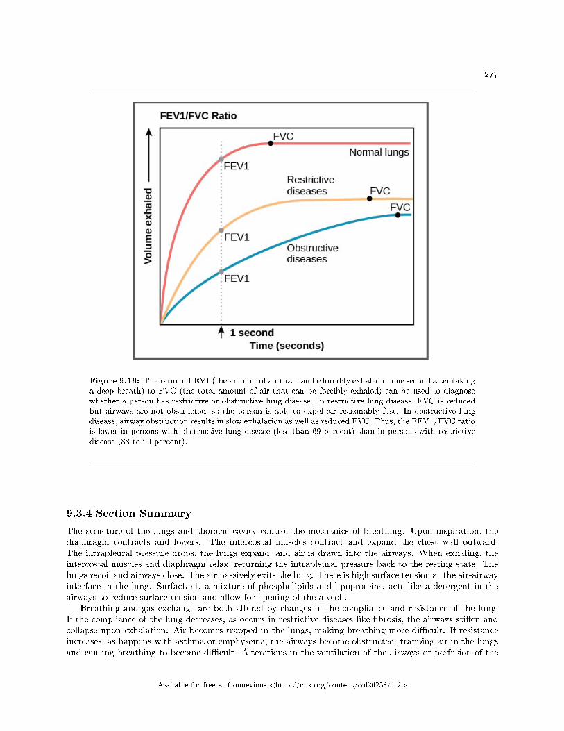
277
Figure 9.16: The ratio of FEV1 (the amount of air that can be forcibly exhaled in one second after takinga deep breath) to FVC (the total amount of air that can be forcibly exhaled) can be used to diagnosewhether a person has restrictive or obstructive lung disease. In restrictive lung disease, FVC is reducedbut airways are not obstructed, so the person is able to expel air reasonably fast. In obstructive lungdisease, airway obstruction results in slow exhalation as well as reduced FVC. Thus, the FEV1/FVC ratiois lower in persons with obstructive lung disease (less than 69 percent) than in persons with restrictivedisease (88 to 90 percent).
9.3.4 Section Summary
The structure of the lungs and thoracic cavity control the mechanics of breathing. Upon inspiration, thediaphragm contracts and lowers. The intercostal muscles contract and expand the chest wall outward.The intrapleural pressure drops, the lungs expand, and air is drawn into the airways. When exhaling, theintercostal muscles and diaphragm relax, returning the intrapleural pressure back to the resting state. Thelungs recoil and airways close. The air passively exits the lung. There is high surface tension at the air-airwayinterface in the lung. Surfactant, a mixture of phospholipids and lipoproteins, acts like a detergent in theairways to reduce surface tension and allow for opening of the alveoli.
Breathing and gas exchange are both altered by changes in the compliance and resistance of the lung.If the compliance of the lung decreases, as occurs in restrictive diseases like �brosis, the airways sti�en andcollapse upon exhalation. Air becomes trapped in the lungs, making breathing more di�cult. If resistanceincreases, as happens with asthma or emphysema, the airways become obstructed, trapping air in the lungsand causing breathing to become di�cult. Alterations in the ventilation of the airways or perfusion of the
Available for free at Connexions <http://cnx.org/content/col26253/1.2>
278 CHAPTER 9. RESPIRATION
arteries can a�ect gas exchange. These changes in ventilation and perfusion, called V/Q mismatch, can arisefrom anatomical or physiological changes.
9.3.5 Review Questions
Exercise 9.3.1 (Solution on p. 285.)
How would paralysis of the diaphragm alter inspiration?
a. It would prevent contraction of the intercostal muscles.b. It would prevent inhalation because the intrapleural pressure would not change.c. It would decrease the intrapleural pressure and allow more air to enter the lungs.d. It would slow expiration because the lung would not relax.
Exercise 9.3.2 (Solution on p. 285.)
Restrictive airway diseases ________.
a. increase the compliance of the lungb. decrease the compliance of the lungc. increase the lung volumed. decrease the work of breathing
Exercise 9.3.3 (Solution on p. 285.)
Alveolar ventilation remains constant when ________.
a. the respiratory rate is increased while the volume of air per breath is decreasedb. the respiratory rate and the volume of air per breath are increasedc. the respiratory rate is decreased while increasing the volume per breathd. both a and c
9.3.6 Free Response
Exercise 9.3.4 (Solution on p. 285.)
How would increased airway resistance a�ect intrapleural pressure during inhalation?
Exercise 9.3.5 (Solution on p. 285.)
Explain how a puncture to the thoracic cavity (from a knife wound, for instance) could alter theability to inhale.
Exercise 9.3.6 (Solution on p. 286.)
When someone is standing, gravity stretches the bottom of the lung down toward the �oor to agreater extent than the top of the lung. What implication could this have on the �ow of air in thelungs? Where does gas exchange occur in the lungs?
9.4 Transport of Gases in Human Bodily Fluids5
Once the oxygen di�uses across the alveoli, it enters the bloodstream and is transported to the tissues whereit is unloaded, and carbon dioxide di�uses out of the blood and into the alveoli to be expelled from the body.Although gas exchange is a continuous process, the oxygen and carbon dioxide are transported by di�erentmechanisms.
5This content is available online at <http://cnx.org/content/m44799/1.6/>.
Available for free at Connexions <http://cnx.org/content/col26253/1.2>
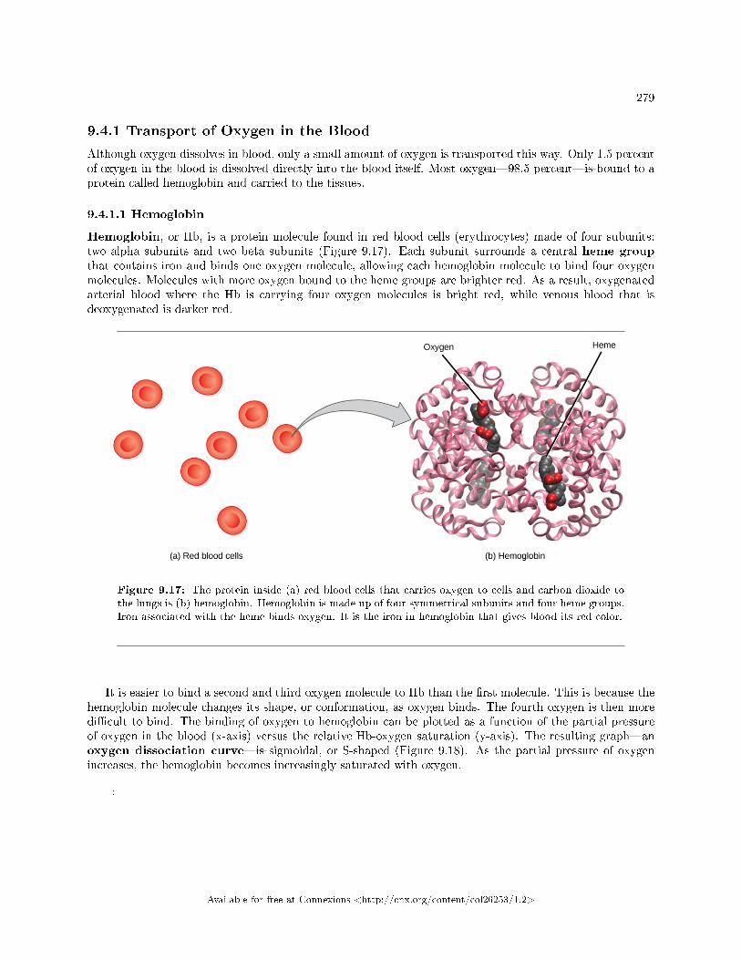
279
9.4.1 Transport of Oxygen in the Blood
Although oxygen dissolves in blood, only a small amount of oxygen is transported this way. Only 1.5 percentof oxygen in the blood is dissolved directly into the blood itself. Most oxygen�98.5 percent�is bound to aprotein called hemoglobin and carried to the tissues.
9.4.1.1 Hemoglobin
Hemoglobin, or Hb, is a protein molecule found in red blood cells (erythrocytes) made of four subunits:two alpha subunits and two beta subunits (Figure 9.17). Each subunit surrounds a central heme groupthat contains iron and binds one oxygen molecule, allowing each hemoglobin molecule to bind four oxygenmolecules. Molecules with more oxygen bound to the heme groups are brighter red. As a result, oxygenatedarterial blood where the Hb is carrying four oxygen molecules is bright red, while venous blood that isdeoxygenated is darker red.
Figure 9.17: The protein inside (a) red blood cells that carries oxygen to cells and carbon dioxide tothe lungs is (b) hemoglobin. Hemoglobin is made up of four symmetrical subunits and four heme groups.Iron associated with the heme binds oxygen. It is the iron in hemoglobin that gives blood its red color.
It is easier to bind a second and third oxygen molecule to Hb than the �rst molecule. This is because thehemoglobin molecule changes its shape, or conformation, as oxygen binds. The fourth oxygen is then moredi�cult to bind. The binding of oxygen to hemoglobin can be plotted as a function of the partial pressureof oxygen in the blood (x-axis) versus the relative Hb-oxygen saturation (y-axis). The resulting graph�anoxygen dissociation curve�is sigmoidal, or S-shaped (Figure 9.18). As the partial pressure of oxygenincreases, the hemoglobin becomes increasingly saturated with oxygen.
:
Available for free at Connexions <http://cnx.org/content/col26253/1.2>
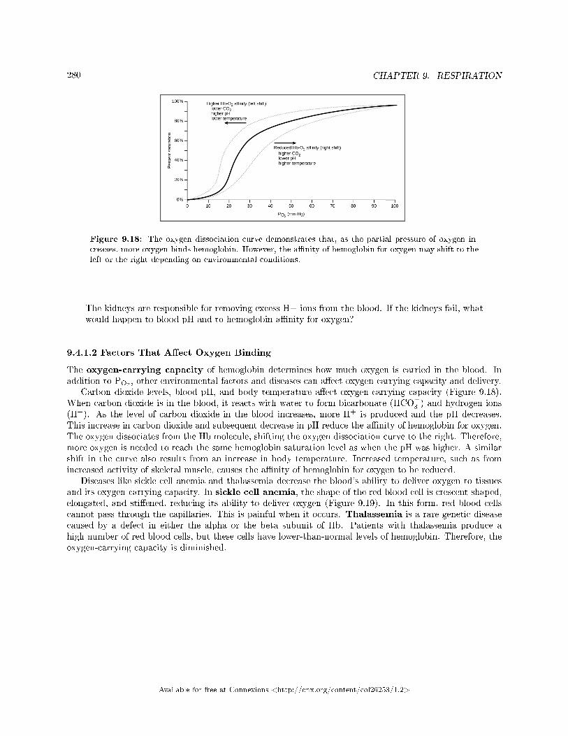
280 CHAPTER 9. RESPIRATION
Figure 9.18: The oxygen dissociation curve demonstrates that, as the partial pressure of oxygen in-creases, more oxygen binds hemoglobin. However, the a�nity of hemoglobin for oxygen may shift to theleft or the right depending on environmental conditions.
The kidneys are responsible for removing excess H+ ions from the blood. If the kidneys fail, whatwould happen to blood pH and to hemoglobin a�nity for oxygen?
9.4.1.2 Factors That A�ect Oxygen Binding
The oxygen-carrying capacity of hemoglobin determines how much oxygen is carried in the blood. Inaddition to PO2
, other environmental factors and diseases can a�ect oxygen carrying capacity and delivery.Carbon dioxide levels, blood pH, and body temperature a�ect oxygen-carrying capacity (Figure 9.18).
When carbon dioxide is in the blood, it reacts with water to form bicarbonate (HCO−3 ) and hydrogen ions
(H+). As the level of carbon dioxide in the blood increases, more H+ is produced and the pH decreases.This increase in carbon dioxide and subsequent decrease in pH reduce the a�nity of hemoglobin for oxygen.The oxygen dissociates from the Hb molecule, shifting the oxygen dissociation curve to the right. Therefore,more oxygen is needed to reach the same hemoglobin saturation level as when the pH was higher. A similarshift in the curve also results from an increase in body temperature. Increased temperature, such as fromincreased activity of skeletal muscle, causes the a�nity of hemoglobin for oxygen to be reduced.
Diseases like sickle cell anemia and thalassemia decrease the blood's ability to deliver oxygen to tissuesand its oxygen-carrying capacity. In sickle cell anemia, the shape of the red blood cell is crescent-shaped,elongated, and sti�ened, reducing its ability to deliver oxygen (Figure 9.19). In this form, red blood cellscannot pass through the capillaries. This is painful when it occurs. Thalassemia is a rare genetic diseasecaused by a defect in either the alpha or the beta subunit of Hb. Patients with thalassemia produce ahigh number of red blood cells, but these cells have lower-than-normal levels of hemoglobin. Therefore, theoxygen-carrying capacity is diminished.
Available for free at Connexions <http://cnx.org/content/col26253/1.2>
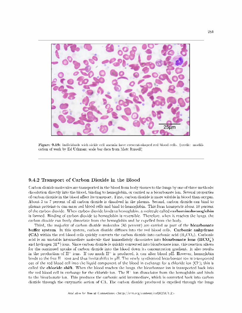
281
Figure 9.19: Individuals with sickle cell anemia have crescent-shaped red blood cells. (credit: modi�-cation of work by Ed Uthman; scale-bar data from Matt Russell)
9.4.2 Transport of Carbon Dioxide in the Blood
Carbon dioxide molecules are transported in the blood from body tissues to the lungs by one of three methods:dissolution directly into the blood, binding to hemoglobin, or carried as a bicarbonate ion. Several propertiesof carbon dioxide in the blood a�ect its transport. First, carbon dioxide is more soluble in blood than oxygen.About 5 to 7 percent of all carbon dioxide is dissolved in the plasma. Second, carbon dioxide can bind toplasma proteins or can enter red blood cells and bind to hemoglobin. This form transports about 10 percentof the carbon dioxide. When carbon dioxide binds to hemoglobin, a molecule called carbaminohemoglobinis formed. Binding of carbon dioxide to hemoglobin is reversible. Therefore, when it reaches the lungs, thecarbon dioxide can freely dissociate from the hemoglobin and be expelled from the body.
Third, the majority of carbon dioxide molecules (85 percent) are carried as part of the bicarbonatebu�er system. In this system, carbon dioxide di�uses into the red blood cells. Carbonic anhydrase(CA) within the red blood cells quickly converts the carbon dioxide into carbonic acid (H2CO3). Carbonicacid is an unstable intermediate molecule that immediately dissociates into bicarbonate ions (HCO
−3 )
and hydrogen (H+) ions. Since carbon dioxide is quickly converted into bicarbonate ions, this reaction allowsfor the continued uptake of carbon dioxide into the blood down its concentration gradient. It also resultsin the production of H+ ions. If too much H+ is produced, it can alter blood pH. However, hemoglobinbinds to the free H+ ions and thus limits shifts in pH. The newly synthesized bicarbonate ion is transportedout of the red blood cell into the liquid component of the blood in exchange for a chloride ion (Cl-); this iscalled the chloride shift. When the blood reaches the lungs, the bicarbonate ion is transported back intothe red blood cell in exchange for the chloride ion. The H+ ion dissociates from the hemoglobin and bindsto the bicarbonate ion. This produces the carbonic acid intermediate, which is converted back into carbondioxide through the enzymatic action of CA. The carbon dioxide produced is expelled through the lungs
Available for free at Connexions <http://cnx.org/content/col26253/1.2>
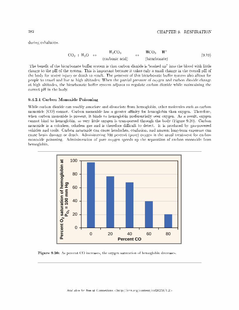
282 CHAPTER 9. RESPIRATION
during exhalation.
CO2 + H2O ↔H2CO3
(carbonic acid)↔
HCO3 + H+
(bicarbonate)(9.19)
The bene�t of the bicarbonate bu�er system is that carbon dioxide is �soaked up� into the blood with littlechange to the pH of the system. This is important because it takes only a small change in the overall pH ofthe body for severe injury or death to result. The presence of this bicarbonate bu�er system also allows forpeople to travel and live at high altitudes: When the partial pressure of oxygen and carbon dioxide changeat high altitudes, the bicarbonate bu�er system adjusts to regulate carbon dioxide while maintaining thecorrect pH in the body.
9.4.2.1 Carbon Monoxide Poisoning
While carbon dioxide can readily associate and dissociate from hemoglobin, other molecules such as carbonmonoxide (CO) cannot. Carbon monoxide has a greater a�nity for hemoglobin than oxygen. Therefore,when carbon monoxide is present, it binds to hemoglobin preferentially over oxygen. As a result, oxygencannot bind to hemoglobin, so very little oxygen is transported through the body (Figure 9.20). Carbonmonoxide is a colorless, odorless gas and is therefore di�cult to detect. It is produced by gas-poweredvehicles and tools. Carbon monoxide can cause headaches, confusion, and nausea; long-term exposure cancause brain damage or death. Administering 100 percent (pure) oxygen is the usual treatment for carbonmonoxide poisoning. Administration of pure oxygen speeds up the separation of carbon monoxide fromhemoglobin.
Figure 9.20: As percent CO increases, the oxygen saturation of hemoglobin decreases.
Available for free at Connexions <http://cnx.org/content/col26253/1.2>
283
9.4.3 Section Summary
Hemoglobin is a protein found in red blood cells that is comprised of two alpha and two beta subunits thatsurround an iron-containing heme group. Oxygen readily binds this heme group. The ability of oxygento bind increases as more oxygen molecules are bound to heme. Disease states and altered conditions inthe body can a�ect the binding ability of oxygen, and increase or decrease its ability to dissociate fromhemoglobin.
Carbon dioxide can be transported through the blood via three methods. It is dissolved directly inthe blood, bound to plasma proteins or hemoglobin, or converted into bicarbonate. The majority of carbondioxide is transported as part of the bicarbonate system. Carbon dioxide di�uses into red blood cells. Inside,carbonic anhydrase converts carbon dioxide into carbonic acid (H2CO3), which is subsequently hydrolyzedinto bicarbonate (HCO
−3 ) and H+. The H+ ion binds to hemoglobin in red blood cells, and bicarbonate
is transported out of the red blood cells in exchange for a chloride ion. This is called the chloride shift.Bicarbonate leaves the red blood cells and enters the blood plasma. In the lungs, bicarbonate is transportedback into the red blood cells in exchange for chloride. The H+ dissociates from hemoglobin and combineswith bicarbonate to form carbonic acid with the help of carbonic anhydrase, which further catalyzes thereaction to convert carbonic acid back into carbon dioxide and water. The carbon dioxide is then expelledfrom the lungs.
9.4.4 Art Connections
Exercise 9.4.1 (Solution on p. 286.)
Figure 9.18 The kidneys are responsible for removing excess H+ ions from the blood. If the kidneysfail, what would happen to blood pH and to hemoglobin a�nity for oxygen?
9.4.5 Review Questions
Exercise 9.4.2 (Solution on p. 286.)
Which of the following will NOT facilitate the transfer of oxygen to tissues?
a. decreased body temperatureb. decreased pH of the bloodc. increased carbon dioxided. increased exercise
Exercise 9.4.3 (Solution on p. 286.)
The majority of carbon dioxide in the blood is transported by ________.
a. binding to hemoglobinb. dissolution in the bloodc. conversion to bicarbonated. binding to plasma proteins
Exercise 9.4.4 (Solution on p. 286.)
The majority of oxygen in the blood is transported by ________.
a. dissolution in the bloodb. being carried as bicarbonate ionsc. binding to blood plasmad. binding to hemoglobin
Available for free at Connexions <http://cnx.org/content/col26253/1.2>
284 CHAPTER 9. RESPIRATION
9.4.6 Free Response
Exercise 9.4.5 (Solution on p. 286.)
What would happen if no carbonic anhydrase were present in red blood cells?
Exercise 9.4.6 (Solution on p. 286.)
How does the administration of 100 percent oxygen save a patient from carbon monoxide poisoning?Why wouldn't giving carbon dioxide work?
Available for free at Connexions <http://cnx.org/content/col26253/1.2>
285
Solutions to Exercises in Chapter 9
to Exercise 9.1.1 (p. 264)Figure 9.6 Bto Exercise 9.1.2 (p. 264)Ato Exercise 9.1.3 (p. 264)Cto Exercise 9.1.4 (p. 264)Bto Exercise 9.1.5 (p. 264)The main bronchus is the conduit in the lung that funnels air to the airways where gas exchange occurs.The main bronchus attaches the lungs to the very end of the trachea where it bifurcates. The trachea is thecartilaginous structure that extends from the pharynx to the primary bronchi. It serves to funnel air to thelungs. The alveoli are the sites of gas exchange; they are located at the terminal regions of the lung and areattached to the respiratory bronchioles. The acinus is the structure in the lung where gas exchange occurs.to Exercise 9.1.6 (p. 264)The sac-like structure of the alveoli increases their surface area. In addition, the alveoli are made ofthin-walled parenchymal cells. These features allow gases to easily di�use across the cells.to Exercise 9.2.1 (p. 270)Figure 9.12 Cto Exercise 9.2.2 (p. 271)Dto Exercise 9.2.3 (p. 271)Dto Exercise 9.2.4 (p. 271)Dto Exercise 9.2.5 (p. 271)FEV1/FVC measures the forced expiratory volume in one second in relation to the total forced vital capacity(the total amount of air that is exhaled from the lung from a maximal inhalation). This ratio changes withalterations in lung function that arise from diseases such as �brosis, asthma, and COPD.to Exercise 9.2.6 (p. 271)If all the air in the lung were exhaled, then opening the alveoli for the next inspiration would be verydi�cult. This is because the tissues would stick together.to Exercise 9.2.7 (p. 271)Oxygen moves from the lung to the bloodstream to the tissues according to the pressure gradient. This ismeasured as the partial pressure of oxygen. If the amount of oxygen drops in the inspired air, there wouldbe reduced partial pressure. This would decrease the driving force that moves the oxygen into the bloodand into the tissues. PO2
is also reduced at high elevations: PO2at high elevations is lower than at sea level
because the total atmospheric pressure is less than atmospheric pressure at sea level.to Exercise 9.2.8 (p. 271)A doctor can detect a restrictive disease using spirometry. By detecting the rate at which air can be expelledfrom the lung, a diagnosis of �brosis or another restrictive disease can be made.to Exercise 9.3.1 (p. 278)Bto Exercise 9.3.2 (p. 278)Bto Exercise 9.3.3 (p. 278)Dto Exercise 9.3.4 (p. 278)Increased airway resistance increases the volume and pressure in the lung; therefore, the intrapleural pressurewould be less negative and breathing would be more di�cult.
Available for free at Connexions <http://cnx.org/content/col26253/1.2>
286 CHAPTER 9. RESPIRATION
to Exercise 9.3.5 (p. 278)A puncture to the thoracic cavity would equalize the pressure inside the thoracic cavity to the outsideenvironment. For the lung to function properly, the intrapleural pressure must be negative. This is causedby the contraction of the diaphragm pulling the lungs down and drawing air into the lungs.to Exercise 9.3.6 (p. 278)The lung is particularly susceptible to changes in the magnitude and direction of gravitational forces. Whensomeone is standing or sitting upright, the pleural pressure gradient leads to increased ventilation furtherdown in the lung.to Exercise 9.4.1 (p. 283)Figure 9.18 The blood pH will drop and hemoglobin a�nity for oxygen will decrease.to Exercise 9.4.2 (p. 283)Ato Exercise 9.4.3 (p. 283)Cto Exercise 9.4.4 (p. 283)Dto Exercise 9.4.5 (p. 284)Without carbonic anhydrase, carbon dioxide would not be hydrolyzed into carbonic acid or bicarbonate.Therefore, very little carbon dioxide (only 15 percent) would be transported in the blood away from thetissues.to Exercise 9.4.6 (p. 284)Carbon monoxide has a higher a�nity for hemoglobin than oxygen. This means that carbon monoxide willpreferentially bind to hemoglobin over oxygen. Administration of 100 percent oxygen is an e�ective therapybecause at that concentration, oxygen will displace the carbon monoxide from the hemoglobin.
Available for free at Connexions <http://cnx.org/content/col26253/1.2>
Chapter 10
Immunity
10.1 Innate Immune Response1
The immune system comprises both innate and adaptive immune responses. Innate immunity occursnaturally because of genetic factors or physiology; it is not induced by infection or vaccination but works toreduce the workload for the adaptive immune response. Both the innate and adaptive levels of the immuneresponse involve secreted proteins, receptor-mediated signaling, and intricate cell-to-cell communication.The innate immune system developed early in animal evolution, roughly a billion years ago, as an essentialresponse to infection. Innate immunity has a limited number of speci�c targets: any pathogenic threattriggers a consistent sequence of events that can identify the type of pathogen and either clear the infectionindependently or mobilize a highly specialized adaptive immune response. For example, tears and mucussecretions contain microbicidal factors.
10.1.1 Physical and Chemical Barriers
Before any immune factors are triggered, the skin functions as a continuous, impassable barrier to potentiallyinfectious pathogens. Pathogens are killed or inactivated on the skin by desiccation (drying out) and bythe skin's acidity. In addition, bene�cial microorganisms that coexist on the skin compete with invadingpathogens, preventing infection. Regions of the body that are not protected by skin (such as the eyes andmucus membranes) have alternative methods of defense, such as tears and mucus secretions that trap andrinse away pathogens, and cilia in the nasal passages and respiratory tract that push the mucus with thepathogens out of the body. Throughout the body are other defenses, such as the low pH of the stomach(which inhibits the growth of pathogens), blood proteins that bind and disrupt bacterial cell membranes,and the process of urination (which �ushes pathogens from the urinary tract).
Despite these barriers, pathogens may enter the body through skin abrasions or punctures, or by collectingon mucosal surfaces in large numbers that overcome the mucus or cilia. Some pathogens have evolvedspeci�c mechanisms that allow them to overcome physical and chemical barriers. When pathogens do enterthe body, the innate immune system responds with in�ammation, pathogen engulfment, and secretion ofimmune factors and proteins.
10.1.2 Pathogen Recognition
An infection may be intracellular or extracellular, depending on the pathogen. All viruses infect cells andreplicate within those cells (intracellularly), whereas bacteria and other parasites may replicate intracellu-larly or extracellularly, depending on the species. The innate immune system must respond accordingly:by identifying the extracellular pathogen and/or by identifying host cells that have already been infected.
1This content is available online at <http://cnx.org/content/m66663/1.13/>.
Available for free at Connexions <http://cnx.org/content/col26253/1.2>
287
288 CHAPTER 10. IMMUNITY
When a pathogen enters the body, cells in the blood and lymph detect the speci�c pathogen-associatedmolecular patterns (PAMPs) on the pathogen's surface. PAMPs are carbohydrate, polypeptide, and nu-cleic acid �signatures� that are expressed by viruses, bacteria, and parasites but which di�er from moleculeson host cells. The immune system has speci�c cells, described in Figure 10.1 and shown in Figure 10.2, withreceptors that recognize these PAMPs. A macrophage is a large phagocytic cell that engulfs foreign par-ticles and pathogens. Macrophages recognize PAMPs via complementary pattern recognition receptors(PRRs). PRRs are molecules on macrophages and dendritic cells which are in contact with the externalenvironment. A monocyte is a type of white blood cell that circulates in the blood and lymph and di�er-entiates into macrophages after it moves into infected tissue. Dendritic cells bind molecular signatures ofpathogens and promote pathogen engulfment and destruction. Toll-like receptors (TLRs) are a type of PRRthat recognizes molecules that are shared by pathogens but distinguishable from host molecules. TLRs arepresent in invertebrates as well as vertebrates, and appear to be one of the most ancient components of theimmune system. TLRs have also been identi�ed in the mammalian nervous system.
Available for free at Connexions <http://cnx.org/content/col26253/1.2>
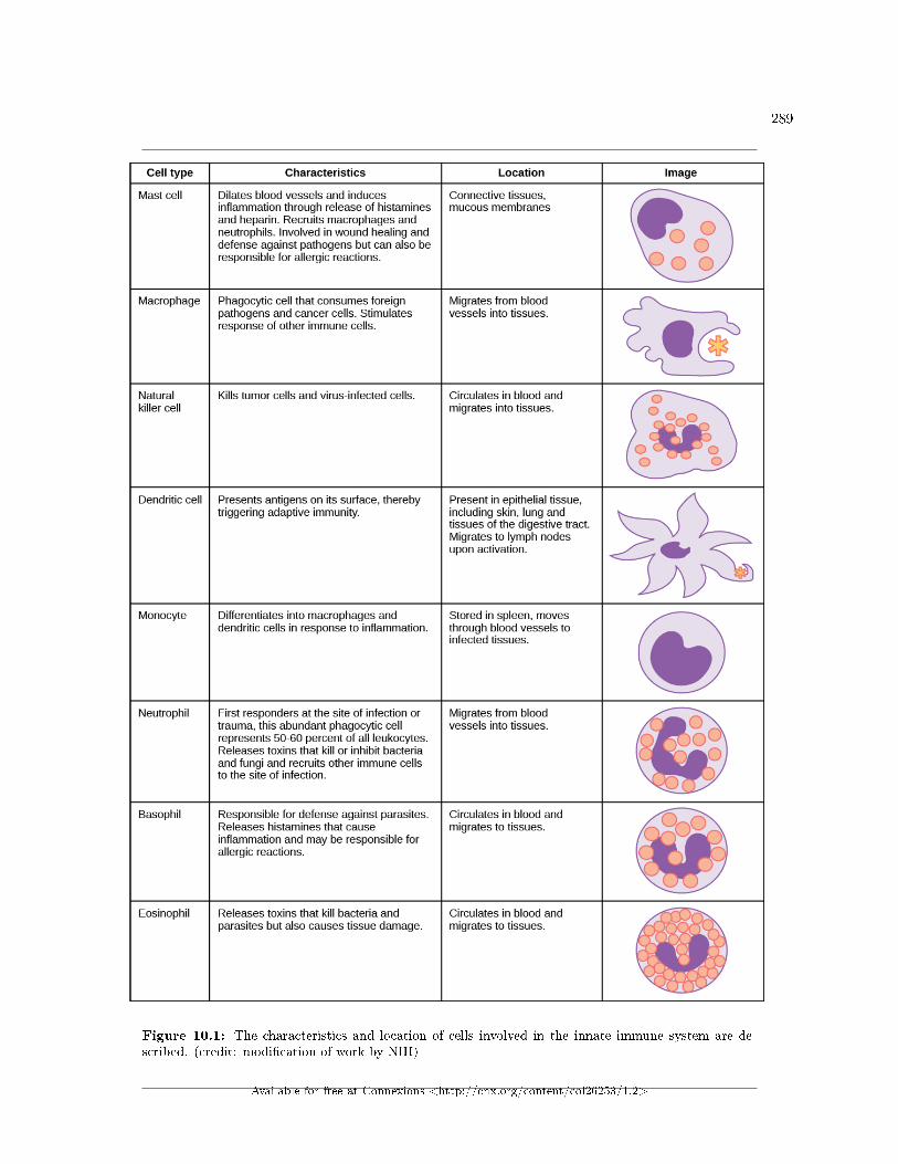
289
Figure 10.1: The characteristics and location of cells involved in the innate immune system are de-scribed. (credit: modi�cation of work by NIH)
Available for free at Connexions <http://cnx.org/content/col26253/1.2>
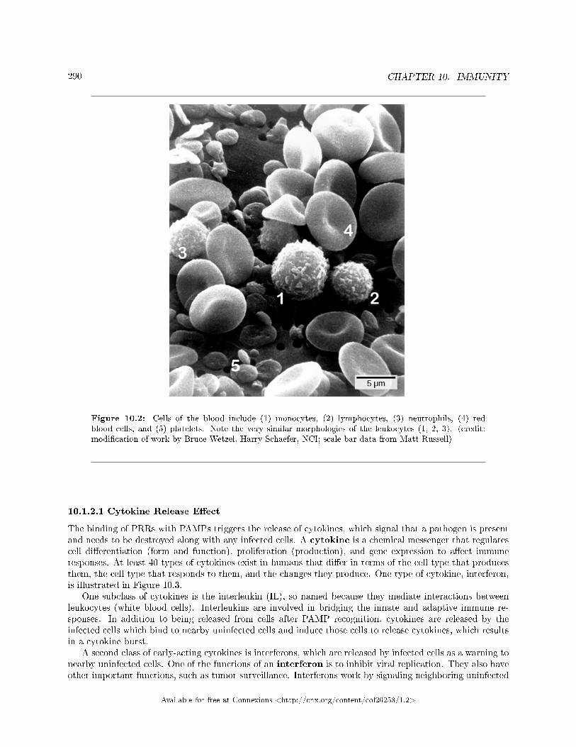
290 CHAPTER 10. IMMUNITY
Figure 10.2: Cells of the blood include (1) monocytes, (2) lymphocytes, (3) neutrophils, (4) redblood cells, and (5) platelets. Note the very similar morphologies of the leukocytes (1, 2, 3). (credit:modi�cation of work by Bruce Wetzel, Harry Schaefer, NCI; scale-bar data from Matt Russell)
10.1.2.1 Cytokine Release E�ect
The binding of PRRs with PAMPs triggers the release of cytokines, which signal that a pathogen is presentand needs to be destroyed along with any infected cells. A cytokine is a chemical messenger that regulatescell di�erentiation (form and function), proliferation (production), and gene expression to a�ect immuneresponses. At least 40 types of cytokines exist in humans that di�er in terms of the cell type that producesthem, the cell type that responds to them, and the changes they produce. One type of cytokine, interferon,is illustrated in Figure 10.3.
One subclass of cytokines is the interleukin (IL), so named because they mediate interactions betweenleukocytes (white blood cells). Interleukins are involved in bridging the innate and adaptive immune re-sponses. In addition to being released from cells after PAMP recognition, cytokines are released by theinfected cells which bind to nearby uninfected cells and induce those cells to release cytokines, which resultsin a cytokine burst.
A second class of early-acting cytokines is interferons, which are released by infected cells as a warning tonearby uninfected cells. One of the functions of an interferon is to inhibit viral replication. They also haveother important functions, such as tumor surveillance. Interferons work by signaling neighboring uninfected
Available for free at Connexions <http://cnx.org/content/col26253/1.2>
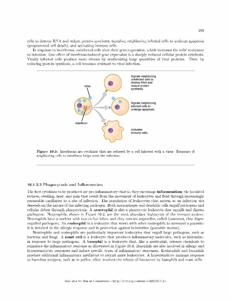
291
cells to destroy RNA and reduce protein synthesis, signaling neighboring infected cells to undergo apoptosis(programmed cell death), and activating immune cells.
In response to interferons, uninfected cells alter their gene expression, which increases the cells' resistanceto infection. One e�ect of interferon-induced gene expression is a sharply reduced cellular protein synthesis.Virally infected cells produce more viruses by synthesizing large quantities of viral proteins. Thus, byreducing protein synthesis, a cell becomes resistant to viral infection.
Figure 10.3: Interferons are cytokines that are released by a cell infected with a virus. Response ofneighboring cells to interferon helps stem the infection.
10.1.2.2 Phagocytosis and In�ammation
The �rst cytokines to be produced are pro-in�ammatory; that is, they encourage in�ammation, the localizedredness, swelling, heat, and pain that result from the movement of leukocytes and �uid through increasinglypermeable capillaries to a site of infection. The population of leukocytes that arrives at an infection sitedepends on the nature of the infecting pathogen. Both macrophages and dendritic cells engulf pathogens andcellular debris through phagocytosis. A neutrophil is also a phagocytic leukocyte that engulfs and digestspathogens. Neutrophils, shown in Figure 10.2, are the most abundant leukocytes of the immune system.Neutrophils have a nucleus with two to �ve lobes, and they contain organelles, called lysosomes, that digestengulfed pathogens. An eosinophil is a leukocyte that works with other eosinophils to surround a parasite;it is involved in the allergic response and in protection against helminthes (parasitic worms).
Neutrophils and eosinophils are particularly important leukocytes that engulf large pathogens, such asbacteria and fungi. A mast cell is a leukocyte that produces in�ammatory molecules, such as histamine,in response to large pathogens. A basophil is a leukocyte that, like a neutrophil, releases chemicals tostimulate the in�ammatory response as illustrated in Figure 10.4. Basophils are also involved in allergy andhypersensitivity responses and induce speci�c types of in�ammatory responses. Eosinophils and basophilsproduce additional in�ammatory mediators to recruit more leukocytes. A hypersensitive immune responseto harmless antigens, such as in pollen, often involves the release of histamine by basophils and mast cells.
Available for free at Connexions <http://cnx.org/content/col26253/1.2>
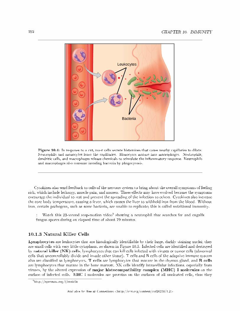
292 CHAPTER 10. IMMUNITY
Figure 10.4: In response to a cut, mast cells secrete histamines that cause nearby capillaries to dilate.Neutrophils and monocytes leave the capillaries. Monocytes mature into macrophages. Neutrophils,dendritic cells, and macrophages release chemicals to stimulate the in�ammatory response. Neutrophilsand macrophages also consume invading bacteria by phagocytosis.
Cytokines also send feedback to cells of the nervous system to bring about the overall symptoms of feelingsick, which include lethargy, muscle pain, and nausea. These e�ects may have evolved because the symptomsencourage the individual to rest and prevent the spreading of the infection to others. Cytokines also increasethe core body temperature, causing a fever, which causes the liver to withhold iron from the blood. Withoutiron, certain pathogens, such as some bacteria, are unable to replicate; this is called nutritional immunity.
: Watch this 23-second stop-motion video2 showing a neutrophil that searches for and engulfsfungus spores during an elapsed time of about 79 minutes.
10.1.3 Natural Killer Cells
Lymphocytes are leukocytes that are histologically identi�able by their large, darkly staining nuclei; theyare small cells with very little cytoplasm, as shown in Figure 10.5. Infected cells are identi�ed and destroyedby natural killer (NK) cells, lymphocytes that can kill cells infected with viruses or tumor cells (abnormalcells that uncontrollably divide and invade other tissue). T cells and B cells of the adaptive immune systemalso are classi�ed as lymphocytes. T cells are lymphocytes that mature in the thymus gland, and B cellsare lymphocytes that mature in the bone marrow. NK cells identify intracellular infections, especially fromviruses, by the altered expression of major histocompatibility complex (MHC) I molecules on thesurface of infected cells. MHC I molecules are proteins on the surfaces of all nucleated cells, thus they
2http://openstax.org/l/conidia
Available for free at Connexions <http://cnx.org/content/col26253/1.2>
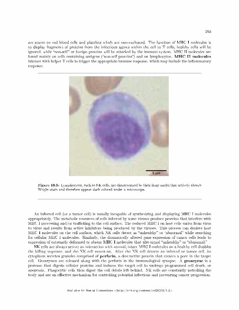
293
are scarce on red blood cells and platelets which are non-nucleated. The function of MHC I molecules isto display fragments of proteins from the infectious agents within the cell to T cells; healthy cells will beignored, while �non-self� or foreign proteins will be attacked by the immune system. MHC II molecules arefound mainly on cells containing antigens (�non-self proteins�) and on lymphocytes. MHC II moleculesinteract with helper T cells to trigger the appropriate immune response, which may include the in�ammatoryresponse.
Figure 10.5: Lymphocytes, such as NK cells, are characterized by their large nuclei that actively absorbWright stain and therefore appear dark colored under a microscope.
An infected cell (or a tumor cell) is usually incapable of synthesizing and displaying MHC I moleculesappropriately. The metabolic resources of cells infected by some viruses produce proteins that interfere withMHC I processing and/or tra�cking to the cell surface. The reduced MHC I on host cells varies from virusto virus and results from active inhibitors being produced by the viruses. This process can deplete hostMHC I molecules on the cell surface, which NK cells detect as �unhealthy� or �abnormal� while searchingfor cellular MHC I molecules. Similarly, the dramatically altered gene expression of tumor cells leads toexpression of extremely deformed or absent MHC I molecules that also signal �unhealthy� or �abnormal.�
NK cells are always active; an interaction with normal, intact MHC I molecules on a healthy cell disablesthe killing sequence, and the NK cell moves on. After the NK cell detects an infected or tumor cell, itscytoplasm secretes granules comprised of perforin, a destructive protein that creates a pore in the targetcell. Granzymes are released along with the perforin in the immunological synapse. A granzyme is aprotease that digests cellular proteins and induces the target cell to undergo programmed cell death, orapoptosis. Phagocytic cells then digest the cell debris left behind. NK cells are constantly patrolling thebody and are an e�ective mechanism for controlling potential infections and preventing cancer progression.
Available for free at Connexions <http://cnx.org/content/col26253/1.2>
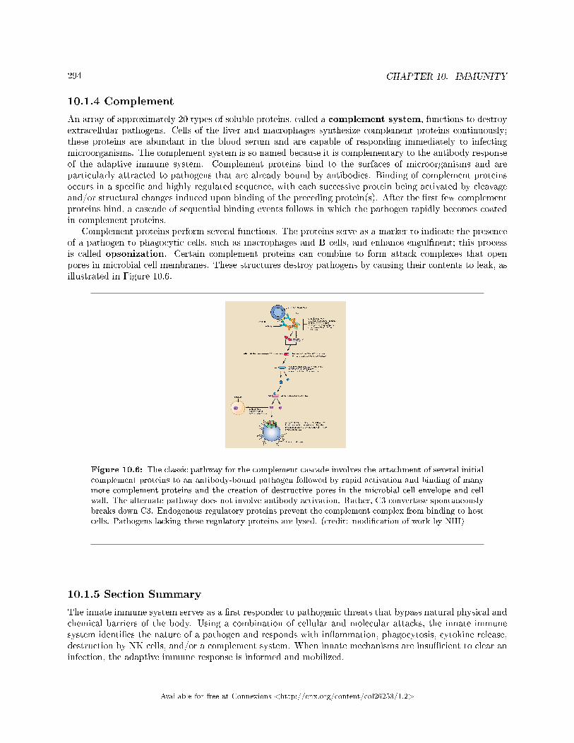
294 CHAPTER 10. IMMUNITY
10.1.4 Complement
An array of approximately 20 types of soluble proteins, called a complement system, functions to destroyextracellular pathogens. Cells of the liver and macrophages synthesize complement proteins continuously;these proteins are abundant in the blood serum and are capable of responding immediately to infectingmicroorganisms. The complement system is so named because it is complementary to the antibody responseof the adaptive immune system. Complement proteins bind to the surfaces of microorganisms and areparticularly attracted to pathogens that are already bound by antibodies. Binding of complement proteinsoccurs in a speci�c and highly regulated sequence, with each successive protein being activated by cleavageand/or structural changes induced upon binding of the preceding protein(s). After the �rst few complementproteins bind, a cascade of sequential binding events follows in which the pathogen rapidly becomes coatedin complement proteins.
Complement proteins perform several functions. The proteins serve as a marker to indicate the presenceof a pathogen to phagocytic cells, such as macrophages and B cells, and enhance engulfment; this processis called opsonization. Certain complement proteins can combine to form attack complexes that openpores in microbial cell membranes. These structures destroy pathogens by causing their contents to leak, asillustrated in Figure 10.6.
Figure 10.6: The classic pathway for the complement cascade involves the attachment of several initialcomplement proteins to an antibody-bound pathogen followed by rapid activation and binding of manymore complement proteins and the creation of destructive pores in the microbial cell envelope and cellwall. The alternate pathway does not involve antibody activation. Rather, C3 convertase spontaneouslybreaks down C3. Endogenous regulatory proteins prevent the complement complex from binding to hostcells. Pathogens lacking these regulatory proteins are lysed. (credit: modi�cation of work by NIH)
10.1.5 Section Summary
The innate immune system serves as a �rst responder to pathogenic threats that bypass natural physical andchemical barriers of the body. Using a combination of cellular and molecular attacks, the innate immunesystem identi�es the nature of a pathogen and responds with in�ammation, phagocytosis, cytokine release,destruction by NK cells, and/or a complement system. When innate mechanisms are insu�cient to clear aninfection, the adaptive immune response is informed and mobilized.
Available for free at Connexions <http://cnx.org/content/col26253/1.2>
295
10.1.6 Review Questions
Exercise 10.1.1 (Solution on p. 320.)
Which of the following is a barrier against pathogens provided by the skin?
a. high pHb. mucusc. tearsd. desiccation
Exercise 10.1.2 (Solution on p. 320.)
Although interferons have several e�ects, they are particularly useful against infections with whichtype of pathogen?
a. bacteriab. virusesc. fungid. helminths
Exercise 10.1.3 (Solution on p. 320.)
Which organelle do phagocytes use to digest engulfed particles?
a. lysosomeb. nucleusc. endoplasmic reticulumd. mitochondria
Exercise 10.1.4 (Solution on p. 320.)
Which innate immune system component uses MHC I molecules directly in its defense strategy?
a. macrophagesb. neutrophilsc. NK cellsd. interferon
10.1.7 Critical Thinking Questions
Exercise 10.1.5 (Solution on p. 320.)
Di�erent MHC I molecules between donor and recipient cells can lead to rejection of a transplantedorgan or tissue. Suggest a reason for this.
Exercise 10.1.6 (Solution on p. 320.)
If a series of genetic mutations prevented some, but not all, of the complement proteins frombinding antibodies or pathogens, would the entire complement system be compromised?
Available for free at Connexions <http://cnx.org/content/col26253/1.2>
296 CHAPTER 10. IMMUNITY
10.2 Adaptive Immune Response3
The adaptive, or acquired, immune response takes days or even weeks to become established�much longerthan the innate response; however, adaptive immunity is more speci�c to pathogens and has memory. Adap-tive immunity is an immunity that occurs after exposure to an antigen either from a pathogen or a vac-cination. This part of the immune system is activated when the innate immune response is insu�cient tocontrol an infection. In fact, without information from the innate immune system, the adaptive responsecould not be mobilized. There are two types of adaptive responses: the cell-mediated immune response,which is carried out by T cells, and the humoral immune response, which is controlled by activated Bcells and antibodies. Activated T cells and B cells that are speci�c to molecular structures on the pathogenproliferate and attack the invading pathogen. Their attack can kill pathogens directly or secrete antibodiesthat enhance the phagocytosis of pathogens and disrupt the infection. Adaptive immunity also involves amemory to provide the host with long-term protection from reinfection with the same type of pathogen; onre-exposure, this memory will facilitate an e�cient and quick response.
10.2.1 Antigen-presenting Cells
Unlike NK cells of the innate immune system, B cells (B lymphocytes) are a type of white blood cell that givesrise to antibodies, whereas T cells (T lymphocytes) are a type of white blood cell that plays an importantrole in the immune response. T cells are a key component in the cell-mediated response�the speci�c immuneresponse that utilizes T cells to neutralize cells that have been infected with viruses and certain bacteria.There are three types of T cells: cytotoxic, helper, and suppressor T cells. Cytotoxic T cells destroy virus-infected cells in the cell-mediated immune response, and helper T cells play a part in activating both theantibody and the cell-mediated immune responses. Suppressor T cells deactivate T cells and B cells whenneeded, and thus prevent the immune response from becoming too intense.
An antigen is a foreign or �non-self� macromolecule that reacts with cells of the immune system. Notall antigens will provoke a response. For instance, individuals produce innumerable �self� antigens and areconstantly exposed to harmless foreign antigens, such as food proteins, pollen, or dust components. Thesuppression of immune responses to harmless macromolecules is highly regulated and typically preventsprocesses that could be damaging to the host, known as tolerance.
The innate immune system contains cells that detect potentially harmful antigens, and then inform theadaptive immune response about the presence of these antigens. An antigen-presenting cell (APC) isan immune cell that detects, engulfs, and informs the adaptive immune response about an infection. Whena pathogen is detected, these APCs will phagocytose the pathogen and digest it to form many di�erentfragments of the antigen. Antigen fragments will then be transported to the surface of the APC, where theywill serve as an indicator to other immune cells. Dendritic cells are immune cells that process antigenmaterial; they are present in the skin (Langerhans cells) and the lining of the nose, lungs, stomach, andintestines. Sometimes a dendritic cell presents on the surface of other cells to induce an immune response,thus functioning as an antigen-presenting cell. Macrophages also function as APCs. Before activation anddi�erentiation, B cells can also function as APCs.
After phagocytosis by APCs, the phagocytic vesicle fuses with an intracellular lysosome formingphagolysosome. Within the phagolysosome, the components are broken down into fragments; the frag-ments are then loaded onto MHC class I or MHC class II molecules and are transported to the cell surfacefor antigen presentation, as illustrated in Figure 10.7. Note that T lymphocytes cannot properly respondto the antigen unless it is processed and embedded in an MHC II molecule. APCs express MHC on theirsurfaces, and when combined with a foreign antigen, these complexes signal a �non-self� invader. Once thefragment of antigen is embedded in the MHC II molecule, the immune cell can respond. Helper T- cells areone of the main lymphocytes that respond to antigen-presenting cells. Recall that all other nucleated cellsof the body expressed MHC I molecules, which signal �healthy� or �normal.�
3This content is available online at <http://cnx.org/content/m44821/1.8/>.
Available for free at Connexions <http://cnx.org/content/col26253/1.2>
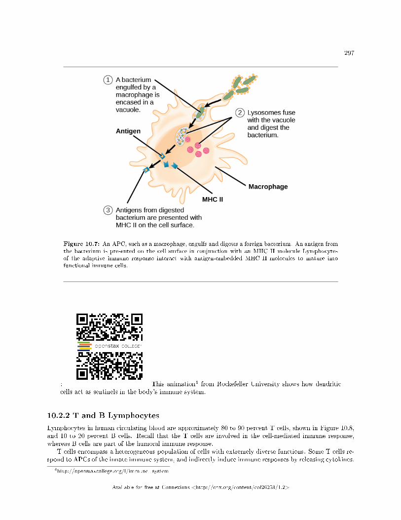
297
Figure 10.7: An APC, such as a macrophage, engulfs and digests a foreign bacterium. An antigen fromthe bacterium is presented on the cell surface in conjunction with an MHC II molecule Lymphocytesof the adaptive immune response interact with antigen-embedded MHC II molecules to mature intofunctional immune cells.
: This animation4 from Rockefeller University shows how dendriticcells act as sentinels in the body's immune system.
10.2.2 T and B Lymphocytes
Lymphocytes in human circulating blood are approximately 80 to 90 percent T cells, shown in Figure 10.8,and 10 to 20 percent B cells. Recall that the T cells are involved in the cell-mediated immune response,whereas B cells are part of the humoral immune response.
T cells encompass a heterogeneous population of cells with extremely diverse functions. Some T cells re-spond to APCs of the innate immune system, and indirectly induce immune responses by releasing cytokines.
4http://openstaxcollege.org/l/immune_system
Available for free at Connexions <http://cnx.org/content/col26253/1.2>
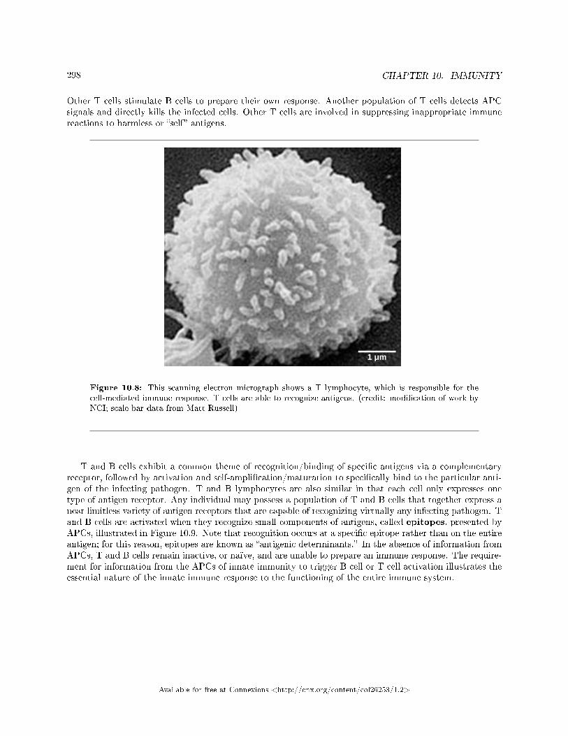
298 CHAPTER 10. IMMUNITY
Other T cells stimulate B cells to prepare their own response. Another population of T cells detects APCsignals and directly kills the infected cells. Other T cells are involved in suppressing inappropriate immunereactions to harmless or �self� antigens.
Figure 10.8: This scanning electron micrograph shows a T lymphocyte, which is responsible for thecell-mediated immune response. T cells are able to recognize antigens. (credit: modi�cation of work byNCI; scale-bar data from Matt Russell)
T and B cells exhibit a common theme of recognition/binding of speci�c antigens via a complementaryreceptor, followed by activation and self-ampli�cation/maturation to speci�cally bind to the particular anti-gen of the infecting pathogen. T and B lymphocytes are also similar in that each cell only expresses onetype of antigen receptor. Any individual may possess a population of T and B cells that together express anear limitless variety of antigen receptors that are capable of recognizing virtually any infecting pathogen. Tand B cells are activated when they recognize small components of antigens, called epitopes, presented byAPCs, illustrated in Figure 10.9. Note that recognition occurs at a speci�c epitope rather than on the entireantigen; for this reason, epitopes are known as �antigenic determinants.� In the absence of information fromAPCs, T and B cells remain inactive, or naïve, and are unable to prepare an immune response. The require-ment for information from the APCs of innate immunity to trigger B cell or T cell activation illustrates theessential nature of the innate immune response to the functioning of the entire immune system.
Available for free at Connexions <http://cnx.org/content/col26253/1.2>
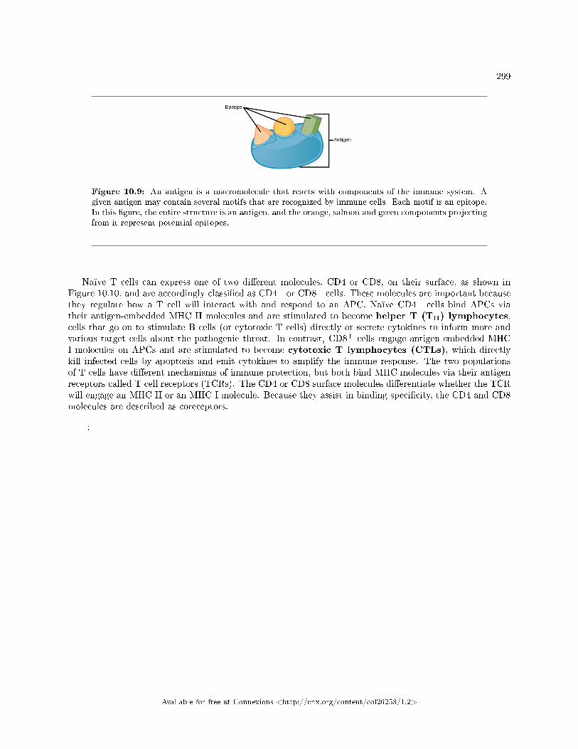
299
Figure 10.9: An antigen is a macromolecule that reacts with components of the immune system. Agiven antigen may contain several motifs that are recognized by immune cells. Each motif is an epitope.In this �gure, the entire structure is an antigen, and the orange, salmon and green components projectingfrom it represent potential epitopes.
Naïve T cells can express one of two di�erent molecules, CD4 or CD8, on their surface, as shown inFigure 10.10, and are accordingly classi�ed as CD4+ or CD8+ cells. These molecules are important becausethey regulate how a T cell will interact with and respond to an APC. Naïve CD4+ cells bind APCs viatheir antigen-embedded MHC II molecules and are stimulated to become helper T (TH) lymphocytes,cells that go on to stimulate B cells (or cytotoxic T cells) directly or secrete cytokines to inform more andvarious target cells about the pathogenic threat. In contrast, CD8+ cells engage antigen-embedded MHCI molecules on APCs and are stimulated to become cytotoxic T lymphocytes (CTLs), which directlykill infected cells by apoptosis and emit cytokines to amplify the immune response. The two populationsof T cells have di�erent mechanisms of immune protection, but both bind MHC molecules via their antigenreceptors called T cell receptors (TCRs). The CD4 or CD8 surface molecules di�erentiate whether the TCRwill engage an MHC II or an MHC I molecule. Because they assist in binding speci�city, the CD4 and CD8molecules are described as coreceptors.
:
Available for free at Connexions <http://cnx.org/content/col26253/1.2>
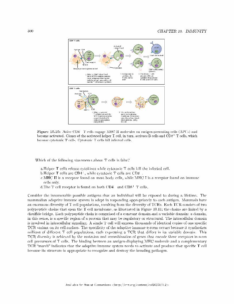
300 CHAPTER 10. IMMUNITY
Figure 10.10: Naïve CD4+ T cells engage MHC II molecules on antigen-presenting cells (APCs) andbecome activated. Clones of the activated helper T cell, in turn, activate B cells and CD8+ T cells, whichbecome cytotoxic T cells. Cytotoxic T cells kill infected cells.
Which of the following statements about T cells is false?
a.Helper T cells release cytokines while cytotoxic T cells kill the infected cell.b.Helper T cells are CD4+, while cytotoxic T cells are CD8+.c.MHC II is a receptor found on most body cells, while MHC I is a receptor found on immunecells only.
d.The T cell receptor is found on both CD4+ and CD8+ T cells.
Consider the innumerable possible antigens that an individual will be exposed to during a lifetime. Themammalian adaptive immune system is adept in responding appropriately to each antigen. Mammals havean enormous diversity of T cell populations, resulting from the diversity of TCRs. Each TCR consists of twopolypeptide chains that span the T cell membrane, as illustrated in Figure 10.11; the chains are linked by adisul�de bridge. Each polypeptide chain is comprised of a constant domain and a variable domain: a domain,in this sense, is a speci�c region of a protein that may be regulatory or structural. The intracellular domainis involved in intracellular signaling. A single T cell will express thousands of identical copies of one speci�cTCR variant on its cell surface. The speci�city of the adaptive immune system occurs because it synthesizesmillions of di�erent T cell populations, each expressing a TCR that di�ers in its variable domain. ThisTCR diversity is achieved by the mutation and recombination of genes that encode these receptors in stemcell precursors of T cells. The binding between an antigen-displaying MHC molecule and a complementaryTCR �match� indicates that the adaptive immune system needs to activate and produce that speci�c T cellbecause its structure is appropriate to recognize and destroy the invading pathogen.
Available for free at Connexions <http://cnx.org/content/col26253/1.2>
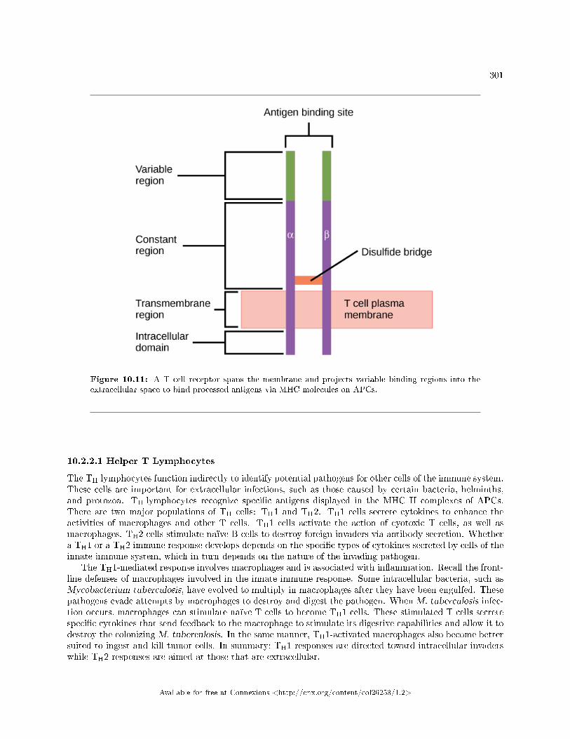
301
Figure 10.11: A T cell receptor spans the membrane and projects variable binding regions into theextracellular space to bind processed antigens via MHC molecules on APCs.
10.2.2.1 Helper T Lymphocytes
The TH lymphocytes function indirectly to identify potential pathogens for other cells of the immune system.These cells are important for extracellular infections, such as those caused by certain bacteria, helminths,and protozoa. TH lymphocytes recognize speci�c antigens displayed in the MHC II complexes of APCs.There are two major populations of TH cells: TH1 and TH2. TH1 cells secrete cytokines to enhance theactivities of macrophages and other T cells. TH1 cells activate the action of cyotoxic T cells, as well asmacrophages. TH2 cells stimulate naïve B cells to destroy foreign invaders via antibody secretion. Whethera TH1 or a TH2 immune response develops depends on the speci�c types of cytokines secreted by cells of theinnate immune system, which in turn depends on the nature of the invading pathogen.
The TH1-mediated response involves macrophages and is associated with in�ammation. Recall the front-line defenses of macrophages involved in the innate immune response. Some intracellular bacteria, such asMycobacterium tuberculosis, have evolved to multiply in macrophages after they have been engulfed. Thesepathogens evade attempts by macrophages to destroy and digest the pathogen. When M. tuberculosis infec-tion occurs, macrophages can stimulate naïve T cells to become TH1 cells. These stimulated T cells secretespeci�c cytokines that send feedback to the macrophage to stimulate its digestive capabilities and allow it todestroy the colonizing M. tuberculosis. In the same manner, TH1-activated macrophages also become bettersuited to ingest and kill tumor cells. In summary; TH1 responses are directed toward intracellular invaderswhile TH2 responses are aimed at those that are extracellular.
Available for free at Connexions <http://cnx.org/content/col26253/1.2>
302 CHAPTER 10. IMMUNITY
10.2.2.2 B Lymphocytes
When stimulated by the TH2 pathway, naïve B cells di�erentiate into antibody-secreting plasma cells. Aplasma cell is an immune cell that secrets antibodies; these cells arise from B cells that were stimulatedby antigens. Similar to T cells, naïve B cells initially are coated in thousands of B cell receptors (BCRs),which are membrane-bound forms of Ig (immunoglobulin, or an antibody). The B cell receptor has twoheavy chains and two light chains connected by disul�de linkages. Each chain has a constant and a variableregion; the latter is involved in antigen binding. Two other membrane proteins, Ig alpha and Ig beta, areinvolved in signaling. The receptors of any particular B cell, as shown in Figure 10.12 are all the same,but the hundreds of millions of di�erent B cells in an individual have distinct recognition domains thatcontribute to extensive diversity in the types of molecular structures to which they can bind. In this state,B cells function as APCs. They bind and engulf foreign antigens via their BCRs and then display processedantigens in the context of MHC II molecules to TH2 cells. When a TH2 cell detects that a B cell is boundto a relevant antigen, it secretes speci�c cytokines that induce the B cell to proliferate rapidly, which makesthousands of identical (clonal) copies of it, and then it synthesizes and secretes antibodies with the sameantigen recognition pattern as the BCRs. The activation of B cells corresponding to one speci�c BCR variantand the dramatic proliferation of that variant is known as clonal selection. This phenomenon drastically,but brie�y, changes the proportions of BCR variants expressed by the immune system, and shifts the balancetoward BCRs speci�c to the infecting pathogen.
Available for free at Connexions <http://cnx.org/content/col26253/1.2>
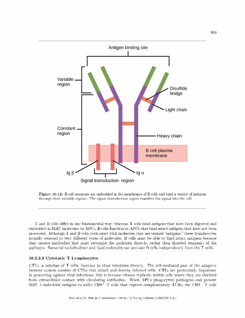
303
Figure 10.12: B cell receptors are embedded in the membranes of B cells and bind a variety of antigensthrough their variable regions. The signal transduction region transfers the signal into the cell.
T and B cells di�er in one fundamental way: whereas T cells bind antigens that have been digested andembedded in MHC molecules by APCs, B cells function as APCs that bind intact antigens that have not beenprocessed. Although T and B cells both react with molecules that are termed �antigens,� these lymphocytesactually respond to very di�erent types of molecules. B cells must be able to bind intact antigens becausethey secrete antibodies that must recognize the pathogen directly, rather than digested remnants of thepathogen. Bacterial carbohydrate and lipid molecules can activate B cells independently from the T cells.
10.2.2.3 Cytotoxic T Lymphocytes
CTLs, a subclass of T cells, function to clear infections directly. The cell-mediated part of the adaptiveimmune system consists of CTLs that attack and destroy infected cells. CTLs are particularly importantin protecting against viral infections; this is because viruses replicate within cells where they are shieldedfrom extracellular contact with circulating antibodies. When APCs phagocytize pathogens and presentMHC I-embedded antigens to naïve CD8+ T cells that express complementary TCRs, the CD8+ T cells
Available for free at Connexions <http://cnx.org/content/col26253/1.2>
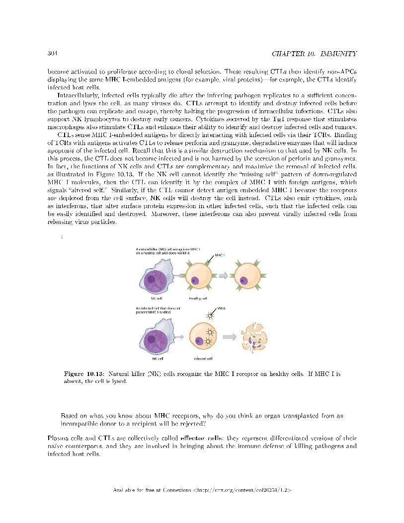
304 CHAPTER 10. IMMUNITY
become activated to proliferate according to clonal selection. These resulting CTLs then identify non-APCsdisplaying the same MHC I-embedded antigens (for example, viral proteins)�for example, the CTLs identifyinfected host cells.
Intracellularly, infected cells typically die after the infecting pathogen replicates to a su�cient concen-tration and lyses the cell, as many viruses do. CTLs attempt to identify and destroy infected cells beforethe pathogen can replicate and escape, thereby halting the progression of intracellular infections. CTLs alsosupport NK lymphocytes to destroy early cancers. Cytokines secreted by the TH1 response that stimulatesmacrophages also stimulate CTLs and enhance their ability to identify and destroy infected cells and tumors.
CTLs sense MHC I-embedded antigens by directly interacting with infected cells via their TCRs. Bindingof TCRs with antigens activates CTLs to release perforin and granzyme, degradative enzymes that will induceapoptosis of the infected cell. Recall that this is a similar destruction mechanism to that used by NK cells. Inthis process, the CTL does not become infected and is not harmed by the secretion of perforin and granzymes.In fact, the functions of NK cells and CTLs are complementary and maximize the removal of infected cells,as illustrated in Figure 10.13. If the NK cell cannot identify the �missing self� pattern of down-regulatedMHC I molecules, then the CTL can identify it by the complex of MHC I with foreign antigens, whichsignals �altered self.� Similarly, if the CTL cannot detect antigen-embedded MHC I because the receptorsare depleted from the cell surface, NK cells will destroy the cell instead. CTLs also emit cytokines, suchas interferons, that alter surface protein expression in other infected cells, such that the infected cells canbe easily identi�ed and destroyed. Moreover, these interferons can also prevent virally infected cells fromreleasing virus particles.
:
Figure 10.13: Natural killer (NK) cells recognize the MHC I receptor on healthy cells. If MHC I isabsent, the cell is lysed.
Based on what you know about MHC receptors, why do you think an organ transplanted from anincompatible donor to a recipient will be rejected?
Plasma cells and CTLs are collectively called e�ector cells: they represent di�erentiated versions of theirnaïve counterparts, and they are involved in bringing about the immune defense of killing pathogens andinfected host cells.
Available for free at Connexions <http://cnx.org/content/col26253/1.2>
305
10.2.2.4 Mucosal Surfaces and Immune Tolerance
The innate and adaptive immune responses discussed thus far comprise the systemic immune system (af-fecting the whole body), which is distinct from the mucosal immune system. Mucosal immunity is formedby mucosa-associated lymphoid tissue, which functions independently of the systemic immune system, andwhich has its own innate and adaptive components. Mucosa-associated lymphoid tissue (MALT),illustrated in Figure 10.14, is a collection of lymphatic tissue that combines with epithelial tissue lining themucosa throughout the body. This tissue functions as the immune barrier and response in areas of the bodywith direct contact to the external environment. The systemic and mucosal immune systems use many ofthe same cell types. Foreign particles that make their way to MALT are taken up by absorptive epithelialcells called M cells and delivered to APCs located directly below the mucosal tissue. M cells function in thetransport described, and are located in the Peyer's patch, a lymphoid nodule. APCs of the mucosal immunesystem are primarily dendritic cells, with B cells and macrophages having minor roles. Processed antigensdisplayed on APCs are detected by T cells in the MALT and at various mucosal induction sites, such as thetonsils, adenoids, appendix, or the mesenteric lymph nodes of the intestine. Activated T cells then migratethrough the lymphatic system and into the circulatory system to mucosal sites of infection.
Available for free at Connexions <http://cnx.org/content/col26253/1.2>
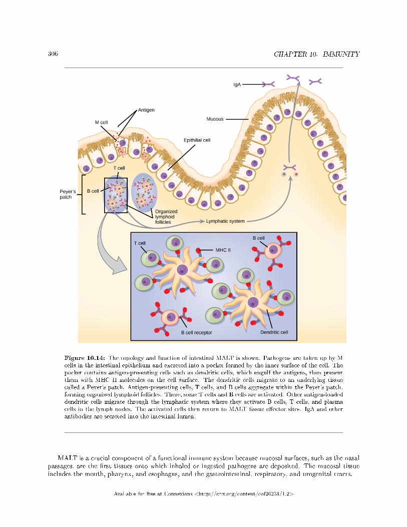
306 CHAPTER 10. IMMUNITY
Figure 10.14: The topology and function of intestinal MALT is shown. Pathogens are taken up by Mcells in the intestinal epithelium and excreted into a pocket formed by the inner surface of the cell. Thepocket contains antigen-presenting cells such as dendritic cells, which engulf the antigens, then presentthem with MHC II molecules on the cell surface. The dendritic cells migrate to an underlying tissuecalled a Peyer's patch. Antigen-presenting cells, T cells, and B cells aggregate within the Peyer's patch,forming organized lymphoid follicles. There, some T cells and B cells are activated. Other antigen-loadeddendritic cells migrate through the lymphatic system where they activate B cells, T cells, and plasmacells in the lymph nodes. The activated cells then return to MALT tissue e�ector sites. IgA and otherantibodies are secreted into the intestinal lumen.
MALT is a crucial component of a functional immune system because mucosal surfaces, such as the nasalpassages, are the �rst tissues onto which inhaled or ingested pathogens are deposited. The mucosal tissueincludes the mouth, pharynx, and esophagus, and the gastrointestinal, respiratory, and urogenital tracts.
Available for free at Connexions <http://cnx.org/content/col26253/1.2>

307
The immune system has to be regulated to prevent wasteful, unnecessary responses to harmless sub-stances, and more importantly so that it does not attack �self.� The acquired ability to prevent an unneces-sary or harmful immune response to a detected foreign substance known not to cause disease is described asimmune tolerance. Immune tolerance is crucial for maintaining mucosal homeostasis given the tremendousnumber of foreign substances (such as food proteins) that APCs of the oral cavity, pharynx, and gastroin-testinal mucosa encounter. Immune tolerance is brought about by specialized APCs in the liver, lymphnodes, small intestine, and lung that present harmless antigens to an exceptionally diverse population ofregulatory T (Treg) cells, specialized lymphocytes that suppress local in�ammation and inhibit the secre-tion of stimulatory immune factors. The combined result of Treg cells is to prevent immunologic activationand in�ammation in undesired tissue compartments and to allow the immune system to focus on pathogensinstead. In addition to promoting immune tolerance of harmless antigens, other subsets of Treg cells areinvolved in the prevention of the autoimmune response, which is an inappropriate immune response tohost cells or self-antigens. Another Treg class suppresses immune responses to harmful pathogens after theinfection has cleared to minimize host cell damage induced by in�ammation and cell lysis.
10.2.3 Immunological Memory
The adaptive immune system possesses a memory component that allows for an e�cient and dramaticresponse upon reinvasion of the same pathogen. Memory is handled by the adaptive immune system withlittle reliance on cues from the innate response. During the adaptive immune response to a pathogen that hasnot been encountered before, called a primary response, plasma cells secreting antibodies and di�erentiatedT cells increase, then plateau over time. As B and T cells mature into e�ector cells, a subset of the naïvepopulations di�erentiates into B and T memory cells with the same antigen speci�cities, as illustrated inFigure 10.15.
A memory cell is an antigen-speci�c B or T lymphocyte that does not di�erentiate into e�ector cellsduring the primary immune response, but that can immediately become e�ector cells upon re-exposure tothe same pathogen. During the primary immune response, memory cells do not respond to antigens and donot contribute to host defenses. As the infection is cleared and pathogenic stimuli subside, the e�ectors areno longer needed, and they undergo apoptosis. In contrast, the memory cells persist in the circulation.
:
Available for free at Connexions <http://cnx.org/content/col26253/1.2>
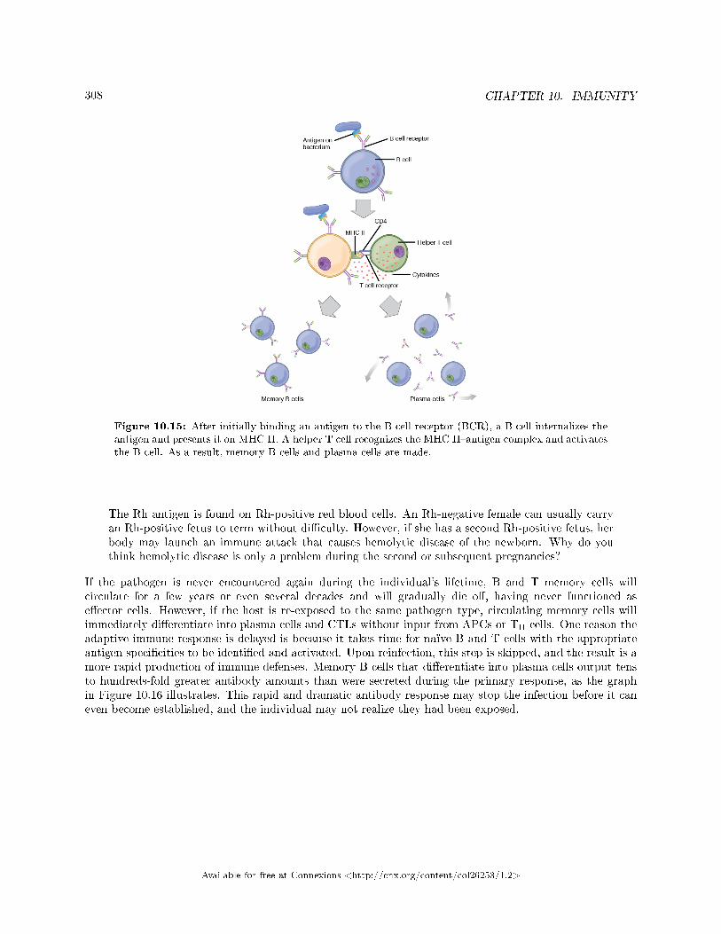
308 CHAPTER 10. IMMUNITY
Figure 10.15: After initially binding an antigen to the B cell receptor (BCR), a B cell internalizes theantigen and presents it on MHC II. A helper T cell recognizes the MHC II�antigen complex and activatesthe B cell. As a result, memory B cells and plasma cells are made.
The Rh antigen is found on Rh-positive red blood cells. An Rh-negative female can usually carryan Rh-positive fetus to term without di�culty. However, if she has a second Rh-positive fetus, herbody may launch an immune attack that causes hemolytic disease of the newborn. Why do youthink hemolytic disease is only a problem during the second or subsequent pregnancies?
If the pathogen is never encountered again during the individual's lifetime, B and T memory cells willcirculate for a few years or even several decades and will gradually die o�, having never functioned ase�ector cells. However, if the host is re-exposed to the same pathogen type, circulating memory cells willimmediately di�erentiate into plasma cells and CTLs without input from APCs or TH cells. One reason theadaptive immune response is delayed is because it takes time for naïve B and T cells with the appropriateantigen speci�cities to be identi�ed and activated. Upon reinfection, this step is skipped, and the result is amore rapid production of immune defenses. Memory B cells that di�erentiate into plasma cells output tensto hundreds-fold greater antibody amounts than were secreted during the primary response, as the graphin Figure 10.16 illustrates. This rapid and dramatic antibody response may stop the infection before it caneven become established, and the individual may not realize they had been exposed.
Available for free at Connexions <http://cnx.org/content/col26253/1.2>
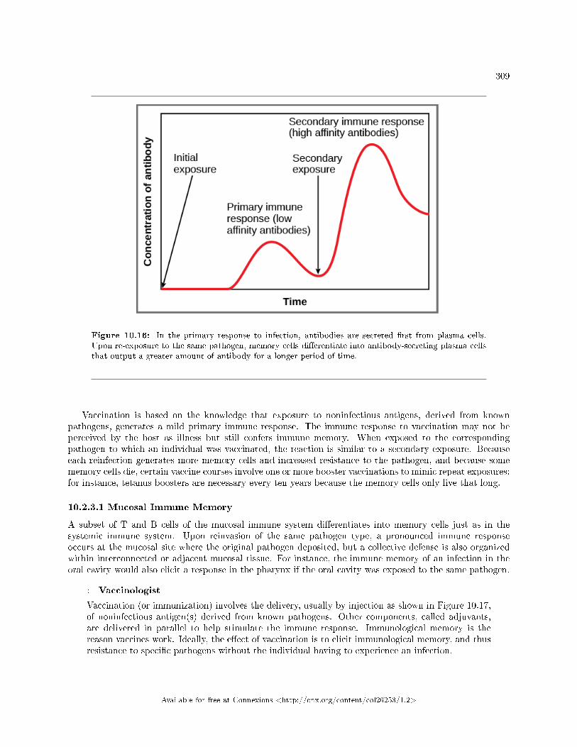
309
Figure 10.16: In the primary response to infection, antibodies are secreted �rst from plasma cells.Upon re-exposure to the same pathogen, memory cells di�erentiate into antibody-secreting plasma cellsthat output a greater amount of antibody for a longer period of time.
Vaccination is based on the knowledge that exposure to noninfectious antigens, derived from knownpathogens, generates a mild primary immune response. The immune response to vaccination may not beperceived by the host as illness but still confers immune memory. When exposed to the correspondingpathogen to which an individual was vaccinated, the reaction is similar to a secondary exposure. Becauseeach reinfection generates more memory cells and increased resistance to the pathogen, and because somememory cells die, certain vaccine courses involve one or more booster vaccinations to mimic repeat exposures:for instance, tetanus boosters are necessary every ten years because the memory cells only live that long.
10.2.3.1 Mucosal Immune Memory
A subset of T and B cells of the mucosal immune system di�erentiates into memory cells just as in thesystemic immune system. Upon reinvasion of the same pathogen type, a pronounced immune responseoccurs at the mucosal site where the original pathogen deposited, but a collective defense is also organizedwithin interconnected or adjacent mucosal tissue. For instance, the immune memory of an infection in theoral cavity would also elicit a response in the pharynx if the oral cavity was exposed to the same pathogen.
: Vaccinologist
Vaccination (or immunization) involves the delivery, usually by injection as shown in Figure 10.17,of noninfectious antigen(s) derived from known pathogens. Other components, called adjuvants,are delivered in parallel to help stimulate the immune response. Immunological memory is thereason vaccines work. Ideally, the e�ect of vaccination is to elicit immunological memory, and thusresistance to speci�c pathogens without the individual having to experience an infection.
Available for free at Connexions <http://cnx.org/content/col26253/1.2>
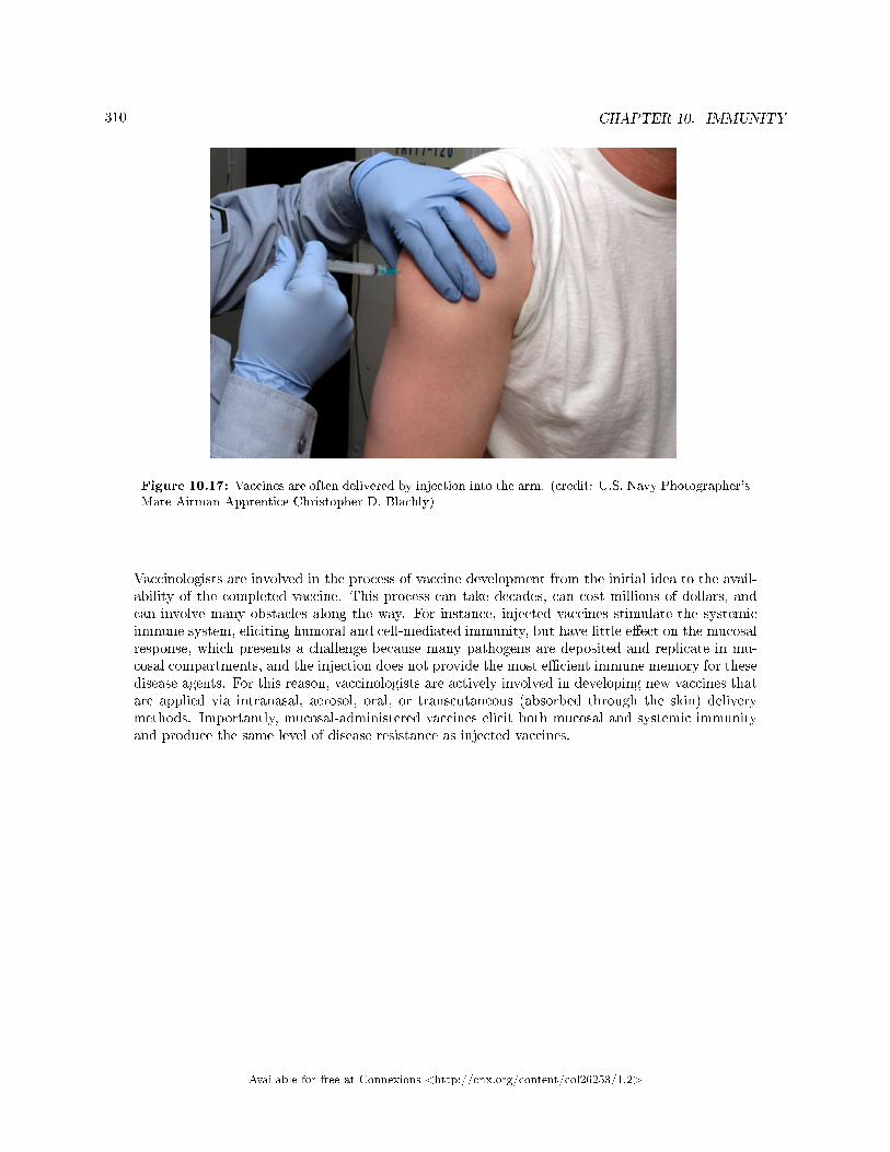
310 CHAPTER 10. IMMUNITY
Figure 10.17: Vaccines are often delivered by injection into the arm. (credit: U.S. Navy Photographer'sMate Airman Apprentice Christopher D. Blachly)
Vaccinologists are involved in the process of vaccine development from the initial idea to the avail-ability of the completed vaccine. This process can take decades, can cost millions of dollars, andcan involve many obstacles along the way. For instance, injected vaccines stimulate the systemicimmune system, eliciting humoral and cell-mediated immunity, but have little e�ect on the mucosalresponse, which presents a challenge because many pathogens are deposited and replicate in mu-cosal compartments, and the injection does not provide the most e�cient immune memory for thesedisease agents. For this reason, vaccinologists are actively involved in developing new vaccines thatare applied via intranasal, aerosol, oral, or transcutaneous (absorbed through the skin) deliverymethods. Importantly, mucosal-administered vaccines elicit both mucosal and systemic immunityand produce the same level of disease resistance as injected vaccines.
Available for free at Connexions <http://cnx.org/content/col26253/1.2>
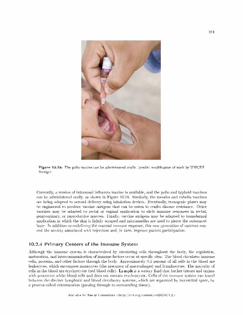
311
Figure 10.18: The polio vaccine can be administered orally. (credit: modi�cation of work by UNICEFSverige)
Currently, a version of intranasal in�uenza vaccine is available, and the polio and typhoid vaccinescan be administered orally, as shown in Figure 10.18. Similarly, the measles and rubella vaccinesare being adapted to aerosol delivery using inhalation devices. Eventually, transgenic plants maybe engineered to produce vaccine antigens that can be eaten to confer disease resistance. Othervaccines may be adapted to rectal or vaginal application to elicit immune responses in rectal,genitourinary, or reproductive mucosa. Finally, vaccine antigens may be adapted to transdermalapplication in which the skin is lightly scraped and microneedles are used to pierce the outermostlayer. In addition to mobilizing the mucosal immune response, this new generation of vaccines mayend the anxiety associated with injections and, in turn, improve patient participation.
10.2.4 Primary Centers of the Immune System
Although the immune system is characterized by circulating cells throughout the body, the regulation,maturation, and intercommunication of immune factors occur at speci�c sites. The blood circulates immunecells, proteins, and other factors through the body. Approximately 0.1 percent of all cells in the blood areleukocytes, which encompass monocytes (the precursor of macrophages) and lymphocytes. The majority ofcells in the blood are erythrocytes (red blood cells). Lymph is a watery �uid that bathes tissues and organswith protective white blood cells and does not contain erythrocytes. Cells of the immune system can travelbetween the distinct lymphatic and blood circulatory systems, which are separated by interstitial space, bya process called extravasation (passing through to surrounding tissue).
Available for free at Connexions <http://cnx.org/content/col26253/1.2>
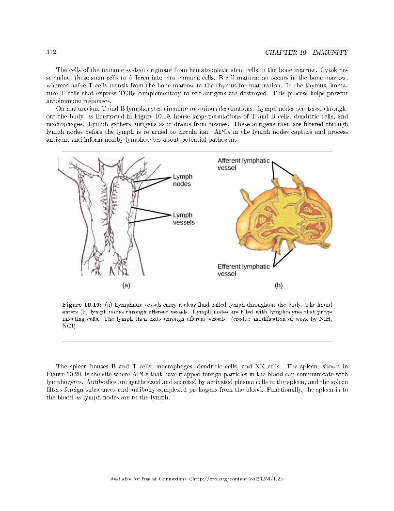
312 CHAPTER 10. IMMUNITY
The cells of the immune system originate from hematopoietic stem cells in the bone marrow. Cytokinesstimulate these stem cells to di�erentiate into immune cells. B cell maturation occurs in the bone marrow,whereas naïve T cells transit from the bone marrow to the thymus for maturation. In the thymus, imma-ture T cells that express TCRs complementary to self-antigens are destroyed. This process helps preventautoimmune responses.
On maturation, T and B lymphocytes circulate to various destinations. Lymph nodes scattered through-out the body, as illustrated in Figure 10.19, house large populations of T and B cells, dendritic cells, andmacrophages. Lymph gathers antigens as it drains from tissues. These antigens then are �ltered throughlymph nodes before the lymph is returned to circulation. APCs in the lymph nodes capture and processantigens and inform nearby lymphocytes about potential pathogens.
Figure 10.19: (a) Lymphatic vessels carry a clear �uid called lymph throughout the body. The liquidenters (b) lymph nodes through a�erent vessels. Lymph nodes are �lled with lymphocytes that purgeinfecting cells. The lymph then exits through e�erent vessels. (credit: modi�cation of work by NIH,NCI)
The spleen houses B and T cells, macrophages, dendritic cells, and NK cells. The spleen, shown inFigure 10.20, is the site where APCs that have trapped foreign particles in the blood can communicate withlymphocytes. Antibodies are synthesized and secreted by activated plasma cells in the spleen, and the spleen�lters foreign substances and antibody-complexed pathogens from the blood. Functionally, the spleen is tothe blood as lymph nodes are to the lymph.
Available for free at Connexions <http://cnx.org/content/col26253/1.2>
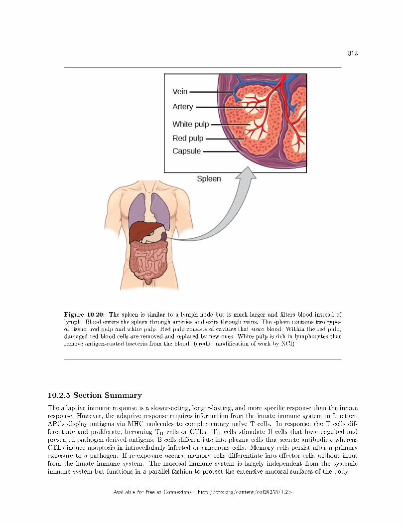
313
Figure 10.20: The spleen is similar to a lymph node but is much larger and �lters blood instead oflymph. Blood enters the spleen through arteries and exits through veins. The spleen contains two typesof tissue: red pulp and white pulp. Red pulp consists of cavities that store blood. Within the red pulp,damaged red blood cells are removed and replaced by new ones. White pulp is rich in lymphocytes thatremove antigen-coated bacteria from the blood. (credit: modi�cation of work by NCI)
10.2.5 Section Summary
The adaptive immune response is a slower-acting, longer-lasting, and more speci�c response than the innateresponse. However, the adaptive response requires information from the innate immune system to function.APCs display antigens via MHC molecules to complementary naïve T cells. In response, the T cells dif-ferentiate and proliferate, becoming TH cells or CTLs. TH cells stimulate B cells that have engulfed andpresented pathogen-derived antigens. B cells di�erentiate into plasma cells that secrete antibodies, whereasCTLs induce apoptosis in intracellularly infected or cancerous cells. Memory cells persist after a primaryexposure to a pathogen. If re-exposure occurs, memory cells di�erentiate into e�ector cells without inputfrom the innate immune system. The mucosal immune system is largely independent from the systemicimmune system but functions in a parallel fashion to protect the extensive mucosal surfaces of the body.
Available for free at Connexions <http://cnx.org/content/col26253/1.2>

314 CHAPTER 10. IMMUNITY
10.2.6 Art Connections
Exercise 10.2.1 (Solution on p. 320.)
Figure 10.10 Which of the following statements about T cells is false?
a. Helper T cells release cytokines while cytotoxic T cells kill the infected cell.b. Helper T cells are CD4+, while cytotoxic T cells are CD8+.c. MHC II is a receptor found on most body cells, while MHC I is a receptor found on immune
cells only.d. The T cell receptor is found on both CD4+ and CD8+ T cells.
Exercise 10.2.2 (Solution on p. 320.)
Figure 10.13 Based on what you know about MHC receptors, why do you think an organ trans-planted from an incompatible donor to a recipient will be rejected?
Exercise 10.2.3 (Solution on p. 320.)
Figure 10.15 The Rh antigen is found on Rh-positive red blood cells. An Rh-negative femalecan usually carry an Rh-positive fetus to term without di�culty. However, if she has a secondRh-positive fetus, her body may launch an immune attack that causes hemolytic disease of thenewborn. Why do you think hemolytic disease is only a problem during the second or subsequentpregnancies?
10.2.7 Review Questions
Exercise 10.2.4 (Solution on p. 320.)
Which of the following is both a phagocyte and an antigen-presenting cell?
a. NK cellb. eosinophilc. neutrophild. macrophage
Exercise 10.2.5 (Solution on p. 320.)
Which immune cells bind MHC molecules on APCs via CD8 coreceptors on their cell surfaces?
a. TH cellsb. CTLsc. mast cellsd. basophils
Exercise 10.2.6 (Solution on p. 320.)
What �self� pattern is identi�ed by NK cells?
a. altered selfb. missing selfc. normal selfd. non-self
Exercise 10.2.7 (Solution on p. 320.)
The acquired ability to prevent an unnecessary or destructive immune reaction to a harmlessforeign particle, such as a food protein, is called ________.
a. the TH2 response
Available for free at Connexions <http://cnx.org/content/col26253/1.2>
315
b. allergyc. immune toleranced. autoimmunity
Exercise 10.2.8 (Solution on p. 320.)
A memory B cell can di�erentiate upon re-exposure to a pathogen of which cell type?
a. CTLb. naïve B cellc. memory T celld. plasma cell
Exercise 10.2.9 (Solution on p. 320.)
Foreign particles circulating in the blood are �ltered by the ________.
a. spleenb. lymph nodesc. MALTd. lymph
10.2.8 Free Response
Exercise 10.2.10 (Solution on p. 320.)
Explain the di�erence between an epitope and an antigen.
Exercise 10.2.11 (Solution on p. 320.)
What is a naïve B or T cell?
Exercise 10.2.12 (Solution on p. 320.)
How does the TH1 response di�er from the TH2 response?
Exercise 10.2.13 (Solution on p. 320.)
In mammalian adaptive immune systems, T cell receptors are extraordinarily diverse. Whatfunction of the immune system results from this diversity, and how is this diversity achieved?
Exercise 10.2.14 (Solution on p. 321.)
How do B and T cells di�er with respect to antigens that they bind?
Exercise 10.2.15 (Solution on p. 321.)
Why is the immune response after reinfection much faster than the adaptive immune responseafter the initial infection?
10.3 Disruptions in the Immune System5
A functioning immune system is essential for survival, but even the sophisticated cellular and moleculardefenses of the mammalian immune response can be defeated by pathogens at virtually every step. In thecompetition between immune protection and pathogen evasion, pathogens have the advantage of more rapidevolution because of their shorter generation time and other characteristics. For instance, Streptococcuspneumoniae (bacterium that cause pneumonia and meningitis) surrounds itself with a capsule that inhibitsphagocytes from engul�ng it and displaying antigens to the adaptive immune system. Staphylococcus aureus
5This content is available online at <http://cnx.org/content/m66666/1.12/>.
Available for free at Connexions <http://cnx.org/content/col26253/1.2>
316 CHAPTER 10. IMMUNITY
(bacterium that can cause skin infections, abscesses, and meningitis) synthesizes a toxin called leukocidinthat kills phagocytes after they engulf the bacterium. Other pathogens can also hinder the adaptive immunesystem. HIV infects TH cells via their CD4 surface molecules, gradually depleting the number of TH cells inthe body; this inhibits the adaptive immune system's capacity to generate su�cient responses to infectionor tumors. As a result, HIV-infected individuals often su�er from infections that would not cause illnessin people with healthy immune systems but which can cause devastating illness to immune-compromisedindividuals. Maladaptive responses of immune cells and molecules themselves can also disrupt the properfunctioning of the entire system, leading to host cell damage that could become fatal.
10.3.1 Immunode�ciency
Failures, insu�ciencies, or delays at any level of the immune response can allow pathogens or tumor cellsto gain a foothold and replicate or proliferate to high enough levels that the immune system becomesoverwhelmed. Immunode�ciency is the failure, insu�ciency, or delay in the response of the immunesystem, which may be acquired or inherited. Immunode�ciency can be acquired as a result of infection withcertain pathogens (such as HIV), chemical exposure (including certain medical treatments), malnutrition,or possibly by extreme stress. For instance, radiation exposure can destroy populations of lymphocytesand elevate an individual's susceptibility to infections and cancer. Dozens of genetic disorders result inimmunode�ciencies, including Severe Combined Immunode�ciency (SCID), Bare lymphocyte syndrome, andMHC II de�ciencies. Rarely, primary immunode�ciencies that are present from birth may occur. Neutropeniais one form in which the immune system produces a below-average number of neutrophils, the body's mostabundant phagocytes. As a result, bacterial infections may go unrestricted in the blood, causing seriouscomplications.
10.3.2 Hypersensitivities
Maladaptive immune responses toward harmless foreign substances or self antigens that occur after tissuesensitization are termed hypersensitivities. The types of hypersensitivities include immediate, delayed,and autoimmunity. A large proportion of the population is a�ected by one or more types of hypersensitivity.
10.3.2.1 Allergies
The immune reaction that results from immediate hypersensitivities in which an antibody-mediated immuneresponse occurs within minutes of exposure to a harmless antigen is called an allergy. In the United States,20 percent of the population exhibits symptoms of allergy or asthma, whereas 55 percent test positiveagainst one or more allergens. Upon initial exposure to a potential allergen, an allergic individual synthesizesantibodies of the IgE class via the typical process of APCs presenting processed antigen to TH cells thatstimulate B cells to produce IgE. This class of antibodies also mediates the immune response to parasiticworms. The constant domain of the IgE molecules interact with mast cells embedded in connective tissues.This process primes, or sensitizes, the tissue. Upon subsequent exposure to the same allergen, IgE moleculeson mast cells bind the antigen via their variable domains and stimulate the mast cell to release the modi�edamino acids histamine and serotonin; these chemical mediators then recruit eosinophils which mediate allergicresponses. Figure 10.21 shows an example of an allergic response to ragweed pollen. The e�ects of an allergicreaction range from mild symptoms like sneezing and itchy, watery eyes to more severe or even life-threateningreactions involving intensely itchy welts or hives, airway contraction with severe respiratory distress, andplummeting blood pressure. This extreme reaction is known as anaphylactic shock. If not treated withepinephrine to counter the blood pressure and breathing e�ects, this condition can be fatal.
Available for free at Connexions <http://cnx.org/content/col26253/1.2>
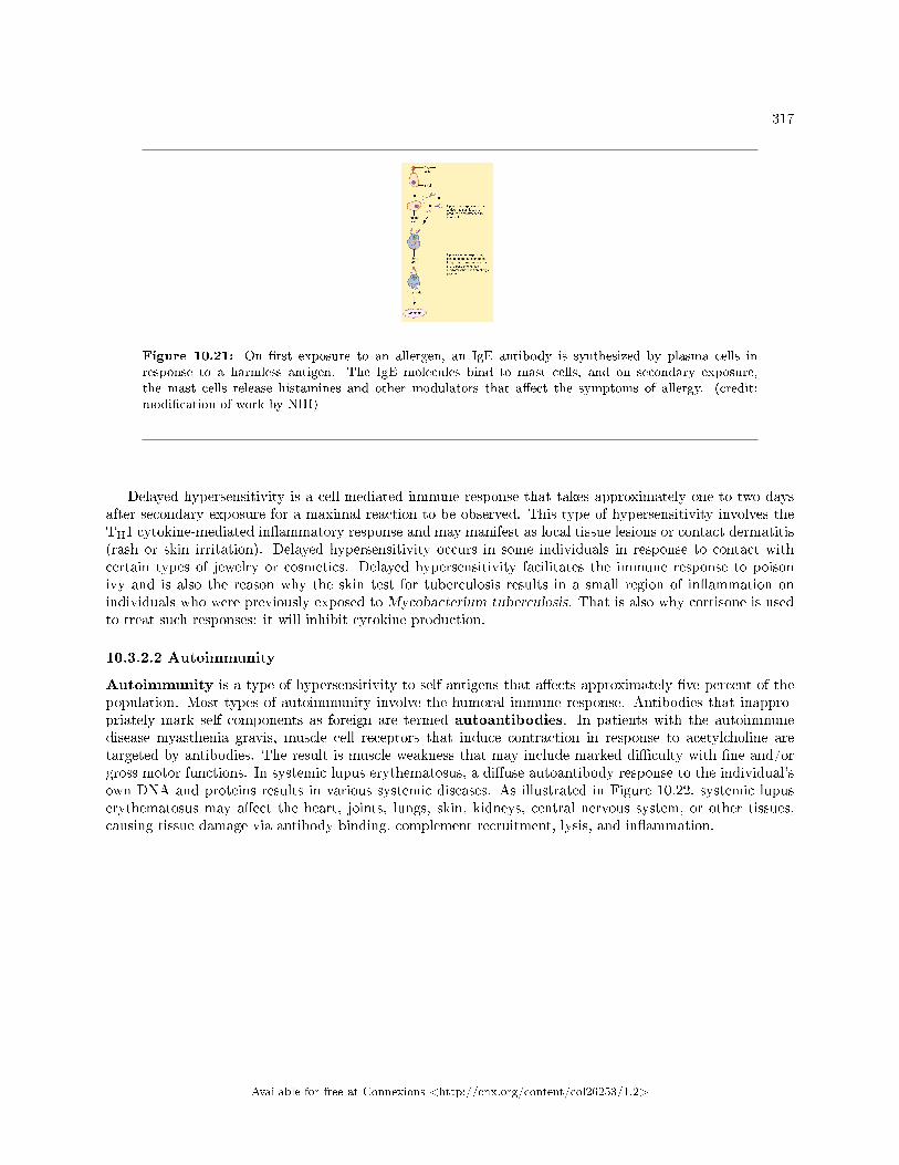
317
Figure 10.21: On �rst exposure to an allergen, an IgE antibody is synthesized by plasma cells inresponse to a harmless antigen. The IgE molecules bind to mast cells, and on secondary exposure,the mast cells release histamines and other modulators that a�ect the symptoms of allergy. (credit:modi�cation of work by NIH)
Delayed hypersensitivity is a cell-mediated immune response that takes approximately one to two daysafter secondary exposure for a maximal reaction to be observed. This type of hypersensitivity involves theTH1 cytokine-mediated in�ammatory response and may manifest as local tissue lesions or contact dermatitis(rash or skin irritation). Delayed hypersensitivity occurs in some individuals in response to contact withcertain types of jewelry or cosmetics. Delayed hypersensitivity facilitates the immune response to poisonivy and is also the reason why the skin test for tuberculosis results in a small region of in�ammation onindividuals who were previously exposed to Mycobacterium tuberculosis. That is also why cortisone is usedto treat such responses: it will inhibit cytokine production.
10.3.2.2 Autoimmunity
Autoimmunity is a type of hypersensitivity to self antigens that a�ects approximately �ve percent of thepopulation. Most types of autoimmunity involve the humoral immune response. Antibodies that inappro-priately mark self components as foreign are termed autoantibodies. In patients with the autoimmunedisease myasthenia gravis, muscle cell receptors that induce contraction in response to acetylcholine aretargeted by antibodies. The result is muscle weakness that may include marked di�culty with �ne and/orgross motor functions. In systemic lupus erythematosus, a di�use autoantibody response to the individual'sown DNA and proteins results in various systemic diseases. As illustrated in Figure 10.22, systemic lupuserythematosus may a�ect the heart, joints, lungs, skin, kidneys, central nervous system, or other tissues,causing tissue damage via antibody binding, complement recruitment, lysis, and in�ammation.
Available for free at Connexions <http://cnx.org/content/col26253/1.2>
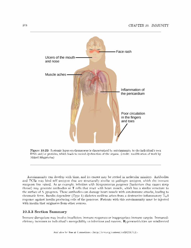
318 CHAPTER 10. IMMUNITY
Figure 10.22: Systemic lupus erythematosus is characterized by autoimmunity to the individual's ownDNA and/or proteins, which leads to varied dysfunction of the organs. (credit: modi�cation of work byMikael Häggstrom)
Autoimmunity can develop with time, and its causes may be rooted in molecular mimicry. Antibodiesand TCRs may bind self antigens that are structurally similar to pathogen antigens, which the immunereceptors �rst raised. As an example, infection with Streptococcus pyogenes (bacterium that causes strepthroat) may generate antibodies or T cells that react with heart muscle, which has a similar structure tothe surface of S. pyogenes. These antibodies can damage heart muscle with autoimmune attacks, leading torheumatic fever. Insulin-dependent (Type 1) diabetes mellitus arises from a destructive in�ammatory TH1response against insulin-producing cells of the pancreas. Patients with this autoimmunity must be injectedwith insulin that originates from other sources.
10.3.3 Section Summary
Immune disruptions may involve insu�cient immune responses or inappropriate immune targets. Immunod-e�ciency increases an individual's susceptibility to infections and cancers. Hypersensitivities are misdirected
Available for free at Connexions <http://cnx.org/content/col26253/1.2>
319
responses either to harmless foreign particles, as in the case of allergies, or to host factors, as in the case ofautoimmunity. Reactions to self components may be the result of molecular mimicry.
10.3.4 Review Questions
Exercise 10.3.1 (Solution on p. 321.)
Allergy to pollen is classi�ed as:
a. an autoimmune reactionb. immunode�ciencyc. delayed hypersensitivityd. immediate hypersensitivity
Exercise 10.3.2 (Solution on p. 321.)
A potential cause of acquired autoimmunity is ________.
a. tissue hypersensitivityb. molecular mimicryc. histamine released. radiation exposure
Exercise 10.3.3 (Solution on p. 321.)
Autoantibodies are probably involved in:
a. reactions to poison ivyb. pollen allergiesc. systemic lupus erythematosusd. HIV/AIDS
Exercise 10.3.4 (Solution on p. 321.)
Which of the following diseases is not due to autoimmunity?
a. rheumatic feverb. systemic lupus erythematosusc. diabetes mellitusd. HIV/AIDS
Available for free at Connexions <http://cnx.org/content/col26253/1.2>
320 CHAPTER 10. IMMUNITY
Solutions to Exercises in Chapter 10
to Exercise 10.1.1 (p. 295)Dto Exercise 10.1.2 (p. 295)Bto Exercise 10.1.3 (p. 295)Ato Exercise 10.1.4 (p. 295)Cto Exercise 10.1.5 (p. 295)If the MHC I molecules expressed on donor cells di�er from the MHC I molecules expressed on recipientcells, NK cells may identify the donor cells as �non-self� and produce perforin and granzymes to induce thedonor cells to undergo apoptosis, which would destroy the transplanted organ.to Exercise 10.1.6 (p. 295)The entire complement system would probably be a�ected even when only a few members were mutatedsuch that they could no longer bind. Because the complement involves the binding of activated proteins ina speci�c sequence, when one or more proteins in the sequence are absent, the subsequent proteins would beincapable of binding to elicit the complement's pathogen-destructive e�ects.to Exercise 10.2.1 (p. 314)Figure 10.10 Cto Exercise 10.2.2 (p. 314)Figure 10.13 MHC receptors di�er from person to person. Thus, MHC receptors on an incompatible donorare considered �non-self� and are rejected by the immune system.to Exercise 10.2.3 (p. 314)Figure 10.15 If the blood of the mother and fetus mixes, memory cells that recognize the Rh antigen can formlate in the �rst pregnancy. During subsequent pregnancies, these memory cells launch an immune attack onthe fetal blood cells. Injection of anti-Rh antibody during the �rst pregnancy prevents the immune responsefrom occurring.to Exercise 10.2.4 (p. 314)Dto Exercise 10.2.5 (p. 314)Bto Exercise 10.2.6 (p. 314)Bto Exercise 10.2.7 (p. 314)Cto Exercise 10.2.8 (p. 315)Dto Exercise 10.2.9 (p. 315)Ato Exercise 10.2.10 (p. 315)An antigen is a molecule that reacts with some component of the immune response (antibody, B cell receptor,T cell receptor). An epitope is the region on the antigen through which binding with the immune componentactually occurs.to Exercise 10.2.11 (p. 315)A naïve T or B cell is one that has not been activated by binding to the appropriate epitope. Naïve T andB cells cannot produce responses.to Exercise 10.2.12 (p. 315)The TH1 response involves the secretion of cytokines to stimulate macrophages and CTLs and improvetheir destruction of intracellular pathogens and tumor cells. It is associated with in�ammation. The TH2response is involved in the stimulation of B cells into plasma cells that synthesize and secrete antibodies.
Available for free at Connexions <http://cnx.org/content/col26253/1.2>
321
to Exercise 10.2.13 (p. 315)The diversity of TCRs allows the immune system to have millions of di�erent T cells, and thereby to bespeci�c in distinguishing antigens. This diversity arises from mutation and recombination in the genes thatencode the variable regions of TCRs.to Exercise 10.2.14 (p. 315)T cells bind antigens that have been digested and embedded in MHC molecules by APCs. In contrast, Bcells function themselves as APCs to bind intact, unprocessed antigens.to Exercise 10.2.15 (p. 315)Upon reinfection, the memory cells will immediately di�erentiate into plasma cells and CTLs without inputfrom APCs or TH cells. In contrast, the adaptive immune response to the initial infection requires time fornaïve B and T cells with the appropriate antigen speci�cities to be identi�ed and activated.to Exercise 10.3.1 (p. 319)Dto Exercise 10.3.2 (p. 319)Bto Exercise 10.3.3 (p. 319)Cto Exercise 10.3.4 (p. 319)D
Available for free at Connexions <http://cnx.org/content/col26253/1.2>
322 CHAPTER 10. IMMUNITY
Available for free at Connexions <http://cnx.org/content/col26253/1.2>
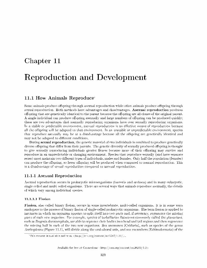
Chapter 11
Reproduction and Development
11.1 How Animals Reproduce1
Some animals produce o�spring through asexual reproduction while other animals produce o�spring throughsexual reproduction. Both methods have advantages and disadvantages. Asexual reproduction produceso�spring that are genetically identical to the parent because the o�spring are all clones of the original parent.A single individual can produce o�spring asexually and large numbers of o�spring can be produced quickly;these are two advantages that asexually reproducing organisms have over sexually reproducing organisms.In a stable or predictable environment, asexual reproduction is an e�ective means of reproduction becauseall the o�spring will be adapted to that environment. In an unstable or unpredictable environment, speciesthat reproduce asexually may be at a disadvantage because all the o�spring are genetically identical andmay not be adapted to di�erent conditions.
During sexual reproduction, the genetic material of two individuals is combined to produce geneticallydiverse o�spring that di�er from their parents. The genetic diversity of sexually produced o�spring is thoughtto give sexually reproducing individuals greater �tness because more of their o�spring may survive andreproduce in an unpredictable or changing environment. Species that reproduce sexually (and have separatesexes) must maintain two di�erent types of individuals, males and females. Only half the population (females)can produce the o�spring, so fewer o�spring will be produced when compared to asexual reproduction. Thisis a disadvantage of sexual reproduction compared to asexual reproduction.
11.1.1 Asexual Reproduction
Asexual reproduction occurs in prokaryotic microorganisms (bacteria and archaea) and in many eukaryotic,single-celled and multi-celled organisms. There are several ways that animals reproduce asexually, the detailsof which vary among individual species.
11.1.1.1 Fission
Fission, also called binary �ssion, occurs in some invertebrate, multi-celled organisms. It is in some waysanalogous to the process of binary �ssion of single-celled prokaryotic organisms. The term �ssion is applied toinstances in which an organism appears to split itself into two parts and, if necessary, regenerate the missingparts of each new organism. For example, species of turbellarian �atworms commonly called the planarians,such asDugesia dorotocephala, are able to separate their bodies into head and tail regions and then regeneratethe missing half in each of the two new organisms. Sea anemones (Cnidaria), such as species of the genusAnthopleura (Figure 11.1), will divide along the oral-aboral axis, and sea cucumbers (Echinodermata) of the
1This content is available online at <http://cnx.org/content/m45547/1.11/>.
Available for free at Connexions <http://cnx.org/content/col26253/1.2>
323
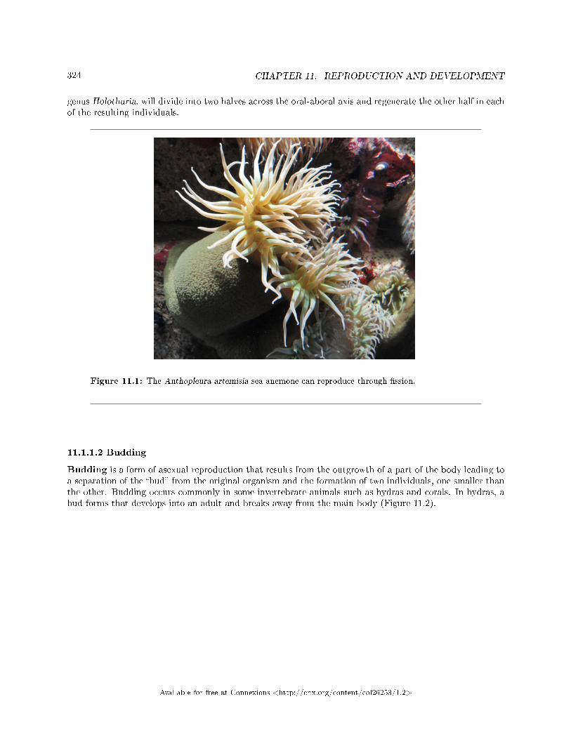
324 CHAPTER 11. REPRODUCTION AND DEVELOPMENT
genus Holothuria, will divide into two halves across the oral-aboral axis and regenerate the other half in eachof the resulting individuals.
Figure 11.1: The Anthopleura artemisia sea anemone can reproduce through �ssion.
11.1.1.2 Budding
Budding is a form of asexual reproduction that results from the outgrowth of a part of the body leading toa separation of the �bud� from the original organism and the formation of two individuals, one smaller thanthe other. Budding occurs commonly in some invertebrate animals such as hydras and corals. In hydras, abud forms that develops into an adult and breaks away from the main body (Figure 11.2).
Available for free at Connexions <http://cnx.org/content/col26253/1.2>
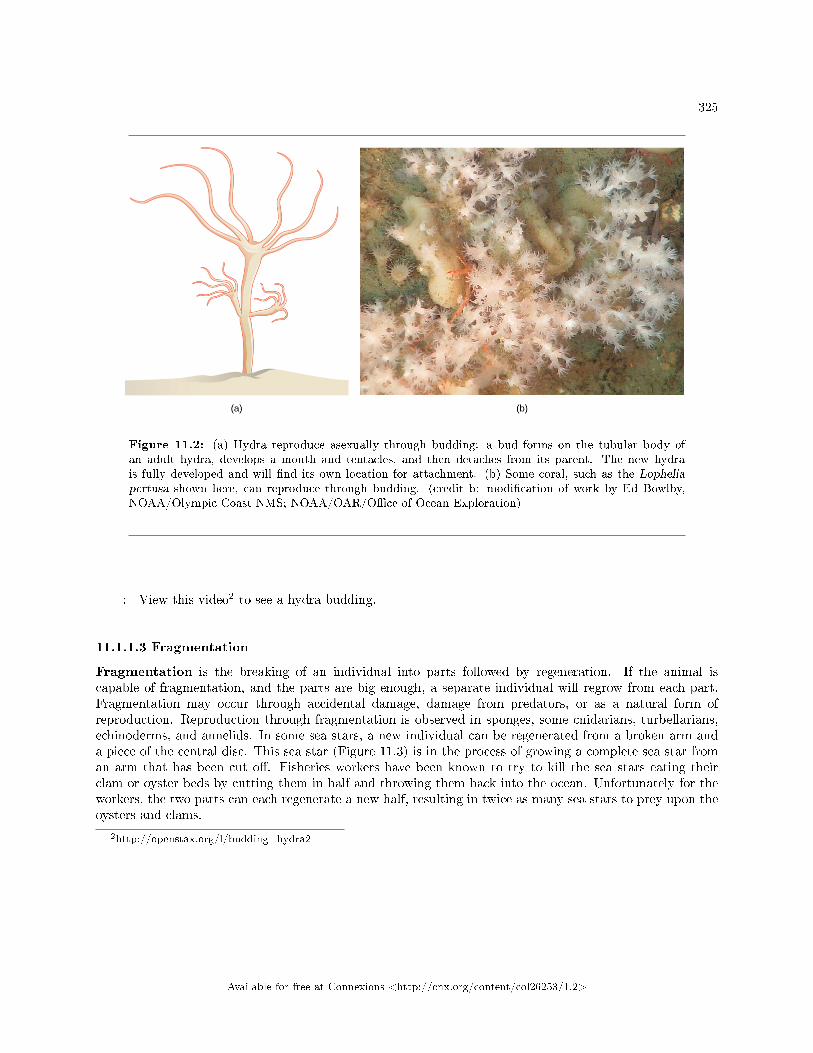
325
Figure 11.2: (a) Hydra reproduce asexually through budding: a bud forms on the tubular body ofan adult hydra, develops a mouth and tentacles, and then detaches from its parent. The new hydrais fully developed and will �nd its own location for attachment. (b) Some coral, such as the Lopheliapertusa shown here, can reproduce through budding. (credit b: modi�cation of work by Ed Bowlby,NOAA/Olympic Coast NMS; NOAA/OAR/O�ce of Ocean Exploration)
: View this video2 to see a hydra budding.
11.1.1.3 Fragmentation
Fragmentation is the breaking of an individual into parts followed by regeneration. If the animal iscapable of fragmentation, and the parts are big enough, a separate individual will regrow from each part.Fragmentation may occur through accidental damage, damage from predators, or as a natural form ofreproduction. Reproduction through fragmentation is observed in sponges, some cnidarians, turbellarians,echinoderms, and annelids. In some sea stars, a new individual can be regenerated from a broken arm anda piece of the central disc. This sea star (Figure 11.3) is in the process of growing a complete sea star froman arm that has been cut o�. Fisheries workers have been known to try to kill the sea stars eating theirclam or oyster beds by cutting them in half and throwing them back into the ocean. Unfortunately for theworkers, the two parts can each regenerate a new half, resulting in twice as many sea stars to prey upon theoysters and clams.
2http://openstax.org/l/budding_hydra2
Available for free at Connexions <http://cnx.org/content/col26253/1.2>
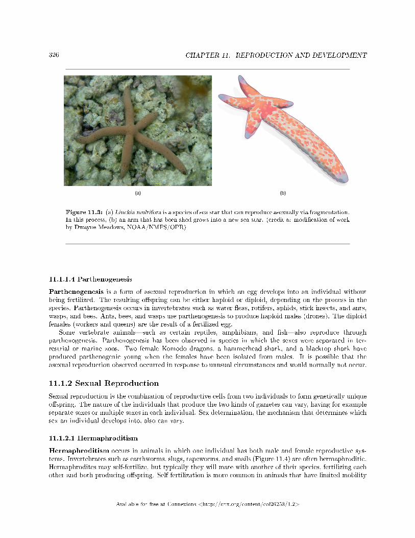
326 CHAPTER 11. REPRODUCTION AND DEVELOPMENT
Figure 11.3: (a) Linckia multifora is a species of sea star that can reproduce asexually via fragmentation.In this process, (b) an arm that has been shed grows into a new sea star. (credit a: modi�cation of workby Dwayne Meadows, NOAA/NMFS/OPR)
11.1.1.4 Parthenogenesis
Parthenogenesis is a form of asexual reproduction in which an egg develops into an individual withoutbeing fertilized. The resulting o�spring can be either haploid or diploid, depending on the process in thespecies. Parthenogenesis occurs in invertebrates such as water �eas, rotifers, aphids, stick insects, and ants,wasps, and bees. Ants, bees, and wasps use parthenogenesis to produce haploid males (drones). The diploidfemales (workers and queens) are the result of a fertilized egg.
Some vertebrate animals�such as certain reptiles, amphibians, and �sh�also reproduce throughparthenogenesis. Parthenogenesis has been observed in species in which the sexes were separated in ter-restrial or marine zoos. Two female Komodo dragons, a hammerhead shark, and a blacktop shark haveproduced parthenogenic young when the females have been isolated from males. It is possible that theasexual reproduction observed occurred in response to unusual circumstances and would normally not occur.
11.1.2 Sexual Reproduction
Sexual reproduction is the combination of reproductive cells from two individuals to form genetically uniqueo�spring. The nature of the individuals that produce the two kinds of gametes can vary, having for exampleseparate sexes or multiple sexes in each individual. Sex determination, the mechanism that determines whichsex an individual develops into, also can vary.
11.1.2.1 Hermaphroditism
Hermaphroditism occurs in animals in which one individual has both male and female reproductive sys-tems. Invertebrates such as earthworms, slugs, tapeworms, and snails (Figure 11.4) are often hermaphroditic.Hermaphrodites may self-fertilize, but typically they will mate with another of their species, fertilizing eachother and both producing o�spring. Self-fertilization is more common in animals that have limited mobility
Available for free at Connexions <http://cnx.org/content/col26253/1.2>
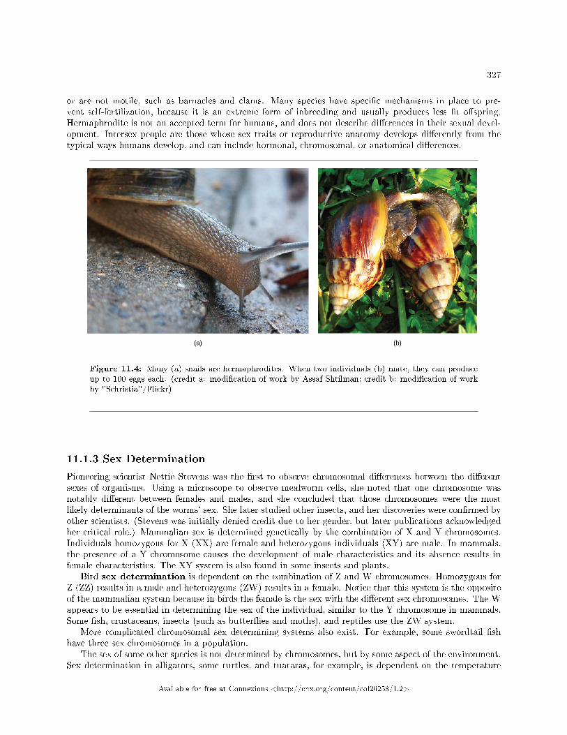
327
or are not motile, such as barnacles and clams. Many species have speci�c mechanisms in place to pre-vent self-fertilization, because it is an extreme form of inbreeding and usually produces less �t o�spring.Hermaphrodite is not an accepted term for humans, and does not describe di�erences in their sexual devel-opment. Intersex people are those whose sex traits or reproductive anatomy develops di�erently from thetypical ways humans develop, and can include hormonal, chromosomal, or anatomical di�erences.
Figure 11.4: Many (a) snails are hermaphrodites. When two individuals (b) mate, they can produceup to 100 eggs each. (credit a: modi�cation of work by Assaf Shtilman; credit b: modi�cation of workby "Schristia"/Flickr)
11.1.3 Sex Determination
Pioneering scientist Nettie Stevens was the �rst to observe chromosomal di�erences between the di�erentsexes of organisms. Using a microscope to observe mealworm cells, she noted that one chromosome wasnotably di�erent between females and males, and she concluded that those chromosomes were the mostlikely determinants of the worms' sex. She later studied other insects, and her discoveries were con�rmed byother scientists. (Stevens was initially denied credit due to her gender, but later publications acknowledgedher critical role.) Mammalian sex is determined genetically by the combination of X and Y chromosomes.Individuals homozygous for X (XX) are female and heterozygous individuals (XY) are male. In mammals,the presence of a Y chromosome causes the development of male characteristics and its absence results infemale characteristics. The XY system is also found in some insects and plants.
Bird sex determination is dependent on the combination of Z and W chromosomes. Homozygous forZ (ZZ) results in a male and heterozygous (ZW) results in a female. Notice that this system is the oppositeof the mammalian system because in birds the female is the sex with the di�erent sex chromosomes. The Wappears to be essential in determining the sex of the individual, similar to the Y chromosome in mammals.Some �sh, crustaceans, insects (such as butter�ies and moths), and reptiles use the ZW system.
More complicated chromosomal sex determining systems also exist. For example, some swordtail �shhave three sex chromosomes in a population.
The sex of some other species is not determined by chromosomes, but by some aspect of the environment.Sex determination in alligators, some turtles, and tuataras, for example, is dependent on the temperature
Available for free at Connexions <http://cnx.org/content/col26253/1.2>
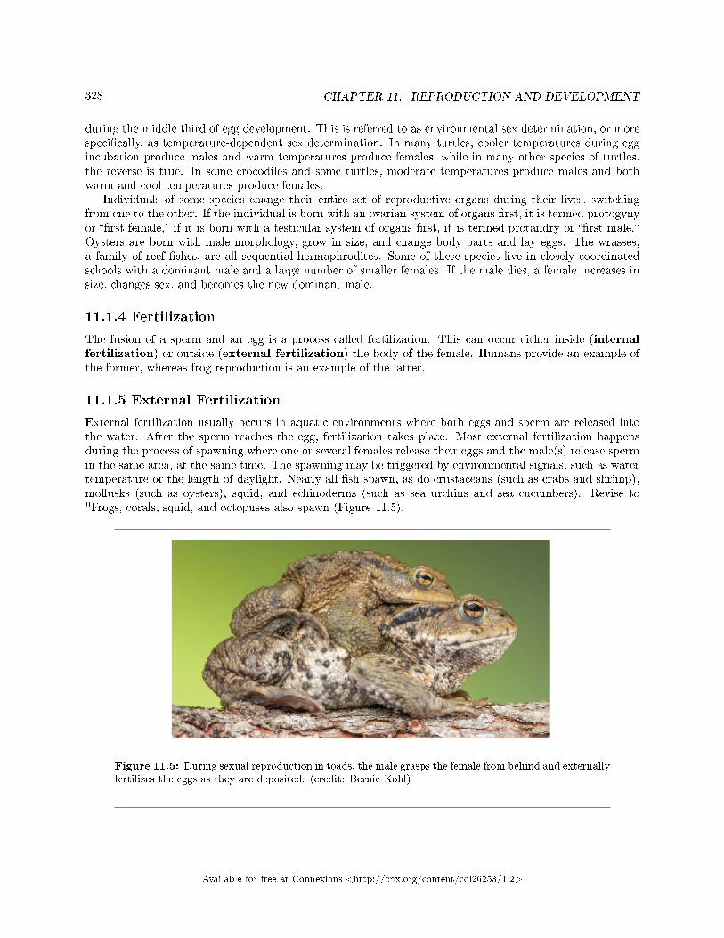
328 CHAPTER 11. REPRODUCTION AND DEVELOPMENT
during the middle third of egg development. This is referred to as environmental sex determination, or morespeci�cally, as temperature-dependent sex determination. In many turtles, cooler temperatures during eggincubation produce males and warm temperatures produce females, while in many other species of turtles,the reverse is true. In some crocodiles and some turtles, moderate temperatures produce males and bothwarm and cool temperatures produce females.
Individuals of some species change their entire set of reproductive organs during their lives, switchingfrom one to the other. If the individual is born with an ovarian system of organs �rst, it is termed protogynyor ��rst female,� if it is born with a testicular system of organs �rst, it is termed protandry or ��rst male.�Oysters are born with male morphology, grow in size, and change body parts and lay eggs. The wrasses,a family of reef �shes, are all sequential hermaphrodites. Some of these species live in closely coordinatedschools with a dominant male and a large number of smaller females. If the male dies, a female increases insize, changes sex, and becomes the new dominant male.
11.1.4 Fertilization
The fusion of a sperm and an egg is a process called fertilization. This can occur either inside (internalfertilization) or outside (external fertilization) the body of the female. Humans provide an example ofthe former, whereas frog reproduction is an example of the latter.
11.1.5 External Fertilization
External fertilization usually occurs in aquatic environments where both eggs and sperm are released intothe water. After the sperm reaches the egg, fertilization takes place. Most external fertilization happensduring the process of spawning where one or several females release their eggs and the male(s) release spermin the same area, at the same time. The spawning may be triggered by environmental signals, such as watertemperature or the length of daylight. Nearly all �sh spawn, as do crustaceans (such as crabs and shrimp),mollusks (such as oysters), squid, and echinoderms (such as sea urchins and sea cucumbers). Revise to"Frogs, corals, squid, and octopuses also spawn (Figure 11.5).
Figure 11.5: During sexual reproduction in toads, the male grasps the female from behind and externallyfertilizes the eggs as they are deposited. (credit: Bernie Kohl)
Available for free at Connexions <http://cnx.org/content/col26253/1.2>
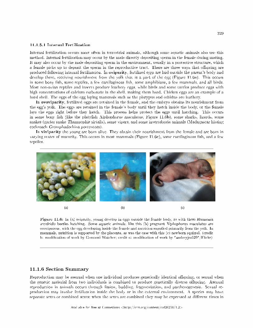
329
11.1.5.1 Internal Fertilization
Internal fertilization occurs most often in terrestrial animals, although some aquatic animals also use thismethod. Internal fertilization may occur by the male directly depositing sperm in the female during mating.It may also occur by the male depositing sperm in the environment, usually in a protective structure, whicha female picks up to deposit the sperm in the reproductive tract. There are three ways that o�spring areproduced following internal fertilization. In oviparity, fertilized eggs are laid outside the parent's body anddevelop there, receiving nourishment from the yolk that is a part of the egg (Figure 11.6a). This occursin some bony �sh, some reptiles, a few cartilaginous �sh, some amphibians, a few mammals, and all birds.Most non-avian reptiles and insects produce leathery eggs, while birds and some turtles produce eggs withhigh concentrations of calcium carbonate in the shell, making them hard. Chicken eggs are an example of ahard shell. The eggs of the egg-laying mammals such as the platypus and echidna are leathery.
In ovoviparity, fertilized eggs are retained in the female, and the embryo obtains its nourishment fromthe egg's yolk. The eggs are retained in the female's body until they hatch inside the body, or the femalelays the eggs right before they hatch. This process helps protect the eggs until hatching. This occursin some bony �sh (like the platy�sh Xiphophorus maculatus, Figure 11.6b), some sharks, lizards, somesnakes (garter snake Thamnophis sirtalis), some vipers, and some invertebrate animals (Madagascar hissingcockroach Gromphadorhina portentosa).
In viviparity the young are born alive. They obtain their nourishment from the female and are born invarying states of maturity. This occurs in most mammals (Figure 11.6c), some cartilaginous �sh, and a fewreptiles.
Figure 11.6: In (a) oviparity, young develop in eggs outside the female body, as with these Harmoniaaxydridis beetles hatching. Some aquatic animals, like this (b) pregnant Xiphophorus maculatus areovoviparous, with the egg developing inside the female and nutrition supplied primarily from the yolk. Inmammals, nutrition is supported by the placenta, as was the case with this (c) newborn squirrel. (creditb: modi�cation of work by Gourami Watcher; credit c: modi�cation of work by "audreyjm529"/Flickr)
11.1.6 Section Summary
Reproduction may be asexual when one individual produces genetically identical o�spring, or sexual whenthe genetic material from two individuals is combined to produce genetically diverse o�spring. Asexualreproduction in animals occurs through �ssion, budding, fragmentation, and parthenogenesis. Sexual re-production may involve fertilization inside the body or in the external environment. A species may haveseparate sexes or combined sexes; when the sexes are combined they may be expressed at di�erent times in
Available for free at Connexions <http://cnx.org/content/col26253/1.2>
330 CHAPTER 11. REPRODUCTION AND DEVELOPMENT
the life cycle. The sex of an individual may be determined by various chromosomal systems or environmentalfactors such as temperature.
Sexual reproduction starts with the combination of a sperm and an egg in a process called fertilization.This can occur either outside the bodies or inside the female. The method of fertilization varies amonganimals. Some species release the egg and sperm into the environment, some species retain the egg andreceive the sperm into the female body and then expel the developing embryo covered with shell, while stillother species retain the developing o�spring throughout the gestation period.
11.1.7 Review Questions
Exercise 11.1.1 (Solution on p. 349.)
In which group is parthenogenesis a normal event?
a. chickensb. beesc. rabbitsd. sea stars
Exercise 11.1.2 (Solution on p. 349.)
Genetically unique individuals are produced through ________.
a. sexual reproductionb. parthenogenesisc. buddingd. fragmentation
Exercise 11.1.3 (Solution on p. 349.)
External fertilization occurs in which type of environment?
a. aquaticb. forestedc. savannad. steppe
11.1.8 Free Response
Exercise 11.1.4 (Solution on p. 349.)
What might be a disadvantage to temperature-dependent sex determination?
Exercise 11.1.5 (Solution on p. 349.)
Compared to separate sexes and assuming self-fertilizing is not possible, what might be one ad-vantage and one disadvantage to hermaphroditism?
11.2 Development and Organogenesis3
The process by which an organism develops from a single-celled zygote to a multi-cellular organism is complexand well regulated. The regulation occurs through signaling between cells and tissues and responses in theform of di�erential gene expression.
3This content is available online at <http://cnx.org/content/m45550/1.9/>.
Available for free at Connexions <http://cnx.org/content/col26253/1.2>
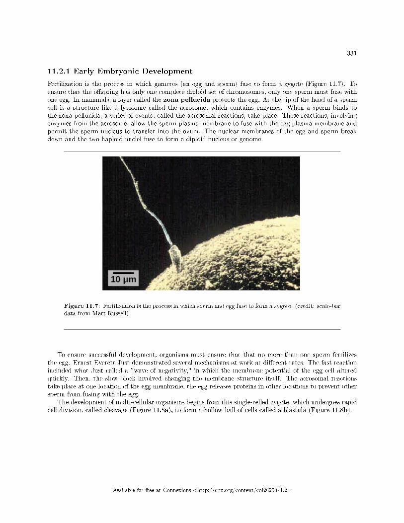
331
11.2.1 Early Embryonic Development
Fertilization is the process in which gametes (an egg and sperm) fuse to form a zygote (Figure 11.7). Toensure that the o�spring has only one complete diploid set of chromosomes, only one sperm must fuse withone egg. In mammals, a layer called the zona pellucida protects the egg. At the tip of the head of a spermcell is a structure like a lysosome called the acrosome, which contains enzymes. When a sperm binds tothe zona pellucida, a series of events, called the acrosomal reactions, take place. These reactions, involvingenzymes from the acrosome, allow the sperm plasma membrane to fuse with the egg plasma membrane andpermit the sperm nucleus to transfer into the ovum. The nuclear membranes of the egg and sperm breakdown and the two haploid nuclei fuse to form a diploid nucleus or genome.
Figure 11.7: Fertilization is the process in which sperm and egg fuse to form a zygote. (credit: scale-bardata from Matt Russell)
To ensure successful development, organisms must ensure that that no more than one sperm fertilizesthe egg. Ernest Everett Just demonstrated several mechanisms at work at di�erent rates. The fast reactionincluded what Just called a "wave of negativity," in which the membrane potential of the egg cell alteredquickly. Then, the slow block involved changing the membrane structure itself. The acrosomal reactionstake place at one location of the egg membrane, the egg releases proteins in other locations to prevent othersperm from fusing with the egg.
The development of multi-cellular organisms begins from this single-celled zygote, which undergoes rapidcell division, called cleavage (Figure 11.8a), to form a hollow ball of cells called a blastula (Figure 11.8b).
Available for free at Connexions <http://cnx.org/content/col26253/1.2>
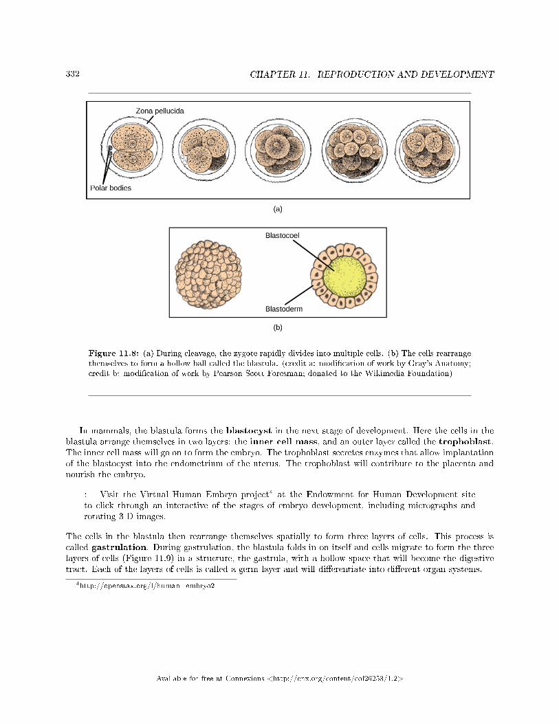
332 CHAPTER 11. REPRODUCTION AND DEVELOPMENT
Figure 11.8: (a) During cleavage, the zygote rapidly divides into multiple cells. (b) The cells rearrangethemselves to form a hollow ball called the blastula. (credit a: modi�cation of work by Gray's Anatomy;credit b: modi�cation of work by Pearson Scott Foresman; donated to the Wikimedia Foundation)
In mammals, the blastula forms the blastocyst in the next stage of development. Here the cells in theblastula arrange themselves in two layers: the inner cell mass, and an outer layer called the trophoblast.The inner cell mass will go on to form the embryo. The trophoblast secretes enzymes that allow implantationof the blastocyst into the endometrium of the uterus. The trophoblast will contribute to the placenta andnourish the embryo.
: Visit the Virtual Human Embryo project4 at the Endowment for Human Development siteto click through an interactive of the stages of embryo development, including micrographs androtating 3-D images.
The cells in the blastula then rearrange themselves spatially to form three layers of cells. This process iscalled gastrulation. During gastrulation, the blastula folds in on itself and cells migrate to form the threelayers of cells (Figure 11.9) in a structure, the gastrula, with a hollow space that will become the digestivetract. Each of the layers of cells is called a germ layer and will di�erentiate into di�erent organ systems.
4http://openstax.org/l/human_embryo2
Available for free at Connexions <http://cnx.org/content/col26253/1.2>
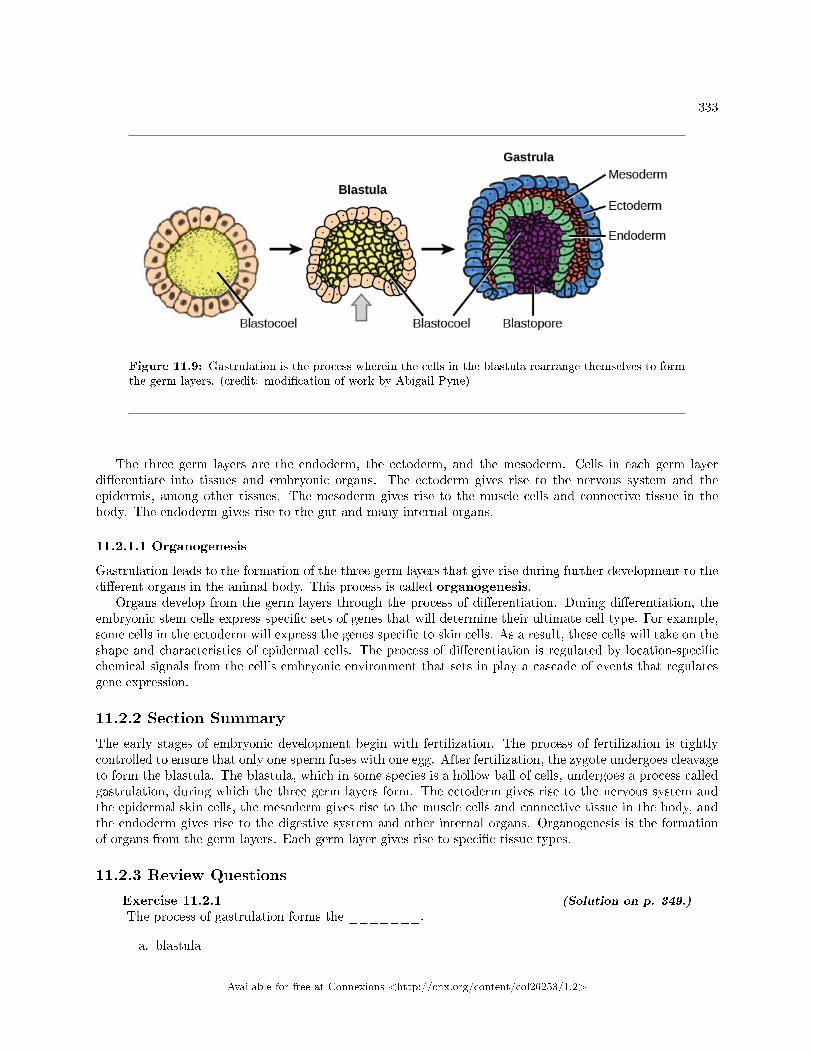
333
Figure 11.9: Gastrulation is the process wherein the cells in the blastula rearrange themselves to formthe germ layers. (credit: modi�cation of work by Abigail Pyne)
The three germ layers are the endoderm, the ectoderm, and the mesoderm. Cells in each germ layerdi�erentiate into tissues and embryonic organs. The ectoderm gives rise to the nervous system and theepidermis, among other tissues. The mesoderm gives rise to the muscle cells and connective tissue in thebody. The endoderm gives rise to the gut and many internal organs.
11.2.1.1 Organogenesis
Gastrulation leads to the formation of the three germ layers that give rise during further development to thedi�erent organs in the animal body. This process is called organogenesis.
Organs develop from the germ layers through the process of di�erentiation. During di�erentiation, theembryonic stem cells express speci�c sets of genes that will determine their ultimate cell type. For example,some cells in the ectoderm will express the genes speci�c to skin cells. As a result, these cells will take on theshape and characteristics of epidermal cells. The process of di�erentiation is regulated by location-speci�cchemical signals from the cell's embryonic environment that sets in play a cascade of events that regulatesgene expression.
11.2.2 Section Summary
The early stages of embryonic development begin with fertilization. The process of fertilization is tightlycontrolled to ensure that only one sperm fuses with one egg. After fertilization, the zygote undergoes cleavageto form the blastula. The blastula, which in some species is a hollow ball of cells, undergoes a process calledgastrulation, during which the three germ layers form. The ectoderm gives rise to the nervous system andthe epidermal skin cells, the mesoderm gives rise to the muscle cells and connective tissue in the body, andthe endoderm gives rise to the digestive system and other internal organs. Organogenesis is the formationof organs from the germ layers. Each germ layer gives rise to speci�c tissue types.
11.2.3 Review Questions
Exercise 11.2.1 (Solution on p. 349.)
The process of gastrulation forms the _______.
a. blastula
Available for free at Connexions <http://cnx.org/content/col26253/1.2>
334 CHAPTER 11. REPRODUCTION AND DEVELOPMENT
b. zygotec. organsd. germ layers
Exercise 11.2.2 (Solution on p. 349.)
Which of the following gives rise to the skin cells?
a. ectodermb. endodermc. mesodermd. none of the above
11.2.4 Free Response
Exercise 11.2.3 (Solution on p. 349.)
What do you think would happen if multiple sperm fused with one egg?
11.3 Human Reproduction5
As in all animals, the adaptations for reproduction in humans are complex. They involve specialized anddi�erent anatomies in the two sexes, a hormone regulation system, and specialized behaviors regulated bythe brain and endocrine system.
11.3.1 Human Reproductive Anatomy
The reproductive tissues of male and female humans develop similarly in utero until about the seventhweek of gestation when, in some cases, a low level of the hormone testosterone is released from the gonads.Testosterone causes the primitive gonads to di�erentiate into sexual organs, such as the scrotum and penis.When testosterone is absent, the primitive gonads develop into ovaries. Tissues that produce a penis inmales produce a clitoris in females. The tissue that will become the scrotum in a male becomes the labiain a female. Thus the male and female anatomies arise from a divergence in the development of what wereonce common embryonic structures.
11.3.1.1 Male Reproductive Anatomy
Sperm are immobile at body temperature; therefore, the testes are external to the body so that a correcttemperature is maintained for motility. In land mammals, including humans, the pair of testes must besuspended outside the body so the environment of the sperm is about 2 ◦C lower than body temperature toproduce viable sperm. If the testes do not descend through the abdominal cavity during fetal development,the individual has reduced fertility.
The scrotum houses the testicles or testes (singular: testis), and provides passage for blood vessels,nerves, and muscles related to testicular function. The testes are a pair of male gonads that produce spermand reproductive hormones. Each testis is approximately 2.5 by 3.8 cm (1.5 by 1 inch) in size and dividedinto wedge-shaped lobes by septa. Coiled in each wedge are seminiferous tubules that produce sperm.
The penis drains urine from the urinary bladder and is a copulatory organ during intercourse (Fig-ure 11.11; Table 11.1). The penis contains three tubes of erectile tissue that become engorged with blood,making the penis erect, in preparation for intercourse. The organ is inserted into the vagina culminating
5This content is available online at <http://cnx.org/content/m45549/1.18/>.
Available for free at Connexions <http://cnx.org/content/col26253/1.2>
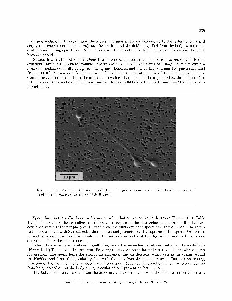
335
with an ejaculation. During orgasm, the accessory organs and glands connected to the testes contract andempty the semen (containing sperm) into the urethra and the �uid is expelled from the body by muscularcontractions causing ejaculation. After intercourse, the blood drains from the erectile tissue and the penisbecomes �accid.
Semen is a mixture of sperm (about �ve percent of the total) and �uids from accessory glands thatcontribute most of the semen's volume. Sperm are haploid cells, consisting of a �agellum for motility, aneck that contains the cell's energy-producing mitochondria, and a head that contains the genetic material(Figure 11.10). An acrosome (acrosomal vesicle) is found at the top of the head of the sperm. This structurecontains enzymes that can digest the protective coverings that surround the egg and allow the sperm to fusewith the egg. An ejaculate will contain from two to �ve milliliters of �uid and from 50�120 million spermper milliliter.
Figure 11.10: As seen in this scanning electron micrograph, human sperm has a �agellum, neck, andhead. (credit: scale-bar data from Matt Russell)
Sperm form in the walls of seminiferous tubules that are coiled inside the testes (Figure 11.11; Table11.1). The walls of the seminiferous tubules are made up of the developing sperm cells, with the leastdeveloped sperm at the periphery of the tubule and the fully developed sperm next to the lumen. The spermcells are associated with Sertoli cells that nourish and promote the development of the sperm. Other cellspresent between the walls of the tubules are the interstitial cells of Leydig, which produce testosteroneonce the male reaches adolescence.
When the sperm have developed �agella they leave the seminiferous tubules and enter the epididymis(Figure 11.11; Table 11.1). This structure lies along the top and posterior of the testes and is the site of spermmaturation. The sperm leave the epididymis and enter the vas deferens, which carries the sperm behindthe bladder, and forms the ejaculatory duct with the duct from the seminal vesicles. During a vasectomy,a section of the vas deferens is removed, preventing sperm (but not the secretions of the accessory glands)from being passed out of the body during ejaculation and preventing fertilization.
The bulk of the semen comes from the accessory glands associated with the male reproductive system.
Available for free at Connexions <http://cnx.org/content/col26253/1.2>
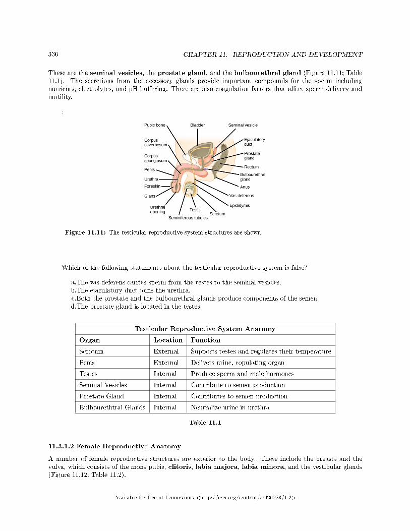
336 CHAPTER 11. REPRODUCTION AND DEVELOPMENT
These are the seminal vesicles, the prostate gland, and the bulbourethral gland (Figure 11.11; Table11.1). The secretions from the accessory glands provide important compounds for the sperm includingnutrients, electrolytes, and pH bu�ering. There are also coagulation factors that a�ect sperm delivery andmotility.
:
Figure 11.11: The testicular reproductive system structures are shown.
Which of the following statements about the testicular reproductive system is false?
a.The vas deferens carries sperm from the testes to the seminal vesicles.b.The ejaculatory duct joins the urethra.c.Both the prostate and the bulbourethral glands produce components of the semen.d.The prostate gland is located in the testes.
Testicular Reproductive System Anatomy
Organ Location Function
Scrotum External Supports testes and regulates their temperature
Penis External Delivers urine, copulating organ
Testes Internal Produce sperm and male hormones
Seminal Vesicles Internal Contribute to semen production
Prostate Gland Internal Contributes to semen production
Bulbourethtral Glands Internal Neutralize urine in urethra
Table 11.1
11.3.1.2 Female Reproductive Anatomy
A number of female reproductive structures are exterior to the body. These include the breasts and thevulva, which consists of the mons pubis, clitoris, labia majora, labia minora, and the vestibular glands(Figure 11.12; Table 11.2).
Available for free at Connexions <http://cnx.org/content/col26253/1.2>
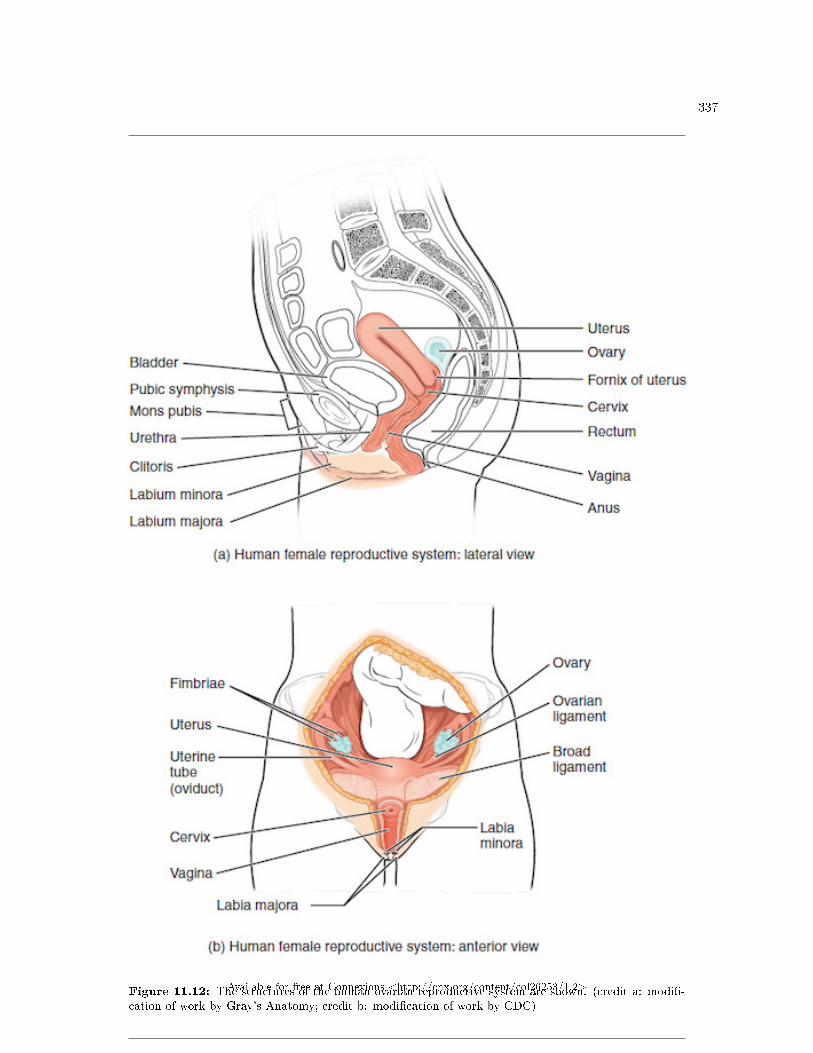
337
Figure 11.12: The structures of the human ovarian reproductive system are shown. (credit a: modi�-cation of work by Gray's Anatomy; credit b: modi�cation of work by CDC)
Available for free at Connexions <http://cnx.org/content/col26253/1.2>
338 CHAPTER 11. REPRODUCTION AND DEVELOPMENT
The breasts consist of mammary glands and fat. Each gland consists of 15 to 25 lobes that have ductsthat empty at the nipple and that supply the nursing child with nutrient- and antibody-rich milk to aiddevelopment and protect the child.
Internal female reproductive structures include ovaries, oviducts, the uterus, and the vagina (Figure 11.12;Table 11.2). The pair of ovaries is held in place in the abdominal cavity by a system of ligaments. Theoutermost layer of the ovary is made up of follicles, each consisting of one or more follicular cells thatsurround, nourish, and protect a single egg. During the menstrual period, a batch of follicular cells developsand prepares their eggs for release. At ovulation, one follicle ruptures and one egg is released. Followingovulation, the follicular tissue that surrounded the ovulated egg stays within the ovary and grows to forma solid mass called the corpus luteum. The corpus luteum secretes additional estrogen and the hormoneprogesterone that helps maintain the uterine lining during pregnancy. The ovaries also produce hormones,such as estrogen.
The oviducts, or fallopian tubes, extend from the uterus in the lower abdominal cavity to the ovaries,but they are not in contact with the ovaries. The lateral ends of the oviducts �are out into a trumpet-likestructure and have a fringe of �nger-like projections called �mbrae. When an egg is released at ovulation,the �mbrae help the nonmotile egg enter into the tube. The walls of the oviducts have a ciliated epitheliumover smooth muscle. The cilia beat, and the smooth muscle contracts, moving the egg toward the uterus.Fertilization usually takes place within the oviduct and the developing embryo is moved toward the uterus.It usually takes the egg or embryo a week to travel through the oviduct.
Sterilization in females is called a tubal ligation; it is analogous to a vasectomy in males in that theoviducts are severed and sealed, preventing sperm from reaching the egg.
The uterus is a structure about the size of a person's �st. The uterus has a thick muscular wall andis lined with an endometrium rich in blood vessels and mucus glands that develop and thicken during thefemale cycle. Thickening of the endometrium prepares the uterus to receive the fertilized egg or zygote,which will then implant itself in the endometrium. The uterus supports the developing embryo and fetusduring gestation. Contractions of the smooth muscle in the uterus aid in forcing the baby through the vaginaduring labor. If fertilization does not occur, a portion of the lining of the uterus sloughs o� during eachmenstrual period. The endometrium builds up again in preparation for implantation. Part of the uterus,called the cervix, protrudes into the top of the vagina.
The vagina is a muscular tube that serves several purposes. It allows menstrual �ow to leave the body.It is the receptacle for the penis during intercourse and the pathway for the delivery of o�spring.
Ovarian Reproductive System Anatomy
Organ Location Function
Clitoris External Sensory organ
Mons pubis External Fatty area overlying pubic bone
Labia majora External Covers labia minora; containssweat and sebaceous glands
Labia minora External Covers vestibule
Greater vestibular glands External Secrete mucus; lubricate vagina
continued on next page
Available for free at Connexions <http://cnx.org/content/col26253/1.2>
339
Breast External Produces and delivers milk
Ovaries Internal Produce and develop eggs
Oviducts Internal Transport egg to uterus; site offertilization
Uterus Internal Supports developing embryo
Vagina Internal Common tube for intercourse,birth canal, passing menstrual�ow
Table 11.2
11.3.2 Gametogenesis (Spermatogenesis and Oogenesis)
Gametogenesis, the production of sperm and eggs, involves the process of meiosis. During meiosis, twonuclear divisions separate the paired chromosomes in the nucleus and then separate the chromatids thatwere made during an earlier stage of the cell's life cycle. Meiosis and its associated cell divisions produceshaploid cells with half of each pair of chromosomes normally found in diploid cells. The production of spermis called spermatogenesis and the production of eggs is called oogenesis.
11.3.2.1 Spermatogenesis
Spermatogenesis occurs in the wall of the seminiferous tubules, with the most primitive cells at the peripheryof the tube and the most mature sperm at the lumen of the tube (Figure 11.13). Immediately under thecapsule of the tubule are diploid, undi�erentiated cells. These stem cells, each called a spermatogonium (pl.spermatogonia), go through mitosis to produce one cell that remains as a stem cell and a second cell calleda primary spermatocyte that will undergo meiosis to produce sperm.
The diploid primary spermatocyte goes through meiosis I to produce two haploid cells called secondaryspermatocytes. Each secondary spermatocyte divides after meiosis II to produce two cells called spermatids.The spermatids eventually reach the lumen of the tubule and grow a �agellum, becoming sperm cells. Foursperm result from each primary spermatocyte that goes through meiosis.
Available for free at Connexions <http://cnx.org/content/col26253/1.2>
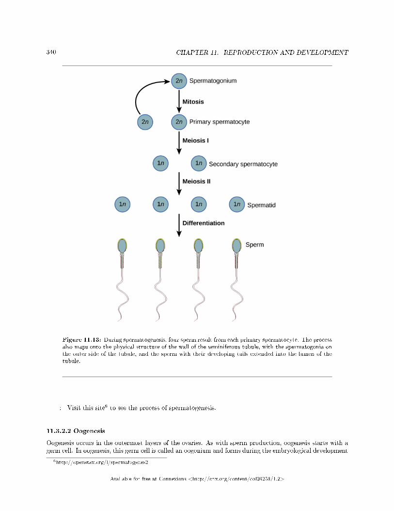
340 CHAPTER 11. REPRODUCTION AND DEVELOPMENT
Figure 11.13: During spermatogenesis, four sperm result from each primary spermatocyte. The processalso maps onto the physical structure of the wall of the seminiferous tubule, with the spermatogonia onthe outer side of the tubule, and the sperm with their developing tails extended into the lumen of thetubule.
: Visit this site6 to see the process of spermatogenesis.
11.3.2.2 Oogenesis
Oogenesis occurs in the outermost layers of the ovaries. As with sperm production, oogenesis starts with agerm cell. In oogenesis, this germ cell is called an oogonium and forms during the embryological development
6http://openstax.org/l/spermatogenes2
Available for free at Connexions <http://cnx.org/content/col26253/1.2>
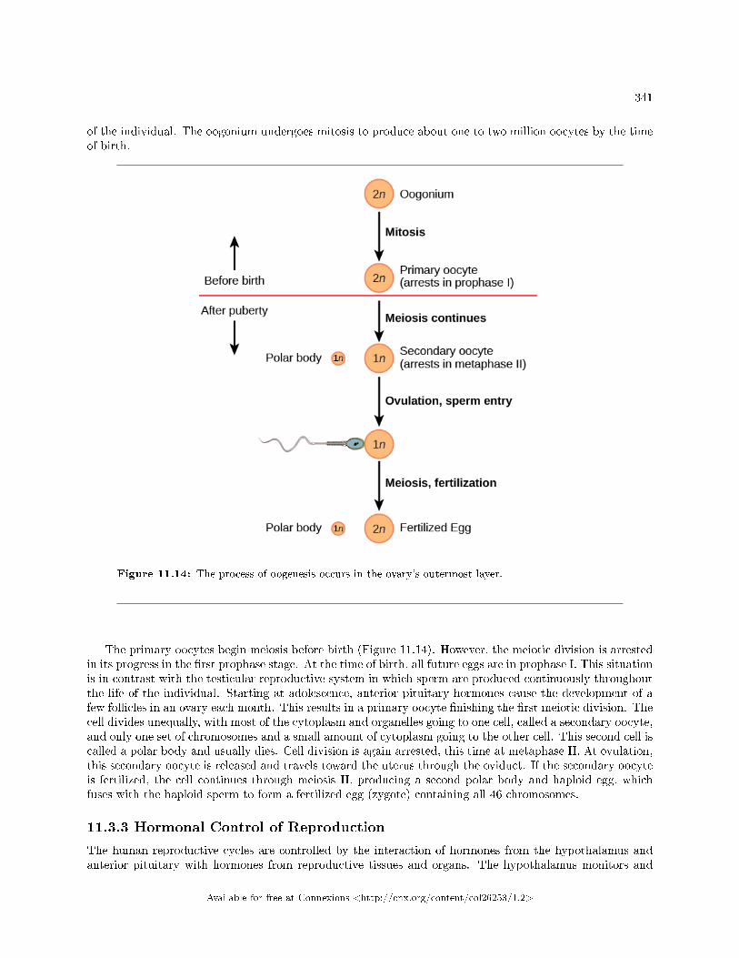
341
of the individual. The oogonium undergoes mitosis to produce about one to two million oocytes by the timeof birth.
Figure 11.14: The process of oogenesis occurs in the ovary's outermost layer.
The primary oocytes begin meiosis before birth (Figure 11.14). However, the meiotic division is arrestedin its progress in the �rst prophase stage. At the time of birth, all future eggs are in prophase I. This situationis in contrast with the testicular reproductive system in which sperm are produced continuously throughoutthe life of the individual. Starting at adolescence, anterior pituitary hormones cause the development of afew follicles in an ovary each month. This results in a primary oocyte �nishing the �rst meiotic division. Thecell divides unequally, with most of the cytoplasm and organelles going to one cell, called a secondary oocyte,and only one set of chromosomes and a small amount of cytoplasm going to the other cell. This second cell iscalled a polar body and usually dies. Cell division is again arrested, this time at metaphase II. At ovulation,this secondary oocyte is released and travels toward the uterus through the oviduct. If the secondary oocyteis fertilized, the cell continues through meiosis II, producing a second polar body and haploid egg, whichfuses with the haploid sperm to form a fertilized egg (zygote) containing all 46 chromosomes.
11.3.3 Hormonal Control of Reproduction
The human reproductive cycles are controlled by the interaction of hormones from the hypothalamus andanterior pituitary with hormones from reproductive tissues and organs. The hypothalamus monitors and
Available for free at Connexions <http://cnx.org/content/col26253/1.2>
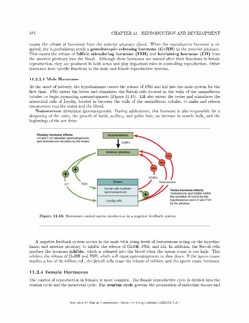
342 CHAPTER 11. REPRODUCTION AND DEVELOPMENT
causes the release of hormones from the anterior pituitary gland. When the reproductive hormone is re-quired, the hypothalamus sends a gonadotropin-releasing hormone (GnRH) to the anterior pituitary.This causes the release of follicle stimulating hormone (FSH) and luteinizing hormone (LH) fromthe anterior pituitary into the blood. Although these hormones are named after their functions in femalereproduction, they are produced in both sexes and play important roles in controlling reproduction. Otherhormones have speci�c functions in the male and female reproductive systems.
11.3.3.1 Male Hormones
At the onset of puberty, the hypothalamus causes the release of FSH and LH into the male system for the�rst time. FSH enters the testes and stimulates the Sertoli cells located in the walls of the seminiferoustubules to begin promoting spermatogenesis (Figure 11.15). LH also enters the testes and stimulates theinterstitial cells of Leydig, located in between the walls of the seminiferous tubules, to make and releasetestosterone into the testes and the blood.
Testosterone stimulates spermatogenesis. During adolescence, this hormone is also responsible for adeepening of the voice, the growth of facial, axillary, and pubic hair, an increase in muscle bulk, and thebeginnings of the sex drive.
Figure 11.15: Hormones control sperm production in a negative feedback system.
A negative feedback system occurs in the male with rising levels of testosterone acting on the hypotha-lamus and anterior pituitary to inhibit the release of GnRH, FSH, and LH. In addition, the Sertoli cellsproduce the hormone inhibin, which is released into the blood when the sperm count is too high. Thisinhibits the release of GnRH and FSH, which will cause spermatogenesis to slow down. If the sperm countreaches a low of 20 million/mL, the Sertoli cells cease the release of inhibin, and the sperm count increases.
11.3.4 Female Hormones
The control of reproduction in females is more complex. The female reproductive cycle is divided into theovarian cycle and the menstrual cycle. The ovarian cycle governs the preparation of endocrine tissues and
Available for free at Connexions <http://cnx.org/content/col26253/1.2>
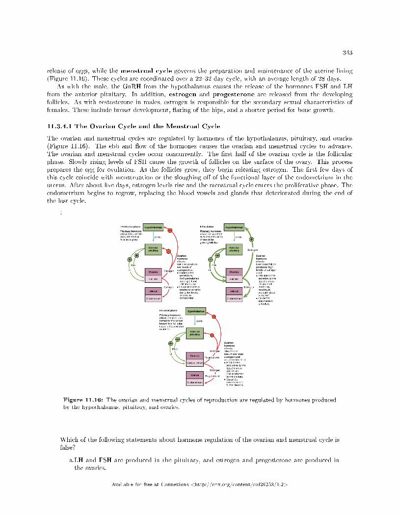
343
release of eggs, while the menstrual cycle governs the preparation and maintenance of the uterine lining(Figure 11.16). These cycles are coordinated over a 22�32 day cycle, with an average length of 28 days.
As with the male, the GnRH from the hypothalamus causes the release of the hormones FSH and LHfrom the anterior pituitary. In addition, estrogen and progesterone are released from the developingfollicles. As with testosterone in males, estrogen is responsible for the secondary sexual characteristics offemales. These include breast development, �aring of the hips, and a shorter period for bone growth.
11.3.4.1 The Ovarian Cycle and the Menstrual Cycle
The ovarian and menstrual cycles are regulated by hormones of the hypothalamus, pituitary, and ovaries(Figure 11.16). The ebb and �ow of the hormones causes the ovarian and menstrual cycles to advance.The ovarian and menstrual cycles occur concurrently. The �rst half of the ovarian cycle is the follicularphase. Slowly rising levels of FSH cause the growth of follicles on the surface of the ovary. This processprepares the egg for ovulation. As the follicles grow, they begin releasing estrogen. The �rst few days ofthis cycle coincide with menstruation or the sloughing o� of the functional layer of the endometrium in theuterus. After about �ve days, estrogen levels rise and the menstrual cycle enters the proliferative phase. Theendometrium begins to regrow, replacing the blood vessels and glands that deteriorated during the end ofthe last cycle.
:
Figure 11.16: The ovarian and menstrual cycles of reproduction are regulated by hormones producedby the hypothalamus, pituitary, and ovaries.
Which of the following statements about hormone regulation of the ovarian and menstrual cycle isfalse?
a.LH and FSH are produced in the pituitary, and estrogen and progesterone are produced inthe ovaries.
Available for free at Connexions <http://cnx.org/content/col26253/1.2>
344 CHAPTER 11. REPRODUCTION AND DEVELOPMENT
b.Estradiol and progesterone secreted from the corpus luteum cause the endometrium to thicken.c.Both progesterone and estrogen are produced by the follicles.d.Secretion of GnRH by the hypothalamus is inhibited by low levels of estrogen but stimulatedby high levels of estrogen.
Just prior to the middle of the cycle (approximately day 14), the high level of estrogen causes FSH andespecially LH to rise rapidly then fall. The spike in LH causes the most mature follicle to rupture and releaseits egg. This is ovulation. The follicles that did not rupture degenerate and their eggs are lost. The levelof estrogen decreases when the extra follicles degenerate.
Following ovulation, the ovarian cycle enters its luteal phase and the menstrual cycle enters its secretoryphase, both of which run from about day 15 to 28. The luteal and secretory phases refer to changes in theruptured follicle. The cells in the follicle undergo physical changes and produce a structure called a corpusluteum. The corpus luteum produces estrogen and progesterone. The progesterone facilitates the regrowthof the uterine lining and inhibits the release of further FSH and LH. The uterus is being prepared to accept afertilized egg, should it occur during this cycle. The inhibition of FSH and LH prevents any further eggs andfollicles from developing, while the progesterone is elevated. The level of estrogen produced by the corpusluteum increases to a steady level for the next few days.
If no fertilized egg is implanted into the uterus, the corpus luteum degenerates and the levels of estrogenand progesterone decrease. The endometrium begins to degenerate as the progesterone levels drop, initiatingthe next menstrual cycle. The decrease in progesterone also allows the hypothalamus to send GnRH to theanterior pituitary, releasing FSH and LH and starting the cycles again.
: Reproductive Endocrinologist
A reproductive endocrinologist is a physician who treats a variety of hormonal disorders relatedto reproduction and infertility in people of any gender. The disorders include menstrual problems,infertility, pregnancy loss, sexual dysfunction, and menopause. Doctors may use fertility drugs,surgery, or assisted reproductive techniques (ART) in their therapy. ART involves the use ofprocedures to manipulate the egg or sperm to facilitate reproduction, such as in vitro fertilization.
Reproductive endocrinologists undergo extensive medical training, �rst in a four-year residency inobstetrics and gynecology, then in a three-year fellowship in reproductive endocrinology. To beboard certi�ed in this area, the physician must pass written and oral exams in both areas.
11.3.5 Gestation
Pregnancy begins with the fertilization of an egg and continues through to the birth of the individual. Thelength of time of gestation, or the gestation period, in humans is 266 days and is similar in other greatapes.
Within 24 hours of fertilization, the egg nucleus has �nished meiosis and the egg and sperm nuclei fuse.With fusion, the cell is known as a zygote. The zygote initiates cleavage and the developing embryo travelsthrough the oviduct to the uterus. The developing embryo must implant into the wall of the uterus withinseven days, or it will deteriorate and die. The outer layers of the developing embryo or blastocyst grow intothe endometrium by digesting the endometrial cells, and healing of the endometrium closes up the blastocystinto the tissue. Another layer of the blastocyst, the chorion, begins releasing a hormone called human betachorionic gonadotropin (β-HCG), which makes its way to the corpus luteum and keeps that structureactive. This ensures adequate levels of progesterone that will maintain the endometrium of the uterus forthe support of the developing embryo. Pregnancy tests determine the level of β-HCG in urine or serum. Ifthe hormone is present, the test is positive.
The gestation period is divided into three equal periods or trimesters. During the �rst two-to-four weeksof the �rst trimester, nutrition and waste are handled by the endometrial lining through di�usion. As thetrimester progresses, the outer layer of the embryo begins to merge with the endometrium, and the placentaforms. The placenta takes over the nutrient and waste requirements of the embryo and fetus, with the
Available for free at Connexions <http://cnx.org/content/col26253/1.2>
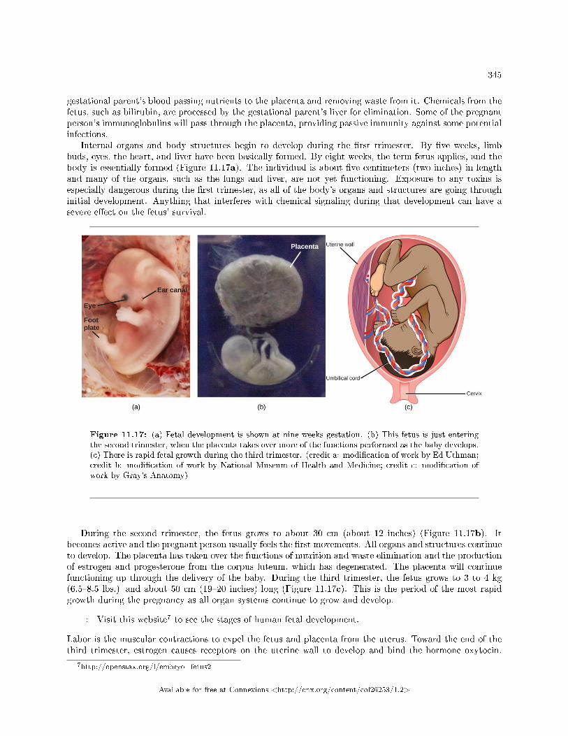
345
gestational parent's blood passing nutrients to the placenta and removing waste from it. Chemicals from thefetus, such as bilirubin, are processed by the gestational parent's liver for elimination. Some of the pregnantperson's immunoglobulins will pass through the placenta, providing passive immunity against some potentialinfections.
Internal organs and body structures begin to develop during the �rst trimester. By �ve weeks, limbbuds, eyes, the heart, and liver have been basically formed. By eight weeks, the term fetus applies, and thebody is essentially formed (Figure 11.17a). The individual is about �ve centimeters (two inches) in lengthand many of the organs, such as the lungs and liver, are not yet functioning. Exposure to any toxins isespecially dangerous during the �rst trimester, as all of the body's organs and structures are going throughinitial development. Anything that interferes with chemical signaling during that development can have asevere e�ect on the fetus' survival.
Figure 11.17: (a) Fetal development is shown at nine weeks gestation. (b) This fetus is just enteringthe second trimester, when the placenta takes over more of the functions performed as the baby develops.(c) There is rapid fetal growth during the third trimester. (credit a: modi�cation of work by Ed Uthman;credit b: modi�cation of work by National Museum of Health and Medicine; credit c: modi�cation ofwork by Gray's Anatomy)
During the second trimester, the fetus grows to about 30 cm (about 12 inches) (Figure 11.17b). Itbecomes active and the pregnant person usually feels the �rst movements. All organs and structures continueto develop. The placenta has taken over the functions of nutrition and waste elimination and the productionof estrogen and progesterone from the corpus luteum, which has degenerated. The placenta will continuefunctioning up through the delivery of the baby. During the third trimester, the fetus grows to 3 to 4 kg(6.5�8.5 lbs.) and about 50 cm (19�20 inches) long (Figure 11.17c). This is the period of the most rapidgrowth during the pregnancy as all organ systems continue to grow and develop.
: Visit this website7 to see the stages of human fetal development.
Labor is the muscular contractions to expel the fetus and placenta from the uterus. Toward the end of thethird trimester, estrogen causes receptors on the uterine wall to develop and bind the hormone oxytocin.
7http://openstax.org/l/embryo_fetus2
Available for free at Connexions <http://cnx.org/content/col26253/1.2>
346 CHAPTER 11. REPRODUCTION AND DEVELOPMENT
At this time, the baby reorients, facing forward and down with the back or crown of the head engaging thecervix (uterine opening). This causes the cervix to stretch and nerve impulses are sent to the hypothalamus,which signals the release of oxytocin from the posterior pituitary. Oxytocin causes smooth muscle in theuterine wall to contract. At the same time, the placenta releases prostaglandins into the uterus, increasing thecontractions. A positive feedback relay occurs between the uterus, hypothalamus, and the posterior pituitaryto assure an adequate supply of oxytocin. As more smooth muscle cells are recruited, the contractions increasein intensity and force.
There are three stages to labor. During stage one, the cervix thins and dilates. This is necessary for thebaby and placenta to be expelled during birth. The cervix will eventually dilate to about 10 cm. Duringstage two, the baby is expelled from the uterus. The uterus contracts and, in most cases, the person givingbirth pushes as they compress the abdominal muscles to aid the delivery. The last stage is the passage ofthe placenta after the baby has been born and the organ has completely disengaged from the uterine wall.If labor should stop before stage two is reached, synthetic oxytocin, known as Pitocin, can be administeredto restart and maintain labor.
11.3.6 Section Summary
The reproductive structures that evolved in land animals allow them to mate, fertilize internally, and supportthe growth and development of o�spring. Gametogenesis, the production of sperm (spermatogenesis) andeggs (oogenesis), takes place through the process of meiosis.
The reproductive cycles are controlled by hormones released from the hypothalamus and anterior pituitaryand hormones from reproductive tissues and organs. The hypothalamus monitors the need for FSH and LHproduction and release from the anterior pituitary. FSH and LH a�ect reproductive structures to cause theformation of sperm and the preparation of eggs for release and possible fertilization. In the male, FSH andLH stimulate Sertoli cells and interstitial cells of Leydig in the testes to facilitate sperm production. TheLeydig cells produce testosterone, which in human males is also responsible for a deepening of the voice, thegrowth of facial, axillary, and pubic hair, and an increase in muscle bulk. In females, FSH and LH causeestrogen and progesterone to be produced. They regulate the female reproductive cycle, which is dividedinto the ovarian cycle and the menstrual cycle.
Human pregnancy begins with fertilization of an egg and proceeds through the three trimesters of ges-tation. The �rst trimester lays down the basic structures of the body, including the limb buds, heart, eyes,and the liver. The second trimester continues the development of all of the organs and systems. The thirdtrimester exhibits the greatest growth of the fetus and culminates in labor and delivery. The labor pro-cess has three stages (contractions, delivery of the fetus, and expulsion of the placenta), each propelled byhormones.
11.3.7 Art Connections
Exercise 11.3.1 (Solution on p. 349.)
Figure 11.11 Which of the following statements about the testicular reproductive system is false?
a. The vas deferens carries sperm from the testes to the seminal vesicles.b. The ejaculatory duct joins the urethra.c. Both the prostate and the bulbourethral glands produce components of the semen.d. The prostate gland is located in the testes.
Exercise 11.3.2 (Solution on p. 349.)
Figure 11.16 Which of the following statements about hormone regulation of the ovarian andmenstrual reproductive cycles is false?
a. LH and FSH are produced in the pituitary, and estrogen and progesterone are produced inthe ovaries.
Available for free at Connexions <http://cnx.org/content/col26253/1.2>
347
b. Estradiol and progesterone secreted from the corpus luteum cause the endometrium to thicken.c. Both progesterone and estrogen are produced by the follicles.d. Secretion of GnRH by the hypothalamus is inhibited by low levels of estrogen but stimulated
by high levels of estrogen.
11.3.8 Review Questions
Exercise 11.3.3 (Solution on p. 349.)
Sperm are produced in the ________.
a. scrotumb. seminal vesiclesc. seminiferous tubulesd. prostate gland
Exercise 11.3.4 (Solution on p. 349.)
Which organ has an endometrial lining that will support a developing baby?
a. labia minorab. breastc. ovariesd. uterus
Exercise 11.3.5 (Solution on p. 349.)
Which hormone causes FSH and LH to be released?
a. testosteroneb. estrogenc. GnRHd. progesterone
Exercise 11.3.6 (Solution on p. 349.)
Nutrient and waste requirements for the developing fetus are handled during the �rst few weeksby ________.
a. the placentab. di�usion through the endometriumc. the choriond. the blastocyst
Exercise 11.3.7 (Solution on p. 349.)
Which hormone is primarily responsible for the contractions during labor?
a. oxytocinb. estrogenc. β-HCGd. progesterone
Available for free at Connexions <http://cnx.org/content/col26253/1.2>
348 CHAPTER 11. REPRODUCTION AND DEVELOPMENT
11.3.9 Free Response
Exercise 11.3.8 (Solution on p. 349.)
Compare spermatogenesis and oogenesis as to timing of the processes, and the number and typeof cells �nally produced.
Exercise 11.3.9 (Solution on p. 349.)
Describe the events in the ovarian cycle leading up to ovulation.
Exercise 11.3.10 (Solution on p. 350.)
Describe the stages of labor.
Available for free at Connexions <http://cnx.org/content/col26253/1.2>
349
Solutions to Exercises in Chapter 11
to Exercise 11.1.1 (p. 330)Bto Exercise 11.1.2 (p. 330)Ato Exercise 11.1.3 (p. 330)Ato Exercise 11.1.4 (p. 330)Temperatures can vary from year to year and an unusually cold or hot year might produce o�spring all ofone sex, making it hard for individuals to �nd mates.to Exercise 11.1.5 (p. 330)A possible advantage of hermaphroditism might be that anytime an individual of the same species isencountered a mating is possible, unlike separate sexes that must �nd an individual of the right sex to mate.(Also, every individual in a hermaphrodite population is able to produce o�spring, which is not the case inpopulations with separate sexes.) A disadvantage might be that hermaphrodite populations are less e�cientbecause they do not specialize in one sex or another, which means a hermaphrodite does not produce asmany o�spring through eggs or sperm as do species with separate sexes. (Other answers are possible.)to Exercise 11.2.1 (p. 333)Dto Exercise 11.2.2 (p. 334)Ato Exercise 11.2.3 (p. 334)If multiple sperm fused with one egg, a zygote with a multiple ploidy level (multiple copies of the chromo-somes) would form, and then would die.to Exercise 11.3.1 (p. 346)Figure 11.11 Dto Exercise 11.3.2 (p. 346)Figure 11.16 Cto Exercise 11.3.3 (p. 347)Cto Exercise 11.3.4 (p. 347)Dto Exercise 11.3.5 (p. 347)Cto Exercise 11.3.6 (p. 347)Bto Exercise 11.3.7 (p. 347)Ato Exercise 11.3.8 (p. 348)Stem cells are laid down in the male during gestation and lie dormant until adolescence. Stem cells in thefemale increase to one to two million and enter the �rst meiotic division and are arrested in prophase. Atadolescence, spermatogenesis begins and continues until death, producing the maximum number of spermwith each meiotic division. Oogenesis continues again at adolescence in batches of eggs with each menstrualcycle. These primary oocytes �nish the �rst meiotic division, producing a viable egg with most of thecytoplasm and its contents, and a second cell called a polar body containing 23 chromosomes. The secondmeiotic division is initiated and arrested in metaphase. At ovulation, one egg is released. If this egg isfertilized, it �nishes the second meiotic division. This is a diploid, fertilized egg.to Exercise 11.3.9 (p. 348)Low levels of progesterone allow the hypothalamus to send GnRH to the anterior pituitary and cause therelease of FSH and LH. FSH stimulates follicles on the ovary to grow and prepare the eggs for ovulation. Asthe follicles increase in size, they begin to release estrogen and a low level of progesterone into the blood.
Available for free at Connexions <http://cnx.org/content/col26253/1.2>

350 CHAPTER 11. REPRODUCTION AND DEVELOPMENT
The level of estrogen rises to a peak, causing a spike in the concentration of LH. This causes the most maturefollicle to rupture and ovulation occurs.to Exercise 11.3.10 (p. 348)Stage one of labor results in uterine contractions, which thin the cervix and dilate the cervical opening.Stage two delivers the baby, and stage three delivers the placenta.
Available for free at Connexions <http://cnx.org/content/col26253/1.2>
GLOSSARY 351
Glossary
A A horizon
consists of a mixture of organic material with inorganic products of weathering
acclimatization
alteration in a body system in response to environmental change
acetylcholinesterase
(AChE) enzyme that breaks down ACh into acetyl and choline
actin
globular contractile protein that interacts with myosin for muscle contraction
action potential
a momentary change in the electrical potential of a neuron (or muscle) membrane
adaptive immunity
immunity that has memory and occurs after exposure to an antigen either from a pathogen or avaccination
adventitious root
aboveground root that arises from a plant part other than the radicle of the plant embryo
afterimage
continuation of a visual sensation after removal of the stimulus
alimentary canal
tubular digestive system with a mouth and anus
allergy
immune reaction that results from immediate hypersensitivities in which an antibody-mediatedimmune response occurs within minutes of exposure to a harmless antigen
alteration
change of the set point in a homeostatic system
alveolar duct
duct that extends from the terminal bronchiole to the alveolar sac
alveolar P O 2
partial pressure of oxygen in the alveoli (usually around 100 mmHg)
alveolar sac
structure consisting of two or more alveoli that share a common opening
alveolar ventilation
how much air is in the alveoli
alveolus
(plural: alveoli) (also, air sac) terminal region of the lung where gas exchange occurs
Available for free at Connexions <http://cnx.org/content/col26253/1.2>
352 GLOSSARY
aminopeptidase
protease that breaks down peptides to single amino acids; secreted by the brush border of smallintestine
amygdala
a structure within the limbic system that processes fear
anatomical dead space
(also, anatomical shunt) region of the lung that lacks proper ventilation/perfusion due to ananatomical block
angina
pain caused by partial blockage of the coronary arteries by the buildup of plaque and lack ofoxygen to the heart muscle
antigen
foreign or �non-self� protein that triggers the immune response
antigen-presenting cell (APC)
immune cell that detects, engulfs, and informs the adaptive immune response about an infectionby presenting the processed antigen on the cell surface
anus
exit point for waste material
aorta
major artery of the body that takes blood away from the heart
apical bud
bud formed at the tip of the shoot
apical meristem
meristematic tissue located at the tips of stems and roots; enables a plant to extend in length
apodeme
ingrowth of an animal's exoskeleton that functions as an attachment site for muscles
appendicular skeleton
composed of the bones of the upper limbs, which function to grasp and manipulate objects, andthe lower limbs, which permit locomotion
appositional growth
increase in the diameter of bones by the addition of bone tissue at the surface of bones
arteriole
small vessel that connects an artery to a capillary bed
artery
blood vessel that takes blood away from the heart
articulation
any place where two bones are joined
asexual reproduction
a mechanism that produces o�spring that are genetically identical to the parent
asymmetrical
Available for free at Connexions <http://cnx.org/content/col26253/1.2>
GLOSSARY 353
describes animals with no axis of symmetry in their body pattern
atherosclerosis
buildup of fatty plaques in the coronary arteries in the heart
atrioventricular valve
one-way membranous �ap of connective tissue between the atrium and the ventricle in the rightside of the heart; also known as tricuspid valve
atrium
(plural: atria) chamber of the heart that receives blood from the veins and sends blood to theventricles
audition
sense of hearing
auditory ossicle
(also, middle ear) transduces sounds from the air into vibrations in the �uid-�lled cochlea
auricle
cartilaginous outer ear
autoantibody
antibody that incorrectly marks �self� components as foreign and stimulates the immune response
autoimmune response
inappropriate immune response to host cells or self-antigens
autoimmunity
type of hypersensitivity to self antigens
autonomic nervous system
the part of the peripheral nervous system that controls bodily functions
axial skeleton
forms the central axis of the body and includes the bones of the skull, the ossicles of the middleear, the hyoid bone of the throat, the vertebral column, and the thoracic cage (ribcage)
axillary bud
bud located in the axil: the stem area where the petiole connects to the stem
axon
a tube-like structure that propagates a signal from a neuron's cell body to axon terminals
B B cell
lymphocyte that matures in the bone marrow and di�erentiates into antibody-secreting plasmacells
B horizon
soil layer that is an accumulation of mostly �ne material that has moved downward
bark
tough, waterproof, outer epidermal layer of cork cells
basal ganglia
an interconnected collections of cells in the brain that are involved in movement and motivation
Available for free at Connexions <http://cnx.org/content/col26253/1.2>
354 GLOSSARY
basal metabolic rate (BMR)
metabolic rate at rest in endothermic animals
basilar membrane
sti� structure in the cochlea that indirectly anchors auditory receptors
basophil
leukocyte that releases chemicals usually involved in the in�ammatory response
bedrock
solid rock that lies beneath the soil
bicarbonate (HCO 3 − ) ion
ion created when carbonic acid dissociates into H+ and (HCO−3 )
bicarbonate bu�er system
system in the blood that absorbs carbon dioxide and regulates pH levels
bicuspid valve
(also, mitral valve; left atrioventricular valve) one-way membranous �ap between the atrium andthe ventricle in the left side of the heart
bile
digestive juice produced by the liver; important for digestion of lipids
binocular cue
cue that relies on the use of both eyes
binocular disparity
slightly di�erent view of the world that each eye receives
bipolar neuron
neuron with two processes from the cell body, typically in opposite directions
blastocyst
the structure formed when cells in the mammalian blastula separate into an inner and outer layer
blind spot
point where we cannot respond to visual information in that portion of the visual �eld
blood pressure (BP)
pressure of blood in the arteries that helps to push blood through the body
bolus
mass of food resulting from chewing action and wetting by saliva
bone
(also, osseous tissue) connective tissue that constitutes the endoskeleton
bone remodeling
replacement of old bone tissue by new bone tissue
brainstem
a portion of brain that connects with the spinal cord; controls basic nervous system functions likebreathing and swallowing
bronchiole
airway that extends from the main tertiary bronchi to the alveolar sac
Available for free at Connexions <http://cnx.org/content/col26253/1.2>
GLOSSARY 355
bronchus
(plural: bronchi) smaller branch of cartilaginous tissue that stems o� of the trachea; air isfunneled through the bronchi to the region where gas exchange occurs in alveoli
budding
a form of asexual reproduction that results from the outgrowth of a part of an organism leadingto a separation from the original animal into two individuals
bulb
modi�ed underground stem that consists of a large bud surrounded by numerous leaf scales
bulbourethral gland
the paired glands in the human male that produce a secretion that cleanses the urethra prior toejaculation
C C horizon
layer of soil that contains the parent material, and the organic and inorganic material that isbroken down to form soil; also known as the soil base
calci�cation
process of deposition of mineral salts in the collagen �ber matrix that crystallizes and hardensthe tissue
canaliculus
microchannel that connects the lacunae and aids di�usion between cells
capillary bed
large number of capillaries that converge to take blood to a particular organ or tissue
capillary
smallest blood vessel that allows the passage of individual blood cells and the site of di�usion ofoxygen and nutrient exchange
carbaminohemoglobin
molecule that forms when carbon dioxide binds to hemoglobin
carbonic anhydrase (CA)
enzyme that catalyzes carbon dioxide and water into carbonic acid
carboxypeptidase
protease that breaks down peptides to single amino acids; secreted by the brush border of thesmall intestine
cardiac cycle
�lling and emptying the heart of blood by electrical signals that cause the heart muscles tocontract and relax
cardiac muscle tissue
muscle tissue found only in the heart; cardiac contractions pump blood throughout the body andmaintain blood pressure
cardiac output
the volume of blood pumped by the heart in one minute as a product of heart rate multiplied bystroke volume
cardiomyocyte
Available for free at Connexions <http://cnx.org/content/col26253/1.2>
356 GLOSSARY
specialized heart muscle cell that is striated but contracts involuntarily like smooth muscle
carnivore
animal that consumes animal �esh
carpus
eight bones that comprise the wrist
cartilage
type of connective tissue with a large amount of ground substance matrix, cells calledchondrocytes, and some amount of �bers
Casparian strip
waxy coating that forces water to cross endodermal plasma membranes before entering thevascular cylinder, instead of moving between endodermal cells
cell-mediated immune response
adaptive immune response that is carried out by T cells
central nervous system (CNS)
the nervous system made up of the brain and spinal cord; covered with three layers of protectivemeninges
cerebellum
the brain structure involved in posture, motor coordination, and learning new motor actions
cerebral cortex
the outermost sheet of brain tissue; involved in many higher-order functions
cerebrospinal �uid (CSF)
a clear liquid that surrounds the brain and �lls its ventricles and acts as a shock absorber
chloride shift
chloride shift exchange of chloride for bicarbonate into or out of the red blood cell
chondrocyte
cell found in cartilage
chylomicron
small lipid globule
chyme
mixture of partially digested food and stomach juices
chymotrypsin
pancreatic protease
clavicle
S-shaped bone that positions the arms laterally
clay
soil particles that are less than 0.002 mm in diameter
clitoris
a sensory and erectile structure in female mammals, homologous to the male penis, stimulatedduring sexual arousal
clonal selection
Available for free at Connexions <http://cnx.org/content/col26253/1.2>
GLOSSARY 357
activation of B cells corresponding to one speci�c BCR variant and the dramatic proliferation ofthat variant
closed circulatory system
system in which the blood is separated from the bodily interstitial �uid and contained in bloodvessels
cochlea
whorled structure that contains receptors for transduction of the mechanical wave into anelectrical signal
collenchyma cell
elongated plant cell with unevenly thickened walls; provides structural support to the stem andleaves
columnar epithelia
epithelia made of cells taller than they are wide, specialized in absorption
compact bone
forms the hard external layer of all bones
companion cell
phloem cell that is connected to sieve-tube cells; has large amounts of ribosomes andmitochondrion
complement system
array of approximately 20 soluble proteins of the innate immune system that enhancephagocytosis, bore holes in pathogens, and recruit lymphocytes; enhances the adaptive responsewhen antibodies are produced
compliance
measurement of the elasticity of the lung
compound leaf
leaf in which the leaf blade is subdivided to form lea�ets, all attached to the midrib
cone
specialized photoreceptor that works best in bright light conditions and detects color
connective tissue
type of tissue made of cells, ground substance matrix, and �bers
corm
rounded, �eshy underground stem that contains stored food
cornea
transparent covering over the eye
coronary artery
vessel that supplies the heart tissue with blood
coronary vein
vessel that takes blood away from the heart tissue back to the chambers in the heart
corpus callosum
a thick nerve bundle that connects the cerebral hemispheres
corpus luteum
Available for free at Connexions <http://cnx.org/content/col26253/1.2>
358 GLOSSARY
the endocrine tissue that develops from an ovarian follicle after ovulation; secretes progesteroneand estrogen during pregnancy
cortex
ground tissue found between the vascular tissue and the epidermis in a stem or root
coxal bone
hip bone
cranial bone
one of eight bones that form the cranial cavity that encloses the brain and serves as anattachment site for the muscles of the head and neck
cuboidal epithelia
epithelia made of cube-shaped cells, specialized in glandular functions
cuticle
waxy covering on the outside of the leaf and stem that prevents the loss of water
cuticle
waxy protective layer on the leaf surface
cytokine
chemical messenger that regulates cell di�erentiation, proliferation, gene expression, and celltra�cking to e�ect immune responses
cytotoxic T lymphocyte (CTL)
adaptive immune cell that directly kills infected cells via perforin and granzymes, and releasescytokines to enhance the immune response
D dead space
area in the lung that lacks proper ventilation or perfusion
dendrite
a structure that extends away from the cell body to receive messages from other neurons
dendritic cell
immune cell that processes antigen material and presents it on the surface of other cells to inducean immune response
depolarization
a change in the membrane potential to a less negative value
depth perception
ability to perceive depth
dermal tissue
protective plant tissue covering the outermost part of the plant; controls gas exchange
diaphragm
domed-shaped skeletal muscle located under lungs that separates the thoracic cavity from theabdominal cavity
diaphysis
central shaft of bone, contains bone marrow in a marrow cavity
diastole
Available for free at Connexions <http://cnx.org/content/col26253/1.2>
GLOSSARY 359
relaxation phase of the cardiac cycle when the heart is relaxed and the ventricles are �lling withblood
digestion
mechanical and chemical breakdown of food into small organic fragments
dipeptidase
protease that breaks down peptides to single amino acids; secreted by the brush border of smallintestine
dorsal cavity
body cavity on the posterior or back portion of an animal; includes the cranial and vertebralcavities
double circulation
�ow of blood in two circuits: the pulmonary circuit through the lungs and the systemic circuitthrough the organs and body
duodenum
�rst part of the small intestine where a large part of digestion of carbohydrates and fats occurs
E ectotherm
animal incapable of maintaining a relatively constant internal body temperature
e�ector cell
lymphocyte that has di�erentiated, such as a B cell, plasma cell, or cytotoxic T lymphocyte
elastase
pancreatic protease
elastic recoil
property of the lung that drives the lung tissue inward
elastic work
work conducted by the intercostal muscles, chest wall, and diaphragm
electrocardiogram (ECG)
recording of the electrical impulses of the cardiac muscle
endocardium
innermost layer of tissue in the heart
endochondral ossi�cation
process of bone development from hyaline cartilage
endodermis
layer of cells in the root that forms a selective barrier between the ground tissue and the vasculartissue, allowing water and minerals to enter the root while excluding toxins and pathogens
endoskeleton
skeleton of living cells that produces a hard, mineralized tissue located within the soft tissue oforganisms
endotherm
animal capable of maintaining a relatively constant internal body temperature
eosinophil
Available for free at Connexions <http://cnx.org/content/col26253/1.2>
360 GLOSSARY
leukocyte that responds to parasites and is involved in the allergic response
epicardium
outermost tissue layer of the heart
epidermis
single layer of cells found in plant dermal tissue; covers and protects underlying tissue
epiphyseal plate
region between the diaphysis and epiphysis that is responsible for the lengthwise growth of longbones
epiphysis
rounded end of bone, covered with articular cartilage and �lled with red bone marrow, whichproduces blood cells
epithelial tissue
tissue that either lines or covers organs or other tissues
epitope
small component of an antigen that is speci�cally recognized by antibodies, B cells, and T cells;the antigenic determinant
esophagus
tubular organ that connects the mouth to the stomach
essential nutrient
nutrient that cannot be synthesized by the body; it must be obtained from food
estivation
torpor in response to extremely high temperatures and low water availability
estrogen
a reproductive hormone, usually present in animals of any sex, which in females assists inendometrial regrowth, ovulation, and calcium absorption
exoskeleton
a secreted cellular product external skeleton that consists of a hard encasement on the surface ofan organism
expiratory reserve volume (ERV)
amount of additional air that can be exhaled after a normal exhalation
external fertilization
the fertilization of eggs by sperm outside an animal's body, often during spawning
F facial bone
one of the 14 bones that form the face; provides cavities for the sense organs (eyes, mouth, andnose) and attachment points for facial muscles
femur
(also, thighbone) longest, heaviest, and strongest bone in the body
FEV1/FVC ratio
ratio of how much air can be forced out of the lung in one second to the total amount that isforced out of the lung; a measurement of lung function that can be used to detect disease states
Available for free at Connexions <http://cnx.org/content/col26253/1.2>
GLOSSARY 361
�brous connective tissue
type of connective tissue with a high concentration of �bers
�brous root system
type of root system in which the roots arise from the base of the stem in a cluster, forming adense network of roots; found in monocots
�bula
(also, calf bone) parallels and articulates with the tibia
�ssion
(also, binary �ssion) a form of asexual reproduction in which an organism splits into two separateorganisms or two parts that regenerate the missing portions of the body
�at bone
thin and relatively broad bone found where extensive protection of organs is required or wherebroad surfaces of muscle attachment are required
�ow-resistive
work of breathing performed by the alveoli and tissues in the lung
follicle stimulating hormone (FSH)
a reproductive hormone that causes sperm production in males and follicle development in females
forced expiratory volume (FEV)
(also, forced vital capacity) measure of how much air can be forced out of the lung from maximalinspiration over a speci�c amount of time
forearm
extends from the elbow to the wrist and consists of two bones: the ulna and the radius
fovea
small indentation in the retina that contains cones
fragmentation
the breaking of an organism into parts and the growth of a separate individual from each part
free nerve ending
ending of an a�erent neuron that lacks a specialized structure for detection of sensory stimuli;some respond to touch, pain, or temperature
frontal (coronal) plane
plane cutting through an animal separating the individual into front and back portions
frontal lobe
the part of the cerebral cortex that contains the motor cortex and areas involved in planning,attention, and language
functional residual capacity (FRC)
expiratory reserve volume plus residual volume
functional vital capacity (FVC)
amount of air that can be forcibly exhaled after taking the deepest breath possible
fusiform
animal body shape that is tubular and tapered at both ends
Available for free at Connexions <http://cnx.org/content/col26253/1.2>
362 GLOSSARY
G gallbladder
organ that stores and concentrates bile
gastrovascular cavity
digestive system consisting of a single opening
gastrulation
the process in which the blastula folds over itself to form the three germ layers
gestation period
the length of time of development, from conception to birth, of the young of a viviparous animal
gestation
the development before birth of a viviparous animal
gill circulation
circulatory system that is speci�c to animals with gills for gas exchange; the blood �ows throughthe gills for oxygenation
gizzard
muscular organ that grinds food
glabrous
describes the non-hairy skin found on palms and �ngers, soles of feet, and lips of humans andother primates
glia
(also, glial cells) the cells that provide support functions for neurons
glomerulus
in the olfactory bulb, one of the two neural clusters that receives signals from one type ofolfactory receptor
Golgi tendon organ
muscular proprioceptive tension receptor that provides the sensory component of the Golgitendon re�ex
gonadotropin-releasing hormone (GnRH)
a hormone from the hypothalamus that causes the release of FSH and LH from the anteriorpituitary
granzyme
protease that enters target cells through perforin and induces apoptosis in the target cells; usedby NK cells and killer T cells
ground tissue
plant tissue involved in photosynthesis; provides support, and stores water and sugars
guard cells
paired cells on either side of a stoma that control stomatal opening and thereby regulate themovement of gases and water vapor
gustation
sense of taste
H Haversian canal
Available for free at Connexions <http://cnx.org/content/col26253/1.2>
GLOSSARY 363
contains the bone's blood vessels and nerve �bers
helper T lymphocyte (TH)
cell of the adaptive immune system that binds APCs via MHC II molecules and stimulates B cellsor secretes cytokines to initiate the immune response
heme group
centralized iron-containing group that is surrounded by the alpha and beta subunits ofhemoglobin
hemocoel
cavity into which blood is pumped in an open circulatory system
hemoglobin
molecule in red blood cells that can bind oxygen, carbon dioxide, and carbon monoxide
hemolymph
mixture of blood and interstitial �uid that is found in insects and other arthropods as well asmost mollusks
herbivore
animal that consumes a strictly plant diet
hermaphroditism
the state of having both male and female reproductive structures within the same individual
hibernation
torpor over a long period of time, such as a winter
hippocampus
the brain structure in the temporal lobe involved in processing memories
homeostasis
dynamic equilibrium maintaining appropriate body functions
horizon
soil layer with distinct physical and chemical properties, which di�ers from other layersdepending on how and when it was formed
human beta chorionic gonadotropin (β-HCG)
a hormone produced by the chorion of the zygote that helps to maintain the corpus luteum andelevated levels of progesterone
humerus
only bone of the arm
humoral immune response
adaptive immune response that is controlled by activated B cells and antibodies
humus
organic material of soil; made up of microorganisms, dead animals and plants in varying stages ofdecay
hydrostatic skeleton
skeleton that consists of aqueous �uid held under pressure in a closed body compartment
hyoid bone
lies below the mandible in the front of the neck
Available for free at Connexions <http://cnx.org/content/col26253/1.2>
364 GLOSSARY
hypersensitivities
spectrum of maladaptive immune responses toward harmless foreign particles or self antigens;occurs after tissue sensitization and includes immediate-type (allergy), delayed-type, andautoimmunity
hypothalamus
the brain structure that controls hormone release and body homeostasis
I ileum
last part of the small intestine; connects the small intestine to the large intestine; important forabsorption of B-12
immune tolerance
acquired ability to prevent an unnecessary or harmful immune response to a detected foreignbody known not to cause disease or to self-antigens
immunode�ciency
failure, insu�ciency, or delay at any level of the immune system, which may be acquired orinherited
incus
(also, anvil) second of the three bones of the middle ear
inferior vena cava
drains blood from the veins that come from the lower organs and the legs
in�ammation
localized redness, swelling, heat, and pain that results from the movement of leukocytes and �uidthrough opened capillaries to a site of infection
ingestion
act of taking in food
inhibin
a hormone made by Sertoli cells, provides negative feedback to hypothalamus in control of FSHand GnRH release
innate immunity
immunity that occurs naturally because of genetic factors or physiology, and is not induced byinfection or vaccination
inner cell mass
the inner layer of cells in the blastocyst, which becomes the embryo
inner ear
innermost part of the ear; consists of the cochlea and the vestibular system
inspiratory capacity (IC)
tidal volume plus inspiratory reserve volume
inspiratory reserve volume (IRV)
amount of additional air that can be inspired after a normal inhalation
intercalary meristem
meristematic tissue located at nodes and the bases of leaf blades; found only in monocots
Available for free at Connexions <http://cnx.org/content/col26253/1.2>
GLOSSARY 365
intercostal muscle
muscle connected to the rib cage that contracts upon inspiration
interferon
cytokine that inhibits viral replication and modulates the immune response
internal fertilization
the fertilization of eggs by sperm inside the body of the female
internode
region between nodes on the stem
interstitial cell of Leydig
a cell type found next to the seminiferous tubules that makes testosterone
interstitial �uid
�uid between cells
intervertebral disc
composed of �brous cartilage; lies between adjacent vertebrae from the second cervical vertebrato the sacrum
intramembranous ossi�cation
process of bone development from �brous membranes
intrapleural space
space between the layers of pleura
iris
colored portion of the eye
irregular bone
bone with complex shapes; examples include vertebrae and hip bones
J jejunum
second part of the small intestine
K kinesthesia
sense of body movement
L labia majora
the large folds of tissue covering inguinal area
labia minora
the smaller folds of tissue within labia majora
labyrinth
bony, hollow structure that is the most internal part of the ear; contains the sites of transductionof auditory and vestibular information
lactase
enzyme that breaks down lactose into glucose and galactose
lacuna
space in cartilage and bone that contains living cells
Available for free at Connexions <http://cnx.org/content/col26253/1.2>
366 GLOSSARY
lamella
layer of compact tissue that surrounds a central canal called the Haversian canal
lamina
leaf blade
large intestine
digestive system organ that reabsorbs water from undigested material and processes waste matter
larynx
voice box, a short passageway connecting the pharynx and the trachea
lateral meristem
meristematic tissue that enables a plant to increase in thickness or girth
lens
curved, transparent structure that provides additional focus for light entering the eye
lenticel
opening on the surface of mature woody stems that facilitates gas exchange
limbic system
a connected brain area that processes emotion and motivation
linear perspective
perceive depth in an image when two parallel lines seem to converge
lipase
enzyme that chemically breaks down lipids
liver
organ that produces bile for digestion and processes vitamins and lipids
loam
soil that has no dominant particle size
long bone
bone that is longer than wide, and has a shaft and two ends
loose (areolar) connective tissue
type of connective tissue with small amounts of cells, matrix, and �bers; found around bloodvessels
lower limb
consists of the thigh, the leg, and the foot
lung capacity
measurement of two or more lung volumes (how much air can be inhaled from the end of anexpiration to maximal capacity)
lung volume
measurement of air for one lung function (normal inhalation or exhalation)
luteinizing hormone (LH)
a reproductive hormone that causes testosterone production in males and ovulation and lactationin females
lymph node
Available for free at Connexions <http://cnx.org/content/col26253/1.2>
GLOSSARY 367
specialized organ that contains a large number of macrophages that clean the lymph before the�uid is returned to the heart
lymph
watery �uid that bathes tissues and organs with protective white blood cells and does not containerythrocytes
lymphocyte
leukocyte that is histologically identi�able by its large nuclei; it is a small cell with very littlecytoplasm
M macrophage
large phagocytic cell that engulfs foreign particles and pathogens
major histocompatibility complex (MHC) I/II molecule
protein found on the surface of all nucleated cells (I) or speci�cally on antigen-presenting cells(II) that signals to immune cells whether the cell is healthy/normal or is infected/cancerous; itprovides the appropriate template into which antigens can be loaded for recognition bylymphocytes
malleus
(also, hammer) �rst of the three bones of the middle ear
maltase
enzyme that breaks down maltose into glucose
mast cell
leukocyte that produces in�ammatory molecules, such as histamine, in response to largepathogens and allergens
matrix
component of connective tissue made of both living and nonliving (ground substances) cells
mechanoreceptor
sensory receptor modi�ed to respond to mechanical disturbance such as being bent, touch,pressure, motion, and sound
megapascal (MPa)
pressure units that measure water potential
Meissner's corpuscle
(also, tactile corpuscle) encapsulated, rapidly-adapting mechanoreceptor in the skin that respondsto light touch
membrane potential
a di�erence in electrical potential between the inside and outside of a cell
memory cell
antigen-speci�c B or T lymphocyte that does not di�erentiate into e�ector cells during theprimary immune response but that can immediately become an e�ector cell upon re-exposure tothe same pathogen
meninges
(singular: meninx) the membranes that cover and protect the central nervous system
menstrual cycle
Available for free at Connexions <http://cnx.org/content/col26253/1.2>
368 GLOSSARY
the cycle of the degradation and re-growth of the endometrium
meristem
plant region of continuous growth
meristematic tissue
tissue containing cells that constantly divide; contributes to plant growth
Merkel's disk
unencapsulated, slowly-adapting mechanoreceptor in the skin that responds to touch
metacarpus
�ve bones that comprise the palm
metatarsal
one of the �ve bones of the foot
middle ear
part of the hearing apparatus that functions to transfer energy from the tympanum to the ovalwindow of the inner ear
midsagittal plane
plane cutting through an animal separating the individual into even right and left sides
mineral
inorganic, elemental molecule that carries out important roles in the body
mineral soil
type of soil that is formed from the weathering of rocks and inorganic material; composedprimarily of sand, silt, and clay
monocular cue
cue that requires only one eye
monocyte
type of white blood cell that circulates in the blood and lymph and di�erentiates intomacrophages after it moves into infected tissue
monogastric
digestive system that consists of a single-chambered stomach
motor end plate
sarcolemma of the muscle �ber that interacts with the neuron
mucin
complex glycoprotein found in mucus
mucosa-associated lymphoid tissue (MALT)
collection of lymphatic tissue that combines with epithelial tissue lining the mucosa throughoutthe body
mucus
sticky protein-containing �uid secretion in the lung that traps particulate matter to be expelledfrom the body
muscle spindle
proprioceptive stretch receptor that lies within a muscle and that shortens the muscle to anoptimal length for e�cient contraction
Available for free at Connexions <http://cnx.org/content/col26253/1.2>
GLOSSARY 369
myelin sheath
a cellular extension containing a fatty substance produced by glia that surrounds and insulatesaxons
myocardial infarction
(also, heart attack) complete blockage of the coronary arteries and death of the cardiac muscletissue
myocardium
heart muscle cells that make up the middle layer and the bulk of the heart wall
myo�bril
long cylindrical structures that lie parallel to the muscle �ber
myo�lament
small structures that make up myo�brils
myosin
contractile protein that interacts with actin for muscle contraction
N nasal cavity
opening of the respiratory system to the outside environment
natural killer (NK) cell
lymphocyte that can kill cells infected with viruses or tumor cells
negative feedback loop
feedback to a control mechanism that increases or decreases a stimulus instead of maintaining it
neuron
a specialized cell that can receive and transmit electrical and chemical signals
neutrophil
phagocytic leukocyte that engulfs and digests pathogens
nociception
neural processing of noxious (such as damaging) stimuli
node
point along the stem at which leaves, �owers, or aerial roots originate
O O horizon
layer of soil with humus at the surface and decomposed vegetation at the base
obstructive disease
disease (such as emphysema and asthma) that arises from obstruction of the airways; complianceincreases in these diseases
occipital lobe
the part of the cerebral cortex that contains visual cortex and processes visual stimuli
odorant
airborne molecule that stimulates an olfactory receptor
olfaction
sense of smell
Available for free at Connexions <http://cnx.org/content/col26253/1.2>
370 GLOSSARY
olfactory bulb
neural structure in the vertebrate brain that receives signals from olfactory receptors
olfactory epithelium
specialized tissue in the nasal cavity where olfactory receptors are located
olfactory receptor
dendrite of a specialized neuron
omnivore
animal that consumes both plants and animals
oogenesis
the process of producing haploid eggs
open circulatory system
system in which the blood is mixed with interstitial �uid and directly covers the organs
opponent-process theory of color perception
color is coded in opponent pairs: black-white, yellow-blue, and red-green
opsonization
process that enhances phagocytosis using proteins to indicate the presence of a pathogen tophagocytic cells
optic chiasm
X-shaped structure that sits just below the brain's ventral surface; represents the merging of theoptic nerves from the two eyes and the separation of information from the two sides of the visual�eld to the opposite side of the brain
optic nerve
carries visual information from the retina to the brain
organ of Corti
in the basilar membrane, the site of the transduction of sound, a mechanical wave, to a neuralsignal
organic soil
type of soil that is formed from sedimentation; composed primarily of organic material
organogenesis
the process of organ formation during development
osseous tissue
connective tissue that constitutes the endoskeleton
ossicle
one of the three bones of the middle ear
ossi�cation
(also, osteogenesis) process of bone formation by osteoblasts
osteoblast
bone cell responsible for bone formation
osteoclast
large bone cells with up to 50 nuclei, responsible for bone remodeling
Available for free at Connexions <http://cnx.org/content/col26253/1.2>
GLOSSARY 371
osteocyte
mature bone cells and the main cell in bone tissue
osteon
cylindrical structure aligned parallel to the long axis of the bone
osteon
subunit of compact bone
ostium
(plural: ostia) holes between blood vessels that allow the movement of hemolymph through thebody of insects, arthropods, and mollusks with open circulatory systems
outer ear
part of the ear that consists of the auricle, ear canal, and tympanum and which conducts soundwaves into the middle ear
oval window
thin diaphragm between the middle and inner ears that receives sound waves from contact withthe stapes bone of the middle ear
ovarian cycle
the cycle of preparation of egg for ovulation and the conversion of the follicle to the corpus luteum
oviduct
(also, fallopian tube) the muscular tube connecting uterus with ovary area
oviparity
a process by which fertilized eggs are laid outside the female's body and develop there, receivingnourishment from the yolk that is a part of the egg
ovoviparity
a process by which fertilized eggs are retained within the female; the embryo obtains itsnourishment from the egg's yolk, and the young are fully developed when they are hatched
ovulation
the release of an oocyte from a mature follicle in the ovary of a vertebrate
oxygen dissociation curve
curve depicting the a�nity of oxygen for hemoglobin
oxygen-carrying capacity
amount of oxygen that can be transported in the blood
P Pacinian corpuscle
encapsulated mechanoreceptor in the skin that responds to deep pressure and vibration
palmately compound leaf
leaf type with lea�ets that emerge from a point, resembling the palm of a hand
pancreas
gland that secretes digestive juices
papilla
one of the small bump-like projections from the tongue
parasympathetic nervous system
Available for free at Connexions <http://cnx.org/content/col26253/1.2>
372 GLOSSARY
the division of autonomic nervous system that regulates visceral functions during relaxation
parenchyma cell
most common type of plant cell; found in the stem, root, leaf, and in fruit pulp; site ofphotosynthesis and starch storage
parent material
organic and inorganic material in which soils form
parietal lobe
the part of the cerebral cortex involved in processing touch and the sense of the body in space
parthenogenesis
a form of asexual reproduction in which an egg develops into a complete individual without beingfertilized
partial pressure
amount of pressure exerted by one gas within a mixture of gases
particulate matter
small particle such as dust, dirt, viral particles, and bacteria that are in the air
patella
(also, kneecap) triangular bone that lies anterior to the knee joint
pathogen-associated molecular pattern (PAMP)
carbohydrate, polypeptide, and nucleic acid �signature� that is expressed by viruses, bacteria, andparasites but di�ers from molecules on host cells
pattern recognition receptor (PRR)
molecule on macrophages and dendritic cells that binds molecular signatures of pathogens andpromotes pathogen engulfment and destruction
pectoral girdle
bones that transmit the force generated by the upper limbs to the axial skeleton
pelvic girdle
bones that transmit the force generated by the lower limbs to the axial skeleton
penis
the male reproductive structure for urine elimination and copulation
pepsin
enzyme found in the stomach whose main role is protein digestion
pepsinogen
inactive form of pepsin
perception
individual interpretation of a sensation; a brain function
perforin
destructive protein that creates a pore in the target cell; used by NK cells and killer T cells
pericardium
membrane layer protecting the heart; also part of the epicardium
pericycle
Available for free at Connexions <http://cnx.org/content/col26253/1.2>
GLOSSARY 373
outer boundary of the stele from which lateral roots can arise
periderm
outermost covering of woody stems; consists of the cork cambium, cork cells, and the phelloderm
peripheral nervous system (PNS)
the nervous system that serves as the connection between the central nervous system and the restof the body; consists of the autonomic nervous system and the sensory-somatic nervous system
peripheral resistance
resistance of the artery and blood vessel walls to the pressure placed on them by the force of theheart pumping
peristalsis
wave-like movements of muscle tissue
permanent tissue
plant tissue composed of cells that are no longer actively dividing
petiole
stalk of the leaf
phalange
one of the bones of the �ngers or toes
pharynx
throat; a tube that starts in the internal nares and runs partway down the neck, where it opensinto the esophagus and the larynx
pheromone
substance released by an animal that can a�ect the physiology or behavior of other animals
photoreceptor
light-detecting cell
phyllotaxy
arrangement of leaves on a stem
physiological dead space
(also, physiological shunt) region of the lung that lacks proper ventilation/perfusion due to aphysiological change in the lung (like in�ammation or edema)
pinnately compound leaf
leaf type with a divided leaf blade consisting of lea�ets arranged on both sides of the midrib
pith
ground tissue found towards the interior of the vascular tissue in a stem or root
placenta
the organ that supports the transport of nutrients and waste between the mothers and fetus'blood in eutherian mammals
plasma cell
immune cell that secrets antibodies; these cells arise from B cells that were stimulated by antigens
plasma
liquid component of blood that is left after the cells are removed
Available for free at Connexions <http://cnx.org/content/col26253/1.2>
374 GLOSSARY
platelet
(also, thrombocyte) small cellular fragment that collects at wounds, cross-reacts with clottingfactors, and forms a plug to prevent blood loss
pleura
tissue layer that surrounds the lungs and lines the interior of the thoracic cavity
pleurisy
painful in�ammation of the pleural tissue layers
positive feedback loop
feedback to a control mechanism that continues the direction of a stimulus
precapillary sphincter
small muscle that controls blood circulation in the capillary beds
primary bronchus
(also, main bronchus) region of the airway within the lung that attaches to the trachea andbifurcates to each lung where it branches into secondary bronchi
primary growth
growth resulting in an increase in length of the stem and the root; caused by cell division in theshoot or root apical meristem
progesterone
a reproductive hormone in usually present in animals of any sex; in human females it assists inendometrial regrowth and inhibition of FSH and LH release
proprioception
sense of limb position; used to track kinesthesia
prostate gland
a structure that is a mixture of smooth muscle and glandular material and that contributes tosemen
proventriculus
glandular part of a bird's stomach
pseudostrati�ed
layer of epithelia that appears multilayered, but is a simple covering
pulmocutaneous circulation
circulatory system in amphibians; the �ow of blood to the lungs and the moist skin for gasexchange
pulmonary circulation
�ow of blood away from the heart through the lungs where oxygenation occurs and then returnsto the heart again
pupil
small opening in the eye through which light passes
R radius
bone located along the lateral (thumb) side of the forearm; articulates with the humerus at theelbow
Available for free at Connexions <http://cnx.org/content/col26253/1.2>
GLOSSARY 375
reception
receipt of a signal (such as light or sound) by sensory receptors
receptive �eld
region in space in which a stimulus can activate a given sensory receptor
receptor potential
membrane potential in a sensory receptor in response to detection of a stimulus
recruitment
process of opening airways that normally remain closed when the cardiac output increases
rectum
area of the body where feces is stored until elimination
red blood cell
small (7�8 µm) biconcave cell without mitochondria (and in mammals without nuclei) that ispacked with hemoglobin, giving the cell its red color; transports oxygen through the body
regulatory T (Treg) cell
specialized lymphocyte that suppresses local in�ammation and inhibits the secretion of cytokines,antibodies, and other stimulatory immune factors; involved in immune tolerance
residual volume (RV)
amount of air remaining in the lung after a maximal expiration
resistance
measurement of lung obstruction
resorption
process by which osteoclasts release minerals stored in bones
respiratory bronchiole
terminal portion of the bronchiole tree that is attached to the terminal bronchioles and alveoliducts, alveolar sacs, and alveoli
respiratory distress syndrome
disease that arises from a de�cient amount of surfactant
respiratory quotient (RQ)
ratio of carbon dioxide production to each oxygen molecule consumed
respiratory rate
number of breaths per minute
restrictive disease
disease that results from a restriction and decreased compliance of the alveoli; respiratory distresssyndrome and pulmonary �brosis are examples
retina
light-sensitive lining of the eye
rhizome
modi�ed underground stem that grows horizontally to the soil surface and has nodes andinternodes
rhizosphere
Available for free at Connexions <http://cnx.org/content/col26253/1.2>
376 GLOSSARY
area of soil a�ected by root secretions and microorganisms
rib
one of 12 pairs of long, curved bones that attach to the thoracic vertebrae and curve toward thefront of the body to form the ribcage
rod
specialized photoreceptor that works well in low light conditions
root cap
protective cells covering the tip of the growing root
root hair
hair-like structure that is an extension of epidermal cells; increases the root surface area and aidsin absorption of water and minerals
root system
belowground portion of the plant that supports the plant and absorbs water and minerals
roughage
component of food that is low in energy and high in �ber
Ru�ni ending
(also, bulbous corpuscle) slowly-adapting mechanoreceptor in the skin that responds to skinstretch and joint position
ruminant
animal with a stomach divided into four compartments
runner
stolon that runs above the ground and produces new clone plants at nodes
S sagittal plane
plane cutting through an animal separating the individual into right and left sides
salivary amylase
enzyme found in saliva, which converts carbohydrates to maltose
sand
soil particles between 0.1�2 mm in diameter
sarcolemma
plasma membrane of a skeletal muscle �ber
sarcomere
functional unit of skeletal muscle
scapula
�at, triangular bone located at the posterior pectoral girdle
sclerenchyma cell
plant cell that has thick secondary walls and provides structural support; usually dead at maturity
scrotum
a sac containing testes, exterior to body
secondary growth
Available for free at Connexions <http://cnx.org/content/col26253/1.2>
GLOSSARY 377
growth resulting in an increase in thickness or girth; caused by the lateral meristem and corkcambium
semen
a �uid mixture of sperm and supporting materials
semicircular canal
one of three half-circular, �uid-�lled tubes in the vestibular labyrinth that monitors angularacceleration and deceleration
semilunar valve
membranous �ap of connective tissue between the aorta and a ventricle of the heart (the aorticor pulmonary semilunar valves)
seminal vesicle
a secretory accessory gland in male; contributes to semen
seminiferous tubule
the structures within which sperm production occurs in the testes
sensory receptor
specialized neuron or other cells associated with a neuron that is modi�ed to receive speci�csensory input
sensory transduction
conversion of a sensory stimulus into electrical energy in the nervous system by a change in themembrane potential
sensory-somatic nervous system
the system of sensory and motor nerves
Sertoli cell
a cell in the walls of the seminiferous tubules that assists developing sperm and secretes inhibin
serum
plasma without the coagulation factors
sesamoid bone
small, �at bone shaped like a sesame seed; develops inside tendons
sessile
leaf without a petiole that is attached directly to the plant stem
set point
midpoint or target point in homeostasis
sex determination
the mechanism by which the sex of individuals in sexually reproducing organisms is initiallyestablished
sexual reproduction
a form of reproduction in which cells containing genetic material from two individuals combinesto produce genetically unique o�spring
shoot system
aboveground portion of the plant; consists of non-reproductive plant parts, such as leaves andstems, and reproductive parts, such as �owers and fruits
Available for free at Connexions <http://cnx.org/content/col26253/1.2>
378 GLOSSARY
short bone
bone that has the same width and length, giving it a cube-like shape
sickle cell anemia
genetic disorder that a�ects the shape of red blood cells, and their ability to transport oxygenand move through capillaries
sieve-tube cell
phloem cell arranged end to end to form a sieve tube that transports organic substances such assugars and amino acids
silt
soil particles between 0.002 and 0.1 mm in diameter
simple epithelia
single layer of epithelial cells
simple leaf
leaf type in which the lamina is completely undivided or merely lobed
sink
growing parts of a plant, such as roots and young leaves, which require photosynthate
sinoatrial (SA) node
the heart's internal pacemaker; located near the wall of the right atrium
skeletal muscle tissue
forms skeletal muscles, which attach to bones and control locomotion and any movement that canbe consciously controlled
skull
bone that supports the structures of the face and protects the brain
small intestine
organ where digestion of protein, fats, and carbohydrates is completed
smooth muscle tissue
occurs in the walls of hollow organs such as the intestines, stomach, and urinary bladder, andaround passages such as the respiratory tract and blood vessels
soil
outer loose layer that covers the surface of Earth
soil pro�le
vertical section of a soil
source
organ that produces photosynthate for a plant
spermatogenesis
the process of producing haploid sperm
sphincter
band of muscle that controls movement of materials throughout the digestive tract
spinal cord
Available for free at Connexions <http://cnx.org/content/col26253/1.2>
GLOSSARY 379
a thick �ber bundle that connects the brain with peripheral nerves; transmits sensory and motorinformation; contains neurons that control motor re�exes
spirometry
method to measure lung volumes and to diagnose lung diseases
spongy bone tissue
forms the inner layer of all bones
squamous epithelia
type of epithelia made of �at cells, specialized in aiding di�usion or preventing abrasion
standard metabolic rate (SMR)
metabolic rate at rest in ectothermic animals
stapes
(also, stirrup) third of the three bones of the middle ear
stele
inner portion of the root containing the vascular tissue; surrounded by the endodermis
stereocilia
in the auditory system, hair-like projections from hair cells that help detect sound waves
sternum
(also, breastbone) long, �at bone located at the front of the chest
stipule
small green structure found on either side of the leaf stalk or petiole
stolon
modi�ed stem that runs parallel to the ground and can give rise to new plants at the nodes
stomach
saclike organ containing acidic digestive juices
strati�ed epithelia
multiple layers of epithelial cells
stroke volume>
- the volume of blood pumped into the aorta per contraction of the left ventricle
sucrase
enzyme that breaks down sucrose into glucose and fructose
superior vena cava
drains blood from the jugular vein that comes from the brain and from the veins that come fromthe arms
surfactant
detergent-like liquid in the airways that lowers the surface tension of the alveoli to allow forexpansion
sutural bone
small, �at, irregularly shaped bone that forms between the �at bones of the cranium
sympathetic nervous system
the division of autonomic nervous system activated during stressful "�ght-or-�ight� situations
Available for free at Connexions <http://cnx.org/content/col26253/1.2>
380 GLOSSARY
synapse
a junction between two neurons where neuronal signals are communicated
synaptic cleft
a space between the presynaptic and postsynaptic membranes
systemic circulation
�ow of blood away from the heart to the brain, liver, kidneys, stomach, and other organs, thelimbs, and the muscles of the body, and then the return of this blood to the heart
systole
contraction phase of cardiac cycle when the ventricles are pumping blood into the arteries
T T cell
lymphocyte that matures in the thymus gland; one of the main cells involved in the adaptiveimmune system
tap root system
type of root system with a main root that grows vertically with few lateral roots; found in dicots
tarsal
one of the seven bones of the ankle
tastant
food molecule that stimulates gustatory receptors
taste bud
clusters of taste cells
tectorial membrane
cochlear structure that lies above the hair cells and participates in the transduction of sound atthe hair cells
temporal lobe
the part of the cerebral cortex that processes auditory input; parts of the temporal lobe areinvolved in speech, memory, and emotion processing
tendril
modi�ed stem consisting of slender, twining strands used for support or climbing
terminal bronchiole
region of bronchiole that attaches to the respiratory bronchioles
testes
a pair of male reproductive organs
testosterone
a reproductive hormone usually present in animals of any sex, and that assists in spermproduction and promoting secondary sexual characteristics
thalamus
the brain area that relays sensory information to the cortex
thalassemia
rare genetic disorder that results in mutation of the alpha or beta subunits of hemoglobin,creating smaller red blood cells with less hemoglobin
Available for free at Connexions <http://cnx.org/content/col26253/1.2>
GLOSSARY 381
thermoregulation
regulation of body temperature
thick �lament
a group of myosin molecules
thin �lament
two polymers of actin wound together along with tropomyosin and troponin
thoracic cage
(also, ribcage) skeleton of the chest, which consists of the ribs, thoracic vertebrae, sternum, andcostal cartilages
thorn
modi�ed stem branch appearing as a sharp outgrowth that protects the plant
threshold of excitation
the level of depolarization needed for an action potential to �re
tibia
(also, shinbone) large bone of the leg that is located directly below the knee
tidal volume (TV)
amount of air that is inspired and expired during normal breathing
torpor
decrease in activity and metabolism that allows an animal to survive adverse conditions
total lung capacity (TLC)
sum of the residual volume, expiratory reserve volume, tidal volume, and inspiratory reservevolume
trabecula
tiny plate that makes up spongy bone and gives it strength
trabeculae
lamellae that are arranged as rods or plates
trachea
cartilaginous tube that transports air from the larynx to the primary bronchi
tracheid
xylem cell with thick secondary walls that helps transport water
transitional epithelia
epithelia that can transition for appearing multilayered to simple; also called uroepithelial
translocation
mass transport of photosynthates from source to sink in vascular plants
transpiration
loss of water vapor to the atmosphere through stomata
transverse (horizontal) plane
plane cutting through an animal separating the individual into upper and lower portions
trichome
hair-like structure on the epidermal surface
Available for free at Connexions <http://cnx.org/content/col26253/1.2>
382 GLOSSARY
trichromatic theory of color perception
color vision is mediated by the activity across the three groups of cones
tricuspid valve
one-way membranous �ap of connective tissue between the atrium and the ventricle in the rightside of the heart; also known as atrioventricular valve
trophoblast
the outer layer of cells in the blastocyst, which gives rise to the embryo's contribution to theplacenta
tropomyosin
acts to block myosin binding sites on actin molecules, preventing cross-bridge formation andpreventing contraction until a muscle receives a neuron signal
troponin
binds to tropomyosin and helps to position it on the actin molecule, and also binds calcium ions
trypsin
pancreatic protease that breaks down protein
tuber
modi�ed underground stem adapted for starch storage; has many adventitious buds
tympanum
(also, tympanic membrane or ear drum) thin diaphragm between the outer and middle ears
U ulna
bone located on the medial aspect (pinky-�nger side) of the forearm
ultrasound
sound frequencies above the human detectable ceiling of approximately 20,000 Hz
umami
one of the �ve basic tastes, which is described as �savory� and which may be largely the taste ofL-glutamate
unidirectional circulation
�ow of blood in a single circuit; occurs in �sh where the blood �ows through the gills, then pastthe organs and the rest of the body, before returning to the heart
uterus
a female reproductive structure in which an embryo develops
V vagina
a muscular tube for the passage of menstrual �ow, copulation, and birth of o�spring
vascular bundle
strands of stem tissue made up of xylem and phloem
vascular stele
strands of root tissue made up of xylem and phloem
vascular tissue
tissue made up of xylem and phloem that transports food and water throughout the plant
Available for free at Connexions <http://cnx.org/content/col26253/1.2>
GLOSSARY 383
vasoconstriction
narrowing of a blood vessel
vasodilation
widening of a blood vessel
vein
blood vessel that brings blood back to the heart
vena cava
major vein of the body returning blood from the upper and lower parts of the body; see thesuperior vena cava and inferior vena cava
venation
pattern of veins in a leaf; may be parallel (as in monocots), reticulate (as in dicots), ordichotomous (as in ginkgo biloba)
venous P CO 2
partial pressure of carbon dioxide in the veins (40 mm Hg in the pulmonary veins)
venous P O 2
partial pressure of oxygen in the veins (100 mm Hg in the pulmonary veins)
ventilation/perfusion (V/Q) mismatch
region of the lung that lacks proper alveolar ventilation (V) and/or arterial perfusion (Q)
ventral cavity
body cavity on the anterior or front portion of an animal that includes the thoracic cavities andthe abdominopelvic cavities
ventricle
(heart) large inferior chamber of the heart that pumps blood into arteries
venule
blood vessel that connects a capillary bed to a vein
vertebral column
(also, spine) surrounds and protects the spinal cord, supports the head, and acts as anattachment point for ribs and muscles of the back and neck
vessel element
xylem cell that is shorter than a tracheid and has thinner walls
vestibular sense
sense of spatial orientation and balance
villi
folds on the inner surface of the small intestine whose role is to increase absorption area
vital capacity (VC)
sum of the expiratory reserve volume, tidal volume, and inspiratory reserve volume
vitamin
organic substance necessary in small amounts to sustain life
viviparity
a process in which the young develop within the female and are born in a nonembryonic state
Available for free at Connexions <http://cnx.org/content/col26253/1.2>
384 GLOSSARY
W water potential (Ψw)
the potential energy of a water solution per unit volume in relation to pure water at atmosphericpressure and ambient temperature
white blood cell
large (30 µm) cell with nuclei of which there are many types with di�erent roles including theprotection of the body from viruses and bacteria, and cleaning up dead cells and other waste
whorled
pattern of leaf arrangement in which three or more leaves are connected at a node
Z zona pellucida
the protective layer around the mammalian egg
Available for free at Connexions <http://cnx.org/content/col26253/1.2>
INDEX 385
Index of Keywords and Terms
Keywords are listed by the section with that keyword (page numbers are in parentheses). Keywordsdo not necessarily appear in the text of the page. They are merely associated with that section. Ex.apples, � 1.1 (1) Terms are referenced by the page they appear on. Ex. apples, 1
( (GnRH), � 11.3(334)(β-HCG), � 11.3(334)
A a horizon, � 2.1(41), 43acclimatization, � 3.3(88), 90acetylcholinesterase, � 6.3(171), 178AChE, � 6.3(171)acini, � 9.1(253)acinus, � 9.1(253)Actin, 172action potential, � 4.1(99), 100adaptive immune response, � 10.2(296)adaptive immune system, � 10.2(296)adaptive immunity, � 10.2(296), 296adipose tissue, � 3.2(71)adventitious root, � 1.3(19)adventitious roots, 19afterimage, � 5.3(126), 132air sacs, � 9.1(253)alimentary canal, � 7.1(185), 188allergies, � 10.3(315)allergy, � 10.3(315), 316alteration, � 3.3(88), 90alveolar duct, � 9.1(253)alveolar ducts, 261alveolar P O 2, 269alveolar sac, � 9.1(253)alveolar sacs, 261alveolar ventilation, � 9.3(272), 276alveoli, � 9.1(253), 261alveolus, � 9.1(253)amino acid, � 7.2(202)aminopeptidase, � 7.3(212), 214amygdala, � 4.1(99), 106amylase, � 7.1(185)anatomical dead space, � 9.3(272)anatomical shunt, � 9.3(272)angina, � 8.3(235), 238animal body, � 3.1(63)animal body function, � 3.1(63)animal digestion, � 7.1(185), � 7.2(202),� 7.3(212)
animal energy, � 3.1(63)animal form, � 3.1(63)animal size, � 3.1(63)animal speed, � 3.1(63)annual ring, � 1.2(5)antigen, � 10.2(296), 296antigen-presenting cell, � 10.2(296)antigen-presenting cell (APC), 296anus, � 7.1(185), 199anvil, � 5.4(136)aorta, � 8.3(235), 237APC, � 10.2(296)apical bud, � 1.2(5), 5apical meristem, � 1.1(1)Apical meristems, 2apodemes, � 3.1(63), 64appendicular skeleton, � 6.1(153), 157appositional growth, � 6.2(160), 167areolar connective tissue, � 3.2(71)Arteries, 241arteriole, � 8.3(235)arterioles, 242artery, � 8.3(235)asexual reproduction, � 11.1(323), 323asthma, � 9.3(272)Asymmetrical, 64asymmetry, � 3.1(63)atherosclerosis, � 8.3(235), 238atria, 227atrioventricular valve, � 8.3(235), 236atrium, � 8.1(223)audition, � 5.4(136), 136auditory ossicle, � 6.1(153)auricle, 137autoantibodies, 317autoantibody, � 10.3(315)autoimmune response, � 10.2(296), 307autoimmunity, � 10.3(315), 317autonomic nervous system, � 4.1(99), 107avian digestion, � 7.1(185)axial skeleton, � 6.1(153), 157
Available for free at Connexions <http://cnx.org/content/col26253/1.2>
386 INDEX
axillary bud, � 1.2(5), 5axon, � 4.1(99), 101
B B cell, � 10.1(287)B cells, 292b horizon, � 2.1(41)bark, 9basal ganglia, � 4.1(99), 106basal metabolic rate, � 3.1(63)basal metabolic rate (BMR), 66basilar membrane, � 5.4(136), 138basophil, � 10.1(287), 291bedrock, 43bicarbonate, � 9.4(278)bicarbonate bu�er system, � 9.4(278), 281bicarbonate ions (HCO 3 − ), 281bicuspid valve, 236bile, � 7.1(185), 198binary �ssion, � 11.1(323)binocular, 133binocular cue, � 5.3(126)binocular disparity, � 5.3(126), 133bioenergetics, � 3.1(63)bipolar neuron, � 5.5(144)bipolar neurons, 144biscuspid valve, � 8.3(235)bitter, � 5.5(144)blastocyst, � 11.2(330), 332blastula, � 11.2(330)blind spot, � 5.3(126), 129blood, � 3.2(71), � 8.2(228)blood �ow, � 8.3(235), � 8.4(246)blood pressure, � 8.4(246)Blood pressure (BP), 246blood pressure regulation, � 8.4(246)blood type, � 8.2(228)blood vessel, � 8.3(235)BMR, � 3.1(63)body cavity, � 3.1(63)body plan, � 3.1(63)body plane, � 3.1(63)body symmetry, � 3.1(63)bolus, � 7.1(185), 194bone, � 3.2(71), � 6.2(160), 160bone cell, � 6.2(160)bone growth, � 6.2(160)bone remodeling, � 6.2(160), 167bone repair, � 6.2(160)bone tissue, � 6.2(160)bone type, � 6.2(160)Boyle's Law, � 9.3(272)BP, � 8.4(246)
brain, � 4.1(99)brainstem, � 4.1(99), 106bronchi, � 9.1(253), 261bronchiole, � 9.1(253)bronchioles, 261bronchus, � 9.1(253)budding, � 11.1(323), 324bulb, � 1.2(5), 15bulboid corpuscle, � 5.2(120)bulbourethral gland, � 11.3(334), 336bulbous corpuscle, � 5.2(120)B horizon, 43
C c horizon, � 2.1(41), 44calci�cation, � 6.2(160), 160calf bone, � 6.1(153)canaliculi, 80capillaries, 243capillary, � 8.3(235)capillary bed, � 8.3(235)Capillary beds, 243carbaminohemoglobin, � 9.4(278), 281carbohydrate digestion, � 7.3(212)carbon dioxide, � 9.2(265)carbon monoxide, � 9.4(278)carbonic acid intermediate, � 9.4(278)carbonic anhydrase, � 9.4(278)Carbonic anhydrase (CA), 281carboxypeptidase, � 7.3(212), 214cardiac arrest, � 8.3(235)cardiac cycle, � 8.3(235), 238cardiac muscle, � 3.2(71), � 8.3(235)cardiac muscle tissue, � 6.3(171), 172cardiac output, � 8.4(246)cardiomyocyte, � 8.3(235)Cardiomyocytes, 239carnivore, � 7.1(185)Carnivores, 186cartilage, � 3.2(71), 78, � 9.1(253)casparian strip, � 1.3(19), 22cell-mediated immune response, � 10.2(296),296central nervous system, � 4.1(99)central nervous system (CNS), 103cephalic phase, � 7.3(212)cerebellum, � 4.1(99), 106cerebral cortex, � 4.1(99), 104cerebrospinal �uid, � 4.1(99)cerebrospinal �uid (CSF), 103cervix, � 11.3(334)chitin, � 9.1(253)chloride shift, � 9.4(278), 281
Available for free at Connexions <http://cnx.org/content/col26253/1.2>
INDEX 387
chondrocytes, � 3.2(71), 78chylomicron, � 7.3(212)chylomicrons, 216chyme, � 7.1(185), 197chymotrypsin, � 7.3(212), 214cilia, � 9.1(253)circulation, � 8.4(246)circulatory system, � 8.1(223), � 8.2(228),� 8.3(235), � 8.4(246)clavicle, � 6.1(153)clay, � 2.1(41), 42cleavage, � 11.2(330)clitoris, � 11.3(334), 336clonal selection, � 10.2(296), 302closed circulatory system, � 8.1(223), 223coagulation, � 8.2(228)cochlea, 138coelom, � 3.1(63)collenchyma cell, � 1.2(5)Collenchyma cells, 7color vision, � 5.3(126), 131columnar epithelia, � 3.2(71)Columnar epithelial, 73Compact bone, 163compact bone tissue, � 6.2(160)Companion cells, 11complement system, � 10.1(287), 294complex plant tissue, � 1.1(1)compliance, � 9.3(272), 276compound leaf, � 1.4(26), 28concentration gradient, � 9.2(265)cone, � 5.3(126)cones, 128connective tissue, � 3.2(71)Connective tissues, 76constrict, � 5.3(126)control of homeostasis, � 3.3(88)copulatory organ, � 11.3(334)corm, � 1.2(5)Corms, 15cornea, � 5.3(126), 127coronal plane, � 3.1(63)coronary arteries, 238coronary artery, � 8.3(235)coronary vein, � 8.3(235)coronary veins, 238corpus callosum, � 4.1(99), 104corpus luteum, 338cortex, � 1.2(5), 11coxal bone, � 6.1(153)cranial bone, � 6.1(153)
CTL, � 10.2(296)cuboidal epithelia, � 3.2(71)Cuboidal epithelial, 72cues, 133cuticle, � 1.4(26), 30, � 2.2(48), 55cytokine, � 10.1(287), 290cytokine release, � 10.1(287)cytotoxic T lymphocyte, � 10.2(296)cytotoxic T lymphocytes (CTLs), 299
D dead space, � 9.3(272)decalci�cation, � 6.2(160)dendrite, � 4.1(99)Dendrites, 100dendritic cell, � 10.2(296)Dendritic cells, 296depolarization, � 4.1(99), 100depth perception, � 5.3(126), 133dermal tissue, � 1.1(1), 3, � 1.2(5)diaphragm, � 9.1(253), 260, � 9.3(272)diaphysis, � 6.2(160), 161diastole, � 8.3(235), 238di�erentiation, � 11.2(330)di�usion, � 3.1(63), � 9.1(253), � 9.2(265)digestion, � 7.3(212), 213digestive process, � 7.3(212)digestive system, � 7.1(185), � 7.2(202),� 7.3(212)dilated, � 5.3(126)dipeptidase, � 7.3(212), 214diploid, � 11.1(323), � 11.2(330)direct di�usion, � 9.1(253)dorsal cavity, � 3.1(63), 68double circulation, � 8.1(223), 227duodenum, � 7.1(185), 198
E ear drum, � 5.4(136)ECG, � 8.3(235)ectoderm, � 3.1(63), � 11.2(330)ectotherm, � 3.1(63)ectothermic, 66e�ector cell, � 10.2(296)e�ector cells, 304egg, � 11.1(323), � 11.2(330), � 11.3(334)ejaculation, � 11.3(334)elastase, � 7.3(212), 214elastic recoil, � 9.3(272), 275elastic work, � 9.3(272), 276electrocardiogram, � 8.3(235)electrocardiogram (ECG), 240elimination, � 7.3(212)embryo, � 11.2(330)
Available for free at Connexions <http://cnx.org/content/col26253/1.2>
388 INDEX
embryonic animal tissue, � 3.1(63)endocardium, � 8.3(235), 237endochondral ossi�cation, � 6.2(160), 166endoderm, � 3.1(63), � 11.2(330), � 11.3(334)endodermis, � 1.3(19), 22endometrium, � 11.2(330), � 11.3(334)endoskeleton, � 6.1(153), 155endotherm, � 3.1(63), 66energy requirements, � 3.1(63)environmental sex determination, � 11.1(323)eosinophil, � 10.1(287), 291epicardium, � 8.3(235), 237epidermis, 9epilepsy, � 4.1(99)epiphyseal plate, � 6.2(160), 166epiphyses, � 6.2(160), 161epithelial tissue, � 3.2(71)Epithelial tissues, 71epitope, � 10.2(296)epitopes, 298erectile tissue, � 11.3(334)esophagus, � 7.1(185), 195essential nutrient, � 7.2(202)essential nutrients, 204estivation, � 3.1(63), 67estrogen, � 11.3(334), 343evapotranspiration, � 2.2(48)excitation�contraction coupling, � 6.3(171)exhalation, � 9.3(272)exoskeleton, � 6.1(153), 154expiratory reserve volume, � 9.2(265)expiratory reserve volume (ERV), 267external fertilization, � 11.1(323), 328
F facial bone, � 6.1(153)fallopian tube, � 11.3(334)fat digestion, � 7.3(212)femur, � 6.1(153)fertilization, � 11.2(330)fetus, � 11.3(334)FEV1/FVC ratio, � 9.2(265), 267�brous connective tissue, � 3.2(71)Fibrous connective tissues, 77�brous root, � 1.3(19)�brous root system, 19�bula, � 6.1(153)�eld, 113�ssion, � 11.1(323), 323�ve senses, � 5.1(113)�at bone, � 6.2(160)Flat bones, 162�atworm, � 9.1(253)
�ow-resistive, � 9.3(272), 276follicle, � 11.3(334)follicle stimulating hormone (FSH),� 11.3(334), 342food energy, � 7.2(202)food requirements, � 7.2(202)forced expiratory volume, � 9.2(265)forced expiratory volume (FEV), 267forced vital capacity, � 9.2(265)forearm, � 6.1(153)fovea, � 5.3(126), 128fragmentation, � 11.1(323), 325free nerve ending, � 5.2(120), 121frontal lobe, � 4.1(99), 105frontal plane, � 3.1(63), 67functional residual capacity, � 9.2(265)functional residual capacity (FRC), 267functional vital capacity, � 9.3(272)functional vital capacity (FVC), 276fusiform, � 3.1(63), 64
G gallbladder, � 7.1(185), 200gas equation, � 9.2(265)gas exchange, � 9.2(265)gas pressure, � 9.2(265)gastrovascular cavity, � 7.1(185), 188gastrulation, � 11.2(330), 332germ layer, � 11.2(330)gestation, � 11.3(334), 344gestation period, � 11.3(334), 344gill circulation, � 8.1(223), 227gills, � 9.1(253)gizzard, � 7.1(185), 190glabrous, � 5.2(120), 121glia, � 4.1(99), 99glial cells, � 4.1(99)glomeruli, 149glomerulus, � 5.5(144)Golgi tendon organ, � 5.2(120)Golgi tendon organs, 123gonadotropin-releasing hormone, � 11.3(334)gonadotropin-releasing hormone (GnRH), 342gonads, � 11.3(334)granzyme, � 10.1(287), 293gray matter, � 4.1(99)ground tissue, � 1.1(1), 3, � 1.2(5)guard cell, � 1.2(5)guard cells, 9gustation, � 5.5(144), 144guttation, � 2.2(48)
H hammer, � 5.4(136)
Available for free at Connexions <http://cnx.org/content/col26253/1.2>
INDEX 389
haploid, � 11.1(323), � 11.2(330)Haversian canal, � 6.2(160), 163hearing, � 5.4(136)heart, � 8.3(235)heart anatomy, � 8.3(235)heart attack, � 8.3(235)heart structure, � 8.3(235)heat conservation, � 3.3(88)heat dissipation, � 3.3(88)helper T (TH) lymphocytes, 299helper T cell, � 10.2(296)helper T lymphocyte, � 10.2(296)heme group, � 9.4(278), 279hemocoel, � 8.1(223), 223hemoglobin, � 9.2(265), � 9.4(278), 279hemolymph, � 8.1(223), 223herbivore, � 7.1(185)Herbivores, 185hermaphroditism, � 11.1(323), 326heterozygous, � 11.1(323)hibernation, � 3.1(63), 67hip bone, � 6.1(153)hippocampus, � 4.1(99), 106homeostasis, � 3.3(88), 88homeostatic process, � 3.3(88)homozygous, � 11.1(323)horizon, � 2.1(41), 43horizontal plane, � 3.1(63)human beta chorionic gonadotropin,� 11.3(334)human beta chorionic gonadotropin (β-HCG),344human skeletal system, � 6.1(153)human skeleton, � 6.1(153)humerus, � 6.1(153)humoral immune response, � 10.2(296), 296humus, � 2.1(41), 42hydrostatic skeleton, � 6.1(153), 153hyoid bone, � 6.1(153)hypersensitivities, � 10.3(315), 316hypersensitivity, � 10.3(315)hypothalamus, � 4.1(99), 106
I ileum, � 7.1(185), 198immune response, � 10.1(287), � 10.2(296),� 10.3(315)immune system, � 10.1(287), � 10.2(296),� 10.3(315)immune tolerance, � 10.2(296), 307immunity, � 10.1(287), � 10.2(296), � 10.3(315)immunode�ciency, � 10.3(315), 316inbreeding, � 11.1(323)
incubation, � 11.1(323)incus, � 5.4(136), 137inferior vena cava, � 8.3(235), 236in�ammation, � 10.1(287), 291ingestion, � 7.3(212), 212inhalation, � 9.3(272)inhibin, � 11.3(334), 342innate immune response, � 10.1(287)innate immune system, � 10.1(287)innate immunity, � 10.1(287), 287inner cell mass, � 11.2(330), 332inner ear, � 5.4(136), 138inspiratory capacity, � 9.2(265)inspiratory capacity (IC), 267inspiratory reserve volume, � 9.2(265)inspiratory reserve volume (IRV), 267intercalary meristem, � 1.1(1)Intercalary meristems, 2intercostal muscle, � 9.3(272)intercostal muscles, 274interferon, � 10.1(287), 290internal fertilization, � 11.1(323), 328internode, � 1.2(5), 5interstitial cell of Leydig, � 11.3(334)interstitial cells of Leydig, 335interstitial �uid, � 8.1(223), 223intervertebral disc, � 6.1(153)intramembranous ossi�cation, � 6.2(160), 166intrapleural space, � 9.3(272)iris, � 5.3(126), 127irregular bone, � 6.2(160)Irregular bones, 162
J jejunum, � 7.1(185), 198just-noticeable di�erence, � 5.1(113)
K kinesthesia, � 5.1(113), 113kneecap, � 6.1(153)Krause end bulb, � 5.2(120)
L labia majora, � 11.3(334), 336labia minora, � 11.3(334), 336labor, � 11.3(334)labyrinth, � 5.4(136), 138lactase, � 7.3(212)lactases, 213lacunae, � 3.2(71), 78lamellae, � 6.2(160), 163lamina, � 1.4(26), 26large intestine, � 7.1(185), 199larynx, � 9.1(253), 259lateral meristem, � 1.1(1)
Available for free at Connexions <http://cnx.org/content/col26253/1.2>
390 INDEX
Lateral meristems, 2leaf, � 1.4(26)leaf adaptation, � 1.4(26)leaf anatomy, � 1.4(26)leaf arrangement, � 1.4(26)leaf form, � 1.4(26)leaf function, � 1.4(26)leaf structure, � 1.4(26)leaves, � 1.4(26)lens, � 5.3(126), 128lenticel, � 1.2(5)lenticels, 13limbic system, � 4.1(99), 106linear perspective, � 5.3(126), 133lipase, � 7.1(185), 194liver, � 7.1(185), 200loam, � 2.1(41)loams, 42locomotion, � 6.1(153), � 6.3(171)long bone, � 6.2(160)Long bones, 161loose connective tissue, � 3.2(71), 76lower limb, � 6.1(153)lung capacities, 265lung capacity, � 9.2(265)lung volume, � 9.2(265)lung volumes, 265luteinizing hormone (LH), � 11.3(334), 342lymph, � 10.2(296), 311lymph node, � 8.4(246)Lymph nodes, 247lymphocyte, � 10.1(287)Lymphocytes, 292
M macrophage, � 10.1(287), 288main bronchus, � 9.1(253)major histocompatability class I molecule,� 10.1(287)major histocompatability class II molecule,� 10.1(287)major histocompatibility complex (MHC) Imolecules, 292malleus, � 5.4(136), 137MALT, � 10.2(296)maltase, � 7.3(212)maltases, 213mammary gland, � 11.3(334)mast cell, � 10.1(287), 291matrix, � 3.2(71), 76maximal exhalation, � 9.2(265)mechanoreceptor, � 5.1(113), 114, � 5.2(120)mechanoreceptor density, � 5.2(120)
megapascal, � 2.2(48)megapascals, 49meiosis, � 11.3(334)Meissner's corpuscle, � 5.2(120)Meissner's corpuscles, 121membrane potential, � 4.1(99)memory cell, � 10.2(296), 307meninges, � 4.1(99), 103menstrual cycle, � 11.3(334), 343meristem, � 1.1(1)meristematic tissue, � 1.1(1), 2meristems, 2Merkel's disc, � 5.2(120)Merkel's disks, 121mesoderm, � 3.1(63), � 11.2(330)mesophyll, � 1.4(26)metatarsal, � 6.1(153)MHC, � 10.1(287)MHC I, � 10.1(287)MHC II, � 10.1(287)MHC II molecules, 293middle ear, � 5.4(136), 137, � 6.1(153)midsagittal plane, � 3.1(63), 67mineral, � 7.2(202)mineral soil, � 2.1(41)mineral soils, 41Minerals, 204mitral valve, � 8.3(235)modi�ed root, � 1.3(19)modi�ed stem, � 1.2(5)monocular cues, 133monocyte, � 10.1(287), 288monogastric, � 7.1(185), 189morning sickness, � 11.3(334)motor end plate, � 6.3(171), 178Muc protein, � 9.1(253)mucin, � 9.1(253), 263mucosa-associated lymphoid tissue, � 10.2(296)Mucosa-associated lymphoid tissue (MALT),305mucus, � 9.1(253), 263muscle contraction, � 6.3(171)muscle �ber structure, � 6.3(171)muscle spindle, � 5.2(120)Muscle spindles, 123muscle tension, � 6.3(171)muscle tissue, � 3.2(71), � 6.3(171)muscle type, � 6.3(171)musculoskeletal system, � 6.1(153), � 6.2(160),� 6.3(171)myelin sheath, � 4.1(99), 101
Available for free at Connexions <http://cnx.org/content/col26253/1.2>
INDEX 391
myocardial infarction, � 8.3(235), 238myocardium, � 8.3(235), 237myo�bril, � 6.3(171)myo�brils, 172myo�lament, � 6.3(171)myo�laments, 174myosin, 172
N nasal cavity, � 9.1(253), 258natural killer (NK) cells, 292natural killer cell, � 10.1(287)negative feedback, � 11.3(334)negative feedback loop, � 3.3(88), 89nervous tissue, � 3.2(71)neural thermoregulation, � 3.3(88)neuron, � 4.1(99)neurons, 99neutrophil, � 10.1(287), 291NK, � 10.1(287)nociception, � 5.2(120), 124node, � 1.2(5)Nodes, 5nutrition, � 7.1(185), � 7.2(202), � 7.3(212)
O o horizon, � 2.1(41), 43obesity, � 7.2(202)obstructive disease, � 9.3(272)Obstructive diseases, 276occipital lobe, � 4.1(99), 105odor, � 5.5(144)odorant, � 5.5(144)Odorants, 144o�spring, � 11.1(323)olfaction, � 5.5(144), 144olfactory bulb, � 5.5(144), 149olfactory epithelium, � 5.5(144), 144olfactory receptor, 144omnivore, � 7.1(185)Omnivores, 187oogenesis, � 11.3(334), 339open circulatory system, � 8.1(223), 223opponent-process theory, 132opponent-process theory of color perception,� 5.3(126)opsonization, � 10.1(287), 294optic chiasm, � 5.3(126), 130optic nerve, � 5.3(126), 129organ of Corti, � 5.4(136), 139organic soil, � 2.1(41)organic soils, 41organogenesis, � 11.2(330), 333orgasm, � 11.3(334)
osseous tissue, � 6.2(160), 160ossicle, � 5.4(136)ossicles, 137ossi�cation, � 6.2(160), 166osteoblast, � 6.2(160)Osteoblasts, 165osteoclast, � 6.2(160)Osteoclasts, 165osteocyte, � 6.2(160)Osteocytes, 165osteon, � 6.2(160)osteons, � 3.2(71), 79, 163Osteoprogenitor cells, 165ostia, 223ostium, � 8.1(223)outer ear, � 5.4(136), 137outward recoil, � 9.3(272)oval window, � 5.4(136), 138ovarian cycle, � 11.3(334), 342ovary, � 11.3(334)oviduct, � 11.3(334)oviducts, 338oviparity, � 11.1(323), 329ovoviparity, � 11.1(323), 329ovulation, � 11.3(334), 344oxygen, � 9.2(265)oxygen dissociation curve, � 9.4(278), 279oxygen-carrying capacity, � 9.4(278), 280
P Pacinian corpuscle, � 5.2(120)Pacinian corpuscles, 122pain, � 5.2(120)palmately compound leaf, � 1.4(26), 28PAMP, � 10.1(287)pancreas, � 7.1(185), 200papilla, � 5.5(144)papillae, 146parasympathetic nervous system, � 4.1(99),109parenchyma cell, � 1.2(5)Parenchyma cells, 6parent material, � 2.1(41), 43parietal lobe, � 4.1(99), 105parthenogenesis, � 11.1(323), 326partial pressure, � 9.2(265), 265particulate matter, � 9.1(253), 262patella, � 6.1(153)pathogen, � 10.1(287)pathogen recognition, � 10.1(287)pathogen-associated molecular pattern,� 10.1(287)pathogen-associated molecular patterns
Available for free at Connexions <http://cnx.org/content/col26253/1.2>
392 INDEX
(PAMPs), 288pathologist, � 3.2(71)pattern recognition receptor, � 10.1(287)pattern recognition receptors (PRRs), 288pectoral girdle, � 6.1(153)penis, � 11.3(334), 334pepsin, � 7.1(185), 197pepsinogen, � 7.1(185), 197perception, � 5.1(113), 116perforin, � 10.1(287), 293pericardium, � 8.3(235), 238pericycle, � 1.3(19), 22periderm, � 1.2(5), 13peripheral nervous system, � 4.1(99)peripheral nervous system (PNS), 107peripheral resistance, � 8.4(246), 249peristalsis, � 7.1(185), 195permanent tissue, � 1.1(1), 2petiole, � 1.4(26), 26pharynx, � 9.1(253), 259pheromone, � 5.5(144), 145photoreceptor, � 5.3(126), 128photosynthate transport, � 2.2(48)phyllotaxy, � 1.4(26), 28physical anthropologist, � 3.1(63)physical properties of soil, � 2.1(41)physiological dead space, � 9.3(272)physiological shunt, � 9.3(272)pinna, � 5.4(136)pinnately compound leaf, � 1.4(26)Pinnately compound leaves, 29pith, � 1.2(5), 11pituitary gland, � 11.3(334)placenta, � 11.2(330), � 11.3(334), 344plant adaptation, � 1.4(26)plant anatomy, � 1.1(1)plant body, � 1.1(1)plant form, � 1.1(1), � 1.2(5), � 1.3(19),� 1.4(26), � 2.2(48)plant nutrition, � 2.1(41)plant organ system, � 1.1(1)plant organs, � 1.1(1)plant physiology, � 1.1(1), � 1.2(5), � 1.3(19),� 1.4(26), � 2.2(48)plant transport, � 2.2(48)plants, � 1.1(1), � 1.2(5), � 1.3(19), � 1.4(26),� 2.2(48)plasma, � 8.2(228), 228plasma cell, � 10.2(296), 302platelet, � 8.2(228)platelets, 228
pleura, � 9.3(272)pleurisy, � 9.3(272)positive feedback, � 11.3(334)positive feedback loop, � 3.3(88), 90precapillary sphincter, � 8.4(246)primary (main) bronchi, 261primary bronchus, � 9.1(253)primary growth, � 1.2(5), 12progesterone, � 11.3(334), 343proprioception, � 5.1(113), 113propriorception, � 5.2(120)prostate gland, � 11.3(334), 336protein digestion, � 7.3(212)proventriculus, � 7.1(185), 190PRR, � 10.1(287)pseudo-ruminant, � 7.1(185)pseudostrati�ed, � 3.2(71), 74pulmocutaneous circulation, � 8.1(223), 227pulmonary circulation, � 8.1(223), 227pupil, � 5.3(126), 127
R rachis, � 1.4(26)radius, � 6.1(153)reception, � 5.1(113), 113, � 5.5(144)receptive, 113receptive �eld, � 5.1(113)receptor potential, � 5.1(113), 114recruitment, � 9.3(272)rectum, � 7.1(185), 199red blood cell, � 8.2(228)Red blood cells, 229regeneration, � 11.1(323)regulatory protein, � 6.3(171)regulatory T (Treg) cells, 307regulatory T cell, � 10.2(296)reproductive endocrinologist, � 11.3(334)residual volume, � 9.2(265)residual volume (RV), 267resistance, � 9.3(272), 276resorption, � 6.2(160)respiratory bronchiole, � 9.1(253)respiratory bronchioles, 261respiratory distress syndrome, � 9.3(272)respiratory quotient, � 9.2(265)respiratory quotient (RQ), 269respiratory rate, � 9.3(272), 276respiratory therapist, � 9.2(265)restrictive disease, � 9.3(272)restrictive diseases, 276retina, � 5.3(126), 128rhizome, � 1.2(5), 15rhizosphere, � 2.1(41), 43
Available for free at Connexions <http://cnx.org/content/col26253/1.2>
INDEX 393
rib, � 6.1(153)ribcage, � 6.1(153)Robert Boyle, � 9.3(272)rods, � 5.3(126), 128root, � 1.3(19)root anatomy, � 1.3(19)root cap, � 1.3(19), 20root growth, � 1.3(19)root hair, � 1.3(19)Root hairs, 21root modi�cation, � 1.3(19)root pressure, � 2.2(48)root structure, � 1.3(19)root system, � 1.1(1), 1, � 1.3(19)root zone, � 1.3(19)roots, � 1.3(19)roughage, � 7.1(185), 192Ru�ni ending, � 5.2(120)Ru�ni endings, 122ruminant, � 7.1(185)Ruminants, 192runner, � 1.2(5)Runners, 15
S sagittal plane, � 3.1(63), 67salivary amylase, � 7.1(185), 194salty, � 5.5(144)sand, � 2.1(41), 42sarcolemma, � 6.3(171), 172sarcomere, � 6.3(171)sarcomeres, 173scapula, � 6.1(153)sclerenchyma cell, � 1.2(5)Sclerenchyma cells, 8scrotum, � 11.3(334), 334secondary growth, � 1.2(5), 12self-fertilization, � 11.1(323)semen, � 11.3(334), 335semicircular canal, � 5.4(136)semicircular canals, 141semilunar valve, � 8.3(235), 236seminal vesicle, � 11.3(334)seminal vesicles, 336seminiferous tubule, � 11.3(334)seminiferous tubules, 335sense, � 5.1(113), � 5.2(120), � 5.4(136),� 5.5(144)sensory perception, � 5.1(113)sensory process, � 5.1(113)sensory receptor, � 5.1(113), 113sensory system, � 5.1(113), � 5.2(120),� 5.4(136), � 5.5(144)
sensory transduction, � 5.1(113), 113sensory-somatic nervous system, � 4.1(99), 107Sertoli cell, � 11.3(334)Sertoli cells, 335serum, � 8.2(228), 233Sesamoid bones, 162sessile, � 1.4(26), 26set point, � 3.3(88), 88sex determination, � 11.1(323), 327sexual reproduction, � 11.1(323), 323shinbone, � 6.1(153)shoot system, � 1.1(1), 1short bone, � 6.2(160)Short bones, 162sickle cell anemia, � 9.4(278), 280sieve-tube cell, � 1.2(5)sieve-tube cells, 11silt, � 2.1(41), 42simple epithelia, � 3.2(71), 71simple leaf, � 1.4(26), 28simple plant tissue, � 1.1(1)sink, � 2.2(48)sinks, 57sinoatrial (SA) node, � 8.3(235), 240skeletal muscle, � 3.2(71)skeletal muscle tissue, � 6.3(171), 172skeletal system, � 6.1(153)skeleton evolution, � 6.1(153)skin sensor, � 5.2(120)skull, � 6.1(153)sliding �lament model of contraction,� 6.3(171)small intestine, � 7.1(185), 197smell, � 5.5(144)smooth muscle, � 3.2(71), � 9.1(253)smooth muscle tissue, � 6.3(171), 172SMR, � 3.1(63)soil, � 2.1(41), 41soil biology, � 2.1(41)soil climate, � 2.1(41)soil composition, � 2.1(41)soil formation, � 2.1(41)soil horizon, � 2.1(41)soil pro�le, � 2.1(41), 42soil properties, � 2.1(41)solute transport, � 2.2(48)somatosensation, � 5.2(120)somatosensory receptor, � 5.2(120)sound, � 5.4(136)sound reception, � 5.4(136)sound transduction, � 5.4(136)
Available for free at Connexions <http://cnx.org/content/col26253/1.2>
394 INDEX
sour, � 5.5(144)source, � 2.2(48)sources, 57sperm, � 11.1(323), � 11.2(330), � 11.3(334)spermatogenesis, � 11.3(334), 339sphincter, 196sphincters, � 7.1(185)spinal cord, � 4.1(99)spine, � 6.1(153)spirometry, � 9.2(265), 267spongy bone, 164spongy bone tissue, � 6.2(160)squamous epithelia, � 3.2(71)Squamous epithelial, 72standard metabolic rate, � 3.1(63)standard metabolic rate (SMR), 66stapes, � 5.4(136), 137stele, � 1.3(19), 22stem, � 1.2(5)stem anatomy, � 1.2(5)stem growth, � 1.2(5)stem modi�cations, � 1.2(5)stereoblindness, � 5.3(126)stereocilia, � 5.4(136), 139sternum, � 6.1(153)stipule, � 1.4(26)stipules, 26stirrup, � 5.4(136)stolon, � 1.2(5)Stolons, 15stomach, � 7.1(185), 196stomatal regulation, � 2.2(48)strati�ed epithelia, � 3.2(71), 71stroke volume, 249sucrase, � 7.3(212)sucrases, 213superior vena cava, � 8.3(235), 236surfactant, � 9.3(272)Sutural bones, 163sweet, � 5.5(144)sympathetic nervous system, � 4.1(99), 109synapse, � 4.1(99)synapses, 100synaptic cleft, � 4.1(99), 103systemic circulation, � 8.1(223), 227systole, � 8.3(235), 238
T T cell, � 10.1(287)T cells, 292tactile corpuscle, � 5.2(120)tap root, � 1.3(19)tap root system, 19
tarsal, � 6.1(153)tastant, � 5.5(144)tastants, 147taste, � 5.5(144)taste bud, � 5.5(144), 146tectorial membrane, � 5.4(136), 138temporal lobe, � 4.1(99), 105tendril, � 1.2(5)Tendrils, 16terminal bronchiole, � 9.1(253)terminal bronchioles, 261testes, � 11.3(334), 334testosterone, � 11.3(334), 342thalamus, � 4.1(99), 106thalassemia, � 9.4(278), 280therapod, � 9.3(272)thermoreception, � 5.2(120)thermoregulation, � 3.3(88), 93thick �lament, � 6.3(171)Thick �laments, 174thighbone, � 6.1(153)thin �lament, � 6.3(171)Thin �laments, 174thoracic cage, � 6.1(153)thoracic cavity, � 9.3(272)thorn, � 1.2(5)Thorns, 16threshold of excitation, � 4.1(99), 100tibia, � 6.1(153)tidal volume, � 9.2(265)Tidal volume (TV), 267topography, � 2.1(41)torpor, � 3.1(63), 67total lung capacity, � 9.2(265)total lung capacity (TLC), 267trabecula, � 6.2(160)trabeculae, 80, 164trachea, � 9.1(253), 259tracheal system, � 9.1(253)tracheid, � 1.2(5)Tracheids, 11transduction, � 5.1(113), � 5.5(144)transfer cell, � 2.2(48)Transitional, 75transitional epithelia, � 3.2(71)translocation, � 2.2(48), 57transpiration, 9, � 2.2(48), 53, 54transpiration regulation, � 2.2(48)transverse plane, � 3.1(63), 67trichome, � 1.2(5)Trichomes, 9
Available for free at Connexions <http://cnx.org/content/col26253/1.2>
INDEX 395
trichromatic theory of color perception,� 5.3(126)trichromatic theory of color vision, 131tricuspid valve, � 8.3(235), 236trimester, � 11.3(334)trophoblast, � 11.2(330), 332tropomyosin, � 6.3(171), 177troponin, � 6.3(171), 177trypsin, � 7.3(212), 214tuber, � 1.2(5)Tubers, 15tympanic membrane, � 5.4(136)tympanum, � 5.4(136), 137
U ulna, � 6.1(153)ultrasound, � 5.4(136), 136umami, � 5.5(144), 144unidirectional circulation, � 8.1(223)unidirectionally, 223uroepithelial, � 3.2(71)uterus, � 11.2(330), � 11.3(334), 338
V vagina, � 11.3(334), 338vas deferens, � 11.3(334)vascular bundle, � 1.1(1), 3vascular cylinder, 3vascular stele, 3vascular tissue, � 1.1(1), 3, � 1.2(5)vasoconstriction, � 8.3(235), 243vasodilation, � 8.3(235), 243vein, � 8.3(235)Veins, 243vena cava, � 8.3(235)venation, � 1.4(26), 27
venous P CO 2, 269venous P O 2, 269ventilation/perfusion, � 9.3(272)ventral cavity, � 3.1(63), 68Ventricaria ventricosa, � 9.1(253)ventricle, � 8.1(223), 227venule, � 8.3(235)venules, 243vertebral column, � 6.1(153)vessel element, � 1.2(5)Vessel elements, 11vestibular information, � 5.4(136)Vestibular sensation, 113, � 5.4(136)vestibular sense, � 5.1(113)villi, � 7.1(185), 197visceral pleura, � 9.3(272)vision, � 5.3(126), 127vital capacity, � 9.2(265)vital capacity (VC), 267vitamin, � 7.2(202)Vitamins, 204viviparity, � 11.1(323), 329
W water potential, � 2.2(48), 49water transport, � 2.2(48)white blood cell, � 8.2(228)white blood cells, 229white matter, � 4.1(99)whorled, � 1.4(26), 28
X xylem, � 2.2(48)
Z zona pellucida, � 11.2(330), 331zygote, � 11.2(330)
Available for free at Connexions <http://cnx.org/content/col26253/1.2>
396 ATTRIBUTIONS
Attributions
Collection: BI 102 General BiologyEdited by: Lena GreenURL: http://cnx.org/content/col26253/1.2/License: http://creativecommons.org/licenses/by/4.0/
Module: "The Plant Body"By: OpenStaxURL: http://cnx.org/content/m44700/1.9/Pages: 1-5Copyright: Rice UniversityLicense: http://creativecommons.org/licenses/by/4.0/
Module: "Stems"By: OpenStaxURL: http://cnx.org/content/m44702/1.7/Pages: 5-19Copyright: Rice UniversityLicense: http://creativecommons.org/licenses/by/3.0/
Module: "Roots"By: OpenStaxURL: http://cnx.org/content/m66598/1.12/Pages: 19-26Copyright: Rice UniversityLicense: http://creativecommons.org/licenses/by/4.0/
Module: "Leaves"By: OpenStaxURL: http://cnx.org/content/m66599/1.14/Pages: 26-37Copyright: Rice UniversityLicense: http://creativecommons.org/licenses/by/4.0/
Module: "The Soil"Used here as: "The Soil and Plant Nutrition"By: OpenStaxURL: http://cnx.org/content/m44715/1.7/Pages: 41-48Copyright: Rice UniversityLicense: http://creativecommons.org/licenses/by/3.0/
Module: "Transport of Water and Solutes in Plants"By: OpenStaxURL: http://cnx.org/content/m66600/1.19/Pages: 48-60Copyright: Rice UniversityLicense: http://creativecommons.org/licenses/by/4.0/
Available for free at Connexions <http://cnx.org/content/col26253/1.2>
ATTRIBUTIONS 397
Module: "Animal Form and Function"By: OpenStaxURL: http://cnx.org/content/m44730/1.10/Pages: 63-71Copyright: Rice UniversityLicense: http://creativecommons.org/licenses/by/4.0/
Module: "Animal Primary Tissues"By: OpenStaxURL: http://cnx.org/content/m66612/1.15/Pages: 71-88Copyright: Rice UniversityLicense: http://creativecommons.org/licenses/by/4.0/
Module: "Homeostasis"By: OpenStaxURL: http://cnx.org/content/m44733/1.11/Pages: 88-95Copyright: Rice UniversityLicense: http://creativecommons.org/licenses/by/4.0/
Module: "Nervous System"By: OpenStaxURL: http://cnx.org/content/m45539/1.8/Pages: 99-111Copyright: Rice UniversityLicense: http://creativecommons.org/licenses/by/4.0/
Module: "Sensory Processes"By: OpenStaxURL: http://cnx.org/content/m66402/1.13/Pages: 113-119Copyright: Rice UniversityLicense: http://creativecommons.org/licenses/by/4.0/
Module: "Somatosensation"By: OpenStaxURL: http://cnx.org/content/m66404/1.13/Pages: 120-126Copyright: Rice UniversityLicense: http://creativecommons.org/licenses/by/4.0/
Module: "Vision"By: OpenStaxURL: http://cnx.org/content/m49042/1.14/Pages: 126-136Copyright: Rice UniversityLicense: http://creativecommons.org/licenses/by/4.0/
Module: "Hearing and Vestibular Sensation"By: OpenStaxURL: http://cnx.org/content/m66407/1.17/Pages: 136-143Copyright: Rice UniversityLicense: http://creativecommons.org/licenses/by/4.0/
Available for free at Connexions <http://cnx.org/content/col26253/1.2>
398 ATTRIBUTIONS
Module: "Taste and Smell"By: OpenStaxURL: http://cnx.org/content/m44764/1.6/Pages: 144-150Copyright: Rice UniversityLicense: http://creativecommons.org/licenses/by/3.0/
Module: "BI 102 Types of Skeletal Systems"Used here as: "Types of Skeletal Systems"By: Lena GreenURL: http://cnx.org/content/m71332/1.1/Pages: 153-160Copyright: Lena GreenLicense: http://creativecommons.org/licenses/by/4.0/Based on: Types of Skeletal SystemsBy: OpenStaxURL: http://cnx.org/content/m66635/1.10/
Module: "Bone"By: OpenStaxURL: http://cnx.org/content/m66638/1.13/Pages: 160-171Copyright: Rice UniversityLicense: http://creativecommons.org/licenses/by/4.0/
Module: "Muscle Contraction and Locomotion"By: OpenStaxURL: http://cnx.org/content/m66643/1.11/Pages: 171-181Copyright: Rice UniversityLicense: http://creativecommons.org/licenses/by/4.0/
Module: "Digestive Systems"By: OpenStaxURL: http://cnx.org/content/m66615/1.14/Pages: 185-202Copyright: Rice UniversityLicense: http://creativecommons.org/licenses/by/4.0/
Module: "Nutrition and Energy Production"By: OpenStaxURL: http://cnx.org/content/m66616/1.14/Pages: 202-212Copyright: Rice UniversityLicense: http://creativecommons.org/licenses/by/4.0/
Module: "Digestive System Processes"By: OpenStaxURL: http://cnx.org/content/m66617/1.15/Pages: 212-220Copyright: Rice UniversityLicense: http://creativecommons.org/licenses/by/4.0/
Available for free at Connexions <http://cnx.org/content/col26253/1.2>
ATTRIBUTIONS 399
Module: "Overview of the Circulatory System"By: OpenStaxURL: http://cnx.org/content/m44801/1.5/Pages: 223-228Copyright: Rice UniversityLicense: http://creativecommons.org/licenses/by/3.0/
Module: "Components of the Blood"By: OpenStaxURL: http://cnx.org/content/m44804/1.5/Pages: 228-235Copyright: Rice UniversityLicense: http://creativecommons.org/licenses/by/3.0/
Module: "Mammalian Heart and Blood Vessels"By: OpenStaxURL: http://cnx.org/content/m44805/1.8/Pages: 235-246Copyright: Rice UniversityLicense: http://creativecommons.org/licenses/by/4.0/
Module: "Blood Flow and Blood Pressure Regulation"By: OpenStaxURL: http://cnx.org/content/m44806/1.8/Pages: 246-250Copyright: Rice UniversityLicense: http://creativecommons.org/licenses/by/4.0/
Module: "Systems of Gas Exchange"By: OpenStaxURL: http://cnx.org/content/m44792/1.14/Pages: 253-264Copyright: Rice UniversityLicense: http://creativecommons.org/licenses/by/4.0/
Module: "Gas Exchange across Respiratory Surfaces"By: OpenStaxURL: http://cnx.org/content/m66646/1.16/Pages: 265-271Copyright: Rice UniversityLicense: http://creativecommons.org/licenses/by/4.0/
Module: "BI 102 Breathing"Used here as: "Breathing"By: Lena GreenURL: http://cnx.org/content/m71333/1.1/Pages: 272-278Copyright: Lena GreenLicense: http://creativecommons.org/licenses/by/4.0/Based on: BreathingBy: OpenStaxURL: http://cnx.org/content/m44797/1.7/
Available for free at Connexions <http://cnx.org/content/col26253/1.2>
400 ATTRIBUTIONS
Module: "Transport of Gases in Human Bodily Fluids"By: OpenStaxURL: http://cnx.org/content/m44799/1.6/Pages: 278-284Copyright: Rice UniversityLicense: http://creativecommons.org/licenses/by/3.0/
Module: "Innate Immune Response"By: OpenStaxURL: http://cnx.org/content/m66663/1.13/Pages: 287-295Copyright: Rice UniversityLicense: http://creativecommons.org/licenses/by/4.0/
Module: "Adaptive Immune Response"By: OpenStaxURL: http://cnx.org/content/m44821/1.8/Pages: 296-315Copyright: Rice UniversityLicense: http://creativecommons.org/licenses/by/3.0/
Module: "Disruptions in the Immune System"By: OpenStaxURL: http://cnx.org/content/m66666/1.12/Pages: 315-319Copyright: Rice UniversityLicense: http://creativecommons.org/licenses/by/4.0/
Module: "How Animals Reproduce"By: OpenStaxURL: http://cnx.org/content/m45547/1.11/Pages: 323-330Copyright: Rice UniversityLicense: http://creativecommons.org/licenses/by/4.0/
Module: "Development and Organogenesis"By: OpenStaxURL: http://cnx.org/content/m45550/1.9/Pages: 330-334Copyright: Rice UniversityLicense: http://creativecommons.org/licenses/by/3.0/
Module: "Human Reproduction"By: OpenStaxURL: http://cnx.org/content/m45549/1.18/Pages: 334-348Copyright: Rice UniversityLicense: http://creativecommons.org/licenses/by/4.0/
Available for free at Connexions <http://cnx.org/content/col26253/1.2>
BI 102 General BiologyWe begin the term by studying plant structure, organization, growth, and nutrition. We will then move onto a study of vertebrate structure and function, concentrating on human biology. Students will study humanbody's organization, including the nervous system, cardiovascular system and reproductive system, amongothers.
AboutRhaptos is a web-based collaborative publishing system for educational material.