Embed Size (px)
Citation preview
Fax +41 61 306 12 34E-Mail [email protected]
Opinion Article
Int Arch Allergy Immunol DOI: 10.1159/000331312
Basophil Activation Test in Allergy:Time for an Update?
Salvatore Chirumbolo
Department of Pathology and Diagnostics, Unit of General Pathology, University of Verona, Verona , Italy
Introduction
Although more than 10 years have elapsed since the first basophil activation test (BAT) was successfully es-tablished, and although it is still the only routine cellular assay for allergy diagnosis, its use shows difficulty in hav-ing an appeal. In the diagnosis of allergy, skin testing and quantification of serum levels of allergen-specific IgE are considered the gold standards for routine laboratory in-vestigations, even though variability in results and dis-crepancies between interpretation and clinical assess-ment have been clearly demonstrated. For example, levels of serum IgE (sIgE) tend to decrease over time, as has been shown with drug hypersensitivity, and this may af-fect the correlation with cellular tests, although a de-crease in sensitivity has also been reported for BAT in certain circumstances [1] . These issues clearly illustrate the difficulty in defining a consensus in the investigation of allergy, which is largely explained by the multiple fac-tors which have to be taken into account. This demon-strates why there is still a need for reliable in vitro meth-ods to complement the usual investigations into allergy when they show discrepancies or are not feasible.
Functional in vitro tests are focused on basophils since, in contrast to mast cells, basophils are circulating cells and are easy to manipulate. Nevertheless, their very low number constitutes a true limitation to research.
Key Words
Allergen challenge � Allergy � Anti-IgE � Basophil activation test � CD203c � CD63 � Flow cytometry
Abstract
A wide range of reported evidence in the literature has shown that the quantification of basophil activation by flow cytometry (basophil activation test, BAT) has proven to be a useful tool for the assessment of immediate-type responses to allergens mediated by IgE or other mechanisms in allergic patients. The usefulness of BAT in anaphylactic adverse reac-tions, late-onset allergy and immunotherapy follow-up has also been demonstrated. To date, most BAT studies reported in the literature involved the capture of basophils only with a fluorochrome-labeled anti-IgE antibody and application of CD63 upregulation in order to evaluate the basophil re-sponse to allergens. Many issues need to be addressed, such as optimizing the analytical performance of the test, check-ing preanalytical conditions, the selection of the flow cytom-etry best gating protocol, the introduction of new algo-rithms and parameters, the search for new activation mark-ers and the introduction of anti-IgE controls. BAT is certainly a useful technique, also for isolated cases of hypersensitivity to various other compounds and drugs, and an update of its application is certainly an interesting topic to expand the debate on allergy diagnosis. Copyright © 2012 S. Karger AG, Basel
Received: May 2, 2011 Accepted after revision: July 29, 2011 Published online: $ $ $
Correspondence to: Dr. Salvatore Chirumbolo Department of Pathology and Diagnostics Unit of General Pathology, University of Verona Strada Le Grazie, 8, IT–37135 Verona (Italy) Tel. +39 045 802 7553, E-Mail salvatore.chirumbolo @ univr.it
© 2012 S. Karger AG, Basel1018–2438/12/0000–0000$38.00/0
Accessible online at:www.karger.com/iaa
IAA331312.indd 1IAA331312.indd 1 12.12.2011 12:02:3712.12.2011 12:02:37
Chirumbolo Int Arch Allergy Immunol 331312 2
Concentration steps may be a good alternative, but they are time-consuming, expensive and may directly in-crease the variability of the results. A possible prelimi-nary functional evaluation would be to measure mediator release, such as histamine or leukotrienes, following al-lergen-induced activation, but this approach involves a number of difficulties, mainly due to cell releasability, which depends on several experimental issues.
The attempt to standardize basophil activation elicited by allergens for pharmacological investigation [2] and the discovery of the first monoclonal antibodies able to target membrane molecules upregulated upon allergic activa-tion [3] led some authors to use flow cytometry to inves-tigate the basophil response to agents able to trigger an IgE-mediated response [4–6] . Different activation mark-ers (CD63 or CD203c) with distinct modes of regulation have been used after stimulation with various allergens. Sensitized basophils express a very high amount of sur-face IgE immunoglobulins bound to Fc � RI. When cells are triggered by different stimulants, for example aller-gens, they upregulate several molecules on the cell mem-brane, for example the tetraspanin CD63 or the ectoen-zyme CD203c.
One of the first applications of BAT was in IgE-medi-ated food allergy diagnosis; in a work by Moneret-Vautrin et al. [7] , only about 60% of basophils released LTC4 and upregulated CD63, and probably these percentages would be lower if one excludes those subjects with negative sIgE replaced by passive sensitization. It was already clear by then that there was no correlation among skin tests, prov-ocation tests, sIgE levels and BAT. Some attempt to ad-dress this issue was made by optimizing the BAT techni-cal approach, for example by improving basophil capture in flow cytometry by adding a CD45 marker [8] . Al-though some improvements were made, several experi-ments showed that there was not always a linear relation-ship between allergen type and/or dose and basophil re-sponse measured as CD63 upregulation; furthermore, in vitro spontaneous priming of basophils by handling the cells can easily occur, and the time interval between ve-nous blood withdrawal and sampling, the working tem-perature and buffer pH and ionic strength play a critical role in CD63 upregulation [9] . So, when functional assays are performed on basophils, no flow cytometry param-eter currently used in BAT is able to fully highlight the activation stage and potential responsiveness of tested basophils. Interleukin-3 (IL-3) used in BAT might even worsen this scenario. Basophils are able to produce IL-3, which induces autocrine priming, an event that may rep-resent a problem for the handling and interpretation of
BAT [10, 11] . Previous reports have shown that very low doses of IL-3 are able to induce basophil degranulation [12] .
For BAT, the different experimental conditions have not yet been standardized, as they depend on different allergens [13] . A lot of factors contribute to differences in BAT predictivity and analytical performance. For exam-ple, during pollen season, basophils respond less to pollen allergens by upregulating CD63 in subjects monosensi-tized to grass pollen, thus dramatically modifying CD63-based BAT sensitivity [14] . The European Interest Group for the Evaluation of BAT in clinical routine is still focus-ing on harmonization of the existing different BAT pro-tocols (even establishing a reference BAT protocol, if pos-sible) to render results comparable across international laboratories. A possibility to optimize BAT would come from the introduction of new phenotyping methods to capture basophils in flow cytometry and new activation markers. Also, the application and performance of BAT should be reappraised. Most CD63-based BATs (more than 40%) are used to diagnose drug hypersensitivity [15] ; their sensitivity could be around 60%, while specific-ity is very high (80–100%), a range clearly confirmed by other recent studies [16, 17] . Due to the potential hazards of drug allergies, an early and reliable diagnosis is crucial; this might explain why the BAT was immediately consid-ered as a possible surrogate for drug hypersensitivity di-agnosis, while replacing other routine assays [18] . A reli-able clinical approach, as well as a detailed examination of the drug intake, remains obligatory to diagnose drug allergy and, hence, research to improve the existing tests and to develop new diagnostic tools is still of paramount importance. However, there is still a long path ahead.
The Use of Basophil Flow Cytometry in Allergy
Diagnosis: State of the Art
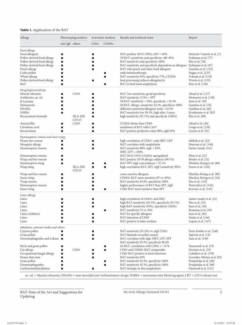
Table 1 summarizes the main reports from the litera-ture in which a BAT approach has been applied for al-lergy diagnosis. Most BATs use anti-IgE as the capturing marker in flow cytometry and CD63 to measure basophil function.
The first application of CD63-based BAT occurred in the 1990s; the percentage of CD63 upregulation in baso-phils triggered with an IgE-mediated stimulus was of the same order of magnitude as histamine release [19] and appeared to be suitable for diagnosing main allergy man-ifestations, even as preliminary attempts [20] . In these first attempts, correlation between expression of a mem-
IAA331312.indd 2IAA331312.indd 2 12.12.2011 12:03:3812.12.2011 12:03:38
BAT: State of the Art and Suggestions for Updating
Int Arch Allergy Immunol 331312 3
brane molecule and a degranulation event was of the ut-most interest. A very good correlation between the hista-mine release test (HRT) and CD63-positive basophils was observed in latex [21] , hymenoptera venom [22, 23] , grass pollen [6] , food [7] and drug allergy [24] , so that flow cy-tometry was proposed for routine diagnosis of allergic reactions [25, 26] . In any case, basophil flow cytometry also presented an opportunity to study basophilic cells in general and to aid in separating them from peripheral blood [27, 28] . Attempts to standardize in vitro flow cy-tometry of basophils to provide a reliable diagnostic tool have also been made [3] . Most of the so-called ‘Basotests’ or ‘basophil activation tests’ or flow-cytometric allergen stimulation tests, available also as affordable commercial kits, used CD63 activation markers, as the anti-ENPP3 (CD203c) antibody, 97A6, was only discovered a few years later [28] . Difficulty in finding and enrolling specific al-lergic subjects in BAT studies has always been a hindering issue, which may have prevented optimization of the an-alytical performance of the cellular test. Sanz et al. [29] , in a study carried out on 58 subjects allergic to � -lactams, confirmed that with CD63-based BAT, the allergic pa-tients showed a statistically higher number of activated basophils than controls but, although BAT showed high-er sensitivity (50%) and specificity (93.3%) than specific sIgE (37.9 and 86.7%, respectively), the test sensitivity was quite a lot lower than expected and differences did not reach statistical significance. Moreover, BATs do not al-ways show a higher sensitivity than serological tests. The use of BAT in diagnosing allergy to � -lactams has been reviewed recently [30] . In the intervening years, some au-thors attempted to introduce the newly discovered 97A6 antibody to highlight the basophil response in hymenop-tera venom allergy by investigating CD203c upregula-tion; the test showed a very good correlation with HRT and CD63 upregulation and proved able to successfully diagnose 20 of 22 allergic patients (91%) [31] . This high sensitivity was also confirmed by other authors [32] . A good correlation with CD63 (r = 0.76) was also reported by other authors in birch and grass pollen allergy [33] . A comparison between CD63 and CD203c in basophil flow cytometry for the diagnosis of insect venom allergy has reported a higher sensitivity of CD203c (97%) compared to CD63 (89%) [34] , while in cat allergy, other contribu-tors have found BAT performance to be quite similar when using either CD63 or CD203c as activation mark-ers, either with or without the introduction of IL-3 [35] . What is interesting is that in this report, the CD63 BAT included an IL-3-containing buffer, while the CD203c BAT did not. In an amoxicillin allergy model, Abuaf et
al. [36] showed that CD203c correlated better with the � -lactam anaphylactic group (60%) than did CD63 (20%), evidence that provoked several comments [37, 38] , as the authors also used CD45, together with anti-IgE, to phe-notype basophils in flow cytometry. Actually, the ques-tion whether CD203c and other gating strategies are tru-ly useful in ameliorating the capture of basophils and their functional investigation in flow cytometry has be-come a fundamental issue. Marked improvements in CD203c BAT were obtained just by ameliorating basophil electronic separation, for example by simply adding CD45 [39] , and even in this case many controversial com-ments arose [40, 41] . Probably, CD63 will have had more account due to its relationship with the degranulation event and because it is not a constitutive membrane marker. After all, not all the evidence suggesting CD203c as a marker to optimize BATs was unambiguous, as con-troversial results were reported [42] . Although in some tests the sensitivity of CD63-based BAT appeared rather low, other authors have described a detailed study on the analytical performance of this test, reaching very differ-ent results. Based on the receiver operating characteris-tics-generated cutoff value of 17% for CD63-positive ba-sophils, as captured by the usual anti-IgE-FITC method, Ebo et al. [43] estimated a BAT sensitivity and specificity of 93.1 and 91.7%, respectively, as evaluated in a popula-tion of subjects allergic to latex, encouraging results that were then confirmed in subsequent reports by others [44] . Several reviews about this promising test began to appear, aimed at achieving possible acceptance while de-scribing the state of the art of its application in allergy diagnosis [45–51] , issues that are still being faced nowa-days [52–74] . However, despite promising initial studies and some encouraging evidence, many authors agreed that CD63-based BAT remained disappointing in terms of sensitivity, while CD203c-based BAT showed a higher sensitivity (75 vs. 50%), though not as exciting as some reports suggested [39] . Where’s the catch? The problem pertains mainly to the different molecular and cellular behaviors of the two activation markers when related to the basophil response to an allergen; this could be an arguable reason why this issue has never ended up with two separate challenging teams among researchers, but on the contrary has always involved the possibility of a constructive debate, although CD63-based BAT has long dominated the scene. The report by Boumiza et al. [39] on CD203c-based BAT in latex allergy, in which a lower sensitivity compared to other studies with CD63-based BAT was estimated, raised many comments [40, 41] . Both Ebo’s and Boumiza’s groups used heparinized blood for
IAA331312.indd 3IAA331312.indd 3 12.12.2011 12:03:3812.12.2011 12:03:38
Chirumbolo Int Arch Allergy Immunol 331312 4
their investigations and used samples 3–4 h following blood withdrawal. The paper by Ebo et al. [43] was well planned with regard to sample design and subject enrol-ment, but, unlike Boumiza et al. [39] , the authors used IL-3 to prime basophils, which might explain the encour-aging results reached for CD63 expression on basophils [43] ; perhaps if they had used CD203c as the activation marker in that preactivating condition, there may not have been any significant increase in this marker, thus resulting in quite a low sensitivity of BAT. This group did not find any correlation between serum-specific IgE and BAT positivity. In the work by Boumiza et al. [39] , the au-thors did not use IL-3, but the experimental conditions would have elicited an autocrine release of IL-3 [9, 10] . CD203c is sensitive to IL-3 priming, and a subsequent challenge with allergen can result in the absence of a re-sponse of this marker, when its membrane expression reaches a plateau, while CD63 is dragged out from vesi-cles by the contribution of allergen to the primed state. CD63 membrane overexpression due to an IL-3 priming mechanism is a mechanism distinct from histamine re-lease; in these circumstances, HRT and CD63 upload to the basophil membrane may show low correlation [75] . Some years ago, a paper by Gamboa et al. [76] described the application of the flow-cytometric allergen stimula-tion test (a commercial CD63-based BAT) on basophils from individuals affected by allergy to pyrazolones; the reported sensitivity was particularly low (42.3%) com-pared to the specificity (100%), but the authors did not use IL-3. Moreover, they used acid citrate dextrose tubes for blood withdrawal and did not use the samples within a few hours of collection but rather stored them at +4 ° C for at least 24 h [76] . In the field of pollen-associated food al-lergy, Erdmann et al. [77] found high sensitivity values (80–85%) for BAT with very good agreement with sIgE and skin prick testing (SPT); the authors used fresh hep-arinized blood samples primed with 2 ng/ml IL-3. Ques-tions about the use of IL-3 to enhance BAT performance have recently been raised [11] , but maybe this is just one of the many problems in dealing with BAT. The goal of achieving optimal performance of BAT leads many re-searchers to attempt to optimize the test to the maximum possible values of sensitivity and specificity, although this target might be realistically unattainable in most circum-stances [78] . In certain cases, the sensitivity values of BAT were very low, less than 50%, while a target of 60% could be reached by combined use of SPT, sIgE and BAT [79] . In a study on hymenoptera sting allergy, the concordance between BAT and SPT was 42.9%, while the concordance between BAT and sIgE was higher (57.1%), as well as the
concordance among all three diagnostic tests [80] . Rela-tively low sensitivity values for BAT were also observed for drug allergy (48.6% [52] , 41.7% [81] , 39.1% [82] , 36.1% [83] ). The overall sensitivity estimation of a routinely used CD63-based BAT should be around 60%, with a higher specificity of close to 90–100%, but sensitivity and also specificity vary widely depending on the different allergens considered [53, 79, 84] or simply the allergen dose [86] and are influenced by possible cross-reaction with different allergens, as in the case of hymenoptera venoms [86] . Furthermore, one report found a lower sen-sitivity for BAT (76%) compared to SPT (100%) and sIgE or RAST (92%) in wasp venom allergy [87] , evidence not confirmed by others who found, on the contrary, compa-rable sensitivity between BAT (87.7%), SPT (93%) and specific sIgE (91.2%) [88] . Why these contradictory re-sults when both studies were carried out in subjects with wasp sting allergy? It is very interesting, for example, to note that Sturm et al. [88] physically purified basophils in the Histopaque-derived mononuclear cell layer and phe-notyped cells using an HLA-DR/CD123 protocol, rather than anti-IgE-FITC or anti-IgE-phycoerythrin, while us-ing CD63 as in other routine BAT. Physical separation of basophils from other blood cells can increase the ana-lytical parameters of BAT performance, but this approach is still too expensive and time-consuming for the routine diagnostic evaluation of allergy. In this regard, there was recent debate about the different phenotyping methods used in basophil flow cytometry aimed at ameliorating BAT technical performance [89] ; although newly discov-ered markers have been introduced, most BATs are based on an IgE/CD63 method panel ( table 1 ). Ebo et al. [90] applied an HLA-DR/CD123 gating protocol to validate flow-assisted diagnostic management of anaphylaxis caused by rocuronium compounds; the sensitivity of BAT was 91.7% and its specificity 100%. This good perfor-mance has not been confirmed by more recently reported contributions, in which the usual BAT gating protocol was applied on the same allergic panel [91] . Frezzolini et al. [92] used an HLA-DR/CD123 gating protocol to eval-uate CD63 expression in the diagnosis of chronic urti-caria; the sensitivity and specificity of the basophil acti-vation assay in flow cytometry, performed on cells from atopic donors, were 95.5 and 90.5%, respectively. Thus, the application of a gating strategy other than anti-IgE seems to greatly improve BAT performance. Eberlein et al. [17] used a CCR3 marker instead of anti-IgE to gate basophils in the new BAT called FlowCAST-2, for the di-agnosis of allergy to antibiotics, showing a higher sensi-tivity compared to the classical routine BAT. This marker
IAA331312.indd 4IAA331312.indd 4 12.12.2011 12:03:3812.12.2011 12:03:38
BAT: State of the Art and Suggestions for Updating
Int Arch Allergy Immunol 331312 5
Table 1. Application of the BAT
Allergy P henotyping markers Activation markers Results and technical notes Report
anti-IgE others CD63 CD203c
Food allergyFood allergens + + BAT positive 18/31 (58%), LRT = 65% Moneret-Vautrin et al. [7]Pollen-derived food allergy + + 29 BAT: sensitivity and specificity >80–85% Erdmann et al. [77]Pollen-derived food allergy + + BAT sensitivity and specificity 100% Ebo et al. [78]Pollen-derived food allergy + + BAT sensitivity and specificity dependent on allergens Erdmann et al. [87]Peach allergy + + BAT with peach and other food allergens Gamboa et al. [132]Cedar pollen + + rush immunotherapy Nagao et al. [133]Wheat allergy + + BAT sensitivity 85%, specificity 77%, CD203c Tokuda et al. [134]Pollen-derived food allergy + + heat processing reduces allergenicity Worm et al. [135]Beef + + BAT in beef meat anaphylaxis Kim et al. [136]
Drug hypersensitivityMuscle relaxants + CD45 + BAT: low sensitivity, good specificity Abuaf et al. [137]Antibiotics, m. rel. + + BAT sensitivity (71%) = SPT Monneret et al. [138]�-Lactams + + 58 BAT: sensitivity = 50%, specificity = 93.3% Sanz et al. [29]Metamizole + + 26 BAT: allergic sensitivity 42.3%, specificity 100% Gamboa et al. [76]NSAIDs + + different sensitivity/allergens; total = 63.3% Gamboa et al. [82]NMBA + + BAT sensitivity low 39.1%, high after 3 years Kvedariene et al. [83]Rocuronium bromide HLA-DR/
CD123+ high sensitivity (91.7%) and specificity (100%) Ebo et al. [90]
Amoxicillin + CD45 + CD203c better than CD63 Abuaf et al. [36]Clavulanic acid + + usefulness of BAT with CAST Longo et al. [139]Rocuronium + + BAT positive predictive value 98%, sIgE 83% Leysen et al. [91]
Hymenoptera venom and insect stingHoney bee venom + + high correlation of CD63+ with HRT, LRT Sabbah et al. [22]Mosquito allergy + + BAT correlates with anaphylaxis Hassoun et al. [140]Hymenoptera venom + + BAT sensitivity 88%, sIgE = 85%, Sainte-Laudy [141]
HRT, LPT = 100%Hymenoptera venom + BAT 20/22 (91%) CD203c upregulated Platz et al. [31]Wasp and bee venom + BAT positive 35/39 allergic subjects (89.7%) Binder et al. [32]Hymenoptera sting + + + BAT-SPT-sIgE concordance = 57.1% Eberlein-König et al. [80]Wasp sting HLA-DR/
CD123+ + high correlation BAT, SPT, sIgE (sensitivity 90%) Sturm et al. [142]
Wasp and bee venom + + cross-reactive allergens Eberlein-König et al. [86]Insect sting + + CD203c BAT more sensitive (97 vs. 89%) Eberlein-König et al. [34]Wasp venom + + BAT sensitivity 83.8%, specificity 100% Ebo et al. [143]Hymenoptera venom + + higher performance of BAT than SPT, sIgE Peternelj et al. [144]Insect sting + + + CD63 BAT more sensitive than SPT Korosec et al. [145]
Latex allergyLatex + + high correlation of CD63+ and HRT Sainte-Laudy et al. [21]Latex + + high BAT sensitivity (93.1%), specificity (91.7%) Ebo et al. [43]Latex + + high BAT sensitivity (93%), specificity (100%) Sanz et al. [44]Latex + + BAT sensitivity 75 vs. 50% Boumiza et al. [39]Latex (children) + + BAT for specific allergens Sanz et al. [84]Latex + + BAT detection of CD63 Nettis et al. [146]Latex + + BAT positive in latex workers Lopata et al. [147]
Inhalants, contrast media and othersCypress pollen + + BAT sensitivity (91.2%) vs. sIgE (76%) Paris-Kohler et al. [148]Grass pollen + + BAT depends on pollen season Saporta et al. [14]Dermatophagoides and Lolium + + BAT correlates with sIgE, HRT, LPT, SPT Sanz et al. [149]
BAT sensitivity 93.3%, specificity 98.4%Birch and grass pollen + + 62 BAT: correlation with CD63, r = 0.76 Hauswirth et al. [33]Cat allergy + CD45 + + CD63 and CD203c BAT comparable Ocmant et al. [35]Occupational fungal allergy + + CD63 BAT positive in feed industries Caballero et al. [150]House dust mite + + BAT sensitivity 83% González-Muñoz et al. [85]Grass pollen + + BAT sensitivity 92.3%, specificity 100% Potapińska et al. [68]Dermatophagoides + + BAT sensitivity 92.3%, specificity 100% Potapińska et al. [68]Carboxymethylcellulose + + BAT strategic in this anaphylaxis Dumond et al. [151]
m. rel. = Muscle relaxants; NSAIDs = non-steroidal anti-inflammatory drugs; NMBA = neuromuscular blocking agent; LRT = LTC4 release test.
IAA331312.indd 5IAA331312.indd 5 12.12.2011 12:03:3812.12.2011 12:03:38
Chirumbolo Int Arch Allergy Immunol 331312 6
has recently been considered a good phenotyping marker to gate basophils in whole blood; CCR3 (CD193), the ba-sophil receptor for eotaxin, has been shown to be a stable, reliable and highly expressed basophil selection marker, independent of the atopic background or basophil activa-tion state, which should allow accurate identification of basophils without need of a second marker [93] . However, other authors have raised criticism about the effective re-liability of this marker, mainly due to its possible down-regulation following activation and its wide expression on other leukocytes [38, 89] . Few newly introduced mark-ers in flow cytometry have been suggested for BAT im-provement. In any case, even the use of CD203c, as can be observed from table 1 , is less frequently considered compared to CD63, although it showed a performance similar to other routine allergy tests [94] . Probably, for most researchers the use of CD63-based BAT proved more feasible for diagnosing hypersensitivity reactions in a routine fashion. Patterns by which CD63 respond to ex-ternal stimuli mean that researchers prefer this marker in cellular investigations. The movement of CD63-associat-ed granules was studied some years ago in rat mast cell models; upon activation, granules reach the membrane in the very early minutes following stimulation, with a mean velocity of 100 nm/s [95] , a trend that has been confirmed also in basophils [96] . Most of the CD63 is associated with membrane integrins, and its recycling turnover, probably using a synaptotagmin-mediated mechanism [97] , oc-curs in the first 30–60 min after ‘bursting’ stimulation [98, 99] . Activation could also be elicited by many pre-analytical factors [9] . As a matter of fact, aside from downregulation mechanisms to be considered, the intra-cellular CD63 pool might be limiting to address CD63 upregulation in a BAT, simply due to its close relationship with basophil degranulation. However, this is a possible pitfall in using CD63 as a single diagnostic marker.
The percentage of CD63-expressing cells has to be con-sidered as a raw average of all basophils in the sample that are upregulating the tetraspanin to the membrane while the stimulus is still present; the calculation is rather arbi-trary and depends on the setting of the non-CD63-ex-pressing cell population based on mean fluorescence and ultimately on the ability of basophils to respond to an ex-ternal stimulus. In this context, a critical point is also the cutoff of basophil expression of CD63 upon activation. All these set points are variable and often depend on the ini-tial experimental conditions. Moreover, CD63 membrane displacement can be elicited by mechanisms quite sepa-rate from histamine release [100, 101] . This mechanism is one of the main issues in the debate about non-IgE-medi-
ated allergic responses. IgE-mediated and non-IgE-medi-ated cellular responses are critical issues in BAT applica-tion [102] . For example, administration of a drug can clas-sically result in an immunoglobulin E (IgE)-type sensi-tization but can also result in more complex activation of the immune system such as the drug-induced hypersen-sitivity syndrome, although the exact mechanisms of drug allergy are not well understood for most clinical manifestations. A complex interaction between individu-al characteristics, environmental factors and the drug it-self is usually responsible for adverse reactions to drugs [103] . Basophil evaluation in these circumstances appears to be one possible way to discriminate between different types of hypersensitivity, as for instance in food allergy, where non-IgE-mediated allergy is frequently present [104] . A non-IgE-mediated response able to upregulate CD63 and degranulation also involves other receptors of the innate immune response. Formylated peptides such as N-formyl-methionyl-leucyl-phenylalanine (fMLP) are commonly used in commercial BATs, but the mechanism by which fMLP induces CD63 translocation to the cell surface seems to be quite different from that elicited by an IgE-mediated event [101] . fMLP can be used as a good positive control to verify if basophils are viable and able to express CD203c and translocate CD63 molecules to the membrane, but its mechanism of action should not share any features of an IgE-mediated response.
Although quite expensive, some authors have suggest-ed the introduction of intracellular flow cytometry to en-rich the evidence obtained by evaluating the basophil re-sponse in allergy [105] . Flow cytometry actually allows a more integrated evaluation of immunophenotyping and the intracellular mechanisms underlying receptor signal-ing in allergy. In this context, phosphorylation of p38-MAP kinase (p38MAPK) represents a crucial step in IgE receptor signaling in basophils. The relationship between p38MAPK and the well-validated diagnostic cell surface marker CD63 has recently been evaluated in a clinical al-lergy model [106] , although criticism of the use of intra-cellular p38MAPK in monitoring therapy has recently been raised [107] .
Moreover, initial experimental conditions represent a fundamental issue. In a buffered medium or in whole blood, naturally not all the basophilic cells are resting cells. A first step should be to allow basophils to reach a ‘starting point’ at which most of the cells are prevented from spontaneously induced activation due to handling procedures, buffers and temperature. This starting point is ideally constituted by a prompt to upregulate the CD63 intracellular pool. Basophils should be used within very
IAA331312.indd 6IAA331312.indd 6 12.12.2011 12:03:3812.12.2011 12:03:38
BAT: State of the Art and Suggestions for Updating
Int Arch Allergy Immunol 331312 7
few hours of venous withdrawal, and the use of anti-IgE to target cells for flow cytometry identification should be avoided, as the phenotyping marker might elicit a stimu-latory response in nonactivated basophils [9] . In this con-text, the collection of basophils is another critical issue; samples should be collected and treated within the first 3 h following venous withdrawal in acid citrate dextrose or heparinized vials (selected in order not to deprive the test of calcium by chelating agents such as EDTA) and should not be stored at +4 ° C for more than this time, but not all authors are in agreement with these assumptions [108] . A good suggestion for performance of a reliable BAT might be to separate basophils from other blood components rather than to extremely purify them. A sim-ple approach would be to remove most of the CD63-ex-pressing platelets, although they should not affect BAT [8] , and plasma-interfering components, such as immu-noglobulins, complement and coagulation factors, able to interact with IgE/Fc � RI activation [109–111] . Further-more, spontaneously induced activation should be arrest-ed, for example by putting washed and buffer-reconsti-tuted samples into an ice bath (+2 ° C/+4 ° C) [11] .
Allergen doses, more than immunochemical proper-ties, represent another critical issue in enhancing BAT performance. Dose-response curves of purified allergens may be a possible strategy to solve BAT sensitivity, but the overall quality of a BAT depends certainly not only on allergen type and dose but on the relationship between these features and sensitized basophils.
Despite these preanalytical issues, membrane upregu-lation of CD63 has become of great interest because of its relationship with histamine release, as intracellular CD63 has been associated with histamine-containing granules [3] . Very few differences may be highlighted whether whole blood or physically purified basophils are used; most of the diagnostic performance optimization is re-lated to the proper choice of functional basophil markers. Purification of basophils represents a time-consuming and expensive approach; attempts to ameliorate and en-rich the endowment of those molecules able to assess ba-sophil function during an allergic response have been widely made. Some activation markers, such as CD164, CD107a or also CD13 and CD69, have been proposed, but their use in BAT as a widely used application in rou-tine diagnosis is still far from being achieved [112, 113] . Why this gap in BAT technical improvement? A possible trivial reason is the availability of new affordable fluoro-chrome-associated markers, while other considerations arise from the many opinions about different gating methods, the weak relationship between activation mark-
er and basophil releasability (except for CD63) and the expression of those markers on other blood cells besides basophils (except for CD203c).
Focusing on BAT: Is It Possible to Optimize Flow
Cytometry for Allergy Diagnosis?
Concerns about the Use of CD63 in BATs In light of the existence of the many issues and sugges-
tions about possible improvements thoroughly discussed above, why are IgE/CD63-based BATs so widespread? Much of the success related to CD63 concerns its upregu-lation response to a stimulus from a basal starting point and its relationship with intracellular granule migration to the membrane, hallmarks that CD203c and other markers do not exhibit. However, what is the real situa-tion of basophil releasability that one might tackle when evaluating a cellular allergy test? A possible reasoning may be suggested, as follows.
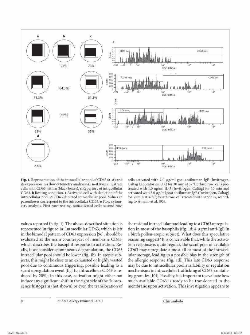
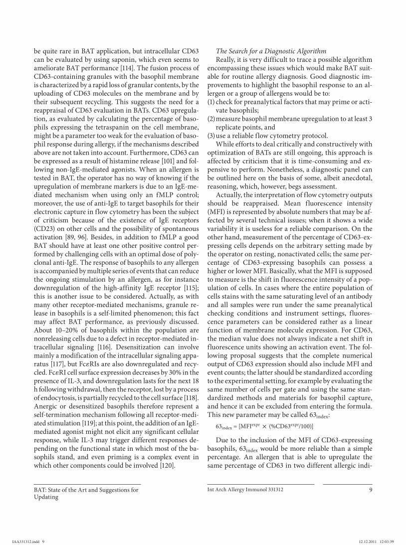
The ideally ‘normal’ basophil state is a previously sen-sitized cell expressing specific IgE on the Fc � RI receptor, not yet degranulated and able to exhibit all the cellular mechanisms responsive to an IgE-mediated stimulus when challenged with an allergen. We call this cellular state ‘resting’. In figure 1 , a schematic representation of basophils with an intracellular pool of CD63 molecules ( fig. 1 a–d) and fluorescence histograms of CD63 expres-sion from flow cytometry ( fig. 1 e) are shown. In this fig-ure, circles with squares within represent schematic pic-tures of basophils containing different levels of prompt to release intracellular CD63. The resting cell represents our analytical functional baseline. This cell is potentially able to degranulate, to translocate CD63 or other activation molecules to the membrane and to release granule media-tors when stimulated. A spontaneously degranulated pop-ulation (for example, 7% CD63) would account for ap-proximately 93% of intracellular CD63 that could be up-regulated onto the membrane upon the forthcoming stimulation ( fig. 1 e, row 1). When activated, in most cases basophils expressing CD63 amount to 20–30% of the whole population investigated (28.7% in the example shown in fig. 1 e, row 2; intracellular CD63 is 71.3%) fol-lowing an optimal IgE-mediated challenge, but this value may increase when basophils are primed with 1.0 ng/ml IL-3 ( fig. 1 e, row 3; 45% CD63-positive cells, thus leaving 55% intracellular CD63). All these values represent an av-erage of the different basophils within the experimental population and can be described as the total CD63 intra-cellular pool (black squares inside circles and percentage
IAA331312.indd 7IAA331312.indd 7 12.12.2011 12:03:3912.12.2011 12:03:39
Chirumbolo Int Arch Allergy Immunol 331312 8
values reported in fig. 1 ). The above-described situation is represented in figure 1 a. Intracellular CD63, which is left in the bimodal pattern of CD63 expression [96] , should be evaluated as the main counterpart of membrane CD63, which describes the basophil response to activation. Re-ally, if we consider spontaneous degranulation, the CD63 intracellular pool should be lower ( fig. 1 b). In atopic sub-jects, this might be close to an exhausted or highly wasted pool due to continuous triggering, possible leading to a scant upregulation event ( fig. 1 c; intracellular CD63 is re-duced by 20%); in this case, activation might either not induce any significant shift in the right side of the fluores-cence histogram (not shown) or even the translocation of
the residual intracellular pool leading to a CD63 upregula-tion in most of the basophils ( fig. 1 d; 4 � g/ml anti-IgE in a birch pollen-atopic subject). What does this speculative reasoning suggest? It is conceivable that, while the activa-tion response is quite regular, the scant pool of available CD63 may upregulate almost all or most of the intracel-lular storage, leading to a possible bias in the strength of the allergic response ( fig. 1 d). This late CD63 response may be due to intracellular pool availability or regulation mechanisms in intracellular trafficking of CD63-contain-ing granules [101] . Possibly, it is important to evaluate how much available CD63 is ready to be translocated to the membrane upon activation. This investigation appears to
93%
a
d
b c
e
73%
51.3%71.3%
35%
CD63 neg CD63 pos
CD63 neg CD63 pos
CD63 neg CD63 pos
CD63 neg CD63 pos
CD63 FITC-A
CD63 FITC-A
CD63 FITC-A102 103 104 105–102–357
Cou
ntC
ount
Cou
nt
0
CD63 FITC-A102 103 104 105–102–351 0
102 103 104 105–102–371 0
102 103 104 105–102–282 0
01
23
45
67
55%
2.6%
(64.3%)
(48%)
Cou
nt0
12
34
56
78
0
0.511
0.522
0.533
0.555
0.544
Cou
nt
0
0.511
0.522
0.533
0.555
0.544
Fig. 1. Representation of the intracellular pool of CD63 ( a–d ) and its expression in a flow cytometry analysis ( e ). a–d Boxes illustrate cells with CD63 within (black boxes). a Repertory of intracellular CD63. b Resting condition. c Activated cell with depletion of the intracellular pool. d CD63 depleted intracellular pool. Values in parentheses correspond to the intracellular CD63. e Flow cytom-etry analysis. First row: resting, nonactivated cells; second row:
cells activated with 2.0 � g/ml goat antihuman IgE (Invitrogen, Caltag Laboratories, UK) for 30 min at 37 ° C; third row: cells pre-treated with 1.0 ng/ml IL-3 (Invitrogen, Caltag) for 10 min and activated with 2.0 � g/ml goat antihuman IgE (Invitrogen, Caltag) for 30 min at 37 ° C; fourth row: cells treated with saponin, accord-ing to Amano et al. [95] .
IAA331312.indd 8IAA331312.indd 8 12.12.2011 12:03:3912.12.2011 12:03:39
BAT: State of the Art and Suggestions for Updating
Int Arch Allergy Immunol 331312 9
be quite rare in BAT application, but intracellular CD63 can be evaluated by using saponin, which even seems to ameliorate BAT performance [114] . The fusion process of CD63-containing granules with the basophil membrane is characterized by a rapid loss of granular contents, by the uploading of CD63 molecules on the membrane and by their subsequent recycling. This suggests the need for a reappraisal of CD63 evaluation in BATs. CD63 upregula-tion, as evaluated by calculating the percentage of baso-phils expressing the tetraspanin on the cell membrane, might be a parameter too weak for the evaluation of baso-phil response during allergy, if the mechanisms described above are not taken into account. Furthermore, CD63 can be expressed as a result of histamine release [101] and fol-lowing non-IgE-mediated agonists. When an allergen is tested in BAT, the operator has no way of knowing if the upregulation of membrane markers is due to an IgE-me-diated mechanism when using only an fMLP control; moreover, the use of anti-IgE to target basophils for their electronic capture in flow cytometry has been the subject of criticism because of the existence of IgE receptors (CD23) on other cells and the possibility of spontaneous activation [89, 96] . Besides, in addition to fMLP a good BAT should have at least one other positive control per-formed by challenging cells with an optimal dose of poly-clonal anti-IgE. The response of basophils to any allergen is accompanied by multiple series of events that can reduce the ongoing stimulation by an allergen, as for instance downregulation of the high-affinity IgE receptor [115] ; this is another issue to be considered. Actually, as with many other receptor-mediated mechanisms, granule re-lease in basophils is a self-limited phenomenon; this fact may affect BAT performance, as previously discussed. About 10–20% of basophils within the population are nonreleasing cells due to a defect in receptor-mediated in-tracellular signaling [116] . Desensitization can involve mainly a modification of the intracellular signaling appa-ratus [117] , but Fc � RIs are also downregulated and recy-cled. Fc � RI cell surface expression decreases by 30% in the presence of IL-3, and downregulation lasts for the next 18 h following withdrawal, then the receptor, lost by a process of endocytosis, is partially recycled to the cell surface [118] . Anergic or desensitized basophils therefore represent a self-termination mechanism following all receptor-medi-ated stimulation [119] ; at this point, the addition of an IgE-mediated agonist might not elicit any significant cellular response, while IL-3 may trigger different responses de-pending on the functional state in which most of the ba-sophils stand, and even priming is a complex event in which other components could be involved [120] .
The Search for a Diagnostic Algorithm Really, it is very difficult to trace a possible algorithm
encompassing these issues which would make BAT suit-able for routine allergy diagnosis. Good diagnostic im-provements to highlight the basophil response to an al-lergen or a group of allergens would be to: (1) check for preanalytical factors that may prime or acti-
vate basophils; (2) measure basophil membrane upregulation to at least 3
replicate points, and (3) use a reliable flow cytometry protocol.
While efforts to deal critically and constructively with optimization of BATs are still ongoing, this approach is affected by criticism that it is time-consuming and ex-pensive to perform. Nonetheless, a diagnostic panel can be outlined here on the basis of some, albeit anecdotal, reasoning, which, however, begs assessment.
Actually, the interpretation of flow cytometry outputs should be reappraised. Mean fluorescence intensity (MFI) is represented by absolute numbers that may be af-fected by several technical issues; when it shows a wide variability it is useless for a reliable comparison. On the other hand, measurement of the percentage of CD63-ex-pressing cells depends on the arbitrary setting made by the operator on resting, nonactivated cells; the same per-centage of CD63-expressing basophils can possess a higher or lower MFI. Basically, what the MFI is supposed to measure is the shift in fluorescence intensity of a pop-ulation of cells. In cases where the entire population of cells stains with the same saturating level of an antibody and all samples were run under the same preanalytical checking conditions and instrument settings, fluores-cence parameters can be considered rather as a linear function of membrane molecule expression. For CD63, the median value does not always indicate a net shift in fluorescence units showing an activation event. The fol-lowing proposal suggests that the complete numerical output of CD63 expression should also include MFI and event counts; the latter should be standardized according to the experimental setting, for example by evaluating the same number of cells per gate and using the same stan-dardized methods and materials for basophil capture, and hence it can be excluded from entering the formula. This new parameter may be called 63 index :
63 index = [MFI expr ! (%CD63 expr /100)]
Due to the inclusion of the MFI of CD63-expressing basophils, 63 index would be more reliable than a simple percentage. An allergen that is able to upregulate the same percentage of CD63 in two different allergic indi-
IAA331312.indd 9IAA331312.indd 9 12.12.2011 12:03:3912.12.2011 12:03:39
Chirumbolo Int Arch Allergy Immunol 331312 10
viduals but with two significantly different MFIs under the same experimental conditions might have a precise significance in the basophil response to the allergen, as MFI is proportional to the number of membrane mole-cules recognized by the fluorochrome-labeled antibody and hence to the amount of surface CD63. The parameter 63 index should indicate the percentage of basophils and how much CD63 they express on their membrane. Thus, an allergen may elicit two different responses as evalu-ated by the percentage of CD63 expressed but with MFIs that lead to the same 63 index :
example 1: sample No. 0087, %CD63 expr basophils = 21.6%,MFI = 8,457.76, 63 index = 1,826.88;
example 2: sample No. 0126, %CD63 expr basophils = 43.3%,MFI = 4,178.65, 63 index = 1,809.35.
The histograms of the first sample will show a marked separation of a few basophils with high expression of CD63. Reporting only the percentage of CD63 expression may introduce a bias in the evaluation of the allergy re-sponse (low in example 1 and high in example 2), while the two responses are qualitatively equivalent, though differing in their fluorescence distribution. In this case, the median would appear to be an alternative; however, while the 63 index includes only parameters of expressing cells, the median concerns the whole population of cells and it might not be so reliable in discriminating differ-ent situations, for example in the follow-up of allergy therapy.
What Meaning Should We Draw from These Arguments? Differences in BAT performance when only %CD-
63 expr basophils are measured may depend on several is-sues, the main two being (1) allergen strength in inducing a CD63-upregulating response and (2) basophil releas-ability. To assess this issue, an anti-IgE control has to be introduced.
The index should allow comparison of a better baso-phil IgE-mediated response to an optimal dose of an al-lergen to that elicited by an optimal dose of anti-IgE used as activation control. For example, the operator can as-sess the following ratio
63 ratio = [(63 index ) sample /(63 index ) control ]
which in turn may give a comparison of the allergen-trig-gered response and the anti-IgE control. The operator may also set a cutoff, better if calculated by the mean of three separate evaluations in the same experimental set-ting, to address a possible diagnostic implication. This
should become a new threshold that replaces the usual 10% cutoff for CD63 expression:
example 3: sample No. 0054, %CD63 expr basophils = 20.6%,MFI = 9,457.76, 63 index = 1,948.29 IgE control = 20.5%, MFI = 2,548.54, 63 index = 522.45, 63 ratio = 3.73;
example 4: sample No. 0068, %CD63 expr basophils = 41.5%,MFI = 2,134.66, 63 index = 885.88 IgE control = 46.4%, MFI = 2,358.33, 63 index = 1,094.26, 63 ratio = 0.81.
Considering that the basophil response to an opti-mal dose of anti-IgE should be at least comparable to that elicited by an antigen and that a cutoff of around 1.0 for 63 ratio may be set, activation in example 3 would require further investigation to clarify if this ratio depends on the concurrence of a non-IgE-mediated reaction or other bi-ases. Example 4 involves the same anti-IgE batch as in example 3. An increase in the percentage of CD63 expres-sion was observed but without a concurrent MFI increase, which would be expected. The basophil response was good (ratio around 1.0), but further investigations may clarify if CD63 upregulation on the membrane was ham-pered by possible other factors (e.g. CD63 intracellular depletion, downregulation mechanism).
In a second step, the operator should build up two CD63 expression controls which aim to evaluate the mo-lecular pool available to the basophil. So, in addition to the resting, nonactivated control, a sample in which all CD63 is drawn out by a saponin-mediated lytic treatment is considered. Two outputs are hence available to focus on the mechanism by which basophils express CD63 on the membrane, i.e. CD63 min and CD63 max , corresponding to the 63 index of resting and extracted CD63, respectively. Furthermore, in the second-level step, at least three anti-IgE controls are considered, including a suboptimal, op-timal and supraoptimal dose, with the purpose of prob-ing potential priming or desensitization mechanisms. In the second step, each 63 index is compared to CD63 max (corrected for resting). The new parameter is called 63 ON :
63 ON = [(63 index ) sample /(CD63 max )] ! 100
Further insights into the mechanisms underlying CD63 storage, trafficking, membrane upregulation and recycling are undoubtedly required to comprehend the feasibility of this parameter. Saponin extraction of intra-cellular CD63 should give clues to possible blocking or depleting mechanisms, downregulation or inhibition of molecule trafficking and surface expression, molecular modifications leading to a decrease in fluorochrome-an-
IAA331312.indd 10IAA331312.indd 10 12.12.2011 12:03:3912.12.2011 12:03:39
BAT: State of the Art and Suggestions for Updating
Int Arch Allergy Immunol 331312 11
tibody recognition and/or affinity binding. A possible reference range for the available CD63 intracellular pool in fresh, nonatopic, nonactivated basophils may be calcu-lated, for example by enrolling a representative sample of screened healthy blood donors. What should occur in a gated basophil population when cells are challenged with an allergen? This issue was dealt with during the discus-sion of figure 1 . Figure 1 c indicates that the percentage of CD63 expression may be underestimated, i.e. 63 ON is un-derestimated, due to the lower CD63 max observed com-pared to the reference range.
What Is Its Possible Functional Meaning? A depletion in intracellular CD63 or a downregulation
in its membrane uploading may come only from mem-brane expression of the molecule, which occurs when cells are activated; however, this fact may be misinter-preted as a decrease in the allergy response on the recep-tor level. Continuously triggered basophils tend to desen-sitize their membrane IgE receptors [101, 114, 118] . In this case, it is arguable that the optimal dose of anti-IgE may result in a lower 63 index in the control dose-response curve, a circumstance that may occur also for the supra-optimal dose included. On the other hand, suboptimal and optimal doses might work excellently, a condition that might indicate an active functional state or a primed cell. Signaling desensitization may be highlighted by in-troducing an intracellular marker into the second-level test; however, due to reasons of expense and time, the need for condition of receptor/signaling downregulation or anergy due to a nonreleaser state, should be addressed only when CD63 max does not suggest exhaustion of the intracellular pool and is followed by a poor response of the IgE-mediated activation. This observation should be verified, keeping the same materials and experimental setting.
This series of arguments may appear too speculative and somewhat hindering. Nonetheless, this proposal con-tains at least three new possible advances: (1) the percent-age of CD63-expressing basophils also accounts for the fluorescence indicator of the amount of molecules ex-pressed on the cell membrane; (2) this parameter is com-pared with the same one calculated on an anti-IgE control, and (3) in order to focus on possible biases, a second-level investigation can be performed, in which calculation of an anti-IgE dose-response curve and evaluation of the intra-cellular CD63 pool are possible new pathways to enhance the diagnostic usefulness of BAT. As a matter of fact, while taking into consideration if a new algorithm can fulfill the criteria according to which a flow cytometry test has to be
applied in order to better evaluate cell function in allergy, any finding has necessarily to be endorsed and possibly supported by further clinical evidence. Although this cannot be the purpose of this work because the spectrum of allergen/BAT responses and related clinical manifesta-tions of allergy that would have to be pointed out is too large, what follows has to be conceived as a proposal to reappraise CD63 evaluation in the light of flow cytometry and from a laboratory point of view.
Finally, the proposal aims at stressing some funda-mental aspects of BAT use, as follows: (1) The application of the simple percentage of CD63-ex-
pressing basophils as a ‘net’ parameter to evaluate ba-sophil response should be reviewed; other algorithms, such as those including fluorescence measurements, and possibly new added markers would be helpful to optimize diagnostic performance of the test.
(2) The introduction of an anti-IgE control and/or a min-imal dose-response curve of this control to compare allergen activation with the IgE-mediated response should be considered. A positive reaction of an aller-gen and of fMLP when basophils are unable to re-spond to optimal doses of anti-IgE may also indicate that the allergen solution contains substances able to activate basophils by non-IgE-mediated mechanisms.
(3) The final judgment regarding whether the subject pos-sesses the correct endowment to exert an allergic re-sponse to a defined allergen and, then, if the individ-ual is allergic to that substance, cannot be based on the single calculation of the percentage of CD63 expres-sion. Here, some possible further parameters are sug-gested.
(4) Due to the high costs of BAT, a first-level analysis may be considered conclusive if other allergic parameters are suggestive or positive, ratio and index parameters of samples investigated in triplicate correspond to cut-off rules, the anti-IgE control is responsive at the opti-mal dose used and preanalytical criteria have been ob-served. The latter point has been discussed elsewhere in the text; in short, the minimum number of baso-phils within the gate has to be no less than 250, and tests have to be carried out in triplicate (a total of 750–800 basophils for each sample) [4, 96] .
Conclusion
Many technical issues and diagnostic algorithms con-cerning BAT have been discussed in the past [121, 122] . BAT should be carried out as soon as possible after blood
IAA331312.indd 11IAA331312.indd 11 12.12.2011 12:03:3912.12.2011 12:03:39
Chirumbolo Int Arch Allergy Immunol 331312 12
References
1 Fernández TD, Torres MJ, Blanca-López N, Rodríguez-Bada JL, Gomez E, Canto G, Mayorga C, Blanca M: Negativization rates of IgE radioimmunoassay and basophil acti-vation test in immediate reactions to penicil-lins. Allergy 2009; 64: 242–248.
2 Sainte-Laudy J: Standardization of basophil degranulation for pharmacological studies. J Immunol Methods 1987; 98: 279–282.
3 Knol EF, Mul FP, Jansen H, Calafat J, Roos D: Monitoring human basophil activation via CD63 monoclonal antibody 435. J Allergy Clin Immunol 1991; 88: 328–338.
4 Sainte-Laudy J: Application of f low cytome-try to the analysis of activation of human basophils. Immunologic validation of the method (in French). Allerg Immunol (Paris) 1998; 30: 41–43.
5 Sabbah A, Drouet M, Sainte-Laudry J, Lauret MG, Loiry M: Contribution of f low cytom-etry to allergologic diagnosis (in French). Al-lerg Immunol (Paris) 1997; 29: 15–21.
6 Sainte-Laudy J, Sabbah A, Vallon C, Guerin JC: Analysis of anti-IgE and allergen induced human basophil activation by flow cytome-try. Comparison with histamine release. In-flamm Res 1998; 47: 401–408.
7 Moneret-Vautrin DA, Sainte-Laudy J, Kan-ny G, Frémont S: Human basophil activa-tion measured by CD63 expression and
LTC4 release in IgE-mediated food allergy. Ann Allergy Asthma Immunol 1999; 82: 33–40.
8 Monneret G, Gutowski MC, Bienvenu J: De-tection of allergen-induced basophil activa-tion by expression of CD63 antigen using a tricolour flow cytometric method. Clin Exp Immunol 1999; 115: 393–396.
9 Sturm EM, Kranzelbinder B, Heinemann A, Groselj-Strele A, Aberer W, Sturm GJ: CD203c-based basophil activation test in al-lergy diagnosis: characteristics and differ-ences to CD63 upregulation. Cytometry B Clin Cytom 2010; 78: 308–318.
10 Schroeder JT, Chichester KL, Bieneman AP: Human basophils secrete IL-3: evidence of autocrine priming for phenotypic and func-tional responses in allergic disease. J Immu-nol 2009; 182: 2432–2438.
11 Chirumbolo S: The use of IL-3 in basophil activation tests is the real pitfall. Cytometry B Clin Cytom 2011; 80: 137–138.
12 Lie WJ, Mul FP, Roos D, Verhoeven AJ, Knol EF: Degranulation of human basophils by picomolar concentrations of IL-3, IL-5, or granulocyte-macrophage colony-stimulating factor. J Allergy Clin Immunol 1998; 101: 683–690.
13 Hausmann OV, Gentinetta T, Bridts CH, Ebo DG: The basophil activation test in immedi-
ate-type drug allergy. Immunol Allergy Clin North Am 2009; 29: 555–566.
14 Saporta M, Kamei S, Persi L, Bousquet J, Ar-noux B: Basophil activation during pollen season in patients monosensitized to grass pollens. Allergy 2001; 56: 442–445.
15 Gómez E, Blanca-Lopez N, Torres MJ, Re-quena G, Rondon C, Canto G, Blanca M, Mayorga C: Immunoglobulin E-mediated immediate allergic reactions to dipyrone: value of basophil activation test in the iden-tification of patients. Clin Exp Allergy 2009; 39: 1217–1224.
16 Sanz ML, Gamboa PM, Mayorga C: Basophil activation tests in the evaluation of immedi-ate drug hypersensitivity. Curr Opin Allergy Clin Immunol 2009; 9: 298–304.
17 Eberlein B, León Suárez I, Darsow U, Ruëff F, Behrendt H, Ring J: A new basophil activa-tion test using CD63 and CCR3 in allergy to antibiotics. Clin Exp Allergy 2010; 40: 411–418.
18 Choquet-Kastylevsky G, Vial T, Descotes J: Drug allergy diagnosis in humans: possibili-ties and pitfalls. Toxicology 2001; 158: 1–10.
19 Sainte-Laudy J, Vallon C, Guérin JC: Analy-sis of membrane expression of the CD63 hu-man basophil activation marker. Applica-tions to allergologic diagnosis (in French). Allerg Immunol (Paris) 1994; 26: 211–214.
withdrawal [9, 11, 123] , and its performance may depend on several factors, such as the type of vial used for speci-men collection [109] . BATs have proved to be useful tools to diagnose early onset of allergy and anaphylactic ad-verse reactions, such as with drugs [124] , but also in mon-itoring immunotherapy [125–127] and to reintroduce food to allergic children [128] . Multicenter and pilot stud-ies have also been carried out in order to achieve a con-sensus agreement and to outline possible guidelines for BAT optimization and application [129–131] . However, many critical issues still remain, and further investiga-tion is needed. This review is an attempt to outline the state of the art of BAT and to address some of the issues concerning its performance. Stringent hallmarks are mostly constituted by (1) preanalytical factors, (2) the phenotyping and gating strategy and (3) analytical algo-rithms and parameters for making a diagnostic supposi-tion. Cellular tests for allergy, of which BAT is the most well known, are functional assays that enable discrimina-tion of passive sensitization to an IgE-mediated allergy. In drug allergy, by combining BAT with in vitro passive sensitization of donor basophils with patients’ serum, one
can prove the IgE dependence of a drug reaction [13] . In allergy immunotherapy, BAT furthermore represents a powerful tool to follow up patients with allergy manifes-tations [51, 102, 107, 127] . Improvements in BAT method-ology to achieve the best performance have also been made [107] , but follow-up of allergy still represents a ma-jor concern, for which BAT seems to be a promising ap-proach.
The introduction of anti-IgE controls and an alterna-tive phenotyping strategy are two important aspects which expand the debate. Compared with the determi-nation of sIgE, BAT offers the opportunity to be able to demonstrate functional responses and to prevent side ef-fects due to SPT. This may be a very good reason to com-bine efforts and to update this interesting tool to facilitate the diagnosis of allergy.
Acknowledgment
A special thanks to Dr. Antonio Vella, PhD, for his precious technical assistance and scientific suggestions.
IAA331312.indd 12IAA331312.indd 12 12.12.2011 12:03:3912.12.2011 12:03:39
BAT: State of the Art and Suggestions for Updating
Int Arch Allergy Immunol 331312 13
20 Sabbah A, Lauret MG, Maillard H: Prelimi-nary study of the basophil activation test for drug allergy using anti-membrane antibod-ies and flow cytometry (in French). Allerg Immunol (Paris) 1995; 27: 274–276.
21 Sainte-Laudy J, Vallon C, Guerin JC: Diag-nosis of latex allergy: comparison of hista-mine release and flow cytometric analysis of basophil activation. Inflamm Res 1996; 45(suppl 1):S35–S36.
22 Sabbah A, Plassais R, Grenapin S, Drouet M, Lauret MG, Loiry M, Sainte-Laudy J: Testing basophil activation by flow cytometry in the diagnosis of allergy to hymenopteran venom (in French). Allerg Immunol (Paris) 1998; 30: 44–48.
23 Stevens S, Drouet M, Lauret MG, Loiry M, Sabbah A: Basophil activation test using flow cytometry in Hymenoptera venom allergy (in French). Allerg Immunol (Paris) 1999; 31: 11–14.
24 Tamim S, Lauret MG, Drouet M, Sabbah A: Level of histamine in supernatants from the basophil activation test: applications to hy-menoptera allergy and drug allergy – pre-liminary study (in French). Allerg Immunol (Paris) 1999; 31: 35–44.
25 Sabbah A, Sainte-Laudy J, Drouet M, Lauret MG, Loiry ML, el Founini M, Oreac J, Guit-ton J, Doucet M: Immunobiologic diagnosis of food allergy (in French). Allerg Immunol (Paris) 1997; 29: 103–107.
26 Sabbah A, Sainte-Laudy J, Hassoun S, Drouet M, Lauret MG, Loiry ML: Allergy to mosquitoes and use of new immunobiologi-cal parameters (in French). Allerg Immunol (Paris) 1997; 29: 126–128.
27 Bochner BS, Sterbinsky SA, Saini SA, Co-lumbo M, Macglashan DW: Studies of cell adhesion and flow cytometric analyses of de-granulation, surface phenotype, and viabili-ty using human eosinophils, basophils, and mast cells. Methods 1997; 13: 61–68.
28 Bühring HJ, Seiffert M, Giesert C, Marxer A, Kanz L, Valent P, Sano K: The basophil acti-vation marker defined by antibody 97A6 is identical to the ectonucleotide pyrophospha-tase/phosphodiesterase 3. Blood 2001; 97: 3303–3305.
29 Sanz ML, Gamboa PM, Antépara I, Uasuf C, Vila L, Garcia-Avilés C, Chazot M, De Weck AL: Flow cytometric basophil activation test by detection of CD63 expression in patients with immediate-type reactions to betalac-tam antibiotics. Clin Exp Allergy 2002; 32: 277–286.
30 Blanca M, Romano A, Torres MJ, Férnandez J, Mayorga C, Rodriguez J, Demoly P, Bous-quet PJ, Merk HF, Sanz ML, Ott H, Atanasković-Marković M: Update on the evaluation of hypersensitivity reactions to betalactams. Allergy 2009; 64: 183–193.
31 Platz IJ, Binder M, Marxer A, Lischka G, Va-lent P, Bühring HJ: Hymenoptera-venom-in-duced upregulation of the basophil activa-tion marker ecto-nucleotide pyrophos-phatase/phosphodiesterase 3 in sensitized
individuals. Int Arch Allergy Immunol 2001; 126: 335–342 (erratum published in Int Arch Allergy Immunol 2002; 127: 293).
32 Binder M, Fierlbeck G, King T, Valent P, Bühring HJ: Individual hymenoptera venom compounds induce upregulation of the baso-phil activation marker ectonucleotide pyro-phosphatase/phosphodiesterase 3 (CD203c) in sensitized patients. Int Arch Allergy Im-munol 2002; 129: 160–168.
33 Hauswirth AW, Natter S, Ghannadan M, Majlesi Y, Schernthaner GH, Sperr WR, Bühring HJ, Valenta R, Valent P: Recom-binant allergens promote expression of CD203c on basophils in sensitized individu-als. J Allergy Clin Immunol 2002; 110: 102–109.
34 Eberlein-König B, Varga R, Mempel M, Dar-sow U, Behrendt H, Ring J: Comparison of basophil activation tests using CD63 or CD203c expression in patients with insect venom allergy. Allergy 2006; 61: 1084–1085.
35 Ocmant A, Peignois Y, Mulier S, Hanssens L, Michils A, Schandené L: Flow cytometry for basophil activation markers: the measure-ment of CD203c up-regulation is as reliable as CD63 expression in the diagnosis of cat allergy. J Immunol Methods 2007; 320: 40–48.
36 Abuaf N, Rostane H, Rajoely B, Gaouar H, Autegarden JE, Leynadier F, Girot R: Com-parison of two basophil activation markers CD63 and CD203c in the diagnosis of amox-icillin allergy. Clin Exp Allergy 2008; 38: 921–928.
37 Monneret G: Is this time for CRTH2/DP2 in a flow cytometric basophil activation test? Clin Exp Allergy 2008; 38: 1239–1240.
38 Monneret G: CCR3 for basophil activation test: a necessary but insufficient step. Clin Exp Allergy 2010; 40: 953, author reply 954.
39 Boumiza R, Monneret G, Forissier MF, Sa-voye J, Gutowski MC, Powell WS, Bienvenu J: Marked improvement of the basophil acti-vation test by detecting CD203c instead of CD63. Clin Exp Allergy 2003; 33: 259–265.
40 Ebo DG, Lechkar B, Schuerwegh AJ, Bridts CH, De Clerck LS, Stevens WJ: Comments regarding ‘Marked improvement of the baso-phil activation test by detecting CD203c in-stead of CD63’ by Boumiza et al. Clin Exp Allergy 2003; 33: 849, author reply 852–853.
41 de Weck AL, Sanz ML: For allergy diagnostic f low cytometry, detection of CD203c instead of CD63 is not at all an improvement in oth-er hands. Clin Exp Allergy 2003; 33: 849–852, author reply 852–853.
42 Bavbek S, Ikincioğullari A, Dursun AB, Guloğlu D, Arikan M, Elhan AH, Misirligil Z: Upregulation of CD63 or CD203c alone or in combination is not sensitive in the diagno-sis of nonsteroidal anti-inflammatory drug intolerance. Int Arch Allergy Immunol 2009; 150: 261–270.
43 Ebo DG, Lechkar B, Schuerwegh AJ, Bridts CH, De Clerck LS, Stevens WJ: Validation of a two-color flow cytometric assay detecting
in vitro basophil activation for the diagnosis of IgE-mediated natural rubber latex allergy. Allergy 2002; 57: 706–712.
44 Sanz ML, Gamboa PM, García-Avilés C, Vila L, Diéguez I, Antépara I, de Weck AL: Flow-cytometric cellular allergen stimulation test in latex allergy. Int Arch Allergy Immunol 2003; 130: 33–39.
45 Sanz ML, Maselli JP, Gamboa PM, Oehling A, Diéguez I, de Weck AL: Flow cytometric basophil activation test: a review. J Investig Allergol Clin Immunol 2002; 12: 143–154.
46 Iancovici-Kidon M, Handzel ZT: Diagnosis of drug hypersensitivity – state of the art (in Hebrew). Harefuah 2002; 141: 731–735.
47 Moneret-Vautrin DA, Kanny G, Frémont S: Laboratory tests for diagnosis of food aller-gy: advantages, disadvantages and future perspectives. Eur Ann Allergy Clin Immu-nol 2003; 35: 113–119.
48 Sanz ML, García MC, Caballero MR, Dié-guez I, Gamboa PM: Basophil activation test in the diagnosis of allergy to medicines (in Spanish). An Sist Sanit Navar 2003; 26(suppl 2):39–47.
49 Valent P, Hauswirth AW, Natter S, Sperr WR, Bühring HJ, Valenta R: Assays for mea-suring in vitro basophil activation induced by recombinant allergens. Methods 2004; 32: 265–270.
50 Ebo DG, Hagendorens MM, Bridts CH, Schuerwegh AJ, De Clerck LS, Stevens WJ: In vitro allergy diagnosis: should we follow the flow? Clin Exp Allergy 2004; 34: 332–339.
51 Erdmann SM, Sachs B, Kwiecien R, Moll-Slodowy S, Sauer I, Merk HF: The basophil activation test in wasp venom allergy: sensi-tivity, specificity and monitoring specific im-munotherapy. Allergy 2004; 59: 1102–1109.
52 Torres MJ, Padial A, Mayorga C, Fernández T, Sanchez-Sabate E, Cornejo-García JA, Antúnez C, Blanca M: The diagnostic inter-pretation of basophil activation test in im-mediate allergic reactions to betalactams. Clin Exp Allergy 2004; 34: 1768–1775.
53 Erdmann SM, Ventocilla S, Moll-Slodowy S, Sauer I, Merk HF: Basophil activation tests in the diagnosis of drug reactions (in German). Hautarzt 2005; 56: 38–43.
54 Boumiza R, Debard AL, Monneret G: The basophil activation test by flow cytometry: recent developments in clinical studies, stan-dardization and emerging perspectives. Clin Mol Allergy 2005; 3: 9.
55 Sanz ML, Gamboa PM, García-Avilés C, De Weck A: Drug hypersensitivities: which room for biological tests? Eur Ann Allergy Clin Immunol 2005; 37: 230–235.
56 Shreffler WG: Evaluation of basophil activa-tion in food allergy: present and future ap-plications. Curr Opin Allergy Clin Immunol 2006; 6: 226–233.
57 Kleine-Tebbe J, Erdmann S, Knol EF, Mac-Glashan DW Jr, Poulsen LK, Gibbs BF: Diag-nostic tests based on human basophils: po-tentials, pitfalls and perspectives. Int Arch Allergy Immunol 2006; 141: 79–90.
IAA331312.indd 13IAA331312.indd 13 12.12.2011 12:03:3912.12.2011 12:03:39
Chirumbolo Int Arch Allergy Immunol 331312 14
58 Ebo DG, Sainte-Laudy J, Bridts CH, Mertens CH, Hagendorens MM, Schuerwegh AJ, De Clerck LS, Stevens WJ: Flow-assisted allergy diagnosis: current applications and future perspectives. Allergy 2006; 61: 1028–1039.
59 de Weck AL, Gamboa PM, Esparza R, Sanz ML: Hypersensitivity to aspirin and other nonsteroidal anti-inflammatory drugs (NSAIDs). Curr Pharm Des 2006; 12: 3347–3358.
60 Romano A, Demoly P: Recent advances in the diagnosis of drug allergy. Curr Opin Al-lergy Clin Immunol 2007; 7: 299–303.
61 Dubois AE, van der Heide S: Basophil-activa-tion tests in Hymenoptera allergy. Curr Opin Allergy Clin Immunol 2007; 7: 346–349.
62 Ebo DG, Hagendorens MM, Bridts CH, De Clerck LS, Stevens WJ: Hymenoptera venom allergy: taking the sting out of difficult cases. J Investig Allergol Clin Immunol 2007; 17: 357–360.
63 Ebo DG, Bridts CH, Hagendorens MM, Aerts NE, De Clerck LS, Stevens WJ: Baso-phil activation test by flow cytometry: pres-ent and future applications in allergology. Cytometry B Clin Cytom 2008; 74: 201–210.
64 de Weck AL, Sanz ML, Gamboa PM, Aberer W, Bienvenu J, Blanca M, Demoly P, Ebo DG, Mayorga L, Monneret G, Sainte-Laudy J: Di-agnostic tests based on human basophils: more potentials and perspectives than pitfalls. Int Arch Allergy Immunol 2008; 146: 177–189.
65 Sanz ML, Gamboa PM, De Weck AL: Cellu-lar tests in the diagnosis of drug hypersensi-tivity. Curr Pharm Des 2008; 14: 2803–2808.
66 Rodríguez-Trabado A, Cámara-Hijón C, Ra-mos-Cantariño A, Porcel-Carreño SL, Jimé-nez-Timón S, Pereira-Navarro G, Hernán-dez-Arbeiza FJ, Fernández-Pereira L: Baso-phil activation test for the in vitro diagnosis of nonsteroidal anti-inflammatory drug hy-persensitivity. Allergy Asthma Proc 2008; 29: 241–249.
67 Ebo DG, Hagendorens MM, Bridts CH, De Clerck LS, Stevens WJ: The basophil activa-tion test in immediate drug allergy. Acta Clin Belg 2009; 64: 129–135.
68 Potapińska O, Demkow U, Wasik M: Flow cytometric basophils activation test as a method of allergy diagnosis (in Polish). Pneumonol Alergol Pol 2009; 77: 152–158.
69 Sanz ML, Gamboa PM, Mayorga C: Basophil activation tests in the evaluation of immedi-ate drug hypersensitivity. Curr Opin Allergy Clin Immunol 2009: 298–304.
70 Scherer K, Bircher AJ, Heijnen IA: Diagnosis of stinging insect allergy: utility of cellular in-vitro tests. Curr Opin Allergy Clin Im-munol 2009; 9: 343–350.
71 Ocmant A, Mulier S, Hanssens L, Goldman M, Casimir G, Mascart F, Schandené L: Ba-sophil activation tests for the diagnosis of food allergy in children. Clin Exp Allergy 2009; 39: 1234–1245.
72 Kosnik M, Korosec P: Importance of baso-phil activation testing in insect venom aller-gy. Allergy Asthma Clin Immunol 2009; 5: 11.
73 Mayorga C, Sanz ML, Gamboa PM, García BE, Caballero MT, García JM, Labrador M, Lahoz C, Longo Areso N, López Hoyos M, Martínez Quesada J, Monteseirín FJ; Immu-nology Committee of the Spanish Society of Allergology and Clinical Immunology of the SEAIC: In vitro diagnosis of immediate al-lergic reactions to drugs: an update. J Inves-tig Allergol Clin Immunol 2010; 20: 103–109.
74 Sanz ML, Gamboa PM, García-Figueroa BE, Ferrer M: In vitro diagnosis of anaphylaxis. Chem Immunol Allergy 2010; 95: 125–140.
75 Brown SG, Haas MA, Black JA, Parameswaran A, Woods GM, Heddle RJ: In vitro testing to diagnose venom allergy and monitor immu-notherapy: a placebo-controlled, crossover trial. Clin Exp Allergy 2004; 34: 792–800.
76 Gamboa PM, Sanz ML, Caballero MR, An-tépara I, Urrutia I, Jáuregui I, González G, Dié-guez I, De Weck AL: Use of CD63 expression as a marker of in vitro basophil activation and leukotriene determination in metamizol aller-gic patients. Allergy 2003; 58: 312–317.
77 Erdmann SM, Heussen N, Moll-Slodowy S, Merk HF, Sachs B: CD63 expression on baso-phils as a tool for the diagnosis of pollen-as-sociated food allergy: sensitivity and speci-ficity. Clin Exp Allergy 2003; 33: 607–614.
78 Ebo DG, Hagendorens MM, Bridts CH, Schuerwegh AJ, De Clerck LS, Stevens WJ: Flow cytometric analysis of in vitro activated basophils, specific IgE and skin tests in the diagnosis of pollen-associated food allergy. Cytometry B Clin Cytom 2005; 64: 28–33.
79 Gamboa PM, García-Avilés MC, Urrutia I, Antépara I, Esparza R, Sanz ML: Basophil activation and sulfidoleukotriene produc-tion in patients with immediate allergy to be-talactam antibiotics and negative skin tests. J Investig Allergol Clin Immunol 2004; 14: 278–283.
80 Eberlein-König B, Rakoski J, Behrendt H, Ring J: Use of CD63 expression as marker of in vitro basophil activation in identifying the culprit in insect venom allergy. J Investig Al-lergol Clin Immunol 2004; 14: 10–16.
81 Sanz ML, Gamboa P, de Weck AL: A new combined test with flowcytometric basophil activation and determination of sulfidoleu-kotrienes is useful for in vitro diagnosis of hypersensitivity to aspirin and other nonste-roidal anti-inflammatory drugs. Int Arch Allergy Immunol 2005; 136: 58–72.
82 Gamboa P, Sanz ML, Caballero MR, Urrutia I, Antépara I, Esparza R, de Weck AL: The flow-cytometric determination of baso-phil activation induced by aspirin and other non-steroidal anti-inflammatory drugs (NSAIDs) is useful for in vitro diagnosis of the NSAID hypersensitivity syndrome. Clin Exp Allergy 2004; 34: 1448–1457.
83 Kvedariene V, Kamey S, Ryckwaert Y, Ron-gier M, Bousquet J, Demoly P, Arnoux B: Di-agnosis of neuromuscular blocking agent hypersensitivity reactions using cytofluori-metric analysis of basophils. Allergy 2006; 61: 311–315.
84 Sanz ML, García-Avilés MC, Tabar AI, Anda M, García BE, Barber D, Salcedo G, Rihs HP, Raulf-Heimsoth M: Basophil Activation Test and specific IgE measurements using a panel of recombinant natural rubber latex aller-gens to determine the latex allergen sensiti-zation profile in children. Pediatr Allergy Immunol 2006; 17: 148–156.
85 González-Muñoz M, Villota J, Moneo I: Analysis of basophil activation by flow cy-tometry in pediatric house dust mite allergy. Pediatr Allergy Immunol 2008; 19: 342–347.
86 Eberlein-König B, Schmidt-Leidescher C, Rakoski J, Behrendt H, Ring J: In vitro ba-sophil activation using CD63 expression in patients with bee and wasp venom allergy. J Investig Allergol Clin Immunol 2006; 16: 5–10.
87 Erdmann SM, Sachs B, Schmidt A, Merk HF, Scheiner O, Moll-Slodowy S, Sauer I, Kwiecien R, Maderegger B, Hoffmann-Som-mergruber K: In vitro analysis of birch-pol-len-associated food allergy by use of recom-binant allergens in the basophil activation test. Int Arch Allergy Immunol 2005; 136: 230–238.
88 Sturm GJ, Böhm E, Trummer M, Weiglhofer I, Heinemann A, Aberer W: The CD63 baso-phil activation test in Hymenoptera venom allergy: a prospective study. Allergy 2004; 59: 1110–1117.
89 Chirumbolo S, Ortolani R, Vella A: CCR3 as a single selection marker compared to CD123/HLADR to isolate basophils in flow cytometry: some comments. Cytometry A 2011; 79: 102–106.
90 Ebo DG, Bridts CH, Hagendorens MM, Mertens CH, De Clerck LS, Stevens WJ: Flow-assisted diagnostic management of anaphylaxis from rocuronium bromide. Al-lergy 2006; 61: 935–939.
91 Leysen J, Bridts CH, De Clerck LS, Vercau-teren M, Lambert J, Weyler JJ, Stevens WJ, Ebo DG: Allergy to rocuronium: from clini-cal suspicion to correct diagnosis. Allergy 2011; 66: 1014–1019.
92 Frezzolini A, Provini A, Teofoli P, Pomponi D, De Pità O: Serum-induced basophil CD63 expression by means of a tricolour flow cyto-metric method for the in vitro diagnosis of chronic urticaria. Allergy 2006; 61: 1071–1077.
93 Hausmann OV, Gentinetta T, Fux M, Du-crest S, Pichler WJ, Dahinden CA: Robust expression of CCR3 as a single basophil se-lection marker in flow cytometry Allergy 2011; 66: 85–91.
94 Ciepiela O, Zwiazek J, Zawadzka-Krajewska A, Kotula I, Kulus M, Demkow U: Basophil activation test based on the expression of CD203c in the diagnostics of cow milk al-lergy in children. Eur J Med Res 2010; 15(suppl 2):21–26.
95 Amano T, Furuno T, Hirashima N, Ohyama N, Nakanishi M: Dynamics of intracellular granules with CD63-GFP in rat basophilic leukemia cells. J Biochem 2001; 129: 739–744.
IAA331312.indd 14IAA331312.indd 14 12.12.2011 12:03:3912.12.2011 12:03:39
BAT: State of the Art and Suggestions for Updating
Int Arch Allergy Immunol 331312 15
96 Chirumbolo S, Vella A, Ortolani R, De Gi-roncoli M, Solero P, Tridente G, Bellavite P: Differential response of human basophil activation markers: a multi-parameter flow cytometry approach. Clin Mol Allergy 2008; 6: 12–24.
97 Sagi-Eisenberg R: The mast cell: where en-docytosis and regulated exocytosis meet. Immunol Rev 2007; 217: 292–303.
98 Wu M, Baumgart T, Hammond S, Holowka D, Baird B: Differential targeting of secre-tory lysosomes and recycling endosomes in mast cells revealed by patterned antigen ar-rays. J Cell Sci 2007; 120: 3147–3154.
99 Gu Z, Noss EH, Hsu VW, Brenner MB: In-tegrins traffic rapidly via circular dorsal ruffles and macropinocytosis during stim-ulated cell migration. J Cell Biol 2011; 193: 61–70.
100 Malbrán A, Yeyati E, Rey GL, Galassi N: Di-clofenac induces basophil degranulation without increasing CD63 expression in sensitive patients. Clin Exp Immunol 2007; 147: 99–105.
101 MacGlashan DW Jr: Expression of CD203c and CD63 in human basophils: relationship to differential regulation of piecemeal and anaphylactic degranulation processes. Clin Exp Allergy 2010; 40: 1365–1377.
102 Ebo DG, Leysen J, Mayorga C, Rozieres A, Knol EF, Terreehorst I: The in vitro diagno-sis of drug allergy: status and perspectives. Allergy 2011; 66: 1275–1286.
103 Caubet JC, Pichler WJ, Eigenmann PA: Ed-ucational case series: mechanisms of drug allergy. Pediatr Allergy Immunol 2011; 22: 559–567.
104 Sabato V, van Hengel AJ, De Knop KJ, Ver-weij MM, Hagendorens MM, Bridts CH, De Clerck LS, Schiavino D, Stevens WJ, Ebo DG: Human basophils: a unique biological instrument to detect the allergenicity of food. J Investig Allergol Clin Immunol 2011; 21: 179–184.
105 Ebo DG, Dombrecht EJ, Bridts CH, Aerts NE, de Clerck LS, Stevens WJ: Combined analysis of intracellular signalling and immunophenotype of human peripheral blood basophils by flow cytometry: a proof of concept. Clin Exp Allergy 2007; 37: 1668–1675.
106 Aerts NE, Dombrecht EJ, Bridts CH, Hagendorens MM, de Clerck LS, Stevens WJ, Ebo DG: Simultaneous flow cytomet-ric detection of basophil activation marker CD63 and intracellular phosphorylated p38 mitogen-activated protein kinase in birch pollen allergy. Cytometry B Clin Cytom 2008; 76B:8–17.
107 Verweij MM, De Knop KJ, Bridts CH, De Clerck LS, Stevens WJ, Ebo DG: P38 mito-gen-activated protein kinase signal trans-duction in the diagnosis and follow up of immunotherapy of wasp venom allergy. Cytometry B Clin Cytom 2010; 78: 302–307.
108 Sousa N, Martínez-Aranguren R, Fernán-dez-Benitez M, Ribeiro F, Sanz ML: Com-parison of basophil activation test results in blood preserved in acid citrate dextrose and EDTA. J Investig Allergol Clin Immunol 2010; 20: 535–536.
109 Uermösi C, Beerli RR, Bauer M, Manolova V, Dietmeier K, Buser RB, Kündig TM, Sau-dan P, Bachmann MF: Mechanisms of aller-gen-specific desensitization. J Allergy Clin Immunol 2010; 126: 375–383.
110 Kepley CL, Taghavi S, Mackay G, Zhu D, Morel PA, Zhang K, Ryan JJ, Satin LS, Zhang M, Pandolfi PP, Saxon A: Co-aggre-gation of FcgammaRII with FcepsilonRI on human mast cells inhibits antigen-induced secretion and involves SHIP-Grb2-Dok complexes. J Biol Chem 2004; 279: 35139–35149.
111 Ali H: Regulation of human mast cell and basophil function by anaphylotoxins C3a and C5a. Immunol Lett 2010; 128: 36–45.
112 Chirumbolo S: CD164 and other recently discovered activation markers as promising tools for allergy diagnosis: what’s new? Clin Exp Med 2011; 11: 255–257.
113 Hennersdorf F, Florian S, Jakob A, Baumgärtner K, Sonneck K, Nordheim A, Biedermann T, Valent P, Bühring HJ: Iden-tification of CD13, CD107a, and CD164 as novel basophil-activation markers and dis-section of two response patterns in time ki-netics of IgE-dependent upregulation. Cell Res 2005; 15: 325–335.
114 Hoffmann HJ, Bøgebjerg M, Nielsen LP, Dahl R: Lysis with Saponin improves detec-tion of the response through CD203c and CD63 in the basophil activation test after crosslinking of the high affinity IgE recep-tor FcepsilonRI. Clin Mol Allergy 2005; 3: 10–18.
115 MacGlashan D Jr: Two regions of down reg-ulation in the IgE-mediated signaling path-way in human basophils. J Immunol 2003; 170: 4914–4925.
116 Kepley CL, Youssef L, Andrews RP, Wilson BS, Oliver JM: Syk deficiency in nonreleas-er basophils. J Allergy Clin Immunol 1999; 104: 279–284.
117 MacGlashan DW Jr, Mogowski M, Lichten-stein LM: Studies of antigen binding on hu-man basophils. II. Continued expression of antigen-specific IgE during antigen-in-duced desensitization. J Immunol 1983; 130: 2337–2342.
118 MacGlashan DW Jr: Endocytosis, recy-cling, and degradation of unoccupied FcepsilonRI in human basophils. J Leukoc Biol 2007; 82: 1003–1010.
119 MacGlashan DW Jr: Self-termination/an-ergic mechanisms in human basophils and mast cells. Int Arch Allergy Immunol 2009; 150: 109–121.
120 Vonakis BM, MacGlashan DW Jr, Vilariño N, Langdon JM, Scott RS, MacDonald SM: Distinct characteristics of signal transduc-tion events by histamine-releasing factor/translationally controlled tumor protein (HRF/TCTP)-induced priming and activa-tion of human basophils. Blood 2008; 111: 1789–1796.
121 De Weck AL, Sanz ML, Gamboa PM, Abe-rer W, Bienvenu J, Blanca M, Demoly P, Ebo DG, Mayorga L, Monneret G, Sainte Laudy J: Diagnostic tests based on human baso-phils: more potentials and perspectives than pitfalls. II. Technical issues. J Investig Allergol Clin Immunol 2008; 18: 143–155.
122 Ponvert C: Diagnosis of allergic and non-allergic hypersensitivity reactions to com-monly used drugs and biological substanc-es in children: diagnostic algorythm (in French). Arch Pediatr 2011; 18: 486–492.
123 Sturm GJ, Kranzelbinder B, Sturm EM, Heinemann A, Groselj-Strele A, Aberer W: The basophil activation test in the diagno-sis of allergy: technical issues and critical factors. Allergy 2009; 64: 1319–1326.
124 Aranda A, Mayorga C, Ariza A, Doña I, Ro-sado A, Blanca-Lopez N, Andreu I, Torres MJ: In vitro evaluation of IgE-mediated hy-persensitivity reactions to quinolones. Al-lergy 2011; 66: 247–254.
125 Kucera P, Cvackova M, Hulikova K, Juzova O, Pachl J: Basophil activation can predict clinical sensitivity in patients after venom immunotherapy. J Investig Allergol Clin Immunol 2010; 20: 110–116.
126 Mikkelsen S, Bibby BM, Dolberg MK, Dahl R, Hoffmann HJ: Basophil sensitivity through CD63 or CD203c is a functional measure for specific immunotherapy. Clin Mol Allergy 2010; 8: 2–12.
127 Chirumbolo S, Zanoni G, Ortolani R, Vella A: In vitro biphasic effect of honey bee ven-om on basophils from screened healthy blood donors. Allergy Asthma Immunol Res 2011; 3: 58–61.
128 Rubio A, Vivinus-Nébot M, Bourrier T, Saggio B, Albertini M, Bernard A: Benefit of the basophil activation test in deciding when to reintroduce cow’s milk in allergic children. Allergy 2011; 66: 92–100.
129 De Weck AL, Sanz ML, Gamboa PM, Abe-rer W, Sturm G, Bilo MB, Montroni M, Blanca M, Torres MJ, Mayorga L, Campi P, Manfredi M, Drouet M, Sainte-Laudy J, Ro-mano A, Merk H, Weber JM, Jermann TM; ENDA (European Network for Drug Aller-gy): Diagnosis of immediate-type beta-lac-tam allergy in vitro by flow-cytometric ba-sophil activation test and sulfidoleuko-triene production: a multicenter study. J Investig Allergol Clin Immunol 2009; 19: 91–109.
130 Ott H, Tenbrock K, Baron J, Merk H, Leh-mann S: Basophil activation test for the di-agnosis of hymenoptera venom allergy in childhood: a pilot study. Klin Padiatr 2011; 223: 27–32.
IAA331312.indd 15IAA331312.indd 15 12.12.2011 12:03:3912.12.2011 12:03:39
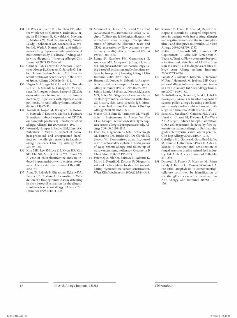
Chirumbolo Int Arch Allergy Immunol 331312 16
131 De Weck AL, Sanz ML, Gamboa PM, Abe-rer W, Blanca M, Correia S, Erdman S, Jer-mann JM, Kanny G, Kowalski M, Mayorga L, Medrala W, Merk A, Sturm GJ, Sainte-Laudy J, Schneider MS, Sczczeklik A, We-ber JM, Wedi A: Nonsteroidal anti-inflam-matory drug hypersensitivity syndrome. A multicenter study. I. Clinical findings and in vitro diagnosis. J Investig Allergol Clin Immunol 2009; 19: 355–369.
132 Gamboa PM, Cáceres O, Antepara I, Sán-chez-Monge R, Ahrazem O, Salcedo G, Bar-ber D, Lombardero M, Sanz ML: Two dif-ferent profiles of peach allergy in the north of Spain. Allergy 2007;62:408–414.
133 Nagao M, Hiraguchi Y, Hosoki K, Tokuda R, Usui T, Masuda S, Yamaguchi M, Fuji-sawa T: Allergen-induced basophil CD203c expression as a biomarker for rush immu-notherapy in patients with Japanese cedar pollinosis. Int Arch Allergy Immunol 2008; 46(Suppl 1):47–53.
134 Tokuda R, Nagao M, Hiraguchi Y, Hosoki K, Matsuda T, Kouno K, Morita E, Fujisawa T: Antigen-induced expression of CD203c on basophils predicts IgE-mediated wheat allergy. Allergol Int 2009;58:193–199.
135 Worm M, Hompes S, Fiedler EM, Illner AK, Zuberbier T, Vieths S: Impact of native, heat-processed and encapsulated hazel -nuts on the allergic response in hazelnut-allergic patients. Clin Exp Allergy 2009; 39:159–166.
136 Kim MH, Lee SM, Lee SH, Kwon HS, Kim SH, Cho SH, Min KU, Kim YY, Chang YS: A case of chlorpheniramine maleate-in-duced hypersensitivity with aspirin intoler-ance. Allergy Asthma Immunol Res 2011; 3:62–64.
137 Abuaf N, Rajoely B, Ghazouani E, Levy DA, Pecquet C, Chabane H, Leynadier F: Vali-dation of a f low cytometric assay detecting in vitro basophil activation for the diagno-sis of muscle relaxant allergy. J Allergy Clin Immunol 1999;104:411–418.
138 Monneret G, Duménil V, Benoit Y, Laffont A, Gutowski MC, Bernon H, Nicolas JF, Pa-checo Y, Bienvenu J: Biological diagnosis of immediate drug allergy. Comparative study of histamine liberation tests and CD63 expression by flow cytometry (pre-liminary results). Allerg Immunol (Paris) 1999;31:307–310.
139 Longo N, Gamboa PM, Gastaminza G, Audícana MT, Antepara I, Jaúregui I, Sanz ML: Diagnosis of clavulanic acid allergy us-ing basophil activation and leukotriene re-lease by basophils. J Investig Allergol Clin Immunol 2008;18:473–475.
140 Hassoun S, Drouet M, Sabbah A: Anaphy-laxis caused by a mosquito: 2 case reports. Allerg Immunol (Paris) 1999;31:285–287.
141 Sainte-Laudy J, Sabbah A, Drouet M, Lauret MG, Loiry M: Diagnosis of venom allergy by flow cytometry. Correlation with clini-cal history, skin tests, specific IgE, hista-mine and leukotriene C4 release. Clin Exp Allergy 2000;30:1166–1171.
142 Sturm GJ, Böhm E, Trummer M, Weigl-hofer I, Heinemann A, Aberer W: The CD63 basophil activation test in Hymenop-tera venom allergy: a prospective study. Al-lergy 2004;59:1110–1117.
143 Ebo DG, Hagendorens MM, Schuerwegh AJ, Beirens LM, Bridts CH, De Clerck LS, Stevens WJ: Flow-assisted quantification of in vitro activated basophils in the diagnosis of wasp venom allergy and follow-up of wasp venom immunotherapy. Cytometry B Clin Cytom 2007;72:196–203.
144 Peternelj A, Silar M, Bajrovic N, Adamic K, Music E, Kosnik M, Korosec P: Diagnostic value of the basophil activation test in eval-uating Hymenoptera venom sensitization. Wien Klin Wochenschr 2009;121:344–348.
145 Korosec P, Erzen R, Silar M, Bajrovic N, Kopac P, Kosnik M: Basophil responsive-ness in patients with insect sting allergies and negative venom-specific immunoglob-ulin E and skin prick test results. Clin Exp Allergy 2009;39:1730–1737.
146 Nettis E, Colanardi MC, Dambra PP, Capuzzimati L, Loria MP, Ferrannini A, Vacca A, Tursi A: Flow cytometric basophil activation test: detection of CD63 expres-sion as a useful aid to diagnosis of latex al-lergy. Ann Allergy Asthma Immunol 2006;97:715–716.
147 Lopata AL, Adams S, Kirstein F, Henwood N, Raulf-Heimsoth M, Jeebhay MF: Occu-pational allergy to latex among loom tuners in a textile factory. Int Arch Allergy Immu-nol 2007;144:64–68.
148 Pâris-Köhler A, Demoly P, Persi L, Lebel B, Bousquet J, Arnoux B: In vitro diagnosis of cypress pollen allergy by using cytofluori-metric analysis of basophils (Basotest). J Al-lergy Clin Immunol 2000;105:339–345.
149 Sanz ML, Sánchez G, Gamboa PM, Vila L, Uasuf C, Chazot M, Diéguez I, De WeckAL: Allergen-induced basophil activation: CD63 cell expression detected by flow cy-tometry in patients allergic to Dermatopha-goides pteronyssinus and Lolium perenne. Clin Exp Allergy 2001;31:1007–1013.
150 Caballero ML, Gómez M, González-Muñoz M, Reinoso L, Rodríguez-Pérez R, Alday E, Moneo I: Occupational sensitization to fungal enzymes used in animal feed indus-try. Int Arch Allergy Immunol 2007;144:231–239.
151 Dumond P, Franck P, Morisset M, Sainte Laudy J, Kanny G, Moneret-Vautrin DA:Pre-lethal anaphylaxis to carboxymethyl-cellulose confirmed by identification of specific IgE – review of the literature. Eur Ann Allergy Clin Immunol 2009;41:171–176.
IAA331312.indd 16IAA331312.indd 16 12.12.2011 12:03:3912.12.2011 12:03:39


