Embed Size (px)
Citation preview
REVIEW ARTICLEpublished: 28 August 2014
doi: 10.3389/fmicb.2014.00455
AuNPs for identification of molecular signatures ofresistanceBruno Veigas 1,2 , Alexandra R. Fernandes 3 and Pedro V. Baptista 1*
1 Nanotheranostics, Centro de Investigação em Genética Molecular Humana, Departamento de Ciências da Vida, Faculdade de Ciências e Tecnologia,Universidade Nova de Lisboa, Caparica, Portugal
2 Centro de Investigação em Materiais, Departamento de Ciências de Materiais, Faculdade de Ciências e Tecnologia, Universidade Nova de Lisboa, Caparica,Portugal
3 Centro Química Estrutural, Departamento de Ciências da Vida, Faculdade de Ciências e Tecnologia, Universidade Nova de Lisboa, Caparica, Portugal
Edited by:
Marta Martins, University CollegeDublin, Ireland
Reviewed by:
Lilia Macovei, The Forsyth Institute,USANoton Kumar Dutta, Johns HopkinsUniversity, USA
*Correspondence:
Pedro V. Baptista, Nanotheranostics,Centro de Investigação em GenéticaMolecular Humana, Departamento deCiências da Vida, Faculdade deCiências e Tecnologia, UniversidadeNova de Lisboa, Campus de Caparica,2829-516 Caparica, Portugale-mail: [email protected]
The increasing levels of drug resistance are one of biggest threats to overcome microbialinfection.The ability to rapidly and accurately detect a given pathogen and its drug resistanceprofile is essential for the appropriate treatment of patients and for preventing furtherspread of drug-resistant strains. The predictive and informative value of these molecularmarkers needs to be translated into robust surveillance tools that correlate to the targetand extent of resistance, monitor multiresistance and provide real time assessment atpoint-of-need. Rapid molecular assays for the detection of drug-resistance signatures inclinical specimens are based on the detection of specific nucleotide sequences and/ormutations within pre-selected biomarkers in the genome, indicative of the presence ofthe pathogen and/or associated with drug resistance. DNA and/or RNA based assaysoffer advantages over phenotypic assays, such as specificity and time from collection toresult. Nanotechnology has provided new and robust tools for the detection of pathogensand more crucially to the fast and sensitive characterisation of molecular signaturesof drug resistance. Amongst the plethora of nanotechnology based approaches, goldnanoparticles have prompt for the development of new strategies and platforms capableto provide valuable data at point-of-need with increased versatility but reduced costs. Goldnanoparticles, due to their unique spectral, optical and electrochemical properties, are oneof the most widely used nanotechnology systems for molecular diagnostics.This review willfocus on the use of gold nanoparticles for screening molecular signatures of drug resistancethat have been reported thus far, and provide a critical evaluation of current and futuredevelopments of these technologies assisting pathogen identification and characterisation.
Keywords: nanotechnology, nanodiagnostics, gold nanoparticles, AuNPs, multidrug resistance, Tuberculosis,
molecular diagnostics technologies
THE CASE FOR MOLECULAR CHARACTERISATION OFPATHOGENSRapid and specific detection and characterisation of agentsinvolved in infection is of paramount importance to deliver suc-cessful treatment. Traditional methodologies for identificationof pathogens, though of extreme relevance, may be laboriousand time-consuming, which may lead to delayed definitive diag-noses and treatment to the patient (Mothershed and Whitney,2005). Several innovative approaches have already made theirway to the clinics and provide for increased sensitivity and speci-ficity pathogen detection and characterisation and doing so ina fast multiplexed (Hauck et al., 2010). Additionally, the litera-ture is full of new concepts for nucleic acid-based tests (NATs)for bacteria detection that may still make their way to the clinicalsetting.
Nucleic acid-based tests can be used to detect the presenceof organisms directly in clinical specimens without the needof culture. In addition, hospital infection control and epi-demiology programs are benefiting from the use of NATs fordetecting antibiotic resistance genes and for subtyping bacteria.
The first NAT cleared for use by the Food and Drug Admin-istration (FDA) was the Gen-Probe PACE test (1988) that usednucleic acid hybridisation to detect Chlamydia sp. and Gonococci.Introduction of PCR allowed the development of a plethora ofdiagnostic approaches for clinically relevant bacterial pathogens(Mothershed and Whitney, 2005). One such example, alreadyin the market, is the line probe assay (LiPA) from Innogenet-ics (Gent, Belgium). Innogenetics produces several line probeNATs for bacterial detection including ones for Mycobacteriumtuberculosis complex and Mycobacterium spp., rpoB gene muta-tions conferring rifampicin resistance, and Treponema pallidumantibodies. The INNO-LiPA Rif. TB test detects the M. tubercu-losis complex (MTBC), specifically five genotypes correspondingto sensitivity to rifampicin and four resistant genotypes. Whilethere is great potential of molecular assays to increase the speedand accuracy of bacterial identification in the clinical labora-tory, limitations of NATs must be considered. For example,sample preparation and DNA purification from complex mediaconstitutes a serious drawback for these assays since quantityand quality of template/target is one of the main aspects that
www.frontiersin.org August 2014 | Volume 5 | Article 455 | 1
Veigas et al. AuNPs for nanodiagnostics in MDR
affect performance. Also, costs associated to specialized trainingfor personal and sophisticated equipment pose a serious obsta-cle for the widespread implementation of NATs as front linediagnostics.
NANOTECHNOLOGY FOR MOLECULAR DIAGNOSTICS(NANODIAGNOSTICS)In the last decade, the use of nanomaterials for biosensinghas been having a great impact and presents a great oppor-tunity to develop fast, accurate and cost effective approachesfor detection of pathogenic infectious agents. Nanodiagnos-tics have focused on the design of systems where researchersmanipulate the properties of nanostructures for diagnostics pur-poses. Compared to standard methodologies, nanotechnologybased approaches have several important practical advantages,including: enhanced surface reactivity, quantum confinementeffects, enhanced electrical conductivity and enhanced magneticproperties, which enable nucleic acid detection to be extremelysensitive (Kaittanis et al., 2010; Chi et al., 2012; Shinde et al.,2012; Hartman et al., 2013). It should be mentioned that severalsystems incorporating small peptide and/or protein recognitionmoieties have also been reported, but fall outside the scopeof the present review (for additional insights please refer toLarguinho and Baptista, 2012).
Despite the wide range of nanoscale systems for biomolec-ular assays (Azzazy et al., 2006; Jain, 2007; Das et al., 2010),the most promising approaches are based on nanoparticles(NPs; Rosi and Mirkin, 2005; Jain, 2007; Baptista et al., 2008;Branton et al., 2008; Tallury et al., 2009; Jung et al., 2010; Chiet al., 2012; Wang et al., 2013; Lin et al., 2014). In particu-lar, the unique properties of noble metal NPs, such as gold,have allowed for the development of new biosensing plat-forms, offering greater sensitivity than conventional reportermolecules (Azzazy and Mansour, 2009). Surface chemistries ofAuNPs can be easily tuned and functionalised with organic thiolmolecules or thiol-containing polymers, leading to the forma-tion of relatively strong covalent bonds (Kaittanis et al., 2010).For example, gold nanoparticles (AuNPs) conjugated with spe-cific oligonucleotides can sense complementary DNA strandsin a nearly one-on-one interaction between the NP and thetarget DNA molecule (Baptista et al., 2005; Jain, 2005; Azzazyet al., 2006, 2007; Veigas et al., 2012a). AuNPs’ simplicity andversatility have attracted considerable attention towards the devel-opment of molecular diagnostic applications and are becom-ing a critical component of nanotechnology-based detection ofpathogens (Liu, 2006). AuNPs support multiple detection plat-forms, i.e., a target analyte can be sensed through more thanone detection methodology, such as spectroscopic, colorimet-ric, fluorimetric and electrochemical methods (Jung et al., 2010;Upadhyayula, 2012).
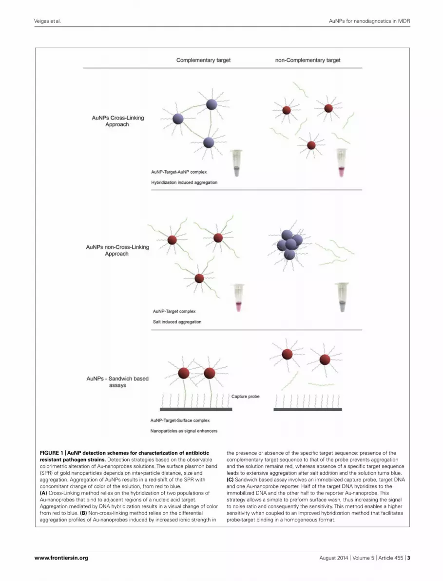
Gold nanoparticles have unique optical properties associatedwith a well-defined surface plasmon resonance (SPR) band inthe visible region of the spectrum (Halfpenny and Wright, 2010),strongly correlated to composition, shape and inter-particle dis-tance (Johnson et al., 2007). For example, AuNP aggregationleads to a pronounced color transition from red to blue dueto plasmon coupling between NPs (Jain, 2007). Consequently,
most AuNPs based methods rely on these colorimetric changesof the colloidal solution upon aggregation derived from changesto the media dielectric and/or due to recognition of a spe-cific target. These detection strategies typically depend on theinteraction between nanostructure-bound oligonucleotides andthe target molecule mediated by a recognition element, which,for DNA/RNA assays, is an oligonucleotide sequence – goldnanoprobe (Au-nanoprobe). A specific complementary targetmay hybridize to the Au-nanoprobes and promote inter-particlecross-linking aggregation (e.g., using two Au-nanoprobes eachfunctionalised with one half of a contiguous target recognitionsequence) or stabilize nanoprobes against changes to the mediadielectric (e.g., pH, ionic strength). In the latter, hybridisation ofAu-nanoprobes to the target sequence will prevent the non-cross-linking aggregation induced by increasing ionic strength (Figure 1;Sato et al., 2003; Baptista et al., 2005). Thus, modulation of AuNPor Au-nanoprobe inter-particle distance allows control over theircorresponding aggregation and dispersion levels providing visualdetection for a wide range of biological entities (Hauck et al., 2010;Ngo et al., 2011).
NANODIAGNOSTICS FOR PATHOGENSIdentification of pathogens based on specific target sequenceshas become the corner-stone of molecular based approaches todiscriminate between organisms and characterize particular vari-ations at the genomic level that provide unique nucleic acidsignatures suitable for diagnostics. This trend has had a newsurge with the development of numerous nanoparticle-basedapproaches designed to identify those pathogen signatures withextra sensitivity and faster than ever before (Costa et al., 2010; Kait-tanis et al., 2010; Chi et al., 2012; Veigas et al., 2012a). In fact, theincrease in sensitivity without loss of selectivity and specificity haspromoted nanodiagnostics in the field of pathogen characterisa-tion. Also, due to the minute dimensions of the signal transductionlabel (e.g., AuNPs), these systems show a high degree of minia-turization that makes them suitable for use at point-of-care orpoint-of-need.
The first proof-of-concept for nanodiagnostics using AuNPswas introduced by Mirkin et al. (1996) who successfully demon-strated that 13 nm AuNPs functionalized with a specific oligonu-cleotide sequence selectively assembled in the presence of acomplementary target DNA. The development of this proof-of-concept led to the development of the first Au-nanoprobe basedpathogen diagnostic system, allowing the detection of anthrax viaa specific lethal factor DNA sequence (Bailey et al., 2003). Thisapproach was further explored in a multitude of targets and sam-ples (for further insights see Kaittanis et al., 2010; Veigas et al.,2012a and references therein).
Following a similar technological approach, Baptista et al.(2006) developed a rapid and relatively low cost method forDNA detection and generated the first application of AuNPsfor the molecular diagnostics of Mycobacterium tuberculosis(Mtb). The method consists in differential stabilization of Au-nanoprobes in presence of DNA targets following salt inducedaggregation: presence of a complementary target preventsnanoprobe aggregation and the solution remains red; whereasnon-complementary/mismatched targets do not prevent gold
Frontiers in Microbiology | Antimicrobials, Resistance and Chemotherapy August 2014 | Volume 5 | Article 455 | 2
Veigas et al. AuNPs for nanodiagnostics in MDR
FIGURE 1 | AuNP detection schemes for characterization of antibiotic
resistant pathogen strains. Detection strategies based on the observablecolorimetric alteration of Au-nanoprobes solutions. The surface plasmon band(SPR) of gold nanoparticles depends on inter-particle distance, size andaggregation. Aggregation of AuNPs results in a red-shift of the SPR withconcomitant change of color of the solution, from red to blue.(A) Cross-Linking method relies on the hybridization of two populations ofAu-nanoprobes that bind to adjacent regions of a nucleic acid target.Aggregation mediated by DNA hybridization results in a visual change of colorfrom red to blue. (B) Non-cross-linking method relies on the differentialaggregation profiles of Au-nanoprobes induced by increased ionic strength in
the presence or absence of the specific target sequence: presence of thecomplementary target sequence to that of the probe prevents aggregationand the solution remains red, whereas absence of a specific target sequenceleads to extensive aggregation after salt addition and the solution turns blue.(C) Sandwich based assay involves an immobilized capture probe, target DNAand one Au-nanoprobe reporter. Half of the target DNA hybridizes to theimmobilized DNA and the other half to the reporter Au-nanoprobe. Thisstrategy allows a simple to preform surface wash, thus increasing the signalto noise ratio and consequently the sensitivity. This method enables a highersensitivity when coupled to an improved hybridization method that facilitatesprobe-target binding in a homogeneous format.
www.frontiersin.org August 2014 | Volume 5 | Article 455 | 3

Veigas et al. AuNPs for nanodiagnostics in MDR
nanoprobe aggregation, resulting in a visible change of colorfrom red to blue. The methodology was tested in clinical sam-ples demonstrating high efficiency with results comparable tothose attained via commercial molecular tuberculosis (TB) diag-nostics test, such as INNO-LiPA Rif. TB (Veigas et al., 2012a).Similar approaches have been used by Liandris et al. (2009) whodeveloped a non-cross-linking approach for the detection of TBwithout the need of target amplification. Following a cross-linkingapproach, Soo et al. (2009) designed a set of gold nanoprobesto specifically hybridize with target DNA from Mtb strains. Thismethodology was evaluated by directly and simultaneously detect-ing M. tuberculosis complex (MTBC) and Mtb in 600 clinicalstrains.
Staphylococcus aureus is also one of the most impor-tant human pathogens, causing more than 500,000 infec-tions in the US each year (Chang et al., 2013). Byusing aptamers that specifically recognize S. aureus, Changet al. (2013) developed an ultrasensitive aptamer-conjugated-AuNPs for rapid bacterial detection. Their non-polymerasechain reaction (PCR)-based method measures the reso-nance light-scattering signal of aptamer-conjugated AuNPsto detect a single cell within 1.5 h. Accordingly tothe authors this platform technology has the potential todevelop a rapid and sensitive bacterial testing at point-of-care(Chang et al., 2013).
GOLD NANOPARTICLES CHARACTERISATION OF ANTIBIOTICRESISTANCE PROFILESFor the past 20 years there has been an increase in the emergenceof antibiotic-resistant microorganisms with elevated pathogenesisat the global level, leading to an urgent need for new and improvedapproaches for bacterial quantification and identification (Pis-suwan et al., 2010). Table 1 summarizes existing AuNP-based
technologies for the antibiotic susceptibility characterization ofpathogens. Particularly problematic drug resistant bacteria includethe methicillin-resistant strains of S. aureus (MRSA), responsiblefor many opportunistic infections, enteropathogenic Escherichiacoli, Mtb showing multidrug resistance (MDR-TB and XRD-TB),and Streptococcus pneumoniae. The prevalence of drug resistantstrains of Mtb (MDR and XDR-TB) and MRSA have demonstratedthe need for the development of drug susceptibility systems thatare capable of delivering an unequivocal response to identify withhigh sensitivity and in a cost-efficient manner the pathogen’s resis-tance profile and allowing fast and accurate therapeutic approach(Kaittanis et al., 2010).
New diagnostic tools for drug resistant pathogen detectionand characterisation ought to overcome the main constraint interms of current molecular diagnostics – time. Several new tech-nologies are currently being developed and validated to providefaster and at a low cost diagnosis of resistant pathogens compar-ing to conventional culture and drug susceptibility tests. Threedistinct operational steps that are typically required for pathogendetection and characterization: sample preparation, target ampli-fication, and signal read-out. Although each step can be consideredindividually, it is important to emphasize that a key challenge fordevelopment of such nucleic acid detection methods is the integra-tion of all these steps into a unified process workflow (Figure 2).Rapid and cost effective diagnosis will have several benefits: earliertreatment of patients, reduction of time spent on inappropriateand ineffective treatment (thereby promoting the developmentof further drug resistance), and reduction of resistant strainsspreading in congregate settings (Veigas et al., 2012a).
AuNPs for molecular detection of antibiotic resistance in S. aureusMethicillin-resistant strains of S. aureus is responsible for 40–60% of all S. aureus infections in hospitals in the United
Table 1 | AuNPs-based systems for pathogen antibiotic susceptibility characterization.
Application Description Target(s) Reference(s)
Colorimetric detection of AuNPs spotted
onto an illuminated glass waveguide
Detection relies on the evaluation of SPR
change upon target hybridization
Detection of mecA gene associated
with methicillin resistance, in S. aureus
and S. aureus 23S rRNA. Validation with
clinical samples
Storhoff et al. (2004), Chan et al. (2014)
Antibiotic susceptibility
characterization
Colorimetric detection with AuNPs.
Detection relies on the evaluation of SPR
change upon aggregation and the
concomitant colorimetric changes that can
be assessed by the naked eye
Detection of rpoB mutations associated
Rifampicin resistance. Integration with
isothermal DNA amplification strategies
Veigas et al. (2010, 2013)
Colorimetric detection with AuNPs.
Detection relies on the evaluation of SPR
change upon aggregation. Sandwich
hybridization assay, AuNPs act as SPR
signal enhancers
Detection of rpoB and inhA mutations
associated with Rifampicin and
Isoniazid resistance. Integration with
surface-anchored rolling circle
amplification for isothermal DNA
amplification. Validation with clinical
samples
Xiang et al. (2013), Pedrosa et al. (2014)
Frontiers in Microbiology | Antimicrobials, Resistance and Chemotherapy August 2014 | Volume 5 | Article 455 | 4

Veigas et al. AuNPs for nanodiagnostics in MDR
FIGURE 2 | Operational steps for pathogen detection and
characterization using NPs: sample preparation, target
amplification/direct detection, and signal read-out. The ideal portabledetection method should perform as accurately as traditional centralizedlaboratory-based testing, while also overcoming the additional challengesassociated with POC testing, such as uncontrolled environmental conditionssuch as inconsistent or non-existent electrical power supply, and operation by
untrained or minimally trained personnel. To overcome all the technicallimitations of actual diagnostic systems, three distinct operational steps arerequired: faster simple and incorporated sample preparation, targetamplification, and signal read-out. Although each step can be individuallyconsidered, a key challenge for development of POC nucleic acid detectionmethods is integration of all these steps into a unified process and preferablywithin a single device.
States and United Kingdom (Pelgrift and Friedman, 2013). Cur-rently, the two major strategies for laboratory detection ofMRSA are bacterial culture-based phenotypic methods and NATs(Chastre et al., 2014). While a bacterial culture-based pheno-typic approach offers the advantages of detecting live bacteriaproviding antimicrobial susceptibilities and a variety of spec-imens, they require 1–2 days for the confirmation of MRSAinfection, due to their dependence on bacterial growth. Incontrast, nucleic acid detection assays allow rapid and sensi-tive detection of MRSA-specific sequences directly from clinicalspecimens with a turnaround time ranging from 1 to 3 h(Bischof et al., 2009; Arbefeville et al., 2011; Kelley et al., 2011;
Chadwick et al., 2013; Chastre et al., 2014). Combining NATs andAuNPs in a single system allows the identification the pathogen’sresistance profile with high sensitivity and in a cost-efficientmanner providing fast and accurate therapeutic approaches (seeTable 2).
In this case, Storhoff et al. (2004) proposed the use of AuNPs forthe colorimetric detection of antibiotic resistant S. aureus strainsback in 2004 via a colorimetric“spot- and-read”assay for the detec-tion of mecA in MRSA genomic DNA samples. In this assay, nucleicacid targets are recognized by DNA-modified Au-nanoprobes thatundergo a color change that is visually detectable when solutionsare spotted onto an illuminated glass waveguide. This scatter-based
www.frontiersin.org August 2014 | Volume 5 | Article 455 | 5

Veigas et al. AuNPs for nanodiagnostics in MDR
Table 2 | AuNPs-based systems for pathogen antibiotic susceptibility characterization, preclinical/clinical metadata of sensitivity and specificity.
Pathogen Target Sensitivity Specificity No. of
isolates
Reference
MRSA Detection of mecA gene associated
with methicillin resistance, in S.
aureus. Genomic DNA samples
isolated from cultured bacterial cells
Analytical sensitivity of 66 pg/μl of
MRSA total genomic DNA
n.d. n.d.Storhoff et al. (2004)
Detection of mecA gene associated
with methicillin resistance, in S.
aureus. Validation with clinical
samples
97.14% (Compared with culture
standard culture methods)
91.89% (Compared
with culture standard
culture methods)
72Chan et al. (2014)
MDRTB Detection of rpoB mutations
associated Rifampicin resistance
84.7% (compared to NNO-LiPA Rif.
TB assay)
100% (compared to
NNO-LiPA Rif. TB
assay)
46Veigas et al. (2010)
Detection of rpoB mutations
associated Rifampicin resistance.
Integration with isothermal DNA
amplification strategies
100% ( compared to NNO-LiPA Rif.
TB assay)
100% ( compared to
NNO-LiPA Rif. TB
assay)
12Veigas et al. (2013)
Detection of rpoB and inhA
mutations associated with
Rifampicin and Isoniazid resistance.
Integration with surface-anchored
rolling circle amplification for
isothermal DNA amplification.
Validation with clinical samples
Analitycal sensitivity of 8.2 pg uL−1
of genomic DNA from clinical
samples
n.d. 5Xiang et al. (2013)
Detection of rpoB and inhA
mutations associated with
Rifampicin and Isoniazid resistance.
Integration with multiplex
amplification strategy. Validation
with clinical samples
100% (compared to NNO-LiPA Rif.
TB assay)
100% (compared to
NNO-LiPA Rif. TB
assay)
25Pedrosa et al. (2014)
method enabled the detection of nucleic acids while demonstrat-ing a remarkable sequence specificity that allowed discriminationof single-base mismatches, deletions or insertions (Storhoff et al.,2004). The method relies on the cross-linking approach target-ing the bacterial mecA gene with a limit of detection of 33 nM.This approach was effective in discriminating genomic DNA sam-ples of MRSA from methicillin-sensitive S. aureus (MSSA) strains,where the detectable color change was observed only for MRSAwith very short hybridisation times (Storhoff et al., 2004). Theuse of scatter light analysis coupled to the molecular identifica-tion approach greatly enhanced detection sensitivity (∼4 ordersof magnitude) compared to previously reported absorbance-based spot test, thus enabling detection of zeptomole amountsof DNA target. This sensitivity is possible in a homogeneous for-mat because aggregate formation is detectable even when onlya very small fraction of the nanoparticle probes is involved inthe hybridisation, suggesting a large change to both color and
intensity of scattered light from the complexes (Storhoff et al.,2004).
Recently, Chan et al. (2014) reported the use of AuNPs fordirect colorimetric PCR detection of MRSA in clinical specimens.The colorimetric assay comprised probes functionalised with spe-cific oligonucleotides targeting S. aureus 23S rRNA and mecAsequences. In this study, 72 clinical samples were tested, includingpositive blood culture, urine, respiratory samples, as well as woundswabs, pus and body fluid. Using conventional bacterial culture asgold standard, the sensitivity, specificity, positive and negative pre-dictive values of this colorimetric assay were 97.14, 91.89, 91.89,and 97.14%, respectively. This performance compares to that ofcommercial real-time PCR assays but at lower cost per reaction.The colorimetric assay also demonstrated very good agreementwith the “gold standard” (94.44%). This study was the first reporton the use of AuNPs colorimetric assay for direct detection ofMRSA in various types of clinical specimens (Chan et al., 2014).
Frontiers in Microbiology | Antimicrobials, Resistance and Chemotherapy August 2014 | Volume 5 | Article 455 | 6
Veigas et al. AuNPs for nanodiagnostics in MDR
Further evaluation of these assays in large-scale trials is neededwhich can also allow for some modifications to streamline theprocedures for routine use.
AuNPs for molecular characterisation of antibiotic resistance inM. tuberculosisTuberculosis is one of the leading causes of infection in humans,causing high morbility and mortality all over the world. At present,the treatment of choice for an active TB infection is long-termantibiotic therapy, with an initial “intensive phase” consistingof the four first-line anti-TB drugs (isoniazid, INH; rifampicin,RIF; ethambutol, ETH; and pyrazinamide) followed by a typicalfour month course of RIF and INH alone (Gaspar et al., 2008).Despite effective treatment, due to the length of antibiotic ther-apy, side effects frequently develop and the associated cost is high(Garner et al., 2007; Aspler et al., 2008; Armstead and Li, 2011).These factors correlate to low patient compliance and contributeto the development of drug-resistant bacteria (Armstead et al.,2011). The rate of new cases of multidrug resistant tuberculo-sis (MDRTB) continues to increase, and due to the difficulty inthe management of such infection, it constitutes a serious healthproblem (World Health Organization [WHO], 2012). The surgeof MDRTB has raised awareness towards extreme resistant TB(XRDTB) or even totally resistant TB. In most cases, drug resis-tance in Mtb has been related to mutations in several loci withinthe pathogen’s genome. The development of fast, cheap and simplescreening methodologies is of paramount relevance for the earlydetection of these mutations, essential for the timely and effectivediagnosis and management of MDRTB patients (Barnard et al.,2008; Veigas et al., 2010; Abebe et al., 2011). Resistance to RIF iscommonly associated with point mutations within the rpoB geneof Mtb whose detection is considered the best early molecularpredictor for MDRTB. Resistance to RIF has been associated tosingle point alterations within a well-defined 81 bp region (codons507–533) of the rpoB gene encoding for the beta subunit of RNApolymerase. Concurrent resistance to INH and RIF is commonlyassociated with point mutations in katG, inhA, and rpoB genes ofMTBC (Musser, 1995; Soini and Musser, 2001). Prompt diagnosisof MDRTB has been the main obstacle to its correct manage-ment and control. This problem would seem to have been solvedwith the development of molecular techniques applicable alsoin high-prevalence, low-income settings, such as the GenotypeMTBDR- Plus and Gene Xpert MTB/RIF assays. However, thoughvery rapid and highly sensitive, these tests are not consideredhighly specific for the diagnosis of RIF resistance, particularlyin low prevalence settings or when mixed strains are present(Van Deun et al., 2013).
Based on the differential non-cross-linking aggregation ofAu-nanoprobes, Baptista’s group developed a simple and straight-forward colorimetric method for Mtb identification and singlebase mutation discrimination in rpoB (Veigas et al., 2010), whichconstitutes the first application of AuNPs for the specific detectionof RIF resistant Mtb. This approach uses an Au-nanoprobe assayfor the rapid detection of MTBC strains and simultaneous char-acterisation of mutations associated with RIF resistance, namelymutations in codons 516, 526, and 531 of rpoB gene from MTBCclinical specimens with remarkable sensitivity in a few hours. To
assure high selectivity and sensitivity, two nanoprobes are simul-taneously used to tackle each mutation – one recognizing thewild-type sequence and another for the mutated. By doing so, thisapproach correctly detected the presence of DNA from membersof the MTBC in 83.3% of all tested samples. The initial approachrequired a simple PCR amplification of a large region spreading thetargets sequences for the nanoprobes, and the resulting ampliconstested directly with the Au-nanoprobe system. The molecular char-acterisation step takes only 15 min to yield a colorimetric resultthat, through the use of a suitable photodetector (e.g., UV/visiblespectrophotometer, microplate reader, etc.) allows for mediumthroughput analysis at a peripheral laboratory. A limit of detectioncould be set at 75 nM, however, for robust single base mismatchdetermination, 117 nM of DNA target were used per assay (seealso Table 2).
More recently, the same group extended and improved thisdetection strategy towards the simultaneous discrimination ofspecific mutations within inhA and rpoB genes in PCR ampli-fied DNA from isolates. Using a multiplex PCR reaction, it waspossible to assess both loci in parallel, and extend the potential ofthe Au-nanoprobe method to MDRTB molecular characterisationwith special application in the most frequent Portuguese geno-types (Pedrosa et al., 2014). Based on the molecular signaturesof susceptibility of MTBC members to first line antibiotics, RIF,INH, and ethionamide (ETH), a two-step approach was developed,based on the multi loci PCR amplification of gene fragments andsubsequent hybridisation with specific Au-nanoprobe. The twotarget sequences harbor the most common mutations associatedwith resistance to these antibiotics, rpoB S531L, inhA C(-)15Tand are amplified by a set of rpoB primer pairs flanking uniqueregions specific for MTBC members – first level of identification.The MTBC Au-nanoprobe constitutes a second level of identifica-tion. Another two sets of nanoprobes are used to discriminatethe desired mutations. This approach brings new possibilitiesfor MDRTB diagnostics as the Au-nanoprobe methodology maybecome an useful tool for MDRTB molecular characterisation ata point-of-need (Pedrosa et al., 2014).
Conventional TB diagnosis methods (such as Ziehl-Neelsen orKinyoun for staining sputum smears, egg-based media for cul-ture, and solid media for antimicrobial susceptibility testing) havebeen used for almost 50 years presenting low sensitivity, speci-ficity, and a high turn-around time. Although some laboratoriesuse fluorochrome stains and liquid-based media for cultures, smallhospitals or clinics cannot use these methods due to the need ofhigh technical expertise, equipment, and expensive materials. Thequality of sputum specimens and contamination of specimensdue to inappropriate storage and/or long transport times to thelaboratory has been a critical bottleneck (Wilson, 2011).
The development of complete, accurate and simple TB diag-nostic tests able to target relevant TB sequences and assessingmultidrug resistance cases has been one of the major bottle-necks for TB effective detection and treatment. Au-nanoprobesintegrated within a paper-based platform may be proven to bean accurate, rapid, low-cost, and user friendly nanosystem forthe identification of specific DNA sequences of TB, confirminginfection and allowing identification of MDRTB strains. Thisnanosystem allows earlier treatment, reduction of time spent
www.frontiersin.org August 2014 | Volume 5 | Article 455 | 7

Veigas et al. AuNPs for nanodiagnostics in MDR
on inappropriate and ineffective treatment and reduction ofMDRTB spread in congregate settings making it ideal for largescreening and/or at point-of-need. The ability of coupling theLAMP amplification strategy to specific Au-nanoprobes translatesinto additional benefits: it eliminates the PCR amplification stepbypassing the need of specialized machines and technicians, assess-ing multiple antibiotic resistances in the field and reaching remotecommunities.
Integration of isothermal amplification techniques to AuNPsstrategies. Isothermal amplification techniques have been recentlydeveloped as an alternative to PCR for target DNA amplifi-cation and detection without the use of a thermocycler (Gilland Ghaemi, 2008). Thermophilic helicase-dependent isother-mal amplification uses a thermostable helicase to unwind thedouble stranded DNA (dsDNA) and generate single stranded tem-plates that are used for further polymerase amplification (Lixinet al., 2005). The dsDNA separation and amplification are per-formed at the same temperature, which makes this techniquesuitable for development of point-of-care microbial detectionsystems, since a thermocycler is not required for DNA denatu-ration and amplification (Tomita et al., 2008; Jeong et al., 2009).Recently, a AuNP based DNA biosensor for the detection ofMtb using thermophilic helicase-dependent isothermal amplifi-cation was developed (Torres-Chavolla and Alocilja, 2011). Inrecent years, Loop-mediated isothermal amplification (LAMP,Eiken Chemical Co. Ltd., Tokyo, Japan) assay has been intro-duced for the diagnosis of pulmonary TB (Yuan et al., 2013).The general LAMP procedure uses four primers to achieve acyclical amplification process based on spontaneous formationof stem-loop DNA structures. This process also utilizes a poly-merase with strand displacement capability. LAMP has beenused to successfully detect a wide range of pathogens includingmalaria, HIV, and multiplexed detection of bacteria (Hartmanet al., 2013).
Further improvements to the Au-nanoprobe system describedabove were attained via LAMP amplification strategy coupledto specific Au-nanoprobes for molecular identification of MTBCmembers and resistance signatures, such as RIF resistance (Veigaset al., 2013). Taking advantage of such features, they demon-strated that the non-cross-linking system is capable to dis-criminate the rpoB S531L point mutation on LAMP productsand, thus, opening new possibilities for MDRTB diagnosticsin remote environments and at a point-of-care. LAMP origi-nates long DNA concatamers that can easily be assessed via aset of nanoprobes for individual sequence identities, demonstrat-ing that it is possible to use an Au-nanoprobe based strategyto detect single point alteration on isothermally amplified DNAproducts.
Despite the several benefits presented by these nanodiagnosticssystems, translation into the clinics is still unaccomplished. Mostof the TB nanosystems reported in the literature still lack validationand for most of them integration in one simple platform capableof eliminating the need for DNA purification and amplificationis of utmost importance. Refinement of these laboratory strate-gies into one single nanodevice may speed up translation into thefield.
Rolling circle amplification. Following on the development ofisothermal amplification methods, Xiang et al. (2013) developeda surface-anchored rolling circle amplification (RCA) integratedwith Au-nanoprobes to isothermally detect multiple point muta-tions associated with MDRTB with a wild-type to mutant ratioof 5000:1. This work introduced a new SPR method for multi-plex mutation detection based on surface signal amplification. Thehigh sensitivity and specificity of this method mainly attributed tothe high-fidelity of ligation, multiplexing characteristics of probes,amplification potential of surface-anchored RCA and Au NPs, andintrinsically high sensitivity of SPR biosensor. The L-RCA by ligaserelies on base pairing principle which requires perfect complemen-tarity on the ligation nick. It not only forbids the mismatch butalso has a low occurrence of false positive results when comparedto PCR (Lizardi et al., 1998). Because RCA amplifies only the cir-cular PLP without accumulation of target templates over time, itminimizes the risk of contamination and the potential biohazard.Besides, the Au-nanoprobes further enhance identification due tothe sandwich hybridisation. Upon recognition, each point muta-tion is identified by locating into the corresponding channel ona chip, which allows the immobilized primer (capture probe)–template (circular PLP) complex to isothermally amplify as RCAand further amplified by AuNPs. Binding of the AuNPs to the RCAproducts acts as the electromagnetic field coupling to the gold film,thus enhancing the plasmon resonance derived by excitation bythe incident light, leading to improvement of the transduction ofsmall changes in refractive index on the chip surface media, thusimproving sensitivity (Petryayeva and Krull, 2011).
NANODIAGNOSTICS FOR POINT OF CARE APPLICATIONSDespite the amazing advances of nanotechnology the effectivetranslation to the clinical setting and to the molecular detec-tion and/or characterisation has not been fully applied (Haucket al., 2010). Nanotechnology, and NPs, based molecular identi-fication systems have focused on increasing sensitivity and speedwhen compared to traditional methodologies. However, nowadaysresearchers have been gearing their efforts towards the devel-opment of nanotechnology-based systems that are affordable,robust and reproducible, making them suitable for applicationseven in areas that lack dedicated and expensive laboratory equip-ment. In fact, AuNPs based systems have been proposed andused for the identification of different pathogens with one com-mon ground – making it simple and affordable. Consideringthat most of these systems rely on the molecular recognition ofselective and specific sequences in DNA, we are only one stepaway from identifying molecular signatures of resistance. In fact,only by bringing together these platforms and those at the fore-front of antibiotic resistance characterisation (e.g., microbiologistand clinicians), definite translation can be achieved. Technol-ogy integration together with the possibility of miniaturizationis of utmost importance for the development of an integratedbiosensor suitable for peripheral laboratories and/or point-of-carediagnostics, providing a new tool in the fight against TB.
Nonetheless, there has been some effort towards bringingthese technologies to point-of-care application. For example, theAu-nanoprobe system for characterisation of mutations associ-ated to drug resistance in TB has been further integrated with a
Frontiers in Microbiology | Antimicrobials, Resistance and Chemotherapy August 2014 | Volume 5 | Article 455 | 8

Veigas et al. AuNPs for nanodiagnostics in MDR
paper-based platform for fast and easy to use detection of MTBCmembers – Gold on Paper (Veigas et al., 2012b; Costa et al., 2014).Gold on Paper is the working concept of integrating a paper microwell platform and a biomolecular detection scheme based on Au-nanoprobes. Gold on Paper showed to be capable of efficientlydetect MTBC members directly and, by means of a smartphonedevice, analyzing data on the spot while maintaining sensitivityand specificity. This demonstrates that systems such as Gold onPaper may be easy to perform without the need for expensive andcomplex laboratory set up. Using this concept, it is possible toattain a positive identification of the pathogen within one hour,which via the use of a generic “smart” mobile device allows forcomplete analysis at a peripheral laboratory, and transmit digitalinformation over existing communications channels, combinedwith GPS location metadata inserted into the captured digitalimages (Veigas et al., 2012b). The limitation imposed by the DNAsample preparation is greatly overcome by the potential use of thismethodology to identify and characterize the molecular signa-tures involved in antibiotic resistance. This integrated diagnosticsscheme can then forward the attained data to a centralized off-site server allowing for monitoring of TB in real-time that couldbe proven extremely useful in remote areas of the globe lackingresources (Veigas et al., 2012b).
Monitoring for drug-induced liver injury (DILI) via serialtransaminase measurements in patients on potentially hepatotoxicmedications (e.g., for HIV and TB) is routine in resource-rich nations, but often unavailable in resource-limited settings.Towards enabling universal access to affordable point-of-carescreening for DILI, Pollock et al. (2013) have performed the firstfield evaluation of a paper-based, microfluidic finger-stick testfor rapid, semi-quantitative, visual measurement of blood ala-nine aminotransferase. The objective was to assess operationalfeasibility, inter-operator variability, lot variability, device fail-ure rate, and accuracy, to inform device modification for furtherfield testing. The paper-based alanine aminotransferase test wasperformed at point-of-care on fingerstick samples from 600 out-patients receiving HIV treatment in Vietnam (Pollock et al., 2013).This first field study performed with a paper-based microflu-idic device opens the door to development of similar assays forother important analytes and also for assessing MDRTB andMRSA.
Based on these principles new technologies were developedand are today available in the market. For example, Nanosphereoffers two products approved by the FDA, one aimed at identi-fying typical mutations in coagulation factors without the needfor nucleic acid amplification; another used to genotype polymor-phisms associated with warfarin metabolism. In both cases thesamples are processed through a cartridge where the sample ana-lyzed via an automated processor and reader (Lefferts et al., 2010;Maurice et al., 2010).
CONCLUSIONS AND FUTURE PERSPECTIVESOver the past decades, noble metal NPs, due to their optical andphysic-chemical properties, have been used in proof-of-conceptbiosensing tools for the sensitive detection of pathogens of inter-est. Amongst these biosensing platforms, several have focused onthe specific identification of DNA/RNA sequences associated to
molecular signatures of infection and antibiotic resistance. AuNPbased assays have progressively been integrated into sensing plat-forms capable of increasing sensitivity and lowering costs. Here,we provided an overview of existing strategies relying on the useof AuNPs for detection of molecular markers of antibiotic resis-tance. Despite the desperate need for robust, yet simple and cheap,screening tools to identify MDR pathogens, there are not thatmany concepts making it through to validation in the laboratoryset. It is clear that microbiologists need to integrate the multidis-ciplinary teams that provide for nanodiagnostics development soas to widen the scope of combinations and modalities that can beeasily coupled to current molecular nanodiagnostics technologiesso as to facilitate integration to the lab and clinical setting.
Detection strategies based on AuNPs provide comparabledetection capability to that of standard techniques but at a fractionof cost and time, usually not requiring cumbersome sample prepa-ration or equipment. As such, nanoparticle based approaches areexpected to be incrementally applied to MDR characterisation andpathogen detection with particular emphasis for systems capableto operate at point-of-need. However, despite the massive invest-ment in these technologies, translation to the clinics is yet to befulfilled. Most of the reported systems in the literature still lackvalidation and/or are in pre-clinic stages with few commerciallyavailable products being available to the clinician. The next stepis clearly to focus on the translation of some of the strategies thatexist in the lab into the field and to the bedside.
ACKNOWLEDGMENTSThe authors thank FCT/MEC for financial support throughCIGMH (PEst-OE/SAU/UI0009/2011–14); PTDC/BBB-NAN/1812/2012 and SFRH/BD/78970/2011 for Bruno Veigas.
REFERENCESAbebe, G., Paasch, F., Apers, L., Rigouts, L., and Colebunders, R. (2011).
Tuberculosis drug resistance testing by molecular methods: opportunities andchallenges in resource limited settings. J. Microbiol. Methods 84, 155–160. doi:10.1016/j.mimet.2010.11.014
Arbefeville, S. S., Zhang, K., Kroeger, J. S., Howard, W. J., Diekema, D. J., andRichter, S. S. (2011). Prevalence and genetic relatedness of methicillin-susceptibleStaphylococcus aureus isolates detected by the Xpert MRSA nasal assay. J. Clin.Microbiol. 49, 2996–2999. doi: 10.1128/JCM.00046-11
Armstead, A. L., and Li, B. (2011). Nanomedicine as an emerging approach againstintracellular pathogens. Int. J. Nanomed. 6, 3281–3293. doi: 10.2147/IJN.S27285
Aspler, A., Menzies, D., Oxlade, O., Banda, J., Mwenge, L., Godfrey-Faussett, P., et al.(2008). Cost of tuberculosis diagnosis and treatment from the patient perspectivein Lusaka, Zambia. Int. J. Tuberc. Lung Dis. 12, 928–935.
Azzazy, H. M. E., and Mansour, M. M. H. (2009). In vitro diagnostic prospects ofnanoparticles. Clin. Chim. Acta 403, 1–8. doi: 10.1016/j.cca.2009.01.016
Azzazy, H. M. E., Mansour, M. M. H., and Kazmierczak, S. C. (2006). Nanodiagnos-tics: a new frontier for clinical laboratory medicine. Clin. Chem. 52, 1238–1246.doi: 10.1373/clinchem.2006.066654
Azzazy, H. M. E., Mansour, M. M. H., and Kazmierczak, S. C. (2007). From diag-nostics to therapy: prospects of quantum dots. Clin. Biochem. 40, 917–927. doi:10.1016/j.clinbiochem.2007.05.018
Bailey, R. C., Nam, J. M., Mirkin, C. A., and Hupp, J. T. (2003). Real-time multi-color DNA detection with chemoresponsive diffraction gratings and nanoparticleprobes. J. Am. Chem. Soc. 125, 13541–13547. doi: 10.1021/ja035479k
Baptista, P. V., Doria, G., Henriques, D., Pereira, E., and Franco, R. (2005). Col-orimetric detection of eukaryotic gene expression with DNA-derivatized goldnanoparticles. J. Biotechnol. 119, 111–117. doi: 10.1016/j.jbiotec.2005.02.019
Baptista, P. V., Koziol-Montewka, M., Paluch-Oles, J., Doria, G., and Franco, R.(2006). Gold-nanoparticle-probe-based assay for rapid and direct detection of
www.frontiersin.org August 2014 | Volume 5 | Article 455 | 9
Veigas et al. AuNPs for nanodiagnostics in MDR
Mycobacterium tuberculosis DNA in clinical samples. Clin. Chem. 52, 1433–1434.doi: 10.1373/clinchem.2005.065391
Baptista, P. V., Pereira, E., Eaton, P., Doria, G., Miranda, A., Gomes, I., et al. (2008).Gold nanoparticles for the development of clinical diagnosis methods. Anal.Bioanal. Chem. 391, 943–950. doi: 10.1007/s00216-007-1768-z
Barnard, M., Albert, H., Coetzee, G., O’Brien, R., and Bosman, M. E. (2008). Rapidmolecular screening for multidrug-resistant tuberculosis in a high-volume publichealth laboratory in South Africa. Am. J. Respir. Crit. Care Med. 177, 787–792.doi: 10.1164/rccm.200709-1436OC
Bischof L. J., Lapsley, L., Fontecchio, K., Jacosalem, D., Young, C., Hankerd, R.,et al. (2009). Comparison of chromogenic media to BD GeneOhm methicillin-resistant Staphylococcus aureus (MRSA) PCR for detection of MRSA in nasalswabs. J. Clin. Microbiol. 47, 2281–2283. doi: 10.1128/JCM.02256-08
Branton, D., Deamer, D. W., Marziali, A., Bayley, H., Benner, S. A., Butler, T., et al.(2008). The potential and challenges of nanopore sequencing. Nat. Biotechnol.26, 1146–1153. doi: 10.1038/nbt.1495
Chadwick, S. G., Prasad, A., Smith, W. L., Mordechai, E., Adelson, M. E., and Gygax,S. E. (2013). Detection of epidemic USA300 community-associated methicillin-resistant Staphylococcus aureus strains by use of a single allele-specific PCR assaytargeting a novel polymorphism of Staphylococcus aureus pbp3. J. Clin. Microbiol.51, 2541–2550. doi: 10.1128/JCM.00417-13
Chan, W. S., Tang, B. S., Boost, M. V., Chow, C., and Leung, P. H. (2014). Detectionof methicillin-resistant Staphylococcus aureus using a gold nanoparticle-basedcolourimetric polymerase chain reaction assay. Biosens. Bioelectron. 15, 105–111.doi: 10.1016/j.bios.2013.09.027
Chang, Y. C., Yang, C. Y., Sun, R. L., Cheng, Y. F., Kao, W. C., and Yang, P. C. (2013).Rapid single cell detection of Staphylococcus aureus by aptamer-conjugated goldnanoparticles. Sci. Rep. 3, 1863 doi: 10.1038/srep01863
Chastre, J., Blasi, F., Masterton, R. G., Rello, J., Torres, A., and Welte, T. (2014).European perspective and update on the management of nosocomial pneumoniadue to methicillin-resistant Staphylococcus aureus after more than 10 years ofexperience with linezolid. Clin. Microbiol. Infect. 4, 19–36. doi: 10.1111/1469-0691.12450
Chi, X., Huang, D., Zhao, Z., Zhou, Z., Yin, Z., and Gao, J. (2012). Nanoprobes forin vitro diagnostics of cancer and infectious diseases. Biomaterials 33, 189–206.doi: 10.1016/j.biomaterials.2011.09.032
Costa, M. N., Veigas, B., Jacob, J. M., Santos, D. S., Gomes, J., Baptista, P. V.,et al. (2014). A low cost, safe, disposable, rapid and self-sustainable paper-basedplatform for diagnostic testing: lab-on-paper. Nanotechnology 25, 94006. doi:10.1088/0957-4484/25/9/094006
Costa, P., Amaro, A., Botelho, A., Inácio, J., and Baptista, P. V. (2010). Goldnanoprobes assay for identification of mycobacteria from the Mycobacteriumtuberculosis complex. Clin. Microbiol. Infect. 16, 1464–1469. doi: 10.1111/j.1469-0691.2010.03120.x
Das, M., Sumana, G., Nagarajan, R., and Malhotra, B. D. (2010). Application ofnanostructured ZnO films for electrochemical DNA biosensor. Thin Solid Films519, 1196–1201. doi: 10.1016/j.tsf.2010.08.069
Garner, P., Smith, H., Munro, S., and Volmink, J. (2007). Promoting adher-ence to tuberculosis treatment. Bull. World Health Organ. 85, 404–406. doi:10.2471/BLT.06.035568
Gaspar, M. M., Cruz, A., Fraga, A. G., Castro, A. G., Cruz, M. E., and Pedrosa, J.(2008). Developments on drug delivery systems for the treatment of mycobac-terial infections. Curr. Top. Med. Chem. 8, 579–591. doi: 10.2174/156802608783955629
Gill, P., and Ghaemi, A. (2008). Nucleic acid isothermal amplification tech-nologies: a review. Nucleosides Nucleotides Nucleic Acids 27, 224–243. doi:10.1080/15257770701845204
Halfpenny, K. C., and Wright, D. W. (2010). Nanoparticle detection of respiratoryinfection, Wiley Interdisciplinary Reviews. Nanomed. Nanobiotechnol. 2, 277–290. doi: 10.1002/wnan.83
Hartman, M. R., Ruiz, R. C., Hamada, S., Xu, C., Yancey, K. G., Yu, Y., et al.(2013). Point-of-care nucleic acid detection using nanotechnology Nanoscale 5,10141–10154. doi: 10.1039/C3NR04015A
Hauck, T. S., Gao, S. G. Y., and Chan, W. C. W. (2010). Nanotechnology diagnosticsfor infectious diseases prevalent in developing countries. Adv. Drug Deliv. Rev. 62,438–448. doi: 10.1016/j.addr.2009.11.015
Jain, K. K. (2005). Nanotechnology in clinical laboratory diagnostics. Clin. Chim.Acta 328, 37–54. doi: 10.1016/j.cccn.2005.03.014
Jain, K. K. (2007). Applications of nanobiotechnology in clinical diagnostics. Clin.Chem. 53, 2002–2009. doi: 10.1373/clinchem.2007.090795
Jeong, Y. J., Park, K., and Kim, D. E. (2009). Isothermal DNA amplification in vitro:the helicase-dependent amplification system. Cell. Mol. Life Sci. 66, 3325–3336.doi: 10.1007/s00018-009-0094-3
Johnson, C. J., Zhukovsky, N., Cass, A. E. G., and Nagy, J. M. (2007). Pro-teomics, nanotechnology and molecular diagnostics. Proteomics 8, 715–730. doi:10.1002/pmic.200700665
Jung, Y. L., Jung, C., Parab, H., Li, T., and Park, H. G. (2010). Direct colori-metric diagnosis of pathogen infections by utilizing thiol-labeled PCR primersand unmodified gold nanoparticles. Biosens Bioelectron. 25, 1941–1946. doi:10.1016/j.bios.2010.01.010
Kaittanis, C., Santra, S., and Perez, J. M. (2010). Emerging nanotechnology-basedstrategies for the identification of microbial pathogenesis. Adv. Drug Deliv. Rev.62, 408–423. doi: 10.1016/j.addr.2009.11.013
Kelley, P. G., Grabsch, E. A., Farrell, J., Xie, S., Montgomery, J., Mayall, B., et al.(2011). Evaluation of the Xpert MRSA/SA Blood Culture assay for the detectionof Staphylococcus aureus including strains with reduced vancomycin susceptibilityfrom blood culture specimens. Diagn. Microbiol. Infect. Dis. 70, 404–407. doi:10.1016/j.diagmicrobio.2011.02.006
Larguinho, M., and Baptista, P. V. (2012). Gold and silver nanoparticles for clinicaldiagnostics – from genomics to proteomics. J. Proteomics 75, 2811–2823. doi:10.1016/j.jprot.2011.11.007
Lefferts, J. A., Schwab, M. C., Dandamudi, U. B., Lee, H. K., Lewis, L. D., andTsongalis, G. J. (2010). Warfarin genotyping using three different platforms. Am.J. Transl. Res. 2, 441–446.
Liandris, E., Gazouli, M., Andreadou, M., Comor, M., Abazovic, N., Sechi, L. A.,et al. (2009). Direct detection of unamplified DNA from pathogenic mycobacteriausing DNA-derivatized gold nanoparticles. J. Microbiol. Methods 78, 260–264.doi: 10.1016/j.mimet.2009.06.009
Lin, C. C., Yang, Y. M., Liao, P. H., Chen, D. W., Lin, H. P., and Chang, H. C. (2014). Afilter-like AuNPs@MS SERS substrate for Staphylococcus aureus detection. Biosens.Bioelectron. 53, 519–527. doi: 10.1016/j.bios.2013.10.017
Liu, W. T. (2006). Nanoparticles and their biological and environmental applica-tions. J. Biosci. Bioeng. 102, 1–7. doi: 10.1263/jbb.102.1
Lixin, A., Tang, W., Ranalli, T. A., Kim, H. J., Wytiaz, J., and Kong, H.(2005). Characterization of a thermostable UvrD helicase and its participa-tion in helicase-dependent amplification. J. Biol. Chem. 280, 28952–28958. doi:10.1074/jbc.M503096200
Lizardi, P. M., Huang, X., Zhu, Z., Bray-Ward, P., Thomas, D. C., and Ward, D.C. (1998). Mutation detection and single-molecule counting using isothermalrolling-circle amplification. Nat. Genet. 19, 225–232. doi: 10.1038/898
Maurice, C. B., Barua, P. K., Simses, D., Smith, P., Howe, J. G., and Stack, G.(2010). Comparison of assay systems for warfarin-related CYP2C9 and VKORC1genotyping. Clin. Chim. Acta 411, 947–954. doi: 10.1016/j.cca.2010.03.005
Mirkin, C. A., Letsinger, R. L., Mucic, R. C., and Storhoff, J. J. (1996). A DNA-based method for rationally assembling nanoparticles into macroscopic materials.Nature 15, 607–609. doi: 10.1038/382607a0
Mothershed, E. A., and Whitney, A. M. (2005). Nucleic acid-based methods for thedetection of bacterial pathogens: present and future considerations for the clinicallaboratory. Clin. Chim. Acta 363, 206–220. doi: 10.1016/j.cccn.2005.05.050
Musser, J. M. (1995). Antimicrobial agent resistance in Mycobacteria: moleculargenetic insights. Clin. Microbiol. Rev. 8, 496–514.
Ngo, Y. H., Li, D., Simon, G. P., and Garnier, G. (2011). Paper surfacesfunctionalized by nanoparticles. Adv. Colloid Interface Sci. 163, 23–38. doi:10.1016/j.cis.2011.01.004
Pedrosa, P., Veigas, B., Machado, D., Couto, I., Viveiros, M., and Baptista, P. V. (2014).Gold nanoprobes for multi loci assessment of multi-drug resistant tuberculosis.Tuberculosis 94, 332–337. doi: 10.1016/j.tube.2013.12.009
Pelgrift, R. Y., and Friedman, A. J. (2013). Nanotechnology as a therapeutic toolto combat microbial resistance. Adv. Drug Deliv. Rev. 65, 1803–1815. doi:10.1016/j.addr.2013.07.011
Petryayeva, E., and Krull, U. J. (2011). Localized surface plasmon resonance: nanos-tructures, bioassays and biosensing–a review. Anal. Chim. Acta 706, 8–24. doi:10.1016/j.aca.2011.08.020
Pissuwan, D., Cortie, C. H., Valenzuela, S. M., and Cortie, M. B. (2010). Function-alised gold nanoparticles for controlling pathogenic bacteria. Trends Biotechnol.28, 207–213. doi: 10.1016/j.tibtech.2009.12.004
Frontiers in Microbiology | Antimicrobials, Resistance and Chemotherapy August 2014 | Volume 5 | Article 455 | 10

Veigas et al. AuNPs for nanodiagnostics in MDR
Pollock, N. R., McGray, S., Colby, D. J., Noubary, F., Nguyen, H., Nguyen, T.A., et al. (2013). Field evaluation of a prototype paper-based point-of-carefingerstick transaminase test. PLoS ONE 8:e75616. doi: 10.1371/journal.pone.0075616
Rosi, N. L., and Mirkin, C. A. (2005). Nanostructures in biodiagnostics. Chem. Rev.105, 1547–1562. doi: 10.1021/cr030067f
Sato, K., Hosokawa, K., and Maeda, M. (2003). Rapid aggregation of gold nanopar-ticles induced by non-cross-linking DNA hybridization. J. Am. Chem. Soc. 125,8102–8103. doi: 10.1021/ja034876s
Shinde, S. B., Fernandes, C. B., and Patravale, V. B. (2012). Recent trends in in-vitronanodiagnostics for detection of pathogens. J. Control Release 159, 164–180. doi:10.1016/j.jconrel.2011.11.033
Soini, H., and Musser, J. M. (2001). Molecular diagnosis of Mycobacteria. Clin.Chem. 47, 809–814.
Soo, P. C., Horng, Y. T., Chang, K. C., Wang, J. Y., Hsueh, P. R.,Chuang, C. Y.,et al. (2009). A simple gold nanoparticle probes assay for identificationof Mycobacterium tuberculosis and Mycobacterium tuberculosis complex fromclinical specimens. Mol. Cell Probes 23, 240–246. doi: 10.1016/j.mcp.2009.04.006
Storhoff, J. J., Lucas, A. D., Garimella, V., Bao, Y. P., and Müller, U. R. (2004).Homogeneous detection of unamplified genomic DNA sequences based on col-orimetric scatter of gold nanoparticle probes. Nat. Biotechnol. 22, 883–887. doi:10.1038/nbt977
Tallury, P., Malhotra, A., Byrne, L. M., and Santra, S. (2009). Nanobioimag-ing and sensing of infectious diseases. Adv. Drug Deliv. Rev. 62, 424–437. doi:10.1016/j.addr.2009.11.014
Tomita, N., Mori, Y., Kanda, H., and Notomi, T. (2008). Loop-mediated isothermalamplification (LAMP) of gene sequences and simple visual detection of products.Nat. Protoc. 3, 877–882. doi: 10.1038/nprot.2008.57
Torres-Chavolla, E., and Alocilja, E. C. (2011). Nanoparticle based DNAbiosensor for tuberculosis detection using thermophilic helicase-dependentisothermal amplification. Biosens. Bioelectron. 26, 4614–4618. doi:10.1016/j.bios.2011.04.055
Upadhyayula, V. K. (2012). Functionalized gold nanoparticle supported sen-sory mechanisms applied in detection of chemical and biological threatagents: a review. Anal. Chim. Acta 715, 1–18. doi: 10.1016/j.aca.2011.12.008
Van Deun, A., Aung, K. J., Bola, V., Lebeke, R., Hossain, M. A., de Rijk, W.B., et al. (2013). Rifampin drug resistance tests for tuberculosis: challeng-ing the gold standard. J. Clin. Microbiol. 51, 2633–2640. doi: 10.1128/JCM.00553-13
Veigas, B., Doria, G., and Baptista, P. V. (2012a). Nanodiagnostics for Tuberculosis,Understanding Tuberculosis – Global Experiences and Innovative Approaches tothe Diagnosis. Winchester: InTech, 562.
Veigas, B., Jacob, J. M., Costa, M. N., Santos, D. S., Viveiros, M., Inácio, J., et al.(2012b). Gold on paper-paper platform for Au-nanoprobe TB detection. LabChip 12, 4802–4808. doi: 10.1039/c2lc40739f
Veigas, B., Machado, D., Perdigão, J., Portugal, I., Couto, I., Viveiros, M., et al. (2010).Au-nanoprobes for detection of SNPs associated with antibiotic resistance inMycobacterium tuberculosis. Nanotechnology 21, 5101–5108. doi: 10.1088/0957-4484/21/41/415101
Veigas, B., Pedrosa, P., Couto, I., Viveiros, M., and Baptista, P. V. (2013). IsothermalDNA amplification coupled to Au-nanoprobes for detection of mutations asso-ciated to Rifampicin resistance in Mycobacterium tuberculosis. J. Nanobiotechnol.11, 1–6. doi: 10.1186/1477-3155-11-38
Wang S., Inci, E., Libero, G., Singhal, A., and Demirci, U. (2013). Point-of-careassays for tuberculosis: role of nanotechnology/microfluidics. Biotechnol. Adv.31, 438–449. doi: 10.1016/j.biotechadv.2013.01.006
Wilson M. L. (2011). Recent advances in the laboratory detection of Mycobac-terium tuberculosis complex and drug resistance. Infect. Dis. 52, 1350–1355. doi:10.1093/cid/cir146
World Health Organization [WHO]. (2012). Global Tuberculosis Report 2012.Geneva: WHO.
Xiang, Y., Deng, K., Xia, H., Yao, C., Chen, Q., Zhang, L., et al. (2013). Isothermaldetection of multiple point mutations by a surface plasmon resonance biosensorwith Au nanoparticles enhanced surface-anchored rolling circle amplification.Biosens. Bioelectron. 49, 442–449. doi: 10.1016/j.bios.2013.04.044
Yuan, L. Y., Li, Y., Wang, M., Ke, Z. Q., and Xu, W. Z. (2013). Rapid and effec-tive diagnosis of pulmonary tuberculosis with novel and sensitive loop-mediatedisothermal amplification (LAMP) assay in clinical samples: a meta-analysis.J. Infect. Chemother. 20, 86–92. doi: 10.1016/j.jiac.2013.07.003
Conflict of Interest Statement: The authors declare that the research was conductedin the absence of any commercial or financial relationships that could be construedas a potential conflict of interest.
Received: 18 July 2014; accepted: 11 August 2014; published online: 28 August 2014.Citation: Veigas B, Fernandes AR and Baptista PV (2014) AuNPs for iden-tification of molecular signatures of resistance. Front. Microbiol. 5:455. doi:10.3389/fmicb.2014.00455This article was submitted to Antimicrobials, Resistance and Chemotherapy, a sectionof the journal Frontiers in Microbiology.Copyright © 2014 Veigas, Fernandes and Baptista. This is an open-access article dis-tributed under the terms of the Creative Commons Attribution License (CC BY). Theuse, distribution or reproduction in other forums is permitted, provided the originalauthor(s) or licensor are credited and that the original publication in this journal is cited,in accordance with accepted academic practice. No use, distribution or reproduction ispermitted which does not comply with these terms.
www.frontiersin.org August 2014 | Volume 5 | Article 455 | 11

![Identification of markers associated with bacterial blight resistance loci in cowpea [Vigna unguiculata (L.) Walp.]](https://img.dokumen.tips/doc/110x75/6345adfb38eecfb33a06aaea/identification-of-markers-associated-with-bacterial-blight-resistance-loci-in-cowpea.jpg)


