Embed Size (px)
Citation preview

Food and Nutrition Sciences, 2013, 4, 191-196 doi:10.4236/fns.2013.48A023 Published Online August 2013 (http://www.scirp.org/journal/fns)
Association between Body Condition and Oxidative Status in Dogs
Anna Pasquini1*, Silvia Roberti1, Valentina Meucci1, Elena Luchetti1, Sergio Canello2, Gianandrea Guidetti2, Giulia Biagi1
1Department of Veterinary Science, University of Pisa, Pisa, Italy; 2Sanypet, Bagnoli di Sopra, Padova, Italy. Email: *[email protected] Received May 17th, 2013; revised June 17th, 2013; accepted June 24th, 2013 Copyright © 2013 Anna Pasquini et al. This is an open access article distributed under the Creative Commons Attribution License, which permits unrestricted use, distribution, and reproduction in any medium, provided the original work is properly cited.
ABSTRACT Oxidative Stress (OS) is considered an underlying mechanism by which dysfunctional metabolism occurs in obese sub- jects but there are still very few studies in canine species. The purpose of this study was to evaluate simultaneously the effects of diet on body weight, oxidative and inflammatory status in a group of 12 adult dogs. The dogs were fed a maintenance diet, integrated with natural antioxidants, for a period of 6 months. At the beginning and the end of the trial, Body Condition Score (BCS) was evaluated and haematological (CBCs, Complete Blood Counts), oxidative parameters (d-ROMs, derivatives of Reactive Oxygen Metabolites; BAP, Biological Antioxidant Potential; Retinol; Į-tocopherol) and inflammatory parameters (Fibrinogen; CRP, C Reactive Protein) were performed. Significant differences (p < 0.05) emerged about BCS, hematocrit (HCT), number of platelets (PLT), d-ROMs, BAP and retinol while no differences were for Į-tocopherol, fibrinogen and CRP between the two periods of different diet. In this study, dogs showed an oxi- dative imbalance documented by the increase in d-ROMs and the reduction of BAP and retinol. Inflammatory parame- ters don’t change in relation to body weight like an alteration of the oxidative status could precede the onset of inflam- mation. The role of oxidative stress and of integration with antioxidants should be taken into special consideration in the dietary treatment in overweight dogs. Keywords: Canine; Nutrition; Overweight; Oxidative Stress; Antioxidants; Inflammation
1. Introduction In recent years, Oxidative Stress (OS) has been postula- ted to be an important factor in the pathogenesis and de- velopment of lifestyle-related diseases [1]. In aerobic cells, free radicals are constantly produced mostly as Re- active Oxygen Species (ROS). Once produced, free ra- dicals are removed by antioxidant defences including en- dogenous and exogenous antioxidants. The imbalance between prooxidant and antioxidant defences in favor of prooxidants results in oxidative stress associated with the oxidative modification of biomolecules such as lipids, proteins, and nucleic acids. Alone or in combination with primary etiological factors, free radicals are involved in a pathogenesis of many inflammatory, degenerative and neoplastic diseases [2,3]. Recently, the prevalence of obe- sity has been related to a decrease of the plasma antio- xidants [4-6], the accumulation of fat has been related to the increase of oxidative stress markers [1,4,6], the oxi-
dative stress has been related to high body mass index (BMI) in human [7] and, finally, obesity has been related to chronic inflammatory status [8]. When caloric intake exceeds energy expenditure, the substrate-induced in- crease in Krebs cycle activity generates an excess of ROS [8]. Energy imbalances lead to the storage of excess energy in adipocytes, resulting in both hypertrophy and hyperplasia. These processes are associated with abnor- malities of adipocyte function, particularly mitochondrial stress and disrupted endoplasmic reticulum function [9]. This adipogenesis implies the differentiation of preadi- pocytes into mature and secreting adipocytes which re- lease a large number of cytokines, termed “adipokines”. In addition, the enlargement of adipocytes by fat storage induces adipose tissue hypoxia and the secretion of high levels of inflammatory cytokines [8]. The activation of in- flammatory signaling pathways in turn increases mito- chondrial ROS generation [9]. Although oxidative stress is considered the underlying mechanism by which dysfunc- tional metabolism occurs in obese subjects, there are still *Corresponding author.
Copyright © 2013 SciRes.����������������������������������������������������������������������������������FNS
Association between Body Condition and Oxidative Status in Dogs 192
very few studies both in feline and canine subjects [10- 12]. The purpose of this study was to evaluate simultane- ously the effects of diet on body weight and on the oxi- dative and inflammatory status in a group of adult dogs.
2. Materials and Methods 2.1. Animals and Experimental Design A group of 12 healthy adult German Shepherd dogs, all living together in the same environment, was fed with a maintenance diet (diet A), integrated with natural anti- oxidants, for a period of 6 months. The amount of feed administered was calculated according to the body weight, in accordance with what is indicated by the manufac- turer (13.5 g/Kg bw/day).
The transition to new diet was gradual and occurred in about a week. At the beginning and the end of the trial, every dog was weighed and its BCS was evaluated. In addition, each dog was subjected to a blood sample for the determination of haematological, oxidative and in- flammatory parameters. Previously, the same dogs were fed with a different maintenance diet (diet B) for a period of at least 6 months, at the dose of 15 g/Kg bw/day.
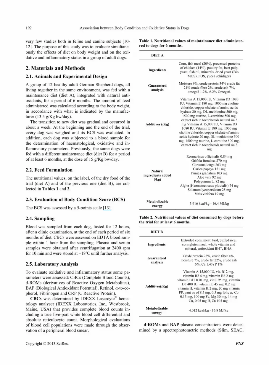
2.2. Feed Formulation The nutritional values, on the label, of the dry food of the trial (diet A) and of the previous one (diet B), are col- lected in Tables 1 and 2.
2.3. Evaluation of Body Condition Score (BCS) The BCS was assessed by a 5-points scale [13].
2.4. Sampling Blood was sampled from each dog, fasted for 12 hours, after a clinic examination, at the end of each period of six months of diet. CBCs were assessed on EDTA blood sam- ple within 1 hour from the sampling. Plasma and serum samples were obtained after centrifugation at 2400 rpm for 10 min and were stored at í18˚C until further analysis.
2.5. Laboratory Analysis To evaluate oxidative and inflammatory status some pa- rameters were assessed: CBCs (Complete Blood Counts), d-ROMs (derivatives of Reactive Oxygen Metabolites), BAP (Biological Antioxidant Potential), Retinol, Į-to-co- pherol, Fibrinogen and CRP (C Reactive Protein).
CBCs was determined by IDEXX Lasercyte® hema- tology analyser (IDEXX Laboratories, Inc., Westbrook, Maine, USA) that provides complete blood counts in- cluding a true five-part white blood cell differential and absolute reticulocyte count. Morphological evaluations of blood cell populations were made through the obser- vation of a peripheral blood smear.
Table 1. Nutritional values of maintainance diet administer- red to dogs for 6 months.
DIET A
Ingredients
Corn, fish meal (20%), processed proteins of chicken (14%), poultry fat, beet pulp, yeast, fish oil, minerals, dried yeast (Bio
MOS), FOS, yucca schidigera
Guaranteed analysis
Moisture 9%, crude protein 34% crude fat 21% crude fibre 2%, crude ash 7%,
omega3 1.2%, 6.2% Omega6.
Additives (/Kg)
Vitamin A 15,000 IU, Vitamin D3 1080 IU, Vitamin E 180 mg, 1000 mg choline chloride, copper chelate of amino acids hydrate 20 mg, DL-methionine 500 mg, 1500 mg taurine, L-carnitine 500 mg, extract rich in tocopherols natural 44.3 mg Vitamin A 15,000 IU, Vitamin D3 1080 IU, Vitamin E 180 mg, 1000 mg
choline chloride, copper chelate of amino acids hydrate 20 mg, DL-methionine 500 mg, 1500 mg taurine, L-carnitine 500 mg,
extract rich in tocopherols natural 44.3 mg
Natural ingredients added
(/kg)
Rosmarinus officinalis 0.84 mg Grifola frondosa 270 mg Curcuma longa 263 mg Carica papaya 151 mg
Punica granatum 103 mg Aloe vera 92 mg
Polygonum L. 82 mg Alghe (Haematococcus pluvialis) 74 mg
Solanum lycopersicum 25 mg Vitis vinifera 19 mg
Metabolizable energy 3.916 kcal/kg - 16.4 MJ/kg
Table 2. Nutritional values of diet consumed by dogs before the trial for at least 6 months.
DIET B
Ingredients Extruded corn, meat, lard, puffed rice, corn gluten meal, whole vitamin and
mineral, antioxidant BHT, BHA.
Guaranteed analysis
Crude protein 28%, crude fiber 4%, moisture 7%, crude fat 22%, crude ash
6%, Ca 1.4% P 1%
Additives(/Kg)
Vitamin A 15,000 IU, vit. B12 mg, vitamin B2 4 mg, vitamin B6 2 mg,
vitamin B12 0.01 mg, vit C 95 mg, vitamin D3 400 IU, vitamin E 45 mg, 0.2 mg
vitamin H, vitamin K 2 mg, 20 mg vitamin PP, pant ac of 8.5 mg, 0.5 mg folic ac Co 0.15 mg, 100 mg Fe, Mg 30 mg, 14 mg
Cu, 0.05 mg If, Zn 105 mg.
Metabolizable energy 4.012 kcal/kg - 16.8 MJ/kg
d-ROMs and BAP plasma concentrations were deter-
mined by a spectrophotometric methods (Slim, SEAC,
Copyright © 2013 SciRes.����������������������������������������������������������������������������������FNS

Association between Body Condition and Oxidative Status in Dogs 193
Florence, Italy). In the d-ROMs test (Diacron Interna- tional, Grosseto, Italy), the reactive oxygen metabolites (hydroperoxides primarily) of a biological sample, in the presence of iron released from plasma proteins by an acidic buffer, are able to generate alkoxyl and peroxyl ra- dicals according to the Fenton’s reaction. Such radicals, in turn, are able to oxidize an alkyl-substituted aromatic amine (N,N-dietylparaphenylendiamine), thus producing a pink-coloured derivative which is photometrically quan- tified at 505 nm. The d-ROMs concentration runs direct- ly parallel with colour intensity and is expressed as Car- ratelli Units (1 CARR U = 0.08 mg hydrogen peroxide/ dL). In the BAP test (Diacron International, Grosseto, Italy), the addition of a plasma sample to a coloured so- lution (thiocyanate) that is obtained by mixing a ferric chloride solution with a thiocyanate derivative solution, causes a colour change. The intensity of the decoloura- tion is measured photometrically at 505 nm and is pro- portional to the ability of plasma to reduce ferric ions. The results are expressed as ȝmoli/L. The normal refer- ence range in dogs is 56.4 - 91.4 U.CARR and 1440 - 3260 ȝmoli/L for d-ROMs and BAP, respectively [14]. Į-tocopherol and retinol serum concentrations were
analysed by HPLC and UV detection using a commercial test kit (Chromsystems Instrument and Chemical Ltd., Munchen, Germany). The HPLC system consisted of a Series 200 Perkin Elmer gradient Pump (Norwalk, CT, USA) coupled to a Series 200 Perkin Elmer variable UV detector (Norwalk, CT, USA), which was set at 325 nm and 295 nm. HPLC was coupled to a personal computer by using an interface SERIES 600 Perkin Elmer (Nor- walk, CT, USA). Integration of peaks was performed through Turbochrome Navigator software (Perkin Elmer, Norwalk, CT, USA). The results were expressed as ȝg/mL.
Fibrinogen was determined by coagulometric method (Clot2, SEAC, Calenzano, Italy). The quantitative deter- mination of fibrinogen is based on the addition of a rela- tively large amount of thrombin to diluted citrate plasma, so that the clotting time depends only on the fibrinogen contained in the sample. The assay procedure consists of placing 200 ȝL of 1:10-diluted plasma in a test tube pre- heated to 37˚C, followed by incubation for 2 minutes at 37˚C, and then adding 200 ȝL of the fibrinogen reagent (preheated to 37˚C). Upon the addition of the fibrinogen reagent, the stopwatch was started, and the clotting time was measured. For this assay, the results in seconds must be converted into mg/dL using a conversion table sup- plied with the kit.
CRP was assessed by immunoturbidimetric test (Beckman Coulter s.r.l., Milan, Italy). CRP is an acute phase protein synthesized by the liver in response to the release of inflammatory cytokines such as interleukin-6. The concentration of CRP increases significantly as a
result of acute or chronic inflammation observed in the case of bacterial infections (the most powerful stimulus for the CRP production), an autoimmune disease or im- mune complex, tissue necrosis and malignant tumors, myocardial infarction and trauma. The increase is re- corded by 24 to 48 hours and the level can be 2000 times higher than the normal value. In many cases, the changes in plasmatic CRP level are preceding the clinical symp- toms. When a sample is mixed with the buffer solution and antiserum solution, the CRP reacts specifically with anti-CRP giving rise to insoluble aggregates. The absor- bance of these aggregates is proportional to the concen- tration of CRP in the sample.
2.6. Statistical Analysis The data were examined for normality on the basis of Kurtosis and Skewness coefficients. t Student test was applied to evidence differences in the investigated pa- rameters, between before and after the trial; Pearson mul- tiple correlation test was applied to measure the strength of the linear relationship between the variables. Results were considered significant at a value of p < 0.05. Sta- tistical analysis was performed by use of commercial sta- tistical software (STATGRAPHICS Plus®, Centurion).
3. Results All the data were normally distributed, except CRP to which has been applied the transformation log10. Table 3 contains data before and after the administration of diet A. At the beginning of the period, 83% of dogs showed a BCS greater than 3 while 17% was attributed a BCS Table 3. Data (M ± SD) of analysed parameters before and after the administration of diet A.
Before After
M ± SD M ± SD
BCS 3.6 ± 0.9 2.9 ± 0.3*
dROMs (U.CARR) 102.3 ± 23.8 45.6 ± 25.9**
BAP (ȝmoli/L) 1602 ± 111 1876 ± 35*
Retinol (ȝg/mL) 0.34 ± 0.13 0.55 ± 0.17**
ǂTocopherol (ȝg/mL) 32.2 ± 13.7 21.5 ± 16.1
HCT (%) 46.3 ± 3.5 42.6 ± 3.9*
PLT (K/ȝL) 367 ± 61 245 ± 75**
Fibrinogen (mg/dL) 323 ± 94 284 ± 126
CRP (mg/dL) 0.12 ± 0.08 0.04 ± 0.07
Mean values with superscript asterisks differ significantly (*p < 0.05 **p < 0.01); LEGEND: BCS, Body Condition Score; d-ROMs, derivatives of Reactive Oxygen Metabolites; BAP, Biological Antioxidant Potential; HCT, hematocrit; PLT, Platelets; CRP, C Reactive Protein.
Copyright © 2013 SciRes.����������������������������������������������������������������������������������FNS
Association between Body Condition and Oxidative Status in Dogs
Copyright © 2013 SciRes.����������������������������������������������������������������������������������FNS
194
slightly less than 3. At the end of the trial, all of the sub- jects reached the ideal weight (p < 0.01). All haemato- logical parameters were found to be within the reference range in both controls; in Table 3 have been reported those which showed a significant variation to vary the diet (HCT and PLT). High significant differences have emerged for d-ROMs, Retinol and PLT; significant dif- ferences for BCS, BAP and HCT; no differences for Į- tocopherol, fibrinogen and CRP.
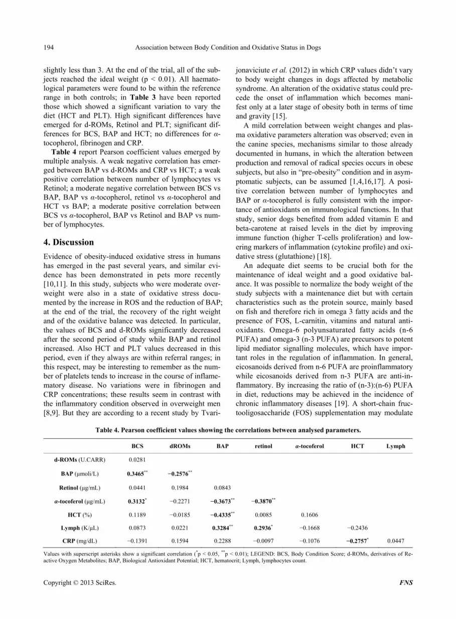
Table 4 report Pearson coefficient values emerged by multiple analysis. A weak negative correlation has emer- ged between BAP vs d-ROMs and CRP vs HCT; a weak positive correlation between number of lymphocytes vs Retinol; a moderate negative correlation between BCS vs BAP, BAP vs Į-tocopherol, retinol vs Į-tocopherol and HCT vs BAP; a moderate positive correlation between BCS vs Į-tocopherol, BAP vs Retinol and BAP vs num- ber of lymphocytes.
4. Discussion Evidence of obesity-induced oxidative stress in humans has emerged in the past several years, and similar evi- dence has been demonstrated in pets more recently [10,11]. In this study, subjects who were moderate over- weight were also in a state of oxidative stress docu- mented by the increase in ROS and the reduction of BAP; at the end of the trial, the recovery of the right weight and of the oxidative balance was detected. In particular, the values of BCS and d-ROMs significantly decreased after the second period of study while BAP and retinol increased. Also HCT and PLT values decreased in this period, even if they always are within referral ranges; in this respect, may be interesting to remember as the num- ber of platelets tends to increase in the course of inflame- matory disease. No variations were in fibrinogen and CRP concentrations; these results seem in contrast with the inflammatory condition observed in overweight men [8,9]. But they are according to a recent study by Tvari-
jonaviciute et al. (2012) in which CRP values didn’t vary to body weight changes in dogs affected by metabolic syndrome. An alteration of the oxidative status could pre- cede the onset of inflammation which becomes mani- fest only at a later stage of obesity both in terms of time and gravity [15].
A mild correlation between weight changes and plas- ma oxidative parameters alteration was observed; even in the canine species, mechanisms similar to those already documented in humans, in which the alteration between production and removal of radical species occurs in obese subjects, but also in “pre-obesity” condition and in asym- ptomatic subjects, can be assumed [1,4,16,17]. A posi- tive correlation between number of lymphocytes and BAP or Į-tocopherol is fully consistent with the impor- tance of antioxidants on immunological functions. In that study, senior dogs benefited from added vitamin E and beta-carotene at raised levels in the diet by improving immune function (higher T-cells proliferation) and low- ering markers of inflammation (cytokine profile) and oxi- dative stress (glutathione) [18].
An adequate diet seems to be crucial both for the maintenance of ideal weight and a good oxidative bal- ance. It was possible to normalize the body weight of the study subjects with a maintenance diet but with certain characteristics such as the protein source, mainly based on fish and therefore rich in omega 3 fatty acids and the presence of FOS, L-carnitin, vitamins and natural anti- oxidants. Omega-6 polyunsaturated fatty acids (n-6 PUFA) and omega-3 (n-3 PUFA) are precursors to potent lipid mediator signalling molecules, which have impor- tant roles in the regulation of inflammation. In general, eicosanoids derived from n-6 PUFA are proinflammatory while eicosanoids derived from n-3 PUFA are anti-in- flammatory. By increasing the ratio of (n-3):(n-6) PUFA in diet, reductions may be achieved in the incidence of chronic inflammatory diseases [19]. A short-chain fruc- tooligosaccharide (FOS) supplementation may modulate
Table 4. Pearson coefficient values showing the correlations between analysed parameters.
BCS dROMs BAP retinol Į-tocoferol HCT Lymph
d-ROMs (U.CARR) 0.0281
BAP (ȝmoli/L) 0.3465** í0.2576**
Retinol (ȝg/mL) 0.0441 0.1984 0.0843
Į-tocoferol (ȝg/mL) 0.3132* í0.2271 í0.3673** í0.3870**
HCT (%) 0.1189 í0.0185 í0.4335** 0.0085 0.1606
Lymph (K/ȝL) 0.0873 0.0221 0.3284** 0.2936* í0.1668 í0.2436
CRP (mg/dL) í0.1391 0.1594 0.2288 í0.0097 í0.1076 í0.2757* 0.0447
Values with superscript asterisks show a significant correlation (*p < 0.05, **p < 0.01); LEGEND: BCS, Body Condition Score; d-ROMs, derivatives of Re- ctive Oxygen Metabolites; BAP, Biological Antioxidant Potential; HCT, hematocrit; Lymph, lymphocytes count. a
Association between Body Condition and Oxidative Status in Dogs 195
insulin resistance and glucose homeostasis in dogs [20]. L-carnitine improves nitrogenous retention and modifies the body composition in favor of lean body mass [21]. Vitamins, A and E in particular, are the most important exogenous antioxidants that act both as preventive anti- oxidants that as chain-breakers [22]. Finally, many vege- tal flavonoids have antiradical and antinflammatory pro- perties [23,34]. The results above discussed can be con- sidered a starting point for further studies aimed at ass- essing the role of oxidative stress in overweight dogs and the dietary intervention with a particular emphasis on the antioxidant addition.
REFERENCES [1] T. Fukui, K. Yamauchi, M. Maruyama, T. Yasuda, M.
Kohno and Y. Abe, “Significance of Measuring Oxidative Stress in Lifestyle-Related Diseases from the Viewpoint of Correlation between d-ROMs and BAP in Japanese Subjects,” Hypertension Research: Official Journal of the Japanese Society of Hypertension, Vol. 34, No. 9, 2011, pp. 1041-1045.
[2] B. Halliwell, “Antioxidants in Human Health and Di- sease,” Annual Review of Nutrition, Vol. 16, 1996, pp. 33-50. doi:10.1146/annurev.nu.16.070196.000341
[3] V. B. Djordjeviü, “Free Radicals in Cell Biology,” Inter- national Review of Cytology, Vol. 237, 2004, pp. 57-89. doi:10.1016/S0074-7696(04)37002-6
[4] K. Kotani and N. Taniguchia, “The Association between Reactive Oxygen Metabolites and Metabolic Syndrome in Asymptomatic Japanese Men,” Journal of Clinical Medi- cine Research, Vol. 3, No. 5, 2011, pp. 247-251.
[5] R. Goyal, M. Singhai and A. F. Faizy, “Glutathione Pero- xidase Activity in Obese and Nonobese Diabetic Patients and Role of Hyperglycemia in Oxidative Stress,” Journal of Mild-Life Health, Vol. 2, No. 2, 2011, pp. 72-76.
[6] D. Venturini, A. N. Simão, N. A. Scripes, L. D. Bahls, P. A. Melo, F. M. Belinetti, M. A. Lozovoy and I. Dichi, “Evaluation of Oxidative Stress in Overweight Subjects with or without Metabolic Syndrome,” Obesity, Vol. 20, No. 12, 2012, pp. 2361-2366. doi:10.1038/oby.2012.130
[7] W. Wonisch, A. Falk, I. Sundl, B. M. Winklhofer-Roob and M. Lindschinger, “Oxidative Stress Increases Conti- nuously with BMI and Age with Unfavourable Profiles in Males,” Aging Male, Vol. 15, No. 3, 2012, pp. 159-165. doi:10.3109/13685538.2012.669436
[8] P. Codoñer-Franch, V. Valls-Bellés, A. Arilla-Codoñer and E. Alonso-Iglesias, “Oxidant Mechanisms in Child- hood Obesity: The Link between Inflammation and Oxi- dative Stress,” Translational Research: The Journal of Laboratory and Clinical Medicine, Vol. 158, No. 6, 2011, pp. 369-384. doi:10.1016/j.trsl.2011.08.004
[9] Y. B. Tripathi and V. Pandey, “Obesity and Endoplasmic Reticulum (ER) Stresses,” Frontiers Inimmunolology, Vol. 3, 2012, p. 240.
[10] A. E. Tanner, J. Martin and K. E. Saker, “Oxidative Stress and Inflammatory State Induced by Obesity in the Heal-
thy Feline,” Journal of Animal Physiology and Animal Nutrition (Berl.), Vol. 91, No. 3-4, 2007, pp. 163-166. doi:10.1111/j.1439-0396.2007.00680_7.x
[11] R. W. Grant, B. M. Vester Boler, T. K. Ridge, T. K. Graves and K. S. Swanson, “Adipose Tissue Transcriptome Changes during Obesity Development in Female Dogs” Physiolo- gical Genomics, Vol. 43, No. 6, 2011, pp. 295-307. doi:10.1152/physiolgenomics.00190.2010
[12] D. J. Laflamme, “Companion Animals Symposium: Obe- sity in Dogs and Cats: What Is Wrong with Being Fat?” Journal of Animal Science, Vol. 90, No, 5, 2012, pp. 1653-1662. doi:10.2527/jas.2011-4571
[13] A. J. German, S. Holden, G. L. Moxham, K. L. Holmes, R. M. Hackett and J. M. Rawlings, “Simple, Reliable Tool for Owners to Assess the Body Condition of Their Dog or Cat,” The Journal of Nutrition, Vol. 136, pp. 2031S- 2033S.
[14] A. Pasquini, E. Luchetti, V. Marchetti, E. L. Iorio and G. Cardini, “Analytical Performances of d-ROMs Test and BAP Test in Canine Plasma. Definition of the Normal Range in Healthy Labrador Dogs,” Veterinary Research Communication, Vol. 32, 2008, pp. 137-143. doi:10.1007/s11259-007-9014-x
[15] A. Tvarijonaviciute, J. J. Ceron, S. L. Holden, D. J. Cuth- bertson, V. Biourge, P. J. Morris and A. J. German, “Obe- sity-Related Metabolic Dysfunction in Dogs: A Compa- rison with Human Metabolic Syndrome BMC,” Veterina- ry Research, Vol. 8, 2012, p. 147.
[16] I. Bondia-Pons, L. Ryan and J. A. Martinez, “Oxidative Stress and Inflammation Interactions in Human Obesity,” Journal of Physiology and Biochemistry, Vol. 68, No. 4, 2012, pp. 701-711. doi:10.1007/s13105-012-0154-2
[17] A. De Lorenzo, V. Del Gobbo, M. G. Premrov, M. Bigi- oni, F. Galvano and L. Di Renzo, “Normal-Weight Obese Syndrome: Early Inflammation?” American Journal of Clinical Nutrition, Vol. 85, 2007, pp. 40-45.
[18] S. Massimino and M. Hayek, “Effect of Supplemental Di- etary Vitamin E and Beta-Carotene on Immune and Oxi- dative Parameters in Young and Old Beagle Dogs,” XVI ESVCP Congress Proceedings, Amsterdam, September 2006, pp. 14-16.
[19] E. Patterson, R. Wall, G. F. Fitzgerald, R. P. Ross and C. Stanton, “Health Implications of High Dietary Omega-6 Polyunsaturated Fatty Acids,” Journal of Nutrition and Metabolism, Vol. 2012, p. 16. http://www.hindawi.com/journals/jnume/2012/539426/
[20] F. Respondek, K. S. Swanson, K. R. Belsito, B. M. Vester, A. Wagner, L. Istasse and M. Diez, “Short-Chain Fructo- oligosaccharides Influence Insulin Sensitivity and Gene Expression of Fat Tissue in Obese Dogs,” Journal of Nu- trition, Vol. 138, 2008, pp. 1712-1718.
[21] K. L. Gross and S. C. Zicker, “L-Carnitine Increases Mu- scle Mass, Bone Mass, and Bone Density in Growing Large Breed Puppies,” Journal of Animal Science, Vol. 78, 2000, p. 176.
[22] B. Halliwell, “Food Derived Antioxidants,” In: E. Cade- nas and L. Packer, Eds., Handbook of Antioxidants, 2nd Edition, CRC Press, New York, 2002, pp. 1-46.
[23] H. S. Chaudhari, U. Bhandari and G. Khanna, “Preventive
Copyright © 2013 SciRes.����������������������������������������������������������������������������������FNS
Association between Body Condition and Oxidative Status in Dogs 196
Effect of Embelin from Embelia Ribes on Lipid Metabo- lism and Oxidative Stress in High-Fat Diet-Induced Obe- sity in Rats,” Planta Medica, Vol. 78, No. 7, 2012, pp. 651-657. doi:10.1055/s-0031-1298379
[24] G. Kaur and C. Meena, “Amelioration of Obesity, Glu- cose Intolerance, and Oxidative Stress in High-Fat Diet
and Low-Dose Streptozotocin-Induced Diabetic Rats by Combination Consisting of ‘Curcumin with Piperine and Quercetin’,” ISRN Pharmacology, Vol. 2012, 2012, p. 7. http://www.hindawi.com/isrn/pharmacology/2012/957283/cta/
Abbreviations
BAP: Biological Antioxidant Potential; BCS: Body Condition Score; BMI: Body Mass Index; CBCs: Complete Blood Counts; CRP: C Reactive Protein; d-ROMs: Derivatives of Reactive Oxygen Metabolites;
FOS: Short-Chain Fructooligosaccharides; HCT: Hematocrit; OS: Oxidative Stress; PLT: Platelets; PUFA: Polyunsaturated Fatty Acids; ROS: Reactive Oxygen Species.
Copyright © 2013 SciRes.����������������������������������������������������������������������������������FNS


