Embed Size (px)
Citation preview
Page 1/24
Assessment of biosafety and toxicity of hydrophilicgel for implantation in experimental in vitro and invivo models Natalia Bezdieniezhnykh ( [email protected] )
RE. Kavetsky Institute of Experimental Pathology, Oncology and Radiobiology, NAS of UkraineAlexandra Lykhova
RE. Kavetsky Institute of Experimental Pathology, Oncology and Radiobiology, NAS of UkraineTamara Kozak
RE. Kavetsky Institute of Experimental Pathology, Oncology and Radiobiology, NAS of UkraineTaras Zadvornyi
RE. Kavetsky Institute of Experimental Pathology, Oncology and Radiobiology, NAS of UkraineOlena Voronina
RE. Kavetsky Institute of Experimental Pathology, Oncology and Radiobiology, NAS of UkraineNatalia Lukyanova
RE. Kavetsky Institute of Experimental Pathology, Oncology and Radiobiology, NAS of Ukraine
Research Article
Keywords: hydrophilic gel for implantation, experimental model systems, toxicity, genotoxicity, in vitro andin vivo studies
Posted Date: May 14th, 2021
DOI: https://doi.org/10.21203/rs.3.rs-435527/v1
License: This work is licensed under a Creative Commons Attribution 4.0 International License. Read Full License
Page 2/24
AbstractBackground: The assessment of biosafety of pharmacologically active substances is crucial fordetermining the feasibility of their medical use. There are controversial issues regarding the use ofsubstances of different origins as implants. Methods: We have conducted the comprehensive studies todetermine the in vivo toxicity and in vitro genotoxicity of new generation of hydrophilic gel forimplantation (production name of the substance "Activegel") to detail its characteristics and assess itsbiosafety. Results: In vivo studies have shown the absence of clinical manifestations of intoxication inanimals and no abnormalities in their physiological condition, general and biochemical blood tests.Evaluation of the site of the gel application showed no in�ammatory reaction and evidenced on normalstate of tissues of animal skin. The results of the genotoxicity test indicated that the gel did not affect theparameters of DNA comets and, accordingly, had no genotoxic effect on human peripheral bloodlymphocytes. When studying the effect of the gel on malignantly transformed cells in vitro, it was foundthat the gel for implantation did not change the proliferative activity and viability of human breast cancercells. Conclusions: Comprehensive in vitro and in vivo study using various experimental model systemsshowed that the hydrophilic gel for implantation "Activegel" is non-toxic.
BackgroundIt is known that toxicological research is now mandatory among the most important steps in thebiological assessment of potential drugs and medical devices, as it allows to identify possible adverseeffects of the test substance on the body and prevent side effects. Such studies include both in vitroexperiments and in vivo toxicity testing [1, 2]. Due to such a comprehensive approach the use of a newpharmacological substance in clinical practice is justi�ed, the doses are adequate, and the mode ofapplication is safe. In particular, the aim of preclinical toxicological studies of a pharmacologicalsubstance in the in vivo system is to establish the nature and severity of its potential harmful effects onthe body of experimental animals and to assess its safety [3].
Therefore, it is extremely important to control the means used for implantation, because today there is alot of controversial problematic issues regarding the use of such substances and their safety in humans[4]. Such substances are used not only for implantation to compensate for soft tissue insu�ciency, toincrease the volume of the mammary glands, correction of soft tissues due to aging, but also to eliminatecertain postoperative and post-traumatic soft tissue deformities, and scars due to injury. It is especiallyimportant to control the substances used in cancer patients, in particular after surgery to correct defects[5, 6].
In the treatment of cancer patients, one of the most common and important methods is chemotherapy,but there are many problems regarding the use of many antitumor agents, including their hydrophobicity,low bioavailability, instability, high toxicity, severe side effects, and lack of targeted action [7- 9].Therefore, an important area is the development and evaluation of new delivery systems for anticancerdrugs for the treatment of tumors with higher e�ciency and lower toxicity. In particular, local application
Page 3/24
of cancer drugs could minimize their toxic effects on normal tissues with maximum effectiveness againstmalignantly transformed cells [10-12]. The hydrogel-based drug delivery system is currently attractingmore and more attention. Due to its liquid structure, hydrogel could be easyly combined with medicinalpreparations for subsequent targeted delivery [13].
The universality of hydrogels is determined by their structure, which could be technically regulated for thepurposes of invention of new polymers, new approaches to their crosslinking and new strategies for theirproduction [14]. But it should be understood that such new approaches to translational biomedicine invivo as the use of polymers as vectors for the delivery of bioactive molecules require proper control oftheir toxicity and safety for the human body [15].
In this work, we have investigated a synthetic polymer (2-4%) - new generation hydrophilic gel with theproduction name "Activegel". In particular, we have evaluated in vivo toxicity of hydrogel using variousexperimental model systems and its effect on tumor cells in vitro to understand the possibleconsequences of its potential use as an implant material or further expansion of its usage in clinicalpractice, in particular, as a vector for targeted delivery of anticancer preparations.
Materials And MethodsTest substance
Sterile medical device "Activegel" is a sterile 2% - 4% mixture of synthetic polymer (new generationhydrophilic gel, produced by LLC "National Center for Medical Technology", Ukraine; packaging: ContainerPP (MAGIFLEX BAG PVC-free) with a volume of 100 ml), designed for use after invasive surgicalprocedures in order to obtain long-term results.
Invivo study
The study was designed to achieve the objectives of the experiment using the minimum number ofanimals. All experiments were conducted in accordance with bioethics standards and permission fromthe Commission on Bioethics of RE Kavetsky IEPOR of NAS of Ukraine. Wistar rats of both sexes 2-2.5months old (males weighing 170-210 g, and females weighing 150-180 g) were used. The conditions forkeeping the animals met the standards speci�ed in the manual The Guide for Care and Use of LaboratoryAnimals (ILAR publication, 1996, National Academy Press, 1996).
In order to study the acute toxicity of the medical product "Activegel", the substance was administered toexperimental animals subcutaneously (s.c.). The animals of each sex were distributed in four groups: 3experimental and 1 control. The results were analyzed 48 hours and 14 days after administration of thetest substance. In the study 3 doses of the product "Activegel" were used (according toENV/JM/MONO(2016)32 No. 237 "Guidance document on considerations for waiving or bridging ofmammalian acute toxicity tests"): 1) low - 500 mg/kg; 2) medium - 2000 mg/kg; 3) high - 5000 mg/kg.
Page 4/24
Animals of the control group received s.c.administration of saline solution (Yuriya-Pharm, Ukraine, S.AA8175/1-1) in a volume corresponding to the highest of the tested doses of the preparation.
48 hours and 14 days after injection of Activegel, the animals were humanely killed by overdose byinhalation anesthesia, and subsequent studies (macroscopic and cytomorphological examination oforgans and tissues, general and biochemical blood tests) were performed.
Macroscopic study of the internal organs of experimental animals. A macroscopic analysis of thecondition of rat skin tissues at the location of the "Actigel" application, as well as the state of the internalorgans of rats (brain, heart, lungs, liver, kidneys, thymus, spleen, testicles and appendages, uterus, ovariesand appendages) was conducted. The size of the organs, their color, shape and weight were evaluated.
General and biochemical analysis of peripheral blood of experimental animals. Blood samples weredrawn from the retroorbital venous sinus, and used for general and biochemical analysis. The cellularcomposition of peripheral blood (erythrocytes, leukocytes, band neutrophils, segmented neutrophils,lymphocytes, monocytes, eosinophils), hemoglobin, erythrocyte sedimentation rate (ESR), as well asbiochemical blood serum indices (concentration of total bilirubin, calcium, creatinine, glucose, totalprotein, urea, C-reactive protein, inorganic phosphorus, as well as the activity of ALT and AST) werequanti�ed on an automatic biochemical analyzer ChemWell 2310 according to the instructions of themanufacturer of the device and the corresponding reagents and using software for GBG ChemWell®.Quality control of laboratory tests was tested using control serum with known normal and abnormalvalues of these indicators (control serum level 1 and 2 Global Scienti�c). The control values were withinthe set range.
Morphological examination of rat skin tissues from the injection site. To carry out cytomorphologicalanalysis, skin, muscle and subcutaneous fat at the location of "Actigel" application were removed andanalyzed. Tissue samples were �xed in 10% neutral buffered formalin solution. After standardhistological conduction in alcohols of increasing concentration, the material was poured into para�n.Sections 4-5 μm thick were made from para�n blocks and stained with hematoxylin and eosin.
Invitro experiments
Cell сultivation. Human breast cancer cells of MCF-7 and MDA-MB-231 lines were obtained from the Bankof Cell Lines from Human and Animal Tissues of the RE Kavetsky IEPOR NASU. The cells were cultured incomplete DMEM medium with 10% fetal calf serum (FCS) in a humidi�ed atmosphere with 5% CO2 at
370C. Replacement of the medium and cell passaging were performed according to standard methods.The cells were passaged after reaching 80% con�uence at a density of 105 cells/ml using Versenesolution. Samples of the test substance were added to the wells of a 12-well plate (0.4 g/well), whilesaline solution was added to the control wells (negative control). Next, 2 ml of complete DMEM mediumwith 10% FCS were added to all wells, and the plate was incubated in the presence of 5% CO2 at 370C and100% relative humidity for 48-96 hours. Then the conditioned media from each well were collected andstored at + 4 ° C.

Page 5/24
Cells of MCF-7 and MDA-MB-231 lines were seeded in wells of a 96-well plate in DMEM medium with 10%FCS (1x10⁵ cells/ml) in a volume of 150 μl/well and cultured for 24 hours. Then the conditioned mediawere added to the appropriate wells of a 96-well plate (150 μl) in different ratios: 100%, 50%, 25%, 12.5%,6.3%, 3.1%, 1.6% (at least three replicates per each concentration). The conditioned media of the cellscultivated after addition of saline solution served as negative control. Then the cells were cultured foranother 72 hours, and the number of living cells was assessed visually (direct microscopy), and using colorimetric method by staining the cells with crystal violet [16]. The results were recorded using aspectrophotometer (Labsystems Multiskan PLUS, Finland) with a vertical beam path at an excitationwavelength of 540 nm.
The number of living cells (X1) in each well of the test sample, as a percentage, was calculated by theformula:
See formula 1 in the supplementary �les section.
where A0 is the average value of the optical density in the control wells; A1 is the average value of theoptical density in the wells with the test sample.
The average number of living cells for each test sample dilution was calculated.
Evaluation of in vitro genotoxicity using comet DNA method. The study was performed in vitro on amodel of human peripheral blood lymphocytes. Peripheral blood samples (9 ml) were taken from 4healthy donors (were informed about the study and gave written consent to the use of biological materialfor research purposes) (2 males aged 29 and 45 years, and 2 females aged 29 and 33 years).
Lymphocytes were isolated from whole peripheral blood using the method of Boyum [17]. Blood twicediluted with saline was layered on a solution of Histopaque in a ratio of 3:1, and centrifuged for 15minutes in a centrifuge with a horizontal rotor at a speed of 1500 rpm. Then an interphase ring containinglymphocytes was collected and washed with 7-10 ml of saline. Next, the cells were pelleted bycentrifugation for 10 min at 1500 rpm, resuspended in saline and the number of cells in the resultingsuspension was determined using a Goryaev chamber by staining them with 0.4% trypan blue solution.The isolated cells were seeded in the wells of a 6-well plate in RPMI 1640 medium with 10% ECT and 40μg/ml gentamicin at a concentration of 1.5x10⁶ cells/ml, 3 ml per well (4.5x10⁶ cells/well). Inserts forculturing with a pore size of 0.4 nm were placed in all wells of the plates at once and 2 ml of RPMI 1640medium with 10% ECS and 40 μg/ml gentamicin were added to the inserts. Next, “Activegel” was added tothe corresponding inserts in a quantity of 0.2 g per 1 ml of nutrient medium. The cells with the testsubstance were incubated in a CO2 incubator for another 96 hours. After incubation, the cells werewashed twice with saline by centrifugation at 1500 rpm for 10 minutes. The resulting cell suspension wasimmediately used in the DNA comets assay.
Gel electrophoresis of single cells (DNA comet assay). The DNA comet assay was performed in analkaline modi�cation developed by Singh and colleagues [18]. The �rst layer of gel contained a 1%
Page 6/24
solution of high melting agarose, pre-treated overnight in a thermostat at 37 0C. 200 μl of the cellsuspension (5 x 10⁵ cells/ml) were mixed with 200 μl of 1% low melting agarose at 37 °C. Thissuspension was applied on the preheated glass slide coated with high-melting agarose and kept at + 4 ° Cuntil the gel was completely solidi�ed. Cell lysis was performed in lysis buffer (2.5 M NaCl, 0.1 M EDTA,10 mM Tris pH 10, 10% DMSO, 1% Triton X-100) at +40С for 1 hour. Before electrophoresis, the slides werewashed with cold water and kept for 20 minutes in electrophoretic buffer (0.3 M NaOH, 1 mM Na2EDTA).Electrophoresis was performed at ≈ 0.8 V/cm (distance between electrodes 27 cm, voltage 24 V, current300 mA for 30 minutes). Neutralization was performed in appropriate buffer (0.4 M Trizma base, pH 7.5)for 10 minutes.
Analysis of DNA comets.
After drying, the samples were stained with acridine orange (2 μg/ml) and SYBR, 50-100 μl per sample.Microscopic analysis of micropreparations was performed using a �uorescence microscope (AxiostarPlus Microscope (Carl Zeiss, Germany), Digital Camera (Canon powershot G5, UK), �uorescent lamp (CarlZeiss, Germany)). At least 50 DNA comets per micropreparation were analyzed. Digital images wereanalyzed using the computer program CometScore. The parameters of the length of the comet's tail, themoment of the tail, as well as the proportion of DNA in the comet's tail were calculated.
Statistical analysis.
Calculations of the mean values of the studied indices (M), standard deviation (SD) and standard error ofthe mean (m) were performed using Excel 2016 software package and Medstatistic program usingStudent's t-test; differences with a probability of at least 95% (p < 0.05) were considered signi�cant. Thenonparametric Mann-Whitney U-test was used to assess the signi�cance levels of the differencesbetween the mean values between the groups. The calculations were performed using the softwarepackage STATISTICA 6.0. Differences were considered signi�cant at p < 0.05.
Results And DiscussionThe �rst step was to assess the acute toxicity of the medical product "Activegel" in vivo to understand andcon�rm the data on the test substance (stated by the manufacturer) on the absence of its toxicity. Duringthe experiment, the survival and physiological parameters of rats (males and females) in the control andexperimental groups were analyzed. During 48 hours (stage I) and 14 days (stage II) we have analyzedlocomotor and respiratory activity of experimental animals, the condition of fur, mucous membranes,behavior, usage of food and water, and gastrointestinal function.
We have found that the highest of the studied doses of “Activegel” (5000 mg/kg) did not cause the deathof animals. In the control and all experimental groups of male rats within 48 and 14 days after the 1stinjection of the test substance, no deviations in the studied indices of the physiological state wereobserved. However, immediately after injection of saline or test substance and for the next 15 minutes in50-100% of the animals the signs of aggression were observed. Since aggressive behavior of animals
Page 7/24
was observed in both control and experimental groups, it is clear that it was not a reaction to the testsubstance, but to the procedure of forced �xation of rats for injection procedure. In groups of female rats,which were administered different doses of "Activegel" or saline, no violations of the physiological stateof the experimental animals were observed during the entire observation period.
Analysis of the dynamics of body weight changes in rats within 48 hours and 14 days after singlesubcutaneous injection of "Activegel" at different doses did not reveal statistically signi�cant differencesin the studied index compared with the control group: animal body weight in the experimental groupsincreased as well as in the control.
Macroscopic examination of the condition of the internal organs of experimental animals and thecondition of the tissues at the injection site after single subcutaneous administration of "Activegel" atdifferent doses showed that it did not affect the internal organs of the animals.
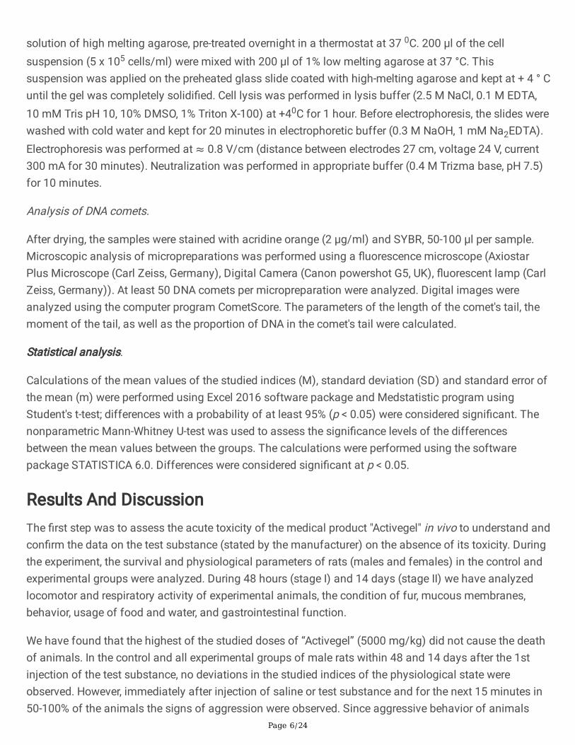
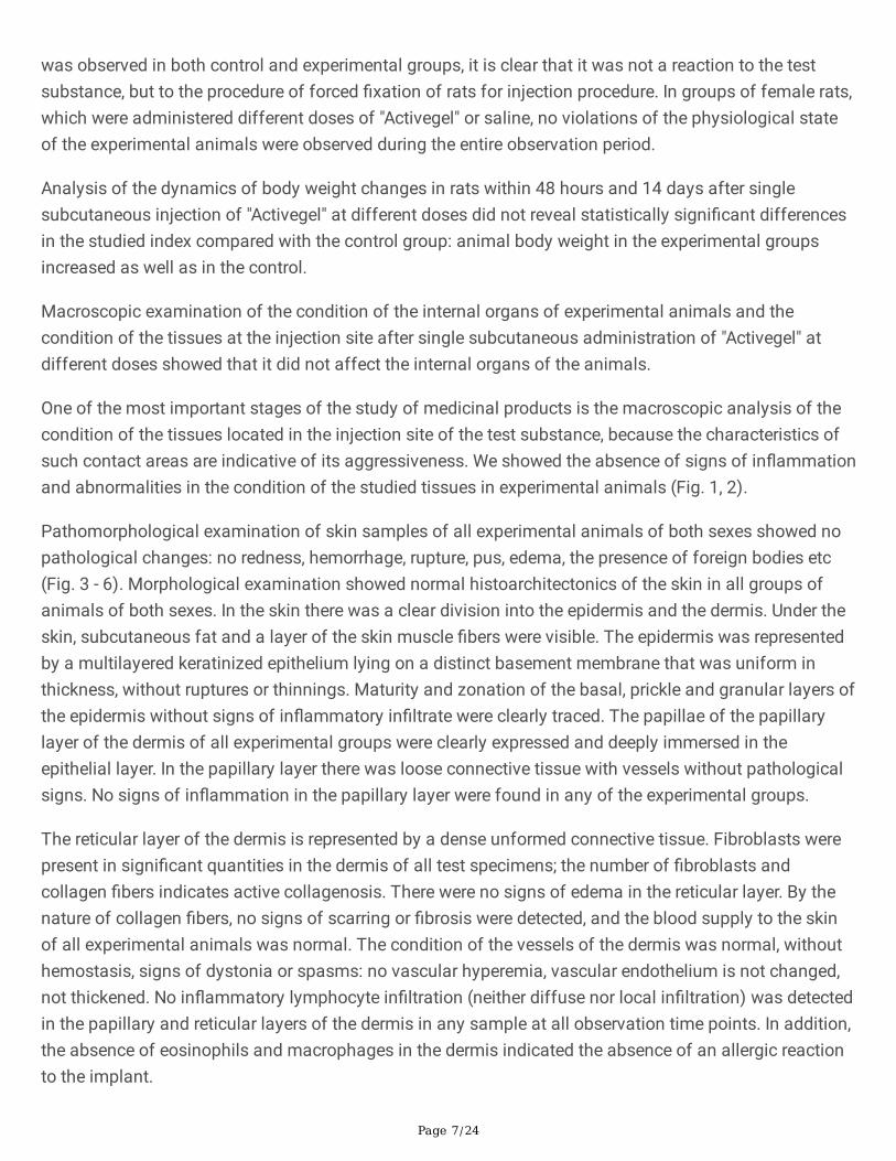
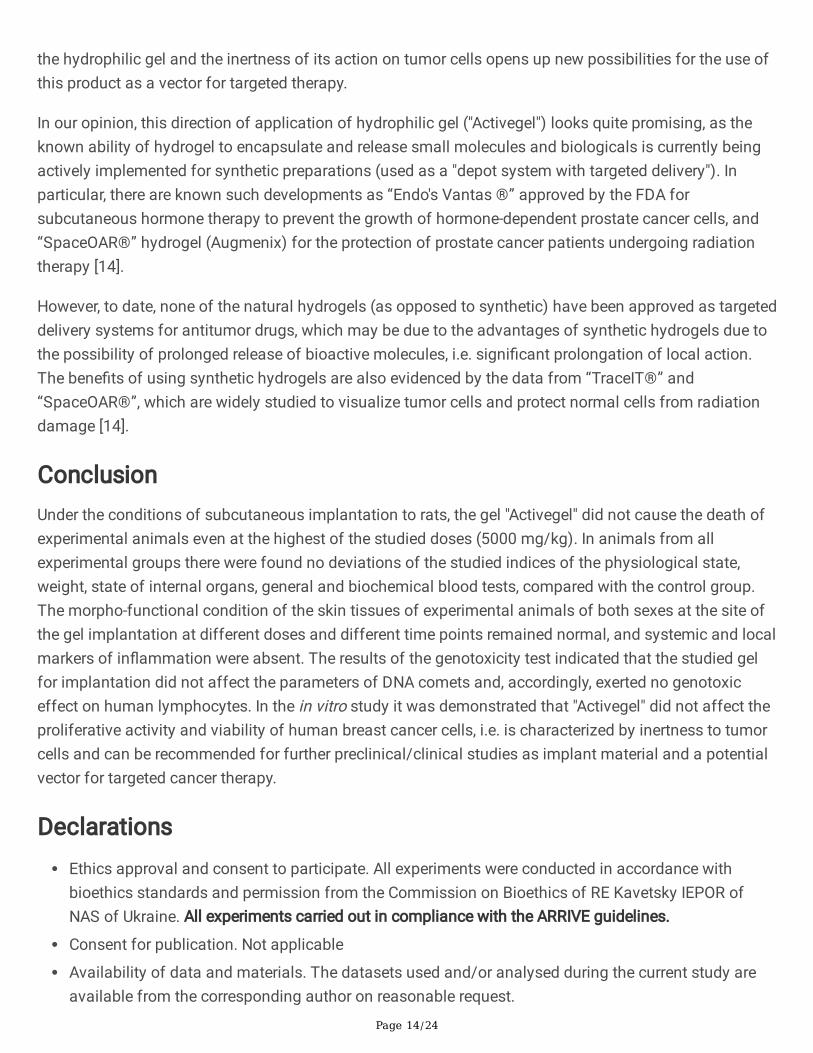
One of the most important stages of the study of medicinal products is the macroscopic analysis of thecondition of the tissues located in the injection site of the test substance, because the characteristics ofsuch contact areas are indicative of its aggressiveness. We showed the absence of signs of in�ammationand abnormalities in the condition of the studied tissues in experimental animals (Fig. 1, 2).
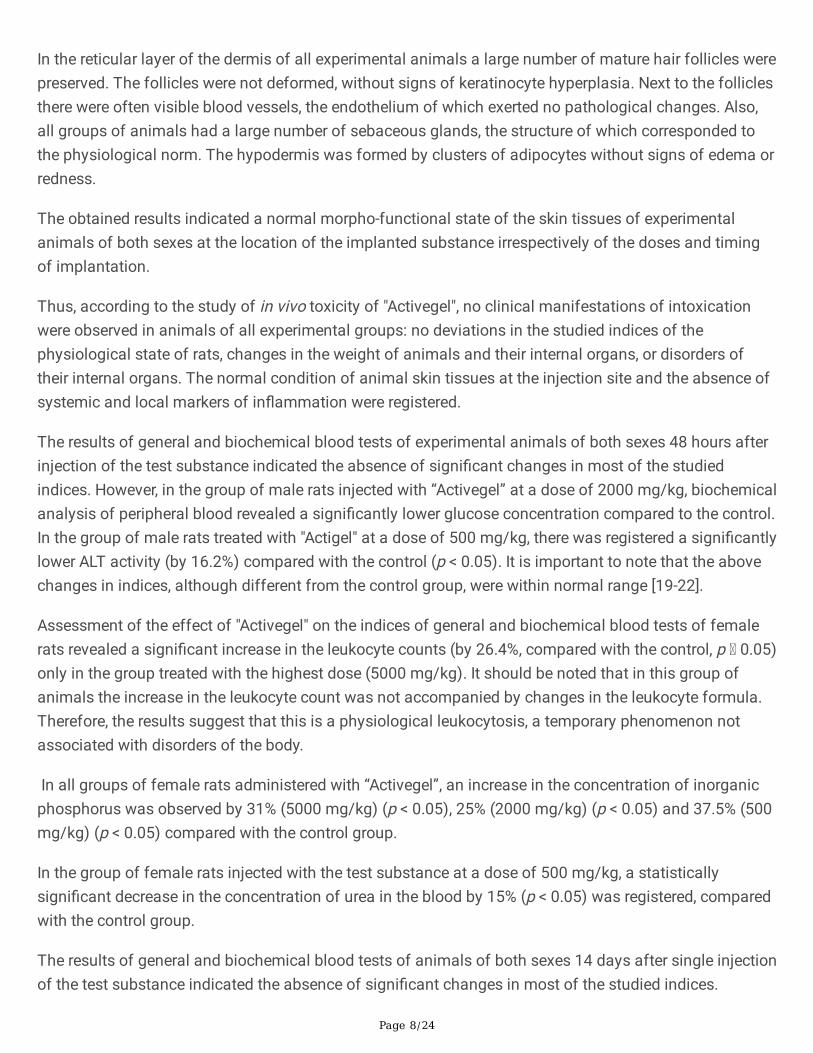
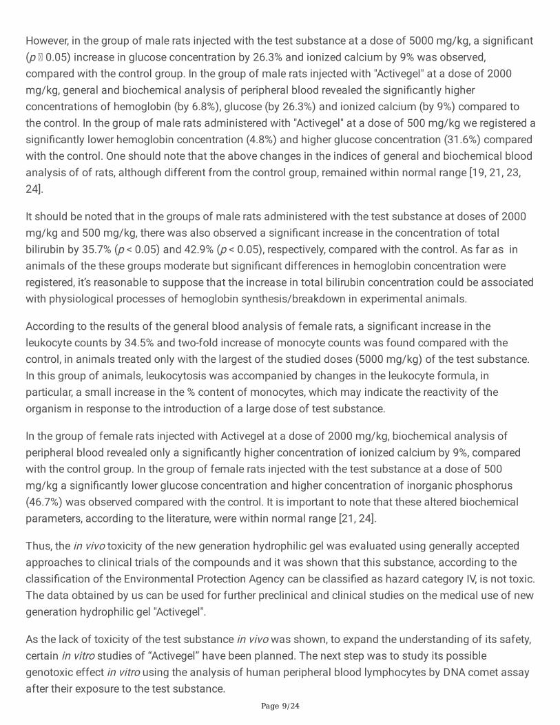
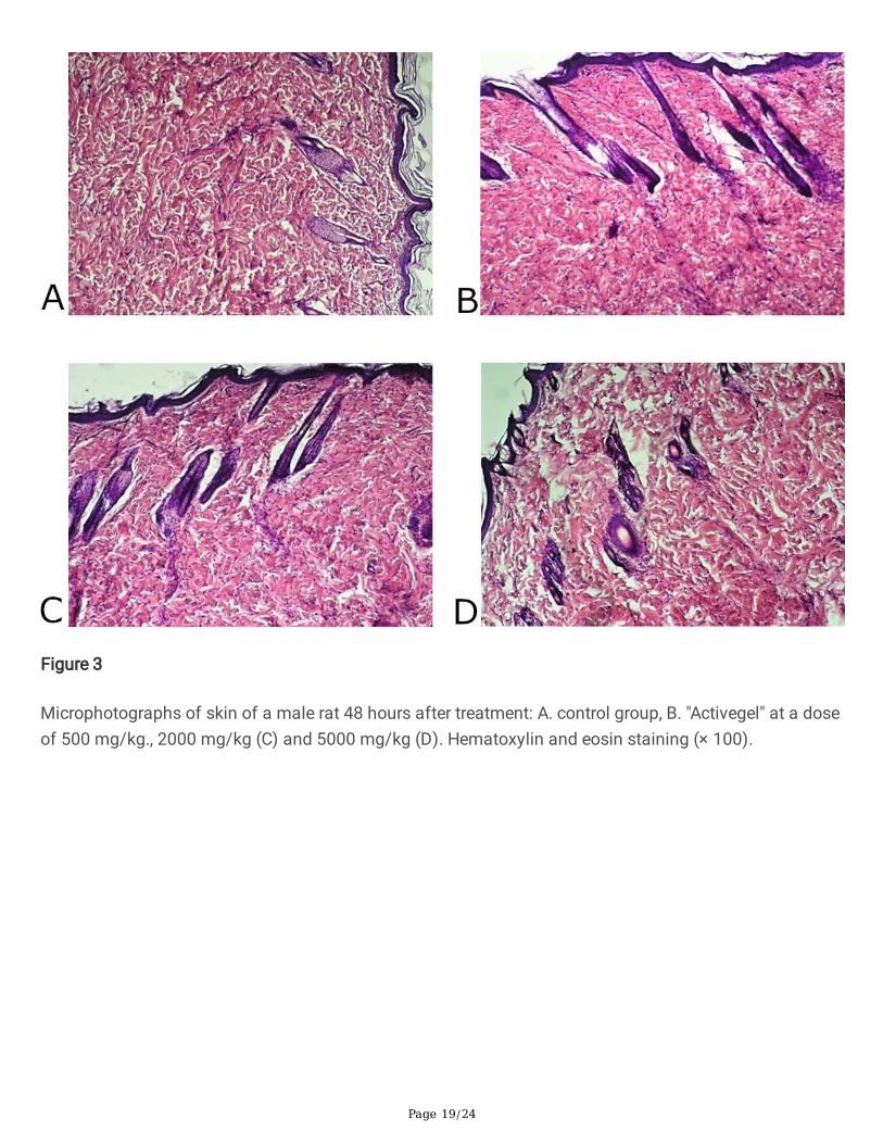
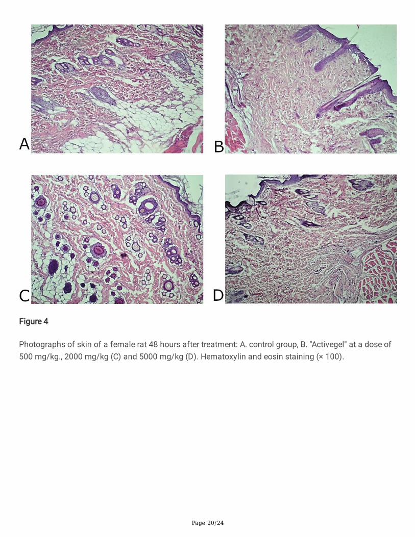
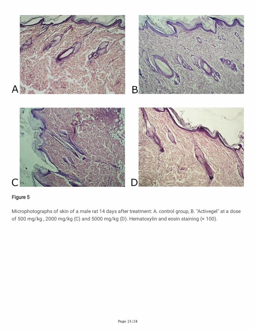
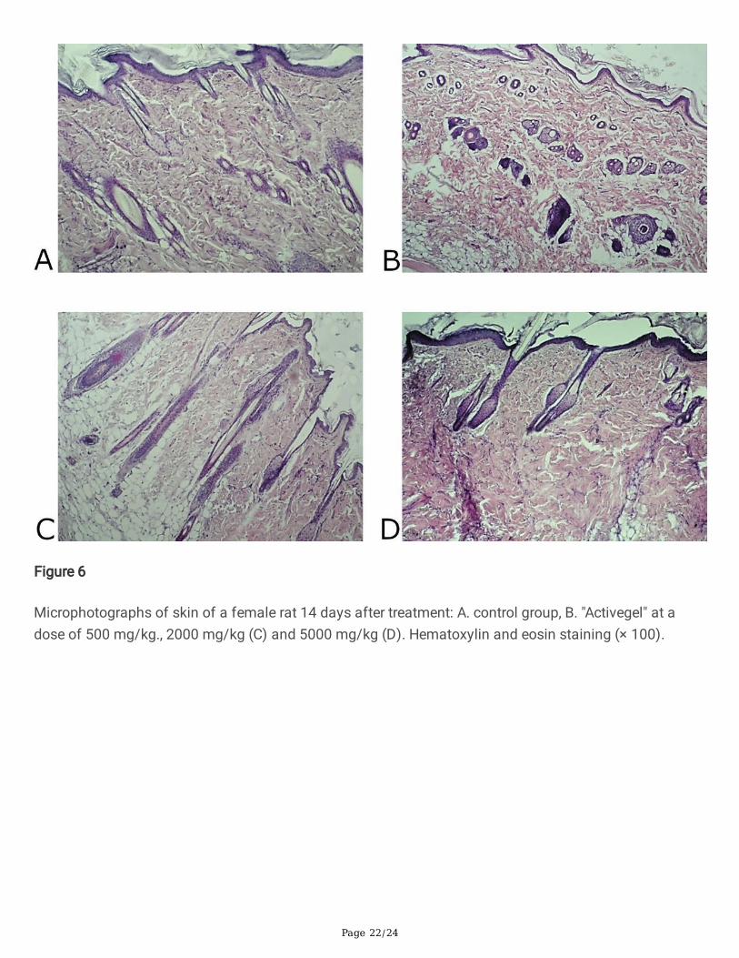
Pathomorphological examination of skin samples of all experimental animals of both sexes showed nopathological changes: no redness, hemorrhage, rupture, pus, edema, the presence of foreign bodies etc(Fig. 3 - 6). Morphological examination showed normal histoarchitectonics of the skin in all groups ofanimals of both sexes. In the skin there was a clear division into the epidermis and the dermis. Under theskin, subcutaneous fat and a layer of the skin muscle �bers were visible. The epidermis was representedby a multilayered keratinized epithelium lying on a distinct basement membrane that was uniform inthickness, without ruptures or thinnings. Maturity and zonation of the basal, prickle and granular layers ofthe epidermis without signs of in�ammatory in�ltrate were clearly traced. The papillae of the papillarylayer of the dermis of all experimental groups were clearly expressed and deeply immersed in theepithelial layer. In the papillary layer there was loose connective tissue with vessels without pathologicalsigns. No signs of in�ammation in the papillary layer were found in any of the experimental groups.
The reticular layer of the dermis is represented by a dense unformed connective tissue. Fibroblasts werepresent in signi�cant quantities in the dermis of all test specimens; the number of �broblasts andcollagen �bers indicates active collagenosis. There were no signs of edema in the reticular layer. By thenature of collagen �bers, no signs of scarring or �brosis were detected, and the blood supply to the skinof all experimental animals was normal. The condition of the vessels of the dermis was normal, withouthemostasis, signs of dystonia or spasms: no vascular hyperemia, vascular endothelium is not changed,not thickened. No in�ammatory lymphocyte in�ltration (neither diffuse nor local in�ltration) was detectedin the papillary and reticular layers of the dermis in any sample at all observation time points. In addition,the absence of eosinophils and macrophages in the dermis indicated the absence of an allergic reactionto the implant.
Page 8/24
In the reticular layer of the dermis of all experimental animals a large number of mature hair follicles werepreserved. The follicles were not deformed, without signs of keratinocyte hyperplasia. Next to the folliclesthere were often visible blood vessels, the endothelium of which exerted no pathological changes. Also,all groups of animals had a large number of sebaceous glands, the structure of which corresponded tothe physiological norm. The hypodermis was formed by clusters of adipocytes without signs of edema orredness.
The obtained results indicated a normal morpho-functional state of the skin tissues of experimentalanimals of both sexes at the location of the implanted substance irrespectively of the doses and timingof implantation.
Thus, according to the study of in vivo toxicity of "Activegel", no clinical manifestations of intoxicationwere observed in animals of all experimental groups: no deviations in the studied indices of thephysiological state of rats, changes in the weight of animals and their internal organs, or disorders oftheir internal organs. The normal condition of animal skin tissues at the injection site and the absence ofsystemic and local markers of in�ammation were registered.
The results of general and biochemical blood tests of experimental animals of both sexes 48 hours afterinjection of the test substance indicated the absence of signi�cant changes in most of the studiedindices. However, in the group of male rats injected with “Activegel” at a dose of 2000 mg/kg, biochemicalanalysis of peripheral blood revealed a signi�cantly lower glucose concentration compared to the control.In the group of male rats treated with "Actigel" at a dose of 500 mg/kg, there was registered a signi�cantlylower ALT activity (by 16.2%) compared with the control (р < 0.05). It is important to note that the abovechanges in indices, although different from the control group, were within normal range [19-22].
Assessment of the effect of "Activegel" on the indices of general and biochemical blood tests of femalerats revealed a signi�cant increase in the leukocyte counts (by 26.4%, compared with the control, p 0.05)only in the group treated with the highest dose (5000 mg/kg). It should be noted that in this group ofanimals the increase in the leukocyte count was not accompanied by changes in the leukocyte formula.Therefore, the results suggest that this is a physiological leukocytosis, a temporary phenomenon notassociated with disorders of the body.
In all groups of female rats administered with “Activegel”, an increase in the concentration of inorganicphosphorus was observed by 31% (5000 mg/kg) (р < 0.05), 25% (2000 mg/kg) (р < 0.05) and 37.5% (500mg/kg) (р < 0.05) compared with the control group.
In the group of female rats injected with the test substance at a dose of 500 mg/kg, a statisticallysigni�cant decrease in the concentration of urea in the blood by 15% (р < 0.05) was registered, comparedwith the control group.
The results of general and biochemical blood tests of animals of both sexes 14 days after single injectionof the test substance indicated the absence of signi�cant changes in most of the studied indices.
Page 9/24
However, in the group of male rats injected with the test substance at a dose of 5000 mg/kg, a signi�cant(р 0.05) increase in glucose concentration by 26.3% and ionized calcium by 9% was observed,compared with the control group. In the group of male rats injected with "Activegel" at a dose of 2000mg/kg, general and biochemical analysis of peripheral blood revealed the signi�cantly higherconcentrations of hemoglobin (by 6.8%), glucose (by 26.3%) and ionized calcium (by 9%) compared tothe control. In the group of male rats administered with "Activegel" at a dose of 500 mg/kg we registered asigni�cantly lower hemoglobin concentration (4.8%) and higher glucose concentration (31.6%) comparedwith the control. One should note that the above changes in the indices of general and biochemical bloodanalysis of of rats, although different from the control group, remained within normal range [19, 21, 23,24].
It should be noted that in the groups of male rats administered with the test substance at doses of 2000mg/kg and 500 mg/kg, there was also observed a signi�cant increase in the concentration of totalbilirubin by 35.7% (р < 0.05) and 42.9% (р < 0.05), respectively, compared with the control. As far as inanimals of the these groups moderate but signi�cant differences in hemoglobin concentration wereregistered, it’s reasonable to suppose that the increase in total bilirubin concentration could be associatedwith physiological processes of hemoglobin synthesis/breakdown in experimental animals.
According to the results of the general blood analysis of female rats, a signi�cant increase in theleukocyte counts by 34.5% and two-fold increase of monocyte counts was found compared with thecontrol, in animals treated only with the largest of the studied doses (5000 mg/kg) of the test substance.In this group of animals, leukocytosis was accompanied by changes in the leukocyte formula, inparticular, a small increase in the % content of monocytes, which may indicate the reactivity of theorganism in response to the introduction of a large dose of test substance.
In the group of female rats injected with Activegel at a dose of 2000 mg/kg, biochemical analysis ofperipheral blood revealed only a signi�cantly higher concentration of ionized calcium by 9%, comparedwith the control group. In the group of female rats injected with the test substance at a dose of 500mg/kg a signi�cantly lower glucose concentration and higher concentration of inorganic phosphorus(46.7%) was observed compared with the control. It is important to note that these altered biochemicalparameters, according to the literature, were within normal range [21, 24].
Thus, the in vivo toxicity of the new generation hydrophilic gel was evaluated using generally acceptedapproaches to clinical trials of the compounds and it was shown that this substance, according to theclassi�cation of the Environmental Protection Agency can be classi�ed as hazard category IV, is not toxic.The data obtained by us can be used for further preclinical and clinical studies on the medical use of newgeneration hydrophilic gel "Activegel".
As the lack of toxicity of the test substance in vivo was shown, to expand the understanding of its safety,certain in vitro studies of “Activegel” have been planned. The next step was to study its possiblegenotoxic effect in vitro using the analysis of human peripheral blood lymphocytes by DNA comet assayafter their exposure to the test substance.
Page 10/24
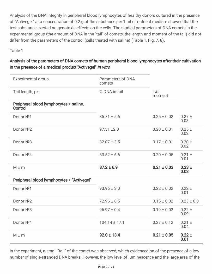
Analysis of the DNA integrity in peripheral blood lymphocytes of healthy donors cultured in the presenceof “Activegel” at a concentration of 0.2 g of the substance per 1 ml of nutrient medium showed that thetest substance exerted no genotoxic effects on the cells. The studied parameters of DNA comets in theexperimental group (the amount of DNA in the "tail" of comets, the length and moment of the tail) did notdiffer from the parameters of the control (cells treated with saline) (Table 1, Fig. 7, 8).
Table 1
Analysis of the parameters of DNA comets of human peripheral blood lymphocytes after their cultivationin the presence of a medical product "Activegel" in vitro
Experimental group Parameters of DNAcomets
Tail length, px % DNA in tail Tailmoment
Peripheral blood lymphocytes + saline,Control
Donor №1 85.71 ± 5.6 0.25 ± 0.02 0.27 ±0.03
Donor №2 97.31 ±2.0 0.20 ± 0.01 0.25 ±0.02
Donor №3 82.07 ± 3.5 0.17 ± 0.01 0.20 ±0.02
Donor №4 83.52 ± 6.6 0.20 ± 0.05 0.21 ±0.01
М ± m 87.2 ± 6.9 0.21 ± 0.03 0.23 ±0.03
Peripheral blood lymphocytes + “Activegel”
Donor №1 93.96 ± 3.0 0.22 ± 0.02 0.22 ±0.01
Donor №2 72.96 ± 8.5 0.15 ± 0.02 0.23 ± 0.0
Donor №3 96.97 ± 0.4 0.19 ± 0.02 0.22 ±0.09
Donor №4 104.14 ± 17.1 0.27 ± 0.12 0.21 ±0.04
М ± m 92.0 ± 13.4 0.21 ± 0.05 0.22 ±0.01
In the experiment, a small "tail" of the comet was observed, which evidenced on of the presence of a lownumber of single-stranded DNA breaks. However, the low level of luminescence and the large area of the
Page 11/24
"tail" of the comet are characteristic of cases where nucleic acid breaks can be attributed to "junk DNA",which is a variant of the norm. Therefore, these results indicated that "Activegel" in the studiedconcentration (0.2 g/ml) did not affect the parameters of DNA comets, ie the test substance exerted nogenotoxic effect on the cells.
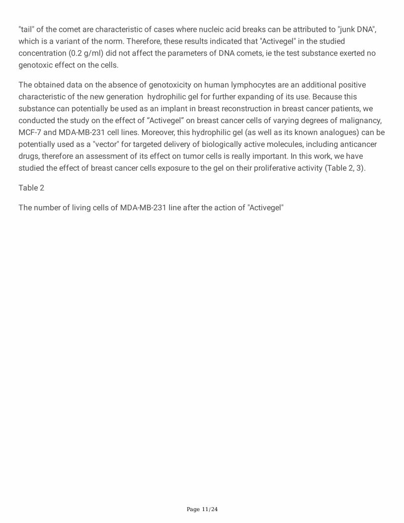
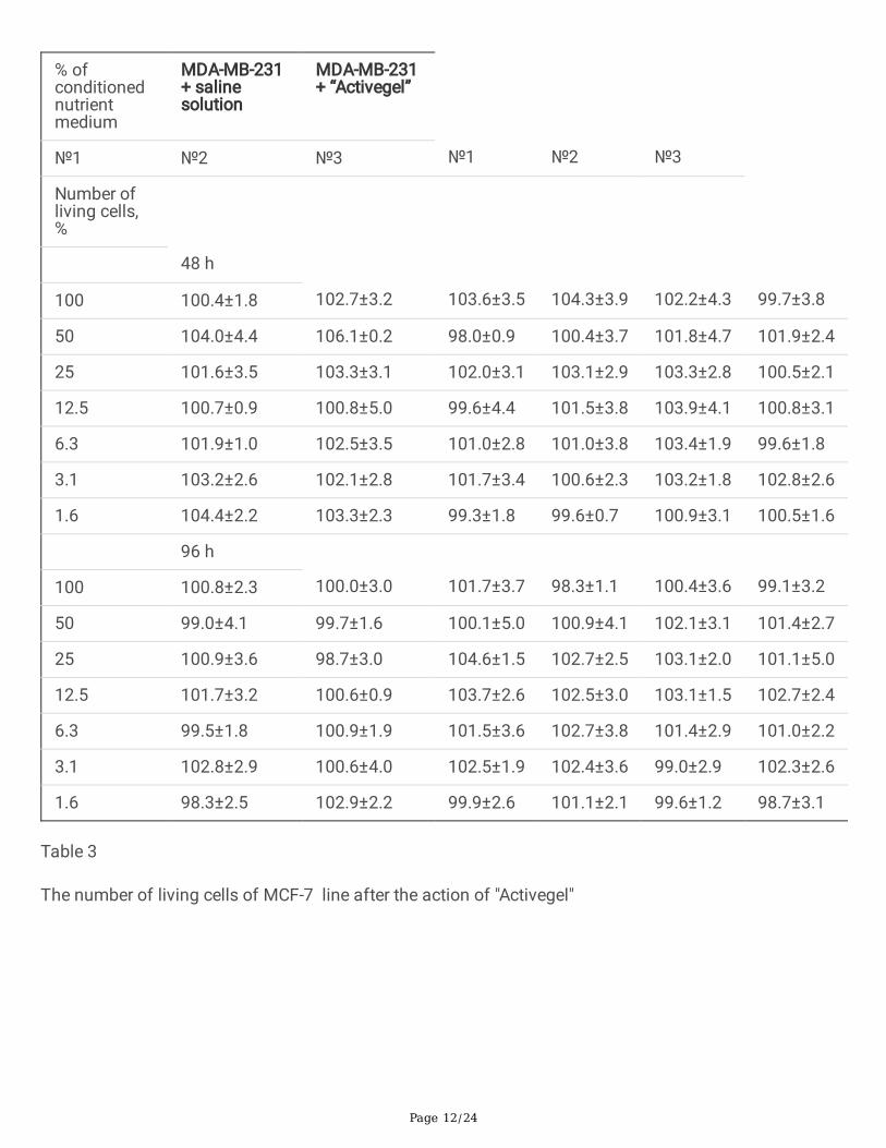
The obtained data on the absence of genotoxicity on human lymphocytes are an additional positivecharacteristic of the new generation hydrophilic gel for further expanding of its use. Because thissubstance can potentially be used as an implant in breast reconstruction in breast cancer patients, weconducted the study on the effect of “Activegel” on breast cancer cells of varying degrees of malignancy,MCF-7 and MDA-MB-231 cell lines. Moreover, this hydrophilic gel (as well as its known analogues) can bepotentially used as a "vector" for targeted delivery of biologically active molecules, including anticancerdrugs, therefore an assessment of its effect on tumor cells is really important. In this work, we havestudied the effect of breast cancer cells exposure to the gel on their proliferative activity (Table 2, 3).
Table 2
The number of living cells of MDA-MB-231 line after the action of "Activegel"
Page 12/24
% ofconditionednutrientmedium
MDA-MB-231+ salinesolution
MDA-MB-231+ “Activegel”
№1 №2 №3 №1 №2 №3
Number ofliving cells,%
48 h
100 100.4±1.8 102.7±3.2 103.6±3.5 104.3±3.9 102.2±4.3 99.7±3.8
50 104.0±4.4 106.1±0.2 98.0±0.9 100.4±3.7 101.8±4.7 101.9±2.4
25 101.6±3.5 103.3±3.1 102.0±3.1 103.1±2.9 103.3±2.8 100.5±2.1
12.5 100.7±0.9 100.8±5.0 99.6±4.4 101.5±3.8 103.9±4.1 100.8±3.1
6.3 101.9±1.0 102.5±3.5 101.0±2.8 101.0±3.8 103.4±1.9 99.6±1.8
3.1 103.2±2.6 102.1±2.8 101.7±3.4 100.6±2.3 103.2±1.8 102.8±2.6
1.6 104.4±2.2 103.3±2.3 99.3±1.8 99.6±0.7 100.9±3.1 100.5±1.6
96 h
100 100.8±2.3 100.0±3.0 101.7±3.7 98.3±1.1 100.4±3.6 99.1±3.2
50 99.0±4.1 99.7±1.6 100.1±5.0 100.9±4.1 102.1±3.1 101.4±2.7
25 100.9±3.6 98.7±3.0 104.6±1.5 102.7±2.5 103.1±2.0 101.1±5.0
12.5 101.7±3.2 100.6±0.9 103.7±2.6 102.5±3.0 103.1±1.5 102.7±2.4
6.3 99.5±1.8 100.9±1.9 101.5±3.6 102.7±3.8 101.4±2.9 101.0±2.2
3.1 102.8±2.9 100.6±4.0 102.5±1.9 102.4±3.6 99.0±2.9 102.3±2.6
1.6 98.3±2.5 102.9±2.2 99.9±2.6 101.1±2.1 99.6±1.2 98.7±3.1
Table 3
The number of living cells of MCF-7 line after the action of "Activegel"
Page 13/24
% ofconditionednutrient medium
MCF-7 +salinesolution
MCF-7 +“Activegel”
№1 №2 №3 №1 №2 №3
Number of livingcells, %
48 h
100 101.3±2.3 97.2±1.5 100.3±0.5 101.5±2.4 101.1±3.0 99.0±1.7
50 99.9±0.8 99.9±2.3 98.7±1.6 100.0±0.9 100.6±2.7 105.0±1.8
25 99.9±2.9 100.3±3.2 102.3±2.7 98.1±1.1 101.2±1.4 100.2±0.7
12.5 100.8±1.9 99.6±0.5 99.2±1.8 100.3±3.2 99.3±3.6 101.0±1.8
6.3 101.7±3.2 101.3±2.9 100.0±1.9 99.7±2.0 101.8±4.2 102.6±2.2
3.1 99.3±2.5 102.2±3.1 99.9±1.5 101.6±1.4 99.2±1.5 101.4±2.7
1.6 99.3±2.8 101.6±1.1 98.7±1.4 100.1±2.2 101.9±2.3 98.7±3.7
96 h
100 100.3±2.0 99.6±2.1 100.3±1.8 99.8±3.1 99.1±1.0 99.4±3.1
50 102.2±2.1 99.0±3.4 99.7±0.5 101.0±3.1 99.2±1.7 100.4±2.7
25 99.5±2.1 101.0±1.3 103.5±1.3 100.0±1.0 101.3±1.3 100.1±0.4
12.5 101.0±2.2 102.0±2.1 100.8±2.6 102.8±1.4 98.4±1.2 101.9±2.8
6.3 100.5±2.0 102.0±2.2 101.2±1.9 99.4±2.9 102.3±3.3 101.7±2.9
3.1 101.3±2.9 103.2±1.6 100.9±2.8 99.7±1.5 100.0±0.2 103.2±2.3
1.6 98.3±3.7 104.0±3.0 103.4±2.2 98.0±2.2 99.2±1.2 99.0±2.5
According to the study, it was determined that the new generation hydrophilic gel for implantation doesnot affect either the proliferative activity (Table 2) or the viability of cells (Table 3) of human breastcancer in the in vitro system, which potentially indicates its inertness ( but requires further more detailedclinical trials when used as an implant after surgery for patients with a history of breast cancer).
There was no difference between the effect of the test gel on malignantly transformed cells of varyingdegrees of malignancy: the inertness of the action of "Activegel" was observed when assessing thegrowth characteristics of both cell lines (MCF7, and MDA-MB-231). The �ndings are highly importantbecause they indicate the potential safety of this product in cancer patients, but such statements requirefurther more detailed study of the effect of "Activegel" on tumor cells (including their phenotypiccharacteristics and tumorigenicity). Such research is important, because the revealed lack of toxicity of
Page 14/24
the hydrophilic gel and the inertness of its action on tumor cells opens up new possibilities for the use ofthis product as a vector for targeted therapy.
In our opinion, this direction of application of hydrophilic gel ("Activegel") looks quite promising, as theknown ability of hydrogel to encapsulate and release small molecules and biologicals is currently beingactively implemented for synthetic preparations (used as a "depot system with targeted delivery"). Inparticular, there are known such developments as “Endo's Vantas ®” approved by the FDA forsubcutaneous hormone therapy to prevent the growth of hormone-dependent prostate cancer cells, and“SpaceOAR®” hydrogel (Augmenix) for the protection of prostate cancer patients undergoing radiationtherapy [14].
However, to date, none of the natural hydrogels (as opposed to synthetic) have been approved as targeteddelivery systems for antitumor drugs, which may be due to the advantages of synthetic hydrogels due tothe possibility of prolonged release of bioactive molecules, i.e. signi�cant prolongation of local action.The bene�ts of using synthetic hydrogels are also evidenced by the data from “TraceIT®” and“SpaceOAR®”, which are widely studied to visualize tumor cells and protect normal cells from radiationdamage [14].
ConclusionUnder the conditions of subcutaneous implantation to rats, the gel "Activegel" did not cause the death ofexperimental animals even at the highest of the studied doses (5000 mg/kg). In animals from allexperimental groups there were found no deviations of the studied indices of the physiological state,weight, state of internal organs, general and biochemical blood tests, compared with the control group.The morpho-functional condition of the skin tissues of experimental animals of both sexes at the site ofthe gel implantation at different doses and different time points remained normal, and systemic and localmarkers of in�ammation were absent. The results of the genotoxicity test indicated that the studied gelfor implantation did not affect the parameters of DNA comets and, accordingly, exerted no genotoxiceffect on human lymphocytes. In the in vitro study it was demonstrated that "Activegel" did not affect theproliferative activity and viability of human breast cancer cells, i.e. is characterized by inertness to tumorcells and can be recommended for further preclinical/clinical studies as implant material and a potentialvector for targeted cancer therapy.
DeclarationsEthics approval and consent to participate. All experiments were conducted in accordance withbioethics standards and permission from the Commission on Bioethics of RE Kavetsky IEPOR ofNAS of Ukraine. All experiments carried out in compliance with the ARRIVE guidelines.
Consent for publication. Not applicable
Availability of data and materials. The datasets used and/or analysed during the current study areavailable from the corresponding author on reasonable request.
Page 15/24
Competing interests. The authors declare that they have no competing interests.
Funding. Scienti�c research work (№ V-24-2019)
Authors' contributions. Bezdieniezhnykh N. (planning of experimental researches, analysis of thereceived results, writing of article), Lykhova O.(experimental work with animals and cell culture,evaluation of results of general and biochemical analysis of blood ,statistical processing of theobtained data), Kozak T. (work with cell culture and preparation of samples for analysis of blood ofexperimental animals), Zadvornyi T.(preparation of histological samples), Voronina O.( evaluation ofhistological specimens, description of morphological studies) Lukyanova N.( evaluation ofmorphological research data, analyzing DNA comet assay, participation in writing an article).Allauthors read and approved the �nal manuscript.
Acknowledgements to LLC "National Center for Medical Technology" for providing us with an implantgel (new generation hydrophilic gel “Activegel”) for experimental research.
Authors' information (optional)
AbbreviationsALT alanineaminotransferase
AST aspartate aminotransferase
DMSO dimethyl sulfoxide
DNA deoxyribonucleic acid
EDTA Ethylenediaminetetraacetic acid
ESR erythrocyte sedimentation rate
FCS fetal calf serum
IEPOR Institute of Experimental Pathology, Oncology and Radiobiology
LLC limited liability company
NAS National Academy of Sciences
References1. Krewski D, Acosta D Jr, Andersen M, et al. Toxicity testing in the 21st century: a vision and a strategy.
J Toxicol Environ Health B Crit Rev. 2010;13(2-4):51-138.
2. Parasuraman S. Toxicological screening. J Pharmacol Pharmacother. 2011;2(2):74-79.
Page 16/24
3. Thorpe AA, Freeman C, Farthing P, et al. In vivo safety and e�cacy testing of a thermally triggeredinjectable hydrogel scaffold for bone regeneration and augmentation in a rat model. Oncotarget.2018; 9(26):18277-18295.
4. Saline, Silicone Gel, and Alternative Breast Implants Guidance for Industry and Food and DrugAdministration Staff Document issued on September 29, 2020. Document originally issued onNovember 17, 2006. https://www.fda.gov/media/71081/download
5. Vieira R.A.C., Ribeiro L., Carrara G.F.A.,· et al. Effectiveness and Safety of Implant-Based BreastReconstruction in Locally Advanced Breast Carcinoma: A Matched Case-Control Study Breast Care.2019;14: 200–210.
�. Yang X, Zhu C, Gu Y. The prognosis of breast cancer patients after mastectomy and immediatebreast reconstruction: a meta-analysis. PLoS One. 2015; 10(5): e0125655.
7. Wei W, Li H, Yin C, Tang F.Research progress in the application of in situ hydrogel system in tumortreatment.Drug Delivery. 2020;27(1):460-468.
�. Almeida H, Amaral MH, Lobao P, et al. In situ gelling systems: a strategy to improve the bioavailabilityof ophthalmic pharmaceutical formulations. Drug Discov Today. 2014;19:400–12.
9. Khaliq NU, Oh KS, Sandra FC, et al. Assembly of polymer micelles through the sol-gel transition foreffective cancer therapy. J Control Release. 2017;255:258–69.
10. Fan DY, Tian Y, Liu ZJ.Injectable hydrogels for localized cancer therapy. Front Chem. 2019;7:675–85.
11. Chen Q, Wang C, Zhang X, et al. In situ sprayed bioresponsive immunotherapeutic gel for post-surgical cancer treatment. Nat Nanotechnol. 2019;14:89–97.
12. Jung YS, Koo DH, Yang JY, et al. Peri-tumor administration of 5-�uorouracil sol-gel using a hollowmicroneedle for treatment of gastric cancer. Drug Deliv. 2018;25:872–9.
13. Paulsamy M, Ponnusamy C, Palanisami M, et al. Nepafenac loaded silica nanoparticles dispersed in-situgel systems: development and characterization. Int J Biol Macromol. 2018;110:336–45
14. Abhirup Mandal, John R. Clegg, Aaron C. Anselmo, Samir Hydrogels in the clinic. Bioeng Transl Med;2020;5(2):e10158. doi:10.1002/btm2.10158
15. Xue K, Wang X, Yong PW, et al. Hydrogels as Emerging Materials for Translational AdvancedTherapeutics. 2019;2(1) https://doi.org/10.1002/adtp.201800088)
1�. Vega-Avila E, Pugsley KM. An overview of colorimetric assay methods used to assess survival orproliferation of mammalian cells. Proc West Pharmacol 2011;54:10-4.
17. Boyum A. Isolation of lymphocytes from blood and bone marrow.Scand Clin Lab 1968;21(97):77-80
1�. Singh NP, McCoy MT, Tice RR, Schneider EL. A simple technique for quanti�cation of low levels ofDNA damage in individual cells.Exp Cell Res.1988;175:184–191.
19. Sholikhah EN, Mustofa M, Nugrahaningsih DAA, et al. Acute and subchronic oral toxicity study ofpolyherbal formulation containing Allium sativum L., Terminalia bellirica (Gaertn.) Roxb., Curcumaaeruginosa Roxb., and Amomum compactum Sol. ex. Maton in rats. BioMed Research International.Article ID 8609364, 2020. DOI: 10.1155/2020/8609364.
Page 17/24
20. https://en.wikivet.net/Rat_Haematology
21. Giknis M, Clifford CB. Clinical laboratory parameters for Crl:WI(Han), 2008. Charles River. 14p.
22. Krasnikova E, Bouchemla F, Krasnikov A et al. The hematobiochemical status of Wistar rat line underthe bovine leukemia virus experimental infection. Veterinary World. 2019;12,(3):382-388.
23. Liberati TA, Sansone SR, Feuston MH. Hematology and Clinical Chemistry Values in PregnantWistar Hannover Rats Compared With Nonmated Controls. Vet Clin Pathol. 2004; 33 (2): 68-73.
24. Eissa MI, El-Sherbiny MA, Ibrahim AM. Biochemical and Histopathological studies on female andmale Wistar rats fed on genetically modi�ed soybean meals (Roundup Ready). The Journal of Basicand Applied Zoology. 2019; 80: 54. https://doi.org/10.1186/s41936-019-0114-2
Figures
Figure 1

Page 18/24
Photo of the site of localization of the substance "Activegel" in the skin tissues of male rats 14 days afterinjection of the product. A – control group, B - + “Activegel” 5000 mg/kg, C - + “Activegel” 2000 mg/kg, D -+ “Activegel” 500 mg/kg.
Figure 2
Photo of the site of localization of the substance "Activegel" in the skin tissues of female rats 14 daysafter injection of the product. A – control group, B - + “Activegel” 5000 mg/kg, C - + “Activegel” 2000mg/kg, D - + “Activegel” 500 mg/kg.
Page 19/24
Figure 3
Microphotographs of skin of a male rat 48 hours after treatment: A. control group, B. "Activegel" at a doseof 500 mg/kg., 2000 mg/kg (C) and 5000 mg/kg (D). Hematoxylin and eosin staining (× 100).
Page 20/24
Figure 4
Photographs of skin of a female rat 48 hours after treatment: A. control group, B. "Activegel" at a dose of500 mg/kg., 2000 mg/kg (C) and 5000 mg/kg (D). Hematoxylin and eosin staining (× 100).
Page 21/24
Figure 5
Microphotographs of skin of a male rat 14 days after treatment: A. control group, B. "Activegel" at a doseof 500 mg/kg., 2000 mg/kg (C) and 5000 mg/kg (D). Hematoxylin and eosin staining (× 100).
Page 22/24
Figure 6
Microphotographs of skin of a female rat 14 days after treatment: A. control group, B. "Activegel" at adose of 500 mg/kg., 2000 mg/kg (C) and 5000 mg/kg (D). Hematoxylin and eosin staining (× 100).
Page 23/24
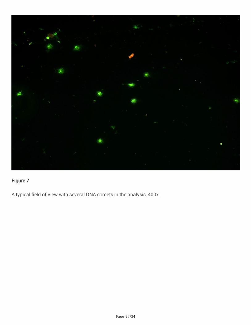
Figure 7
A typical �eld of view with several DNA comets in the analysis, 400x.
Page 24/24
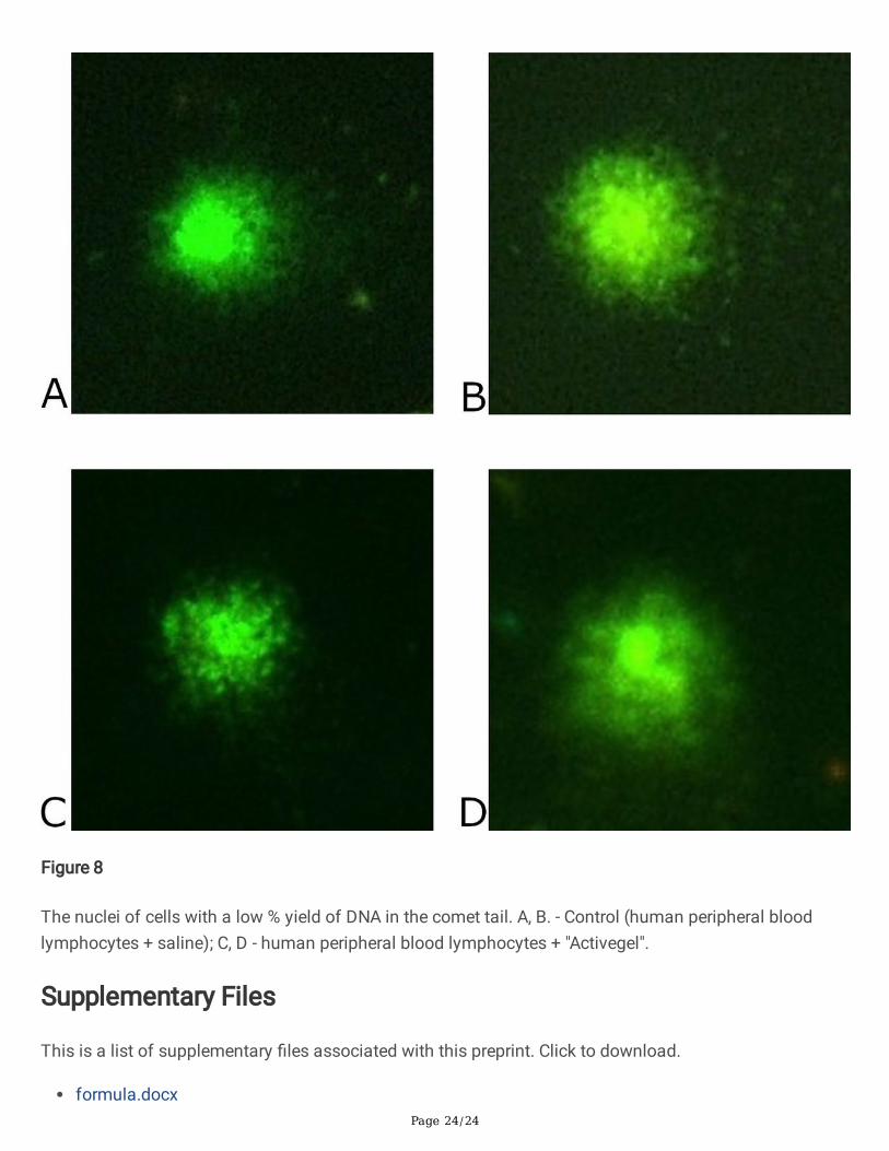
Figure 8
The nuclei of cells with a low % yield of DNA in the comet tail. A, B. - Control (human peripheral bloodlymphocytes + saline); C, D - human peripheral blood lymphocytes + "Activegel".
Supplementary Files
This is a list of supplementary �les associated with this preprint. Click to download.
formula.docx