Embed Size (px)
Citation preview
tsa1Citanng1lirst
Virology 255, 138–149 (1999)Article ID viro.1998.9571, available online at http://www.idealibrary.com on
0CA
Antisense Downregulation of a Mouse Mammary Tumor Virus Activated Protooncogenein Mouse Mammary Tumor Cells Reverses the Malignant Phenotype
Yin-Xiong Li,* Jackie Papkoff,† and Nurul H. Sarkar*,1
*Institute of Molecular Medicine and Genetics, Medical College of Georgia, Augusta, Georgia 30912;and †Megabios Corporation, Burlingame, California 94010
Received September 16, 1998; returned to author for revision October 26, 1998; accepted December 11, 1998
Activation of the protooncogene Wnt-1 by insertion of the mouse mammary tumor virus (MMTV) is known to causemammary tumors in mice. Wnt-1 expression in mammary glands has been postulated to confer direct local growthstimulation of mammary epithelial cells leading to their acquisition of a preneoplastic state. Wnt-1 expression also inducesmorphological alterations in cultured normal mammary cells. However, it has not been determined whether or not trans-formed mammary cells require continuous Wnt-1 expression for their ability to form tumors in vivo. To address this question,we constructed antisense and sense Wnt-1 expression vectors containing a synthetic promoter composed of five high-affinityglucocorticoid response elements (GRE5). This promoter is at least 50-fold more inducible by dexamethasone than thepromoter contained in the long terminal repeats of MMTV. The vectors were introduced into a mouse mammary tumor cellline (R/Sa-MT) that expresses high levels of endogenous Wnt-1 mRNA and forms rapidly growing tumors when transplantedinto syngeneic hosts. Of the 12 stably transfected cell lines established (9 with antisense and 3 with sense constructs), 2antisense cell lines (R/Sa-MT/antisense) and 1 sense cell line (R/Sa-MT/sense) were examined for inducibility by dexameth-asone of antisense and sense Wnt-1 RNAs, changes in endogenous Wnt-1 RNA expression, and changes in cell morphology.The growth patterns of the cells in vitro and in vivo were also examined. Our results show that (1) the levels of the expressionof endogenous Wnt-1 mRNA and protein were reduced significantly (.80%) in those cells (R/Sa-MT/antisense) thatexpressed antisense Wnt-1 RNA at high levels following exposure to dexamethasone, compared to the R/Sa-MT/sense andR/Sa-MT control cells and (2) transplantation of the R/Sa-MT/antisense cells produced smaller tumors (.0.2 cm in 16 weeks)compared to the tumors (.2.0 cm in 8 weeks) that were produced by the R/Sa-MT/sense and R/Sa-MT cells. We thereforesuggest that Wnt-1 expression is required not only for the transformation of normal mammary cells into tumor cells, but alsofor the maintenance of their tumorigenicity. © 1999 Academic Press
Key Words: Wnt-1 protooncogene; antisense RNA; cell morphology; mouse mammary tumor; tumor growth.
c(f(stmf(
mofmrjmtcte
INTRODUCTION
Insertional mutations caused by the mouse mammaryumor virus (MMTV) in a number of protooncogenes,uch as int-1/Wnt-1 (Nusse and Varmus, 1982; Nusse etl., 1984), int-2/Fgf-3 (Peters et al., 1983; Dickson et al.,984; Shackleford et al., 1993a), and int-3 (Gallahan andallahan, 1987; Sarkar et al., 1994), are thought to be
nvolved in the development of spontaneous mammaryumors in mice. To address the question of how MMTVctivated int genes contribute to the transformation oformal mammary epithelial cells into cancer cells, aumber of structural and functional studies of the Wnt-1ene have been carried out. Since its identification in984, Wnt-1 has been demonstrated to be a member of a
arge group of developmentally regulated genes, includ-ng the segment polarity gene wingless of Drosophila (foreview see Nusse and Varmus, 1992). In fact, the Dro-ophila gene, like mouse Wnt-1, is capable of causing
ransformation and mitogenesis of mouse mammary
i1 To whom reprint requests should be addressed.
042-6822/99 $30.00opyright © 1999 by Academic Pressll rights of reproduction in any form reserved.
138
ells and of converting them to a tumorigenic phenotypeRamakrishna and Brown, 1993). Wnt-1 also normallyunctions in the embryonic central nervous systemNusse and Varmus, 1992; McMahon, 1992); its expres-ion is required for the development of a large region of
he mouse brain (McMahon and Bradley, 1990). Further-ore, Wnt-1 expression in Xenopus embryos has been
ound to promote the duplication of the embryonic axisMcMahon and Moon, 1989).
In addition to its roles in embryonic development andouse mammary tumorigenesis, Wnt-1 shows other bi-
logical activities in vitro. Wnt-1 expression has beenound to induce morphological alterations in one mam-
ary epithelial cell line (Brown et al., 1986) while itendered another cell line (RAC311C) tumorigenic (Ri-sewijk et al., 1987). Further, Wnt-1-expressing rat or
ouse fibroblasts that show no detectable responsehemselves can transform neighboring C57BL cells inoculture experiments. This indicates that the Wnt-1 pro-
ein, a cysteine-rich glycoprotein of 41 to 44 kDa, appar-ntly acts via a paracrine mechanism (Jue et al., 1992). It
s of interest to note that Wnt-1 does not normally accu-
msvPw1
mmtscyotamoucttmfpaanta
popFii(oaarhtpdmRcasitWsW
asrt
HeW
gtpA1osss(hd(
ai(aosprT
139Wnt-1 ANTISENSE RNA REVERSES MAMMARY TUMOR GROWTH
ulate in the tissue culture medium, but instead, isecreted onto the surface of cells or into their microen-ironment (Papkoff et al., 1987; Bradley and Brown, 1990;apkoff and Schryver, 1990) and seems to be activehen it exists as a transmembrane protein (Parkin et al.,
993).Results from one in vivo study in a Wnt-1 transgenic
ouse model have indicated that Wnt-1 expression inammary glands confers a direct local growth stimula-
ion of mammary epithelial cells, leading to their acqui-ition of a preneoplastic state (Lin et al., 1992). This isonsistent with the finding that the mammary glands ofoung virgin transgenic mice (approximately 3 monthsld) resemble the mammary glands of pregnant non-
ransgenic animals with regard to the development oflveolar hyperplasias (Tsukamoto et al., 1988). Even theammary glands of 2-week-old Wnt-1 mice show alve-
lar hyperplasias (Lin et al., 1992). However, it is stillnknown whether these hyperplastic cells need to beontinuously stimulated by the Wnt-1 protein in order for
he cells to be transformed into tumor cells. More impor-antly, it remains to be established whether or not mam-
ary tumor cells acquire growth autonomy during trans-ormation or whether they still require continuous ex-ression of Wnt-1 for their ability to form tumors in vivond subsequently metastasize. These concerns can beddressed by reducing the expression of Wnt-1 in pre-eoplastic and/or neoplastic mammary tumor cell lines
hrough antisense technology and testing whether thebility of the cells to form tumors in vivo is impaired.
The antisense approach has been successfully ap-lied by numerous investigators to inhibit the expressionf a variety of target genes, and hence, the activity of therotein product (for review see Stein and Cheng, 1993).or example, the expression of antisense RNA to the
nsulin-like growth factor-1 receptor has been shown tonduce regression of wild-type rat glioblastoma tumorsResnicoff et al., 1994), loss of the metastatic phenotypef the Lewis lung carcinoma line H-59 (Long et al., 1995),nd suppression of malignant phenotypes of a humanlveolar rhabdosarcoma (Shapiro et al., 1994). Down-
egulation of N-myc1 by antisense RNA in woodchuckepatoma cells has recently been observed to reverse
he malignant phenotype (Wang et al., 1997). In theresent study, we have used an antisense approach toetermine the role of Wnt-1 in the growth in vivo of aammary tumor cell line (R/Sa-MT) expressing Wnt-1./Sa-MT cells were transfected with vectors designed toonstitutively express a segment of Wnt-1 RNA in thentisense or sense (as control) orientations relative to ateroid-inducible promoter. Stable transfectants were
solated and tested for tumorigenicity. Our results showhat the expression of ectopic antisense, but not sense,
nt-1-specific RNA in R/Sa-MT cells resulted in loweringignificantly the levels of expression of endogenous
nt-1 mRNA and protein. In addition, the cells lost their ebility to form tumors in mice by more than 90%. Thus, weuggest that continuous signaling by Wnt-1 protein isequired for the maintenance of the tumorigenic pheno-ype of mammary tumor cells in mice.
RESULTS
ormonal regulation of antisense Wnt-1 RNAxpression and the downregulation of endogenousnt-1 mRNA
To gain insights into the regulatory role of Wnt-1 in therowth of mammary tumors, we transfected a mammary
umor cell line, R/Sa-MT, with two expression vectors,GRE5-1/Wnt-1(sense, S) and pGRE5-2/tnW-1(antisense,S) containing a 290-bp segment of the Wnt-1 cDNA (Fig.), together with pSV2neo plasmid DNA. The rationale forur choice of the vectors was that they contained aynthetic promoter composed of five high-affinity bindingites for the glucocorticoid receptor (GRE5) placed up-tream of the adenovirus 2 major late promoter
Ad2MLP) TATA box/initiation site (Fig. 1). This promoteras been shown to be at least 50-fold more inducible byexamethasone than the MMTV long terminal repeat
LTR) promoter (Mader and White, 1993). We therefore
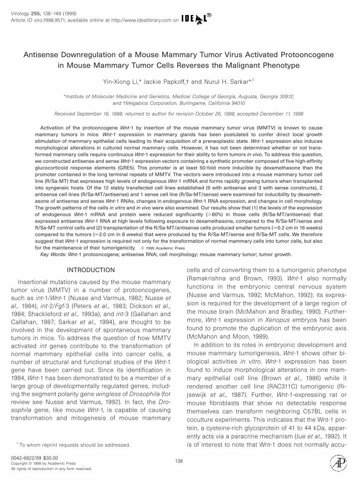
FIG. 1. Schematic representation of the construction of sense andntisense Wnt-1 expression plasmids. An EcoRI/XhoI fragment contain-
ng the 59 end of the Wnt-1 cDNA was isolated from the plasmid pSP65third panel) and ligated to the expression vectors pGRE 5-1 (first panel)nd pGRE 5-2 (second panel) in the sense (fourth panel) and antisenserientations (fifth panel) at the EcoRI/XhoI and XhoI/EcoRI sites, re-pectively. The expression vectors pGRE 5-1/5-2 contained a syntheticromoter composed of five sites for the binding of glucocorticoid
eceptor (GRE) and the adenovirus 2 major late promoter (Ad2) andATA box/initiation site.
xpected that it would induce high levels of Wnt-1 anti-
sf
p(BAAsitecloccccfF
sAolbsl(Acnpclnbtm
sWPEputpiatoc(sTml
a(snaliRw
140 LI, PAPKOFF, AND SARKAR
ense RNA, but not endogenous Wnt-1 mRNA, in trans-ectants exposed to small amounts of dexamethasone.
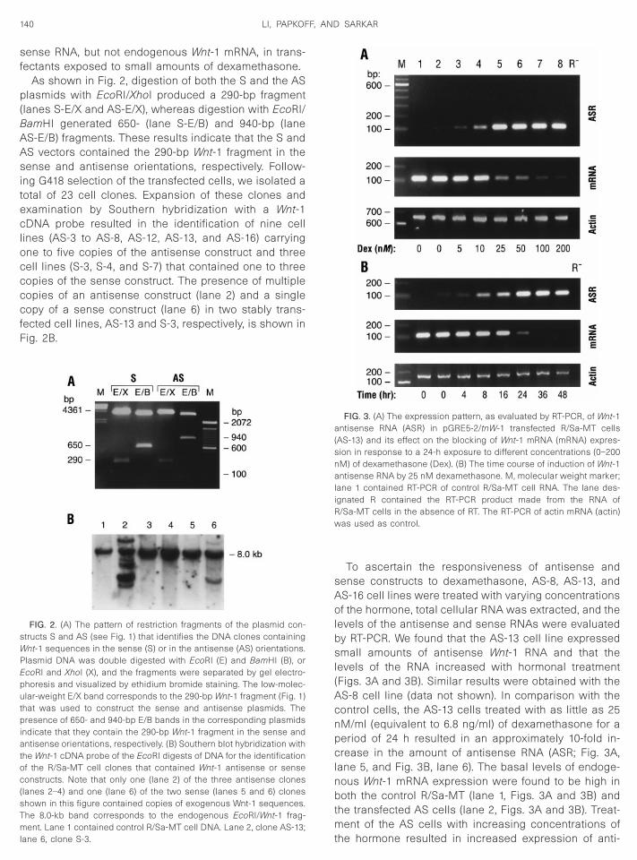
As shown in Fig. 2, digestion of both the S and the ASlasmids with EcoRI/XhoI produced a 290-bp fragment
lanes S-E/X and AS-E/X), whereas digestion with EcoRI/amHI generated 650- (lane S-E/B) and 940-bp (laneS-E/B) fragments. These results indicate that the S andS vectors contained the 290-bp Wnt-1 fragment in theense and antisense orientations, respectively. Follow-
ng G418 selection of the transfected cells, we isolated aotal of 23 cell clones. Expansion of these clones andxamination by Southern hybridization with a Wnt-1DNA probe resulted in the identification of nine cell
ines (AS-3 to AS-8, AS-12, AS-13, and AS-16) carryingne to five copies of the antisense construct and threeell lines (S-3, S-4, and S-7) that contained one to threeopies of the sense construct. The presence of multipleopies of an antisense construct (lane 2) and a singleopy of a sense construct (lane 6) in two stably trans-
ected cell lines, AS-13 and S-3, respectively, is shown inig. 2B.
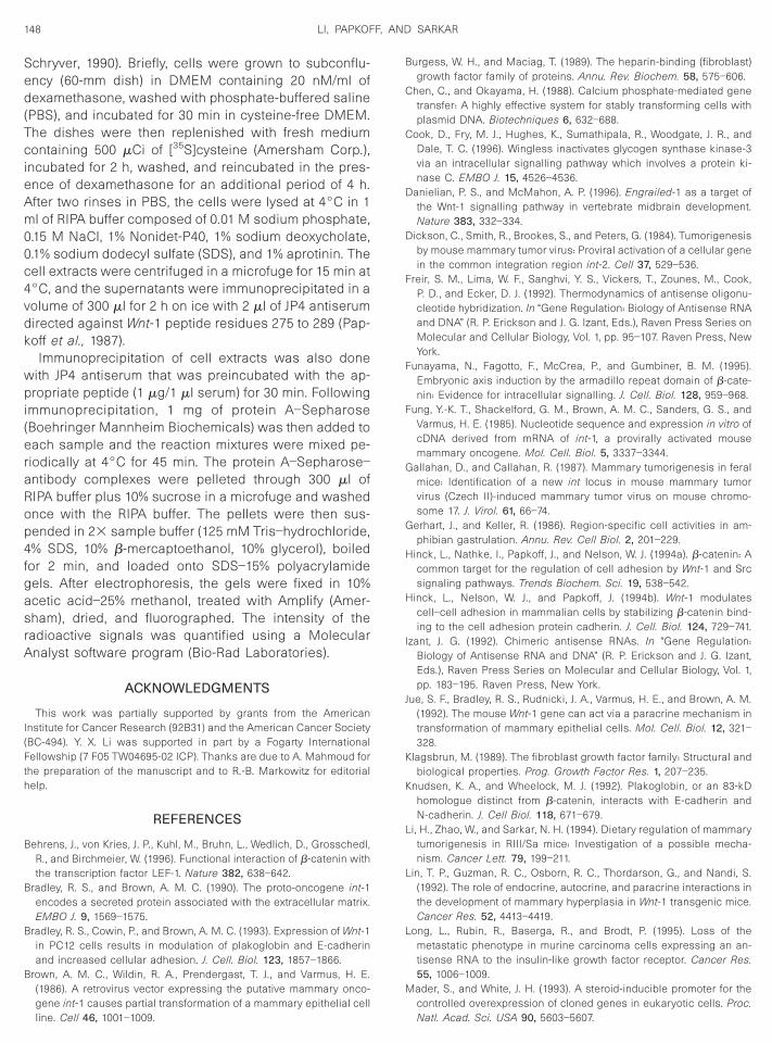
FIG. 2. (A) The pattern of restriction fragments of the plasmid con-tructs S and AS (see Fig. 1) that identifies the DNA clones containingnt-1 sequences in the sense (S) or in the antisense (AS) orientations.
lasmid DNA was double digested with EcoRI (E) and BamHI (B), orcoRI and XhoI (X), and the fragments were separated by gel electro-horesis and visualized by ethidium bromide staining. The low-molec-lar-weight E/X band corresponds to the 290-bp Wnt-1 fragment (Fig. 1)
hat was used to construct the sense and antisense plasmids. Theresence of 650- and 940-bp E/B bands in the corresponding plasmids
ndicate that they contain the 290-bp Wnt-1 fragment in the sense andntisense orientations, respectively. (B) Southern blot hybridization with
he Wnt-1 cDNA probe of the EcoRI digests of DNA for the identificationf the R/Sa-MT cell clones that contained Wnt-1 antisense or senseonstructs. Note that only one (lane 2) of the three antisense clones
lanes 2–4) and one (lane 6) of the two sense (lanes 5 and 6) cloneshown in this figure contained copies of exogenous Wnt-1 sequences.he 8.0-kb band corresponds to the endogenous EcoRI/Wnt-1 frag-ent. Lane 1 contained control R/Sa-MT cell DNA. Lane 2, clone AS-13;
tane 6, clone S-3.
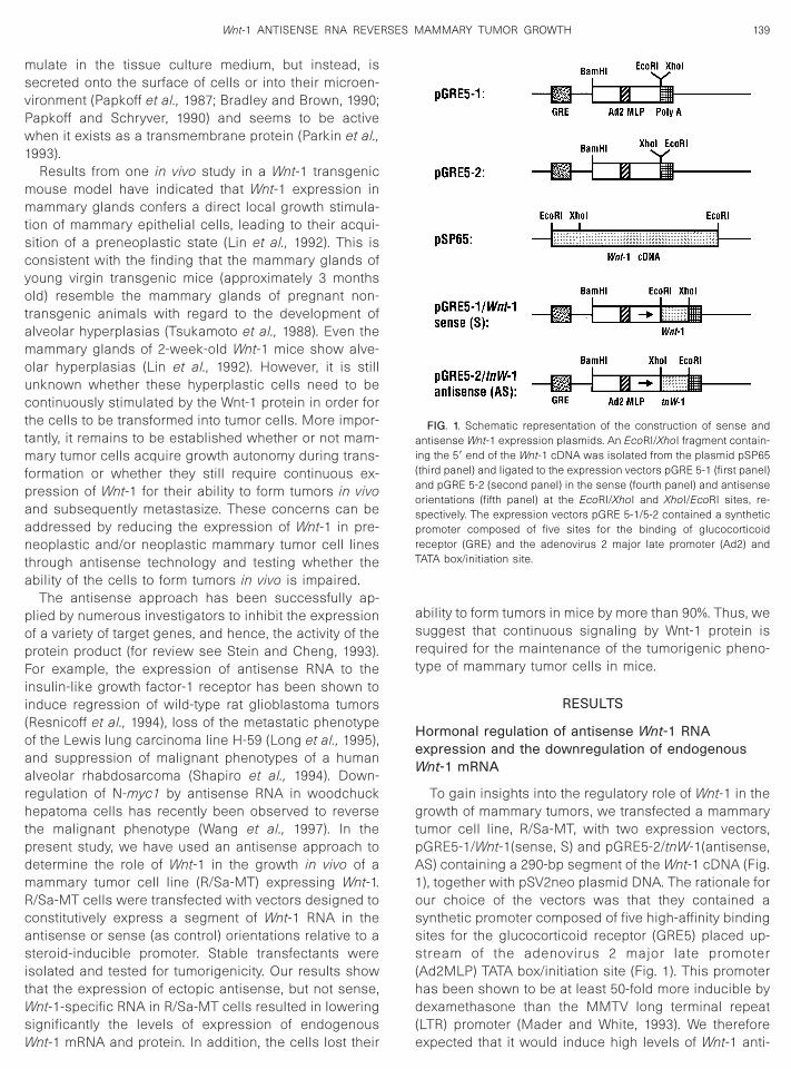
To ascertain the responsiveness of antisense andense constructs to dexamethasone, AS-8, AS-13, andS-16 cell lines were treated with varying concentrationsf the hormone, total cellular RNA was extracted, and the
evels of the antisense and sense RNAs were evaluatedy RT-PCR. We found that the AS-13 cell line expressedmall amounts of antisense Wnt-1 RNA and that the
evels of the RNA increased with hormonal treatmentFigs. 3A and 3B). Similar results were obtained with theS-8 cell line (data not shown). In comparison with theontrol cells, the AS-13 cells treated with as little as 25M/ml (equivalent to 6.8 ng/ml) of dexamethasone for aeriod of 24 h resulted in an approximately 10-fold in-rease in the amount of antisense RNA (ASR; Fig. 3A,
ane 5, and Fig. 3B, lane 6). The basal levels of endoge-ous Wnt-1 mRNA expression were found to be high inoth the control R/Sa-MT (lane 1, Figs. 3A and 3B) and
he transfected AS cells (lane 2, Figs. 3A and 3B). Treat-ent of the AS cells with increasing concentrations of
FIG. 3. (A) The expression pattern, as evaluated by RT-PCR, of Wnt-1ntisense RNA (ASR) in pGRE5-2/tnW-1 transfected R/Sa-MT cells
AS-13) and its effect on the blocking of Wnt-1 mRNA (mRNA) expres-ion in response to a 24-h exposure to different concentrations (0–200M) of dexamethasone (Dex). (B) The time course of induction of Wnt-1ntisense RNA by 25 nM dexamethasone. M, molecular weight marker;
ane 1 contained RT-PCR of control R/Sa-MT cell RNA. The lane des-gnated R contained the RT-PCR product made from the RNA of/Sa-MT cells in the absence of RT. The RT-PCR of actin mRNA (actin)as used as control.
he hormone resulted in increased expression of anti-
stopntrsft(
scheTsema
fcplARcswwhoAbsssads
Te
ipcptitAsSst
Tm
vslocppptdst
p
R(AoucasammRso
141Wnt-1 ANTISENSE RNA REVERSES MAMMARY TUMOR GROWTH
ense Wnt-1 RNA, and as a consequence the levels ofhe Wnt-1 mRNA decreased. The optimum concentrationf the hormone was found to be 25 nM. It should beointed out that this low concentration of hormone didot increase the levels of Wnt-1 mRNA despite the fact
hat the expression of the gene in these cells is under theegulation of an MMTV LTR that contains hormone re-ponsive elements (data not shown). This is due to the
act that the MMTV LTR responds to a higher concentra-ion (1–10 mg/ml, i.e., 3.7–37 mM) of dexamethasoneRingold et al., 1975; Sarkar et al., 1977).
To define the optimum time of dexamethasone expo-ure for maximum induction of antisense RNA, the AS-13ell line was treated with a 25 nM concentration of theormone for various time periods and the levels of thexpression of antisense RNA were evaluated by RT-PCR.he results indicated that the maximum amount of anti-ense RNA was induced with 20 to 30 h of hormonalxposure (Fig. 3B). The effects of dexamethasone treat-ent for a period of 24 h on the expression of Wnt-1
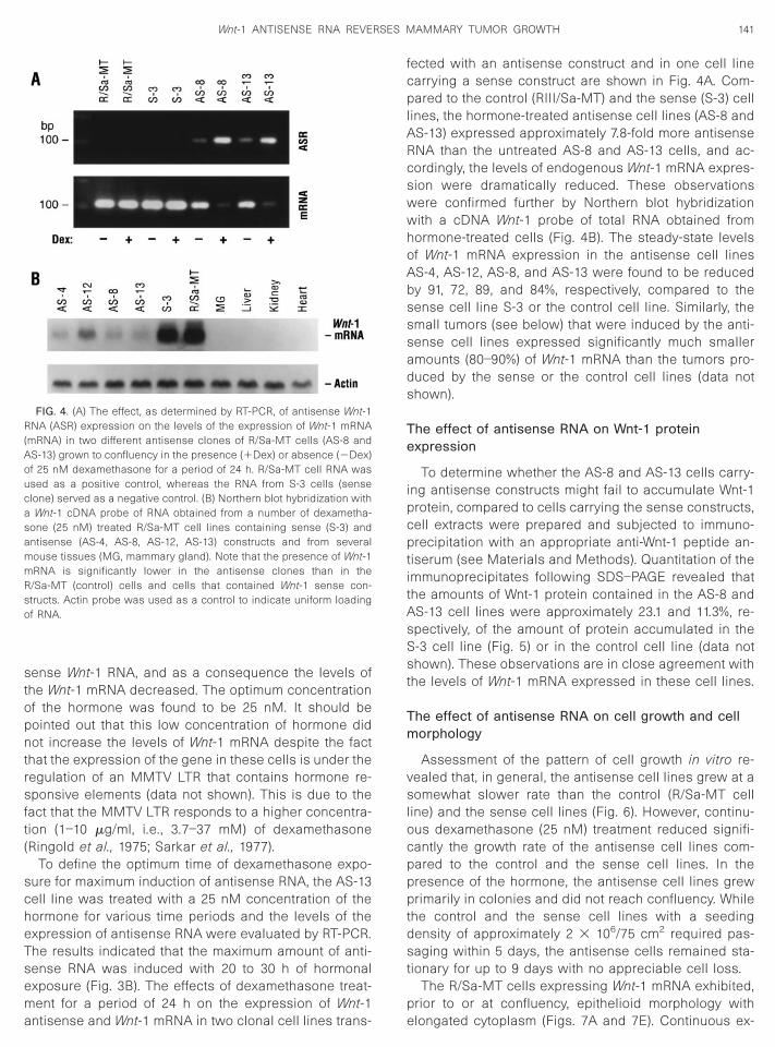
FIG. 4. (A) The effect, as determined by RT-PCR, of antisense Wnt-1NA (ASR) expression on the levels of the expression of Wnt-1 mRNA
mRNA) in two different antisense clones of R/Sa-MT cells (AS-8 andS-13) grown to confluency in the presence (1Dex) or absence (2Dex)f 25 nM dexamethasone for a period of 24 h. R/Sa-MT cell RNA wassed as a positive control, whereas the RNA from S-3 cells (senselone) served as a negative control. (B) Northern blot hybridization with
Wnt-1 cDNA probe of RNA obtained from a number of dexametha-one (25 nM) treated R/Sa-MT cell lines containing sense (S-3) andntisense (AS-4, AS-8, AS-12, AS-13) constructs and from severalouse tissues (MG, mammary gland). Note that the presence of Wnt-1RNA is significantly lower in the antisense clones than in the/Sa-MT (control) cells and cells that contained Wnt-1 sense con-tructs. Actin probe was used as a control to indicate uniform loadingf RNA.
ntisense and Wnt-1 mRNA in two clonal cell lines trans- e
ected with an antisense construct and in one cell linearrying a sense construct are shown in Fig. 4A. Com-ared to the control (RIII/Sa-MT) and the sense (S-3) cell
ines, the hormone-treated antisense cell lines (AS-8 andS-13) expressed approximately 7.8-fold more antisenseNA than the untreated AS-8 and AS-13 cells, and ac-ordingly, the levels of endogenous Wnt-1 mRNA expres-ion were dramatically reduced. These observationsere confirmed further by Northern blot hybridizationith a cDNA Wnt-1 probe of total RNA obtained fromormone-treated cells (Fig. 4B). The steady-state levelsf Wnt-1 mRNA expression in the antisense cell linesS-4, AS-12, AS-8, and AS-13 were found to be reducedy 91, 72, 89, and 84%, respectively, compared to theense cell line S-3 or the control cell line. Similarly, themall tumors (see below) that were induced by the anti-ense cell lines expressed significantly much smallermounts (80–90%) of Wnt-1 mRNA than the tumors pro-uced by the sense or the control cell lines (data nothown).
he effect of antisense RNA on Wnt-1 proteinxpression
To determine whether the AS-8 and AS-13 cells carry-ng antisense constructs might fail to accumulate Wnt-1rotein, compared to cells carrying the sense constructs,ell extracts were prepared and subjected to immuno-recipitation with an appropriate anti-Wnt-1 peptide an-
iserum (see Materials and Methods). Quantitation of themmunoprecipitates following SDS–PAGE revealed thathe amounts of Wnt-1 protein contained in the AS-8 andS-13 cell lines were approximately 23.1 and 11.3%, re-pectively, of the amount of protein accumulated in the-3 cell line (Fig. 5) or in the control cell line (data nothown). These observations are in close agreement with
he levels of Wnt-1 mRNA expressed in these cell lines.
he effect of antisense RNA on cell growth and cellorphology
Assessment of the pattern of cell growth in vitro re-ealed that, in general, the antisense cell lines grew at aomewhat slower rate than the control (R/Sa-MT cell
ine) and the sense cell lines (Fig. 6). However, continu-us dexamethasone (25 nM) treatment reduced signifi-antly the growth rate of the antisense cell lines com-ared to the control and the sense cell lines. In theresence of the hormone, the antisense cell lines grewrimarily in colonies and did not reach confluency. While
he control and the sense cell lines with a seedingensity of approximately 2 3 106/75 cm2 required pas-aging within 5 days, the antisense cells remained sta-
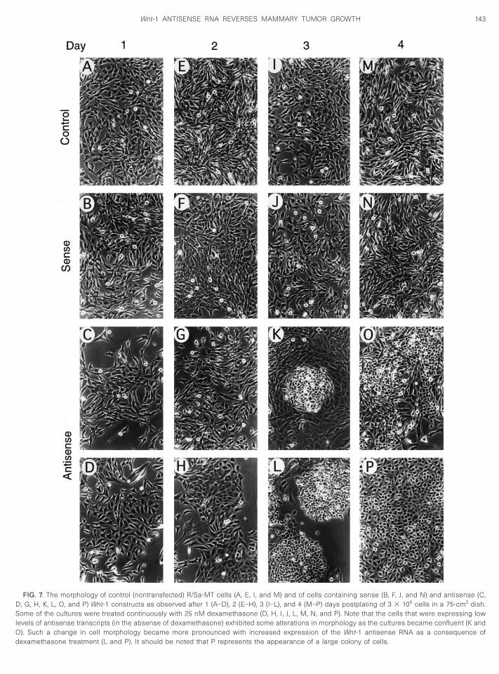
ionary for up to 9 days with no appreciable cell loss.The R/Sa-MT cells expressing Wnt-1 mRNA exhibited,
rior to or at confluency, epithelioid morphology with
longated cytoplasm (Figs. 7A and 7E). Continuous ex-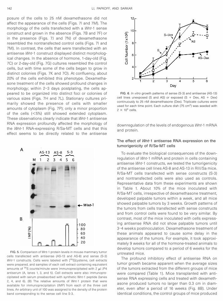
pamcir7ai7cd2smpvmaoTRte
da
Tt
raoRaRiRdstaci3tamdu
towsae
cWwaac2alb
ccu2
142 LI, PAPKOFF, AND SARKAR
osure of the cells to 25 nM dexamethasone did notffect the appearance of the cells (Figs. 7I and 7M). Theorphology of the cells transfected with a Wnt-1 sense
onstruct and grown in the absence (Figs. 7B and 7F) orn the presence (Figs. 7J and 7N) of dexamethasoneesembled the nontransfected control cells (Figs. 7I andM). In contrast, the cells that were transfected with anntisense Wnt-1 construct displayed distinct morpholog-
cal changes. In the absence of hormone, 1-day-old (Fig.C) or 2-day-old (Fig. 7G) cultures resembled the controlells, but with time some of the cells began to grow inistinct colonies (Figs. 7K and 7O). At confluency, about0% of the cells exhibited this phenotype. Dexametha-one treatment of the cells showed profound changes inorphology; within 2–3 days postplating, the cells ap-
eared to be organized into distinct foci or colonies ofarious sizes (Figs. 7H and 7L). Stationary cultures pri-arily showed the presence of cells with smaller
mounts of cytoplasm (Fig. 7P); only a minor proportionf the cells (,5%) still showed extended cytoplasm.hese observations clearly indicate that Wnt-1 antisenseNA expression profoundly affected the morphology of
he Wnt-1 RNA-expressing R/Sa-MT cells and that thisffect seems to be directly related to the antisense
FIG. 5. Comparison of Wnt-1 protein levels in mouse mammary tumorells transfected with antisense (AS-13 and AS-8) and sense (S-3)nt-1 constructs. Cells were labeled with [35S]cysteine, cell extractsere prepared, and samples of each cell extract containing equivalentmounts of 35S counts/minute were immunoprecipitated with 2 ml JP4ntiserum (A, lanes 1, 3, and 5). Cell extracts were also immunopre-ipitated with serum preabsorbed with synthetic Wnt-1 peptide (lanes, 4, and 6). (B) The relative amounts of Wnt-1 protein that werevailable for immunoprecipitation (IMP) from each of the three cell
ines. An arbitrary unit of 100 was assigned to the density of the protein
iand corresponding to the sense cell line S-3.ownregulation of the levels of endogenous Wnt-1 mRNAnd protein.
he effect of Wnt-1 antisense RNA expression on theumorigenicity of R/Sa-MT cells
To evaluate the biological consequences of the down-egulation of Wnt-1 mRNA and protein in cells containingntisense Wnt-1 constructs, we tested the tumorigenicityf the antisense cell lines AS-8 and AS-13 in RIII/Sa mice./Sa-MT cells transfected with sense constructs (S-3)nd nontransfected cells were also used as controls.epresentative data from these experiments are shown
n Table 1. About 10% of the mice inoculated with/Sa-MT cells, irrespective of dexamethasone treatment,eveloped palpable tumors within a week, and all micehowed palpable tumors by 3 weeks. Growth patterns of
he tumors from cells transfected with sense constructsnd from control cells were found to be very similar. Byontrast, most of the mice inoculated with cells express-
ng antisense RNA did not show palpable tumors until–4 weeks postinoculation. Dexamethasone treatment of
hese animals appeared to cause some delay in theppearance of the tumors. For example, it took approxi-ately 9 weeks for all of the hormone-treated animals to
evelop tumors compared to a period of 6 weeks for thentreated mice.
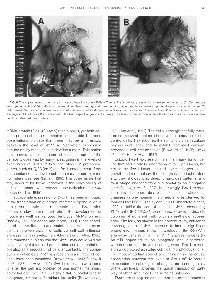
The profound inhibitory effect of antisense RNA onumor growth became apparent when the average sizesf the tumors extracted from the different groups of miceere compared (Table 1). Mice transplanted with anti-
ense RNA expressing cells and treated with dexameth-sone produced tumors no larger than 0.3 cm in diam-ter, even after a period of 16 weeks (Fig. 8B). Under
FIG. 6. In vitro growth patterns of sense (S-3) and antisense (AS-13)ell lines unexposed (S and AS) or exposed (S 1 Dex, AS 1 Dex)ontinuously to 25 nM dexamethasone (Dex). Triplicate cultures weresed for each time point. Each culture dish (75 cm2) was seeded with3 106 cells.
dentical conditions, the control groups of mice produced
DSlOd
143Wnt-1 ANTISENSE RNA REVERSES MAMMARY TUMOR GROWTH
FIG. 7. The morphology of control (nontransfected) R/Sa-MT cells (A, E, I, and M) and of cells containing sense (B, F, J, and N) and antisense (C,, G, H, K, L, O, and P) Wnt-1 constructs as observed after 1 (A–D), 2 (E–H), 3 (I–L), and 4 (M–P) days postplating of 3 3 106 cells in a 75-cm2 dish.ome of the cultures were treated continuously with 25 nM dexamethasone (D, H, I, J, L, M, N, and P). Note that the cells that were expressing low
evels of antisense transcripts (in the absense of dexamethasone) exhibited some alterations in morphology as the cultures became confluent (K and). Such a change in cell morphology became more pronounced with increased expression of the Wnt-1 antisense RNA as a consequence of
examethasone treatment (L and P). It should be noted that P represents the appearance of a large colony of cells.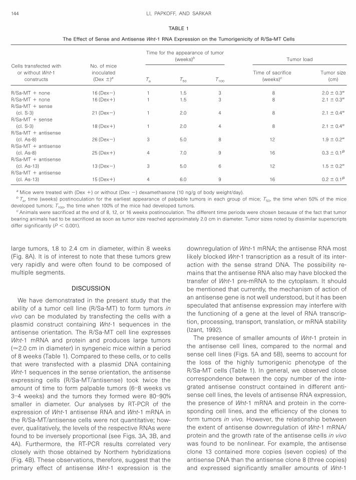
l(vm
avpaW(otWea3setef4c(p
dlamtbastt(
tstRcgstsftpwca
C
RRR
R
R
R
R
R
d
bd
144 LI, PAPKOFF, AND SARKAR
arge tumors, 1.8 to 2.4 cm in diameter, within 8 weeksFig. 8A). It is of interest to note that these tumors grewery rapidly and were often found to be composed ofultiple segments.
DISCUSSION
We have demonstrated in the present study that thebility of a tumor cell line (R/Sa-MT) to form tumors inivo can be modulated by transfecting the cells with alasmid construct containing Wnt-1 sequences in thentisense orientation. The R/Sa-MT cell line expressesnt-1 mRNA and protein and produces large tumors
.2.0 cm in diameter) in syngeneic mice within a periodf 8 weeks (Table 1). Compared to these cells, or to cells
hat were transfected with a plasmid DNA containingnt-1 sequences in the sense orientation, the antisense
xpressing cells (R/Sa-MT/antisense) took twice themount of time to form palpable tumors (6–8 weeks vs–4 weeks) and the tumors they formed were 80–90%maller in diameter. Our analyses by RT-PCR of thexpression of Wnt-1 antisense RNA and Wnt-1 mRNA in
he R/Sa-MT/antisense cells were not quantitative; how-ver, qualitatively, the levels of the respective RNAs were
ound to be inversely proportional (see Figs. 3A, 3B, andA). Furthermore, the RT-PCR results correlated verylosely with those obtained by Northern hybridizations
Fig. 4B). These observations, therefore, suggest that the
T
The Effect of Sense and Antisense Wnt-1 RNA
ells transfected withor without Wnt-1
constructs
No. of miceinoculated(Dex 6)a
Time for th
Ta
/Sa-MT 1 none 16 (Dex2) 1/Sa-MT 1 none 16 (Dex1) 1/Sa-MT 1 sense
(cl. S-3) 21 (Dex2) 1/Sa-MT 1 sense
(cl. S-3) 18 (Dex1) 1/Sa-MT 1 antisense
(cl. As-8) 26 (Dex2) 3/Sa-MT 1 antisense
(cl. As-8) 25 (Dex1) 4/Sa-MT 1 antisense
(cl. As-13) 13 (Dex2) 3/Sa-MT 1 antisense
(cl. As-13) 15 (Dex1) 4
a Mice were treated with (Dex 1) or without (Dex 2) dexamethasonb Ta, time (weeks) postinoculation for the earliest appearance of pa
eveloped tumors; T100, the time when 100% of the mice had developec Animals were sacrificed at the end of 8, 12, or 16 weeks postinocu
earing animals had to be sacrificed as soon as tumor size reached apiffer significantly (P , 0.001).
rimary effect of antisense Wnt-1 expression is the a
ownregulation of Wnt-1 mRNA; the antisense RNA mostikely blocked Wnt-1 transcription as a result of its inter-ction with the sense strand DNA. The possibility re-ains that the antisense RNA also may have blocked the
ransfer of Wnt-1 pre-mRNA to the cytoplasm. It shoulde mentioned that currently, the mechanism of action ofn antisense gene is not well understood, but it has beenpeculated that antisense expression may interfere with
he functioning of a gene at the level of RNA transcrip-ion, processing, transport, translation, or mRNA stabilityIzant, 1992).
The presence of smaller amounts of Wnt-1 protein inhe antisense cell lines, compared to the normal andense cell lines (Figs. 5A and 5B), seems to account for
he loss of the highly tumorigenic phenotype of the/Sa-MT cells (Table 1). In general, we observed closeorrespondence between the copy number of the inte-rated antisense construct contained in different anti-ense cell lines, the levels of antisense RNA expression,
he presence of Wnt-1 mRNA and protein in the corre-ponding cell lines, and the efficiency of the clones to
orm tumors in vivo. However, the relationship betweenhe extent of antisense downregulation of Wnt-1 mRNA/rotein and the growth rate of the antisense cells in vivoas found to be nonlinear. For example, the antisense
lone 13 contained more copies (seven copies) of thentisense DNA than the antisense clone 8 (three copies)
ssion on the Tumorigenicity of R/Sa-MT Cells
arance of tumors)b Tumor load
T100
Time of sacrifice(weeks)c
Tumor size(cm)
3 8 2.0 6 0.3a
3 8 2.1 6 0.3a
4 8 2.1 6 0.4a
4 8 2.1 6 0.4a
8 12 1.9 6 0.2a
9 16 0.3 6 0.1b
6 12 1.5 6 0.2a
9 16 0.2 6 0.1b
g/g of body weight/day).tumors in each group of mice; T50, the time when 50% of the micers.he different time periods were chosen because of the fact that tumor
ately 2.0 cm in diameter. Tumor sizes noted by dissimilar superscripts
ABLE 1
Expre
e appe(week
T50
1.51.5
2.0
2.0
5.0
7.0
5.0
6.0
e (10 nlpabled tumo
lation. Tproxim
nd expressed significantly smaller amounts of Wnt-1
mlobamvega(mig
tismBncaiobqletee
1fcbda
lngsttscm1PcadpaSwsTaeow
wntp
145Wnt-1 ANTISENSE RNA REVERSES MAMMARY TUMOR GROWTH
RNA/protein (Figs. 4B and 5) than clone 8, yet both cellines produced tumors of similar sizes (Table 1). Thesebservations indicate that there may be a thresholdetween the level of Wnt-1 mRNA/protein expressionnd the ability of the cells to develop tumors. This notionay provide an explanation, at least in part, for the
ariability observed by many investigators in the levels ofxpression of Wnt-1 mRNA and other int protoonco-enes, such as Fgf-3 (int-2) and int-3, among most, if notll, spontaneously developed mammary tumors of mice
for references see Sarkar, 1995). The other factor thatay account for these variations is the polyclonality of
ndividual tumors with respect to the activation of the intenes (Sarkar, 1995).
Inappropriate expression of Wnt-1 has been attributedo the transformation of normal mammary epithelial cellsnto preneoplastic and neoplastic cells. Wnt-1 alsoeems to play an important role in the development ofouse as well as Xenopus embryos (McMahon andradley, 1992; McMahon and Moon, 1989). Since coordi-ated cell proliferation and maintenance of close asso-iation between groups of cells via cell–cell adhesionre essential in development (Gerhart and Keller, 1986),
t is reasonable to assume that Wnt-1 may act in vivo notnly as a regulator of cell proliferation and differentiation,ut also as a modulator of cellular contacts. The conse-uences of ectopic Wnt-1 expression in a number of cell
ines have been examined (Brown et al., 1986; Rijsewijkt al., 1987; Jue et al., 1992). Wnt-1 expression was found
o alter the cell morphology of one normal mammarypithelial cell line (C57BL) from a flat, cuboidal type to
FIG. 8. The appearance of mammary tumors produced by control R/Saas injected with 5 3 106 cells subcutaneously on the same day, and fM/mouse). The mouse in A was sacrificed after 8 weeks, while the m
he largest of the tumors that developed in the two respective groups ofoint to individual tumor lobes.
longated, refractile, fibroblast-like cells (Brown et al.,
986; Jue et al., 1992). The cells, although not fully trans-ormed, showed another phenotypic change: unlike theontrol cells, they acquired the ability to divide in cultureeyond confluency and to exhibit increased calcium-ependent cell–cell adhesion (Brown et al., 1986; Jue etl., 1992; Hinck et al., 1994b).
Ectopic Wnt-1 expression in a mammary tumor celline that had a MMTV integration at the Fgf-3 locus, butot at the Wnt-1 locus, showed some changes in cellrowth and morphology; the cells grew to a higher den-ity, they showed disordered, criss-cross patterns, and
heir shape changed from a cuboidal to an elongatedype (Rijsewijk et al., 1987). Interestingly, Wnt-1 expres-ion has also been observed to cause morphologicalhanges in one nonmammary, neural crest-derived tu-or cell line PC12 (Bradley et al., 1993; Shackleford et al.,
993b). Unlike the control cells, the Wnt-1 expressingC12 cells (PC12/Wnt-1) were found to grow in discreteolonies of adherent cells with an epitheloid appear-nce. Similarly, as shown in the present study, antisenseownregulation of Wnt-1 seemed to induce significanthenotypic changes in the morphology of the R/Sa-MT/ntisense cells in vitro. The Wnt-1 expressing cells (R/a-MT) appeared to be elongated and disordered,hereas the cells in which endogenous Wnt-1 expres-
ion was blocked exhibited cuboidal morphology (Fig. 7).he most important aspect of our finding is the causalssociation between the levels of Wnt-1 mRNA/proteinxpression, morphological changes, and tumorigenicityf the cell lines. However, the signal transduction path-ay of Wnt-1 in our cell line remains unknown.
lls (A) and cells expressing Wnt-1 antisense transcript (B). Each mousethird day on, each mouse was injected daily with dexamethasone (25B was sacrificed after 16 weeks. A and B represent the smallest and
ls. The black curved arrows outline the tumors; the small white arrows
-MT cerom theouse inanima
There are strong indications that the protein encoded
bd1(MeoaPPEcsgbm(1Wetah1Naan
ceisaba(ofivtcscts
P
fcTo
atatoPlisaosreevMEWt
T
sntlmttrtcmnth
DtBtpDDcmnstf
D
146 LI, PAPKOFF, AND SARKAR
y Wnt-1 acts as an intracellular signaling molecule, aso the transforming growth factors (TGFs) (Massague,992; Moses, 1990) and fibroblast growth factors (FGFs)Burgess and Maciag, 1989; Klagsburn, 1989; Rifkin and
oscatelli, 1989). A number of studies have shown thatctopic expression of Wnt-1 results in the accumulationf plakoglobin and/or b-catenin in some cells (Bradley etl., 1993; Funayama et al., 1995; Hinck et al., 1994a, b;apkoff et al., 1996). Furthermore, Wnt-1 expressingC12 cells show increased levels of plakoglobin and-cadherin and increased levels of calcium-dependentell–cell adhesion (Bradley et al., 1993). These findingsuggest that b-catenin and plakoglobin may be the tar-ets of the Wnt-1 signal transduction cascade. Since-catenin and plakoglobin bind to the cytoplasmic do-ain of the cadherin family of cell adhesion molecules
Nagafuchi and Takeichi, 1988; Knudsen and Wheelock,992; Piepenhagen and Nelson, 1993), it is likely thatnt-1 functions through b-catenin and plakoglobin in
liciting morphogenetic changes in cells. In this pathway,he involvement of glycogen synthase kinase 3b (GSK3b)nd the T cell factor/lymphocyte enhancer binding factoras been suggested (Cook et al., 1996; Behrens et al.,996; Porfiri et al., 1997; Papkoff and Aikawa, 1998;usse, 1997). Wnt-1-induced cell fate determination maylso involve the participation of Engrailed-1 (Danieliannd McMahon, 1996) and the gap-junctional protein con-exin 43 (van der Heyden et al., 1998).
At present, it is virtually unknown whether or nothanges in b-catenin/plakoglobin and/or connexin 43xpression may affect the growth potential of Wnt-1-
nduced mammary tumor cells in vivo. In this context, ithould be noted that abnormal expression of E-cadherin,
modulator of the cytoplasmic pools of b-catenin, haseen found to occur frequently in adenocarcinomas suchs breast cancer, gastric cancer, and prostate cancer
see Voeller et al., 1998). As shown in the present study,ur R/Sa-MT cell line is highly tumorigenic, and there-
ore, it could be used as an ideal model system for thedentification of the downstream molecules that are in-olved in the Wnt-1 signal transduction pathway duringumorigenesis of the mouse mammary glands. We areurrently interested in determining whether or not anti-ense downregulation of b-catenin/plakoglobin and/oronnexin 43 will modulate the tumorigenic phenotype of
his cell line in a manner similar to what we have ob-erved with the downregulation of Wnt-1.
MATERIALS AND METHODS
lasmid construction
The coding region of the Wnt-1 gene is composed ofour exons with a single open reading frame that en-odes a protein of 370 amino acids (Fung et al., 1985).he mode of action of antisense RNA-mediated inhibition
f gene expression presumably involves hybridization of fntisense RNA to a target mRNA (Freir et al., 1992). It isherefore desirable that the sequences targeted for thentisense RNA do not contain stem loop structures con-
aining more than 6-bp stems. On the basis of the sec-ndary structural analysis of Wnt-1 cDNA (using the CCGrogram from Genetic Computer Group, Inc.), we se-
ected a 290-bp segment of Wnt-1 that contained thenitiation codon for Wnt-1 protein (Fung et al., 1985). Thisegment of Wnt-1 was released by digestion with EcoRInd XhoI from pSP65 plasmid DNA that contained a copyf full-length Wnt-1 cDNA (Fig. 1). The digested DNA wasubjected to preparative electrophoresis in a 1.2% aga-ose gel and the appropriate fragment was isolated bylectroelution and ligated separately into the eukaryoticxpression vectors pGRE5-1 and pGRE5-2 (kindly pro-ided by Sylvie Mader of McGill University, Canada;
ader and White, 1993) at the EcoRI/XhoI and XhoI/coRI cloning sites to create Wnt-1 sense (pGRE5-1/nt-1) and antisense (pGRE/tnW-1) constructs, respec-
ively.
issue culture and cell transfection
The mammary tumor cell line (R/Sa-MT) used in thistudy was established in our laboratory from a lungodule that developed in an RIII/Sa mouse carrying a
ransplantable metastatic mammary tumor. This epithe-ial cell line expresses constitutively high levels of Wnt-1
RNA and protein as a consequence of insertional mu-ation of the oncogene by a MMTV provirus. Transplan-ation of R/Sa-MT cells in syngeneic male or female miceesults in rapid tumor development. A full characteriza-ion of this cell line will be reported elsewhere. Routineell cultures were done in Dulbecco’s modified Eagle’sedium (DMEM) containing 10% (v/v) fetal calf serum,
onessential amino acids, L-glutamine, vitamins, and an-ibiotics. Cell cultures were maintained at 37°C in aumidified atmosphere of 5% CO2.
The pGRE5-1/Wnt-1 and pGRE5-2/tnW-1 plasmidNAs, carrying the sense and antisense Wnt-1, respec-
ively, were cotransfected with pSV2neo (Southern anderg, 1982) plasmid DNA into the R/Sa-MT cells using
he standard calcium phosphate (CaPO4) transfectionrocedure (Chen and Okayama, 1988). A total of 30 mgNA at a 20:1 molar ratio of sense/antisense to pSV2neoNA was used for each transfection. The transfectedells were then cultured from day 2 onward in the sameedium, but supplemented with 400 mg/ml of G418 (Ge-
eticin; Gibco Laboratories, Grand Island, NY) in order toelect stable transfectants. After 12–14 days, G418-resis-
ant colonies were isolated and replated in fresh mediumor the expansion of the clones.
etermination of cell growth
Equal numbers of cells (1 or 2 3 106/75-cm2 dishes)
rom the parental, antisense, and sense cell lines were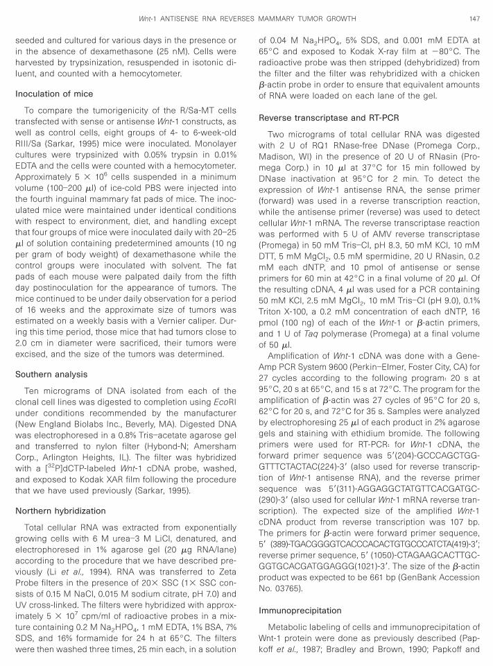
sihl
I
twRcEAvtuwtmpcpdmoei2e
S
cu(waCwat
N
geavPsUitSw
o6rtbo
R
wMmDe(wcw(Dmpt5Tpao
A29a6bgpfGts(scT5rGpN
I
W
147Wnt-1 ANTISENSE RNA REVERSES MAMMARY TUMOR GROWTH
eeded and cultured for various days in the presence orn the absence of dexamethasone (25 nM). Cells werearvested by trypsinization, resuspended in isotonic di-
uent, and counted with a hemocytometer.
noculation of mice
To compare the tumorigenicity of the R/Sa-MT cellsransfected with sense or antisense Wnt-1 constructs, as
ell as control cells, eight groups of 4- to 6-week-oldIII/Sa (Sarkar, 1995) mice were inoculated. Monolayerultures were trypsinized with 0.05% trypsin in 0.01%DTA and the cells were counted with a hemocytometer.pproximately 5 3 106 cells suspended in a minimumolume (100–200 ml) of ice-cold PBS were injected intohe fourth inguinal mammary fat pads of mice. The inoc-lated mice were maintained under identical conditionsith respect to environment, diet, and handling except
hat four groups of mice were inoculated daily with 20–25l of solution containing predetermined amounts (10 nger gram of body weight) of dexamethasone while theontrol groups were inoculated with solvent. The fatads of each mouse were palpated daily from the fifthay postinoculation for the appearance of tumors. Theice continued to be under daily observation for a period
f 16 weeks and the approximate size of tumors wasstimated on a weekly basis with a Vernier caliper. Dur-
ng this time period, those mice that had tumors close to.0 cm in diameter were sacrificed, their tumors werexcised, and the size of the tumors was determined.
outhern analysis
Ten micrograms of DNA isolated from each of thelonal cell lines was digested to completion using EcoRInder conditions recommended by the manufacturer
New England Biolabs Inc., Beverly, MA). Digested DNAas electrophoresed in a 0.8% Tris–acetate agarose gelnd transferred to nylon filter (Hybond-N; Amershamorp., Arlington Heights, IL). The filter was hybridizedith a [32P]dCTP-labeled Wnt-1 cDNA probe, washed,nd exposed to Kodak XAR film following the procedure
hat we have used previously (Sarkar, 1995).
orthern hybridization
Total cellular RNA was extracted from exponentiallyrowing cells with 6 M urea–3 M LiCl, denatured, andlectrophoresed in 1% agarose gel (20 mg RNA/lane)ccording to the procedure that we have described pre-iously (Li et al., 1994). RNA was transferred to Zetarobe filters in the presence of 203 SSC (13 SSC con-ists of 0.15 M NaCl, 0.015 M sodium citrate, pH 7.0) andV cross-linked. The filters were hybridized with approx-
mately 5 3 107 cpm/ml of radioactive probes in a mix-ure containing 0.2 M Na2HPO4, 1 mM EDTA, 1% BSA, 7%DS, and 16% formamide for 24 h at 65°C. The filters
ere then washed three times, 25 min each, in a solution kf 0.04 M Na2HPO4, 5% SDS, and 0.001 mM EDTA at5°C and exposed to Kodak X-ray film at 280°C. Theadioactive probe was then stripped (dehybridized) fromhe filter and the filter was rehybridized with a chicken-actin probe in order to ensure that equivalent amountsf RNA were loaded on each lane of the gel.
everse transcriptase and RT-PCR
Two micrograms of total cellular RNA was digestedith 2 U of RQ1 RNase-free DNase (Promega Corp.,adison, WI) in the presence of 20 U of RNasin (Pro-ega Corp.) in 10 ml at 37°C for 15 min followed byNase inactivation at 95°C for 2 min. To detect thexpression of Wnt-1 antisense RNA, the sense primer
forward) was used in a reverse transcription reaction,hile the antisense primer (reverse) was used to detect
ellular Wnt-1 mRNA. The reverse transcriptase reactionas performed with 5 U of AMV reverse transcriptase
Promega) in 50 mM Tris–Cl, pH 8.3, 50 mM KCl, 10 mMTT, 5 mM MgCl2, 0.5 mM spermidine, 20 U RNasin, 0.2M each dNTP, and 10 pmol of antisense or sense
rimers for 60 min at 42°C in a final volume of 20 ml. Ofhe resulting cDNA, 4 ml was used for a PCR containing0 mM KCl, 2.5 mM MgCl2, 10 mM Tris–Cl (pH 9.0), 0.1%riton X-100, a 0.2 mM concentration of each dNTP, 16mol (100 ng) of each of the Wnt-1 or b-actin primers,nd 1 U of Taq polymerase (Promega) at a final volumef 50 ml.
Amplification of Wnt-1 cDNA was done with a Gene-mp PCR System 9600 (Perkin–Elmer, Foster City, CA) for7 cycles according to the following program: 20 s at5°C, 20 s at 65°C, and 15 s at 72°C. The program for themplification of b-actin was 27 cycles of 95°C for 20 s,2°C for 20 s, and 72°C for 35 s. Samples were analyzedy electrophoresing 25 ml of each product in 2% agaroseels and staining with ethidium bromide. The followingrimers were used for RT-PCR: for Wnt-1 cDNA, the
orward primer sequence was 59(204)-GCCCAGCTGG-TTTCTACTAC(224)-39 (also used for reverse transcrip-
ion of Wnt-1 antisense RNA), and the reverse primerequence was 59(311)-AGGAGGCTATGTTCACGATGC-
290)-39 (also used for cellular Wnt-1 mRNA reverse tran-cription). The expected size of the amplified Wnt-1DNA product from reverse transcription was 107 bp.he primers for b-actin were forward primer sequence,9 (389)-TGACGGGGTCACCCACACTGTGCCCATCTA(419)-39;everse primer sequence, 59 (1050)-CTAGAAGCACTTGC-GTGCACGATGGAGGG(1021)-39. The size of the b-actinroduct was expected to be 661 bp (GenBank Accessiono. 03765).
mmunoprecipitation
Metabolic labeling of cells and immunoprecipitation ofnt-1 protein were done as previously described (Pap-
off et al., 1987; Bradley and Brown, 1990; Papkoff and
Sed(TcieAm00c4vdk
wpi(eraRop4fgasrA
I(Fth
B
B
B
B
B
C
C
D
D
F
F
F
G
G
H
H
I
J
K
K
L
L
L
M
148 LI, PAPKOFF, AND SARKAR
chryver, 1990). Briefly, cells were grown to subconflu-ncy (60-mm dish) in DMEM containing 20 nM/ml ofexamethasone, washed with phosphate-buffered saline
PBS), and incubated for 30 min in cysteine-free DMEM.he dishes were then replenished with fresh mediumontaining 500 mCi of [35S]cysteine (Amersham Corp.),
ncubated for 2 h, washed, and reincubated in the pres-nce of dexamethasone for an additional period of 4 h.fter two rinses in PBS, the cells were lysed at 4°C in 1l of RIPA buffer composed of 0.01 M sodium phosphate,
.15 M NaCl, 1% Nonidet-P40, 1% sodium deoxycholate,
.1% sodium dodecyl sulfate (SDS), and 1% aprotinin. Theell extracts were centrifuged in a microfuge for 15 min at°C, and the supernatants were immunoprecipitated in aolume of 300 ml for 2 h on ice with 2 ml of JP4 antiserumirected against Wnt-1 peptide residues 275 to 289 (Pap-off et al., 1987).
Immunoprecipitation of cell extracts was also doneith JP4 antiserum that was preincubated with the ap-ropriate peptide (1 mg/1 ml serum) for 30 min. Following
mmunoprecipitation, 1 mg of protein A–SepharoseBoehringer Mannheim Biochemicals) was then added toach sample and the reaction mixtures were mixed pe-
iodically at 4°C for 45 min. The protein A–Sepharose–ntibody complexes were pelleted through 300 ml ofIPA buffer plus 10% sucrose in a microfuge and washednce with the RIPA buffer. The pellets were then sus-ended in 23 sample buffer (125 mM Tris–hydrochloride,% SDS, 10% b-mercaptoethanol, 10% glycerol), boiled
or 2 min, and loaded onto SDS–15% polyacrylamideels. After electrophoresis, the gels were fixed in 10%cetic acid–25% methanol, treated with Amplify (Amer-ham), dried, and fluorographed. The intensity of theadioactive signals was quantified using a Molecularnalyst software program (Bio-Rad Laboratories).
ACKNOWLEDGMENTS
This work was partially supported by grants from the Americannstitute for Cancer Research (92B31) and the American Cancer SocietyBC-494). Y. X. Li was supported in part by a Fogarty Internationalellowship (7 F05 TW04695-02 ICP). Thanks are due to A. Mahmoud for
he preparation of the manuscript and to R.-B. Markowitz for editorialelp.
REFERENCES
ehrens, J., von Kries, J. P., Kuhl, M., Bruhn, L., Wedlich, D., Grosschedl,R., and Birchmeier, W. (1996). Functional interaction of b-catenin withthe transcription factor LEF-1. Nature 382, 638–642.
radley, R. S., and Brown, A. M. C. (1990). The proto-oncogene int-1encodes a secreted protein associated with the extracellular matrix.EMBO J. 9, 1569–1575.
radley, R. S., Cowin, P., and Brown, A. M. C. (1993). Expression of Wnt-1in PC12 cells results in modulation of plakoglobin and E-cadherinand increased cellular adhesion. J. Cell. Biol. 123, 1857–1866.
rown, A. M. C., Wildin, R. A., Prendergast, T. J., and Varmus, H. E.(1986). A retrovirus vector expressing the putative mammary onco-gene int-1 causes partial transformation of a mammary epithelial cell
line. Cell 46, 1001–1009.urgess, W. H., and Maciag, T. (1989). The heparin-binding (fibroblast)growth factor family of proteins. Annu. Rev. Biochem. 58, 575–606.
hen, C., and Okayama, H. (1988). Calcium phosphate-mediated genetransfer: A highly effective system for stably transforming cells withplasmid DNA. Biotechniques 6, 632–688.
ook, D., Fry, M. J., Hughes, K., Sumathipala, R., Woodgate, J. R., andDale, T. C. (1996). Wingless inactivates glycogen synthase kinase-3via an intracellular signalling pathway which involves a protein ki-nase C. EMBO J. 15, 4526–4536.
anielian, P. S., and McMahon, A. P. (1996). Engrailed-1 as a target ofthe Wnt-1 signalling pathway in vertebrate midbrain development.Nature 383, 332–334.
ickson, C., Smith, R., Brookes, S., and Peters, G. (1984). Tumorigenesisby mouse mammary tumor virus: Proviral activation of a cellular genein the common integration region int-2. Cell 37, 529–536.
reir, S. M., Lima, W. F., Sanghvi, Y. S., Vickers, T., Zounes, M., Cook,P. D., and Ecker, D. J. (1992). Thermodynamics of antisense oligonu-cleotide hybridization. In “Gene Regulation: Biology of Antisense RNAand DNA” (R. P. Erickson and J. G. Izant, Eds.), Raven Press Series onMolecular and Cellular Biology, Vol. 1, pp. 95–107. Raven Press, NewYork.
unayama, N., Fagotto, F., McCrea, P., and Gumbiner, B. M. (1995).Embryonic axis induction by the armadillo repeat domain of b-cate-nin: Evidence for intracellular signalling. J. Cell. Biol. 128, 959–968.
ung, Y.-K. T., Shackelford, G. M., Brown, A. M. C., Sanders, G. S., andVarmus, H. E. (1985). Nucleotide sequence and expression in vitro ofcDNA derived from mRNA of int-1, a provirally activated mousemammary oncogene. Mol. Cell. Biol. 5, 3337–3344.
allahan, D., and Callahan, R. (1987). Mammary tumorigenesis in feralmice: Identification of a new int locus in mouse mammary tumorvirus (Czech II)-induced mammary tumor virus on mouse chromo-some 17. J. Virol. 61, 66–74.
erhart, J., and Keller, R. (1986). Region-specific cell activities in am-phibian gastrulation. Annu. Rev. Cell Biol. 2, 201–229.
inck, L., Nathke, I., Papkoff, J., and Nelson, W. J. (1994a). b-catenin: Acommon target for the regulation of cell adhesion by Wnt-1 and Srcsignaling pathways. Trends Biochem. Sci. 19, 538–542.
inck, L., Nelson, W. J., and Papkoff, J. (1994b). Wnt-1 modulatescell–cell adhesion in mammalian cells by stabilizing b-catenin bind-ing to the cell adhesion protein cadherin. J. Cell. Biol. 124, 729–741.
zant, J. G. (1992). Chimeric antisense RNAs. In “Gene Regulation:Biology of Antisense RNA and DNA” (R. P. Erickson and J. G. Izant,Eds.), Raven Press Series on Molecular and Cellular Biology, Vol. 1,pp. 183–195. Raven Press, New York.
ue, S. F., Bradley, R. S., Rudnicki, J. A., Varmus, H. E., and Brown, A. M.(1992). The mouse Wnt-1 gene can act via a paracrine mechanism intransformation of mammary epithelial cells. Mol. Cell. Biol. 12, 321–328.
lagsbrun, M. (1989). The fibroblast growth factor family: Structural andbiological properties. Prog. Growth Factor Res. 1, 207–235.
nudsen, K. A., and Wheelock, M. J. (1992). Plakoglobin, or an 83-kDhomologue distinct from b-catenin, interacts with E-cadherin andN-cadherin. J. Cell Biol. 118, 671–679.
i, H., Zhao, W., and Sarkar, N. H. (1994). Dietary regulation of mammarytumorigenesis in RIII/Sa mice: Investigation of a possible mecha-nism. Cancer Lett. 79, 199–211.
in, T. P., Guzman, R. C., Osborn, R. C., Thordarson, G., and Nandi, S.(1992). The role of endocrine, autocrine, and paracrine interactions inthe development of mammary hyperplasia in Wnt-1 transgenic mice.Cancer Res. 52, 4413–4419.
ong, L., Rubin, R., Baserga, R., and Brodt, P. (1995). Loss of themetastatic phenotype in murine carcinoma cells expressing an an-tisense RNA to the insulin-like growth factor receptor. Cancer Res.55, 1006–1009.ader, S., and White, J. H. (1993). A steroid-inducible promoter for thecontrolled overexpression of cloned genes in eukaryotic cells. Proc.
Natl. Acad. Sci. USA 90, 5603–5607.
MM
M
M
M
N
NN
N
NP
P
P
P
P
P
P
P
R
R
R
R
R
S
S
S
S
S
S
S
S
T
v
V
W
149Wnt-1 ANTISENSE RNA REVERSES MAMMARY TUMOR GROWTH
assague, J. (1992). Receptors for the TGF-b family. Cell 69, 1067–1070.cMahon, A. P. (1992). The Wnt family of developmental regulators.Trends Genet. 8, 236–242.cMahon, A. P., and Bradley, A. (1990). The Wnt-1 (int-1) proto-onco-gene is required for the development of a large region of the mousebrain. Cell 62, 1073–1085.cMahon, A. P., and Moon, R. T. (1989). Ectopic expression of theproto-oncogene int-1 in Xenopus embryos leads to duplication of theembryonic axis. Cell 58, 1075–1084.oses, H. L. (1990). TGF-b stimulation and inhibition of cell prolifera-tion: New mechanistic insights. Cell 63, 245–247.
agafuchi, A., and Takeichi, M. (1988). Cell binding function of E-cadherin is regulated by the cytoplasmic domain. EMBO J. 7, 3679–3684.
usse, R. (1997). Cell 89, 321–323.usse, R., van Ooyen, A., Cox, D., Fung, Y. K. T., and Varmus, H. (1984).Mode of proviral activation of a putative mammary oncogene (int-1)on mouse chromosome 15. Nature 307, 131–136.
usse, R., and Varmus, H. E. (1982). Many tumors induced by themouse mammary tumor virus contain a provirus integrated in thesame region of the host genome. Cell 31, 99–109.
usse, R., and Varmus, H. E. (1992). Wnt genes. Cell 69, 1073–1087.apkoff, J., and Aikawa, M. (1998). WNT-1 and HGF regulate GSK3b
activity and b-catenin signalling in mammary epithelial cells. Bio-chem. Biophys. Res. Commun. 247, 851–858.
apkoff, J., Brown, A. M. C., and Varmus, H. E. (1987). The int-1 proto-oncogene products are glycoproteins that appear to enter the se-cretory pathway. Mol. Cell. Biol. 7, 3978–3984.
apkoff, J., Rubinfeld, B., Schryver, B., and Polakis, P. (1996). Wnt-1regulates free pools of catenins and stabilizes APC–catenin com-plexes. Mol. Cell. Biol. 16, 2128–2134.
apkoff, J., and Schryver, B. (1990). Secreted int-1 protein is associatedwith the cell surface. Mol. Cell. Biol. 10, 2723–2730.
arkin, N. T., Kitajewski, J., and Varmus, H. E. (1993). Activity of Wnt-1 asa transmembrane protein. Genes Dev. 7, 2181–2193.
eters, G., Brookes, S., Smith, R., and Dickson, C. (1983). Tumorigenesisby mouse mammary tumor virus: Evidence for a common region forprovirus integration in mammary tumors. Cell 33, 369–377.
iepenhagen, P. A., and Nelson, W. J. (1993). Defining E-cadherin-associated protein complexes in epithelial cells: Plakoglobin, b-cate-nin and g-catenin are distinct components. J. Cell Sci. 104, 751–762.
orfiri, E., Rubinfeld, B., Albert, I., Hovanes, K., Waterman, M., andPolakis, P. (1997). Induction of a b-catenin–LEF-1 complex by Wnt-1and transforming mutants of b-catenin. Oncogene 15, 2833–2839.
amakrishna, N. R., and Brown, A. M. C. (1993). Wingless, the Drosoph-ila homolog of the proto-oncogene Wnt-1, can transform mousemammary epithelial cells. Development Suppl., 95–103.
esnicoff, M., Sell, C., Rubini, M., Coppola, D., Ambrose, D., Baserga, R.,and Rubin, R. (1994). Rat glioblastoma cells expressing an antisenseRNA to the insulin-like growth factor-1 (IGF-1) receptor are non-tumorigenic and induce regression of wild-type tumors. Cancer Res.54, 2218–2222.
ifkin, D. B., and Moscatelli, D. (1989). Recent developments in the cell
biology of basic fibroblast growth factor. J. Cell Biol. 109, 1–6.ijsewijk, F., van Deemeter, L., Wagenaar, E., Sonnenburg, A., andNusse, R. (1987). Transfection of int-1 mammary oncogene in cuboi-dal RAC mammary cell line results in morphological transformationand tumorigenicity. EMBO J. 6, 127–131.
ingold, G., Lasfargues, E. Y., Bishop, M., and Varmus (1975). Produc-tion of mouse mammary tumor virus by cultured cells in the absenceand presence of hormones: Assay by molecular hybridization. Virol-ogy 65, 35–147.
arkar, N. H. (1995). Clonal variations among multiple primary mam-mary tumors and within a tumor of individual mice: Insertion muta-tions of int oncogenes. Virology 212, 490–499.
arkar, N. H., Haga, S., Lehner, A. F., Zhao, W., Imai, S., and Moriwaki,K. (1994). Insertional mutation of int protooncogenes in the mammarytumors of a new strain of mice derived from the wild in China:Normal- and tumor-tissue-specific expression of int-3 transcripts.Virology 203, 52–62.
arkar, N. H., Pomenti, A. A., and Dion, A. S. (1977). Replication ofmouse mammary tumor virus in tissue culture. I. Establishment of amouse mammary tumor cell line, virus characterization and quanti-tation of virus production. Virology 77, 12–30.
hackleford, G. M., MacArthur, C. A., Kwan, H. C., and Varmus, H. E.(1993a). Mouse mammary tumor virus infection accelerates mam-mary carcinogenesis in Wnt-1 transgenic mice by insertional activa-tion of int-2/Fgf-3 and hst/Fgf-4. Proc. Natl. Acad. Sci. USA 90,740–744.
hackleford, G. M., Willert, K., Wang, J., and Varmus, H. E. (1993b). TheWnt-1 proto-oncogene induces changes in morphology, gene expres-sion, and growth factor responsiveness in PC12 cells. Neuron 11,865–875.
hapiro, D. N., Jones, B. G., Shapiro, L. H., Dias, P., and Houghton, P. J.(1994). Antisense-mediated reduction in insulin-like growth factor-1receptor expression suppresses the malignant phenotype of a hu-man alveolar rhabdomyosarcoma. J. Clin. Invest. 94, 1235–1242.
outhern, P. J., and Berg, P. (1982). Transformation of mammalian cellsto antibiotic resistance with a bacterial gene under control of theSV40 early region promoter. J. Mol. Appl. Genet. 1, 327–341.
tein, C. A., and Cheng, Y.-C. (1993). Antisense oligonucleotides astherapeutic agents—Is the bullet really magical. Science 261, 1004–1012.
sukamoto, A. S., Grosschedl, R., Guzman, R. C., Parslaow, T., andVarmus, H. E. (1988). Expression of the int-1 gene in transgenic miceis associated with mammary gland hyperplasia and adenocarcino-mas in male and female mice. Cell 55, 619–625.
an der Heyden, M. A. G., Rook, M. B., Hermans, M. M. P., Rijksen, G.,Boonstra, J., Defize, L. H. K., and Destree, O. H. J. (1998). Identificationof connexin43 as a functional target for Wnt signalling. J. Cell Sci. 111,1741–1749.
oeller, H. J., Truica, C. I., and Gelmann, E. P. (1998). b-catenin muta-tions in human prostate cancer. Cancer Res. 58, 2520–2523.
ang, H.-P., Zhang, L., Dandri, M., and Rogler, C. E. (1997). Antisensedownregulation of Nmyc1 in woodchuck hepatoma cells reverses
the malignant phenotype. J. Virol. 72, 2192–2198.


