Embed Size (px)
Citation preview
Research Article
Analytical validation of the CellMaxplatform for early detection of cancerby enumeration of rare circulatingtumor cells
Pratyush Gupta, Zulfiqar Gulzar, Ben Hsieh, Austin Lim,Drew Watson, and Rui Mei
AbstractThe CellMax (CMx!) platform was developed to enrich for epithelial circulating tumor cells (CTCs) in the whole blood.This report provides assay performance data, including accuracy, linearity, limit of blank, limit of detection (LOD), spe-cificity, and precision of enumeration of cancer cell line cells (CLCs) spiked in cell culture medium or healthy donor bloodsamples. Additionally, assay specificity was demonstrated in 32 young healthy donors and clinical feasibility was demon-strated in a cohort of 47 subjects consisting of healthy donors and patients who were colonoscopy verified to havecolorectal cancer, adenomas, or a negative result. The CMx platform demonstrated high accuracy, linearity, and sensitivityfor the enumeration of all CLC concentrations tested, including the extremely low range of 1 to 10 cells in 2 mL of blood,which is most relevant for early cancer detection. Theoretically, the assay LOD is 0.71 CTCs in 2 mL of blood. Theanalytical specificity was 100% demonstrated using 32 young healthy donor samples. We also demonstrated precisionacross multiple days and multiple operators, with good reproducibility of recovery efficiency. In a clinical feasibility study,the CMx platform identified 8 of 10 diseased subjects as positive (80% clinical sensitivity) and 4 of 5 controls as negative(80% clinical specificity). We also compared processing time and transportation effects for similar blood samples from twodifferent sites and assessed an artificial intelligence-based counting method. Finally, unlike other platforms for whichcaptured CTCs are retained on ferromagnetic beads or tethered to the slide surface, the CMx platform’s unique airfoam-enabled release of CTCs allows captured cells to be transferred from a microfluidic chip to an Eppendorf tube, enabling aseamless transition to downstream applications such as genetic analyses and live cell manipulations.
KeywordsCirculating tumor cell (CTC), liquid biopsy, biomarker, colorectal cancer, metastasis, CellMax CTC platform,microfluidics, analytical validation
Date received: 3 July 2019; accepted: 10 December 2019
Introduction
In the United States, the risk of developing cancer in one’slifetime is 40%,1 with most cancers detected too late fortreatments to have any significant impact on survival. TheWorld Health Organization defined the current dilemmathe best, “ . . . we cannot treat our way out of the cancerproblem.” There is a need for “more commitment to theprevention and early detection” of cancer.2 Colorectal can-cer (CRC) in particular is a disease, afflicting countries
CellMax Life, Sunnyvale, CA, USA
Corresponding Author:Rui Mei, CellMax Life, 1271 Oakmead Parkway, Sunnyvale, CA 94085,USA.Email: [email protected]
Journal of Circulating BiomarkersVolume 8: 1–13
ª The Author(s) 2019Article reuse guidelines:
sagepub.com/journals-permissionsDOI: 10.1177/1849454419899214
journals.sagepub.com/home/cbx
Creative Commons Non Commercial CC BY-NC: This article is distributed under the terms of the Creative CommonsAttribution-NonCommercial 4.0 License (https://creativecommons.org/licenses/by-nc/4.0/) which permits non-commercial use,
reproduction and distribution of the work without further permission provided the original work is attributed as specified on the SAGE and OpenAccess pages (https://us.sagepub.com/en-us/nam/open-access-at-sage).
with a high human development index, and CRC incidenceis positively correlated with increasing prosperity. Earlydetection of CRC disease and recurrence has been shownto significantly improve overall survival, inclusive of col-orectal polyp or adenoma detection.3,4 There exists anunmet medical need for a novel test that can detect pre-cancerous lesions or early stage diseases.
Circulating epithelial cells, commonly referred to as cir-culating tumor cells (CTCs), provide rich and varied infor-mation in the context of detecting and staging cancer. Thisinformation can be mined for early detection, characteriza-tion of metastasis, and treatment monitoring.5,6 CTCs werefirst discovered in the late 19th century.7 They present anopportunity for diagnosis of cancer via liquid biopsy per-formed on less than 10 mL of blood from a single blooddraw, superseding solid tissue biopsy and related invasiveprocedures.8 This is of great benefit to patients weakenedby the disease and possibly by chemotherapy. Secondly, thecost of testing can be lowered since radiographic imageguidance is not required and less time needs to be investedby skilled health-care personnel. Thirdly, peripheralvenous phlebotomy is a more suitable technique for therepeat sampling required to monitor disease progressionor recurrence. Monitoring cancer by the detection of CTCshas several key advantages over solid tissue biopsies.9,10
CTCs in the peripheral blood are involved in tumormetastasis, with greater numbers detected in metastaticpatients compared to patients with localized and benigndisease.11,12 CTCs increase in the hematologic phase oftumor metastasis. A tumor that grows larger than 2 mm13
may undergo angiogenesis and shed tumor cells that enterthe vascular system and migrate to distant locations. Clin-ical validation has supported the prognostic value of CTCenumeration to predict progression-free survival and over-all survival in metastatic breast cancer,14,15 prostate can-cer,16,17 and colorectal carcinoma.12 Beyond enumeration,the molecular characterization of CTCs has the potential topredict response to therapy.12,16 The CMx assay is distin-guished by its ability to harvest live cells for analysis viagentle airfoam release, without damaging these cells.18
This facilitates downstream analyses of CTCs comprisingenumeration, gene expression, methylation, and mutations.
Although CTCs have been extensively studied for morethan two decades, there are few reliable methods of detect-ing and isolating CTCs in the early stages of cancer.19–21
Experimental in vivo studies have suggested that CTCs arepresent early in the natural history of solid tumor growth,before the development of metastasis.22 More recently,CTCs have been detected in the blood at early stages andat recurrence in women with breast cancer,14,23 and in pre-malignant stages of prostate cancer tumor progression.24
The problem of isolating rare CTCs is technologically chal-lenging and complex.25 CTCs exist in frequencies in therange of one in one billion blood cells.11,26 Numerousresearch and commercial efforts have failed to isolateCTCs in early stage cancer, utilizing techniques ranging
from gradient centrifugation,27 affinity separation24,28 tofiltration.29–33 Currently, there are at least three clinicaltrials registered with the US National Institutes ofHealth that use CTCs for cancer screening. They includelung cancer34 (https://clinicaltrials.gov/ct2/show/NCT02500693), breast cancer35 (https://clinicaltrials.gov/ct2/show/NCT01322750), and CRC trials (https://clinicaltrials.gov/ct2/show/NCT02005913). In studies utilizing theCellSearch" FDA-cleared CTC test with an EpCAM cellenrichment strategy, authors concluded that multiple sam-ple processing steps resulted in the loss of CTCs36 andsuggested that gentler methods could reduce this loss andenable the detection of CTCs at earlier stages of disease.21
While isolating CTCs in patients with precancerouslesions or early stage CRC has been a formidable chal-lenge, a recent prospective study correlating CTCs to out-comes in nonmetastatic CRC concluded that preoperativeCTC detection is a powerful prognostic marker in nonme-tastatic CRC.37,38 Technologies that increase sensitivityhave the potential to enable early cancer detection.21 Per-ipheral blood CTCs can be detected in patients with pre-cancerous colorectal polyps as well as CRC.39 Asignificant difference in peripheral CTC counts has beenobserved between benign and malignant disease; a CTCcount >3/3 mL has been correlated with the presence of aprimary tumor. CTC counts also vary with respect to ana-tomical location and degree of tissue differentiation of theprimary tumor.39
The CellMax (CMx") platform is uniquely suited forCTC detection in early stage disease due to technologicalaspects that include (i) a biomimetic surface coating on amicrofluidic chip that reduces non-specific binding andenables capture of CTCs with threefold greater sensitivityand sixfold greater purity (less contamination than whiteblood cells) than conventional coating, (ii) proprietaryhigh-affinity antibodies with sixfold greater affinity forcancer cells than conventional antibodies, and (iii) a gen-tle airfoam release mechanism that enables the captureand collection of low EpCAM expressing cancer cells thatcan be further used for several downstream applica-tions.18,37,40 This study validated the accuracy, linearity,limits of blank and detection, and the reproducibility ofthe CMx platform. It also explored the possibility of aCMx test for the early detection of CRC with the inclusionof a clinical feasibility study.
Methods
CMx CTC assay
The CMx CTC assay (Figure 1(a)) utilizes a microfluidicchip (Figure 1(b)) consisting of a top layer of poly(methylmethacrylate) with a proprietary etched pattern and a glassbottom coverslip sandwiched together with double-sided3M tape. The inner surface of the glass layer is coated witha supported lipid bilayer (SLB). This SLB mimics the cell
2 Journal of Circulating Biomarkers
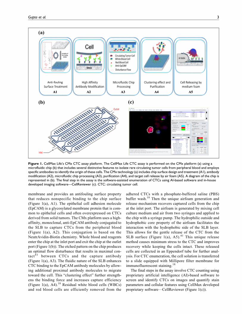
membrane and provides an antifouling surface propertythat reduces nonspecific binding to the chip surface(Figure 1(a), A1). The epithelial cell adhesion molecule(EpCAM) is a glycosylated membrane protein that is com-mon to epithelial cells and often overexpressed on CTCsderived from solid tumors. The CMx platform uses a high-affinity, monoclonal, anti-EpCAM antibody conjugated tothe SLB to capture CTCs from the peripheral blood(Figure 1(a), A2). This conjugation is based on theNeutrAvidin-Biotin chemistry. Whole blood and reagentsenter the chip at the inlet port and exit the chip at the outletport (Figure 1(b)). The etched pattern on the chip producesan optimal flow disturbance that results in maximal con-tact41 between CTCs and the capture antibody(Figure 1(a), A3). The fluidic nature of the SLB enhancesCTC binding to the EpCAM antibody molecules by allow-ing additional proximal antibody molecules to migratetoward the cell. This “clustering effect” further strength-ens the binding force and increases capture efficiency(Figure 1(a), A4).18 Residual white blood cells (WBCs)and red blood cells are efficiently removed from the
adhered CTCs with a phosphate-buffered saline (PBS)buffer wash.18 Then the unique airfoam generation andrelease mechanism recovers captured cells from the chipat the inlet port. The airfoam is generated by mixing cellculture medium and air from two syringes and applied tothe chip with a syringe pump. The hydrophilic outside andhydrophobic core property of the airfoam facilitates theinteraction with the hydrophobic side of the SLB layer.This allows for the gentle release of the CTC from theSLB surface (Figure 1(a), A5).41 This unique releasemethod causes minimum stress to the CTC and improvesrecovery while keeping the cells intact. These releasedcells are collected in an Eppendorf tube for further anal-ysis. For CTC enumeration, the cell solution is transferredto a slide equipped with Millipore filter membrane forimmunofluorescent staining.18
The final steps in the assay involve CTC counting usingproprietary artificial intelligence (AI)-based software toscreen and identify CTCs on images and quantify stainparameters and cellular features using CellMax developedproprietary software—CellReviewer (Figure 1(c)).
Figure 1. CellMax Life’s CMx CTC assay platform. The CellMax Life CTC assay is performed on the CMx platform (a) using amicrofluidic chip (b) that includes several distinctive features to isolate rare circulating tumor cells from peripheral blood and employsspecific antibodies to identify the origin of these cells. The CMx technology (a) includes chip surface design and treatment (A1), antibodymodification (A2), microfluidic chip processing (A3), purification (A4), and target cell release by air foam (A5). A diagram of the chip isrepresented in (b). The final step in the assay is the software-assisted enumeration of CTCs using AI-based software and in-housedeveloped imaging software—CellReviewer (c). CTC: circulating tumor cell.
Gupta et al. 3
CLC preparation, sample collection, processing,and CTC detection
Peripheral blood (4–8 mL) is drawn from the median decu-bitus vein by a trained phlebotomist and collected in a BDvacutainer tube containing K2EDTA as the anticoagulant.Streck cell preservative (Streck Inc., Omaha, Nebraska,USA) is added to the tube, typically within 2–4 h at a ratioof 4:1 (blood:preservative), then gently inverted 5 to 10times to mix, and stored at room temperature until delivery.The sample tubes are delivered in ambient conditions to theCellMax Laboratories in Sunnyvale (CLIA#: 05D2119031,CAP #: 9478056) or Taiwan (CAP #: 9258554) forprocessing.
The standard operating procedures for microfluidic chipfabrication, micropattern etching, chip surface modifica-tion (lipid and EpCAM coating), and airfoam generationand application originate from published research studies atAcademia Sinica.18,40 Whole blood (2 mL mixed with 0.5mL preservative) is loaded by syringe into the inlet port andpulled through a microfluidic channel by a syringe pumpconnected to the outlet port at a flow rate of 1.5 mL/h. Theunbound cells are washed out of the chip with three washesof PBS buffer (0.2 mL at 3 mL/h). The bound cells are fixedon the chip with 4% paraformaldehyde and are recoveredusing the airfoam mechanism connected to the outlet port.Released cells are collected in an Eppendorf tube via theinlet port. A small volume of ethanol is added to de-bubblethe foam–cell mixture in the tube. These cells are thentransferred to the membrane chip for counting.
Contrived samples for analytical validation includedboth donor blood and cell culture medium (Dulbecco’smodified Eagle’s medium, Thermo Scientific 11965084with 10% fetal bovine serum, Thermo Scientific26140079) spiked with prestained cancer cell line cells(CLCs) HT29 (HTB-38, ATCC). Prestaining was per-formed by adding CellTracker# Green CMFDA or DeepRed Dye (ThermoFisher C2925 or C34565) to the cellculture prior to harvesting, following the manufacturer’srecommendation. The stained cells were fixed with 4%paraformaldehyde and stored in the refrigerator for use.The prestained cell concentration was first estimated witha Scepter 2.0 automated cell counter (EMD MilliporePHCC20060) and then diluted to appropriate concentra-tions. Aliquots of prestained cells for the final workingconcentrations were mounted on membranes, imaged withthe microscope, and counted by two separate operators todetermine precise counts for spiking. Post-capture andrelease, the cell solution was carefully placed on a mem-brane, and liquid was wicked away with a blotting pad. Thesample was then mounted for imaging and counting.
For samples collected for the clinical feasibility analy-sis, the released cells were incubated with goat serum(10%) for 60 min at room temperature. Primary antibodieswere incubated overnight at 4!C in a refrigerator. The cellswere washed with PBS and incubated with fluorescently
tagged secondary antibodies. After final washes, the cellswere mounted with mounting medium containing 40,6-diamidino-2-phenylindole (DAPI) antifade (Thermo FisherP36931) onto a glass slide for image capture.
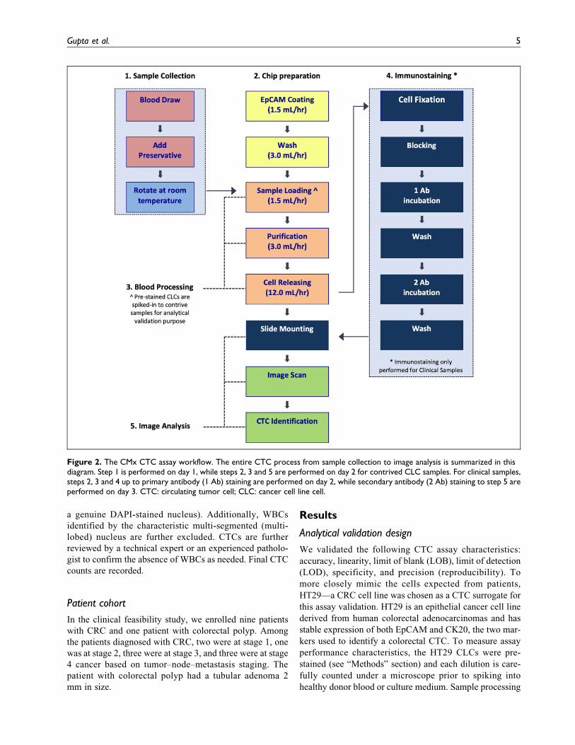
Figure 2 provides a schematic depiction of the CMxassay workflow. The samples are accessioned on day 1 andblood processing commences on day 2. For contrived sam-ples, captured cell release, slide mounting and image cap-ture are performed on day 2, followed by CLC counting.For clinical samples, immunostaining, slide mounting, andCTC enumeration are performed on day 3.
CLC enumeration
A CLC is defined as a cell that is round to oval in shape andthat demonstrates both a nucleus stain (DAPI, blue color)and a prestain (green color). Cells with a single stain werenot counted. Counting of prestained HT29 cells was per-formed with the commercial software MetaMorph (Version7.8, Molecular Device). The LAS-X stitched images (fromStep I described below) were loaded into MetaMorph,assigned colors (DAPI-blue, prestain-green), and mergedinto a single image. This image was then magnified andreviewed by an operator using a raster scanning mechanismwith the aid of location marks on the monitor.
CTC enumeration
Step I: Image capture. For each CTC sample (cells on 10-mmdiameter membrane), 100 (10 " 10) frames of images arecaptured in each of three channels: red, green, and blue,corresponding to TRITC for cytokeratin 20 (CK20), FITCfor lymphocyte common antigen (CD45), and DAPI fornucleus counterstain. The Leica autofocus mappingmechanism is applied to ensure images are in focus. Usingthe built-in stitching function of Leica Las-X software,these 100 image frames are stitched together as one imagevolume for each of the three channels.
Step II: Identification of candidate cells. We use custom soft-ware based on an AI algorithm developed specifically tosearch the stitched images for regions that have the char-acteristics of a cell. The coordinates of each target of inter-est on the image are recorded. The AI algorithm isrepeatedly trained with confirmed CTCs and WBCs toimprove its sensitivity and specificity. Each of the candi-date cells is assigned a confidence index, which allowsidentification for further morphology-based review andconfirmation.
Step III: Reviewing candidates to enumerate CTCs. For CTCconfirmation and enumeration, CellMax Life has devel-oped stringent criteria to avoid false positivity using a cus-tom software—CellReviewer. The CTC candidate cell’smorphology is reviewed by a trained technician. CTCsmust be round-to-oval shaped, have a cell size between 8mm and 40 mm and be CK20#, CD45$, and DAPI# (have
4 Journal of Circulating Biomarkers
a genuine DAPI-stained nucleus). Additionally, WBCsidentified by the characteristic multi-segmented (multi-lobed) nucleus are further excluded. CTCs are furtherreviewed by a technical expert or an experienced patholo-gist to confirm the absence of WBCs as needed. Final CTCcounts are recorded.
Patient cohort
In the clinical feasibility study, we enrolled nine patientswith CRC and one patient with colorectal polyp. Amongthe patients diagnosed with CRC, two were at stage 1, onewas at stage 2, three were at stage 3, and three were at stage4 cancer based on tumor–node–metastasis staging. Thepatient with colorectal polyp had a tubular adenoma 2mm in size.
Results
Analytical validation design
We validated the following CTC assay characteristics:accuracy, linearity, limit of blank (LOB), limit of detection(LOD), specificity, and precision (reproducibility). Tomore closely mimic the cells expected from patients,HT29—a CRC cell line was chosen as a CTC surrogate forthis assay validation. HT29 is an epithelial cancer cell linederived from human colorectal adenocarcinomas and hasstable expression of both EpCAM and CK20, the two mar-kers used to identify a colorectal CTC. To measure assayperformance characteristics, the HT29 CLCs were pre-stained (see “Methods” section) and each dilution is care-fully counted under a microscope prior to spiking intohealthy donor blood or culture medium. Sample processing
Figure 2. The CMx CTC assay workflow. The entire CTC process from sample collection to image analysis is summarized in thisdiagram. Step 1 is performed on day 1, while steps 2, 3 and 5 are performed on day 2 for contrived CLC samples. For clinical samples,steps 2, 3 and 4 up to primary antibody (1 Ab) staining are performed on day 2, while secondary antibody (2 Ab) staining to step 5 areperformed on day 3. CTC: circulating tumor cell; CLC: cancer cell line cell.
Gupta et al. 5
and CLC enumeration of spiked samples followed the pro-tocol described in the “Methods” section. Table 1 liststhe analytical performance characteristics validated inthis study.
Assay accuracy
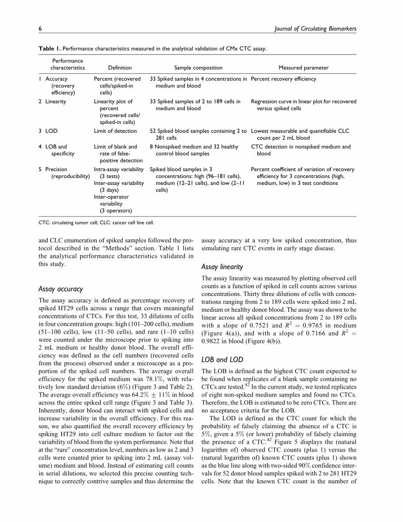
The assay accuracy is defined as percentage recovery ofspiked HT29 cells across a range that covers meaningfulconcentrations of CTCs. For this test, 33 dilutions of cellsin four concentration groups: high (101–200 cells), medium(51–100 cells), low (11–50 cells), and rare (1–10 cells)were counted under the microscope prior to spiking into2 mL medium or healthy donor blood. The overall effi-ciency was defined as the cell numbers (recovered cellsfrom the process) observed under a microscope as a pro-portion of the spiked cell numbers. The average overallefficiency for the spiked medium was 78.1%, with rela-tively low standard deviation (6%) (Figure 3 and Table 2).The average overall efficiency was 64.2% + 11% in bloodacross the entire spiked cell range (Figure 3 and Table 3).Inherently, donor blood can interact with spiked cells andincrease variability in the overall efficiency. For this rea-son, we also quantified the overall recovery efficiency byspiking HT29 into cell culture medium to factor out thevariability of blood from the system performance. Note thatat the “rare” concentration level, numbers as low as 2 and 3cells were counted prior to spiking into 2 mL (assay vol-ume) medium and blood. Instead of estimating cell countsin serial dilutions, we selected this precise counting tech-nique to correctly contrive samples and thus determine the
assay accuracy at a very low spiked concentration, thussimulating rare CTC events in early stage disease.
Assay linearity
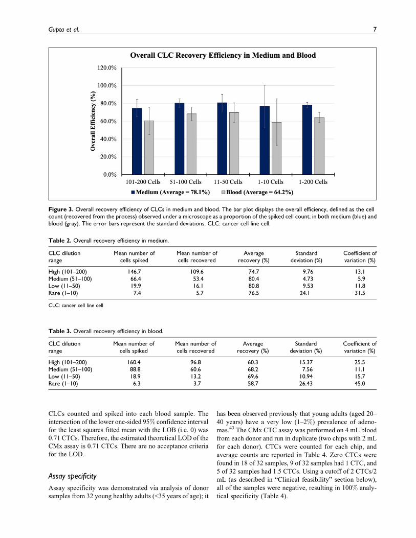
The assay linearity was measured by plotting observed cellcounts as a function of spiked in cell counts across variousconcentrations. Thirty three dilutions of cells with concen-trations ranging from 2 to 189 cells were spiked into 2 mLmedium or healthy donor blood. The assay was shown to belinear across all spiked concentrations from 2 to 189 cellswith a slope of 0.7521 and R2 % 0.9765 in medium(Figure 4(a)), and with a slope of 0.7166 and R2 %0.9822 in blood (Figure 4(b)).
LOB and LOD
The LOB is defined as the highest CTC count expected tobe found when replicates of a blank sample containing noCTCs are tested.42 In the current study, we tested replicatesof eight non-spiked medium samples and found no CTCs.Therefore, the LOB is estimated to be zero CTCs. There areno acceptance criteria for the LOB.
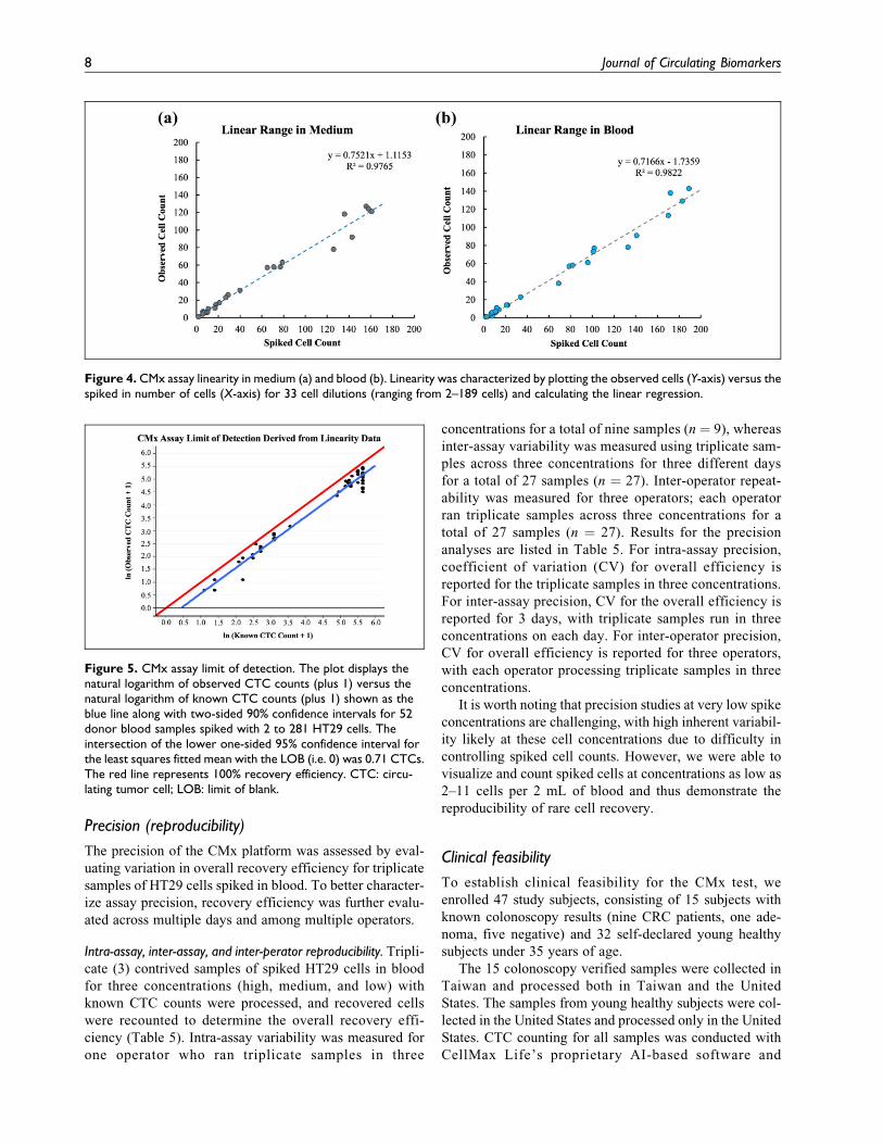
The LOD is defined as the CTC count for which theprobability of falsely claiming the absence of a CTC is5%, given a 5% (or lower) probability of falsely claimingthe presence of a CTC.42 Figure 5 displays the (naturallogarithm of) observed CTC counts (plus 1) versus the(natural logarithm of) known CTC counts (plus 1) shownas the blue line along with two-sided 90% confidence inter-vals for 52 donor blood samples spiked with 2 to 281 HT29cells. Note that the known CTC count is the number of
Table 1. Performance characteristics measured in the analytical validation of CMx CTC assay.
Performancecharacteristics Definition Sample composition Measured parameter
1 Accuracy(recoveryefficiency)
Percent (recoveredcells/spiked-incells)
33 Spiked samples in 4 concentrations inmedium and blood
Percent recovery efficiency
2 Linearity Linearity plot ofpercent(recovered cells/spiked-in cells)
33 Spiked samples of 2 to 189 cells inmedium and blood
Regression curve in linear plot for recoveredversus spiked cells
3 LOD Limit of detection 52 Spiked blood samples containing 2 to281 cells
Lowest measurable and quantifiable CLCcount per 2 mL blood
4 LOB andspecificity
Limit of blank andrate of false-positive detection
8 Nonspiked medium and 32 healthycontrol blood samples
CTC detection in nonspiked medium andblood
5 Precision(reproducibility)
Intra-assay variability(3 tests)
Spiked blood samples in 3concentrations: high (96–181 cells),medium (12–21 cells), and low (2–11cells)
Percent coefficient of variation of recoveryefficiency for 3 concentrations (high,medium, low) in 3 test conditionsInter-assay variability
(3 days)Inter-operator
variability(3 operators)
CTC: circulating tumor cell; CLC: cancer cell line cell.
6 Journal of Circulating Biomarkers
CLCs counted and spiked into each blood sample. Theintersection of the lower one-sided 95% confidence intervalfor the least squares fitted mean with the LOB (i.e. 0) was0.71 CTCs. Therefore, the estimated theoretical LOD of theCMx assay is 0.71 CTCs. There are no acceptance criteriafor the LOD.
Assay specificity
Assay specificity was demonstrated via analysis of donorsamples from 32 young healthy adults (<35 years of age); it
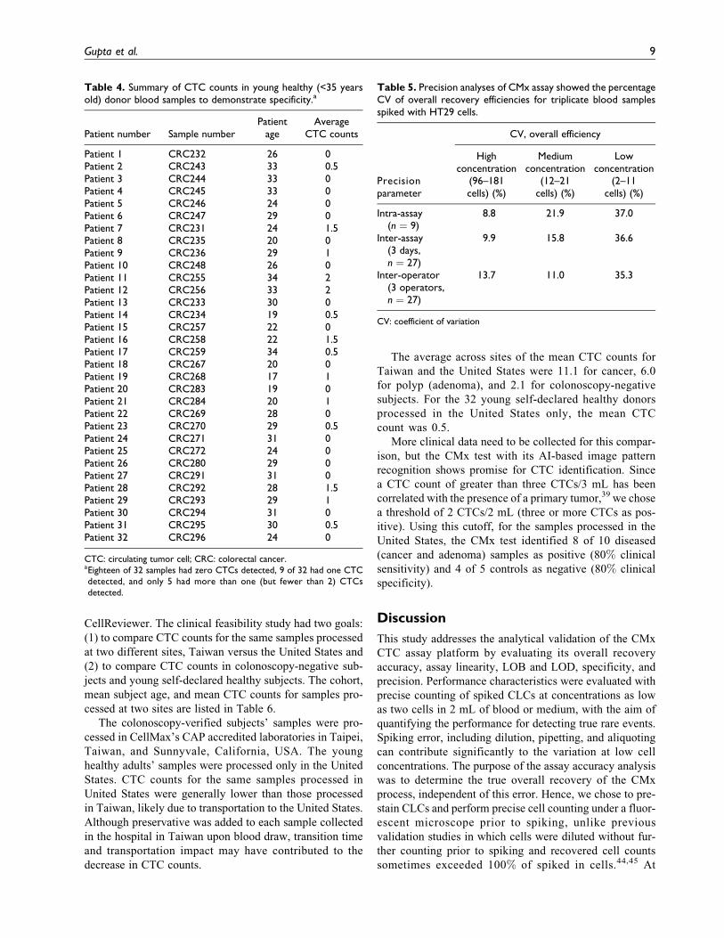
has been observed previously that young adults (aged 20–40 years) have a very low (1–2%) prevalence of adeno-mas.43 The CMx CTC assay was performed on 4 mL bloodfrom each donor and run in duplicate (two chips with 2 mLfor each donor). CTCs were counted for each chip, andaverage counts are reported in Table 4. Zero CTCs werefound in 18 of 32 samples, 9 of 32 samples had 1 CTC, and5 of 32 samples had 1.5 CTCs. Using a cutoff of 2 CTCs/2mL (as described in “Clinical feasibility” section below),all of the samples were negative, resulting in 100% analy-tical specificity (Table 4).
Figure 3. Overall recovery efficiency of CLCs in medium and blood. The bar plot displays the overall efficiency, defined as the cellcount (recovered from the process) observed under a microscope as a proportion of the spiked cell count, in both medium (blue) andblood (gray). The error bars represent the standard deviations. CLC: cancer cell line cell.
Table 2. Overall recovery efficiency in medium.
CLC dilutionrange
Mean number ofcells spiked
Mean number ofcells recovered
Averagerecovery (%)
Standarddeviation (%)
Coefficient ofvariation (%)
High (101–200) 146.7 109.6 74.7 9.76 13.1Medium (51–100) 66.4 53.4 80.4 4.73 5.9Low (11–50) 19.9 16.1 80.8 9.53 11.8Rare (1–10) 7.4 5.7 76.5 24.1 31.5
CLC: cancer cell line cell
Table 3. Overall recovery efficiency in blood.
CLC dilutionrange
Mean number ofcells spiked
Mean number ofcells recovered
Averagerecovery (%)
Standarddeviation (%)
Coefficient ofvariation (%)
High (101–200) 160.4 96.8 60.3 15.37 25.5Medium (51–100) 88.8 60.6 68.2 7.56 11.1Low (11–50) 18.9 13.2 69.6 10.94 15.7Rare (1–10) 6.3 3.7 58.7 26.43 45.0
Gupta et al. 7
Precision (reproducibility)
The precision of the CMx platform was assessed by eval-uating variation in overall recovery efficiency for triplicatesamples of HT29 cells spiked in blood. To better character-ize assay precision, recovery efficiency was further evalu-ated across multiple days and among multiple operators.
Intra-assay, inter-assay, and inter-perator reproducibility. Tripli-cate (3) contrived samples of spiked HT29 cells in bloodfor three concentrations (high, medium, and low) withknown CTC counts were processed, and recovered cellswere recounted to determine the overall recovery effi-ciency (Table 5). Intra-assay variability was measured forone operator who ran triplicate samples in three
concentrations for a total of nine samples (n % 9), whereasinter-assay variability was measured using triplicate sam-ples across three concentrations for three different daysfor a total of 27 samples (n % 27). Inter-operator repeat-ability was measured for three operators; each operatorran triplicate samples across three concentrations for atotal of 27 samples (n % 27). Results for the precisionanalyses are listed in Table 5. For intra-assay precision,coefficient of variation (CV) for overall efficiency isreported for the triplicate samples in three concentrations.For inter-assay precision, CV for the overall efficiency isreported for 3 days, with triplicate samples run in threeconcentrations on each day. For inter-operator precision,CV for overall efficiency is reported for three operators,with each operator processing triplicate samples in threeconcentrations.
It is worth noting that precision studies at very low spikeconcentrations are challenging, with high inherent variabil-ity likely at these cell concentrations due to difficulty incontrolling spiked cell counts. However, we were able tovisualize and count spiked cells at concentrations as low as2–11 cells per 2 mL of blood and thus demonstrate thereproducibility of rare cell recovery.
Clinical feasibility
To establish clinical feasibility for the CMx test, weenrolled 47 study subjects, consisting of 15 subjects withknown colonoscopy results (nine CRC patients, one ade-noma, five negative) and 32 self-declared young healthysubjects under 35 years of age.
The 15 colonoscopy verified samples were collected inTaiwan and processed both in Taiwan and the UnitedStates. The samples from young healthy subjects were col-lected in the United States and processed only in the UnitedStates. CTC counting for all samples was conducted withCellMax Life’s proprietary AI-based software and
Figure 4. CMx assay linearity in medium (a) and blood (b). Linearity was characterized by plotting the observed cells (Y-axis) versus thespiked in number of cells (X-axis) for 33 cell dilutions (ranging from 2–189 cells) and calculating the linear regression.
Figure 5. CMx assay limit of detection. The plot displays thenatural logarithm of observed CTC counts (plus 1) versus thenatural logarithm of known CTC counts (plus 1) shown as theblue line along with two-sided 90% confidence intervals for 52donor blood samples spiked with 2 to 281 HT29 cells. Theintersection of the lower one-sided 95% confidence interval forthe least squares fitted mean with the LOB (i.e. 0) was 0.71 CTCs.The red line represents 100% recovery efficiency. CTC: circu-lating tumor cell; LOB: limit of blank.
8 Journal of Circulating Biomarkers
CellReviewer. The clinical feasibility study had two goals:(1) to compare CTC counts for the same samples processedat two different sites, Taiwan versus the United States and(2) to compare CTC counts in colonoscopy-negative sub-jects and young self-declared healthy subjects. The cohort,mean subject age, and mean CTC counts for samples pro-cessed at two sites are listed in Table 6.
The colonoscopy-verified subjects’ samples were pro-cessed in CellMax’s CAP accredited laboratories in Taipei,Taiwan, and Sunnyvale, California, USA. The younghealthy adults’ samples were processed only in the UnitedStates. CTC counts for the same samples processed inUnited States were generally lower than those processedin Taiwan, likely due to transportation to the United States.Although preservative was added to each sample collectedin the hospital in Taiwan upon blood draw, transition timeand transportation impact may have contributed to thedecrease in CTC counts.
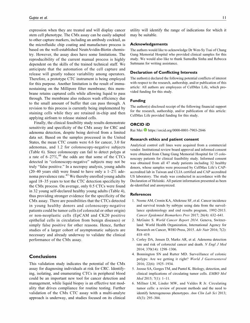
The average across sites of the mean CTC counts forTaiwan and the United States were 11.1 for cancer, 6.0for polyp (adenoma), and 2.1 for colonoscopy-negativesubjects. For the 32 young self-declared healthy donorsprocessed in the United States only, the mean CTCcount was 0.5.
More clinical data need to be collected for this compar-ison, but the CMx test with its AI-based image patternrecognition shows promise for CTC identification. Sincea CTC count of greater than three CTCs/3 mL has beencorrelated with the presence of a primary tumor,39 we chosea threshold of 2 CTCs/2 mL (three or more CTCs as pos-itive). Using this cutoff, for the samples processed in theUnited States, the CMx test identified 8 of 10 diseased(cancer and adenoma) samples as positive (80% clinicalsensitivity) and 4 of 5 controls as negative (80% clinicalspecificity).
Discussion
This study addresses the analytical validation of the CMxCTC assay platform by evaluating its overall recoveryaccuracy, assay linearity, LOB and LOD, specificity, andprecision. Performance characteristics were evaluated withprecise counting of spiked CLCs at concentrations as lowas two cells in 2 mL of blood or medium, with the aim ofquantifying the performance for detecting true rare events.Spiking error, including dilution, pipetting, and aliquotingcan contribute significantly to the variation at low cellconcentrations. The purpose of the assay accuracy analysiswas to determine the true overall recovery of the CMxprocess, independent of this error. Hence, we chose to pre-stain CLCs and perform precise cell counting under a fluor-escent microscope prior to spiking, unlike previousvalidation studies in which cells were diluted without fur-ther counting prior to spiking and recovered cell countssometimes exceeded 100% of spiked in cells.44,45 At
Table 4. Summary of CTC counts in young healthy (<35 yearsold) donor blood samples to demonstrate specificity.a
Patient number Sample numberPatient
ageAverage
CTC counts
Patient 1 CRC232 26 0Patient 2 CRC243 33 0.5Patient 3 CRC244 33 0Patient 4 CRC245 33 0Patient 5 CRC246 24 0Patient 6 CRC247 29 0Patient 7 CRC231 24 1.5Patient 8 CRC235 20 0Patient 9 CRC236 29 1Patient 10 CRC248 26 0Patient 11 CRC255 34 2Patient 12 CRC256 33 2Patient 13 CRC233 30 0Patient 14 CRC234 19 0.5Patient 15 CRC257 22 0Patient 16 CRC258 22 1.5Patient 17 CRC259 34 0.5Patient 18 CRC267 20 0Patient 19 CRC268 17 1Patient 20 CRC283 19 0Patient 21 CRC284 20 1Patient 22 CRC269 28 0Patient 23 CRC270 29 0.5Patient 24 CRC271 31 0Patient 25 CRC272 24 0Patient 26 CRC280 29 0Patient 27 CRC291 31 0Patient 28 CRC292 28 1.5Patient 29 CRC293 29 1Patient 30 CRC294 31 0Patient 31 CRC295 30 0.5Patient 32 CRC296 24 0
CTC: circulating tumor cell; CRC: colorectal cancer.aEighteen of 32 samples had zero CTCs detected, 9 of 32 had one CTCdetected, and only 5 had more than one (but fewer than 2) CTCsdetected.
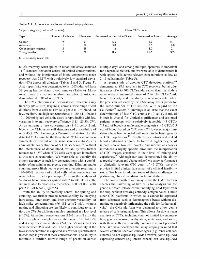
Table 5. Precision analyses of CMx assay showed the percentageCV of overall recovery efficiencies for triplicate blood samplesspiked with HT29 cells.
Precisionparameter
CV, overall efficiency
Highconcentration
(96–181cells) (%)
Mediumconcentration
(12–21cells) (%)
Lowconcentration
(2–11cells) (%)
Intra-assay(n % 9)
8.8 21.9 37.0
Inter-assay(3 days,n % 27)
9.9 15.8 36.6
Inter-operator(3 operators,n % 27)
13.7 11.0 35.3
CV: coefficient of variation
Gupta et al. 9
64.2% recovery when spiked in blood, the assay achieved11% standard deviation across all spiked concentrations,and without the interference of blood components meanrecovery was 78.1% with a relatively low standard devia-tion (6%) across all dilutions (Tables 2 and 3; Figure 3).Assay specificity was determined to be 100%, derived from32 young healthy donor blood samples (Table 4). More-over, using 8 unspiked medium samples (blank), wedemonstrated LOB of zero CTCs.
The CMx platform also demonstrated excellent assaylinearity (R2 % 0.98) (Figure 4) across a wide range of celldilutions from 2 cells to 189 cells per 2 mL of blood. Atlow, medium, and high concentrations (11–50, 51–100, and101–200) of spiked cells, the assay is reproducible with lowvariation in overall recovery efficiency (11.1–25.5% CV).At an extremely rare concentration (1–10 cells/ 2 mLblood), the CMx assay still demonstrated a variability ofonly 45% CV. Assuming a Poisson distribution for thedetected CTCs/sample, the theoretical CV for a set of mea-surements carried out on the same samples is 44.7% at acomparable concentration of 5 CTCs/7.5 mL.46 Withoutthe interference of donor blood, variability was furtherreduced to 31.5% when HT29 cells were spiked in mediumat this rare concentration. We were able to quantify thesystem accuracy at such low concentrations with a combi-nation of prestaining and precise counting. Dilutions and/orcounting errors likely led to previous attempts resulting in120–200% recovery of spiked cells when concentrationswere below 10 cells per sample.45 From the analysis of52 donor blood samples spiked with 2 to 281 HT29 cells,we were able to establish a theoretical LOD of 0.71 cellsper 2 mL of blood (Figure 5).
With the ability to precisely control for spiking andcounting, we further proved reproducibility by studyingintra-assay, inter-assay, and inter-operator variability. Athigh spike concentrations (96–181 cells/2 mL), whereinmixing and aliquoting are less likely to contribute to varia-tion, the CVs for triplicate samples remained at a low level(<15%). At medium concentrations (12–21 cells/2 mL), theCV for triplicate samples was in the range of 11.1–21.9%and at very low concentrations (2–11 cells/2 mL), the CVswere between 35% and 37%. The higher variability at thelowest concentration is expected as error for quantificationin each step is greater at these concentrations. The ability tomaintain a similar, narrow range of precision across
multiple days and among multiple operators is importantfor a reproducible test, and we were able to demonstrate itwith spiked cells across relevant concentrations as low as2–11 cells/sample (Table 5).
A recent study of another CTC detection platform44
demonstrated 88% accuracy in CTC recovery, but at dilu-tion rates of 6 to 300 CLCs/slide, rather than this study’smore realistic measured range of 2 to 189 CLCs/2 mLblood. Linearity and specificity were comparable, whilethe precision achieved by the CMx assay was superior forthe same number of CLCs/slide. With regard to theCellSearch" system, Cummings et al. state that the exactdetermination of low CTC counts (<10 cells/7.5 mL ofblood) is crucial for clinical significance and assignedpatients to groups with a relatively favorable (<5 CTCs/7.5 mL of blood) or unfavorable prognosis (& 5 CTCs/7.5mL of blood) based on CTC count.46 However, major lim-itations have been reported with regard to the heterogeneityof CTC populations.19 Results from controls and spikedblood confirmed a three- to fourfold higher degree ofimprecision at low cell counts, and individual analystsintroduced a highly specific error into the interpretationof CTC images, correlated to their level of training andexperience.46 Although our data demonstrated the abilityto precisely count and characterize CMx assay performanceat clinically relevant CTC count of <5 CTCs, we onlyprovide limited clinical data as part of a clinical feasibilitystudy. We hope to address some of these challenges byperforming clinical validation in future studies.
The core strength of our assay is that the CMx platformenables the harvesting of live cells for analysis via thegentle air foam release of the underlying lipid layer fromthe chip, without breaking antibody–antigen bonds. Unlikeother CTC platforms in which cells cannot be separatedfrom substrates such as ferromagnetic beads without dis-rupting or negatively influencing the cells for further anal-ysis,47 the CMx platform was designed to allow gentlerelease of cells using airfoam. This allows for downstreamanalyses of CTCs, including (but not limited to) enumera-tion, gene expression, methylation, mutations, and so on,with these cells conveniently contained in an Eppendorftube. We have developed the assay keeping in mind thatseveral epithelial-derived cancer types (e.g. renal cell car-cinoma) do not express EpCAM; however, some EpCAMexpressing cancers (e.g. breast cancer) can lose EpCAM
Table 6. CTC counts in healthy and diseased subpopulations.
Subject category (total % 47 patients) Mean CTC counts
Type Number of subjects Mean age Processed in the United States Processed in Taiwan Average
Cancer 9 51 6.6 15.7 11.1Adenoma 1 66 3.0 9.0 6.0Colonoscopy negative 5 59 1.2 3.0 2.1Young healthy 32 26 0.5 N/A N/A
CTC: circulating tumor cell.
10 Journal of Circulating Biomarkers
expression when they are treated and will display cancerstem cell phenotype. The CMx assay can be easily adaptedto other capture markers, including an antibody cocktail, asthe microfluidic chip coating and manufacture process isbased on the well-established NeutrAvidin-Biotin chemis-try. However, the assay does have some limitations. Thereproducibility of the current manual process is highlydependent on the skills of the trained technical staff. Weanticipate that the automation of the cell capture andrelease will greatly reduce variability among operators.Therefore, a prototype CTC instrument is being employedfor this purpose. Another limitation is the result of immu-nostaining on the Millipore filter membrane; this mem-brane retains captured cells while allowing liquid to passthrough. The membrane also reduces wash efficiency dueto the small amount of buffer that can pass through. Arevision to this process is currently being implemented bystaining cells while they are retained in-chip and thenapplying airfoam to release stained cells.
Finally, the clinical feasibility study results demonstratesensitivity and specificity of the CMx assay for CRC andadenoma detection, despite being derived from a limiteddata set. Based on the samples processed in the UnitedStates, the mean CTC counts were 6.6 for cancer, 3.0 foradenomas, and 1.2 for colonoscopy-negative subjects(Table 6). Since colonoscopy can fail to detect polyps ata rate of 6–27%,48 the odds are that some of the CTCsdetected in “colonoscopy-negative” subjects may not betruly “false positive.” In a necropsy analysis, young adults(20–40 years old) were found to have only a 1–2% ade-noma prevalence rate.43 We thereby enrolled young adultsaged 18–35 years to test the CTC detection specificity bythe CMx process. On average, only 0.5 CTCs were foundin 32 young self-declared healthy young adults (Table 4),thus providing stronger evidence for the specificity of theCMx assay. There are possibilities that the CTCs detectedin young healthy donors and colonoscopy-negativepatients could be tumor cells (of colorectal or other origin)or non-neoplastic cells (EpCAM and CK20 positiveepithelial cells in circulation from benign diseases) orsimply false positive for other reasons. Hence, furtherstudies of a larger cohort of asymptomatic subjects arenecessary and already underway to validate the clinicalperformance of the CMx assay.
Conclusions
This validation study indicates the potential of the CMxassay for diagnosing individuals at risk for CRC. Identify-ing, isolating, and enumerating CTCs in peripheral bloodcould be an important new tool for cancer detection andmanagement, while liquid biopsy is an effective test mod-ality that drives compliance for routine testing. Furthervalidation of the CMx CTC assay with a multi-analyteapproach is underway, and studies focused on its clinical
utility will identify the range of indications for which itmay be suitable.
Acknowledgements
The authors would like to acknowledge Dr Wen-Sy Tsai of ChangGung Memorial Hospital who provided clinical samples for thisstudy. We would also like to thank Sumedha Sinha and RebeccaSuttmann for writing assistance.
Declaration of Conflicting Interests
The author(s) declared the following potential conflicts of interestwith respect to the research, authorship, and/or publication of thisarticle: All authors are employees of CellMax Life, which pro-vided funding for this study.
Funding
The author(s) disclosed receipt of the following financial supportfor the research, authorship, and/or publication of this article:CellMax Life provided funding for this study.
ORCID iD
Rui Mei https://orcid.org/0000-0001-7903-2846
Research ethics and patient consent
Analytical control cell lines were acquired from a commercialvendor. Institutional review board approval and informed consentwere obtained from Chang Gung Memorial Hospital for 15 colo-noscopy patients for clinical feasibility study. Informed consentwas obtained from all 47 study patients including 32 healthydonors, whose samples were processed by CellMax Life’s CAP-accredited lab in Taiwan and CLIA certified and CAP accreditedUS laboratory. The study was conducted in accordance with theDeclaration of Helsinki. All patient information presented as beende-identified and anonymized.
References
1. Noone AM, Cronin KA, Altekruse SF, et al. Cancer incidence
and survival trends by subtype using data from the surveil-
lance epidemiology and end results program, 1992-2013.
Cancer Epidemiol Biomarkers Prev 2017; 26(4): 632–641.
2. McGuire S. World Cancer Report 2014. Geneva, Switzer-
land: World Health Organization, International Agency for
Research on Cancer, WHO Press, 2015. Adv Nutr 2016; 7(2):
418–419.
3. Corley DA, Jensen D, Marks AR, et al. Adenoma detection
rate and risk of colorectal cancer and death. N Engl J Med
2014; 370(14): 1298–1306.
4. Bonnington SN and Rutter MD. Surveillance of colonic
polyps: Are we getting it right? World J Gastroenterol
2016; 22(6): 1925–1934.
5. Joosse SA, Gorges TM, and Pantel K. Biology, detection, and
clinical implications of circulating tumor cells. EMBO Mol
Med 2015; 7(1): 1–11.
6. Millner LM, Linder MW, and Valdes R Jr. Circulating
tumor cells: a review of present methods and the need to
identify heterogeneous phenotypes. Ann Clin Lab Sci 2013;
43(3): 295–304.
Gupta et al. 11
7. Ashworth TR. A case of cancer in which cells similar to those
in the tumours were seen in the blood after death. Med J Aust
1869; 14: 146–147.
8. Jeong KY, Kim EK, Park MH, et al. Perspective on cancer
therapeutics utilizing analysis of circulating tumor cells.
Diagnostics (Basel) 2018; 8(2): 23.
9. Nesteruk D, Rutkowski A, Fabisiewicz S, et al. Evaluation of
prognostic significance of circulating tumor cells detection in
rectal cancer patients treated with preoperative radiotherapy:
prospectively collected material data. Biomed Res Int 2014;
2014: 712827.
10. Pantel K and Alix-Panabieres C. Real-time liquid biopsy in
cancer patients: fact or fiction? Cancer Res 2013; 73(21):
6384–6388.
11. Miller MC, Doyle GV, and Terstappen LW. Significance of
circulating tumor cells detected by the cellsearch system in
patients with metastatic breast colorectal and prostate cancer.
J Oncol 2010; 2010: 617421.
12. Cohen SJ, Punt CJ, Iannotti N, et al. Relationship of circulat-
ing tumor cells to tumor response, progression-free survival,
and overall survival in patients with metastatic colorectal
cancer. J Clin Oncol 2008; 26(19): 3213–3221.
13. Siegel RL, Miller KD, and Jemal A. Cancer statistics, 2016.
CA Cancer J Clin 2016; 66(1): 7–30.
14. Budd GT, Cristofanilli M, Ellis MJ, et al. Circulating tumor
cells versus imaging–predicting overall survival in meta-
static breast cancer. Clin Cancer Res 2006; 12(21):
6403–6409.
15. Hayes DF, Cristofanilli M, Budd GT, et al. Circulating tumor
cells at each follow-up time point during therapy of meta-
static breast cancer patients predict progression-free and
overall survival. Clin Cancer Res 2006; 12(14 Pt 1):
4218–4224.
16. Gorin MA, Verdone JE, van der Toom E, et al. Circulating
tumour cells as biomarkers of prostate, bladder, and kidney
cancer. Nat Rev Urol 2017; 14(2): 90–97.
17. de Bono JS, Scher HI, Montgomery RB, et al. Circulating
tumor cells predict survival benefit from treatment in meta-
static castration-resistant prostate cancer. Clin Cancer Res
2008; 14(19): 6302–6309.
18. Wu JC, Tseng PY, Tsai WS, et al. Antibody conjugated sup-
ported lipid bilayer for capturing and purification of viable
tumor cells in blood for subsequent cell culture. Biomaterials
2013; 34(21): 5191–5199.
19. Andree KC, van Dalum G and Terstappen LW. Challenges in
circulating tumor cell detection by the CellSearch system.
Mol Oncol 2016; 10(3): 395–407.
20. Ren C, Chongxu H, Daxin W, et al. Detection of circulating
tumor cells: Clinical relevance of a novel metastatic tumor
marker. Exp Ther Med 2011; 2(3): 385–391.
21. Parkinson DR, Dracopoli N, Petty BG, et al. Considerations
in the development of circulating tumor cell technology for
clinical use. J Transl Med 2012; 10: 138.
22. Glaves D. Correlation between circulating cancer cells and
incidence of metastases. Br J Cancer 1983; 48(5): 665–673.
23. Rack B, Schindlbeck C, Juckstock J, et al. Circulating tumor
cells predict survival in early average-to-high risk breast can-
cer patients. J Natl Cancer Inst 2014; 106(5): dju066.
24. Nagrath S, Sequist LV, Maheswaran S, et al. Isolation of rare
circulating tumour cells in cancer patients by microchip tech-
nology. Nature 2007; 450(7173): 1235–1239.
25. Li P, Stratton ZS, Dao M, et al. Probing circulating tumor
cells in microfluidics. Lab Chip 2013; 13(4): 602–609.
26. Huang MY, Tsai HL, Huang JJ, et al. Clinical implications
and future perspectives of circulating tumor cells and biomar-
kers in clinical outcomes of colorectal cancer. Transl Oncol
2016; 9(4): 340–347.
27. Gertler R, Rosenberg R, Fuehrer K, et al. Detection of circu-
lating tumor cells in blood using an optimized density gradi-
ent centrifugation. Recent Results Cancer Res 2003; 162:
149–155.
28. Stott SL, Hsu CH, Tsukrov DI, et al. Isolation of circulating
tumor cells using a microvortex-generating herringbone-chip.
Proc Natl Acad Sci U S A 2010; 107(43): 18392–18397.
29. Gupta V, Jafferji I, Garza M, et al. ApoStream(), a new die-
lectrophoretic device for antibody independent isolation and
recovery of viable cancer cells from blood. Biomicrofluidics
2012; 6(2): 24133.
30. Hur SC, Henderson-MacLennan NK, McCabe ER, et al.
Deformability-based cell classification and enrichment using
inertial microfluidics. Lab Chip 2011; 11(5): 912–920.
31. Hur SC, Mach AJ, and Di Carlo D. High-throughput size-
based rare cell enrichment using microscale vortices. Biomi-
crofluidics 2011; 5(2): 22206.
32. Zheng S, Lin HK, Lu B, et al. 3D microfilter device for viable
circulating tumor cell (CTC) enrichment from blood. Biomed
Microdevices 2011; 13(1): 203–213.
33. Tan SJ, Yobas L, Lee GY, et al. Microdevice for the isolation
and enumeration of cancer cells from blood. Biomed Micro-
devices 2009; 11(4): 883–892.
34. Ilie M, Hofman V, Long-Mira E, et al. “Sentinel” circulating
tumor cells allow early diagnosis of lung cancer in patients
with chronic obstructive pulmonary disease. PLoS One 2014;
9(10): e111597.
35. Murray NP, Miranda R, Ruiz A, et al. Diagnostic yield of
primary circulating tumor cells in women suspected of breast
cancer: the BEST (Breast Early Screening Test) study. Asian
Pac J Cancer Prev 2015; 16(5): 1929–1934.
36. Flores LM, Kindelberger DW, Ligon AH, et al. Improving the
yield of circulating tumour cells facilitates molecular char-
acterisation and recognition of discordant HER2 amplifica-
tion in breast cancer. Br J Cancer 2010; 102(10): 1495–1502.
37. Tsai WS, Chen JS, Shao HJ, et al. Circulating tumor cell
count correlates with colorectal neoplasm progression and
is a prognostic marker for distant metastasis in non-
metastatic patients. Sci Rep 2016; 6: 24517.
38. Bork U, Rahbari NN, Scholch S, et al. Circulating tumour
cells and outcome in non-metastatic colorectal cancer: a pro-
spective study. Br J Cancer 2015; 112(8): 1306–1313.
39. Yang C, Zhuang W, Hu Y, et al. Clinical significance of
peripheral circulating tumor cell counts in colorectal polyps
12 Journal of Circulating Biomarkers
and non-metastatic colorectal cancer. World J Surg Oncol
2018; 16(1): 13.
40. Lai JM, Shao HJ, Wu JC, et al. Efficient elusion of viable
adhesive cells from a microfluidic system by air foam. Bio-
microfluidics 2014; 8(5): 052001.
41. Chen JY, Tsai WS, Shao HJ, et al. Sensitive and specific
biomimetic lipid coated microfluidics to isolate viable circu-
lating tumor cells and microemboli for cancer detection.
PLoS ONE 2016; 11(3): e0149633.
42. Armbruster DA and Pry T. Limit of blank, limit of detection
and limit of quantitation. Clin Biochem Rev 2008; 29(Suppl
1): S49–52.
43. Pendergrass CJ, Edelstein DL, Hylind LM, et al. Occurrence
of colorectal adenomas in younger adults: an epidemiologic
necropsy study. Clin Gastroenterol Hepatol 2008; 6(9):
1011–1015.
44. Werner SL, Graf RP, Landers M, et al. Analytical validation
and capabilities of the epic CTC platform: enrichment-free
circulating tumour cell detection and characterization. J Circ
Biomark 2015; 4:3.
45. Veridex_LLC_510 K, CellSearch 510 K Summary k071729.
2003.
46. Cummings J, Morris K, Zhou C, et al. Method validation of
circulating tumour cell enumeration at low cell counts. BMC
Cancer 2013; 13: 415–415.
47. Lv SW, Wang J, Xie M, et al. Photoresponsive immunomag-
netic nanocarrier for capture and release of rare circulating
tumor cells. Chem Sci 2015; 6(11): 6432–6438.
48. Ahn SB, Han DS, Bae JH, et al. The miss rate for colorectal
adenoma determined by quality-adjusted, back-to-back colo-
noscopies. Gut Liver 2012; 6(1): 64–70.
Gupta et al. 13