Embed Size (px)
Citation preview
Cite this: Lab Chip, 2013, 13, 468
An intravenous implantable glucose/dioxygen biofuelcell with modified flexible carbon fiber electrodes3
Received 3rd September 2012,Accepted 3rd November 2012
DOI: 10.1039/c2lc41007a
www.rsc.org/loc
Fernanda C. P. F. Sales,a Rodrigo M. Iost,b Marccus V. A. Martins,a Maria C. Almeidaa
and Frank N. Crespilho*b
An intravenous implantable glucose/dioxygen hybrid enzyme–Pt micro-biofuel cell (BFC) was investigated.
In this miniaturized BFC, a flexible carbon fiber (FCF) microelectrode modified with neutral red redox
mediator and glucose oxidase was used as the bioanode, and an FCF modified with platinum
nanoparticles stabilized on PAMAM-G4 dendrimer was used as the cathode. In vitro experiments
conducted using the BFC in a phosphate buffer solution (50 mmol L21, pH = 7.2) and glucose (47
mmol L21) showed high electrocatalytic performance with an open circuit voltage (OCV) of 400 mV, a
maximum current density of 2700 mA cm22 at 0.0 V and a maximum output power of 200 mW cm22 at 250
mV. Under physiological conditions, glucose from rat blood is used as a fuel in anodic reactions and
dissolved molecular oxygen is used as the oxidizing agent on the cathode. For in vivo experiments, the BFC
was inserted into the jugular vein of a living rat (Rattus novergicus) using a catheter (internal diameter 0.5
mm). The power density of the implantable BFC was evaluated over a period of 24 h, and an OCV of 125
mV with a maximum power density of 95 mW cm22 was obtained at 80 mV.
Introduction
Enzymatic biofuel cells1–5 (BFCs) have attracted attentionbecause of their potential to supply power to bionic implants.Thus far, several designs have been proposed for BFCs used inhigh power biodevices.1–3 The architecture and constituents ofthe electrodes are critical factors in the development of BFCs.Conductive carbon materials, such as highly porous carbon1
and carbon nanotubes,2 have been used for wiring redoxenzymes to the electrode surface. Furthermore, the use ofhighly porous carbon as a component of bioanodes maximizesthe interaction between the enzyme and the electrode surface,4
yielding high output power densities of up to 750 mW cm22.In addition, the use of renewable fuels, such as glucose, has
been extensively explored.6 A great advantage of glucose is thatit is easily found in the blood of living organisms. Typically,blood glucose concentration is in the range of 3–5 mmol L21,and the venous level of dioxygen (O2) is approximately 45mmol L21, thus enhancing the possibility for developingglucose/dioxygen BFCs that can be used to develop portableenergy sources or electronic implants for humans. Size and
biocatalytic efficiency are the main features of an implantableBFC. Heller’s group7 pioneered the concept of miniatureimplantable glucose/O2 BFCs; they operated a BFC in a livingplant, with a maximum output power of 4.3 mW mm22 at 0.52V. Cosnier’s group demonstrated for the first time the use ofimplantable BFCs with an output power of 2 mW8 in living rats.More recently, Rasmussen and co-workers showed that animplantable BFC yields an output power density of 55mW cm22 in an insect.9
In the present study, we demonstrate for the first time theoperation of an intravenously implanted glucose/O2 hybridenzyme–Pt micro-BFC with a high output power density in aliving rat. Herein, we developed an efficient enzymaticbioanode based on a flexible carbon fiber (FCF) microelectrodemodified with neutral red (NR) redox mediator and enzymeglucose oxidase (GOx). Furthermore, an FCF microelectrodemodified with platinum nanoparticles (PtNPs) stabilized onpolyamidoamine dendrimer (PAMAM–PtNP) was used as thecathode for O2 reduction. The glucose/O2 micro-BFC wasimplanted in the thoracic region of living rats using a catheter.
Materials and methods
Materials
Flexible carbon fiber (FCF) microelectrodes were obtainedfrom a flexible carbon cloth electrode (PWB-3, Stackpole, USA).The FCF electrodes were pre-treated with concentrated nitricacid (65%), washed with ultrapure water (Millipore Milli-Q
aCentro de Ciencias Naturais e Humanas, Universidade Federal do ABC CEP: 09210-
970, Santo ndre, SP, Brazil. Tel: 11 4996 7960bInstituto de Quımica de Sao Carlos, IQSC, Universidade de Sao Paulo, USP, CEP:
13560-970, Sao Carlos, SP, Brazil. E-mail: [email protected];
Fax: 16 3373 9903; Tel: 16 3373 9946
3 Electronic Supplementary Information (ESI) available: details of thesynthesized nanoparticles, AFM, long-term stability for the power density of theBFC and electrodes composition. See DOI: 10.1039/c2lc41007a
Lab on a Chip
PAPER
468 | Lab Chip, 2013, 13, 468–474 This journal is � The Royal Society of Chemistry 2013
Dow
nloa
ded
by U
NIV
ER
SID
AD
SA
O P
AU
LO
on
16 J
anua
ry 2
013
Publ
ishe
d on
07
Nov
embe
r 20
12 o
n ht
tp://
pubs
.rsc
.org
| do
i:10.
1039
/C2L
C41
007A
View Article OnlineView Journal | View Issue
nanopure water, resistivity > 18 MV?cm) and dried at roomtemperature. Enzyme glucose oxidase (GOx, E.C. 1.1.3.4, fromAspergillus niger, 24 U mg21), a-D(+)-glucose, glutaraldehyde(25% v/v), bovine serum albumin (BSA) and NR (90%) werepurchased from Sigma Aldrich. Nitric acid (68%) waspurchased from Vetec, Brazil. Stock solution of glucose (1mol L21) was stored overnight in a refrigerator at 7 uC. All ofthe solutions were prepared using Millipore Milli-Q water.Supporting electrolytes NaPBS and NaH2PO4/Na2HPO4 (50mmol L21, pH = 7.2) were used for in vitro electrochemicalexperiments (T = 25.0 uC).
Preparation and characterization of the cathode and anode
Both the anode and cathode were prepared using FCF. Thecathode was prepared by modifying an FCF electrode (surfacearea 1.0 6 1024 cm2) with PtNPs (diameter 3.0 nm) stabilizedon PAMAM-G4 dendrimer. The stabilization of PtNPs onPAMAM was carried out as follows: 1 mL of PAMAM-G4dendrimer (1 mmol L21) was mixed with 1 mL of precursorHPtCl4 (1 mmol L21). Then, formic acid (1 mmol L21) wasadded to the mixture to reduce the Pt ions to PtNPs. The colorof the final solution changed upon the formation of NPs with adiameter of ca. 3.0 nm (Fig. S1, ESI3). The morphology and sizedistribution of the PtNPs were characterized using a 200-kVtransmission electron microscope (TEM, CM200; Philips,Eindhoven, Netherlands). The size distribution of the PtNPswas estimated by measuring the diameter of at least 200particles from the TEM images. For cathode preparation, theFCF electrode was immersed in the PAMAM–PtNP suspensionfor 90 min and dried at room temperature to obtain a cathodeconfiguration of FCF–PAMAM–PtNP.
The FCF electrode (surface area 1.0 6 1024 cm2) was alsoused to fabricate a bioanode. First, the FCF electrode wasmodified by immersing it in an NR aqueous solution (1mmol L21) for 24 h and then dried at room temperature for 10min. After the adsorption of NR, the FCF electrode wasimmersed in a solution of GOx enzyme (1 mg mL21), BSA (1mg mL21) and GA (2.5%) in NaPBS (pH = 7.2). The finalconfiguration of the bioanode was FCF–NR/GOx. The compo-sition of the FCF–NR/GOx electrodes was, the mass of NR perFCF electrode area: 13.4 ng cm22; enzyme loading: 38.5 fmolof GOx per microelectrode used (supporting information3).The adsorption of enzyme GOx onto FCF microelectrodes andthe corresponding thickness of the GOx–BSA film wereinvestigated using AFM. For this purpose, we used tippingcontact mode to obtain the information of the thin filmformed onto the FCF microelectrode surface after GOx–BSAimmobilization. Fig. S8A, ESI3 shows the images of the FCFmicroelectrode without modification and Fig. S8B, ESI3 showsthe FCF-modified microelectrode with GOx–BSA. For thispurpose, the same FCF microelectrode fixed at a glassysubstrate was positioned and evaluated before and aftermodification. For these experiments, FCF microelectrodeswere fixed at a glassy substrate surface (50.00 mm 6 24.00mm, thickness 0.16 mm) upon fixation using an epoxy resin.This procedure was carried out with the intent to maintain thefixing of FCF and FCF-modified microelectrodes for AFMimaging. After substrate preparation, AFM measurements werecarried out with the objective to achieve information about the
morphology and thickness of the GOx–BSA film. The mixtureof enzymes GOx, NR redox mediator, GA and BSA were alsoused in proportion 3 : 1 : 1 : 1 for FCF modification, aspreviously detailed. AFM measurements were performed usinga Nanosurf Easyscan 2 in contact tip mode, phase contrastmode, amplitude 0.5 V, set point 50% for FCF withoutmodification and 80% for FCF-modified GOx–BSA film andtip step of 2.0 lines per second.
In vitro and in vivo experiments
First, in vitro experiments were carried out using micro-electrochemical half-cells (volume 500 mL) with the developedcathode, bioanode, Ag/AgClsat as the reference electrode and aPt wire as the counter electrode. The electrolyte used was PBS(50 mmol L21) and the data was recorded using a potentiostat/galvanostat (AUT84223, Autolab) coupled with a low-current-module electron capture detector (ECD). To prevent erroneousmeasurements of the geometric area of the working electrodes,10 electrodes were subjected to optical microscope measure-ments and the surface area was measured using standardtools. The sinuous electrodes were divided into severalcylindrical sections, and their geometric areas were summedto obtain the geometric area of the entire electrode. Thecurrent density (J) was estimated by normalizing the measuredcurrents by the estimated geometric area (current/geometricarea).
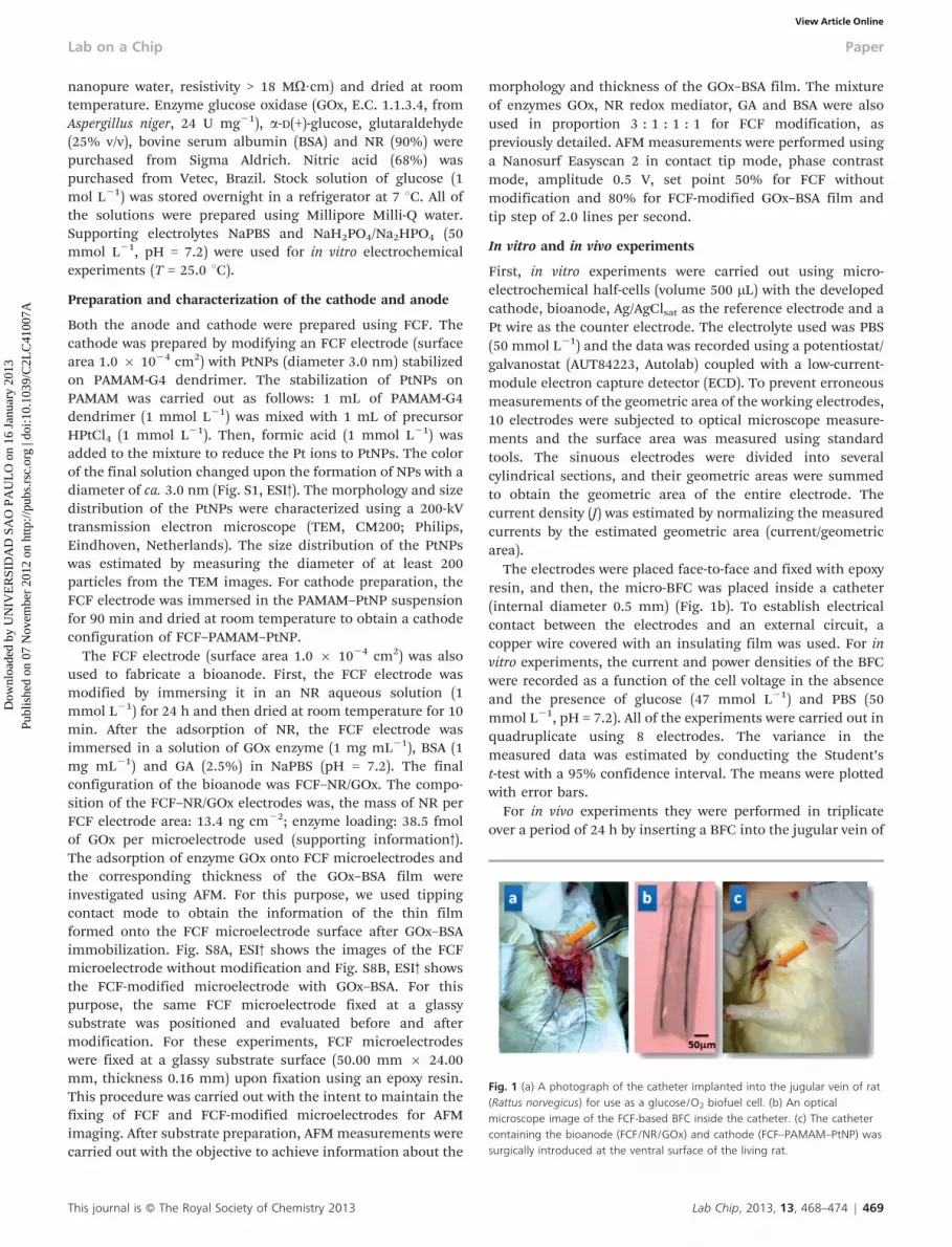
The electrodes were placed face-to-face and fixed with epoxyresin, and then, the micro-BFC was placed inside a catheter(internal diameter 0.5 mm) (Fig. 1b). To establish electricalcontact between the electrodes and an external circuit, acopper wire covered with an insulating film was used. For invitro experiments, the current and power densities of the BFCwere recorded as a function of the cell voltage in the absenceand the presence of glucose (47 mmol L21) and PBS (50mmol L21, pH = 7.2). All of the experiments were carried out inquadruplicate using 8 electrodes. The variance in themeasured data was estimated by conducting the Student’st-test with a 95% confidence interval. The means were plottedwith error bars.
For in vivo experiments they were performed in triplicateover a period of 24 h by inserting a BFC into the jugular vein of
Fig. 1 (a) A photograph of the catheter implanted into the jugular vein of rat(Rattus norvegicus) for use as a glucose/O2 biofuel cell. (b) An opticalmicroscope image of the FCF-based BFC inside the catheter. (c) The cathetercontaining the bioanode (FCF/NR/GOx) and cathode (FCF–PAMAM–PtNP) wassurgically introduced at the ventral surface of the living rat.
This journal is � The Royal Society of Chemistry 2013 Lab Chip, 2013, 13, 468–474 | 469
Lab on a Chip Paper
Dow
nloa
ded
by U
NIV
ER
SID
AD
SA
O P
AU
LO
on
16 J
anua
ry 2
013
Publ
ishe
d on
07
Nov
embe
r 20
12 o
n ht
tp://
pubs
.rsc
.org
| do
i:10.
1039
/C2L
C41
007A
View Article Online
3 adult male Wistar rats (CEMIB/Unicamp, Campinas, Brazil)under protocols approved by the Ethical Animal Care and UseCommittee of UFABC (Protocol number 2010/001) (Fig. 1a).Before performing the experiments, the rats were subjected toa 12 : 12 h light–dark cycle (lights were switched on at7:00 am) at an ambient temperature of 25 ¡ 1 uC. Standardrat chow and tap water were available ad libitum. At the time ofthe experiments, the rats weighed approximately 320 g. Thesurgical procedure and experiments were performed underurethane anesthesia (1.2 g kg21 i.p.). To prevent hypothermia,the rats were placed on an isothermal pad that was maintainedat 37 uC. The rats were placed in a supine position on theoperating board and a 1.0 cm longitudinal incision was madeon the ventral surface of the neck, 1.0 cm to the left of thetrachea. After retracting the surrounding muscles, the leftjugular vein was released from the surrounding connectivetissues and it was exposed. The BFC was implanted with apolyethylene jugular catheter (inner diameter 0.5 mm, outerdiameter 0.9 mm) used for blood sampling (Fig. 1c). Thecatheter was filled with pyrogen-free saline and inserted intothe superior vena cava through the left jugular vein and wassecured in place with ligatures. Long-term stability for thepower density of the intravenous implantable BFC lasted up to24 h. Regarding the stability, all experiments were performedin 24 h (when all the experiments were realized in the time ofanesthesia of the animal).
Results and discussion
In vitro BFC
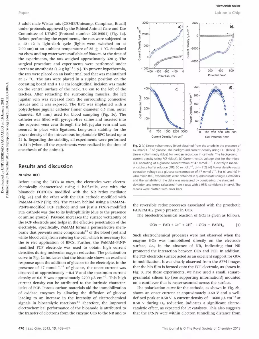
Before using the BFCs in vitro, the electrodes were electro-chemically characterized using 2 half-cells, one with thebioanode FCF/GOx modified with the NR redox mediator(Fig. 2a) and the other with the FCF cathode modified withPAMAM–PtNP (Fig. 2b). The reason behind using a PAMAM–PtNPs-modified FCF cathode and not just a PtNPs-modifiedFCF cathode was due to its hydrophilicity (due to the presenceof amino groups). PAMAM increases the surface wettability ofthe FCF electrode and helps in the effective penetration of theelectrolyte. Specifically, PAMAM forms a permselective mem-brane that prevents some components10 of the blood (red andwhite blood cells) from entering the cell, which is necessary forthe in vivo application of BFCs. Further, the PAMAM–PtNP-modified FCF electrode was used to obtain high currentdensities during molecular oxygen reduction. The polarizationcurve in Fig. 2a indicates that the bioanode shows an excellentresponse upon the addition of glucose to the electrolyte. In thepresence of 47 mmol L21 of glucose, the onset current wasobserved at approximately 20.4 V and the maximum currentdensity at 0.0 V was approximately 2700 mA cm22. This highcurrent density can be attributed to the intrinsic character-istics of FCF. Porous carbon materials aid the immobilizationof oxidase enzymes by allowing the diffusion of glucoseleading to an increase in the intensity of electrochemicalsignals in biocatalytic reactions.11 Therefore, the improvedelectrochemical performance of the bioanode is attributed tothe transfer of electrons from the enzyme GOx to the NR and to
the reversible redox processes associated with the prostheticFAD/FADH2 group present in GOx.
The bioelectrochemical reaction of GOx is given as follows.
GOx 2 FAD + 2e2 + 2H+ A GOx 2 FADH2 (1)
Such electrochemical processes were not observed when theenzyme GOx was immobilized directly on the electrodesurface, i.e., in the absence of NR, indicating that NRpromoted the interaction between GOx and FCF. In addition,the FCF electrode surface acted as an excellent support for GOximmobilization. It was clearly observed from the AFM imagesthat the bio-film is formed onto the FCF electrode, as shown inFig. 3. For these experiments, we have used a small, square-pyramidal silicon tip (see supporting information3) mountedon a cantilever that is raster-scanned across the surface.
The polarization curve for the cathode, as shown in Fig. 2b,shows an onset current at approximately 0.60 V and a well-defined peak at 0.50 V. A current density of 23600 mA cm22 at0.50 V during O2 reduction indicates a significant electro-catalytic effect, as expected for Pt catalysts. This also suggeststhat the PtNPs were within electron tunnelling distance from
Fig. 2 (a) Linear voltammetry (blue) obtained from the anode in the presence of47 mmol L21 of glucose. The background current density using FCF (black). (b)Linear voltammetry (blue) for oxygen reduction in cathode. The backgroundcurrent density using FCF (black). (c) Current versus voltage plot for the micro-BFC operating at a glucose concentration of 47 mmol L21. Electrolyte media:phosphate buffer solution (PBS, 50 mmol L21, pH = 7.2). (d) Power density versusoperation voltage at a glucose concentration of 47 mmol L21. For (c) and (d) invitro micro-BFC, experiments were obtained in quadruplicate using 8 electrodes,and the variability of the data was measured by considering the standarddeviation and errors calculated from t-tests with a 95% confidence interval. Themeans were plotted with error bars.
470 | Lab Chip, 2013, 13, 468–474 This journal is � The Royal Society of Chemistry 2013
Paper Lab on a Chip
Dow
nloa
ded
by U
NIV
ER
SID
AD
SA
O P
AU
LO
on
16 J
anua
ry 2
013
Publ
ishe
d on
07
Nov
embe
r 20
12 o
n ht
tp://
pubs
.rsc
.org
| do
i:10.
1039
/C2L
C41
007A
View Article Online
the FCF surface and that O2 was able to penetrate thedendrimer chain and interact with the encapsulated PtNPs.12
This observation is in agreement with that of Ye and Crooks,13
who showed that PtNPs prepared with hydroxyl-terminatedPAMAM dendrimers and immobilized on carbon electrodesare electrocatalytically active in O2 reduction, and that they areremarkably stable in that they retain their electrocatalyticproperties even after several consecutive cyclic voltammetricscans through the O2 reduction wave.
After performing the half-cell experiments, the electrodeswere used in a membrane-less micro-BFC operated underphysiological conditions. Fig. 2d shows the power densitycurves measured in the presence of glucose. The OCV of 0.400V remained stable for the same glucose concentration and themicro-BFC showed a maximum voltage at 0.225 V with a powerdensity of 240 mW cm22. On the basis of the above results, wepropose a possible mechanism whereby NR acts as an electrontransfer mediator to promote the electro-oxidation of FADH2 atthe FCF electrode, an explanation follows. Recent studies haveshown that NR can interact with a graphite-based electrode viaelectrostatic adsorption and p–p stacking interaction toincrease the electron transfer rate. For instance, Kuang andco-workers14 showed that NR molecules can act as an electrontransfer mediator in anode electrocatalysts used for methanoloxidation. Although a Pt/NR/graphite electrode requires thesame loading mass of a Pt catalyst as an electrode without NR,the former shows a 1.25 times larger exchange current density,a 1.83 times higher specific activity and better long-term cyclestability, as compared to the electrode without NR.
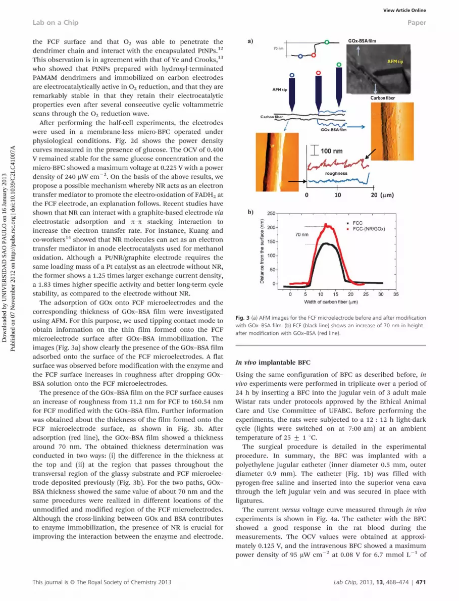
The adsorption of GOx onto FCF microelectrodes and thecorresponding thickness of GOx–BSA film were investigatedusing AFM. For this purpose, we used tipping contact mode toobtain information on the thin film formed onto the FCFmicroelectrode surface after GOx–BSA immobilization. Theimages (Fig. 3a) show clearly the presence of the GOx–BSA filmadsorbed onto the surface of the FCF microelectrodes. A flatsurface was observed before modification with the enzyme andthe FCF surface increases in roughness after dropping GOx–BSA solution onto the FCF microelectrodes.
The presence of the GOx–BSA film on the FCF surface causesan increase of roughness from 11.2 nm for FCF to 160.54 nmfor FCF modified with the GOx–BSA film. Further informationwas obtained about the thickness of the film formed onto theFCF microelectrode surface, as shown in Fig. 3b. Afteradsorption (red line), the GOx–BSA film showed a thicknessaround 70 nm. The obtained thickness determination wasconducted in two ways: (i) the difference in the thickness atthe top and (ii) at the region that passes throughout thetransversal region of the glassy substrate and FCF microelec-trode deposited previously (Fig. 3b). For the two paths, GOx–BSA thickness showed the same value of about 70 nm and thesame procedures were realized in different locations of theunmodified and modified region of the FCF microelectrodes.Although the cross-linking between GOx and BSA contributesto enzyme immobilization, the presence of NR is crucial forimproving the interaction between the enzyme and electrode.
In vivo implantable BFC
Using the same configuration of BFC as described before, invivo experiments were performed in triplicate over a period of24 h by inserting a BFC into the jugular vein of 3 adult maleWistar rats under protocols approved by the Ethical AnimalCare and Use Committee of UFABC. Before performing theexperiments, the rats were subjected to a 12 : 12 h light-darkcycle (lights were switched on at 7:00 am) at an ambienttemperature of 25 ¡ 1 uC.
The surgical procedure is detailed in the experimentalprocedure. In summary, the BFC was implanted with apolyethylene jugular catheter (inner diameter 0.5 mm, outerdiameter 0.9 mm). The catheter (Fig. 1b) was filled withpyrogen-free saline and inserted into the superior vena cavathrough the left jugular vein and was secured in place withligatures.
The current versus voltage curve measured through in vivoexperiments is shown in Fig. 4a. The catheter with the BFCshowed a good response in the rat blood during themeasurements. The OCV values were obtained at approxi-mately 0.125 V, and the intravenous BFC showed a maximumpower density of 95 mW cm22 at 0.08 V for 6.7 mmol L21 of
Fig. 3 (a) AFM images for the FCF microelectrode before and after modificationwith GOx–BSA film. (b) FCF (black line) shows an increase of 70 nm in heightafter modification with GOx–BSA (red line).
This journal is � The Royal Society of Chemistry 2013 Lab Chip, 2013, 13, 468–474 | 471
Lab on a Chip Paper
Dow
nloa
ded
by U
NIV
ER
SID
AD
SA
O P
AU
LO
on
16 J
anua
ry 2
013
Publ
ishe
d on
07
Nov
embe
r 20
12 o
n ht
tp://
pubs
.rsc
.org
| do
i:10.
1039
/C2L
C41
007A
View Article Online
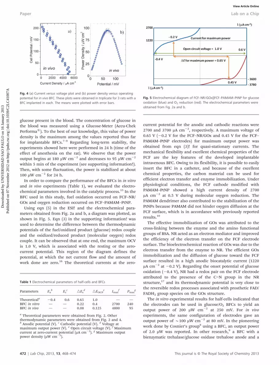
glucose present in the blood. The concentration of glucose inthe blood was measured using a Glucose-Meter (Accu-ChekPerforma1). To the best of our knowledge, this value of powerdensity is the maximum among the values reported thus farfor implantable BFCs.7–9 Regarding long-term stability, theexperiments showed here were performed in 24 h (time of theeffect of anesthesia on the rat). We observe that the poweroutput begins at 180 mW cm22 and decreases to 95 mW cm22
within 5 min of the experiment (see supporting information3).Then, with some fluctuation, the power is stabilized at about100 mW cm22 for 24 h.
In order to compare the performance of the BFCs in in vitroand in vivo experiments (Table 1), we evaluated the electro-chemical parameters involved in the catalytic process.15 In theBFC used in this study, fuel oxidation occurred on FCF–NR/GOx and oxygen reduction occurred on FCF–PAMAM–PtNP.
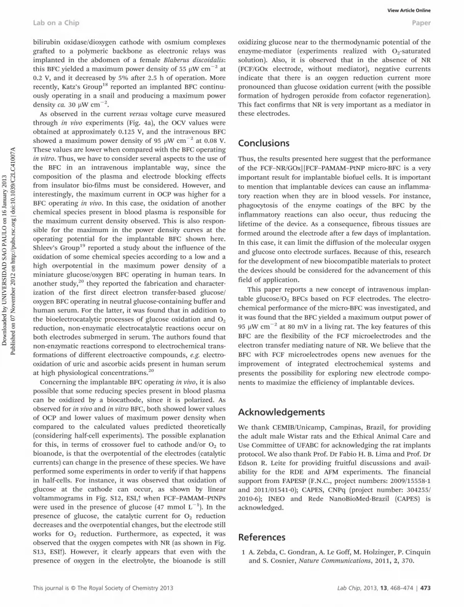
Using eqn (5) in the ESI3 and the electrochemical para-meters obtained from Fig. 2a and b, a diagram was plotted, asshown in Fig. 5. Eqn (3) in the supporting information3 wasused to determine the difference between the thermodynamicpotentials of the fuel/oxidized product (glucose) redox coupleand the oxidized/reduced product (molecular oxygen) redoxcouple. It can be observed that at one end, the maximum OCVis 1.0 V, which is associated with the resting or the zero-current potential. This region of the diagram defines thepotential, at which the net current flow and the amount ofwork done are zero.16 The theoretical currents at the zero-
current potential for the anodic and cathodic reactions were2700 and 3700 mA cm22, respectively. A maximum voltage of0.65 V (20.2 V for the FCF–NR/GOx and 0.45 V for the FCF–PAMAM–PtNP electrodes) for maximum output power wasobtained from eqn (3)3 for quasi-stationary currents. Themechanical flexibility and excellent chemical properties of theFCF are the key features of the developed implantableintravenous BFC. Owing to its flexibility, it is possible to easilyinsert the BFC in a catheter, and because of the excellentchemical properties, the carbon material can be used forefficient electron transfer and enzyme immobilization. Underphysiological conditions, the FCF cathode modified withPAMAM–PtNP showed a high current density of 2700mA cm22 at 0.5 V during molecular oxygen reduction. ThePAMAM dendrimer also contributed to the stabilization of thePtNPs because PAMAM did not hinder oxygen diffusion at theFCF surface, which is in accordance with previously reportedresults.10
The effective immobilization of GOx was attributed to thecross-linking between the enzyme and the amino functionalgroups of BSA. NR acted as an electron mediator and improvedthe efficiency of the electron transfer on the FCF electrodesurface. The bioelectrochemical reaction of GOx was due to theelectron transfer from the enzyme to NR. The efficient GOximmobilization and the diffusion of glucose toward the FCFsurface resulted in a high anodic biocatalytic current (1220mA cm22 at 20.2 V). Regarding the onset potential of glucoseoxidation (20.4 V), NR had a redox pair on the FCF electrodeattributed to the presence of the CLN group in the NRstructure,17 and its thermodynamic potential is very close tothe reversible redox processes associated with prosthetic FAD/FADH2 group species on the GOx structure.
The in vitro experimental results for half-cells indicated thatthe electrodes can be used in glucose/O2 BFCs to yield anoutput power of 200 mW cm22 at 250 mV. For in vivoexperiments, the same configuration of electrodes gave anoutput power of y100 mW cm22 at 80 mV. In the pioneeringwork done by Cosnier’s group8 using a BFC, an output powerof 2.0 mW was reported. In other research,9 a BFC with abienzymatic trehalase/glucose oxidase trehalose anode and a
Fig. 4 (a) Current versus voltage plot and (b) power density versus operatingpotential for in vivo BFC. These plots were obtained in triplicate for 3 rats with aBFC implanted in each. The means were plotted with error bars.
Table 1 Electrochemical parameters of half-cells and BFCs
Parameters Eab Ec
c nEsd nEOCP
e Imaxf Pmax
g
Theoreticala 20.4 0.6 0.65 1.0 — —BFC in vitro — — 0.22 0.4 2700 240BFC in vivo — — 0.08 0.125 4800 95
a Theoretical parameters were obtained from Fig. 2. Otherthermodynamic parameters were obtained from Fig. 2 and 4.b Anodic potential (V). c Cathodic potential (V). d Voltage atmaximum output power (V). e Open circuit voltage (V). f Maximumcurrent at zero-current potential (mA cm22). g Maximum outputpower density (mW cm22).
Fig. 5 Electrochemical diagram of FCF–NR/GOx||FCF–PAMAM–PtNP for glucoseoxidation (blue) and O2 reduction (red). The electrochemical parameters wereobtained from Fig. 2a and b.
472 | Lab Chip, 2013, 13, 468–474 This journal is � The Royal Society of Chemistry 2013
Paper Lab on a Chip
Dow
nloa
ded
by U
NIV
ER
SID
AD
SA
O P
AU
LO
on
16 J
anua
ry 2
013
Publ
ishe
d on
07
Nov
embe
r 20
12 o
n ht
tp://
pubs
.rsc
.org
| do
i:10.
1039
/C2L
C41
007A
View Article Online
bilirubin oxidase/dioxygen cathode with osmium complexesgrafted to a polymeric backbone as electronic relays wasimplanted in the abdomen of a female Blaberus discoidalis:this BFC yielded a maximum power density of 55 mW cm22 at0.2 V, and it decreased by 5% after 2.5 h of operation. Morerecently, Katz’s Group18 reported an implanted BFC continu-ously operating in a snail and producing a maximum powerdensity ca. 30 mW cm22.
As observed in the current versus voltage curve measuredthrough in vivo experiments (Fig. 4a), the OCV values wereobtained at approximately 0.125 V, and the intravenous BFCshowed a maximum power density of 95 mW cm22 at 0.08 V.These values are lower when compared with the BFC operatingin vitro. Thus, we have to consider several aspects to the use ofthe BFC in an intravenous implantable way, since thecomposition of the plasma and electrode blocking effectsfrom insulator bio-films must be considered. However, andinterestingly, the maximum current in OCP was higher for aBFC operating in vivo. In this case, the oxidation of anotherchemical species present in blood plasma is responsible forthe maximum current density observed. This is also respon-sible for the maximum in the power density curves at theoperating potential for the implantable BFC shown here.Shleev’s Group19 reported a study about the influence of theoxidation of some chemical species according to a low and ahigh overpotential in the maximum power density of aminiature glucose/oxygen BFC operating in human tears. Inanother study,20 they reported the fabrication and character-ization of the first direct electron transfer-based glucose/oxygen BFC operating in neutral glucose-containing buffer andhuman serum. For the latter, it was found that in addition tothe bioelectrocatalytic processes of glucose oxidation and O2
reduction, non-enzymatic electrocatalytic reactions occur onboth electrodes submerged in serum. The authors found thatnon-enzymatic reactions correspond to electrochemical trans-formations of different electroactive compounds, e.g. electro-oxidation of uric and ascorbic acids present in human serumat high physiological concentrations.20
Concerning the implantable BFC operating in vivo, it is alsopossible that some reducing species present in blood plasmacan be oxidized by a biocathode, since it is polarized. Asobserved for in vivo and in vitro BFC, both showed lower valuesof OCP and lower values of maximum power density whencompared to the calculated values predicted theoretically(considering half-cell experiments). The possible explanationfor this, in terms of crossover fuel to cathode and/or O2 tobioanode, is that the overpotential of the electrodes (catalyticcurrents) can change in the presence of these species. We haveperformed some experiments in order to verify if that happensin half-cells. For instance, it was observed that oxidation ofglucose at the cathode can occur, as shown by linearvoltammograms in Fig. S12, ESI,3 when FCF–PAMAM–PtNPswere used in the presence of glucose (47 mmol L21). In thepresence of glucose, the catalytic current for O2 reductiondecreases and the overpotential changes, but the electrode stillworks for O2 reduction. Furthermore, as expected, it wasobserved that the oxygen competes with NR (as shown in Fig.S13, ESI3). However, it clearly appears that even with thepresence of oxygen in the electrolyte, the bioanode is still
oxidizing glucose near to the thermodynamic potential of theenzyme-mediator (experiments realized with O2-saturatedsolution). Also, it is observed that in the absence of NR(FCF/GOx electrode, without mediator), negative currentsindicate that there is an oxygen reduction current morepronounced than glucose oxidation current (with the possibleformation of hydrogen peroxide from cofactor regeneration).This fact confirms that NR is very important as a mediator inthese electrodes.
Conclusions
Thus, the results presented here suggest that the performanceof the FCF–NR/GOx||FCF–PAMAM–PtNP micro-BFC is a veryimportant result for implantable biofuel cells. It is importantto mention that implantable devices can cause an inflamma-tory reaction when they are in blood vessels. For instance,phagocytosis of the enzyme coatings of the BFC by theinflammatory reactions can also occur, thus reducing thelifetime of the device. As a consequence, fibrous tissues areformed around the electrode after a few days of implantation.In this case, it can limit the diffusion of the molecular oxygenand glucose onto electrode surfaces. Because of this, researchfor the development of new biocompatible materials to protectthe devices should be considered for the advancement of thisfield of application.
This paper reports a new concept of intravenous implan-table glucose/O2 BFCs based on FCF electrodes. The electro-chemical performance of the micro-BFC was investigated, andit was found that the BFC yielded a maximum output power of95 mW cm22 at 80 mV in a living rat. The key features of thisBFC are the flexibility of the FCF microelectrodes and theelectron transfer mediating nature of NR. We believe that theBFC with FCF microelectrodes opens new avenues for theimprovement of integrated electrochemical systems andpresents the possibility for exploring new electrode compo-nents to maximize the efficiency of implantable devices.
Acknowledgements
We thank CEMIB/Unicamp, Campinas, Brazil, for providingthe adult male Wistar rats and the Ethical Animal Care andUse Committee of UFABC for acknowledging the rat implantsprotocol. We also thank Prof. Dr Fabio H. B. Lima and Prof. DrEdson R. Leite for providing fruitful discussions and avail-ability for the RDE and AFM experiments. The financialsupport from FAPESP (F.N.C., project numbers: 2009/15558-1and 2011/01541-0); CAPES, CNPq (project number: 304255/2010-6); INEO and Rede NanoBioMed-Brazil (CAPES) isacknowledged.
References
1 A. Zebda, C. Gondran, A. Le Goff, M. Holzinger, P. Cinquinand S. Cosnier, Nature Communications, 2011, 2, 370.
This journal is � The Royal Society of Chemistry 2013 Lab Chip, 2013, 13, 468–474 | 473
Lab on a Chip Paper
Dow
nloa
ded
by U
NIV
ER
SID
AD
SA
O P
AU
LO
on
16 J
anua
ry 2
013
Publ
ishe
d on
07
Nov
embe
r 20
12 o
n ht
tp://
pubs
.rsc
.org
| do
i:10.
1039
/C2L
C41
007A
View Article Online
2 T. Miyake, S. Yoshino, T. Yamada, K. Hata andM. Nishizawa, J. Am. Chem. Soc., 2011, 133, 5129–5134.
3 J. A. Cracknell, K. A. Vincent and F. A. Armstrong, Chem.Rev., 2008, 108, 2439–2461.
4 F. Gao, L. Viry, M. Maugey, P. Poulin and N. Mano, NatureCommunications, 2010, 1, 2.
5 E. Katz, O. Lioubashevski and I. Willner, J. Am. Chem. Soc.,2005, 127, 3979–3988.
6 S. K. Chaudhuri and D. R. Lovley, Nat. Biotechnol., 2003, 21,1229–1232.
7 N. Mano, F. Mao and A. Heller, J. Am. Chem. Soc., 2003, 125,6588–6594.
8 P. Cinquin, C. Gondran, F. Giroud, S. Mazabrard,A. Pellissier, F. Boucher, J.-P. Alcaraz, K. Gorgy,F. Lenouvel, S. Mathe, P. Porcu and S. Cosnier, Plos One,2010, 5.
9 M. Rasmussen, R. E. Ritzmann, I. Lee, A. J. Pollack andD. Scherson, J. Am. Chem. Soc., 2012, 134, 1458–1460.
10 F. N. Crespilho, M. C. Esteves, P. T. A. Sumodjo, E.J. Podlaha and V. Zucolotto, J. Phys. Chem. C, 2009, 113,6037–6041.
11 J. Njagi, M. M. Chernov, J. C. Leiter and S. Andreescu, Anal.Chem., 2010, 82, 989–996.
12 F. N. Crespilho, F. Huguenin, V. Zucolotto, P. Olivi, F.C. Nart and O. N. Oliveira, Electrochem. Commun., 2006, 8,348–352.
13 H. C. Ye and R. M. Crooks, J. Am. Chem. Soc., 2005, 127,4930–4934.
14 X. Zhong, J. Chen, B. Liu, Y. Xu and Y. Kuang, J. Solid StateElectrochem., 2007, 11, 463–468.
15 C. M. A. Brett and A. M. O. Brett, Electrochemistry principles,methods, and applications, Oxford New York OxfordUniversity Press, New York, 1993.
16 A. J. Bard and L. R. Faulkner, Electrochemical Methods:Fundamentals and Applications, 2nd edn, John Wiley, NewJersey, 2000.
17 G. Inzelt and E. Csahok, Electroanalysis, 1999, 11.18 L. Halamkova, J. Halamek, V. Bocharova, A. Szczupak,
L. Alfonta and E. Katz, J. Am. Chem. Soc., 2012, 134,5040–5043.
19 M. Falk, V. Andoralov, Z. Blum, J. Sotres, D.B. Suyatin,T. Ruzgas, T. Arnebrant and S. Shleev, Biosens. Bioelectron.,2012, 37, 38–45.
20 V. Coman, R. Ludwig, W. Harreither, D. Haltrich,L. Gorton, T. Ruzgas and S. Shleev, Fuel Cells, 2010, 10,9–16.
474 | Lab Chip, 2013, 13, 468–474 This journal is � The Royal Society of Chemistry 2013
Paper Lab on a Chip
Dow
nloa
ded
by U
NIV
ER
SID
AD
SA
O P
AU
LO
on
16 J
anua
ry 2
013
Publ
ishe
d on
07
Nov
embe
r 20
12 o
n ht
tp://
pubs
.rsc
.org
| do
i:10.
1039
/C2L
C41
007A
View Article Online


