Embed Size (px)
Citation preview
This is the peer reviewed version of the following article: Rose C F. Ammonia-Lowering Strategies for the Treatment
of Hepatic Encephalopathy. Clin Pharmacol Ther . 2012-09;92(3):321–331, which has been published in final form at
10.1038/clpt.2012.112. This article may be used for non-commercial purposes in accordance with Wiley Terms and
Conditions for Use of Self-Archived Versions.
Rose C F. Ammonia-Lowering Strategies for the Treatment of Hepatic Encephalopathy. Clin Pharmacol Ther . 2012-09;92(3):321–331
AMMONIA-LOWERING STRATEGIES FOR THE
TREATMENT OF HEPATIC ENCEPHALOPATHY
Christopher F. Rose
Author affiliations: 1. Neuroscience Research Unit, Hôpital Saint-Luc (CRCHUM), Université de
Montréal, Québec, Canada
Corresponding author: Christopher F. Rose Ph.D., Neuroscience Research Unit, Hôpital Saint-Luc
(CRCHUM), Université de Montréal, Québec, Canada. Phone: +1 514 890 8000, ext. 35739; email:
Number of references : 75
Number of figures : 8
Keywords : Brain, Hepatic, Metabolism, Neurological, Therapeutics
ABSTRACT
Hyperammonemia leads to neurotoxic levels of brain ammonia and is a major factor
involved in the pathogenesis of hepatic encephalopathy (HE). Ammonia‐lowering
treatments primarily involve two strategies: inhibiting ammonia production and/or
increasing ammonia removal. Targeting the gut has been the primary focus for many
years, with the goal of inhibiting the generation of ammonia. However, in the context of
liver failure, extrahepatic organs containing ammonia metabolic pathways have become
new potential ammonia‐lowering targets. Skeletal muscle has the capacity to remove
ammonia by producing glutamine through the enzyme glutamine synthetase (amidation
of glutamate) and, given its large mass, has the potential to be an important ammonia‐removing organ. On the other hand, glutamine can be deaminated to glutamate by
phosphate‐activated glutaminase, thus releasing ammonia (ammonia rebound). Therefore,
new treatment strategies are being focused on stimulating the removal of both ammonia
and glutamine.
Hepatic encephalopathy (HE) is a common, debilitating, and clinically challenging
neuropsychiatric complication of both acute liver failure and chronic liver disease.
Characterized by a constellation of symptoms, including a spectrum of cognitive,
psychiatric, and motor disturbances, HE can progressively lead to coma and death. In
1998, the working party at the 11th World Congress of Gastroenterology classified HE
This is the peer reviewed version of the following article: Rose C F. Ammonia-Lowering Strategies for the Treatment
of Hepatic Encephalopathy. Clin Pharmacol Ther . 2012-09;92(3):321–331, which has been published in final form at
10.1038/clpt.2012.112. This article may be used for non-commercial purposes in accordance with Wiley Terms and
Conditions for Use of Self-Archived Versions.
Rose C F. Ammonia-Lowering Strategies for the Treatment of Hepatic Encephalopathy. Clin Pharmacol Ther . 2012-09;92(3):321–331
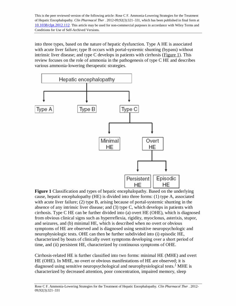
into three types, based on the nature of hepatic dysfunction. Type A HE is associated
with acute liver failure; type B occurs with portal‐systemic shunting (bypass) without
intrinsic liver disease; and type C develops in patients with cirrhosis (Figure 1). This
review focuses on the role of ammonia in the pathogenesis of type C HE and describes
various ammonia‐lowering therapeutic strategies.
Figure 1 Classification and types of hepatic encephalopathy. Based on the underlying
cause, hepatic encephalopathy (HE) is divided into three forms: (1) type A, associated
with acute liver failure; (2) type B, arising because of portal‐systemic shunting in the
absence of any intrinsic liver disease; and (3) type C, which develops in patients with
cirrhosis. Type C HE can be further divided into (a) overt HE (OHE), which is diagnosed
from obvious clinical signs such as hyperreflexia, rigidity, myoclonus, asterixis, stupor,
and seizures, and (b) minimal HE, which is described when no overt or obvious
symptoms of HE are observed and is diagnosed using sensitive neuropsychologic and
neurophysiologic tests. OHE can then be further subdivided into (i) episodic HE,
characterized by bouts of clinically overt symptoms developing over a short period of
time, and (ii) persistent HE, characterized by continuous symptoms of OHE.
Cirrhosis‐related HE is further classified into two forms: minimal HE (MHE) and overt
HE (OHE). In MHE, no overt or obvious manifestations of HE are observed; it is
diagnosed using sensitive neuropsychological and neurophysiological tests.1 MHE is
characterized by decreased attention, poor concentration, impaired memory, sleep

This is the peer reviewed version of the following article: Rose C F. Ammonia-Lowering Strategies for the Treatment
of Hepatic Encephalopathy. Clin Pharmacol Ther . 2012-09;92(3):321–331, which has been published in final form at
10.1038/clpt.2012.112. This article may be used for non-commercial purposes in accordance with Wiley Terms and
Conditions for Use of Self-Archived Versions.
Rose C F. Ammonia-Lowering Strategies for the Treatment of Hepatic Encephalopathy. Clin Pharmacol Ther . 2012-09;92(3):321–331
disturbances, reduced speed of information processing, and altered motor abilities. In
addition, the subclinical cognitive impairment that characterizes MHE increases the risk
of having a car accident. As such, MHE has a significant impact on patients' health‐related quality of life and their ability to carry out day‐to‐day functions.
1 As many as 80%
(20–80%, depending on the severity of the disease) of patients with chronic liver disease
may have MHE. Its presence also identifies patients with a fourfold higher risk of
developing OHE.2
OHE may involve several clinical signs, including hyperreflexia, rigidity, myoclonus,
asterixis, stupor, and seizures; the West Haven Criteria are routinely used to grade the
severity of manifestations. In addition, based on the duration and characteristics of
neurologic dysfunction, OHE can manifest as either episodic or persistent. HE is said to
be episodic when bouts of clinically overt symptoms develop over a short period of time,
whereas persistent HE is defined as the continuous presence of symptoms (Figure 1).
Patients with persistent HE are usually unresponsive to treatment. In patients with end‐stage liver disease, there is a 20% annual risk of developing an episode of OHE, and the
prevalence of OHE among these patients is 30–45%.3 The clinical prognosis is poor after
OHE has developed, with 1‐year survival estimated at 42% and 3‐year survival at 23%.2
Overall, the burden of HE is immense, considering its wide‐ranging effects on patients,
their families, and society. The economic drain caused by HE on health‐care systems is
estimated at more than US$1 billion annually. With the increasing prevalence of hepatitis
C and nonalcoholic steatohepatitis–related cirrhosis, the situation is expected to
worsen.2,4
REGULATION OF AMMONIA
The pathophysiologic basis of HE is multifactorial and complex, and it remains poorly
understood. However, there is general consensus that ammonia plays a pivotal role in
HE.5,6
Ammonia, a by‐product of nitrogen metabolism, is produced mainly within the gut
through the deamination of glutamine by glutaminase in the enterocytes of the small
intestine and colon, as well as through the hydrolysis of urea, catalyzed by urease‐producing bacteria that exist abundantly in the human gut. Gut‐derived ammonia is
transported and absorbed across the mucosal epithelium into the hepatic portal
circulation, from which, in the case of a healthy liver, it is removed primarily through the
urea cycle. This low‐affinity, high‐capacity ammonia detoxification system is present in
the periportal hepatocytes located around the portal vein. Glutamine synthetase, another
important ammonia‐removing pathway located in the liver, catalyzes the amidation of
glutamate into glutamine, thereby removing an ammonia molecule. This high‐affinity,
low‐capacity reaction takes place in the perivenous hepatocytes located around the
hepatic vein and acts as a scavenger for the ammonia that escapes periportal urea
synthesis. The production of ammonia within the gut and its detoxification by the liver
are the main pathways through which ammonia homeostasis is maintained in the body.
However, other organs also contribute to ammonia metabolism. In addition to the liver

This is the peer reviewed version of the following article: Rose C F. Ammonia-Lowering Strategies for the Treatment
of Hepatic Encephalopathy. Clin Pharmacol Ther . 2012-09;92(3):321–331, which has been published in final form at
10.1038/clpt.2012.112. This article may be used for non-commercial purposes in accordance with Wiley Terms and
Conditions for Use of Self-Archived Versions.
Rose C F. Ammonia-Lowering Strategies for the Treatment of Hepatic Encephalopathy. Clin Pharmacol Ther . 2012-09;92(3):321–331
and intestines, glutamine synthetase is found in the muscles and the brain (particularly in
the astrocytes) and in phosphate‐activated glutaminase in the kidneys and the brain
(primarily in the neurons). In the presence of a healthy liver, blood ammonia levels are
maintained in the low range of 35–60 µmol/l (Figure 2). However, during liver disease,
given the reduced hepatic capacity for ammonia removal, the extrahepatic interorgan
ammonia metabolism is altered (including glutamine metabolism),7 thus upsetting the
balance between ammonia‐producing/removing organs and ammonia homeostasis
(Figure 2). This results in a two‐ to fivefold increase in blood ammonia, leading to an
increase in ammonia levels in the brain, with deleterious consequences.5,6
Figure 2 Interorgan ammonia (NH3) and glutamine (GLN) trafficking under normal and
cirrhotic conditions. Many organs are involved in regulating whole‐body ammonia
homeostasis. (a) The liver is the most important organ in regulating the circulating
concentration of ammonia to remain in the range 35–60 µmol/l. (b) During liver
disease, the arterial concentration of ammonia can increase fivefold. This is obviously
due to lower ammonia detoxification in the liver (reduced urea‐cycle and glutamine
synthetase (GS) activity). Furthermore, under these conditions, interorgan metabolism is

This is the peer reviewed version of the following article: Rose C F. Ammonia-Lowering Strategies for the Treatment
of Hepatic Encephalopathy. Clin Pharmacol Ther . 2012-09;92(3):321–331, which has been published in final form at
10.1038/clpt.2012.112. This article may be used for non-commercial purposes in accordance with Wiley Terms and
Conditions for Use of Self-Archived Versions.
Rose C F. Ammonia-Lowering Strategies for the Treatment of Hepatic Encephalopathy. Clin Pharmacol Ther . 2012-09;92(3):321–331
altered. The muscle becomes the major ammonia‐removing organ, through the GS
mechanism; however, attempts by the muscle to detoxify ammonia by means of forming
and releasing GLN are negated when GLN is metabolized by phosphate‐activated
glutaminase (PAG) in both the kidney and the intestine. With a net balance of zero
ammonia removed, and continuous production of ammonia in the gut (by diet as well as
PAG/urease activity), hyperammonemia will persist. For a complete review of interorgan
ammonia metabolism in liver disease, please see ref. 7. Red circles: circulating ammonia;
blue circles: circulating GLN; orange circles: GLN‐derived ammonia.
PROPERTIES OF AMMONIA
Ammonia as a gas (NH3) can freely diffuse across all cell membranes, and as an ion
(NH4+) it can be transported across plasma membranes through K
+ transporters and
channels because it has properties similar to those of K+ (comparable ionic radius and
diffusion coefficient). The ratio of NH3/NH4+ is a function of pH as defined by the
Henderson‐Hasselbach equation: log10 (NH3/NH4+) = pH − pKa, where pKa is dissociation
constant. With the pKa of ammonia being 9.15 (at 37 ºC and pH 7.4) >98% of ammonia is
present as NH4+. There is increasing evidence to indicate that ammonia can also be
transported through aquaporin channels, as well as through human nonerythroid Rhesus
glycoproteins B and C (Figure 3). It has been proposed that an ammonia concentration
gradient exists between the brain and the blood. However, studies in animals with and
without liver failure have demonstrated similar concentrations of ammonia in the blood,
cerebrospinal fluid, and extracellular fluid (sampled using in vivo cerebral
microdialysis),8,9
including a strong correlation between arterial and extracellular cerebral
ammonia.10
It is understood that a difference in pH across plasma membranes, i.e.,
between intracellular and extracellular compartments, will alter the ratio of NH3/NH4+ on
either side of the membrane. A pH gradient between blood and brain will create
concentration gradients for NH3 and NH4+ across the blood–brain barrier. However,
given that both NH3 and NH4+ are capable of crossing biological membranes (NH3
diffuses faster than NH4+ is transported), the concentration levels of ammonia (NH3 +
NH4+) in the blood and in the brain will reach equilibrium and be similar. Nevertheless, it
has been demonstrated that an increase in blood ammonia leads to an increase in cerebral
uptake;11
but this does not necessarily dictate an accumulation of ammonia in the brain,
nor does it, therefore, entail a higher concentration in the brain. Given that ammonia
follows its concentration gradient, and that astrocytes (which outnumber neurons ~1.5:1)
detoxify ammonia through the synthesis of glutamine by glutamine synthetase, the brain
becomes a sink for ammonia. Therefore, the concentration of blood ammonia as well as
the efficiency of ammonia removal in the brain will influence its uptake across the blood–
brain barrier.12
This is the peer reviewed version of the following article: Rose C F. Ammonia-Lowering Strategies for the Treatment
of Hepatic Encephalopathy. Clin Pharmacol Ther . 2012-09;92(3):321–331, which has been published in final form at
10.1038/clpt.2012.112. This article may be used for non-commercial purposes in accordance with Wiley Terms and
Conditions for Use of Self-Archived Versions.
Rose C F. Ammonia-Lowering Strategies for the Treatment of Hepatic Encephalopathy. Clin Pharmacol Ther . 2012-09;92(3):321–331
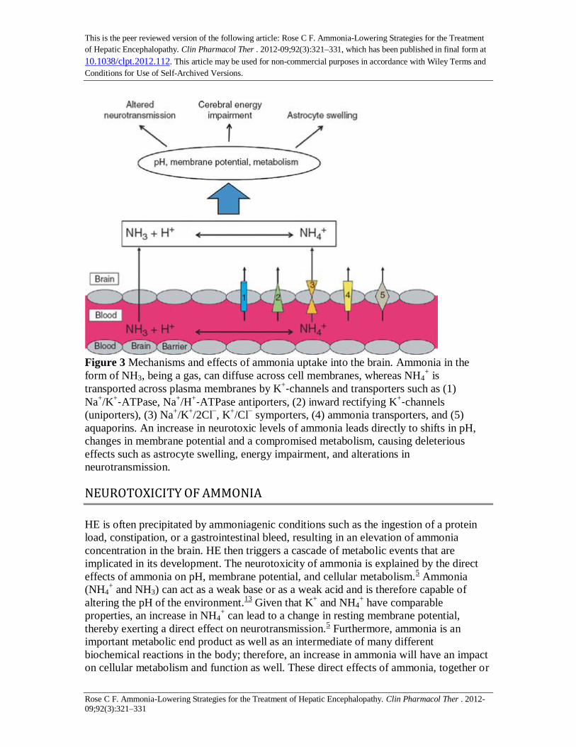
Figure 3 Mechanisms and effects of ammonia uptake into the brain. Ammonia in the
form of NH3, being a gas, can diffuse across cell membranes, whereas NH4+ is
transported across plasma membranes by K+‐channels and transporters such as (1)
Na+/K
+‐ATPase, Na+/H
+‐ATPase antiporters, (2) inward rectifying K+‐channels
(uniporters), (3) Na+/K
+/2Cl
−, K
+/Cl
− symporters, (4) ammonia transporters, and (5)
aquaporins. An increase in neurotoxic levels of ammonia leads directly to shifts in pH,
changes in membrane potential and a compromised metabolism, causing deleterious
effects such as astrocyte swelling, energy impairment, and alterations in
neurotransmission.
NEUROTOXICITY OF AMMONIA
HE is often precipitated by ammoniagenic conditions such as the ingestion of a protein
load, constipation, or a gastrointestinal bleed, resulting in an elevation of ammonia
concentration in the brain. HE then triggers a cascade of metabolic events that are
implicated in its development. The neurotoxicity of ammonia is explained by the direct
effects of ammonia on pH, membrane potential, and cellular metabolism.5 Ammonia
(NH4+ and NH3) can act as a weak base or as a weak acid and is therefore capable of
altering the pH of the environment.13
Given that K+ and NH4
+ have comparable
properties, an increase in NH4+ can lead to a change in resting membrane potential,
thereby exerting a direct effect on neurotransmission.5 Furthermore, ammonia is an
important metabolic end product as well as an intermediate of many different
biochemical reactions in the body; therefore, an increase in ammonia will have an impact
on cellular metabolism and function as well. These direct effects of ammonia, together or
This is the peer reviewed version of the following article: Rose C F. Ammonia-Lowering Strategies for the Treatment
of Hepatic Encephalopathy. Clin Pharmacol Ther . 2012-09;92(3):321–331, which has been published in final form at
10.1038/clpt.2012.112. This article may be used for non-commercial purposes in accordance with Wiley Terms and
Conditions for Use of Self-Archived Versions.
Rose C F. Ammonia-Lowering Strategies for the Treatment of Hepatic Encephalopathy. Clin Pharmacol Ther . 2012-09;92(3):321–331
independently, can lead to several pathophysiologic mechanisms that affect cellular
functions, including cellular energy metabolism, neurotransmission, and astrocyte
swelling6 (Figure 3).
NEUROPATHOLOGY
The neuropathology of HE in chronic liver disease primarily reveals morphological
changes in astrocytes, including cell swelling and the development of Alzheimer type II
astrocytosis. Loss of neuronal cells is rarely observed in liver disease. Astrocyte swelling,
which is responsible for brain edema, is a common feature found in cirrhotic rats with
MHE8,14
as well as in patients with cirrhosis who develop HE.15
When there is an
increase in ammonia levels in the brain, it is the astrocyte that bears the brunt of
removing ammonia because these glial cells are the only cells in the brain capable of
detoxifying ammonia through the synthesis of glutamine by glutamine synthetase. It has
been postulated that the accumulation of glutamine in astrocytes, subsequent to ammonia
detoxification, results in an increase in osmotic forces, leading to swelling in HE.
However, our group and others have demonstrated that glutamine is not the sole factor
involved in the pathogenesis of astrocyte swelling,16
and that the precise
pathophysiologic mechanisms underlying the development of brain edema in HE remain
unexplained. More importantly, given that the swelling of astrocytes not only leads to
brain edema but is also capable of compromising cell function, it remains controversial
whether brain edema symbolizes a neuropathologic feature of HE, or whether HE is a
clinical manifestation of brain edema.
THE ROLE OF AMMONIA IN HE
Although there is overwhelming evidence of the central role of ammonia in the
pathogenesis of HE, and data show that 80% of patients with cirrhosis present some
degree of hyperammonemia,17
it is important to note that many studies have reported an
imperfect correlation between hyperammonemia and the severity of HE in chronic liver
disease.18
This suggests that pathogenic factors besides ammonia are involved in the
pathogenesis of HE. It has been speculated that oxidative stress and inflammation may
exacerbate the neuropsychologic effects of hyperammonemia.8,19,20
The roles of these
pathophysiologic factors in the development of HE are not addressed in this review.
TREATMENTS
Currently, treatment for HE is based on strategies aimed at reducing the concentration of
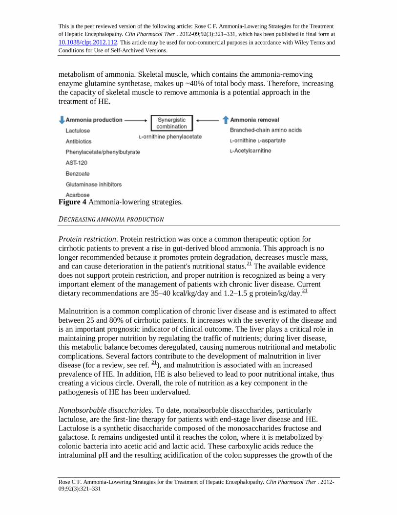
circulating blood ammonia (Figure 4). One obvious strategy is to address the source of
ammonia production, with the gut being a primary target. Reducing ammonia production
will minimize its absorption into the systemic circulation and hence the brain's exposure
to it. However, because interorgan ammonia metabolism is altered during the onset of
liver disease,7 additional organs, such as skeletal muscle, become important players in the
This is the peer reviewed version of the following article: Rose C F. Ammonia-Lowering Strategies for the Treatment
of Hepatic Encephalopathy. Clin Pharmacol Ther . 2012-09;92(3):321–331, which has been published in final form at
10.1038/clpt.2012.112. This article may be used for non-commercial purposes in accordance with Wiley Terms and
Conditions for Use of Self-Archived Versions.
Rose C F. Ammonia-Lowering Strategies for the Treatment of Hepatic Encephalopathy. Clin Pharmacol Ther . 2012-09;92(3):321–331
metabolism of ammonia. Skeletal muscle, which contains the ammonia‐removing
enzyme glutamine synthetase, makes up ~40% of total body mass. Therefore, increasing
the capacity of skeletal muscle to remove ammonia is a potential approach in the
treatment of HE.
Figure 4 Ammonia‐lowering strategies.
DECREASING AMMONIA PRODUCTION
Protein restriction. Protein restriction was once a common therapeutic option for
cirrhotic patients to prevent a rise in gut‐derived blood ammonia. This approach is no
longer recommended because it promotes protein degradation, decreases muscle mass,
and can cause deterioration in the patient's nutritional status.21
The available evidence
does not support protein restriction, and proper nutrition is recognized as being a very
important element of the management of patients with chronic liver disease. Current
dietary recommendations are 35–40 kcal/kg/day and 1.2–1.5 g protein/kg/day.21
Malnutrition is a common complication of chronic liver disease and is estimated to affect
between 25 and 80% of cirrhotic patients. It increases with the severity of the disease and
is an important prognostic indicator of clinical outcome. The liver plays a critical role in
maintaining proper nutrition by regulating the traffic of nutrients; during liver disease,
this metabolic balance becomes deregulated, causing numerous nutritional and metabolic
complications. Several factors contribute to the development of malnutrition in liver
disease (for a review, see ref. 21
), and malnutrition is associated with an increased
prevalence of HE. In addition, HE is also believed to lead to poor nutritional intake, thus
creating a vicious circle. Overall, the role of nutrition as a key component in the
pathogenesis of HE has been undervalued.
Nonabsorbable disaccharides. To date, nonabsorbable disaccharides, particularly
lactulose, are the first‐line therapy for patients with end‐stage liver disease and HE.
Lactulose is a synthetic disaccharide composed of the monosaccharides fructose and
galactose. It remains undigested until it reaches the colon, where it is metabolized by
colonic bacteria into acetic acid and lactic acid. These carboxylic acids reduce the
intraluminal pH and the resulting acidification of the colon suppresses the growth of the
This is the peer reviewed version of the following article: Rose C F. Ammonia-Lowering Strategies for the Treatment
of Hepatic Encephalopathy. Clin Pharmacol Ther . 2012-09;92(3):321–331, which has been published in final form at
10.1038/clpt.2012.112. This article may be used for non-commercial purposes in accordance with Wiley Terms and
Conditions for Use of Self-Archived Versions.
Rose C F. Ammonia-Lowering Strategies for the Treatment of Hepatic Encephalopathy. Clin Pharmacol Ther . 2012-09;92(3):321–331
intestinal urease bacteria (ammoniagenic bacteria), leaving acid‐resistant,
nonammoniagenic bacteria. Lactulose also decreases the absorption of ammonia through
a cathartic effect, clearing the gut of ammonia before it is systemically absorbed,
resulting in increased fecal nitrogen excretion. Lactulose has also been shown to impede
the uptake of glutamine by the intestinal wall, thus preventing glutamine from being
metabolized into ammonia.22
As a result, numerous studies have demonstrated that
lactulose treatment leads to a reduction in blood ammonia levels.23
However, in 2004, a
Cochrane review evaluating 22 clinical trials concluded that there was not enough
convincing evidence arising from high‐quality randomized trials to suggest that
nonabsorbable disaccharides should be used to treat HE.24
Since then, the results from a
randomized clinical trial have shown that lactulose is beneficial in treating HE in patients
with chronic liver disease.25
Although lactulose is safe, compliance is often
underachieved, given the need to titrate the dose in order to reach two or three semi‐soft
stools per day. In addition, lactulose treatment has been shown to cause abdominal
cramping, bloating, nausea, vomiting, flatulence, and abdominal distension, the last
potentially leading to technical difficulties during surgery for liver transplantation.
Moreover, lactulose treatment affects intestinal absorption, and this may amplify the
nutritional deficits in patients with end‐stage liver disease, leading to a poorer outcome
after liver transplantation.26
Antibiotics. Orally administered antimicrobial agents targeting the gut have long been
utilized with the primary aim of inhibiting urease‐containing bacteria in the colon,
thereby decreasing ammonia production and preventing absorption through the
gastrointestinal tract. Antibiotics such as neomycin, metronidazole, and vancomycin have
all been demonstrated to lower blood ammonia in patients with end‐stage liver
disease.27,28,29
Nonetheless, because of the systemic absorption of these antimicrobial
agents, serious adverse effects have been recorded, and these have limited their
widespread use. Neomycin, an aminoglycoside antimicrobial, exerts its effect by binding
to the 30S ribosomal unit and inhibiting protein synthesis. Because neomycin is
systemically absorbed (~4%), oral treatment potentially causes nephrotoxicity and
ototoxicity.30
Metronidazole, a nitroimidazole antimicrobial that is primarily taken up by
anaerobes and causes cell toxicity, has been found to be as effective as neomycin.28
However, because of its high systemic absorption, cases of neurotoxicity (peripheral
neuropathy) have been documented with long‐term use.31
Furthermore, metronidazole is
eliminated by the liver through hepatic oxidation, which adds to the risk of toxicity in
patients with chronic liver disease.32
Vancomycin, a glycopeptide antimicrobial that
inhibits cell‐wall synthesis in Gram‐positive bacteria, has been demonstrated to lower
blood ammonia and attenuate HE in patients with cirrhosis.29
On the other hand,
vancomycin treatment has also been demonstrated to lead to bacterial overgrowth and
increase the risk of enteric bacterial resistance.33
In 2010, results from a randomized,
double‐blind study led to the approval (in the United States) of the antibiotic rifaximin
(Salix Pharmaceuticals) as reducing the risk of recurrence of overt HE in patients with
end‐stage liver disease.34
Rifaximin is a semisynthetic antibiotic that is poorly absorbed
(<0.4%) because of its pyridoimidazole ring. Rifaximin binds to the β‐subunit of bacterial
This is the peer reviewed version of the following article: Rose C F. Ammonia-Lowering Strategies for the Treatment
of Hepatic Encephalopathy. Clin Pharmacol Ther . 2012-09;92(3):321–331, which has been published in final form at
10.1038/clpt.2012.112. This article may be used for non-commercial purposes in accordance with Wiley Terms and
Conditions for Use of Self-Archived Versions.
Rose C F. Ammonia-Lowering Strategies for the Treatment of Hepatic Encephalopathy. Clin Pharmacol Ther . 2012-09;92(3):321–331
DNA‐dependent RNA polymerase and prevents the catalysis of polymerization of
deoxyribonucleotides into a DNA strand, thereby inhibiting bacterial RNA and protein
synthesis. As a result, rifaximin has a broad spectrum of antibacterial activity, including
both aerobic and anaerobic and Gram‐positive and Gram‐negative bacteria. Rifaximin
has proven to be efficient in lowering blood ammonia levels in patients with HE by
reducing the growth of ammonia‐producing bacteria.35
In contrast to other antibiotics,
rifaximin is poorly absorbed in healthy individuals; however, its absorption in patients
with severe liver disease remains to be tested. Rifaximin treatment has resulted in fewer
adverse effects and a faster and greater decrease in blood ammonia in comparison with
neomycin.36
Furthermore, higher compliance rates have been documented with rifaximin
as compared with lactulose.37
Rifaximin treatment has also been shown to reduce both the
frequency and duration of hospitalization, thereby lowering hospital‐related costs.4,37
All
these factors make rifaximin an ideal choice for the treatment of HE, despite a higher per‐treatment cost.
Probiotics. Probiotic therapy involves monocultures or mixed cultures of live
microorganisms, administered orally to improve the properties of the intestinal
microflora. Studies have shown that probiotics are beneficial in the treatment of HE,
possibly by modulating intestinal bacteria via the colonization of non‐urease bacteria and
by lowering blood ammonia concentrations.38
Moreover, probiotic supplementation in
patients with HE has been shown to be very well tolerated, and the compliance rates were
excellent.39
A Cochrane review published in 2011 on the use of probiotics in patients
with HE concluded that probiotics do reduce the concentration of plasma ammonia, but
their efficacy in altering clinically relevant outcomes was inconclusive; further
randomized clinical trials were suggested.40
Moreover, some hesitation has been
expressed with respect to introducing live bacteria into immunocompromised patients.
An additional concern regarding probiotic treatment is that there are significant variations
of the live microorganisms and a lack of standardization among probiotics manufacturers.
VSL #3 (VSL Pharmaceuticals), a combination of probiotics (bifidobacteria, lactobacilli,
and a mixture of Streptococcus thermophilus strains), was proposed in 2003 as a
promising probiotic therapy for HE.41
However, it has not yet been investigated in
patients with HE. Efforts to identify the specific bacterial families/strains/species
associated with urease activity and hyperammonemia may help advance the field of
probiotic therapy in HE.42
Carbon microspheres. AST‐120 (Ocera Therapeutics) is an oral adsorbent that has been
shown to be beneficial in patients with chronic kidney disease,43
irritable bowel
syndrome,44
and active pouchitis.45
AST‐120 consists of engineered activated carbon
microspheres (0.2–0.4 mm in diameter) with high nonspecific adsorptive surface area
(>1,600 m2/g). These microspheres are not absorbed or degraded in the gastrointestinal
tract and provide sustained binding surface for low‐molecular‐weight compounds (<10
kDa) such as those present in the bowel.45
AST‐120 has been shown to absorb ammonia
in vitro and also to reduce hyperammonemia, attenuate brain edema, and attenuate HE in
cirrhotic rats.46
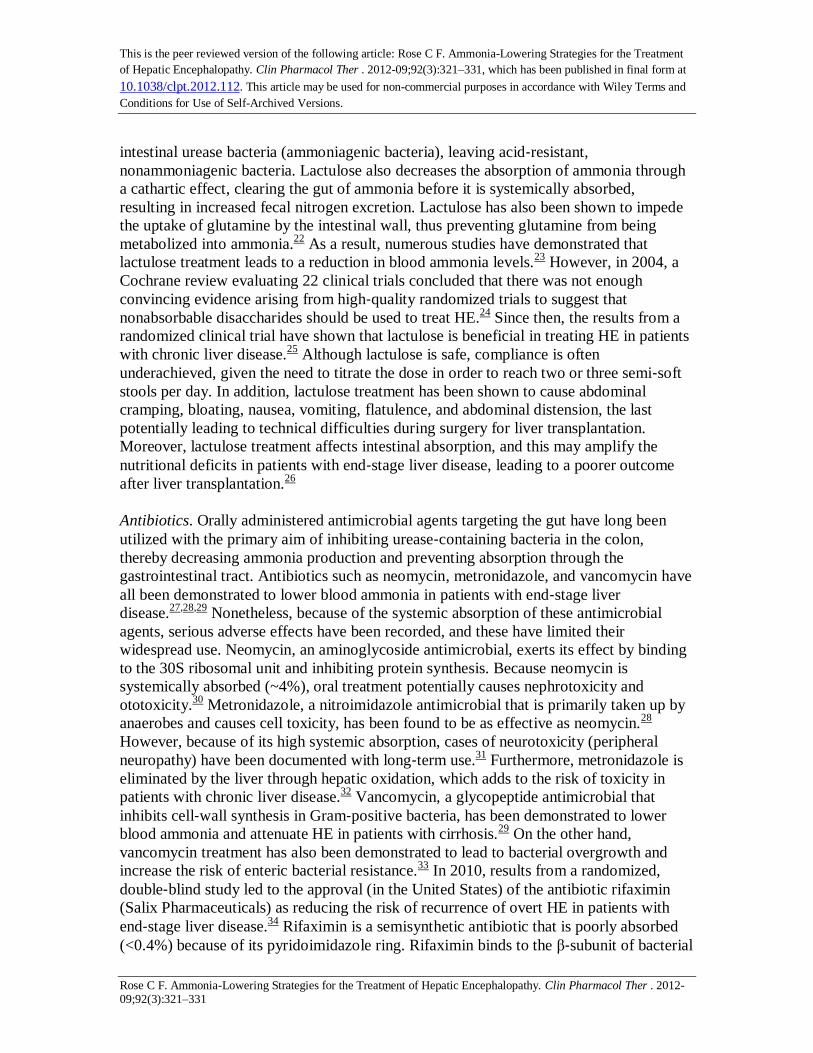
In addition, AST‐120 has the capacity to act as an ammonia sink by
This is the peer reviewed version of the following article: Rose C F. Ammonia-Lowering Strategies for the Treatment
of Hepatic Encephalopathy. Clin Pharmacol Ther . 2012-09;92(3):321–331, which has been published in final form at
10.1038/clpt.2012.112. This article may be used for non-commercial purposes in accordance with Wiley Terms and
Conditions for Use of Self-Archived Versions.
Rose C F. Ammonia-Lowering Strategies for the Treatment of Hepatic Encephalopathy. Clin Pharmacol Ther . 2012-09;92(3):321–331
drawing ammonia in from the systemic circulation46
(Figure 5). This means that AST‐120
absorbs not only the ammonia produced locally in the gut but also the ammonia produced
by extraintestinal organs. This property makes AST‐120 attractive as an ammonia‐lowering treatment for HE. In preliminary studies, patients with low‐grade HE showed
significant neurocognitive improvements after AST‐120 treatment.47
The efficacy, safety,
and tolerability of AST‐120 in the treatment of MHE were evaluated in a recent
multicenter, randomized, double‐blind, placebo‐controlled, dose‐ranging clinical trial.
This study (the ASTUTE trial) was carried out in 150 patients randomized to AST‐120 (6
and 12 g/day) or placebo for 8 weeks. AST‐120 failed to attenuate MHE after 4 and 8
weeks of treatment. However, AST‐120 treatment at both doses (6 and 12 g/day) did
result in a reduction in blood ammonia levels at both 4 and 8 weeks in patients with
elevated ammonia levels at baseline. Given its high ammonia‐absorbing capacity (300
µmol/l/g) and the high compliance rate associated with it, AST‐120 thus remains a
promising treatment for HE. However, a well‐designed clinical trial with a longer
duration of treatment is needed to prove the efficacy of AST‐120 as a treatment for HE.
Moreover, evaluations of the recurrence rates of HE, hospital readmissions and costs
would also be important end points.
Figure 5 AST‐120 as an ammonia sink. With the large capacity of AST‐120 to absorb
ammonia, it can also draw ammonia from blood circulation. Once absorbed by AST‐120
in the lumen, ammonia will cross the intestinal wall into the lumen following its
concentration gradient.
Sodium benzoate. Sodium benzoate conjugates with the amino acid glycine to form
hippuric acid, which is excreted by the kidneys. Glycine is metabolized through the
glycine cleavage system, an enzyme complex that consists of four proteins and generates
ammonia as an end‐product. Sodium benzoate is administered to prevent glycine
metabolism and thereby prevent the production of ammonia. Sodium benzoate has been
widely used in the therapeutic regimen to alleviate hyperammonemia in children born
with urea cycle disorders.48
In the context of liver disease, a randomized, double‐blind
study demonstrated that sodium benzoate reduces blood ammonia levels and attenuates

This is the peer reviewed version of the following article: Rose C F. Ammonia-Lowering Strategies for the Treatment
of Hepatic Encephalopathy. Clin Pharmacol Ther . 2012-09;92(3):321–331, which has been published in final form at
10.1038/clpt.2012.112. This article may be used for non-commercial purposes in accordance with Wiley Terms and
Conditions for Use of Self-Archived Versions.
Rose C F. Ammonia-Lowering Strategies for the Treatment of Hepatic Encephalopathy. Clin Pharmacol Ther . 2012-09;92(3):321–331
the symptoms of HE as effectively as lactulose, in cirrhotic patients.49
However, it has
also been demonstrated that sodium benzoate can inhibit the production of urea, inducing
hyperammonemia. The conjugation of sodium benzoate with glycine proceeds in two
steps. First, benzoate and ATP (adenosine triphosphate) react with CoA (coenzyme A) to
form benzoyl CoA, AMP (adenosine monophosphate), and pyrophosphate. Next, benzoyl
CoA and glycine react to form hippuric acid and CoA. This second step is dependent on
the concentration of glycine. Therefore, under conditions of low concentrations of
glycine, benzoate‐induced utilization of CoA could lead to a decrease in the activation of
carbamyl phosphate synthetase I, thereby reducing urea cycle activity. This suggests that
the efficacy of sodium benzoate is dependent on the level of functioning of the urea
cycle, with a beneficial response occurring when the urea cycle is impaired.50
However,
the effectiveness of using sodium benzoate as an ammonia‐lowering strategy in the
context of liver disease and residual hepatocyte function remains unclear.
Sodium phenylacetate/phenylbutyrate. Sodium phenylbutyrate, which is rapidly oxidized
into phenylacetate, is used to treat hyperammonemia by attenuating hyperglutaminemia
in children with urea‐cycle enzyme deficiencies.51
Phenylacetate conjugates with
glutamine in the liver and kidney to form phenylacetylglutamine, which is incapable of
being metabolized by glutaminase, and therefore glutamine‐stimulated ammoniagenesis
is prevented. Phenylacetylglutamine is then excreted in the urine. The use of sodium
phenylacetate/phenylbutyrate has never been investigated in the treatment of HE. This is
because the degree of hyperglutaminemia seen in patients with urea‐cycle enzyme
deficiencies and highly functional glutamine synthetase in the perivenous hepatocytes is
rarely observed in patients with end‐stage liver disease or severe hepatocyte necrosis.
Moreover, because the conjugation of phenylacetate is limited by the concentration of
glutamine, limited availability of glutamine can lead to CoA sequestering, resulting in the
inhibition of urea‐cycle synthesis and hyperammonemia. In addition, the sodium load
associated with the treatment, which could lead to fluid retention and exacerbate ascites,
has limited its use in patients with end‐stage liver disease. However, glycerol
phenylbutyrate (HPN‐100; Hyperion Therapeutics), a liquid triglyceride that does not
contain sodium and has a favorable palatability, has recently been shown to be as
effective as sodium phenylacetate.52
It is currently being investigated as a treatment for
HE in patients with end‐stage liver disease.
Glutaminase inhibitors. The small intestine is an important source of ammonia
generation, through the uptake and breakdown of glutamine by enterocytes. Phosphate‐activated glutaminase is the main glutamine‐catabolizing enzyme in the small intestine,
and current evidence indicates that 85% of intestinal ammonia production is the result of
glutaminase activity. It has been demonstrated that the duodenal glutaminase activity in
cirrhotic patients is nearly fourfold higher than that in healthy controls.53
Recently, a
microsatellite (a sequence of repeated base pairs) discovered within the promoter region
of the glutaminase gene was shown to be associated with the development of HE in
patients with cirrhosis.54
Not only does this study identify a possible genetic marker for
the development of HE, it also strongly suggests that glutaminase should be targeted as a
This is the peer reviewed version of the following article: Rose C F. Ammonia-Lowering Strategies for the Treatment
of Hepatic Encephalopathy. Clin Pharmacol Ther . 2012-09;92(3):321–331, which has been published in final form at
10.1038/clpt.2012.112. This article may be used for non-commercial purposes in accordance with Wiley Terms and
Conditions for Use of Self-Archived Versions.
Rose C F. Ammonia-Lowering Strategies for the Treatment of Hepatic Encephalopathy. Clin Pharmacol Ther . 2012-09;92(3):321–331
potential ammonia‐lowering strategy. Currently, specific glutaminase inhibitors do exist,
such as BPTES (bis‐2[5‐phenylacetamido‐1,2,4‐thiadiazol‐2‐yl]ethylsulfide), THDP17,
and DON (6‐diazo‐5‐oxo‐L‐norleucine), but they have only been tested in vitro. The
challenge is to avoid targeting and inhibiting all glutaminase activity, especially in in vivo
systems, because glutaminase plays a vital role in cellular metabolism and organ
function.
Acarbose. Acarbose, an approved treatment for diabetes mellitus, is an inhibitor of α‐glycosidase that primarily prevents the conversion of carbohydrates into
monosaccharides. In addition, it has been demonstrated that acarbose can decrease
colonic proteolytic flora and dietary nitrogenous substances. In a double‐blind, crossover,
randomized study, 107 cirrhotic patients with OHE and type 2 diabetes mellitus received
acarbose treatment and demonstrated a significant reduction in serum ammonia levels
and attenuation of HE.55
Acarbose is well tolerated and is not associated with serious
adverse effects. Additional studies are required to further evaluate the role and
mechanism of action of acarbose in the treatment of HE.
INCREASING THE AMOUNT OF AMMONIA REMOVAL
Branched‐chain amino acids. Treatment with branched‐chain amino acids (BCAAs) has
been shown to lower blood ammonia and improve the mental status of patients with end‐stage liver disease.
56 However, in 2004, a Cochrane review involving a meta‐analysis of
11 randomized, controlled clinical trials concluded that BCAA supplementation does not
affect morbidity or mortality in patients with end‐stage liver disease.57
Since then, a large
multicenter, randomized study demonstrated that BCAA treatment reduces the risk of
death and attenuates the progression of liver failure in patients with cirrhosis.58
The
mechanisms underlying the ammonia lowering by BCAA remain elusive. It is known that
BCAAs (valine, leucine, and isoleucine) are metabolically very active. In addition,
through the enzyme branched‐chain aminotransferase, the amino group from the BCAA
is utilized to form glutamate from α‐ketoglutarate and to detoxify ammonia through the
production of glutamine via glutamine synthetase. Besides forming glutamate, BCAA
metabolism, through branched‐chain aminotransferase, can generate branched‐chain
ketoacids. These ketoacids are further metabolized into CoA compounds through
branched‐chain α‐ketoacid dehydrogenase before being utilized in the tricarboxylic acid
cycle for oxidative energy production (Figure 6). Therefore, in addition to lowering
ammonia levels through biochemical action, BCAAs can indirectly increase ammonia
removal by acting as anticatabolic agents. Leucine, in particular, is capable of stimulating
protein synthesis in the liver and attenuating the degradation of muscle protein. The
maintenance of muscle mass is critical in patients with liver dysfunction, because the
muscle is an important ammonia‐removing organ, especially in the context of liver
failure.59
BCAAs are believed to be beneficial for patients with cirrhosis because they
function as a nutritional supplement, reducing protein catabolism, preventing muscle
wasting, and arresting progression of liver failure. This action of BCAAs would be
This is the peer reviewed version of the following article: Rose C F. Ammonia-Lowering Strategies for the Treatment
of Hepatic Encephalopathy. Clin Pharmacol Ther . 2012-09;92(3):321–331, which has been published in final form at
10.1038/clpt.2012.112. This article may be used for non-commercial purposes in accordance with Wiley Terms and
Conditions for Use of Self-Archived Versions.
Rose C F. Ammonia-Lowering Strategies for the Treatment of Hepatic Encephalopathy. Clin Pharmacol Ther . 2012-09;92(3):321–331
expected to decrease ammonia production and improve clearance. However, a recent
study showed that BCAA treatment in patients with cirrhosis resulted in an increase of
blood ammonia levels.60
This study, using [13
N] ammonia positron emission tomography,
concluded that BCAA metabolism in the skeletal muscle leads to an increase in
glutamine production (through glutamine synthetase) from BCAA‐derived ammonia
rather than from blood‐derived ammonia, as seen from the finding that the amount of
glutamine released from the muscles was six times greater than the ammonia uptake. In
this case, the glutamate derived from the BCAA reacts with the cofactor NAD+
(nicotinamide adenine dinucleotide) to form α‐ketoglutarate, and ammonia, through the
enzyme glutamate dehydrogenase (Figure 6). This BCAA‐induced ammonia is then
removed through the formation of glutamine, released into the blood, and metabolized by
glutaminase in extramuscular organs such as the kidney and intestine,7 causing a release
of ammonia. Although BCAA treatment is regarded as safe, noncompliance with long‐term treatment has been reported—primarily due to its poor palatability and solubility,
the latter necessitating consumption of large quantities of water. Overall, BCAA
treatment can be potentially beneficial as an ammonia‐lowering strategy and as a
treatment for HE, but to date its efficacy remains a matter of debate. A therapeutic dosing
range of BCAA needs to be established for reliably lowering blood ammonia levels in
cirrhotic patients with HE.
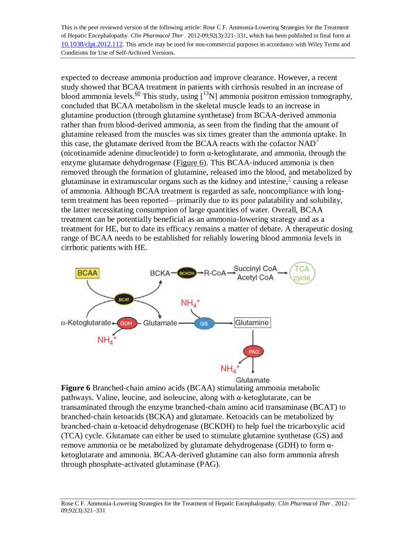
Figure 6 Branched‐chain amino acids (BCAA) stimulating ammonia metabolic
pathways. Valine, leucine, and isoleucine, along with α‐ketoglutarate, can be
transaminated through the enzyme branched‐chain amino acid transaminase (BCAT) to
branched‐chain ketoacids (BCKA) and glutamate. Ketoacids can be metabolized by
branched‐chain α‐ketoacid dehydrogenase (BCKDH) to help fuel the tricarboxylic acid
(TCA) cycle. Glutamate can either be used to stimulate glutamine synthetase (GS) and
remove ammonia or be metabolized by glutamate dehydrogenase (GDH) to form α‐ketoglutarate and ammonia. BCAA‐derived glutamine can also form ammonia afresh
through phosphate‐activated glutaminase (PAG).
This is the peer reviewed version of the following article: Rose C F. Ammonia-Lowering Strategies for the Treatment
of Hepatic Encephalopathy. Clin Pharmacol Ther . 2012-09;92(3):321–331, which has been published in final form at
10.1038/clpt.2012.112. This article may be used for non-commercial purposes in accordance with Wiley Terms and
Conditions for Use of Self-Archived Versions.
Rose C F. Ammonia-Lowering Strategies for the Treatment of Hepatic Encephalopathy. Clin Pharmacol Ther . 2012-09;92(3):321–331
L‐ornithine–L‐aspartate. L‐ornithine–L‐aspartate (LOLA), both substrates of the urea
cycle, were first tried as infusion treatments in patients suffering from end‐stage liver
disease and HE, in an attempt to lower blood ammonia by stimulating ureagenesis in the
residual hepatocytes (Figure 7a). This concept has been proven to work in ammonia‐injected portacaval‐shunted rats, in which LOLA infusion prevented a rise in ammonia
with a concomitant increase in urea production.61
In addition, LOLA has the
demonstrated ability to stimulate glutamine synthesis, particularly in the skeletal
muscle.61,62
L‐ornithine is a substrate for ornithine aminotransferase that gives rise to
glutamate and glutamate semialdehyde, which can be further metabolized through
glutamate semialdehyde dehydrogenase to form another molecule of glutamate. L‐aspartate is also capable of generating a molecule of glutamate through aspartate‐α‐ketoglutarate transaminase. Therefore, L‐ornithine and L‐aspartate taken together can
produce three molecules of glutamate, which in turn can fuel glutamine synthesis activity
and detoxify ammonia (Figure 7b). It should also be noted that glutamate can be
metabolized by glutamine dehydrogenase to form α‐ketoglutarate and ammonia. Several
clinical studies have demonstrated that LOLA (administered either orally or
intravenously) lowers ammonia concentrations, provides symptomatic improvement in
cirrhotic patients with HE,63
markedly improves health‐related quality of life, and is as
effective as lactulose in the management of HE. In 2008, a Cochrane review, which
included a meta‐analysis, reported that LOLA treatment led to the attenuation of OHE; it
was, however, less beneficial in patients with MHE.64
In 2009, Acharya et al. concluded,
following a randomized trial of 201 patients with acute liver failure, that treatment with
LOLA did not improve ammonia reduction, attenuate encephalopathy, or increase
survival.65
However, it is important to note that in this study, mortality in the placebo
group was as low as 33% in comparison with an earlier study by the same group that
reported 52% mortality in the placebo group.66
This reduction in mortality may be due to
improvements in standard care, including prophylactic administration of antibiotics
(piperacillin–tazobactam, fluconazole, and vancomycin). In addition, vancomycin has
been shown to reduce ammonia levels in patients with cirrhosis.29
Therefore, it was
conclusively shown that, in patients with acute liver failure, LOLA administration could
not induce further reductions in ammonia to levels below those achieved by the standard
care given to a placebo group, and that LOLA (administered at a dose of 30 g/day for 3
days) was not a good adjuvant to the standard care. However, very good tolerability has
been documented for LOLA treatment, with no major adverse effects. The results of
many studies suggest that LOLA is an efficient and promising treatment for HE in
patients with end‐stage liver disease. However, caution should be exercised in using
LOLA to treat patients with acute liver failure. It appears that the duration and dosage of
the LOLA treatment regimen are vital to its success and therefore need to be established.
LOLA is currently not available for clinical use in North America.

This is the peer reviewed version of the following article: Rose C F. Ammonia-Lowering Strategies for the Treatment
of Hepatic Encephalopathy. Clin Pharmacol Ther . 2012-09;92(3):321–331, which has been published in final form at
10.1038/clpt.2012.112. This article may be used for non-commercial purposes in accordance with Wiley Terms and
Conditions for Use of Self-Archived Versions.
Rose C F. Ammonia-Lowering Strategies for the Treatment of Hepatic Encephalopathy. Clin Pharmacol Ther . 2012-09;92(3):321–331
Figure 7 Possible ammonia pathways induced by L‐ornithine L‐aspartate. (a) L‐ornithine
L‐aspartate, a substrate and intermediate of the urea cycle, can lower ammonia by
stimulating ureagenesis. (b) L‐ornithine and L‐aspartate can also be transaminated with
α‐ketoglutarate to glutamate, through ornithine aminotransferase (OAT) and aspartate
aminotransferase (AAT), respectively. Ornithine‐derived glutamate semialdehyde can be
further metabolized to glutamate through glutamate semialdehyde dehydrogenase
(GSDH). These three molecules of glutamate can be used to stimulate glutamine
synthetase (GS), thus forming glutamine and removing ammonia. However, L‐ornithine
L‐aspartate–derived glutamine can be metabolized through phosphate‐activated
glutaminase (PAG), thereby regenerating ammonia. GDH, glutamate dehydrogenase.
L‐carnitine. L‐carnitine, a metabolite produced by the degradation of the essential amino
acid lysine, serves as a carrier for short‐chain fatty acids across the mitochondrial
membrane. Treatment with L‐acetylcarnitine (the acetylated form of L‐carnitine, known
to increase its bioavailability) significantly reduced serum ammonia levels and improved
mental status as compared with placebo in 150 cirrhotic patients with mild to moderate
HE.67
However, despite reductions in blood ammonia levels, an overall worsening per the
Glasgow Coma Scale was noted in critically ill cirrhotic patients with hepatic coma who
had received L‐carnitine.68
L‐acetylcarnitine, given as pretreatment to portacaval‐shunted
This is the peer reviewed version of the following article: Rose C F. Ammonia-Lowering Strategies for the Treatment
of Hepatic Encephalopathy. Clin Pharmacol Ther . 2012-09;92(3):321–331, which has been published in final form at
10.1038/clpt.2012.112. This article may be used for non-commercial purposes in accordance with Wiley Terms and
Conditions for Use of Self-Archived Versions.
Rose C F. Ammonia-Lowering Strategies for the Treatment of Hepatic Encephalopathy. Clin Pharmacol Ther . 2012-09;92(3):321–331
rats, was shown to prevent ammonia‐induced encephalopathy; however, it did not
provide a significant protection against an increase in blood ammonia levels after rectal
administration of ammonia in subjects with cirrhosis.69
COMBINATION OF AMMONIA‐LOWERING AGENTS
Targeting a single strategy. Recently, numerous studies have been published describing
various combinations of ammonia‐lowering agents as treatment for HE in patients with
cirrhosis. Some were intentionally designed to investigate the benefit of simultaneously
administering two ammonia‐removing agents, such as the study by Sharma et al., which
investigated the beneficial effect of lactulose vs. probiotics vs. lactulose + probiotics. All
three treatments were equally effective in lowering blood ammonia and in treating
MHE.70
Other studies generated results of combination strategies as part of comparisons
with lactulose, the standard care regimen in these studies. Recently, lactulose + rifaximin
has been demonstrated to be more beneficial in preventing a recurrence of HE as
compared to lactulose alone.34
Each of the above‐mentioned studies describes the effect
of combining two treatments, each targeting the gut, modulating the microflora, and, in
effect, lowering the production of ammonia. In another study, two agents with ammonia‐removing properties (L‐acetylcarnitine + BCAA) were combined. The results of this
study demonstrated that L‐acetylcarnitine + BCAA causes greater reduction in blood
ammonia and is more beneficial in attenuating HE as compared to BCAA treatment
alone.71
Sodium phenylacetate and sodium benzoate were also evaluated as a
combination (Ammonul; Hyperion Therapeutics), and this regimen was approved for the
treatment of hyperammonemia associated with urea‐cycle disorders.72
The strategy
behind combining sodium phenylacetate and sodium benzoate is that each of these agents
chelates different ammoniagenic substrates. With sodium phenylacetate conjugating with
glutamine, and sodium benzoate conjugating with glycine, two substrates for potential
ammoniagenesis are thus removed. In the context of HE, this combination has previously
been tested in a small study that suggested its possible benefit.73
It was to be tested in a
clinical trial to investigate the beneficial effects of sodium phenylacetate and sodium
benzoate (Ammonul; Hyperion Therapeutics) in cirrhotic patients with severe HE.
However, for undisclosed reasons, the study was withdrawn prior to enrollment
commenced (http://www.clinicaltrials.gov).
Overall, the idea of combining treatments targeting different ammonia‐producing/lowering systems is a valuable concept. However, selecting two agents that
both inhibit ammonia production or increase ammonia removal in the same organs may
be an excessive and redundant strategy.
Synergy of two strategies: L‐ornithine phenylacetate. L‐ornithine phenylacetate (OCR‐002; Ocera Therapeutics) represents a new combination treatment strategy by which, for
the first time, an agent is administered in order to activate ammonia removal in
combination with another agent that stimulates a reduction in ammonia production. The

This is the peer reviewed version of the following article: Rose C F. Ammonia-Lowering Strategies for the Treatment
of Hepatic Encephalopathy. Clin Pharmacol Ther . 2012-09;92(3):321–331, which has been published in final form at
10.1038/clpt.2012.112. This article may be used for non-commercial purposes in accordance with Wiley Terms and
Conditions for Use of Self-Archived Versions.
Rose C F. Ammonia-Lowering Strategies for the Treatment of Hepatic Encephalopathy. Clin Pharmacol Ther . 2012-09;92(3):321–331
idea of this synergistic treatment, developed by Rajiv Jalan in the United Kingdom,74
is
based on the fact that L‐ornithine (the active component of LOLA)75
generates glutamate
(through ornithine aminotransferase in muscle), thereby stimulating glutamine synthesis
and reducing blood ammonia. This, in turn, leads to an increase in ornithine‐derived
glutamine. To prevent glutamine from being metabolized by glutaminase and forming
ammonia again, phenylacetate is administered to conjugate with the ornithine‐derived
glutamine to form phenylacetylglutamine. Consequently, given that glutaminase is
incapable of metabolizing phenylacetylglutamine, an ammonia rebound is prevented and
phenylacetylglutamine is excreted by the kidneys (Figure 8). The proof of concept for the
mechanism of L‐ornithine phenylacetate was first tested in liver‐devascularized pigs to
negate the possibility of L‐ornithine stimulating ureagenesis.10
In that study, L‐ornithine
phenylacetate treatment attenuated increases in blood and brain ammonia, brain edema,
and intracranial hypertension—results that were not achieved with either L‐ornithine or
phenylacetate treatment alone. L‐ornithine phenylacetate has also been shown to be
effective in lowering blood ammonia in cirrhotic rats and portacaval‐shunted rats with
simulated gastrointestinal bleed.9,14
To date, animal studies have demonstrated the
efficacy of L‐ornithine phenylacetate as an ammonia‐lowering strategy. Currently, two
phase II clinical trials of the safety and tolerability of L‐ornithine phenylacetate treatment
are under way in Spain and the United States (http://www.clinicaltrials.gov): (i) in
patients with acute liver failure due to acetaminophen overdose and (ii) in patients with
cirrhosis and upper gastrointestinal bleeding. This promising synergistic treatment, which
combines two agents that do not target the gut, is very novel. As a therapeutic strategy, it
would allow patients with cirrhosis to be properly nourished (stable protein diet) and
would inhibit the development of hyperammonemia by increasing ammonia elimination.
Figure 8 The synergistic mechanism of L‐ornithine phenylacetate. L‐ornithine (with α‐ketoglutarate) can produce two molecules of glutamate through glutamate semialdehyde
dehydrogenase (GSDH) and ornithine aminotransferase (OAT) that can stimulate
glutamine synthetase (GS), thereby removing ammonia and forming glutamine. To

This is the peer reviewed version of the following article: Rose C F. Ammonia-Lowering Strategies for the Treatment
of Hepatic Encephalopathy. Clin Pharmacol Ther . 2012-09;92(3):321–331, which has been published in final form at
10.1038/clpt.2012.112. This article may be used for non-commercial purposes in accordance with Wiley Terms and
Conditions for Use of Self-Archived Versions.
Rose C F. Ammonia-Lowering Strategies for the Treatment of Hepatic Encephalopathy. Clin Pharmacol Ther . 2012-09;92(3):321–331
prevent ornithine‐derived glutamine from being metabolized by phosphate‐activated
glutaminase (PAG), phenylacetate conjugates with glutamine to form
phenylacetylglutamine, which is then excreted in the urine, and thus regeneration of
ammonia is prevented. GDH, glutamate dehydrogenase.
SUMMARY
HE, a devastating neuropsychiatric syndrome, is a major complication of liver failure.
Because ammonia plays such a major role in the pathogenesis of HE, ammonia‐lowering
strategies remain a focus for HE treatments. Historically, ammonia‐lowering strategies
have concentrated on inhibiting the source of ammonia production in the gut; currently,
lactulose and rifaximin are the treatments of choice for HE. Other agents that inhibit
ammonia production include probiotics, glutaminase inhibitors, and AST‐120. However,
they all need to be evaluated in high‐quality clinical trials. An alternative ammonia‐lowering strategy involves increasing ammonia removal. This focuses primarily on
stimulating glutamine synthetase in the large mass capacity of skeletal muscle. BCAAs,
LOLA, and L‐ornithine phenylacetate have all been shown to be capable, through
transamination, of generating glutamate, which stimulates glutaminase synthetase,
producing glutamate and eliminating ammonia. However, under certain conditions, the α‐ketoglutarate generated can form ammonia through glutamate dehydrogenase; also,
glutamine, the ammonia‐removing product, can itself be metabolized by glutaminase to
produce ammonia afresh. Given that glutaminase has been described as an important
enzyme associated with the risk of developing HE, glutamine would seem to be a
secondary product that should be targeted for elimination. Paradoxically, however, the
production of glutamine is required for the removal of ammonia. In this situation, the use
of L‐ornithine phenylacetate is an excellent strategy, not only to stimulate glutamine
synthesis (ammonia removal) but also to prevent a glutamine‐induced ammonia rebound.
Furthermore, given that malnourishment is a major concern in cirrhotic patients, targeting
the gut to lower ammonia, thereby possibly altering intestinal absorption, may exacerbate
malnutrition. Therefore, exploitation of ammonia‐removing pathways in other organs is a
potential treatment strategy; for instance, the large mass capacity of skeletal muscle
renders it an attractive therapeutic target. However, sarcopenia is commonly observed in
patients with cirrhosis; considered independently, it may be a result of malnutrition,
which, as noted above, has a detrimental effect on outcome in terms of complications,
survival after liver transplantation, and overall liver‐related mortality. Therefore, to
benefit from using the muscle as a therapeutic target, the detection of malnutrition is of
pivotal importance. Proper nutrition that prevents loss of muscle mass in cirrhotic
patients can independently lead to an increase in ammonia‐removal capacity, as well as to
a much better response to BCAA, LOLA, and L‐ornithine phenylacetate in stimulating
glutamine synthesis, with L‐ornithine phenylacetate being the most beneficial in
preventing glutaminase‐induced ammonia rebound.

This is the peer reviewed version of the following article: Rose C F. Ammonia-Lowering Strategies for the Treatment
of Hepatic Encephalopathy. Clin Pharmacol Ther . 2012-09;92(3):321–331, which has been published in final form at
10.1038/clpt.2012.112. This article may be used for non-commercial purposes in accordance with Wiley Terms and
Conditions for Use of Self-Archived Versions.
Rose C F. Ammonia-Lowering Strategies for the Treatment of Hepatic Encephalopathy. Clin Pharmacol Ther . 2012-09;92(3):321–331
REFERENCES
1. Stewart, C. A. & Smith, G. E. Minimal hepatic encephalopathy. Nat Rev
Gastroenterol Hepatol 4, 677–685 (2007).
2. Poordad, F. F. Review article: the burden of hepatic encephalopathy. Aliment
Pharmacol Ther 25 Suppl 1, 3–9 (2007).
3. Bajaj, J. S. Review article: the modern management of hepatic encephalopathy.
Aliment Pharmacol Ther 31, 537–47 (2010).
4. Neff, G. Pharmacoeconomics of Hepatic Encephalopathy. Pharmacotherapy 30,
28S–32S (2010).
5. Bosoi, C. R. & Rose, C. F. Identifying the direct effects of ammonia on the brain.
Metab Brain Dis 24, 95–102 (2009).
6. Felipo, V. & Butterworth, R. F. Neurobiology of ammonia. Prog Neurobiol 67,
259–279 (2002).
7. Wright, G., Noiret, L., Damink, S. W. M. O. & Jalan, R. Interorgan ammonia
metabolism in liver failure: the basis of current and future therapies. Liver Int. 31, 163–75
(2011).
8. Bosoi, C. R. et al. Systemic oxidative stress is implicated in the pathogenesis of
brain edema in rats with chronic liver failure. Free Radic. Biol. Med 52, 1228–1235
(2012).
9. Oria, M. et al. Ornithine phenylacetate prevents disturbances of motor-evoked
potentials induced by intestinal blood in rats with portacaval anastomosis. J Hepatol 56,
109–14 (2012).
10. Ytrebø, L. M. et al. L-ornithine phenylacetate attenuates increased arterial and
extracellular brain ammonia and prevents intracranial hypertension in pigs with acute
liver failure. Hepatology 50, 165–174 (2009).
11. Keiding, S. et al. Brain metabolism of13N-ammonia during acute hepatic
encephalopathy in cirrhosis measured by positron emission tomography. Hepatology 43,
42–50 (2006).
12. Sørensen, M. & Keiding, S. New findings on cerebral ammonia uptake in HE
using functional (13)N-ammonia PET. Metab Brain Dis 22, 277–284 (2007).
13. Rose, C., Kresse, W. & Kettenmann, H. Acute insult of ammonia leads to
calcium-dependent glutamate release from cultured astrocytes, an effect of pH. J. Biol.
Chem. 280, 20937–20944 (2005).
14. Davies, N. A. et al. L-ornithine and phenylacetate synergistically produce
sustained reduction in ammonia and brain water in cirrhotic rats. Hepatology 50, 155–164
(2009).
15. Rovira, A., Alonso, J. & Córdoba, J. MR imaging findings in hepatic
encephalopathy. AJNR Am J Neuroradiol 29, 1612–1621 (2008).
16. Chatauret, N., Zwingmann, C., Rose, C., Leibfritz, D. & Butterworth, R. F.
Effects of hypothermia on brain glucose metabolism in acute liver failure: a H/C-nuclear
magnetic resonance study. Gastroenterology 125, 815–824 (2003).
17. Schiano, T. Complications of chronic liver disease. Current Diagnosis and
Treatment in Gastroenterology (2002).

This is the peer reviewed version of the following article: Rose C F. Ammonia-Lowering Strategies for the Treatment
of Hepatic Encephalopathy. Clin Pharmacol Ther . 2012-09;92(3):321–331, which has been published in final form at
10.1038/clpt.2012.112. This article may be used for non-commercial purposes in accordance with Wiley Terms and
Conditions for Use of Self-Archived Versions.
Rose C F. Ammonia-Lowering Strategies for the Treatment of Hepatic Encephalopathy. Clin Pharmacol Ther . 2012-09;92(3):321–331
18. Kundra, A., Jain, A., Banga, A., Bajaj, G. & Kar, P. Evaluation of plasma
ammonia levels in patients with acute liver failure and chronic liver disease and its
correlation with the severity of hepatic encephalopathy and clinical features of raised
intracranial tension. Clin Biochem 38, 696–699 (2005).
19. Shawcross, D. L., Davies, N. A., Williams, R. & Jalan, R. Systemic inflammatory
response exacerbates the neuropsychological effects of induced hyperammonemia in
cirrhosis. J Hepatol 40, 247–254 (2004).
20. Montoliu, C. et al. 3-Nitro-Tyrosine as a Peripheral Biomarker of Minimal
Hepatic Encephalopathy in Patients With Liver Cirrhosis. Am J Gastroenterol 106, 1629–
1637 (2011).
21. Bémeur, C., Desjardins, P. & Butterworth, R. F. Role of nutrition in the
management of hepatic encephalopathy in end-stage liver failure. J Nutr Metab 2010,
489823 (2010).
22. van Leeuwen, P. A., van Berlo, C. L. & Soeters, P. B. New mode of action for
lactulose. Lancet 1, 55–56 (1988).
23. Sharma, P., Agrawal, A., Sharma, B. C. & Sarin, S. K. Prophylaxis of hepatic
encephalopathy in acute variceal bleed: a randomized controlled trial of lactulose versus
no lactulose. J. Gastroenterol. Hepatol. 26, 996–1003 (2011).
24. Als-Nielsen, B., Gluud, L. L. & Gluud, C. Non-absorbable disaccharides for
hepatic encephalopathy: systematic review of randomised trials. BMJ 328, 1046 (2004).
25. Prasad, S. et al. Lactulose improves cognitive functions and health-related quality
of life in patients with cirrhosis who have minimal hepatic encephalopathy. Hepatology
45, 549–559 (2007).
26. Teperman, L. W. & Peyregne, V. P. Considerations on the impact of hepatic
encephalopathy treatments in the pretransplant setting. Transplantation 89, 771–8 (2010).
27. Conn, H. O. et al. Comparison of lactulose and neomycin in the treatment of
chronic portal-systemic encephalopathy. A double blind controlled trial.
Gastroenterology 72, 573–583 (1977).
28. Morgan, M. H., Read, A. E. & Speller, D. C. Treatment of hepatic encephalopathy
with metronidazole. Gut 23, 1–7 (1982).
29. Tarao, K. et al. Successful use of vancomycin hydrochloride in the treatment of
lactulose resistant chronic hepatic encephalopathy. Gut 31, 702–706 (1990).
30. Greenberg, L. H. & Momary, H. Audiotoxicity and nephrotoxicity due to orally
administered neomycin. JAMA 194, 827–828 (1965).
31. Heaney, C. J., Campeau, N. G. & Lindell, E. P. MR imaging and diffusion-
weighted imaging changes in metronidazole (Flagyl)-induced cerebellar toxicity. AJNR
Am J Neuroradiol 24, 1615–1617 (2003).
32. Loft, S., Sonne, J., Døssing, M. & Andreasen, P. B. Metronidazole
pharmacokinetics in patients with hepatic encephalopathy. Scand. J. Gastroenterol. 22,
117–123 (1987).
33. Smith, T. L. et al. Emergence of vancomycin resistance in Staphylococcus aureus.
Glycopeptide-Intermediate Staphylococcus aureus Working Group. N. Engl. J. Med. 340,
493–501 (1999).
34. Bass, N. M. et al. Rifaximin treatment in hepatic encephalopathy. N. Engl. J. Med.
This is the peer reviewed version of the following article: Rose C F. Ammonia-Lowering Strategies for the Treatment
of Hepatic Encephalopathy. Clin Pharmacol Ther . 2012-09;92(3):321–331, which has been published in final form at
10.1038/clpt.2012.112. This article may be used for non-commercial purposes in accordance with Wiley Terms and
Conditions for Use of Self-Archived Versions.
Rose C F. Ammonia-Lowering Strategies for the Treatment of Hepatic Encephalopathy. Clin Pharmacol Ther . 2012-09;92(3):321–331
362, 1071–81 (2010).
35. Mas, A. et al. Comparison of rifaximin and lactitol in the treatment of acute
hepatic encephalopathy: results of a randomized, double-blind, double-dummy,
controlled clinical trial. J. Hepatol. 38, 51–58 (2003).
36. Pedretti, G., Calzetti, C., Missale, G. & Fiaccadori, F. Rifaximin versus neomycin
on hyperammoniemia in chronic portal systemic encephalopathy of cirrhotics. A double-
blind, randomized trial. Ital J Gastroenterol 23, 175–178 (1991).
37. Leevy, C. B. & Phillips, J. A. Hospitalizations during the use of rifaximin versus
lactulose for the treatment of hepatic encephalopathy. Dig. Dis. Sci. 52, 737–741 (2007).
38. Malaguarnera, M. et al. Bifidobacterium longum with fructo-oligosaccharide
(FOS) treatment in minimal hepatic encephalopathy: a randomized, double-blind,
placebo-controlled study. Dig. Dis. Sci. 52, 3259–3265 (2007).
39. Bajaj, J. S. et al. Probiotic Yogurt for the Treatment of Minimal Hepatic
Encephalopathy. Am J Gastroenterol 103, 1707–1715 (2008).
40. McGee, R. G., Bakens, A., Wiley, K., Riordan, S. M. & Webster, A. C. Probiotics
for patients with hepatic encephalopathy. Cochrane Database Syst Rev CD008716
(2011).doi:10.1002/14651858.CD008716.pub2
41. Solga, S. F. Probiotics can treat hepatic encephalopathy. Med. Hypotheses 61,
307–313 (2003).
42. Bajaj, J. S. et al. Linkage of gut microbiome with cognition in hepatic
encephalopathy. Am. J. Physiol. Gastrointest. Liver Physiol. 302, G168–175 (2012).
43. Hatakeyama, S. et al. Effect of an Oral Adsorbent, AST-120, on Dialysis
Initiation and Survival in Patients with Chronic Kidney Disease. Int J Nephrol 2012,
376128 (2012).
44. Tack, J. F., Miner, P. B., Jr, Fischer, L. & Harris, M. S. Randomised clinical trial:
the safety and efficacy of AST-120 in non-constipating irritable bowel syndrome - a
double-blind, placebo-controlled study. Aliment. Pharmacol. Ther. 34, 868–877 (2011).
45. Shen, B. et al. The efficacy and tolerability of AST-120 (spherical carbon
adsorbent) in active pouchitis. Am. J. Gastroenterol. 104, 1468–1474 (2009).
46. Bosoi, C. R., Parent-Robitaille, C., Anderson, K., Tremblay, M. & Rose, C. F.
AST-120 (spherical carbon adsorbent) lowers ammonia levels and attenuates brain edema
in bile-duct ligated rats. Hepatology 53, 1995–2002 (2011).
47. Pockros, P. et al. Phase 2, Multicenter, Randomized Study of AST-120 (spherical
Carbon Adsorbent) Vs. Lactulose in the Treatment of Low-Grade Hepatic
Encephalopathy. J Hepatol. 50, S43–44 (2009).
48. Batshaw, M. L. et al. Treatment of inborn errors of urea synthesis: activation of
alternative pathways of waste nitrogen synthesis and excretion. N. Engl. J. Med. 306,
1387–1392 (1982).
49. Sushma, S. et al. Sodium benzoate in the treatment of acute hepatic
encephalopathy: a double-blind randomized trial. Hepatology 16, 138–144 (1992).
50. Maswoswe, S. M. & Tremblay, G. C. Biosynthesis of hippurate, urea and
pyrimidines in the fatty liver: studies with rats fed orotic acid or a diet deficient in choline
and inositol, and with genetically obese (Zucker) rats. J. Nutr. 119, 273–279 (1989).
51. Honda, S. et al. Successful treatment of severe hyperammonemia using sodium

This is the peer reviewed version of the following article: Rose C F. Ammonia-Lowering Strategies for the Treatment
of Hepatic Encephalopathy. Clin Pharmacol Ther . 2012-09;92(3):321–331, which has been published in final form at
10.1038/clpt.2012.112. This article may be used for non-commercial purposes in accordance with Wiley Terms and
Conditions for Use of Self-Archived Versions.
Rose C F. Ammonia-Lowering Strategies for the Treatment of Hepatic Encephalopathy. Clin Pharmacol Ther . 2012-09;92(3):321–331
phenylacetate powder prepared in hospital pharmacy. Biol. Pharm. Bull. 25, 1244–1246
(2002).
52. Lichter-Konecki, U. et al. Ammonia control in children with urea cycle disorders
(UCDs); phase 2 comparison of sodium phenylbutyrate and glycerol phenylbutyrate. Mol.
Genet. Metab. 103, 323–329 (2011).
53. Romero-Gómez, M., Jover, M., Galán, J. J. & Ruiz, A. Gut ammonia production
and its modulation. Metab Brain Dis 24, 147–57 (2009).
54. Romero-Gómez, M. et al. Variations in the promoter region of the glutaminase
gene and the development of hepatic encephalopathy in patients with cirrhosis: a cohort
study. Ann. Intern. Med. 153, 281–8 (2010).
55. Gentile, S. et al. A randomized controlled trial of acarbose in hepatic
encephalopathy. Clin. Gastroenterol. Hepatol. 3, 184–191 (2005).
56. Marchesini, G. et al. Long-term oral branched-chain amino acid treatment in
chronic hepatic encephalopathy. A randomized double-blind casein-controlled trial. The
Italian Multicenter Study Group. J. Hepatol. 11, 92–101 (1990).
57. Als-Nielsen, B., Koretz, R. L., Kjaergard, L. L. & Gluud, C. Branched-chain
amino acids for hepatic encephalopathy. Cochrane Database Syst Rev CD001939
(2003).doi:10.1002/14651858.CD001939
58. Marchesini, G. et al. Nutritional supplementation with branched-chain amino
acids in advanced cirrhosis: a double-blind, randomized trial. Gastroenterology 124,
1792–1801 (2003).
59. Chatauret, N. et al. Direct molecular and spectroscopic evidence for increased
ammonia removal capacity of skeletal muscle in acute liver failure. J. Hepatol. 44, 1083–
1088 (2006).
60. Dam, G. et al. Branched-chain amino acids increase arterial blood ammonia in
spite of enhanced intrinsic muscle ammonia metabolism in patients with cirrhosis and
healthy subjects. Am. J. Physiol. Gastrointest. Liver Physiol. 301, G269–277 (2011).
61. Rose, C. et al. L-ornithine-L-aspartate in experimental portal-systemic
encephalopathy: therapeutic efficacy and mechanism of action. Metab Brain Dis 13, 147–
157 (1998).
62. Rose, C. et al. L-ornithine-L-aspartate lowers plasma and cerebrospinal fluid
ammonia and prevents brain edema in rats with acute liver failure. Hepatology 30, 636–
640 (1999).
63. Kircheis, G. et al. Therapeutic efficacy of L-ornithine-L-aspartate infusions in
patients with cirrhosis and hepatic encephalopathy: results of a placebo-controlled,
double-blind study. Hepatology 25, 1351–1360 (1997).
64. Jiang, Q., Jiang, X.-H., Zheng, M.-H. & Chen, Y.-P. L-Ornithine-l-aspartate in the
management of hepatic encephalopathy: a meta-analysis. J. Gastroenterol. Hepatol. 24,
9–14 (2009).
65. Acharya, S. K., Bhatia, V., Sreenivas, V., Khanal, S. & Panda, S. K. Efficacy of
L-ornithine L-aspartate in acute liver failure: a double-blind, randomized, placebo-
controlled study. Gastroenterology 136, 2159–2168 (2009).
66. Bhatia, V., Singh, R. & Acharya, S. K. Predictive value of arterial ammonia for
complications and outcome in acute liver failure. Gut 55, 98–104 (2006).

This is the peer reviewed version of the following article: Rose C F. Ammonia-Lowering Strategies for the Treatment
of Hepatic Encephalopathy. Clin Pharmacol Ther . 2012-09;92(3):321–331, which has been published in final form at
10.1038/clpt.2012.112. This article may be used for non-commercial purposes in accordance with Wiley Terms and
Conditions for Use of Self-Archived Versions.
Rose C F. Ammonia-Lowering Strategies for the Treatment of Hepatic Encephalopathy. Clin Pharmacol Ther . 2012-09;92(3):321–331
67. Malaguarnera, M. et al. Effects of L-carnitine in patients with hepatic
encephalopathy. World J. Gastroenterol. 11, 7197–7202 (2005).
68. Malaguarnera, M. et al. Effects of L-acetylcarnitine on cirrhotic patients with
hepatic coma: randomized double-blind, placebo-controlled trial. Dig. Dis. Sci. 51, 2242–
2247 (2006).
69. del Olmo, J. A. et al. Effect of L-carnitine upon ammonia tolerance test in
cirrhotic patients. Adv. Exp. Med. Biol. 272, 197–208 (1990).
70. Sharma, P., Sharma, B. C., Puri, V. & Sarin, S. K. An open-label randomized
controlled trial of lactulose and probiotics in the treatment of minimal hepatic
encephalopathy. Eur J Gastroenterol Hepatol 20, 506–511 (2008).
71. Malaguarnera, M. et al. Branched chain amino acids supplemented with L-
acetylcarnitine versus BCAA treatment in hepatic coma: a randomized and controlled
double blind study. Eur J Gastroenterol Hepatol 21, 762–770 (2009).
72. Enns, G. M. et al. Survival after treatment with phenylacetate and benzoate for
urea-cycle disorders. N. Engl. J. Med. 356, 2282–2292 (2007).
73. Mendenhall, C. L., Rouster, S., Marshall, L. & Weesner, R. A new therapy for
portal systemic encephalopathy. Am. J. Gastroenterol. 81, 540–543 (1986).
74. Jalan, R., Wright, G., Davies, N. A. & Hodges, S. J. L-Ornithine phenylacetate
(OP): a novel treatment for hyperammonemia and hepatic encephalopathy. Med.
Hypotheses 69, 1064–1069 (2007).
75. Zieve, L., Lyftogt, C. & Raphael, D. Ammonia toxicity: comparative protective
effect of various arginine and ornithine derivatives, aspartate, benzoate, and carbamyl
glutamate. Metab Brain Dis 1, 25–35 (1986).


