Embed Size (px)
Citation preview
AllergoOncology: the role of IgE-mediated allergy in cancer
E. Jensen-Jarolim1, G. Achatz2, M. C. Turner3, S. Karagiannis4, F. Legrand5, M. Capron5, M.L. Penichet6, J. A. Rodríguez7, A. G. Siccardi8, L. Vangelista8, A. B. Riemer1, and H. Gould4
1 IPP– Department of Pathophysiology, Center of Physiology, Pathophysiology and Immunology,Medical University Vienna, Waehringer G. 18, 1090 Vienna, Austria2 Division of Genetics and General Biology, Hellbrunnerstraße, Salzburg, Austria3 McLaughlin Centre for Population Health Risk Assessment, Institute of Population Health,University of Ottawa, Canada4 King’s College London, UK5 Unité Inserm 547, Université Lille 2, Institut Pasteur de Lille, France6 Division of Surgical Oncology, Department of Surgery; Department of Microbiology,Immunology, and Molecular Genetics; and the Jonsson Comprehensive Cancer Center, DavidGeffen School of Medicine, University of California, LA (UCLA), USA7 Division of Surgical Oncology, Department of Surgery; David Geffen School of Medicine,University of California, LA (UCLA), USA8 Dibit, San Raffaele Scientific Institute and Department of Biology and Genetics, University ofMilan, Italy
AbstractEpidemiological studies have suggested inverse associations between allergic diseases andmalignancies. As a proof of concept for the capability of immunoglobulin E (IgE) to destructtumor cells, several experimental strategies have evolved to specifically target this antibody classtowards relevant tumor antigens. It could be demonstrated that IgE antibodies specific tooverexpressed tumor antigens have been superior to any other immunoglobulin class with respectto antibody-dependent cellular cytotoxicity (ADCC) and phagocytosis (ADCP) reactions. In analternative approach, IgE nonspecifically attached to tumor cells proved to be a powerful adjuvantestablishing tumor-specific immune memory. Active Th2 immunity could also be achieved byapplying an oral immunization regimen using mimotopes, i.e. epitope mimics of tumor antigens.The induced IgE antibodies could be cross-linked by live tumor cells leading to tumoricidicmediator release. Thus, IgE antibodies may not only act in natural tumor surveillance, but couldpossibly also be exploited for tumor control in active and passive immunotherapy settings.Thereby, eosinophils, mast cells and macrophages can be armed with the cytophilic IgE andbecome potent anti-tumor effectors, able to trace viable tumor cells in the tissues. It is stronglysuggested that the evolving new field AllergoOncology will give new insights into the role of IgE-mediated allergy in malignancies, possibly opening new avenues for tumor therapy.
KeywordsAllergoOncology; cancer; eosinophils; IgE; tumoricidic
Before their identification as immunoglobulin E (IgE) antibodies (1–4), research on‘reagins’ was conducted during five decades (5–7). Since this era, allergology andparasitology have been traditionally considered as a whole and this notion helped in
Europe PMC Funders GroupAuthor ManuscriptAllergy. Author manuscript; available in PMC 2010 December 09.
Published in final edited form as:Allergy. 2008 October ; 63(10): 1255–1266. doi:10.1111/j.1398-9995.2008.01768.x.
Europe PM
C Funders A
uthor Manuscripts
Europe PM
C Funders A
uthor Manuscripts
evolving a lively picture of the IgE-mediated immune response. It is, however, more or lessneglected today that the association of allergic diseases and tumor occurrence has beendiscussed already very early (8, 9). In the 1950s, precise experiments investigated ‘allergicresponses’ towards tumor transplants (10). Consequently, the observed immunologicalphenomena were even termed tumor allergy (11). The discussion went on asking about thebiological relevance of tumor allergy for tumor progression (12, 13), until a negativeassociation between allergy and cancer was announced for the first time (14). Later, the IgElevels and atopy reactions in the skin of cancer patients were examined (15, 16), finallyrendering knowledge that the prevalence of atopy was decreased in cancer patients (17).Passive anaphylaxis or weekly injections of histamine and serotonin inhibited tumor growthin a transplant mouse model, pointing towards a possible role of anaphylactic reactions intumor immunity. Interestingly, an immunohistochemical study on the distribution ofimmunoglobulin classes in head and neck cancer revealed IgE antibodies to be the mostabundant class, fixed in the cancer tissues on dispersed macrophage-like cells (18). Thiswork suggests that IgE may have a natural surveillance function in malignancies.Advancements in immunology and molecular biology enable us today to go one step furtherand exploit this knowledge for developing IgE-based targeted cancer therapies.
This review aims at giving a comprehensive overview on recent epidemiological andexperimental evidence for the occurrence of Th2-type immune mechanisms in tumordisease. Moreover, also targeted therapies with IgE antibodies and vaccination strategieswill be discussed as a novel perspective to combat cancer.
IgE antibodies: prime target unknownIgE is an evolutionary conserved member of the Ig family with the highest determinedaffinity to receptors (19–22) and antigens among all antibody classes (23). The titer of IgE isvery low (nano- to micrograms per milliliter range) in plasma of normal healthy individualsand of normal laboratory mouse strains, but IgE is most prominent in epithelia and mucosawhere it is bound to specific receptors on very potent effector cells like eosinophil orbasophil granulocytes and mast cells. This suggests that IgE plays a role in local (rather thansystemic) immune defence mechanisms. In these days, IgE is best known for its strong,unwanted effector functions, in the form of allergic reactions (19). However, the primetarget for IgE is still unknown. From an evolutionary point of view, IgE is conserved andcan be found in all mammalia, including monotremata (24). It therefore originated at least160 million years ago, possibly even more than 300 million years ago (25), from a geneduplication of IgY, in which the anaphylactic and opsonic activities of IgY were separated,giving rise to IgE and IgG, respectively (26). Apparently, in an evolutionary sense,anaphylactic defence mechanisms are needed. The division of anaphylactic and opsonicactivities in separate genes allowed principally a tighter and more specific control of bothimmune mechanisms.
In the recent past, five B-cell specific control mechanisms have been described that indicatea tight control of the IgE response, in agreement with the arguments shown, and that aredifferent from the opsonic type of response.
1. IgE has the shortest free serum half-life (t½) of all immunoglobulins averaging 12 hin mice (27) and 1–5 days in humans (28, 29), limiting the danger of a systemicanaphylactic reaction. After production serum IgE is rapidly bound by the high-affinity FcεRI on the surface of effector cells like mast cells and basophils where itacquires a long half-life time (weeks to months).
2. Several studies with mice deficient of, or overexpressing the low-affinity IgEreceptor CD23, clearly demonstrated CD23’s role as a major negative feedback
Jensen-Jarolim et al. Page 2
Allergy. Author manuscript; available in PMC 2010 December 09.
Europe PM
C Funders A
uthor Manuscripts
Europe PM
C Funders A
uthor Manuscripts

regulator of IgE production. Yu et al. (30) showed that disruption of the CD23 geneled to increased specific IgE levels after immunization with 2,4-dinitrophenyl-ovalbumin (DNP-OVA), while IgG1 levels were twice as high, specific IgE levelswere 6–12 times higher in these CD23−/− mice.
3. Two mIgE knock-out mice (31) underlined the key function of the IgE receptor inthe regulation of IgE expression in vivo and by analyzing the phenotypes of themice strains it could clearly be demonstrated that the antigen receptor is the onlydevice for an effective antigen presentation (32, 33). In the first strain, theintracellular domain of IgE was removed except for three amino acids (Lys, Val,Lys; KVKΔtail line). The cytoplasmic domain of IgE in these mice is the same asthat of mIgM and mIgD. In the second line, both the intracellular andtransmembrane domains of IgE (ΔM1M2 line) are lacking (Fig. 1). In ΔM1M2mice serum IgE is reduced to less than 10% of normal mice, while KVKΔ tail miceshow a reduction of 50%, reflecting a serious impairment of the IgE-mediatedimmune response. Upon stimulation of isolated spleen cells of wild type, ΔM1M2and KVKΔtail mice with LPS and IL4 in vitro, concentrations of IgE and IgG1 inthe culture supernatants were comparable in wild-type and mutant mice. Theseresults imply that the reduced IgE titers found in both mutant lines are solely areflection of the loss of biological activities associated with the transmembrane andcytoplasmic domains of IgE.
4. The process of alternative polyadenylation restricts surface IgE expression and thusinfluences further serum IgE production. mRNA for the membrane form of both themurine and human epsilon (ε) heavy chain is poorly expressed, compared with themRNA for the secreted form in activated, mIgE-bearing B cells (32, 34).
5. Finally, also the establishment of humoral memory is limited for IgE responses invivo. IgE plasmablasts have an intrinsic, lower chance to contribute to the long-lived plasma cell pool and thus to humoral immunologic memory than IgG1plasmablasts. Apparently, an IgE immune response is in all stages of the responsenegatively regulated. The IgE antibodies may have strong effector functions, butthe IgE response is slow and limited in developing memory responses.
Therefore, the IgE antigen receptor itself plays a major role in the decision of quantity andquality of serum IgE antibodies and besides, is probably the most powerful surface receptor,influencing developmental processes of the cell. It was suggested that the signaling cascadeunderlies a permanent stochastic fluctuation, which induces no cellular response, but isimportant for maintenance of cell viability [reviewed in (35)]. Taken together, all theseobservations point towards the existence of mechanisms to restrain potentially dangerous,but apparently necessary, serum IgE titers.
Epidemiological association of IgE and malignanciesHints from clinical observations triggered numerous epidemiological studies to examine thepotential association between a history of IgE-mediated allergy and cancer. These studies,and important methodological considerations, were summarized in recent review articles(36, 37). Although the results are not entirely clear, there is some limited evidence tosuggest a possible inverse relation.
In perhaps the largest study of its kind, over 1.1 million US adults, for whom self-reportedphysician-diagnosed asthma or hay fever status at baseline was known, were followed-up fora period of 18 years (38). Results suggested a significant inverse association between ahistory of both asthma and hay fever, possibly the most relevant indicator of allergic statusexamined here, and all cancer mortality [relative risk (RR) = 0.88, 95% confidence interval
Jensen-Jarolim et al. Page 3
Allergy. Author manuscript; available in PMC 2010 December 09.
Europe PM
C Funders A
uthor Manuscripts
Europe PM
C Funders A
uthor Manuscripts

(CI) 0.83–0.93]. The risk of mortality from several site-specific cancers was also reduced,with that for colorectal cancer significant (RR = 0.76, 95% CI 0.64–0.91). In a separateanalysis of never smokers, results were similar, although they attenuated slightly and wereno longer significant. Results from other smaller prospective studies were mixed (39–42),including some who used skin-prick testing to define allergic status (43–45). Conversely,one study recently followed 70 136 patients for whom data on total serum IgE levels wereknown and 57 815 patients for whom data on allergen-specific IgE levels were known (46).No association with cancer incidence was reported for either measure.
Case-control studies using self-reported allergy history information have also suggestedseveral potential inverse associations between allergic status and site-specific cancersincluding pancreatic (47) and glioma (48) with the magnitude of the effect ranging fromapproximately 30–40% reductions in risk. Inverse associations were also reported in studiesof childhood leukaemia (49–51) or myeloma of adults (52). Case-control studies, measuringallergen-specific IgE in cases following cancer diagnosis, have reported mixed findings.Wiemels et al. (53) reported inverse associations between glioma and total IgE levels [oddsratio (OR) elevated total IgE = 0.37, 95% CI 0.22–0.64) and allergen-specific IgE levels,particularly food IgE (OR = 0.12, 95% CI 0.04–0.41). Melbye et al. (54), in a large multi-center study, reported a significant inverse association between allergen-specific IgE andnon-Hodgkin’s lymphoma (NHL; OR = 0.68, 95% CI 0.58–0.80). However, upon furtheranalysis, NHL dissemination in cases was found to be inversely associated with specific IgE,and in a second, prospective, study, an inverse association was found only immediately priorto NHL diagnosis, leading the authors to conclude that inverse associations reported inprevious studies were likely due to the suppression of the allergic response in NHL cases.Positive associations were reported between allergen-specific IgE and both prostate (55, 56)and breast cancer (57).
The following discussion will focus on the elucidation of the immunological mechanisticprinciples potentially underlying the epidemiological observations.
IgE and its receptors: interaction with intratumoral effector cellsIf IgE antibodies (Abs) were directed against tumor-associated antigens (TAA) they couldmediate the cell-to-cell association between tumor and effector cells, possibly resulting inantibody-dependent cellular cytotoxicity (ADCC) and antibody-dependent cellularphagocytosis (ADCP). For these reactions, the high- and low-affinity receptors, FcεRI andCD23 (19, 20), on effector cells being on site in the tumor tissue are crucial. The affinity ofIgE for FcεRI is by two to five orders of magnitude higher than that of IgG for theirreceptors, making IgE the only antibody class being strongly retained by effector cells in theabsence of antigen. Thus, IgE engaged to FcεRI in tissues could be more effective in anti-tumor responses than IgG and its receptors. FcεRI-expressing monocytes exerted primarilyADCC towards tumor cells. Upon upregulation of FcεRI by preincubation with IgE specificfor the ovarian tumor antigen folate receptor, an increase in ADCC was observed (58–60).
Recent strategies, aiming at enhancing the anti-tumor responses of T cells, exploited thehigh affinty of the IgE–FcεRI complex by transfecting T lymphocytes with a chimericmolecule comprising the extracellular domain of FcεRI with the cytoplasmic domains ofCD28 and T cell receptor zeta chains (61). FcεRI-expressing human T cells engaged bytumor-specific IgE monoclonal antibody (mAb) secreted cytokines, proliferated andmediated cytotoxic functions also in vivo following antigen ligation.
CD23, the low-affinity IgE receptor, exists in two forms, CD23a and CD23b, differing at thecytoplasmic N-terminus which contains different signaling motifs that determine theirfunctions (62). The expression of CD23b by interleukin (IL)-4, e.g. derived from natural
Jensen-Jarolim et al. Page 4
Allergy. Author manuscript; available in PMC 2010 December 09.
Europe PM
C Funders A
uthor Manuscripts
Europe PM
C Funders A
uthor Manuscripts

killer cells in breast cancer tissues, is induced on various cells, notably mast cells, basophilsand monocytes (63, 64). By the way, IL-4, like IL-13, is also an important switch factor forIgE production, the latter being also directly derived from cancer cells (65). Engagement ofCD23b by IgE–antigen complexes promotes monocyte/macrophage activation, inducesnitric oxide synthase (iNOS) and generates pro-inflammatory cytokines (66, 67). CD23bmediates IgE ADCP (63) and may thereby, besides its control function for IgE production(30), mediate tumor cell death. This function has been confirmed as a CD23–IgE complex-driven mechanism of engaging monocytes in ADCP of ovarian tumor cells upregulated byIL-4 (59).
IgE antibodies can thus engage both cell surface IgE receptors, FcεRI and CD23, andactivate several lines of effector cells against tumor cells in vitro and in vivo. Indeed, solidtumors are associated with inflammatory responses involving the infiltration by not only Band T lymphocytes, neutrophils and natural killer cells, but also mast cells, macrophages andeosinophils expressing the IgE receptors (68).
It has been shown that mast cells infiltrate the invasive fronts of tumor lesions where theirdegranulation promotes the remodeling of tissue architecture, angiogenesis, tumor cellgrowth and metastasis (69). The location of mast cells away from the core of solid tumors,loss of local tissue architecture and production of angiogenic factors [vascular endothelialgrowth factor (VEGF), basic fibroblast growth factor (bFGF), IL-8, tumor necrosis factor(TNF)α, matrix metalloproteinase (MMP)-9], may play a role in their inability to targettumor cells. Mast cells have even been described to be a negative prognostic marker inMerkel cell carcinoma (70). Moreover, there is genetic evidence for a role of mast cells fortumor expansion in a pancreatic islet cell tumor model (71). On the other hand, the intensityof mast cell activation/response in the absence of antigenic stimuli may also contribute totumor cell death (69, 72), e.g. by releasing preformed proapoptotic TNFα upon triggering(73, 74), as well as histamine, which acts to promote or inhibit tumor growth dependent onthe type of histamine receptor expressed (75).
Tumor-associated macrophages (TAM) are found in virtually all types of tumors and cancomprise more than 50% of the total tumor mass (76). Blood monocytes are recruited to thetumor sites by chemokines and cytokines released by tumor cells and neighboringendothelial cells. They can be stimulated to either kill tumor cells and release angiostaticcompounds, or, like mast cells, promote tumor growth and metastasis by producingangiogenic factors and MMP (77). Immunohistochemical studies in breast cancer confirmthe presence of CD23 on the surface of TAM and the expression of iNOS (78, 79).Cytotoxic mediators such as NO are produced by monocytes/macrophages in cancer tissuesat levels below those required for tumor cell cytotoxicity, so that the balance of NOproduction has been suggested to be tipped in favor of tumor growth (80, 81). In contrast,recent studies report that NO is necessary to mediate the antiangiogenic effect of TNF via itsreceptor TNFR2 in the tumor (74). It has been suggested that these potential cytotoxiceffects could be enhanced to activate TAM against tumor cells. Activation signals by tumorantigen-specific IgE may ‘awake’ TAM to this end.
Like macrophages, also other antigen-presenting cells such as B lymphocytes, Langerhanscells and dendritic cells present in tumor infiltrates express CD23 and/or FcεRI and can beactivated by locally secreted lymphokines as well as tumor antigen-specific IgE. All theseevents can initiate IgE antibody-dependent antigen presentation to autologous T cells andinduce active immunity (82–86). Therefore, although the mechanisms by which IgEantibodies exert their anti-tumor effects against cancer cells are only starting to emerge, itseems clear that all the ingredients necessary for an IgE-mediated response are in place inhuman malignant disease.
Jensen-Jarolim et al. Page 5
Allergy. Author manuscript; available in PMC 2010 December 09.
Europe PM
C Funders A
uthor Manuscripts
Europe PM
C Funders A
uthor Manuscripts
Eosinophils: exquisite effectors in anti-tumor immunityEosinophils are today considered as multifunctional leukocytes, involved in inflammatoryprocesses, tissue remodeling as well as in modulation of innate and adaptive immunity (87).Typically, differentiation and function of eosinophils is regulated by the CD4 Th2 cytokinesIL-3, IL-5 and granulocyte macrophage (GM)-colony-stimulating factor (CSF), and may beregarded as typical cells in type 2 immunity (88). Eosinophils express membrane receptorsinvolved in adaptive immune responses like Fc receptors for IgG, IgA or IgE, class II majorhistocompatability complex (MHC) or co-stimulatory molecules including CD86 and CD28that allow interactions with lymphocytes and antigen-presenting cells (89). Triggeringeosinophils by engagement of receptors for cytokines, immunoglobulins or complement leadto the secretion of numerous cytokines [IL-2, -4, -5, -10, -12, -13, -16, -18, (transforminggrowth factor (TGF)-α/β], chemokines (RANTES, eotaxin) and lipid mediators (platelet-activating factor, leukotriene C4). Moreover, release of Th2 or Th1 immunoregulatorycytokines and cationic proteins can be specific and dependent upon the nature of stimulus(90, 91). In their specific granules, eosinophils store great quantities of cytokines andcationic proteins, known to be highly cytotoxic mediators [reviewed in (92)]. They can alsogenerate reactive oxygen species (ROS). This high cytotoxic potential can lead todeleterious effects in inflammatory and allergic processes, and eosinophil numbers have alsobeen documented to be elevated in peripheral blood and/or infiltrate the tissues in somemalignant disorders (93, 94). This eosinophilia is named TATE for ‘tumor-associated tissueeosinophilia’. High infiltrating eosinophil counts were associated with a significantly betterprognosis in esophageal squamous cell carcinoma, gastric cancer, head and neck cancer andcolorectal carcinoma (95–99). Activated, they may moreover prevent the metastasispotential of prostate cancer (100, 101). Although TATE may have favorable prognosticvalue, little is known however on the exact role played by eosinophils in anti-tumorresponses.
Recent in vitro (102) and in vivo (103, 104) studies have suggested a potential tumoricidalactivity of eosinophils. For the evaluation of the tumoricidal activity of human eosinophilstwo tumor cell lines, a T lymphoma and a colorectal adenocarcinoma, Jurkat and Colo-205respectively, were used. Several in vitro arguments allow postulating that human eosinophilspurified from the peripheral blood of different eosinophilic donors are able to use effectivelytheir cytotoxic potential towards these human tumor cell lines by inducing their apoptosis indifferent ways (Capron et al., unpublished observations). Indeed, eosinophils from allergicdonors are more efficient, and eosinophils from patients with HES (hyper eosinophilicsyndrome) less efficient than eosinophils from normal donors. The heterogeneity ofeosinophil-mediated tumor cytotoxicity according to eosinophil donors led to the suggestionthat allergic patients are more efficient towards tumor development with a potential tumorsensing role of IgE. Experiments in the Capron-lab are in progress to define the respectivecytotoxic properties of specific eosinophil cationic proteins vs tumoricidal molecules sharedwith other effector cells, such as granzymes and perforin. Besides IgE-dependent tumor cellcytotoxicity of eosinophils (59), there is evidence for IgE antibody-independent killingmechanisms.
IgE for passive immunotherapy of cancer patients?In the late 1980s, experiments comparing the capacity of various mouse/human chimericantibodies of different classes and subclasses to elicit ADCC and complement-dependentcytotoxicity (CDC), found that IgG1 was more effective than the other IgG subclasses testedsuggesting its superior tumoricidal effect (105). In contrast to IgG, human IgE is incapableof directing complement activation against the targeted tumors (106). The use ofrecombinant DNA technology has then allowed the construction of recombinant IgGantibodies that can recognize TAA and confer protection against cancer cells by passive
Jensen-Jarolim et al. Page 6
Allergy. Author manuscript; available in PMC 2010 December 09.
Europe PM
C Funders A
uthor Manuscripts
Europe PM
C Funders A
uthor Manuscripts

immunotherapy. The success of these efforts has resulted in FDA-approved therapeuticssuch as rituximab (Rituxan®) and trastuzumab (Herceptin®) both with human IgG1constant regions and variable regions targeting CD20 (B-cell lymphoma TAA) or HER2/neu(breast and ovarian cancer TAA), respectively (107).
Coupled with the logical concern over IgE-induced type I hypersensitivity, these resultshave demotivated most researchers from further pursuing IgE for passive immunotherapy.However, the use of IgE for the passive immunotherapy of cancer would offer severaladvantages over conventional IgG-based approaches, including its high- and low-affinityreceptors present on a broad spectrum of effector cells, its capacity for antigen uptake andpresentation leading to a secondary immune response. In contrast to IgE where serumconcentrations compose only 0.02% of the total antibody population, IgG constitutes up to85%, suggesting that a larger pool of endogenous IgG competitors for cell surface receptorscould reduce the ADCC efficacy of a therapeutic IgG dose (108). From this fact and itscytophilicity it may be expected that lower passive doses of IgE antibody preparations thannecessary for IgG will be sufficient to achieve therapeutic effects at the targeted tumor.
The first studies using IgE mAbs for the passive immunotherapy of tumors were performedin the early 1990s (109). Using two IgE-producing hybridomas, Nagi et al. (109) generatedmurine IgE antibodies targeting a glycoprotein (gp36) of the mouse mammary tumor virus(MMTV). Intraperitoneal injections of the monoclonal IgE were given at 4-day intervals toCH3/HeJ mice to treat a syngeneic subcutaneously injected MMTV-secreting mammarycarcinoma (H2712). After 6 or 8 weeks of treatment, the monoclonal IgE therapy wascapable of preventing subcutaneous tumor development in 50% of the animals treated, butdid not protect mice exposed to MMTV-negative mammary carcinoma cells (MA16/c).However, this study did not offer side-by-side comparison of IgE and IgG for the treatmentsof tumors. It was not until the late 1990s that studies by Kershaw et al. (110) investigatedboth the IgE- and IgG1-mediated growth inhibition of solid tumors using the humancolorectal carcinoma (COLO 205) cell line. A murine IgE (30.6) targeting an antigenicdeterminant on the colorectal carcinoma cells was shown to transiently inhibit the growth ofCOLO 205 cells injected subcutaneously into SCID mice while both a mouse/humanchimeric IgG1 and IgE containing the (30.6) variable region and corresponding humanconstant regions showed no effect. The immune response against the COLO 205 tumorsdirected by the murine IgE offered a robust yet transient reduction of tumor growth using adose of 1 μg per mouse of IgE in contrast to the optimum dose of 4 × 250 μg per mouserequired for IgG1. This effect may be attributed to the superior affinity of IgE to its receptorFcεRI and to the usage of highly tumoricidic effector cells. In fact, the lack of an anti-tumoreffect observed with the mouse/human chimeric IgE containing human constant regions isnot surprising as human IgE does not cross-react with murine FcεRI.
The potency of IgE interactions with effector cells and their receptors in tumor cell killinghas been demonstrated using chimeric antibodies (MOv18 IgE and MOv18 IgG1) against anovarian tumor-specific antigen, folate binding protein, expressed in 80% of ovarian cancers.In two mouse xenograft models of human ovarian carcinoma, treatment of the mice withIgE, combined with human peripheral blood mononuclear cells (PBMC), had a longer-lasting effect in restricting tumor growth than the same treatment with IgG1 (105). In thesecond model, grown orthotopically in nude mice, MOv18 IgE with human PBMC, gavesignificantly greater protection than PBMC alone, while MOv18 IgG1 with human PBMCoffered no survival advantage (111). Immunohistochemical studies of tumor sectionsshowed the infiltration of human monocytes/macrophages into tumor lesions, associatedwith tumor necrosis and increased survival.
Jensen-Jarolim et al. Page 7
Allergy. Author manuscript; available in PMC 2010 December 09.
Europe PM
C Funders A
uthor Manuscripts
Europe PM
C Funders A
uthor Manuscripts

Although a number of studies have demonstrated the potential of IgE in the passiveimmunotherapy of tumors, most attempts have been riddled with limitations includingseveral incompatibilities between the mouse and human immune systems in the domain ofIgE-mediated immunity (112). The complete potential of IgE-mediated therapy that wouldbe expected in humans has not been achieved in many studies possibly because theexpression of FcεRI in mice is limited to mast cells and basophils, whereas in humans it isalso expressed in monocytes, macrophages, eosinophils, Langerhans cells and dendriticcells. Moreover, the experiments described thus far using mouse/human chimeric IgE for thetreatment of tumor xenografts in immune-suppressed mice suffered from a limited supply ofexogenous human PBMC; better results may be potentially achieved in patients that have afully active immune system and a constant supply of effector cells.
Some of the incompatibilities challenging IgE-mediated immunity models have been solvedwith the development of FcεRI alpha chain transgenic mice. One of these mice carries both,a human and a murine FcεRI alpha chain (113, 114), while in the other the murine FcεRIalpha chain is replaced by the human FcεRI alpha chain (113, 114). The latter transgenicmouse exhibits the cell-specific pattern of expression of the human alpha chain as well asthe correct structure of the receptor for each cell type. This unique model can be potentiallyused in future IgE immunotherapy studies in which a true IgE-directed effector cell responsecould be mounted against a syngeneic tumor model in vivo. Treatment of tumors using amouse/human chimeric IgE should generate a response in mice closer to that of a human IgEresponse, but would still be limited by the risk of a mouse anti-human hypersensitivityresponse, preventing sequential administrations of chimeric IgE. These and otherimprovements of transgenic models may soon provide a fully functional in vivo readoutsystem to test the efficacy of IgE against various types of cancers in murine models.
IgE as adjuvant in tumor vaccinationConsidering that the activation of the antigen–IgE–FcεRI axis profoundly affects theimmune system both in its cellular orchestration and programming, resulting in a potentinflammatory state, an attempt of using IgE as a potent adjuvant in anti-tumor vaccinationhas been undertaken. In an initial study, the capability of targeting mouse IgE on tumor cellsand the consecutive effect on tumor vaccination was assessed in C57BL/6 mice using a Tcell lymphoma line and an adenocarcinoma cell line (115). Targeting of tumor cells wasobtained exploiting biotin–avidin bridges both in vitro, prior to cell administration, or invivo by the three-step targeting method (116). The use of biotin–avidin bridges eliminatedthe need for IgE tumor specificity, however biotinylated IgG anti-tumor antigenmonoclonals were required to achieve selective IgE targeting on tumor cells. Immunizationprotocols were implemented in which irradiated tumor cells (loaded with IgE or IgG) wereadministered, followed by tumor cell challenge. Vaccinated mice exhibited a strong tumorprotection in the IgE-targeted group, indicating that an IgE-specific adjuvant effect wasestablished. Conversely, nonimmunized mice or mice immunized with IgG-targeted cellspresented comparably fast tumor growth and death. As IgE was absent during tumor cellchallenge, the protective effect should be ascribed to the immune system stimulation uponthe IgE-targeted cell vaccination. Indeed, both eosinophils and T cells (CD4+ and CD8+)proved to be crucial for the anti-tumor immunity, as their depletion led to abolishment oftumor protection also in the IgE-treated mice. In addition, dendritic cells loaded with tumorcell fragments were found exclusively on draining lymph nodes of IgE-treated animals(Siccardi et al., unpublished). This represents a strong evidence for tumor antigen-loadeddendritic cell migration from the tumor site to peripheral lymph nodes, an important piece inthe IgE-driven T cell immune memory picture.
Jensen-Jarolim et al. Page 8
Allergy. Author manuscript; available in PMC 2010 December 09.
Europe PM
C Funders A
uthor Manuscripts
Europe PM
C Funders A
uthor Manuscripts

Based on these evidences, the anti-tumor IgE adjuvanticity approach is being progressedtrying to address three major issues: (i) improvement of the vaccination strategy; (ii)understanding IgE receptors involvement (FcεRI and CD23); and (iii) humanization of thereagents to move towards clinical applications.
In view of the possibility to engineer modified vaccinia virus Ankara (MVA) to producerecombinant anti-tumor factors, MVA additive/synergic effect in combination with IgEtumor cell loading was tested in mice immunization studies with encouraging results; inaddition, several IgE-targeting protocols have been tested, with tumor cell haptenization andsurface biotinylation as the most promising ones (Nigro et al., in preparation). Inperspective, MVA being widely recognized as a safe and promising tool for human therapy(117), its use in IgE-based anti-tumor vaccination goes in the right direction for futureclinical trials. Use of recombinant MVA to expressed membrane tumor antigens on thesurface of (IgE-loaded) infected tumor cells should then induce an enhanced anti-tumorimmunity. Most importantly, MVA could be engineered to express membrane IgE (mIgE)on the surface of tumor cells, exploiting the reported capability by mIgE to activate FcεRI(118). This strategy would simplify the vaccination protocol of MVA and IgE by fusingthem into a single mIgE-expressing MVA. Moreover, knowledge on the IgE–FcεRI bindingfeatures (119) allowed the production of a truncated heavy chain mIgE (cleaved at the jointbetween the Cε2 and the Cε3 domain) that could represent the ultimate IgE version for anti-tumor vaccination: guaranteeing FcεRI activation, preventing soluble IgE circulation andexcluding antigen binding. Testing FcεRI α-chain knock out BALB/c mice (120) in theMVA infection/IgE-loading tumor cell vaccination system provided strong evidence onFcεRI importance for the IgE-orchestrated adjuvanticity (unpublished observation).However, keeping in mind the differences in FcεRI expression between mouse and humancell types, a humanized FcεRI α-chain mouse (113) is presently being investigated.
Overall, the mechanism behind IgE-driven tumor vaccination appears to imply IgE–FcεRIrecognition, in a tumor cell–FcεRI+ cell scenario. Following, it is highly likely that tumorcell killing occurs, either directly by the FcεRI+-activated cells (mast cells/basophils) and/orby the on-site recruitment of specialized killer cells such as eosinophils. Cell killing releasestumor cell debris containing tumor-specific antigenic determinant available for dendriticcells that in turn transfer the information to peripheral immune districts for the establishmentand consolidation of a tumor-specific immune memory (Fig. 2).
From allergy to oncology: how to make a tumor antigen an allergenFrom the discussion shown, it is clear that IgE antibodies targeted or fixed to tumor antigens– in contrast to overall elevated IgE levels – cause a marked effect on tumor developmentand growth. However, all these strategies used passive applications of IgE. A combinationof knowledge in active cancer immunotherapy on the one hand, and in basic allergymechanisms on the other, prompted recently the development of a vaccine that would inducetumor-specific IgE in vivo. Two strategies were combined: first, an epitope-specificvaccination against the tumor antigen HER-2, rendering antibodies with similar biologicalproperties as the monoclonal antibody trastuzumab (121). Second, an oral immunizationregimen discovered in food allergy research, resulting in IgE induction (122–125).
HER-2 is a member of the epidermal growth factor receptor (EGFR, also know as ErbB)family. This receptor is overexpressed in approximately 30% of breast cancer patients andconfers a detrimental prognosis in the course of early as well as advanced breast cancer(126). The monoclonal antibody trastuzumab (Herceptin®) targets this molecule, andbeneficially influences progression of early and advanced HER-2 overexpressing tumors. Inactive cancer immunotherapy studies, trastuzumab was used to generate an epitope-specific
Jensen-Jarolim et al. Page 9
Allergy. Author manuscript; available in PMC 2010 December 09.
Europe PM
C Funders A
uthor Manuscripts
Europe PM
C Funders A
uthor Manuscripts

vaccine targeting HER-2. Peptide mimics (so-called mimotopes) were generated from theantibody-binding site. These mimotopes induced trastuzumab-like IgG antibodies in miceupon intraperitoneal immunization. Importantly, actively induced antibodies upon mimotopevaccination induce antibodies with similar biologic anti-tumor features as the originalantibody itself (121, 127, 128).
Second, food proteins can effectively lead to IgE formation and sensitization when theypersist the gastric passage undegraded (129). Gastric digestion is impaired in conditions ofhypoacidity, as e.g. under anti-ulcer treatment. Consequently, an oral immunization regimenwas developed that leads to induction of a Th2 bias in mice, namely high levels of food-protein-specific IgG1 and IgE antibodies, eosinophilia and hypersensitivity of skin andmucosa, when antigens are fed under concomitant gastric acid suppression (125, 130).
When the oral immunization regimen was adapted for the mimotope vaccine construct, itcould indeed demonstrate that feedings of trastuzumab mimotopes under anti-acidicconditions induced HER-2-specific IgE antibodies in BALB/c mice. Apparently, this wasthe first documented active induction of tumor-specific IgE in vivo. The antibodies provedto be functional in an allergologic mediator release assay, where mimotope-induced IgE wasfound to react specifically with HER-2 overexpressing breast cancer cells. In an assayevaluating the ADCC-mediating potential of the induced anti-HER-2 IgE antibodies, theywere shown to be effective in mediating lysis of these breast cancer cells. The degree ofIgE–ADCC mediated by the sera correlated with the level of HER-2-specific IgE detectedby the mediator release assay. These experiments show that it is possible to turn a tumorantigen into an allergen (Fig. 3).
In the context of eliciting IgE towards self-antigens, it is self-evident that safety issues areparamount, and the risk of allergic reactions has to be carefully ruled out. Similarly as onallergens, the optimal target antigen/epitope should be present on the tumor cell surface in asufficient density to cause cross-linking of IgE antibodies bound to the effector cells (131).On the other hand, the target antigen should either not be shed into the blood-stream, or itssoluble form should not form aggregates that could lead to undue effector cell activation andsystemic anaphylaxis. These issues will have to be thoroughly monitored in future in vivotrials in the setting of active as well as passive administrations of anti-tumor IgE antibodies.
Taken together, it is feasible to induce IgE anti-tumor antibodies by active immunizationand it can be hypothesized that for suitable antigens, this approach could be developed intoan easily applicable oral vaccination.
ConclusionsIgE antibodies favor the recognition of conformationally intact antigens with a dense epitopedisplay. Intact, viable tumor cells fulfill these requirements because they overexpressantigens. Via interaction with its receptors on numerous defence cells, IgE directs potenteffector cells into tumor tissues with proven tumoricidic activity. Thus it can behypothesized that IgE antibodies might physiologically survey malignant cells. Theseprinciples will hopefully in the future be exploited for vaccines and passive antibodytherapies.
AcknowledgmentsThis work was supported by grant PA-18238-B13 of the Austrian Science Fund (FWF), by the Italian MURST(Cofin 2005), University of California Cancer Research Coordinating Committee (CRCC), Susan G. Komen BreastCancer Foundation, Howard Hughes Medical Institute Gilliam Fellowship for Advanced Studies, and UCLA MBIWhitcome Fellowship.
Jensen-Jarolim et al. Page 10
Allergy. Author manuscript; available in PMC 2010 December 09.
Europe PM
C Funders A
uthor Manuscripts
Europe PM
C Funders A
uthor Manuscripts

References1. Ishizaka K, Ishizaka T. Physicochemical properties of reaginic antibody. 1. Association of reaginic
activity with an immunoglobulin other than gammaA- or gammaG-globulin. J Allergy. 1966;37:169–185. [PubMed: 4160257]
2. Ishizaka K, Ishizaka T. Identification of gamma-E-antibodies as a carrier of reaginic activity. JImmunol. 1967; 99:1187–1198. [PubMed: 4168663]
3. Johansson SG. Raised levels of a new immunoglobulin class (IgND) in asthma. Lancet. 1967;2:951–953. [PubMed: 4167514]
4. Johansson SG, Bennich H. Immunological studies of an atypical (myeloma) immunoglobulin.Immunology. 1967; 13:381–394. [PubMed: 4168094]
5. Scherrer E. The distribution of reagins in the blood plasma. J Allergy. 1930; 2:467.
6. Layton LL, Yamanaka E. Demonstration of human reagins to foods, cat dander, an insect, andragweed and grass pollens. J Allergy. 1962; 33:271–275. [PubMed: 14463068]
7. WEIR, Handbook of experimental immunology. Blackwell; Oxford-Edinburgh: 1967. AugustinRDemonstration of reagins in the serum of allergic subjects; p. 1076
8. Martin EG. Predisposing factors and diagnosis of rectal cancer: a discussion of allergy. Ann Surg.1935; 102:56–61. [PubMed: 17856600]
9. Bienengraber A. Tumor metastasis in the light of allergology. Zentralbl Chir. 1952; 77:1873–1881.[PubMed: 13039472]
10. Molomut N, Spain DM, Kreisler L, Warshaw LJ. The effect of an allergic inflammatory responsein the tumor bed on the fate of transplanted tumors in mice. Cancer Res. 1955; 15:181–183.[PubMed: 14352212]
11. Berdel W, Nass G, Wiedemann G. Mechanism of tumor allergy and its importance in tumorpathogenesis. Int Arch Allergy Appl Immunol. 1956; 9:200–221. [PubMed: 13398156]
12. Schlitter HE. Is there an allergy against malignant tumor tissue and what can it signify in regard tothe defense of the body against cancer? Strahlentherapie. 1961; 114:203–224. [PubMed:13747829]
13. McCormick DP, Ammann AJ, Ishizaka K, Miller DG, Hong R. A study of allergy in patients withmalignant lymphoma and chronic lymphocytic leukemia. Cancer. 1971; 27:93–99. [PubMed:5539630]
14. Ure DM. Negative assoication between allergy and cancer. Scott Med J. 1969; 14:51–54.[PubMed: 5812963]
15. Augustin R, Chandradasa KD. IgE levels and allergic skin reactions in cancer and non-cancerpatients. Int Arch Allergy Appl Immunol. 1971; 41:141–143. [PubMed: 4104662]
16. Jacobs D, Landon J, Houri M, Merrett TG. Circulating levels of immunoglobulin E in patients withcancer. Lancet. 1972; 2:1059–1061. [PubMed: 4117380]
17. Allegra J, Lipton A, Harvey H, Luderer J, Brenner D, Mortel R, et al. Decreased prevalence ofimmediate hypersensitivity (atopy) in a cancer population. Cancer Res. 1976; 36:3225–3226.[PubMed: 975086]
18. Neuchrist C, Kornfehl J, Grasl M, Lassmann H, Kraft D, Ehrenberger K, et al. Distribution ofimmunoglobulins in squamous cell carcinoma of the head and neck. Int Arch Allergy Immunol.1994; 104:97–100. [PubMed: 7950411]
19. Gould HJ, Sutton BJ, Beavil AJ, Beavil RL, McCloskey N, Coker HA, et al. The biology of IGEand the basis of allergic disease. Annu Rev Immunol. 2003; 21:579–628. [PubMed: 12500981]
20. Zhang M, Murphy RF, Agrawal DK. Decoding IgE Fc receptors. Immunol Res. 2007; 37:1–16.[PubMed: 17496343]
21. Keegan AD, Conrad DH. The receptor for the Fc region of IgE. Springer Semin Immunopathol.1990; 12:303–326. [PubMed: 2151402]
22. Macglashan D Jr. IgE and FcεRI regulation. Ann N Y Acad Sci. 2005; 1050:73–88. [PubMed:16014522]
23. Hantusch B, Scholl I, Harwanegg C, Krieger S, Becker WM, Spitzauer S, et al. Affinitydeterminations of purified IgE and IgG antibodies against the major pollen allergens Phl p 5a and
Jensen-Jarolim et al. Page 11
Allergy. Author manuscript; available in PMC 2010 December 09.
Europe PM
C Funders A
uthor Manuscripts
Europe PM
C Funders A
uthor Manuscripts

Bet v 1a: discrepancy between IgE and IgG binding strength. Immunol Lett. 2005; 97:81–89.[PubMed: 15626479]
24. Vernersson M, Aveskogh M, Hellman L. Cloning of IgE from the echidna (Tachyglossusaculeatus) and a comparative analysis of epsilon chains from all three extant mammalian lineages.Dev Comp Immunol. 2004; 28:61–75. [PubMed: 12962983]
25. Kumar S, Hedges SB. A molecular timescale for vertebrate evolution. Nature. 1998; 392:917–920.[PubMed: 9582070]
26. Warr GW, Magor KE, Higgins DA. IgY: clues to the origins of modern antibodies. ImmunolToday. 1995; 16:392–398. [PubMed: 7546196]
27. Vieira P, Rajewsky K. The half-lives of serum immunoglobulins in adult mice. Eur J Immunol.1988; 18:313–316. [PubMed: 3350037]
28. Meno-Tetang GM, Lowe PJ. On the prediction of the human response: a recycled mechanisticpharmacokinetic/pharmacodynamic approach. Basic Clin Pharmacol Toxicol. 2005; 96:182–192.[PubMed: 15733213]
29. Waldmann TA, Iio A, Ogawa M, McIntyre OR, Strober W. The metabolism of IgE. Studies innormal individuals and in a patient with IgE myeloma. J Immunol. 1976; 117:1139–1144.[PubMed: 977946]
30. Yu P, Kosco-Vilbois M, Richards M, Kohler G, Lamers MC. Negative feedback regulation of IgEsynthesis by murine CD23. Nature. 1994; 369:753–756. [PubMed: 8008068]
31. Achatz G, Nitschke L, Lamers MC. Effect of transmembrane and cytoplasmic domains of IgE onthe IgE response. Science. 1997; 276:409–411. [PubMed: 9103198]
32. Achatz G, Lamers MC. In vivo analysis of the cytoplasmic domain of mIgE antibodies. Int ArchAllergy Immunol. 1997; 113:142–145. [PubMed: 9130505]
33. Luger E, Lamers M, Achatz-Straussberger G, Geisberger R, Infuhr D, Breitenbach M, et al.Somatic diversity of the immunoglobulin repertoire is controlled in an isotype-specific manner.Eur J Immunol. 2001; 31:2319–2330. [PubMed: 11477544]
34. Karnowski A, Achatz-Straussberger G, Klockenbusch C, Achatz G. Lamers MCInefficientprocessing of mRNA for the membrane form of IgE is a genetic mechanism to limit recruitment ofIgE-secreting cells. Eur J Immunol. 2006; 36:1917–1925. [PubMed: 16783846]
35. Geisberger R, Crameri R, Achatz G. Models of signal transduction through the B-cell antigenreceptor. Immunology. 2003; 110:401–410. [PubMed: 14632636]
36. Turner MC, Chen Y, Krewski D, Ghadirian P. An overview of the association between allergy andcancer. Int J Cancer. 2006; 118:3124–3132. [PubMed: 16395696]
37. Wang H, Diepgen TL. Is atopy a protective or a risk factor for cancer? A review ofepidemiological studies Allergy. 2005; 60:1098–1111.
38. Turner MC, Chen Y, Krewski D, Ghadirian P, Thun MJ, Calle EE. Cancer mortality among USmen and women with asthma and hay fever. Am J Epidemiol. 2005; 162:212–221. [PubMed:15987724]
39. Kallen B, Gunnarskog J, Conradson TB. Cancer risk in asthmatic subjects selected from hospitaldischarge registry. Eur Respir J. 1993; 6:694–697. [PubMed: 8519380]
40. McWhorter WP. Allergy and risk of cancer. A prospective study using NHANESI follow-up data.Cancer. 1988; 62:451–455. [PubMed: 3383143]
41. Mills PK, Beeson WL, Fraser GE, Phillips RL. Allergy and cancer: organ site-specific results fromthe Adventist Health Study. Am J Epidemiol. 1992; 136:287–295. [PubMed: 1415150]
42. Vesterinen E, Pukkala E, Timonen T, Aromaa A. Cancer incidence among 78,000 asthmaticpatients. Int J Epidemiol. 1993; 22:976–982. [PubMed: 8144310]
43. Eriksson NE, Holmen A, Hogstedt B, Mikoczy Z, Hagmar L. A prospective study of cancerincidence in a cohort examined for allergy. Allergy. 1995; 50:718–722. [PubMed: 8546265]
44. Gergen PJ, Turkeltaub PC, Sempos CT. Is allergen skin test reactivity a predictor of mortality?Findings from a national cohort Clin Exp Allergy. 2000; 30:1717–1723.
45. Talbot-Smith A, Fritschi L, Divitini ML, Mallon DF, Knuiman MW. Allergy, atopy, and cancer: aprospective study of the 1981 Busselton cohort. Am J Epidemiol. 2003; 157:606–612. [PubMed:12672680]
Jensen-Jarolim et al. Page 12
Allergy. Author manuscript; available in PMC 2010 December 09.
Europe PM
C Funders A
uthor Manuscripts
Europe PM
C Funders A
uthor Manuscripts

46. Lindelof B, Granath F, Tengvall-Linder M, Ekbom A. Allergy and cancer. Allergy. 2005;60:1116–1120. [PubMed: 16076294]
47. Gandini S, Lowenfels AB, Jaffee EM, Armstrong TD. Maisonneuve PAllergies and the risk ofpancreatic cancer: a meta-analysis with review of epidemiology and biological mechanisms.Cancer Epidemiol Biomarker Prev. 2005; 14:1908–1916.
48. Linos ERT, Alonso A, Michaud D. Atopy and risk of brain tumors: a meta-analysis. J Natl CancerInst. 2007; 99:1544–1550. [PubMed: 17925535]
49. Rosenbaum PF, Buck GM, Brecher ML. Allergy and infectious disease histories and the risk ofchildhood acute lymphoblastic leukaemia. Paediatr Perinat Epidemiol. 2005; 19:152–164.[PubMed: 15787890]
50. Schuz J, Morgan G, Bohler E, Kaatsch P, Michaelis J. Atopic disease and childhood acutelymphoblastic leukemia. Int J Cancer. 2003; 105:255–260. [PubMed: 12673688]
51. Wen W, Shu XO, Linet MS, Neglia JP, Potter JD, Trigg ME, et al. Allergic disorders and the riskof childhood acute lymphoblastic leukemia (United States). Cancer Causes Control. 2000; 11:303–307. [PubMed: 10843442]
52. Matta GM, Battaglio S, Dibello C, Napoli P, Baldi C, Ciccone G, et al. Polyclonalimmunoglobulin E levels are correlated with hemoglobin values and overall survival in patientswith multiple myeloma. Clin Cancer Res. 2007; 13:5348–5354. [PubMed: 17875762]
53. Wiemels JL, Wiencke JK, Patoka J, Moghadassi M, Chew T, McMillan A, et al. Reducedimmunoglobulin E and allergy among adults with glioma compared with controls. Cancer Res.2004; 64:8468–8473. [PubMed: 15548720]
54. Melbye M, Smedby KE, Lehtinen T, Rostgaard K, Glimelius B, Munksgaard L, et al. Atopy andrisk of non-Hodgkin lymphoma. J Natl Cancer Inst. 2007; 99:158–166. [PubMed: 17227999]
55. Wang H, Diepgen TL. Atopic dermatitis and cancer risk. Br J Dermatol. 2006; 154:205–210.[PubMed: 16433786]
56. Wang H, Rothenbacher D, Low M, Stegmaier C, Brenner H, Diepgen TL. Atopic diseases,immunoglobulin E and risk of cancer of the prostate, breast, lung and colorectum. Int J Cancer.2006; 119:695–701. [PubMed: 16506215]
57. Petridou ET, Chavelas C, Dikalioti SK, Dessypris N, Terzidis A, Nikoulis DI, et al. Breast cancerrisk in relation to most prevalent IgE specific antibodies: a case control study in Greece.Anticancer Res. 2007; 27:1709–1713. [PubMed: 17595802]
58. Bracher M, Gould HJ, Sutton BJ, Dombrowicz D, Karagiannis SN. Three-colour flow cytometricmethod to measure antibody-dependent tumour cell killing by cytotoxicity and phagocytosis. JImmunol Methods. 2007; 323:160–171. [PubMed: 17531261]
59. Karagiannis SN, Bracher MG, Hunt J, McCloskey N, Beavil RL, Beavil AJ, et al. IgE-antibody-dependent immunotherapy of solid tumors: cytotoxic and phagocytic mechanisms of eradication ofovarian cancer cells. J Immunol. 2007; 179:2832–2843. [PubMed: 17709497]
60. Karagiannis SN, Bracher MG, Beavil RL, Beavil AJ, Hunt J, McCloskey N, et al. Role of IgEreceptors in IgE antibody-dependent cytotoxicity and phagocytosis of ovarian tumor cells byhuman monocytic cells. Cancer Immunol Immunother. 2008; 57:247–263. [PubMed: 17657488]
61. Teng MW, Kershaw MH, Jackson JT, Smyth MJ, Darcy PK. Adoptive transfer of chimeric FcεRIgene-modified human T cells for cancer immunotherapy. Hum Gene Ther. 2006; 17:1134–1143.[PubMed: 17052145]
62. Yokota A, Kikutani H, Tanaka T, Sato R, Barsumian EL, Suemura M, et al. Two species of humanFcε receptor II (Fcε RII/CD23): tissue-specific and IL-4-specific regulation of gene expression.Cell. 1988; 55:611–618. [PubMed: 2972386]
63. Yokota A, Yukawa K, Yamamoto A, Sugiyama K, Suemura M, Tashiro Y, et al. Two forms of thelow-affinity Fc receptor for IgE differentially mediate endocytosis and phagocytosis: identificationof the critical cytoplasmic domains. Proc Natl Acad Sci USA. 1992; 89:5030–5034. [PubMed:1534410]
64. Lorenzen J, Lewis CE, McCracken D, Horak E, Greenall M, McGee JO. Human tumour-associatedNK cells secrete increased amounts of interferon-gamma and interleukin-4. Br J Cancer. 1991;64:457–462. [PubMed: 1911184]
Jensen-Jarolim et al. Page 13
Allergy. Author manuscript; available in PMC 2010 December 09.
Europe PM
C Funders A
uthor Manuscripts
Europe PM
C Funders A
uthor Manuscripts

65. Aspord C, Pedroza-Gonzalez A, Gallegos M, Tindle S, Burton EC, Su D, et al. Breast cancerinstructs dendritic cells to prime interleukin 13-secreting CD4+ T cells that facilitate tumordevelopment. J Exp Med. 2007; 204:1037–1047. [PubMed: 17438063]
66. Paul-Eugene N, Mossalayi D, Sarfati M, Yamaoka K, Aubry JP, Bonnefoy JY, et al. Evidence fora role of Fcε RII/CD23 in the IL-4-induced nitric oxide production by normal human mononuclearphagocytes. Cell Immunol. 1995; 163:314–318. [PubMed: 7606802]
67. Mossalayi MD, Paul-Eugene N, Ouaaz F, Arock M, Kolb JP, Kilchherr E, et al. Involvement ofFcε RII/CD23 and l-arginine-dependent pathway in IgE-mediated stimulation of human monocytefunctions. Int Immunol. 1994; 6:931–934. [PubMed: 7947461]
68. Brigati C, Noonan DM, Albini A, Benelli R. Tumors and inflammatory infiltrates: friends or foes?Clin Exp Metastasis. 2002; 19:247–258. [PubMed: 12067205]
69. Ribatti D, Crivellato E, Roccaro AM, Ria R, Vacca A. Mast cell contribution to angiogenesisrelated to tumour progression. Clin Exp Allergy. 2004; 34:1660–1664. [PubMed: 15544587]
70. Beer T, Ng L, Murray K. Mast cells have prognostic value in Merkel cell carcinoma. Am JDermatopathol. 2008; 30:27–30. [PubMed: 18212540]
71. Soucek L, Lawlor E, Soto D, Shchors K, Swigart L, Evan G. Mast cells are required forangiogenesis and macroscopic expansion of Myc-induced pancreatic islet tumors. Nat Med. 2007;13:1211–1218. [PubMed: 17906636]
72. Crivellato E, Beltrami CA, Mallardi F, Ribatti D. The mast cell: an active participant or aninnocent bystander? Histol Histopathol. 2004; 19:259–270. [PubMed: 14702194]
73. Gordon JR, Galli SJ. Mast cells as a source of both preformed and immunologically inducibleTNF-alpha/cachectin. Nature. 1990; 346:274–276. [PubMed: 2374592]
74. Zhao X, Mohaupt M, Jiang J, Liu S, Li B, Qin Z. Tumor necrosis factor receptor 2-mediated tumorsuppression is nitric oxide dependent and involves angiostasis. Cancer Res. 2007; 67:4443–4450.[PubMed: 17483359]
75. Medina V, Croci M, Crescenti E, Mohamad N, Sanchez-Jimenez F, Massari N, et al. The role ofhistamine in human mammary carcinogenesis: H3 and H4 receptors as potential therapeutic targetsfor breast cancer treatment. Cancer Biol Ther. 2008; 7:28–35. [PubMed: 17932461]
76. Leek RD, Lewis CE, Whitehouse R, Greenall M, Clarke J, Harris AL. Association of macrophageinfiltration with angiogenesis and prognosis in invasive breast carcinoma. Cancer Res. 1996;56:4625–4629. [PubMed: 8840975]
77. Mantovani A, Sozzani S, Locati M, Allavena P, Sica A. Macrophage polarization: tumor-associated macrophages as a paradigm for polarized M2 mononuclear phagocytes. TrendsImmunol. 2002; 23:549–555. [PubMed: 12401408]
78. Schoppmann SF, Birner P, Stockl J, Kalt R, Ullrich R, Caucig C, et al. Tumor-associatedmacrophages express lymphatic endothelial growth factors and are related to peritumorallymphangiogenesis. Am J Pathol. 2002; 161:947–956. [PubMed: 12213723]
79. Thomsen LL, Miles DW. Role of nitric oxide in tumour progression: lessons from human tumours.Cancer Metastasis Rev. 1998; 17:107–118. [PubMed: 9544426]
80. Xie K, Huang S, Dong Z, Juang SH, Gutman M, Xie QW, et al. Transfection with the induciblenitric oxide synthase gene suppresses tumorigenicity and abrogates metastasis by K-1735 murinemelanoma cells. J Exp Med. 1995; 181:1333–1343. [PubMed: 7535333]
81. Keller R, Geiges M. Keist Rl-arginine-dependent reactive nitrogen intermediates as mediators oftumor cell killing by activated macrophages. Cancer Res. 1990; 50:1421–1425. [PubMed:2302707]
82. Luiten RM, Fleuren GJ, Warnaar SO, Litvinov SV. Target-specific activation of mast cells byimmunoglobulin E reactive with a renal cell carcinoma-associated antigen. Lab Invest. 1996;74:467–475. [PubMed: 8780164]
83. Luiten RM, Warnaar SO, Schuurman J, Pasmans SG, Latour S, Daeron M, et al. Chimericimmunoglobulin E reactive with tumor-associated antigen activates human FcεRI bearing cells.Hum Antibodies. 1997; 8:169–180. [PubMed: 9395919]
84. Sapino A, Cassoni P, Ferrero E, Bongiovanni M, Righi L, Fortunati N, et al. Estrogen receptoralpha is a novel marker expressed by follicular dendritic cells in lymph nodes and tumor-associated lymphoid infiltrates. Am J Pathol. 2003; 163:1313–1320. [PubMed: 14507640]
Jensen-Jarolim et al. Page 14
Allergy. Author manuscript; available in PMC 2010 December 09.
Europe PM
C Funders A
uthor Manuscripts
Europe PM
C Funders A
uthor Manuscripts

85. Dadabayev AR, Sandel MH, Menon AG, Morreau H, Melief CJ, Offringa R, et al. Dendritic cellsin colorectal cancer correlate with other tumor-infiltrating immune cells. Cancer ImmunolImmunother. 2004; 53:978–986. [PubMed: 15197496]
86. de Visser KE, Korets LV, Coussens LM. De novo carcinogenesis promoted by chronicinflammation is B lymphocyte dependent. Cancer Cell. 2005; 7:411–423. [PubMed: 15894262]
87. Rothenberg ME, Hogan SP. The eosinophil. Annu Rev Immunol. 2006; 24:147–174. [PubMed:16551246]
88. Asquith KL, Ramshaw HS, Hansbro PM, Beagley KW, Lopez AF, Foster PS. The IL-3/IL-5/GM-CSF common receptor plays a pivotal role in the regulation of Th2 immunity and allergic airwayinflammation. J Immunol. 2008; 180:1199–1206. [PubMed: 18178860]
89. Woerly G, Roger N, Loiseau S, Dombrowicz D, Capron A, Capron M. Expression of CD28 andCD86 by human eosinophils and role in the secretion of type 1 cytokines (interleukin 2 andinterferon gamma): inhibition by immunoglobulin a complexes. J Exp Med. 1999; 190:487–495.[PubMed: 10449520]
90. Woerly G, Roger N, Loiseau S, Capron M. Expression of Th1 and Th2 immunoregulatorycytokines by human eosinophils. Int Arch Allergy Immunol. 1999; 118:95–97. [PubMed:10224349]
91. Woerly G, Lacy P, Younes AB, Roger N, Loiseau S, Moqbel R, et al. Human eosinophils expressand release IL-13 following CD28-dependent activation. J Leukoc Biol. 2002; 72:769–779.[PubMed: 12377947]
92. Decot V, Capron M. Eosinophils: structure and functions. Presse Med. 2006; 35:113–124.[PubMed: 16462676]
93. Ionescu MA, Rivet J, Daneshpouy M, Briere J, Morel P, Janin A. In situ eosinophil activation in 26primary cutaneous T-cell lymphomas with blood eosinophilia. J Am Acad Dermatol. 2005; 52:32–39. [PubMed: 15627078]
94. Munitz A, Levi-Schaffer F. Eosinophils: ‘new’ roles for ‘old’ cells. Allergy. 2004; 59:268–275.[PubMed: 14982507]
95. Ishibashi S, Ohashi Y, Suzuki T, Miyazaki S, Moriya T, Satomi S, et al. Tumor-associated tissueeosinophilia in human esophageal squamous cell carcinoma. Anticancer Res. 2006; 26:1419–1424.[PubMed: 16619553]
96. Fernandez-Acenero MJ, Galindo-Gallego M, Sanz J, Aljama A. Prognostic influence of tumor-associated eosinophilic infiltrate in colorectal carcinoma. Cancer. 2000; 88:1544–1548. [PubMed:10738211]
97. Nielsen HJ, Hansen U, Christensen IJ, Reimert CM, Brunner N, Moesgaard F. Independentprognostic value of eosinophil and mast cell infiltration in colorectal cancer tissue. J Pathol. 1999;189:487–495. [PubMed: 10629548]
98. Samoszuk M. Eosinophils and human cancer. Histol Histopathol. 1997; 12:807–812. [PubMed:9225164]
99. Iwasaki K, Torisu M, Fujimura T. Malignant tumor and eosinophils. I. Prognostic significance ingastric cancer. Cancer. 1986; 58:1321–1327. [PubMed: 3742457]
100. Furbert-Harris PM, Parish-Gause D, Hunter KA, Vaughn TR, Howland C, Okomo-Awich J, et al.Activated eosinophils upregulate the metastasis suppressor molecule E-cadherin on prostatetumor cells. Cell Mol Biol (Noisy-le-grand). 2003; 49:1009–1016. [PubMed: 14682382]
101. Ellyard JI, Simson L, Parish CR. Th2-mediated anti-tumour immunity: friend or foe? TissueAntigens. 2007; 70:1–11. [PubMed: 17559575]
102. Munitz A, Bachelet I, Fraenkel S, Katz G, Mandelboim O, Simon HU, et al. 2B4 (CD244) isexpressed and functional on human eosinophils. J Immunol. 2005; 174:110–118. [PubMed:15611233]
103. Cormier SA, Taranova AG, Bedient C, Nguyen T, Protheroe C, Pero R, et al. Pivotal advance:eosinophil infiltration of solid tumors is an early and persistent inflammatory host response. JLeukoc Biol. 2006; 79:1131–1139. [PubMed: 16617160]
104. Mattes J, Hulett M, Xie W, Hogan S, Rothenberg ME, Foster P, et al. Immunotherapy ofcytotoxic T cell-resistant tumors by T helper 2 cells: an eotaxin and STAT6-dependent process. JExp Med. 2003; 197:387–393. [PubMed: 12566422]
Jensen-Jarolim et al. Page 15
Allergy. Author manuscript; available in PMC 2010 December 09.
Europe PM
C Funders A
uthor Manuscripts
Europe PM
C Funders A
uthor Manuscripts
105. Gould HJ, Mackay GA, Karagiannis SN, O’Toole CM, Marsh PJ, Daniel BE, et al. Comparisonof IgE and IgG antibody-dependent cytotoxicity in vitro and in a SCID mouse xenograft modelof ovarian carcinoma. Eur J Immunol. 1999; 29:3527–3537. [PubMed: 10556807]
106. Murphy, KM.; Travers, P.; Walpost, M. Janeway’s Immunobiology. 7th edn. Garland SciencePublishing; Oxford: 2008.
107. Carter PJ. Potent antibody therapeutics by design. Nat Rev Immunol. 2006; 6:343–357. [PubMed:16622479]
108. Manz RA, Hauser AE, Hiepe F, Radbruch A. Maintenance of serum antibody levels. Annu RevImmunol. 2005; 23:367–386. [PubMed: 15771575]
109. Nagy E, Berczi I, Sehon AH. Growth inhibition of murine mammary carcinoma by monoclonalIgE antibodies specific for the mammary tumor virus. Cancer Immunol Immunother. 1991;34:63–69. [PubMed: 1662114]
110. Kershaw MH, Darcy PK, Trapani JA, MacGregor D, Smyth MJ. Tumor-specific IgE-mediatedinhibition of human colorectal carcinoma xenograft growth. Oncol Res. 1998; 10:133–142.[PubMed: 9700724]
111. Karagiannis SN, Wang Q, East N, Burke F, Riffard S, Bracher MG, et al. Activity of humanmonocytes in IgE antibody-dependent surveillance and killing of ovarian tumor cells. Eur JImmunol. 2003; 33:1030–1040. [PubMed: 12672069]
112. Kinet JP. The high-affinity IgE receptor (FcεRI): from physiology to pathology. Annu RevImmunol. 1999; 17:931–972. [PubMed: 10358778]
113. Dombrowicz D, Brini AT, Flamand V, Hicks E, Snouwaert JN, Kinet JP, et al. Anaphylaxismediated through a humanized high affinity IgE receptor. J Immunol. 1996; 157:1645–1651.[PubMed: 8759751]
114. Fung-Leung WP, De Sousa-Hitzler J, Ishaque A, Zhou L, Pang J, Ngo K, et al. Transgenic miceexpressing the human high-affinity immunoglobulin (Ig) E receptor alpha chain respond tohuman IgE in mast cell degranulation and in allergic reactions. J Exp Med. 1996; 183:49–56.[PubMed: 8551243]
115. Reali E, Greiner JW, Corti A, Gould HJ, Bottazzoli F, Paganelli G, et al. IgEs targeted on tumorcells: therapeutic activity and potential in the design of tumor vaccines. Cancer Res. 2001;61:5517–5522. [PubMed: 11454701]
116. Paganelli G, Magnani P, Zito F, Villa E, Sudati F, Lopalco L, et al. Three-step monoclonalantibody tumor targeting in carcinoembryonic antigen-positive patients. Cancer Res. 1991;51:5960–5966. [PubMed: 1933860]
117. Drexler I, Staib C, Sutter G. Modified vaccinia virus Ankara as antigen delivery system: how canwe best use its potential? Curr Opin Biotechnol. 2004; 15:506–512. [PubMed: 15560976]
118. Vangelista L, Soprana E, Cesco-Gaspere M, Mandiola P, Di Lullo G, Fucci RN, et al. MembraneIgE binds and activates FcεRI in an antigen-independent manner. J Immunol. 2005; 174:5602–5611. [PubMed: 15843559]
119. Vangelista L. Current progress in the understanding of IgE-FcεRI interaction. Int Arch AllergyImmunol. 2003; 131:222–233. [PubMed: 12915765]
120. Dombrowicz D, Flamand V, Brigman KK, Koller BH, Kinet JP. Abolition of anaphylaxis bytargeted disruption of the high affinity immunoglobulin E receptor alpha chain gene. Cell. 1993;75:969–976. [PubMed: 8252632]
121. Riemer AB, Klinger M, Wagner S, Bernhaus A, Mazzucchelli L, Pehamberger H, et al.Generation of peptide mimics of the epitope recognized by trastuzumab on the oncogenic proteinHer-2/neu. J Immunol. 2004; 173:394–401. [PubMed: 15210798]
122. Untersmayr E, Jensen-Jarolim E. The effect of gastric digestion on food allergy. Curr OpinAllergy Clin Immunol. 2006; 6:214–219. [PubMed: 16670517]
123. Untersmayr E, Bakos N, Scholl I, Kundi M, Roth-Walter F, Szalai K, et al. Anti-ulcer drugspromote IgE formation toward dietary antigens in adult patients. FASEB J. 2005; 19:656–658.[PubMed: 15671152]
124. Scholl I, Untersmayr E, Bakos N, Roth-Walter F, Gleiss A, Boltz-Nitulescu G, et al. Antiulcerdrugs promote oral sensitization and hypersensitivity to hazelnut allergens in BALB/c mice andhumans. Am J Clin Nutr. 2005; 81:154–160. [PubMed: 15640475]
Jensen-Jarolim et al. Page 16
Allergy. Author manuscript; available in PMC 2010 December 09.
Europe PM
C Funders A
uthor Manuscripts
Europe PM
C Funders A
uthor Manuscripts

125. Untersmayr E, Scholl I, Swoboda I, Beil WJ, Forster-Waldl E, Walter F, et al. Antacid medicationinhibits digestion of dietary proteins and causes food allergy: a fish allergy model in BALB/cmice. J Allergy Clin Immunol. 2003; 112:616–623. [PubMed: 13679824]
126. Yarden Y, Sliwkowski MX. Untangling the ErbB signalling network. Nat Rev Mol Cell Biol.2001; 2:127–137. [PubMed: 11252954]
127. Riemer AB, Kurz H, Klinger M, Scheiner O, Zielinski CC, Jensen-Jarolim E. Vaccination withcetuximab mimotopes and biological properties of induced anti-epidermal growth factor receptorantibodies. J Natl Cancer Inst. 2005; 97:1663–1670. [PubMed: 16288119]
128. Bramswig KH, Knittelfelder R, Gruber S, Untersmayr E, Riemer AB, Szalai K, et al.Immunization with mimotopes prevents growth of carcinoembryonic antigen positive tumors inBALB/c mice. Clin Cancer Res. 2007; 13:6501–6508. [PubMed: 17975163]
129. Riemer AB, Untersmayr E, Knittelfelder R, Duschl A, Pehamberger H, Zielinski CC, et al. Activeinduction of tumor-specific IgE antibodies by oral mimotope vaccination. Cancer Res. 2007;67:3406–3411. [PubMed: 17409451]
130. Untersmayr E, Ellinger A, Beil WJ, Jensen-Jarolim E. Eosinophils accumulate in the gastricmucosa of food-allergic mice. Int Arch Allergy Immunol. 2004; 135:1–2. [PubMed: 15286438]
131. Scholl I, Kalkura N, Shedziankova Y, Bergmann A, Verdino P, Knittelfelder R, et al.Dimerization of the major birch pollen allergen Bet v 1 is important for its in vivo IgE cross-linking potential in mice. J Immunol. 2005; 175:6645–6650. [PubMed: 16272319]
Jensen-Jarolim et al. Page 17
Allergy. Author manuscript; available in PMC 2010 December 09.
Europe PM
C Funders A
uthor Manuscripts
Europe PM
C Funders A
uthor Manuscripts

Figure 1.In the immunoglobulin E (IgE)KVKΔtail mouse strain, the intracellular domain of IgE wasremoved except for the three conserved amino acids (Lys, Val, Lys). Thus, the cytoplasmicdomain of IgE in these mice is identical to the cytoplasmic tail expressed by mIgM andmIgD. In the IgEΔM1M2 line, both the intracellular and transmembrane domains of IgE arelacking. The cytoplasmic tails of the membrane immunoglobulins have the capacity to bindproteins, committing a signal transduction pathway which is independent of the well-knownIg-α/Ig-β pathways. The green domains represent Ig heavy chains, red domains indicate Iglight chains, white domain indicates transmembrane domain, clewed domain indicatesintracellular tail and pink and brown domains indicate Ig coat proteins Ig-α/Ig-β.
Jensen-Jarolim et al. Page 18
Allergy. Author manuscript; available in PMC 2010 December 09.
Europe PM
C Funders A
uthor Manuscripts
Europe PM
C Funders A
uthor Manuscripts

Figure 2.Schematics of the mechanisms behind immunoglobulin E (IgE) adjuvanticity in anti-tumorvaccination. The IgE–FcεRI interaction is represented by the crystal structure of the FcεRI-α chain–IgE Cε3Cε4 complex.
Jensen-Jarolim et al. Page 19
Allergy. Author manuscript; available in PMC 2010 December 09.
Europe PM
C Funders A
uthor Manuscripts
Europe PM
C Funders A
uthor Manuscripts
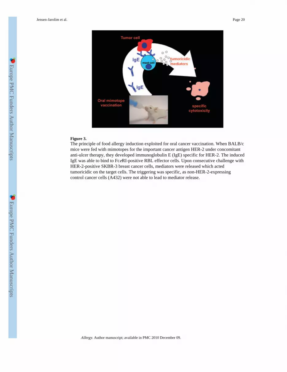
Figure 3.The principle of food allergy induction exploited for oral cancer vaccination. When BALB/cmice were fed with mimotopes for the important cancer antigen HER-2 under concomitantanti-ulcer therapy, they developed immunoglobulin E (IgE) specific for HER-2. The inducedIgE was able to bind to FcεRI-positive RBL effector cells. Upon consecutive challenge withHER-2-positive SKBR-3 breast cancer cells, mediators were released which actedtumoricidic on the target cells. The triggering was specific, as non-HER-2-expressingcontrol cancer cells (A432) were not able to lead to mediator release.
Jensen-Jarolim et al. Page 20
Allergy. Author manuscript; available in PMC 2010 December 09.
Europe PM
C Funders A
uthor Manuscripts
Europe PM
C Funders A
uthor Manuscripts