Embed Size (px)
Citation preview

Mechanisms of Ageing and Development 132 (2011) 298–304
Age-related changes in the renal dopaminergic system and expression of renalamino acid transporters in WKY and SHR rats
Vanda Pinto, Joao Amaral, Elisabete Silva, Sonia Simao, Jose Miguel Cabral, Joana Afonso,Maria Paula Serrao, Pedro Gomes 1, Maria Joao Pinho, Patrıcio Soares-da-Silva *
Institute of Pharmacology & Therapeutics, Faulty of Medicine, University of Porto, 4200-319 Porto, Portugal
A R T I C L E I N F O
Article history:
Received 25 February 2011
Received in revised form 1 May 2011
Accepted 6 June 2011
Available online 14 June 2011
Keywords:
Aging
Hypertension
Renal dopaminergic system
Amino acid transporters
Neurohumoral activity
A B S T R A C T
This study examined age-related changes in renal dopaminergic activity and expression of amino acid
transporters potentially involved in renal tubular uptake of L-DOPA in Wistar Kyoto (WKY) and
spontaneously hypertensive rats. Aging (from 13 to 91 weeks) was accompanied by increases in systolic
blood pressure (SBP) in both WKY and SHR. The sum of urinary dopamine and DOPAC and the urinary
dopamine/L-DOPA ratio were increased in aged SHR but not in aged WKY. The urinary dopamine/renal
delivery of L-DOPA ratio was increased in both rat strains with aging. LAT2 abundance was increased in
aged WKY and SHR. The expression of 4F2hc was markedly elevated in aged SHR but not in aged WKY.
ASCT2 was upregulated in both aged WKY and SHR. Plasma aldosterone levels and urinary noradrenaline
levels were increased in aged WKY and SHR though levels of both entities were more elevated in aged
SHR. Activation of the renal dopaminergic system is more pronounced in aged SHR than in aged WKY and
is associated with an upregulation of renal cortical ASCT2 in WKY and of LAT2/4F2hc and ASCT2 in SHR.
This activation may be the consequence of a counter-regulatory mechanism for stimuli leading to sodium
reabsorption.
� 2011 Elsevier Ireland Ltd. All rights reserved.
Contents lists available at ScienceDirect
Mechanisms of Ageing and Development
jo ur n al ho mep ag e: www .e lsev ier . c om / lo cate /m ec hag ed ev
1. Introduction
Dopamine is a major regulator of mammalian proximal tubulesalt and water reabsorption. In the mammalian kidney, dopamineis primarily produced in the proximal tubule (Aperia, 2000; Gomesand Soares-da-Silva, 2008; Soares-da-Silva and Vieira-Coelho,1998). The dopamine precursor L-dihydroxyphenylalanine (L-DOPA) is filtered at the glomerulus and is taken up by theproximal tubule via luminal transporters and converted todopamine by aromatic L-amino acid decarboxylase (AADC), whichis highly expressed in the proximal tubule (Soares-da-Silva andFernandes, 1991). The regulation of this non-neuronal dopaminer-gic system depends mainly on the availability of L-DOPA, on its
Abbreviations: 4F2hc, 4F2 heavy chain; ANOVA, one-way analysis of variance;
COMT, catechol-O-methyl-transferase; Ccr, creatinine clearance; DOPAC, 3,4-
dihydroxyphenylacetic acid; FENa+, fractional excretion of Na+; GAPDH, glyceralde-
hyde-3-phosphate dehydrogenase; GFR, glomerular filtration rate; L-DOPA, L-
dihydroxyphenylalanine; LAT1, L-type amino acid transporter 1; LAT2, L-type amino
acid transporter 2; SDS-PAGE, sodium dodecyl sulfate polyacrylamide gel
electrophoresis; SEM, standard error of the mean; SHR, spontaneously hypertensive
rat; WKY, Wistar Kyoto rat.
* Corresponding author. Tel.: +351 22 5513642; fax: +351 22 5513643.
E-mail address: [email protected] (P. Soares-da-Silva).1 Present address: Department of Biochemistry, Faculty of Medicine, University
of Porto, 4200-319 Porto, Portugal.
0047-6374/$ – see front matter � 2011 Elsevier Ireland Ltd. All rights reserved.
doi:10.1016/j.mad.2011.06.003
decarboxylation into dopamine and on cell outward amine transfermechanisms (Pestana and Soares-da-Silva, 1994; Soares-da-Silvaand Fernandes, 1991). In the kidney, dopamine is metabolizedpredominantly by catechol-O-methyl-transferase (COMT) andmonoamine oxidase to 3,4-dihydroxyphenylacetic acid (DOPAC),and to homovallinic acid (HVA) (Pestana and Soares-da-Silva,1994; Soares-da-Silva and Fernandes, 1991). A considerableamount of evidence favours the view that dopamine of renalorigin plays a role in the regulation of central blood volume byreducing the tubular reabsorption of sodium as a paracrine orautocrine substance (Jose et al., 2003). The mechanisms throughwhich renal dopamine is thought to produce natriuresis involvethe activation of D1-like receptors that inhibit the activity of bothapical (e.g., Na/H exchange and chloride–bicarbonate exchangeand Na–P cotransport) and basolateral (Na–K-ATPase and NaHCO3
cotransport) transporters (Aperia et al., 1987; Felder et al., 1990;Jose et al., 1992; Lokhandwala and Amenta, 1991). The availabilityof dopamine to activate its specific receptors is determined byfactors affecting renal synthesis, mainly the amounts of L-DOPAand sodium delivered to the kidney and the degree of degradationof the amine (Soares-da-Silva et al., 1993).
The spontaneously hypertensive rat (SHR) is a genetic model ofhypertension characterized by the resistance to the natriureticeffect of dopamine and D1-like receptor agonists, as a result of adefective transduction of the D1 receptor signal in renal proximal

V. Pinto et al. / Mechanisms of Ageing and Development 132 (2011) 298–304 299
tubules (Jose et al., 2010; Sanada et al., 1999; Zeng and Jose, 2011).It has been suggested that increased oxidative stress in renalproximal tubules of the SHR could be a mechanism for defectivedopamine D1 receptor/G-protein coupling (White and Sidhu,1998). Moreover, recent studies have shown the overexpressionof Na+-independent and pH-sensitive amino acid transporter LAT2(Slc7a8) in the SHR kidney, which might contribute to enhanced L-DOPA uptake in the proximal tubule and increased dopamineproduction (Pinho et al., 2004), as an attempt to overcome thedefect in D1 receptor function.
The aging kidney undergoes structural changes that result inquantitative alterations in some renal functions, such as a declinein renal blood flow and glomerular filtration rate (Zhou et al.,2008). An increasing number of studies have shown that oldanimals may present particular deficiencies in the renal handlingof L-DOPA, its subsequent conversion to dopamine (Armando et al.,1995; Soares-da-Silva and Fernandes, 1991) and at the level ofreceptor number or coupling to G proteins (Kansra et al., 1997). Inthe presence of age-related diseases, such as heart failure andhypertension, these changes can be aggravated (Fischer andO’Hare, 2010).
This study was aimed at evaluating age-related changes in theactivity of the renal dopaminergic system and the regulation of theamino acid transporters that are potentially involved in the uptakeof L-DOPA: Na+-independent LAT1 and LAT2 and Na+-dependentASCT2 in SHR and their normotensive Wistar Kyoto (WKY)counterparts.
2. Materials and methods
2.1. Animal preparation and experimental design
Five-week old male WKY and SHR were obtained from Harlan-Interfauna Iberica
(Barcelona, Spain). The rats were housed under controlled conditions (12 h light/
dark cycle and room temperature at 22 � 2 8C) and had free access to tap water and
standard rat chow (PANLAB, Barcelona, Spain). The animals were carefully maintained
and monitored until 13 or 91 weeks of age. Blood pressure (systolic and diastolic) and
heart rate were measured in conscious animals using a photoelectric tail-cuff detector
(LE 5000, Letica, Barcelona, Spain). A minimum of 5 measures were made each time and
the mean values were used for further calculations. All rat interventions were
performed in accordance with the European Directive number 86/609, and the rules of
the ‘‘Guide for the Care and Use of Laboratory Animals’’, 7th edition, 1996, Institute for
Laboratory Animal Research (ILAR), Washington, DC.
2.2. Metabolic study
Forty eight hours before experiments, 13- and 91-week old rats were placed in
metabolic cages (Tecniplast, Buguggiate, Italy) for a 24 h urine collection. The urine
samples were collected in vials containing 1 ml of 6 M HCl to prevent spontaneous
decomposition of monoamines and amine metabolites. After completion of this
protocol, rats were anesthetized with sodium pentobarbital (60 mg/kg, i.p.). The
animals were then sacrificed by exsanguination using cardiac puncture and the
blood collected into tubes containing K3 EDTA for later determination of plasma
biochemical parameters. Before excising the kidneys, a cannula was inserted in the
right ventricle of the heart and animals were perfused with ice-cold saline (0.9%
NaCl) to remove all blood from the kidneys. The kidneys were then excised,
Table 1Cardiovascular and physiological parameters in 13- and 91-week old WKY and SHR.
Parameter WKY
13 weeks
n = 6
Systolic blood pressure (mm Hg) 122 � 6
Diastolic blood pressure (mm Hg) 97 � 3
Pulse pressure (mm Hg) 25 � 2
Creatinine clearance (ml/min) 2.64 � 0.26
Urinary protein excretion (mg/24 h) 14.01 � 0.80
FENa+ (%) 0.38 � 0.04
Kidney weight/tibia length (% of control) 100 � 2
* Significantly different from corresponding values in 13-week old animals (P < 0.05# Significantly different from age-matched WKY (P < 0.05).
weighed, decapsulated, and the renal cortex and medulla rapidly separated by fine
dissection. Tissue pieces were immediately frozen in liquid nitrogen and stored at
�80 8C for Western blot analysis.
2.3. Plasma and urine biochemistry
The quantification of sodium and potassium in plasma and urine was performed
by ion-selective electrodes. Creatinine was measured by the Jaffe method (Chromy
et al., 2008). All assays were performed by Cobas Mira Plus analyzer (ABX
Diagnostics, Switzerland). Creatinine clearance (in ml/min) was calculated using
the formula Ccr = (Ucreat � Vu)/(Pcreat � 24 h � 60) where Ucreat and Vu are the
urinary creatinine concentration and urinary 24 h volume and Pcreat is the plasma
creatinine concentration. Aldosterone in plasma samples was performed by
radioimmuno assay (Diagnostic Products Corporation; Los Angeles, CA).
2.4. Assay of catecholamines
The assay of catecholamines in urine (L-DOPA, dopamine, DOPAC and
norepinephrine) and plasma samples (L-DOPA, dopamine and DOPAC) was
performed by HPLC with electrochemical detection, as previously described
(Soares-da-Silva et al., 1994, 1993). The lower limit of detection of L-DOPA,
dopamine, norepinephrine, and DOPAC ranged from 350 to 1000 fmol.
2.5. Western blotting
Renal cortices from 13- and 91-week old WKY and SHR, were lysed in RIPA
buffer containing 150 mM NaCl, 50 mM Tris–HCl, pH 7.4, 5 mM EDTA, 1% Triton
X-100, 0.5% sodium deoxycholate, 0.1% SDS, 100 mg/ml PMSF, 2 mg/ml leupeptin
and 2 mg/ml aprotinin. Protein concentration was determined using a protein
assay kit (Bio-Rad Laboratories, Hercules, CA), with bovine serum albumin as
standard. Lysates were boiled in sample buffer (35 mM Tris–HCl, pH 6.8, 4% SDS,
9.3% dithiothreitol, 0.01% bromophenol blue, 30% glycerol) at 95 8C for 5 min.
Samples containing 50–75 mg of protein, were separated by SDS-PAGE with 10%
polyacrylamide gel and then electroblotted onto nitrocellulose membranes (Bio-
Rad). Blots were blocked for 1 h with 5% non-fat dry milk in PBS (10 mmol/l
phosphate-buffered saline) at room temperature with constant shaking. Blots
were then incubated with the antibodies rabbit polyclonal anti-LAT1 (1:500;
Serotec); goat polyclonal anti-LAT2 (1:500; Santa Cruz Biotechnology); rabbit
polyclonal anti-4F2hc (1:500; Santa Cruz Biotechnology); rabbit polyclonal anti-
ASCT2 (1:500; Chemicon International); mouse monoclonal anti-b-actin
(1:20,000; Santa Cruz Biotechnology) or mouse monoclonal anti-GAPDH
(1:60,000; Santa Cruz Biotechnology) in 5% non-fat dry milk in PBS-T overnight
at 4 8C. The immunoblots were subsequently washed and incubated with
fluorescently labeled goat anti-rabbit (1:20,000; IRDyeTM 800, Rockland);
fluorescently labeled donkey anti-goat (1:10,000; IRDyeTM 800, Rockland); or
the fluorescently labeled goat anti-mouse secondary antibody (1:20,000;
AlexaFluor 680, Molecular Probes) for 60 min at room temperature and
protected from light. The membrane was washed and imaged by scanning at
both 700 and 800 nm, with an Odyssey Infrared Imaging System (LI-COR
Biosciences).
2.6. Drugs
All chemicals were obtained from Sigma (St. Louis, MO) unless otherwise stated.
2.7. Data analysis
Arithmetic means are given with standard error of the mean (SEM). Statistical
analysis was performed by one-way analysis of variance (ANOVA) followed by
Newman–Keuls test. A P value less than 0.05 was assumed to denote a significant
difference.
SHR
91 weeks
n = 6
13 weeks
n = 6
91 weeks
n = 6
148 � 3* 191 � 2# 224 � 5*,#
88 � 3 165 � 2# 132 � 3*,#
60 � 1* 26 � 2 92 � 7*,#
3.9 � 0.23* 1.85 � 0.15# 2.05 � 0.18#
14.77 � 1.21 26.02 � 1.60# 41.06 � 3.38*,#
0.23 � 0.01* 0.37 � 0.04 0.23 � 0.04*
114 � 1* 100 � 1 122 � 2*,#
).

V. Pinto et al. / Mechanisms of Ageing and Development 132 (2011) 298–304300
3. Results
3.1. Blood pressure data and renal function
As expected, the systolic and diastolic blood pressures (SBP andDBP) determined by the tail-cuff method were significantly higherin both 13- and 91-week old SHR than in age-matched WKY (Table1). Moreover, aging was accompanied by increases in SBP in bothWKY and SHR. DBP remained unaltered in aged WKY, but asignificant decrease was observed in aged versus young SHR (Table1). No difference in pulse pressure (defined as SBP minus DBP) wasfound between young WKY and SHR. Pulse pressure increased withage in both WKY and SHR but at 91 weeks of age SHR had higherpulse pressure than age-matched WKY (Table 1). Creatinineclearance (Ccr) levels were decreased in SHR in comparison toage-matched WKY (Table 1). Urinary protein excretion wassignificantly higher in SHR than in age-matched WKY at 13 and91 weeks of age and increased significantly with age (Table 1).Moreover, evaluation of FENa+ in WKY and SHR showed asignificant decrease in this parameter at the age of 91 weeks inboth rat strains (Table 1). Kidney/tibia length ratios were assessedfor WKY and SHR (Table 1). Aging was associated with increases inkidney/tibia length ratio in both WKY and SHR. However, increasesin kidney size were more marked in SHR (Table 1).
3.2. Activity of the renal dopaminergic system
In the present study, the urinary excretion of dopamine and itsmetabolite DOPAC was evaluated in 13- and 91-week old WKY andSHR (Fig. 1). No changes were found in urinary L-DOPA indexed tourinary creatinine in aged WKY. However, L-DOPA excretion wasdecreased in aged SHR, though the difference did not reachstatistical significance (Fig. 1A). Aging was accompanied by slightincreases in urinary dopamine in WKY and SHR, though notstatistically significant. However, urinary dopamine in 91-weekold SHR was higher than that in age-matched WKY (Fig. 1B).Urinary DOPAC was significantly increased in aged SHR but not inaged WKY rats (Fig. 1C). More complete information on the L-DOPA
WKY SHR0
10
20
30
*
13 week s91 week s#
Urin
ary
L-D
OPA
(nm
ol/m
g cr
eatin
ine)
WKY SHR0
2
4
6*# 13 week s
#
91 week s
Urin
ary
DO
PAC
(nm
ol/m
g cr
eatin
ine)
A
C
WKY SHR0
10
20
30
*
13 wee ks91 wee ks#
Urin
ary
L-D
OPA
(nm
ol/m
g cr
eatin
ine)
WKY SHR0
2
4
6*# 13 week s
#
91 week s
Urin
ary
DO
PAC
(nm
ol/m
g cr
eatin
ine)
A
C
Fig. 1. Urinary excretion of L-DOPA (A), dopamine (B), DOPAC (C), and sum of urinary dopa
SHR. Each bar represents the mean � SEM of 6 rats. Significantly different from correspo
matched WKY (#P < 0.05) using the Newman–Keuls test.
renal turnover is obtained when the sum of urinary dopamine andDOPAC is considered. The sum of urinary dopamine and DOPACwas found to be increased in aged SHR but not in aged WKY(Fig. 1D). Furthermore, urinary dopamine + DOPAC was markedlyincreased in aged SHR in comparison to age-matched WKY(Fig. 1D).
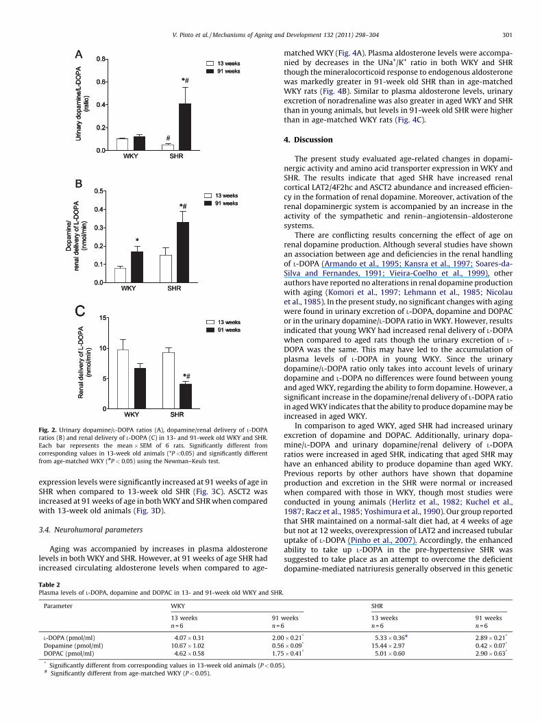
The enhanced urinary excretion of dopamine and DOPAC in theSHR may reflect their enhanced ability to synthesize dopamine.The urinary dopamine/L-DOPA ratio (a measure of renal L-DOPAutilization and of renal dopamine-synthesis efficiency) in 91-weekold SHR was markedly higher than in young SHR (Fig. 2A). Thedopamine/L-DOPA ratio was also greater in 91-week old SHR thanin age-matched WKY (Fig. 2A). No differences were detected withaging in the WKY (Fig. 2A). On the other hand, the ratio betweenurinary dopamine and the renal delivery of L-DOPA (another indexof renal dopamine production) was greater in aged WKY and SHRthan in young animals (Fig. 2B). However, the dopamine/renaldelivery of L-DOPA ratio was significantly increased in aged SHRwhen compared to age-matched WKY (Fig. 2B). The renal deliveryof L-DOPA, which considers L-DOPA plasma levels and creatinineclearance (plasma L-DOPA � creatinine clearance), decreased withage in WKY and SHR, though the difference did not reach statisticalsignificance in WKY (Fig. 2C). Moreover, the renal delivery of L-DOPA in 91-week old SHR was significantly lower than in age-matched WKY (Fig. 2C). As depicted in Table 2, aging wasaccompanied by decreases in plasma levels of L-DOPA, dopamineand DOPAC in both WKY and SHR.
3.3. Renal expression of LAT1, LAT2, 4F2hc and ASCT2
Age-related changes in the amino acid transporters that arepotentially involved in the uptake of L-DOPA were evaluated in therenal cortex of 13- and 91-week old WKY and SHR. As depicted inFig. 3A LAT1 expression levels were downregulated in 91-week oldWKY and SHR rats when compared to young animals. On the otherhand, LAT2 abundance was significantly upregulated in 91-weekold WKY and SHR, as compared to young animals (Fig. 3B). Aginghad no effect on 4F2hc protein abundance in WKY, whereas 4F2hc
WKY SHR0.0
0.5
1.0
1.5
2.0
2.5
#
#13 weeks91 weeks
Urin
ary
dopa
min
e(n
mol
/mg
crea
tinin
e)
WKY SHR0
2
4
6
8 13 weeks91 weeks*#
#
Urin
ary
dopa
min
e+D
OPA
C(n
mol
/mg
crea
tinin
e)
B
D
WKY SHR0.0
0.5
1.0
1.5
2.0
2.5
#
#13 weeks91 weeks
Urin
ary
dopa
min
e(n
mol
/mg
crea
tinin
e)
WKY SHR0
2
4
6
8 13 weeks91 weeks*#
#
Urin
ary
dopa
min
e+D
OPA
C(n
mol
/mg
crea
tinin
e)
B
D
mine and DOPAC (D) indexed to urinary creatinine in 13- and 91-week old WKY and
nding values in 13-week old animals (*P < 0.05) and significantly different from age-
A
WKY SHR0.0
0.2
0.4
0.6
0.8
*#
13 week s91 week s
#Urin
ary
dopa
min
e/L-
DO
PA(ra
tio)
B
WKY SHR0.0
0.1
0.2
0.3
0.4
0.5
*#13 week s91 week s
Dop
amin
e/re
nal d
eliv
ery
of L
-DO
PA(n
mol
/min
)
*
C
WKY SHR0
5
10
15
*#
13 weeks91 weeks
Ren
al d
eliv
ery
of L
-DO
PA(n
mol
/min
)
A
WKY SHR0.0
0.2
0.4
0.6
0.8
*#
13 week s91 week s
#Urin
ary
dopa
min
e/L-
DO
PA(ra
tio)
A
WKY SHR0.0
0.2
0.4
0.6
0.8
*#
13 week s91 week s
#Urin
ary
dopa
min
e/L-
DO
PA(ra
tio)
B
WKY SHR0.0
0.1
0.2
0.3
0.4
0.5
*#13 week s91 week s
Dop
amin
e/re
nal d
eliv
ery
of L
-DO
PA(n
mol
/min
)
*
B
WKY SHR0.0
0.1
0.2
0.3
0.4
0.5
*#13 week s91 week s
Dop
amin
e/re
nal d
eliv
ery
of L
-DO
PA(n
mol
/min
)
*
C
WKY SHR0
5
10
15
*#
13 weeks91 weeks
Ren
al d
eliv
ery
of L
-DO
PA(n
mol
/min
)
C
WKY SHR0
5
10
15
*#
13 weeks91 weeks
Ren
al d
eliv
ery
of L
-DO
PA(n
mol
/min
)
Fig. 2. Urinary dopamine/L-DOPA ratios (A), dopamine/renal delivery of L-DOPA
ratios (B) and renal delivery of L-DOPA (C) in 13- and 91-week old WKY and SHR.
Each bar represents the mean � SEM of 6 rats. Significantly different from
corresponding values in 13-week old animals (*P <0.05) and significantly different
from age-matched WKY (#P < 0.05) using the Newman–Keuls test.
V. Pinto et al. / Mechanisms of Ageing and Development 132 (2011) 298–304 301
expression levels were significantly increased at 91 weeks of age inSHR when compared to 13-week old SHR (Fig. 3C). ASCT2 wasincreased at 91 weeks of age in both WKY and SHR when comparedwith 13-week old animals (Fig. 3D).
3.4. Neurohumoral parameters
Aging was accompanied by increases in plasma aldosteronelevels in both WKY and SHR. However, at 91 weeks of age SHR hadincreased circulating aldosterone levels when compared to age-
Table 2Plasma levels of L-DOPA, dopamine and DOPAC in 13- and 91-week old WKY and SHR
Parameter WKY
13 weeks
n = 6
91 w
n = 6
L-DOPA (pmol/ml) 4.07 � 0.31 2.00
Dopamine (pmol/ml) 10.67 � 1.02 0.56
DOPAC (pmol/ml) 4.62 � 0.58 1.75
* Significantly different from corresponding values in 13-week old animals (P < 0.05# Significantly different from age-matched WKY (P < 0.05).
matched WKY (Fig. 4A). Plasma aldosterone levels were accompa-nied by decreases in the UNa+/K+ ratio in both WKY and SHRthough the mineralocorticoid response to endogenous aldosteronewas markedly greater in 91-week old SHR than in age-matchedWKY rats (Fig. 4B). Similar to plasma aldosterone levels, urinaryexcretion of noradrenaline was also greater in aged WKY and SHRthan in young animals, but levels in 91-week old SHR were higherthan in age-matched WKY rats (Fig. 4C).
4. Discussion
The present study evaluated age-related changes in dopami-nergic activity and amino acid transporter expression in WKY andSHR. The results indicate that aged SHR have increased renalcortical LAT2/4F2hc and ASCT2 abundance and increased efficien-cy in the formation of renal dopamine. Moreover, activation of therenal dopaminergic system is accompanied by an increase in theactivity of the sympathetic and renin–angiotensin–aldosteronesystems.
There are conflicting results concerning the effect of age onrenal dopamine production. Although several studies have shownan association between age and deficiencies in the renal handlingof L-DOPA (Armando et al., 1995; Kansra et al., 1997; Soares-da-Silva and Fernandes, 1991; Vieira-Coelho et al., 1999), otherauthors have reported no alterations in renal dopamine productionwith aging (Komori et al., 1997; Lehmann et al., 1985; Nicolauet al., 1985). In the present study, no significant changes with agingwere found in urinary excretion of L-DOPA, dopamine and DOPACor in the urinary dopamine/L-DOPA ratio in WKY. However, resultsindicated that young WKY had increased renal delivery of L-DOPAwhen compared to aged rats though the urinary excretion of L-DOPA was the same. This may have led to the accumulation ofplasma levels of L-DOPA in young WKY. Since the urinarydopamine/L-DOPA ratio only takes into account levels of urinarydopamine and L-DOPA no differences were found between youngand aged WKY, regarding the ability to form dopamine. However, asignificant increase in the dopamine/renal delivery of L-DOPA ratioin aged WKY indicates that the ability to produce dopamine may beincreased in aged WKY.
In comparison to aged WKY, aged SHR had increased urinaryexcretion of dopamine and DOPAC. Additionally, urinary dopa-mine/L-DOPA and urinary dopamine/renal delivery of L-DOPAratios were increased in aged SHR, indicating that aged SHR mayhave an enhanced ability to produce dopamine than aged WKY.Previous reports by other authors have shown that dopamineproduction and excretion in the SHR were normal or increasedwhen compared with those in WKY, though most studies wereconducted in young animals (Herlitz et al., 1982; Kuchel et al.,1987; Racz et al., 1985; Yoshimura et al., 1990). Our group reportedthat SHR maintained on a normal-salt diet had, at 4 weeks of agebut not at 12 weeks, overexpression of LAT2 and increased tubularuptake of L-DOPA (Pinho et al., 2007). Accordingly, the enhancedability to take up L-DOPA in the pre-hypertensive SHR wassuggested to take place as an attempt to overcome the deficientdopamine-mediated natriuresis generally observed in this genetic
.
SHR
eeks 13 weeks
n = 6
91 weeks
n = 6
� 0.21* 5.33 � 0.36# 2.89 � 0.21*
� 0.09* 15.44 � 2.97 0.42 � 0.07*
� 0.41* 5.01 � 0.60 2.90 � 0.63*
).

Fig. 3. Expression of LAT1 (A), LAT2 (B), 4F2hc (C) and ASCT2 (D) in the renal cortex of 13- and 91-week old WKY and SHR. Representative immunoblots are depicted on top of
the bar graphs. Values are normalized to the level of GAPDH expression in each condition and expressed as % of 13 week-old rats. Each bar represents the mean � SEM (n = 4
per group). Significantly different from values in 13-week old animals (*P <0.05) using the Newman–Keuls test.
V. Pinto et al. / Mechanisms of Ageing and Development 132 (2011) 298–304302
model of hypertension (Jose et al., 2002; Pinho et al., 2007).Moreover, at 4 and 12 weeks of age no differences in the urinaryexcretion of dopamine or DOPAC, or in plasma aldosterone levelswere found between age-matched WKY and SHR (Pinho et al.,2007).
The renal cortical abundance of Na+-independent LAT1 andLAT2, 4F2hc and Na+-dependent ASCT2, amino acid transporterspotentially involved in renal tubular uptake of L-DOPA, wasevaluated in 13- and 91-week old WKY and SHR rats. The system L-type amino acid transporters is a major route for providing livingcells with neutral amino acids including several essential aminoacids that cells are unable to synthesize such as leucine, isoleucine,valine, phenylalanine, tryptophan, methionine and histidine(Christensen, 1990; Silbernagl, 1979). Although the transport ofleucine by LAT1 in pig LLC-PK1 renal cells has been previouslydescribed (Soares-da-Silva and Serrao, 2004), LAT1 has a verylimited tissue distribution in the kidney (Pinho et al., 2007). Globalgene expression monitoring by cDNA microarrays showed adecline in the expression of y+LAT1 and B0AT1 with age in the renalcortex (Melk et al., 2005). Similarly, in the present study aging wasaccompanied by decreases in LAT1 abundance in WKY and SHR.LAT2 is a major Na+-independent amino acid transporter expressedmainly in transporting epithelia, such as in the kidney andintestine (Broer, 2008), and its functionality is dependent on theabundance of 4F2hc (Pineda et al., 1999). The heterodimerization
of LAT2 with 4F2hc is necessary for the transporter to reach the cellsurface (Nakamura et al., 1999). Therefore, increases in 4F2hc andLAT2 abundance may translate in increases in LAT2 functionality inaged SHR. On the other hand, the abundance of 4F2hc does not varywith age in WKY, which would limit the translocation of LAT2 to thecell surface. At the apical membrane of renal proximal tubule cellsonly Na+-dependent amino acid transporters ASCT2 and B0AT1 arecapable of transporting amino acids with similar characteristics tosubstrates transported through system L. Analogous to the LAT2abundance profile, ASCT2 was found to be upregulated in aged WKYand SHR. Overall, these results suggest that activation of the renaldopaminergic system is accompanied by increases in LAT2/4F2hcfunctionality and ASCT2 overexpression in aged SHR. In contrast,LAT2/4F2hc functionality may not have a role in L-DOPA uptake inthe renal cortex of aged WKY.
Plasma aldosterone and renal noradrenaline levels werehigher in aged SHR than in aged WKY, indicating a markedneurohumoral activation in aged SHR. The result of thesehemodynamic and neurohumoral alterations was an increase inrenal sodium transport (as indicated by a decrease in urinaryUNa+/K+ ratio), proteinuria and reductions of the renal deliveryof L-DOPA in aged SHR. Another indication of aldosterone actionsis the marked increases in kidney size in aged SHR rats.Aldosterone directly modulates renal cell proliferation anddifferentiation via stimulation of rapidly activated protein

WKY SHR0
500
1000
1500
2000
2500 13 week s91 week s
*
*#P
lasm
a al
dost
eron
e(p
mol
/L)
#
WKY SHR0.0
0.5
1.0
1.5
2.0 13 week s91 week s
**#U
rinar
y Na
+ /K+
(ratio
)
A B
WKY SHR0.0
0.5
1.0
1.5
2.0 13 wee ks91 wee ks
*
*#
Urin
ary
nora
dren
alin
e(n
mol
/mg
crea
tinin
e)
C
WKY SHR0
500
1000
1500
2000
2500 13 week s91 week s
*
*#P
lasm
a al
dost
eron
e(p
mol
/L)
#
WKY SHR0.0
0.5
1.0
1.5
2.0 13 week s91 week s
**#U
rinar
y Na
+ /K+
(ratio
)
A B
WKY SHR0.0
0.5
1.0
1.5
2.0 13 wee ks91 wee ks
*
*#
Urin
ary
nora
dren
alin
e(n
mol
/mg
crea
tinin
e)
C
Fig. 4. Plasma levels (pmol/l) of aldosterone (A) changes in urinary Na+/K+ ratio (B) and urinary noradrenaline levels (nmol/mg creatinine) in 13- and 91-week old WKY and
SHR. Each column represents the mean � SEM of 6 rats. Significantly different from corresponding values in 13-week old animals (*P <0.05) and significantly different from age-
matched WKY (#P < 0.05) using the Newman–Keuls test.
V. Pinto et al. / Mechanisms of Ageing and Development 132 (2011) 298–304 303
kinase cascades as part of normal kidney development (Thomaset al., 2010). The renal dopaminergic and renin–angiotensin–aldosterone systems (RAAS) control renal electrolyte balancethrough various receptor mediated pathways with counter-regulatory interactions. In order to conserve sodium during lowsodium intake, the RAAS is upregulated in order to produceangiotensin II (Ang II). Stimulation of the principal membranebound cell surface receptor for Ang II, the AT1R, leads to sodiumreabsorption. In order to eliminate sodium during high sodiumintake the local renal production of dopamine is increasedleading to inhibition of sodium reabsorption (Felder and Jose,2006). The natriuretic renal dopaminergic system opposes theanti-natriuretic activity of the RAAS by downregulating theAT1R, upregulating the AT2R and inhibiting ROS generation. Eachof the individual dopamine receptors has been shown to opposethe activity of the AT1R, with the D1R, D3R, and D5R physicallyinteracting with the AT1R (Gildea, 2009). Taken together, it issuggested that the renal dopaminergic system might be acompensatory mechanism activated by stimuli that lead tosodium reabsorption in aged WKY and SHR. However, thiscounter-regulatory mechanism is considerably more enhancedin aged SHR. A similar mechanism has been shown in patientswith heart failure. Stimuli leading to activation of anti-natriuretic systems and sodium retention are accompanied byactivation of the renal dopaminergic system characterized by anincrease in the renal utilization of filtered L-DOPA (Alvelos et al.,2004; Ferreira et al., 2001, 2002).
The specific effects of aldosterone on the expression of 4F2hcand LAT2 have recently been explored by our group (Pinho et al.,2009). Eight-week old Wistar rats were submitted to high saltintake (1% NaCl in their drinking water) and treated chronicallywith aldosterone and/or spironolactone, a mineralocorticoid
receptor (MR) antagonist. Treatment with aldosterone signifi-cantly increased LAT2 mRNA expression via the MR (abolishedby spironolactone), though protein levels remained unchanged.On the other hand, aldosterone treated rats had decreased 4F2hcprotein expression in a spironolactone-independent manner.These effects of aldosterone were accompanied by decreases inurinary dopamine and DOPAC in a spironolactone-sensitivemanner (Pinho et al., 2009).
Studies have shown that cardiac function and coronaryhemodynamics progressively deteriorate with aging in both SHRand WKY and that very old WKY tend to develop a significantdegree of isolated systolic hypertension (Susic et al., 1998,2001). In the present study SBP was found to be increased inaged WKY and SHR, displaying the same trend as the plasmaaldosterone levels. Pulse pressure has been reported to increasesignificantly with age in SHR but not in WKY (Chamiot-Clercet al., 2001). However, these studies were conducted in ratsbetween 3 and 78 weeks of age. The findings show that agedSHR has in fact an intense dopaminergic response but SBP andpulse pressure values remain increased. The cause for thisoutcome is possibly related to the defective transduction of theD1 receptor signal in renal proximal tubules usually attributedto this strain (Jose et al., 2010). On the other hand, the activationof the renal dopaminergic system is not as effective in aged WKYand SBP and pulse pressure are increased in these animals.
In conclusion, aging in WKY and SHR is accompanied byincreases in renal cortical ASCT2 abundance in the former and inincreases in LAT2/4F2hc and ASCT2 abundances in the latter.Moreover, the dopaminergic response is more enhanced in agedSHR than in aged WKY and this is probably a result of acompensatory mechanism activated by stimuli leading to sodiumreabsorption.
V. Pinto et al. / Mechanisms of Ageing and Development 132 (2011) 298–304304
References
Alvelos, M., Ferreira, A., Bettencourt, P., Serrao, P., Pestana, M., Cerqueira-Gomes, M.,Soares-da-Silva, P., 2004. The effect of dietary sodium restriction on neurohu-moral activity and renal dopaminergic response in patients with heart failure.Eur. J. Heart Fail. 6, 593–599.
Aperia, A., Bertorello, A., Seri, I., 1987. Dopamine causes inhibition of Na+–K+-ATPaseactivity in rat proximal convoluted tubule segments. Am. J. Physiol. 252,F39–F45.
Aperia, A.C., 2000. Intrarenal dopamine: a key signal in the interactive regulation ofsodium metabolism. Annu. Rev. Physiol. 62, 621–647.
Armando, I., Nowicki, S., Aguirre, J., Barontini, M., 1995. A decreased tubular uptakeof dopa results in defective renal dopamine production in aged rats. Am. J.Physiol. 268, F1087–F1092.
Broer, S., 2008. Amino acid transport across mammalian intestinal and renalepithelia. Physiol. Rev. 88, 249–286.
Chamiot-Clerc, P., Renaud, J.F., Safar, M.E., 2001. Pulse pressure, aortic reactivity,and endothelium dysfunction in old hypertensive rats. Hypertension 37,313–321.
Christensen, H.N., 1990. Role of amino acid transport and countertransport innutrition and metabolism. Physiol. Rev. 70, 43–77.
Chromy, V., Rozkosna, K., Sedlak, P., 2008. Determination of serum creatinine byJaffe method and how to calibrate to eliminate matrix interference problems.Clin. Chem. Lab. Med. 46, 1127–1133.
Felder, C.C., Campbell, T., Albrecht, F., Jose, P.A., 1990. Dopamine inhibits Na(+)–H+
exchanger activity in renal BBMV by stimulation of adenylate cyclase. Am. J.Physiol. 259, F297–F303.
Felder, R.A., Jose, P.A., 2006. Mechanisms of disease: the role of GRK4 in the etiologyof essential hypertension and salt sensitivity. Nat. Clin. Pract. Nephrol. 2,637–650.
Ferreira, A., Bettencourt, P., Pestana, M., Correia, F., Serrao, P., Martins, L., Cerqueira-Gomes, M., Soares-da-Silva, P., 2001. Heart failure, aging, and renal synthesis ofdopamine. Am. J. Kidney Dis. 38, 502–509.
Ferreira, A., Bettencourt, P., Pimenta, J., Frioes, F., Pestana, M., Soares-da-Silva, P.,Cerqueira-Gomes, M., 2002. The renal dopaminergic system, neurohumoralactivation, and sodium handling in heart failure. Am. Heart J. 143, 391–397.
Fischer, M.J., O’Hare, A.M., 2010. Epidemiology of hypertension in the elderly withchronic kidney disease. Adv. Chronic Kidney Dis. 17, 329–340.
Gildea, J.J., 2009. Dopamine and angiotensin as renal counterregulatory systemscontrolling sodium balance. Curr. Opin. Nephrol. Hypertens. 18, 28–32.
Gomes, P., Soares-da-Silva, P., 2008. Dopamine. In: Baden, M. (Ed.), CardiovascularHormone Systems: From Molecular Mechanisms to Novel Therapeutics. Wiley-VCH, Weinheim, pp. 251–293.
Herlitz, H., Lundin, S., Henning, M., Aurell, M., Karlberg, B.E., Berglund, G., 1982.Hormonal pattern during development of hypertension in spontaneously hy-pertensive rats (SHR). Clin. Exp. Hypertens. A 4, 915–935.
Jose, P.A., Eisner, G.M., Felder, R.A., 2002. Dopamine receptor-coupling defect inhypertension. Curr. Hypertens. Rep. 4, 237–244.
Jose, P.A., Eisner, G.M., Felder, R.A., 2003. Dopamine and the kidney: a role inhypertension? Curr. Opin. Nephrol. Hypertens. 12, 189–194.
Jose, P.A., Raymond, J.R., Bates, M.D., Aperia, A., Felder, R.A., Carey, R.M., 1992. Therenal dopamine receptors. J. Am. Soc. Nephrol. 2, 1265–1278.
Jose, P.A., Soares-da-Silva, P., Eisner, G.M., Felder, R.A., 2010. Dopamine and Gprotein-coupled receptor kinase 4 in the kidney: role in blood pressure regula-tion. Biochim. Biophys. Acta 1802, 1259–1267.
Kansra, V., Hussain, T., Lokhandwala, M.F., 1997. Alterations in dopamine DA1receptor and G proteins in renal proximal tubules of old rats. Am. J. Physiol. 273,F53–F59.
Komori, T., Habuchi, Y., Inoue, H., Sakai, T., Suzuki, K., Ohtsuka, K., Tanaka, H., Fujita,N., Yoshimura, M., 1997. Application of urinary free dopamine as a marker ofrenal function, and comparison with other renal marker. Rinsho Byori 45,679–684.
Kuchel, O., Racz, K., Debinski, W., Falardeau, P., Buu, N.T., 1987. Contrastingdopaminergic patterns in two forms of genetic hypertension. Clin. Exp. Hyper-tens. 9, 987–1008.
Lehmann, M., Spori, U., Keul, J., 1985. Excretion of free dopamine, noradrenaline andadrenaline in 190 males in relation to age and blood pressure. Klin. Wochenschr.63, 264–271.
Lokhandwala, M.F., Amenta, F., 1991. Anatomical distribution and function ofdopamine receptors in the kidney. FASEB J. 5, 3023–3030.
Melk, A., Mansfield, E.S., Hsieh, S.C., Hernandez-Boussard, T., Grimm, P., Rayner, D.C.,Halloran, P.F., Sarwal, M.M., 2005. Transcriptional analysis of the molecularbasis of human kidney aging using cDNA microarray profiling. Kidney Int. 68,2667–2679.
Nakamura, E., Sato, M., Yang, H., Miyagawa, F., Harasaki, M., Tomita, K., Matsuoka, S.,Noma, A., Iwai, K., Minato, N., 1999. 4F2 (CD98) heavy chain is associatedcovalently with an amino acid transporter and controls intracellular traffickingand membrane topology of 4F2 heterodimer. J. Biol. Chem. 274, 3009–3016.
Nicolau, G.Y., Haus, E., Lakatua, D., Sackett-Lundeen, L., Bogdan, C., Plinga, L.,Petrescu, E., Ungureanu, E., Robu, E., 1985. Differences in the circadian rhythmparameters of urinary free epinephrine, norepinephrine and dopamine betweenchildren and elderly subjects. Endocrinologie 23, 189–199.
Pestana, M., Soares-da-Silva, P., 1994. The renal handling of dopamine originatingfrom L-dopa and gamma-glutamyl-L-dopa. Br. J. Pharmacol. 112, 417–422.
Pineda, M., Fernandez, E., Torrents, D., Estevez, R., Lopez, C., Camps, M., Lloberas, J.,Zorzano, A., Palacin, M., 1999. Identification of a membrane protein, LAT-2, thatco-expresses with 4F2 heavy chain, an L-type amino acid transport activity withbroad specificity for small and large zwitterionic amino acids. J. Biol. Chem. 274,19738–19744.
Pinho, M., Amaral, J., Pinto, V., Serrao, M., Soares-da-Silva, P., 2009. Regulation ofrenal LAT2 and 4F2hc expression by aldosterone. J. Epithelial Biol. Pharmacol. 2,36–43.
Pinho, M.J., Serrao, M.P., Gomes, P., Hopfer, U., Jose, P.A., Soares-da-Silva, P., 2004.Over-expression of renal LAT1 and LAT2 and enhanced L-DOPA uptake in SHRimmortalized renal proximal tubular cells. Kidney Int. 66, 216–226.
Pinho, M.J., Serrao, M.P., Soares-da-Silva, P., 2007. High-salt intake and the renalexpression of amino acid transporters in spontaneously hypertensive rats. Am.J. Physiol. Renal Physiol. 292, F1452–F1463.
Racz, K., Kuchel, O., Buu, N.T., Tenneson, S., 1985. Peripheral dopamine synthesisand metabolism in spontaneously hypertensive rats. Circ. Res. 57, 889–897.
Sanada, H., Jose, P.A., Hazen-Martin, D., Yu, P.Y., Xu, J., Bruns, D.E., Phipps, J., Carey,R.M., Felder, R.A., 1999. Dopamine-1 receptor coupling defect in renal proximaltubule cells in hypertension. Hypertension 33, 1036–1042.
Silbernagl, S., 1979. Renal transport of amino acids. Klin. Wochenschr. 57,1009–1019.
Soares-da-Silva, P., Fernandes, M.H., 1991. A study on the renal synthesis ofdopamine in aged rats. Acta Physiol. Scand. 143, 287–293.
Soares-da-Silva, P., Fernandes, M.H., Pinto-do, O.P., 1994. Cell inward transport of L-DOPA and 3-O-methyl-L-DOPA in rat renal tubules. Br. J. Pharmacol. 112, 611–615.
Soares-da-Silva, P., Pestana, M., Fernandes, M.H., 1993. Involvement of tubularsodium in the formation of dopamine in the human renal cortex. J. Am. Soc.Nephrol. 3, 1591–1599.
Soares-da-Silva, P., Serrao, M.P., 2004. High- and low-affinity transport of L-leucineand L-DOPA by the hetero amino acid exchangers LAT1 and LAT2 in LLC-PK1renal cells. Am. J. Physiol. Renal Physiol. 287, F252–F261.
Soares-da-Silva, P., Vieira-Coelho, M.A., 1998. Nonneuronal dopamine. In: Gold-stein, D.S., Eisenhofer, G., McCarty, R. (Eds.), Catecholamines. Bridging BasicScience with Clinical Medicine. Academic Press, San Diego, pp. 866–869.
Susic, D., Nunez, E., Hosoya, K., Frohlich, E.D., 1998. Coronary hemodynamics inaging spontaneously hypertensive and normotensive Wistar Kyoto rats. J.Hypertens. 16, 231–237.
Susic, D., Varagic, J., Frohlich, E.D., 2001. Isolated systolic hypertension in elderlyWKY is reversed with L-arginine and ACE inhibition. Hypertension 38,1422–1426.
Thomas, W., Dooley, R., Harvey, B.J., 2010. Aldosterone as a renal growth factor.Steroids 75, 550–554.
Vieira-Coelho, M.A., Hussain, T., Kansra, V., Serrao, M.P., Guimaraes, J.T., Pestana, M.,Soares-da-Silva, P., Lokhandwala, M.F., 1999. Aging, high salt intake, and renaldopaminergic activity in Fischer 344 rats. Hypertension 34, 666–672.
White, B.H., Sidhu, A., 1998. Increased oxidative stress in renal proximal tubules ofthe spontaneously hypertensive rat: a mechanism for defective dopamine D1Areceptor/G-protein coupling. J. Hypertens. 16, 1659–1665.
Yoshimura, M., Ikegaki, I., Nishimura, M., Takahashi, H., 1990. Role of dopaminergicmechanisms in the kidney for the pathogenesis of hypertension. J. Auton.Pharmacol. 10 (Suppl. 1), s67–s72.
Zeng, C., Jose, P.A., 2011. Dopamine receptors: important antihypertensive coun-terbalance against hypertensive factors. Hypertension 57, 11–17.
Zhou, X.J., Rakheja, D., Yu, X., Saxena, R., Vaziri, N.D., Silva, F.G., 2008. The agingkidney. Kidney Int. 74, 710–720.


