Embed Size (px)
Citation preview
Advances in multiphoton microscopy for imaging embryos
Willy Supatto1, Thai V Truong2, Delphine Debarre1, and Emmanuel Beaurepaire1
1Laboratory for optics and Bioscience, Ecole Polytechnique, CNRS, INSERM, Palaiseau, France2Biological Imaging Center, California Institute of Technology, Pasadena, California, USA
Summary of recent advancesMultiphoton imaging is a promising approach for addressing current issues in systems biology andhigh-content investigation of embryonic development. Recent advances in multiphotonmicroscopy, including light-sheet illumination, optimized laser scanning, adaptive and label-freestrategies, open new and promising opportunities for embryo imaging. However, the literature isoften unclear about which microscopy technique is most adapted for achieving specificexperimental goals. In this review, we describe and discuss the key concepts of imaging speed,imaging depth, photodamage, and nonlinear contrast mechanisms in the context of recent advancesin live embryo imaging. We illustrate the potentials of these new imaging approaches with aselection of recent applications in developmental biology.
KeywordsNonlinear microscopy; Light-sheet microscopy; 2-photon excited fluorescence; Second-harmonicgeneration (SHG); Third-harmonic generation (THG); Coherent raman scattering (CARS, SRS)
IntroductionFrom a microscopy perspective, live embryos present uniquely challenging characteristicscompared to other biological samples. Embryos are smaller than 1 millimeter, at least duringearly developmental stages, making them accessible for three-dimensional (3D) imagingwith light microscopy. However, they typically have an ellipsoidal shape and their innerstructure is inhomogeneous and constantly changing. In addition, embryos are sensitive tomanipulation and photodamage, and their labeling can be difficult. These propertieschallenge the performance of microscopy techniques in terms of imaging depth, imagingspeed, photodamage and contrast. Since its introduction in 1990 [1], 2-photon excitedfluorescence (2PEF) microscopy has proven to be the most effective approach for deeptissue fluorescence microscopy. It has found many applications in neuroscience [2–3] andmore recently in other fields, such as in immunology [4]. Multiphoton (or nonlinear)imaging is attractive also for embryo imaging and in recent years has been applied to anincreasing number of published studies in developmental biology using various modelsystems, such as fruit fly [5–8], quail [9], zebrafish [10], or mouse embryos [11–12].Multiphoton imaging is also promising for addressing current issues in systems biology andhigh-content experimental investigation of embryonic development [13] requiring novel
© 2011 Elsevier Ltd. All rights reserved.Publisher's Disclaimer: This is a PDF file of an unedited manuscript that has been accepted for publication. As a service to ourcustomers we are providing this early version of the manuscript. The manuscript will undergo copyediting, typesetting, and review ofthe resulting proof before it is published in its final citable form. Please note that during the production process errors may bediscovered which could affect the content, and all legal disclaimers that apply to the journal pertain.
NIH Public AccessAuthor ManuscriptCurr Opin Genet Dev. Author manuscript; available in PMC 2012 October 1.
Published in final edited form as:Curr Opin Genet Dev. 2011 October ; 21(5): 538–548. doi:10.1016/j.gde.2011.08.003.
NIH
-PA Author Manuscript
NIH
-PA Author Manuscript
NIH
-PA Author Manuscript
methods for faster and deeper imaging of embryos with better contrast and resolution. In thisreview we analyze the parameters limiting imaging speed and depth in the currentlyavailable imaging modalities, and we discuss promising recent advances in multiphotonmicroscopy of live embryos, including light-sheet excitation and label-free imaging.
Fast imaging of live embryos with multiphoton light-sheet microscopyImaging developmental processes often requires time-lapse 3D-image acquisitions (4Dimaging). The imaging speed of a microscope can be defined by its pixel (or voxel) rate, i.e.the number of pixels per unit time that can be obtained with sufficient signal and contrast. Ahigh pixel rate permits capturing with adequate time resolution fast processes such as heartdevelopment (50–130 frames per second (fps) in [14–16]), cilia beating (900 fps in [17]) orfluid flow in developing embryos (44 fps in [18]). A high pixel rate is also required to studyslower large-scale processes such as collective cell migration or cell division patterns with alarge number of pixels per image to reach the appropriate spatial resolution: for instance, intoto imaging of early development [16,19–20] typically requires acquiring ~100 millionvoxels per 3D-image stack in less than a minute.
In this context, point-scanning confocal or multiphoton approaches are usually too slow, asthe image is recoreded one pixel at a time (Fig. 1). Indeed, in these approaches signal levelprescribes pixel accumulation times of typically 1–10 μs, corresponding to pixel rates ofonly 105 to 106 pixels.s−1.
Several approaches have been explored during the last 15 years to improve the imagingspeed of multiphoton microscopy up to ~107 pixels.s−1, including fast point-scanning andmultifocal approaches (Fig. 1, Tables 1 and 2, and [21] for a review). However, besideshardware limitations (i.e. scanning speed, readout time, data transfer or storage) the pixelrate of any microscope is fundamentally limited by the signal level that can be obtainedwithin the pixel accumulation time without causing fluorophore saturation or photodamage(including phototoxicity to the biological sample and photobleaching of the fluorophores).Hence, even though fast point-scanning can be implemented using resonant scanners,polygonal mirrors or acousto-optic deflectors [21], the useful pixel rate is still limited byfluorophore photophysics of the single-point excitation approach (third column in Table 1).The main strategy to circumvent this limitation is to parallelize the sample illumination andthe signal detection. Using multifocal excitation (Fig. 1), overall pixel rate can be increasedwhile maintaining the same illumination time per pixel (Table 1). However with thisapproach, an increase in imaging speed requires a proportional increase in laser averagepower (Table 1 and Table 2), similar to linear microscopy (Supp. Table 1). Available laserpower therefore limits the achievable speed gain. Moreover, increasing the laser averagepower may eventually lead to linear absorption and photodamage, as it is the case in linearmicroscopy.
Among the strategies for improving the imaging speed of multiphoton microscopy, therecent implementation of scanned light-sheet microscopy using two-photon excitation (2p-SPIM in [16] and light-sheet 2p-microscopy in this review) introduces a new paradigm. Inthis technique, a sheet of light is generated by scanning a weakly focused Gaussian beamfaster than the image acquisition time to illuminate an entire plane of the sample, which isthen imaged with a camera oriented orthogonally to the sheet. Compared to a static light-sheet generated with a cylindrical lens [22], this scanned light-sheet approach generatestypically 100 times stronger 2PEF signal [16], which is critical for live imaging. Light-sheet2p-microscopy is the only technique improving overall pixel rate over point-scanning 2p-microscopy with longer pixel accumulation time and lower peak intensity (Table 1 andTable 2). This fundamental property results from the orthogonal geometry of the
Supatto et al. Page 2
Curr Opin Genet Dev. Author manuscript; available in PMC 2012 October 1.
NIH
-PA Author Manuscript
NIH
-PA Author Manuscript
NIH
-PA Author Manuscript
illumination and detection pathways (which are collinear in conventional microscopy),allowing the use of a low numerical aperture (NA) illumination focusing (resulting in a largeillumination volume) without degrading the axial resolution and the overall signal rate [16].The use of low-NA illumination has three important advantages for multiphoton liveimaging. First, it results in lower peak intensity, and therefore less higher-order nonlinearphotodamage to the tissue [23]. Second, parallelization of the illumination is done along thelight propagation direction, reusing the same excitation energy, thus requiring less laserpower than in multifocal approaches, in turn limiting linear absorption and photodamage.Finally, the weakly focused excitation beam is less sensitive to sample-induced opticalaberrations and resolution loss with depth than in the case of high-NA focusing [16]. Inaddition, in the conditions presented in Table 3 [16], the laser is scanned ~15 times duringthe image acquisition with 1 ms between two passes. This temporal excitation patternpotentially results in lower photobleaching, as time is given for fluorophore dark staterelaxation [24].
Overall, compared to other fast multiphoton techniques, light-sheet 2p-microscopy providesfast acquisition while reducing photodamage and requiring minimal increase in laser poweras demonstrated in live embryos [16] (Fig. 2a). To date, it is the fastest implementation ofmultiphoton microscopy with up to ~1.1 107 pix/s (Table 2). We note however that light-sheet microscopy relies on widefield (camera-based) detection, which leads to compromisesin terms of imaging depth, as discussed in the next section.
Parameters governing imaging depth in tissue microscopyThe imaging depth corresponds to how deep into the tissue images can be recorded withsufficient quality (resolution, signal intensity, contrast). Imaging depth is limited by lightscattering [25] and by sample-induced optical aberrations. Point-scanning 2p-microscopy isusually considered as the gold-standard in imaging depth for in vivo fluorescence imaging oftissues and embryos [26] (Fig. 3). For instance, the large-scale dynamic analysis of thedeepest mesoderm cells during Drosophila gastrulation has been made possible only usingpoint-scanning 2p-microscopy [6].
The depth performance of point-scanning 2p-microscopy relies on three phenomena: (i)superior penetration of illumination light, (ii) robust confinement of excitation volume, and(iii) efficient collection of the fluorescence. Let us compare this technique with confocalmicroscopy, light-sheet 2p-microscopy and light-sheet 1p-microscopy with respect to thesethree points (Fig. 3).
i. Scattering of near infrared light is reduced compared to that of visible light inbiological tissues. For this reason, both point-scanning and light-sheet 2p-microscopies benefit from greater penetration of the illumination light, and reduceddegradation of the illumination volume with depth compared to linear techniques(Fig. 3a-d).
ii. In multiphoton microscopy, the nonlinear dependence of fluorescence generationon illumination confines excitation to the regions with highest intensity. As a result,in both point-scanning and light-sheet 2p-microscopy fluorescence excitation isrobustly confined in space and is less sensitive to scattering of illumination light. Incontrast, in light-sheet 1p-microscopy, the excitation volume is identical to theillumination volume, resulting in a direct loss of axial resolution at high sampledepth due to scattering-induced thickening of the light-sheet (Fig. 3e-h).
iii. Finally, in point-scanning 2p-microscopy, all fluorescence is emitted from aconfined volume corresponding to a single voxel in the 3D-image, meaning thatboth scattered and ballistic (non-scattered) photons can be collected and attributed
Supatto et al. Page 3
Curr Opin Genet Dev. Author manuscript; available in PMC 2012 October 1.
NIH
-PA Author Manuscript
NIH
-PA Author Manuscript
NIH
-PA Author Manuscript
to the signal (Fig. 3i). This collection efficiency is a unique feature of point-scanning 2p-microscopy: in all other techniques, only ballistic photons contributeto the signal while scattered photons need to be rejected with a pinhole or otherwisewould cause contrast degradation (Fig. 3j–l). In light-sheet microscopy, thescattering of the fluorescence on its way to the camera results in cross talk betweenadjacent pixels and image blurring.
However, light-sheet microscopy does has one advantage other collinear techniques(confocal and point-scanning 2p-microscopy): the use of lower illumination NA leads to lesssensitivity to optical aberrations and thus contributes to maintain better axial resolution inlight-sheet 2p-microscopy imaging of inhomogeneous embryos [16].
In summary, light-sheet microscopy with 2p-excitation provides deeper imaging than with1p-excitation for two fundamental reasons: deeper penetration of illumination light androbust confinement of fluorescence excitation. At large depths, light-sheet 2p-microscopylacks the background-free collection advantage of point-scanning 2p-microscopy, but is lesssensitive to aberration-induced degradation in axial resolution.
Adaptive advantages of point-scanning for multiphoton imaging ofembryos
A developing embryo is a dynamic and inhomogeneous biological system. Opticalproperties vary between species and tissues [26], and they also constantly evolve in time andspace during embryonic development [27]. As a consequence, embryo imaging wouldstrongly benefit from the ability of microscope illumination and acquisition schemes toadapt to the changing properties of the developing tissue. In this context, point-scanningmultiphoton techniques have a fundamental advantage compared to parallelized illuminationstrategies, which is the ability to readily tailor the imaging parameters for each individualpoint of the embryo.
Recent advances in active and adapted control of microscope illumination and acquisitionhold great potential for embryo imaging. The active adjustment of illumination powerdepending on local signal levels in confocal microscopy [28] and point-scanning 2p-microscopy [29] have been shown to reduce photodamage and avoid fluorophore anddetection saturation. Recently, the novel concept of conformal scanning was demonstratedfor multiphoton imaging of zebrafish embryos [30]: spiral scanning was used to match theembryo spherical shape, allowing the constant adjustment of scanning speed to the imagingdepth. Using slow scanning in deep regions and fast scanning in peripheral regions, theillumination is optimized to obtain homogeneous signal and reduced phototoxicity whileminimizing the acquisition time (Fig. 2b), providing effective in toto imaging [30]. Moresophisticated techniques using adaptive optics to correct for optical aberrations withinembryos are promising directions for improved imaging of embryos [31–33]. Adaptivemultiphoton microscopy has been shown to correct for resolution losses during deepimaging (Fig. 2c), and can be performed dynamically in evolving embryos [34].
Beyond fluorescence: other nonlinear contrast mechanisms (SHG, THG,CARS, and SRS)
Another advantage of nonlinear microscopy is that in addition to fluorescence, othermultiphoton processes can be used as contrast mechanisms to provide complementaryinformation. These include second-harmonic generation (SHG), third-harmonic generation(THG), coherent anti-Stokes Raman scattering (CARS) and stimulated Raman scattering(SRS). These imaging modalities [35] share the benefits of point-scanning 2p-microscopy in
Supatto et al. Page 4
Curr Opin Genet Dev. Author manuscript; available in PMC 2012 October 1.
NIH
-PA Author Manuscript
NIH
-PA Author Manuscript
NIH
-PA Author Manuscript
terms of 3D resolution and penetration depth. However they rely on coherent opticalprocesses, and therefore have more complex contrast mechanisms than fluorescencemicroscopy. For example, signal strength is generally sensitive to the spatial distribution ofmolecules within the excitation volume, and signal radiation usually occurs in the directionof the excitation beam.
In many cases such signals can be obtained from unstained tissues, with the additionalbenefit of not suffering from photobleaching. SHG is exclusively observed from denseorganized non-centrosymmetric electronic structures. Some natural sources of SHG arefibrillar collagen, myofilaments, astroglial fibers, starch, and polarized tubulin assembliessuch as mitotic spindles that can be observed in embryos (Fig. 2d-f). THG does not requiremolecular asymmetry but is observed only near optical heterogeneities. In practice, THGsignals are obtained from dense non-aqueous objects such as lipid droplets [36], mineralizedor absorbing structures, and generally from interfaces between media of different refractiveindices. Coherent Raman processes such as CARS and SRS derive their contrast frommolecular vibrational modes and can be used for micro-spectroscopy and chemicallyselective imaging. SRS provides increased contrast compared with CARS at the cost ofincreased experimental complexity. The most widespread use of coherent Ramanmicroscopy for biological studies is currently the selective imaging of lipid distribution intissues based on contrast from CH-bond vibration [37–40].
THG and SHG are efficiently produced using femtosecond excitation pulses and require asingle laser. For that reason, combination with fluorescence-based point-scanning 2p-microscopy is straightforward. Several studies have reported harmonic and multimodalharmonic/fluorescence imaging of embryos in various models: fruit fly [5,41], zebrafish[30,42–43], mouse [44], or worm [45]. SHG imaging carried out in light-sheet mode andcombined with light-sheet 2p-microscopy, has also been recently demonstrated [16]. CARS/SRS microscopy requires two synchronized and overlapped excitation beams, and contrast isoptimized with picosecond rather than femtosecond pulses. For these reasons combinationof coherent Raman scattering with 2PEF is more complex, but multimodal fluorescence/harmonic/Raman imaging is becoming a reality.
One particularly attractive aspect of these imaging approaches for embryo imaging is thatthey provide label-free structural or molecular vibrational imaging. In some experimentalsituations, it is challenging to obtain long-term, strong, specific, and non-invasivefluorescent labeling. Label-free nonlinear imaging has proven to be a particularly effectiveaddition to fluorescence for studying early division patterns in the zebrafish embryo [30](Fig. 2d), and lipid storage in worms [37–38,46]. Harmonic-generation contrast usuallyprovides additional structural information on molecular or supra-molecular order that wouldnot be easily detectable with fluorescent labeling strategies: for instance, collagenmacromolecular organization into fibrils [47], myosin structural conformation in sarcomere[48], microtubule array polarization during brain maturation [49] (Fig. 2e), or detection ofsub-micron-scale anisotropy in the cornea [50]. In addition, we note that artificialnanostructures usually produce strong nonlinear optical signals that, unlike fluorescence, donot bleach or saturate with high laser intensity. Thus, harmonic signals from nanoprobesmay be detected with high sensitivity, as illustrated by SHG imaging of nanocrystals insidezebrafish embryos [51] (Fig. 2f).
To summarize, non-fluorescent nonlinear signals should generally not be viewed only as alabel-free substitute to fluorescence, but rather as providing additional information, thepotential of which we think has not been fully explored yet for developmental biology.
Supatto et al. Page 5
Curr Opin Genet Dev. Author manuscript; available in PMC 2012 October 1.
NIH
-PA Author Manuscript
NIH
-PA Author Manuscript
NIH
-PA Author Manuscript
Conclusion and perspectivesRecent advances in multiphoton microscopy, including light-sheet, adaptive and label-freestrategies, open promising avenues for embryo imaging. However, the literature is oftenunclear about the comparative performances of microscopy modalities. Therefore, it isimportant to understand the principles, advantages and limitations of each microscopeimplementation. In this review, we provide keys to understand recent methodologicaldevelopments in the perspective of their application to developmental biology. We dicsusswhy light-sheet illumination provides faster imaging with lower photodamage than othermultiphoton microscopy geometries. We review how multiphoton microscopy achieves highimaging depth into tissues and clarify why the standard point-scanning approach, thoughlacking in imaging speed, hold fundamental advantages compared to parallelizedillumination strategies for imaging dynamic and inhomogeneous embryos. We discuss themechanisms and advantages of non-fluorescent techniques of multiphoton microscopy andillustrate how label-free imaging can be applied to developmental biology. A number ofadditional experimental developments are still under investigation with potential benefits forapplication to developmental biology. These include the use of laser pulse shaping [52] andof Bessel beam illumination [53–55]. Finally, we note that many recent developments inlinear microscopy can be also be applied in a straightforward manner to multiphotonmicroscopy: for instance light-sheet 2p-microscopy would benefit from techniquesdeveloped for light-sheet 1p-microscopy, such as deconvolution [56], background rejectionusing structured illumination [57] or HiLo [58].
Supplementary MaterialRefer to Web version on PubMed Central for supplementary material.
References1. Denk W, Strickler JH, Webb WW. Two-photon laser scanning fluorescence microscopy. Science.
1990; 248:73–76. [PubMed: 2321027]2. Svoboda K, Yasuda R. Principles of two-photon excitation microscopy and its applications to
neuroscience. Neuron. 2006; 50:823–839. [PubMed: 16772166]3. Wilt BA, Burns LD, Wei Ho ET, Ghosh KK, Mukamel EA, Schnitzer MJ. Advances in Light
Microscopy for Neuroscience. Annual Review of Neuroscience. 2009; 32:435–506.4. Cahalan MD, Parker I. Choreography of cell motility and interaction dynamics imaged by two-
photon microscopy in lymphoid organs. Annual Review of Immunology. 2008; 26:585–626.5. Supatto W, Debarre D, Moulia B, Brouzes E, Martin J-L, Farge E, Beaurepaire E. In vivo
modulation of morphogenetic movements in Drosophila embryos with femtosecond laser pulses.Proceedings of the National Academy of Sciences of the United States of America. 2005;102:1047–1052. [PubMed: 15657140]
6. McMahon A, Supatto W, Fraser SE, Stathopoulos A. Dynamic Analyses of Drosophila GastrulationProvide Insights into Collective Cell Migration. Science. 2008; 322:1546–1550. [PubMed:19056986]
7. Rebollo E, Roldán M, Gonzalez C. Spindle alignment is achieved without rotation after the first cellcycle in Drosophila embryonic neuroblasts. Development. 2009; 136:3393–3397. [PubMed:19762421]
8. Brodland GW, Conte V, Cranston PG, Veldhuis J, Narasimhan S, Hutson MS, Jacinto A, Ulrich F,Baum B, Miodownik M. Video force microscopy reveals the mechanics of ventral furrowinvagination in Drosophila. Proceedings of the National Academy of Sciences. 2010; 107:22111–22116.
9. Sato Y, Poynter G, Huss D, Filla MB, Czirok A, Rongish BJ, Little CD, Fraser SE, Lansford R.Dynamic Analysis of Vascular Morphogenesis Using Transgenic Quail Embryos. Plos One. 2010;5:e12674. [PubMed: 20856866]
Supatto et al. Page 6
Curr Opin Genet Dev. Author manuscript; available in PMC 2012 October 1.
NIH
-PA Author Manuscript
NIH
-PA Author Manuscript
NIH
-PA Author Manuscript
10. O’Brien GS, Rieger S, Martin SM, Cavanaugh AM, Portera-Cailliau C, Sagasti A. Two-photonaxotomy and time-lapse confocal imaging in live zebrafish embryos. J Vis Exp. 2009
11. Squirrell JM, Wokosin DL, White JG, Bavister BD. Long-term two photon fluorescence imagingof mammalian embryos without compromising viability. Nat Biotechnol. 1999; 17:763–767.[PubMed: 10429240]
12. McDole K, Xiong Y, Iglesias PA, Zheng Y. Lineage mapping the pre-implantation mouse embryoby two-photon microscopy, new insights into the segregation of cell fates. DevelopmentalBiology. 2011; 355:239–249. [PubMed: 21539832]
13. Truong TV, Supatto W. Toward high-content/high-throughput imaging and analysis of embryonicmorphogenesis. Genesis. 2011; 49:555–569. [PubMed: 21504047]
14. Liebling M, Forouhar AS, Wolleschensky R, Zimmermann B, Ankerhold R, Fraser SE, Gharib M,Dickinson ME. Rapid three-dimensional imaging and analysis of the beating embryonic heartreveals functional changes during development. Developmental Dynamics. 2006; 235:2940–2948.[PubMed: 16921497]
15. Arrenberg AB, Stainier DYR, Baier H, Huisken J. Optogenetic Control of Cardiac Function.Science. 2010; 330:971–974. [PubMed: 21071670]
16**. Truong TV, Supatto W, Koos DS, Choi JM, Fraser SE. Deep and fast live imaging with two-photon scanned light-sheet microscopy. Nature Methods. 2011 advance online publication.Implemetation of multiphoton scanned light-sheet microscopy for live imaging of embryos.Experimental comparison with point-scanning 2p-microscopy and light-sheet 1p-microscopy.10.1038/nmeth.1652
17. Hirota Y, Meunier A, Huang S, Shimozawa T, Yamada O, Kida YS, Inoue M, Ito T, Kato H,Sakaguchi M, et al. Planar polarity of multiciliated ependymal cells involves the anterior migrationof basal bodies regulated by non-muscle myosin II. Development. 2010
18. Supatto W, Fraser SE, Vermot J. An all-optical approach for probing microscopic flows in livingembryos. Biophysical Journal. 2008; 95:L29–L31. [PubMed: 18556762]
19. Megason SG, Fraser SE. Digitizing life at the level of the cell: high-performance laser-scanningmicroscopy and image analysis for in toto imaging of development. Mechanisms of Development.2003; 120:1407–1420. [PubMed: 14623446]
20. Keller PJ, Schmidt AD, Wittbrodt J, Stelzer EHK. Reconstruction of Zebrafish Early EmbryonicDevelopment by Scanned Light Sheet Microscopy. Science. 2008; 322:1065–1069. [PubMed:18845710]
21. Carriles R, Schafer DN, Sheetz KE, Field JJ, Cisek R, Barzda V, Sylvester AW, Squier JA. InvitedReview Article: Imaging techniques for harmonic and multiphoton absorption fluorescencemicroscopy. Rev Sci Instrum. 2009:80.
22. Palero J, Santos S, Artigas D, Loza-Alvarez P. A simple scanless two-photon fluorescencemicroscope using selective plane illumination. Opt Express. 2010; 18:8491–8498. [PubMed:20588695]
23. Ji N, Magee JC, Betzig E. High-speed, low-photodamage nonlinear imaging using passive pulsesplitters. Nature Methods. 2008; 5:197–202. [PubMed: 18204458]
24. Donnert G, Eggeling C, Hell SW. Major signal increase in fluorescence microscopy through dark-state relaxation. Nat Meth. 2007; 4:81–86.
25. Helmchen F, Denk W. Deep tissue two-photon microscopy. Nature Methods. 2005; 2:932–940.[PubMed: 16299478]
26. Ntziachritos V. Going deeper than microscopy: the optical imaging frontier in biology. NatureMethods. 2010; 7:603–614. [PubMed: 20676081]
27*. Supatto W, McMahon A, Fraser SE, Stathopoulos A. Quantitative imaging of collective cellmigration during Drosophila gastrulation: multiphoton microscopy and computational analysis.Nature Protocols. 2009; 4:1397–1412. Practical considerations for multiphoton imaging ofembryos.
28. Hoebe RA, Van Oven CH, Gadella TWJ, Dhonukshe PB, Van Noorden CJF, Manders EMM.Controlled light-exposure microscopy reduces photobleaching and phototoxicity in fluorescencelive-cell imaging. Nat Biotech. 2007; 25:249–253.
Supatto et al. Page 7
Curr Opin Genet Dev. Author manuscript; available in PMC 2012 October 1.
NIH
-PA Author Manuscript
NIH
-PA Author Manuscript
NIH
-PA Author Manuscript
29. Chu KK, Lim D, Mertz J. Practical implementation of log-scale active illumination microscopy.Biomed Opt Express. 2010; 1:236–245. [PubMed: 21258461]
30**. Olivier N, Luengo-Oroz MA, Duloquin L, Faure E, Savy T, Veilleux I, Solinas X, Debarre D,Bourgine P, Santos A, et al. Cell Lineage Reconstruction of Early Zebrafish Embryos UsingLabel-Free Nonlinear Microscopy. Science. 2010; 329:967–971. Demonstration of conformalscan strategy for improved illumination and in toto imaging of zebrafish embryos.Reconstruction of cell lineage in early zebrafish embryos using label-free multiphotonmicroscopy. [PubMed: 20724640]
31. Rueckel M, Mack-Bucher JA, Denk W. Adaptive wavefront correction in two-photon microscopyusing coherence-gated wavefront sensing. PNAS. 2006; 103:17137–17142. [PubMed: 17088565]
32. Jesacher A, Thayil A, Grieve K, Debarre D, Watanabe T, Wilson T, Srinivas S, Booth M. Adaptiveharmonic generation microscopy of mammalian embryos. Optics Letters. 2009; 34:3154–3156.[PubMed: 19838257]
33*. Débarre D, Botcherby EJ, Watanabe T, Srinivas S, Booth MJ, Wilson T. Image-based adaptiveoptics for two-photon microscopy. Opt Lett. 2009; 34:2495–2497. Adaptive optics for improvingmultiphoton imaging of fixed mouse embryo. [PubMed: 19684827]
34. Olivier N, Debarre D, Beaurepaire E. Dynamic aberration correction for multiharmonicmicroscopy. Optics Letters. 2009; 34:3145–3147. [PubMed: 19838254]
35. Masters, BR.; So, PT. Handbook of biomedical nonlinear optical microscopy. New York: OxfordUniversity Press; 2008.
36. Debarre D, Supatto W, Pena AM, Fabre A, Tordjmann T, Combettes L, Schanne-Klein MC,Beaurepaire E. Imaging lipid bodies in cells and tissues using third-harmonic generationmicroscopy. Nature Methods. 2006; 3:47–53. [PubMed: 16369553]
37. Hellerer T, Axäng C, Brackmann C, Hillertz P, Pilon M, Enejder A. Monitoring of lipid storage inCaenorhabditis elegans using coherent anti-Stokes Raman scattering (CARS) microscopy.Proceedings of the National Academy of Sciences. 2007; 104:14658–14663.
38. Wang MC, Min W, Freudiger CW, Ruvkun G, Xie XS. RNAi screening for fat regulatory geneswith SRS microscopy. Nature Methods. 2011; 8:135–U152. [PubMed: 21240281]
39. Saar BG, Freudiger CW, Reichman J, Stanley CM, Holtom GR, Xie XS. Video-Rate MolecularImaging in Vivo with Stimulated Raman Scattering. Science. 2010; 330:1368–1370. [PubMed:21127249]
40. Huff TB, Shi Y, Sun W, Wu W, Shi R, Cheng J-X. Real-Time CARS Imaging Reveals a Calpain-Dependent Pathway for Paranodal Myelin Retraction during High-Frequency Stimulation. PlosOne. 2011; 6:e17176. [PubMed: 21390223]
41. Debarre D, Supatto W, Farge E, Moulia B, Schanne-Klein MC, Beaurepaire E. Velocimetric third-harmonic generation microscopy: micrometer-scale quantification of morphogenetic movements inunstained embryos. Optics Letters. 2004; 29:2881–2883. [PubMed: 15645811]
42. Chu S-W, Chen S-Y, Tsai T-H, Liu T-M, Lin C-Y, Tsai H-J, Sun C-K. In vivo developmentalbiology study using noninvasive multi-harmonic generation microscopy. Optics Express. 2003;11:3093–3099. [PubMed: 19471431]
43. Sun CK, Chu SW, Chen SY, Tsai TH, Liu TM, Lin CY, Tsai HJ. Higher harmonic generationmicroscopy for developmental biology. Journal of Structural Biology. 2004; 147:19–30. [PubMed:15109602]
44. Thayil A, Watanabe T, Jesacher A, Wilson T, Srinivas S, Booth M. Long-term imaging of mouseembryos using adaptive harmonic generation microscopy. Journal of Biomedical Optics. 2011:16.
45. Tserevelakis GJ, Filippidis G, Krmpot AJ, Vlachos M, Fotakis C, Tavernarakis N. ImagingCaenorhabditis elegans embryogenesis by third-harmonic generation microscopy. Micron. 2010;41:444–447. [PubMed: 20207548]
46. Yen K, Le TT, Bansal A, Narasimhan D, Cheng JX, Tissenbaum HA. A Comparative Study of FatStorage Quantitation in Nematode Caenorhabditis elegans Using Label and Label-Free Methods.Plos One. 2010:5.
47. Strupler M, Pena AM, Hernest M, Tharaux PL, Martin JL, Beaurepaire E, Schanne-Klein MC.Second harmonic imaging and scoring of collagen in fibrotic tissues. Optics Express. 2007;15:4054–4065. [PubMed: 19532649]
Supatto et al. Page 8
Curr Opin Genet Dev. Author manuscript; available in PMC 2012 October 1.
NIH
-PA Author Manuscript
NIH
-PA Author Manuscript
NIH
-PA Author Manuscript
48. Nucciotti V, Stringari C, Sacconi L, Vanzi F, Fusi L, Linari M, Piazzesi G, Lombardi V, PavoneFS. Probing myosin structural conformation in vivo by second-harmonic generation microscopy.Proceedings of the National Academy of Sciences. 2010; 107:7763–7768.
49. Kwan AC, Dombeck DA, Webb WW. Polarized microtubule arrays in apical dendrites and axons.Proceedings of the National Academy of Sciences. 2008; 105:11370–11375.
50. Olivier N, Aptel F, Plamann K, Schanne-Klein M-C, Beaurepaire E. Harmonic microscopy ofisotropic and anisotropic microstructure of the human cornea. Opt Express. 2010; 18:5028–5040.[PubMed: 20389515]
51*. Pantazis P, Maloney J, Wu D, Fraser SE. Second harmonic generating (SHG) nanoprobes for invivo imaging. Proceedings of the National Academy of Sciences. 2010; 107:14535–14540.Imaging SHG nanoprobes in live zebrafish embryos.
52. Labroille G, Pillai RS, Solinas X, Boudoux C, Olivier N, Beaurepaire E, Joffre M. Dispersion-based pulse shaping for multiplexed two-photon fluorescence microscopy. Optics Letters. 2010;35:3444–3446. [PubMed: 20967094]
53. Olivier N, Mermillod-Blondin A, Arnold CB, Beaurepaire E. Two-photon microscopy withsimultaneous standard and extended depth of field using a tunable acoustic gradient-index lens.Optics Letters. 2009; 34:1684–1686. [PubMed: 19488148]
54. Fahrbach FO, Simon P, Rohrbach A. Microscopy with self-reconstructing beams. NaturePhotonics. 2010; 4:780–785.
55. Planchon TA, Gao L, Milkie DE, Davidson MW, Galbraith JA, Galbraith CG, Betzig E. Rapidthree-dimensional isotropic imaging of living cells using Bessel beam plane illumination. NatureMethods. 2011; 8:417–U468. [PubMed: 21378978]
56. Verveer PJ, Swoger J, Pampaloni F, Greger K, Marcello M, Stelzer EHK. High-resolution three-dimensional imaging of large specimens with light sheet-based microscopy. Nature Methods.2007; 4:311–313. [PubMed: 17339847]
57. Keller PJ, Schmidt AD, Santella A, Khairy K, Bao ZR, Wittbrodt J, Stelzer EHK. Fast, high-contrast imaging of animal development with scanned light sheet-based structured-illuminationmicroscopy. Nature Methods. 2010; 7:637–642. [PubMed: 20601950]
58. Mertz J, Kim J. Scanning light-sheet microscopy in the whole mouse brain with HiLo backgroundrejection. Journal of Biomedical Optics. 2010:15.
59. Zipfel WR, Williams RM, Webb WW. Nonlinear magic: multiphoton microscopy in thebiosciences. Nature Biotechnology. 2003; 21:1369–1377.
Supatto et al. Page 9
Curr Opin Genet Dev. Author manuscript; available in PMC 2012 October 1.
NIH
-PA Author Manuscript
NIH
-PA Author Manuscript
NIH
-PA Author Manuscript

Box 1
Definition of microscopy terms and acronyms used in this review
Linear microscopy Microscopy using on a linear contrast mechanism (the signal scaleslinearly with the laser illumination intensity).
Nonlinear microscopy Microscopy using on a nonlinear contrast mechanism (the signalscales nonlinearly with the laser illumination intensity), including2PEF, SHG, THG, CARS, and SRS signals.
Multiphoton microscopy Synonym to nonlinear microscopy.
1p 1-photon (corresponding to a linear excitation)
2p 2-photon (corresponding to a nonlinear excitation)
Contrast mechanisms:
1PEF 1-Photon Excited Fluorescence
2PEF 2-Photon Excited Fluorescence
SHG Second Harmonic Generation
THG Third Harmonic Generation
CARS Coherent Anti-stokes Raman Scattering
SRS Stimulated Raman Scattering
Microscope implementation:
Point-scanning 2p-microscopy Microscopy based on 2PEF signal using raster scanning of singlepoint (also called TPLSM, 2p-LSM, 2p-microscopy, 2PEFmicroscopy, two-photon microscopy in the literature).
Light-sheet illumination Microscopy using light-sheet illumination in orthogonal geometry(also called SPIM, DSLM, Plane illumination microscopy,… in thelitterature)
Light-sheet 2p-microscopy Microscopy based on 2PEF signal using light-sheet illumination(also called 2p-SPIM in [16])
Light-sheet 1p-microscopy Microscopy based on 1PEF signal using light-sheet illumination(also called 1p-SPIM in [16], DSLM in [20])
Multifocal Multiphoton Microscopy Microscopy based on 2PEF signal using raster scanning of multiplepoints
Supatto et al. Page 10
Curr Opin Genet Dev. Author manuscript; available in PMC 2012 October 1.
NIH
-PA Author Manuscript
NIH
-PA Author Manuscript
NIH
-PA Author Manuscript
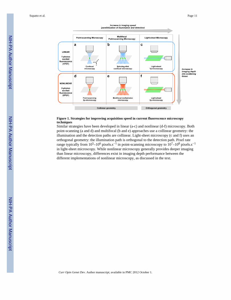
Figure 1. Strategies for improving acquisition speed in current fluorescence microscopytechniquesSimilar strategies have been developed in linear (a-c) and nonlinear (d-f) microscopy. Bothpoint-scanning (a and d) and multifocal (b and e) approaches use a collinear geometry: theillumination and the detection paths are collinear. Light-sheet microscopy (c and f) uses anorthogonal geometry: the illumination path is orthogonal to the detection path. Pixel raterange typically from 105–106 pixels.s−1 in point-scanning microscopy to 107–108 pixels.s−1
in light-sheet microscopy. While nonlinear microscopy generally provides deeper imagingthan linear microscopy, differences exist in imaging depth performance between thedifferent implementations of nonlinear microscopy, as discussed in the text.
Supatto et al. Page 11
Curr Opin Genet Dev. Author manuscript; available in PMC 2012 October 1.
NIH
-PA Author Manuscript
NIH
-PA Author Manuscript
NIH
-PA Author Manuscript
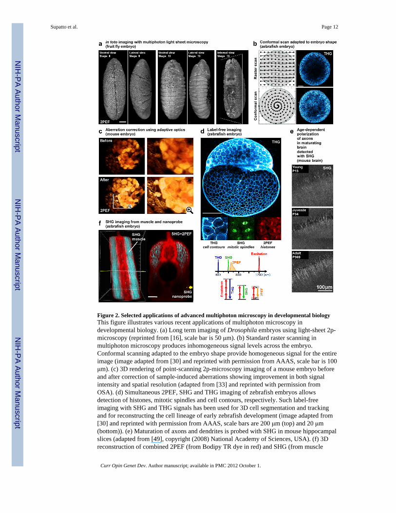
Figure 2. Selected applications of advanced multiphoton microscopy in developmental biologyThis figure illustrates various recent applications of multiphoton microscopy indevelopmental biology. (a) Long term imaging of Drosophila embryos using light-sheet 2p-microscopy (reprinted from [16], scale bar is 50 μm). (b) Standard raster scanning inmultiphoton microscopy produces inhomogeneous signal levels across the embryo.Conformal scanning adapted to the embryo shape provide homogeneous signal for the entireimage (image adapted from [30] and reprinted with permission from AAAS, scale bar is 100μm). (c) 3D rendering of point-scanning 2p-microscopy imaging of a mouse embryo beforeand after correction of sample-induced aberrations showing improvement in both signalintensity and spatial resolution (adapted from [33] and reprinted with permission fromOSA). (d) Simultaneous 2PEF, SHG and THG imaging of zebrafish embryos allowsdetection of histones, mitotic spindles and cell contours, respectively. Such label-freeimaging with SHG and THG signals has been used for 3D cell segmentation and trackingand for reconstructing the cell lineage of early zebrafish development (image adapted from[30] and reprinted with permission from AAAS, scale bars are 200 μm (top) and 20 μm(bottom)). (e) Maturation of axons and dendrites is probed with SHG in mouse hippocampalslices (adapted from [49], copyright (2008) National Academy of Sciences, USA). (f) 3Dreconstruction of combined 2PEF (from Bodipy TR dye in red) and SHG (from muscle
Supatto et al. Page 12
Curr Opin Genet Dev. Author manuscript; available in PMC 2012 October 1.
NIH
-PA Author Manuscript
NIH
-PA Author Manuscript
NIH
-PA Author Manuscript

endogenous signal and BaTiO3 nanoparticle in blue) signals recorded in zebrafish embryos:the strong SHG signal from the nanoprobe (yellow arrow) is detected twice deeper than theendogeneous SHG from muscles (image adapted from [51] and reprinted with permissionfrom authors, scale bar is 50 μm).
Supatto et al. Page 13
Curr Opin Genet Dev. Author manuscript; available in PMC 2012 October 1.
NIH
-PA Author Manuscript
NIH
-PA Author Manuscript
NIH
-PA Author Manuscript
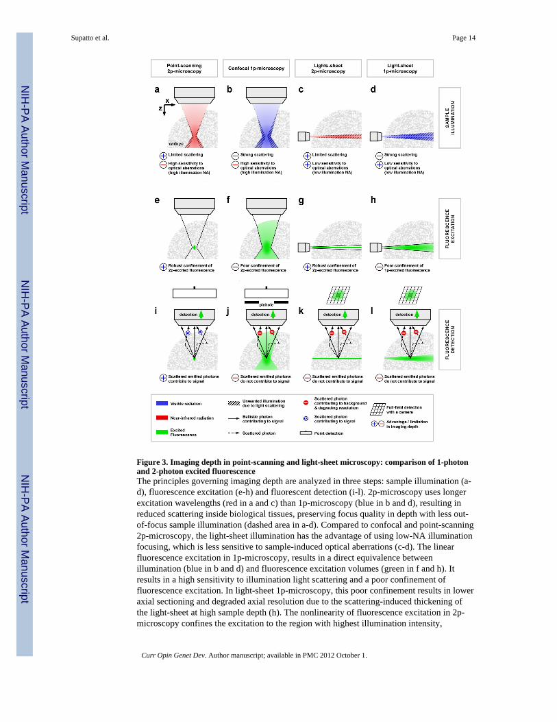
Figure 3. Imaging depth in point-scanning and light-sheet microscopy: comparison of 1-photonand 2-photon excited fluorescenceThe principles governing imaging depth are analyzed in three steps: sample illumination (a-d), fluorescence excitation (e-h) and fluorescent detection (i-l). 2p-microscopy uses longerexcitation wavelengths (red in a and c) than 1p-microscopy (blue in b and d), resulting inreduced scattering inside biological tissues, preserving focus quality in depth with less out-of-focus sample illumination (dashed area in a-d). Compared to confocal and point-scanning2p-microscopy, the light-sheet illumination has the advantage of using low-NA illuminationfocusing, which is less sensitive to sample-induced optical aberrations (c-d). The linearfluorescence excitation in 1p-microscopy, results in a direct equivalence betweenillumination (blue in b and d) and fluorescence excitation volumes (green in f and h). Itresults in a high sensitivity to illumination light scattering and a poor confinement offluorescence excitation. In light-sheet 1p-microscopy, this poor confinement results in loweraxial sectioning and degraded axial resolution due to the scattering-induced thickening ofthe light-sheet at high sample depth (h). The nonlinearity of fluorescence excitation in 2p-microscopy confines the excitation to the region with highest illumination intensity,
Supatto et al. Page 14
Curr Opin Genet Dev. Author manuscript; available in PMC 2012 October 1.
NIH
-PA Author Manuscript
NIH
-PA Author Manuscript
NIH
-PA Author Manuscript
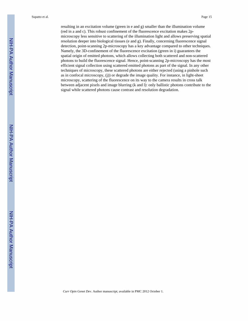
resulting in an excitation volume (green in e and g) smaller than the illumination volume(red in a and c). This robust confinement of the fluorescence excitation makes 2p-microscopy less sensitive to scattering of the illumination light and allows preserving spatialresolution deeper into biological tissues (e and g). Finally, concerning fluorescence signaldetection, point-scanning 2p-microscopy has a key advantage compared to other techniques.Namely, the 3D-confinement of the fluorescence excitation (green in i) guarantees thespatial origin of emitted photons, which allows collecting both scattered and non-scatteredphotons to build the fluorescence signal. Hence, point-scanning 2p-microscopy has the mostefficient signal collection using scattered emitted photons as part of the signal. In any othertechniques of microscopy, these scattered photons are either rejected (using a pinhole suchas in confocal microscopy, (j)) or degrade the image quality. For instance, in light-sheetmicroscopy, scattering of the fluorescence on its way to the camera results in cross talkbetween adjacent pixels and image blurring (k and l): only ballistic photons contribute to thesignal while scattered photons cause contrast and resolution degradation.
Supatto et al. Page 15
Curr Opin Genet Dev. Author manuscript; available in PMC 2012 October 1.
NIH
-PA Author Manuscript
NIH
-PA Author Manuscript
NIH
-PA Author Manuscript
NIH
-PA Author Manuscript
NIH
-PA Author Manuscript
NIH
-PA Author Manuscript
Supatto et al. Page 16
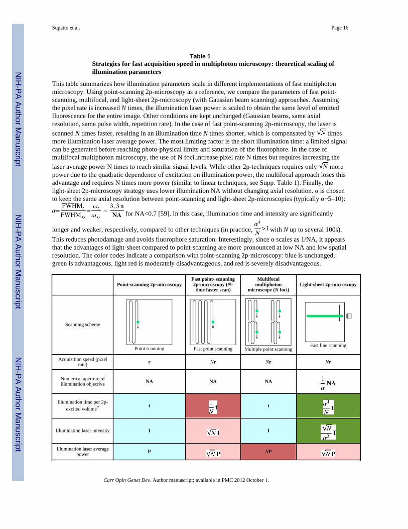
Table 1Strategies for fast acquisition speed in multiphoton microscopy: theoretical scaling ofillumination parameters
This table summarizes how illumination parameters scale in different implementations of fast multiphotonmicroscopy. Using point-scanning 2p-microscopy as a reference, we compare the parameters of fast point-scanning, multifocal, and light-sheet 2p-microscopy (with Gaussian beam scanning) approaches. Assumingthe pixel rate is increased N times, the illumination laser power is scaled to obtain the same level of emittedfluorescence for the entire image. Other conditions are kept unchanged (Gaussian beams, same axialresolution, same pulse width, repetition rate). In the case of fast point-scanning 2p-microscopy, the laser isscanned N times faster, resulting in an illumination time N times shorter, which is compensated by timesmore illumination laser average power. The most limiting factor is the short illumination time: a limited signalcan be generated before reaching photo-physical limits and saturation of the fluorophore. In the case ofmultifocal multiphoton microscopy, the use of N foci increase pixel rate N times but requires increasing thelaser average power N times to reach similar signal levels. While other 2p-techniques requires only morepower due to the quadratic dependence of excitation on illumination power, the multifocal approach loses thisadvantage and requires N times more power (similar to linear techniques, see Supp. Table 1). Finally, thelight-sheet 2p-microscopy strategy uses lower illumination NA without changing axial resolution. α is chosento keep the same axial resolution between point-scanning and light-sheet 2p-microscopies (typically α~5–10):
for NA<0.7 [59]. In this case, illumination time and intensity are significantly
longer and weaker, respectively, compared to other techniques (in practice, with N up to several 100s).This reduces photodamage and avoids fluorophore saturation. Interestingly, since α scales as 1/NA, it appearsthat the advantages of light-sheet compared to point-scanning are more pronounced at low NA and low spatialresolution. The color codes indicate a comparison with point-scanning 2p-microscopy: blue is unchanged,green is advantageous, light red is moderately disadvantageous, and red is severely disadvantageous.
Point-scanning 2p-microscopyFast point- scanning2p-microscopy (N-time faster scan)
Multifocalmultiphoton
microscope (N foci)Light-sheet 2p-microscopy
Scanning scheme
Point scanning Fast point scanning Multiple point scanningFast line scanning
Acquisition speed (pixelrate) r Nr Nr Nr
Numerical aperture ofillumination objective NA NA NA
Illumination time per 2p-excited volume* t t
Illumination laser intensity I I
Illumination laser averagepower P NP
Curr Opin Genet Dev. Author manuscript; available in PMC 2012 October 1.

NIH
-PA Author Manuscript
NIH
-PA Author Manuscript
NIH
-PA Author Manuscript
Supatto et al. Page 17
*If the spatial sampling (or pixel size) is the same for every technique, this value is related to the illumination time per pixel. See Supp. Table 1 for
the comparison with linear techniques.
Curr Opin Genet Dev. Author manuscript; available in PMC 2012 October 1.

NIH
-PA Author Manuscript
NIH
-PA Author Manuscript
NIH
-PA Author Manuscript
Supatto et al. Page 18
Table 2Strategies for fast acquisition speed in multiphoton microscopy: example of experimentalillumination parameters
Experimental values from published work confirm the parameter scaling presented in Table 1. Importantly,they demonstrate the advantageous use of long illumination time and low illumination intensity in light-sheet2p-microscopy with limited increase in laser power compared to point-scanning 2p-microscopy. In addition,they show that the main limitation of multifocal multiphoton microscopy is the requirements for high laseraverage power. Note that these experimental values have been used for imaging different biological samplewith different labeling: therefore, the comparison is only indicative.
Reference Mc Mahon et al. [6] Bahnmann et al. [59] Truong et al. [16]
Microscopy Point-scanning 2p-microscopy Multifocal multiphoton microscopy Light-sheet 2p-microscopy
Sample Live Drosophila embryos Dissociated adult rat cells (cardiacmyocytes)
Live zebrafish embryonic heart
Fluorophore GFP Fluo3 calcium dye GFP
Image (or frame) size 400×400 pixels200×200 μm2
128×128 pixels64×64 μm2
400×400 pixels160×160 μm2
Frame rate 2.2 fps 640 fps 70 fps
Acquisition speed (pixelrate)
0.36 106 pix/s 10.5 106 pix/s 11.2 106 pix/s
Illumination time perpixel
2.8 μs 3.4 μs ~ 100 μs
Illumination laserintensity per focus
~ 10 MW.cm−2 ~ 10 MW.cm−2 ~ 0.1 MW.cm−2
Illumination laseraverage power
30 mW 360 mW 50 mW
Excitation wavelength 940 nm 780 nm 920 nm
Curr Opin Genet Dev. Author manuscript; available in PMC 2012 October 1.


