Embed Size (px)
Citation preview
American Journal of Analytical Chemistry, 2012, 3, 779-789 doi:10.4236/ajac.2012.312104 Published Online December 2012 (http://www.SciRP.org/journal/ajac)
Acylhydrazide and Isatin Schiff Bases as Alternate UV-Laser Desorption Ionization (LDI) Matrices for Low
Molecular Weight (LMW) Peptides Analysis
Syed Ghulam Musharraf1,2*, Aisha Bibi2, Najia Shahid2, Muhammad Najam-ul-Haq3, Momin Khan2,
Muhammad Taha2, Uzma Rasool Mughal2, Khalid Mohammed Khan2 1Panjwani Center for Molecular Medicine and Drug Research, International Center for Chemical and Biological Sciences,
University of Karachi, Karachi, Pakistan 2 Hussain Ebrahim Jamal Research Institute of Chemistry, International Center for Chemical and Biological Sciences,
University of Karachi, Karachi, Pakistan 3Department of Chemistry, Bahauddin Zakariya University, Multan, Pakistan
Email: *[email protected]
Received September 27, 2012; revised October 30, 2012; accepted November 12, 2012
ABSTRACT
Matrix-assisted laser desorption/ionization (MALDI) is a preferred and widely used mass spectrometric technique for the analysis of macromolecules. Limited UV-LDI matrices are available for the analysis of biomolecules due to the re-stricted structural features to serve in the laser desorption/ionization mechanism with a problem of background signals appearing in the low mass region. This paper describes the application of Schiff base derivatives of acylhydrazide and isatin as alternate UV-LDI matrices for the analysis of peptides with significantly low background signals. Thirty one compounds have been successfully employed as matrices for the analysis of low molecular weight (LMW) peptides (2000 Da) including bradykinin and renin substrate tetra-decapeptide. Bovine serum albumin (BSA)-digest was also analyzed and identified through database search against Swiss-Prot by using MASCOT. The MS measurements were recorded by using dried droplet sample preparation procedures by mixing the matrix solution with analyte at a volume ratio of 1:2. Finally, LMW organic compounds (500 Da) were also analyzed by the synthesized matrix materials which showed better S/N ratios and minimal background signals for low mass range in comparison to the comparable results with α-Cyano-4-hydroxycinnamic acid (HCCA), a preferred choice for peptide analysis. Keywords: MALDI-MS; LDI Matrix; Acylhydrazide Schiff Bases; Bis-Schiff Bases of Isatin; Schiff Bases of Isatin;
Peptides
1. Introduction
Soft ionization by matrix-assisted laser desorption/ioni- zation mass spectrometry (MALDI) has become very important in mass spectrometric analysis, particularly, for biological macromolecules [1]. In the MALDI proc- esses, laser and matrix play an effective role for the ioni- zation of analyte. MALDI utilizes both UV and IR lasers as a source of energy [2], while UV lasers are widely used in various commercially available instruments. Commonly utilized UV lasers includes N2 laser (337 nm) and frequency tripled and quadrupled Nd: YAG laser (355 nm and 266 nm, respectively). Moreover, smart beam, a modified UV lasers, has recently been intro- duced by the Bruker which provides significant enhance- ment in MALDI performance [3].
Likewise, different organic compounds have been
screened for MALDI matrices. Initially, derivatives of benzoic acid, and related aromatic compounds were rec- ognized as good MALDI matrices [4] for example, 2-(4-hydroxyphenylazo) benzoic acid, (HABA) for pep- tides, proteins and glycoproteins analysis [5] and 3-hydroxypicolinic acid (3-HPA) for oligonucleotides [6] were recognized as good UV-MALDI matrices. Com- pounds having functional groups other than carboxylic acid also have the tendency to be employed as MALDI matrices, for example, 3-aminoquinoline is a good matrix for polysaccharides and proteins [7], 2,4,6-trihydroxy acetophenone [8] and the laser dye coumarin 120 [9] has been found useful in MALDI-MS of oligonucleotides and monosulfated oligosaccharides, respectively. Fitz- gerald et al. [10] introduced a number of basic matrices derived from substituted pyrimidines, pyridines, and ani- lines for the analysis of small proteins and nucleic acids. Recently, Y. Fukuyama et al. [11] have modified HCCA *Corresponding author.
Copyright © 2012 SciRes. AJAC
S. G. MUSHARRAF ET AL. 780
by attaching C-8 alkyl chain which enables it to analyze hydrophobic peptides. Similarly, S. Martic et al. [12] have prepared allylated derivatives of DHB, HCCA and caffeic acid for the efficient analysis of compounds by MALDI-MS in low molecular weight region. Overall, efforts related to the different structural modifications of existing MALDI matrices or screening of new class of compounds as MALDI matrices are limited. However, more understanding of the structural correlation with mechanistic approach of matrices is required that may results in the development of better MALDI matrices with improved performance. Moreover, appearance of a large number of interfering matrix signals in the low m/z region is significant problem in MALDI MS analysis [13]. Different strategies have been published to analyze the analyte in the low mass region [14-27].
In this work, we have explored new classes of Schiff bases as alternate UV-LDI matrices for the analysis of low molecular weight peptides by MALDI-TOF-MS and to correlate the performance of a matrix with its chemical structure. The selection criteria of compounds was based on the structural features required for a MALDI matrix i.e. conjugated aromatic system, λmax etc. A large number of Schiff base derivatives of acylhydrazide and isatin with necessary functionalities have been screened as po- tential matrices.
2. Experimental
2.1. Chemicals
Bradykinin (≥98%), renin substrate tetra-decapeptide (≥ 97%), insulin from bovine pancreas were purchased from Sigma-Aldrich (USA). Cholic acid (≥97%) was obtained from Wako (Japan) and chenodeoxycholic acid (≥97%) was obtained from MP Biomedical (Japan) while bovine serum albumin (BSA)-digest was purchased from Bruker Daltonics (Germany). All the solvents methanol (MeOH), trifluoroacetic acid (TFA), acetone ((CH3)2CO), and ace- tonitrile (ACN) were of HPLC grade and purchased from Sigma-Aldrich (USA). Deionized water (Milli-Q) was used during the study (Millipore, USA).
2.2. Synthesis of Matrix Materials
Acylhydrazide Schiff bases (Class I) were synthesized from acylhydrazides which were synthesized from dif- ferent esters by refluxing with hydrazine hydrate for 2 h. Synthesized acylhydrazides were recrystallized in meth- anol. Acylhydrazide Schiff bases were prepared by con- densing equimolar concentration of different acylhy- drazide with different aromatic aldehydes and acetophe- nones by refluxing in ethanol for 3 to 4 h. The crude pro- duct was further recrystallized in methanol.
Bis-Schiff bases of isatin (Class II) were synthesized
from the hydrazones which prepared by refluxing a mix- ture of isatin (1 g) and hydrazine hydrate (10 mL). The synthesized hydrazone (1 mmol) were refluxed with dif- ferent substituted aromatic aldehydes (1 mmol) in meth- anol for 3 h. The progress of reaction was monitored by TLC. After cooling and filtration, the crystalline bis- Schiff bases were collected, washed with methanol and dried to afford compounds in high yields (>80%). Re- crystallization from methanol resulted in pure crystals.
The Schiff bases of isatin (Class III) were synthesized by stirring a mixture of isatin (1 mmol) and different sub- stituted aromatic amines (1 mmol) in small amount of water at room temperature for 20 - 30 h. The reaction was monitored by TLC. The yellow crystalline Schiff bases were collected by filtration, washed with water and dried to get pure crystals. Detail synthetic conditions and the spectroscopic data (1H NMR) of synthesized Schiff bases of various classes have been reported earlier [28,29].
2.3. Preparation of Standard Solutions
Peptide standards (Bradykinin and renin substrate tetra- decapeptide) were prepared in 1mM concentrations in 0.1% TFA:ACN (1:1) and working standard solutions were prepared through the dilution of stock solution. BSA-digest and insulin from bovine pancreas were dis- solved in 0.1% trifluoroacetic acid (TFA) in a concentra- tion of 4 pmol/L and 100 pmol, respectively. Cholic acid and chenodeoxycholic acid were prepared in meth- anol at the concentration of 0.1 M.
2.4. UV-Vis Absorption Measurements
Schiff bases of acylhydrazide and bis-Schiff bases of isa- tin were dissolved in methanol, while Schiff bases of isa- tin were dissolved in acetone in a concentration range of 0.5 - 1.0 mM. The UV-Visible absorption maxima was recorded in the region of 200 - 800 nm by scanning against the reagent blank on a double beam UV/Visible spectrophotometer (Thermo Scientific Evolution 300, UK).
2.5. Mass Spectrometry
MALDI-MS measurements were carried out on Ultraflex III TOF/TOF (Bruker Daltonics, Bremen, Germany) mass spectrometer, equipped with a Smart beam laser (Nd: YAG, 355 nm) and an electrostatic reflector. The instru- ment was operated in reflector mode with the ion source 1 (ISI) set to 25.00 kV, source 2 (ISI) set to 21.50 kV, a lens voltage of 9.51 kV and laser energy was set between 50% - 60%. Flex analysis was used for the data analysis and the validation of data including the baseline subtract- tion. External calibration was carried out by using pep-
Copyright © 2012 SciRes. AJAC
S. G. MUSHARRAF ET AL. 781
tide calibration standards (Bruker Daltonics, Bremen, Germany). 0.1% TFA:ACN (1:1) was used as a solvent to prepare solutions of different synthetic matrix materi- als, in a concentration range of 2 - 5 g/L. The solu-tions were sonicated for 5 minutes (Ultrasonic LC 38 H) and then centrifuged for 5 minutes (Centrifuge 5804 R Eppendorf). The supernatant was then used as matrix for the analysis. The dried droplet preparation method was used for the sample preparation by applying 1 L satu-rated solution of different synthetic matrix materials with analyte at a volume ratio of 1:2 to a MALDI target plate and then allowing the droplet to dry at ambient condi-tions which formed a homogeneous film of the matrix. The dried co-crystallized samples were then analyzed by MALDI-MS.
Recorded BSA digest spectra with a newly developed matrices were submitted to the MASCOT search engine (Matrix Science, London, UK), using UniProt/Swiss-Prot (release July 2010, Homo sapiens, 18055 sequences) as the reference database. MASCOT search parameters were as follows: enzyme specificity trypsin, fixed modi- fications cysteine carbamidomethylation, variable modi- fication methionine oxidation. The maximum number of missed cleavages was set to 3 and mass tolerance at (0.5 Da).
3. Results and Discussion
Schiff base derivatives of different class of compounds, including acylhydrazide Schiff bases (class I, nine com- pounds), bis-Schiff bases of isatin (class II, four com- pounds) and Schiff bases of isatin (class III, eighteen compounds) were screened as alternate matrices for the detection of low molecular weight peptides (Table 1).
3.1. Spectrophotometric Analysis of Synthesized Matrices
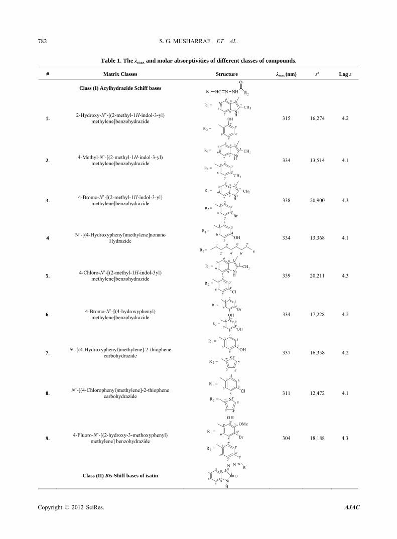
Wavelength maxima (λmax) and the molar absorptivity (ε) of synthetic matrix materials were determined to verify their ability to absorb the laser energy at wavelength ei- ther matching or closer to the laser wavelength (355 nm) employed in UV-MALDI-MS (Table 1). Strong rela- tionship was found between the signal intensities and UV spectrophotometric properties of the matrices. This ob- servation is attributed to the spectral data obtained by various matrices having λmax closer to the laser and has high value of the molar absorptivity (ε). However, de- sorption/ionization process is not exclusively dependent upon these two factors. The matrices based desorp- tion/ionization mechanism is still not completely under- stood and other factors such as vacuum stability and sol- vent compatibility may also affect the performance of such matrices. The values of signal intensities and S/N ratio also indicate this anomaly (Table 1).
3.2. Screening of Schiff Base of Various Classes
Sample preparation method and solvent selection are two important parameters which played important role in achieving better signal to noise ratio (S/N) [30,31]. All screened synthetic compounds were prepared in 0.1% TFA:ACN for screening with matrix to analyte volume ratio of 1:2 for the detection of low molecular weight peptides (<2000 Da), bradykinin and renin substrate tetra-decapeptide.
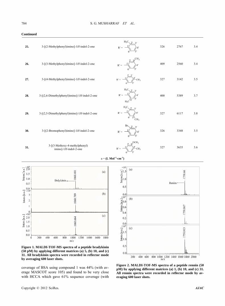
Nine compounds of class I (1 - 9) possessing λmax be- tween 304 - 339 nm were screened and found compara- ble to the existing MALDI matrix, HCCA in terms of intensity for the detection of peptides. Compounds 1, 2, 5 and 7 showed very high signal intensity (>1 × 10⁵) for peptides (Table 1). Four compounds of class II (10 - 13) possessing λmax between 331 - 346 nm were screened as LDI matrix. All of them showed high intensity (>1 × 10⁴) of analyzed samples and were found suitable to be used as MALDI matrices for the analysis of low molecular weight peptides (Table 1). Similarly, eighteen com-pounds of class III (14 - 31), having λmax between 326 - 416 nm, showed good signal intensity (>1 × 10⁴) of both peptides (Table 1). Overall, newly developed matrices produced excellent signal intensities of analytes with high reproducibility. Compounds 5, 10 and 31 when em-ployed to analyze bradykinin peptide produced very high intensities with a standard deviation of ±0.15 (Figure 1), while the renin peptide analyzed with compounds 1, 10 and 31 afford similar results with a standard deviation of ±0.10 (Figure 2). However, almost no background sig-nals were observed in the low mass region from these new matrices, hence, found suitable for the analysis of small molecules.
In all classes, a common chromophore was Schiff base while different aromatic substituents were attached to ty each class which enhanced both n-* and -* transitions through electron lone pair on hetro atom and conjugated double bond system which produced the hyperchromic and bathochromic effects thus resulted in longer λmax therefore, requires less energy for the excitation. This factor is directly related to the amount of laser power required to get a good intensity signal from a sweet spot on MALDI target.
Moreover, among the all screened compounds, acyl- hydrazide Schiff bases (class I) showed excellent signal intensities. Therefore, compounds from class I were se- lected for further studies. BSA-digest sample was ana-lyzed by using compounds of class I (1 - 7) to evaluate their ability to analyze peptides in complex biological samples. The spectrum showed promising signals of pep-tides (Figure 3) and the resulting mass spectrum were subjected to the database search against Swiss-Prot, using MASCOT for the identification. The sequence
Copyright © 2012 SciRes. AJAC
S. G. MUSHARRAF ET AL.
Copyright © 2012 SciRes. AJAC
782
Table 1. The λmax and molar absorptivities of different classes of compounds.
# Matrix Classes Structure λmax (nm) εa Log ε
Class (I) Acylhydrazide Schiff bases
HC N NH
O
R2R1
1. 2-Hydroxy-N’-[(2-methyl-1H-indol-3-yl)
methylene]benzohydrazide
1
2
34
5
6NH7
8
9
CH3R1 =
R2 =1'
2'3'
4'
5'
6'
OH
315 16,274 4.2
2. 4-Methyl-N’-[(2-methyl-1H-indol-3-yl)
methylene]benzohydrazide
R1 =
1
2
34
5
6NH7
8
9
CH3
1'2'
3'
4'
5'
6'CH3
R2 =
334 13,514 4.1
3. 4-Bromo-N’-[(2-methyl-1H-indol-3-yl)
methylene]benzohydrazide
R1 =
1
2
34
5
6NH7
8
9CH3
1'2'
3'
4'
5'
6'Br
R2 =
338 20,900 4.3
4 N’-[(4-Hydroxyphenyl)methylene]nonano
Hydrazide 1'
2'
3'
4'
5'
6'
7'
8
R1=1
23
4
5
6OH
R2=
334 13,368 4.1
5. 4-Chloro-N’-[(2-methyl-1H-indol-3yl)
methylene]benzohydrazide
1
23
45
6NH7
8
9
CH3R1 =
R2 =1'
2'
3'
4'
5'6'
Cl
339 20,211 4.3
6. 4-Bromo-N’-[(4-hydroxyphenyl)
methylene]benzohydrazide 1' 2'
3'
4'
5'6'
OH
OH
R1 =1
23
4
56 Br
R2 =
334 17,228 4.2
7. N’-[(4-Hydroxyphenyl)methylene]-2-thiophene
carbohydrazide 1'2'
3' 4'
S5'
R1 =
R2 =
12
3
4
5
6OH
337 16,358 4.2
8. N’-[(4-Chlorophenyl)methylene]-2-thiophene
carbohydrazide 1'2'
3' 4'
S5'
R1 =
R2 =
12
3
4
5
6Cl
311 12,472 4.1
9. 4-Fluoro-N’-[(2-hydroxy-3-methoxyphenyl)
methylene] benzohydrazide
R1 =
R2 =1'
2'3'
4'
5'6'
F
1'2'
3'
5'6'
Br4'
OH
OMe
304 18,188 4.3
Class (II) Bis-Shiff bases of isatin 1
234
5
6N
7
8
9
O
N NR'
H
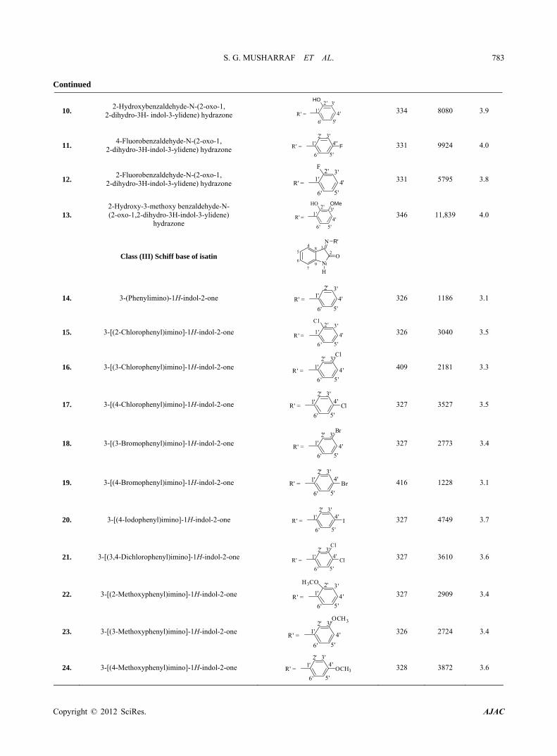
S. G. MUSHARRAF ET AL. 783
Continued
10. 2-Hydroxybenzaldehyde-N-(2-oxo-1,
2-dihydro-3H- indol-3-ylidene) hydrazone 1'
2' 3'
4'
5'6'
R' =
HO
334 8080 3.9
11. 4-Fluorobenzaldehyde-N-(2-oxo-1,
2-dihydro-3H-indol-3-ylidene) hydrazone 1'
2' 3'4''
5'6'
R' = F
331 9924 4.0
12. 2-Fluorobenzaldehyde-N-(2-oxo-1,
2-dihydro-3H-indol-3-ylidene) hydrazone 1'
2' 3'
4'
5'6'
R' =
F
331 5795 3.8
13. 2-Hydroxy-3-methoxy benzaldehyde-N- (2-oxo-1,2-dihydro-3H-indol-3-ylidene)
hydrazone 1'
2' 3'
4'
5'6'
R' =
HO OMe
346 11,839 4.0
Class (III) Schiff base of isatin 1
234
5
6 N7
8
9
O
N
H
R'
14. 3-(Phenylimino)-1H-indol-2-one 1'2' 3'
4'
5'6'
R' =
326 1186 3.1
15. 3-[(2-Chlorophenyl)imino]-1H-indol-2-one 1 '2' 3 '
4'
5 '6 '
R' =
Cl
326 3040 3.5
16. 3-[(3-Chlorophenyl)imino]-1H-indol-2-one 1'2' 3'
4'
5'6'
R' =
Cl
409 2181 3.3
17. 3-[(4-Chlorophenyl)imino]-1H-indol-2-one 1'2' 3'
4'
5'6'
R' = Cl
327 3527 3.5
18. 3-[(3-Bromophenyl)imino]-1H-indol-2-one 1'2' 3'
4'
5'6'
R' =
Br
327 2773 3.4
19. 3-[(4-Bromophenyl)imino]-1H-indol-2-one 1'2' 3'
4'
5'6'
R' = Br
416 1228 3.1
20. 3-[(4-Iodophenyl)imino]-1H-indol-2-one 1'2' 3'
4'
5'6'
R' = I
327 4749 3.7
21. 3-[(3,4-Dichlorophenyl)imino]-1H-indol-2-one 1'2' 3'
4'
5'6'
R' = Cl
Cl
327 3610 3.6
22. 3-[(2-Methoxyphenyl)imino]-1H-indol-2-one 1'2' 3'
4'
5'6'
R' =
H3CO
327 2909 3.4
23. 3-[(3-Methoxyphenyl)imino]-1H-indol-2-one 1'2' 3'
4'
5'6'
R' =
OCH3
326 2724 3.4
24. 3-[(4-Methoxyphenyl)imino]-1H-indol-2-one 1'2' 3'
4'
5'6'
R' = OCH3
328 3872 3.6
Copyright © 2012 SciRes. AJAC
S. G. MUSHARRAF ET AL.
Copyright © 2012 SciRes. AJAC
784
Continued
25. 3-[(2-Methylphenyl)imino]-1H-indol-2-one 1'2' 3'
4'
5'6'
R' =
H3C
326 2767 3.4
26. 3-[(3-Methylphenyl)imino]-1H-indol-2-one 1'2' 3'
4'
5'6'
R' =
CH3
409 2560 3.4
27. 3-[(4-Methylphenyl)imino]-1H-indol-2-one 1'2' 3'
4'
5'6'
R' = CH3
327 3142 3.5
28. 3-[(2,6-Dimethylphenyl)imino]-1H-indol-2-one 1'
2' 3'
4'
5'6'
R' =
H3C
H3C
400 5389 3.7
29. 3-[(2,5-Dimethylphenyl)imino]-1H-indol-2-one 1'
2' 3'
4'
5'6'
R' =
H3C
CH3
327 6117 3.8
30. 3-[(2-Bromophenyl)imino]-1H-indol-2-one 1'2' 3'
4'
5'6'
R' =
Br
326 3348 3.5
31. 3-[(3-Methoxy-4-methylphenyl)
imino]-1H-indol-2-one 1'
2' 3'4'
5'6'
R' = CH3
OCH3
327 3635 3.6
a = (L Mol–1·cm–1)
Figure 1. MALDI-TOF-MS spectra of a peptide bradykinin (50 pM) by applying different matrices (a) 5, (b) 10, and (c) 31. All bradykinin spectra were recorded in reflector mode by averaging 600 laser shots.
Figure 2. MALDI-TOF-MS spectra of a peptide rennin (50 pM) by applying different matrices (a) 1, (b) 10, and (c) 31. All rennin spectra were recorded in reflector mode by av-eraging 600 laser shots.
coverage of BSA using compound 1 was 44% (with av-erage MASCOT score 105) and found to be very close with HCCA which gave 61% sequence coverage (with
S. G. MUSHARRAF ET AL. 785
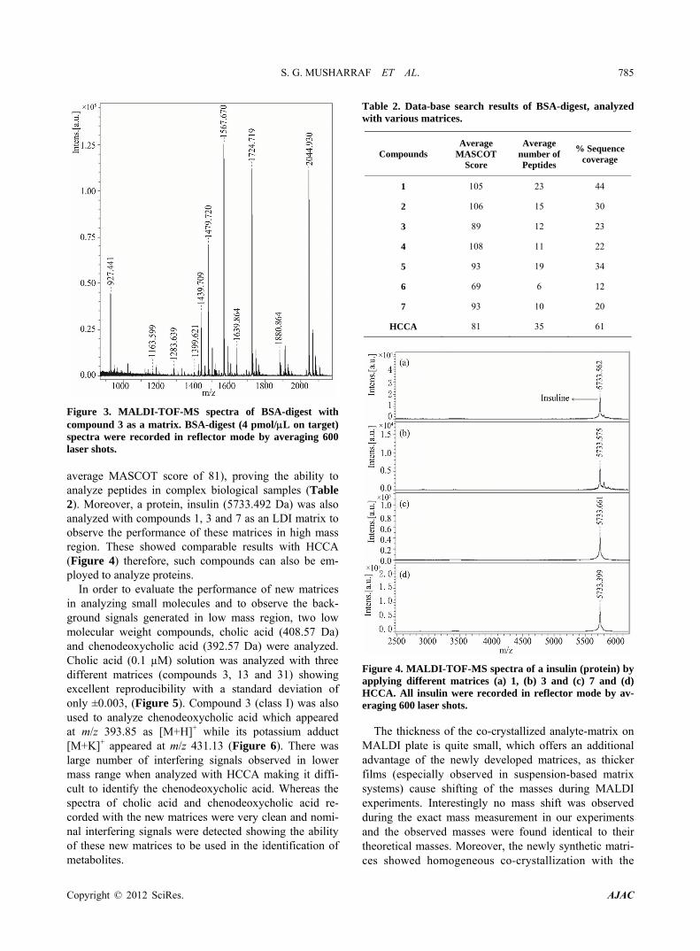
Figure 3. MALDI-TOF-MS spectra of BSA-digest with compound 3 as a matrix. BSA-digest (4 pmol/L on target) spectra were recorded in reflector mode by averaging 600 laser shots. average MASCOT score of 81), proving the ability to analyze peptides in complex biological samples (Table 2). Moreover, a protein, insulin (5733.492 Da) was also analyzed with compounds 1, 3 and 7 as an LDI matrix to observe the performance of these matrices in high mass region. These showed comparable results with HCCA (Figure 4) therefore, such compounds can also be em-ployed to analyze proteins.
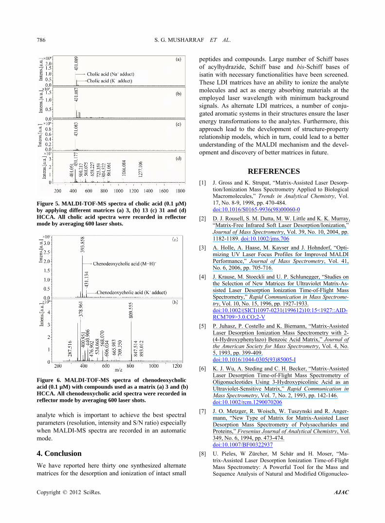
In order to evaluate the performance of new matrices in analyzing small molecules and to observe the back- ground signals generated in low mass region, two low molecular weight compounds, cholic acid (408.57 Da) and chenodeoxycholic acid (392.57 Da) were analyzed. Cholic acid (0.1 µM) solution was analyzed with three different matrices (compounds 3, 13 and 31) showing excellent reproducibility with a standard deviation of only ±0.003, (Figure 5). Compound 3 (class I) was also used to analyze chenodeoxycholic acid which appeared at m/z 393.85 as [M+H]+ while its potassium adduct [M+K]+ appeared at m/z 431.13 (Figure 6). There was large number of interfering signals observed in lower mass range when analyzed with HCCA making it diffi- cult to identify the chenodeoxycholic acid. Whereas the spectra of cholic acid and chenodeoxycholic acid re- corded with the new matrices were very clean and nomi- nal interfering signals were detected showing the ability of these new matrices to be used in the identification of metabolites.
Table 2. Data-base search results of BSA-digest, analyzed with various matrices.
Compounds Average
MASCOT Score
Average number of Peptides
% Sequence coverage
1 105 23 44
2 106 15 30
3 89 12 23
4 108 11 22
5 93 19 34
6 69 6 12
7 93 10 20
HCCA 81 35 61
Figure 4. MALDI-TOF-MS spectra of a insulin (protein) by applying different matrices (a) 1, (b) 3 and (c) 7 and (d) HCCA. All insulin were recorded in reflector mode by av-eraging 600 laser shots.
The thickness of the co-crystallized analyte-matrix on MALDI plate is quite small, which offers an additional advantage of the newly developed matrices, as thicker films (especially observed in suspension-based matrix systems) cause shifting of the masses during MALDI experiments. Interestingly no mass shift was observed during the exact mass measurement in our experiments and the observed masses were found identical to their theoretical masses. Moreover, the newly synthetic matri- ces showed homogeneous co-crystallization with the
Copyright © 2012 SciRes. AJAC
S. G. MUSHARRAF ET AL. 786
Figure 5. MALDI-TOF-MS spectra of cholic acid (0.1 µM) by applying different matrices (a) 3, (b) 13 (c) 31 and (d) HCCA. All cholic acid spectra were recorded in reflector mode by averaging 600 laser shots.
Figure 6. MALDI-TOF-MS spectra of chenodeoxycholic acid (0.1 µM) with compounds used as a matrix (a) 3 and (b) HCCA. All chenodeoxycholic acid spectra were recorded in reflector mode by averaging 600 laser shots. analyte which is important to achieve the best spectral parameters (resolution, intensity and S/N ratio) especially when MALDI-MS spectra are recorded in an automatic mode.
4. Conclusion
We have reported here thirty one synthesized alternate matrices for the desorption and ionization of intact small
peptides and compounds. Large number of Schiff bases of acylhydrazide, Schiff base and bis-Schiff bases of isatin with necessary functionalities have been screened. These LDI matrices have an ability to ionize the analyte molecules and act as energy absorbing materials at the employed laser wavelength with minimum background signals. As alternate LDI matrices, a number of conju- gated aromatic systems in their structures ensure the laser energy transformations to the analytes. Furthermore, this approach lead to the development of structure-property relationship models, which in turn, could lead to a better understanding of the MALDI mechanism and the devel- opment and discovery of better matrices in future.
REFERENCES [1] J. Gross and K. Strupat, “Matrix-Assisted Laser Desorp-
tion/Ionization Mass Spectrometry Applied to Biological Macromolecules,” Trends in Analytical Chemistry, Vol. 17, No. 8-9, 1998, pp. 470-484. doi:10.1016/S0165-9936(98)00060-0
[2] D. J. Rousell, S. M. Dutta, M. W. Little and K. K. Murray, “Matrix-Free Infrared Soft Laser Desorption/Ionization,” Journal of Mass Spectrometry, Vol. 39, No. 10, 2004, pp. 1182-1189. doi:10.1002/jms.706
[3] A. Holle, A. Haase, M. Kayser and J. Hohndorf, “Opti-mizing UV Laser Focus Profiles for Improved MALDI Performance,” Journal of Mass Spectrometry, Vol. 41, No. 6, 2006, pp. 705-716.
[4] J. Krause, M. Stoeckli and U. P. Schlunegger, “Studies on the Selection of New Matrices for Ultraviolet Matrix-As- sisted Laser Desorption Ionization Time-of-Flight Mass Spectrometry,” Rapid Communication in Mass Spectrome-try, Vol. 10, No. 15, 1996, pp. 1927-1933. doi:10.1002/(SICI)1097-0231(199612)10:15<1927::AID-RCM709>3.0.CO;2-V
[5] P. Juhasz, P. Costello and K. Biemann, “Matrix-Assisted Laser Desorption Ionization Mass Spectrometry with 2- (4-Hydroxyphenylazo) Benzoic Acid Matrix,” Journal of the American Society for Mass Spectrometry, Vol. 4, No. 5, 1993, pp. 399-409. doi:10.1016/1044-0305(93)85005-I
[6] K. J. Wu, A. Steding and C. H. Becker, “Matrix-Assisted Laser Desorption Time-of-Flight Mass Spectrometry of Oligonucleotides Using 3-Hydroxypicolinic Acid as an Ultraviolet-Sensitive Matrix,” Rapid Communication in Mass Spectrometry, Vol. 7, No. 2, 1993, pp. 142-146. doi:10.1002/rcm.1290070206
[7] J. O. Metzger, R. Woisch, W. Tuszynski and R. Anger-mann, “New Type of Matrix for Matrix-Assisted Laser Desorption Mass Spectrometry of Polysaccharides and Proteins,” Fresenius Journal of Analytical Chemistry, Vol. 349, No. 6, 1994, pp. 473-474. doi:10.1007/BF00322937
[8] U. Pieles, W Zürcher, M Schär and H. Moser, “Ma-trix-Assisted Laser Desorption Ionization Time-of-Flight Mass Spectrometry: A Powerful Tool for the Mass and Sequence Analysis of Natural and Modified Oligonucleo-
Copyright © 2012 SciRes. AJAC
S. G. MUSHARRAF ET AL. 787
tides,” Nucleic Acids Research, Vol. 21, No. 14, pp. 3191-3196.
[9] Y. Dai, R. M. Whittal, C. A. Bridges, Y. Isogai, O. Hindsgaul and L. Li, “Matrix-Assisted Laser Desorption Ionization Mass Spectrometry for the Analysis of Mono-sulfated Oligosaccharides,” Carbohydrate Research, Vol. 304, No. 1, 1997, pp. 1-9. doi:10.1016/S0008-6215(97)00195-X
[10] M. C. Fitzgerald, G. R. Parr and L. M. Smith, “Basic Matrices for the Matrix-Assisted Laser Desorption/Ioni- zation Mass Spectrometry of Proteins and Oligonucleo- tides,” Analytical Chemistry, Vol. 65, No. 22, 1993, pp. 3204-3211. doi:10.1021/ac00070a007
[11] Y. Fukuyama, R. Tanimura, K. Maeda, M. Watanabe, S. Kawabata, S. Iwamoto, S. Izumi and K. Tanaka, “Alky-lated Dihydroxybenzoic Acid as a MALDI Matrix Addi-tive for Hydrophobic Peptide Analysis,” Analytical Che- mistry, Vol. 84, No. 9, 2012, pp. 4237-4243. doi:10.1021/ac300540r
[12] S. Martic, J. D. Brennan, M. A. Brook, S. Ackloo and N. Nagy, “Towards the Development of a Covalently Teth-ered MALDI System—A Study of Allyl-Modified MALDI Matrixes,” Canadian Journal of Chemistry, Vol. 85, No. 1, 2007, pp. 66-76. doi:10.1139/v06-185
[13] Z. Guo, Q. Zhang , H. Zou, B. Guo and J. Ni, “A Method for the Analysis of Low-Mass Molecules by MALDI- TOF Mass Spectrometry,” Analytical Chemistry, Vol. 74, No. 7, 2002, pp. 1637-1641. doi:10.1021/ac010979m
[14] G. McCombie and R. Knochenmuss, “Small-Molecule MALDI using the Matrix Suppression Effect to Reduce or Eliminate Matrix Background Interferences,” Analyti-cal Chemistry, Vol. 76, No. 17, 2004, pp. 4990-4997. doi:10.1021/ac049581r
[15] F. O. Ayorinde, P. Hambright, T. N. Porter and Q. L. Keith, “Use of Meso-Tetrakis (Pentafluorophenyl) Porphyrin as a Matrix for Low Molecular Weight Alkyl Phenol Ethoxy- lates in Laser Desorption/Ionization Time-of-Flight Mass Spectrometry,” Rapid Communication in Mass Spectrome-try, Vol. 13, No. 24, 1999, pp. 2474-2479. doi:10.1002/(SICI)1097-0231(19991230)13:24<2474::AID-RCM814>3.0.CO;2-0
[16] A. Tholey and E. Heinzle, “Ionic (Liquid) Matrices for Matrix-Assisted Laser Desorption/Ionization Mass Spec-trometry—Applications and Perspectives,” Analytical and Bioanalytical Chemistry, Vol. 386, No. 1, 2006, pp. 24-37. doi:10.1007/s00216-006-0600-5
[17] D. W. Armstrong, L. Zhang, L. He and M. L. Gross, “Ionic Liquids as Matrixes for Matrix-Assisted Laser Desorption/Ionization Mass Spectrometry,” Analytical Chemistry, Vol. 73, No. 15, 2001, pp. 3679-3686. doi:10.1021/ac010259f
[18] M. Najam-ul-Haq, M. Rainer, C. W. Huck, P. Hausberger, H. Kraushaar and G. K. Bonn, “Nanostructured Dia-mond-Like Carbon on Digital Versatile Disc as a Ma-trix-Free Target for Laser Desorption/Ionization Mass Spectrometry,” Analytical Chemistry, Vol. 80, No. 19, 2008, pp. 7467-7472.
[19] J. Wei, J. M. Buriak and G. Siuzdak, “Desorption-Ioni- zationmass Spectrometry on Porous Silicon,” Letters to
Nature, Vol. 399, No. 77, 1999, pp. 243-246. doi:10.1038/20400
[20] S. Zhang, Y. Chen, J. A. Liu, S. X. Xiong, G. H. Wang, J. Chen and G. Q. Yang, “New Matrix of MALDI-TOF MS for Analysis of Small Molecules,” Chinese Chemical Letters, Vol. 20, No. 12, 2009, pp. 1495-1497. doi:10.1016/j.cclet.2009.06.031
[21] T. Kinumi, T. Saisu, M. Takayama and H. Niwa, “Ma-trix-Assisted Laser Desorption/Ionization Time-of-Flight Mass Spectrometry Using an Inorganic Particle Matrix for Small Molecule Analysis,” Journal of Mass Spec-trometry, Vol. 35, No. 3, 2000, pp. 417-422. doi:10.1002/(SICI)1096-9888(200003)35:3<417::AID-JMS952>3.0.CO;2-#
[22] S. Ren, L. Zhang, Z. Cheng and Y. Guo, “Immobilized Carbon Nanotubes as Matrix for MALDI-TOF-MS Analy-sis: Applications to Neutral Small Carbohydrates,” Journal of the American Society for Mass Spectrometry, Vol. 16, No. 3, 2005, pp. 333-339. doi:10.1016/j.jasms.2004.11.017
[23] X. Dong, J. Cheng, J. Li and Y. Wang, “Graphene as a Novel Matrix for the Analysis of Small Molecules by MALDI-TOF MS,” Analytical Chemistry, Vol. 82, No. 14, 2010, pp. 6208-6214. doi:10.1021/ac101022m
[24] H. Kim, J. Lee, S. Park, H. W. Ro, D. Y. Yoo and D. Y. Yoon, “Observation of Low Molecular Weight Poly (Me-thylsilsesquioxane)s by Graphite Plate Laser Desorption/ Ionization Time-of-Flight Mass Spectrometry,” Analytical Chemistry, Vol. 72, No. 22, 2000, pp. 5673-5678. doi:10.1021/ac0003899
[25] M. J. Dale, R. Knochenmuss and R. Zenobi, “Graphite/ Liquid Mixed Matrices for Laser Desorption/Ionization Mass Spectrometry,” Analytical Chemistry, Vol. 68, No. 19, 1996, pp. 3321-3329. doi:10.1021/ac960558i
[26] C. Black, C. Poile, J. Langley and J. Herniman , “The Use of Pencil Lead as a Matrix and Calibrant for Matrix-As- sisted Laser Desorption/Ionization,” Rapid Communica-tion in Mass Spectrometry, Vol. 20, No. 7, 2006, pp. 1053-1060. doi:10.1002/rcm.2408
[27] G. J. Langley, J. M. Herniman and M. S. Townell, “2B Or Not 2B, That is the Question: Further Investigations into the Use of Pencil as a Matrix for Matrix-Assisted Laser Desorption/Ionization,” Rapid Communication in Mass Spectrometry, Vol. 21, No. 2, 2007, pp. 180-190. doi:10.1002/rcm.2827
[28] K. M. Khan, M. Khan, M. Ali, M. Taha, S. Rasheed, S. Perveen and M. I. Choudhary, “Synthesis of Bis-Schiff Bases of Isatins and their Antiglycation Activity,” Bioor-ganic & Medicinal Chemistry, Vol. 7, No. 22, 2009, pp. 7795-7801. doi:10.1016/j.bmc.2009.09.028
[29] K. M. Khan, U. R. Mughal, Samreen, S. Perveen and M. I. Choudhary, “Schiff Bases of Isatin: Potential Anti-Leish- manial Agents,” Letters in Drug Design & Discovery, Vol. 5, No. 4, 2008, pp. 243-249. doi:10.2174/157018008784619915
[30] M. W. F. Neilen, “MALDI Time-of-Flight Mass Spec-trometry of Synthetic Polymers.” Mass Spectrometry Re-views, Vol. 18, No. 5, 1999, pp. 309-344. doi:10.1002/(SICI)1098-2787(1999)18:5<309::AID-MAS
Copyright © 2012 SciRes. AJAC
S. G. MUSHARRAF ET AL.
Copyright © 2012 SciRes. AJAC
788
2>3.0.CO;2-L
[31] S. F. Macha, P. A. Limbach and P. J. Savickas, “Applica-tion of Nonpolar Matrices for the Analysis of Low Mo-lecular Weight Nonpolar Synthetic Polymers by Matrix-
Assisted Laser Desorption/Ionization Time-of-Flight Mass Spectrometry,” Journal of the American Society for Mass Spectrometry, Vol. 11, No. 8, 2000, pp. 731-773. doi:10.1016/S1044-0305(00)00137-9

S. G. MUSHARRAF ET AL. 789
Appendix
Table A1. Signal intensity and S/N ratio of synthetic matrix material with low molecular weight analytes.
Bradykinin Renin substrate tetra-decapeptide Compounds
S/N Sig. Int. S/N Sig. Int.
1 1243 303,774 378 89,505
2 186 122,638 760 70,670
3 1078 59,705 702 55,347
4 513 79,804 191 34,052
5 1114 97,047 7151 82,709
6 246 73,297 192 34,052
7 892 1,268,674 1417 74,896
8 160 1687 236 10,335
9 951 58,606 834 95,126
10 352 12,917 643 88,445
11 992 126,386 250 59,958
12 706 83,512 648 53,400
13 344 69,710 356 60,418
14 523 62,378 423 23,762
15 204 40,104 236 41,150
16 292 73,000 362 38,828
17 497 77,183 426 33,273
18 1835 153,067 781 150,047
19 386 86,938 276 45,939
20 699 106,798 330 98,533
21 502 105,059 308 98,153
22 288 45,002 434 47,010
23 150 51,728 318 75,890
24 538 87,681 451 105,636
25 444 26,336 1015 110,685
26 509 86,173 681 79,172
27 736 58,769 297 59,142
28 330 56,341 800 21,265
29 347 64,819 375 131,250
30 214 19,307 265 18,867
31 171 62,204 357 102,072
HCCA 832 86,824 378 89,505
Copyright © 2012 SciRes. AJAC


