Embed Size (px)
Citation preview
Journal of Toxicology
CLINICAL TOXICOLOGY
Vol. 42, No. 4, pp. 395–564, 2004
Abstracts of the European Association of Poisons Centres and ClinicalToxicologists XXIV International Congress
1. Nerve Agents: Their Mechanisms of Action and the Implications for Treatment
Marrs TC. West Midlands Poisons Unit, Birmingham, UK.
Structure: Nerve agents are highly toxic derivatives of phosphonic, or less commonly phosphoric acid (1). The G
agents are mostly dialkylphosphonofluoridates, the leaving group being the fluorine atom. Exceptions are cyclosarin
(GF), which has one alkyl group replaced by a cyclohexyl group and tabun (GA), which has a cyanidate group. The
V agents as exemplified by VX, are S-substituted phosphonothioates. Physical Properties: The G agents, of which
tabun, sarin and soman were first synthesized before or during World War II in Germany, are volatile and therefore
inhalation hazards. The V agents, including VX, which was first synthesized in the UK, are less volatile and are
primarily percutaneous hazards, unless aerosolized. Mechanism of Toxic Action: Nerve agents are esterase inhibitors,
the main site of toxic action being on acetylcholinesterase in nervous tissue. This enzyme is intricately concerned in
cholinergic neurotransmission. The reaction of organophosphates (OPs) has been studied in many systems, but
particularly in Torpedo californica. OPs, including nerve agents, bind to the active site of the enzyme and although
the nature of the binding of nerve agents is similar to that of the natural enzyme substrate, acetylcholine, the
resultant complex with nerve agents is much more long-lasting. While the nerve agent (minus its leaving group)
remains bound to the enzyme, the enzyme is inactive in the hydrolysis of acetylcholine. If the patient survives the
acute episode, the enzyme-inhibitor complex is hydrolyzed and the enzyme’s activity is restored. Ageing and
Carbamate Pretreatment: To some extent with all anticholinesterase OPs but especially with soman, a further
reaction known as ageing can occur. This reaction is monodealkylation of the dialkylphosphyl enzyme. This renders
the inhibited enzyme resistant to hydrolysis and therefore incapable of reactivation, either spontaneously or by
reactivating drugs, such as the pyridinium oximes. In these circumstances, recovery of cholinesterase activity is
dependent upon synthesis de novo of enzyme, and one of the two main components of nerve agent therapy (the other
being atropine) is unavailable. Consequently carbamate drugs such as pyridostigmine have been studied as
pretreatments for soman poisoning (2). These drugs carbamylate acetylcholinesterase producing a complex that
reactivates quickly, but which is protected against phosphylation. Acetylcholine Accumulation: The signs and
symptoms observed with nerve agents poisoning are caused by accumulation of acetylcholine at sites of cholinergic
neurotransmission (3). These are largely reversible providing the patient survives, and survival is possible of some
multiples of the lethal doses of nerve agents providing treatment is instituted quickly. Symptoms and clinical signs
are usually grouped according to which type of cholinergic receptor is involved. Nicotinic effects include those on
autonomic ganglia (pallor, tachycardia and hypertension) and those at the neuromuscular junction (muscle
fasciculation, weakness and paralysis). Stimulation of muscarinic receptors causes effects on exocrine glands such as
rhinorrhea, bronchorrhea, sweating, lachrymation and salivation as well as myosis, failure of accommodation,
abdominal cramps and involuntary micturition by action on smooth muscle. Parasympathetic effects on the heart
(bradycardia) may be seen. In the central nervous system, where both types of cholinergic receptors are seen,
395
DOI: 10.1081/CLT-200028846 0731-3810 (Print); 1097-9875 (Online)
Copyright D 2004 by Marcel Dekker, Inc. www.dekker.com
dizziness, anxiety, confusion or convulsions may occur depending on the severity of the poisoning. Nerve Agents
and Organophosphate-Induced Delayed Polyneuropathy (OPIDP): Except in very unusual circumstances it seems
unlikely that nerve agents could cause OPIDP. The reason is that nerve agents are powerful inhibitors of
acetylcholinesterase but relatively weak inhibitors of neuropathy target esterase. Moreover, OPIDP has not, in
general, been observed in experimental studies with these agents (4). References: 1. Marrs TC, Maynard RL.
Organophosphorus chemical warfare agents. In: Karalleidde LK, Feldman SJ, Henry J and Marrs TC (eds).
Organophosphates and Human Health. London: Imperial College Press, 2001; 83–108. 2. Berry WK, Davies DR.
The use of carbamates and atropine in the protection of animals against poisoning by 1,2,2-trimethylpropyl
methylphosphonofluoridate. Biochem Pharmacol 1970; 19:927–934. 3. Marrs TC. Organophosphate poisoning.
Toxic Subst Mechanisms 1996; 15:357–388. 4. Marrs TC, Maynard RL. Neurotoxicity of chemical warfare agents.
In: de Wolff FA (ed). Handbook of Clinical Neurology, Volume 64, Intoxications of the nervous system part I.
Amsterdam: Elsevier, 1994:223–238.
2. Nerve Agent Poisoning: Features and Management
Vale JA. National Poisons Information Service (Birmingham Centre) and West Midlands Poisons Unit, City
Hospital, Birmingham, UK.
Introduction: Nerve agents are related chemically to organophosphorus insecticides and have a similar mechanism of
toxicity, but a much higher mammalian acute toxicity, particularly via the dermal route. The G agents are both
dermal and respiratory hazards, whereas the V agents, unless aerosolized, are contact poisons. Features: Systemic
poisoning may follow inhalation, ingestion or dermal exposure, though the onset of systemic toxicity is slower by
the latter route. Ocular Exposure: Miosis, which may be painful and last for several days, occurs rapidly following
exposure to nerve agent vapor and appears to be a very sensitive index of exposure (1). Ciliary muscle spasm may
impair accommodation and conjunctival injection and eye pain may occur. Dermal Exposure: Contact with liquid
nerve agent may produce localized sweating and fasciculation, which may spread to involve whole muscle groups.
Inhalation: Chest tightness, rhinorrhea and increased salivation may occur within minutes. Ingestion: Ingestion of
contaminated food or water may cause abdominal pain, nausea, vomiting, diarrhea and involuntary defecation.
Systemic Features: Miosis may also occur as a systemic feature but more usually it follows topical exposure.
Abdominal pain, nausea and vomiting, involuntary micturition and defecation, muscle weakness and fasciculation,
tremor, restlessness, ataxia and convulsions may follow dermal exposure, inhalation or ingestion of a nerve agent.
Bradycardia, tachycardia and hypotension may occur dependent on whether muscarinic or nicotinic effects
predominate. If exposure is substantial, death may occur from respiratory failure within minutes. Chronic Sequelae:
Mild or moderately exposed individuals usually recover completely, though EEG abnormalities have been reported
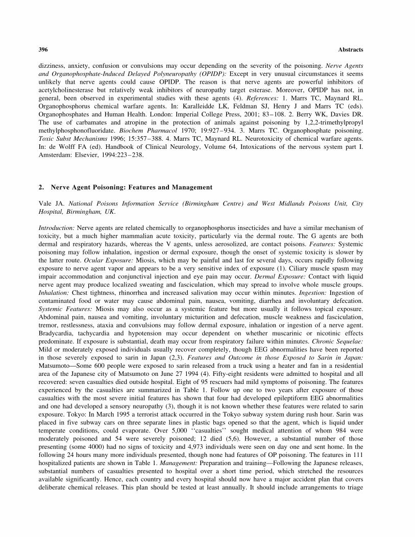
in those severely exposed to sarin in Japan (2,3). Features and Outcome in those Exposed to Sarin in Japan:
Matsumoto—Some 600 people were exposed to sarin released from a truck using a heater and fan in a residential
area of the Japanese city of Matsumoto on June 27 1994 (4). Fifty-eight residents were admitted to hospital and all
recovered: seven casualties died outside hospital. Eight of 95 rescuers had mild symptoms of poisoning. The features
experienced by the casualties are summarized in Table 1. Follow up one to two years after exposure of those
casualties with the most severe initial features has shown that four had developed epileptiform EEG abnormalities
and one had developed a sensory neuropathy (3), though it is not known whether these features were related to sarin
exposure. Tokyo: In March 1995 a terrorist attack occurred in the Tokyo subway system during rush hour. Sarin was
placed in five subway cars on three separate lines in plastic bags opened so that the agent, which is liquid under
temperate conditions, could evaporate. Over 5,000 ‘‘casualties’’ sought medical attention of whom 984 were
moderately poisoned and 54 were severely poisoned; 12 died (5,6). However, a substantial number of those
presenting (some 4000) had no signs of toxicity and 4,973 individuals were seen on day one and sent home. In the
following 24 hours many more individuals presented, though none had features of OP poisoning. The features in 111
hospitalized patients are shown in Table 1. Management: Preparation and training—Following the Japanese releases,
substantial numbers of casualties presented to hospital over a short time period, which stretched the resources
available significantly. Hence, each country and every hospital should now have a major accident plan that covers
deliberate chemical releases. This plan should be tested at least annually. It should include arrangements to triage
396 Abstracts
substantial numbers of non-poisoned casualties as well as those who are severely poisoned and require urgent
treatment and admission. Impact of a delay in administration of atropine and oxime: In experimental studies (9), a
delay of even 12 minutes in the administration of atropine and oximes reduced the protection ratio (LD50 with
treatment/LD50 without treatment) substantially, even in the case of nerve agents other than soman. While it is
important that an oxime is administered as soon after soman exposure as possible, so that some reactivation of AChE
occurs before all the enzyme becomes ‘‘aged’’, early atropine and oxime administration is still clinically important
in patients poisoned with other nerve agents, even though ‘‘aging’’ occurs more slowly and reactivation occurs
relatively rapidly. Which oxime should be employed? With the possible exception of the treatment of GF and soman
poisoning, when HI-6 might be preferred, a review of available experimental evidence suggests that there are no
clinically important differences between pralidoxime, obidoxime and HI-6 in the treatment of nerve agent poisoning,
if pre-treatment with pyridostigmine has not been undertaken. Management of Nerve Agent Poisoning Outside
Hospital: The release of a nerve agent among a civilian population requires the deploy of special measures and
personnel to ensure the rescue of casualties and the rapid administration of antidotes. Rescue and drug administration
should be undertaken by trained staff who are protected by personal protective equipment (PPE) and equipped with
pressure demand, self-contained breathing apparatus, to prevent nerve agent exposure in contaminated areas and
secondary contamination from casualties, which has been reported (10,11). The priority is to remove the casualty
from further nerve agent exposure and to establish and maintain a clear airway; supplemental oxygen should be
given as required. If possible, the victim should remove contaminated clothing to reduce further nerve agent
absorption. For the reasons stated above, civilian casualties who have been exposed substantially to a nerve agent
should receive antidotal treatment as soon as possible after exposure; the rapid parenteral administration of atropine
to patients presenting with rhinorrhea and bronchorrhea may be life saving. It is also recommended that these
casualties should receive immediately whichever oxime is available, as it is very unlikely that the identity of the
nerve agent will be known before the admission of casualties to hospital. This can be done most conveniently in
adults by the administration of the contents of an autoinjector, such as the ComboPen (the UK version contains
atropine 2 mg, pralidoxime mesilate 500 mg and avizafone 10 mg) intramuscularly. Severely intoxicated adult
casualties may require the administration of the contents of up to three ComboPens at 5–10 min intervals prior to
admission to hospital. In small children alternative administration arrangements will need to be made. Casualties
receiving antidotes should be moved to hospital as soon as possible. Casualties who do not develop the features of
systemic toxicity, notably rhinorrhea and bronchorrhea, should be triaged but not given atropine or oxime.
Management of Nerve Agent Poisoning in Hospital: In symptomatic patients, intravenous access should be
established and blood should be taken for measurement of erythrocyte cholinesterase activity to confirm the
diagnosis. If the characteristic features of nerve agent poisoning are present, however, antidotal treatment should not
be delayed until the result is available. If rhinorrhea or bronchorrhea develops, atropine 2 mg in an adult (20 mg/kg
Table 1. Features in those exposed to sarin in Japan in 1994 and 1995.
Features
Matsumoto (7)
(n=264) %
Tokyo (8)
(n=111) %
Miosis
(pupil diameter <1.5 mm)
44 99
Decreased visual
acuity and miosis
57 N/A
Eye pain N/A 45
Blurred vision N/A 40
Nausea N/A 60
Rhinorrhea 37 N/A
Breathlessness 25 63
Headache 23 75
Malaise 12 N/A
Low-grade fever 6 N/A
Dysesthesia of
the extremities
6 N/A
Abstracts 397
in a child) should be administered intravenously every 5–10 minutes until secretions are minimal and the patient is
atropinized (dry skin and sinus tachycardia). An oxime, such as pralidoxime chloride or mesilate, should be
administered in a dose of 30 mg/kg body weight intravenously every 4–6 hr to patients with systemic features and
who require atropine. Alternatively, an infusion of pralidoxime 8–10 mg/kg/hr may be administered, the infusion
rate depending on severity. In the case of GF and soman poisoning, consideration should be given to the use of HI-6,
if supplies are available. The duration of oxime treatment will depend on the presence of features, the clinical
response, and the erythrocyte AChE activity. It is recommended that the oxime should be administered for as long as
atropine is indicated. For the majority of individuals this will be for less than 48 hours; the exception would be
individuals exposed dermally to VX where a depot of VX might result in prolonged intoxication. Intravenous
diazepam (adult 10–20 mg; child 1–5 mg) is useful in controlling apprehension, agitation, fasciculation and
convulsions; the dose may be repeated as required. In some experimental studies, the addition of diazepam to an
atropine and oxime regimen has increased survival further (12). If ocular exposure has occurred the victim should
remove contact lenses, if present, and they are easily removable. The eyes should be irrigated immediately with
lukewarm water or sodium chloride 0.9% solution. Local anesthetic should be applied if ocular pain is present.
References: 1. Nozaki H, Hori S, Shinozawa Y, et al. Relationship between pupil size and acetylcholinesterase
activity in patients exposed to sarin vapor. Intens Care Med 1997; 23:1005–1007. 2. Murata K, Araki S, Yokoyama
K, et al. Asymptomatic sequelae to acute sarin poisoning in the central and autonomic nervous system 6 months
after the Tokyo subway attack. J Neurol 1997; 244:601–606. 3. Sekijima Y, Morita H, Yanagisawa N. Follow-up of
sarin poisoning in Matsumoto. Ann Intern Med 1997; 127:1042–1042. 4. Okudera H, Morita H, Iwashita T, et al.
Unexpected nerve gas exposure in the city of Matsumoto: report of rescue activity in the first sarin gas terrorism. Am
J Emerg Med 1997; 15:527–528. 5. Okumura T, Takasu N, Ishimatsu S, et al. Report on 640 victims of the Tokyo
subway sarin attack. Ann Emerg Med 1996; 28:129–135. 6. Sidell FR. Chemical agent terrorism. Ann Emerg Med
1996; 28:223–224. 7. Morita H, Yanagisawa N, Nakajima T, et al. Sarin poisoning in Matsumoto, Japan. Lancet
1995; 346:290–293. 8. Ohbu S, Yamashina A, Takasu N, et al. Sarin poisoning on Tokyo subway. South Med J
1997; 90:587–593. 9. Green DM, Inns RH, Leadbeater L. Unpublished observations on the consequences of
delaying oxime administration. 10. Nozaki H, Aikawa N. Sarin poisoning in Tokyo subway. Lancet 1995;
345:1446–1447. 11. Nozaki H, Aikawa N, Shinozawa Y, et al. Sarin poisoning in Tokyo subway. Lancet 1995;
345:980–981. 12. Marrs TC. Diazepam in the treatment of organophosphorus ester pesticide poisoning. Toxicol Rev
2003; 22:75–81.
3. Abrin and Ricin Poisoning: Mechanism of Toxicity, Features and Management
Bradberry SM. National Poisons Information Service (Birmingham Centre) and West Midlands Poisons Unit, City
Hospital, Birmingham, UK.
Introduction: Abrin and ricin are natural toxins isolated from seeds of Abrus precatorius (Rosary pea) and Ricinus
communis (Castor plant) respectively (1,2). They are glycoproteins with remarkably similar chemical structures.
Each consists of an A chain and a B chain linked by disulphide bonds. Their potential use as chemical weapons
derives from their extreme toxicity to mammalian cells, coupled with naturally occurring availability and relative
ease of preparation. The mode of delivery is an important determinant of toxicity; neither toxin is absorbed
significantly across intact skin. Absorption via inhalation or injection is some 1000 fold greater than following
ingestion, as abrin and ricin are absorbed poorly across intact gastrointestinal mucosa and some enzymic degradation
of ingested toxin is likely. The current threat posed by these agents is exemplified by their discovery in London,
Paris and Greenville, South Carolina during 2003. Mechanism of Toxicity: Ricin and abrin share an identical
mechanism of toxic action (3). The B chain behaves as a lectin, that is, a plant molecule with a high affinity for
binding to cell surface glycoproteins. The B chain is responsible for toxin recognition of, and attachment to, target
cells. The A chain is a ribosome inactivating protein which cleaves the N-glycosidic bond between a specific
adenine residue of ribosomal RNA and the ribosyl moiety to which it is attached. Protein synthesis is thus
irreversibly inhibited. The ricin and abrin B chains bind to mammalian cell surface galactolipids or glycoproteins.
Alternatively, if the target cell bears mannose receptors, the B chains can bind directly to these by virtue of their
high number of mannose-type glycans. This has important implications in the manifestations of ricin and abrin
398 Abstracts
poisoning since reticuloendothelial cells are one of a limited number of cell lines to bear mannose receptors and thus
are particularly susceptible to ricin and abrin-induced destruction. Once attached to the target cell, ricin and abrin are
endocytosed and transported to the endoplasmic reticulum where the A and B chains separate. Partial unfolding of
the A chain then occurs to allow it to cross the endoplasmic reticulum by hijacking a pathway normally taken by
faulty proteins targeted for destruction. The A chain avoids destruction itself by virtue of its chemical composition
and then refolds to its toxic form once safely in the cytosol, ready to commence the process of ribosomal RNA
depurination (3). Features: The features of ricin and abrin poisoning can be explained largely in terms of destruction
of reticuloendothelial cells, causing fluid and protein leakage and impaired perfusion to vital organs. Ingestion: Most
reports involve ingestion of the parent seeds rather than the pure toxin (1,2). The extent of mastication is a crucial
determinant of toxicity in these cases. Seeds swallowed whole may pass through the intestinal tract intact without
toxin release. If well chewed, just a few beans, even one in a child, may lead to symptoms. In these cases, features
are usually apparent within a few hours, with oropharyngeal irritation, nausea, vomiting, diarrhoea and abdominal
pain. Haematemesis, bloody diarrhoea, or malaena may ensue in severe cases (1,2). Subsequent features are
predominantly manifestations and complications of fluid and electrolyte loss, including hypovolaemic shock, renal
failure, disorientation, drowsiness, confusion and seizures. Fatalities following ingestion are comparatively rare and
occur typically from multiple organ failure several days after poisoning. Intramuscular injection: There are few
published data of confirmed or presumed ricin or abrin poisoning by parenteral administration. The most well known
is that of the assassination of the Bulgarian defector Georgi Markov who was injected in his right thigh by a pellet
delivered via an umbrella in 1978. He died three days after the injection following a clinical course consistent with
ricin poisoning (fever, local necrotic lymphadenopathy, nausea, vomiting, haematemesis, hypovolaemic shock and
preterminally, complete heart block), though the toxin was not identified analytically. It has been estimated that the
pellet contained approximately 500 mg ricin. Historically, needles made from Abrus precatorius seeds were used in
acts of homicide to pierce the victim’s skin. Intravenous injection: Both abrin and ricin have been employed in
clinical trials as potential chemotherapeutic agents. Intravenous abrin 0.3 mg/kg and ricin 0.5 mg/kg and have been
tolerated without serious adverse effects. Ricin A chain alone has been used in cancer immunotherapy and in these
circumstances endothelial cell damage, causing the vascular leak syndrome, is the principle dose-limiting side-effect,
affecting some 20% patients. Inhalation: In experimental studies both abrin and ricin cause physical injury only to
the lungs without systemic toxicity. The pathological process involves destruction of alveolar macrophages leading
to necrotising interstitial and alveolar inflammation over some 12–48 hours post exposure. The damage is dose-
related and animals that survive the first 1–2 days typically go on to recover over the ensuing 1–2 weeks.
Confirmation of the diagnosis: Confirmation of the presence of the toxin in tissues is possible by enzyme-linked
immunosorbant assay for at least two days following exposure. Anti-toxin antibodies are only detectable in those
who survive 2–3 weeks. Management: Treatment is essentially symptomatic and supportive, with the emphasis on
exemplary resuscitative care. There is presently no therapeutic approach that could be applied in a civilian context in
the event of abrin and ricin being released. Prophylaxis against ricin toxicity using ricin toxoid or anti-ricin
immunoglobulin is under investigation, particularly with respect to protection against lung damage following ricin
inhalation. Animal data also suggest that prompt administration of antiricin antibody following ricin exposure may
improve outcome. References: 1. Bradberry SM, Dickers KJ, Rice P, Griffiths GD, Vale JA. Ricin poisoning.
Toxicol Rev 2003; 22:65–70. 2. Dickers KJ, Bradberry SM, Rice P, Griffiths GD, Vale JA. Abrin poisoning. Toxicol
Rev 2003; 22. In press. 3. Lord MJ, Jolliffe NA, Marsden CJ, et al. Ricin: mechanisms of toxicity. Toxicol Rev 2003;
22:53–64.
4. Specific Treatments for Lung Damaging Agents: Do They Exist?
Meulenbelt J. National Poisons Information Centre, National Institute for Public Health and the Environment,
Bilthoven, and Department of Intensive Care and Clinical Toxicology, University Medical Center, Utrecht, The
Netherlands.
Introduction: It is important for physicians to be prepared for inhalatory exposure to warfare agents in order to be
able to organize adequate medical aid in the case of a terrorist attack. A warfare agent can be dispersed as a gas/
vapor or an aerosol (mists, fumes, smokes, or dusts) depending on the agent involved. A number of substances
Abstracts 399
appear also as a fluid, in warm environments the evaporation of fluids is increased, making inhalation of vapors
more likely. However, increased humidity increases particle size by hygroscopic effect, and therefore larger particles
may precipitate before inhalation is possible. If inhaled, the distribution of particles depends on speed of inhalation,
and depth of inhalation (=respiratory minute ventilation). During exercise the respiratory minute ventilation
increases considerably and thus the amount of toxic substance that is inhaled. In peripheral airways air motion is
relatively slow, occurring primarily by molecular diffusion, consequently precipitation of particles occurs more
easily in the peripheral airways. The severity of the symptoms after inhalatory gas exposure depend on the
concentration of the intoxicating substance, the duration of exposure, toxic potency of the substance, water
solubility, minute ventilation, and the individual susceptibility of the victim. More water soluble toxic gases affect
the upper airways and the more central airways. The less soluble gases tend to produce effects in the peripheral
airways and alveoli. In order to assess the severity of exposure it is also important to know: 1) color and smell of
warfare agent, warfare agent heavier than air, weather conditions (temperature, rain, wind, daylight, fog), landscape
(warfare agents heavier than air can accumulate in lower situated areas); 2) victim wearing protective clothing,
wearing a gas mask with an adequate filter?; 3) medical history of the victim?; etc. Clinically, three types of
responses to acute inhalatory intoxications can be discerned. In the first type (type I) the clinical symptoms appear
instantly on exposure and may consist of pain in the upper airways while breathing, nasal discharge and lacrimation.
In more severe cases dyspnea due to bronchospasms, bronchial edema, glottis edema and increased mucus
production may be present. In the worst cases, the bronchospasms are more intense. Haemoptysis and cyanosis may
become manifest. Although pulmonary edema can be observed in type I inhalatory intoxication, it will never be the
sole phenomenon. The severity of type I inhalatory intoxication is generally manifest shortly after cessation of the
exposure. Generally, compounds causing type I inhalatory intoxication dissolve easily in water and therefore also in
the mucus of the upper airways, because mucus predominately consist of water. The process causing symptoms
occurs usually at the site where the intoxicating substance encounters mucosal membranes of the airways. After
being dissolved, molecules react with elements of the cell walls. The process involved is mostly of an inorganic
chemical nature such as oxidation, reduction or pH change. After cessation of the initial exposure the process stops.
Bronchoconstriction can be caused by bronchospasms or by inflammation. Via yet unknown mediators the airway
epithelial cells may exert an important down-regulatory effect on smooth muscle contraction. When the epithelial
cells are damaged this down-regulatory mechanism may be disturbed, which may lead to bronchospasms. Damage to
the mucous membranes can also result in release of mediators causing an inflammatory cascade that alters vascular
permeability and act as chemotactic factors. The vascular permeability may lead to influx of plasma that can
decrease airway caliber, and consequently increasing airway resistance. Furthermore, increased mucus production in
combination with plasma influx may cause additional airway obstruction. Patients with preexisting pulmonary
diseases such as chronic bronchitis or asthma, are usually more susceptible, particularly concerning the occurrence of
bronchospasms and excessive mucus production. Examples of substances causing type I intoxication are chlorine,
ammonia, hydrochloric acid vapor, lacrimators (‘‘tear gas’’, such as CS {o-chlorobenzylidene malonnitrile}, CN
{1-chloroacetophenone}, chloropicrin, DM {diphenylaminearsine}, CR {dibenz(b.f)-1:4-oxazepine} or CA {bromo-
benzylcyanide}), sulfur trioxide-chlorosulfonic acid (consists of 50% sulfur trioxide and 50% chlorosulfonic acid) or
Lewisite vapor. Smoke forming materials such as zinc chloride, titanium tetrachloride and stannic chloride are a
group of related metal chlorides producing hydrochloric acid on contact with moisture. In type I intoxications, the
clinical effects in combination with blood gas analysis will give the most relevant information about the severity of
the exposure. If there is no mucosal irritation of the eyes or nose, it can be concluded that the exposure was not
severe. Initially, the chest X-ray is less valuable to access the severity of the intoxication. Mustard ‘‘gas’’ causes
primarily effects that can also be observed in agents causing type I injury, but after exposure to higher doses of
mustard the lower airways may also be involved, thus causing similar effects that can be observed after exposure to
agents causing type II injury (see below). The inflammation reaction after mustard exposure becomes more intense
in a period of 4–6 hours. In contrast to type I inhalatory intoxication, in the second type (type II), clinical symptoms
are usually absent during the first hours after exposure. Consequently, physical examination of the patient
immediately after exposure may not provide information regarding the full extent of the clinical severity of the
intoxication. Rarely, minor irritative effects of the upper airways or nausea may be present. Generally, bronchospasm
is not a prominent symptom. After several hours, depending on the concentration and the duration of exposure, acute
lung injury (ALI) may become clinically manifest. Generally, compounds causing type II inhalatory intoxication
dissolve badly in water and therefore penetrate deeper into the lung. Consequently, the process causing symptoms is
usually situated much lower in the respiratory tract, i.e. in alveoli and bronchioli terminales. Especially the ciliated
400 Abstracts
cells of the bronchioli and the alveolar type I cells are susceptible to injury. As a result of membrane injuries, the
alveolar and terminal bronchiolar cellular layer and basement membranes are interrupted. Following the alveolar
damage an influx of plasma and inflammatory cells will occur causing ALI. Acute respiratory distress syndrome
(ARDS) represents a severe form of ALI. Both states are characterized by stiff, noncompliant lungs, nonhydrostatic
pulmonary edema and hypoxemia. The clinical findings in an ARDS are dyspnea, tachypnea, hypoxemia and
decreased lung compliance. The diffuse pulmonary infiltrates on chest radiography represent the consequences of
diffuse alveolar damage, which is a nonspecific response of the lung to various forms of lung injury. The full
development of ALI/ARDS takes time, because the formation of toxic reactive intermediates continues after
cessation of the exposure. Unfortunately, the repair process itself can result in further harm to the lung. Swelling of
the alveolar type I cells and the endothelial cells causes a further thickening of the air-blood barrier. Alveolar
macrophages are also exposed to and injured by substances reaching the peripheral lung. Macrophage functions that
may be affected after oxidant injury are: recognition of particles as foreign material, attachment of particles to the
membrane, membrane fluidity, and fagocytosis of particles. Consequently, the clearance of particles from the alveoli
is less efficient and increases the exposure of alveolar cells to toxic material and micro-organisms, and therefore
increasing the susceptibility to infections. Furthermore, the mucus retention, as a result of more mucus production,
damaged ciliary cells and bronchospasms contribute to the sensitivity to infections. Mucus retention may also be
caused by obliterating bronchiolitis. Arterial hypoxemia can be induced by ventilation-perfusion mismatch and
disorders in the exchange of gasses as a result of interstitial and alveolar edema. Arterial hypoxemia can further be
provoked by collapse of the alveoli as a result of reduced surfactant production by the alveolar type II cells and/or
denaturation of surfactant by serum proteins. Obliterating bronchiolitis causes arterial hypoxemia by alveolar
hypoventilation. The alveolar hypoventilation is provoked by air trapping. Substances responsible for type II
intoxication are, for example, nitrogen dioxide, ozone and phosgene. In type II inhalatory intoxication, the chest
X-ray in combination with blood gas analysis will give the most relevant information about the severity of the
intoxication. Generally, the clinical effects become manifest later and, therefore, initially they are less valuable to
assess the severity of the intoxication. In the third type (type III) of response to inhalatory intoxication, substances
are absorbed via the lung. Although some compounds involved may cause minor irritation of the upper respiratory
tract, they primarily exert their toxic action elsewhere in the body. Because of the great variety of substances that
may be responsible for this kind of intoxication, the clinical picture may be diverse. The compounds involved may
influence the function of the central nervous system. Severe depression of the central nervous system may cause
respiratory depression and, therefore, indirectly inadequate ventilation. Examples of compounds inducing type III
inhalatory intoxications are carbon monoxide, cyanide, or organic solvents such as toluene and xylene. For risk
analysis, it is essential to be informed about the nature of the substance involved and the type of clinical symptoms it
may cause. This is relevant, because if, in the case of type I inhalatory exposure, no symptoms are manifest when
the patient consults the physician, it is unlikely that symptoms will appear later. Thus no treatment is needed. In type
II inhalatory intoxication, however, judgement is often impossible at the time when the patient visits the physician,
because the full extent of the intoxication may only become manifest after several hours. The patient should
therefore be kept under observation until more information is obtained regarding the severity of exposure or until
clinical effects can no longer reasonably be expected. If, 6 hours after exposure, normal arterial blood gas values
have been determined and the chest X-ray is normal, there is little likelihood that a life-threatening lung damage will
develop. If in the blood gas analyses CO2 concentration is lowered while oxygen concentration is normal or lowered,
lung damage can be expected. In the case hypoxia and elevated CO2 concentrations are observed, the lung damage
may even be more severe. When within 6 hours no effects are observed patients can be discharged with instruction.
If they experience increasing dyspnea in the hours after discharge then they should be under medical observation
again. Therapy: No specific prophylactic or post-exposure therapy for type I and type II lung injury is available.
Adequate supportive therapy should be given such as oxygen supply, bronchodilating medicines and mechanical
ventilation. Prophylactic administration of antibiotics is not useful. There is no evidence that corticosteroids and
radical scavengers diminish the clinical effects after inhalation of toxic agents. With the above guidelines the triage
of patients exposed to agents causing type I or type II lung injury can easily be performed in order to prevent
unnecessary observation of patients, and therefore creating an optimal use of health care facilities in situations that
the need for it is urgent. General guidelines concerning the treatment of type III inhalatory exposure can not be
given because of the great variety of substances and the effects involved. References: Artigas A, et al. The
American-European Consensus Conference on ARDS, part 2: ventilatory, pharmacologic, supportive therapy, study
design strategies, and issues related to recovery and remodeling. Acute respiratory distress syndrome. Am J Respir
Abstracts 401
Crit Care Med 1998; 157:1332–1347. Ware LB, Matthay MA. The acute respiratory distress syndrome. N Engl J
Med 2000; 342:1334–1349. Ventilation with lower tidal volumes as compared with traditional tidal volumes for
acute lung injury and the acute respiratory distress syndrome. The Acute Respiratory Distress Syndrome Network. N
Engl J Med 2000; 342:1301–1308. Clinics in Chest Medicine 2000; 21(3), September. Brower RG, et al. Treatment
of ARDS. Chest 2001; 120: 1347–1367. Critical Care Clinics 2002; 18(1), January.
5. Sulphur Mustard Poisoning: Features and New Approaches to Treatment
Rice P. Biomedical Sciences Department, Dstl Porton Down, Salisbury, UK.
Sulphur mustard is a vesicant (blistering) agent, which produces chemical burns with widespread blistering. It was
used extensively as a chemical warfare agent in the First World War, and has allegedly been employed in a number
of conflicts since then, most recently by Iraq against Iran (1984–1988). The potential further use of mustard in
military conflicts and by terrorists remains a significant threat that if realised in practice would result in a large
number of casualties with severely incapacitating, partial thickness burns. Such injuries clearly present a huge
potential wound care problem. The development and healing of mustard-induced skin injuries has not only been
observed in human casualties, but has been studied recently at the microscopic and ultrastructural levels in several
animal models. Vesication generally begins on the second day after exposure, and may progress for up to two weeks.
Wound healing is considerably slower than for a comparable thermal burn, and patients often require extended
hospital treatment. The current management strategy is essentially symptomatic and supportive. Recently, two
techniques for removing damaged tissue and improving wound healing have been investigated. Mechanical
dermabrasion and laser debridement (‘lasablation’) have both produced an increase in the rate of healing in animal
models, and may be of benefit in a clinical context.
6. Use of Activated Charcoal in the Pre-Hospital Situation
Personne M. The Swedish Poisons Information Centre, Stockholm, Sweden.
Objective: To discuss and evaluate the potential benefits and risks of introducing oral activated charcoal (AC) as
first-line treatment administered by ambulance personnel. Background: It is widely accepted that AC should be
given early to be effective. After one hour it is of unproven value (1). However, in the individual case there can be
several factors that contribute to retarding absorption from the gut, e.g. ingestion of anticholinergic drugs and use of
slow-release preparations. Previous studies have shown that the delay before administration of AC in the hospital
setting is substantial (2,3). Strict application of a one-hour time limit in hospital routines will exclude a majority of
poisoning cases from this treatment (4). Early administration by paramedics to a selected group of high-risk patients
would therefore seem relevant. Today this is an established and well functioning practice in pre-hospital care in
several regions of Europe, but scientific evaluation is still scanty. A recent survey in Sweden (unpublished data)
showed that approximately 70% of the ambulance districts have this routine established and that others are in the
process of implementing it. Indications: AC is indicated in all patients who have ingested a potentially hazardous
amount of a substance that can be expected to bind to this adsorbent. Preferably the administration should be
completed within one hour after the exposure, but in certain instances this time limit can be extended. Advice on
such potential exceptions can be obtained on-line by a telephone call to the regional poisons centre.
Contraindications: In the pre-hospital setting AC should not be used in patients with a lowered level of
consciousness, in whom the laryngeal reflexes might be insufficient to protect the airway. Insertion of a nasogastric
tube for delivering AC may also put the patient at risk in this situation and lead to delayed transportation. In cases of
oral exposure to petroleum products or corrosives there is no role for AC. Intoxications with substances that do not
bind to AC (e.g. iron, lithium) are also to be excluded. The treatment should never be forced upon an uncooperative
patient. Discussion: If the treatment is restricted to include only alert cooperative patients, the risk of complications
from a single dose of charcoal is almost non-existent. Published reports on serious events almost invariably involve
402 Abstracts
forced or repeated dosage. A certain number of unnecessary AC administrations will certainly occur, but considering
the low risk of side effects and the moderate cost of AC, this is not a reason to refrain from putting the procedure
into practice. The charcoal preparation should preferably be a ready-to-use slurry to diminish time-consuming
mixing before use. In borderline cases the ambulance staff should be encouraged to call a poison centre to obtain
advice whether it is appropriate to give charcoal or not. In sparsely populated areas with long transportation times
the gain of pre-hospital administration of AC can be expected to be higher. A common concern expressed by
paramedics is the risk that the patient might vomit charcoal all over the vehicle. This occurrence seems to be rather
rare, however, since it is not mentioned in the literature and has not been reported in the four-year experience of
ambulance AC use in the Stockholm area. In this region AC is administered by paramedics on average twenty times
each month. Conclusion: In patients in whom AC is judged to be of clinical benefit it seems clearly logical to let
trained ambulance personnel administer the AC dose to minimise treatment delay. References: 1. American
Academy of Clinical Toxicology; European Association of Poison Centres and Clinical Toxicologists. Position
Statement: Single-dose activated charcoal. J Toxicol Clin Toxicol 1997; 35:721–741. 2. Crockett R, et al. Prehospital
use of activated charcoal: a pilot study. J Emerg Med 1996; 14:335–338. 3. Allison TB, et al. Potential time savings
by prehospital administration of activated charcoal. Prehosp Emerg Care 1997; 1:73–75. 4. Karim A, et al. How
feasible is it to conform to the European guidelines on administration of activated charcoal within one hour of an
overdose? Emerg Med J 2001; 18:390–392.
7. Differences in Treatment Advice for Common Poisons by Poisons Centres—An InternationalComparison
Good AM, Kelly CA, Bateman DN. NPIS (Edinburgh Centre), Scottish Poisons Information Bureau, Royal
Infirmary, Edinburgh, UK.
Objective: To investigate how poisons centres advise on management of common drug poisonings and compare
advice on gut decontamination with the Position Statements (1). Method: An interactive questionnaire was sent to a
number of poisons centres asking about working practices, top 20 enquiries in 2002 and management of 4 specific
poisonings (paracetamol, diazepam, amitriptyline and paroxetine). Results: Replies were received from 10 countries:
Australia, Germany, Iceland, Holland, Ireland, Norway, New Zealand, Philippines, Sri Lanka, USA and were
compared with Scotland. All centres except Holland and Scotland answer enquiries from both medical professionals
and the public. Annual telephone enquiry numbers varied from 620 (Sri Lanka) to over 50 000 (Germany 2001). Top
poisons were reported as agents, product types or some combination. The top 20 products included paracetamol in 9
centres; diazepam or other benzodiazepine 6 centres; amitriptyline and paroxetine less frequently. Recommendations
for paracetamol poisoning were: activated charcoal (AC) only (5 centres); gastric lavage (GL) only (1); AC and/or
GL (3); AC, GL and/or ipecac (2). Of centres recommending specific treatments, only 40% (4/10) recommended AC
and 50% (3/6) GL within 1 hr. Intervention doses in 9 centres ranged from 100–200 mg/kg. Three centres also had
‘‘high-risk’’ groups (75–100 mg/kg). Plasma concentration for N-acetylcysteine (NAC) treatment ranged from 150
(4 centres) to 200 mg/L (6) @ 4 hr. Five treated those at high risk at a lower level. Eight centres recommended NAC
intravenously; 2 both oral and IV. For diazepam 2 centres recommended no gut decontamination, 4 AC only, 4 AC
and/or GL, 1 GL and/or ipecac. 50% (3/6) recommended AC within 1 hr; 25% (1/4) GL within 1 hr. Intervention
doses were 1 mg/kg (2 centres) and one centre 1.5 mg/kg for AC and 3.5 mg/kg for GL. Three centres did not
recommend flumazenil, 4 did in some cases. For amitriptyline 4 centres recommended AC only, 5 AC and/or GL,
one AC, GL and/or WBI, one GL and/or I. 50% (4/8) recommended AC within 1 hr; 57% (4/8) 4 of 7 GL within 1
hr. Where stated intervention doses also varied (2.5–10 mg/kg). Only 7 mentioned sodium bicarbonate as a specific
treatment. For paroxetine gut decontamination data was similar but intervention doses varied from 5 to 20 mg/kg.
For none of the drugs discussed was gut decontamination recommended later than 4 hours but one centre
recommended MDAC for all 4 drugs. Conclusions: Most poisons centres have protocols for management of common
drugs. These differ in terms of gut decontamination, timing and intervention doses. Many centres recommend
charcoal or gastric lavage after the 1 hr limit set in the Position Statements. There is considerable scope for
standardisation. Acknowledgement: We thank our collaborating centres. Reference: 1. J Toxicol Clin Toxicol 1997;
35:695–741.
Abstracts 403
8. Out of Hospital Naloxone: Appropriate Dose and Route
Nelson LS. New York University School of Medicine, New York City Poison Control Center, New York, USA.
Objective: To review the scientific literature on the clinical use of naloxone in the prehospital arena to treat opioid
intoxication. Methods: Medline was searched using the keyword ‘‘naloxone’’ and several modifiers including
‘‘prehospital’’ and ‘‘paramedic’’ and relevant articles were retrieved and reviewed. Additional resources identified
during the initial review were subsequently examined. Results: Naloxone is a potent, nonselective opioid antagonist
that is used primarily to reverse opioid intoxication. Naloxone has no intrinsic activity at conventional doses.
Naloxone reverses all of the adverse clinical effects of opioid agonists, including respiratory depression and sedation,
by antagonism at the mu-2 opioid receptor. Naloxone also antagonizes the beneficial effects of opioids, such as
analgesia, by antagonism at the mu-1 opioid receptor. Naloxone readily reverses the effects of all common mu-
opioid agonists, including morphine, oxycodone, heroin, methadone, and fentanyl. Buprenorphine, a partial agonist,
may be unique among the mu-opioid agonists since its high affinity for this receptor makes it resistant to antagonism
by naloxone. Since naloxone has no significant toxicity even at extremely high doses it is often considered to be
‘‘harmless’’ to administer in the prehospital setting. However, since naloxone readily precipitates the opioid
withdrawal syndrome in opioid-dependent patients, its use in this population is associated with vomiting, diarrhea,
mild autonomic instability, and psychomotor agitation. Although none of these are generally concerning per se, in
some situations they may consequential. For example, opioid dependent patients with concomitant intoxication by a
naloxone-insensitive central nervous system depressant (e.g. benzodiazepine) may develop naloxone-related vomiting
while still unconscious, leading to pulmonary aspiration. Alternatively, successful awakening of an unconscious,
presumably opioid intoxicated patient may provide reassurance to the prehospital care provider, correctly or not (as
following methadone overdose), that the patient is safe for scene release. Thus indiscriminate use should be
discouraged. Several alternative routes of naloxone administration (e.g., endotracheal, intranasal, subcutaneous) are
suggested as providing enhanced clinical pharmacokinetics and are particularly suited to the prehospital
environment. Among the potential advantages of these routes are the avoidance of intravenous catheter placement
and a more gradual onset of drug effect than when administered by intravenous bolus. The slowed onset of opioid
antagonism simulates natural withdrawal and should be associated with less complications. Thus gradual onset
should be the goal regardless of route of administration. This may be attained with intravenous naloxone by starting
with a dose of 50 micrograms (0.05 mg) in an adult while providing respiratory support. In patients with incomplete
response, 50 or 100 micrograms can be readministered several times over the next 10 minutes. Failure to respond to
400 micrograms suggests that the patient does not have isolated opioid intoxication (unless it is from a difficult
to reverse agents such as buprenorphine). Alternatively, one study of subcutaneous naloxone at a dose of
800 micrograms suggested that the time from paramedic arrival to adequate ventilation was equal to that of
400 micrograms of intravenous naloxone (Wanger). Since the absorption from the subcutaneous depot is slow, this
should reduce the onset and intensity of the resulting withdrawal syndrome should it develop (however, this study
did not discuss adverse events). Intranasal and nebulized inhalational naloxone have been suggested as effective
(typical dose, 1000 micrograms) but since the absorption and pharmacokinetics are unpredictable it is difficult to
predict the clinical effects (also not well described in the efficacy studies). Although naloxone is one of the agents
suggested as efficacious by administration via an endotracheal tube, the emergent clinical use of this route should be
avoided since definitive therapy has already been provided. If done for diagnostic purposes, intravenous doses of 50
micrograms as described above should be utilized as this is the most predictable/titratable route. Conclusion:
Naloxone is a highly effective and safe antidote when utilized in a conservative fashion for prehospital patients with
appropriate indications. The intravenous route is most well accepted, but further research on the subcutaneous route
is warranted. Data on safety, not just efficacy, should be included. References: Barton ED, Ramos J, Colwell C,
Benson J, Baily J, Dunn W. Intranasal administration of naloxone by paramedics. Prehosp Emerg Care 2002; 6:54–
58. Gaddis GM, Watson WA. Naloxone-associated patient violence: an overlooked toxicity? Ann Pharmacother
1992; 26:196–198. Loimer N, Hofmann P, Chaudhry HR. Nasal administration of naloxone is as effective as the
intravenous route in opiate addicts. Int J Addict 1994; 29:819–827. Sterrett C, Brownfield J, Korn CS, Hollinger M,
Henderson SO. Patterns of presentation in heroin overdose resulting in pulmonary edema. Am J Emerg Med 2003;
21:32–34. Vilke GM, Sloane C, Smith AM, Chan TC. Assessment for deaths in out-of-hospital heroin overdose
patients treated with naloxone who refuse transport. Acad Emerg Med 2003; 10:893–896. Wanger K, Brough L,
Macmillan I, Goulding J, MacPhail I, Christenson JM. Intravenous vs subcutaneous naloxone for out-of-hospital
management of presumed opioid overdose. Acad Emerg Med 1998; 5:293–299. Yealy DM, Paris PM, Kaplan RM,
404 Abstracts
Heller MB, Marini SE. The safety of prehospital naloxone administration by paramedics. Ann Emerg Med 1990;
19:902–905.
9. Out of Hospital Use of Naloxone by Drug Users in the Context of Drug Abuse
Henry JA. Academic Department of Accident and Emergency Medicine, Imperial College, St. Mary’s Hospital,
London, UK.
Illicit opioid use is a major problem worldwide. The UNODC estimated that in 2001 over 15 million people globally
were abusing opioid drugs (1). The numbers who overdose is not known, but opioid overdose by drug abusers is a
common and life-threatening medical emergency. Most people survive their overdose, but deaths from heroin or
morphine overdose are common. The figure in the UK is just under 1000 per year; most of these occur among the
250,000 regular opioid misusers in Britain. Naloxone now has an established place as the drug of choice for reversal of
acute opioid toxicity in hospital and by paramedics. Among the means available to prevent death from opioid toxicity is
the provision of naloxone to opioid users for administration in case of accidental overdose. This usually means that a
family member or fellow addict will be trained to administer naloxone as a short-term potentially lifesaving measure to
the drug user who has collapsed or stopped breathing after injecting opioids. An initial survey of drug users showed
extensive support for the provision of supplies to take away (2), although a survey among San Francisco intravenous
drug users suggested that a small proportion might be tempted to take greater risks (3). This idea was first piloted in
Berlin and the island of Jersey (4). Since then, it has become an accepted part of management of drug users in many
places. The Berlin project: Opiate users attending a healthcare project in Berlin were offered training in emergency
resuscitation after overdose, provided with naloxone (two 400 microgramme ampoules), needles, syringes, an
emergency handbook, and information on naloxone. They were asked to report on any use of the drug. After 16 months
(in January 1999), 124 opiate misusers had received training in resuscitation and were provided with supplies of
naloxone to take away; 40 reported back. 22 had given emergency naloxone (two on two occasions, one on three, and one
on four). In 10 instances the individual was unknown to the person resuscitating. Naloxone use was judged appropriate in
26 (90%) cases, of uncertain benefit (no life threatening situation) in two (7%), and pointless in one (cocaine overdose).
More risky consumption as a result of the availability of naloxone was not reported. The Jersey project: Over 16 months
from October 1998 naloxone (one minijet ready filled with 800 microgrammes naloxone) was provided to 101 drug
misusers in contact with local drug services, with instructions on intramuscular administration and the general principles
of resuscitation from overdose and recovery. Five instances of resuscitation using naloxone were reported; all recovered
fully. No adverse consequences, other than withdrawal symptoms, were reported. Safety: The preliminary studies have
led to the use of take-home naloxone in several other countries. Despite initial concerns, there is little evidence of
medical problems. One concern might be that users would not call an ambulance if they recovered. However, a report
from San Diego has shown that none of the users who refused transport to hospital after being resuscitated with naloxone
by paramedics died (5). Alternative Routes of Administration: Naloxone is most commonly administered intravenously,
because its effect is required immediately. Although other routes appear to have been used with effect in take-home
naloxone cases (and may be required because veins are often difficult to find in intravenous drug users), the current
recommendation must be to attempt intravenous administration. Extravasation is not known to cause a problem with
naloxone, so that a failed attempt at intravenous administration would result in absorption from nearby tissues. There is
evidence that intranasal administration is effect in reversal of opioid toxicity, and this route of administration could also
be considered for the future (6). Effect on the person administering naloxone. There is only one report of the effect on
those who administer naloxone in this setting. It appears that some of those who use it are disturbed by the experience
(7). Costs: The costs of this initiative are relatively low. Training takes time but can be carried out by staff already
working in a local drug service. Preparation and printing of a leaflet or brochure also costs money. Ready prepared
syringes of naloxone typically cost 8–9 per 400 microgrammes, and even if 90% of them are never used, most would
regard setting up a take-home naloxone programme as a worthwhile venture. Ethical Considerations: This procedure can
clearly be criticised on the grounds of excessive cooperation with drug misusers and possibly promoting drug use.
However, there is no evidence that it leads to more risky drug use. It saves lives and the occasion when an individual is
resuscitated may act as a ‘‘teachable moment’’, providing the user with motivation to stop their habit. Overall Effect:
Clinical trials in this field are clearly difficult to carry out, and the outcomes in many cases are anecdotic. The provision
of naloxone for opioid addicts is clearly a high risk procedure, but the available evidence is that giving naloxone to
Abstracts 405
addicted persons and their families is successful, with few drawbacks. The current evidence is that the risks outweigh the
benefits, and this move should reduce the toll of lives lost (8). References: 1. Report of the Office on Drugs and Crime of
the United Nations Secretariat—United Nations Economic and Social Council, March 2004. 2. Strang J, Powis B, Best
D, Vingoe L, Griffiths P, Taylor C, et al. Preventing opiate overdose fatalities with take-home naloxone: pre-launch
study of possible impact and acceptability. Addiction 1999; 94:199–204. 3. Seal KH, Downing M, Kral AH, Singleton–
Banks S, Hammond JP, Lorvick J, Ciccarone D, Edlin BR. Attitudes about prescribing take-home naloxone to injection
drug users for the management of heroin overdose: a survey of street-recruited injectors in the San Francisco Bay Area. J
Urban Health 2003; 80:291–301. 4. Dettmer K, Saunders B, Strang J. Take home naloxone and the prevention of deaths
from opiate overdose: two pilot schemes. BMJ 2001; 322:895–896. 5. Vilke GM, Sloane C, Smith AM, Chan TC.
Assessment for deaths in out-of-hospital heroin overdose patients treated with naloxone who refuse transport. Acad
Emerg Med 2003; 10:893–896. 6. Kelly AM, Koutsogiannis Z. Intranasal naloxone for life threatening opioid toxicity.
Emerg Med J 2002; 19:375. 7. Bigg D. Data on take home naloxone are unclear but not condemnatory. BMJ 2002;
324:678. 8. Sporer KA. Strategies for preventing heroin overdose. BMJ 2003; 326:442–444.
10. Intentions and Causes of Non-Fatal Drug Overdoses in Opiate Addicts
Pfab R, Jetzinger E, Eyer F, Zilker T. Toxicological Department of the II. Medical Clinic, Technical University,
Munich, Germany.
Introduction: Reasons for drug overdose in drug addicts are either deliberate with or without suicidal intention or
they are accidental. Objective: We conducted a study to find the causes for overdosing in drug addicts. Methods: 54
overdosed opioid drug addicts, admitted consecutively to our toxicological emergency room, were included in the
study if their urine tests were positive for opiates or opioids. They underwent a standardized interview after
recovery. Items of the interview were number of previous overdoses, number of institutional detoxifications,
psychological long term treatments, duration of abstinence, present maintenance treatment and intention of the actual
overdose. Patients with deliberate overdose were questioned for suicidal intention, carelessness, conflicts, depression,
boredom or intentional excess to produce a better high. Patients with accidental overdose were asked for the last
episode and time of abstinence, unexpected purity of the drug, unfamiliar drugs and ethanol coingestion. Drug
testing was performed in the urine using an immunoassay (CEDIA1) and HPLC (Remedi1). The intoxication was
graded following the poison severity score. Results: 27 patients were included in each group (deliberate/accidental).
Both groups did not differ in respect of age (28.5/28.4 years), number of drugs detected simultaneously in the urine
(3.6/3.4), gender (74% male, 26% female/76% male, 24% female), number of detoxifications (5.0/5.2), number of
long term inpatients therapies (1.5/1.3), longest duration of abstinence (16/15 month). In the group of patients under
present maintenance, there were more overdoses (65%/40%) than in patients without maintenance (statistically not
significant). Differences without statistical significance were also seen concerning duration of addiction (14.4/11.4
years), number of overdoses (5.2±6.6/2.7±3.6 p=0.11). Significantly more severe intoxications were found in the
accidental group (19) than in the deliberate group (9). 13 accidental overdoses happened after abstinence, 10 with
unexpected pure heroin, 11 with substances unfamiliar to the patients and 8 after ethanol coingestion. In the group of
deliberate overdoses 5 were suicidal, 2 open verdicts, 16 had expected no harm (carelessness). Causes given by the
addicts were conflicts (11), boredom (2), depression (3) and joyful events (2). Conclusion: Conflicts in nonfatal
intoxications of drug addicts are the main reasons for deliberate overdoses, accidental overdoses exhibit a higher
proportion of severe intoxication and happen most often after episodes of abstinence. Drug addicts should be
informed about this risk.
11. The Pros and Cons of Prehospital Flumazenil Use
Hoffman RS. New York City Poison Center, NY, USA.
Objective: Flumazenil is pure competitive benzodiazepine receptor antagonist. It has an extensive record of safety and
efficacy as a reversal agent when benzodiazepines have been administered as therapeutic agents for short procedures
406 Abstracts
such as endoscopy, bronchoscopy, and cardioversion. Additionally, the drug is frequently used to improve the mental
status in patients with known and unknown overdoses. This more recent use remains somewhat controversial based on
a risk:benefit analysis. Some argue that benzodiazepine overdoses are relatively benign making reversal both
unnecessary and potentially harmful since reversal can produce withdrawal and unmask hazardous manifestations of
mixed overdose. Proponents argue that patient selection and experience limit the risk of adverse effects, and can help
confirm a diagnosis and eliminate the need for unnecessary testing. The purpose of this investigation is to evaluate the
role of flumazenil in the prehospital setting. Methods: A computer search of MEDLINE, EMBASE, TOXLINE and
BIOSYS was performed using all languages to identify papers that mentioned prehospital use of flumazenil (including
the trade name Anexate and the chemical name Ro 15-1788). Those articles identified were read and their references
were hand searched to find additional papers. This process was continued until no new papers could be found. In
addition, the authors of papers were contacted by email to ask if they were aware of any additional references, and to
supply protocols if they had access to them. Results: Only 6 papers were identified, one was a discussion of antidote
availability, and another was a review article and a third presented preliminary data that was subsequently published in
a larger series. Only one author responded to e-mail and offered no additional information. Two studies included a
total of 139 patients in open label trials. Complete arousal was achieved in 55 patients and partial arousal in another
52, such that some benefit was claimed in 107/139 patients. The remaining 22 had no effect. Although adverse effects
were not specifically listed in the larger series (94 patients), the smaller series (45 patients) identified episodes of
agitation, anxiety, hypertension, nausea and vomiting. Seizures occurred in 3 patients with coingestions of isoniazid
(1) and antidepressants (2). The third study enrolled only 12 patients in a double-blind randomized trial. Placebo
produced transient arousal in 4/5 patients, one of whom developed agitation. Although all 7 patients who were given
flumazenil demonstrated improvement of their mental status, 4 suffered adverse effects including anxiety, agitation
and aggressive behavior. All authors claim that the prehospital use of flumazenil helped to confirm diagnoses, reduced
the need for intubation, and prevented aspiration, although these endpoints were not rigorously investigated.
Conclusion: The existing data support the concept that prehospital personnel can use flumazenil to improve the
consciousness of patients with known or suspected benzodiazepines overdose. They also highlight the risks, which
include manifestations of seizures when proconvulsant agents are coingested and withdrawal in dependent patients.
While the prehospital use of flumazenil is said to reduce aspiration and prevent the need for intubation, the available
data neither support nor refute this claim. The ideal community for the use of flumazenil in the prehospital setting
would include a high rate of benzodiazepine overdose combined with a low rate of benzodiazepine dependence. In
addition, children may be an ideal group for reversal. They are generally more difficult to intubate in the prehospital
setting, have less drug dependence, fewer coingestions, and more reliable histories of ingestion. Since the prehospital
use of flumazenil is common in some communities, further studies should be conducted to help clarify the risks and
benefits of this practice.
12. Emergency Use of Anticonvulsants: Dose and Route
Martin TG. UW-TOX, University of Washington; Washington Poison Center, Seattle, WA, USA.
Introduction: Drug/toxin-induced seizures are common complications of acute poisoning and substance withdrawal.
Many of these seizures are simple, brief and don’t require anticonvulsant therapy. Status epilepticus is the
occurrence of a prolonged seizure or multiple seizures without intervening consciousness. The length of time
required for a prolonged seizure to be defined as status epilepticus varies from 5 to 30 minutes. Because impaired
consciousness in poisoned patients may be unrelated to seizures, recurrent simple seizures may be difficult to
distinguish from status epilepticus. This presentation will review the pathophysiology and management of
complicated drug/toxin-induced seizures (multiple or status) with emphasis on anticonvulsant agents, dose and route.
Discussion: Numerous drugs, chemicals and toxins can cause seizures. Commonly implicated drug categories
(prototypes) include: antiarrhythmics (flecainide, propranolol), antidepressants (amoxapine, bupropion, lithium,
maprotiline, monoamine oxidase inhibitors, tricyclic antidepressants), anticholinergics (diphenhydramine, scopol-
amine), anti-infective (isoniazid, penicillin), carbamates (physostigmine, pilocarpine), hypoglycemics (insulin,
sulfonylureas), local anesthetics methylxanthines (caffeine, theophylline), nonsteroidal antiinflammatory agents
(mefenamic acid, salicylates), opioids (meperidine, propoxyphene, tramadol), sedatives (baclofen, gamma-
hydroxybutyrate), stimulants (amphetamine, cocaine, methamphetamine/analogs ‘‘ecstasy’’, phencyclidine). Certain
Abstracts 407
anticonvulsants (carbamazepine, valproate) in toxic concentrations may cause seizures. Chemical and natural toxins
notorious for seizures include: belladonna alkaloids, camphor, carbon monoxide, cicutoxin, cyanide, domoic acid,
monomethylhydrazine, metaldehyde, nerve agents, nicotine, organophosphates, organochlorines and pyrethrins.
Withdrawal from sedative hypnotics (ethanol, barbiturates, gamma-hydroxybutyrate) and anticonvulsants may cause
seizures. Seizures result from excess excitatory and/or deficient inhibitory CNS processes. GABA is the major CNS
inhibitory neurotransmitter and GABA inhibitors elicit seizures. GABA binds to the GABAA channel, which
increases chloride influx resulting in neuronal hyperpolarization and decreased excitability. Benzodiazepines,
barbiturates, gamma-hydroxybutyrate, propofol and ethanol attach to the GABAA receptor and enhance GABA
agonism and reduce seizures. Valproate stimulates the production of GABA through its effects on several enzymes.
Isoniazid and monomethylhydrazine bind pyridoxine and inhibit production of GABA leading to seizures. Pyridoxine
(Vitamin B6) therapy can restore GABA production and terminate these seizures. GABAA receptor antagonists
(organochlorines, monoamine oxidase inhibitors, penicillin, tricyclic antidepressants) cause seizures. Complications
of multiple seizures or status epilepticus may include aspiration pneumonia, metabolic acidosis, hyperthermia,
hypoxia, rhabdomyolysis, shock, hypoxic encephalopathy, ventricular dysrhythmias and death. Complications of
anticonvulsant therapies in these already critically ill patients may include loss of airway protection, respiratory
failure, shock, heart block and death. The initial management priorities are rapid assessment and support of vital
functions including airway, breathing and circulation (ABC’s), O2, IV access and treating shock with a fluid
challenge. The next priorities are continuous monitoring (heart rhythm/rate, blood pressure, temperature,
percutaneous O2 saturation [SpO2]), rapid head to toe patient assessment, obtaining history and laboratory testing
(including bedside glucose and toxicology tests). The subsequent priorities are identifying the cause and stopping the
seizures. Significant hypoglycemia (less than 40–50 mg/dl) should be rapidly corrected with IV dextrose. Suspected
pyridoxine inhibition/deficiency should be treated with IV pyridoxine. Most prehospital care providers use diazepam
or lorazepam as their first line anticonvulsant. While lorazepam has a slightly longer onset of action, it has a
substantially longer duration of effect and is the preferred of the two. Phenytoin or fosphenytoin (water soluble
phenytoin prodrug) are often recommended as second line therapy for other causes of acute seizures but not for
complicated drug/toxin induced seizures. Data suggest that phenytoin is ineffective for theophylline, tricyclic
antidepressants, isoniazid and ethanol withdrawal related seizures. Phenobarbital or high dose benzodiazepines are
more preferred second line therapies for complicated drug/toxin-induced seizures. In refractory cases, pyridoxine
should be given empirically. Further therapeutic options include midazolam, pentobarbital or propofol infusions,
which cause deep sedation. When deep sedation is undesired, phenytoin, fosphenytoin or valproate may be useful.
The IM route should be utilized when IV access is unavailable. IM agents include fosphenytoin, lorazepam,
midazolam, phenobarbital and valproate. The oral, rectal and intranasal routes are not recommended for emergency
management of complicated drug/toxin-induced seizures. Rapid sequence intubation, mechanical ventilation,
decontamination (gastrointestinal or skin), systemic alkalinization (salicylates, cocaine, tricyclic antidepressants),
vasopressors or enhanced elimination may be required. Conclusions: Complicated drug/toxin-induced seizures and
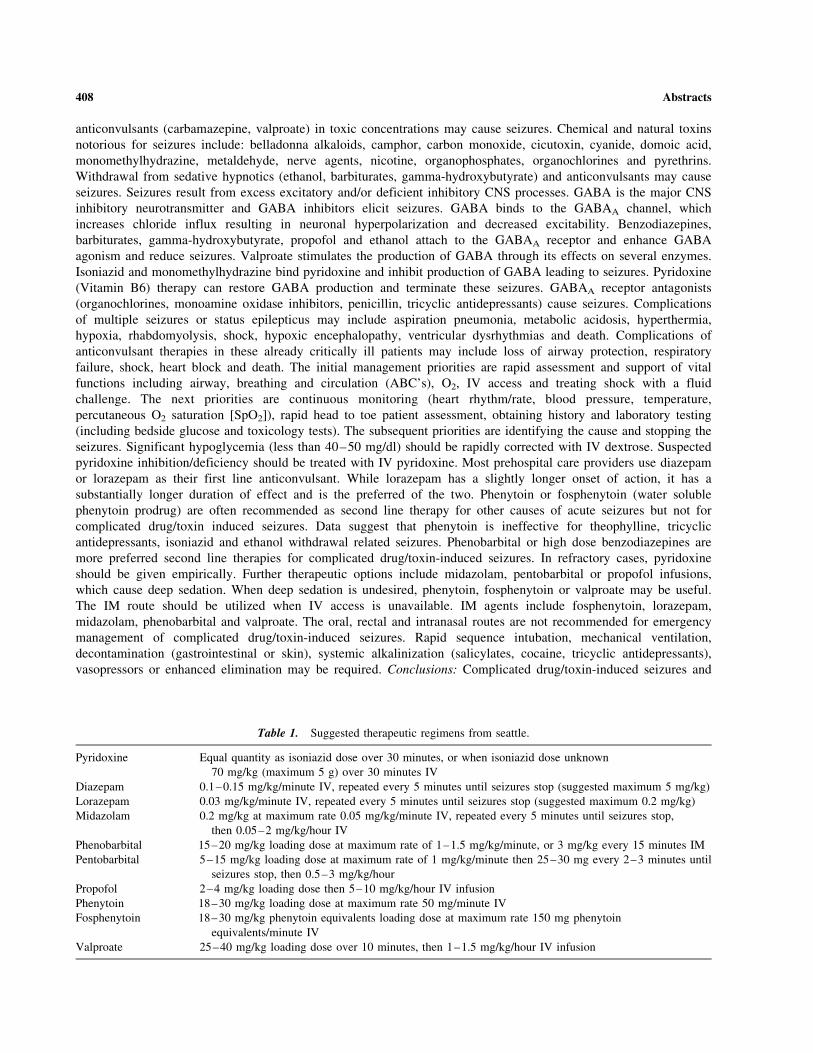
Table 1. Suggested therapeutic regimens from seattle.
Pyridoxine Equal quantity as isoniazid dose over 30 minutes, or when isoniazid dose unknown
70 mg/kg (maximum 5 g) over 30 minutes IV
Diazepam 0.1–0.15 mg/kg/minute IV, repeated every 5 minutes until seizures stop (suggested maximum 5 mg/kg)
Lorazepam 0.03 mg/kg/minute IV, repeated every 5 minutes until seizures stop (suggested maximum 0.2 mg/kg)
Midazolam 0.2 mg/kg at maximum rate 0.05 mg/kg/minute IV, repeated every 5 minutes until seizures stop,
then 0.05–2 mg/kg/hour IV
Phenobarbital 15–20 mg/kg loading dose at maximum rate of 1–1.5 mg/kg/minute, or 3 mg/kg every 15 minutes IM
Pentobarbital 5–15 mg/kg loading dose at maximum rate of 1 mg/kg/minute then 25–30 mg every 2–3 minutes until
seizures stop, then 0.5–3 mg/kg/hour
Propofol 2–4 mg/kg loading dose then 5–10 mg/kg/hour IV infusion
Phenytoin 18–30 mg/kg loading dose at maximum rate 50 mg/minute IV
Fosphenytoin 18–30 mg/kg phenytoin equivalents loading dose at maximum rate 150 mg phenytoin
equivalents/minute IV
Valproate 25–40 mg/kg loading dose over 10 minutes, then 1–1.5 mg/kg/hour IV infusion
408 Abstracts
their treatment may result in significant morbidity or mortality. There are practically no comparative data to guide
therapy in these cases (Table 1).
13. Is There a Reason to Use Physostigmine in Acute Poisoning?
Burkhart KK, O’Donnell SJ. PinnacleHealth Toxicology Center, Harrisburg, Pennsylvania, USA.
Objective: Review indications and contra-indications to the pre-hospital use of physostigmine in patients with
anticholinergic delirium and coma. Discussion: A Medline search did not discover any studies or reports of pre-
hospital physostigmine use. In fact the use of physostigmine in the hospital setting remains controversial to the
toxicology community. At annual meetings some toxicologists have spoken out against it ever being used because of
its potential for adverse outcomes (seizures, dysrhythmias) in the face of other alternatives (physical and chemical
restraint). In one of last year’s EAPCCT keynote addresses Hoffman concluded that for isolated anti-muscarinic
toxicity without contraindications the use of physostigmine is supported. That same conclusion should apply to the
prehospital setting. Patients with an anticholinergic delirium are safety threats to themselves and to the ambulance
crews whom transport them. Burns et al. have demonstrated that chemical restraints often require such doses that
oversedation, aspiration and possible intubation may be consequences. Physical restraints also place the patient at
risk for rhabdomyolysis. The ideal patient for pre-hospital physostigmine therefore is the patient with a single agent
anticholinergic ingestion with a threatening agitated delirium. The physical examination should include such findings
as mydriasis, dry skin and mouth, and possibly dysphasia. In this setting the patient should first receive a therapeutic
dose of a benzodiazepine, diazepam, midazolam or lorazepam. This benzodiazepine trial, theoretically, should
increase the margin of safety for the subsequent administration of physostigmine. Another benefit of physostigmine
may be the ability to safely perform gastrointestinal decontamination by having the patient drink activated charcoal
rather than inserting a nasogastric tube into an agitated patient. On the other hand, the use of physostigmine for the
comatose patient, as an analeptic, should mostly be reserved for special circumstances. The possibility of such co-
ingestants as sympathomimetic, serotonergic and sodium channel-blocking drugs create the risk for seizures and
dysrhythmias. One coma scenario that might warrant prehospital physostigmine is that of mass anticholinergic
poisoning. Atropine, scopolamine, 3-quinuclidinyl benzilate (BZ), and Jimsonweed could be possible scenarios. Pre-
hospital use of physostigmine might save resources, especially if intubation was being considered. Anecdotal
evidence also speaks to the slow administration of physostigmine to increase the margin of safety. Conclusions:
There are many reasons to use physostigmine in the pre-hospital setting. While the diagnostic use should mostly be
reserved for the hospital setting, the therapeutic use has the potential to make ambulance transport and
gastrointestinal decontamination safer for the patient and the crew. Research is needed to better characterize adverse
effects following physostigmine use, especially following the pre-administration of benzodiazepines. References:
Hoffman RS. Physostigmine: the pendulum swings. J Toxicol Clin Toxicol 2003; 41:411–412. Burns MJ, Linden
CH, Graudins A, et al. A comparison of physostigmine and benzodiazepines for the treatment of anticholinergic
poisoning. Ann Emerg Med 2000; 35:374–381.
14. Oxime Therapy and the Perihospital Situation—Which One and How Much?
Zilker Th. Klinikum r.d. Isar, Toxicological Department TU Munich, Germany.
Atropine is effective in patients with organophosphate (OP) intoxication but it is not sufficient to reverse all
symptoms. Atropine can antagonize the surplus of acetylcholine at most peripheral, and partially at central,
muscarinic receptors, but fails to block the nicotinic receptors which are responsible for the neurotransmission at the
neuromuscular junction. This is why patients with severe OP-poisoning have to be treated with intubation and
respiratory support, as the respiratory muscles are not functioning. Theoretically a reactivation of synaptic
acetylcholinesterase by oximes should overcome this neuromuscular block. In clinical practice this effect is not often
seen. There may be several reasons for this lack of efficacy. Firstly the dose of oxime may be wrong, secondly
Abstracts 409
reactivation is not adequate, thirdly no effect is observed due to the necessary sedation after intubation and the use
of diazepam for controlling convulsions. Two oximes are on the market and therefore easily available: 1. Pralidoxime
as Pralidoxime chloride (Protopan1), Pralidoxime methylsufonate (P2S1) and Pralidoxime methylsulfate (Contra-
thion1), 2. Obidoxime (Toxogonin1). Pralidoxime is used in the UK and the USA and most parts of the rest of the
world, whereas Obidoxime is used in Germany and some neighbouring countries. Obidoxime can reactivate the
phosphorylated AChE faster and at a lower dose than Pralidoxime. Obidoxime was blamed to be hepatotoxic though
this was only seen in overdosing it (9 g/d). The recommendation for dosing Pralidoxime is 1 to 2 g dissolved in
normal saline and infused over 30 to 120 min., with a second dose within one hour and further on redosing in 4–12
hours intervals. Obidoxime according to the manufacture’s proposition should be given in a dose of 0.25 g and
repeated several times. This recommended dosing is due to outdated studies and makes no sense because there are
troughs in the oxime plasma levels, where reactivation is near zero. Oximes can only work if the phosphorylated
acetylcholinesterase is not aged. Aging means that the phosphorylation can no longer be reversed. This happens if
one of the O-methyl or O-ethyl groups has come off. As a rule dimethyl OPs age fast, diethyl OPs age slowly. This
does not mean that dimethyl OPs are more toxic. The toxicity depends on the inhibitory activity which is reached at
much lower concentrations in some diethyl OPs and the velocity by which the enzyme is blocked. Newer studies
have shown clearly that Pralidoxime has to be given in a dose of 40 mg/kg b.w. followed by continuous infusion at
0.5 g/hour to keep up a steady-state concentration of some 80 mmol/l (14 mg/l) to achieve optimal reactivation.
Obidoxime has to be given in a dose of 0.25 g followed by an infusion of 0.03 g/h to keep up 10 mmol/l (4 mg/l).
Some examples about the different toxicity: Dimethoate (dimethyl) has a LD50 in rats of 500 mg/kg, Oxidemeton-
methyl (dimethyl-OP) a LD50 of 80 mg/kg whereas Parathion-ethyl (diethyl-OP) has an LD50 of 3 mg/kg. What
does this mean for treatment? Though the AChE can be reactivated for a longer time in diethyl-poisoning the patient
may die due to an extremely fast onset of the intoxication, followed by numerous complications. To judge for how
long it is possible to achieve reactivation AChE in a poisoned person it is important to know the half-lives of ageing
for the different OPs. On average the ageing half-lives of dimethyl OPs are 3.7 h, and of diethyl OPs 31 h. Whereas
the synaptic and erythrocyte-ChE can be reactivated by oximes, plasma ChE cannot. The determination of plasma-
ChE is not a useful measure of the effect of oxime therapy. What are the reasons for inadequate reactivation by
oxime therapy? The first reason may be that the oxime-therapy is given too late, the second is that the amount of
poison is too much and the third that the oxime is not specific enough. As far as OP-pesticide poisoning is concerned
Pralidoxime and Obidoxime are sufficiently specific whereas for nerve agents OP-poisoning HI6 and HLo7 are more
specific. HLo7 has a broad spectrum with a high efficacy against nerve agents along with high efficacy against the
OP-pesticides. HLo 7 would theoretically be the best if it were marketed. In suicidal poisonings the amount of
poison is often too much for oxime-reactivation. Even in diethyl-OP poisoning the reinhibition and ageing of the
enzyme can happen due to a high load of poison. Putting up the dosage in this situation makes no sense as oxime-
concentrations higher than 10�4 mol are not able to reactivate any more acetylcholinesterase. The other limiting
factor is ageing. It is about 3–12 hours before the enzyme is aged in dimethyl OP-poisoning. In diethyl OP-
poisoning, as long as the poison load is not too high, reactivation is possible because the patient will be treated
within a reasonable time (<30 h). There is another problem to be discussed: During the reactivation of AChE by
oximes phosphoryloximes (POXs) are produced. These compounds can inhibit the AChE in its own right. This has
brought about the idea that oximes my be harmful by enhancing the toxicity. This is probably not a real problem as
only small amounts of POXs are created during oxime-therapy, and as the degradation of POXs is very fast. Since
oximes are not able to penetrate the blood-brain-barrier easily little is known about the beneficial effects on CNS-
symptoms. It is unlikely that convulsions can be controlled by oximes, and this means that sedation and ventilation
cannot be avoided in severe cases. What can be concluded for the perihospital situation? Though not proven by
clinical trials it seems sensible to use oximes in the pre-hospital treatment of OP-poisoning, as done in Germany by
the emergency doctor who carries with him a Tox-Box with a special OP-treatment kit that includes atropine,
obidoxime and diazepam. In other countries where paramedics go to the scene oximes should be used immediately
after hospital admission. It is not necessary in this situation to know which organophosphate is responsible for the
intoxication. In case of a poisoning with a OP that is ageing fast, no time has to be lost. Even in carbamate
poisoning oximes don’t do any harm, with the exception of poisoning by the carbamate Carbaryl. After an initial
bolus of the oxime a continuous infusion of it should be administered as described above. Especially in cases where
the intoxication is not severe and tachycardia is prominent oximes could be used even without atropine. Possibly in
mild or in the early stage of the intoxication even CNS and respiratory impairment might be reversed. In severe
intoxications oximes may shorten the duration of the intoxication and allow a lower dose of atropine but are most
410 Abstracts
likely not life saving. As most of the intoxications we see in Europe are very severe due to the suicidal intent,
oximes are of limited use. In all such cases full intensive care treatment must be started on the scene and after
hospital admission. The determination of the red cell-AChE can be used to monitor the effect of oxime-treatment.
Repetitive nerve stimulation with measuring the compound muscle action potential (CMAP) can show if the neuro
muscular endplate is reactivated. Both methods are usually not available in ordinary hospitals therefore the
determination of PChE may be the only guide for treatment. The PChE-level does not correlate with severity but if
PChE is low it proves OP/Carbamate-poisoning. As soon as PChE is increasing there is no more poison in the
system and oxime and atropine treatment can be terminated. Conclusion: Oximes don’t do much harm and may be
of some use.
15. The Use of Antidotes in the Emergency Treatment of Cyanide Poisoning
Thompson JP. Therapeutics and Toxicology Centre, University of Wales College of Medicine, Cardiff, UK.
The deliberate release of hydrogen cyanide would result in poisoning primarily by the inhalational route, producing
symptoms rapidly. In severe cases early death may occur. For this reason, in cases of severe poisoning, life saving
treatment with antidotes must be undertaken rapidly if lives are to be saved. Conversely, if casualties are still alive
after removal from a purely inhalational source of poisoning, they are likely to survive with, or without, antidotal
treatment, albeit that recovery may be faster if an antidote were to be administered. An ideal cyanide antidote for
pre-hospital use must therefore be available rapidly to be effective. It should be easy to administer by responders
with limited training and it should be effective, non toxic and should not adversely interact with other antidotes or
poisons. Cyanide antidotes act by altering the toxicokinetics of cyanide poisoning, altering the distribution,
metabolism or excretion of cyanide. They result in the more rapid removal of cyanide from the cytochrome a3
complex and the return of oxidative metabolism. They may be considered as those which bind to cyanide either
directly, or indirectly by the formation of methaemoglobin, and those which enhance its metabolism by the acting as
sulphur donors. Methaemoglobin Formers: Cyanides have a higher binding affinity for methaemoglobin than for
cytochrome oxidases resulting in the formation of cyanmethaemoglobin. Provision of methaemoglobin therefore
results in the release of cyanide from the inhibited enzyme complex freeing the inhibited enzyme to perform its
respiratory function. Cyanmethaemoglobin releases cyanide slowly which is subsequently detoxified by endogenous
rhodanese producing thiocyanate which is excreted in the urine. Methaemoglobin formers include sodium nitrite,
amyl nitrite and 4-dimethylaminophenol (DMAP). Amyl nitrite has the attraction of being able to be administered by
inhalation. However, conventional administration by this route produces only low concentrations of methaemoglobin.
Methaemoglobin formers reduce the oxygen carrying capacity of the blood and therefore may worsen poisoning,
particularly if coexistent carbon monoxide poisoning is present. The optimum therapeutic concentration of
methaemoglobin is uncertain, and biochemical response to therapy difficult to monitor. Cobalt Compounds: Cyanide
forms stable complexes with many transition metals including cobalt. Unfortunately many inorganic cobalt salts are
toxic. Dicobalt edetate (a cobalt chelate) binds with cyanide to form stable complexes (cobaltocyanide and
cobalticyanide). However, commercially available dicobalt edetate chelates also contain a proportion of free cobalt.
Free cobalt has a higher cyanide-binding affinity than dicobalt edetate and although this may contribute to the
antidote’s effectiveness it also contributes to its toxicity, particularly if cyanide poisoning is not actually present.
Hydroxocobalamin (Vitamin B12B) detoxifies cyanide by giving up its hydroxyl group and binding a cyanyl group,
forming cyanocobalamin (Vitamin B12). Cyanocobalamin is subsequently excreted in the urine or releases the
cyanide which is subsequently detoxified by rhodanese. The reaction requires one molecule of hydroxocobalamin for
each molecule of cyanide detoxified and therefore large quantities may be required in cases of severe poisoning.
Sulphur Donors: Cyanide is detoxified by the enzyme rhodanese. In cyanide poisoning the rate limiting step is due
to the availability of sulphur donors. Sodium thiosulfate provides an exogenous source of sulphane sulphur,
enhancing the production of thiocyanate.
Na2S2O3 þ CN�!rhodaneseSCN� þ Na2SO3
Abstracts 411
Sodium thiosulphate hastens the elimination of cyanide but its use alone in life-threatening inhalational poisoning
would not be sufficiently rapid to be effective. Conclusion: Antidotes can not be considered in isolation in the
treatment of cyanide poisoning. However, to be effective an efficient, rapid means of delivery and administration
must be available to enable their use outside the hospital environment. Without prior warning of release, the
effective use of cyanide antidotes for mass casualties will prove difficult and current antidotes all have limitations to
their use. References: Cobalt Compounds as Antidotes for Hydrocyanic Acid. Lovatt Evans C. British Journal of
Pharmacology 1964; 23:455–475. Methaemoglobin levels following Inhalation of amyl Nitrite. Galdun JP, Weiss
LD, Paris PM, Kaplan RM, Stewart RD. Annals of Emergency Medicine 1987; 484:157. Relation of blood cyanide to
plasma cyanocobalamin concentration after a fixed dose of hydroxocobalamin in cyanide poisoning. Houete P,
Hoffman JR, Imbert M, Levillain P, Baud FJ. Lancet 1995; 346:605–608.
16. Changes in the Approaches to Drug Elimination in Poisoning over the Last 40 Years
Jaeger A. Service de Reanimation Medicale, Hopital de Hautepierre, Strasbourg, France.
Introduction: The use of haemodialysis in treating poisoned patients was first reported by Schreiner et al in 1955 in
a patient with aspirin poisoning (1). Following this report, haemodialysis (HD) gained acceptance in the 1960s and
early 1970s and poisonings by many toxic agents (up to 141) were considered potentially treatable by dialysis (2,3).
During the last 40 years, several other techniques for enhancing drug elimination have been proposed: forced
alkaline diuresis (FAD), peritoneal dialysis (PD), exchange blood transfusion (EBT), plasma exchange (PE), and,
more recently, continuous arteriovenous or venovenous haemodiafiltration (CAVHDF, CVVHDF), and molecular
absorbents recirculating system (MARS). Each of these techniques, by turns, gave rise to enthusiasm (e.g.,
haemoperfusion in the 70s) and numerous reports of their apparent values have been published. However, most of
these reports suffered of a lack of evaluation, especially without toxicokinetic and/or toxicodynamic data supporting
the efficacy of these treatments. In fact, a scientific evaluation of these techniques has to take in account many
parameters and aspects. Over the last 40 years, different approaches with their respective pitfalls, have been
proposed for assessing the indications of elimination techniques. The Technical Approach: It is based on the
principles and technical aspects of the procedure which include the following parameters: diffusion, filtration,
adsorption, blood flow rate, dialysate flow rate, dialyser surface area, pore structure of the chosen membrane,
adsorptive capacity of HP cartridges. Dependent on the different techniques, drugs and chemicals must meet given
criteria in order to reach a high extraction ratio: small molecular size, high water solubility, low protein binding for
HD and HDF, lipid solubility, adsorption coefficient for HP, albumine binding for MARS. According to these
parameters and to in vitro studies, lists of drugs which may be extracted by the different techniques have been
established and are still published. However, such lists include for instance drugs and chemicals such as carbon
monoxide, amanitin, snake bite, cardiovascular drugs, tranquilizers antidepressants (4)! The Toxicokinetic Approach:
The amount of the poison which may be removed depends on the plasma concentration, on the clearance and on the
time of the procedure. Only compounds with a small volume of distribution (<1 L/kg) and an extraction ratio
exceeding endogenous elimination will be removed significantly. Therefore, the kinetic efficacy can be based neither
on the decrease of plasma concentrations, nor on the comparison of the procedure clearance with the renal clearance,
nor on the decrease of the plasma half-life (criteria mostly used in the 70s and 80s for efficacy assessment), but only
on the comparison of the procedure clearance with the spontaneous clearance and on the amount of drug in fact
eliminated (5). Moreover, this amount should be compared with the body burden or with the amount of drug
ingested (6). The extracorporeal removal is also dependent on the rate of distribution of the drug from the tissues
into the blood. For compounds, such as lithium, with a slow rate of diffusion, the equilibrium between plasma and
intracellular concentrations will only be achieved after a delay which explains the rebound in plasma concentration
observed after the procedure. Taking in account these criteria, the number of poisonings which may benefit from
elimination techniques is small and most of the indications proposed in the 70s and 80s do not meet these kinetic
criteria. Moreover, the level of kinetic changes induced by the elimination technique which may have an effect on
the evolution of the poisoning is not established for most of the poisons. Kinetic data must be interpreted critically,
the major objective being the decrease of the poison concentration at the target organ and not the changes of some
kinetic parameters. For instance, the hypothesis that the rebound in serum lithium concentration may give rise to
412 Abstracts
recurrence of the effects of poisoning is a misinterpretation of lithium kinetic (6). The Toxicodynamic Approach: The
toxicodynamic efficacy can be estimated by the decrease of severity and/or the duration of the poisoning or by the
prevention of a severe toxicity. Therefore, an enhanced elimination will only be effective if the severity of the
poisoning is related to the drug (and/or active metabolites) plasma concentration or body burden, or if the prognosis
depends on the elimination of the drug from the target organ. In fact, only few poisonings meet these requirements.
For functional poisons, such as phenobarbital, lithium and theophylline, for instance, the increase of the elimination
may decrease the duration of the poisoning. For lesional poisons, an increase in elimination will have an effect on
the severity of the poisoning only if the procedure is performed before the poison has reached the target organs and
has induced tissue damages (5). In paraquat poisoning, haemodialysis and haemoperfusion increase substantially the
elimination but they have no effect on the dynamic or prognosis of the poisoning because paraquat is rapidly
absorbed and induces pulmonary damages before the techniques can be performed (7). The Need for a Toxicokinetic-
Toxicodynamic Approach: In the past, many reports of successful use of elimination techniques were only based
either on kinetic or dynamic criteria. Recommendations for the use of the elimination techniques were made
according to a decrease of plasma concentrations, an increase of drug elimination, a clinical improvement or the
recovery of the patient. For instance, elimination techniques were proposed in amatoxins poisoning without knowing
the kinetic of amatoxins in human poisonings. Haemodialysis was proposed in metal poisonings because in some
cases it decreased the plasma concentrations. The lack to take in account the kinetic-dynamic efficacy was (and is
still) the source of wrong interpretation of the efficacy of the elimination techniques. For instance, many drugs are
still listed in recent reports as being removed by HD and HP despite the absence of scientific data demonstrating that
the evolution and the prognosis is improved by these techniques (4). Most poisoned patients recover with supportive
treatment and the overall mortality from acute poisoning is now less that 1%. However, for some drugs the
morbidity and mortality remains high and a small group of patients may benefit from elimination techniques (8–11).
In fact, only a few number of drugs fulfil the kinetic and dynamic requirements for an indication of the elimination
techniques. Haemodialysis may be indicated in methanol, ethylene glycol, lithium and salicylates poisonings.
Haemoperfusion increases the elimination of phenobarbital, theophylline and carbamazepine but multi-dose activated
charcoal has the same kinetic efficacy. The Need for an Evaluation of the Efficiency: Does the elimination technique
compare favourably, in terms of morbidity, mortality, cost/benefit ratio, to other alternatives? Which poisoned
patient will effectively benefit from an elimination technique? Even if a drug fulfils the kinetic and dynamic criteria
for the efficacy of a technique, the indications are not clearly established. For instance, the number of lithium
poisoned patients which should be treated by HD varies by a factor of 10 according to recommendations of the
different authors (6). Moreover, recent reports suggest that CAVHDF or CVVHD should be preferred to HD but in
this study no selection of the patients who really needed treatment by an elimination technique was made (12). In
ethylene glycol and methanol poisonings, the efficacy of HD has to be compared to the antidotal treatment with 4
methyl-pyrazole. Although, most ethylene glycol and methanol poisonings may be treated only by 4-MP, some
patients may benefit from HD which eliminates also the active metabolites. A recent meta trial (ethylene glycol) and
a meta study (methanol) identified the group of patients who should be treated by HD (13,14). Guidelines on
treatment of methanol poisoning have also been published by the AACT (15). Concerning the use of multi-dose
activated charcoal and alkaline diuresis, position statements have been established by the AACT and the EAPCCT
(16,17). Recently, the MARS has been proposed for the elimination of protein-bound drugs. Such a proposition may
be misleading concerning the indications of this technique because, according to kinetic and dynamic criteria, only a
small number of drug poisonings may effectively benefit from this treatment. For instance, what might be the
usefulness of MARS in midazolam poisoning (18)? MARS may be indicated in some rare poisonings such as
phenytoin, but its efficiency remains to be established (19). Conclusion: Over the last 40 years the use of elimination
techniques was singled out by phases of enthusiasms and disappointments due to a lack of strategy for the evaluation
of their efficacy. The indications of the techniques should be evaluated critically according to kinetic and dynamic
criteria. This evidence based strategy may avoid many errors made in the past and should be applied systematically
before a new technique is recommended. Reporting an inefficacy of a treatment is as important and useful as
reporting successes. It is one of the duties of scientific societies to establish guidelines and position statements with
an extensive critical review of the literature in order to clarify the indications of the elimination techniques.
References. 1. Schreiner GE, Berman LB, Griffin J, et al. Specific therapy for salicylism. N Engl J Med 1955;
253:213–217. 2. Winchester JF, Gelfand MC, Knepshield JH, et al. Dialysis and hemoperfusion of poisons and
drugs. Update. Trans Am Soc Artifial Organs 1977; 23:762–852. 3. Schreiner GE. Perspectives on the
hemoperfusion of drugs and toxins. Biomat Art Cells 1987; 15:305–321. 4. Winchester JF, Salsberg JA. Drugs
Abstracts 413
removed by dialysis and hemoperfusion; updated tables. http://www;arrtjournal.org. 5. Jaeger A, Sauder P,
Kopferschmitt J, et al. Toxicokinetics in clinical toxicology. Acta Clin Belg 1990; 45:1–12. 6. Jaeger A, Sauder P,
Kopferschmitt J, et al. When should dialysis be performed in lithium poisoning? A kinetic study in 14 cases of
lithium poisoning. Clin Toxicol 1993; 31:429–447. 7. Hampson E. Failure of haemodialysis and haemoperfusion to
prevent death in paraquat poisoning. A retrospective review of 42 patients. Med Toxicol 1988; 3:64–71. 8. Vale A,
Meredith T, Buckley B. Br J Med 1984; 289:366–269. 9. Goldberg MJ, Spector R, Park GD, et al. An approach to
the management of the poisoned patient. Arch Intern Med 1986; 146:1381–1385. 10. Garella S. Extracorporeal
techniques in the treatment of exogenous intoxications. Kidney Intern 1988; 33:735–754. 11. Pond SM. Extra-
corporeal techniques in the treatment of poisoned patients. Med J Aus 1991; 154:617–622. 12. Leblanc M, Raymond
M, Bonnardeaux A, et al. Lithium poisoning treated by high performance continuous arteriovenous and venovenous
hemodiafiltration. Am J Kid Dis 1996; 27:365–372. 13. Brent J, McMartin K, Phillips S, et al. Fomepizole for the
treatment of ethylene glycol poisoning. N Engl J Med 1999; 340:832–838. 14. Brent J, McMartin K, Phillips S, et al.
Fomepizole for the treatment of methanol poisoning. N Engl J Med 2001; 344:424–429. 15. Barceloux DG, Bond
GR, Krenzelok EP, et al. American Academy of Clinical Toxicology practice guidelines on the treatment of
methanol poisoning. Clin Toxicol 2002; 40:415–446. 16. Vale JA, Krenzelok EP, Barceloux GD. Position statement
and practice guidelines on the use of multi-dose activated charcoal in the treatment of acute poisoning. Clin Toxicol
1999; 37:731–751. 17. Proudfoot AT, Krenzelok EP, Vale JA. Position paper on urine alkalinization. Clin Toxicol
2004; 42:1–26. 18. Sen S, Ytebe LM, Rose C, et al. Albumin dialysis: a new therapeutic strategy for intoxication
from protein-bound drugs. Intensive Care Med 2004; 30:496–501. 19. Sen S, Ratnaraj N, Davies NA, et al.
Treatment of phenytoin toxicity by the molecular adsorbents recirculating system (MARS). Epilepsia 2003; 44:
265–267.
17. Corrosive Ingestion: The Evidence Base. Are Steroids Still Indicated in Second and Third-DegreeCorrosive Burns of the Oesophagus?
Pelclova D, Navratil T. Department of Occupational Medicine with Poisons Information Centre, Charles University,
Prague, Czech Republic.
Objective: The ingestion of caustic substances frequently poses problems with both diagnosis and treatment. Stricture
formation is the most important complication of corrosive damage to the oesophagus. Use of steroids in corrosive
injury in man in second and third-degree corrosive burns to prevent stenosis is a controversial subject. The two most
frequently cited studies (1,6) have discordant results. The aim of this study was to compare more recent data from
the literature and evaluate the usefulness of steroids treatment. Methods: Human reports of therapy for caustic
oesophageal injury published between 1991 and 2003 were reviewed. Medline database and Scirus were used to
search for citations (in English, German, French, and Spanish language) concerning caustic injuries of the
oesophagus. Additional citations based on the reference sections were found. The time interval was selected to
continue a complex review by Howell et al. (6) from the years 1956–1991, only one study in German from 1989
was added. Inclusion criteria were: 1) oesophageal injury documented by endoscopy and patient management either
with no steroid or with at least 10 days course of steroids. The data were evaluated with the Pearson chi square test
and alpha was set at 0.01. Results: Only six studies with total 211 patients fulfilled our criteria; five were
retrospective and one prospective (2). Four studies used steroids in the treatment of second and third-degree burns;
two studies did not use steroids. Details concerning numbers of patients with second and third-degree corrosive
burns, and the number of strictures are given in Table 1. Two studies used only prednisolone 2 mg/kg/day (7,8); two
other studies (2,4) compared the effect of prednisolone (2 mg/kg/day) and dexamethasone (1 mg/kg/day) treatment.
Data on second and third-degree burns were amalgamated in one study that did not describe these burns separately
(7). The effects of prednisolone and dexamethasone on second and third-degree burns separately were compared
based on three studies (2,4,8). In these studies 18.4% and 7.1% of patients treated respectively with prednisolone and
dexamethasone for second-degree burns developed strictures. The difference was not statistically significant. Third-
degree injury resulted in strictures in 100.0% and 69.2%, of patients treated with prednisolone and dexamethasone
respectively. The difference was not statistically significant. Two studies not using steroids, with total 58 patients,
met our criteria (3,5); details are given in Table 1. In second-degree burns, the difference between the steroids
414 Abstracts
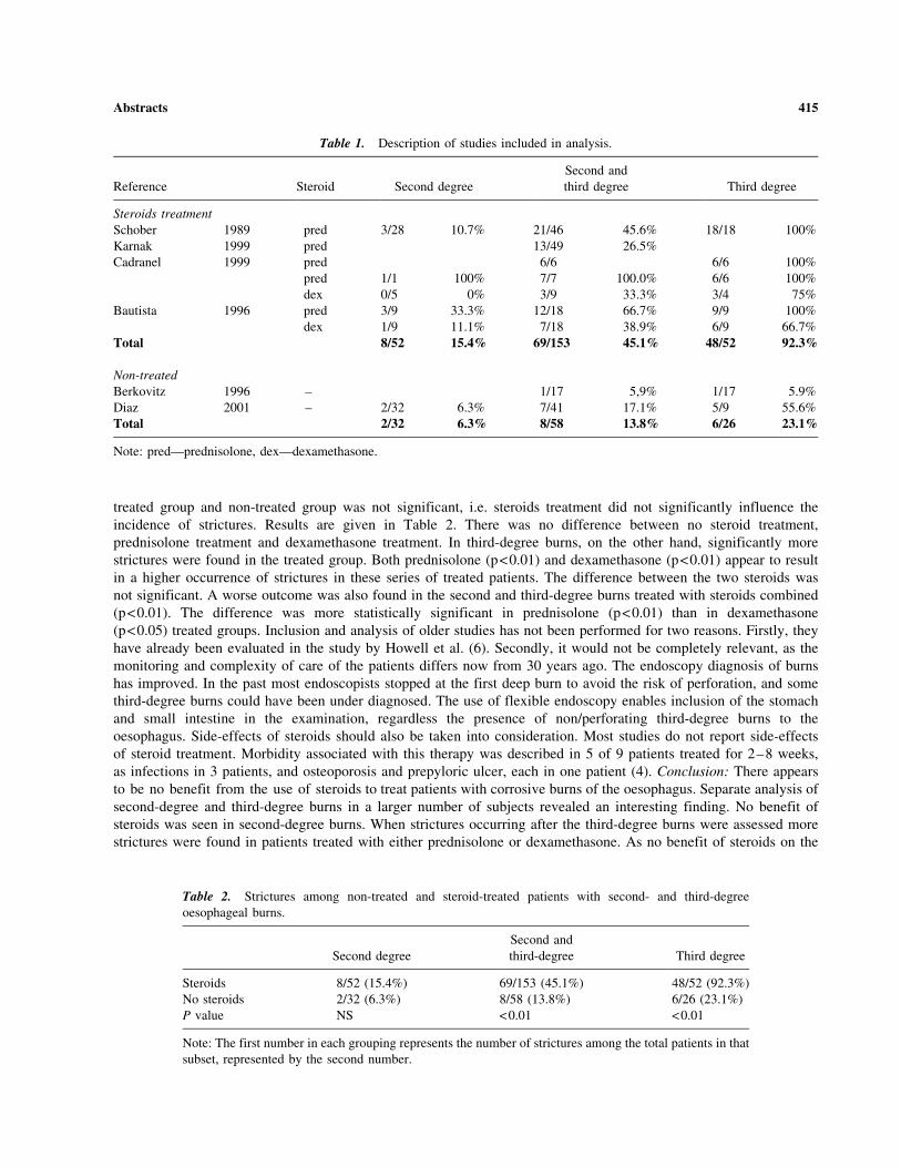
treated group and non-treated group was not significant, i.e. steroids treatment did not significantly influence the
incidence of strictures. Results are given in Table 2. There was no difference between no steroid treatment,
prednisolone treatment and dexamethasone treatment. In third-degree burns, on the other hand, significantly more
strictures were found in the treated group. Both prednisolone (p<0.01) and dexamethasone (p<0.01) appear to result
in a higher occurrence of strictures in these series of treated patients. The difference between the two steroids was
not significant. A worse outcome was also found in the second and third-degree burns treated with steroids combined
(p<0.01). The difference was more statistically significant in prednisolone (p<0.01) than in dexamethasone
(p<0.05) treated groups. Inclusion and analysis of older studies has not been performed for two reasons. Firstly, they
have already been evaluated in the study by Howell et al. (6). Secondly, it would not be completely relevant, as the
monitoring and complexity of care of the patients differs now from 30 years ago. The endoscopy diagnosis of burns
has improved. In the past most endoscopists stopped at the first deep burn to avoid the risk of perforation, and some
third-degree burns could have been under diagnosed. The use of flexible endoscopy enables inclusion of the stomach
and small intestine in the examination, regardless the presence of non/perforating third-degree burns to the
oesophagus. Side-effects of steroids should also be taken into consideration. Most studies do not report side-effects
of steroid treatment. Morbidity associated with this therapy was described in 5 of 9 patients treated for 2–8 weeks,
as infections in 3 patients, and osteoporosis and prepyloric ulcer, each in one patient (4). Conclusion: There appears
to be no benefit from the use of steroids to treat patients with corrosive burns of the oesophagus. Separate analysis of
second-degree and third-degree burns in a larger number of subjects revealed an interesting finding. No benefit of
steroids was seen in second-degree burns. When strictures occurring after the third-degree burns were assessed more
strictures were found in patients treated with either prednisolone or dexamethasone. As no benefit of steroids on the
Table 1. Description of studies included in analysis.
Reference Steroid Second degree
Second and
third degree Third degree
Steroids treatment
Schober 1989 pred 3/28 10.7% 21/46 45.6% 18/18 100%
Karnak 1999 pred 13/49 26.5%
Cadranel 1999 pred 6/6 6/6 100%
pred 1/1 100% 7/7 100.0% 6/6 100%
dex 0/5 0% 3/9 33.3% 3/4 75%
Bautista 1996 pred 3/9 33.3% 12/18 66.7% 9/9 100%
dex 1/9 11.1% 7/18 38.9% 6/9 66.7%
Total 8/52 15.4% 69/153 45.1% 48/52 92.3%
Non-treated
Berkovitz 1996 – 1/17 5,9% 1/17 5.9%
Diaz 2001 – 2/32 6.3% 7/41 17.1% 5/9 55.6%
Total 2/32 6.3% 8/58 13.8% 6/26 23.1%
Note: pred—prednisolone, dex—dexamethasone.
Table 2. Strictures among non-treated and steroid-treated patients with second- and third-degree
oesophageal burns.
Second degree
Second and
third-degree Third degree
Steroids 8/52 (15.4%) 69/153 (45.1%) 48/52 (92.3%)
No steroids 2/32 (6.3%) 8/58 (13.8%) 6/26 (23.1%)
P value NS <0.01 <0.01
Note: The first number in each grouping represents the number of strictures among the total patients in that
subset, represented by the second number.
Abstracts 415
prevention of strictures of the oesophagus was found in this study, systemic steroids treatment should not be used in
corrosives ingestion. References: 1. Anderson KD. A controlled trial of corticosteroids in children with corrosive injury
of the esophagus. N Engl J Med 1990; 323:637–640. 2. Bautista A, Varela R, Villanueva A, Estevez E, Tojo R, Cadranel
S. Effects of prednisolone and dexamethasone in children with alkali burns of the oesophagus. J Pediatr Surg 1996;
6:198–203. 3. Berkovits RN, Bos CE, Wijburg FA, Holzki J. Caustic injury of the oesophagus. Sixteen years experience
and introduction of a new model oesophageal stent. J Laryngol Otol 1996; 110:1041–1045. 4. Cadranel S, Scaillon M,
Goyens P, Rodesch P. Treatment of esophageal caustic injuries: experience with high-dose dexamethasone. Pediatr Surg
Int 1993; 8:97–102. 5. Dıaz EG, Fernandez MC, Gomez MR, Higuero LC. Upper gastrointestinal tract injury by
ingestion of caustic substances (in Spanish). Gastroenterol Hepatol 2001; 24:191–195. 6. Howell JM, Dalsey WC,
Hartsell FW, Butzin CA. Steroids for the treatment of corrosive esophageal injury. Am J Emerg Med 1992; 10:421–425.
7. Karnak I, Tanyel FC, Buyukpamukcu N, Hicsonmez A. Combined use of steroid, antibiotics and early bougienage
against stricture formation following caustic esophageal burns. J Cardiovasc Sur 1999; 40:307–310. 8. Schober PH,
Sauer H, Hollwarth ME, Kerbler S, Lackner H. Ingestion of corrosives in children (in German). Wiener Klein
Wochenschrift 1989; 28:318–322. Acknowledgement: The work was supported by MSM J13/98 111100002
and 111100005.
18. The Ingestion of Corrosive Substances. The Point of View of the Surgeon
Meyer Ch (1), Brigand C (1), Reche F (1), Lutun P (2), Jaeger A (2). 1. Service de Chirurgie Generale et Digestive;
2. Service de Reanimation Medicale, Hopital de Hautepierre, Strasbourg, France.
Introduction: The ingestion of corrosive substances, rightly considered as serious, usually occurs accidentally in
children and, mostly, in an intentional way in adults. Three types of substances may have corrosive effects: acids,
alkali and oxidizers. Each of these substances can exist in a solid or liquid form. In industrial countries, ingestions of
corrosive substances are relatively frequent with, for instance, an incidence of 15,000 cases/year in France. In
children, the substances involved are mostly alkali in crystalline form, such as washing powder, and usually the
amount ingested is small. This explains why, in such cases, mortality is almost nil. In adults, the average age of
victims is 40 years, and the ingestion of acids usually in liquid form are more frequent. However, in serious cases
needing surgical treatment, alkali are more often involved than acids because of the deeper, more intense irritant
effects. Location and Stages of Injury: The severity of the injuries depends on several factors: the type, the form and
the concentration of the product, the amount ingested and the duration of contact with the digestive mucous
membrane. The oeso-gastric damage can be localized or diffuse and sometimes extends to the pylorus to affect the
proximal small bowel. The corrosive-induced injuries evolve in 4 successive phases: an initial phase (Day 1) caused
by intense vasculo-blood phenomena (oedema, ulceration, perforation, acidosis, haemolysis); a deterging phase (Day
2 to Day 8) where the vasculo-blood phenomena are major (bleeding, perforation); a repair phase (Day 8 to Day 30)
characterized by the proliferation of fibroblastic granular tissues (leakage and infection); a healing phase (>Day 30)
during which the fibrosis retraction at the origin of stenosis is the dominant feature. Initial Management: The
immediate treatment, depending on the degree of severity may include washing of the mouth, oxygen in case of
respiratory disturbances, intravenous lines and vascular filling in case of circulatory compromise. The patient should
be transferred to an hospital possessing intensive care, digestive surgery, digestive and bronchial endoscopy
facilities. The oeso-gastro-duodenal endoscopy is the most important procedure for the assessment of the severity of
the injury and must be carried out as soon as possible within the first 24 hours. The classification of the severity of
the injuries, which goes from oedema through ulcerations up to necrosis, is of major importance for treatment
strategy. While 2/3 of patients have small initial injuries (stage 1 superficial) not requiring hospitalization, the other
patients with more serious injuries (stages 2 transmucosal and stage 3 transmural) require specific measures:
intensive care with parenteral nutrition and, in some cases, surgical procedures, either as a matter of emergency or at
a later date. In patients with respiratory disturbances, a tracheo-bronchial endoscopy may be indicated for the
evaluation of airway injuries. Early Surgical Treatment: The criteria of a surgical operation as a matter of
emergency are: 1) perforation of the oesophagus, which is, in fact, very rare, or more often gastric perforation,
2) serious cases without perforation (mostly due to the massive ingestion of an acid or a strong alkali in excess of
150 ml) but complicated by shock, coagulation disturbances (DIVC, fibrinolysis), acidosis, 3) diffuse oeso-gastric
416 Abstracts
necrosis on endoscopy. The prevention of digestive perforation is based on the excision of the oesophagus and the
stomach. The surgical technique most often used is ‘‘stripping’’ of the oesophagus without thoracotomy, followed by
a cervical oesophagostomy and jejunostomy for feeding. The reconstruction is carried out after a delay of four
months by an oesophago-coloplasty procedure. In those cases having an exclusive gastric injury, total gastrectomy is
sufficient and the oeso-jejunal reconstruction can be immediate. Wider excision, including the gall-bladder, a part of
the small bowel, the spleen, the tail of the pancreas, even the duodenum and the head of the pancreas, may be
necessary and is sometimes life saving. The operational mortality is of the order of 25% when the intervention is
carried out within the first 12 hours but it may exceed 80% if the operation is delayed. In patients which needed
intubation and mechanical ventilation, early tracheostomy is also indicated. Delayed Surgical Treatment: Deep
burning with tissular necrosis results in an irreversible production of scar tissue entailing the constitution of a
stenosis which may require a surgical treatment, if endoscopic dilation is not successful. This can be proposed at
once in case of long and severe stenosis, but generally the operation is made between the 4th and 6th month. At this
point, it is a question of carrying out an oesophagoplasty without opening the thorax, and using the stomach, if it is
undamaged, or the colon placed in position retro-sternal while leaving in place the scarred oesophagus. Indeed,
complications inherent to a excision of the oesophagus by thoracotomy are higher than the risk of cancerous
degeneration of the damaged oesophagus. Conclusion: Injuries of the digestive tract following ingestion of corrosive
substances may require early or delayed surgical treatment. Management of severe injuries need the cooperation
between different medical teams: intensive care, digestive endoscopy and digestive surgery. In these severe cases,
surgical treatment should be carried out before irreversible systemic complications had occurred. References:
Christesen HB. Caustic ingestion in adults. Epidemiology and prevention. J Toxicol Clin Toxicol 1994; 32:557–568.
Hugh TB, Kelly M. Corrosive ingestion and the surgeon. J Am Coll Surg 1999; 189:508–522. Sarfati E, Gossot D,
Assens P, Celerier M. Management of caustic ingestion in adults. Br J Surg 1987; 74:146–148. Zargar SA, Kochhar
R, Mehta S, Mehta SK. The role of fiberoptic endoscopy in the management of corrosive ingestion and modified
endoscopic classification of burns. Gastrointest Endosc 1991; 37:165–169.
19. Steroid Treatment of Corrosive Injury
Jovic-Stosic J, Todorovic V, Doder R. National Poison Control Centre, Belgrade, Serbia.
Objective: The use of steroids to prevent strictures formation after corrosive substance ingestion remains controversial.
To evaluate the role of steroid therapy, a three year prospective study was done. Methods: We analyzed the effects of 3
most frequently ingested liquids by our patients: households containing sodium hydroxide (NaOH) or hydrochloric acid
(HCl) and food preservative containing acetic acid (CH3COOH). Corrosive burns, depending on the depth and the
extent, were graded as I, IIa, IIb, IIIa and IIIb, and depending on the localization as esophageal and gastric. The
criterion for inclusion into the study was the presence of at least IIa burns proved by emergency endoscopy. The group
treated by steroids (ST) received methylprednisolone 3 mg/kg and the dose was tapered over at least 3 weeks. The
antibiotics were given concomitantly. The patients not treated by steroids (NT) received antibiotics only in case of
documented infection. The groups didn’t significantly differ by age, sex, type of ingested agent and severity of burns.
Steroid treatment was initiated in 52 patients, but because of lethal outcome or treatment cessation in case of suspected
perforation and gastrointestinal hemorrhage, full 3-week course received 39 patients. NT group contained 54 patients.
Results: Mortality was almost equal among the groups: 21% in ST group and 22% in NT group. Survivors of ST group
in 9 cases (30%) developed esophageal stenosis and in 5 cases (15%) developed antropyloric stenosis. The incidence of
esophageal stenosis (21%) and antropyloric stenosis (9.5%) in NT group was lesser, though not significantly (p>0.05).
In ST group 4 cases of gastric perforation and 1 case of esophageal perforation were proved by autopsy. In NT group
autopsy revealed 3 cases of gastric perforation and 1 case of esophago-tracheal fistula. Gastric perforations occurred
only after HCl ingestion. Esophageal perforation and fistula were caused by NaOH ingestion. Analysis of corrosive
effects caused by considered agents did not revealed significant differences of the depth, extent and localization of
burns detected by emergency endoscopy. However, regardless the treatment, after NaOH ingestion esophageal stenosis
developed more frequently (p<0.05) than gastric. In case of HCl ingestion antropyloric stenosis and gastric perforation
occurred more frequently than esophageal (p<0.05). Incidence of esophageal and antropyloric stenosis did not
significantly differ in case of CH3COOH ingestion. The most frequent infective complication was pneumonia, with
Abstracts 417
similar incidence in both groups (13% in ST group and 16% in NT group). Sepsis developed in single cases in both
groups. Conclusion: The study failed to prove that applied steroid treatment could prevent stenosis development. The
incidence of lethal outcome, gastrointestinal hemorrhage and perforation and infective complications did not increase
among steroid-treated patients.
20. Delayed Toxic Effects of Sulphur Mustard in Iranian Veterans
Balali-Mood M, Hefazi M, Mahmoudi M. Mashhad University of Medical Sciences, Iran.
Objective: Sulphur mustard (SM) is a chemical warfare alkylating agent that may cause delayed toxic effects on
different organs of the body years after exposure. It was thus aimed to study the delayed toxic effects of SM in
Iranian veterans. Methods: All SM intoxicated veterans who had disability more than 40% were studied after
informed consents. Clinical and para-clinical investigations including haematological, biochemical and electrophys-
iological tests, as well as spirometry, chest x-ray, HRCT of the lungs and skin biopsy were performed by certain
specialized staff and were recorded in pre-designed forms. For the hematological studies a control group of 35
healthy individuals were also employed. Results: Forty male patients aged 43.8±9.8 years were studied. The most
organs affected were lungs (95%), peripheral nerves (77%), skin (73%) and eyes (68%). Common respiratory signs
were generalized wheezing (65%), crackles (50%) and stridor (10%). On x-rays; cystic or tubular broncheas (38%),
hyperinflation (28%), prominent pulmonary artery (15%), reticulonodular pattern (10%) were found. Arterial blood
gas revealed hypoxemia (95%), hypercapnea (32%) and hypocapnea (15%). Spirometry resulted obstructive pattern
(58%), restrictive pattern (23%) and mixed (10%). Respiratory complications were diagnosed as bronchiectasis
(38%), chronic bronchitis (35%), asthma (25%), large airway narrowing (13%), interstitial fibrosis (10%) and
tracheal stenosis (2.5%). Objective dermal findings were hyperpigmentation (55%), erythma-papular rash (42%), dry
skin (39%), cherry angioma (39%) atrophic scar (28%), hypopigmentation (26%), hair loss (10%), eczema (8%) and
skin hypertrophy (2.5%). The 2nd degree burns were found in genital area (48%), back (48%), breast and abdomen
(44%), lower extremities (44%) and upper extremities (41%). Light microscopy revealed basal membrane
hyperpigmentaion, epidermal atrophy, hyperkeratosis, non-specific dermal fibrosis and mononuclear infiltration in
derm. Electron microscopy showed increased melanocytes and melanosomes, increases collagen fibres and
macrophages in derm. Eye examinations with slit lamp revealed perilimbal hyperpigmentaion (18%), tortuosity of
vessels (15%), sub-epithelial corneal opacity (15%), corneal thinning (15%), severe corneal opacity (10%), corneal
vascularization (8%) and corneal epithelial defect (5%). Nerve conduction velocity measurement indicated that
sensory nerve lesions of the lower extremities particularly tibial (72% to 77%) were more prominent than the motor
nerves (tibial 36% to 38%). Electromyographic studies revealed myogenic pattern with decreased amplitude and/or
interference in 38% of all cases. Hematologic studies showed a significant increase in WBC, RBC and hematocrit
levels compared with the control group (P<0.05). Conclusion: Delayed toxic effects of SM were mostly found
on the lungs, peripheral nerves, skin and eyes. Bronchiectasis, chronic bronchitis and asthma were the major
respiratory complications.
21. Controversies in the Early Management of Hydrofluoric Acid Poisoning
Brent J, Palmer R. Toxicology Associates, University of Colorado Health Sciences Center, Denver, Colorado, USA.
Although hydrofluoric acid (HF) is probably the most clinically consequential acid poisoning dealt with by clinical
toxicologists, there exists considerable misunderstanding in the literature about the fundamental nature of this
substance. This confusion results in misconceptions regarding the pathophysiology and physiologic basis underlying
the therapeutic approaches to HF poisoning. The pKa of HF is 3.8, yet multiple common toxicologic references say
that it exists primarily in a non-dissociated state at physiologic pH. However, by definition, an acid with a pKa of
3.8 is already 50% dissociated at a pH of 3.8. Therefore, it is an unalterable chemical truism that this same acid is
virtually 100% ionized in a solution of pH 5–6. Clinically, the systemic toxicity of significant HF exposures is
418 Abstracts
comparable to that caused by exposure to fully dissociated fluoride (F�) salts such as sodium fluoride, adding
substantial credence to the concept that HF is highly dissociated at physiologic pH. The injury caused by HF is
unique in that both the liberated proton (H+) and the fluoride counterion play individually separate, yet significant,
roles. Thus, the pathophysiology of HF burns is the product of superficial proton-induced damage combined with
systemic F� toxicity. Of these two contributory mechanisms, the latter appears to be the most clinically significant in
HF-induced toxicity. Among the best characterized reactions of F� is its binding to endogenous divalent calcium
ions (Ca+ +), resulting in local and systemic hypocalcemia. However, the nature of the calcium-fluoride complex
formed physiologically has not been fully established. Calcium bound into insoluble complexes does not contribute
to measured ionized calcium in clinical laboratory assays. Therefore, the prominent hypocalcemia associated with
HF poisoning suggests that the calcium-fluoride complex is insoluble. As calcium fluoride (CaF2) is moderately
water soluble (KSP 5.3�10�9), it is possible that this salt is not the primary entity formed. This theory is further
supported by in vitro data demonstrating that when calcium and HF are mixed, predominantly insoluble salts are
formed only a small amount of which is identified as CaF2 (1). Other investigators have suggested that the insoluble
complex is fluoroapatite, which is formed in vivo through the fluoridation of hydroxyapatite (2). This theory offers a
reasonable explanation for the often profound hypocalcemia found with severe HF poisoning as the stoichiometry of
the fluoroapatite production is such that five calcium cations are consumed for every one fluoride anion complexed.
Following ingestion or dermal exposure to HF, the principal clinical manifestations of significant exposure are
excruciating pain and ventricular dysrhythmias. Additionally, inhalation of HF may also cause a clinical syndrome
similar to ARDS (3). The pain associated with dermal exposure, which is often described as disproportionate to the
extent of visible acid injury, is thought to be secondary to altered nerve transmission from hypocalcemia. Whether or
not the hypocalcemia is the cause of the often-terminal ventricular dysrhythmias seen with HF poisoning is unknown
since reports of ventricular dysrhythmias with normal serum concentrations of ionized calcium (and magnesium)
exist (4). A role for magnesium complexation is frequently discussed in the literature although there are few data
addressing the effects of HF on magnesium homeostasis. The same is true for phosphate. Most of the information
concerning various therapeutic modalities that may be utilized in the treatment of HF injury is based on anecdotal
experience or animal studies. No controlled human clinical trials on the management of HF poisoning exist. There
are, nonetheless, numerous therapeutic modalities that have been traditionally used, or more recently advocated, for
the treatment of serious HF poisoning. The unifying theme of HF therapy is repletion of calcium and
recommendations for topical, local subcutaneous and systemic administration of calcium salts have been proposed.
The common practice of applying calcium gluconate gel to skin burns has not been shown to be clinically effective
in reducing pain or morbidity in a controlled clinical trial. Animal studies have demonstrated efficacy in reducing
burn size when applied shortly after HF exposure. However, this beneficial effect of calcium gluconate treatment can
only be demonstrated if applied less than 3–6 hours post-exposure (5). Burns with dilute (<20%) HF commonly
encountered with the use of household products such as rust remover does not typically become symptomatic for
many hours post-exposure. The benefit, if any, of the commonly recommended practice of applying HF gel greater
than 6 hours post-exposure has not been studied but must be questioned based on the animal data. Further, the
transdermal availability of an ion such as Ca+ + is expected to be insufficient to effectively manage systemic or
significant underlying tissue hypocalcemia. The subcutaneous administration of calcium gluconate for the treatment
of HF burns was first reported in 1939 (6). Although a theoretically attractive treatment for local injury, such
injections are painful, may cause tissue injury from overzealous injection of calcium chloride, and may produce local
ischemia as a result of the volume of fluid that has to be administered. Recent experimental studies have evaluated
the utility of iontophoresis, rather than injection, to enhance calcium delivery to subcutaneous tissues. This technique
causes ions to migrate along an applied electromotive gradient. In a rat model of poisoning with 50% HF,
transdermal iontophoresis of Ca+ + was found to be superior to untreated control and to either topical or locally
infiltrated calcium gluconate (7). However, as there is no reported clinical experience with this treatment, it must be
considered experimental. Several questions exist regarding the role of intravascular calcium administration in the
management of HF burns. It is generally accepted that a patient with systemic toxicity and markedly decreased
ionized serum Ca+ + concentrations, determined either on the basis of laboratory assay or monitoring the
electrocardiographic QTc interval, should receive intravenous (IV) calcium. However, it is theoretically possible that
excessive Ca+ + administration could result in increased formation of potentially harmful insoluble calcium salts,
though this possibility has not been experimentally evaluated. For serious extremity injuries, a role for intravascular
Ca+ + administration is generally accepted, although it has also not been systemically studied. In most cases, this
therapeutic approach has been advocated in order to control pain and possibly to reduce morbidity. Intravascular
Abstracts 419
administration of calcium as either the chloride or gluconate salt may be accomplished by IV administration with a
Bier Block or via the intra-arterial (IA) route. Anecdotal experience suggests more variable results in pain relief
using the IV route when compared to the IA approach (8). Although there is concern about potential adverse effects
of IA calcium administration, clinical experience with this technique provides only a low rate of minor
complications (7,9,10). Fluoride is not protein bound and has a volume of distribution of 0.5–0.7 L/kg (11).
Therefore, management of the systemic hyperfluoridemia associated with severe HF poisoning with hemodialysis has
been suggested. Prior reports of apparent benefit of hemodialysis in fluoride poisoning have been published (12),
including one case of a patient with ventricular dysrhythmias and elevated serum and urine F- concentrations who
had significant improvement in clinical status temporally associated with hemodialysis (4). However, no assessment
of F- clearance by hemodialysis was obtained. An anecdotal case of severe pulmonary injury associated with HF
exposure and treated with calcium gluconate nebulizations was reported to have done well (3). However, there is
very limited clinical experience with this technique. The theoretical possibility of the formation of insoluble calcium
salts with unknown consequences, such as inflammation, pulmonary shunting, or microembolic events cannot be
excluded. References: 1. Larsen MJ, Jensen SJ. Inactivation of hydrofluoric acid solutions by solutions intended for
gastric lavage. Pharmacol Toxicol 1994; 68:447–448. 2. Boink AB, Wemer J, Meulenbelt J, et al. The mechanism
of fluoride–induced hypocalcaemia. Hum Exp Toxicol 1994; 13:149–155. 3. Kono K, Takemasa W, Tomotaro D,
et al. Successful treatment of lung injury and skin burn due to hydrofluoric acid exposure. Int Arch Occup Environ
Health 2000; 73:S93–S97. 4. Bjornhagen V, Hojer J, Karlson-Stiber C, et al. Hydrofluoric acid-induced burns and life-
threatening systemic poisoning—favorable outcome after hemodialysis. J Toxicol Clin Toxicol 2003; 31:855–860.
5. Yasuda H, Honda S, Yamamoto O, et al. Therapeutic effect of topical calcium gluconate for hydrofluoric acid burn.
Time limit for the start of the treatment. J Uoeh 1999; 21:209–216. 6. Jones AT. The treatment of hydrofluoric acid
burns. J Ind Hyg Toxicol 1939; 21:205–212. 7. Yamashita M, Yamashita M, Suzuki M, et al. Iontophoretic delivery of
calcium for experimental hydrofluoric acid burns. Crit Care Med 2001; 29:1575–1578. 8. Isbister GK. Failure of
intravenous calcium gluconate hydrofluoric acid burns. Ann Emerg Med 2000; 36:398–399. 9. Burd A. The
management of hydrofluoric acid burns. Occup and Environ Med 2002; 44:309–310. 10. Siegel DC, Heard JM. Intra-
arterial calcium infusion for hydrofluoric acid burns. Aviat Space Environ Med 1992; 63:206-211. 11. Ekstrand J, Alvan
G, Boreus LO, et al. Pharmacokinetics of fluoride in man after single and multiple oral doses. Eur J Clin Pharmacol
1977; 12:311-317. 12. Vries I de, Ververs FFT, Dijk A van, et al. Haemodialysis in acute fluoride intoxication. Abstract,
EAPCCT Scientific Meeting, Oslo, Norway, July 2–5, 1997.
22. Skin Decontamination of 49% and 60% Hydrofluoric Acid: Relation Between Burn Model andDecontamination in an Immature Domestic Pig
Rusch G (1), Mathieu L (2), Hall AH (3), Mackinnon M (1), Padgett E (4). 1. Honeywell, Morristown, New Jersey, USA;
2. Prevor Laboratory, Valmondois, France; 3. TCMTS, Elk Mountain, WY, USA; 4. WIL Research Laboratories,
Ashland, Ohio, USA.
Objective: To determine the benefit of a comparative rinsing between no decontamination, tap water, and
Hexafluorine1 using a previously validated skin model of hydrofluoric acid (HF) burn in an immature pig. Methods:
This study was approved by the testing facility’s Animal Care and Use Committee. Ten separate sites were used on
the shaved and depilated back of four anaesthetised immature domestic pigs. Each test site was exposed to 400 mL
of 49% or 60% HF using a 25mm Hill Top Chamber. Subjective skin reaction scores (standard Draize scale) and
digital photographs are taken before HF exposure, after HF exposure, and at each post-decontamination observation
point (0.25, 1, 4, 8 hours). Biopsies are performed at 8 hours after the end of decontamination (Table 1). Results: All HF-
exposed skin sites with no decontamination developed severe HF burns. For HF-exposed skin sites receiving
decontamination, both active treatments produced less severe burns than non decontamination. No significant
differences were observed in the biopsy results from the treated and untreated animals. Conclusion: This experiment
showed possible differences between visual observations and the evolution of the burn assessed by biopsy. The model of
decontamination confirms that the concentration of HF and time of contact prior to rinsing are important variables.
420 Abstracts
Present animal models need to be compared to human skin models. Further experiments are required to demonstrate
differences between the active product, Hexafluorine1 and tap water.
23. Management of Ocular Chemical Injuries
Sharma AN. Elmhurst Hospital Center, Mount Sinai School of Medicine, New York, USA.
Background: Ocular chemical injuries are common. The vast majority of significant exposures (�70%) are
occupational in nature (Brodovsky, Kuckelkorn). Typical occupational exposures include alkali sources such as
sodium or potassium hydroxide cleaning agents, calcium hydroxide from plaster, and anhydrous ammonia from
fertilizers. Acids, such as sulfuric acid from batteries and hydrochloric acid used to clean metal in the electroplating
industry, are also causes of severe eye injury. Hydrofluoric acid (HF) and sulfurous acid exposure may also occur.
Other exposures (�20%) include household cleaning agents, cyanoacrylate, pepper sprays and airbags (Pearlman).
The remaining 10% of exposures are secondary to assaults (Young). Injuries occur in males 3 times more often than
in females. Although less than 20% of injuries are severe, only 15% of patients with severe ocular chemical injury
regain functional visual status. Acids are typically neutralized by tissue proteins and injury is usually limited to
superficial structures (Grant). Injuries typically affect the eyelids and anterior segments of the eye; the cornea,
conjuctiva and anterior chamber and spare the deeper structures such as the iris, lens and posterior chamber. By
contrast, alkali, strong acids and HF tend to penetrate deeper into ocular tissues resulting in more consequential
injuries with a higher incidence of long term sequelae. Terroristic release of chemical agents is a contemporary
concern, and such an attack might involve mustard agents, which predominantly affect dermal and ocular tissues
(Solberg). Optimal management of ocular exposure to toxins includes reducing exposure time, minimizing pain and
preventing or decreasing injury with the goal of maintaining visual acuity and ocular function. Objective: The
primary management of ocular chemical exposures is decontamination by irrigation. Prompt irrigation results in
improved outcomes not only in terms of visual acuity, but has been shown to reduce the number of surgical
procedures required (Kuckelkorn). The available irrigation fluids include H2O, normal saline, lactated Ringers,
balanced saline solution (BSS). Newer amphoteric have had promising results in a limited study, but have not had
large scale evaluations. These fluids are not necessarily interchangeable and some of the fluids are more effective at
correcting ocular pH. Unique exposures may require specific solutions, such as the utilization of disodium
ethylenediaminetetraacetate (EDTA) to remove particles of calcium hydroxide (Pfister) or magnesium-containing
solutions for HF exposures (McCulley). Additionally, desferrioxamine has been recommended for mustard agents
Table 1. HF exposure times, delay times to decontamination, and type of decontamination.
Side
(flank)
Skin
site Animal 1 and 3 Animal 2 and 4
HF conc.%
HF exposure
time (seconds)
Decontamination solution
(1000 mL delivered in
6 min period)
HF
conc.%
HF exposure
time (seconds)
Decontamination solution
(1000 mL delivered in
6 min period)
Right 1 49 30 No decontamination 60 15 Tap water
2 49 30 Tap water 60 15 Hexafluorine1
3 49 30 Hexafluorine1 60 30 Tap water
4 49 60 Tap water 60 30 Hexafluorine1
5 49 60 Hexafluorine1 60 30 No decontamination
Left 6 60 15 No decontamination 49 30 Tap water
7 60 15 Tap water 49 30 Hexafluorine1
8 60 15 Hexafluorine1 49 60 Tap water
9 60 30 Tap water 49 60 Hexafluorine1
10 60 30 Hexafluorine 49 60 No decontamination
Abstracts 421
(Banin). Ocular irrigation can be performed by simple manual irrigation which may require two providers; one to
irrigate and the other to manipulate the eyelids. Alternatively, special lenses can be placed on the eye allowing for
continuous irrigation without the need for additional staff. Topical ophthalmic anesthesia should be administered to
reduce pain and to facilitate irrigation. At a minimum irrigation with 500–1000 mL of fluid should be supplied over
15–20 minutes. The duration of therapy is guided by the offending agent and by pH measurements for patients with
caustic exposure. Conjunctival pH should be measure quickly if possible prior to irrigation, but, should not delay
treatment. For patients with caustic exposure, the normalization of conjunctival pH is the desired endpoint of
irrigation. Following the completion of irrigation therapy the clinician should perform a thorough assessment of
visual acuity as well as a slit lamp examination. The slit lamp exam allows for evaluation of the superficial
structures (eyelids, cornea, conjuctiva) as well as the deep structures (anterior chamber, iris and lens). Topical
antibiotics, ocular lubricants and analgesics should be prescribed. Patients with severe injuries should be admitted
and referral to a burn center may also be required depending on the degree of injury. A system to grade injuries
(from I–IV) can be used to describe the extent of damage when referring patients to an ophthalmologist for follow-
up care. Consultants can also provide recommendation for the application of adjunctive therapeutic agents such as
topical steroids, cycloplegic agents, ascorbate and agents to lower the intraocular pressure when needed.
Furthermore, immediate consultation may allow for aggressive management techniques, such as bandage contact
lenses or anterior chamber irrigation, to be instituted early in the management of the patient. Other surgical
interventions such as debridement and corneal transplant may be eventually necessary. References: Banin E, Morad
Y, Berenshtein E, et al. Injury induced by chemical warfare agents: characterization and treatment of ocular tissues
exposed to nitrogen mustard. Invest Ophthalmol Vis Sci 2003; 44:2966–2972. Brodovsky SC, McCarty CA, Snibson
G, et al. Management of alkali burns: an 11-year retrospective review. Ophthalmology 2000; 107:1829–1835. Grant
WM. Toxicology of the eye. In: Charles C Thomas (ed.) Drugs, Chemicals, Plants, Venoms. 1974; Springfield: 5–22
and 96–101. Kuckelkorn R, Schrage N, Keller G, Redbrake C. Emergency treatment of chemical and thermal eye
burns. Acta Ophthalmol Scand 2002; 80:4–10. McCulley JP. Ocular hydrofluoric acid burns: animal model,
mechanism of injury and therapy. Trans Am Ophthalmol Soc 1990; 88:649–684. Pearlman JA, Au Eong KG, Kuhn
F, Pieramici DJ. Airbags and eye injuries: epidemiology, spectrum of injury, and analysis of risk factors. Surv
Ophthalmol 2001; 46:234–242.
24. Survival and Duration of Severe Organoposphate Poisoning with Different Regimes ofObidoxime Treatment
Zilker T, Thiermann C (1), Hibler A, Schubert D, Baumstark D, Felgenhauer N, Pfab R, Eyer F, Eyer P (2).
Toxicological Department TU Munich; 1. Armed Forces´ Institute of Pharmacology and Toxicology; 2. Institute of
Pharmacology and Toxicology of LMU Munich, Germany.
Introduction: The influence of oxime treatment on outcome of OP-poisoning is still unclear. Objective: From 1975
to 2002 we treated 79 patients with severe Diethylparathion (DP) and 69 patients with severe Oxydemeton-methyl
(ODM) poisonings. Both groups received either obidoxime by several IV boli or by a continuous infusion (750 mg/
24 h) after a 250 mg IV bolus or no oxime treatment at all. The lethality rate, the time of the recovery of the PChE
to 1/3 of normal and the duration of respiratory therapy were determined to judge the impact of this antidotal
therapy. Results: The results are given in the table. There is no significant difference (Fisher’s-exact-test p=1.0/0.79/
0.75) of lethality rate for OP-poisoning between the groups. Continuous infusion shows slightly higher lethality rate
in the ODM poisoning 11.1% (infusion), 5.2% (boli), 8.3% (no treatment) but the numbers are too small for
statistics. There are no statistical differences between the different therapy groups (unpaired t-Test p<0.5–1.0) in the
time of recovery of PChE or duration of respiratory therapy. In the infusion group for ODM poisoning less time in
duration of artificial respiration was required compared to the other two groups (not significant). The therapy
commenced on average after 61 min. (15 min–4 hours) in the DP- and 3 hours 46 min (30 min–14 hours) in ODM
poisoning, showing that the much faster onset of DP poisoning leaves less time for early intervention. Conclusion:
Diethylparathion has a 7 times higher lethality rate (p<0.001) compared to oxydemeton-methyl poisoning.
422 Abstracts
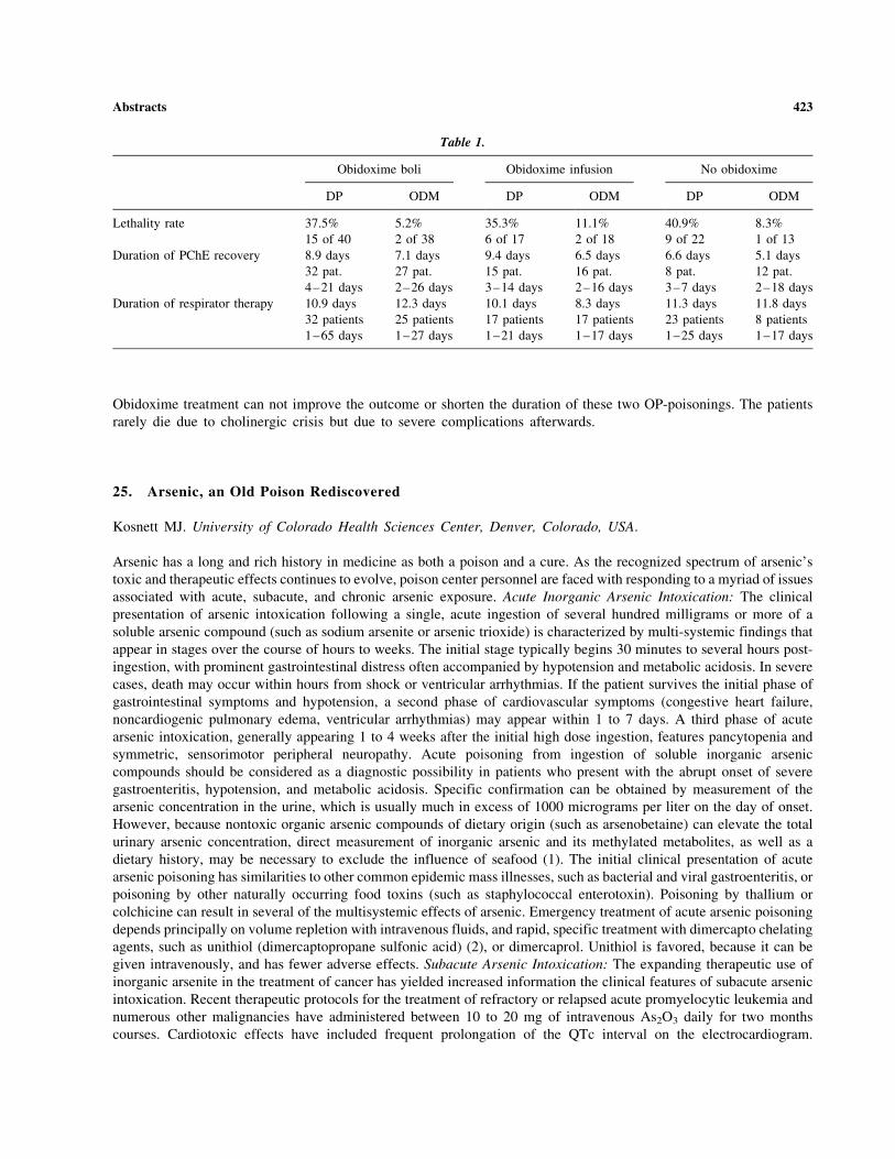
Obidoxime treatment can not improve the outcome or shorten the duration of these two OP-poisonings. The patients
rarely die due to cholinergic crisis but due to severe complications afterwards.
25. Arsenic, an Old Poison Rediscovered
Kosnett MJ. University of Colorado Health Sciences Center, Denver, Colorado, USA.
Arsenic has a long and rich history in medicine as both a poison and a cure. As the recognized spectrum of arsenic’s
toxic and therapeutic effects continues to evolve, poison center personnel are faced with responding to a myriad of issues
associated with acute, subacute, and chronic arsenic exposure. Acute Inorganic Arsenic Intoxication: The clinical
presentation of arsenic intoxication following a single, acute ingestion of several hundred milligrams or more of a
soluble arsenic compound (such as sodium arsenite or arsenic trioxide) is characterized by multi-systemic findings that
appear in stages over the course of hours to weeks. The initial stage typically begins 30 minutes to several hours post-
ingestion, with prominent gastrointestinal distress often accompanied by hypotension and metabolic acidosis. In severe
cases, death may occur within hours from shock or ventricular arrhythmias. If the patient survives the initial phase of
gastrointestinal symptoms and hypotension, a second phase of cardiovascular symptoms (congestive heart failure,
noncardiogenic pulmonary edema, ventricular arrhythmias) may appear within 1 to 7 days. A third phase of acute
arsenic intoxication, generally appearing 1 to 4 weeks after the initial high dose ingestion, features pancytopenia and
symmetric, sensorimotor peripheral neuropathy. Acute poisoning from ingestion of soluble inorganic arsenic
compounds should be considered as a diagnostic possibility in patients who present with the abrupt onset of severe
gastroenteritis, hypotension, and metabolic acidosis. Specific confirmation can be obtained by measurement of the
arsenic concentration in the urine, which is usually much in excess of 1000 micrograms per liter on the day of onset.
However, because nontoxic organic arsenic compounds of dietary origin (such as arsenobetaine) can elevate the total
urinary arsenic concentration, direct measurement of inorganic arsenic and its methylated metabolites, as well as a
dietary history, may be necessary to exclude the influence of seafood (1). The initial clinical presentation of acute
arsenic poisoning has similarities to other common epidemic mass illnesses, such as bacterial and viral gastroenteritis, or
poisoning by other naturally occurring food toxins (such as staphylococcal enterotoxin). Poisoning by thallium or
colchicine can result in several of the multisystemic effects of arsenic. Emergency treatment of acute arsenic poisoning
depends principally on volume repletion with intravenous fluids, and rapid, specific treatment with dimercapto chelating
agents, such as unithiol (dimercaptopropane sulfonic acid) (2), or dimercaprol. Unithiol is favored, because it can be
given intravenously, and has fewer adverse effects. Subacute Arsenic Intoxication: The expanding therapeutic use of
inorganic arsenite in the treatment of cancer has yielded increased information the clinical features of subacute arsenic
intoxication. Recent therapeutic protocols for the treatment of refractory or relapsed acute promyelocytic leukemia and
numerous other malignancies have administered between 10 to 20 mg of intravenous As2O3 daily for two months
courses. Cardiotoxic effects have included frequent prolongation of the QTc interval on the electrocardiogram.
Table 1.
Obidoxime boli Obidoxime infusion No obidoxime
DP ODM DP ODM DP ODM
Lethality rate 37.5% 5.2% 35.3% 11.1% 40.9% 8.3%
15 of 40 2 of 38 6 of 17 2 of 18 9 of 22 1 of 13
Duration of PChE recovery 8.9 days 7.1 days 9.4 days 6.5 days 6.6 days 5.1 days
32 pat. 27 pat. 15 pat. 16 pat. 8 pat. 12 pat.
4–21 days 2–26 days 3–14 days 2–16 days 3–7 days 2–18 days
Duration of respirator therapy 10.9 days 12.3 days 10.1 days 8.3 days 11.3 days 11.8 days
32 patients 25 patients 17 patients 17 patients 23 patients 8 patients
1–65 days 1–27 days 1–21 days 1–17 days 1–25 days 1–17 days
Abstracts 423
Malignant ventricular arrhythmias, including fatal torsades des pointes, have been reported (3). Other notable toxicity in
this setting has included peripheral neuropathy, gastrointestinal disturbance, and hepatotoxicity (4). Chronic Arsenic
Intoxication: A growing number of epidemiological studies have linked chronic arsenic ingestion, particularly from
naturally occurring arsenic in drinking water, to a strikingly broad spectrum of serious chronic illness. Although arsenic
ingestion has long been recognized as a risk factor for skin cancer, more recent epidemiological investigations
conducted in Taiwan, Chile, and Argentina have established arsenic ingestion as a risk factor for lung cancer and bladder
cancer (5). Studies in Taiwan and Bangladesh have reported a dose-response relationship between chronic arsenic
ingestion and diabetes mellitus (6). Additional investigations in Taiwan have associated chronic arsenic ingestion with
an elevated risk of hypertension, and ischemic heart disease (7). Although epidemiological data on the potential
reproductive and developmental effects of low level arsenic ingestion is sparse, a recent prospective cohort study in
Chile suggests a link to decrements in birth weight (8). Collectively, these findings suggest that chronic arsenic ingestion
may exert a considerable impact on public health morbidity and mortality in populations with significant environmental
exposure. References: 1. Le XC, Ma M. Short-column liquid chromatography with hydride generation atomic
fluorescence detection for the speciation of arsenic. Anal Chem 1998; 70:1926–1933. 2. Moore DF, O’Callaghan CA,
Berlyne G, et al. Acute arsenic poisoning: absence of polyneuropathy after treatment with 2,3-dimercaptopropane-
sulphonate (DMPS). J Neurol Neurosurg Psychiatr 1994; 57:1133–1135. 3. Unnikrishnan D, Dutcher JP, Varshneya
N, et al. Torsades de pointes in 3 patients with leukemia treated with arsenic trioxide. Blood 2001; 97:1514–1516.
4. Huang S-Y, Chang C-S, Tang J-L, et al. Acute and chronic arsenic poisoning associated with treatment of acute
promyelocytic leukaemia. Brit J Haem 1998; 103:1092–1095. 5. National Research Council. Arsenic in Drinking
Water: 2001 Update. National Academy Press: Washington, DC; 2001. 6. Tseng CH, Tai TY, Chong CP, et al. Long-
term arsenic exposure and incidence of non-insulin dependent diabetes mellitus: a cohort study in arseniasis-
hyperendemic villages in Taiwan. Environ Health Perspect 2000; 108:847–851. 7. Chen CJ, Chiou HY, Chiang MH,
et al. Dose-response relationship between ischemic heart disease mortality and long-term arsenic exposure.
Arterioscler Thromb Vasc Biol 1996; 16:504–510. 8. Hopenhayn C, Ferreccio C, Browning SR, et al. Arsenic exposure
from drinking water and birth weight. Epidemiology 2003; 14:593–602.
26. Pregnancy and Chemical Exposure at the Workplace: Individual Risk Assessment and Results ofProspective Follow-Up of 151 Cases
Testud F, Lambert-Chhum R, Descotes J. Toxicovigilance Unit, Poison Center, Hospices Civils de Lyon, France.
Objective: Many women of childbearing age are occupationally exposed to chemicals and concerned with the
ensuing risk when pregnant. In most cases exposure has been present since conception when the pregnancy is
discovered. Questions to be addressed include the likely issue of the pregnancy and the management of risk. We
describe the principles of individual risk assessment to be applied in pregnant women or women wishing to become
pregnant that are exposed to chemicals at the workplace. The results of the prospective follow-up study of 151
exposed pregnant women will be presented. Methods: Since 1996, Lyon Poison Center has been conducting a
prospective follow-up of all requests concerning pregnant women occupationally exposed to chemicals. Each request
for risk assessment is based on a comprehensive review of the hazards of the handled products together with a
thorough evaluation of the actual exposure at the workplace, including biomonitoring as often as possible. A written
toxicological advice is then sent to the gynaecologist or general practitioner in charge of the patient. When the
exposure is estimated hazardous for the pregnancy, either total withdrawal from the workplace, avoidance of certain
activities or improvements of individual protective measures are recommended. The outcome of pregnancy is
followed-up systematically. A specific computerized database allows for epidemiological analysis. Results: From a
total of 299 risk assessment requests, 151 pregnant women were included between 1996 and 2002. Based on the
nature of chemicals, two groups were identified: the first includes 106 women exposed to organic solvents and the
second group 45 women exposed to miscellaneous products. In 21% of cases (32/151), total withdrawal from the
workplace was recommended. But in 45% of cases (68/151), the occupational exposure was not considered
hazardous to the outcome of the pregnancy. No increase in adverse outcomes was evidenced: 7 miscarriages and 146
living births were observed with 2 major malformations and 2 minor malformations, which is similar to spontaneous
outcomes in the general population. For organic solvents, this case series is second in size among published studies
424 Abstracts
with prospective follow-up. Conclusion: Patients who must be withdrawn or benefit from improvements of their
workplace can be objectively selected using such a rational occupational and toxicological approach. Occupational
exposure was not found to adversely affect the outcome of pregnancy in these 151 exposed women.
27. Occupational Lamp Oil Exposure in Catholic Priests
Ballesteros S, Ramon MF, Martınez-Arrieta R, Larrotcha C. Spanish Poison Control Centre. Instituto Nacional de
Toxicologıa, Madrid, Spain.
Objective: Lamp oil is used as fuel in ornamental and decorative candles in churches. The formulations include
kerosene, petroleum distillates and, in more recently introduced products, paraffin fractions with chain lengths
between C8 and C16. This investigation was prompted by the detection of lamp oil exposures among Catholic priests
from different regions of Spain noted by the staff of our Poison Control Centre. Methods: We retrospectively
reviewed all oral exposures to lamp oil in churches reported to our Poison Centre from January 1991 to July 2003.
Data analysed were: patient age, sex, aetiology and clinical symptoms. Results: Over the 13 years, 16 cases met
inclusion criteria. The first case was recorded in November 1997 and afterwards there were ingestions every year,
especially in 2000 and 2001 (6 and 4 cases, respectively). The inquirer was the patient (7 cases), a hospital (6) or a
general practitioner (3). The number of exposures was higher on Wednesday (4 cases), Sunday and Saturday (3
each). The moment of the incidents reflected the timetable of Mass in Spanish churches (early morning 2, midday 5,
afternoon 3 and evening 6). All of the intoxicated patients were male and the mean age was 49 years old. Paraffin
containing lamp oil was drunk by the priests after being mixed with wine during the Mass or between services when
the bottle was confused with mineral water. The location was not the usual workplace for the priest and included
chapels in monasteries or funeral homes, church halls and village churches. The amount swallowed was small, often
only one or two sips (5–30 ml) although in two episodes the mixture of oil and wine was drunk totally (70–100 ml).
In one occasion not only the priest but also the parishioners (around 35) drunk from the communion cup. Symptoms
were: diarrhoea (4 patients), vomiting (2), abdominal pain (2) and blood stained sputum (1). Ten of the patients
remained asymptomatic. Conclusion: Occupational toxic exposure in a religious environment is an uncommon event.
In observance of the Catholic doctrine, wine is mixed with water during the celebration of Mass. The confusion was
due to the similarity between the lamp oil container and those of mineral or distillate water. Moreover, one episode
not included in the study involved 5 babies baptised with lamp oil. The data reported in this study indicates the need
for workplace surveillance in unusual places.
28. Methanol Poisoning Treated with Fomepizole and Hemodialysis: Studies in 7 Cases
Hovda KE (1), Froyshov S (1), Gudmundsdottir H (2), Urdal P (3), Jacobsen D (1). 1. Department of Emergency
Medicine; 2. Department of Nephrology; 3. Department of Clinical Chemistry, Ullevaal University Hospital, N-0407
Oslo, Norway.
Objective: Methanol poisoning is a medical emergency characterised by metabolic acidosis, visual disturbances and
circulatory and respiratory collapse. Recently, fomepizole has been approved as an antidote by inhibiting the
methanol metabolism to formic acid. We present seven cases of severe methanol poisoning treated with fomepizole
and hemodialysis. Case Series: Two women and five men, average age 52 years (range 35–69), were treated for
methanol poisoning. They all drank illegal spirit consisting of a mixture of 20% methanol and 80% ethanol. They
were all treated with hemodialysis (average 7 hrs, range 5–8), in addition to fomepizole and buffer. Upon admission,
their average pH was 7.1 (range 6.6–7.5), pCO2 4.6 kPa (range 3.6–6.1), HCO3- 11.8 mmol/L (range 3.0–28) and
base deficit 15.8 mmol/L (range 5.1–28). Their average osmolal gap was 86 mOsm/kgH2O (range 16–148), anion
gap 31 mmol/L (range 23–40), S-methanol 68.4 mmol/L (range 15.6–140.6) and S-formate 13.1 mmol/L (range
3.3–21). The mean half-life of methanol during fomepizole treatment and without hemodialysis was 71.2 hrs (range
69.3–77), during dialysis 2.6 hrs (range 1.7–3.3), while the mean half-life of formate during dialysis was 1.7 hrs
Abstracts 425
(range 1.5–1.9). The mean dialysance of methanol was 234 mL/min (range 219–249, n=5) and for formate 234 mL/
min (range 233–235, n=1). During hemodialysis the dialysator clearance of methanol was close to its total body
clearance. One of the patients died and two were discharged with permanent visual and cerebral sequelae.
Conclusion: The present cases demonstrate that rapid treatment with alkali and fomepizole may reverse the
metabolic disturbances of methanol poisoning, whereas hemodialysis is associated with a rapid elimination of
methanol and formate. Rapid initiation of treatment also reversed the pronounced visual disturbances seen in
two cases.
29. Formate Kinetics in Methanol Poisoning
Hantson P (1), Haufroid V (2), Wallemacq P (2). 1. Department of Intensive Care; 2. Laboratory of Toxicology,
Cliniques St-Luc, Universite catholique de Louvain, Brussels, Belgium.
Background: The objective of this work is to describe formate kinetics in a retrospective series of 25 patients treated
in a single center for severe methanol poisoning. Material and Methods: We retrospectively reviewed the medical
records of the methanol-poisoned patients treated in the intensive care unit (ICU) between 1987 and 2001. The cases
were identified by the discharge diagnosis obtained from the ICU patient’s database. Inclusion criteria were: a
history of deliberate methanol ingestion, with a blood methanol concentration greater than 6.2 mmol/L or a high
anion gap metabolic acidosis. The data were extracted and analyzed independently by two physicians. On the whole,
data were available for 25 methanol poisoned patients. Hemodialysis indications were: methanol >15.8 mmol/L,
metabolic acidosis, visual toxicity. Ethanol was used as antidote in 22 cases, fomepizole in 3 cases. All patients had
received bicarbonate and folate supplementation. Serial blood formate determination by headspace gas
chromatography was obtained in 20 patients. Toxicokinetic calculation was performed when at least three data
points were available either before, during or after hemodialysis. Endogenous and dialysis elimination half-lives
were calculated as t1/2=0.693/Ke. The determination of endogenous elimination half-life was possible in 9 cases. In
this group, 3 patients were not hemodialyzed. Elimination half-life during hemodialysis was calculated in 14 cases;
spontaneous elimination half-life at the end of hemodialysis was calculated in 7 cases. The elimination half-lives
with and without hemodialysis were compared using an unpaired t-test. Results: The mean initial blood methanol
concentration in the 20 patients was 71.4 mmol/L with range 8.7–321.4 mmol/L. The mean initial formate was 12.6
mmol/L with range 0.33–22.4 mmol/L. Mean duration time of hemodialysis was 10.2±6.6 hr. Half-life before
hemodialysis or in the absence of hemodialysis was 6.03±3.25 hr. Elimination half-life during hemodialysis was
1.80±0.77 hr, which was statistically different (p=0.004). After hemodialysis, endogenous elimination half-live was
3.89±1.97 hr, which was comparable to the values observed before or in the absence of hemodialysis. Discussion: In
contrast to a recent publication (1) and in agreement with previous papers, our data confirm that hemodialysis
effectively reduces formate elimination half-life after methanol poisoning (2). References: 1. Kerns W 2nd,
Tomaszewski C, McMartin K, Ford M, Brent J; META Study Group. Methylpyrazole for Toxic Alcohols. Formate
kinetics in methanol poisoning. J Toxicol Clin Toxicol 2002; 40:137–143. 2. Yip L, Jacobsen D. Endogenous formate
elimination and total body clearance during hemodialysis. J Toxicol Clin Toxicol 2003; 41:257–258.
30. Charcoal Haemoperfusion: Does It Still Have a Role in the Management of the Poisoned Patient?
Dargan PI, Jones AL. National Poisons Information Service (London), Guy’s & St Thomas’ NHS Trust, London, UK.
Background: Charcoal haemoperfusion (cHPF) was first used for the management of barbiturate poisoning in the
1960s (1). Since these initial reports it has also been used for the management of a number of other poisonings
including theophylline, carbamazepine, phenytoin, salicylate, sodium valproate and paraquat (2,3). There have been
no prospective randomised-controlled trials looking at the role of cHPF in poisoning and so most of the information
on its use comes from uncontrolled case-series/reports in which kinetics have been studied before, during and after
426 Abstracts
the procedure. It is most commonly used in severe theophylline and carbamazepine poisoning and this review will
concentrate on these poisonings and the relative role of cHPF compared to multi-dose activated charcoal (MDAC)
and haemodialysis (HDX). The procedure: cHPF involves pumping blood, at a flow rate of 150–400 mL/min,
through a cartridge containing activated charcoal coated with an ultrathin (0.05 mcm) membrane (2,4). The cHPF
cartridge can be placed in the pump and circuit used for haemodialysis or continuous haemofiltration and so the only
special equipment required is the cHPF cartridge itself (4). As with any extracorporeal circuit, priming and
maintenance anti-coagulation are required either with heparin, or in the presence of thrombocytopenia, prostacyclin
(4). The adsorptive capacity of the cartridges reduces with time because of the deposition of drug, cellular debris and
plasma proteins. Complications: In addition to those associated with other extracorporeal procedures, complications
associated with cHPF include: 1) thrombocytopenia (this is generally 10–25% with the use of ultrathin membranes
compared to the 40–50% decrease seen with older uncoated cartridges) (4,5) 2) embolisation of charcoal particles,
this is prevented by the ultrathin membrane and a filter in the venous limb (4), 3) mild hypocalcaemia, which is
rarely of clinical significance (4,5). cHPF in theophylline and carbamazepine poisoning: Poisoning with either of
these agents can cause severe and prolonged morbidity and mortality. Volunteer studies and case reports have
demonstrated that the use of MDAC enhances the elimination of both of these drugs but no studies to date have
demonstrated that this has an impact on outcome and the use of MDAC is often limited by vomiting and
development of ileus (6,7). Carbamazepine is poorly cleared by conventional haemodialysis because of its high
(75%) protein binding (8). There are numerous reports describing successful removal of carbamazepine and its active
10,11-epoxide metabolite by cHPF with extraction ratios of up to 0.87 and clearance values of 80–129 mL/min
(comparable to that seen with MDAC) (7,9). Recent reports show significant carbamazepine removal using high-flux
HDX and suggest that this may prove to be an alternative therapy; in one case of carbamazepine poisoning treated
with both high flux HDX and cHPF clearance values during the two procedures were 59 mL/min and 110 mL/min
respectively (10). MDAC and HDX remove theophylline to a similar extent, increasing clearance to 88–144 mL/min
(6,11–13). Haemoperfusion is more effective at increasing elimination in theophylline poisoning with reported
clearance of up to 233 mL/min (12,13). Future developments: Adsorbents may have other roles in critically ill
patients and a recent studies have looked at their use in conventional HDX circuits to adsorb inflammatory
molecules in sepsis (e.g. cytokines) and uraemic toxins (e.g. middle molecules, b2 microglobulin) in acute renal
failure (14,15). Preliminary in-vitro studies in our laboratory have shown that novel mesoporous polymer-based
carbons can result in improved adsorption capacity and kinetics of adsorption of drugs such as carbamazepine (16);
the flexibility offered by these adsorbents through close control of their chemical purity, pore-size distribution and
surface chemistry makes them highly biocompatible and may also allow targeting of specific drugs. Conclusion:
There is a continuing role for cHPF in the management of severe theophylline and carbamazepine poisoning,
particularly in patients who are deteriorating despite MDAC therapy or in those in whom MDAC use is limited by
the development of ileus. Recent and future developments in carbon technologies may allow an expansion in the
indications for cHPF in the poisoned patient. References: 1. Yatzidis H. Research on extrarenal purification with the
aid of activated charcoal. Nephron 1964; 72:310–312. 2. Pond SM. Extracorporeal techniques in the treatment of
poisoned patients. Med J Aust 1991; 154:617–622. 3. Winchester JF. Dialysis and haemoperfusion in poisoning. Adv
Renal Repl Ther 2002; 9:26–30. 4. Webb D. Charcoal haemoperfusion in drug intoxication. Br J Hosp Med 1993;
49:493–496. 5. Pond SM, et al. Pharmacokinetics of haemoperfusion for drug overdose. Clin Pharmacokin 1979;
4:329–354. 6. Position statement and practice guidelines on the use of MDAC in the treatment of acute poisoning. J
Toxicol Clin Toxicol 1999; 37:731–751. 7. Spiller HA, et al. Carbamazepine overdose: a prospective study of serum
levels and toxicity. J Toxicol Clin Toxicol 1990; 28:445–458. 8. Schlafke D, et al. Carbamazepine poisoning.
Anaesth Reanim 1991; 16:220–224. 9. Cameron RJ, et al. Efficacy of charcoal haemoperfusion in massive
carbamazepine poisoning. J Toxicol Clin Toxicol 2002; 40:507–12. 10. Kielstein JT, et al. High-flux haemodialysis:
an alternative to haemoperfusion in the treatment of carbamazepine intoxication. Clin Nephrol 2002; 57:484–486.
11. Lee CS, et al. Haemodialysis of theophylline in uraemic patients. J Clin Pharmacol 1979; 19:219–226. 12.
Shannon MW. Comparative efficacy of haemodialysis and haemoperfusion in theophylline intoxication. Acad Emerg
Med 1997; 4:674–678. 13. Heath A, et al. Role of extracorporeal drug removal in acute theophylline intoxication.
Med Toxicol 1987; 2:294–308. 14. Winchester JF, et al. Sorbents in acute renal failure and the systemic in-
flammatory response syndrome. Blood Purif 2003; 21:79–84. 15. Winchester JF, et al. Removal of middle molecules
with sorbents. Artif Cells Blood Subst Immob Biotech 2002; 30:547–554. 16. Scorgie KA, et al. The use of novel
pyrolysed carbons in poisoning: an in-vitro study. J Toxicol Clin Toxicol 2002; 40:383.
Abstracts 427
31. Management of Hepatic Failure: The Role of New Techniques
Laterre P-F, Galvez R, Wittebole X. Department of CCM, St. Luc University Hospital, Brussels, Belgium.
Acute liver failure remains associated with a high morbidity and mortality. Currently, the only recognised therapy in
this setting is orthotopic liver transplantation. However, some conditions may be associated with potential liver
recovery, limiting the need of transplantation and subsequent consequences of chronic immunosuppression. Also, the
organ shortage may not only lead to delayed transplantation but may adversely indicate too early graft request when
spontaneous recovery is likely. Over the last 25 years, various liver supportive care techniques have been evaluated.
Hemoperfusion using charcoal cartridge, human or animal hepatocytes devices and molecular absorbents
recirculating systems (MARS) have been used to treat patients with acute or acute-on-chronic liver failures. There
are limited well designed controlled trials evaluating the benefits of these techniques compared to the most
appropriate standard care, including transplantation. However, in a phase III controlled study, comparing standard
care to liver assist device using porcine hepatocytes in acute liver failure, a trend in improved survival was observed
in patients treated with artificial liver system. Especially, a significant reduction in hospital mortality was
demonstrated in patients included for viral or paracetamol induced acute liver failure. No difference in adverse
events was observed and in particular viral transmission. The MARS system, using albumin as a carrier of various
potential toxic substances, has been evaluated in various causes of liver failure. Despite limited controlled studies,
the MARS technique seems to improve circulatory failure, present in liver failure, renal dysfunction, but mostly to
reduce intracranial hypertension and encephalopathy. This technique, even though not studied in randomized
controlled trials, might improve liver failure induced organ dysfunctions and serve as a bridge to liver
transplantation, when indicated. Future studies should better determine adequate indications for these techniques with
appropriate end-points. Also, the so-called toxic substances removed by these support systems and biomarkers should
be better evaluated. References: 1. Stange J, Ramlow W, Mitzner S, Schmidt R, Klinkmann H. Dialysis against a
recycled albumin solution enables the removal of albumin bound toxins. Artif Organs 1993; 17:809–813. 2. Mitzner
SR, Stange J, Klammt S, Risler T, Erley CM, Bader BD, Berger ED, et al. Improvement of hepatorenal syndrome
with extracorporeal albumin dialysis MARS, results of a prospective, randomized, controlled clinical trial. Liver
Transpl 2000; 6:277–286. 3. Stange J, Hassanein TI, Mehta R, Mitzner S, Bartlett RH. The Molecular Adsorbents
Recycling System (MARS) as a liver support system based on albumin dialysis: a summary of preclinical
investigations, prospective randomized controlled clinical trials and clinical experience from 19 centers. Artif Organs
2002; 26:103–110. 4. Sorkine P, Abraham RB, Szold O, Bidermann P, Kidron A, Merchav H, Brill S, et al. Role of
the molecular adsorbents recycling system (MARS) in the treatment of patients with acute exacerbation of chronic
liver failure. Crit Care Med 2001; 29:1332–1336. 5. Schmidt LE, Svendson LB, Sorensen VR, Hansen BA, Larsen
FS. Cerebral blood velocity increases during a single treatment with the molecular adsorbents recirculating system in
patients with acute on chronic liver failure. Liver Transpl 2001; 7:709–712. 6. Heemann U, Treichel U, Look J,
Philipp T, Gerken G, Malago M, Klammt S, Loehr M, Liebe S, Mitzner S, Schmitdt R, Stange J. Albumin dialysis in
cirrhosis with superimposed acute liver injury: a prospective, controlled study. Hepatology 2002; 36:949–958.
32. ICU Regimens—Why They Must Be Altered for the Poisoned Patient
Seger DL. Vanderbilt University Medical Center, Nashville, TN, USA.
The critically ill poisoned patient presents unique management issues. Typical ICU protocols must be reassessed for
patients at risk for comorbidity related to poisoning. GID and Elimination: Depending on how quickly patients are
moved from the Emergency Department to the ICU, issues of gastric lavage, whole bowel irrigation, multiple dose
activated charcoal and urinary alkalinization must be decided. Indications, complications, risks and benefits of each
procedure must be assessed in each patient. Hypotension: Hypotension in the poisoned patient is most frequently
caused by receptor blockade, drug-induced myocardial depression, or drug-induced vasodilatation. Unless the patient
is hypovolemic, treating hypotension with large volumes of fluid may predispose the patient to the development of
Acute Respiratory Failure. Catecholamine pressors are the agents of choice for treating most hypotensive ICU
patients who are older, chronically ill, or are acutely ill from an infectious process. Treatment approaches for
428 Abstracts
drug-induced vasodilatation or myocardial depression must address the cause of the hypotension and not assume that
all hypotensive patients can be treated in a similar manner. The young healthy poisoned patient will respond to
hypotension with adrenal outpouring of catecholamines. Adrenergic receptors are sensitive in the young patient.
Administering further catecholamines is unlikely to be of benefit as all catecholamine receptors are stimulated by
endogenous catecholamines. Agents that must be considered for the treatment of hypotension in the poisoned patient
are sodium bicarbonate (for sodium channel-blocking agents), glucagon, and insulin/glucose. Treatment of Cardiac
Arrhythmias: A diseased heart is the cause of most cardiac arrhythmias that are treated in the ICU. Most poisoned
patients do not have preexisting heart disease. Treatment of an arrhythmia must take into consideration the
pharmacology of the toxin causing the arrhythmia. Acute Renal Failure: In the poisoned patient, acute renal failure
(ARF) is most frequently due to a decrease in extracellular fluid volume and renal hypoperfusion caused by drug (or
chemical)-induced vasodilatation, and/or myocardial depression, or rhabdomyolysis. Attempts to prevent ARF are
crucial as there is no specific therapy once ARF has occurred. Once intravascular volume is adequate, the efficacy of
administering low-dose dopamine (0.5–3.0mg/kg/min) to prevent ARF is controversial. Studies evaluating the
efficacy of low-dose dopamine in preventing ARF have not demonstrated any benefit, but the patient population
consisted of critically ill patients with established ARF or high risk for developing ARF (1). The efficacy of low-
dose dopamine following periods of drug-induced hypotension in poisoned patients, who are typically younger and
without chronic disease, has not been evaluated. Following drug-induced hypotension, it seems reasonable to
administer low-dose dopamine to poisoned patients with adequate vascular volume who remain oliguric or anuric
despite maximal diuretic therapy. Ventilation and Extubation: Poisoned patients are intubated because they have
ingested a drug that causes either respiratory depression or depression of their sensorium resulting in loss of
protective airway reflexes. As the drug is metabolized, the drug effects abate and the patient’s sensorium improves—
sometimes very suddenly. Attempts to attain weaning parameters prior to extubation may result in an awake agitated
patient with an endotracheal tube in place. Administration of sedatives to help the patient ‘‘tolerate the tube’’ result
in prolonged extubation times and potential complications. Seizures: Acidosis following a seizure may contribute to
the morbidity and mortality of the poisoned patient. Acidosis following a 30–60 second seizure may persist for 60
minutes. pH may be as low as 7.17 at 30 minutes and 7.20 at 60 minutes following resolution of a 30 to 60 second
seizure (2). Acidosis decreases cardiac output, oxygen extraction, left ventricular end diastolic pressure and impairs
myocardial contractility. If a patient has ingested a cardiotoxic drug which causes significant myocardial depression,
the consequences of acidosis may increase the toxicity of the drug. Ictal increases in plasma epinephrine levels may
add to the potential of cardiac arrhythmias. Additionally, airway reflexes are inhibited postictally, which add to the
potential for aspiration. Whether seizures increase mortality in poisoned patients is difficult to ascertain. Deaths that
occur in poisoned patients that seize are usually attributed to the toxicity of the drug. Due to the number of
variables, one cannot know if mortality would have occurred had the patient not seized. Other Issues: Other issues to
consider are indications in the poisoned patient for the administration of calcium, potassium, and magnesium.
Toxicology laboratory analysis is unique for the poisoned patient. References: 1. Baldwin L, Henderson A, Hickman
P. Effect of postoperative dopamine on renal function after elective major vascular surgery. Am J Nephrol 1994;
20:744–747. 2. Orringer D, Eustace J, Wunsch C, et al. Natural history of lactic acidosis after grand mal seizures.
NEJM 1977; 15:796–799.
33. Glucagon or Insulin and Glucose: When and Why?
Megarbane B, Baud F. Reanimation Medicale et Toxicologique and INSERM U26–Universite Paris VII, Hopital
Lariboisiere, Paris, France.
Objectives: To describe the relative place and indications of glucagon and insulin+glucose as antidotes in acute
poisonings. Both have been used as antidotes in poisoned patients with cardiotropics-associated cardiovascular
shock. Results: Glucagon, a hyperglycemic 3.5 kD oligopeptide hormone, acts by stimulating neoglucogenesis and
glycogenolysis. The place of glucagon in hypoglycemic poisoned patients should be first discussed. In contrast to IV
0.5–1.0 g/kg 50%-dextrose that immediately reverses hypoglycemia, glucagon’s action requires time and may be
ineffective in case of insufficient liver glycogen storage. Glycogen stores may be depleted in chronic alcoholism,
liver failure, prolonged fasting, cortisol or growth hormone deficiencies. Moreover, in the presence of high insulin
Abstracts 429
levels, glucagon ability to raise blood glucose may be reduced. In addition to its hyperglycemic properties, glucagon
may be useful in cardiotropic poisonings. Glucagon interacts with specific membrane glucagon receptors, a Gs
protein that activates an adenylate cyclase, which converts ATP to cAMP, accounting for glucagon’s antidotal
cardiovascular effects. Glucagon produces positive inotropic and chronotropic actions. In healthy men, 3–5 mg IV
by bolus increased the force of cardiac contraction, the heart rate, the cardiac index, the blood pressure and the
stroke work, without changing the systemic vascular resistances. Glucagon action is transient, with a plasma half-life
of 3–6 min. Its action starts 1–3 min after infusion, is maximal at 5–7 min and lasts up to 10–15 min. An initial IV
bolus of 50 mg/kg infused over 1 min is recommended. Higher doses may be necessary if the initial bolus is
ineffective, with doses up to 10 mg. Glucagon is a standard therapy of beta-blockers (BB) and calcium channel
blockers (CCB) poisoned patients, although to date, no controlled human study has been published to assess its
efficacy. In BB poisonings, glucagon may correct hypotension and bradydysrhythmias, by circumventing the
blockade of beta-adrenergic receptors. In rat studies, the maximal chronotropic effect of glucagon was correlated to
circulating ionized calcium. Moreover, whereas beta-adrenergic agents may be dysrhythmogenic, glucagon appears
not to be. Thus, in BB poisonings, we recommend in case of symptomatic treatment failure (in addition to adequate
fluids and atropine), the administration of antidotes in the following order: dobutamine (or isoprenaline, especially in
sotalol intoxication), glucagon, and epinephrine. Glucagon also reverses CCB-associated myocardial depression in
animal studies and several human case reports support this use. In animal models of CCB overdose, it transiently
increased heart rate, cardiac output and reversed high degree AV blocks, without significant effect on arterial
pressure. Glucagon appeared to have no effect on survival rate, either in BB or CBB intoxication models. More
recently, in CCB poisoning, a more effective action was attributed to high-dose insulin. Glucagon is derived from
beef or pork pancreas and is available as a 1-mg or 10-mg lyophilized powder that has to be diluted in an
accompanying 2 mg/ml-phenol solvent. Thus, in case of parenteral use of high doses, it is recommended that
glucagon be diluted with sterile water. Adverse effects of glucagon include dose-dependent nausea, vomiting,
hyperglycemia, hypokalemia and general allergic reactions. Availability of adequate supplies and high cost raises
questions as to its use as a first-line antidote, but its safety profile should encourage it. Insulin is a polypeptidic
hypoglycemic hormone, promoting glucose utilization and trapping in muscles, adipose tissue and the liver. High-
dose (0.5 UI/kg/h) insulin has been proposed to treat CCB and BB poisonings. This treatment requires glucose
infusion in high concentration to maintain euglycemia, and careful monitoring of glucose and potassium levels to
avoid serious side effects. This therapy has been tested in animals and human series of CBB poisonings, with
cardiovascular shock. In severely poisoned patients, insulin permitted cardiovascular stabilization, decrease in
catecholamine infusion rate and appeared to improve survival. However, in our experience, this therapy may not
always be effective. Blockade of L-type calcium channels induces an inhibition of the pancreatic insulin secretion,
resulting in hyperglycemia and alteration of the normal cardiac fatty acid metabolism, forcing myocardial cells to
become carbohydrate-dependent. Insulin and glucose administration improves basic cellular metabolism and
intracellular calcium effects, resulting in positive inotropic activity. The metabolic basis for this beneficial effect is
still not completely understood, but appears to be related to a decrease of free fatty acid uptake by the heart cells,
and a switch to carbohydrates. In a canine model of verapamil poisoning, high-dose insulin improved survival when
compared to calcium, epinephrine or glucagon. It enhanced both myocardial glucose and lactate uptake, significantly
increased the ratio of myocardial oxygen delivery/work, resulting in a better improvement in myocardial function, in
comparison to other inotropic therapies including glucagon. High-dose insulin was also shown to improve cardiac
performance and increase survival in a canine model of propranolol intoxication. Improvement in hemodynamic
status and survival was greater in insulin-treated animals than in glucagon-treated ones. However, as high-dose
insulin has not yet been evaluated in humans, only selected BB poisoned patients may benefit from this therapy. In
contrast, present published material and clinical experience suggests that in patients with CCB poisoning persistent
hypotension, bradyarrhythmias or conduction disturbances should be considered for treatment treated with insulin
and glucose after the failure of IV fluids, calcium salts, glucagon, and especially in the case of a poor response to
vasopressor agents. Conclusions: Glucagon and insulin+glucose represent two major antidotes. At present only
controlled studies in animals established their efficacy in BB and CCB poisoning, showing a significant superiority
to insulin+glucose in comparison to glucagons in each case. However, glucagon is still largely used in BB
poisoning, despite the lack of clinical study to support its beneficial effects. Following recent published clinical data,
high-dose insulin should now be systematically considered in severe CCB poisonings. More data are still needed to
reach a conclusion regarding its efficacy in BB poisoning. References: 1. Taboulet P, et al. J Toxicol Clin Toxicol
1993; 31:531–551. 2. Kerns W II, et al. Ann Emerg Med 1997; 30:711–712. 3. Kline JA, et al. Crit Care Med 1995;
430 Abstracts
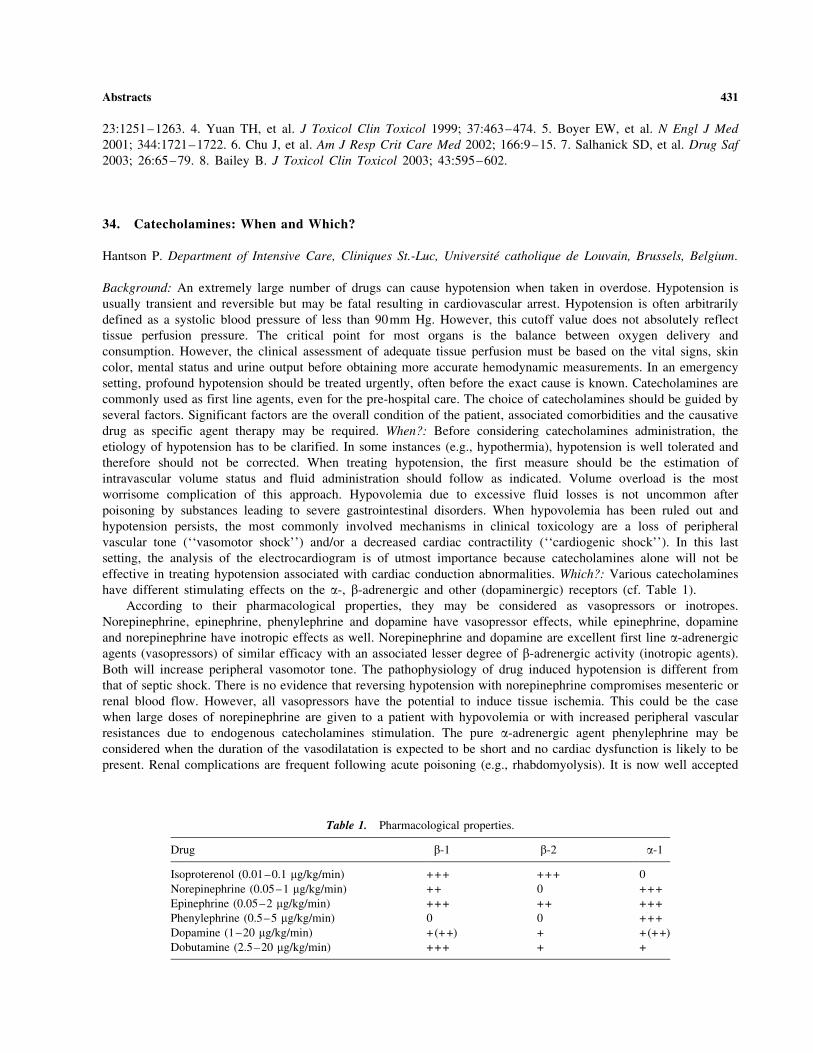
23:1251–1263. 4. Yuan TH, et al. J Toxicol Clin Toxicol 1999; 37:463–474. 5. Boyer EW, et al. N Engl J Med
2001; 344:1721–1722. 6. Chu J, et al. Am J Resp Crit Care Med 2002; 166:9–15. 7. Salhanick SD, et al. Drug Saf
2003; 26:65–79. 8. Bailey B. J Toxicol Clin Toxicol 2003; 43:595–602.
34. Catecholamines: When and Which?
Hantson P. Department of Intensive Care, Cliniques St.-Luc, Universite catholique de Louvain, Brussels, Belgium.
Background: An extremely large number of drugs can cause hypotension when taken in overdose. Hypotension is
usually transient and reversible but may be fatal resulting in cardiovascular arrest. Hypotension is often arbitrarily
defined as a systolic blood pressure of less than 90mm Hg. However, this cutoff value does not absolutely reflect
tissue perfusion pressure. The critical point for most organs is the balance between oxygen delivery and
consumption. However, the clinical assessment of adequate tissue perfusion must be based on the vital signs, skin
color, mental status and urine output before obtaining more accurate hemodynamic measurements. In an emergency
setting, profound hypotension should be treated urgently, often before the exact cause is known. Catecholamines are
commonly used as first line agents, even for the pre-hospital care. The choice of catecholamines should be guided by
several factors. Significant factors are the overall condition of the patient, associated comorbidities and the causative
drug as specific agent therapy may be required. When?: Before considering catecholamines administration, the
etiology of hypotension has to be clarified. In some instances (e.g., hypothermia), hypotension is well tolerated and
therefore should not be corrected. When treating hypotension, the first measure should be the estimation of
intravascular volume status and fluid administration should follow as indicated. Volume overload is the most
worrisome complication of this approach. Hypovolemia due to excessive fluid losses is not uncommon after
poisoning by substances leading to severe gastrointestinal disorders. When hypovolemia has been ruled out and
hypotension persists, the most commonly involved mechanisms in clinical toxicology are a loss of peripheral
vascular tone (‘‘vasomotor shock’’) and/or a decreased cardiac contractility (‘‘cardiogenic shock’’). In this last
setting, the analysis of the electrocardiogram is of utmost importance because catecholamines alone will not be
effective in treating hypotension associated with cardiac conduction abnormalities. Which?: Various catecholamines
have different stimulating effects on the a-, b-adrenergic and other (dopaminergic) receptors (cf. Table 1).
According to their pharmacological properties, they may be considered as vasopressors or inotropes.
Norepinephrine, epinephrine, phenylephrine and dopamine have vasopressor effects, while epinephrine, dopamine
and norepinephrine have inotropic effects as well. Norepinephrine and dopamine are excellent first line a-adrenergic
agents (vasopressors) of similar efficacy with an associated lesser degree of b-adrenergic activity (inotropic agents).
Both will increase peripheral vasomotor tone. The pathophysiology of drug induced hypotension is different from
that of septic shock. There is no evidence that reversing hypotension with norepinephrine compromises mesenteric or
renal blood flow. However, all vasopressors have the potential to induce tissue ischemia. This could be the case
when large doses of norepinephrine are given to a patient with hypovolemia or with increased peripheral vascular
resistances due to endogenous catecholamines stimulation. The pure a-adrenergic agent phenylephrine may be
considered when the duration of the vasodilatation is expected to be short and no cardiac dysfunction is likely to be
present. Renal complications are frequent following acute poisoning (e.g., rhabdomyolysis). It is now well accepted
Table 1. Pharmacological properties.
Drug b-1 b-2 a-1
Isoproterenol (0.01–0.1 mg/kg/min) +++ +++ 0
Norepinephrine (0.05–1 mg/kg/min) ++ 0 +++
Epinephrine (0.05–2 mg/kg/min) +++ ++ +++
Phenylephrine (0.5–5 mg/kg/min) 0 0 +++
Dopamine (1–20 mg/kg/min) +(++) + +(++)
Dobutamine (2.5–20 mg/kg/min) +++ + +
Abstracts 431
that there is no evidence that dopamine provides any benefit to patients with impending or existing renal failure, or
renal protective effect in the setting of vasoconstrictor therapy. Epinephrine remains the drug of choice when
impaired cardiac contractility is present. Adverse effects of excessive epinephrine administration include lactic
acidosis and hyperglycemia. Dobutamine is often used in combination with norepinephrine when the cause of
hypotension is multifactorial (cardiogenic associated with vasomotor shock). Conclusions: Catecholamines are
commonly used to correct hypotension after drug poisoning. They should be used with caution after the underlying
mechanisms are carefully considered. References: Oldner A, Rossi P, Karason S, Aneman A, Scandinavian Critical
Care Trials Group. A practice survey on vasopressor and inotropic drug therapy in Scandinavian intensive care units.
Acta Anaesthesiol Scand 2003; 47:693–701. Kellum JA, Pinsky MR. Use of vasopressor agents in critically ill
patients. Curr Opin Crit Care 2002; 8:236–241. Bellomo R, Giantomasso DD. Noradrenaline and the kidney:
friends or foes? Crit Care 2001; 5:294–298. Holmes CL, Walley KR. Bad medicine: low-dose dopamine in the ICU.
Chest 2003; 123:1266–1275.
35. Peripheral Cardiocirculatory Assistance in Patients Admitted with Severe Membrane StabilizingAgent Poisonings—Three Case Reports
Megarbane B (1), LePrince P (2), Delhotal B (3), Delerme S (1), Resiere D (1), Guerrier G (1), Deye N (1), Baud F
(1). 1. Reanimation Medicale et Toxicologique, Hopital Lariboisiere; 2. Service de Chirurgie Cardiaque, Hopital
Pitie; and 3. Laboratoire de Toxicologie, Hopital Ambroise Pare, Paris, France.
Objectives: Membrane stabilising agents are still responsible of fatal poisonings, despite optimal management in
intensive care unit (ICU). No pharmacological alternative to sodium bicarbonate and adrenaline is available to date
to treat refractory toxic shock. Experimental studies show that outcome may be improved in poisoned animals, using
cardiac assistance. We previously demonstrated that refractory shock (defined by a systolic BP <90 mm Hg, despite
adequate (1 L fluid loading, 375 ml 84% bicarbonate and >3 mg/h adrenaline infusion), with respiratory (PaO2/FiO2
>150 mm Hg) or renal failure (oliguria or serum creatinine concentration >120 mmol/l) is a very sensitive and
specific predictor of death (1). Methods: All the patients received adequate symptomatic treatments and antidotes.
Extra Corporeal Life Support (ECLS) was performed using cardiovascular assist device (Jostra1) through femoral
arterial and venous pre-heparinated percutaneous cannula surgically implanted, with a retro-perfusion of the
superficial femoral artery. Verbal informed consents were obtained from next of kin. Results: Three severely
intoxicated patients were assisted because of refractory shock or cardiac arrest: one carbamazepine poisoning (M, 26
years, ingested dose: 32 g, concentration: 224 mmol/l), one acebutolol poisoning (F, 29 years, dose: 38 g,
acebutolol+diacetolol concentration: 14.35 mg/l) and one propafenone poisoning (F, 50 years, dose: 9 g,
concentration: 2.9 mg/l). A satisfactory non-beating blood pressure was immediately obtained and all the
hemodynamic parameters were progressively improved after the assistance. The duration of assistance was
respectively: 109, 85 and 48 hours. All the 3 patients recovered and returned back home, without significant
neurological sequelae. Complications were: severe ENT and local bleeding necessitating multiple transfusions (2
cases), extensive inferior vena cava thrombosis (1 case) and local infection of the surgical site (1 case).
Toxicokinetics demonstrated an initial persistent absorption phase and liver metabolism saturation, prolonging the
elimination half-lives. Conclusions: Peripheral cardiocirculatory assistance may represent a unique therapeutic
alternative in patients admitted for severe membrane stabilising agent poisoning with non-response to shock or
failure of cardiopulmonary resuscitation. The early recognition of an indication for ECLS is crucial and should be
based on validated predictive factors of death. The benefit of this technique should be prospectively evaluated on a
larger cohort. Reference: 1. Megarbane B. J Toxicol Clin Toxicol 2003; 41:553–554.
36. Management of Acute Arsenic Poisoning in Children: Extracorporeal Elimination Techniques asAdjunctive Therapy
Lai MW, Boyer E, Kleinman M, Rodig N, Burns MM. Regional Center for Poison Control and Prevention and
Divisions of Critical Care Medicine and Nephrology, Children’s Hospital and Harvard Medical School, Boston,
432 Abstracts
Massachusetts; University of Massachusetts Department of Emergency Medicine, Worcester, Massachusetts;
Department of Emergency Medicine, Mount Auburn Hospital, Cambridge, Massachusetts, USA.
Objective: report a case series of arsenic poisoning and extracorporeal elimination techniques used as adjunctive
therapy to remove arsenic. Case Series: Due to a labeling error, a four-month old male (BG) ingested 60 mL baby
formula reconstituted from arsenical herbicide containing 7.6% sodium trioxide (As2O3) by weight; his two-year old
sister (MG) ingested approximately 2.5 mL of the same herbicide. Both children became acutely ill, requiring
aggressive fluid resuscitation and chelation therapy. BG ingested approximately 411 mg(As)/kg, far exceeding
maximum chelatable amounts. He was unresponsive and anuric and required cardiopulmonary resuscitation for
nonperfusing ventricular tachyarrhythmias including Torsades de Pointes refractory to electrical and pharmacological
cardioversion. He received parenteral chelation available in the United States (US)—intramuscular dimercaprol;
permission was obtained for intravenous administration of 2,3-dimercapto-1-propane sulfonate (still an
investigational chelator in the US) under compassionate care protocols. He received extracorporeal membrane
oxygenation (ECMO), exchange transfusion and hemodialysis (HD) in attempts to alleviate his arsenic body burden.
Following placement on ECMO at 12 hours after ingestion, his physical exam changed from being comatose with
fixed and dilated pupils to having sluggishly reactive pupils and some spontaneous movement; he produced 107 mL
urine over two hours before becoming anuric again. HD did not produce appreciable change in his exam. HD
dialysate had 20 mcg(As)/L measured at the midpoint of the first of two runs. BG died 36 hours after ingestion.
Serum arsenic concentration at autopsy was 730 mcg/L. MG did well with chelation therapy available in the US:
intramuscular dimercaprol transitioned to oral succimer. She showed no clinical sequelae seven months following
her poisoning. Conclusion: Arsenic’s Phase 1 serum half-life (t1/2<2 hours) makes extracorporeal elimination
techniques seem impractical. Phase 2 clearance is considerably longer (t1/2=30 hours), when arsenic redistributed to
erythrocytes is found in a 3:1 ratio compared to serum. BG’s placement on ECMO was equivalent to a partial
exchange transfusion; the improvement in his exam following commencement of this therapy may have been due to
elimination or dilution of arsenic redistributed to erythrocytes during Phase 2 clearance. The absence of clinical
improvement with HD and the magnitude of variance between dialysate and serum arsenic concentrations may
indicate limitations of extracorporeal elimination techniques which filter serum plasma alone. In readily-identified
severe arsenic poisoning, exchange transfusion may merit consideration as additional adjunctive therapy to enhance
arsenic elimination.
37. The Role of Temporary Cardiac Pacing in Acute Toxicological Syndromes
Fijen JW, Leenders MEC, Meulenbelt J. National Poisons Control Centre, National Institute for Public Health and the
Environment, Bilthoven, the Netherland.
Objective: Overview of the use of temporary cardiac pacing in acute toxicological syndromes. Introduction:
Bradycardia with or without hypotension or shock is a prominent sign and clue to identify the ingested toxin and/or
drug. Cardiovascular agents like digoxin, betablockers and calcium channel blockers are the most frequent causes of
bradycardia. What is the role of temporary placement of a pacemaker in treating bradycardia and hypotension or
shock due to the toxicity of drugs or toxins? Is there any evidence for temporary pacing? Methods: To identify
relevant studies, MEDLINE and PubMed (1966 onwards), The Cochrane Library (1996 onwards), and BIOSIS (1989
onwards) were searched. We used the keywords cardiac pacing, temporary pacing, pacemaker, poison, toxicology,
drugs and toxin. All papers concerning cardiac pacing in poisoned patients were included in the review. The
inclusion criteria made no mention of study design, and it was unclear what levels of evidence were represented. The
reference lists of identified papers and published reviews were checked and screened. Result of the search: We
identified 231 (14 non-English) references describing randomised controlled trials in human (2), uncontrolled case
series (82), and review papers (26) in human (108) and animals (123). Only two randomised controlled trials were
performed both concerning yellow oleander toxicity. Treatment guided by the published papers: The main cause of
sinus bradycardia and AV-conduction disturbance by toxins or poisons are cardiovascular drugs i.e. calcium channel
blockers, sodium and potassium channel blockers, digitalis, beta-blockers, cholinomimetic agents and class IA-III
antiarrhythmic drugs. The therapy of calcium channel blocker overdose, leading to decreased myocardial
contractility, decreasing AV-node conduction, and arterial vasodilation, focuses on restoring cardiac function and
Abstracts 433
systemic blood pressure. No randomised-controlled trials concerning the treatment of calcium channel blocker
toxicity have been performed yet. There are several proposed therapies, but no one modality has demonstrated
superiority over the others (1). Calcium (2), glucagon (3–5), insulin infusion (6,7), were used although usefulness is
not clear. The experience with atropine, isoproterenol (8–10), norepinephrine and dopamine is limited to some case
reports or studies with a very small number of patients (11). An estimate of their therapeutic efficacy is unreliable.
Supportive care including the use of phosphodiesterase inhibitors, cardiac pacing, balloon pump or extracorporeal
bypass is occasionally mentioned, but a concordant effect on morbidity and mortality has not yet been proven.
Several anecdotal case-reports have suggested a positive response (however, not always) to cardiac pacing in
calcium channel blocker toxicity (12). Cardiac pacing is probably indicated for significant therapy-resistant
bradycardia and AV-conduction blocks. In conclusion, no one agent or strategy is likely to be definitively
therapeutic in all cases of calcium channel blocker toxicity (13). The role for temporary pacing is very limited.
Blockade of fast inward sodium channels by for example lidocaine results in decrease of the maximum rate of
depolarisation and shortens the action potential duration (APD). Blockade of medium inward sodium channels by for
example disopyramide, procainamide or quinidine leads to the decrease in conduction, automaticity, and excitability
of the myocardial cells with an increase of APD and refractoriness. Blockade of slow inward sodium channels by for
example propafenone and flecainide results to the decrease in conduction without any effect on APD. Treatment
options are sodium salts, and is supportive with inotropic agents. Temporary pacing will not resolve the problem of
the insensitive heart, because the decrease in conduction, automaticity, and excitability of the myocardial cells will
persist. Potassium channels blockers e.g. sulfonylureas lead to a decrease of sinus node conduction, automaticity, and
an increase of refractoriness. Potassium salts and symptomatic therapy by inotropic agents are the main options.
Again, temporary pacing will not resolve the problem of the insensitive heart. The general therapy in acute digitalis
poisoning leading to inhibition of Na±K±ATPase pump is supportive. Before commercial availability of immuno-
therapy with Fab fragments, management of severe digitalis-induced arrhythmias relied on ventricular pacing and
antiarrhythmic drugs (14). Unfortunately, during digitalis poisoning, the fibrillatory threshold is lowered, and both
accidental electrode displacement and manipulation of the pacemaker for adjustment of pacing may lead to overdrive
inhibition of spontaneous rhythm and a longer complicating pacing period leading to infectious complications.
Immunotherapy is the specific treatment for severe digitalis intoxication (15–17). In a retrospective study the safety
and efficacy of immunotherapy and pacing has been assessed. It was concluded that pacing has very limited
preventive and curative effects, is difficult to handle, and exposes patients to iatrogenic accidents. Immunotherapy is
powerful, safe and much easier to use (18) and cost-effective (19). b-blockers lead to suppression of impulse
generation and conduction, and decreased myocardial contractility. The b-blockers that have marked anti-arrhythmic
activity are more lethal (e.g. propranolol, sotalol). Similarly, pre-existing cardiac pathology or co-ingestion of
psychotropic or cardioactive drugs increases mortality. b-blockers are competitive antagonists and their binding to
the receptor is by definition reversible, and can thus be displaced by b-agonists such as isoproterenol or dobutamine
(20). However, their administration can lead to a more pronounced b2-vasodilator effect with a deleterious effect on
blood pressure. Glucagon is currently considered the treatment of choice in circulatory failure caused by b-blockers
(20,21). Atropine improved the bradycardia induced by the b-blockers (22), but it’s beneficial effect is limited.
Furthermore, b-mimetic agents such as isoproterenol (rate/conduction) and epinephrine (inotropism and arterial
constriction) can be useful. Inhibition of phosphodiesterases by agents such as theophylline and enoximone prevents
the breakdown of cAMP maintaining. Theoretically, these drugs are of importance in case of b-blocker intoxication.
However, preventing the breakdown of cAMP in vascular smooth muscle cells may lead to a further vasodilation and
hypotension (20). The pacemaker has been proposed in severe bradycardia resistant to appropriate pharmacological
treatment. It neither corrects nor improves the depressed myocardial function nor hypotension (23). The potential
chronotropic benefits do not compensate for the risks associated with the procedure such as mechanical and
infectious complications. The prognosis of self-poisoning with beta-blockers is good, especially if medical manage-
ment is started immediately but the wide variety of clinical symptoms and proposed treatments complicate the
therapeutic strategy. Poisoning with cholinomimetic agents (i.e. prostigmine, organophosphates, amanita com-
pounds), via activation of muscarinic cholinergic receptors and inhibition of acetylcholinesterase leading to via sinus
node hypoexcitability and bradycardia, can be treated by atropine. Atropine is a muscarinic receptor antagonist that
prevents the effects of acetylcholine by blocking its binding to muscarinic cholinergic receptors. The possible benefits of
temporary pacing do not outweigh the risks for the patients. Atropine is the first drug of choice in the therapy of
cholinomimetic intoxication. Torsade de pointes is a polymorphic ventricular tachyarrhythmia, in which the QRS
complex is characterised by changing amplitude, contour and polarity, and is associated with a long QT interval. While
434 Abstracts
many episodes of torsade resolve spontaneously, a few may persist and degenerate to ventricular fibrillation and death.
The common causes and predisposing conditions in the acquired QT syndrome are the use of antiarrhythmic drugs (class
IA or III such as medium inward sodium channels blockers), hypokalemia and bradycardia (24). The most commonly
used treatments for torsade de pointes with an acquired long QT syndrome are lidocaine (25), magnesium (26),
electrolyte replacement, isoproterenol (27), and cardiac overdrive pacing. The evidence for magnesium drugs
suppressing the early after-depolarisation by blocking sodium channels leading to a shorten QT interval, is stronger than
for lidocaine (25,26) and isoproterenol (28). Successive atrial and/or ventricular overdrive pacing in torsade des pointes
is written anecdotally (29), and may be useful if electrolyte replacement fails. Prior to this, the benefits of temporary
pacing will presumably not outweigh the disadvantages and the risks of the procedure. Conclusion: Cardiovascular
disturbances in poisoned patients are often complex. Initially treatment should be focussed on the mechanism of toxicity.
Evidence for the use of temporary pacing in acute toxicological syndromes with severe bradycardia and hypotension is
lacking. Theoretically, pacing of a poisoned heart with a decreased automaticity, conduction, and excitability, is
presumably useless. The level of evidence is level V (case series, uncontrolled studies, and expert opinion). The routine
use of cardiac pacing in such emergencies is debatable (recommendation grade E). References: 1. Merigian KS, et al. Am
J Emerg Med 1990; 8:479–483. 2. Pond SM, et al. Med J Aust 1995; 163:345–349. 3. Parmley WW, et al. N Engl J Med
1971; 285:801–802. 4. Zaritsky AL, et al. Crit Care Med 1988; 16:246–251. 5. Crump BJ, et al. Lancet 1982; 2:939–
940. 6. Kline JA, et al. Cardiovasc Res 1997; 34:289–298. 7. Yuan TH, et al. J Toxicol Clin Toxicol 1999; 37:463–474.
8. Christensen C, et al. Ugeskr Laeger 1986; 148:2929. 9. Anthony T, et al. Ann Emerg Med 1986; 15:1344–1348.
10. Malcolm N, et al. Drug Intell Clin Pharm 1986; 20:888. 11. Hofer CA, et al. Am J Med 1993; 95:431–438.
12. Watling SM, et al. Ann Pharmacother 1992; 26:1373–1378. 13. Chu J, et al. Am J Respir Crit Care Med 2002;
166:9–15. 14. Bismuth C, et al. J Toxicol Clin Toxicol 1977; 10:443–456. 15. Smith TW, et al. N Engl J Med 1982;
307:1357–1362. 16. Antman EM, et al. Circulation 1990; 81:1744–1752. 17. Ochs HR, et al. J Clin Invest 1977;
60:1303–1313. 18. Taboulet P, et al. J Toxicol Clin Toxicol 1993; 31:261–273. 19. Mauskopf JA, et al. Am J Cardiol
1991; 68:1709–1714. 20. Taboulet P, et al. J Toxicol Clin Toxicol 1993; 31:531–551. 21. Smith RC, et al. JAMA
1985; 254:2412. 22. Weinstein RS, et al. Ann Emerg Med 1984; 13:1123–1131. 23. Lane AS, et al. Ann Emerg Med
1987; 16:1381–1383. 24. Janeira LF, et al. Am Fam Physician 1995; 52:1447–1453. 25. Assimes TL, et al. Can J
Cardiol 1998; 14:753–756. 26. Tzivoni D, et al. Circulation 1988; 77:392–397. 27. Morrison Y, et al. Ann
Pharmacother 1993; 27:189–190. 28. Tzivoni D, et al. Am J Cardiol 1984; 53:528–530. 29. Anderson JL, et al. Am
J Med 1978; 64:715–718.
38. Respiratory Support: What, Where and When?
Bedry R (1), Hilbert G (2). 1. Polyvalent and Toxicological Intensive Care Unit, Clinique Mutualiste, Pessac,
France; 2. Medical Intensive Care Department, Pellegrin Hospital, Bordeaux, France.
The indications of mechanical ventilation are usually divided into three categories: neurologic failure, acute
hypoxemic respiratory distress and hypercapnic respiratory failure. In a recent publication (1), acute hypoxemic
respiratory distress was the commonest cause of pulmonary support (66%), followed by coma (15%) and
hypercapnic respiratory failure (13%). However very little information is published specifically on intoxication.
Drug-induced coma is a leading cause of poisoning-related admissions to hospital, and the main cause of respiratory
insufficiency is related to hypoventilation. Whatever the cause of the respiratory failure, i.e., central apnoea
(barbiturates, opioids), obstructive apnoea (hypnotics, organophosphates. . .), chemical ARDS (solvents), aspiration
pneumonia or circulatory failure, pulmonary support remains the most appropriate symptomatic treatment. In a study
concerning the experience of 361 acute poisonings managed by a French mobile intensive care unit (2), 21% patients
were intubated and placed on mechanical ventilation. The main toxin involved in these patients were psychotropic
medications (82%). In countries where paramedics are trained to perform out-hospital emergency airway manage-
ment, recent publications have shown a significative proportion of technical mishaps (oesophagus intubation,
unrecognized dislodgment of the endotracheal tube) (3,4). Although tracheal intubation remains the most common
technique, non invasive ventilation has proved efficacious in intensive care units, especially in hypercapnic res-
piratory failure in COPD patients or hypoxemic respiratory distress caused by cardiogenic pulmonary oedema
(consensus). The latest technique is not usually employed in case of poisoning because most of the patients are
Abstracts 435
comatose, and a Glasgow coma scale under 10 is generally regarded as a contra indication of this technique
(consensus). It could be employed only if the patient remains conscious. Three types of patients could respond to this
technique: 1) conscious patients presenting cardiogenic pulmonary oedema linked to an intoxication with only
cardiotoxic substances, and if the patient remains conscious, 2) hypercapnic respiratory failure in COPD patients
intoxicated with benzodiazepines and under flumazenil treatment, 3) mild ARDS following pneumonitis (solvent or
aspiration pneumonia, organophosphates). The most challenging aspect is probably pre-hospital management, where the
proportion of difficult intubations was estimated to be 20% of those performed for intoxicated patients (5). The difficulty
in intubating these patients is not linked to the presence of a deep coma, as assessed by the Glasgow Coma Scale (5).
Pulmonary support is an emergency technique, and it should be performed, by whichever method, as soon as possible
when a life-threatening condition occurs. References: 1. Esteban A, Anzueto A, Alia I, Gordo F, Apezteguia C, Palizas F,
et al. How is mechanical ventilation employed in the intensive care unit? An international utilisation review. Am J Respir
Crit Care Med 2000; 16:1450–1458. 2. Ould-Ahmed M, Drouillard I, Savio C, Baud FJ, Migliani R, Michel A.
Intoxications aigues prises en charge par un service mobile d’urgence et de reanimation. Rean Urg 1999; 8:93–97. 3.
Gausche M, Lewis RJ, Stratton SJ, et al. Effect of out-of-hospital pediatric endotracheal intubation on survival and
neurological outcome: a controlled clinical trial. JAMA 2000; 283:783–790. 4. Katz S, Falk J, Wash M. Misplaced
endotracheal tubes by paramedics in an urban emergency medical service system. Abstr. Acad Emerg Med 1998; 5:429.
5. Adnet F, Mineado JP, Finot MA, Borron SW, Fauconnier V, Lapandry C. A survey of sedation protocols used for
emergency endotracheal intubation in poisoned patients in the French prehospital medical system. Eur J Emerg Med
1998; 5:415–419. 6. International Consensus Conferences in Intensive Care Medicine: noninvasive positive pressure
ventilation in acute respiratory failure. Am J Respir Crit Care Med 2001; 163:283–291.
39. Hemodialysis, Hemofiltration and Their Modalities in the Treatment of Acute Poisoning
Groszek B. Department of Clinical Toxicology, College of Medicine Jagiellonian University, Krakow, Poland.
Background: The treatment of serious drug or chemical poisoning relies mostly on the use of standard intensive care
measures. Whereas mortality from poisoning is generally low, exposure to drugs and chemicals is common, and the
toxicologist is often faced with management decisions regarding extracorporeal removal of drugs and toxins.
Although very few poisonings require, or are effectively treated by extracorporeal elimination procedures, these
procedures may be lifesaving in selected poisoning. This paper will discuss the role of dialysis and its newer
modalities (high-efficiency and high-flux dialysis) in the management of poisoning together with other techniques
used in treating poisoned patients, such as hemofiltration—continuous arteriovenous hemofiltration (CAVH),
continuous venovenous hemofiltration (CVVH) and continuous venovenous hemodiafiltration (CVVHD).
Hemodialysis: The use of hemodialysis in the treatment of poisoning dates back to the middle of the twenty
century. Hemodialysis was first used in 1950 by Doolan in patient poisoned with salicylates. This was followed by
extensive use of hemodialysis in the treatment of barbiturate overdose, the major cause of intoxication of that decade
(1). Numerous reports appeared in the literature at the 1950s, 1960s and 1970s of the efficacy of hemodialysis for
the treatment of poisoning, but it was found that the limitations of hemodialysis in the removal of endogenous toxins
also applied to its use in poisoning with exogenous substances. Hemodialysis is the process in which the blood is
circulated through a system in which a semi-permeable membrane separates the components of the blood from the
constituents of the dialysis fluid. The diffusion of dialyzable substances across the membrane, from an area of high
concentration to an area of low concentration, allows their removal. Chemical and drug removal by dialysis is
determined by dialyzer conditions (blood flow rate, dialysate flow rate, dialyzer surface area, and pore structure of
the chosen membrane) and by toxicokinetic factors of the toxic substances. Toxicodynamic factors may also be
important if cardiovascular function is impaired. The characteristics that predict the successful removal of a toxic
agent by dialysis include low volume of distribution (<1 L/kg), the presence of the toxin in the central compartment,
low endogenous clearance, a low molecular weight (<500 daltons), low protein binding and high water solubility. To
be clinically effective dialysis should improve the removal of a toxin by at least 30% compared with total body
clearance. Hemodialysis effectiveness is measured by either by hemodialysis clearance, as amount of toxin removed
or half-life during hemodialysis (t1/2). Clinical improvement is not a reliable index. Certain agents that predictably
cause severe toxicity are routinely dialyzed, based on the blood concentrations or clinical manifestation of toxicity.
436 Abstracts
These include methanol, ethylene glycol, isopropanol, lithium, salicylates (3). Early removal of toxic alcohols
prevents toxicity from their toxic metabolites, which are also removed by hemodialysis (4–6). The metabolic
acidosis associated with methanol, ethylene glycol, salicylates, metformin poisoning is also corrected during
hemodialysis (1,2). High-efficiency and high-flux hemodialysis. Since the 1970s the drive for shorter dialysis time
with high urea clearance rates has led to the development of high-efficiency hemodialysis. In the 1990s, certain
biocompatible features and the desire to remove amyloidogenic a2-microglobulin has led to the popularity of high-
flux dialysis. These new modalities shorten the time of hemodialysis and have ability to remove middle molecules.
During the 1990s, the use of high-efficiency and high-flux membranes has steadily increased. Since initial reports
describing their effectiveness in the renal replacement therapy, they have also been used for the treatment of severe
poisoning. Several of the published papers there are case-reports in which the effectiveness of these modalities were
studied. In acute poisoning with carbamazapine (7–10), phenobarbital (11) and valproic acid (12,13) clinical
improvement was observed, the serum drug concentrations were decreased during treatment, dialyzer clearances
were higher than those achieved during haemoperfusion. No complications of the procedure were observed.
Effectiveness of high-flux hemodialysis in the treatment of life-threatening methanol intoxication also was confirmed
(14). Hemofiltration and Continuous Therapies: Hemofiltration achieves solute clearance by convection (or the
solvent drag effect) through the membrane, with pore dimensions exceeding those in conventional dialysis
membranes, by removing plasma water (which contains dissolved solutes); the removed fluid must be replaced. In
hemodiafiltration, diffusive transport is added to hemofiltration to augment the clearance of solutes. Solute clearance
is accomplished by circulating dialysate in the dialysate-ultrafiltrate compartment. CAVH is done without a blood
pump, the ultrafiltration rate is determined by the positive pressure caused by venous resistance and the negative
pressure generated by the height of the drain bag. In CVVH ultrafiltration rate is controlled by a variety of
sophisticated machines. All these methods allow continuous removal of small volume of fluid and toxins at low
blood flow rates. Also larger molecules up to 50 000 daltons are removed and they are better tolerated in
hemodynamically unstable patients. CAVH is mainly useful in intensive care settings in patients with fluid overload.
In the treatment of acute poising CVVH and CVVHD, according to the published case reports, effectively removed
salicylates in poisoning, (where ongoing absorption from gastrointestinal tract was observed) (15), fluorides, and
barium in poisoning with very severe organ toxicity (16,17). In lithium poisoning post-dialysis rebound
concentration is the main risk. In such cases CVVH and CVVHD effectively prevent rebound toxicity (18,19).
Conclusions: The criteria related to the drugs and chemicals toxicokinetics, the efficacy of the certain dialysis and
ultrafiltration techniques and the severity of poisoning should be a guide to their implementation. Clear indices of
efficacy need to be defined and agreed for these treatments. References: 1. Blye E, Lorch J, Cortel S. Extracorporeal
therapy in the treatment of intoxication. Am J Kidney Dis 1984; 3:321–338. 2. Pond SM. Extracorporeal techniques
in the treatment of poisoned patients. Med J Aust 1991; 154:617–622. 3. Winchester JF. Dialysis and haemo-
perfusion in poisoning. Adv Renal Repl Ther 2002; 9:26–30. 4. Jacobsen D, McMartin KE. Antidotes for methanol
and ethylene glycol poisoning. J Toxicol Clin Toxicol 1997; 35:127–143. 5. Barceloux DG, Bond GR, Krenzelok
EP, Cooper H, Vale JA. American Academy of Clinical Toxicology Ad Hoc Committee on the Treatment Guidelines
for Methanol Poisoning. American Academy of Clinical Toxicology practice guidelines on the treatment of methanol
poisoning. J Toxicol Clin Toxicol 2002; 40:415–446. 6. Moreau CL, Kerns W 2nd, Tomaszewski CA, McMartin KE,
Rose SR, Ford MD, Brent J. Glycolate kinetics and hemodialysis clearance in ethylene glycol poisoning. META Study
Group. J Toxicol Clin Toxicol 1998; 36:659–666. 7. Schuerer DJ, Brophy PD, Maxvold NJ, Kudelka T, Bunchman TE.
High-efficiency dialysis for carbamazepine overdose. J Toxicol Clin Toxicol 2000; 38:321–323. 8. Chetty M, Sarkar P,
Aggarwal A, Sakhuja V. Carbamazepine poisoning: treatment with hemodialysis. Nephrol Dial Transplant 2003;
18:220–221. 9. Kielstein JT, Schwarz A, Arnavaz A, Sehlberg O, Emrich HM, Fliser D. High-flux hemodialysis—an
effective to hemoperfusion in the treatment of carbamazepine intoxication. Clin Nephrol 2002; 57:484–486.
10. Tapolyai M, Campbell M, Dailey K, Udvari-Nagy S. Hemodialysis is as effective as haemoperfusion for drug
removal in carbamazepine poisoning. Nephron 2002; 90:213–215. 11. Palmer BF. Effectiveness of hemodialysis in the
extracorporeal therapy of phenobarbital overdose. Am J Kidney Dis 2000; 36:640–643. 12. Kielstein JT, Woywodt A,
Schumann G, Haller H, Fliser D. Efficacy of high-flux hemodialysis in the treatment of valproic acid intoxication. J
Toxicol Clin Toxicol 2003; 41(6):873–876. 13. Kane SL, Constantiner M, Staubus AE, Meinecke CD, Sedor JR. High-
flux hemodialysis without hemoperfusion is effective in acute valproic acid overdose. Ann Pharmacother 2000;
34:1146–1151. 14. Peces R, Alvarez R. Effectiveness of hemodialysis with high-flux polysulfone membrane in the
treatment of life-threatening methanol intoxication. Nephron 2002; 90:216–218. 15. Chase PB, Walter FG, James S.
Whole bowel irrigation, hemodialysis, and continuous venovenous hemodiafiltration in the successful treatment of
Abstracts 437
severe salicylate poisoning: case report. Dial Transplant 2002; 31:387–391. 16. Bjornhagen V, Hojer J, Karlson-Stiber
C, Selden AI, Sundbom M. Hydrofluoric acid-induced burns and life-threatening systemic poisoning–favorable
outcome after hemodialysis. J Toxicol Clin Toxicol 2003; 41:855–860. 17. Koch M, Appoloni O, Haufroid V, Vincent
JL, Lheureux P. Acute barium intoxication and hemodiafiltration. J Toxicol Clin Toxicol 2003; 41:363–367. 18. Meyer
RJ, Flynn JT, Brophy PD, Smoyer WE, Kershaw DB, Custer JR, Bunchman TE. Hemodialysis followed by continuous
hemofiltration for treatment of lithium intoxication in children. Am J Kidney Dis 2001; 37:1044–1047. 18. van Bommel
EF, Kalmeijer MD, Ponssen HH. Treatment of life-threatening lithium toxicity with high-volume continuous
venovenous hemofiltration. Am J Nephrol 2000; 20:408–411.
40. Surgical Therapy in Cocaine Body Packers and Pushers
Schaper A (1), Hofmann R (2), Bargain Ph (3), Ebbecke M (1), Desel H (1), Langer C (4). 1. GIZ-Nord Poison Centre for
the Federal States of Bremen, Hamburg, Lower Saxony and Schleswig-Holstein, Georg-August-University, University
Hospital, Robert-Koch-Straße 40, D-37075 Gottingen, Germany; 2. Airport-Clinic Frankfurt/Main, Germany;
3. Aeroport Charles de Gaulle, Roissy (Paris), France; 4. Department of General Surgery, University of Gottingen,
Germany.
Objective: Body packers or ‘‘mules’’ are drug smugglers who swallow cocaine filled condoms in order to conceal
them during air travel. Body pushers hide drug packages in rectum or vagina. In a cooperation between Charles de
Gaulle Airport in Paris, Frankfurt/Main Airport-Clinic and GIZ-Nord (Goettingen University poison centre) we
performed a retrospective study and developed an algorithm for the problem ‘‘rupture of a cocaine-filled condom in
a body packer.’’ Methods: In a retrospective analysis the data of all cocaine body packers and body pushers, that
were identified at the international airports of Frankfurt and Paris from 1985–2002, were evaluated. Temporal
development, demographic data and surgical aspects were of special interest. Results: From 1985–2002 altogether
312 body pushers and 4660 body packers were identified. The mean age of the drug smugglers was 37.3 (16–80)
years for Frankfurt and 25 (17–67) years for Paris. The sex ratio was 1:1. 64 ‘‘mules’’ (1.4%) developed symptoms
of a life threatening cocaine intoxication following rupture of a condom. Usually the first symptom was a
generalized seizure. In 20 patients (i.e. 31% of all symptomatic body packers) an emergency laparotomy was
performed and the condoms were removed. All these operated patients survived. 44 body packers (69%) died before
surgical therapy could be carried out. Concerning body pushers, the numbers decreased constantly and there did not
occur any major medical complications. In one case of a female body pusher a drug package had to be removed in
general anesthesia by rectoscopy. Conclusion: Cocaine intoxication in body packers is life-threatening. Patients die
of complications caused by generalized vasoconstriction. If the reason for severe cocaine intoxication is the rupture
of a cocaine filled condom, the only possible therapy consists of immediate laparotomy for removal of the
drug packages.
41. Organ Transplants from Poisoned Donors—A 10 Year Retrospective Review
Casey PB (1), Tracey JA (1), Counihan A (2), Cunningham P (2), Fleming J (3), Parlon B (4), O’Mahony U (4),
Hickey D (2), Hegarty J. 1. National Poisons Information Centre, Beaumont Hospital; 2. Renal Transplant Unit,
Beaumont Hospital; 3. Liver Unit, St. Vincents University Hospital; 4. Heart Lung Programme, Mater Hospital,
Dublin, Ireland.
Objective: To determine the number, type and outcome of organ transplants from poisoned donors in Ireland.
Methods: The national transplant coordinators identified poisoned patients who donated organs between 1993 and
2002 inclusive by retrospectively reviewing their paper and computer records. The national renal, liver and heart
transplant coordinators then identified the patients who received the organs and determined their current status.
Results: Seven poisoned patients donated 13 kidneys to 11 recipients during the study period. Four hearts and four
livers were also donated (Table 1). Two donors were children; a one year old who ingested dothiepin and a three
438 Abstracts
year old who took prochlorperazine and dihydrocodeine. The other five donors were aged between 18 and 32 years.
All kidney transplant recipients are still alive, nine have good renal function and two received second transplants
following rejection of the first graft. The four liver transplant recipients are also alive and all have good liver
function. Two of the four patients receiving heart transplants died, one from a subarachnoid haemorrhage 10 days
after transplant and the other from right ventricular failure four days post transplant. The other two heart recipients
are alive and well. Conclusions: Patient and graft survival following renal and liver transplants from poisoned donors
compared favourably to transplants in general (1,2). Patient survival was poorer following heart transplant although
the number of such transplants was small. References: 1. Organ Procurement and Transplantation Network. All
Kaplan-Meier patient survival rates for transplants performed 1996–2001. www.optn.org (accessed November 2003).
2. Organ Procurement and Transplantation Network. All Kaplan-Meier graft survival rates for transplants performed
1996–2001. www.optn.org (accessed November 2003).
42. Toxic Exposure Surveillance System (TESS): Key Components for Toxicosurveillance AcrossMultiple Poison Centers
Watson WA, Litovitz TL, Belson M, Funk AB, Patel M, Schier JG, Kilbourne E, Rubin C. American Association of
Poison Control Centers, Washington DC, USA, ATSDR/NCEH, CDC, Atlanta, GA, USA.
Toxicosurveillance enhances the utility of data collected by poison centers during poisoned patient case
management. A fundamental approach of toxicosurveillance is detecting aberrances in the gathered data. The
application of various statistical methodologies allows for the early detection of aberrances and patterns of toxicity
that may have broad public health significance. It is then up to the investigator to determine what additional
information is necessary to determine whether a clinically significant public health incident, hazardous or
contaminated product, emerging pattern of substance abuse, or an intentional chemical release is present. Since a
Table 1. Status of organ transplant recipients.
Toxic agent(s) causing death of donor Organ
Recipient age
at transplant Status
Smoke Right kidney 75 Creatinine 115 umol/L
Liver 36 Alive and well
Heart 54 Alive and well
Ecstasy, alcohol Left kidney 56 Creatinine 139 umol/L
Right kidney 39 Creatinine 126 umol/L
Liver 40 Alive and well
Heart 44 Alive and well
Barbiturates, alcohol Left kidney 29 Re-transplanted
Right kidney 32 Creatinine 155 umol/L
Heart 20 Deceased (subarachnoid haemorrhage)
Lithium, thioridazine,
dextropropoxyphene,
paracetamol
Left kidney 30 Creatinine 137 umol/L
Right kidney 60 Creatinine 88 umol/L
Dothiepin Kidneys
(paediatric en bloc)
56 Creatinine 98 umol/L
Liver 9 months Alive and well
Smoke Left kidney 17 Creatinine 138 umol/L
Right kidney 32 Re-transplanted
Heart 56 Deceased (right ventricular failure)
Prochlorperazine, dihydrocodeine Kidneys
(paediatric en bloc)
53 Creatinine 89 umol/L
Liver 6 months Alive and well
Abstracts 439
toxic event may not be confined within political boundaries or poison center coverage areas, a major objective of
toxicosurveillance is to include large, naturally-defined geographical areas in order to detect widespread events. This
fact argues for coordinated toxicosurveillance using data from multiple poison centers. At the same time,
surveillance at the local level is also necessary and a critical step in identification of outbreaks that would be lost
against the background noise when only national data is utilized. Using examples from aberrant values detected by
TESS, this presentation will focus on the key components of toxicosurveillance involving multiple poison centers. The
key components of surveillance include timely collection and transfer of data to a central site, continuous and
systematic assessment of the data, and dissemination of the results with public-health significance in order to contain
events and reduce morbidity and mortality. Effective toxicosurveillance requires consistency in poison center
definitions, training, and operations, resulting in uniform data collection. Additionally, the assessment of data for
signals must include adequate resources to perform the desired assessment in a timely manner, at regular intervals, with
clear definitions of the processes and outcomes desired. An evaluation process will help determine the optimal
surveillance methodology for detection of toxic events. TESS was developed so there would be a uniform data set and
standard definitions, replacing multiple pre-existing systems. The AAPCC member poison centers and committees have
processes in place to standardize any modifications to data fields. Development of an automated upload of any new or
updated case, whether open or closed, with upload occurring by ftp about every 4 to 10 minutes, was the first step in the
initiation of continuous toxicosurveillance. With an impending war in Iraq and threatened increase in chem/
bioterrorism, real-time toxicosurveillance was initiated by AAPCC in March 2003 at the urgent request of the CDC.
Current programs include those monitoring the volume of cases reported to each poison center and the frequency of
clinical effects reported in exposed patients. Case counts (for volume and clinical effects) for the immediately preceding
time intervals (hours or days) are compared at 2 to 24 hour intervals to those expected based on their specific baseline
during similar time periods over the last 3 years. Cases that generate outlier signals are reviewed by a clinical
toxicologist to determine if unusual, worrisome patterns exist. The pattern review focuses on the substance, outcome,
and geographical distribution of events. Surveillance case definitions are used to identify specific types of exposures,
such as cyanide, arsenic, botulism, nerve agents, and paraquat. Finally, exposure frequency for specific groups of
products (e.g. contaminated water, food poisoning/food product cases, and carbon monoxide cases) is continuously
monitored. All of these toxicosurveillance approaches are continuously evaluated and updated to determine time from
case reporting to detection, false positive and false negative rates, and usefulness of findings. Various statistical
approaches, including ARIMA modeling, Bayesian methods, and concurrent space-time detection of clusters are being
assessed to determine what approaches are valid, effective, and efficient. The current toxicosurveillance programs have
identified a number of local chemical events, including covert exposure to arsenic of a small group of people, chem/
bioterrorism preparedness exercises sponsored by local and federal agencies, and exposure to environment toxins. The
electrical power blackout of the northeastern United States and adjacent areas of Canada in August 2003 resulted in
significant changes in the call types reported to poison centers serving the impacted population; these changes were
easily detected by toxicosurveillance. Although most AAPCC toxicosurveillance programs implemented to date are
based on national data for all US poison centers, AAPCC is also providing surveillance of local data for individual
poison centers. One approach includes a pilot program which enables poison centers to receive and evaluate
surveillance findings for their region. Toxicosurveillance with poison center data supplements other public health or
security surveillance processes. The availability of real-time surveillance has increased the role and visibility of
US poison centers. This may lead to increased recognition and support for poison centers in addition to improving
public health.
43. Early Warning in Terrorist Chemical Attacks. Experience with a Computer-Based DifferentialDiagnostic System
Vries I de (1,2), Los PM (3), Brekelmans PJAM (1), Riel AJHP van (1), Zoelen GA van (1), Mensinga TjT (1),
Meulenbelt J (1,2). 1. National Poisons Information Centre, National Institute for Public Health and the
Environment, Bilthoven; 2. Department of Intensive Care and Clinical Toxicology, University Medical Centre
Utrecht; 3. Delta Quality Products and Services, Amerongen, The Netherlands.
Background: Early detection of biological, chemical, or nuclear attacks is essential in managing and diminishing
public health threat caused by these agents. Healthcare facilities may be the first site of recognition of illness or
440 Abstracts
injuries caused by terrorist attacks. This is especially true in covert attacks, in which persons are unaware of
exposure. Although these type of attacks are often solely considered in relation to biological agents, it could also
occur with chemical agents, e.g. the release of chemicals in drinking water systems or in food. While patients
present certain signs and symptoms, it may take days or weeks to detect the cause of their illness. In order to treat
these patients in the best possible manner and safeguard others, not yet exposed, prompt recognition is necessary. As
poison centre specialists and clinical toxicologists are frequently consulted for unrecognized symptoms in patients,
they have a role in assisting treating physicians, first responders and governments in this process. In case of
presumed poisonings there is ample experience in providing a suitable list of possible causative agents. However,
given the vast amount of chemicals that could be involved and the need for rapid information provision, a computer-
based differential diagnostic system clearly offers advantages. In the field of infectious diseases a computer program
called GIDEON (Global Infectious Disease and Epidemiology Network) has been designed to help diagnose
infectious diseases based on symptoms and laboratory findings of the patient. Recently a bioterrorism module has
been added to this system. For chemical agents involved in poisonings no such extensive computerized differential
diagnostic system exists. Hence the challenge to develop a chemical differential diagnostic support system as early
warning tool. Methods: Within the Dutch National Poisons Information Centre for everyday use a specially designed
database (TIK, Toxicological Information and Knowledge base) is used to perform rapid clinical toxicological risk
analyses and provide information. The data in this database are based on reviews written by scientific literature
researchers and medical doctors. The system runs on Windows computers and is very accessible. Linked to the
various chemical agents, this database includes all relevant clinical signs and symptoms, which can serve as a sound
foundation for the differential diagnostic support system. First of all, the functional design for the computer software
was developed by the staff of the poison centre and the informatics expert who co-designed TIK. This artificial
intelligence expert is responsible for translating the clinical knowledge into a functional design for computer
programmers. A prototype of this design was built in Excel to facilitate testing and explore new ideas during
development. Next, the applications software was built and the compound monographs already in use in the database
were completed with information needed for this new application. Results: The functional design consists of several
components. First, an input module with recording of relevant details (address) of the physician requiring
information, the patient involved (area, signs, symptoms, laboratory tests), and if known characteristics regarding the
exposure that may indicate the chemical agent involved. Second, the differential diagnostic analysis module, in
which the causative chemical agents are put in the right order of probability. Simplified, this process can be
described in terms of a mathematical set theory with three sets. These are the total number of documented symptoms
in each chemical compound monograph, the total number of observed symptoms of the patient, and the number of
observed symptoms that fit the documented symptoms (‘‘hits’’). In addition a characteristic value (out of four levels)
has been attached to all documented symptoms, for each agent they relate to. For instance, nausea can occur with
many poisonings and is therefore not very discriminative in the consideration or elimination of a large group of
chemicals. Mydriasis and miosis are somewhat more differentiating, whereas a low cholinesterase activity in plasma
or erythrocytes strongly indicates a certain group of chemicals. Also, prevalence of symptoms is taken into account
in these scoring values. Third, the output module with presentation of the differential diagnosis and the ability to
compare several agents in order to find the most plausible chemical agent involved. From this module every
compound monograph can be approached for further specific information. All information in the database is filed
and can be reproduced for extensive analyses and reports. A management tool is designed for performing trend
analyses. This tool also contains a map for geographical presentation of the cases in order to recognise regional
spread. In the past few months the functionality of the program has been tested. Conclusion: A chemical computer-
based differential diagnostic system can save time and increase diagnostic accuracy in detecting chemical attacks. In
addition, the program can be used in daily practice and is now ready for daily use in the poison information service.
No doubt, in the next year the program will be refined and several adjustments will be made with regard to scoring
values of signs and symptoms in order to increase the discriminative power. Clearly, such a program is never a
replacement for sound clinical judgement, but it might just proof to be a valuable tool.
44. Use of Poisons Centre Data for Surveillance Purposes
Bond GR. Drug and Poison Information Center, Cincinnati Children’s Hospital Medical Center, Cincinnati,
Ohio, USA.
Abstracts 441
Introduction: What do we mean by surveillance? Like a guard on a watchtower, we watch for invaders so we have
time to develop an appropriate response before they become too close or too strong and overwhelm our current
defenses. We are not looking for any unknown. We are looking for the occurrence of an anticipated potentiality. The
difference from the past is that we are not awaiting human observation of an unusual or potentially dangerous
pattern then evaluating the significance of the observation. We are looking for certain patterns we believe may be
the first signs of a specific problem. Some of these patterns clearly represent the anticipated event, others require
further human interpretation. Our goal is early intervention to protect the public. Scope: It is hoped that using poison
centre databases for surveillance will assist in detecting such events as product tampering, food poisoning, terrorist
activity, new drug adverse events (including OD death), emerging drugs of abuse, new drug abuse toxicity (MTPT,
PMA, ‘‘bad shit’’), changing patterns of drug abuse. It is also possible that annual changes in calls to poison centres
in particular regions (spikes in CO cases, unintentional cold medication ingestions) may be better markers than other
sentinels (weather change, influenza lab tests) for related public health announcements and regional resource
planning. Methods: Poison centres receive calls from a widely dispersed population base. The pattern of calls (what
kinds of events prompt a call, who make the calls, substances involved changes only slowly over time but varies
seasonally, allowing the establishment of a baseline and the identification of a sentinel event. Significant data is
already available about hourly, daily and seasonal poison type, call volume and variation in other data fields. Using a
moving baseline of past patterns for the same week and the last few days, and ranges of daily variation, statistical
aberrations can be detected. Depending on the event of interest, the threshold sensitivity can be set low (any seafood
poisoning, any death related to a new pharmaceutical) or high (vomiting). All events exceeding threshold require
human evaluation. Once set, thresholds must be continuously re-evaluated to see if they are detecting too many
meaningless variations. Baselines at regional poison centres are more vulnerable to specific past aberrations in call
volume (space shuttle crash in Southern U.S., anthrax outbreak in October 2001 in the Eastern U.S., local water
contamination announcements) and these must be considered, ignored or other periods substituted where relevant.
However, local data may be the most meaningful and most likely to detect sentinel variation. Although terrorists
may disperse a toxin widely, most events of interest, including product tampering (cyanide in Tylenol1 capsules)
and food poisoning (enterotoxigenic E. coli, nicotine tainted meat, scombroid) have clustered in local areas. Geo-
coding inputs using postal codes allows analysis by state, city or county. Events of interest are more likely to exceed
baseline in a smaller dataset. Three or four heroin related deaths in one day may not exceed threshold nationally, but
may be key to identifying tainted heroin if all occur in one local area. Changes in patterns of drug abuse are
particularly important from a regional perspective. An epidemic of teen abuse of dextromethorphan containing
products began in 1998 but has reached various communities at different times. On the wane in some regions it is
still waxing in others. Geo-coding of prescription drug abuse calls may allow identification of moving ‘‘prescription
mills’’ and suggest allocation of prevention and treatment resources within a state. Several poison centres are
providing data for such a project now (RADARSTM
). Limitations: The nature of data collected by poison centres
limits on the utility of surveillance. Poison centres receive the bulk of their calls following a known, acute exposure
to possibly toxic substance. Callers are seeking advice on treatment of current symptoms and on which signs or
symptoms to anticipate, and how to avoid or treat them. Patients rarely call with unexplained symptoms looking for
a cause. Data collected by poison centres include age, sex, location, substance, quantity, reason, symptoms, treatment
and outcome. These data are most likely to allow identification of a sentinel important event (paralytic seafood
poisoning), rare outcome associated with a substance (death) or unusual number of calls related to some combination
of a substance, symptom, location and outcome per time period (tainted heroin, emergence of prescription drug
abuse in a new region). Poison centres passively receive information. Events of interest may occur without poison
centre contact. Calls are received when the caller perceives a likely benefit from poison centre consultation, not
every time the criteria of interest to the surveillance program are present. Callers must have already made a possible
link between an event and a poison (food poisoning, drug interaction induced dysrhythmias, contaminated OTC
medications). This may not happen if the symptoms experienced and the usual symptoms related to a particular
exposure are unexpected or the timing does not implicate the poison. If the event (cancer, dermatitis) or exposure
occurs in patients whose caregivers are unlikely to seek help from a poison centre (e.g., occupational physicians,
oncologists or pathologists) poison centre databases are poor surveillance tools. If the event is not likely to be related
to a poison—because the predominant symptom is fever—the poison centre may never be involved. Also, the
information entered into the database only reflects what is asked, offered and recorded. Symptom based surveillance
may fail to detect events if the symptom of interest is not likely to be the predominant symptom (staff may have
little motivation to write every symptom), if the symptom is common and non-specific (vomiting), or the symptom is
442 Abstracts
unique (erythema multiforme major) but classed in a larger symptom category (rash). Diagnosis based surveillance
depends on the initial caregiver or the poison centre staff recognizing an event correctly (linking vomiting and food
exposure) If the event of interest (e.g., cancer, idiopathic drug related effect) is related to subacute or chronic
exposure the association with a poison may not be made. Conclusion: Investment is being made by government to
develop and assess systems of automated surveillance using pooled national data. Some pharmaceutical companies
are investing in trials for assessment of specific surveillance parameters. To get the maximum benefit from the
system, more funding and effort must be directed to the development, implementation and refinement of systems to
utilize local and regional data.
45. Sudden Increase of Acute Respiratory Illness After Using a Spray Product to Waterproof Clothingand Shoes
Groot R de (1), Vries I de (1,2), Meulenbelt J1 (2). 1. National Poisons Information Centre, National Institute for
Public Health and the Environment, Bilthoven, The Netherlands; 2. Department of Intensive Care and Clinical
Toxicology, University Medical Centre Utrecht, The Netherlands.
Introduction: In October 2002, the National Poisons Information Centre (NPIC), noted an alarming increase in the
number of severe intoxications with products used to waterproof clothing and shoes. In one month, 18 exposures were
reported. In recent years exposures were only incidental. The Dutch NPIC immediately conducted a prospective follow-
up study. Case Series: From October 2002 until April 2003 the Dutch NPIC received 83 requests for information in
which 99 persons were involved. The most common health effects were acute respiratory symptoms of dyspnea and
persistent cough. Many patients also reported chest tightness, fever, headache, sore throat and nausea. A chest X-ray
was performed in at least 25 cases, of which 11 showed interstitial lung oedema comparable with acute respiratory
distress syndrome. Analysis: In general, waterproofing sprays contain a mixture of solvents, propellants (propane,
butane e.a.) and a repellant (fluorocarbon resin or silicone resin). Investigation by our centre revealed that the different
brands of waterproofing sprays associated with intoxications were produced by one manufacturer. The products were
recently reformulated to reduce the odour. This new formulation contained a different mixture of solvents, especially a
higher amount of heptane. Because of the dramatic increase in exposures the Dutch NPIC immediately informed the
Inspectorate for Health Protection. The products involved were withdrawn from the market and a warning was
published in the national newspapers. Because of export to other countries, the Inspectorate for Health Protection
decided to inform the EU Member States through the Rapid Alert System. The Dutch NPIC informed the European
poisons centres through the European Association of Poisons Centres and Clinical Toxicologists (EAPCCT). The
Inspectorate for Health Protection is researching the exact cause of the increased toxicity of the new formulation. The
most probable hypothesis, based on animal studies, is that by changing the solvent constitution, the aerosol particle size
of the spray decreased so the toxic repelling agent can penetrate deeper into the lungs. Furthermore, the unpleasant
odour was removed and, as a consequence, the product was used more frequently in unventilated rooms, leading to more
severe exposures. Conclusion: An adequate surveillance of the Dutch NPIC and an early warning of the Inspectorate for
Health Protection, helped preventing further exposures to the waterproofing sprays. The fast exchange of information
through the EAPCCT’s mailing list enabled European Poison Centres to be notified immediately.
46. Quetiapine Overdose—A Case Series
Hermanns-Clausen M (1), Stedtler U (1), Reinhardt M (2). 1. Poisons Information Center VIZ-Freiburg, D-79106
Freiburg, Germany; 2. Kreiskrankenhaus Ettenheim, D-77955 Ettenheim, Germany.
Objectives: In 2001 prescriptions for the newer atypical antipsychotic agent quetiapine have tripled in Germany (1).
Overdoses reported to VIZ-Freiburg poison center have grown from zero in 2000 to 32 in 2003. Experience in
overdose is still limited. Overdoses ranging from 1.2–20 g have resulted in mild toxicity. Somnolence, agitation and
confusion, tachycardia, QRS-widening. QT-prolongation and hypotension were reported. Case Series: We
Abstracts 443
retrospectively analysed all sufficiently documented single-drug overdoses with quetiapine reported to the poison
center VIZ-Freiburg. 7 adult patients were included in this study after ingestion of 2 g to 36 g quetiapine. 3 cases
after ingestion of 2 g to 6 g resulted in mild toxicity. The symptoms were somnolence (3) and tachycardia (1). One
intoxication with 4.2 g quetiapine was moderate: The 46 yo woman was found comatose with miosis. 10 h after
admission she was awake but agitated requiring sedation (diazepam). She had sinus tachycardia (120/min) and
hypotonia requiring IV administration of fluids. All 3 patients who ingested >10 g were severely intoxicated: A 53
yo woman who ingested 10.7 g quetiapine developed rhabdomyolysis (CK 17900 U/l) and somnolence. A 38 yo
woman who had tried to commit suicide with 19 g quetiapine was only sleepy initially. However, 4.5 hours after
ingestion she deteriorated and became comatose with respiratory failure requiring mechanical ventilation. A 22 yo
man who ingested 36 g quetiapine was drowsy with hypotension and tachycardia. Gastric lavage was performed. 3.5
hours after ingestion he deteriorated and became deeply comatose. Laboratory tests showed rhabdomyolysis (CK
2851 U/l), hyperglycemia and respiratory acidosis initially (pH 7.31, pO2 65.7 mm Hg, pCO2 46 mm Hg, 90% O2-sat.).
Serum level of quetiapine 12 h after ingestion was 2374 mg/l. 18 h after ingestion he was awake but confused and agitated
requiring benzodiazepine-therapy. Conclusions: Rhabdomyolysis occurred after ingestion of 10.7 g and 36 g quetiapine.
This has not been reported before in quetiapine overdose. On the other hand 36 g is the highest reported overdose until
now. Two patients deteriorated with latency (3.5–4.5 h) after initially being drowsy and became deeply comatose.
Patients with overdoses >10 g and mild toxicity should therefore be monitored at ICU for at least 12 hours. After CNS-
depression by quetiapine for about 12 hours or more agitation and confusion may occur and may require sedation.
Laboratory tests should include creatine kinase, liver enzymes and glucose. Rhabdomyolysis may require urine
alkalinization. Reference: 1. Schwabe U, Paffrath D (eds, 2002) Arzneiverordnungsreport 2002. Berlin: Springer.
47. Strengthening Global Health Protection: The Public Health Role of Poisons Centres
Palmer SR (1), Olowokure B (2), Tempowski J (2), Meredith T (2). 1. Chemical Hazards and Poisons Division,
Health Protection Agency, Cardiff, United Kingdom; 2. International Programme on Chemical Safety (WHO/ILO/
UNEP), World Health Organization, Geneva, Switzerland.
Objectives: The 20th century saw a huge expansion in the chemicals industry with the global production of
chemicals increasing from one million tonnes in 1930 to 400 million tonnes at the present time. About 100 000
chemicals are currently commercially available, while about 70 000 are produced in volumes greater than one tonne.
Information on the human toxicity of the majority of these chemicals is scanty or non-existent. Chemicals, whether
manufactured or of natural origin, are a fundamental component of human life, but as well as providing many
benefits, they can also do harm. Release of chemicals through natural, accidental or intentional events poses a risk to
public health. Moreover these risks may not be confined to a single geographical area or even country but may
impact across national boundaries. A pilot study carried out by the International Programme on Chemical Safety
(IPCS) identified 69 chemical incidents of international public health concern over a period of 15 months. In recent
times concerns have grown about the possible overt or covert use of chemical, as well as biological and
radionuclear, agents for terrorist purposes. Recognition of these dangers has given a new impetus to governments to
strengthen systems, both at national and international level, for preparedness, detection and response to chemical
incidents and outbreaks of illness of chemical aetiology. Method: Poisons centres usually operate 24 hours a day,
seven days a week and most deal with enquiries from the general public. Poisons centres provide information and
advice about the diagnosis and management of exposure to chemicals. Some also provide medical treatment and
analytical support for the management of poisoning. The information that poisons centres collect from their enquirers
is logged and analysed to provide the basis for surveillance and toxicovigilance, and for developing prevention
activities. Poisons centres are therefore well placed to identify a sentinel event indicating a possible chemical
release. Moreover, the multi-disciplinary human resources available to them, their knowledge and resources on the
health effects of chemicals, and their interactions with the public, health authorities, hospitals, academic centres and
laboratories, mean that poisons centres also have the potential to play a strategic role in health protection. Some
countries have already recognized the role of poisons centres and have started to explore the possibilities of
analysing poisons centre data in real-time for signal detection. Poisons centres could also become proactive partners
in the area of global health protection. In the field of communicable diseases the network approach has been shown
444 Abstracts
to be a successful vehicle for surveillance, prevention, control and preparedness. The same could be true for poisons
centres. There already exist regional and global networks of poisons centres including the INTOX network that has,
for many years, been coordinated by IPCS. These networks have already played an informal role in providing alerts
of new toxicological problems. For poisons centres really to play a full role in global health protection then a
number of challenges must be met. Firstly countries must acknowledge the important national role of poisons centres
and provide resources to ensure that their centres are established on a secure basis. Many developing countries lack
adequate poisons centre provision and require support and assistance to establish and strengthen centres. Countries need
to be willing to share information with their neighbours: chemical incidents can sometimes be politically sensitive
events. The degree of harmonization and, therefore, international comparability of data collected by poisons centres
needs to be improved. This would greatly enhance the scope of international networks for surveillance. It would also
contribute to strengthening the evidence base for the effects of chemicals on human health. Finally, poisons centres need
to play a proactive role in contributing to research, information dissemination, training and other activities required for
the protection of the public health. Results: Following mandates given by two World Health Assembly Resolutions
(WHA 55.161 and WHA 56.282) IPCS has started piloting a global alert surveillance and response system that includes,
amongst other partners, poisons centres. Conclusion: WHO, its partners and others are involved in facilitating the
development of strategies that will strengthen poisons centres and their ability to contribute to global health protection.
Although careful planning is essential and will involve more than just poisons centres , there is now a unique
opportunity to strengthen local, national and international health protection structures. References: 1. WHA 55.16:
Global public health response to natural occurrence, accidental release or deliberate use of biological and chemical
agents or radionuclear material that affect health. 2. WHA 56.28: Revision of the International Health Regulations.
48. Fighting the Poisons of Terror. . .Understanding the Threat of Biological and Chemical Weapons
Krenzelok EP. Pittsburgh Poison Center, Children’s Hospital of Pittsburgh; Schools of Pharmacy and Medicine,
University of Pittsburgh, USA.
Objective: Poison centers continue to develop educational materials that meet the needs of their lay and professional
publics. The armamentarium of poison education materials may include telephone stickers, activity packets and
brochures on a variety of topics. However, many poison centers have not embraced their role in the area of
biological and chemical terrorism and do not have an educational brochure which addresses terrorism. Methods: In
an effort to develop a brochure that would respond to the multitude of requests for proactive information about
biological and chemical terrorism, a RPIC assembled a panel of experts in clinical toxicology, public relations,
writing and graphic arts. The panel was charged with producing a brochure that placed the issue of terrorism in
perspective and to provide guidelines for reacting to a potential terrorism incident. Results: The brochure developed
the concept of the role of the poison center in responding to terrorism events, shared lessons from history,
demystified the poisons that constitute a likely threat, helped the lay public to understand the terminology of
terrorism and offered practical recommendations for addressing the perceived and real threats associated with
biological and chemical terrorism. Conclusions: It is hoped that the brochure will identify to the public the poison
center role and expertise in responding to biological and chemical terrorism. An additional goal is to educate the
public and thereby reduce the number unnecessary calls to the poison center.
49. Detecting Chemical Events Using Syndromic Surveillance—Is There a Role for Poisons Centres?
Baka A (1), Vittozzi L (1), Olowokure B (2), Tempowski J (2). 1. European Commission, Public Health Directorate,
Health Threats Unit, Luxembourg; 2. International Programme on Chemical Safety (WHO, ILO, UNEP), World
Health Organization, Geneva, Switzerland.
Objectives: Surveillance of infectious diseases is considered a mainstay of public health in many countries and
systems to rapidly identify naturally occurring infectious disease outbreaks or bioterrorism attacks are being
Abstracts 445
developed at global, regional, national and local levels. However, the rapid expansion of new public health
surveillance systems for infectious diseases has not been matched by similar progress in the surveillance of illnesses
due to chemical exposure. This is despite acknowledgement of the need for such systems by Member States of the
World Health Organization (WHO), European Union (EU) and other groups. Although a number of countries have
developed surveillance systems for chemical incidents, these tend to suffer from certain limitations including poor
timeliness, reliance on the official reporting of incidents and lack of sensitivity to more covert releases of chemicals.
WHO is piloting a global alert and response system for public health events of chemical, or possible chemical,
aetiology. However a recent evaluation of this system suggests the need for speedier detection of, and response to,
incidents, and wider geographic coverage. Methods: Syndromic surveillance by poisons centres has been proposed as
one means of detecting illness associated with exposure to chemicals in a more timely manner. Syndromic
surveillance may be defined as the systematic and ongoing collection, analysis and interpretation of data that
precedes diagnosis, leading to a measured and appropriate public health response. Such surveillance may provide the
first indicators of a covert chemical agent release or sentinel event for a public health or environmental problem.
Poisons centres and their networks are a potentially rich source of syndromic surveillance information. Since most
centres operate 24 hours-a-day and provide telephone advice to both the lay public and medical professionals
poisons centre staff may be among the first healthcare providers to recognize a new illness in the community,
especially one related to exposure to a chemical substance. Moreover poisons centres are already recognized as vital
parts of an anti-terrorism preparedness plan. In the USA work has been initiated by the Centers for Disease Control
and Prevention/Agency for Toxic Substances and Disease Registry together with the American Association of Poison
Control Centers for the development of national surveillance capabilities for detecting chemical-release-related
illnesses. As yet, in Europe, no surveillance system is in place to collect and analyse population-based poisons centre
data in a systematic manner. The European Commission together with the WHO International Programme on
Chemical Safety (IPCS) recognizes the importance of surveillance for illnesses associated with chemical exposure.
The Health Threats Unit-C3 of the Public Health Directorate, European Commission, has surveyed poisons centres
within Europe to assess their interest and current involvement in the area of preparedness against chemical terrorism.
Results: There was 79% (49/60) response rate to the survey. This showed that 43.5% of poisons centres were already
involved in stockpiling of antidotes and 35% were delivering training related to chemical terrorism agents. Most
poisons centres (87%) were in regular contact with other poisons centres and 72% also reported contact with Public
Health authorities. Conclusion: Poisons centres in Europe are already preparing to deal with chemical terrorism and
there is an opportunity to develop this preparedness further. The Commission is now in the process of developing a
plan of action for the creation of syndromic surveillance capability among poisons centres. References: Krenzelok
EP, Allswede MP, Mrvos R. The poison center role in biological and chemical terrorism. Vet Human Toxicol 2000;
42:297–300. Krenzelok EP. Poison centers at the millennium and beyond. J Toxicol Clin Toxicol 2000; 38:693–696.
Anon. Recognition of illness associated with exposure to chemical agents. MMWR 2003; 52:938–940.
50. Future of Poisons Centres and Their Funding: A Personal View
Borys DJ. Central Texas Poison Center, Scott and White Memorial Hospital, Temple, TX, USA.
Today there are poison centers and poison treatment centers in 74 countries around the world. The European
Association of Poisons Centres and Clinical Toxicologists represents 78 centers. The American Association of
Poison Control Centers, 63. Together they are a collection of sites developed to serve the public, medical
professionals, government and industry. Poison centers are funded through direct patient revenue, academic
affiliations, hospitals, industry and government. Patients access poison centers directly (telephone and walk-up), or
through a variety of referral pathways. The direct funding of US poison center has changed over the last ten years. In
1993 total government funding totaled 59%. In 2001 that percentage had increased to 72.6%. The largest increase
was in federal funding, from 1.6% to 9.09%. Direct support from a host institution has dropped from 16.8 to 12.3%.
Of interest is that in 2001 industry and insurance provided only 2.08 and 0.02% of direct poison center support
respectively. Poison centers, much like technology and all of health care will dramatically change over the next two
decades. Technology will drive the way patients access poison centers. Changes in health care will impact patient
mix as well as funding. Today most poison centers have an active website for the dissemination of information. In
446 Abstracts
the near future that web presence will require 24�7 live monitoring to facilitate communication. Patients and health
care professionals will expect to reach a center via instant messaging. Advanced telephony will allow the
transmission of video to the center in addition to voice and data. Homes will be wired with scanners that will allow
the clinician to get instant access to the product information, including toxicology data. Voice recognition via
Internet protocols will eventually lead to the replacement of the telephone, as we know it today. Patients now seen in
clinic will be evaluated and monitored from home. Blood chemistries drawn and diagnostics procedures performed
while the patient and clinician are miles apart. Medications will be genetically engineered and be far safer than
today. Industrial products will also be safer. Nevertheless, poisoned patients will be far more complicated and
difficult to treat. Funding will need to continue to shift. Increased diversity of funding sources will be required. The
balance between local (state) and Federal funding will equalize. Industry and insurance will shoulder an increasing
share of costs. To survive poison centers will need to continue to develop surveillance capabilities. It is aggregate
patient data and surveillance that will provide the link between poison centers, toxicology, industry, insurance and
government. The poison center of the future will require a balanced array of access points, an aggressive surveillance
system and alliances with industry, government and insurance.
51. Poisoning Deaths of Children Less Than 5 Years of Age in Finland 1950 – 2003
Hoppu K. Poison Information Centre, Helsinki University Central Hospital, Helsinki, Finland.
Objective: The high morbidity and mortality of young children in acute poisonings 50 years ago contributed to the
emergence of the poison information centres. The development in the poisoning deaths in Finland 1950–2003 of
children younger than 5 yrs was investigated. Methods: Official cause-of-death statistics are published yearly by
Statistics Finland. The coverage of the cause-of-death statistics in Finland is in practice 100%. The determination of
the cause of death is based on the medical or forensic evidence providing the grounds for the issuing of a death
certificate. Causes of death have been classified according to the ICD valid at the time. The data on poisoning deaths
for children younger than 6 yrs of age has been collected by our centre yearly since its inaugural in 1961. Earlier data
is available from scientific publications. Case histories of the poisoning deaths in children of this age group have been
collected by our centre since 1967 and were also analysed. Data on all poisoning deaths in Finland is also regularly
published by the Department of Forensic Medicine, University of Helsinki, where forensic toxicological analyses for
the whole country are performed, and provides a second source for poisoning death statistics in Finland continuously
since 1980. Data from these sources was compiled for this study. Deaths in carbon monoxide poisoning were
excluded from the study. Results: In 1959–2003 altogether 269 children younger than 5 yrs of age died in accidental
acute poisoning (excluding the CO poisonings). The summary statistics for each decennium is presented in Table 1.
No deaths were reported between 1982–2002 (Table 1). In 1967–2003 altogether 25 children died in accidental acute
poisonings, all but one before 1982. The groups of substances causing the deaths were: medicinal products (n=18),
parathion (n=2), corrosives (n=2) and others (n=3). The medicinal products involved were CNS drugs (n=10),
analgesics (n=3), cardiovascular drugs (n=2), electrolyte solutions (n=1) and antihistamines (n=1). The most recent
death in 2003 was caused by ingestion of hydrocarbons. Conclusion: During the last 54 years the mortality in acute
Table 1. Deaths in accidental poisonings of children <5 years of age in Finland 1950–2003.
Years N
Yearly
median
Yearly
minimum
Yearly
maximum
1950–1959 157 15.5 10 24
1060–1969 96 11.0 0 18
1970–1979 14 1.0 0 3
1980–1989 1 0.0 0 1
1990–1999 0 0.0 0 0
2000–2003 1 0.0 0 1
Abstracts 447
poisonings in Finland of children less than 5 yrs old has been reduced to practically zero. However, the death
observed in 2003, after 21 years without a single fatal accidental acute poisoning, demonstrates that the danger has
not disappeared.
52. Poisoning Deaths in Denmark Through 30 Years—Changes in Causes and Ways of Death
Harr MB (1,2), Juul K (2), Jacobsen P (1). 1. Danish Poison Centre; 2. National Institute for Public Health,
Copenhagen, Denmark.
Objective: The study was conducted in order to describe trends in poisoning deaths over a prolonged period and to
assess whether changes in coding practise might explain trends for subgroups of ways of death. Methods: The study
included the total Danish population and the period 1970–99. Cases were defined by ICD-8 codes N960-89 for the
years 1970–93 and ICD-10 codes T36-65 for the years 1994–99 in the Danish Registry of Causes of Death. Rates were
calculated per 105 person-years. Age-, sex- and cause-specific death rates were analysed relative to four ways of death:
Accident, suicide, homicide and unknown way of death. Comparisons between groups were evaluated by chi2 test. A
5% level for statistical significance was chosen. Results: A total of 24.369 poisoning deaths occurred in the study
period, corresponding to a rate of 15.8 deaths per 105 person-years. Male mortality rates were significantly higher than
female rates in most subgroups and overall approximately 50% higher throughout the study period. A maximal age
specific rate at 32.5 deaths per 105 person-years was found in the group of 35–44 year old men and at 23.7 deaths per
105 person-years among 45–54 year old women. The number of poisoning deaths corresponds to 1–2% of all deaths in
Denmark. However, the public health impact is high due to the young age at time of death in this group. An average of
19.000 living years before the age of 70 were lost each year. Mortality rate increased significantly from 14.4 per 105
person-years in 1970 to almost 20 per 105 person-years in the mid eighties. This increase could be ascribed to suicide
and unknown way of death and to poisoning with opiates, weak analgesics and other/unspecified drugs as the causes of
death. From 1985 to 1999 there was a significant reduction in poisoning mortality from 19.5 to 10.4 deaths per 105
person-years, which only could be explained by decline in suicide rates. The 50% reduction in poisoning mortality was
mainly caused by reduced numbers of poisonings by barbiturates, carbon monoxide and other/unspecified drugs.
Mortality rates for opiates increased gradually during most of the study period with a minor decline in the latest 5 years.
Conclusions: Poisoning death had a pronounced public health impact in Denmark due to the relatively young age of
persons dying from these causes. From 1985 to 1999 poisoning mortality was almost halved due to a marked reduction
in suicides caused by poisoning by barbiturates, carbon monoxide and an unspecified group of drugs.
53. European Standardization Activity for Improvement of Product Identification
Hahn A (1), Klug D (2). 1. Bundesinstitut fur Risikobewertung (BfR), D-14195 Berlin; 2. DIN Deutsches Institut fur
Normung e.V., Berlin, Germany.
Objectives: Every day, numerous accidents occur in European households due to inadvertent or incorrect (oral)
ingestion of products. In 1995, data of the European Commission indicated that the European poison control centres
handle at least 800.000–900.000 poison calls per year. These were inquiries on products which are being used in
households, do-it-yourself activities, agriculture, plant protection etc. An appropriate treatment requires pre-
dominantly a precise knowledge of the substances contained in the product, their percentage and their toxic
relevance. Especially in emergency situations, it is of particular importance to find the exact composition of a
product immediately to provide the patient with a tailor-made medical treatment in time. Any delay in the
identification of the product’s exact composition will involve the risk of inadequate medical treatment possibly
followed by long-term effects or late damage (sequelae) for the patient. In Europe, it is estimated that approximately
20–40% of inquiries addressed to poison centres cause problems with regard to the identification of products
primarily arising for the following reasons: 1) The formulation of the product is not available. 2) The formulation is
448 Abstracts
available but the means of electronic research in the database must be improved. 3) Although having the packaging on
hand, doctors/non-professionals cannot find and pass on the right name of the product on the label or packaging. Method:
After a first presentation of proposals for a product identification (PI)-area on product labels at the 20th E.A.P.C.C.T
Congress in Amsterdam (2000), a German standardization committee DIN ‘‘AA 1.5 Product identification’’ was
founded consisting of representatives of the industry, associations, public authorities, science and research, poison
centres and consumers in April 2001. The project was initiated by the Federal Institute for Risk Assessment (BfR) and
supported by the German Ministry for the Environment, Nature Conservation and Nuclear Safety. The aim of the project
is to find a graphical and verbal solution which allows a simple identification of product names for the consumer.
Results: The result of this standardization work has already been drafted as a DIN working document. This document
was submitted to the European Committee for Standardization (CEN) in 2002 as a proposal for a European
standardization project. From November 2003 onwards, a CEN Working group started to design a European
standardized Product identification project. The existing working document already provides the user with significant
information for a fast identification of products in case of emergency. A graphical symbol (possibly an ‘‘I’’ in a
monitor) has been determined in the working document followed by an unambiguous element of product iden-
tification. This element may consist of the product/brand name (possibly abbreviated), the corresponding number of
product, registration number (i. e. UBA (Federal Environmental Agency)-number) and/or official registration
numbers. In any case, the identification element shall refer unambiguously to the registered formulations. The
graphical symbol together with the identification element will form the area of product identification, which should
be located close to the barcode whenever possible, in order to facilitate identification. However, it should not be
neglected that the implementation of a (PI)-area will only resolve part of the product identification problem. The PI-
area can only assure quick identification of the correct product name and reduce problems of correct transmission of
the information. It is therefore considered indispensable that a general concept will be developed in the future
showing ways to improve registration discipline and increase voluntary reports of formulations. However, this project
of the standardization of elements for identification of products on labels and packaging and its design is an
international activity for the improvement of consumer protection in cases of suspected poisoning and also in the
monitoring of adverse effects of products. The final draft of the CEN project CEN/BT/154 ‘‘Product identification’’
will be expected in 2006.
54. Valproate Overdoses: Is There an Influence of the Pharmaceutical Formulation on DelayedSevere Toxicity?
Payen C, Frantz P, Pulce C, Descotes J. Poison Centre, Lyon, France.
Objective: Published reports suggest that valproate overdoses are usually associated with only minor toxicity. Severe
poisonings and fatalities, however, have been described. This study is intended to describe valproate overdoses
recorded by Lyon Poison Centre with a particular attention to possible differences in delayed toxicity related to the
four pharmaceutical formulations commercially available in France. Method: 498 cases of valproate overdose
recorded between November 1999 and September 2003 were included in this study. The following pharmaceutical
formulations were involved: valpromide (Depamide1), divalproex (Depakote1), sustained-release sodium valproate
(Depakine Chrono 5001) and regular sodium valproate (Depakine1). Valpromide and divalproex are prodrugs of
valproic acid mainly used as lithium substitute. Sodium valproate is used as an anticonvulsant. Results: Of the 498
recorded cases of valproate overdose, 265 were suicidal attempts involving valpromide (61.1%), divalproex (8.7%),
sustained-release sodium valproate (20%) and regular sodium valproate (10.2%). The median ingested dose was
higher in the sustained-release sodium valproate group than in the other groups (12.5 g vs. 6 g, 5 g and 6 g,
respectively). In 93 cases, the observation period was longer than 24 hours post ingestion including 56 cases in the
valpromide group, 11 in the divalproex group, 15 in the sustained-release sodium valproate group, and 9 in the
regular sodium valproate group. Of these, 82%, 82%, 80% and 78%, respectively, were symptomatic, but most of
them had multiple ingestions. Impairment of consciousness was the most frequent feature including somnolence
(41%, 35.4%, 53.3%, 66.6%, respectively) or coma (37.5%, 45.5%, 6.7%, 11%). At least, 5 patients presented with
delayed coma more than 10 hours post-ingestion. Four had ingested valpromide and one, a 500 mg tablet of sodium
Abstracts 449
valproate possibly misreported as regular sodium valproate. Patients with delayed coma due to valpromide
overdose were first considered as mildly intoxicated as they presented with no or moderate symptoms and their
serum valproate levels were below or at the therapeutic level more than 3 hours post-ingestion. Deterioration was
always associated with a delayed increase in serum valproate levels. Toxicity involving mitochondrial functions
was evidenced in 4 (3 with valpromide overdose and 1 with regular sodium valproate overdose). Conclusion:
Although it is widely accepted that valproate overdoses usually have a favourable outcome and there is a poor
correlation between serum levels and toxic symptoms, patients with sustained-release valproate overdose, even
though asymptomatic, should not be discharged until serum valproate levels are documented to remain at low
levels for at least 10 hours post-ingestion.
55. Acute Mortality Related to Prescription and Illicit Drug Overdose in New Zealand from1998 – 2001
Morgan E, Smith NA. Department of Pharmacology and Toxicology, University of Otago, Dunedin, New Zealand.
Objective: To provide recent, comprehensive data describing drug-mortality in the New Zealand population and
identify preventable factors involved in drug-related deaths. Methods: Mortality data were obtained from the
Coroners’ inquest files. Drug-related deaths were identified for the 4 year period 1998–2001 following examination
of each inquest completed between January 1998–May 2002. Results: Of the 335 drug-related deaths meeting the
inclusion criteria, the majority were males. Age at death averaged 41 years. People who were unemployed, or had
been diagnosed with a mental/chronic physical illness were over-represented in the decedent population compared to
the general population. Suicides made up a greater proportion of drug-related deaths among females compared to
males. Males were more likely to die in ‘‘unintentional’’ circumstances and were also more likely to involve
‘‘additional circumstances’’ such as falling. The drugs most frequently reported as the agents resulting in death were
opioids (particularly morphine/heroin and methadone) and tricyclic antidepressants (particularly dothiepin, doxepin
and amitriptyline). Where deaths were attributed to opioid drugs, they were more likely to be ‘‘unintentional’’ than
suicide; and the drugs were more likely to have been obtained from illicit sources than to have been prescribed for
the deceased. In contrast, deaths attributed to the tricyclic antidepressants were more likely to be suicide and these
drugs had usually been prescribed for the decedents. Benzodiazepines were one of the multiple agents resulting in
death in a number of cases; but very rarely were they found to be the sole agent resulting in death. Although
prescription rates relative for the selective serotonin re-uptake inhibitors (e.g., fluoxetine) have grown substantially
in recent years, these drugs featured in relatively few deaths. The findings will be discussed in relation to drug
availability and toxicity profile, as well as factors specific to individual cases, with comparisons made with
international and other New Zealand drug-related mortality data. Conclusions: Factors identified which, if addressed,
may prevent further deaths from occurring in similar situations included: sharing medicines and ‘‘self medicating’’,
stockpiling of medications, inadequate storage of medicines, and the lack of knowledge about medicines. Further
research areas identified include investigating factors influencing Coronial inquest verdicts, the development and
implementation of a minimum data set for Coronial inquests, the relationship between deprivation, unemployment
and drug-related mortality, the relationship between drug suicide mortality and prescribing patterns, whether making
the opioid antidote naloxone more widely available to opioid users could prevent some opioid overdose deaths
and the factors involved in the high rates of unintentional drug-related mortality among people with chronic
physical illness.
56. Retrospective Evaluation of Tiagabine Overdose
Spiller HA, Winter ML, Ryan M, Krenzelok EP, Anderson DL, Thompson M, Kumas S. Kentucky Regional Poison
Center, Louisville, KY, and the Tiagabine Study Group. (Texas Poison Center Network, Louisiana, Pittsburgh PA,
Minnesota, Missouri and Nashville, TN), USA.
450 Abstracts
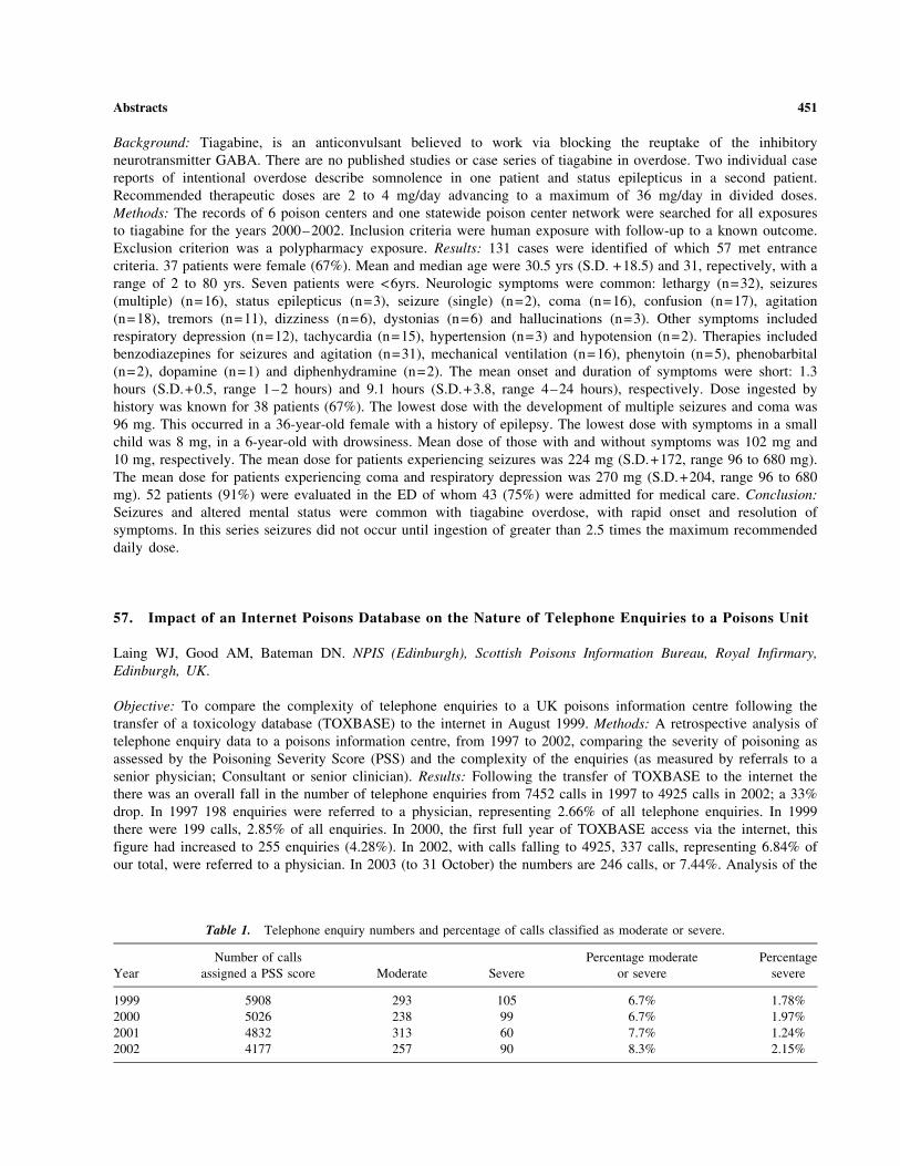
Background: Tiagabine, is an anticonvulsant believed to work via blocking the reuptake of the inhibitory
neurotransmitter GABA. There are no published studies or case series of tiagabine in overdose. Two individual case
reports of intentional overdose describe somnolence in one patient and status epilepticus in a second patient.
Recommended therapeutic doses are 2 to 4 mg/day advancing to a maximum of 36 mg/day in divided doses.
Methods: The records of 6 poison centers and one statewide poison center network were searched for all exposures
to tiagabine for the years 2000–2002. Inclusion criteria were human exposure with follow-up to a known outcome.
Exclusion criterion was a polypharmacy exposure. Results: 131 cases were identified of which 57 met entrance
criteria. 37 patients were female (67%). Mean and median age were 30.5 yrs (S.D. +18.5) and 31, repectively, with a
range of 2 to 80 yrs. Seven patients were <6yrs. Neurologic symptoms were common: lethargy (n=32), seizures
(multiple) (n=16), status epilepticus (n=3), seizure (single) (n=2), coma (n=16), confusion (n=17), agitation
(n=18), tremors (n=11), dizziness (n=6), dystonias (n=6) and hallucinations (n=3). Other symptoms included
respiratory depression (n=12), tachycardia (n=15), hypertension (n=3) and hypotension (n=2). Therapies included
benzodiazepines for seizures and agitation (n=31), mechanical ventilation (n=16), phenytoin (n=5), phenobarbital
(n=2), dopamine (n=1) and diphenhydramine (n=2). The mean onset and duration of symptoms were short: 1.3
hours (S.D.+0.5, range 1–2 hours) and 9.1 hours (S.D.+3.8, range 4–24 hours), respectively. Dose ingested by
history was known for 38 patients (67%). The lowest dose with the development of multiple seizures and coma was
96 mg. This occurred in a 36-year-old female with a history of epilepsy. The lowest dose with symptoms in a small
child was 8 mg, in a 6-year-old with drowsiness. Mean dose of those with and without symptoms was 102 mg and
10 mg, respectively. The mean dose for patients experiencing seizures was 224 mg (S.D.+172, range 96 to 680 mg).
The mean dose for patients experiencing coma and respiratory depression was 270 mg (S.D.+204, range 96 to 680
mg). 52 patients (91%) were evaluated in the ED of whom 43 (75%) were admitted for medical care. Conclusion:
Seizures and altered mental status were common with tiagabine overdose, with rapid onset and resolution of
symptoms. In this series seizures did not occur until ingestion of greater than 2.5 times the maximum recommended
daily dose.
57. Impact of an Internet Poisons Database on the Nature of Telephone Enquiries to a Poisons Unit
Laing WJ, Good AM, Bateman DN. NPIS (Edinburgh), Scottish Poisons Information Bureau, Royal Infirmary,
Edinburgh, UK.
Objective: To compare the complexity of telephone enquiries to a UK poisons information centre following the
transfer of a toxicology database (TOXBASE) to the internet in August 1999. Methods: A retrospective analysis of
telephone enquiry data to a poisons information centre, from 1997 to 2002, comparing the severity of poisoning as
assessed by the Poisoning Severity Score (PSS) and the complexity of the enquiries (as measured by referrals to a
senior physician; Consultant or senior clinician). Results: Following the transfer of TOXBASE to the internet the
there was an overall fall in the number of telephone enquiries from 7452 calls in 1997 to 4925 calls in 2002; a 33%
drop. In 1997 198 enquiries were referred to a physician, representing 2.66% of all telephone enquiries. In 1999
there were 199 calls, 2.85% of all enquiries. In 2000, the first full year of TOXBASE access via the internet, this
figure had increased to 255 enquiries (4.28%). In 2002, with calls falling to 4925, 337 calls, representing 6.84% of
our total, were referred to a physician. In 2003 (to 31 October) the numbers are 246 calls, or 7.44%. Analysis of the
Table 1. Telephone enquiry numbers and percentage of calls classified as moderate or severe.
Year
Number of calls
assigned a PSS score Moderate Severe
Percentage moderate
or severe
Percentage
severe
1999 5908 293 105 6.7% 1.78%
2000 5026 238 99 6.7% 1.97%
2001 4832 313 60 7.7% 1.24%
2002 4177 257 90 8.3% 2.15%
Abstracts 451
risk presented by the poisonings also shows a positive trend. In 1997, 305 calls, (4.09%) were said to have
confirmed toxicity. From there the overall percentage rose: 337 calls (4.79%) in 1998, 337 calls (4.83%) in 1999,
475 enquiries (7.98%) in 2000, 416 (7.43%) in 2001 and 323 calls (6.56%) in 2002. The percentage of patients said
to have moderate or severe poisoning, as assessed by their PSS score, has also increased over the period 1999–2002
(Table 1). Conclusion: In early 2000, shortly after the TOXBASE poisons information database was transferred to
the internet, the Department of Health formally recommended TOXBASE as the first-line source of poisons
information. These figures suggest that, as intended, telephone enquiries to a poisons unit are being reserved for the
more serious and/or complicated poisonings, those where the risk of toxicity is greatest, and those showing moderate
or severe symptoms; despite reducing the overall burden of telephone enquiries, our end users are maintaining
contact with the service where it is most appropriate. Reference: Persson HE, Sjoberg GK, Haines JA, Pronczuk de
Garbino J. Poisoning Severity Score. Grading of Acute Poisoning. J Toxicol Clin Toxicol 1998; 36:205–213.
58. Carbon Monoxide: Friend or Foe of the Cell?
Schneider F, Lavigne T, Meziani F, Bilbault P, Castelain V, Jaeger A. Service de Reanimation Medicale, Hopital de
Hautepierre, Strasbourg, France.
Introduction: For several centuries, carbon monoxide (CO) has been regarded as a toxic pollutant which poisons by
binding to the heme-containing group found in haemoglobin. Indeed, innumerable deaths have resulted from
inhalation of this odourless, tasteless and colourless gas generated by incomplete combustion of organic materials. In
addition, CO is frequently found in the smoggy atmosphere of our cities as a result of technological processes and
fumes produced by cars; it is even present in high concentrations in cigarettes. Toxic Effects: The most widely
accepted explanation for the toxicity of CO is based upon an hypoxia-related hypothesis first reported by Haldane:
cell injuries occur as a result of hypoxemia linked to the formation of carboxyhemoglobin rather than
oxyhemoglobin. Higher affinity of substances containing prosthetic structures (i.e.: mitochondrial cytochromes,
myoglobin. . .) for CO than for O2 but also interactions of CO with other substances such as common proteins,
enzymes or lipoproteins are also of importance in explaining the acute toxicity of CO. Additionally, recent data of
the late nineties underscore how delayed toxicity of CO occurs in addition to acute toxicity in tissues like the brain.
A double-step mechanism is suggested. First, CO challenges circulating polymorphonuclear cells which subsequently
undergo diapedesis and then cause brain lipid peroxidation; furthermore, CO affects nitric oxide release by platelets,
which in turn interferes with white cells binding to endothelial cells with a subsequent delay in diapedesis. This
complex process is inhibited by high oxygen supply (>300 kPa): this might partially explain some success of
hyperbaric oxygenation in preventing delayed brain injury. Biological effects: In contrast to these noxious and well-
established actions on cellular and molecular targets, CO has recently been acknowledged as a biological signalling
messenger in some pathways. Although the formation of CO in the body was demonstrated as early as 1952, and
heme oxygenase (HO) recognised as the endogenous enzymatic source of this diatomic gaseous molecule in 1968, it
has only become obvious in the last few years that HO exist in vivo under three isoforms and that their by-products
(CO, biliverdin) play important roles in cellular metabolism. Although much remains to be elucidated about the
mechanisms of physiological and pathophysiological cell signalling by CO, it has become clear that this molecule is
involved in two major biological pathways: the soluble guanylyl cyclase activation (sGC) and the mitogen-activated
protein kinase (MAP-K) cascades. As a result, at sGC activation, CO is involved not only in the regulation of
vascular tone with a possible role in the genesis of hypertension, but also in neurotransmission and therefore in
diverse diseases of the central nervous system. The studies on the effect of CO on the MAP-K cascades have also
brought to our knowledge that CO may contribute to anti-inflammatory activity involving interesting protection in
some pathophysiological conditions. Additionally, an inhibitory effect of CO on cell proliferation and a
concentration-dependent effect on apoptosis have been reported with potential consequences in the field of vascular
diseases. Conclusion: There is no doubt that CO exerts significant effects on many pathways in the cells, mainly—
but not only—mediated by the cyclic guanine monophosphate pathway. These effects occur at very low
concentration of the gas. It must nevertheless be kept in mind that at higher concentrations CO increases the
oxidative stress, especially when free iron release occurs during cell injuries. These data shed new light on unknown
452 Abstracts
aspects of the metabolism of CO but they warrant further investigation before this tiny molecule can definitively be
considered to be a better friend than Janus. References: Brouard S, Otterbein LE, Anrather J, Tobiasch E, Bach FH,
Choi AM, Soares MP. Carbon monoxide generated by heme oxygenase-1 suppresses endothelial cell apoptosis. J
Exp Med 2000; 1992:1015–1026. Dawson TM, Snyder SH. Gases as biological messengers: nitric oxide and carbon
monoxide in the brain. J Neurosci 1994; 14:5147–5159. Ke B, Buelow R, Shen XD, Melinek J, Amersi F, Gao F,
Ritter T, Volk HD, Busuttil RW, Kupiec-Weglinski JW. Heme oxygenase-1 gene transfer prevents CD95/Fas-ligand-
mediated apoptosis and improves liver allograft survival via carbon monoxide signalling pathway. Hum Gen Ther
2002; 13:1189–1199. Maines MD. The heme oxygenase system: a regulator of second messenger gases. Ann Rev
Pharmacol Toxicol 1997; 37:517–554. Otterbein LE, Mantell LL, Choi AM. Carbon monoxide provides protection
against hyperoxic lung injury. Am J Physiol 1999; 276:L688–L694. Villamor E, Perez-Vizcaino F, Cogolludo AL,
Conde-Oviedo J, Zaragoza-Arnaez F, Lopez-Lopez JG, Tamargo J. Relaxant effects of carbon monoxide compared with
nitric oxide in pulmonary and systemic vessels of newborn piglets. Pediatr Res 2000; 48:546–553.
59. Acute Effects and Long Term Sequelae of Carbon Monoxide Poisoning
Mathieu D. Service d’Urgence Respiratoire, de Reanimation Medicale et de Medecine Hyperbare, Hopital Calmette,
Centre Hospitalier Universitaire, Lille, France.
Carbon monoxide (CO) poisoning causes a wide variety of signs and symptoms, often non specific leading to
numerous diagnostic errors (1). Most commonly, early clinical manifestations include neurological changes as:
headache, fatigue, nausea and vomiting, dizziness, memory and personality changes. Neurological examination may
evidence abnormal findings such as hypertonia, hyperreflexia, Babinsky sign. Loss of consciousness reflects a more
severe state of poisoning and precedes the coma phase. Death occurs due to respiratory failure, circulatory collapse
and cardiac arrest. Multiple unusual clinical presentations exist and may be psychiatric (dementia, depression),
cardiovascular (chest pain, rhythm disturbances), respiratory (dyspnoea), neuromuscular (flaccidity, muscle
weakness). Besides these acute manifestations, it has long been recognised CO poisoning may cause long term
manifestations and sequelae (2). These include malaise, apathy, memory disturbances, depression, anxiety, focal
neurological abnormalities, ataxia and Parkinson-like syndromes. These manifestations may be differentiated
according to the time of their occurrence in 2 types: persistent manifestations are signs or symptoms occurring
during the CO intoxication that persist after treatment; delayed manifestations require a lucid interval in which the
CO-poisoned patient improves, often to normal and has a subsequent deterioration. These have also to be
differentiated in transient manifestations, which improve back to normal, and sequelae, which induce permanent
disability. Neuro-imaging studies have demonstrated in some of these patients, brain lesions in the deep white
matter, the hippocampus and the globus pallidus. The incidence of these long term manifestations and sequelae is
variously reported in the literature between 3% and 45% depending of terminology used, studied population, initial
severity of CO poisoning, treatment, time and method of evaluation (3). For example, gross neurological impairment
inducing severe disability and abnormal neuro-imaging findings is reported in 5–15% of CO poisoned patients when
subjective complains in response to standardized questionnaire are reported in 25–45%. Neuropsychological testing
is an even more sensitive method. Time of evaluation is also critical as CO induced persistent manifestations may be
difficult to differentiate from post-traumatic stress disorder symptoms in the 6 first weeks after CO poisoning.
Careful long-term follow-up studies show immediately after the intoxication a high incidence of persistent
manifestations. Between 25–40% of patients at one month complain of subjective symptoms. This rate declines at 3
and 6 months and stabilizes at 1 and 2 years. This fact explains in part the discrepancies between studies and the
difficulties in interpreting results from randomised controlled studies, especially when short follow-up are considered
(4). Prognostic factors are not clearly established. If the experimental studies show that CO exposure (time,
concentration), patient condition (age, physiological status), minute ventilation, cardiac output, sur-imposed causes of
tissue hypoxia or toxicity are important factors determining the initial severity of CO intoxication, clinical predictive
factors are less well established. Age, loss of consciousness during poisoning, initial neurological abnormality are the
more frequently evidenced factors predicting the occurrence of persistent manifestations. The role of
carboxyhemoglobin level is a matter of discussion but is usually considered as non predictive if the sampling is
Abstracts 453
not taken on the site of poisoning. Although long claimed, oxygen treatment modality (hyperbaric or normobaric)
(5,6) has recently been proved to have an important role in the incidence of persistent neurological manifestations.
References: 1. Raub JA, Mathieu-Nolf M, Hampson NB, Thom SR. Carbon monoxide poisoning—a public health
perspective. Toxicology 2000; 145:1–14. 2. Smith J, Brandon S. Morbidity from acute carbon monoxide poisoning
at three years follow up. Br Med J 1973; 1:318–321. 3. Seger D, Welch L. Carbon monoxide controversies:
neuropsychologic testing, mechanism of toxicity, and hyperbaric oxygen. Ann Emerg Med 1994; 24:242–248. 4.
Hampson NB, Mathieu D, Piantadosi CA, Thom SR, Weaver LK. Carbon monoxide poisoning: interpretation of
randomized clinical trials and unresolved treatment issues. Undersea Hyperb Med 2001; 28:157–164. 5. Mathieu D,
Nolf M, Durocher A, Saulnier F, Frimat P, Furon D, Wattel F. Acute carbon monoxide intoxication. Treatment by
hyperbaric oxygen and risk of late sequelae. J Toxicol Clin Toxicol 1985; 23:315–324. 6. Weaver LK, Hopkins RO,
Chan KJ, Churchill S, Elliott CG, Clemmer TP, Orme JF Jr, Thomas FO, Morris AH. Hyperbaric oxygen for acute
carbon monoxide poisoning. N Engl J Med 2002; 347:1057–1067.
60. S100B Protein in Carbon Monoxide Poisoning
Brvar M, Osredkar J, Mozina M, Bunc M. University Medical Center, Ljubljana, Slovenia.
Objective: The aim of the study was to assess the possible role of S100B (1,2), the structural protein of astroglia, as
a biochemical marker in carbon monoxide (CO) poisoning in an animal model and humans. Methods: Study I:
Serum S100B determination was performed in 30 CO poisoned rats. On the day before exposure, under brief
ketamine-xylazine intraperitoneal anesthesia, unilateral jugular vein catheterization was performed. The rats were
grouped and exposed to high CO concentration (5000 ppm) for a short period (30 minutes) (group 1, n=14) and
lower CO concentration (3000 ppm) for a longer period (60 minutes) (group 2, n=16). Blood samples were taken
from jugular vein before and immediately after CO poisoning. Blood samples were processed to serum, centrifuged
at 4000 rpm for 15 minutes and stored at �20�C until assayed. Study II: Serum S100B determination was performed
in 38 consecutive CO poisoned patients admitted at the Emergency Department (ED). Blood samples were drawn
from peripheral vein immediately after arrival at the ED and processed to serum, centrifuged at 4000 rpm for 15 minutes
and stored at 4�C for less than 24 hours. The S100B concentrations were measured with a commercial
radioimmunoassay (Liaisona Sangteca 100, Sangtec Medical AB, Bromma, Sweden). Blood samples were also
analysed for creatine kinase, troponin T and carboxyhaemoglobin level. Results: Study I: In the first group, exposed
to 5000 ppm CO, all 14 (100%) rats were unconscious after exposure and their S100B levels were significantly
higher after exposure compared to their S100B levels before exposure (0.54 microg/l versus 0.06 microg/l, p<0.05).
8 rats (57%) later died and their S100B levels were significantly higher compared to S100B levels of the remaining
6 (43%) rats that regained conciseness and survived (0.86 microg/l versus 0.21 microg/l, p<0.05). In the second group,
exposed to 3000 ppm CO, only 6 (38%) rats were unconscious after exposure. Rats with and without loss of
consciousness had significantly higher S100B levels after exposure compared to their S100B levels before exposure
(1.05 microg/l versus 0.10 microg/l, p<0.05 and 0.28 microg/l versus 0.08 microg/l, p<0.05; respectively). All
6 unconscious rats later died and their S100B levels were significantly higher compared to S100B levels of the remain-
ing 10 (62%) rats that were without loss of consciousness and survived (1.05 microg/l versus 0.28 microg/l, p<0.05).
Study II: All 3 unconscious patients had elevated S100B. The patient with the highest S100B died. S100B was
elevated in 2 out of 6 patients with initial transitory unconsciousness at the scene. All 29 patients without loss of
consciousness had normal S100B. Conclusion: CO poisoning is associated with elevated S100B levels. S100B was
significantly elevated in CO poisoned rats. The unconscious rats after CO exposure had significantly higher S100B
levels compared to the rats without loss of consciousness. The unconscious rats that later died had significantly higher
S100B levels compared to the rats that survived. CO poisoning with loss of consciousness was associated with
elevated S100B levels in humans. S100B could be a useful biochemical marker/parameter for clinical evaluation and
prognosis of CO-poisoned patients. References: 1. Bottiger BW, et al. Astroglial protein S-100 is an early and sensitive
marker of hypoxic brain damage and outcome after cardiac arrest in humans. Circulation 2001; 103:2694–2698. 2.
Brvar M, et al. The potential value of the protein S-100B level as a criterion for hyperbaric oxygen treatment and
prognostic marker in carbon monoxide poisoned patients. Resuscitation 2003; 56:105–109.
454 Abstracts
61. Clinical and Scintigraphic Heart Evaluation in Young Acutely Carbon Monoxide PoisonedPatients: Rest 99 mTc-MIBI Spect and Stress-Rest 99mTc-MIBI Spect Follow Up
Hubalewska A, Pach D, Pach J (1), Staszczak A, Huszno B, Winnik L (1). 1. Department of Clinical Toxicology,
Nuclear Medicine Unit, Chair and Dep. of Endocrinology Medical College Jagiellonian University, Krakow, Poland.
Objective: Unlike the brain, CO cardiotoxicity may be clinically occult, and despite electrocardiography and
echocardiography, remains often undiagnosed. Over the years radionuclide myocardial perfusion imaging has become
an important diagnostic tool. The results of 8-years follow up study of CO cardiotoxicity using 99 mTc-MIBI
SPECT are presented in the paper. Material and Methods: Under examination there were 96 acutely CO poisoned
patients without cardiac history (male 54, female 42; mean age 26±8.4 years) treated at the Krakow Department of
Clinical Toxicology Jagiellonian University Medical College in 1996–2003. Measurement of COHb, blood lactate
level, duration of exposure and ECG examination were performed on admission. ALT, AST, CPK, MB-CK activity
were evaluated after 24–48 hours. CO poisoning severity was graded according to own clinical scale. The
scintigraphic examinations were carried out using Siemens two-head gamma camera (E-cam) equipped with parallel
high resolution collimators. Rest 99mTc-MIBI SPET scans were performed on days 1 to 5 post admission. The control
stress-rest 99mTc-MIBI SPET were done about 6 month post-exposure with routine procedure using moving track
exercise test according to Bruce protocol. The changes in -MIBI scan were graded: 0—normal scan; I0—diminished
and non-homogenic uptake II0—diminished and small foci of tracer absence; III0—visible diminished uptake of
tracer+one bigger ‘‘cold spot’’; IV0—large and numerous ‘‘cold spots’’. T-student test for unpaired data and Mann–
Whitney test for non parametric data were used in statistical evaluation. Results: Significant differences were found in
HbCO level, blood lactate concentration and CK activity between the subgroups of patients divided according to the
degree of scintigraphic changes. The exacerbation of scintigraphic changes were depended on the degree of CO
poisoning severity. In 6 of 47 patients who underwent control scintigraphic examination the worsening but in 19 the
cases an improvement in scintigraphic scan were observed six months post-exposure. An exercise ischemia (stress-rest
99mTc-MIBI SPET) was observed in 15 patients: 73% of them were qualified to the third degree of scintigraphic
changes during acute phase of intoxication. Conclusions: The results confirm that heart metabolic injury due to CO
intoxication is different than that after coronary occlusion, and the heart damage process usually takes a longer time.
99mTc-MIBI myocardial perfusion scintigraphy, electrocardiography, measurement of chosen enzymes activity
performed simultaneously and poisoning severity scoring appear to be well-fitted methods in predicting the heart
damage in CO acutely poisoned patients.
62. Managing Carbon Monoxide Poisoning with Hyperbaric Oxygen
Raphael JC (1), Chevret S (2), Driheme A (2), Annane D (1). 1. Service de Reanimation Medicale, Hopital Raymond
Poincare; Garches; 2. Departement de Biostatistique et Informatique Medicale, Hopital Saint Louis, Paris, France.
Objective: The aim of this study was to define the indications and modalities of hyperbaric oxygen therapy (HBO) in
patients with acute carbon monoxide (CO) poisoning. HBO is warranted only as an adjunct to symptomatic measures
and after 100% normobaric oxygen therapy (NBO) started at the accident site. Methods: Review of the recent literature
including therapeutic trials published as articles or presented as abstracts. Results: Preventing secondary neurological
complications is the main objective of HBO. There is general agreement that HBO should be used routinely in pregnant
women, regardless of the clinical severity in the mother. This consensus stems, not from therapeutic trials, but from
pathophysiological considerations (high risk of severe CO toxicity in the fetus) and from noncomparative pilot studies
showing that HBO is well tolerated by pregnant women, with no increase in the abortion rate as compared to the general
population. There is no evidence that cognitive function in the offspring is better with than without HBO in the mother.
No studies have specifically investigated direct CO poisoning in children. Rules for HBO use are usually considered to
be the same in children and adults. Nevertheless, incipient disorders in consciousness may be difficult to detect in
younger children and infants. Neurological sequelae are identical to those seen in adults but seem less prevalent.
Conflicting results have been obtained in adults. Explanations include marked differences in inclusion criteria
Abstracts 455
(intentional, unintentional, or both), treatment protocols, evaluation criteria, and sample size. A Cochrane systematic
review (1) found no evidence that HBO reduced the rate of neurological symptoms after 1 month (OR for benefit with
HBO: 0.82; 95%CI, 0.41–1.66). A recent study (2) suggested that HBO might diminish residual cognitive impairments
as assessed by neuropsychological testing, although this had no impact on occupational activities. Recently we have
done two randomized controlled trials (unpublished data). According to initial neurological complications, before
randomization, victims of accidental domestic CO poisoning were classified into group A, i.e. transient loss of
consciousness, and group B, i.e. comatose patients. Patients in group A were randomly assigned to a 6-hour course of
normobaric oxygenation (NBO) (arm A0) or to one session of HBO at a plateau of 2 ATA for 60 minutes plus 4-hour
NBO (A1). Patients in group B were randomly assigned to one (arm B1) or two (arm B2) sessions of HBO at a plateau of
2 ATA for 60 minutes plus 4-hour NBO. The primary endpoint was the proportion of patients who recovered at one
month following randomization. Recovery was defined by normal self-assessment questionnaire and normal blinded
neurological examination. Three hundred and eighty five patients were enrolled, 179 in group A (A0, n=86 and A1,
n=93) and 206 in group B (B1, n=101, B2, n=105). At one month, in group A, they were 45/74 (61%) patients who
recovered in A0, and 46/79 (58%) in A1, odds ratio, 0.90 (95% CI, 0.47 to 1.71). In group B, they were 54/80 (68%) who
recovered in B1 compared to 42/90 (47%) in B2, odds ratio, 0.42 (CI, 0.23 to 0.79). For patients died, and four survivors
in B2 arm had severe neurological sequelae. Among patients who died or had severe neurological sequelae all one were
in B group. The baseline HbCO level had no impact on outcomes in most studies. Severe neurological sequelae occur
only in those patients with a coma at presentation. Repeating the HBO sessions does not seem to influence the risk of
severe neurological sequelae; given the absence of comparative studies versus NBO, there is no proof that HBO
decreases this risk. Although the literature recommends HBO within the first 24 hours after CO exposure, this has not
been validated. The dosage of each session (duration and pressure) varies in the literature. Dosage recommendations rely
neither on pathophysiological data available in humans nor on comparative clinical studies. The dosage is often
influenced both by local habits and by efforts to minimize intolerance and safety risks in the patient. Conclusions:
Except in pregnant women, there is no evidence supporting routine use of HBO in patients with acute CO
poisoning. Given the high cost and potential morbidity associated with HBO, this treatment should probably be
reserved for patients with a coma at presentation. Several other criteria indicating severe poisoning have been
suggested (advanced age, consciousness impairments other than coma, and coronary involvement) but not validated.
There is no evidence that HBO sessions should be repeated. Available data are insufficient for making recom-
mendations about HBO session modalities. References: Juurlink DN, Stanbrook MB, McGuigan MA. Hyperbaric
oxygen for carbon monoxide poisoning (Cochrane Review). In: The Cochrane Library, issue 4, 2000. Oxford:
Update Software. Weaver LK, Hopkins RO, Chan KJ, Churchill S, Elliott CG, Clemmer TP, Orme JF, Thomas FO,
Morris AH. Hyperbaric oxygen for acute carbon monoxide poisoning. N Engl J Med 2002; 347:1057–1067.
63. The Controversial Role of Hyperbaric Oxygen Therapy in Pregnancy and Young Children
Liebelt EL. Children’s Hospital and University of Alabama at Birmingham School of Medicine, Alabama,
USA.
Currently, there are no standard guidelines for defining severity of carbon monoxide (CO) poisoning or indications for
hyperbaric oxygen therapy (HBOT) in pregnant women or in young children. CO is a fetotoxin and teratogen.
Controversy regarding HBOT during pregnancy stems primarily from the concern for oxygen toxicity to the fetus.
Various physiological differences may increase the susceptibility of the fetus and young infants/children to the
deleterious effects of both carbon monoxide and HBOT. Fetal COHgb depends on maternal COHgb levels, placental
diffusion capacity for CO, endogenous maternal and fetal production of CO, and relative affinities of maternal and
fetal Hgb for CO and oxygen. The fetus is vulnerable to the effects of CO because of low oxygen tension in fetal
arterial blood and a decreased ability to compensate for hypoxia. The fetal oxyhemoglobin dissociation curve is
shifted to the left normally and in the presence of CO shifts further to the left, resulting in significant fetal tissue
hypoxia when there is a small decrease in maternal oxygen tension (i.e. increased maternal COHgb). On the basis of
pathophysiologic arguments, HBOT would appear to be a useful treatment by potentially reducing peak fetal COHgb
levels, increasing oxygen delivery to the fetus, promoting more rapid fetal CO clearance, and reducing apoptosis in the
456 Abstracts
fetal brain. However, there are concerns that high partial pressure oxygen may cause teratogenicity, retinopathy, and/
or cardiovascular defects (primarily closure of the ductus arteriosus) in the developing fetus. Animal studies of HBOT
on the developing fetus are equivocal. There are no controlled human trials of HBOT in CO poisoned pregnant
women. However, there are numerous case reports/series in the US and international literature reporting normal fetal
outcomes after HBOT. Infants and young children may have an increased susceptibility to CO toxicity because of: 1) a
higher basal metabolic rates and consequently higher tissue oxygen demands; 2) the presence of anemia; and 3) a
higher percentage of fetal hemoglobin (i.e. children with sickle cell anemia and healthy young infants). Clinical
evidence for the beneficial effects of HBOT in CO-poisoned children is limited to a few small retrospective case series
and isolated case reports. Most of these studies lack formal psychological testing and significant long-term follow-up.
There are also special and practical considerations for the young pediatric patient receiving HBOT. In conclusion,
although there are theoretical concerns, limited data suggest that HBOT appears to be safe for the pregnant patient and
fetus. HBOT should be considered for pregnant women with significant CO toxicity. If there are no maternal
indications, HBOT should be considered if there is evidence of fetal distress. Whether HBOT is beneficial for young
infants/children remains unanswered. References: Elkharrat D, Raphael JC, Korach JM, et al. Acute carbon monoxide
intoxication and hyperbaric oxygen in pregnancy. Intensive Care Med 1991; 17:289–292. Koren G, Sharav T,
Pastuszak A, et al. A Multicenter, prospective study of fetal outcome following accidental carbon monoxide poisoning
in pregnancy. Reprod Toxicol 1991; 5:397–403. Thombs PA, Martorano FJ. Hyperbaric medicine in pediatric
practice. In: Hyperbaric Medicine Practice. Kindwall EP (ed). Flagstaff: Best Publishing, 1995:261–275. Liebelt EL.
Hyperbaric oxygen therapy in childhood carbon monoxide poisoning. Curr Opin Pediatr 1999; 11:259–264. Longo
LD. The Biological effects of carbon monoxide on the pregnant woman, fetus, and newborn infant. Am J Obstet
Gynecol 1977; 129:69–103. van Hoesen KB, Camporesi EM, Moon RE, et al. Should hyperbaric oxygen be used to
treat the pregnant patient for acute carbon monoxide poisoning? JAMA 1989; 261:1039–1043.
64. Carbon Monoxide Poisoning: A Critical Analysis of Clinical Trials
Buckley NA. Clinical Pharmacology and Toxicology, Australian National University Medical School, ACT,
Australia.
There have been five randomised controlled trials published on hyperbaric oxygen (HBO) for acute carbon monoxide
poisoning (1–5). There is also a further very large trial, the interim results of which have been published as an
abstract (6), and which is continuing to enrol patients and a further unpublished trial completed in 2000. There has
also been a Cochrane collaboration review which provided a summary estimate of effect from data from three of
these studies (7). However, there is continuing debate about the merits of this treatment resulting from less than ideal
study designs in these trials, and markedly different and directly conflicting results in the outcomes. Assessment of
‘quality’ of trials in Cochrane reviews usually focuses on a small number of sources of bias—randomisation
procedures, blinding, intention to treat analysis and follow up. This ignores many other possible sources of bias, and
also perhaps favours negative studies for which such biases are less relevant. In this analysis I will focus particularly
on those aspects of good trial design that if not addressed commonly lead to a false positive result in the positive
studies (randomisation, blinding, intention to treat, analytical bias) and those that commonly lead to a false negative
result in the negative studies (power, follow-up, optimal intervention and control arm). It is apparent that there is
considerable room for errors in the negative trials—due to insensitive outcome measures or failure of follow-up in
particular. However the positive trials provide much evidence of bias suggesting that their results should not be
taken at face value. Any attempt to pool such heterogenous trials to provide a common summary measure of
outcome should be viewed with some scepticism. The trials examined had very different interventions, populations
and outcome measures. Moreover, no provision is made in such meta-analyses for the direction of bias or
measurement error. Even so, pooling Weaver with other studies from the Cochrane meta-analysis using a random
effects model gives a very wide confidence interval (odds ratio: 0.65, CI 0.34 to 1.23), suggesting benefits but
conceding a significant possibility of harm from HBO treatment. Adding in the results from unpublished studies still
does not provide a clear outcome. Moreover, the benefits may not be clinically important enough to justify any risk
to the patient in travelling to a facility that has a chamber. The mean neuropsychological test results in the control
arm of Weaver’s trial were within the normal range, suggesting only the most severe patients may derive a large
Abstracts 457
enough benefit to warrant the risks and expense of such interventions. There have been no significant long-term
functional differences in outcome in any studies. Further studies are required looking at such outcomes. The high
costs of HBO treatment and the care of the severely neurological disabled justify an externally audited and analysed
large multi-centre trial. References: 1. Weaver LK, Hopkins RO, Chan KJ, Churchill S, Elliott CG, Clemmer TP,
et al. Hyperbaric oxygen for acute carbon monoxide poisoning. N Engl J Med 2002; 347:1057–1067. 2. Thom SR,
Taber RL, Mendiguren II, Clark JM, Hardy KR, Fisher AB. Delayed neuropsychologic sequelae after carbon
monoxide poisoning: prevention by treatment with hyperbaric oxygen. Ann Emerg Med 1995; 25:474–80. 3. Ducasse
JL, Celsis P, Marc-Vergnes JP. Non-comatose patients with acute carbon monoxide poisoning: hyperbaric or
normobaric oxygenation? Undersea & Hyperbaric Medicine 1995; 22:9–15. 4. Raphael JC, Elkharrat D, Jars GM,
Chastang C, Chasles V, Vercken JB, et al. Trial of normobaric and hyperbaric oxygen for acute carbon monoxide
intoxication. Lancet 1989; 2:414–419. 5. Scheinkestel CD, Bailey M, Myles PS, Jones K, Cooper DJ, Millar IL, et al.
Hyperbaric or normobaric oxygen for acute carbon monoxide poisoning: a randomised controlled clinical trial. Med
J Aust 1999; 170:203–210. 6. Mathieu D, Wattel F, Mathieu-Nolf M, Durak C, Temepe JP, Bouachour G, et al.
Interim analysis—controlled clinical trial of hyperbaric oxygen in acute carbon monoxide (CO) poisoning. Undersea
& Hyperbaric Medicine 1996; 23(suppl):7–8. 7. Juurlink DN, Stanbrook MB, McGuigan MA. Hyperbaric oxygen
for carbon monoxide poisoning. Cochrane Database Syst Rev 2000; (2):CD002041.
65. Mechanisms and Clinical Features of Poisoning by European Mushrooms
Persson H. Swedish Poisons Information Centre, Stockholm, Sweden.
Objective: To review pathophysiology and clinical features of poisoning by European mushrooms. Methods: Survey
of current literature and local experience from non-published patient materials. Mushroom toxicology is well suited
for a systematic approach, based on the action of the toxins and related clinical syndromes. Results: (I)
Gastrointestinal irritants. Many fungal families include species that cause gastrointestinal upset as the only toxic
manifestation. Most of these toxins have note been chemically identified. Symptoms occur within a few hours and are
normally of short duration. Only rarely do fluid and electrolyte balance disturbances ensue, although there are some
exceptions (e.g. Entoloma sinuatum, Paxillus involutus). (II) Cytotoxic fungi: 1) Amatoxins occur in certain spp. of the
families Amanitaceae (genus Amanita), Agaricaceae (genus Lepiota) and Cortinariaceae (genus Galerina). Amatoxins
are bicyclic octapeptides with a molecular weight of around. 9000. Toxicity is exceptional and 0.1 mg/kg bodyweight
may be fatal in humans. Amatoxins block mRNA polymerase II activity in eucaryotic cells. Hence, mRNA content will
progressively decrease, resulting in deficient protein synthesis and cell death. Furthermore, it has been postulated that
amatoxins act in synergy with endogenous cytokines and cause cell damage through the induction of apoptosis. Cells
providing a transport device (intestinal mucosal cells, hepatocytes and proximal, tubular cells) are especially vulnerable.
Amatoxins are rapidly absorbed and distributed in the body. Elimination is mainly renal. After ingestion there is a
latency period or 8–24 h (mean 12 h) before onset of intense gastrointestinal symptoms dominated by violent, watery
diarrhoea. These symptoms may persist 1–2 days or longer with subsequent dehydration and metabolic disturbances.
After 36–48 h, sometimes earlier, biochemical signs of hepatic damage appear, later on followed by clinical signs of
liver damage. Fulminant hepatic failure may ensue. The severity of the toxic reaction is related to the toxin dose and the
heavier the exposure is, the earlier is the onset of symptoms and the more intense is the clinical course. The high
mortality is still a matter of concern. 2) Orellanine is found in certain spp. of the family Cortinariaceae, genus
Cortinarius. The toxicity of orellanine, a bipyridine N-oxide, is probably related to a metabolite. Oxidation of orellanine
cause accumulation of quinone compounds that bind covalently to renal cell structures resulting in tubular necrosis,
interstitial nephritis and fibrosis. Kinetics is not well known, but a fairly rapid distribution is likely. Orellanine has not
been found in plasma or urine samples 2 days post-ingestion, but fixed in renal tissue after 9 and 60 days. The clinical
syndrome of orellanine poisoning is ghostlike in that initial warning symptoms are either lacking or mild. Therefore
patients do not seek medical attention until symptoms of renal dysfunction are at hand. In typical cases the patient is
admitted to hospital 2 days or more post-ingestion because of abdominal and lumbar pain, chills, headache, fatigue,
anorexia, intense thirst and poly- or oliguria. Anuria may develop and end-stage renal failure may ensue. A method for
estimating the prognosis in individual cases has recently been established by Holmdahl. 3) Other nephrotoxic
fungi. According to reports from Southern France, Amanita proxima ingestion has been associated with moderate
458 Abstracts
gastro-intestinal symptoms (onset after 2–48 hrs, average 13 hrs) and renal damage. Hemodialysis was required in about
one fourth of the cases because of renal failure. Chronic renal failure has not bee reported. The toxin and possible
mechanisms are still unknown. Severe and long-lasting gastrointestinal symptoms and renal damage have also been
reported after ingestion of A. smithiana (North America) and A. pseudoporphyria Hongo (Japan). The toxin has in these
cases been claimed to be allenic norleucin. 4) Rhabdomyolysis. Clinicians from France and Poland have described acute
rhabdomyolysis after large and repeated meals of Tricholoma equestre ( T. Flavovirens). Onset of fatigue, muscular
weakness and myalgias occurred within 24–72 hours after the last of a number of meals. Clinical and laboratory data
indicated acute rhabdomyolysis. Experimental data support the presence of some myotoxic compound, not yet
chemically identified. While awaiting further evidence to support the causality, it might be wise to refrain from eating
this Tricholoma species. 5) Gyromitrin occurs in some species of the family Helvellaceae, genus Gyromitra. The toxin is
water soluble and volatile, and exposure can occur both through inhalation and ingestion. When ingested, gyromitrin is
rapidly hydrolysed to hydrazines (mainly MFH and MMH). Hydrazines are irritants, reduce CNS pyridoxin contents and
GABA synthesis, cause hepatocellular damage and hemolysis. However, in reality most gyromitrin poisonings are
relatively benign and fatalities are uncommon. Gastrointestinal symptoms may occur after 5–8 hrs, but are not seldom
lacking. More typical are neurological symptoms like vertigo, nystagmus, diaphoresis, fatigue, confusion, somnolence
and in severe cases (rare) coma and convulsions. After 1–3 days signs of moderate hepatic injury, haemolysis
(sometimes associated with methaemoglobinemia) and kidney damage may follow. (III) Neurotoxic mushrooms: 1)
Muscarine occurs in certain Inocybe, Clitocybe and Omphalatus spp. Symptoms observed after ingestion are related to
stimulation of muscarine receptors in the autonomic nervous system. Typically symptoms appear 2 hrs post-ingestion
and include diaphoresis, nausea, abdominal pain and diarrhoea, hypersalivation, lacrimation and rhinorroea. More
serious symptoms are bronchorroea, bronchospasm, bradycardia and hypotension. 2) Isoxazoles (e.g. ibotenic acid,
muscimol, muscazone) are present especially in Amanita muscaria and A. pantherina and act as GABA agonists.
Symptoms occur within 3 hrs. The clinical syndrome includes gastro-intestinal discomfort, inebriation, euphoria,
anxiety, confusion and hallucinations. Heavy exposure may result in severe agitation, violent behaviour and even
convulsions and coma (rare). Poisoning has been misdiagnosed as acute psychosis. Occasionally cholinergic symptoms
have been observed in parallel, especially in A. muscaria poisoning. 3) Psilocybin occurs in Psilocybe, Panaeolus,
Conocybe, Gymnopilus, Stropharia, Pluteus and Panaelina spp. It is a potent hallucinogen with effects similar to those
of LSD (stimulation of central serotonin receptors and blockade of peripheral serotonin receptors). Phenylethylamine has
been found in some species and may be responsible for the sympathomimetic effects. Symptoms are normally seen
within an hour. Hallucinations, not seldom bizarre and unpleasant, are the main feature. Euphoria is often followed by
anxiety and agitation. Other symptoms are altered time and space sense, depersonalisation, headache, vertigo, nausea,
tachycardia, mydriasis, flushing, fever and seizures (rare). Symptoms peak after around 2 hrs and vanish within 4–6 hrs,
although flashbacks may recur for weeks and months. Poisoning has been misdiagnosed as acute psychosis. (IV)
Miscellaneous: 1) Antabuse syndrome. Coprinus atramentarius contains coprin, which blocks acetaldehyde
dehydrogenase, and together with alcohol it may induce an ‘antabuse syndrome’. After ingestion of the mushroom
this risk persists for about a week. 2) Paxillus syndrome is uncommon but important as a differential diagnosis in case of
an acute haemolytic syndrome. This mushroom Paxillus involutus contains less well defined, strongly antigenic
components that after repeated exposure may induce severe gastro-enteritis, haemolysis and renal failure. 3)
Erythromelalgia is a strange syndrome, described in Japan 1918 after ingestion of Clitocybe acromelalga. Interestingly
cases with a striking similarity have lately been reported from France after ingestion of Clitocybe amoenolens. Isolated
toxins are glutamate agonists. Symptoms include burning pain in hands and feet, occurring as paroxysmal crises,
especially during night and upon touch. Symptoms seem to be aggravated by warmth and a lowered position of the
extremity. Pain may be accompanied by local erythema and oedema. References: Faulstich H. Mushroom poisoning.
Lancet 1980; 2:794–795. Wieland T. The toxic peptides from Amanita mushrooms. Int J Peptide Protein Res 1983;
22:257–76. Leist M, et al. Tumour necrosis factor-induced apoptosis during the poisoning of mice with hepatotoxins.
Gastroenterology 1997; 112:923–934. Jaeger A, et al. Kinetics of amatoxins in human poisoning: therapeutic
implications. J Toxicol Clin Toxicol 1993; 31:63–80. Antkowiak WZ, Gessner WP. Isolation and characteristics of toxic
components of Cortinarius orellanus Fries. Bull Acad Pol Sci Ser Sci Cmi 1975; 23:729. Antkowiak WZ, Gessner WP.
The structures of orellanine and orelline. Tetrahedon Lett 1979; 21:1931. Richard JM, et al. Orellanine inhibits protein
synthesis in Madin-Darby canine kidney cells, in rat liver mitochondria, and in vivo: indication for its activation prior to
in vitro inhibition. Toxicology 1991; 67:53–62. Richard JM, et al. First electron spin resonance evidence for the
production of semiquinone and oxygen free radicals from orellanine, a mushroom nephrotoxin. Free Radic Biol Med
1995; 19:417–429. Oubrahim H, et al. Peroxidase mediated oxidation, a possible pathway for activation of the fungal
Abstracts 459
nephrotoxin orellanine and related compounds. ESR and spin-trapping studies. Free Rad Res 1998; 28:497–505.
Rohrmoser et al. Orellanine poisoning: rapid detection of fungal toxin in renal biopsy material. J Toxicol Clin Toxicol
1997; 35:63–66. Holmdahl J. Mushroom poisoning: Cortinarius speciocissimus nephrotoxicity. Thesis. Institute of
Internal Medicine, Goteborg University 2001. De Haro et al. Insuffisance renale aigue lors d’intoxications par Amanita
proxima: experience du Centre Antipoison de Marseille. Nephrologie 1998; 9:21–24. Bedry R, et al. Wild-mushroom
intoxication as a cause of rhabdomyolysis. N Engl J Med 2001; 345:798–801. Chodorowski Z, Waldman W. Acute
poisoning with Tricholoma equestre. Prezegl Lek 2002; 59:386–387. De Haro, et al. Syndrome sudorien ou muscarinien.
La Presse Medicale 1999; 28:1069–70. Saviuc PF, et al. Erythromelalgia and mushroom poisoning. J Toxicol Clin
Toxicol 2001; 39:403–407.
66. Is There an Experimental Basis for the Use of Antidotes in Amatoxin Poisoning?
Meier-Abt PJ. Division of Clinical Pharmacology and Toxicology, University Hospital Zurich, and Swiss
Toxicological Information Center, Zurich, Switzerland.
Objective: Severe poisoning with amatoxin containing mushroom species is a medical emergency characterized by
gastrointestinal symptoms, liver damage and multiorgan failure resulting in coma and death. Amatoxins block
cellular DNA transcription by binding to RNA polymerase II. Several agents have been proposed to counteract
amatoxin toxicity and to possibly increase survival. This report summarizes the existing experimental evidence of the
efficacy of amatoxin antidotes in animals and man. Methods: Experimental molecular and clinical studies reported
during the last 25 years are considered and evaluated with respect to their value as a rational basis for antidotal therapy
in amatoxin poisoning. Results: Antidotes frequently used in amatoxin poisoning include ß-lactam antibiotics (e.g.
benzylpenicillin, ceftazidime), silymarin/silybin, thioctic acid, antioxidant drugs [e.g. ascorbic acid, cimetidine, N-
acetylcysteine (NAC)] and miscellaneous ‘‘hepatoprotective’’ agents (e.g. steroids, iridoid glycosides (i.e. aucubin,
kutkin)). - Benzylpenicillin (in high doses) is the most frequently used antidote, either as mono-chemotherapy or
combined with other antidotes. Its exact antitoxic action(s) is (are) not known, although ß-lactams appear to protect
eukaryotic DNA replication. It inhibits a-amatoxin toxicity in rodent liver, but appears to exert little (if any at all)
clinical benefit in man. - Silymarin/silybin exerted beneficial effects in a-amatoxin intoxicated mice (decreased death
rate, increased survival time) and dogs (decreased hepatotoxicity). Its antitoxic effects include inhibition of a-amanitin
uptake into hepatocytes, intracellular scavenging of radicals, activation of RNA polymerase I and interruption of the
enterohepatic circulation of amatoxins. Evidence indicates that silymarin/silybin might have some beneficial effects in
Amanita poisoned patients. Thioctic acid is a free radical scavenger and has potent antioxidative activity. However, its
potential benefit in amatoxin hepatotoxicity is not established, neither experimentally nor clinically. Furthermore,
hypoglycemia is a major side effect of thioctic acid. Among antioxidative drugs NAC has been recommended on the
basis of amatoxin-induced decreased intracellular GSH levels. However, NAC treatment did not show any positive
effects in amatoxin poisoned mice. Lack of positive experimental evidence also exist for vitamin C and cimetidine
(inhibition of CYP450). Among miscellaneous ‘‘hepatoprotective’’ drugs steroids have no place in any form of acute
hepatic failure. Although some hepatoprotective effects of aucubin has been reported in a-amanitin intoxicated beagle
dogs, the potential benefit of iridoid glycosides in amatoxin poisoning requires further investigations. Conclusions: In
general, there is little experimental evidence to support the rationale of antidotal therapy in amatoxin poisoning. In
most cases the rationale remains theoretical on the basis of assumed or proven alterations of distinct cellular functions.
Best experimental (and clinical) evidence, but no prove, for a beneficial effect in amatoxin poisoning exist for
silymarin/silybin. Whether the addition of some form of antioxidative therapy is of any value in amatoxin poisoning
requires further investigations. References: Enjalbert et al. J Toxicol Clin Toxicol 2002; 40:715–757. Kroncke et al. J
Biol Chem 1986; 261:12562–12567. Flora et al. Am J Gastroenterol 1998; 93:139–143.
67. Treatment of Amatoxin Poisoning with Intravenous Acetylcysteine: Clinical Results
Butera R, Locatelli C, Petrolini V, Lonati D, Bernareggi G, Manzo L. Pavia Poison Center, IRCCS Maugeri
Foundation and University of Pavia, Italy.
460 Abstracts
Background: The use of N-acetylcysteine (NAC) in amatoxin poisoning has been documented in previous clinical
studies (1,2), although results from experimental models are conflicting (3). Objective: To describe the outcome of a
prospective cohort of amatoxin poisoned patients treated with NAC as monotherapy. Methods: Consecutive cases of
confirmed amatoxin poisoning observed from January 2002 to November 15, 2003 were eligible for the study. Patients
were included if they were treated according to a therapeutic protocol consisting of general supportive care, and (1)
gastrointestinal decontamination, (2) repeated doses activated charcoal and (3) forced diuresis until negative urinary
amanitin levels, and (4) acetylcysteine 150 mg/kg followed by 300 mg/kg/day at least until the third day after mushroom
ingestion in patients without hepatitis and as long as AST values are <200 UI/l in patients with hepatic damage. Results:
Among 56 eligible cases, 2 were excluded because of deviations from the treatment protocol. Fifty-four patients
(53.0±18 years) were studied. NAC was started 36.2±17.0 hours after poisoning (range 14–80 hours). Twenty-five
patients (25/54, 46%) did not develop liver damage; in 13 patients (13/54, 24%), serum transaminases peak values were
<2000 UI; in the remaining 16 patients (16/54, 30%), severe liver damage occurred. The mortality rate was 2% (1/54) in
the entire case series, and 6% (1/16) in the group of patients with severe liver damage. Conclusion: The observed
mortality rate was lower than in published case series (10–30%) in which NAC was not used; this is confirmed even
when subgroups of severe patients are compared. However, the improvement over time in general management should
be considered too. In a metanalysis of therapeutic options for this poisoning, NAC was shown to be the most effective
agent as monotherapy (4). In amatoxin poisoning NAC may favorably act as GSH precursor: preclinical in vitro (5) and
in vivo (6) studies documented a reduction in cellular GSH content after amatoxin exposure. Moreover, indirect
evidences including interactions among alpha-amanitin, TNF and NAC (7), and NAC effectiveness in fulminant hepatic
failure (8,9), support the use of NAC in this poisoning. References: 1. Butera et al. Toxicology Letters 1996; 88(S1):8–9.
2. Montanini et al. Arzneim Forsch 1999; 49:1044–1047. 3. Schneider et al. J Appl Toxicol 1992; 12:141–142.
4. Enjalbert et al. J Toxicol Clin Toxicol 2002; 40:715–757. 5. Kawaji et al. J Toxicol Sci 1990; 15:145–156. 6. Coccini
et al. Toxicology Letters 1999; 109(S1):30. 7. Leist et al. Gastroenterology 1997; 112:923–934. 8. Harrison et al. N Engl
J Med 1991; 324:1852–1857. 9. Keays et al. Br Med J 1991; 303:1026–1029.
68. Is There a Role for Use of Elimination Techniques in Amanita Poisoning?
Jaeger A. Service de Reanimation Medicale, Hopital de Hautepierre, Strasbourg, France.
Objective: The treatment of amanita poisoning is still controversial. Among treatments, elimination techniques have
been, and are still, proposed, but their role and indications remain a subject of debate. Most reports lack of scientific
evidence that elimination techniques increase the elimination of amatoxins. This review will analyse the rationale
and the different factors which should be taken in account for evaluation of elimination techniques in amanita
poisoning. Physico-chemical Properties: Amatoxins are cyclic octapeptides with a molecular weight of about 900
daltons. They are soluble in water and also adsorbed by activated charcoal. These properties meet the criteria
required for a potential removal by haemodialysis and haemoperfusion. Kinetics in Animals: In dogs, kinetic studies
with iv radioactively labeled amatoxins have shown a volume of distribution close to the volume of the extracellular
space, no binding to plasma proteins. The total clearance was near the creatinine clearance and the plasma
elimination half-life ranged between 27 and 50 min. More than 80% of the dose injected was eliminated in the urine
and less than 10% in the bile. No amatoxins could be detected 5 hours post administration. In mice after injection
of a LD100 of the toxin, peak concentration was reached within 1 hour (100 ng/ml) but no toxin was detected at the
4th hour. In dogs, after oral administration of lethal doses of amanitins, plasma peak concentrations were reached at
1–8 h post-ingestion (maximal concentration of 38 ng/ml) but no toxins were detectable at 20 h. Amatoxins are not
metabolized. Kinetics in Humans: Amatoxins were analysed either by radioimmunoassay (limit of detection: 0.5–1
ng/ml) or by HPLC (limit of detection: 5 ng/ml). All studies have shown that amatoxins disappear rapidly from the
plasma. Amatoxins may be present in plasma up to 36 hours after ingestion but at low concentrations. Even in
patients who develop hepatitis, amatoxins may not be detectable in serum at an early stage before the 24th hour. In
the opposite, amatoxins are eliminated in large amounts in urine up to 72 h post-ingestion. In patients with
simultaneous analyses in plasma and urine, concentrations in urine were 10 to 100 times higher than in plasma.
Amatoxins can be detected in gastroduodenal fluids until day 5 post-ingestion. That is probably related to the
excretion of amatoxins via the bile. Amatoxins were also detected in feces, sometimes in large amounts, but these
toxins were probably unabsorbed rather than eliminated in the bile. In conclusion, despite the low volume of
Abstracts 461
distribution and no protein binding of amatoxins, the rapid elimination from the bloodstream and the very low serum
concentrations are not in favour of a important elimination by extrarenal techniques. Elimination Techniques: All
elimination techniques have been used in amanita poisoning: haemodialysis, haemoperfusion with charcoal or
polymers cartridges, continuous haemodiafiltration, plasma-exchange, exsanguino transfusion and more recently
MARS. Sometimes 2 techniques were used in combined mode. Some authors concluded that elimination techniques
are useless whereas others emphasized a beneficial effect. The opinions of the authors, who claimed that these
procedures had a favorable effect, were based on clinical outcomes. However, no prospective clinical trial comparing
groups of patient treated with and without elimination techniques has been reported. The beneficial effect was only
based on the outcome (mortality) in series of patients treated with elimination techniques compared to the outcome
in previous series. The major bias concerning these comparisons lies on the fact that supportive treatment has also
improved over the decades and, therefore, mortality rate in recent series is lower even in patients without treatment
by liver transplantation. In rare cases amatoxins were eliminated by the procedure but only in a very low amount
compared to the amount eliminated spontaneously in urine. No report has shown a substantial kinetic efficacy of the
elimination techniques. This is in agreement with the kinetic data observed in animals and in humans. Indeed, most
patients are admitted to hospital 10–12 hours post-ingestion, at a stage where amatoxins are rarely detected in blood
or only at low levels. In theory, elimination techniques may be discussed in a patient with previous renal failure or in
a patient admitted very early to hospital (within the 5–6 hours post-ingestion) but even in these cases the usefulness
of the techniques to remove amatoxins has still to be demonstrated. Therefore, if elimination techniques improve
amanita poisonings, it should be conferred upon a beneficial effect the procedures on renal or hepatic failure rather
than to an enhanced elimination of the toxins. Administration of oral repeated activated charcoal may in theory be
indicated because of the enterohepatic cycle of amatoxins, but its efficacy has not been demonstrated. Moreover,
most patients present with severe vomiting which contraindicates oral administration. Based on kinetic data the most
useful treatment may be enhanced diuresis. Because of the large elimination of amatoxins in urine, maintenance of
an adequate diuresis (100–200 ml/h) is recommended but no report has demonstrated that increasing diuresis at
higher flows may increase the amount of amatoxin eliminated. Conclusion: Although elimination techniques have
been widely used in amanita poisoning, no kinetic data and no clinical study have demonstrated an efficacy for
increasing amatoxin elimination. This is in total agreement with the kinetic of amatoxins known from animal and
human studies. Maintenance of an adequate diuresis by an aggressive treatment of dehydration during the 3 to 4 days
following ingestion appears to be the best way to eliminate amatoxins from the body. Nevertheless, elimination
techniques may be indicated for treatment of renal or hepatic failure. References: Jaeger A, Jehl F, Flesch F, Sauder
P, Kopferschmitt F. J Toxicol Clin Toxicol 1993; 31:63–80. Enjalbert F, Rapior S, Nougier-Soule J, Guillon S,
Amouraoux N, Cabot C. J Toxicol Clin Toxicol 2002; 40; 715–757.
69. Magic Mushroom Poisoning in Spain
Ballesteros S, Ramon MF, Martınez-Arrieta R, Iturralde MJ, Larrotcha C. Spanish Poison Control Centre. Instituto
Nacional de Toxicologıa, Madrid, Spain.
Objective: During the eighties recreational use of hallucinogenic mushrooms were reported in several European
countries. In the third Millennium the growing popularity of natural drugs has lead to new forms of substance abuse. The
objective of this study was to analyse the epidemiology of ‘‘magic mushroom’’ poisoning in Spain between 1991 and
2002. Methods: Computer records of the Spanish Poison Control Centre were analysed retrospectively to evaluate the
annual patterns of exposure. Results: There was a total of 75 calls about Psilocybe over the study period which is less
than 1% of the total calls and 14% of enquiries concerning mushrooms. It accounted for 30% and 53.4% of the enquiries
involving fungi in 2001 in 2002, respectively, whereas magic mushroom exposures previously remained static around
2–4 per year. The poisonings were as a result of deliberate ingestion, except in 4 occasions. The mean age was 18 years
(range 13–40). The patients were more often males (74.7%). The amount ingested varied from to 50 samples (average
dose: 1–2 mushrooms). The consumption took place with friends in 4 episodes and in combination with alcohol or
marihuana in 3 occasions. Most episodes occurred between Friday and Sunday (81.3%) and at night (74.7%). Only 4 of
the users were established drug addicts. Site of exposures: 46.7% at home, 28% in the street, 16% in schools, pubs/discos
or in nature and 9.3% in other places. Patients consulted because of dysphoric effects an average of 3 h after ingesting the
462 Abstracts
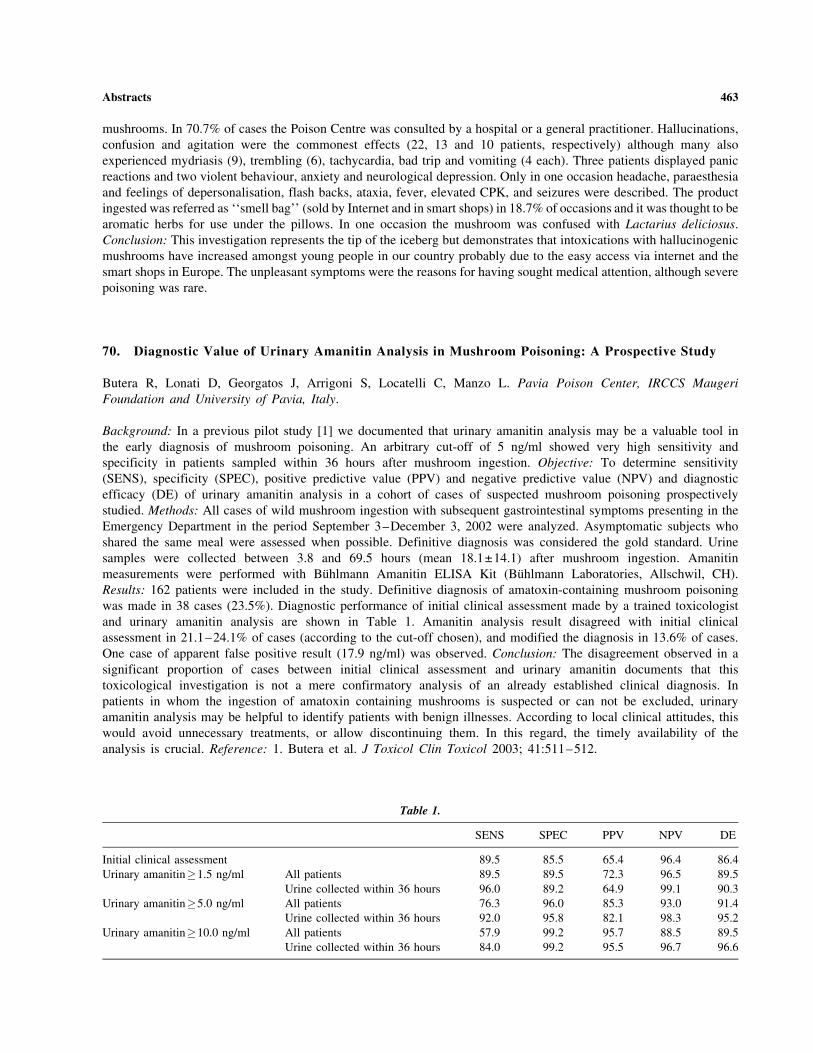
mushrooms. In 70.7% of cases the Poison Centre was consulted by a hospital or a general practitioner. Hallucinations,
confusion and agitation were the commonest effects (22, 13 and 10 patients, respectively) although many also
experienced mydriasis (9), trembling (6), tachycardia, bad trip and vomiting (4 each). Three patients displayed panic
reactions and two violent behaviour, anxiety and neurological depression. Only in one occasion headache, paraesthesia
and feelings of depersonalisation, flash backs, ataxia, fever, elevated CPK, and seizures were described. The product
ingested was referred as ‘‘smell bag’’ (sold by Internet and in smart shops) in 18.7% of occasions and it was thought to be
aromatic herbs for use under the pillows. In one occasion the mushroom was confused with Lactarius deliciosus.
Conclusion: This investigation represents the tip of the iceberg but demonstrates that intoxications with hallucinogenic
mushrooms have increased amongst young people in our country probably due to the easy access via internet and the
smart shops in Europe. The unpleasant symptoms were the reasons for having sought medical attention, although severe
poisoning was rare.
70. Diagnostic Value of Urinary Amanitin Analysis in Mushroom Poisoning: A Prospective Study
Butera R, Lonati D, Georgatos J, Arrigoni S, Locatelli C, Manzo L. Pavia Poison Center, IRCCS Maugeri
Foundation and University of Pavia, Italy.
Background: In a previous pilot study [1] we documented that urinary amanitin analysis may be a valuable tool in
the early diagnosis of mushroom poisoning. An arbitrary cut-off of 5 ng/ml showed very high sensitivity and
specificity in patients sampled within 36 hours after mushroom ingestion. Objective: To determine sensitivity
(SENS), specificity (SPEC), positive predictive value (PPV) and negative predictive value (NPV) and diagnostic
efficacy (DE) of urinary amanitin analysis in a cohort of cases of suspected mushroom poisoning prospectively
studied. Methods: All cases of wild mushroom ingestion with subsequent gastrointestinal symptoms presenting in the
Emergency Department in the period September 3–December 3, 2002 were analyzed. Asymptomatic subjects who
shared the same meal were assessed when possible. Definitive diagnosis was considered the gold standard. Urine
samples were collected between 3.8 and 69.5 hours (mean 18.1±14.1) after mushroom ingestion. Amanitin
measurements were performed with Buhlmann Amanitin ELISA Kit (Buhlmann Laboratories, Allschwil, CH).
Results: 162 patients were included in the study. Definitive diagnosis of amatoxin-containing mushroom poisoning
was made in 38 cases (23.5%). Diagnostic performance of initial clinical assessment made by a trained toxicologist
and urinary amanitin analysis are shown in Table 1. Amanitin analysis result disagreed with initial clinical
assessment in 21.1–24.1% of cases (according to the cut-off chosen), and modified the diagnosis in 13.6% of cases.
One case of apparent false positive result (17.9 ng/ml) was observed. Conclusion: The disagreement observed in a
significant proportion of cases between initial clinical assessment and urinary amanitin documents that this
toxicological investigation is not a mere confirmatory analysis of an already established clinical diagnosis. In
patients in whom the ingestion of amatoxin containing mushrooms is suspected or can not be excluded, urinary
amanitin analysis may be helpful to identify patients with benign illnesses. According to local clinical attitudes, this
would avoid unnecessary treatments, or allow discontinuing them. In this regard, the timely availability of the
analysis is crucial. Reference: 1. Butera et al. J Toxicol Clin Toxicol 2003; 41:511–512.
Table 1.
SENS SPEC PPV NPV DE
Initial clinical assessment 89.5 85.5 65.4 96.4 86.4
Urinary amanitin�1.5 ng/ml All patients 89.5 89.5 72.3 96.5 89.5
Urine collected within 36 hours 96.0 89.2 64.9 99.1 90.3
Urinary amanitin�5.0 ng/ml All patients 76.3 96.0 85.3 93.0 91.4
Urine collected within 36 hours 92.0 95.8 82.1 98.3 95.2
Urinary amanitin�10.0 ng/ml All patients 57.9 99.2 95.7 88.5 89.5
Urine collected within 36 hours 84.0 99.2 95.5 96.7 96.6
Abstracts 463
71. Muscle Function and Acetylcholinesterase Activity in Mouse Diaphragm Preparation andOrganophosphate Poisoned Patients
Thiermann H (1), Worek F (1), Szinicz L (1), Felgenhauer N (2), Zilker T (2), Eyer P (3). 1. Institut fuer Pharmakologie
und Toxikologie der Bundeswehr, Muenchen, Germany; 2. Toxikologische Abteilung der Technischen Universitaet
Muenchen, Germany; 3. Walther-Straub-Institut fuer Pharmakologie und Toxikologie, Muenchen, Germany.
Objectives: Effectiveness of oxime therapy in organophosphate (OP) poisoning was demonstrated in numerous
animal models. Nevertheless, the clinical usefulness of oximes is a matter of debate. To unravel this controversy,
effectiveness of obidoxime was investigated in isolated mouse diaphragms and the results compared to results of a
multicentre clinical trial on patients with severe OP intoxication. Methods: Using the isolated phrenic-diaphragm
preparation of the mouse, isometric muscle force after indirect stimulation was measured after inhibition of
acetylcholinesterase (AChE; circumfusion with paraoxon, followed by wash out) and reactivation (circumfusion with
obidoxime, followed by wash out). Thereafter, the diaphragms were homogenised and muscle AChE activity was
measured radiometrically. The effectiveness of obidoxime in 34 severely OP intoxicated patients, treated with a 250
mg IV bolus, followed by continuous infusion of 750 mg/24 hours, was evaluated using red blood cell
acetylcholinesterase (RBC-AChE) activity and neuromuscular transmission as surrogate parameters. The correlation
between muscle response to stimulation and AChE activity was compared in mouse diaphragms and patients.
Results: Incubation of mouse diaphragms with paraoxon resulted in almost complete reduction of force generation,
when muscle AChE activity was inhibited by more than 90%. Obidoxime, already at concentrations of about
10 mmol/l was able to reactivate muscle AChE and to restore muscle force. An increase in muscle AChE activity
was associated with an increase in muscle force and almost complete recovery at AChE levels higher than 40% of
control. In patients with OP intoxication a targeted effective plasma concentration of about 10 to 20 mmol/
l obidoxime could be adjusted with the regimen used. Obidoxime was able to reactivate non-aged AChE, if the
poison load was not too high. A quantitative correlation could be shown between RBC-AChE activity and paraoxon
concentration in plasma. Muscle function was severely disturbed, when RBC-AChE activity was inhibited by more
than 90%. With increasing RBC-AChE activity disturbance of muscle function subsided and clinical conditions
improved. At an RBC-AChE activity of more than 30% muscle function was hardly disturbed, indicating an
analogous correlation as found in isolated mouse diaphragms. Conclusions: The data indicate muscle function
recovery by AChE reactivation to be an important effect of obidoxime. Therefore, it may be concluded that data on
reactivation obtained with human RBC-AChE are helpful for prediction of oxime efficacy in man. This is of special
importance for therapy of nerve agent poisoning with newly developed oximes, since clinical studies on antidote
effectiveness towards such compounds cannot be performed.
72. Efficiency of a Non-Equimolar Neutralisation of Digoxin by Immune Fab Therapy
Lavaux T (1), Assemi P (1), Casset A (2), Lavigne T (1), Castelain V (1), Seydi A (1), Charra B (1), Jaeger A (1).
1. Service de Reanimation Medicale; 2. Laboratoire de Biochimie et Biologie Moleculaire, Hopital de Hautepierre,
Strasbourg, France.
Objective: To assess the efficiency of a protocol of anti-digoxin Fab fragments administration used in our intensive
care unit (ICU) to treat digitalis intoxication. This protocol consists of a non-equimolar neutralisation of digitalis
intoxication, i.e. a first infusion of 160 mg of Fab repeated after 1 hour in case of non-response. Methods:
Retrospective collection of clinical data and outcome of all patients with digitalis poisoning admitted over the last
5 years in the ICU. Results: 15 patients (3 men, 12 women; mean age 76±17 years) with digitalis poisoning (14
chronic and 1 acute) were included. All patients with chronic intoxication had underlying heart disease. 12 patients
received 160 mg of Fab fragments and 3 a second dose of 160 mg because of persistent hyperkalemia >6 mmol/L.
In these 3 cases, neutralisation was partial after the first dose (89±4%). Bradycardia and other cardiac manifestations
were both corrected in 10 patients. Only 4 patients presented a persistent bradycardia (< 60 beats/min) and 1 patient
a first degree atrioventricular block after immune therapy. Four patients died: death was related to digoxin poisoning
in two patients who had developed ventricular arrhythmia and asystole before Fab fragments administration. Four
patients received a theorical non-equimolar dose (2 a dose between 75% and 90% and 2 a dose between 50% and
464 Abstracts
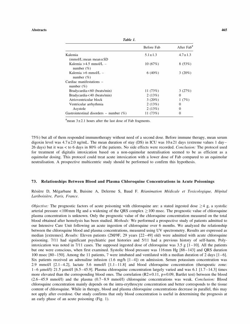
75%) but all of them responded immunotherapy without need of a second dose. Before immune therapy, mean serum
digoxin level was 4.7±2.0 ng/mL. The mean duration of stay (DS) in ICU was 10±21 days (extreme values 1 day–
26 days) but it was < to 6 days in 80% of the patients. No side effects were recorded. Conclusion: The protocol used
for treatment of digitalis intoxication based on a non-equimolar neutralisation seemed to be as efficient as a
equimolar dosing. This protocol could treat acute intoxication with a lower dose of Fab compared to an equimolar
neutralisation. A prospective multicentric study should be performed to confirm this hypothesis.
73. Relationships Between Blood and Plasma Chloroquine Concentrations in Acute Poisonings
Resiere D, Megarbane B, Buisine A, Delerme S, Baud F. Reanimation Medicale et Toxicologique, Hopital
Lariboisiere, Paris, France.
Objective: The prognostic factors of acute poisoning with chloroquine are: a stated ingested dose �4 g, a systolic
arterial pressure <100mm Hg and a widening of the QRS complex �100 msec. The prognostic value of chloroquine
plasma concentration is unknown. Only the prognostic value of the chloroquine concentration measured on the total
blood obtained after hemolysis has been studied. Methods: We performed a prospective study of patients admitted to
our Intensive Care Unit following an acute ingestion of chloroquine over 6 months. We analyzed the relationship
between the chloroquine blood and plasma concentrations, measured using UV spectrometry. Results are expressed as
median [extremes]. Results: Eleven patients (2M/9F, 29 years [22–49] old) were admitted with acute chloroquine
poisoning. 7/11 had significant psychiatric past histories and 5/11 had a previous history of self-harm. Poly-
intoxication was noted in 7/11 cases. The supposed ingested dose of chloroquine was 3.5 g [1–10]. All the patients
but one were conscious, when first examined. Systolic blood pressure was 116mm Hg [88–143] and QRS duration
100 msec [80–150]. Among the 11 patients, 7 were intubated and ventilated with a median duration of 2 days [1–6].
Six patients received an adrenaline infusion (1.6 mg/h [1–4]) on admission. Serum potassium concentration was
2.9 mmol/l [2.1–4.2], lactate 5.6 mmol/l [1.1–11.8] and blood chloroquine concentration (therapeutic zone:
1–6 mmol/l) 21.5 mmol/l [6.5–45.9]. Plasma chloroquine concentration largely varied and was 6.1 [1.7–14.3] times
more elevated than the corresponding blood ones. The correlation (R2=0.11, p=0.09, Bartlet test) between the blood
(2.6–45.9 mmol/l) and the plasma (0.7–8.9 mmol/l) chloroquine concentrations was weak. Conclusion: Blood
chloroquine concentration mainly depends on the intra-erythrocyte concentration and better corresponds to the tissue
content of chloroquine. While in therapy, blood and plasma chloroquine concentrations decrease in parallel, this may
not apply after overdose. Our study confirms that only blood concentration is useful in determining the prognosis at
an early phase of an acute poisoning (Fig. 1).
Table 1.
Before Fab After Fab#
Kalemia
(mmol/L:mean mean±SD
5.1±1.3 4.7±1.3
Kalemia >4.5 mmol/L –
number (%)
10 (67%) 8 (53%)
Kalemia >6 mmol/L –
number (%)
6 (40%) 3 (20%)
Cardiac manifestations –
number (%)
Bradycardia<60 (beats/min) 11 (73%) 3 (27%)
Bradycardia<40 (beats/min) 2 (13%) 0
Atrioventricular block 3 (20%) 1 (7%)
Ventricular arrhythmia 2 (13%) 0
Asystole 2 (13%) 0
Gastrointestinal disorders – number (%) 11 (73%) 0
#mean 3±2.1 hours after the last dose of Fab fragments.
Abstracts 465
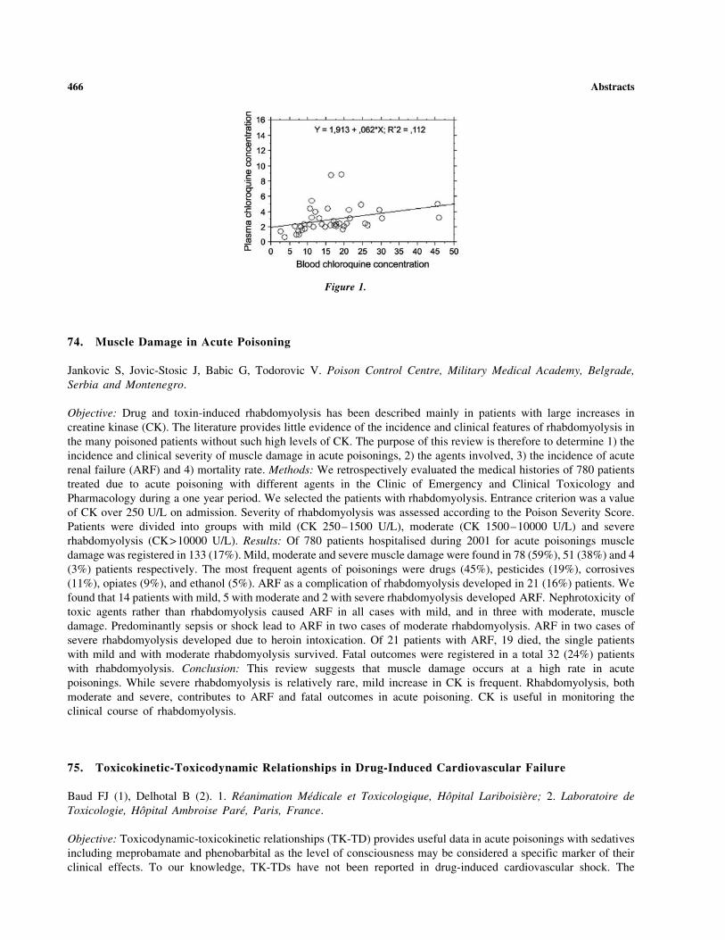
74. Muscle Damage in Acute Poisoning
Jankovic S, Jovic-Stosic J, Babic G, Todorovic V. Poison Control Centre, Military Medical Academy, Belgrade,
Serbia and Montenegro.
Objective: Drug and toxin-induced rhabdomyolysis has been described mainly in patients with large increases in
creatine kinase (CK). The literature provides little evidence of the incidence and clinical features of rhabdomyolysis in
the many poisoned patients without such high levels of CK. The purpose of this review is therefore to determine 1) the
incidence and clinical severity of muscle damage in acute poisonings, 2) the agents involved, 3) the incidence of acute
renal failure (ARF) and 4) mortality rate. Methods: We retrospectively evaluated the medical histories of 780 patients
treated due to acute poisoning with different agents in the Clinic of Emergency and Clinical Toxicology and
Pharmacology during a one year period. We selected the patients with rhabdomyolysis. Entrance criterion was a value
of CK over 250 U/L on admission. Severity of rhabdomyolysis was assessed according to the Poison Severity Score.
Patients were divided into groups with mild (CK 250–1500 U/L), moderate (CK 1500–10000 U/L) and severe
rhabdomyolysis (CK>10000 U/L). Results: Of 780 patients hospitalised during 2001 for acute poisonings muscle
damage was registered in 133 (17%). Mild, moderate and severe muscle damage were found in 78 (59%), 51 (38%) and 4
(3%) patients respectively. The most frequent agents of poisonings were drugs (45%), pesticides (19%), corrosives
(11%), opiates (9%), and ethanol (5%). ARF as a complication of rhabdomyolysis developed in 21 (16%) patients. We
found that 14 patients with mild, 5 with moderate and 2 with severe rhabdomyolysis developed ARF. Nephrotoxicity of
toxic agents rather than rhabdomyolysis caused ARF in all cases with mild, and in three with moderate, muscle
damage. Predominantly sepsis or shock lead to ARF in two cases of moderate rhabdomyolysis. ARF in two cases of
severe rhabdomyolysis developed due to heroin intoxication. Of 21 patients with ARF, 19 died, the single patients
with mild and with moderate rhabdomyolysis survived. Fatal outcomes were registered in a total 32 (24%) patients
with rhabdomyolysis. Conclusion: This review suggests that muscle damage occurs at a high rate in acute
poisonings. While severe rhabdomyolysis is relatively rare, mild increase in CK is frequent. Rhabdomyolysis, both
moderate and severe, contributes to ARF and fatal outcomes in acute poisoning. CK is useful in monitoring the
clinical course of rhabdomyolysis.
75. Toxicokinetic-Toxicodynamic Relationships in Drug-Induced Cardiovascular Failure
Baud FJ (1), Delhotal B (2). 1. Reanimation Medicale et Toxicologique, Hopital Lariboisiere; 2. Laboratoire de
Toxicologie, Hopital Ambroise Pare, Paris, France.
Objective: Toxicodynamic-toxicokinetic relationships (TK-TD) provides useful data in acute poisonings with sedatives
including meprobamate and phenobarbital as the level of consciousness may be considered a specific marker of their
clinical effects. To our knowledge, TK-TDs have not been reported in drug-induced cardiovascular shock. The
Figure 1.
466 Abstracts
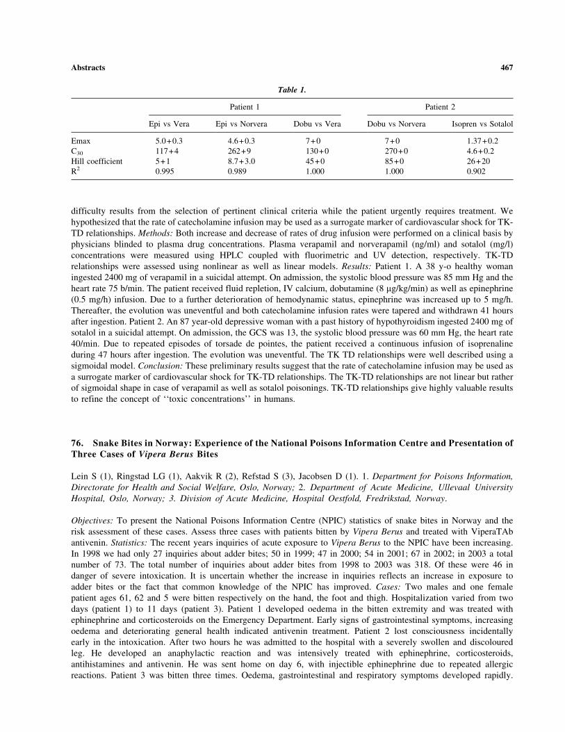
difficulty results from the selection of pertinent clinical criteria while the patient urgently requires treatment. We
hypothesized that the rate of catecholamine infusion may be used as a surrogate marker of cardiovascular shock for TK-
TD relationships. Methods: Both increase and decrease of rates of drug infusion were performed on a clinical basis by
physicians blinded to plasma drug concentrations. Plasma verapamil and norverapamil (ng/ml) and sotalol (mg/l)
concentrations were measured using HPLC coupled with fluorimetric and UV detection, respectively. TK-TD
relationships were assessed using nonlinear as well as linear models. Results: Patient 1. A 38 y-o healthy woman
ingested 2400 mg of verapamil in a suicidal attempt. On admission, the systolic blood pressure was 85 mm Hg and the
heart rate 75 b/min. The patient received fluid repletion, IV calcium, dobutamine (8 mg/kg/min) as well as epinephrine
(0.5 mg/h) infusion. Due to a further deterioration of hemodynamic status, epinephrine was increased up to 5 mg/h.
Thereafter, the evolution was uneventful and both catecholamine infusion rates were tapered and withdrawn 41 hours
after ingestion. Patient 2. An 87 year-old depressive woman with a past history of hypothyroidism ingested 2400 mg of
sotalol in a suicidal attempt. On admission, the GCS was 13, the systolic blood pressure was 60 mm Hg, the heart rate
40/min. Due to repeated episodes of torsade de pointes, the patient received a continuous infusion of isoprenaline
during 47 hours after ingestion. The evolution was uneventful. The TK TD relationships were well described using a
sigmoidal model. Conclusion: These preliminary results suggest that the rate of catecholamine infusion may be used as
a surrogate marker of cardiovascular shock for TK-TD relationships. The TK-TD relationships are not linear but rather
of sigmoidal shape in case of verapamil as well as sotalol poisonings. TK-TD relationships give highly valuable results
to refine the concept of ‘‘toxic concentrations’’ in humans.
76. Snake Bites in Norway: Experience of the National Poisons Information Centre and Presentation ofThree Cases of Vipera Berus Bites
Lein S (1), Ringstad LG (1), Aakvik R (2), Refstad S (3), Jacobsen D (1). 1. Department for Poisons Information,
Directorate for Health and Social Welfare, Oslo, Norway; 2. Department of Acute Medicine, Ullevaal University
Hospital, Oslo, Norway; 3. Division of Acute Medicine, Hospital Oestfold, Fredrikstad, Norway.
Objectives: To present the National Poisons Information Centre (NPIC) statistics of snake bites in Norway and the
risk assessment of these cases. Assess three cases with patients bitten by Vipera Berus and treated with ViperaTAb
antivenin. Statistics: The recent years inquiries of acute exposure to Vipera Berus to the NPIC have been increasing.
In 1998 we had only 27 inquiries about adder bites; 50 in 1999; 47 in 2000; 54 in 2001; 67 in 2002; in 2003 a total
number of 73. The total number of inquiries about adder bites from 1998 to 2003 was 318. Of these were 46 in
danger of severe intoxication. It is uncertain whether the increase in inquiries reflects an increase in exposure to
adder bites or the fact that common knowledge of the NPIC has improved. Cases: Two males and one female
patient ages 61, 62 and 5 were bitten respectively on the hand, the foot and thigh. Hospitalization varied from two
days (patient 1) to 11 days (patient 3). Patient 1 developed oedema in the bitten extremity and was treated with
ephinephrine and corticosteroids on the Emergency Department. Early signs of gastrointestinal symptoms, increasing
oedema and deteriorating general health indicated antivenin treatment. Patient 2 lost consciousness incidentally
early in the intoxication. After two hours he was admitted to the hospital with a severely swollen and discoloured
leg. He developed an anaphylactic reaction and was intensively treated with ephinephrine, corticosteroids,
antihistamines and antivenin. He was sent home on day 6, with injectible ephinephrine due to repeated allergic
reactions. Patient 3 was bitten three times. Oedema, gastrointestinal and respiratory symptoms developed rapidly.
Table 1.
Patient 1 Patient 2
Epi vs Vera Epi vs Norvera Dobu vs Vera Dobu vs Norvera Isopren vs Sotalol
Emax 5.0+0.3 4.6+0.3 7+0 7+0 1.37+0.2
C30 117+4 262+9 130+0 270+0 4.6+0.2
Hill coefficient 5+1 8.7+3.0 45+0 85+0 26+20
R2 0.995 0.989 1.000 1.000 0.902
Abstracts 467
The condition of the patient deteriorated, she lost consciousness. Despite early administration of ephinephrine and
antihistamines, severe hypotension developed. Immediately after the arrival at the hospital, ViperaTAb was given
and the condition improved. The blood pressure normalized and she awoke within 1/2 hour. Treatment with
antivenin had to be repeated twice because of the recurrence of hypotension and CNS symptoms. Despite repeated
antivenin treatment the local oedema progressed and became extensive. Because of development of compartment
syndrome a fasciotomy was performed on the lower extremity. Three days after the accident blood analysis
indicated pronounced hemolysis (Hb:6.2 g/100 ml) and the patient was given two blood tranfusions. Conclusion:
The three cases reflect some of the serious clinical effects that can be seen with Vipera Berus bites. In the course of
the intoxication all three required ViperaTAb antivenin infusion and symptomatic support. All outcomes
were benign.
77. Time- and Dose-Dependent Changes in the Pharmacokinetics and Metabolite Patterns ofParacetamol Dosed at 4, 6, and 8 G/Day in Healthy Volunteers
Gelotte CK, Auiler JF, Lynch JM, Temple AR, Slattery JT (1), Bowen DL. R&D, McNeil Consumer & Specialty
Pharmaceuticals, Fort Washington, PA USA, 1. Indiana University, Departments of Medicine & Biology,
Bloomington, IN, USA.
Objective: To determine the tolerability, pharmacokinetics (PK) and metabolism of repeat-dose paracetamol (PAR),
at and above recommended doses. Methods: The Research Ethics Committee, Royal Group of Hospitals in Belfast
reviewed and approved the protocol. Repeat-dose PK of 1, 1.5, or 2 g PAR was evaluated using a double-blinded,
placebo-controlled, three-regimen study design. Each group of 12 volunteers received either (A) 1g every six hours
(Q6H) for three days (4 g/d), washout four days, then 1.5g Q6H for three days (6 g/d); (B) 1g Q6H for three days
(4 g/d), washout four days, then 2g Q6H for three days (8 g/d); or (C) placebo Q6H for three days, washout four
days, then placebo Q6H for three days. Safety and hepatic function were monitored daily. Blood samples were
collected after the first and last repeat doses, and urine collected for 24 hours on Day 3 of each dose level. Results:
Volunteers weighed between 64 and 85 kg, and received total daily doses of 47 to 124 mg/kg PAR. The PK results
showed that PAR plasma concentrations did not accumulate with repeat doses, and that steady-state concentrations
were linearly related to dose. Plasma metabolite data showed an unexpected increase in production of the major
metabolite PAR-glucuronide and a decrease of PAR-sulfate between the first and last doses for 4, 6, and 8 g/d,
indicating time-dependent changes in PAR metabolism with repeat doses. The urinary pattern of two major
metabolites changed in opposite directions with dose: a higher amount (67%) of PAR-glucuronide was produced at
8 g/d compared with 59% and 61% for 4 and 6 g/d, and a lower amount (11%) of PAR-sulfate was produced at 8 g/d
compared with 19% and 14% for 4 and 6 g/d. The urinary pattern of thiol metabolites, which include PAR-cysteine
and PAR-mercapturate, was independent of dose. Paired t-test comparisons detected differences in the metabolite
formation clearances of PAR-glucuronide between 4 and 8 g/day (p<0.007) and of PAR-sulfate between 4 and both
6 and 8 g/d (p <0.0001). No statistical differences were detected in the formation clearances of the thiols with dose.
Conclusions: PAR exhibited linear, dose-proportional PK at steady state. Time-and dose-dependent changes in both
plasma and urine metabolite patterns suggest autoinduction of glucuronidation and saturation of the sulfate pathway.
Patterns of thiol metabolites did not differ with dose. All PAR doses were well tolerated, and all hepatic
transaminase values remained within normal limits.
78. Hepatotoxicity in Alcoholic Patients from Therapeutic Dosing of Acetaminophen
Dart RC, Palmer R, Kuffner E. Rocky Mountain Poison and Drug Center, Denver Health; University of Colorado;
Toxicology Associates, Denver, Colorado, USA.
Objective: Despite prospective studies of 260 alcoholic patients showing no change in AST/ALT levels in patients
treated with acetaminophen (APAP), calls for reduction or elimination of APAP use in alcoholic patients continue.
468 Abstracts
The purpose of this study was to address concerns with previous studies by expanding previous studies and
reviewing the medical literature. Methods: A prospective, double-blind, randomized, placebo-controlled trial of
confirmed active alcoholics was conducted at a detoxification center. Exclusion criteria included a recent history of
ingestion >4 g of APAP, serum APAP>20 mg/L, AST or ALT>200 IU/L or INR>1.5. Patients were randomized
(2:1 ratio) to receive APAP, 4 g/day in divided doses or placebo for 3 days. Serum AST, ALT, INR and serum
glutathione were measured. For the systematic review, standard systematic review techniques were used. A broad
electronic search of MEDLINE (1966–Current) and EMBASE (1980–Current) in all languages was limited only to
the APAP CAS registry number. Only articles that included the term alcohol or alcoholism and ingestion of a
therapeutic dosage of acetaminophen (4 g/d or less) were included. Each article was abstracted by trained personnel.
Results: In the clinical trial, patients were primarily male (95%). The APAP and placebo groups were not different
based on demographics, severity of alcoholism, nutritional status or baseline laboratory studies. Of 210 patients (145
APAP, 65 placebo), there was no statistical difference between the two groups with respect to AST, ALT or INR
means at baseline or 3 days. Three patients (2%) in the APAP group and two patients (6%) in the placebo group
developed and AST or ALT >200 IU/L. There was no difference in serum glutathione between groups. The
systematic review returned 16,184 papers. All 22 prospective studies (648 patients) of patients with alcohol abuse
received therapeutic doses of acetaminophen without evidence of liver injury. A total of 30 class III papers provided
53 patients with evidence of liver injury suspected to be the result of APAP. Each of these cases contained major
confounding information. Conclusions: The medical literature is remarkably dichotomous regarding the toxicity of
therapeutic doses of APAP in alcoholic patients. Multiple prospective studies have reported no injury during the use
of therapeutic doses, while retrospective reports describe an association of liver injury with therapeutic intent. Given
the known inaccuracy and bias involved in retrospective studies and case reviews, the existence of an alcohol-
acetaminophen syndrome cannot be established with the evidence currently available. The most likely explanation is
historical inaccuracy of retrospective reports.
79. Acetaminophen Metabolism and the Varied Effects of Ethanol
Slattery JT. Indiana University, Departments of Medicine and Biology, Bloomington, IN, USA.
Objective: Acetaminophen generally is not thought to cause hepatotoxicity in doses less than about 15 g, but
clinicians have suspected that ethanol may enhance the probability of acetaminophen-induced hepatotoxicity at lower
doses. Hepatotoxicity accrues when a quinoneimine, NAPQI, produced by oxidation of acetaminophen by
cytochromes p450 overcomes the major defense mechanism, chemical reduction or conjugation with glutathione
(GSH), and produces a cascade of events that lead to hepatocellular necrosis. In vivo experiments in human
volunteers receiving selective cytochrome p450 inhibitors and inducers have shown that the cytochrome p450 that
dominates production of NAPQI is CYP2E1. The objective of this study is to systematically describe the varied
effects of alcohol on acetaminophen metabolism. Results: Studies by several laboratories have shown that CYP2E1
is induced by several mechanisms. The most unusual is that it is stabilized by ligands that bind to the active site. The
prolonged half-life that results from ligand stabilization accounts for its induction by several chemicals. This is the
case with ethanol, particularly with blood concentrations less than 250 mg/dl. At higher concentrations, ethanol also
induces by enhancing de novo synthesis of the enzyme. The result of this inhibitory/induction action of ethanol is
that CYP2E1 activity is inhibited when ethanol is present in blood (although enzyme abundance in the liver has
increased due to slowed degradation). Enhanced CYP2E1 activity is seen when ethanol ingestion is stopped and
ethanol is cleared. The window of enhanced activity is therefore narrow, conforming to the half-life of the
unstabilized holoenzyme. The magnitude of enhanced activity seems to be about 2–3 fold. Ethanol also is a selective
depletor of hepatocellular mitochondrial GSH. Ethanol does not deplete hepatocellular cytosolic GSH. In rats fed
ethanol as 36% of total calories (the Lieber-DiCarli liquid diet), it takes 3–6 weeks for this effect to be maximized.
Under these conditions, acetaminophen hepatocellular toxicity is approximately doubled. Other studies in rats have
shown that the effect on hepatocellular mitochondrial GSH reverses very quickly (within about 12 hours in a rat)
after discontinuation of the ethanol diet. Conclusions: Ethanol enhances the toxicity of acetaminophen through two
mechanisms, induction of CYP2E1 and, on prolonged administration, depletion of hepatocellular mitochondrial
GSH. However, because enhanced activity of CYP2E1 is observed only when ethanol is withdrawn and the depletion
Abstracts 469
of hepatocellular mitochondrial GSH is quickly reversed upon withdrawal of ethanol, the probability of the two
mechanisms operating simultaneously is small.
80. Frequency of Serious Hepatic Dysfunction when Paracetamol Concentrations are Below CurrentUnited Kingdom Nomogram Treatment Lines
Beer C, Hudson M, Smith L, Thomas SHL. Newcastle upon Tyne Hospitals NHS Trust and Wolfson Unit of Clinical
Pharmacology, University of Newcastle, UK.
Objective: Paracetamol is involved in 35–45% of all episodes of drug overdose in the United Kingdom and is a
common cause of fulminant hepatic failure. Early administration of intravenous acetylcysteine is effective at
preventing liver damage. In the UK, treatment guidelines recommended that all patients with paracetamol
concentrations above a line with first order decline connecting 200 mg/L at 4 hours with 30 mg at 15 h (the ‘200
line’) receive intravenous acetylcysteine. This line is now extrapolated to 24 h. Patients considered to have risk
factors for enhanced hepatotoxicity are treated at half these concentrations (‘100 line’). In 1998, 4 cases were
described who developed fatal hepatic failure after presenting with low plasma paracetamol concentrations and in 3
cases the failure to use acetylcysteine was consistent with UK guidance. The authors proposed lowering the
thresholds for treatment by 25% (‘150 line’). This study was performed to establish the frequency of severe
paracetamol-induced hepatotoxicity when the plasma paracetamol concentration at presentation was below the
current appropriate nomogram treatment line. Methods: The records of all patients presenting to the regional liver
service in Newcastle between September 1996 and March 2003 with paracetamol poisoning and severe liver
dysfunction were reviewed. Clinical details were sought from liver unit notes or the referring hospital, including the
paracetamol concentration at presentation and the timing of this in relation to the overdose. Results: Of 172 patients
referred with possible paracetamol-induced hepatotoxicity (peak PT 47�>160s), paracetamol poisoning could be
verified in 160 (74 females, 86 males, mean age 33 y). Adequate data on plasma paracetamol concentration and the
timing of this in relation to overdose were available in 67. Of those without risk factors for enhanced hepatotoxicity,
paracetamol concentrations at presentation were below the ’200 line’ but on or above the ‘150 line’ in 3 patients,
below the ’150 line’ but above the ‘100 line’ in 2 patients and below the ‘100 line’ in one patient. One of these
patients died (paracetamol concentration 170 mg/L at 4 hours). There was also 1 patient at excess risk of
hepatotoxicity who presented with a paracetamol concentration below the ‘100 line’. Conclusion: Some patients with
initial paracetamol concentrations that do not require acetylcysteine according to current UK treatment guidelines
and the information available at the time of admission subsequently develop severe hepatotoxicity. They constitute
an important minority of patients with paracetamol-induced severe liver dysfunction but a very small proportion of
all people with paracetamol overdose. Reference: (1). Bridger S, et al. Deaths from low dose paracetamol poisoning.
Br Med J 1998; 316:1724–1725.
81. Molecular Adsorbents Recirculating System (MARS)—Liver Support Therapy in AcuteParacetamol Intoxication and Alcohol-Paracetamol Syndrome
Hydzik P, Gawlikowski T, Pach JD. Targosz Department of Clinical Toxicology Jagiellonian University Medical
College, Krakow, Poland.
Objective: The hepatotoxic properties of paracetamol are well known. Acetylcysteine (ACC) does not always
prevent liver injury: for example a late ACC administration in severe overdosing, or in patients with chronic alcohol
use and paracetamol co-ingestion. Chronic alcohol consumption induces cytochrome P-4502E1 activity and GSH
depletion. Resultant increases in NAPQI synthesis in paracetamol poisoning may lead to liver injury. MARS is a
useful liver support device saving time for spontaneous liver regeneration or acting as a bridging technique to liver
transplantation. Case Series: Two cases admitted to the Department in 19 and 20 hour post-ingestion of hepatotoxic
paracetamol doses as confirmed by toxicological examination (serum paracetamol concentration was 207 mg/L, and
470 Abstracts
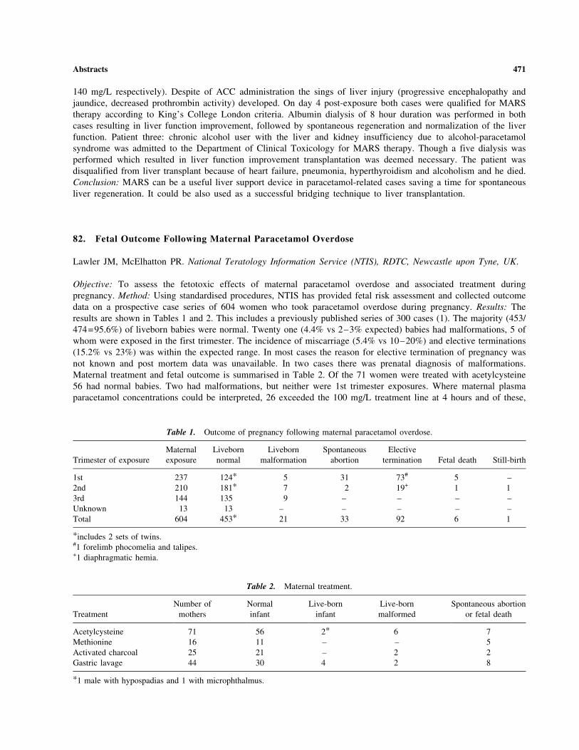
140 mg/L respectively). Despite of ACC administration the sings of liver injury (progressive encephalopathy and
jaundice, decreased prothrombin activity) developed. On day 4 post-exposure both cases were qualified for MARS
therapy according to King’s College London criteria. Albumin dialysis of 8 hour duration was performed in both
cases resulting in liver function improvement, followed by spontaneous regeneration and normalization of the liver
function. Patient three: chronic alcohol user with the liver and kidney insufficiency due to alcohol-paracetamol
syndrome was admitted to the Department of Clinical Toxicology for MARS therapy. Though a five dialysis was
performed which resulted in liver function improvement transplantation was deemed necessary. The patient was
disqualified from liver transplant because of heart failure, pneumonia, hyperthyroidism and alcoholism and he died.
Conclusion: MARS can be a useful liver support device in paracetamol-related cases saving a time for spontaneous
liver regeneration. It could be also used as a successful bridging technique to liver transplantation.
82. Fetal Outcome Following Maternal Paracetamol Overdose
Lawler JM, McElhatton PR. National Teratology Information Service (NTIS), RDTC, Newcastle upon Tyne, UK.
Objective: To assess the fetotoxic effects of maternal paracetamol overdose and associated treatment during
pregnancy. Method: Using standardised procedures, NTIS has provided fetal risk assessment and collected outcome
data on a prospective case series of 604 women who took paracetamol overdose during pregnancy. Results: The
results are shown in Tables 1 and 2. This includes a previously published series of 300 cases (1). The majority (453/
474=95.6%) of liveborn babies were normal. Twenty one (4.4% vs 2–3% expected) babies had malformations, 5 of
whom were exposed in the first trimester. The incidence of miscarriage (5.4% vs 10–20%) and elective terminations
(15.2% vs 23%) was within the expected range. In most cases the reason for elective termination of pregnancy was
not known and post mortem data was unavailable. In two cases there was prenatal diagnosis of malformations.
Maternal treatment and fetal outcome is summarised in Table 2. Of the 71 women were treated with acetylcysteine
56 had normal babies. Two had malformations, but neither were 1st trimester exposures. Where maternal plasma
paracetamol concentrations could be interpreted, 26 exceeded the 100 mg/L treatment line at 4 hours and of these,
Table 1. Outcome of pregnancy following maternal paracetamol overdose.
Trimester of exposure
Maternal
exposure
Liveborn
normal
Liveborn
malformation
Spontaneous
abortion
Elective
termination Fetal death Still-birth
1st 237 124* 5 31 73# 5 –
2nd 210 181* 7 2 19+ 1 1
3rd 144 135 9 – – – –
Unknown 13 13 – – – – –
Total 604 453* 21 33 92 6 1
*includes 2 sets of twins.#1 forelimb phocomelia and talipes.+1 diaphragmatic hemia.
Table 2. Maternal treatment.
Treatment
Number of
mothers
Normal
infant
Live-born
infant
Live-born
malformed
Spontaneous abortion
or fetal death
Acetylcysteine 71 56 2* 6 7
Methionine 16 11 – – 5
Activated charcoal 25 21 – 2 2
Gastric lavage 44 30 4 2 8
*1 male with hypospadias and 1 with microphthalmus.
Abstracts 471
13 exceeded the 200 mg line. Two of these patients received methionine and 24 received acetylcysteine.
Conclusions: Although the malformation rate was slightly higher than expected, the majority of babies (76%) were
not exposed during the sensitive period of organogenesis. No specific pattern of malformations was observed.
Furthermore, no causal relationship could be identified between adverse fetal outcome and maternal paracetamol
overdose, plasma paracetamol concentration, or maternal treatment. Reference: McElhatton PR, Sullivan F, Volans
G. Paracetamol overdose in pregnancy analysis of the outcomes of 300 cases referred to the Teratology Information
Service Reproductive Toxicology 1997; 11(1): 85–94.
83. Challenges of Prehospital Guideline Development: The Beta Blocker Experience
Wax PM. Banner Good Samaritan Medical Center, Phoenix, AZ, USA.
In 2002, the U.S. Maternal and Child Health Bureau of the Health Resources and Services Administration (HRSA)
funded a cooperative agreement with the American Association of Poison Control Centers (AAPCC) in conjunction
with the American Academy of Clinical Toxicology (ACMT) and the American College of Medical Toxicology
(ACMT) to develop guidelines that will assist poison center personnel in managing out-of-hospital patients who had
been exposed to poisonous substances. This project grew out of a US federal effort to stabilize and enhance US
poison centers. One of these enhancements was to develop a single, national toll free number for poison centers. To
optimize this single national telephone access, it became clear that closer coordination between poison centers was
required, and that, preferably, a caller would receive similar guidance regardless of which poison center handled the
call. The goal of this prehospital guideline project is to provide a single set of evidence based and consensus driven
recommendations for use by poison centers across the U.S. A consensus panel with representatives from AAPCC,
AACT, and ACMT was charged with overseeing guideline development. The panel uses a modified Delphi method
to achieve consensus. Each guideline undergoes an extensive secondary review process by individual members of
these three organizations as well as 31 other secondary review organizations. The substances initially targeted for
prehospital guideline development were nontoxics, ipecac, acetaminophen, beta blockers, calcium channel blockers,
and sulfonylureas. Many lessons have been learned from this undertaking. This keynote focuses on some of the early
challenges that confronted the consensus panel as it pertained to the development of the beta blocker guideline.
Given the ‘‘out of hospital’’ scope of the guideline, the major decision point pertains to triage and appropriate
referral to a healthcare facility. Prehospital decontamination and intervention assumed less importance in the
guideline development process. Formulating an evidence based triage recommendation presents formidable
challenges. While few would disagree that intentional and/or symptomatic ingestions merit evaluation in a healthcare
facility, much more problematic is how best to triage asymptomatic accidental exposures-either adult or pediatric. A
review of current individual poison center guidelines suggested the lack of any one standard approach with widely
varying practice patterns. Some centers had no specific printed guidelines. Regarding propranolol, the threshold
doses for referral ranged from 2 to 8 mg/kg. A number of centers routinely referred to health care facilities all beta
blocker exposures regardless of the dose. Ideally, guideline development would be driven by the best evidence.
Unfortunately, regarding appropriate triage criteria for accidental beta blocker exposures (as well as with many other
substances under guideline development), almost all the evidence examined was level 4 (case reports and case series)
and level 5 (expert opinion). A critical review of this evidence reinforces the vagaries of case report data including
the imprecisions of dose and outcome measurements. Many centers have taken the approach that a therapeutic dose
(or a maximum therapeutic dose) is a safe dose and the ingestion of such a dose (or perhaps double that dose) does
not need health care referral. But the exact amount that constitutes a therapeutic dose may be subject to much
interpretation. An examination of six standard reference texts suggests a wide variability of therapeutic drug dosing.
The possibility also exists that the accidental ingestion of certain ‘‘supertherapeutic’’ doses in an otherwise health
individual does not automatically require referral to a healthcare facility. However, identifying that safe dose is often
elusive. This guideline also intends to provide information on the duration of time that an asymptomatic patient
needs observation. Despite the evidentiary limitations, guidelines on beta blockers and at least 15 other substances
will be developed over the next 2 years providing poison centers with consensus driven recommendations.
Hopefully, the utilization of these guidelines will promote future research and the collection of a stronger data set
that can be used to support or drive future guideline modification.
472 Abstracts
84. Assessment of the Efficacy and Safety of Gut Decontamination in Patients with Acute TherapeuticDrug Overdose
Nogue S, Amigo M, Sanjurjo E, Badal P, Vazquez E, Gomez E. Emergency Department and Toxicology Unit,
Hospital Clınic, Barcelona, Spain.
Objective: Gut decontamination (GD) may be used as a treatment for acute therapeutic drug overdose (ATDO). The
objective of this study was to assess the efficacy and safety of GD in ATDO patients. Methods: A 4-month
prospective observational study was designed to include all patients admitted to the emergency department due to an
ATDO. On admittance, epidemiological data, vital signs and the physical examination were all recorded and a blood
sample was taken for toxicological analysis if the drug was quantifiable. An algorithm was used to determine the
method of GD to be applied. A clinical reassessment was made at 3–6 hours and a further sample taken for
toxicological analysis. The patient was followed until hospital discharge, with possible adverse events due to the GD
being recorded. Results: Ninety-four patients were included. GD was indicated in 60 patients (63.8%): 3.3% received
ipecacuana syrup, 8.3% gastric lavage, 21.6% gastric lavage followed by activated charcoal and 71.6% oral activated
charcoal alone. The clinical state worsened in 19.1% of patients, usually due to diminished consciousness. Adverse
events attributable to GD were observed in 5.2% of patients. A toxicological analysis was made in 50 patients, and
in 42%, drug concentrations were higher at 3 or 6 hours than on admission. Analysis of the method of
decontamination used showed that the procedure recommended by the algorithm was applied in 70 patients (74.5%)
(group A) while in the remaining 24 (group B) another decontamination technique was used. Clinical deterioration
was observed in 14.3% of patients in group A and 33.3% in group B (p=0.041); there was a favourable evolution of
the analytic curve in 60.6% of patients in group A and 33.3% in group B (p=0.105) and severe adverse events
attributable to GD were suffered by 2.4% in group A and 11.1% in group B (p=0.171). Conclusion: The efficacy
and safety of GD in ATDO increases in patients where the decision-making algorithm is applied, although this does
not prevent clinical deterioration or continued drug absorption in all cases and may be accompanied by adverse
events. Reference: Amigo M, Faro J, Estruch D, et al. Descontaminacion digestiva en pacientes con intoxicacion
medicamentosa aguda. Validacion de un algoritmo para la toma de decisiones sobre la indicacion y el metodo prioritario.
Emergencias 2003; 15:18–26.
85. Prospective Assessment of Clinical Criteria of Refractoriness to Medical Treatment in Poisoningswith Membrane Stabilising Agents
Baud FJ (1), Guerrier G (1), Megarbane B (1), Resiere D (1), Delerme S (1), Thabet H (2). 1. Reanimation medicale
et toxicologie, Hopital Lariboisiere, Paris France; 2. Centre d’assistance Medicale Urgente et de Reanimation,
Tunis, Tunisia.
Objective: Drug-induced membrane stabilising effect (MSE) remains a leading cause of death. Experimental and
clinical data suggest that extracorporeal life-support (ECLS) may improve the outcome of life-threatening
poisonings. In a retrospective study, we determined predictive criteria of death resulting from cardiovascular events
in drug-induced MSE. The objective of this study was to prospectively confirm these criteria. We present the
preliminary data. Methods: Patients were included if admitted in our hospital as well as those hospitalised in other
hospitals but requiring medical advice for life-threatening MSE. Life-threatening MSE was defined by the presence
within 24 hours after ingestion of either cardiac arrest or widening of QRS complex >0.12 s or transient or sustained
systolic blood pressure <90 mm Hg. MSE refractory to medical treatment was defined by persisting cardiac arrest at
the time of admission to hospital or refractory shock defined by a systolic blood pressure <90 mm Hg despite fluid
repletion, the administration of 375 mL of molar sodium bicarbonate, and the infusion of at least 3 mg/h of
epinephrine while the patient remained oliguric or had a PaO2/FIO2 ratio less than or equal to 250. The cardiogenic
origin of shock was assumed using either hemodynamic study or in patients with very poor hemodynamic status by
the presence of a slow heart rate (<60/min) in conjunction with shock. Patients treated with ECLS were excluded
from this study. Results: Over an eight-month period, Twenty-four life-threatening MSE poisonings were included.
There were 9 males and 15 females, the median age was 35 year-old. Seven patients eventually died. Death resulted
Abstracts 473
from the ingestion of propranolol (3), acebutol in addition to verapamil (1), citalopram alone (1) and associated with
celiprolol (1), and intravenous cocaine (1). Five of the dead patients met the criteria of refractoriness to medical
treatment. One patient died from post-anoxic brain damage after successful resuscitation of cardiopulmonary arrest
while another anuric patient died lately (day 13) from septic shock. None of the patients who eventually survived
met the criteria of refractoriness to medical treatment. The criteria were able to predict death with a sensitivity of
89% and a specificity of 100%. During the same period of time, 2 patients were treated with ECLS for MSE
refractory to medical treatment and were not included in this study. Conclusion: These preliminary results suggest
that easily collected criteria can predict refractoriness to medical treatment in drug poisonings involving MSE. ECLS
should be considered in patients meeting these criteria.
86. Prospective Study on the Effects of Xylometazoline Overdose
Riel AJHP van (1), Riemsdijk TE van (1), Vries I de (1,2), Meulenbelt J (1,2). 1. Dutch National Poisons
Information Centre, part of the National Institute for Public Health and the Environment, Bilthoven, The
Netherlands; 2. University Medical Centre Utrecht, The Netherlands.
Objective: Xylometazoline is an imidazoline derivative used in over-the-counter (OTC) preparations for topical
application to relieve nasal congestion associated with acute or chronic rhinitis, common cold, sinusitis and hay fever
or other allergies. Xylometazoline is an alpha-2 agonist and topically applied it acts vasoconstrictive, thus resolving
nasal congestion. However in overdose systemic absorption takes place and according to the literature symptoms like
drowsiness, diaphoresis, hypotension, shock, bradycardia, respiratory depression and coma may occur. The toxic
dose has not been well established. There are a few odd case reports of severe central nervous system effects
including coma, caused by xylometazoline in children. Because of these reports, clinical toxicologists tend to
approach patients with xylometazoline overdose very cautiously, and according to the results that we present,
perhaps too cautiously. Methods: From 15 May 2002 we conducted a prospective study of all reports to the Dutch
National Poisons Information Centre concerning xylometazoline. Results: Out of 56 cases reported (until 21-11-
2003; the study will be continued) we received follow-up information in 35 cases. In 3 cases the patient was an adult
exposing himself, the other 53 cases concerned children between 14 days and 5 years of age (average 2,1). Most
children accidentally exposed themselves, in 11 cases the exposure was confirmed by a witnessing parent or the
parent him/herself accidentally exposed the child. Dutch decongestants contain between 2.5–10 mg xylometazoline
per packaging unit; the estimated dose ranged from 0.004 to 0.952 mg/kg (mean 0.24 mg/kg). No clinical symptoms
attributable to xylometazoline were reported in any of these cases. In one case vomiting was reported and in another
case fever was reported, both existed before exposure to xylometazoline took place. In 85% of the cases the
estimated dose was below 0.4 mg/kg, in the range above 0.4 mg/kg more data is needed to establish the risk.
Conclusion: We conclude that accidental exposure to xylometazoline decongestants remains predominantly
asymptomatic. Measures to reduce absorption are considered to be of low efficacy because of the small volumes in
which the preparations occur and the rapid systemic absorption via all mucous membranes. With doses below 0.4
mg/kg it seems safe to observe the patient at home, if any of the mentioned symptoms occurs the patient needs to be
hospitalized. If the patient remains asymptomatic until 2 hours post-ingestion there are no new symptoms to
be expected.
87. Suspected Clozapine Poisoning in the UK/EIRE, 1992 – 2003
Flanagan RJ (1), Spencer EP (1), Morgan P (1), Dunk L (2). 1. Medical Toxicology Unit, Guy’s and St Thomas’
Hospital, London, UK; 2. Novartis Pharmaceuticals UK Ltd, Surrey, UK.
Objective: Toxicological analysis of post-mortem blood is performed to investigate not only fatal poisoning, but also
patient compliance. We report the results of blood clozapine (Clozaril, Novartis) and norclozapine analyses
performed whilst investigating suspected clozapine poisoning, 1992–2003. Methods: Samples were referred from
474 Abstracts
clinicians, pathologists/coroners, or via the Clozaril Patient Monitoring Service (CPMS, Novartis). Information was
gathered from clinical, post-mortem, or coroner’s reports. Results: There were 5 fatal (3 male, 2 female; median age
28 yr [range 24–40 yr]) and 4 non-fatal (3 male, 1 female; median age 32 yr [range 26–41 yr]) clozapine overdoses.
The median post-mortem blood clozapine and norclozapine concentrations were 8.7 (range 3.9–12) and 2.0 (range
1.4–2.4) mg/L, respectively (median clozapine:norclozapine ratio 4.1 [range 2.9–5.1]). In the non-fatal overdoses
the median plasma clozapine and norclozapine concentrations (initial samples) were 4.4 (range 1.7–7.0) and 0.36
(range 0.30–0.44) mg/L, respectively (median clozapine:norclozapine ratio 13 [range 5.3–18)]. These overdoses
were in patients who were poorly- or non-compliant or who had taken tablets prescribed for someone else. Fifty
deaths (34 male, 16 female; median age 41 yr [range 22–70 yr]) were not attributed to clozapine self-poisoning. The
median post-mortem blood clozapine and norclozapine concentrations were 1.7 (range 0–7.7, n=39) and 1.4 (range
0–6.0, n=38) mg/L, respectively (median clozapine:norclozapine ratio 1.4 [range 0.4–7.6, n=37]). The median post-
mortem increase in blood clozapine and norclozapine as compared to the most recent ante-mortem measurement was
437% (range 98–5350%) and 379% (range 139–831%), respectively [median sample time before death 12 d (range
0–30 d, n=18)]. Nine deaths were attributed to suicide/poisoning [‘clozapine toxicity’ (2), heroin toxicity,
methadone toxicity, paracetamol overdose, sulpiride overdose, amitriptyline overdose, mixed [chlorpromazine,
procyclidine, moclobemide] overdose, and hanging (1 each)]. Other deaths were attributed to cardiorespiratory arrest
(5), fit (5, 2 during restraint), pneumonia (3, 1 after aspiration), pulmonary embolism (2), large bowel infarct,
ischaemic heart disease, cerebrobvascular accident, myocardial infarct, hepatitis, myocarditis, water intoxication, and
renal failure(1 each). No cause of death was established in 18 cases. Conclusions: There was considerable overlap
between the blood clozapine and norclozapine concentrations in the overdoses and in the other deaths. In part this is
because blood clozapine and norclozapine often increase after death, but another factor is that clozapine is much
more toxic in patients who are not taking the drug regularly. The availability of ante-mortem blood specimens
collected originally for haematology was helpful in investigating many deaths.
88. Fetal Outcome Following Maternal Treatment with Antiepileptic Drugs
Smallshaw K, Hassan SA, McElhatton PR, Thomas SHL. National Teratology Information Service (NTIS), RDTC,
Newcastle upon Tyne, UK.
Objective: To assess the fetotoxic effects of maternal treatment with AEDs during pregnancy. Although the
teratogenic potential of the older AED’s is well known there are few data on the newer drugs. Only limited local
guidelines are available to clinicians as to which drugs are most appropriate for women of childbearing age (1,2).
Method: Using standardised procedures, NTIS has provided prospective fetal risk assessment and collected outcome
data in 184 women (115 monotherapy, 69 polytherapy) taking AEDs throughout pregnancy. Results: The results are
shown in Tables 1 and 2. In both groups, the majority of liveborn babies were normal (91.8% monotherapy, 85.7%
polytherapy). The incidence of malformations was higher than expected; (8.1% monotherapy, 14.3% polytherapy vs
2–3% expected). The incidence of miscarriage (12.1% and 1.4% vs 10–20%) and elective terminations (14.7% and
8% vs 23%) was within the expected range. The wide range of congenital malformations was observed including
Table 1. Outcome of pregnancy following maternal treatment with AEDs for epilepsy.
Maternal exposures Liveborn normal Liveborn malformation Miscarriages Elective termination Stillbirth
Monotherapy 79*# 7 13 17 1
115 91.8% 8.1% 11.1% 14.7% 0.9%
Polytherapy 54+ 9 1 4 1
69 85.7% 14.3% 1.4% 5.8% 1.4%
*two twin pregnancies.#24 with neonatal problems.+12 with neonatal problems.
Abstracts 475
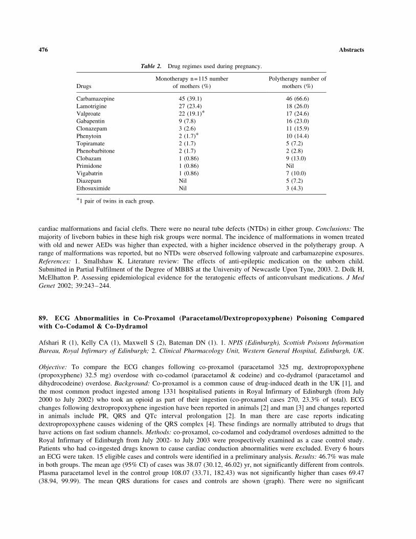
cardiac malformations and facial clefts. There were no neural tube defects (NTDs) in either group. Conclusions: The
majority of liveborn babies in these high risk groups were normal. The incidence of malformations in women treated
with old and newer AEDs was higher than expected, with a higher incidence observed in the polytherapy group. A
range of malformations was reported, but no NTDs were observed following valproate and carbamazepine exposures.
References: 1. Smallshaw K. Literature review: The effects of anti-epileptic medication on the unborn child.
Submitted in Partial Fulfilment of the Degree of MBBS at the University of Newcastle Upon Tyne, 2003. 2. Dolk H,
McElhatton P. Assessing epidemiological evidence for the teratogenic effects of anticonvulsant medications. J Med
Genet 2002; 39:243–244.
89. ECG Abnormalities in Co-Proxamol (Paracetamol/Dextropropoxyphene) Poisoning Comparedwith Co-Codamol & Co-Dydramol
Afshari R (1), Kelly CA (1), Maxwell S (2), Bateman DN (1). 1. NPIS (Edinburgh), Scottish Poisons Information
Bureau, Royal Infirmary of Edinburgh; 2. Clinical Pharmacology Unit, Western General Hospital, Edinburgh, UK.
Objective: To compare the ECG changes following co-proxamol (paracetamol 325 mg, dextropropoxyphene
(propoxyphene) 32.5 mg) overdose with co-codamol (paracetamol & codeine) and co-dydramol (paracetamol and
dihydrocodeine) overdose. Background: Co-proxamol is a common cause of drug-induced death in the UK [1], and
the most common product ingested among 1331 hospitalised patients in Royal Infirmary of Edinburgh (from July
2000 to July 2002) who took an opioid as part of their ingestion (co-proxamol cases 270, 23.3% of total). ECG
changes following dextropropoxyphene ingestion have been reported in animals [2] and man [3] and changes reported
in animals include PR, QRS and QTc interval prolongation [2]. In man there are case reports indicating
dextropropoxyphene causes widening of the QRS complex [4]. These findings are normally attributed to drugs that
have actions on fast sodium channels. Methods: co-proxamol, co-codamol and codydramol overdoses admitted to the
Royal Infirmary of Edinburgh from July 2002- to July 2003 were prospectively examined as a case control study.
Patients who had co-ingested drugs known to cause cardiac conduction abnormalities were excluded. Every 6 hours
an ECG were taken. 15 eligible cases and controls were identified in a preliminary analysis. Results: 46.7% was male
in both groups. The mean age (95% CI) of cases was 38.07 (30.12, 46.02) yr, not significantly different from controls.
Plasma paracetamol level in the control group 108.07 (33.71, 182.43) was not significantly higher than cases 69.47
(38.94, 99.99). The mean QRS durations for cases and controls are shown (graph). There were no significant
Table 2. Drug regimes used during pregnancy.
Drugs
Monotherapy n=115 number
of mothers (%)
Polytherapy number of
mothers (%)
Carbamazepine 45 (39.1) 46 (66.6)
Lamotrigine 27 (23.4) 18 (26.0)
Valproate 22 (19.1)* 17 (24.6)
Gabapentin 9 (7.8) 16 (23.0)
Clonazepam 3 (2.6) 11 (15.9)
Phenytoin 2 (1.7)* 10 (14.4)
Topiramate 2 (1.7) 5 (7.2)
Phenobarbitone 2 (1.7) 2 (2.8)
Clobazam 1 (0.86) 9 (13.0)
Primidone 1 (0.86) Nil
Vigabatrin 1 (0.86) 7 (10.0)
Diazepam Nil 5 (7.2)
Ethosuximide Nil 3 (4.3)
*1 pair of twins in each group.
476 Abstracts
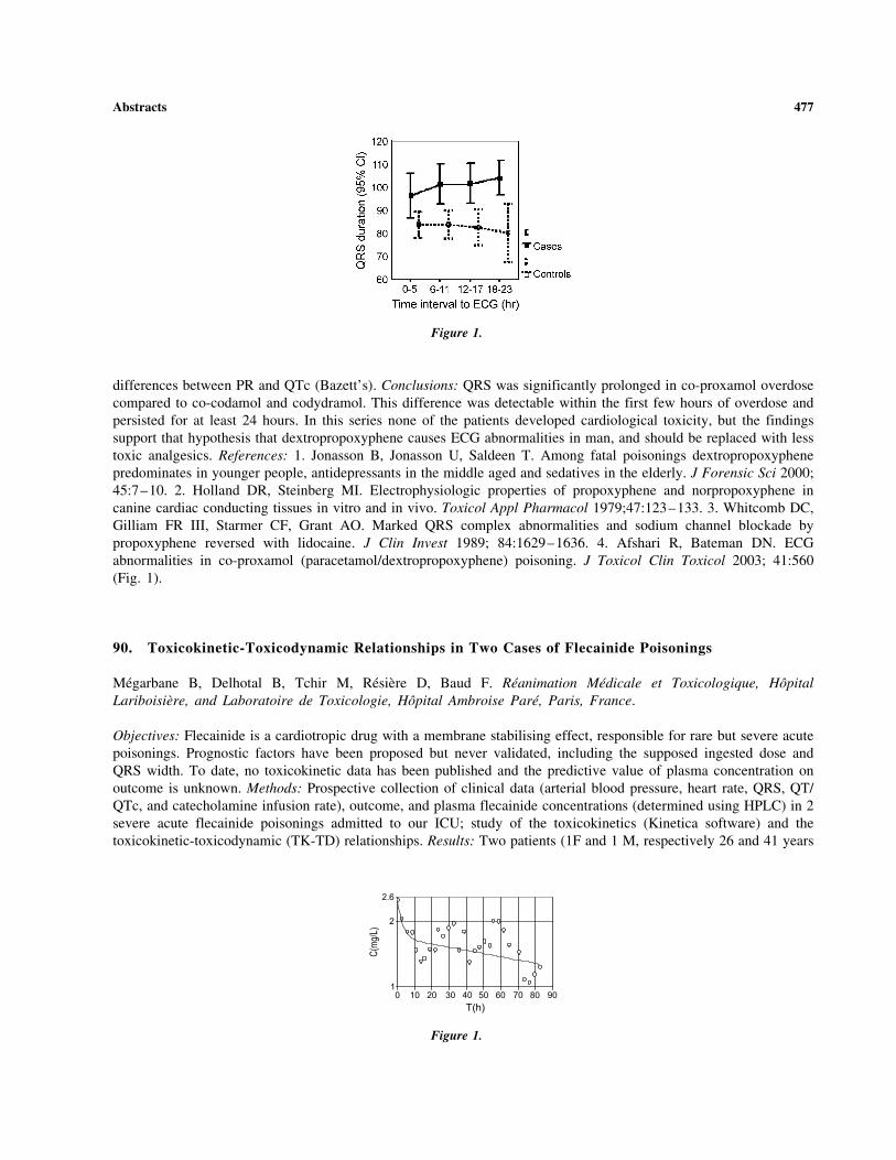
differences between PR and QTc (Bazett’s). Conclusions: QRS was significantly prolonged in co-proxamol overdose
compared to co-codamol and codydramol. This difference was detectable within the first few hours of overdose and
persisted for at least 24 hours. In this series none of the patients developed cardiological toxicity, but the findings
support that hypothesis that dextropropoxyphene causes ECG abnormalities in man, and should be replaced with less
toxic analgesics. References: 1. Jonasson B, Jonasson U, Saldeen T. Among fatal poisonings dextropropoxyphene
predominates in younger people, antidepressants in the middle aged and sedatives in the elderly. J Forensic Sci 2000;
45:7–10. 2. Holland DR, Steinberg MI. Electrophysiologic properties of propoxyphene and norpropoxyphene in
canine cardiac conducting tissues in vitro and in vivo. Toxicol Appl Pharmacol 1979;47:123–133. 3. Whitcomb DC,
Gilliam FR III, Starmer CF, Grant AO. Marked QRS complex abnormalities and sodium channel blockade by
propoxyphene reversed with lidocaine. J Clin Invest 1989; 84:1629–1636. 4. Afshari R, Bateman DN. ECG
abnormalities in co-proxamol (paracetamol/dextropropoxyphene) poisoning. J Toxicol Clin Toxicol 2003; 41:560
(Fig. 1).
90. Toxicokinetic-Toxicodynamic Relationships in Two Cases of Flecainide Poisonings
Megarbane B, Delhotal B, Tchir M, Resiere D, Baud F. Reanimation Medicale et Toxicologique, Hopital
Lariboisiere, and Laboratoire de Toxicologie, Hopital Ambroise Pare, Paris, France.
Objectives: Flecainide is a cardiotropic drug with a membrane stabilising effect, responsible for rare but severe acute
poisonings. Prognostic factors have been proposed but never validated, including the supposed ingested dose and
QRS width. To date, no toxicokinetic data has been published and the predictive value of plasma concentration on
outcome is unknown. Methods: Prospective collection of clinical data (arterial blood pressure, heart rate, QRS, QT/
QTc, and catecholamine infusion rate), outcome, and plasma flecainide concentrations (determined using HPLC) in 2
severe acute flecainide poisonings admitted to our ICU; study of the toxicokinetics (Kinetica software) and the
toxicokinetic-toxicodynamic (TK-TD) relationships. Results: Two patients (1F and 1 M, respectively 26 and 41 years
Figure 1.
Figure 1.
Abstracts 477
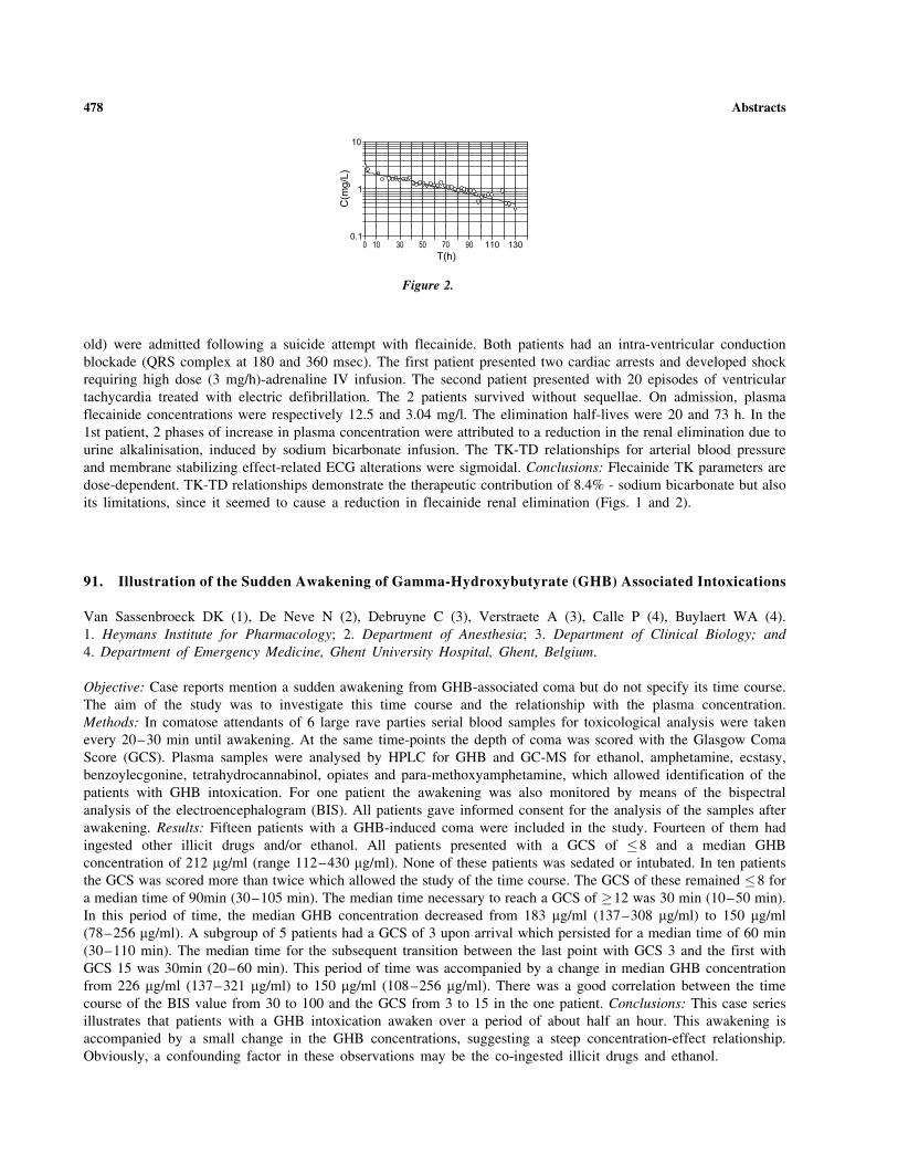
old) were admitted following a suicide attempt with flecainide. Both patients had an intra-ventricular conduction
blockade (QRS complex at 180 and 360 msec). The first patient presented two cardiac arrests and developed shock
requiring high dose (3 mg/h)-adrenaline IV infusion. The second patient presented with 20 episodes of ventricular
tachycardia treated with electric defibrillation. The 2 patients survived without sequellae. On admission, plasma
flecainide concentrations were respectively 12.5 and 3.04 mg/l. The elimination half-lives were 20 and 73 h. In the
1st patient, 2 phases of increase in plasma concentration were attributed to a reduction in the renal elimination due to
urine alkalinisation, induced by sodium bicarbonate infusion. The TK-TD relationships for arterial blood pressure
and membrane stabilizing effect-related ECG alterations were sigmoidal. Conclusions: Flecainide TK parameters are
dose-dependent. TK-TD relationships demonstrate the therapeutic contribution of 8.4% - sodium bicarbonate but also
its limitations, since it seemed to cause a reduction in flecainide renal elimination (Figs. 1 and 2).
91. Illustration of the Sudden Awakening of Gamma-Hydroxybutyrate (GHB) Associated Intoxications
Van Sassenbroeck DK (1), De Neve N (2), Debruyne C (3), Verstraete A (3), Calle P (4), Buylaert WA (4).
1. Heymans Institute for Pharmacology; 2. Department of Anesthesia; 3. Department of Clinical Biology; and
4. Department of Emergency Medicine, Ghent University Hospital, Ghent, Belgium.
Objective: Case reports mention a sudden awakening from GHB-associated coma but do not specify its time course.
The aim of the study was to investigate this time course and the relationship with the plasma concentration.
Methods: In comatose attendants of 6 large rave parties serial blood samples for toxicological analysis were taken
every 20–30 min until awakening. At the same time-points the depth of coma was scored with the Glasgow Coma
Score (GCS). Plasma samples were analysed by HPLC for GHB and GC-MS for ethanol, amphetamine, ecstasy,
benzoylecgonine, tetrahydrocannabinol, opiates and para-methoxyamphetamine, which allowed identification of the
patients with GHB intoxication. For one patient the awakening was also monitored by means of the bispectral
analysis of the electroencephalogram (BIS). All patients gave informed consent for the analysis of the samples after
awakening. Results: Fifteen patients with a GHB-induced coma were included in the study. Fourteen of them had
ingested other illicit drugs and/or ethanol. All patients presented with a GCS of �8 and a median GHB
concentration of 212 mg/ml (range 112–430 mg/ml). None of these patients was sedated or intubated. In ten patients
the GCS was scored more than twice which allowed the study of the time course. The GCS of these remained �8 for
a median time of 90min (30–105 min). The median time necessary to reach a GCS of �12 was 30 min (10–50 min).
In this period of time, the median GHB concentration decreased from 183 mg/ml (137–308 mg/ml) to 150 mg/ml
(78–256 mg/ml). A subgroup of 5 patients had a GCS of 3 upon arrival which persisted for a median time of 60 min
(30–110 min). The median time for the subsequent transition between the last point with GCS 3 and the first with
GCS 15 was 30min (20–60 min). This period of time was accompanied by a change in median GHB concentration
from 226 mg/ml (137–321 mg/ml) to 150 mg/ml (108–256 mg/ml). There was a good correlation between the time
course of the BIS value from 30 to 100 and the GCS from 3 to 15 in the one patient. Conclusions: This case series
illustrates that patients with a GHB intoxication awaken over a period of about half an hour. This awakening is
accompanied by a small change in the GHB concentrations, suggesting a steep concentration-effect relationship.
Obviously, a confounding factor in these observations may be the co-ingested illicit drugs and ethanol.
Figure 2.
478 Abstracts
92. Serum Alkalinization and Markers of Toxicity in Tricyclic Antidepressant Overdose
Ricci G, Buonocore F, Zannoni M, Legnaro E, Menegazzi S. Servizio di Pronto Soccorso—Unita di Tossicologia
Clinica O.C.M. Verona, Italy.
Introduction: Tricyclic antidepressant (TCA) overdose is a common cause of death. Clinical presentation of overdose
includes cardiac arrhythmias, hypotension, seizures, coma and anticholinergic signs such as hyperthermia, flushing and
intestinal ileus. Toxic levels (>1,000 ng/mL) are manifested as QRS prolongation to 100 msec or more. TCA poisoning
has been reported to cause a right-axis deviation of 130� to 270� in the T40 ms-axis. Objectives: to prevent life-
threatening arrhythmias due to QRS prolongation with serum alkalinization; to describe the changes in the terminal 40
msec-axis; to identify clinical and prognostic factors related to such changes. Methods: the QRS interval and T40 ms-
axis were manually measured in 60 patients divided in two groups: TCA overdose (n=40) and non TCA overdose
(n=20). The T40 ms-axis was significantly more rightward in the TCA overdose group (>120�–270� in our experience).
All patients (n=19) with T40 rightward deviation >150� not treated with serum alkalinization developed wide complex
arrhythmias and seizures; patients with rightward deviation >150� treated with IV sodium bicarbonate to achieve a
plasma pH of 7.50–7.55 did not develop life-threatening complications. Results: these findings suggest that the T40 ms-
axis is a better marker of TCA toxicity than the QRS interval, and an important prognostic indication of potentially life-
threatening events. In these cases, sodium bicarbonate administration may minimize the cardiological complications
related to TCA overdose.
93. Acute Chemical Poisonings and Human Health in Russia
Ostapenko YuN, Litvinov NN, Kakorina EP, Khonelidze RS. Russian Toxicology Information and Advisory Centre,
The Russian Federation Ministry of Health, Russia.
Objective: Presentation of the main trends with acute poisonings in Russia during 1998–2002. Methods: Data of State
medical statistical system and 43 regional toxicology centers dealing with approximately 50% of population were
used. Results: The annual number of in-patients with acute poisoning was about 260 thousand cases and had no
tendency to increase. However, the main concern is significant increase in mortality (up to 53%) during this period of
time and more than 1.5 fold increase of acute poisonings in children aged by 14 years (Table 1). Main agents used
were psychotropic and other drugs (40–60%), alcohol (15–20%), acids and alkalis (10–13%), carbon monoxide (up to
9.8%). Acute poisonings are domestic in 98.5–99% of total cases. 90% of lethal cases were due to acute poisonings.
87–90% of lethal cases occurred out of hospital, with alcohol poisoning accounting for 50.0–58.5%. In Moscow
during 2000 and 2001 acute drug poisonings reached 39.9% and 48.8% respectively, and mortality increased by 18.8%
compared to 1999. The main causes of acute poisonings were psychopharmaceuticals, and antiarrhythmic drugs. The
same tendency was noticed in other big cities of Russia. Comparative morbidity and mortality data of the most
prevalent diseases and chemical poisonings among the population of Russia in 2002 are presented in Table 2.
Conclusion: Acute chemical poisonings are among the leading causes of morbidity and mortality in Russia and are an
important factor in the health of population.
Table 1. Acute poisoning morbidity dynamics in Russia in 1998–2002.
Indices 1998 2000 2002
Total number of acute chemical poisonings 258 438 261 564 268 511
Children included 27 532 38 656 43 213
Number of lethal cases 61 868 85 089 95 045
Including lethal cases of alcohol poisonings 29 872 37 214 52 465
Abstracts 479

94. Fire Eater’s Pneumonia: Incidence of this Dangerous Complication Increases
Stedtler U (1), Michalak H (2), Hahn A (2), Hermanns-Clausen M (1). 1. Poisons Information Centre (VIZ),
Department of Paediatrics and Adolescent Medicine, Freiburg, Germany; 2. Federal Institute for Risk Assessment
(BfR), Berlin, Germany.
Objective: Throughout the EU strong efforts have been made in recent years to reduce accidents of children aspirating
lamp oil. Another population at risk for aspiration of hydrocarbons are fire breathers who spit petroleum-like fluids
through torches to give the illusion to spit or breathe flames. We analysed reports to the VIZ or the BfR in respect of age,
gender and experience of the patients and as to site and severity of lung damage. Case Series: 45 cases of aspiration
accidents were reported to the BfR (16 cases; 01/97 to 09/03) or VIZ (30 cases; 01/00 to 09/03). One case was reported to
both centres. 42 cases concerned hydrocarbons with follow up information available in 39 cases. Incidence increased
from 6 cases in 2000 to 15 cases in 2002 and 12 further cases until September 2003. The mean age of the patients was 28
years, 33 of 39 were male, 13 stated to perform fire breathing at least as semi professionals. Most patients initially
experienced only mild symptoms. 29 patients (9 professionals) developed radiologically confirmed pneumonia. In 2
patients no x-ray was performed, but clinical course did not suggest lung damage. Mean stay in hospital was 7.7 days,
mean poison severity score of respiratory symptoms was 1.6. More patients were reported to the VIZ than to the BfR.
Those reported to the latter were in general more serious. This possibly reflects the fact that a poison control centre is
contacted by lay persons as well as professionals for advice while reports to the BfR are only done by professionals to
satisfy their duty of notification. In 18 patients information about the site of lesion was provided: 2 times the left lung
only, 5 times the right one only. In 11 cases both lungs were affected. Analysis of a fire breathing product shows physical
properties like a common lamp fuel even though the chemical composition differs. Conclusion: The number of
aspiration accidents due to fire breathing increases. Experienced artists are also at risk. Most times both lungs are
affected. Despite the duty of notification many of these substance related illnesses are not reported to the BfR.
95. Poisoning with a Vesicant Agent—Acute and Late Effects
Kehe K, Schmidt S, Thiermann H, Szinicz L. Bundeswehr Institute of Pharmacology and Toxicology, Munich,
Germany.
Objective: Sulfur mustard (bis (2-chloroethyl) sulfide, SM) is a vesicant which is still a threat in war and terroristic
attacks. The clinical effects of sulphur mustard are delayed. Erythema, blisters and necrosis of the skin appear several
hours after exposure. SM is a DNA alkylating agent that has been linked to an increased risk of several cancers. Here we
report about a patient exposed to a blistering agent. Case Report: An otherwise healthy 35 year old patient was poisoned
with an unknown liquid by covered application at the back of his upper left leg in 1964. The victim firstly noticed only a
strong smell of garlic at this time point, but no eye or respiratory symptoms. Blisters at the left thigh and strong erythema
occurred 14 hours later. The patient was hospitalized 2 days later. He presented several bullae and secondary vesicles,
erythema, scrotal swelling. The glans penis showed several ulcerations. Wounded skin was superinfected with candida
albicans. Systemic treatment with methylprednisolone, paracetamol, pethidine and penicillin reduced the symptoms and
Table 2. The structure and quantity of most prevalent diseases and mortality of population in Russia in 2002.
Indices Neoplasms
Cerebrovascular
diseases Pneumonia
Acute
chemical
poisonings Tuberculosis
Myocardial
infarction
Total number
of patients
1 367 695 870 589 401 252 268 511 209 243 207 657
Number of
lethal outcomes
47 251 83 472 13 588 95 045 18 142 37 788
480 Abstracts
the patient could be dismissed after 3 months. 38 years later the patient showed hypopigmentation and alopecia at the
back of the left thigh, poikiloderma of the glans penis. Dermatohistopathological examination of the affected skin
revealed marked elastosis. ECG, chest x-ray, laboratory tests and ENT consultation were unremarkable. Conclusion:
The typical clinical course and the strong smell of garlic strongly suggest the use of SM. Today, emergency care units
can be confronted with SM poisoning during accidental contamination or acts of terrorism. Therefore, the patient’s
history is very important in the initial work-up. Exposure to unknown gases or fluids should be queried. The onset of the
blisters showed a characteristic latency after the suspected exposure. In this case no significant late effect was detected.
It has to be assumed that the level of SM exposure, which was sufficient to cause skin reactions (erythema, vesicles, and
ulcerations), was associated with mild late effects (pigmentation disorders) but no cancer.
96. Ethyl Glucuronide as a Post Mortem Marker of Alcohol Consumption
Pennings EJM (1), Steutel K (1), Lusthof KJ (2), Wolff FA de (1). 1. Leiden University Medical Center, Toxicology
Laboratory, Leiden; and 2. Netherlands Forensic Institute, Rijswijk, The Netherlands.
Objective: In post-mortem toxicology, blood alcohol measurements have limited value because of post mortem
alcohol formation by microorganisms. Therefore, additional biological markers of alcohol consumption are needed to
assess alcohol consumption. Ethylglucuronide has been suggested as a suitable biomarker because it is formed from
alcohol in man and is not formed by microbial activity (1). The aim of the present study was to measure
ethylglucuronide levels in post mortem heart and femoral blood samples from different individuals, and to compare
the results with alcohol measurements in these samples. Methods: Gas chromatography with mass spectrometric
detection. Results: In 5 out of 8 forensic blood samples ethylglucuronide was found in concentrations ranging from
1.3 to 3.4 mg/L. In the remaining 3 samples the ethylglucuronide concentration was below the lower limit of
quantitation (0.5 mg/L). Ethylglucuronide concentrations did not correlate with femoral or heart blood alcohol
concentrations, i.e. ethylglucuronide was found in blood samples both with and without alcohol. In one case with
negative femoral and heart blood alcohol but positive vitreous fluid alcohol, ethylglucuronide was found in heart
blood at a concentration of 2.7 mg/L. Conclusion: The results suggest that ethylglucuronide is a useful detector of
alcohol consumption if blood alcohol measurements are negative or not possible. Reference: 1. Schmitt G, Droenner
P, Skopp G, Aderjan R. Ethyl glucuronide concentration in serum of human volunteers, teetotalers, and suspected
drinking drivers. J Forensic Sci 1997; 42:1099–1102.
97. Inhalation of Butane: 4 Deaths in a Case Series of 13 Consecutive Patients
Kaes J, von Mach MA, Wiechelt J, Weilemann LS. Department of Clinical Toxicology and Poison Center,
University of Mainz, Germany.
Objective: A fatal outcome after inhalation of butane has repeatedly been published in recent years. The present study was
to investigate the epidemiology and course of intoxications with butane analyzing the inquiries to a poison center. Case
Series: From the 1st of January 2000 until the 31st of October 2003 (46 months) 13 intoxications with butane were
observed (5 female, 8 male, average age 19.8±8.5 years). The documentation and an explorative data analysis was
performed using our poison center database (ADAM-Dok, based on Microsoft1 Access1). For all patients presenting
with inhalation of butane a previous abuse was known. 4 patients (number 3, 5, 11, 13) died after the exposition to butane
(30.8%) and 9 patients showed a complete recovery. Patient 3 was a 14-year-old boy who was found by the paramedics
with electromechanical dissociation and died after 45 minutes of cardiopulmonary resuscitation. Patient 5 was a 14-year-
old boy who was found with ventricular fibrillation and was successfully resuscitated. After initial stabilization the patient
developed a massive rhabdomyolysis with acute renal failure and finally died of an adult respiratory distress syndrome.
Patient 11 was a 16-year-old girl who was found after prolonged circulatory arrest and showed cerebral death after
initially successful cardiopulmonary resuscitation. Patient 13 was a 14-year-old boy who was found with ventricular
fibrillation and died under prolonged cardiopulmonary resuscitation. Conclusion: Inhalation of butane may cause
Abstracts 481
hypoxemia accompanied by acidosis and circulatory failure due to a displacement of oxygen. The present case series
revealed that teenage patients with a history of butane abuse are at high risk for a lethal outcome. The lethal complications
included most frequently ventricular fibrillation, electromechanical dissociation, and cardiac arrest. These cardiac
disorders have previously been discussed to result from a sensitization of the myocardial tissue to endogenous
catecholamines which may cause serious dysrhythmias even in healthy teenagers.
98. Fluoride Poisoning at a Toothpaste Factory
Meggs WJ. Brody School of Medicine at East Carolina University, Greenville, USA.
Objective: Fluoride salts are known to be irritants and systemic toxins. Toxicity from occupational exposure is unusual.
Two workers demolished hoppers used to store sodium and stannous fluoride at a toothpaste factory which had not been
cleaned of fluoride salts. They worked without protective equipment and had an chronic then an acute exposure to a dust
of fluoride salts. Signs and symptoms of toxicity are reported below. Case Report #1: A 26 year old man in prior good
health except for eczema worked in a poorly ventilated area used to store sodium fluoride. Removal of floor pads
exposed a white powder. Sweeping the powder with a broom generated fluoride dust. After an exposure period of
approximately 2 hours, he became ill. Initial symptoms were emesis, visual disturbances (seeing spots), and difficulties
with comprehension. Over a few hours he developed tremors, motor weakness, abdominal pain, dyspnea, and headache.
Respiratory symptoms of dyspnea, wheezing, and cough developed. Gastrointestinal symptoms included nausea, emesis,
diarrhea, abdominal pains, and anorexia. Anorexia developed, and he lost 15 pounds over a 3 week period. There was a
flare in his eczema. He suffered myalgias and muscle spasms. Urine fluoride level 3 days after the exposure was
1.3 mg/dL, within the reference range of 0.2 to 3.2 mg/dL. FEV1 3 days after the exposure was 80% of predicted.
He was unable to work for 3 weeks. Case Report #2: A 37 year old man was in good health with a prior history
inactive peptic ulcer disease prior to the fluoride exposure. During the period of exposure, he developed headaches,
vision difficulties, sinusitis, and nasal congestion. He also develops wheezing, dyspnea, and cough. Gastrointestinal
symptoms included nausea, emesis, diarrhea, and abdominal pain. There were also muscle pains. Neurological
symptoms included vertigo, insomnia, and difficulties with memory. There was a transient skin rash. Urine fluoride
level three days after the acute exposure was 0.8 mg/dL, within the reference range of 0.2 to 3.2 mg/dL. Endoscopy
was positive for gastritis. CT of head and MRI were normal except for sinus disease. Pulmonary function tests were
normal. Conclusions: Occupational exposure to fluoride salts as dust can lead to multiple symptoms, including head-
ache, gastrointestinal symptoms, respiratory irritation, musculoskeletal symptoms, and rash. Workers exposed to
fluoride dust should use proper protective equipment.
99. Carcinogenic, Mutagenic and Reprotoxic Hazards on a Petrochemical Site
Humbert P (1), Raspiller MF (1), Andrieu P (2), Chabrand C (2), Poinas R (2), Smolik HJ (3). 1. CHU Hopital
Central, Nancy France; 2. BP Lavera SNC 13117 Lavera France; 3. Institut de Medecine du Travail et d’Ergonomie
de Bourgogne Franche-Comte Faculte de Medecine, Dijon, France.
Objective: To meet the new regulatory requirements concerning carcinogenic, mutagenic and reprotoxic (CMR)
substances laid out in Decree No. 2001-97 of 1st February 2001 implementing European Directives 97/42/EC and 99/38/
EC, on a site employing close to 1000 people (BP Lavera). Methods: The CMR agents affected by the decree are those
found on the European list. Since February 2001 a CMR risk assessment procedure, initially launched in 1991 for
chemical hazards, has been set up by a multi-discipline team involving the Legal Department, the Industrial Health and
Safety Department, the Medical Department and Human Resources, with the aid of the Environmental Health and Safety
Directorate, Operational Unit/Production Line Management and the Personnel Administration Department. In parallel
with this, a procedure operates which involves other industrial sites within the Etang de Berre region. This assesses the
risks to co-contractors from dangers produced by the user organisations (APHI procedure - Association pour la
Promotion de l’Hygiene Industrielle, association for promotion of industrial health). The means selected to operate this
procedure within the organisation are: 1/a unit-based inventory of the products present on site and the creation of a Safety
482 Abstracts
Data Sheet intranet database 2/a list of work-stations and of the personnel at each station, and the identification of groups
which have similar employee exposure, so that biometric and other measurements may be carried out 3/the establishment
of a single document for work-unit based risk assessments 4/a quantitative approach to individual exposure levels of
employees or group exposure using atmosphere measurements over 8 hours, including those CMR substances with no
regulatory occupational exposure limits 5/issue of an individual exposure sheet model to be used when completing fitness
for work reports. Results: The product inventory is regularly updated, provides better information for employees and
results in more appropriate safety measurements (both individual and collective) being carried out. The single document/
individual exposure sheet combination allows better medical supervision and enables better fitness to work evaluations to
be carried out. Apart from the financial constraints involved, there are inherent limitations due to the lack of an approved
laboratory, limitations in the atmospheric measurements that can be performed, the limited number of Biological
Exposure Indices as well as the difficulties involved in replacing the CMR agents. Conclusion: The process being
undertaken on this petrochemical site demonstrates the usefulness of a multi-disciplinary approach in effective
prevention of CMR risks.
100. Clinical Management of Suspected Ethylene Glycol/Methanol Poisoning: Assessment of the Needfor Laboratory Testing and Antidotal Treatment
Lonati D, Butera R, Petrolini V, Bove A, Locatelli C, Manzo L. Pavia Poison Center, IRCCS Maugeri Foundation
and University of Pavia, Italy.
Background: In current guidelines for ethylene glycol (EG) and methanol (M) poisoning proposed indications for
antidotal treatment are based on history, serum EG/M level, osmolar gap and metabolic acidosis. However, in Italy EG/M
analysis is performed by few laboratories, and osmolar gap assessment is rarely available in emergency departments.
Therefore, deviations from standard recommendations may occur. Objective: To describe in a field study the clinical
management and the criteria used to start antidotal treatment in patients with suspected EG/M poisoning. Methods: A
retrospective analysis of all cases of EG/M exposure referred to a Poison Center (PC) from January 2002 to October 2003
was performed. All human cases were considered eligible. The following parameters were evaluated: history, time
elapsed from exposure to PC assessment, clinical presentation, presence of metabolic acidosis, serum EG/M levels, time
required to obtain analytical result, and management of antidotal therapy. Patients with incomplete data were excluded
from the study. Results: Among 77 eligible patients, 63 were included in the study. EG and M were involved in 57 and 6
cases, respectively. One patient with previously unrecognized methanol poisoning was referred to PC when neurotoxic
damage was already established and serum methanol levels were no more detectable. Twenty-four patients (24/63, 38%)
were considered to not be at risk of acute poisoning because of the circumstances of exposure or the minimal amount
ingested. In the remaining 38 patients (38/63, 60%) history was consistent with a recent, potentially toxic exposure. All
patients underwent gastrointestinal decontamination; osmolar gap was never available. In 23 cases (23/38, 61%) at mild
to moderate risk (amount ingested 0.5±0.4 ml/kg) the development of metabolic acidosis was used to start antidotal
treatment; none showed abnormalities and antidotal treatment was not performed; for 6 patients only serum EG/M levels
were available (within 4.4±1.1 hours) and returned <20 mg/dl. In 15 cases (15/38, 39%) the amount ingested (1.2±1.0
ml/kg) and/or clinical findings were highly suggestive for acute EG/M poisoning requiring antidotal treatment: EG/M
levels obtained 14.2±18.5 hours after sampling allowed discontinuation of avoidable treatment in 10 cases, and
confirmed the need for further therapy in 5 patients. Conclusion: The unavailability of timely EG/M testing may lead to
unnecessary treatment in a considerable proportion of cases. However, significant delays in antidotal treatment can occur
if therapy is withheld until toxicological analysis is obtained. Monitoring of metabolic disturbances may be useful in
patients with mild to moderate risk of poisoning.
101. Immunological Long-Term Effects of Sulfur Mustard Poisoning in Iranian Veterans
Mahmoudi M (1), Hefazi M (2), Rastin M (1), Balali Mood M (2). 1. Immunology Research Center, 2. Department
of Toxicology, Mashhad University of Medical Sciences, Mashhad, Iran.
Abstracts 483
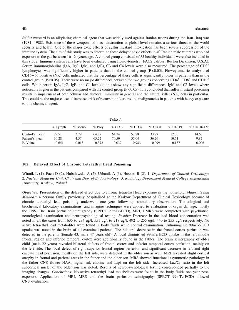
Sulfur mustard is an alkylating chemical agent that was widely used against Iranian troops during the Iran–Iraq war
(1981–1988). Existence of these weapons of mass destruction at global level remains a serious threat to the world
security and health. One of the major toxic effects of sulfur mustard intoxication has been severe suppression of the
immune system. The aim of this study was to determine these delayed toxic effects in 40 Iranian male veterans who had
exposure to the gas between 16–20 years ago. A control group consisted of 35 healthy individuals were also included in
this study. Immune system cells have been evaluated using flowcytometry (FACS calibur, Becton Dickinson, U.S.A).
Serum immunoglobulins (IgA, IgG, IgM, and IgE), C3 and C4 levels were also measured. The percentage of CD3+
lymphocytes was significantly higher in patients than in the control group (P<0.05). Flowcytometric analysis of
CD16+56 positive (NK) cells indicated that the percentage of these cells is significantly lower in patients than in the
control group (P<0.05). There were no major differences between the two groups concerning CD4+, CD8+ and CD19+
cells. While serum IgA, IgG, IgE, and C4 levels didn’t show any significant differences, IgM and C3 levels where
noticeably higher in the patients compared with the control group (P<0.05). It is concluded that sulfur mustard poisoning
results in impairment of both cellular and humoral immunity in general and the natural killer (NK) cells in particular.
This could be the major cause of increased risk of recurrent infections and malignancies in patients with heavy exposure
to this chemical agent.
102. Delayed Effect of Chronic Tetraethyl Lead Poisoning
Winnik L (1), Pach D (2), Hubalewska A (2), Urbanik A (3), Huszno B (2). 1. Department of Clinical Toxicology;
2. Nuclear Medicine Unit, Chair and Dep. of Endocrinology; 3. Radiology Department Medical College Jagiellonian
University, Krakow, Poland.
Objective: Presentation of the delayed effect due to chronic tetraethyl lead exposure in the household. Materials and
Methods: 4 persons family previously hospitalised at the Krakow Department of Clinical Toxicology because of
chronic tetraethyl lead poisoning underwent one year follow up ambulatory observation. Toxicological and
biochemical laboratory examinations, and imagine techniques were applied to evaluation of organ damage, mostly
the CNS. The Brain perfusion scintigraphy (SPECT 99mTc-ECD), MRI, HMRS were completed with psychiatric,
neurological examination and neuropsychological testing. Results: Decrease in the lead blood concentration was
noted in all the cases from 635 to 294 ug/l, 551 ug/l to 217 ug/l, 492 to 255 ug/l, 440 to 255 ug/l respectively. No
active tetraethyl lead metabolites were found in body fluids while control examination. Unhomogenic 99mTc-ECD
uptake was noted in the brain of all examined patients. The bilateral decrease in the frontal cortex perfusion was
detected in the parents (female 43, male 47 years old). A focal diminished 99mTc-ECD uptake in the left middle
frontal region and inferior temporal cortex were additionally found in the father. The brain scintygraphy of older
child (male 22 years) revealed bilateral defects of frontal cortex and inferior temporal cortex perfusion, mainly on
the left side. The focal defect of right superior frontal region perfusion and significant decrease in left and right
caudate head perfusion, mostly on the left side, were detected in the older son as well. MRI revealed slight cortical
atrophy in frontal and parietal areas in the father and the older son. MRS showed functional asymmetric pathology in
the father CNS (lower NAA, higher ml, choline and Lip) on the left side. Increased Lac/Cr ratio in the left
subcortical nuclei of the older son was noted. Results of neuropsychological testing corresponded partially to the
imaging changes. Conclusions: No active tetraethyl lead metabolites were found in the body fluids one year post-
exposure. Application of MRI, MRS and the brain perfusion scintigraphy (SPECT 99mTc-ECD) allowed
CNS evaluation.
Table 1.
% Lymph % Mono % Poly % CD 3 % CD 4 % CD 8 % CD 19 % CD 16+56
Control’s mean 29.51 3.79 64.89 64.74 57.28 33.27 12.36 14.66
Patient’s mean 30.28 4.57 63.22 70.59 57.04 36.26 10.51 9.82
P. Value 0.651 0.013 0.372 0.037 0.983 0.099 0.187 0.006
484 Abstracts
103. Rapid Fatal Outcomes After Acid Ingestion
Roh HK, Cho HJ, Kim PS. Department of Internal Medicine, Inha University Hospital, Incheon, South Korea.
Objective: Acid ingestion may cause fatal outcome as well as caustic injuries on esophagus and stomach. Ingested large
amount of acid can be absorbed through gastrointestinal mucosa and rapidly deteriorate various organs, which causes
serious multi-organ damage and death. This study aimed to investigate how the acid ingestion affects organs by analyzing
patients who died early after large amount of acid ingestion. Case Series: Five deaths after ingestion of strong or
concentrated acid were reviewed retrospectively. These cases were all suicide attempts and they arrived at the hospital
within one hour after the ingestion. Four patients ingested 99% acetic acid and one patient had 10% hydrochloric acid. The
estimated amounts of acid ingested were between 60 and 300 ml. In the beginning, epigastric pain, nausea and vomiting
were commonly observed and all five patients showed severe gross hematuria, although there were only slight increases of
the serum creatinine level. Their vital signs deteriorated rapidly and intubation was performed in all patients due to
respiratory difficulty within 6 hours after the ingestion. In the arterial blood gas analyses performed at the time of arrival,
the pH and bicarbonate ranged from 7.06 to 7.17 and from 7 to 14 mmol/L, respectively. Blood chemistry analyses showed
marked increases in serum aspartate and alanine (6007–13600 IU/L and 2751–5150 IU/L, respectively) and in serum total
bilirubin (7.2–11.6 mg/dL). Blood hemoglobin levels decreased by 3–5 g/dL between 3 and 11 hours after the ingestion,
suggesting hemolysis. Despite of intensive care with administration of bicarbonate and ventilatory support, they never
recovered from the shock state and pH lower than 7.2. Two patients died around 6 hours and the rest died within 31 hours
after the ingestion. Conclusion: Large amounts of weak acid with high concentration as well as strong acid cause death due
to the rapid multi-organ failure. These deteriorations might be due to severe acidemia after rapid absorption of large
amount of acid.
104. Survival After Intentional Oral Ingestion of an Ammonium Bifluoride Containing CommercialRust Remover
Greller HA, Hoffman RS, Nelson LS. New York City Poison Control Center, New York, NY, USA.
Objective: Only three published ammonium bifluoride (ABF) ingestions are reported. All are in children, and all
were fatal. We report an adult with a massive ABF ingestion who survived with early aggressive therapy. Case
Report: A healthy 36 year-old man was witnessed to ingest approximately 180 mL of an ABF containing rust
remover (reported concentration ‘‘2.5 mg/m (ASF)’’; pH 1.5). Within minutes, he was prostrate and vomiting
profusely, complaining of severe abdominal pain. He was seen by an onsite toxicologist and rapidly transported to
the hospital. An initial ECG showed a prolonged QTc of 0.516 s. Resuscitation measures included IV CaCl2 and
MgSO4 (2gms each). The QTc narrowed to 0.400 s. Initial serum K+ was 5.8 mmol/L, ionized Ca2 + was 0.935
mmol/L, and lactate was 4.8 mmol/L. During direct nasopharyngeal laryngoscopy, he had hematemesis, and was
endotracheally intubated. After intubation, his QTc widened again to 0.500 s. Boluses of 1 g of Ca gluconate and 2 g
of MgSO4 were given, and he was started on a continuous infusion of NaHCO3 (27 mEq/h). Central venous access was
obtained, and he received an additional 2 g of CaCl2, as well as 2 g of MgSO4 followed by the initiation of a continuous
infusion of MgSO4 at 2 g/hr. His QTc narrowed to 0.413 s. Esophagogastroduodenoscopy performed at the bedside
revealed mild edema throughout the length of the esophagus, moderate hemorrhagic gastritis, and a pale appearance to
the fundus and greater curvature of the stomach consistent with ischemia. Although a nasogastric tube was placed under
direct visualization, nothing was administered. The patient continued to receive intermittent boluses of CaCl2 when his
QTc prolonged. By 4 hours after admission, he was admitted to the ICU. Once in the ICU, he required no additional
boluses of calcium or magnesium although his infusions of bicarbonate and magnesium were continued for 24 hours.
His serum calcium peaked at 5.4 mmol/L two hours post presentation, with ionized calcium of 2.85 mmol/L. His serum
magnesium peaked at 3.8 mmol/L at three hours post presentation. Both values declined to normal reference range by
his discharge. 36 hours post admission he was extubated, stable, and was transferred to psychiatry on hospital day four.
Conclusion: We present the first case of an adult with an intentional ingestion of ammonium bifluoride. This route of
exposure is previously reported to be uniformly fatal. Using the ECG as a guide, therapy was aggressively initiated to
Abstracts 485
prevent significant electrolyte abnormalities. We believe that these interventions directly contributed to the patient’s
favorable outcome.
105. Intestinal Ischemia from an Ephedra Containing ‘‘smoothie’’
Greller HA, Flomenbaum M,* Nelson LS, Hoffman RS. New York City Poison Control Center, New York, NY *Office of
the Chief Medical Examiner, City of New York, USA.
Objective: Although intestinal ischemia is reported from sympathomimetic pharmaceuticals and drugs of abuse, there
are no reports of ephedra associated intestinal ischemia. Case: A 30 year-old man without past medical history was
exercising at a gym. He ingested a ‘‘fat burner’’ ‘‘smoothie’’ (a protein-supplemented drink blended with ice) advertised
as containing Ephedra spp. Shortly after his ingestion, he complained of severe epigastric pain. He collapsed, and was
unable to be resuscitated in the Emergency Department. A forensic autopsy revealed focal mucosal and sub-mucosal
hemorrhage of the intestine, consistent with a recent ischemic event, as well as epicardial ischemia. Forensic toxicologic
analysis performed subsequent to the autopsy revealed a blood ephedrine level of 0.1 mg/L by GC-MS. The remainder of
the toxicology screen was negative for cocaine metabolites, amphetamines, ethanol, salicylates, lidocaine, opiates,
barbiturates and benzodiazepines, or anabolic steroids. The only other positive finding was a low but detectable GHB
level, less than that commonly reported to cause sedation. Conclusion: The derivatives of Ephedra spp. contain many
vasoactive compounds that can have significant morbidity and mortality. In this case, these compounds are the putative
cause of intestinal and myocardial ischemia and presumed sudden death in an otherwise healthy man. Further regulation
and investigation should be performed on these substances.
106. Severe Valproate-Induced Hyperammonemia Cleared Without L-Carnitine
Martin TG. UW-TOX, University of Washington; Washington Poison Center, Seattle, WA, USA.
Introduction: L-carnitine has been recommended for symptomatic hyperammonemia due to therapeutic or toxic
valproate exposures by many authors including the Pediatric Neurological Advisory Committee in 1996. Objective:
Report case of severe, valproate-induced hyperammonemia, which resolved rapidly without L-carnitine. Case Report:
37-yo male found by medics diaphoretic, responsive only to painful stimuli but with gag reflex. Past history: seizures,
bipolar depression, heavy ethanol abuse, hepatitis B (without known cirrhosis), benzodiazepine abuse, gout, and chronic
osteomyelitis. Medications: valproate 500 mg bid, allopurinol, carbamazepine, gabapentin, lorazepam, ranitidine and
lithium SR. Emergency department physical examination was otherwise unremarkable except for spider angiomas on
chest, abrasion on forehead and cheek and old murmur. Initial labs: valproate 74 mcg/mL, plasma ammonia 1,519 mcg/
dL (891 uMol/L), albumin 4.2 g/dL, protein 7.1 g/dL, PT INR 0.9, neg urine organic acid screen & venous pyruvate,
toxicology screen positive for benzodiazepines and barbiturates; ethanol 14 mg/dl. Ammonia levels fell to 540, 485, 186
and 17 mcg/dL at 1, 2, 6 and 33 hr after admission. Treatment: valproate withheld, IV fluids, 2 doses lorazepam, 1 dose
lactulose. Patient left AMA and restarted valproate on his own. Returned somnolent 3 days later, valproate 14, ammonia
level 80 mcg/dl. Valproate was discontinued again. He was hospitalized again 48 days later for leg pain, started on
valproate, per his medication history. Three days later, he was somnolent, disoriented, valproate 85 mcg/ml and
ammonia 356 mcg/dL (on second specimen drawn 3 hr after 1st specimen discarded because mislabeled). Record review
revealed an episode of somnolence 17 months prior to index presentation with ammonia 216 mcg/dL and valproate
25 mcg/mL. Only two ammonia levels recorded on two different occasions were WNL, both times with non-detectable
valproate levels. Discussion: This patient with multiple risk factors but without advanced cirrhosis had four episodes of
somnolence, hyperammonemia and non-toxic valproate levels. By inducing CYP2E1, animal data suggest that ethanol
abuse could predispose to valproate hepatotoxicity (1). Urea cycle disorders can result in ammonia levels greater than
1,000 mcg/dL. Conclusions: Poor compliance should be a contraindication to valproate therapy. This is the highest
reported ammonia level associated with valproate therapy and it rapidly cleared with intravenous fluids but without
L-carnitine. Case reports of successful use of L-carnitine to reduce valproate-induced hyperammonemia must be
486 Abstracts
interpreted cautiously. A randomized, blinded clinical trial may be required to clarify the necessity of L-carnitine in
valproate-induced hyperammonemia. Reference: 1. Neuman MG, Shear NH, Jacobson-Brown PM, et al. CYP2E1-
mediated modulation of valproic acid-induced hepatocytotoxicity. Clin Biochem 2001; 34:211–218.
107. Severe Acute Lung Oedema After Rectal Enema with Cade Oil
Rahmani H (1), Leonhardt S (1), Beladdale D (2), Krencker F (3), Bartier JC (1), Jaeger A (4), Dupeyron JP (1). 1. SAMU
67; 2. Service de Reanimation Pediatrique; 3. Centre Anti-Poison; 4. Service de Reanimation Medicale, Hopitaux
Universitaires, Strasbourg, France.
Objective: to report a case of pulmonary oedema and cardiovascular collapse after rectal enema with cade oil. Cade oil is
an essential oil, obtained by distillation of the branches of Juniperus oxycedrus, a Mediterranean bush. In traditional
medicine cade oil is used in local skin application for anti-psoriasis, anti-dermatophyte and antiseptic effects. It is also
used in the preparation of creams, washings, soaps and shampoos. Case Report: A 4-month-old baby, coming from
Cameroon, was ‘‘treated’’ for an atopic dermatosis by his mother (after the grandmother’s advice) with a rectal enema of
a traditional medicine based on cade oil. The baby developed immediately a respiratory failure with hypotonia and loss
of consciousness. The physician called at home provided oxygen therapy and performed a rectal enema with 100 ml of
isotonic NaCl solution, in units of 10 ml, to eliminate the cade oil. A blackish brown liquid with a characteristic odour
was passed. When the physician of the emergency team arrived at home, the baby was reactive and had acute respiratory
distress, with pulse oxymetry of 60%, a pulse rate of 170/min, a temperature of 37.5�C; blood pressure was not
measurable. Physical examination showed signs of an acute diffuse pulmonary oedema. Treatment included
administration of oxygen 100% by facial mask and furosemide 3 mg by intra-osseous route. Clinical symptoms
improved progressively and after 0.5 hour blood pressure was 100/50 mm Hg, pulse was 145/min and pulse oxymetry
was 100%. On admission in the Paediatric ICU, 2.5 hours after the cade oil enema, physical examination was normal
except some vesicular rales on the right lung. Blood investigations and chest X-ray were normal. The child recovered and
was discharged from the paediatric unit after 3 days. Conclusion: Cade oil is a volatile essential oil, characterized by its
acrid odour. Principal components are phenol, cadinene and sesquiterpene. In rats, methanol leaves extracts decrease the
blood pressure and inhibit the histamine, serotonine and acetylcholine responses. Cade oil is rapidly absorbed after
ingestion and is mainly metabolized by the liver. Symptoms include lung oedema, hypotension and hepatic failure.
Death has been reported after massive ingestion in suicidal attempts. Our case is particular by the route of administration.
The high capacity of absorption of the rectal mucosa explains probably the rapid onset of severe symptoms. In our case,
rapid clinical improvement may be related to early rectal enema and, perhaps also, to pulmonary elimination given the
high volatility of cade oil.
108. Iron Overdose in Pregnancy
McElhatton PR, Hedgley CA. National Teratology Information Service (NTIS), RDTC, Newcastle-upon-Tyne, UK.
Objective: To assess the fetotoxic effects of iron overdose in pregnancy and its treatment with desferrioxamine. Iron
overdose is potentially fatal, and there are few published data concerning pregnancy outcome (1,2). Method: Using
standardised procedures, NTIS has provided fetal risk assessment and collected prospective outcome data on 88
pregnancies in which the mothers took an iron overdose. Where possible, serum iron levels were recorded. Results:
Sixty patients received treatment; 40 with desferrioxamine. Fifty-four (61%) patients had serum iron concentrations
within the minimal–toxic range, 10 (11%) of whom had levels above 90 micromol/L. There were no maternal
deaths. The results are shown in Table 1. There were 6 (6.8%) overdoses in the first trimester, 37(42%) 2nd trimester
and 45 (51%) 3rd trimester (1 twin pregnancy). Pregnancy outcomes included 72 (91%) normal liveborn infants and
7(9%) with malformations; 2 (2.3%) miscarriages, 1 (1.1%) stillbirth and 6 (6.8%) elective terminations (ETOPs).
The malformations are shown in Table 2. Conclusion: The majority (91%) of babies were normal. Although the
Abstracts 487
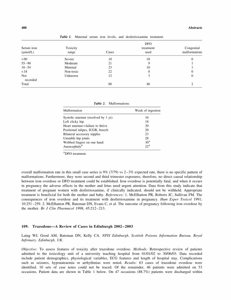
overall malformation rate in this small case series is 9% (7/79) vs 2–3% expected rate, there is no specific pattern of
malformations. Furthermore, they were second and third trimester exposures, therefore, no direct causal relationship
between iron overdose or DFO treatment could be established. Iron overdose is potentially fatal, and when it occurs
in pregnancy the adverse effects in the mother and fetus need urgent attention. Data from this study indicate that
treatment of pregnant women with desferrioxamine, if clinically indicated, should not be withheld. Appropriate
treatment is beneficial for both the mother and baby. References: 1. McElhatton PR, Roberts JC, Sullivan FM. The
consequences of iron overdose and its treatment with desferrioxamine in pregnancy. Hum Exper Toxicol 1991;
10:251–259. 2. McElhatton PR, Bateman DN, Evans C, et al. The outcome of pregnancy following iron overdose by
the mother. Br J Clin Pharmacol 1998; 45:212–213.
109. Trazodone—A Review of Cases in Edinburgh 2002 – 2003
Laing WJ, Good AM, Bateman DN, Kelly CA. NPIS Edinburgh, Scottish Poisons Information Bureau, Royal
Infirmary, Edinburgh, UK.
Objective: To assess features of toxicity after trazodone overdose. Methods: Retrospective review of patients
admitted to the toxicology unit of a university teaching hospital from 01/01/02 to 30/06/03. Data recorded
include patient demographics, physiological variables, ECG features and length of hospital stay. Complications
such as seizures, hyponatraemia or arrhythmias were noted. Results: 63 cases of trazodone overdose were
identified. 10 sets of case notes could not be traced. Of the remainder, 46 patients were admitted on 53
occasions. Patient data are shown in Table 1 below. On 47 occasions (88.7%) patients were discharged within
Table 1. Maternal serum iron levels, and desferrioxamine treatment.
Serum iron
(mmol/L)
Toxicity
range Cases
DFO
treatment
used
Congenital
malformations
>90 Severe 10 10 0
55–90 Moderate 21 9 1
18–54 Minimal 23 10 1
<18 Non-toxic 22 8 0
Not
recorded
Unknown 12 3 0
Total 88 40 2
Table 2. Malformations.
Malformation Week of ingestion
Systolic murmur (resolved by 1 yr) 16
Left clicky hip 18
Heart murmur+failure to thrive 20
Positional talipes, IUGR, breech 20
Bilateral accessory nipples 23
Unstable hip joints 28
Webbed fingers on one hand 30*
Anencephaly# 22*
*DFO treatment.
488 Abstracts
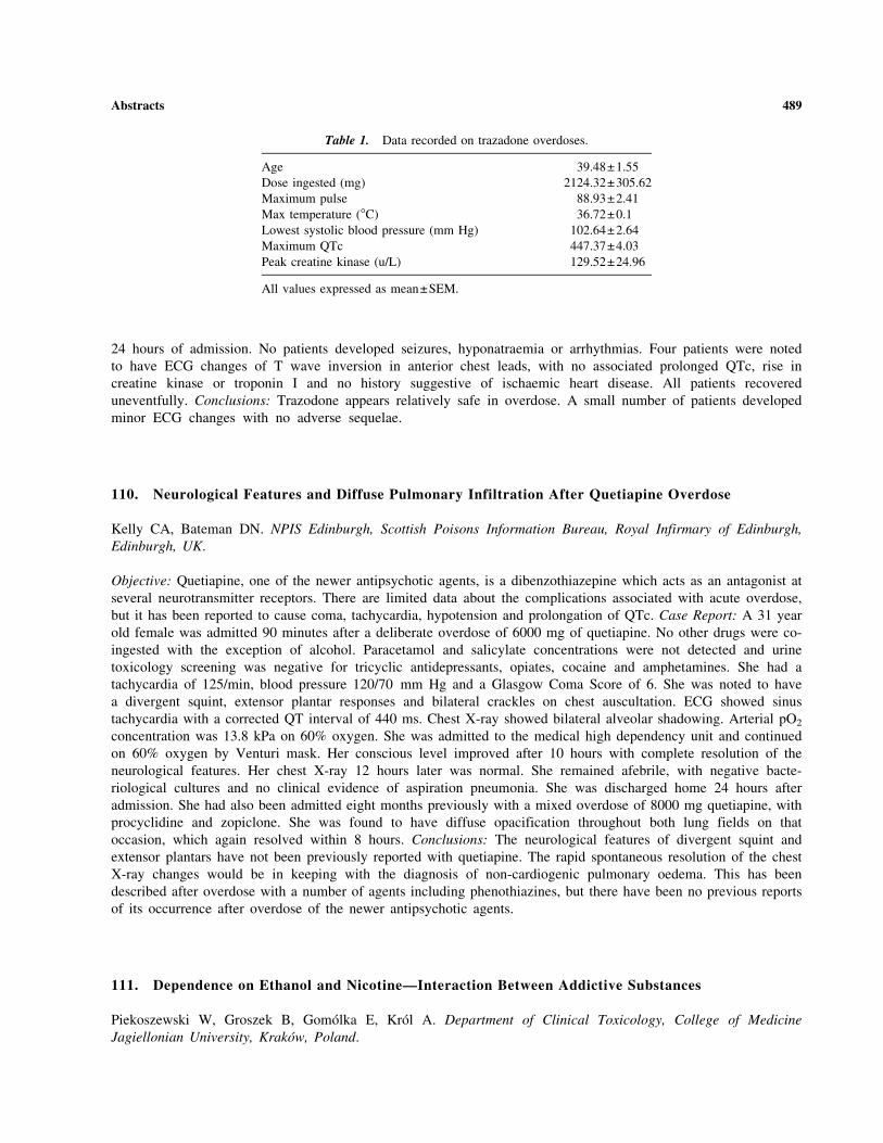
24 hours of admission. No patients developed seizures, hyponatraemia or arrhythmias. Four patients were noted
to have ECG changes of T wave inversion in anterior chest leads, with no associated prolonged QTc, rise in
creatine kinase or troponin I and no history suggestive of ischaemic heart disease. All patients recovered
uneventfully. Conclusions: Trazodone appears relatively safe in overdose. A small number of patients developed
minor ECG changes with no adverse sequelae.
110. Neurological Features and Diffuse Pulmonary Infiltration After Quetiapine Overdose
Kelly CA, Bateman DN. NPIS Edinburgh, Scottish Poisons Information Bureau, Royal Infirmary of Edinburgh,
Edinburgh, UK.
Objective: Quetiapine, one of the newer antipsychotic agents, is a dibenzothiazepine which acts as an antagonist at
several neurotransmitter receptors. There are limited data about the complications associated with acute overdose,
but it has been reported to cause coma, tachycardia, hypotension and prolongation of QTc. Case Report: A 31 year
old female was admitted 90 minutes after a deliberate overdose of 6000 mg of quetiapine. No other drugs were co-
ingested with the exception of alcohol. Paracetamol and salicylate concentrations were not detected and urine
toxicology screening was negative for tricyclic antidepressants, opiates, cocaine and amphetamines. She had a
tachycardia of 125/min, blood pressure 120/70 mm Hg and a Glasgow Coma Score of 6. She was noted to have
a divergent squint, extensor plantar responses and bilateral crackles on chest auscultation. ECG showed sinus
tachycardia with a corrected QT interval of 440 ms. Chest X-ray showed bilateral alveolar shadowing. Arterial pO2
concentration was 13.8 kPa on 60% oxygen. She was admitted to the medical high dependency unit and continued
on 60% oxygen by Venturi mask. Her conscious level improved after 10 hours with complete resolution of the
neurological features. Her chest X-ray 12 hours later was normal. She remained afebrile, with negative bacte-
riological cultures and no clinical evidence of aspiration pneumonia. She was discharged home 24 hours after
admission. She had also been admitted eight months previously with a mixed overdose of 8000 mg quetiapine, with
procyclidine and zopiclone. She was found to have diffuse opacification throughout both lung fields on that
occasion, which again resolved within 8 hours. Conclusions: The neurological features of divergent squint and
extensor plantars have not been previously reported with quetiapine. The rapid spontaneous resolution of the chest
X-ray changes would be in keeping with the diagnosis of non-cardiogenic pulmonary oedema. This has been
described after overdose with a number of agents including phenothiazines, but there have been no previous reports
of its occurrence after overdose of the newer antipsychotic agents.
111. Dependence on Ethanol and Nicotine—Interaction Between Addictive Substances
Piekoszewski W, Groszek B, Gomolka E, Krol A. Department of Clinical Toxicology, College of Medicine
Jagiellonian University, Krakow, Poland.
Table 1. Data recorded on trazadone overdoses.
Age 39.48±1.55
Dose ingested (mg) 2124.32±305.62
Maximum pulse 88.93±2.41
Max temperature (�C) 36.72±0.1
Lowest systolic blood pressure (mm Hg) 102.64±2.64
Maximum QTc 447.37±4.03
Peak creatine kinase (u/L) 129.52±24.96
All values expressed as mean±SEM.
Abstracts 489
Objective: The aim of the study was to test if an ethanol dependency has influence on cotinine concentration in urine
and if cigarette smoking has influence on ethanol metabolism. Material and Methods: The study was performed in
145 persons, 89 smokers who started smoking cessation programme with bupropion and 56 patients, alcoholics, who
were treated due to withdrawal syndrome, among them were 44 smokers and 12 non-smokers. In all patients cotinine
concentration was measured in urine by means of HPLC, in alcoholics ethanol and acetaldehyde concentration were
measured in the blood by using GC. Results: Mean age in cigarette smokers group was 42 years, in alcoholic
smokers �43.5 years, and non-smoking group �48 years. As per questionnaire data daily number of cigarettes in
smokers was 15–24, in smoking alcoholic-more than 25 cigarettes per day. In 80% of examined time of nicotine
addiction was longer than 10 years. Mean cotinine concentration in urine in smokers group was 2116.7 ng/ml
(SD±726.5), in smoking alcoholics �965.4 ng/ml (SD±524), in non-smoking alcoholics �60.54 ng/ml (SD±54.14).
Mean ethanol concentration in smoking and non-smoking alcoholics was 2.0 g/L, and there was no difference
between smokers and non-smokers (relatively 2.03 g/L c.f. 1.99 g/L). Mean acetaldehyde concentration in blood was
in smoking alcoholics �3.1 mg/L (SD±3.21), in non-smoking alcoholics �2.2 mg/L (SD±1.31). Conclusion: Mean
cotinine urine concentration in the alcoholics smoking group was more than 50% lower than in the smokers group.
This supports the hypothesis that ethanol may affect the kinetics of nicotine. Acetaldehyde concentration in the
smoking alcoholics group was higher than in non-smoking. This suggests that nicotine can decrease the metabolism
rate of ethanol.
112. Fatal Outcome Due to Accidental Ingestion of a Condiment Induced Hypernatremia. WhichOptions Do We Have Preventing Life-Threatening Course? A Case Report
Eyer F, Felgenhauer N, Zilker T. Toxicological Department, Technical University, Munich, Germany.
Objective: Incidence of severe hypernatremia due to intoxication in adults is rare. Hypernatremia causes cellular
dehydration, resulting in extracellular fluid shift. Severity of clinical presentation depends on the rate of its onset.
CNS-Symptoms like convulsions, lethargy and coma are most common. Due to cerebral contraction, subdural and
subarachnoid bleeding may occur. To prevent those eventually life-limiting complications, intensivists should be
familiar with the management of acute hypernatremia. We report on a case of fatal hypernatremia due to accidental
ingestion of a sapidity agent. Case Report: A 26-year old autistic male patient accidentally ingested about 875 mL of
a flavour intensifier containing a total amount of 234 g sodium chloride and 2.6 g sodium glutamate, respectively.
Recurrent vomiting and reduced consciousness were the main preclinical findings. On admission to hospital, about
1.5 hours after ingestion, serum-sodium revealed 170 mmol/L. Besides immediate diluting-therapy with glucose
5% therapy with furosemide was started. On admission to our ICU about 4 hours after ingestion, serum sodium
and chloride was 176 mmol/L and 134 mmol/L, respectively and serum-osmolality was 381 mosmol/kg. Another
10 hours after ingestion, hypertensive crisis occurred with systolic blood pressure of some 250 mm Hg, the patient
developed generalised convulsions and respiratory depression. Intubation and mechanical ventilation were started. At
that time, CCT showed generalised cerebral edema with imminent cerebral herniation. Criteria of brain death were
fulfilled, so due to the poor prognosis no further escalation of therapy was performed. 7 days after accidental
ingestion of a sapidity agent, the patient succumbed. Conclusion: Threatening hypernatremia despite of normal renal
function is rare, but instantaneous management of this complication can become a vital aspect. Sodium restriction,
administration of glucose 5% and diuretics are recommended. In case of renal dysfunction or life threatening
hypernatremia, extracorporeal elimination procedures like hemodialysis or hemodiafiltration could possibly be
necessary. If the patient develops rapid onset of hypernatremia, forced correction can prevent brain intracellular
dehydration and shrinking. Therefore HD and AVHDF are suitable techniques. In cases, where there is enough time
to permit the production of idiogenic osmoles, more gradual correction of serum sodium is needed to limit rapid
intracellular shifts of water and electrolytes. Ideal rate for serum sodium decrease is between 10–15 mmol per day.
The role of sodium glutamate in the development of cerebral edema in our case remains unclear. Existing data in
literature suggests that glutamate could enhance formation of cerebral edema. However, a total amount of 2.6 g
glutamate seems not enough to effectuate cerebral edema alone.
490 Abstracts
113. Infliximab Induced Critical Thrombocytopenia
Tarabar AF (1), Allori L (1), Hoffman RS (2), Nelson LS (2). 1. Yale Emergency Medicine, Yale, New Haven, CT;
2. New York City Poison Control Center, NY, USA.
Background: Infliximab is chimeric IgG1 monoclonal antibody that binds to human tumor necrosis factor alpha (TNFa)
and is used for the treatment of the rheumatoid arthritis (RA) and Crohn’s disease. Reported side effects include:
disseminated TB and fungal infections, CHF exacerbation, hypersensitivity reaction including dyspnea, hypotension
and serum sickness like syndrome. We present a case of infliximab-associated thrombocytopenia. Case Report: A
71 year old man with a history of RA and HTN, on celecoxib, lisinopril, finasteride, terazosin, selenium and folic acid
presented to the ED with ecchymoses on his face, tongue, and extremities. The previous day he received 4 vials, 100 mg
each, of infliximab. During administration of the 2nd vial, he developed flushing and cough that responded to
diphenhydramine. Infliximab infusion was completed without further symptoms. On the following day he noticed skin
lesions and was referred to the ED. In the ED, petechiae and ecchymoses were found on his tongue, face, both arms and
legs. Vital signs were normal. Laboratory testing was notable for platelet count of less than 1000/mcL. Peripheral smear
showed no signs of hemolysis or platelet clumping. MPV on day 3 with a platelet count of 7000/mcL was 12.4. He
denied any recent viral illnesses or change in his medications. IV steroids, IVIG and platelets were given. On day 5, his
platelet count was 62,000/mcL and he was discharged. Conclusion: This is the first reported case of infliximab-induced
critical thrombocytopenia requiring platelet transfusion. Clinicians should be aware of this complication and should
warn the patients to be observant of any signs of bleeding including petechiae, melena or headache. The case was
reported to Medwatch.
114. Poison Control Center and Calls for Information on Drugs and Chemicals Exposures inPregnancy and Lactation
Vernile L (1), Bacis G (1), Bonati M (2), Farina ML (1). 1. Bergamo Poison Control Center, Ospedali Riuniti,
Bergamo; 2. ‘Mario Negri’ Pharmacological Research Institute, Milan, Italy.
Introduction: After the thalidomide disaster, several Teratology Information Services were opened around the world
offering the opportunity for women and health professionals to discuss with specialists the potential effects on
fetuses and newborns induced by drugs, chemicals and radiation. Also Poison Centers sometimes can be called by
pregnant women, obstetricians or physicians asking for information about the risk after or before exposures during
pregnancy and lactation. Results: In 2002 Bergamo Poison Control Center received 751 calls (31.4% of total calls)
about information requests for potential effects of drugs and chemicals in pregnancy (80%) and lactation (20%). The
66% of the callers were women who were directly involved, 23% were physicians (8.7% general practitioners and
91.3% specialists: gynaecologists, psychiatrists, pediatricians, etc.), 8% were relatives and 3% obstetricians. At the
time of the consultation, the 69% of the calls was for an actual pregnancy. The majority of women (69.5%) were at
the first trimester of pregnancy, which is the period at the highest risk when the fetus has the major susceptibility to
teratogenic factors. The asked questions were for 1,140 causes; 89.2% regarding drugs, 3.2% parapharmaceutic
products, 2.2% chemical substances used in laboratory or industrial settings, 1.4% radiation and 4% others. About
the use of drugs during pregnancy or lactation 31% of the questions were related to drugs of the CNS, 17%
antimicrobial for systemic use, 11.5% drugs for the gastrointestinal tract and metabolism, 10.6% drugs for the
respiratory apparatus and others for 29.9%. Sixty nine women were called again after one year for the follow-up. In
84% of cases the baby was delivered without malformations; the voluntary interruption of pregnancy was decided in
the 9% and spontaneous abortion was observed in 7%. Conclusions: The need for systematic permanent surveillance
of effects of drugs and chemicals in pregnancy and lactation is identified. Cooperation between Teratology
Information Services and Poison Centers, and a wider knowledge of such services should increase the number of
enquiries, and result in more data for surveillance and research. For that reason reproductive toxicology and
teratology should be implemented in the services offered by poison control centers.
Abstracts 491
115. Temporal Relationship of Oxycodone Media Attention and Reports of Oxycodone Exposures toPoison Centers
Spiller HA, Krenzelok EP, Villalobos D, Muller AA, Lovecchio F. Kentucky Regional Poison Center, Louisville KY
and the Oxycodone Study Group (Pittsburgh Poison Center, Texas Poison Center System, Poison Control Center
Philadelphia, Banner Poison Center, Phoenix AZ.), USA.
Background: There was significant media attention (TV, radio, print) concerning the reported increase in abuse of
oxycodone. Most reports focused on anecdotal information. Method: A survey was performed of US media markets
for stories concerning OxyContin1 or oxycodone. A review of five US poison center databases for all human
exposures involving oxycodone was conducted. Results: Prior to January 2001 media stories on oxycodone abuse
were infrequent with less then 20 per month in the US. In 2001, following a New York Times story on March 01,
2001, there was a sudden sustained increase in media stories averaging between 200 to 400 stories per month. This
sustained media attention lasted approximately 18 months. By the end of 2002 media stories per month had
declined to approximately 50 per month. In the five-year study period 1998–2002 there were 1,714,781 exposures
reported to the five poison centers of which 4268 (0.249%) were exposures to oxycodone. During this five year
period there was an increase in all poisoning exposures and oxycodone exposures of 15% and 150%, respectively.
The range of oxycodone exposure increases over the five year period were 74% to 203% in the 5 five centers.
Comparing media activity and poison center data, those centers with larger oxycodone exposures prior to the media
attention (>0.2% of total exposures Philadelphia, Pittsburgh and KY) saw the largest annual increase occur in 2000.
However those centers (n=2) with lower numbers in 1998 and 1999 (<0.2% of total exposures, Texas, Arizona)
showed the greatest annual increase in 2001, after the increase in media activity. When evaluating the reason for the
oxycodone exposure, the category ‘‘intentional-abuse’’ was the only category that showed an increase over the five-
year period. All other categories declined or remained the same as a percentage of total cases in all five centers.
The greatest annual increase in intentional-abuse oxycodone exposures occurred in 2000 in one center, in 2001 in
one center and in 2002 in three centers. Conclusion: There was a temporal relationship between an increase in
media activity and increases in the percentage of oxycodone exposures reported as intentional-abuse cases. There
was a temporal relationship between increases in media activity and increases in oxycodone exposures in states with
a low number of oxycodone exposures prior to 2001. Adverse media attention may paradoxically increase cases of
drug abuse.
116. Epidemiological Profile of Poisonings in Morocco (1992 – 2002)
Ilham SI, Sanae BS, Rhizlane ER, Mohammed BM, Rachida SR. Moroccan Poison Control Center and
Pharmacovigilance (CAPM), Rabat, Morocco.
Introduction: In Morocco, industrials, chemicals, domestic products and drugs are taking more importance in the daily
uses, which cause poisonings. The Toxicovigilance (TV) unit in CAPM is similar to an observatory to supervise the
intoxications in Morocco and to take action of prevention. Objective: We report a retrospective study over the last
11 years, carried out by the TV unit, the aim of which is to determine the epidemiological profile of intoxications in
Morocco and to compare our results to international data. Methods: We analyzed the poisoning’s file reported monthly to
TV unit by health professionals, from January 1992 to December 2002. Results: During this period, 80879 cases of
poisoning have been collected. 57.9% cases of animal poisoning (56.2% represent scorpion stings who were analysed
separately). 40% of cases were of other types of poisoning: the most common was food poisoning in 26.6%, followed by
drugs in 22.5%, gas products in 17.1%, pesticides in 13.3%, domestic products in 9.3%, industrial products in 7%
(paraphenylene diamine in 1%), and plant poisoning in 2.9%. The average age of patients is 25.45+15 years with sex
ratio=0.78. Poisoning occurred in a house in 88.8%, followed by public and professional places (7.9%, 3.25%)
respectively. Accidental poisoning was commonest (46.4%), followed by suicide attempt (26.1%) and mental
disturbance (1.5%). The oral was used in 80.3%, 96.8% of these being a single agent. Inhalation occurred in 19.2% and
percutaneous in only 0.5%. Clinical manifestations were gastrointestinal in 67.9%, neurological in 11.9%, respiratory
signs in 11.4%, cardiovascular signs in 8.2%, rarely neurovegetative states in 0.4%. The treatments used were gastric
492 Abstracts
lavage in 11.9%, the induced vomiting in 3.6%, and combined treatment for the remainder. The outcome patients
was favourable on 97%, complications occurred in 1%, and transferred to another medical unit in 0.7%. Considering
the hospital origin of the enquiries, moderate intoxications were most frequent (48.8%), followed by the severe
intoxications (16.8%), the minor intoxications (15%). 1.3% were fatal. In general, the results obtained are different
from data of Swiss Toxicological Information. This difference could be linked to collecting methodology and
socioeconomic characteristics. Conclusion: The centralization and the management of poisoning by CAPM, allow
determination of the local epidemiological profile and reinforce health vigilance. A program of education, and
prevention should be initiated by the Health Ministry to decrease morbidity and mortality caused by poisoning.
117. The Toxicity of Growth Promoters in Food Animals and its Implications on Human Healthin Brazil
Wong A, Millan LS, Aldunate JLCB, Sandron CA. CEATOX—Sao Paulo Regional Poison Centre, Instituto da
Crianca, School of Medicine-Univ, Sao Paulo, Brazil.
Objectives and Methods: The use of growth promoters in mammalian and poultry husbandry has been widespread as
an inexpensive manner of ensuring increased animal weight and nutrition, less disease and better conservation of
feed products, thus decreasing costs and increasing profits. These substances have included antibiotics or antifungals,
probiotics and hormones. The use of nitrofuran class antibiotics has resulted in accumulation of metabolites that may
be hazardous to human health, such as renal toxicity, and have been shown to have carcinogenic and mutagenic risks
in test animals. Avoparcine, a glycopeptide antibiotic added to animal feed, may result in the development of
vancomycin-resistant bacteria, especially of Staphylococcus aureus and Enterococcus sp. strains, which in a
nosocomial setting increases the likelihood of severe infections by these agents. A search into the files of patients
who died from vancomycin-resistant enterococci (VRE) was made among the hospitalized patients from heart
surgery and organ transplantation units at the Hospital das Clınicas Complex of the School of Medicine of the
University of Sao Paulo. We also report on the results of epidemiological surveys of nitrofurans residues analysis in
poultry exported from the Brazilian market, which is the world’s greatest exporter and the second largest producer,
as well as the available scientific data of their impact on human health. An estimate is made of the total volume of
exports and the number of positive results. Results: We report on 30 cases of deaths of hospitalized patients, who
had VRE. The number of VRE-related deaths indicated that a significant number of the bacteria had acquired the
Van-A gene from a non-hospital setting. The possibilities and means of this gene transmission are discussed. We
likewise report on the impact of nitrofuran class antibiotics in food, where their presence has been detected but the
levels were mostly below the safety limit of the European Union. As these antibiotics are available for human use,
the safety of long-term use (e.g., recurrent urinary and gynecological infections) is discussed. Conclusions: Additives
to food have been regulated and most are considered safe. However, globalization and regional practices may lead to
critical situations and the presence of chemicals that may pose a risk to human health. The need for further research
and surveillance on mid- and long-term safety of agricultural practices is mandatory.
118. Prosecution of a Producer Marketing Look-Alike Products Containing Denatonium Benzoate inthe Netherlands
Velzen AG van (1), Vries I de (1,2), Meulenbelt J (1,2). 1. National Poisons Information Centre, National Institute
for Public Health and the Environment, Bilthoven; 2. Department of Intensive Care & Clinical Toxicology,
University Medical Center, Utrecht, The Netherlands.
Background: Look-alike products are non-food products packaged in such a way that they resemble foodstuffs.
Look-alike products may increase the risk of accidental ingestions, especially in young children. In 1989 a Royal
Decree was issued in the Netherlands, stating that it is prohibited to market look-alike products, if they pose a
health risk by poisoning. Case: The Inspectorate for Health Protection instigated a legal case versus a producer of
Abstracts 493
a look-alike product, with the aim to protect public health by prohibition of the look-alike product. A toxicologist of
the National Poisons Information Centre of the Netherlands was called to court as an expert witness in this case. The
case concerned a bath foam product, packaged in a container with great similarity to a beer bottle. After accidental
ingestion of bath foam, there is a risk of aspiration and the subsequent development of pneumonitis. The product
contained denatonium benzoate, a bittering agent used to prevent ingestion of toxic products. Thus, the case revolved
around the question whether the addition of denatonium benzoate is sufficiently effective in preventing ingestion of
bath foam to allow marketing of the look-alike product. Arguments: The producer stated that the bath foam product
would not pose a risk to public health by poisoning, because denatonium benzoate effectively prevents ingestion of
the product. The expert witness stated that the product could pose a risk to public health in spite of the addition of
denatonium benzoate, based on the following data: several studies show that denatonium benzoate may reduce the
mean amount of liquid ingested by children. However, not all children react identical to the bitter taste of the
compound: one study showed that from 30 young children who were offered orange juice containing denatonium
benzoate, 7 took more than one sip (1). Even a high concentration of denatonium benzoate may not always prevent
toxic ingestions, given the case of a 4-year old girl who ingested an unknown amount of antifreeze containing
denatonium benzoate, resulting in a serious intoxication (2). The ineffectiveness of denatonium benzoate in some
children may be the result of the fact that children seem to perceive taste in a different way than adults. A study,
using drinks with atypical colour-flavour pairing, suggested that drink identification in children becomes more
influenced by taste as children get older, while in young children (2–7 years of age), drink identification seems to be
more reliant on colour (3). Even if denatonium benzoate is able to prevent most children from taking a second sip, a
significant volume of liquid may be ingested with just the first taste. Thus, denatonium benzoate is not sufficiently
effective in preventing ingestion of toxic products by children and must not be a substitute for child-safe packages.
Conclusion: Considering the arguments of both the expert witness and the producer of the look-alike product, the
judge decided that addition of denatonium benzoate to the look-alike product is not sufficient to allow marketing of
the product. The legal case was concluded with a penalty for the producer and marketing of the bath foam product
was prohibited. References: 1. Sibert JR, et al. Bittering agents in the prevention of accidental poisoning: children’s
reactions to denatonium benzoate (Bitrex). Arch Emerg Med 1991; 8:1–7. 2. Harry P, et al. Ethylene glycol poisoning
in a child treated with 4-methylpyrazole. Pediatrics 1998; 102:E31. 3. Oram N, et al. The influence of flavor and color
on drink identification by children and adults. Dev Psychobiol 1995; 28:239–246.
119. Poisons Service Use by Paediatricians—Not for All Poisons?
Good AM (1), Beattie TF (2), Bateman DN (1). 1. NPIS (Edinburgh), Scottish Poisons Information Bureau, Royal
Infirmary; 2. Paediatric Emergency Department, Royal Hospital for Sick Children, Edinburgh, UK.
Aim: To investigate use of National Poisons Information Service (NPIS) information sources by a paediatric A&E
department. Background: Medical professionals in the UK have access to poisons information in two ways:
TOXBASE, the UK Internet poisons database and the NPIS telephone enquiry service. The Royal Hospital for Sick
Children (RHSC) in Edinburgh has used TOXBASE since 1984 to help manage poisoned patients. Methods: All
presentations involving ingestion/poisoning to RHSC for the period 1 November 2001 to 31 October 2002 were
reviewed. This was compared with data on telephone enquiries to NPIS, Edinburgh Centre and accesses to
TOXBASE. Results: During the period 304 children presented to accident and emergency (A&E) at RHSC with a
diagnosis of ingestion or poisoning. The average age was 2.7 years (range <1 month to 16 years; interquartile
range1.8–3.1). The most common reason for presentation was ingestion of a foreign body (85, including 9 batteries),
followed by paracetamol containing products (49), petroleum distillates (18), ibuprofen (14) and alcohol (13). Two
hundred and three children (66.8%) were discharged without follow-up, 6 to follow-up at A&E, 3 to outpatient
follow-up at RHSC, 5 elsewhere, 14 to GPs and 3 left without being seen. 69 (22.7%) were admitted to RHSC and
one was admitted to another hospital. The most common causes of admission were petroleum distillate ingestion
(12), alcohol (8), unknown ingestion (7), cardiac drugs (6), essential oils (5), antidepressants (5). In 107 cases
TOXBASE entries were accessed for the ingestion and in 13 cases telephone enquiries were made to SPIB (7 both
telephone and TOXBASE). For the most common presentations TOXBASE was consulted for foreign bodies (0/76);
494 Abstracts
batteries (3/9); paracetamol (5/49), petroleum distillates (12/18); ibuprofen (5/14) and alcohol (0/13). The most
common reasons for consulting TOXBASE were cardiac poisons (7/9), bleach (7/9), essential oils (6/8), anti-
depressants (5/6). 34% of those patients for which TOXBASE was accessed were admitted compared with 17% of
those where it was not used. A further 829 TOXBASE accesses (excluding further accesses to the same product
within 30 minutes but including searches of similar named or type of products) to products for which there was not
an attendance. Discussion with the department revealed that these probably represent telephone calls to A&E which
did not result in an attendance, teaching of medical staff, or access for general information. Conclusions: Staff
seldom use TOXBASE for common ingestions such as foreign bodies, paracetamol, ibuprofen and alcohol.
TOXBASE is used for less common and more toxic ingestions. TOXBASE is used for information and for other
reasons. Staff seldom need to telephone the NPIS when they use TOXBASE.
120. Antidote Availability for the Treatment of Acute Poisoning in Caribbean Hospitals
Resiere D, Fray D, Megarbane B. Cornwall Regional Hospital, Montego Bay, Jamaica and Reanimation Medicale
et Toxicologique, Hopital Lariboisiere, Paris, France.
Objective: Over the last decade, a number of new antidotes have been developed, leading to improvement in the
prognosis and treatment of certain poisonings. Mechanical ventilation, circulatory support, and development of new
regional poison centres have played an important role in the management of acute poisoning, in most developed
countries. However, poisoned patients are still dying in developing countries, because of the lack of availability of
specific antidotes to neutralize or to prevent the toxic effects of acute poisonings. The objectives of this review are
to study the availability of antidotes in the Caribbean countries and to describe the most frequently used antidotes in
the hospitals. Methods: Information was collected from the main regional hospital (Intensive Care Unit and
Emergency department) and from the Ministry of Health of each Caribbean country, using a mailed questionnaire
and a telephonic interview. Results: Twenty responses were obtained among the 25 contacted hospitals in the
Caribbean. Adrenaline, atropine, diazepam, sodium bicarbonate, 50%-glucose, oxygen, flumazenil, and activated
charcoal were available in all these hospitals. Dobutamine, atropine, isoprenaline, naloxone, calcium chloride,
sodium lactate, magnesium sulfate, propranolol, Ipecac syrup, vitamin C, vitamin K1, N-acetylcysteine were
available in most of them. However, glucagon, hydroxocobalamin, 4-methylpyrazole, anti-venom antiserum, anti-
digoxin specific Fab fragments, dimercaprol, EDTA, Fuller’s earth, protamine, and pyridoxine were missing in a
majority of centres, suggesting a deficiency in the specific management of the corresponding poisonings.
Conclusions: Government and national poison centres in Caribbean countries should promote information about the
proper place of antidotes in the management of poisoning, encouraging a larger availability of antidotes. Such
policies may also be appropriate in other developing countries.
121. Escitalopram Overdose: A Case Series
Seifert SA, Meissner GK. Nebraska Regional Poison Center, Omaha, Nebraska, USA.
Objective: To describe a case series of escitalopram (Lexapro1) overdoses. Methods: Regional Poison Center
retrospective case series. Results: Between 9/02 and 3/03, 18 escitalopram exposures were reported. Five involved
escitalopram alone and 13 as a co-ingestant. In the isolated exposures, the approximate mean escitalopram dose was
58 mg (1 mg/kg) with a range of 10 to 170 mg (0.4–2.9 mg/kg), with a mean age of 26.2 years (range 5–44 years).
Three of the five cases were therapeutic errors, one an unintentional exposure and one an intentional overdose. All
were managed at home, with the intentional ingestion refusing a health care facility evaluation. Treatments included
observation in all and GI dilution in one. One patient became symptomatic (30 mg therapeutic error), with complaints of
burning sensation of the skin on the upper half of the body, headache, shakiness, and anxiety. Four cases remained
asymptomatic. Average TESS Outcome Score in the five isolated exposure cases was 1.2 (range 1–2, with 1=no
Abstracts 495
effects; 2=minor effects; 3=moderate effects; 4=major effects; 5=death). The approximate dose of escitalopram
was known in 9 of the 13 cases in which it was a co-ingestant. The mean dose of escitalopram in those cases was 126 mg
(1.8 mg/kg) with a range of 5–600 mg (0.1–8.6 mg/kg). CNS depression occurred in seven of 13 (54%) cases,
cardiovascular effects in seven (54%), and ECG changes in three (23%). In all cases, other agents also capable
of causing those effects were involved in the exposure. Discussion: Escitalopram is a selective serotonin reuptake
inhibitor (SSRI) and is the S(+)-enantiomer of citalopram. It was approved by the US FDA in January, 2002. Overdose
experience with escitalopram is very limited. A case series of 14 isolated escitalopram ingestions greater than 100 mg
(range 100–600 mg) demonstrated no effect in 50% and a minor effect in 50% (1). In this case series, isolated exposures
to escitalopram at or below 2.9 mg/kg produced no to minimal effects. Conclusion: At doses at or below 2.9 mg/kg,
isolated exposures to escitalopram produced no to minimal effects. In higher doses or in poly-substance ingestions,
escitalopram may contribute to CNS depression, cardiovascular or ECG effects. Reference: 1. Lindgren KN, Bangh
SA, Ling L, Stremski E, Mueller C. Escitalopram, a review of adverse effects in overdose reported to select regional
poison centers. J Toxicol Clin Toxicol 2003; 41:658.
122. Intoxications with Insulin: An Analysis of 160 Cases
von Mach MA, Kaes J, Weilemann LS. Department of Clinical Toxicology and Poison Center, University of
Mainz, Germany.
Objective: Intoxications with insulin are a common problem and in case of hypoglycemic coma fatal courses as well as
remaining cerebral defects have been observed. However, published data on this topic is rare consisting mostly of case
reports or very small case series. The present investigation was to analyze the inquiries to a poison center concerning
intoxications with insulin. Methods: A total number of 175890 inquiries from 1995 to 2003 (until September) was
evaluated. 160 inquiries were received by telephone concerning insulin intoxications and a standardized questionnaire
was sent to the calling physicians for follow-up information. The cases were analyzed concerning gender, age, etiology,
type of insulin, concomitant substances, time of exposition, degree of observed symptoms, and clinical outcome.
Results: From the 160 inquiries investigated (53.1% female, 43.1% male, 3.8% sex unknown, average age 44.7 years)
the etiology was in 89.4% suicidal or parasuicidal, in 5.0% accidental, and in 1.9% a criminal poisoning (3.7% various).
Rapid acting insulins were used predominantly (57.8%) as compared to long acting formulations (42.8%). Benzo-
diazepines were most frequently (37.5%) ingested concomitantly (ethanol 15.0%, antihypertensive drugs 12.5%,
antidepressants 10.0%). Most patients presented after a time of exposition of 2 to 3 hours (15.0%). Almost 50% of the
patients presented within the first 6 hours. No symptoms were observed in 16.8% of the patients, minor symptoms in
36.8%, major symptoms in 25.2%, and serious symptoms in 21.3% according to the Poisoning Severity Score.
Information concerning the clinical outcome was obtained in 74 cases showing a full recovery for most patients (94.6%;
2.7% recovered with remaining cerebral defects, 2.7% died). Conclusion: The present investigation provides useful
information for the clinical handling of patients with insulin intoxication like the considerable number of patients using
long acting insulin formulations or concomitant substances. Physicians should think of insulin intoxication in patients
with hypoglycemia and consider even a criminal etiology.
123. Mushroom Poisoning in the Slovak Republic
Plackova S, Kresanek J, Caganova B, Klobusicka Z, Batora I. Toxicological Information Centre, Bratislava,
Slovak Republic.
Objective: In Slovakia, mushrooms enjoy a very high popularity both among hobby mycologists and gourmets. Some
people consider wild mushrooms as particularly healthy and digestible. Accordingly, mushroom poisoning is an
invariably frequent event in our country. Every year the Toxicological Information Centre (TIC) in Bratislava responds
to about 2000 inquires from all over Slovakia. Mushroom poisoning represents 6.2% of all cases collected by TIC.
Mushroom intoxications can be the serious often resulting in death. To obtain more information we performed a
496 Abstracts
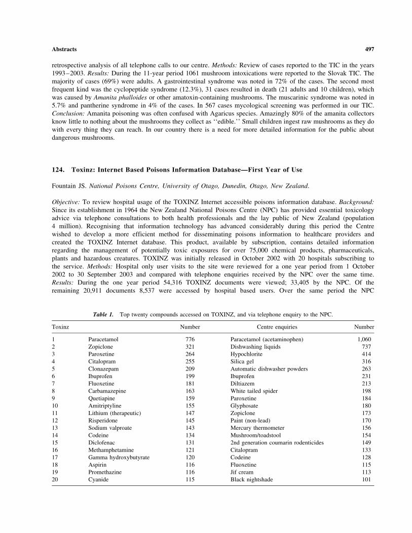
retrospective analysis of all telephone calls to our centre. Methods: Review of cases reported to the TIC in the years
1993–2003. Results: During the 11-year period 1061 mushroom intoxications were reported to the Slovak TIC. The
majority of cases (69%) were adults. A gastrointestinal syndrome was noted in 72% of the cases. The second most
frequent kind was the cyclopeptide syndrome (12.3%), 31 cases resulted in death (21 adults and 10 children), which
was caused by Amanita phalloides or other amatoxin-containing mushrooms. The muscarinic syndrome was noted in
5.7% and pantherine syndrome in 4% of the cases. In 567 cases mycological screening was performed in our TIC.
Conclusion: Amanita poisoning was often confused with Agaricus species. Amazingly 80% of the amanita collectors
know little to nothing about the mushrooms they collect as ‘‘edible.’’ Small children ingest raw mushrooms as they do
with every thing they can reach. In our country there is a need for more detailed information for the public about
dangerous mushrooms.
124. Toxinz: Internet Based Poisons Information Database—First Year of Use
Fountain JS. National Poisons Centre, University of Otago, Dunedin, Otago, New Zealand.
Objective: To review hospital usage of the TOXINZ Internet accessible poisons information database. Background:
Since its establishment in 1964 the New Zealand National Poisons Centre (NPC) has provided essential toxicology
advice via telephone consultations to both health professionals and the lay public of New Zealand (population
4 million). Recognising that information technology has advanced considerably during this period the Centre
wished to develop a more efficient method for disseminating poisons information to healthcare providers and
created the TOXINZ Internet database. This product, available by subscription, contains detailed information
regarding the management of potentially toxic exposures for over 75,000 chemical products, pharmaceuticals,
plants and hazardous creatures. TOXINZ was initially released in October 2002 with 20 hospitals subscribing to
the service. Methods: Hospital only user visits to the site were reviewed for a one year period from 1 October
2002 to 30 September 2003 and compared with telephone enquiries received by the NPC over the same time.
Results: During the one year period 54,316 TOXINZ documents were viewed; 33,405 by the NPC. Of the
remaining 20,911 documents 8,537 were accessed by hospital based users. Over the same period the NPC
Table 1. Top twenty compounds accessed on TOXINZ, and via telephone enquiry to the NPC.
Toxinz Number Centre enquiries Number
1 Paracetamol 776 Paracetamol (acetaminophen) 1,060
2 Zopiclone 321 Dishwashing liquids 737
3 Paroxetine 264 Hypochlorite 414
4 Citalopram 255 Silica gel 316
5 Clonazepam 209 Automatic dishwasher powders 263
6 Ibuprofen 199 Ibuprofen 231
7 Fluoxetine 181 Diltiazem 213
8 Carbamazepine 163 White tailed spider 198
9 Quetiapine 159 Paroxetine 184
10 Amitriptyline 155 Glyphosate 180
11 Lithium (therapeutic) 147 Zopiclone 173
12 Risperidone 145 Paint (non-lead) 170
13 Sodium valproate 143 Mercury thermometer 156
14 Codeine 134 Mushroom/toadstool 154
15 Diclofenac 131 2nd generation coumarin rodenticides 149
16 Methamphetamine 121 Citalopram 133
17 Gamma hydroxybutyrate 120 Codeine 128
18 Aspirin 116 Fluoxetine 115
19 Promethazine 116 Jif cream 113
20 Cyanide 115 Black nightshade 101
Abstracts 497
received 23,800 telephone enquiries. Conclusion: TOXINZ is successfully providing poisons information to the
population of New Zealand via the Internet with hospital subscribers, on average, accessing the service daily.
Information derived from TOXINZ usage indicate the subject of these enquiries differ from those received via
telephone consultations.
125. Potential Hepatotoxic and Nephrotoxic Substances Reported to the Czech ToxicologicalInformation Centre in the Past 3 Years
Krenova M, Pelclova D. Toxicological Information Centre, Department of Occupational Medicine, Charles
University and General Teaching Hospital, Prague, Czech Republic.
Objective: Exposures to hepatotoxic and nephrotoxic substances represent important part of the calls to the Czech
Toxicological Information Centre (TIC). Our objective was to evaluate this problem in a 3-year retrospective study.
Methods: From the calls to the Czech TIC data concerning hepatotoxic and nephrotoxic exposures were extracted.
Information about sex and age, reason of exposure and dose was analysed. Discharge reports from the hospitals were
asked concerning cases when the dose was estimated as toxic or lethal. Biochemical markers of liver and kidney
damage were compared with normal values of aspartate aminotransferase, alanine aminotransferase, urea and
creatinine during and at the end of the hospitalisation. Results: During the years 2000–2002 the TIC received
2995 calls (11.7%) concerning followed substances. Exposures in women (39.8%) were more frequent than those in
men (30.5%) and in children (29.7%). Suicidal intoxications (56.7%) were more frequent than accidental in-
toxications (29.5%). Pharmaceuticals were more often involved (81.2%) than chemicals (16.5%) and mushrooms
(2.3%). From 586 asked discharge reports 342 (58.4%) were received. From 39 reports concerning ibuprofen the
toxic dose was ingested in 24 patients. Biochemical markers of hepatotoxicity were elevated in 4 patients, markers of
nephrotoxicity in 5 patients. The dose of paracetamol according to 97 reports was toxic in 50 cases, lethal in
6 patients. Markers of hepatotoxicity were elevated in 13 patients, markers of nephrotoxicity were present in
3 patients. Antidotal therapy with N-acetylcysteine was given in 61 patients. 57 reports were obtained concerning
ethylene glycol. The dose was toxic in 27 hospitalised patients, lethal in 6 patients. 40 patients were treated with
ethanol and 20 with haemodialysis. Signs of nephrotoxicity were seen in 13 patients. Three patients died due to renal
failure. From 13 hospitalisations after boric acid ingestion only 4 involved a toxic dose. No signs of damage were
documented. Of 25 patients with possible Amanita phalloides ingestion mycological analysis excluded the
intoxication in 9 cases. 15 patients were treated with G-penicillin. One patient died due to hepatic failure. Increased
biochemical parameters were found in a further 5 intoxications (2x carbon tetrachloride, 1x salicylate, 1x chlor-
methane, 1x chloroform). No more deaths were recorded. Conclusion: Damage of liver or kidney after intoxication
with potential hepatotoxic and nephrotoxic substances is not common. A follow-up of 24 patients with abnor-
mal findings at the end of hospitalization will continue to evaluate the reversibility of these toxic injuries.
Acknowledgement: Supported by MSM J13/98 111100002 and 111100005.
126. Chinese Herbal Medicine and Fulminant Hepatic Failure: Case Report and Renewed Warning
Audi J, Poon H, Kazzi Z, Morgan B. Grady Health Systems/Georgia Poison Center. Emory University/Department
of Emergency Medicine, Division of Toxicology. Department of Occupational & Environmental Medicine. Atlanta,
Georgia, USA.
Introduction: Several case reports in the literature detail adverse health effects from traditional Chinese medicine
(TCM). These effects are typically mild, and usually without long term sequelea. We present the interesting case of a
young, otherwise healthy Chinese immigrant who developed fatal fulminant hepatic failure after using an herbal tea
prescribed by a TCM practitioner. Case Report: A previously healthy 28 year old Chinese male presented to a local
emergency department with severe confusion and lethargy. His mental status had been rapidly deteriorating over the
498 Abstracts
few days prior to admission. He was found to have significantly elevated liver transaminases and serum ammonia
level. He was transferred to our University Hospital for further care. There was no history of acetaminophen
ingestion and an initial level was undetectable. Serologies for viral hepatitis were all negative. At time of our
consultation, the patient was jaundiced, obtunded, intubated, but hemodynamically stable. On physical exam he had
an eczematous rash, with greatest distribution over his extremities. He had coarse breath sounds and a palpable liver
edge. Liver enzymes remained significantly elevated. The patient developed multi-system organ failure within a few
days of presentation, despite aggressive supportive care. Cerebral edema and brain death rapidly followed. Further
history was obtained and reportedly the patient had a pruritic, eczematous rash for three weeks antecedent to this
admission. He obtained an herbal prescription from local practitioner of TCM, and he was instructed to boil the
herbs into a tea and consume twice a day. His girlfriend reported that the remedy was not alleviating the rash and
that the patient took it for 21/2 weeks, several times per day. Subsequent translation of the patient’s prescription by a
practitioner of TCM, revealed that it contained at least two herbs that have been shown to be hepatotoxic; skullcap
and bulpleurum. Literature review supports the risk of hepatic injury associated with using these herbs. The patient
was not known to have any other co-morbid conditions, antecedent illnesses or other ingestions that would put him
at risk for hepatic failure. Conclusion: This previously healthy young man presented with fulminant hepatic failure,
temporally related to frequent consumption of a traditional Chinese medicinal herbal remedy for eczema. Several
case reports describe liver injury associated with use of herbs for skin conditions. This case report serves to remind
us of the potential dangers of traditional remedies as well as impetus to consider less common causes of fulminant
hepatic failure when the etiology is unclear.
127. Smaller Pack Size has Positive Impact on Deliberate Paracetamol Overdose in Ireland
Walsh N, Donohoe E, Tracey JA. National Poisons Information Centre, Dublin, Ireland.
Objective: Legislation on paracetamol pack-size was introduced in Ireland in October 2001. This study assessed the
impact of that legislation on self-poisoning. Methods: We retrospectively examined Poisons Centre data on acute
deliberate paracetamol overdoses reported during two 24-month periods before and after October 2001. The two
periods were compared using non-parametric statistical analysis. Differences in the number of tablets taken in each
overdose were examined using a chi-square test. This data was then further tested using directional Mann-Whitney
tests. Cases involving co-ingestion of other drugs were examined using a non-directional Mann-Whitney test.
Result: A total of 2,375 acute deliberate paracetamol overdoses were reported to the Poisons Centre during the
study period –1,353 before the legislation and 1,022 after the legislation. There was a significant drop in the
number of tablets taken in each overdose (w2=17.277, P <0.01). Specifically, there was a significant drop in
the number of cases involving 12–24 tablets (UA=16, P<0.001), and in the number of cases involving >24 tablets
(UA=15.5, P<0.001). There was no significant difference in cases involving 0–12 tablets. Co-ingestion fell
significantly (UA=29.5, P<0.05). Discussion: Paracetamol induced hepatotoxicity continues to be a major concern
in self-poisoning. Non-legislative recommendations on paracetamol pack-size had no impact on overdoses in Ireland
(1) possibly due to poor compliance by retailers. It has previously been shown that paracetamol overdose is usually
a spontaneous act (2). Ingestion of one packet of paracetamol now involves a maximum of 12 tablets where
previously it involved up to 24 tablets. Smaller pack sizes should reduce the severity of deliberate overdoses. We
found a significant drop in the number of tablets taken in deliberate overdose after October 2001. Fewer cases
involved 12–24 tablets, and fewer cases involved >24 tablets. Cases involving 0–12 tablets remained consistent.
We expected to see more cases of co-ingestion with smaller paracetamol overdoses but this did not happen. We are
now examining overdoses with other OTC drugs to assess whether paracetamol is being replaced as the drug of
choice in self-harm since October 2001. Conclusion: There has been a substantial decrease in the number of tablets
taken in acute deliberate self-poisonings since the introduction of legislation to limit the availability of paracetamol
in Ireland. References: 1. Donohoe E, Tracey JA. Restrictions on the sale of paracetamol in Ireland had no impact
on the number of tablets ingested in acute deliberate overdose. J Toxicol Clin Toxicol 2000; 38:251. 2. Hawton K,
Ware C, Mistry H. Paracetamol self-poisoning: characteristics, prevention and harm reduction. Br J Psychiatr 1996;
168:43–8.
Abstracts 499

128. Centro De Informacion Toxicologica: A Retrospective Study of Five Years of Service
Torres-Alanıs O, Garza-Ocanas L. Centro de Informacion Toxicologica, Depto de Farmacologia y Toxicologia, Fac.
de Medicina, Universidad Autonoma de Nuevo Leon, Monterrey N.L. Mexico.
Background: The Toxicological Information Center was founded in 1989 and is situated in the Department of
Pharmacology and Toxicology of the Medicine School of the Universidad Autonoma de Nuevo Leon. It provides a
24-h information service 7 days a week to the general public and health professionals. The present is a report of calls
received by the center during the last 5 years of activities. Methods: A retrospective study of calls received by the
center from January 1, 1998 to December 31, 2002. The data collected included date and time, gender, age, toxic
substance involved, exposure reason, exposure site, treatment provided and outcome. Results: Our center had 8240
calls, of which 65% were real intoxications and 35% were calls regarding toxicologic information about antidotes,
adverse drug effects or asking for analysis (lead, mercury, drugs of abuse). Sixty three per cent of the calls came from
hospital physicians and practitioners, 23% from the general public and 14% from industries. Children were involved in
57% of the cases, adults in 43%. The children were mainly of pre-school age (69%). Among adults, women were
slightly more represented than men. A single substance was implicated in 86% of the cases. Among the products
involved, medicinal drugs were the most important group (37%), household products and cleaning agents were second
in frequency (25%) followed by pesticides (14%). Accidental was the predominant situation (77%) and the main route
of exposure was oral (91%). Only 5% of poison exposure resulted in toxic effects serious enough to warrant admission
to hospital. Ten to fifteen per cent of those hospitalized patients required specific treatment or antidotes. Conclusions:
In our experience to date, the center has been a valuable resource for reducing unnecessary hospitalizations. These data
will serve as a resource for improving patient care, guiding emergency medicine resident training, and developing an
education program.
129. Medication Errors: Retrospective and Prospective Data
Pulce C, Frantz P, Payen C, Merindol S, Bavuz C, Descotes J. Poison Center, Lyon, France.
Objectives: To describe where and how medication errors occur in the general population in order to support
preventive measures. Methods: All medication errors recorded at Lyon Poison Centre in 2001 were retrospectively
studied. In order to better identify the characteristics of medication errors, a prospective was also conducted during a
6-month period. In this study, a specific questionnaire was used, including items on the patient’s age, symptoms, the
involved drug(s), the prescribed dose, the formulation, the route of administration, and the cause of the error. Results:
Of 24,693 phone calls analysed retrospectively, 2187 (8%) concerned medication errors. In 90% of the cases the error
occurred at home, 7.3% in health care institutions. Children were involved in 48% of cases. In 66% of cases, no
treatment was recommended. Actually, only 10% of patients were treated and treatment included activated charcoal
(6.5%), symptomatic measures (2 %), local decontamination (0.7%) and antidote administration (0.3%). Less than
10% of patients were hospitalized. Two deaths were noted in old in-patients (70 and 90 years, respectively) with a
severe pathological condition prior to the error. The prospective study included 407 cases. The most common errors
occurred during prescription drug intake (60%), followed by auto medication (30%), dispensing (3.5%) and
prescription (1%) errors. Physicians were responsible of only 2.2% cases. The causes of the error were inattention
(30%), inadequate storage (11%), similarities in packaging (9.5%), unread prescription (8.3%), misunderstanding
between mother an father (8%), confusion with another product (3.3%), omission (2.5%), miscomprehension (2,5%),
change of packaging (2%), spelling error (1%). Patients were asymptomatic in 79%. The most frequent symptoms
where digestive (45%), neurologic (30%), cutaneous (6%) and ophthalmologic (5%). Only 3% of patients required
medical surveillance, and one patient was hospitalized in an intensive care unit for 24 hours. All patients fully
recovered. Conclusion: Medication errors are frequent. Most errors involved non-health professionals, but adverse
consequences were then rare or mild. Medication errors in hospitals were rare, but much more severe. However,
spontaneous reporting to Poison Centres probably results in underreporting and is not deemed a very reliable method
of detection of medication errors. Nevertheless, our results suggest that medication errors may by a significant health
issue that requires more systematic detection.
500 Abstracts
130. Paediatric Iron Ingestion—Enquiries to a Poisons Centre
Lawler JM. National Poisons Information Service (NPIS)(Newcastle), Wolfson Unit, Newcastle upon Tyne, UK.
Objective: To ascertain the frequency of enquiries concerning ingestion of iron containing products by children under
5 years of age and potential toxicity. Method: Enquiries received by NPIS (Newcastle) are recorded using standardised
procedures. All enquiries regarding children under 5 years old received between 1999 and 2002, including the keywords
‘‘iron,’’ ‘‘ferrous’’ and ‘‘multivitamin’’ were retrieved. Results: Iron products accounted for 1.3% of enquiries
involving children under 5 years old (n=306), 57.5% involving males. Mean age was 2.4 years. 131 children (42.8%)
ingested 200 mg ferrous sulphate tablets and 138 (45%) ingested multivitamins containing iron. 68.62% ingested adult
preparations. Estimated number of tablets ingested ranged from 0.25 to 56 and in 75 cases (24.5%) estimated ingestion
exceeded 20 mg/kg elemental iron. 243 children (79.4%) were asymptomatic at the time of enquiry. Measurement of
serum iron concentration was recommended in 97 (32%) cases where estimated elemental iron ingested exceeded
20 mg/kg or was unknown. Home care was advised in 138 cases where elemental iron ingested was below 20 mg/kg.
The most common symptoms were vomiting (28 cases), diarrhoea (7 cases) and dark stool (14 cases). Significant toxicity
was present in 6 cases, including one child with liver failure and serum iron concentration of 60.2 mcmol/L, one case
of shock, and one with haematemesis, black stool and iron concentration of 57 mcmol/L. Another had iron concentration
75.8 mcmol/L and vomiting. In these 4 cases, desferrioxamine had been started prior to the enquiry. 2 children were
ventilated, but no specific symptoms recorded. Serum iron concentration above 55 mcmol/L (maximum 86 mcmol/L)
was reported in 17 other cases, all involving ingestion of adult ferrous sulphate preparations. Of these, 8 were
symptomatic, one with haematemesis, 5 with minor gastrointestinal disturbance and one child was drowsy.
Desferrioxamine was not recommended in any of these cases and where subsequent enquiries were received, iron
concentration reduced with supportive care. Conclusion: Age and gender difference are consistent with reported trends1.
Reports of serious poisoning following paediatric iron ingestion are uncommon, ingestion of adult preparations more
frequently causing toxicity. Potential toxicity is estimated by maximum amount of elemental iron ingested. Introduction
of child-resistant blister packs for iron products in the UK may reduce the amount of iron ingested and allow more
accurate estimation ingested dose, preventing unnecessary hospital referral and measurement of iron concentration.
References: 1. Woolf A, Lovejoy F. Epidemiology of drug overdose in children. Drug Safety 1993; 9:291–308. 2. Anon.
Child-resistant blister packs for aspirin, paracetamol and iron. Pharmaceutical Journal 2002; 269:767.
131. Toxicology in the Medical School Curriculum: An Innovative Longitudinal Curriculum Theme
Mahoney JF (1), Akhtar J (1), Brooks DE (1), Tofovic SP (2). 1. Department of Emergency Medicine and
Toxicology Treatment Program; 2. Center for Clinical Pharmacology, University of Pittsburgh School of Medicine,
USA.
Background: 21st century society faces growing threats from chemical weapons, environmental toxins, and
emerging and familiar drugs of abuse, yet medical school curricula typically provide a minimum of toxicology
education. A major challenge to introducing toxicology into the curriculum is acquiring adequate and appropriately
timed opportunities to present the material. Elective rotations for senior students can only partially meet this
curricular need. Objective: Provide increased opportunities for student learning in toxicology by content integration
throughout the curriculum. Methods: Toxicology content was integrated into existing courses. Examples of content
and integration include: pharmacokinetics; pathophysiology of hepatotoxins–digestion course; mechanism of action of
chemical weapons agents/antidotes–neuroscience course; and principles/treatment of addiction. The medicine clerkship
and senior emergency medicine elective include workshops on principles of clinical toxicology and medical
management of chemical/biological casualties. Since its inception in 1996, the Clinical Pharmacology senior
selective course has included a full-day of toxicology lectures and small-group, case-based workshops. Specific
subjects includes: Specific subjects include: general principles, such as syndrome recognition, decontamination
controversies, and antidotes; acetaminophen and analgesic toxicity; contemporary drugs of abuse; mechanisms of
chemical weapons agents and antidotes, including vesicants and nerve agents; and radiation toxicity. Results:
Students reported a high degree of satisfaction with their toxicology experiences. In the medicine clerkship, student
Abstracts 501
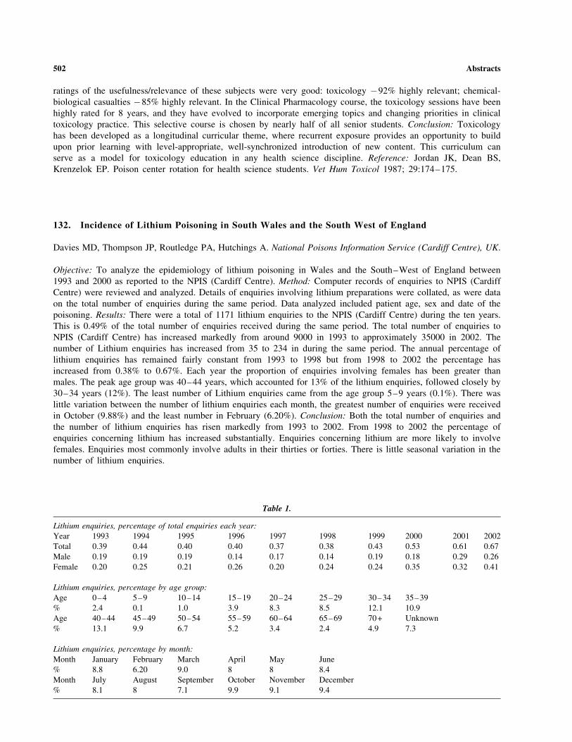
ratings of the usefulness/relevance of these subjects were very good: toxicology �92% highly relevant; chemical-
biological casualties �85% highly relevant. In the Clinical Pharmacology course, the toxicology sessions have been
highly rated for 8 years, and they have evolved to incorporate emerging topics and changing priorities in clinical
toxicology practice. This selective course is chosen by nearly half of all senior students. Conclusion: Toxicology
has been developed as a longitudinal curricular theme, where recurrent exposure provides an opportunity to build
upon prior learning with level-appropriate, well-synchronized introduction of new content. This curriculum can
serve as a model for toxicology education in any health science discipline. Reference: Jordan JK, Dean BS,
Krenzelok EP. Poison center rotation for health science students. Vet Hum Toxicol 1987; 29:174–175.
132. Incidence of Lithium Poisoning in South Wales and the South West of England
Davies MD, Thompson JP, Routledge PA, Hutchings A. National Poisons Information Service (Cardiff Centre), UK.
Objective: To analyze the epidemiology of lithium poisoning in Wales and the South–West of England between
1993 and 2000 as reported to the NPIS (Cardiff Centre). Method: Computer records of enquiries to NPIS (Cardiff
Centre) were reviewed and analyzed. Details of enquiries involving lithium preparations were collated, as were data
on the total number of enquiries during the same period. Data analyzed included patient age, sex and date of the
poisoning. Results: There were a total of 1171 lithium enquiries to the NPIS (Cardiff Centre) during the ten years.
This is 0.49% of the total number of enquiries received during the same period. The total number of enquiries to
NPIS (Cardiff Centre) has increased markedly from around 9000 in 1993 to approximately 35000 in 2002. The
number of Lithium enquiries has increased from 35 to 234 in during the same period. The annual percentage of
lithium enquiries has remained fairly constant from 1993 to 1998 but from 1998 to 2002 the percentage has
increased from 0.38% to 0.67%. Each year the proportion of enquiries involving females has been greater than
males. The peak age group was 40–44 years, which accounted for 13% of the lithium enquiries, followed closely by
30–34 years (12%). The least number of Lithium enquiries came from the age group 5–9 years (0.1%). There was
little variation between the number of lithium enquiries each month, the greatest number of enquiries were received
in October (9.88%) and the least number in February (6.20%). Conclusion: Both the total number of enquiries and
the number of lithium enquiries has risen markedly from 1993 to 2002. From 1998 to 2002 the percentage of
enquiries concerning lithium has increased substantially. Enquiries concerning lithium are more likely to involve
females. Enquiries most commonly involve adults in their thirties or forties. There is little seasonal variation in the
number of lithium enquiries.
Table 1.
Lithium enquiries, percentage of total enquiries each year:
Year 1993 1994 1995 1996 1997 1998 1999 2000 2001 2002
Total 0.39 0.44 0.40 0.40 0.37 0.38 0.43 0.53 0.61 0.67
Male 0.19 0.19 0.19 0.14 0.17 0.14 0.19 0.18 0.29 0.26
Female 0.20 0.25 0.21 0.26 0.20 0.24 0.24 0.35 0.32 0.41
Lithium enquiries, percentage by age group:
Age 0–4 5–9 10–14 15–19 20–24 25–29 30–34 35–39
% 2.4 0.1 1.0 3.9 8.3 8.5 12.1 10.9
Age 40–44 45–49 50–54 55–59 60–64 65–69 70+ Unknown
% 13.1 9.9 6.7 5.2 3.4 2.4 4.9 7.3
Lithium enquiries, percentage by month:
Month January February March April May June
% 8.8 6.20 9.0 8 8 8.4
Month July August September October November December
% 8.1 8 7.1 9.9 9.1 9.4
502 Abstracts
133. Hospital Care in Sweden Due to Childhood Poisoning—A Ten Years Perspective
Irestedt B., Backman E., Persson H. Swedish Poisons Information Centre, Stockholm, Sweden.
Objective: The aim of this study was to describe poisoning in children requiring treatment in hospital in 2000, and to
compare with the situation ten years earlier. Methods: Case records from Swedish hospitals concerning poisoning in
children <10 years during 2000 were analyzed retrospectively. Epidemiological data were documented and the cases
were graded according to the Poisoning Severity Score (PSS). Results: According to official national statistics about
650 children <10 years were hospitalized in Sweden with a diagnosis code of poisoning during 2000. The PC
received this year 305 case records concerning children treated in hospital because of poisoning, mainly in the age
1–3 years. The poisoning accidents were caused by pharmaceuticals (46%), chemical products (37%) and plants,
mushrooms or snake bites (17%). Most children had no or mild symptoms and only 6 children (2%) developed
severe poisoning. This pattern is the same as observed in a study undertaken in 1990. However, the total number of
children treated as in-patients because of poisoning has been more than halved in the period 1990–2000. This
decrease is primarily related to certain pharmaceuticals that are commonly used in the homes, e.g. paracetamol and
terbutaline. For these preparations child-resistant closures have been introduced during the actual period. A switch to
less corrosive dishwasher detergents has also significantly reduced the need for hospital treatment. Gradually a more
liberal policy of the PC, due to increased experience and access to more reliable toxicity data, has further reduced
the number of hospital admissions. Thus, observation at home can be undertaken more often, rather than of referring
the child to hospital. Conclusion: Hospitalization due to childhood poisoning has decreased markedly in Sweden and
severe intoxications are uncommon. The PC has an important role in maintaining this favourable development, by
influencing manufacturers and authorities. Furthermore, on the basis of continuously updated toxicity data, the PC
should adjust their advice to avoid unnecessary hospitalizations, while always considering and ascertaining safety.
134. Lethal Chloroquine Self Poisonings: 13 Cases of the Marseilles Poison Centre
de Haro L, Menut A, Tichadou L, Pommier P, Arditti J, Hayek-Lanthois M. Centre Antipoison, Hopital Salvator,
Marseille, France.
Objective: All the severe or lethal cases of intoxication collected by poison centres in France are declared to the
‘‘Toxicovigilance’’ system. The deaths due to medicine self poisonings are not very numerous, and during a ten years
survey, the molecule responsible in South Eastern France of the highest number of suicide was chloroquine.
Case Series: 13 cases of lethal chloroquine self poisonings were observed in the poison centre of Marseilles between
January 1993 and December 2002, involving 5 men and 8 women (between 14 and 84 years old, average 34 years).
The ingested quantities were unknown for 2 patients, and were very high for 11 of them (average ingested dose of 5.9g
of chloroquine). This medicine was absorbed alone for 11 cases, and associated with low doses of paracetamol in one
case or alcohol in the last one. The 13 cases are surprisingly very similar: All patients alerted the emergency system
quickly after the ingestion, and the delay between the poisoning and the medical management was unusually short (1 to
4 hours, average 2H30). For all patients, the medical team observed cardiac problems as soon as they arrived, and
4 patients died at home before practitioners had time to begin any treatment. The 9 patients who were transported to
the hospital were treated with high doses of diazepam, completed with gastric lavage (4 patients) or activated charcoal
(3 patients). All of them had several cardiac arrests treated with external shock and adrenaline. 7 of them died during
the first hour of hospitalisation, and the last 2 patients were managed in intensive care units where they finally died in
less than 15 hours. The average delay between ingestion and death for this case series was 4H30. Conclusion: High
ingested doses of chloroquine are responsible of so rapid cardiac complications that the antidote (diazepam) completed
with symptomatic treatments are not still able to avoid all lethal evolution of such severe poisoning.
135. Physicians’ Evaluation of a National Poisons Information Centre
Goetschi S, Kupferschmidt H. Swiss Toxicological Information Centre (STIC), and Division of Clinical
Pharmacology & Toxicology, University Hospital, Zurich, Switzerland.
Abstracts 503
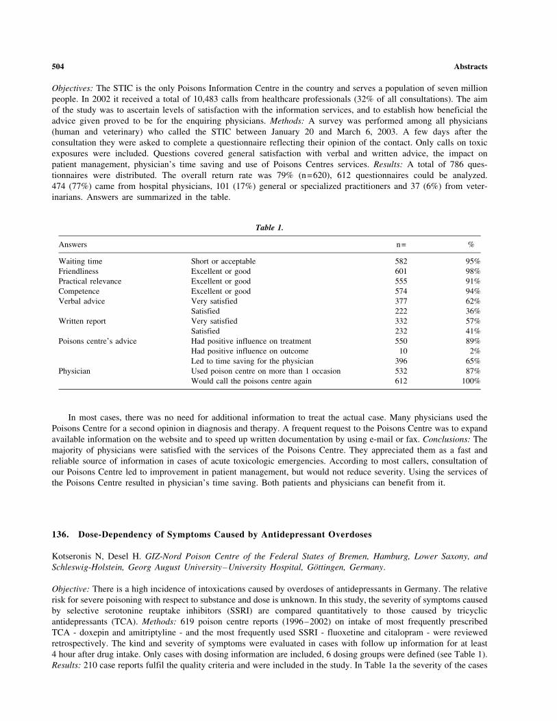
Objectives: The STIC is the only Poisons Information Centre in the country and serves a population of seven million
people. In 2002 it received a total of 10,483 calls from healthcare professionals (32% of all consultations). The aim
of the study was to ascertain levels of satisfaction with the information services, and to establish how beneficial the
advice given proved to be for the enquiring physicians. Methods: A survey was performed among all physicians
(human and veterinary) who called the STIC between January 20 and March 6, 2003. A few days after the
consultation they were asked to complete a questionnaire reflecting their opinion of the contact. Only calls on toxic
exposures were included. Questions covered general satisfaction with verbal and written advice, the impact on
patient management, physician’s time saving and use of Poisons Centres services. Results: A total of 786 ques-
tionnaires were distributed. The overall return rate was 79% (n=620), 612 questionnaires could be analyzed.
474 (77%) came from hospital physicians, 101 (17%) general or specialized practitioners and 37 (6%) from veter-
inarians. Answers are summarized in the table.
In most cases, there was no need for additional information to treat the actual case. Many physicians used the
Poisons Centre for a second opinion in diagnosis and therapy. A frequent request to the Poisons Centre was to expand
available information on the website and to speed up written documentation by using e-mail or fax. Conclusions: The
majority of physicians were satisfied with the services of the Poisons Centre. They appreciated them as a fast and
reliable source of information in cases of acute toxicologic emergencies. According to most callers, consultation of
our Poisons Centre led to improvement in patient management, but would not reduce severity. Using the services of
the Poisons Centre resulted in physician’s time saving. Both patients and physicians can benefit from it.
136. Dose-Dependency of Symptoms Caused by Antidepressant Overdoses
Kotseronis N, Desel H. GIZ-Nord Poison Centre of the Federal States of Bremen, Hamburg, Lower Saxony, and
Schleswig-Holstein, Georg August University–University Hospital, Gottingen, Germany.
Objective: There is a high incidence of intoxications caused by overdoses of antidepressants in Germany. The relative
risk for severe poisoning with respect to substance and dose is unknown. In this study, the severity of symptoms caused
by selective serotonine reuptake inhibitors (SSRI) are compared quantitatively to those caused by tricyclic
antidepressants (TCA). Methods: 619 poison centre reports (1996–2002) on intake of most frequently prescribed
TCA - doxepin and amitriptyline - and the most frequently used SSRI - fluoxetine and citalopram - were reviewed
retrospectively. The kind and severity of symptoms were evaluated in cases with follow up information for at least
4 hour after drug intake. Only cases with dosing information are included, 6 dosing groups were defined (see Table 1).
Results: 210 case reports fulfil the quality criteria and were included in the study. In Table 1a the severity of the cases
Table 1.
Answers n= %
Waiting time Short or acceptable 582 95%
Friendliness Excellent or good 601 98%
Practical relevance Excellent or good 555 91%
Competence Excellent or good 574 94%
Verbal advice Very satisfied 377 62%
Satisfied 222 36%
Written report Very satisfied 332 57%
Satisfied 232 41%
Poisons centre’s advice Had positive influence on treatment 550 89%
Had positive influence on outcome 10 2%
Led to time saving for the physician 396 65%
Physician Used poison centre on more than 1 occasion 532 87%
Would call the poisons centre again 612 100%
504 Abstracts
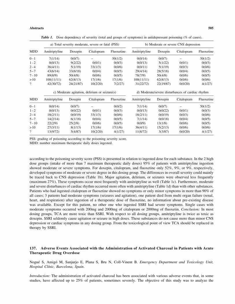
according to the poisoning severity score (PSS) is presented in relation to ingested dose for each substance. In the 2 high
dose groups (intake of more than 7 maximum therapeutic daily doses) 95% of patients with amitriptyline ingestion
showed moderate or severe symptoms. For doxepin, citalopram, and fluoxetine only 52%, 9%, or 9%, respectively,
developed symptoms of moderate or severe degree in this dosing group. The differences in overall severity could mainly
be traced back to CNS depression (Table 1b). Major agitation, delirium, or seizures were observed less frequently
(maximum 27%), These symptoms occur most frequently with amitriptyline as well (Table 1c). Furthermore, moderate
and severe disturbances of cardiac rhythm occurred more often with amitriptyline (Table 1d) than with other substances.
Patients who had ingested citalopram or fluoxetine showed no symptoms or only minor symptoms in more than 90% of
all cases; 3 patients had moderate symptoms (seizures and agitation), one patient died from multi organ failure (renal,
heart, and respiration) after ingestion of a therapeutic dose of fluoxetine, no information about pre-existing disease
was available. Except for this patient, no other one who ingested SSRI had severe symptoms. Single cases with
moderate symptoms occurred with 200mg and 2000mg of citalopram or 2000mg of fluoxetin. Conclusion: In most
dosing groups, TCA are more toxic than SSRI. With respect to all dosing groups, amitriptyline is twice as toxic as
doxepin. SSRI seldomly cause agitation or seizure in high doses. These substances do not cause more than minor CNS
depression or cardiac symptoms in any dosing group. From the toxicological point of view TCA should be replaced in
therapy by SSRI.
137. Adverse Events Associated with the Administration of Activated Charcoal in Patients with AcuteTherapeutic Drug Overdose
Nogue S, Amigo M, Sanjurjo E, Plana S, Bru N, Coll-Vinent B. Emergency Department and Toxicology Unit,
Hospital Clınic, Barcelona, Spain.
Introduction: The administration of activated charcoal has been associated with various adverse events that, in some
studies, have affected up to 25% of patients, sometimes severely. The objective of this study was to analyze the
Table 1. Dose dependency of severity (total and groups of symptoms) in antidepressant poisoning (% of cases).
a) Total severity moderate, severe or fatal (PSS) b) Moderate or severe CNS depression
MDD Amitriptyline Doxepin Citalopram Fluoxetine Amitriptyline Doxepin Citalopram Fluoxetine
0–1 7(1/14) 0(0/7) – 50(1/2) 0(0/14) 0(0/7) – 50(1/2)
1–2 0(0/13) 9(2/22) 0(0/1) 0(0/3) 0(0/13) 5(1/22) 0(0/1) 0(0/3)
2–4 36(4/11) 5(1/19) 33(1/3) 0(0/6) 0(0/11) 5(1/19) 0(0/3) 0(0/6)
5–7 43(6/14) 33(6/18) 0(0/4) 0(0/5) 29(4/14) 28(5/18) 0(0/4) 0(0/5)
7–10 89(8/9) 50(4/8) 0(0/6) 0(0/5) 78(7/9) 50(4/8) 0(0/6) 0(0/5)
>10 100(11/11) 62(8/13) 17(1/6) 17(1/6) 100(11/11) 62(8/13) 0(0/6) 0(0/6)
? 42(30/72) 24(21/87) 10(2/20) 7(2/27) 31(22/72) 22(19/87) 0(0/20) 4(1/27)
c) Moderate agitation, delirium or seizure(s) d) Moderate/severe disturbances of cardiac rhythm
MDD Amitriptyline Doxepin Citalopram Fluoxetine Amitriptyline Doxepin Citalopram Fluoxetine
0–1 0(0/14) 0(0/7) – 0(0/2) 7(1/14) 0(0/7) – 50(1/2)
1–2 0(0/13) 0(0/22) 0(0/1) 0(0/3) 0(0/13) 0(0/22) 0(0/1) 0(0/3)
2–4 18(2/11) 0(0/19) 33(1/3) 0(0/6) 18(2/11) 0(0/19) 0(0/3) 0(0/6)
5–7 14(2/14) 6(1/18) 0(0/4) 0(0/5) 7(1/14) 0(0/18) 0(0/4) 0(0/5)
7–10 22(2/9) 25(2/8) 0(0/6) 0(0/5) 0(0/9) 13(1/8) 0(0/6) 0(0/5)
>10 27(3/11) 8(1/13) 17(1/6) 17(1/6) 36(4/11) 15(2/13) 0(0/6) 0(0/6)
? 13(9/72) 5(4/87) 10(2/20) 4(1/27) 11(8/72) 3(3/87) 0(0/20) 4(1/27)
PSS: grading of poisoning according to the poisoning severity score.
MDD: number maximum therapeutic daily doses ingested.
Abstracts 505
adverse events observed after the administration of activated charcoal in patients with acute therapeutic drug
overdose (ATDO). Patients and Method: Patients admitted to the Emergency Department of our Hospital due to
ATDO were studied prospectively for 7 months. Some patients were treated with one or more doses of 25 g of
activated charcoal. Epidemiological variables relating to the overdose and the treatment given were recorded.
Results: A total of 213 patients with acute ATDO were included, of which 136 (63.8%) received some form of
digestive decontamination, which in 128/136 (94.1%) was activated charcoal (administered orally in 100 cases and
by nasogastric tube in 28 cases). Of these patients, 9/128 (7%) presented vomiting, with three patients suffering
pulmonary aspiration, one of whom developed aspiration pneumonia. When the activated charcoal was administered
orally and was the only decontamination therapy, vomiting was observed in 7% of patients. When gastric lavage was
accompanied by activated charcoal, 7.1% of cases suffered vomiting. The average ingestion in patients who vomited
after activated charcoal was 58 tablets of therapeutic drugs. Five of the 9 patients who vomited after administration
of activated charcoal (55.6%) had vomited spontaneously previous to decontamination; three had also consumed
alcohol. Three patients suffered pulmonary aspiration, one of whom developed pneumonia; two of these patients
were in coma (Glasgow 5), and tracheal intubation was carried out in one of these to protect the airways. All patients
evolved favourably. Conclusions: Adverse reactions to activated charcoal in ATDO are not frequent and are
independent of whether the charcoal is administered orally or by nasogastric tube. However, they should be
prevented if the patient has taken a large number of tablets together with alcohol or has vomited spontaneously
previous to treatment. If the patient is in coma, the beneficial effect of therapy should be weighed against the risk of
pulmonary aspiration, with tracheal intubation being carried out before decontamination if necessary.
138. Epidemiological Characteristics of Acute Drug Intoxications in Suicidal Attempts Managed in anEmergency-Appended Temporary Hospitalization Department
Hubin V, Feye F, Morelle J, Verschuren F, Hantson P, Thys P. Department of Emergency and Intensive Care,
Cliniques St-Luc, Universite catholique de Louvain, Brussels, Belgium.
Background: Acute drug intoxications (ADI) in suicidal attempts are a frequent cause of admission in emergency
departments; these situations require a rapid evaluation of the potential complications, an effective short-term follow-
up and an initial contact with a psychiatrists team. Emergency-appended temporary hospitalization department
(THD) seems to be an adequate solution for the management of ADI. Objective: The aim of our study was to
determine the main characteristics of the patients admitted following ADI, the nature of the involved substances, and
to analyze the patients’ short-term outcome in THD. Methods: This is a retrospective, single-center study. All the
patients admitted for a suicidal attempt in the emergency department between the 1st February 2002 and the 1st
February 2003 and staying at least one night in the THD were included. A blood sample confirmed the presence of
drugs in all patients. Results: One hundred and fifty-six patients were included, with an average age of 36.9 years
(±13.8 years) and a female-male sex ratio of 2.2: 1. In 93.9% of the patients, suicidal attempts involved ADI,
followed by venosection (4.2%), exposure to domestic substances (1.2%) and drowning (0.6%). The drugs used were
benzodiazepines (60.9%), ethanol (41.7%), alkaline substances (32.1%) and antidepressive medications (26.9%);
acetaminophen was the most frequent cause of iterative drug monitoring (12.2%). The simultaneous intake of several
drugs was common: two types of toxins in 35.9%; three in 23.7%. A clinical examination, a blood test and a
psychiatrist’s consultation were organized for all the patients. 76.3% of them had an intravenous line and one patient
on five needed a second blood sample analysis. The follow-up of the patients during the THD stay did not reveal
any fatality and all the patients except one were able to go back home after a mean duration stay shorter than 17
hours (16hr44min±7h33). Conclusions: Patients admitted after suicidal attempts in the THD are young and mostly
women. Multiple drug intoxication is common. ADI requires a short-term neurologic and hemodynamic follow-up
mainly based on the physical examination and on a blood sample analysis. Supportive therapy was applied without
any need for antidotes. In all the analyzed cases, short-term patient’s outcome is good. Moreover, the one-night stay
in THD allows the patient to meet the psychiatrist, and to make the first steps of a long-term therapy. For all these
reasons, THD seems to be a secure and effective way to manage patients admitted in emergency departments
for ADI.
506 Abstracts
139. Peripheral Cholinergic Crisis in Donepezil Overdose Treated with Pralidoxime
Mazzola JL, Brush DE, Bird SB, Boyer EW. University of Massachusetts Medical School, Department of Emergency
Medicine, Division of Toxicology, Massachusetts College of Pharmacy and Health Sciences, Department of
Pharmacy Practice, Worcester, Massachusetts, USA.
Donepezil is a piperidine derivative that selectively and reversibly inhibits acetylcholinesterase in the central nervous
system with minimal peripheral effects. Objective: To our knowledge, treatment of donepezil induced cholinergic
symptoms with pralidoxime has not been described. Case Report: A 27 year-old female presented to the emergency
ward three hours after ingesting 35 mg of donepezil and an unknown quantity of verapamil SR 240 mg, simvastatin
20 mg, and hydrochlorothiazide. Symptoms included urinary and fecal incontinence, agitation, and nausea. Her
initial vitals revealed a HR of 130 bpm, BP 146/78 mm Hg, RR 22, and Pox 98% on room air. Physical examination
was noted for profuse diaphoresis, pupils 2 mm bilaterally, clear lungs, resting tremor, and facial fasciculations. She
was alert and oriented with a normal mental status. Electrocardiogram demonstrated sinus tachycardia at 121 bpm
with QRS 108 msec. The patient received 50 g of activated charcoal, 2 mg of atropine, and a pralidoxime bolus of
1gm IV. Her symptoms improved dramatically within twenty minutes of this therapy. Six hours later she became
symptomatic and a pralidoxime drip at 3.2 mg/kg/hr. was infused with continued symptomatic improvement. The
pralidoxime drip was discontinued twenty-eight hours after presentation and the patient was subsequently discharged
to mental health services. Conclusion: The loss of central selectivity of acetylcholinesterase inhibition in overdoses
occurs with donepezil. Her response to pralidoxime suggests that this may be a useful drug in the treatment of
donepezil overdose. References: Mihara M, Ohnishi A, Tomono Y, et al. Pharmacokinetics of E2020, a new
compound for Alzheimer’s disease, in healthy male volunteers. Int J Clin Pharmacol Ther Toxicol 1993; 31:223–
229. Rogers SL, Friedhoff LT and the donepezil study group. The efficacy and safety of donepezil in patients with
Alzheimer’s disease: results of a US multicenter, randomized, double-blind, placebo-controlled trial. Dementia 1996;
7:293–303. Ohnishi A, Mihara M Kamakura H, et al. Comparison of the pharmacokinetics of E2020, a new
compound for Alzheimer’s disease, in healthy young and elder subjects. J Clin Pharmacol 1993; 33:1086–1091.
140. Overdose of Dipyridamole Causing Intense Yellow Discoloration of the Skin
Personne M. The Swedish Poisons Information Centre, Stockholm, Sweden.
Objective: To describe the symptomatology of an exceptionally high overdose of the antithrombotic agent
dipyridamole. Case Report: A 66-year old man called for an ambulance from his home because of acute chest pain.
When the crew arrived he was obtunded and hypotensive. Sublingual nitroglycerine was given and subsequently the
patient collapsed developing circulatory failure and respiratory arrest. He was intubated and transported to the nearest
hospital. Circulation was improved with intravenous fluids and norepinephrine. During the next few hours the patient’s
skin gradually turned intensely yellow, as did the urine. An acute antero-septal myocardial infarction was diagnosed
and renal failure developed. Relatives of the patient revealed that he suffered from schizophrenia and also that he was
on medical treatment for hypertension and atrial fibrillation. The cause of the neon-yellow colour was a complete
mystery until a police patrol sent to his apartment found several empty packages of dipyridamole (Persantin Depot)
corresponding to 34 g (170 tablets). If ingested, this would be an overdose seven times higher than any previous case
reported in the literature. This substance has a distinct yellowish colour. The diagnosis was confirmed by measuring
serum concentration of the glucuronide metabolite of dipyridamole. The level was 370 micromol/L, to be compared to
a therapeutic concentration of approximately 2 micromol/L. The patient’s condition improved slowly and he could be
extubated within a week. Haemodialysis was needed for two weeks because of acute tubular necrosis, most probably
related to the prolonged period of hypotension. Thereafter the patient recovered completely. Discussion: Transient
non-icteric yellow discoloration of the skin can occur with large overdoses of dipyridamole along with pronounced
hypotension and occasionally acute myocardial infarction. The circulatory effects of dipyridamole are mainly caused
by increased amounts of endogenous adenosine. These high levels can possibly lead to a ‘‘steal syndrome’’ in patients
with coronary disease, where the blood flow is shunted away from an already stenosed coronary artery. If diagnosed
early, therapeutic intervention with a partial adenosine antagonist such as theophyllamine can be considered.
Abstracts 507
141. Acute Opiate Withdrawal After Consumption of Naltrexone-Tainted Cocaine
Faas A, Weber M, Rentsch K, Ruggieri F, Kupferschmidt H. Department of Internal Medicine, Institute of Clinical
Chemistry, Division of Clinical Pharmacology & Toxicology, University Hospital, Zurich, and Swiss Toxicological
Information Centre, Zurich, Switzerland.
Background: Naltrexone is a pure opiate receptor antagonist used for the treatment of opioid dependence. Adulteration
is common in illicit street drugs either with inactive substances to dilute the drug, or with active drugs to modify or
increase the desired effect. We report a series of three patients who consumed naltrexone-tainted cocaine. Case Series:
Three white Caucasian males with known history of opioid dependence, currently on a methadone substitution
programme, were admitted to our emergency department on the same day. Two patients had consumed cocaine nasally,
one patient intravenously. All three patients showed immediate opiate withdrawal symptoms. Patient A, a 38 year-old
male, consumed cocaine by nasal insufflation 6 hours after having received the last methadone dose (165 mg). He
showed agitation, hallucinations, abdominal pain, sweating, and massive diarrhea. He was treated with benzodiazepines
without effect. A single dose of 80 mg methadone calmed him temporarily, whereas diarrhea persisted. His creatine
kinase was slightly elevated (1186 U/L). Symptoms subsided 16 hours after cocaine intake. Patient B, a 33 year-old
male, snuffed cocaine a few hours after having received the last methadone dose (100 mg) and flunitrazepam. He
suffered from agitation, hallucinations, vertigo, nausea, abdominal pain, diaphoresis, massive diarrhea, and shortness of
breath. He responded well to intravenous midazolam. He was admitted to the ward for 5 days because of
rhabdomyolysis (creatin kinase 7348 U/L). Patient C, a 33 year-old male, felt massive immediate opiate withdrawal
after consuming cocaine intravenously a few hours after having received his last methadone dose (130 mg). He was
severely agitated and showed nausea, emesis, diaphoresis, and massive diarrhea. His symptoms were controlled by
intravenous midazolam and three oral doses of methadone (total 110 mg). He was discharged from the emergency
department without symptoms 17 hours after cocaine intake. In the urine of patients A and B naltrexone was detectable
besides benzoylecgonin, methadone, codeine, lidocaine, and the drugs administered in the ED. No urine was available
from patient C. Patient A provided us with a drug specimen (white powder) which turned out to be a 1:1 cocaine/
naltrexone mixture. Conclusions: The adulteration of cocaine with an opioid antagonist such as naltrexone is
uncommon. Our case series demonstrates that naltrexone is well absorbed together with cocaine either by intravenous
or nasal routes; the magnitude of withdrawal symptoms was similar for both routes of administration. The unpredictable
dose together with the unexpected onset of severe opiate withdrawal exposes the addict to a substantial health risk.
142. Assessment of the Severity of Acute Clozapine (Azaleptine) Poisonings from the Position ofClinical Toxicometry
Luzhnikov EA, Ermokhina TV, Iliashenko KK, Lisovik ZA, Belova MV, Ostapenko YuN. N.V. Sklifosovsky Urgent
Medical Aid Research Institute, Moscow, Russia.
Objective: To determine the objective criteria of the poisoning severity of clozapine (Azaleptin) blood level was
compared to clinical symptoms of clozapine acute poisoning. Methods: Samples from 220 patients were obtained on
admission to the hospital to determine clozapine concentration in blood. The results were compared to clinical and
functional indices of these patients. Clozapine blood concentration was determined by HPLC and gas chromatography-
mass spectrometry. Results are shown as means and sem. Results: For patients with low concentrations of clozapine in
blood (0.12±0.06 microgram/ml) the clinical symptoms were minimal but some neurological effects were documented.
The clinical course was accompanied by flaccidity, drowsiness, ataxia, and moderate tachycardia. Outcome was always
favourable. The typical feature of acute clozapine poisoning at higher concentrations (1.16±0.57 microgram/ml) was the
development of the neuroleptic syndrome which appeared in 43.7% of cases, and continued for 30.10±1.34 hours. In
22.1% of cases sedation developed continuing for 8.59±0.59 hours and in 34.2% of observations coma developed
continuing for 13.8±0.48 hours. In 73% of cases who were sedated or unconscious a neuroleptic syndrome developed.
This continued for periods between 41.05±2.71 hours and 50.8±2.7 hours, depending on depth of coma. Intoxication in
patients of this group was accompanied by respiratory depression, requiring tracheal intubation in 46.6% of cases
(continuing 22.18±2.6 hours) and artificial pulmonary ventilation in 52% (for 16.44±1.76 hours). There was a tendency
to increasing hypotension against a background tachycardia in the majority of patients. A frequent complication of
508 Abstracts
clozapine poisoning was pneumonia, which occurred in 42% of cases. With clozapine concentrations more than
3.5±1.5 microgram/ml the most frequent symptom in all patients was deep coma and respiratory insufficiency, requiring
artificial pulmonary ventilation for 56.2±12.2 hours. Tachycardia was noticed against a background of arterial
hypotension. Death occurred at a mean of 2.8±0.42 days. In 81.8% of fatal cases the basic reason for lethal outcome was
pneumonia. Overall 18.2% of ingestions were lethal. Conclusion: Clozapine blood concentrations 0.12±0.06 mkg/ml,
1.16±0.57 mkg/ml and 3.5±1.5 mkg/ml are suggested as ‘‘threshold,’’ ’’serious’’ and potentially lethal levels, and
represent objective indices of poisoning severity which may help the physician determine optimal treatment.
143. Carvedilol Overdose with Quantitative Confirmation
Bouchard NC, Hoffman RS. NYC Poison Control Center, New York, NY, USA.
Objective: Carvedilol is a non-selective beta-adrenoreceptor-antagonist with a1-andrenoreceptor-antagonist activity.
Despite its widespread use in both hypertension and congestive heart failure there is only one previous report of a
case of carvedilol overdose. We report the first carvedilol overdose with quantitative confirmation demonstrating
clinical effects at a markedly elevated serum drug level. Case Report: An 84 year-old man with severe Alzheimer’s
dementia and hypertension chewed and swallowed a full bottle (60, 6.25 mg tablets) of his wife’s carvedilol. She
found him chewing the pills and drove him immediately to his physician’s office. His blood pressure was 70/palp
and he was sent to the hospital by ambulance. His own medications included donepezil and lisinopril. In the ED he
was pale, responsive and alert. Vital signs: systolic blood pressure, 70 mm Hg; pulse, 62 min�1; respirations,
18 min�1; temperature 37�C. His examination was otherwise unremarkable. His electrocardiogram showed AV
dissociation with a junctional escape rhythm at 49 min�1. He was resuscitated with normal saline and received at
total of 7 mg of intravenous glucagon divided over 3 boluses. Each dose was followed by a transient improvement of
his vital signs (blood pressure 100/60 mm Hg, pulse 65 min�1). He was lavaged via NG tube and given activated
charcoal and a cathartic. Upon transfer to the ICU he was started on a dopamine infusion at 10 micrograms/min and
continued to receive intermittent glucagon boluses in 2–3 mg doses for further drops in blood pressure throughout
the night. His mental status remained intact and he was weaned off all medications and was ambulatory at 14 hours
post ingestion. His serum carvedilol level (assayed by the manufacturer) was 472 ng/ml on presentation, with
80 ng/ml achieved after therapeutic dosing of 25 mg PO bid in healthy volunteers. Discussion: Carvedilol is a
unique beta-antagonist in that its alpha1-antagonism produces marked hypotension, without reflex tachycardia. Its
high volume of distribution and degree of protein binding make it unsuitable for hemodialysis. The pharmacologic
management of carvedilol overdose appears to be similar to other beta-antagonists. Theoretically, dobutamine and
isoproterenol may not offer adequate alpha agonism in such ingestions. Conclusions: Carvedilol overdose produces
hypotension without reflex tachycardia that responds to intravenous fluid, small repeated boluses of glucagon and/or
a dopamine infusion. The duration of toxicity appears to be consistent with it’s published half-life of 6–10 hours.
We recommend similar management in future cases with caution regarding the alpha-antagonistic effects of the drug.
144. The Experience of National Poison Control Centre in Recognition and Management ofPlant Poisoning
Djordjevic D, Vucinic S, Segrt Z. National Poison Control Centre, Crnotravska 17, Belgrade, Serbia and Montenegro.
A few plant species can produce serious toxicity including oleander, foxglove, jimsonweed, Jerusalem cherry etc.
Each year poison control centres in different localities report deaths from plant poisoning. Close communication with
toxicologists can provide a valuable resource to optimise recognition and management of these poisonings. Atropine,
scopolamine and hyoscyamine appear in a variety of plants, the most common of which are jimsonweed (Datura
stramonium), deadly nightshade (Atropa belladona), henbane (Hyoscyamus niger), angel’s trumpet (Datura
sauveolens) and matrimony vine (Lycium halimifolium). Clinical manifestations of poisoning are similar to those of
classic atropine poisoning with headache, nausea, dry skin and mouth, tachycardia mydriasis and urinary retention,
so without proper anamnestic data, a differential diagnosis can sometimes be difficult. Objective: To analyse the
Abstracts 509
frequency, severity and the treatment modality of acute plant poisoning. Methods: retrospective analysis of acute
plant poisoning treated in National Poison Control Centre (NPCC) during one year period was done. Results: Acute
plant poisoning is rare considering that during one year period five patients were treated in NPCC, which represents
0.4% of all poisonings. Four patients had deliberate poisoning with jimsonweed. They used jimsonweed seeds which
have the highest concentrations of atropine and scopolamine. Symptoms of toxicity occurred within 30 to 60 minutes
after ingestion with disorientation, confusion, agitation, combative behavior, hallucinations, dry mucous membranes,
dry and red skin, difficulty swallowing, dysarthria, blurred vision, photophobia, mydriasis, tachycardia, hypertension
and urinary retention. Electrocardiography showed sinus tachycardia in all patients. High creatine phosphokinase
(CPK) values were registered in all patients while two of them also had elevated aspartate-aminotranspherase (AST).
Atropine and scopolamine were confirmed in patients urine. Despite supportive care and symptomatic treatment
symptoms continued for 24 to 48 hours. One patient had accidental poisoning with an unknown plant. He reported
that he ate corn-bread made of buckwheat. Thirty minutes later he became agitated, disorientated, tachycardic and
complained of blurred vision. He was admitted to a local hospital soporose with hypertension, ventricular premature
beats and CPK 2439 iu/l. Lumbar puncture and CT scan were done immediately to exclude encephalitis or
cerebrovascular incident. After information about consumption of buckwheat flour a toxicologist was consulted and
he proposed the transfer to NPCC. The patient presented with signs of atropine poisoning: sopor, mydriasis, urinary
retention, hyperthermia, dry skin and mucous membranes, hypertension and tachycardia. Atropine was confirmed in
gut lavage and food sample. After supportive and symptomatic treatment the patient regained consciousness and
completely recovered on the third day. Antidote treatment was not applied in these patients. Conclusion: Acute plant
poisoning is rare but sometimes severe. Visual recognition of the plant and its toxic manifestations in cooperation
with toxicologists from poison control centres is lifesaving.
145. Prevalence of Intoxications at the Emergency Department
Ricci G, Buonocore F, Zannoni M, Menegazzi S, Perlato I. Servizio di Pronto Soccorso—Unita di Tossicologia
Clinica O.C.M. Verona, Italy.
Objectives: To implement a prospective observational study performed in the Emergency Department over a period
of 8 weeks; to evaluate the influence of Triage1 Drugs of Abuse Panel on the diagnosis and the management of
suspected toxic patients. We analysed: a. Diagnosis improvement; b. Speed of turn around time; c. Changes in
patient management. Urine samples were collected from these 97 patients suspected of drugs toxicity on arriving to
the Emergency department for: crash accidents, confused behavior, loss of consciousness, self poisoning, alcohol
abuse. Each urine sample have been analyzed with Triage1 8 Drugs of Abuse panel. This study based on the
analysis of 97 patients admitted at the emergency department of Verona hospital demonstrates the importance of
intoxication for the tested population: 58% of the individuals provided at least one positive result Triage18. This
percentage was particularly high (95%�) for patients admitted for self poisoning. Equally importantly the test was
positive in 27% of patients admitted for automobile accidents appears. BZO , TCA and THC, appears to be the more
frequent positive drugs. Patients admitted for self poisoning demonstrate the highest rate of poly intoxication, 43% of
them were positive for more than one drug. 22% of the patients admitted for self poisoning presented with BZO and
TCA, this association is particularly important to detect when selecting appropriate therapy.
146. Altered Mental Status from Olanzapine Overdose Treated with Physostigmine
Weizberg M, Su M. SUNY Downstate Medical Center/Kings County Hospital, Brooklyn, NY, USA.
Objective: Olanzapine is an atypical antipsychotic medication commonly prescribed to patients with schizophrenia.
Although several cases of olanzapine overdose are reported with clinical features including an alteration in mental
status (AMS), only a few case reports and a single retrospective study demonstrate efficacy of physostigmine in
reversing mental status changes induced by olanzapine. None of these reports included a quantitative assessment of
olanzapine concentration. We report the case of a patient with an olanzapine overdose who had a serum olanzapine
concentration 16 times the therapeutic range, whose mental status transiently returned to normal following
510 Abstracts
administration of physostigmine. Case Report: A 20 year-old female presented to the Emergency Department (ED)
after ingesting 30 tablets of olanzapine (20 mg each). Her initial vital signs revealed a pulse rate of 128 min�1, a
respiration rate of 19 min�1, a blood pressure of 129/61 mm Hg, a temperature of 37�C (97.8�F), and an O2
saturation of 100%. She was obtunded, minimally responsive to painful stimuli, had a Glasgow Coma Score of 7;
pupils were 3 mm and minimally responsive to light, with a disconjugate gaze. She also had decreased bowel
sounds, dry skin and dry mucous membranes. Seven hours after ingestion, the patient remained obtunded. The
patient was given physostigmine 2 mg intravenously, and within 5–10 minutes, the patient regained consciousness,
became alert and oriented, and began speaking coherently. The patient remained awake for approximately
30 minutes, and became obtunded again. A serum olanzapine concentration obtained in the ED was 1230 ng/mL
(normal therapeutic range 5–75 ng/mL). The patient was admitted to the medical service. On the same day of
admission, the patient was noted to have a temperature of 40.3�C (104.6�F) and without an obvious source of
infection. On day 2 of admission, the patient remained lethargic and her temperature was 39.8�C (103.6�F). Broad
spectrum antibiotics were initiated and a repeat chest radiograph revealed a right lower lobe consolidation consistent
with aspiration pneumonia. On day 3 of admission her mental status returned to normal. During hospital days 3
through 6 she continued to receive intravenous antibiotics. On day 7, the patient was transferred to a psychiatric unit.
Conclusion: To our knowledge there are no reports of the successful use of physostigmine to treat AMS associated
with olanzapine overdose with a quantitative analysis of olanzapine concentration. Since only a single dose of
physostigmine was given, it is unclear whether additional doses could have been given with similar efficacy and
safety. Further work is needed to determine if physostigmine is indicated to treat AMS from olanzapine overdose.
147. Fatal Poisoning with Zinc Hexafluorosilicate (Vogel – Fluat)
Greyer H, Bergmann I, Hentschel H. Poisons Information Centre Erfurt, Erfurt, Germany.
Objective: Vogel–Fluat contains 54.95% zinc hexafluorosilicate. It is a colourless salt with good solubility in water
used as ‘‘insulating salt’’ for wood preservation and for brickwork draining. Alkali liberates the fluoride ion. Case
Report: A 51-year-old woman ingested about 50 ml solution with an unknown amount of Vogel–Fluat. She was
admitted to hospital two hours later with unconsciousness, cardiovascular shock, and respiratory depression. Cardiac
monitoring showed irregular rhythm with atrioventricular block turning into ventricular fibrillation about four hours
after ingestion. Oesophagogastroscopy confirmed the burn of distal oesophagial and gastric mucosa with discolouration
to black. Cornea was also burned by ascending vapours at the moment of drinking. Chest X-ray was without pathologic
findings. Laboratory findings proved metabolic acidosis (serum pH 7.0; serum bicarbonate 8 mmol/L), and
hypocalcaemia (0.3 mmol/L). Calcium gluconate was given but the serum calcium level couldn’t be stabilised.
Resuscitative measures were done over three hours without success. Case Series: From 1994 to 2003 our poison centre
recorded 21 cases (7 children, 14 adults) of zinc hexafluorosilicate exposures. Majority of cases (81%) was accidental
with small amounts without symptoms. A 2-year-old child suffered from metabolic acidosis, hypocalcaemia,
hyperglycaemia (18.0 mmol/L), ventricular fibrillation, and seizures after ingestion of ‘‘washing powder’’ but
survived. Mistakes without dangerous consequences (use instead of curing agent and salt for icy roads, respectively)
were reported twice. In two cases the ingestion was suicidal with fatality. Conclusion: Clinical presentation and
treatment of poisoning with zinc hexafluorosilicate correspond to sodium fluoride. Death has been reported in adults
after ingestion of 1–2 g after 3–4 hours as result of cardiac fibrillation. The prognosis of severe suicidal poisoning
is unfavourable. References: Kramer M, Giebelmann R. Todliche Intoxikationen mit Zinkhexafluosilikat. Dt
Gesundh.-Wesen 1975; 30:2057–2059.
148. Epidemiology of Cardiac Dysrhythmias in Acute Intoxication
von Mach MA, Brinkmann X, Weilemann LS. Department of Clinical Toxicology and Poison Center, University of
Mainz, Germany.
Objective: Cardiac dysrhythmias are common in acute intoxications. However, epidemiological data is rare and
restricted to specific substances. Methods: From 1995 to 2003 (until September) 91285 inquiries of physicians and
Abstracts 511
paramedics to a poison center concerning acute intoxications were analyzed revealing 9888 patients (10.8%)
suffering from disturbances of the cardiac rhythm. In a first step of the explorative data analysis dysrhythmias were
graduated into three categories (I: tachycardia/bradycardia; II: arrhythmia/conduction disorder; III: ventricular
dysrhythmia/cardiac arrest) and the frequencies of the involved substances were determined. In a second step
substances which resulted to be of significant interest were investigated for their specific pattern of dysrhythmias.
Results: For category I (n=8730) predominantly tricyclic antidepressants, neuroleptics, benzodiazepines, beta-
blockers, and nonsteroidal anti-inflammatory drugs were registered. In category II (n=949) tricyclic antidepressants,
digitalis glycosides, benzodiazepines, neuroleptics, and Ca-antagonists were predominantly observed. The most
frequently ingested substances in category III (n=209) were tricyclic antidepressants, neuroleptics, sotalol, ethanol,
and central nervous system stimulants. The presently most frequently observed tricyclic antidepressants resulted in
23.4% of the reported cases in symptoms of category I (category II: 2.3%, category III: 0.6%; n=8535). The highest
rates of dysrhythmias were observed for sotalol (category I: 34.7%, category II: 21.6%, category III: 8.0%; n=176)
as compared to the lowest rates found for paracetamol (category I: 5.2%, category II: 0.3%, category III: 0.1%;
n=6429). Theophylline (n=523) was found to be the only substance to be associated almost solely with one
characteristical cardiac dysrhythmia (tachycardia in 52.8% of the reported intoxications). Conclusion: The present
investigation provided a comprehensive clinical overview about the frequency of dysrhythmias and involved
substances during acute poisonings in emergency medicine. Furthermore, the specific effects of selected substances
concerning dysrhythmias could be determined in view of a clinical database.
149. Poisoning with Amanita Regalis (Brown Fly Agaric)
Hentschel H (1), Volkmann H (2), Grummt B (2), Feist D (2). 1. Poisons Information Centre Erfurt, Erfurt;
2. Hospital Annaberg-Buchholz, Germany.
Objective: Amanita regalis represents a variant of Amanita muscaria. The aspect and the favourable taste of this
species causes confusion with edible mushrooms of Amanita family (A. rubescens, A. spissa), and Macrolepiota
procera (parasol mushroom). Case Report: A 40-year-old woman was found unconscious in a sedentary position
with clonic-tonic convulsions about two and a half hours after ingestion of four or five mushrooms. Cholinergic
symptoms such as vomiting, hypersalivation, miosis, enuresis, and encopresis were shown. The emergency physician
was unable to interrupt recurrent convulsions. Therefore the patient was anaesthetised intravenously, intubated
intratracheally, and artificial respiration was done. Gastric lavage was performed, activated charcoal, and atropine
was given. Both initial laboratory, sonographic, and computer-tomographic findings and ECG were normal.
Symptoms resolved within one day and the patient extubated herself. Aspiration pneumonia manifested on the
second day. No liver or kidney damage or other complications occurred, so that the patient was discharged in a
satisfactory condition on the seventh day. The mushrooms were identified later as A. regalis from the patient’s
garden. Case Series: From 1994 to 2002 our poison centre has learned from a further seven poisoning cases with A.
regalis that gastrointestinal (nausea, emesis, hypersalivation) and/or central nervous symptoms (somnolence,
hallucination, coma, seizures) develop most often within a few hours of ingestion. Coma and recurrent generalised
seizures may persist over 24 hours. No fatality has been reported. Conclusion: Laymen are obviously not able to
distinguish with certainty mushrooms of the Amanita family. The most cases are unintentional ingestions, but owing
to consumption of larger portions poisoning symptoms occur with acute severity. To all appearances the prognosis of
this poisoning is good with complete recovery without permanent organ damage. Reference: Elonen E, Tarssanen L,
Harkonen M. Poisoning with brown fly agaric, Amanita regalis. Acta Med Scand 1979; 205:121–123.
150. Betaxolol Poisoning with Severe Shock Treated with Circulatory Assist Device
Bilbault P (1), Assemi P (1), Lutun P (1), Mazzucotelli JP (2), Kintz P (3), Meziani F (1), Harlay ML (1), Schneider
F (1), Jaeger A (1). 1. Service de Reanimation Medicale; 2. Service de Chirurgie Cardiaque, Hopitaux Universitaires
de Strasbourg; 3. Laboratoire de Toxicologie, Universite Louis Pasteur, Strasbourg, France.
512 Abstracts
Objective: The use of circulatory assist devices has been proposed in the management of drug-induced
cardiovascular shock refractory to maximal medical therapy. About 30 cases have been reported in the literature:
most of them were poisonings by membrane stabilizing agents and in seven cases beta-blockers were involved. We
report a case of betaxolol poisoning treated with circulatory assist device for a severe shock which did not respond
to the use of high-dose vasopressors. Case Report: A 38-year-old woman ingested in a suicide attempt 5.23 g
betaxolol, 30 mg lorazepam and alcohol. When the emergency medical unit arrived at home, 6 hours post ingestion
the patient was comatose (GCS 3), cyanotic with a blood pressure of 90/70 mm Hg and a pulse rate of 65/min.
Treatment with mechanical ventilation, vascular filling and dopamine iv was started. On admission in the ICU
(7 hr post ingestion), the patient was in severe shock (BP not recordable) which needed immediate adrenaline,
isoproterenol and dobutamine iv infusions. ECG showed a sinus rhythm of 65/min. Chest X ray was normal.
Blood gases showed a metabolic acidosis (pH 7.10, bicarbonate 12 mmol/L) with a hyperlactatemia of 11 mmol/L.
Swan Ganz catheterisation performed at 8 hours showed severe cardiogenic shock: cardiac index=0.9 L/min/m2,
POAP=30mm Hg, SVR=4300 dynes/cm-5/sec. Despite aggressive treatment with adrenaline (4 mg/kg/min), iso-
proterenol (0.08 mg/kg/min), noradrenaline (1.8 mg/kg/min), dobutamine (25 mg/kg/min) and glucagon (2 x 5 mg iv),
shock with acute renal failure persisted. Cardiac echography (14 hours post ingestion) showed a complete akinesis of
the left ventricle with no measurable systemic flow. At 18 hours, because of refractory shock, cardiovascular support
by percutaneous cardiopulmonary bypass (ECMO) and intra-aortic balloon pump was initiated and maintained
for 78 hours. Acute renal failure needed continuous hemodiafiltration for 5 days. Shock progressively improved over
the 3 days of ECMO: cardiac index was 2, 2.8 and 3.1 L/min/m2 at 30, 54 and 78 hours respectively. Cardiac
echography showed a left ventricular ejection fraction of 50% at H 48 and a normal ventricular kinesis at 78 hours post
ingestion. The patient was weaned from vasoactive drugs on day 5, extubated on day 14 and recovered from acute
renal failure on day 19 after 6 haemodialyses. Local bleeding from canulation related ischemia at femoral vascular
access needed surgical treatment. The patient was discharged from the ICU after 1 month. Kinetic study of serum
betaxolol showed a peak concentration of 11.6 mg/L, and a calculated half life of 11 hours. Conclusion: In rare cases
of poison-induced cardiovascular shock refractory to maximal catecholamine therapy, cardiovascular assist devices
may allow survival while the critical period until the drug has been eliminated passes. The decision to use, based on
hemodynamic and cardiac echographic data, should be taken rapidly, before irreversible complications.
151. Phencyclidine (PCP) Induced Myocardial Infarction
Tarabar AF, Jolin S, Trepp R, Nelson LS, Hoffman RS. Yale New Haven Hospital, Yale School of Medicine, New
Haven, CT and NYC Poison Control Center, NY, USA.
Background: Phencyclidine is widely abused for its psychoactive properties. Unlike cocaine and amphetamines, PCP
exerts only mild direct sympathomimetic effects and is therefore not typically associated with significant
cardiovascular complications. We report a patient who developed an inferior wall myocardial infarction associated
with PCP use. Case Report: A 22 year-old man presented to the ED complaining of intermittent chest pain associated
with the use of PCP on the previous night. Three days prior, he had experienced a similar episode of chest and back
pain, also associated with PCP use. His pain spontaneously resolved within 24 hours and he felt well enough to resume
PCP use. His repeated use provoked chest pain that prompted him to seek medical care. He had a history of seizures
(also associated with PCP use) and a bullet wound to his right leg. Although he had no allergies and was on no
medications, he frequently used PCP, occasionally used ETOH, tobacco, and marijuana, but denied using cocaine or
amphetamines. Other cardiac risk factors were negative. Vital signs were: temperature, 36.6�C; pulse, 86 min�1;
respirations, 18 min�1; blood pressure, 173/85 mm Hg; O2 saturation, 100%. The patient was diaphoretic and
uncomfortable, but alert and oriented. His physical examination was otherwise unremarkable. His ECG showed a
junctional rhythm at 67 min�1 with 4 mm ST elevations in inferior (II, III, AvF) and anterior (V4–V6). His pain
completely resolved in the ED and ST elevations improved to 1–2 mm in inferolateral leads after treatment with ASA,
nitroglycerine, lorazepam and morphine. An echocardiogram showed trace pericardial fluid, normal chambers size and
systolic function, without wall motion abnormalities. The estimated EF was 65%. He was anticoagulated and an
emergency cardiac catheterization revealed normal coronary arteries and normal LVED pressure. He had a CPK of 828
IU/L (<500 IU/L), MB fraction of 21.8 IU/L (<5 IU/L), troponin of 17.6 ng/mL (<0.3 ng/mL), ESR of 14 mm/hr
Abstracts 513
(0–20 mm/hr) and C-reactive protein of 0.56 mg/dL (0.01–0.49 mg/dL). The patient’s troponin peaked at 73.1 ng/mL
early on day three. His ECG changes further improved. Urine EMIT screen for drugs of abuse was positive for PCP; it
was negative for cocaine, amphetamines, opiates, and barbiturates. The patient was discharged home on hospital day
four without complications and instructed to refrain from PCP use. Conclusion: Similar to other sympathomimetic
drugs, PCP may be associated with cardiac ischemia. Prudent clinicians should initiate a cardiac evaluation in
otherwise young and healthy patients who present with chest pain after PCP use.
152. Toxicokinetics of Chromium Elimination in Hemodialysis and Forced Diuresis Based on a Case ofPotassium Dichromate Intoxication
Kolacinski Z, Kruszewska S, Winnicka R, Razniewska G, Kolacinska M. Clinic of Acute Poisonings. The Nofer
Institute of Occupation Medicine and Biomonitoring Laboratory. Department of Chemical and Dust Hazards. The
Nofer Institute of Occupational Medicine, Lodz, Poland.
Objective: In the toxicological literature there is no one defined and commonly accepted standard concerning chromium
intoxication treatment. The question of administrating elimination procedures (hemodialysis, forced diuresis) in such
cases is especially controversial. The aim of this report is a comparative study of the effectiveness of hemodialysis and
forced diuresis as the methods of treatment in acute potassium dichromate poisoning. Case Report: A 54-years-old
patient accidentally drank an aqueous solution of wood preservative. The main ingredient of the solution was potassium
dichromate. It was impossible to specify neither the exact volume of the drunken liquid or the concentration of the
active agent. The patient drank water and provoked vomiting immediately after the intake of the solution. Patient’s
general state on admission (4 hours after the intoxication) was quite good. He complained of abdominal pain, nausea
and a burning sensation localised along the oesophagus. The patient was vomiting gastric contents with a considerable
amount of blood. Urine output was appropriate, lab tests were normal, not suggestive of renal failure. An 8-hour
hemodialysis together with forced diuresis were initiated approximately one hour after admission. Cuprofan dialysator
of the surface of 1.6 m2 was used, blood flow (Qb) was 200 mL/min, dialysate flow (Qd) 616 mL/min. Renal clearance
for chromium and dialysance were checked every 2 hours during the dialysis. Chromium levels in plasma, urine and
dialysate samples were determined by graphite furnace atomic absorption spectrophotometry using Perkin Elmer model
4100 ZL Zeeman with an autosampler AS-70 and computer data station PE 6200. The wavelength for chromium was
357.9 nm. Mean renal clearance value during the dialysis was 24.98 mL/min (SD 2.984). In that time mean dialysance
value was 8.96 mL/min (SD 1.84). After the completion of dialysis forced diuresis alone was proceeded for another
33 hours. In that time chromium renal clearance was measured three times (after first 9 hours and then in two 12-hour
cycles). Mean renal clearance value was 18.7 mL/min (SD 10.27). In that time a small increase in plasma urea and
creatinine levels was shown in lab tests. Urine output was appropriate. The patient was discharged after 17 days of
treatment. Conclusion: Kinetics of chromium elimination presented in our report proved that hemodialysis and forced
diuresis are of low efficiency. However, three-fold increase in chromium renal clearance suggests that forced diuresis
should be taken into consideration as a method of treatment in early chromium intoxication. Renal sufficiency is
absolutely necessary for this kind of treatment. References: Ellis EN, Brouhard BH, Lynch R, et al. Effects of
hemodialysis and dimercaprol in acute dichromate poisoning. J Toxicol Clin Toxicol 1982; 19:249–258. Iserson K,
Banner W, Froede R, et al. Failure of dialysis therapy in potassium dichromate poisoning. J Emerg Med 1983; 1:143–
149. Sanz P, Nogue S, Munne P. Acute potassium dichromate poisoning. Hum Exper Toxicol 1991; 10:229–229.
153. Carbamazepine Suspension: Beware it Acts Fast
Perez A, Wiley JF. Division of Medical Toxicology, Connecticut Children’s Medical Center, University of Connecticut
Health Center, Hartford, Connecticut, USA.
Objective: To report the first information on the elimination kinetics for carbamazepine (CBZ) suspension following
overdose in children. Case Reports: Two toddlers (female 3 years/male 21/2 years) ingested unknown quantities of
514 Abstracts
their older sibling’s CBZ suspension (100 mg/5 mL). Within 30 minutes of the ingestion, both became sleepy and
ataxic and were brought by ambulance to the emergency department. In the ED, the girl vomited then became apneic
and required endotracheal intubation. The boy was found to be listless but rousable. Neither child displayed an
anticholinergic syndrome. No seizure activity was observed and electrocardiograms showed sinus tachycardia with
normal intervals in both patients. Both were admitted to the Pediatric Intensive Care Unit (PICU). The girl remained
comatose throughout the day but unexpectantly extubated herself 10 hours after arrival. The boy’s mental status
resolved progressively but he remained ataxic with nystagmus for approximately 14 hours. Initial CBZ
concentrations were 36.6 mg/L (girl) and 22.7 mg/L (boy) approximately ninety minutes after ingestion. The
elimination rate (zero-order kinetic) was approximately: 0.9 mg/L/hour. Both had negative toxicological screens.
Conclusion: We provide the first toxicokinetic data for CBZ suspension overdose in children. We confirm that the
oral absorption of suspension CBZ is rapid; therefore, early referral for these patients is warranted.
154. Vietnamese Centipede Envenomation
Bouchard NC, Hoffman RS. NYC Poison Control Center, New York, USA.
Objective: The Vietnamese centipede, Scolopendra subspinipes, is one of the largest and most aggressive of the
tropical centipedes. They measure up to 23 cm and are known for their painful bites. The sale of these centipedes over
the Internet has led to their increased availability worldwide as pets. Despite their reputation, few well-documented
cases of envenomation are reported in the medical literature. Case Report: A 53 year-old man who was bitten on his
left hand by his pet Vietnamese centipede presented to the Emergency Department 12 hours later with severe pain and
swelling of the hand. Vital signs were stable and the patient was afebrile. Physical examination revealed the hand to
be to be tender to touch. Two small puncture wounds were noted at the base of the left index finger on the dorsal
aspect. The hand was mildly edematous, with erythema and warmth, which spanned from the affected digit all the
way up to his elbow. The neurological and vascular examination of the arm was intact. The remainder of his physical
examination and laboratory analysis was unremarkable. The patient was admitted to the hospital, his arm was elevated
and he was treated with cefazolin, steroids and diphenhydramine all intravenously. The diphenhydramine did not
appear to alter the clinical course. His symptoms gradually resolved and he was discharged after four days in good
health. A full recovery was made, with no neurological or cosmetic sequelae. Discussion: The increased ease of sale
of these arthropods, their aggressive nature and their painful bite make them a pet of concern to clinical toxicologists
worldwide. Venom glands at the base of long hollow fangs inject venom into the centipede’s prey. To date, the venom
of Scolopendra subspinipes has not been characterized, but other centipedes are known to have venom containing
cytolysin (a proteolytic enzyme) and 5-hydroxy-tryptamine. There is a single report (Internet) of a death in a child
bitten in the head by a Vietnamese centipede, and an extensive Medline search identified another single case report
with a similar outcome to our case. Treatment recommendations are generally considered to be supportive, although
local Vietnamese lore suggests a role for rooster spittle. Conclusion: Supportive therapy is probably adequate care for
Vietnamese centipede envenomations. Antihistamines appear to have no significant role in the treatment, and
antibiotics and tetanus prophylaxis should be given on a case basis as deemed appropriate for suspected superinfection
or prophylaxis for puncture wounds. Residual neurological or cosmetic sequelae are as of yet unreported. Potential pet
owners and clinicians need to be made aware of the dangers of handling Scolopendra subspinipes.
155. Delayed Severe Toxicity from Intentional Subcutaneous Injection of Diazinon
Hayashi SA (1), Meier KH (1), DeLaney A (2), Olson KR (1). 1. California Poison Control System, University of
California San Francisco, San Francisco; and 2. Department of Plastic Surgery, Kaiser Permanente, San Rafael,
CA, USA.
Objective: Diazinon, a diethyl organophosphorus (OP) insecticide, typically causes onset of toxicity within 6 to
12 hours. We report an unusual case of OP injection resulting in necrotic fasciitis, in which symptoms of severe
Abstracts 515
acetylcholinesterase inhibition were delayed for 5 days. Case Report: A 52-year-old male intentionally injected
diazinon into his lacerated antecubital fossa with a plastic tipped syringe. Atropine (0.5 mg) and pralidoxime (2 gm)
were administered in the ED. He had one episode of vomiting and decreasing episodes of diarrhea overnight. Except
for localized wound swelling and mild diarrhea, he was well until day 5, when he became weak and lethargic with
rhonchi and decreased oxygen saturation (86%). On day 6, his condition deteriorated further with development of
diaphoresis, lacrimation, incontinence, copious thin secretions, tremor and respiratory weakness requiring
endotracheal intubation. Increasing doses of atropine (up to 0.8 mg IV hourly) were required to control symptoms
of cholinergic excess. On day 7 his plasma pseudocholinesterase and RBC cholinesterase levels were 242 and 8954
IU/L, respectively. Pralidoxime was restarted on day 10. On day 11, ultrasound imaging showed fluid accumulation
around the wound. Surgical drainage produced 300 cc of necrotic fluid, with a distinct chemical odor. Further surgical
exploration revealed extensive fat and tissue necrosis extending to the axilla. Debridement and treatment with the
Vacuum Assisted Closure system (KCI Inc.) was followed by skin grafting. Postoperatively, cholinergic symptoms
improved. By day 17, symptoms of cholinesterase inhibition resolved, all antidotes were discontinued, and he was
extubated. He was discharged on day 36. Conclusions: Injection of diazinon led to formation of a subcutaneous depot
and delayed onset of systemic cholinesterase inhibition. Fat and tissue necrosis were consistent with solvent exposure.
156. Acute Pneumonitis After Subcutaneous Injection of Liquid Silicone for Mammaplasty ina Transsexual
Climent B, Sanz F, Herrera P, Juan G, Lloret T. Unidad de Toxicologıa Clınica y Servicio de Neumologia.
Consorcio Hospital General Universitario, Valencia, Spain.
Objective: The administration of liquid silicone, mainly to augment breast size, is illegal and is associated with
serious adverse effects. We present a patient with acute pneumonitis after the injection of large quantities of liquid
silicone. Case Report: A 23-year-old, non-surgical, male-to-female transsexual patient was given several injections
of liquid silicone by unqualified personnel in the subcutaneous cellular tissue of the mammary region by unqualified
personnel. Subsequently, the patient presented an intense pleuritic chest pain, dyspnoea at rest, coughing and
hemoptoic haemoptysis expectoration. He appeared tachypneic, was cyanosed, with lung crepitations and fever. The
radiological CXR and thoracic computed tomography showed a patchy alveolar and interstitial infiltrative pattern.
Defects in repletion Filling defects were not observed in the pulmonary arteries after the administration of iodide
contrast dye. Arterial blood gases, breathing room air, showed PaO2 39 mm Hg, PaCO2 40 mm Hg, pH 7.38. The
blood count revealed leucocytes 15,200/mm3 (neutrophilia 77%), biochemical parameters were within normal values,
except ALT 96 IU/L. Antinuclear antibodies, complement levels, sericum immunoglobulins and antiHIV antibodies
were negative. Respiratory function tests showed a moderate obstructive-restrictive pattern appeared, and an
increased diffusing capacity (FEV1 61.6%, FVC 62.7%, FEV1/FVC 86%, TLC 55.3%, RV 70.6%, DLCO 125%). A
fibrobronchoscopy was carried out where haematic remains were and clot was observed in the upper left bronchus.
The transbronchial biopsy showed a perivascular interstitial infiltrate of histiocytes and macrophages with
intracytoplasmic lipid inclusions and signs of parenchymatous bleeding with the presence of haemosiderophages.
The patient received a treatment with high-flow oxygen and intravenous methylprednisolone at a dose of
2 mg/kg/day, with a rapid symptom improvement and correction of the hypoxemia occurred. The patient was
discharged with prednisolone at 1 mg/kg/day. In a check-up 2 months later, resolution of the infiltrates along and
with a normal DLCO was documented. Discussion: There are numerous descriptions of the adverse effects of the
injection of liquid silicone for cosmetic purposes. The injection of liquid silicone for a mammaplasty is performed
through the injection into the subcutaneous cellular tissue of the breast, of using large amounts of this product,
normally at high pressure and by carrying out accompanied by local massage manoeuvres. The intrapulmonary
lesion which occurs is similar to fatty embolism, with alveolar haemorrhage and perivascular infiltration of
macrophages with lipid vacuoles. We want to highlight the role of transbronchial biopsy as an accurate diagnostic
tool. The main strategy of treatment involves respiratory support and use of corticosteroids. In our case steroids
appeared to be beneficial. References: Villa A, Sparacio F. Severe pulmonary complications after silicone fluid
injection. Am J Emerg Med 2000; 18:336–337. Lai YF, Chao TY, Wong SL. Acute pneumonitis after subcutaneous
injections of silicone for augmentation mammaplasty. Chest 1994; 106:1152–1155.
516 Abstracts
157. Mushroom Poisoning in Children
Riedel R, Nedomova B, Buzassyova D, Koppl J, Sagat T, Plackova S. Department of Pediatric Anesthesiology and
Intensive Care, Comenius University Children´s Hospital, & Toxicological Information Centre, Bratislava, Slovakia.
Department of Pediatric Anesthesiology and Intensive Care, Bratislava, Slovakia.
Objective: To investigate the severity of mushroom poisoning, the therapy and the outcome in children treated in the
Department of Paediatric Anaesthesia and Intensive Care, the Children´s University Hospital in Bratislava. Methods:
We carried out a retrospective review of the case notes of children treated for mushroom poisoning between 1997–
2002. Where there was mycological verification of the mushroom this was noted. Results: 26 patients (age 3–
18 years) with a diagnosis mushroom poisoning were treated. Of these, 24 were cases of accidental mushroom
poisoning and 2 cases were deliberate. In 11 cases the ingestion of the cyclopeptide group of mushrooms was
diagnosed, with 5 cases confirmed by mycological examination to be Amanita phalloides. 13 children ingested
gastrointestinal irritant mushrooms and 2 ingested hallucinogenic mushrooms. Clinical features of ingestion of the
cyclopeptide group of mushrooms, such as nausea, vomiting and abdominal pain, appeared at 7–24 hrs post
ingestion. In the primary hospital activated charcoal was administered immediately. In 4 of the 11 cases treatment
was also started with silibinin. In a further 4 cases silibinin therapy was started in our department between 20–36 hrs.
Three cases were admitted with signs of severe toxicity, including hepatic damage. Treatment with silibinin was
started at 48, 64 and 96 hrs after ingestion, however all three patients died. Three of the remaining eight patients
showed some signs of liver damage, however, all survived. Clinical features of gastrointestinal irritant mushrooms
started at 3–12 hrs post ingestion. In the primary hospital activated charcoal was administered in 5 cases. In 1 case
silibinin was also given. In our department in all 13 patients were given activated charcoal. We started therapy with
silibinin in 9 cases up to 24 hrs from ingestion. The 2 cases who ingested hallucinogenic mushrooms were
administered activated charcoal; benzodiazepines were used for hallucinations and agitation. Conclusion: This
retrospective analysis showed the severity of mushroom poisoning. It also showed the problem of parents tending to
underestimate the problem of mushroom ingestion because of the latent period before onset of gastrointestinal
effects. This resulted in late presentation to hospital and late treatment.
158. Severe Poisoning with Sotalol and Verapamil. Recovery After 4 Hours CardiopulmonaryResuscitation and Extra Corporeal Heart Lung Assist
Rygnestad T (1,2), Wahba A (2), Lien S (2), Bøe GH (1), Knapstad SE (1). 1. National Poisons Information Centre,
Oslo, Norway; 2. The University Hospital in Trondheim, Norway.
Objective: Acute poisoning with beta-blockers and calcium channel blockers is often serious and in severe cases
cardiac failure often ensue with a high mortality. If the heart stops, mechanical circulatory support has to be started. We
report a case with cardiac arrest after severe poisoning with a beta-blocker and a calcium channel blocker treated with
ECLHA after 4 hours of CPR. Case Report: A 29-year-old female was admitted to our hospital a few hours after she
had taken 3.6 gr verapamil and 4.8 gr sotalol. She was breathing and had palpable pulse when she was found. She was
intubated. On admission she was unconscious with dilated pupils. The ECG showed bradycardia. The BP could not be
measured but the radial and femoral pulse could be palpated. On basis of the history treatment with iv fluids, dopamine,
nor-adrenaline, high doses of adrenaline, glucagon and calcium was started. A pacemaker was introduced. The
cardiovascular condition gradually deteriorated and CPR was started. Echocardiography showed that the heart had
stopped, thus intra aortic balloon pumping was not an option. It was decided to start Extra Corporeal Heart Lung Assist
(ECHLA), which was established after 4 hours of CPR. After 1 day the heart still was not beating. She was given
milrinone. After 10 minutes the heart started beating, but adequate BP could not be maintained without mechanical
circulatory support. After 2 days ECHLA and vasoactive drugs gradually could be stopped. After 5 days she was
extubated. The patient experienced several complications (intestinal bleeding, transient nerve paralysis, renal failure
due to rhabdomyolysis), which was treated successfully. Eventually she made complete recovery and started working
6 months after the poisoning. She was no longer depressed. Discussion and Conclusion: Severe poisonings with beta-
blockers and calcium channel blockers are difficult to treat. The first line pharmacological treatment is vasoactive drugs
Abstracts 517
(vasopressors and atropine), glucagon and calcium. Phosphodiesterase inhibitors (e.g. milrinone) have been reported to
be beneficial. A pacemaker has to be introduced in severe bradycardia. In cardiac failure intra aortic balloon pumping is
recommended. If the heart stops CPR and eventually ECHLA must be started. It is important to recognise the need for
ECHLA early as a delay worsens the prognosis. Our case shows that an otherwise healthy young woman can survive
long time CPR and ECHLA after severe poisoning. When the drugs are metabolised the cardiovascular system will
recover. Complete recovery is possible if treatment is started in time and complications are avoided or treated properly.
159. Hemodialysis vs. Forced Alkaline Diuresis in Acute Barbiturate Poisoning
Botti P, Garcia S, Dannaoui B, Ieri A, Pistelli A. Toxicology Unit and Poison Control Centre, Azienda Ospedaliera
Careggi, Firenze, Italy.
Introduction: Barbiturates, replaced by benzodiazepines as hypnotic and sedative drugs, are still widely used as
anticonvulsants (phenobarbital) or in association with analgesics (butalbital). In acute poisoning both phenobarbital and
butalbital show pharmacokinetic properties that allow them to be more susceptible to forced alkaline diuresis (FAD) and
hemodialysis (HD) than other and older barbiturates. Objective: The aim of our presentation is to retrospectively
evaluate acute barbiturate poisoning and the efficacy of the two above mentioned purifying systems together with the
possible criteria for recruiting the patients in HD protocol. Methods: 87 patients have been admitted to the Toxicology
Unit of AOC from January 1st, 1993, to December 31st, 2002, for acute barbiturate self-poisoning. Sex, age, kind and
ingested dose of drug, interval from ingestion, severity of coma scale (Glasgow Coma Scale) on admission, cardiac and/
or respiratory complications, plasmatic concentration of the drug before and after HD or FAD, length of coma, other
possible complications and length of stay in hospital were all evaluated for each patient. 25 patients (6 males,
19 females, mean age 45.56±15.45) who, on admission, had the same severity coma scale (GCS 3–8) were divided in
two groups: group A (16 patients) was submitted to HD, group B (9 patients) was submitted to FAD. Presence of
cutaneous blisters, hyperpyrexia, ECG changes, respiratory impairment and high plasmatic barbiturate concentration
(mean 116 vs 83 mg/L) have been considered as additional severity elements and criteria for assignment to group A.
Results: The mean decrease in plasma barbiturate was 43.3% after the first dialytic treatment and 38.6% after the second
one. The total mean reduction was 63% after 24 hours, while the total mean reduction of FAD was 40% after the same
time. The length of coma was significantly shorter in group A vs group B (12.2 hours vs 61.3, p<0.00001). Only one
patient did not improve in short time from HD and had to be submitted to continous veno-venous hemofiltration
(CVVH). No complications were observed in group A, while a patient from group B died of massive pulmonary
embolism. Conclusions: Our experience confirms that HD is more effective than FAD for barbiturate purification in
acute poisoning. HD allows the elimination of large amount of plasmatic barbiturate in short time and a five-fold
increase in resolution of coma state when compared with FAD. For these reasons HD is our first therapeutic choice in all
barbiturate poisoning where particularly high plasmatic drug concentration let us presume a very long time for drug
elimination or where the critical conditions of the patient require an urgent resolution of the poisoning.
160. Poison Center Utilization by Correctional Facilities
Kurta DL, Mrvos R, Krenzelok EP. Pittsburgh Poison Center, Children’s Hospital of Pittsburgh; Schools
of Pharmacy and Medicine, University of Pittsburgh, USA.
Objective: Summarize the demographics of poisoning exposures reported to a RPIC that occurred in correctional
facilities. Methods: The RPIC electronic medical record database was queried retrospectively for two years for all
exposures that occurred in a correctional facility. The records of all patients over the age of 18 were reviewed for the
following data: age, gender, substance, route, reason for the exposure, treatment site and outcome. Results: One
hundred and four cases met the study criteria. There were 88 males (84.6%) and 16 females (15.4%). A total of
132 substances were involved with the most common being prescription medications (34%) followed by chloramine
gas (24%) and cleaning products (21.2%). Routes of exposure included ingestion (57.7%) followed by inhalation
518 Abstracts
(30.8%). The primary reasons for exposure were unintentional (61.5%), suicide attempts (23.1%) and occupational
(12.5%). Substance abuse was rare in this cohort. The majority of patients (77%) were treated at the correctional
facility while 23% were treated at a HCF. Six (25%) of the hospitalized patients were already enroute to a HCF
while 18 (75%) were referred by the RPIC. Eight (33%) were admitted for further medical or psychiatric care. Two
patients (1.9%) were lost to follow-up. There were 20 patients (19.2%) with no effect, 44 (42.3%) had minor effects,
4 (3.8%) moderate, while 28 (26.9%) had no follow-up but minimal effects expected and 3 (2.8%) were lost to
follow-up and potentially toxic. Five (4.8%) patients had unrelated effects. No fatalities occurred. Conclusions: The
poison center is a valuable 24-hour resource available to correctional facilities. Most cases can be managed on site
avoiding transfer to a health care facility. There were an inordinate number of inhalational and occupational
exposures when compared to customary poison center data.
161. Drug Facilitated Sexual Assault: A Review of 24 Incidents
Dyer JE, Kim SY. California Poison Control System–San Francisco Division and School of Pharmacy, University of
California San Francisco, USA.
Background: Depressant drugs are effectively used to facilitate sexual assault. Alcohol use by victims and assailants
has long been associated with sexual assault. Prosecution of sexual assault after surreptitious administration of
commonly available depressant drugs appears to be increasing. The true incidence of drug facilitated sexual assault
(DFSA) is not known due to a lack of consistent reporting. Methods: We conducted a descriptive review of collected
legal cases from experts in trials of DFSA. 24 criminally prosecuted incidents were reviewed. One of these was also
a civil case. Incidents were reviewed for age of victim and assailant, suspected drug and vehicle, time to toxicology
testing and results, and the trial outcome. Results: Victims’ ages ranged 16–44 years. The victims in all but one case
consumed ethanol. In fact, alcoholic beverages were thought to be the drug vehicle in all but 3 cases. The drugs
involved, confirmed by either toxicology testing or admission of the assailant, were GHB in 5 incidents and
flunitrazepam in one. Other co-ingestions revealed in toxicology testing were THC, cocaine as benzoylecgonine,
marijuana and diphenhydramine. The earliest collection of a biological sample resulting in a positive test was GHB
at 6 hours. Eight other toxicology samples were collected from 16 to 120 hours after the incident. The assailants’
ages ranged 17–45 years. 15 incidents resulted in conviction of the assailant. Ten criminal prosecutions were
acquitted. Conclusion: Drug facilitated sexual assault incidents typically involve ethanol and delayed reporting. Fast
recognition enhances the ability to capture toxicology results that support DFSA. Poison Centers are called for
advice and expertise in DFSA cases.
162. Acute Poisonings: A Prospective, Comparative Study of Hospital Admissions Versus PoisonCentre Consultations
Muller GJ, Hoffman BA, Marks CJ. Tygerberg Hospital Poison Information Centre, Cape Town, South Africa.
Objective: To determine to what extent poison information centre statistics reflect the true spectrum of acute poisonings
in a community. Methods: The study was conducted at Tygerberg Hospital over a period of 1 year. Acute poisonings
processed by the Hospital’s Poison Information Centre were recorded and compared to cases of acute poisoning admitted
to the Hospital. Results: In the Hospital Admissions Group (1010 patients), acute poisonings were more common in
adults (83%) than in children (17%) and drug overdose was by far the most common clinical entity in adult admissions
(89% of cases). Most poisonings in adults were intentional (97%), 75% of them were female, predominantly in the 20–
40 year age group. The incidence of non-drug chemical exposures in adults was relatively low (11%), whereas in
children, non-drug chemical exposures were higher (59%) than drug overdose (41%). Paracetamol was the drug most
commonly involved in overdose in both adults and children, however, ingestion of volatile hydrocarbons was the most
important cause of acute poisoning in children. Exposures to plant and animal toxins were low in both adults and children
(<2%). In the Poison Centre Consultation Group (2690 patients), there were also more cases recorded in adults (61%)
Abstracts 519
than in children (39%). In contrast to the Hospital Admission Group, however, 44% of Poison Information Centre
enquiries were about non-drug chemicals, 40% about drugs, and 16% about plant and animal toxins. Another marked
difference from the Hospital Group was that only 55% of poisonings in adults were deliberate, 45% accidental, and 52%
in females. In children, enquiries about exposures to non-drug household chemicals comprised 56% of calls, whilst only
28% were drug/medicine related; a finding similar to that in the Hospital Group. Conclusions: There is a marked
difference between Hospital and Poison Centre derived data in some categories. This is especially striking with regard to
the high incidence of drug overdose recorded in the 20–40 year old females admitted to Hospital. Also of note was the
higher percentage of non-drug chemical and biological toxin enquiries dealt with by the Information Centre. Poison
information centres tend to be consulted about less commonly encountered exposures. The spectrum of poison centre
enquiries may well be a reflection of the need of the enquirers, rather than being an accurate reflection of the true
spectrum of poisonings. To get a more reliable barometer of the spectrum and incidence of acute poisonings, it is
recommended that both hospital admissions and poison centre data be utilized.
163. Poisonings from Analgesics in Norway with Emphasis on Paracetamol. an Epidemiological Study
Andrew E, Bøe GH, Haga C, Berg KJ. Dep. for Poisons Information, Directorate for Health and Social Affairs,
Oslo, Norway.
Objective: After the introduction of new regulations in 1981 and 1990 in Norway, the over the counter sale of
paracetamol and acetylsalicylic acid has been limited to 10 grams. From 1990 the sale of paracetamol has increased
dramatically and that of acetylsalicylic acid has accordingly been reduced. We have investigated the morbidity and
mortality from overdose with analgesics, especially paracetamol in the period 1990–2001. Method: Data from the
inquiries to the National Poisons Information Centre in Norway have been collected. Data on hospital admission and
mortality have been recorded from the Norwegian Patient Register. Data from the Norwegian Patient Register have also
been used to collect anonymous case records from all patients who died following intake of paracetamol. We also
forwarded questionnaires to 57 hospital departments to map the mortality of paracetamol poisonings. Results: Calls on
paracetamol poisonings to the National Poisons Information Centre in Norway have doubled to about 400 calls per year
in the period, in 30% of the cases serious poisoning were anticipated. Hospital admissions diagnosed primarily as
analgesic poisoning increased from 848 to 1162. In average 52% of the poisonings were due to paracetamol, 13%
opioids, 5% acetylsalicylic acid and in 27% the analgesic involved was not specified. 59 deaths were diagnosed as
poisoning from analgesics as primary cause. 13 of these deaths were due to paracetamol, 26 due to opioids, 2 due to
acetylsalicylic acid and in 18 cases the analgesic involved was not specified. The questionnaires gave insignificant
additional information. Conclusion: The number of paracetamol poisonings has increased since 1990 in accordance
with the sale of paracetamol in Norway. Although the mortality of paracetamol poisoning is low (1–2 deaths annually)
compared to other countries, it represents the most critical poisoning problem among the non-opioid analgesics. It is
important to monitor the morbidity and mortality of paracetamol poisoning in the future, as new regulations in 2003
have increased the availability of paracetamol and other selected non-opioid analgesics.
164. Increased Criminal Poisoning in Mashhad, Iran
Zare GA, Balali-Mood M. Medical Toxicology Centre, Imam Reza Hospital, Medical School, Mashhad University of
Medical Sciences, Mashhad, Iran.
Objective: Criminal poisoning was previously very rare, but has increased over the past few years in Mashhad. It was
decided, therefore, to study its epidemiology. Methods: Diagnosis of criminal poisoning was based on the history,
clinical findings and toxicological analyses of blood and urine samples. All patients with criminal poisoning admitted
to the centre between 10th November 2001 and 9th November 2003 were studied prospectively. Results: Over the two
years of study, 19846 cases were referred to the Emergency Toxicology Clinic of the centre, of which 12296(62%)
were intentional exposure, 7505 (37.80%) were accidental and 45(0.21%) patients with alleged criminal poisoning. The
520 Abstracts
patients who needed hospitalisation were 3068, of which 32(1.04%) of them were confirmed as criminal poisonings.
More than three quarters (25 patients) were males. The youngest was a one year old girl and the oldest was a 75 year
old man. The mean age of the males was 49.4 and of the females was 17.2 years. The number of criminal cases was
gradually increased particularly in younger adults. The motive of criminal poisoning was robbery (25 cases), killing
(5 cases) and sexual abuse (2 cases). The agents were identified as sedatives and hypnotics, mainly barbiturates and
benzodiazepines, trivalent arsenic oxide, carbon monoxide, opium and organophosphate insecticide in 26, 3, 1, 1 and
1 patient, respectively. The beverages used as the poison carrier were reported as fruit juice and other soft drinks in
26 cases, cookies in 5 and dates in one patient. Toxicological analyses of the urine and blood samples confirmed the
clinical diagnosis. The patients were treated in the toxicology ward and ICU for 2 to 15 days (mean of 3.7) with
recovery in 30 patients. A 45 year old man with severe arsenic oxide and a 73 year old man with severe phenothiazine
poisoning expired on the first and 3rd day of hospitalization, respectively. Conclusion: 1. Criminal poisoning is
increasing, particularly in younger adults. 2. Young males and the elderly are the most vulnerable groups for criminal
poisoning due to the lack of experience, and mental confusion, respectively. 3. Supportive care of the elderly in the
society is essential. 4. Public awareness and education together with restriction on sales of sedative drugs and toxic
chemicals such as arsenic oxide are required to prevent criminal poisoning.
165. Current Pattern of Intoxications in Adolescents (Sweden)
Landgren A, Sjoberg G, Akerberg U. Swedish Poisons Information Centre, SE-171 76 Stockholm, Sweden.
Objective: The aim of this study was to document the current pattern of intoxications among adolescents in Sweden
in 2000. Methods: The Swedish PC annually receives case records concerning around one third of all cases
hospitalized in the country because of poisoning. Case records from the year 2000 involving adolescents, aged
10–19 years, were analyzed retrospectively. Epidemiological data were registered and symptoms were graded
according to the Poisoning Severity Score (PSS). Results: A total of 3557 case records (children <10 years excluded)
were submitted to the PC in 2000 and of these 15% dealt with adolescents (n=547). Also according to official
national statistics this year 15% of all patients hospitalized in Sweden with a diagnosis code of poisoning were
youngsters. The main groups of toxic agents were pharmaceuticals (including drugs of abuse) 70%, chemical
products 10%, ethanol 14% and biological toxins 6%. Exposure to chemicals were often accidental. Poisonings with
ethanol, chemicals products and biologic toxins were not further analyzed in this study. Pharmaceuticals and drugs
of abuse had been taken in overdose by 384 youngsters. Females were heavily over-represented compared to males
(312 versus 72). The majority of poisonings were intentional (86%), whereas abuse was responsible for 10% of the
intoxications. The severity of poisoning was mild in 77%, moderate in 17% and severe in 5% of the cases. The
substances most commonly involved were analgesics (40%), mainly paracetamol and to some extent dextropro-
poxyphene. Non-prescription pharmaceuticals had been taken in almost two thirds of the cases. Among 20 poisonings
graded as severe analgesics were involved in ten. Paracetamol overdose caused severe poisoning in eight of these
cases, including one fatality. Abuse was more common among males, where 29% of the intoxications were caused
by narcotics. Among females the corresponding figure was 5%. Out of the 20 severe cases, nine were caused by
drugs of abuse and GHB was the most commonly involved substance. Conclusion: Analgesics, mostly non-
prescription drugs, are dominating the current pattern of intoxications among adolescents in Sweden. The majority of
patients are females and ingestion is usually intentional. Males are in the majority of cases poisoned by narcotics. In
this material most cases graded as severe were caused by analgesics or drugs of abuse.
166. Childhood Poisoning as Recorded by the NPIS (Cardiff) in 2002
Cooper GA, Routledge PA, Thompson JP. National Poisons Information Service (Cardiff Centre), Cardiff, Wales.
Objective: To assess the epidemiology of poisoning in children under 15 years of age in Wales and the South-West
of England in 2002 as reported by the NPIS (Cardiff). Methods: All calls to the NPIS (Cardiff Centre) are recorded
Abstracts 521
on a computer database. These records were reviewed and analysed retrospectively to evaluate the patterns of
exposure. Data was analysed for patient age, sex, time of day, time of year and type of exposure. Results: A total of
35851 calls were made to the NPIS (Cardiff) in 2002 of which 34% involved children under 15 years. These
telephone calls most commonly concerned 2 year old children (31% of the total) and 78% involved children under
5 years. The ratio of males to females was 1.07:1. Only 22% of enquiries were made between 10p.m. and 9a.m.
There was a gradual rise in the number of enquiries until lunchtime when a slight fall occurred before another
gradual rise until calls peaked again at 8p.m. There was no overall increase throughout 2002 with the number of
enquiries ranging from 815 (Dec) to 1181 (July). The enquiries regarding medicinal products did not exhibit a peak
during the year, but those regarding non-medicinal products showed a slight peak in the summer months compared
with the autumn, winter and spring. Table 1 shows that the distribution of enquiries regarding plants and pesticides
demonstrates significantly higher rates in the summer months. Conclusion: The higher proportion of enquiries
regarding this age group can be explained by the incidence of suspected poisoning of both medicinal and non-
medicinal products being a problem in this group. The high percentage of enquiries regarding under 5 year olds can
be explained by assuming that most were accidental cases whereas children over the age of 5 are more likely to be
aware of potential dangers and be able to verbalise what actually happened. Seasonal poisoning is not a new
phenomenon, particularly when it involves poisoning with pesticides and plants. The suggestion that availability is a
major determinant in the risk of poisoning with any particular product in childhood poisoning is still a major factor
and is supported by these data.
167. Multidisciplinary Training Needs in the Clinical Management of the Poisoned Patient
Strachan FE (1), Good AM (1), Gillies L (2), Willocks L (2), Bateman DN (1). 1. NPIS Edinburgh Centre, Scottish
Poisons Information Bureau; 2. NHS Education for Scotland, Edinburgh, Scotland, UK.
Background: Patients with poisoning can present in both primary and secondary care settings, often in areas where
staff do not have specialist training in the clinical management of poisoning. Objective: As part of a joint project set
up to provide training materials relevant to multidisciplinary clinical staff, an Education and Training Needs
Analysis (ETNA) was carried out to assess clinical practitioners’ training needs in relation to the management of the
poisoned patient. Methods: 451 questionnaires were sent to 141 centres; which included A&E (adult and paediatric),
Acute Medicine, Primary Care, Psychiatry and Pharmacy departments. Responses were sought from medical, nursing
and pharmacy staff within each clinical area. The questionnaire asked respondents to grade the relevance of clinical
indicators and specify whether these were covered in existing training. Questions relating to computer access and
preferred method of training delivery were also included. Results are expressed as a percentage of completed
questionnaires. Results: 170 (38%) completed questionnaires were returned: 36% from A&E departments and 48%
from hospital based nursing staff. The majority of staff (62—80%) felt that each of the identified clinical
competencies (ability to assess and examine poisoned patients; identify specific treatments; initiate appropriate
clinical management; identify key points in on-going management; identify key points in the psychosocial
management of deliberate self poisoning patients) were relevant to clinical practice, however 41% or less felt that
these competencies were covered in their existing training Although 70% of staff had access to TOXBASE (the UK
clinical toxicology database), 59% used it in clinical practice and only 31% had been trained in its use; 62%
Table 1. Seasonal distribution of enquiries omitting those where no classification was provided
Winter Spring Summer Autumn
Pesticides 28 77 121 36
Plants 57 97 253 203
Total medicinal 1444 1450 1490 1448
Total non-medicinal 1178 1475 1787 1518
Total unclassifiable 10 16 6 27
Total enquiries 2632 2941 3283 2993
522 Abstracts
expressed interest in further training in TOXBASE but only 5% were aware of the training materials available
through TOXBASE. Training workshop and e-based learning were considered appropriate methods of training
delivery for 71% and 60% of staff, retrospectively. Most staff have access to computers and the internet, either at
home or at work. Summary: We report the results of a questionnaire assessing the training needs of clinical
practitioners in various clinical settings, in relation to the clinical management of the patient with poisoning.
Conclusion: From these results it would appear that we have identified a clear need for further training in a number
of key areas to support the clinical management of the patients presenting with poisoning. Development of e-based
learning materials may be useful in meeting clinical practitioners and clinical trainers training needs in the first
instance and would allow training delivery at a local level. These results support our general aim to develop training
materials relevant to clinical staff involved in the management of patients presenting with poisoning.
168. Child Paracetamol Poisonings: How and Why They Occur
Simpson BH, Smith NA. Department of Pharmacology and Toxicology, University of Otago, Dunedin, New Zealand.
Methods: Telephone follow-up interviews using a structured, standardized survey were made to 58 caregivers who
had contacted the NPC between 1 July 2002 and 31 October 2002 following a paracetamol poisoning incident
involving a child aged 5 years of age and under. Results: Nearly all incidents (91%) involved children aged 2 to
3 years of age accessing liquid paracetamol (68% comprised 200 mL bottles; 65% were 120 mg/5mL strength)
obtained on prescription. None described paracetamol as a non-toxic substance. All caregivers provided vivid
descriptions of the incident that lead to them contacting the NPC for advice. The child safety cap was not correctly
used by 5% of caregivers, but many commented that the child could access the child safety cap anyway. The
majority of the cases involved the children obtaining the paracetamol from positions easily accessed: dragging chairs
to cupboards then climbing to reach the paracetamol, grabbing the paracetamol when the caregiver’s attention was
diverted. In most cases the paracetamol was left in a different place to the usual storage area, e.g., windowsills,
coffee tables, kitchen tables, on top of bathroom vanity units. However, in none of the cases was the usual storage
area locked and in some cases paracetamol was stored within reach of children and next to foodstuffs. Many
caregivers reported that using an unfamiliar measuring device resulted in incorrect increased doses that were
sometimes given for several days until the mistake was finally noticed. In some cases a dose was given twice to a
child by different caregivers in the same home. Caregivers stated that the attractive taste of the formulation appeared
to be the main driving force in children drinking the paracetamol liquid. Respondents expressed concerns about the
ready access to paracetamol via large supplies (up to 500 mL or more) on prescription and its ease of purchase
through pharmacies. They received little or no information about paracetamol, independent of whether it was
prescribed or purchased. Conclusions: There is a definite need in New Zealand for a child paracetamol poison
prevention strategy. Steps should be taken at the community pharmacy level to ensure that caregivers can use
measuring devices, are provided with a dosing chart to record when doses are administered and receive specific
advice on safe storage and supervision of dosing. Although the medicine caps are supposed to meet the Child
Resistant Packaging standard this aspect also needs further investigation.
169. Consumer Risk Car Batteries—Accident Categorization of Eye and Skin Injuries
Hahn A, Michalak H, Begemann K, Heinemeyer G, Gundert-Remy U. Bundesinstitut fur Risikobewertung (BfR),
Berlin, Germany.
Objectives: On 1 August 1990 an amendment to the German Chemicals Act (para 16e) introduced compulsory
notification of poisonings by attending physicians. Any German physician who has been appointed for the treatment or
assessment of the consequences of an illness suspected of being ascribable to the effects of hazardous substances or
hazardous preparations must inform the Federal Institute for Risk Assessment (BfR) about substances or preparations
implicated, cause and route of exposure, amount absorbed and symptoms/signs established. Owing to good cooperation
Abstracts 523
between physicians, especially in occupational medicine, hospitals and Poison Control Centres, the Federal Institute
receives sufficient data on accidents. The standard data file of the BfR Documentation Centre had previously no accident
categorization. To approve the effectiveness of an accident categorization we selected a sample of cases of well
documented car battery accidents (175 cases in 2001) to test out the possibility of an accident categorization. Method:
The reported cases were completed to a standard file, assessed by expert judgement, collected and analyzed by the
routine procedures of the BfR Documentation Centre system EVA (SAS). The mode of accident was assessed and
categorized in defined groups analogous to our existing system of groups of agents. Results: The medical notifications of
poisonings have confirmed a major risk to be entailed in the handling of starter batteries. 175 accidents involving starter
batteries were notified to the BfR, mainly by the German employers’ liability insurance associations. Mostly, the cases
involved gas deflagration or explosions in which acid leaked out of the batteries and caused burns to the eyes and/or face.
Even if the notifications did not always contain all the details of the course of accidents it was possible to identify three
frequent causes of accidents: 1) Electrical short-circuits when inserting or removing batteries, 2) Sparking during
recharging or when pushing batteries over carpets and 3) Major jerking during transport when the batteries - obviously
because of their heavy weight - are put down roughly or dropped. After the survey, the documented car battery accidents
can be categorized into three major accident events, namely accidents on inserting and removing, on recharging and
transport of the batteries. In nearly all types of accidents, types and degrees of severity of health impairments observed
were similar. From the categorization of car battery accidents, the following advice can be derived for the consumer with
regard to the prevention of accidents: Considerable caution should be exercised when recharging or replacing batteries,
the right electrical equipment should be used and insufficiently ventilated spaces should be avoided. Otherwise it is
recommended to go to a garage. Safety goggles and protective clothing should always be worn when handling car
batteries. Batteries must not be reinserted into a vehicle immediately after recharging as sufficient time must be allowed
for gas to escape. Many manufacturers offer informative leaflets on how to avoid explosions of starter batteries.
Recently, manufactured batteries normally carry informative accident prevention pictograms which really should be
complied with. If, however, an accident does occur, the following steps should be taken immediately: In the case of burns
of the eyes, the affected eye should be irrigated for at least 15 minutes with plenty of clear water. Then immediately
consult an ophthalmologist! Burns of the skin should also be rinsed with plenty of water and a physician consulted.
170. Changes in Advice on Gastric Decontamination in the Finnish PIC
Kuisma P, Hoppu K. Poison Information Centre, Helsinki, Finland.
Objective: The aim of the study was to evaluate the changes on advice for gastric decontamination given to callers.
Methods: A retrospective study of the available summary data from 1972–1999 and the on-line database from 2000–
2002 was conducted. Animal poisonings were excluded. Results: The total number of inquiries increased during 1972–
2002 from less than 10000 to nearly 40000 per year. Through the decades about 75% of inquiries were received from
the public and about 60% of all inquiries concerned children under 5 years of age. The advice to use activated charcoal
increased sharply from 10% of the calls in the middle of the 1970s and reached a maximum of 38.9% in 1983. In the
1990s the use of activated charcoal slowly decreased and it was recommended in less than 20% at the end of the decade.
Ipecac-induced emesis was routinely used in the 1970s and about 25% of patients were given advice to induce emesis.
As the use of activated charcoal increased in the 1980s, the use of emesis began to decline and it was recommended to
about 15% of patients at that time. The decrease continued in the 1990s when induced emesis was recommended to
only 2% of patients. Nowadays induced emesis is recommended to less than 0.5%. Data on gastric lavage was available
from 1980s, when it was recommended to approximately 4% of patients comparing to the around 2% in the 1990s.
Nowadays gastric lavage is recommended to less than 0.5% of patients. Our practices changed about 10 years before
the change could be seen in the major reference handbooks and treatment recommendations on acute poisonings,
probably because we had early access to the results of studies on activate charcoal performed by the Finnish group of
Neuvonen et al. in the 1980 (1). Conclusion: The recommendations on gastric decontamination given by our centre
have changed over the years, with the shift from inducing emesis to primarily recommending activated charcoal taking
place before it appeared in the major reference handbooks. The recommendation to use activated charcoal has followed
the curve of initial enthusiasm leading to overuse followed by a decline to more sober use as generally seen for most
new treatments with the passage of time (2). References: 1. Neuvonen PJ, Olkkola KT. Oral activated charcoal in the
treatment of intoxications. Role of single and repeated doses. Medical Toxicology 1988; 3:33–58. 2. Jawetz E.
524 Abstracts
Infectious diseases. Problems of antimicrobial therapy. In: Cutting WC, Newman HW, eds. Annual Review of
Medicine. Stanford: Annual Reviews Inc; 1954:1–26.
171. New Zealand National Antidote Database
Fountain JS. National Poisons Centre, University of Otago, Dunedin, Otago, New Zealand.
Objective: To describe the New Zealand National Antidote Database. Background: It has been recognised in many
countries that hospitals frequently do not hold certain antidotes, or stock insufficient quantities to manage a severely
poisoned patient(s). It was recognised that New Zealand is no different in this regard and funding was secured from the
Ministry of Health to develop an Internet accessible National Antidote Database (NAD). Methods: The NAD was
conceived and designed and a software development company (ProSouth Technology Solutions) engaged to write the
system software. The product comprises two components—a Web-based Component and an Administrative
Component. The Web Component is written using PHP server-side programming language, forming a front end around
a mySQL database. Generated HTML conforms to w3c HTML 4.01 Transitional standard. The administrative
component was developed using the Borland Delphi environment and is compatible with all 32 bit versions of
Microsoft Windows. Web-based data is securely transmitted via 128 bit Secure Socket Layer (SSL) encryption. The
server is housed and maintained at the National Poisons Centre and a direct link is provided to the TOXINZ poisons
information database for access to poisoning management recommendations. Once built the product was beta-tested by
six hospitals, and following modification, access was granted to twenty nine hospitals. Results: The NAD allows real-
time review of antidotes available in participating hospitals, the quantity held and their expiry. In case of urgent need
pharmacists can access this information via the Internet, identify the closest suitable stock of antidote, and be provided
with contact details for the appropriate hospital pharmacy or on-call pharmacist. Locations and holdings are also
monitored on a national basis providing potential to rationalise both the distribution and quantities of antidote; and
provide the ability to bulk purchase replacement stock at a national rather than individual hospital level. In the event of
mass casualty chemical release, either intentional or otherwise, response and planning authorities have immediate
access to national antidote stock levels to aid planning or response, and guide requirement for procurement and re-
supply from hospital and non-hospital based sources. The database also provides ability to locate and make available
rarely used, but expensive, antidotes/antivenoms which are not cost effective to widely distribute. In New Zealand this
will be used for the provision of antivenom against envenoming following border incursions of exotic creatures.
Conclusion: The NAD now provides real-time access to the location and stock levels of antidotes on a national basis;
an ability to effectively mobilise that resource; and, an effective platform for future rationalisation of antidote stock
and supply.
172. Munich Antidote Depot for Use in the Pre-Hospital Situation
Felgenhauer N, Zilker T. Toxikologische Abteilung, II. Medizinische Klinik, Klinikum rechts der Isar, Technische
Universitat Munchen, Germany.
Objective: The Munich Department of Toxicology developed already in 1964 a ‘‘Toxicological Emergency Service’’
for coping major chemical accidents in the Munich area. Part of this service is the storing of antidotes for pre-hospital
treatment. Supply of Antidotes: We built up stocks of nine different antidotes, including 40 bottles with 100 ml
atropine 0.2%, 500 ampoules with 250 mg obidoxime, 400 ampoules with 250 mg 4-dimethylaminophenol, 50 bottles
with 500 ml sodium thiosulfate 10%, 10 bottles with 10 g cloramine T, 200 inhalation aerosol containers with
beclomethasone, 200 inhalation aerosol containers with epinephrine, 100 ampoules with 10 mg diazepam and
200 ampoules with 300 mg toluidine blue each. These antidotes are intended to be used in the pre-hospital situation.
The total cost price for these antidotes is about 42.000 Euro, the annual maintenance costs add up to 16.500 Euro.
Indications for the Use of These Antidotes: Atropine and obidoxime are indicated in poisonings with organic
phosphorus compounds. If we calculate that in the case of dermal or inhalational exposure an initial dosage of 2–5 mg
atropine is adequate, the supply of atropine would be sufficient for 1500–2000 persons. The administration of
Abstracts 525
obidoxime at the place of accident is of great importance because in the early phase of most organophosphate
poisonings the reactivation of inhibited AChE is still possible. The available supply is sufficient for about 500 adult
persons. 4-dimethylaminophenol is indicated in severe cyanide poisonings and is available for the treatment of
400 adult persons. Sodium thiosulfate is intended to be used for the treatment of mild or moderate cyanide poisonings
and is stored for the treatment of 300 persons. 10 g cloramine T are filled in 100 ml bottles to receive a 10% solution
of cloramine T which is used for decontamination of local spots of mustard gas, a further dilution to a 0.2% solution
may be used for a more extensive application as washing of the skin or the treatment with wet packs. The
beclomethasone inhalation aerosol delivering 100 mg beclomethasone by each inhalation is indicated in poisonings
with irritant gases with delayed toxicity. The epinephrine inhalation aerosol delivering 220 mg epinephrine by each
inhalation is for the treatment of bronchospasm after inhalation of irritant gases with immediate toxicity. Diazepam is
stored for the therapy of panic reactions. Stocks of toluidine blue are built up for accidents with methemoglobin-
forming agents and are available for the treatment of 200 adult persons. Conclusion: This list of antidotes is not
exhaustive. Planning of such antidote depots has also to consider special risk profiles of the area like the location of
chemical plants or of storage buildings with hazardous materials which may require the storing of additional antidotes.
173. Epidemiology of Acute Poisonings in Bergamo, Italy
Eleftheriou J, Bacis G, Farina ML. Bergamo Poison Control Center, Ospedali Riuniti, Bergamo, Italy.
Introduction: Poisoning in many countries is a common reason for admission to emergency departments. In Italy, the
Poison Control Centers report 66,000 poisonings by year (30,000 in pediatric age). We present our epidemiological study
enclosing all patients with acute poisoning arrived in our hospital from the city and the province of Bergamo, during the
first 3 years of our Poison Control Center activity (1999–2001). Results: 1,038 acute intoxicated patients were admitted
to the emergency department. Children (0–15 y.o.) were involved in 39.9% of cases. Pharmaceutical and household
products were the most common agents involved: 46.6% of patients (30.2% of whom were children) were intoxicated
with drugs and 33.2% (60% children) with household products. Other categories were mushrooms (4.2%), industrial
products (4.1%) and carbon monoxide (3.3%). All other intoxications (such snake and scorpion envenomations, plants
ingestion, alcohol intoxication, food poisoning and toxic gas inhalation) involved 88 patients (8.6%). Among poisonings
with drugs, the CNS drugs were the most frequent category (30.5%). About household products, the ingestion of caustic
substances was the most common involving 131 cases (12.6%). Other agents were detergents (63 patients), solvents
(16 cases), disinfectants (28 cases), household insecticides and other pesticides (35 cases), cosmetic products (29 cases)
and in 43 patients other products. Unintentional exposure was present in 701 cases (67.5%). In 337 patients with
intentional exposure, 187 cases (18%) were for suicide with the female gender (66%) and the 20–29 year-old group most
frequent. The frequency of patients’ presentation peaked between 06.30 p.m. and 08.30 p.m. The clinical status on
arrival and during the medical observation was generally good. Serious symptoms occurred in 92 cases (9%) and
there were 6 fatalities. Treatment was necessary for 71.2% of cases: activated charcoal was given in 47% and gastric
lavage was performed in 21%. 136 patients (13.1%) received antidotes and in only 3 patients extracorporeal
detoxification techniques were performed. Conclusions: Almost all patients (99%) were discharged from hospital with
complete recovery. The low mortality of acute intoxications is primarily due to the fact that the greatest number of
intoxications is accidental, so the dose consumed is limited, and secondly, due to the progress of the toxicological
science and intensive care treatment, the approach of the acute intoxications is more effective and appropriate.
174. A Survey of the Spanish Poison Control Centre of Plant and Herb Exposures During One Year
Ramon MF, Ballesteros S, Martinez-Arrieta R, Cabrera J. Spanish Poison Control Centre. Instituto Nacional de
Toxicologıa, Madrid, Spain.
Objective: Our service has received around 2000 consults about plant and herb exposures from 1991 to 2003. The
purpose of this work was to study prospectively this kind of poisoning. Methods: A questionnaire was systematically
526 Abstracts
carried out from 18 September 2002 to 19 September 2003 in all consults about plant and herb exposures. Results: A
total of 211 consults were recorded during the study period. Forty-seven percent of callers answered our survey. Men
accounted for 52%, female (46%), and animals (2%). Adults corresponded to 39.4%, and children to 58.6% (69% of
them were <2 years old). Unintentional exposures were 72%, adverse effects 8%, suicidal attempts 8%, abuse 7%,
interactions 5%. Routes of exposure: Ingestion (92%), dermal (4%), ocular (2%), chewed (1%), several routes (1%).
Plants more frequently involved were: Araceaes (7 cases), Ficus sp. (5 cases), Datura stramonium and Solanum sp.
(4 cases each), Euphorbia pulcherrima, Nerium oleander, Prunus sp. (3 cases each), Viscum album, Pyracantha
angustifolia, Laurus nobilis, Calla palustris, Iris germanica (2 cases each), In one occasion: Viola odorata, Thevetia
peruviana, Taxus baccata, Spathyphillum, Schinus molle, Sambucus nigra, Rosa canina, Ricinus communis,
Rhododendrum ferrugineum, Quercus ilex, Phytolaca, Phoenix canariensis, Matricaria, Magnolia grandiflora,
Robinia pseudoacacia, Ephedra fragilis, Cyclamen neopolitanum, Cotoneaster horizontalis, Coryanthe yohimbe,
Catalpas bignonioides, Capsicum frutescens, Arum italicum, Aesculus hippocastanum (Vitex+abuse drugs,
Ayahuasca each). In 3 cases there was an association with chemicals. The rest were unknown or a mixture of
plants. Medicinal herbs were implicated in 29.3%: Valeriana officinalis (7 cases), Cassia acutifolia (3), Aloe vera
and Allium sativa (2 cases each); in one occasion: Atropa belladona, Salvia officinalis, Ruta graveolens, Pimpinella
anisum, Pelargonium, Passiflora, Ornithogalum umbellatum, Medicago sativa, Illicium anisatum, Euphrasia
officinalis, Camellia sinensis each. Several plant and herb parts were involved. Of the total outcomes, clinical
manifestations were: gastrointestinal (33.3%), neurological (16.2%), respiratory (6%), cardiovascular (5%), dermal-
ocular (5%), hepato-renal (2%). Outcome: Asymptomatic (51.5%), Mild (12.1 %), Moderate (16.2%), Severe
(18.2%), 2 death in animals ( Alocasia and Thevetia peruviana). Clinical Effect Duration: <3 hours: 10%, <24 hours:
57%; 24–72 hours 26% (Illicium anisatum), >1 month: 7% (Vitex+abuse drugs, Datura stramonium, hepatic failure
due to Cassia acutifolia). Therapeutic measures: only observation (34.4%), oral fluids (26.4%), Symptomatic
(12.1%); oral fluids and symptomatic (5%), decontamination (12.1%), decontamination and oral fluids (1%),
decontamination and symptomatic: (7%), decontamination and symptomatic and antidote (2%). (Datura
stramonium). Conclusions: Most plants and herb exposures were mild and did not need aggressive treatment.
However complications were detected specially in intentional or chronic cases.
175. Acute Intoxication in the Elderly: Results of a 2-Year Prospective Epidemiology in aGeneral Hospital
Taiana A, Ravasio R, Bacis G, Farina ML. Bergamo Poison Control Center, Ospedali Riuniti, Bergamo, Italy.
Objective: Adverse drug events are more common and mortality rate from intoxication is higher in older patients
(greater than or equal to 65 years) (1,2). Multiple drug therapy, co-morbidity and aging-related alteration in
pharmacokinetics and pharmacodynamics contribute to increase the risk and worsen the outcome. A 2-phase
(retrospective and prospective) epidemiological study and toxicokinetic analysis of poisoned elderly patients
admitted to our hospital have been planned as a part of a multicenter program on the ‘‘frail elderly’’ (PROMTO);
the aim was to obtain local epidemiological data in order to support targeted preventive and therapeutic
interventions. The prospective and toxicokinetic study final results are presented. Methods: We prospectively
analysed data of two-year admissions to our hospital (reference population: 968,723) of patients greater than or equal
to 65 years old with acute intoxication; hospital discharge diagnosis (ICD-9-CM codes from 960.0 to 989.9) and
clinical records have been used to retrieve the eligible patients. Results: During the 2-year study period, 67 elderly
were admitted with a diagnosis of intoxication (6.75% of the 993 overall poisoned patients admitted). Of the
64 evaluable patients, 42 (66%) required hospitalization (50/100,000/year). Forty-three patients (67%) were female,
while 49 (77%) were greater than or equal to 70 years old. Drug intoxication accounted for 61% of cases. Fifty-two
patients (81%) were exposed to a single agent; in almost all patients the way of exposure was ingestion (83%) and
the exposure was unintentional (84%). In many patients (47%) exposure has been chronic or acute on chronic and in
most of these the intoxication was due to unintentional drug overdose in complex clinical situations. Almost all
patients (89%) had symptoms (most frequently involving CNS, gastrointestinal and respiratory tracts) and underwent
symptomatic/supportive care. Hospitalization lasted more than 4 days in 54% of cases (mean 7.95 days, median
5 days). No fatalities occurred, but one patient was discharged in critical conditions following family decision. We
Abstracts 527
were able to analyse toxicokinetic in two elderly poisoned with psychotropic drugs (BDZs, trazodone, mirtazapine):
an increase in the elimination half-lives was observed. Conclusion: In spite of limitation due to the small sample
size, our data are in agreement with literature and European Poison Centers reports: these data are relevant because
unintentional poisonings are avoidable focusing on preventive and therapeutic interventions; they moreover confirm
that particular attention is needed in elderly with polypharmacy and comorbid conditions. References: 1. Litovitz TL,
Klein-Swartz W, White S, et al. 2000 Annual report of AAPCC toxic exposure surveillance system. Am J Emerg
Med 2001; 19:337–395. 2. Rothschild JM, Bates DW & Leap LL: Preventable medical injuries in older patients.
Arch Int Med 2000; 160:2717–2728.
176. Fatal Myocardial Infarction from Ephedra—An Educational Intervention
Greller HA, Flomenbaum M,* Nelson LS, Hoffman RS. New York City Poison Control Center, New York, *Office of
the Chief Medical Examiner, City of New York, NY, USA.
Objective: Although cited in a few case reports, ephedra associated myocarditis and fatal myocardial ischemia is often
overlooked as a cause of death. We report a case of an ephedrine-associated fatality, where an educational intervention
led to the appropriate identification of the cause of death. Case: A 23 year-old man without past medical history
collapsed while jogging, and died. A forensic autopsy revealed myocardial ischemic changes that were not in a vascular
distribution and appeared similar to myocarditis. After a medical toxicologist delivered a lecture on the complications
associated with ephedra alkaloids to the medical examiners, a forensic toxicologic analysis was performed which
revealed an blood ephedrine level of 0.1 mg/L by GC-MS. The remainder of the toxicologic analysis was negative for
cocaine metabolites, amphetamines, salicylates, ethanol, opiates, barbiturates and benzodiazepines. Subsequently it was
learned that in an attempt to lose weight, he had been using the supplement Xenadrine(TM), known to contain
ephedrine. The medical examiner certified the cause of death as ‘‘adverse effect of ephedrine’’ and the manner of death
as ‘‘accidental.’’ Conclusion: Educational efforts by medical toxicologists may be useful to help medical examiners to
recognize uncommon causes of death and guide appropriate diagnostic studies.
177. The Impact of the Moroccan Strategy of the Fight Against Scorpion Sting
El Oufir R, Semlali I, Benlarabi S, Soulaymani R. Poison center and Pharmacovigilance of Morocco, Rabat,
Morocco.
Background: In Morocco, scorpion stings and envenomation are frequent and serious. The Moroccan Poison Control
Center (CAPM) has drawn up a national strategy to reduce the risk of scorpion sting and envenomations (ministerial
circular 17/03/1999). The aim of the strategy adopted was to reduce both mortality and morbidity and rationalize the
expenditure of health resources. Follow up of morbidity and mortality indicators were one part of the strategy. Aim:
To evaluate the impact of the strategy on morbidity and mortality indicators before and during the years 2001, 2002
& 2003. Methods: We report scorpion stings in the years 2001, 2002 & 2003 in Morocco, recorded from the
Moroccan registers and sent to the centre (CAPM). This register is a database in which all information about the
victims (age, sex etc) and the sting (community, time of the stings, time between sting and admission to health
centres, admission time, kind of care, hospitalisation if any and evolution) is recorded. The analysis of the study
consists of the following indicators: notification, incidence, general fatality rate, envenomation rate, hospitalisation
cases and the percentage of the victims that did not need a treatment. Results: All results before and after the
campaign are recorded in the following table. The comparison of these results before the campaign shows the impact
of the strategy with the increase in the notification, and reduction in fatality rate, especially among children aged less
than 15 years. In addition the rate of hospitalisation has decreased with resultant lower health expenditure.
Conclusion: Conscious of the importance of scorpion envenomations, the CAPM is committed to continue its fight
against the scorpion sting.
528 Abstracts
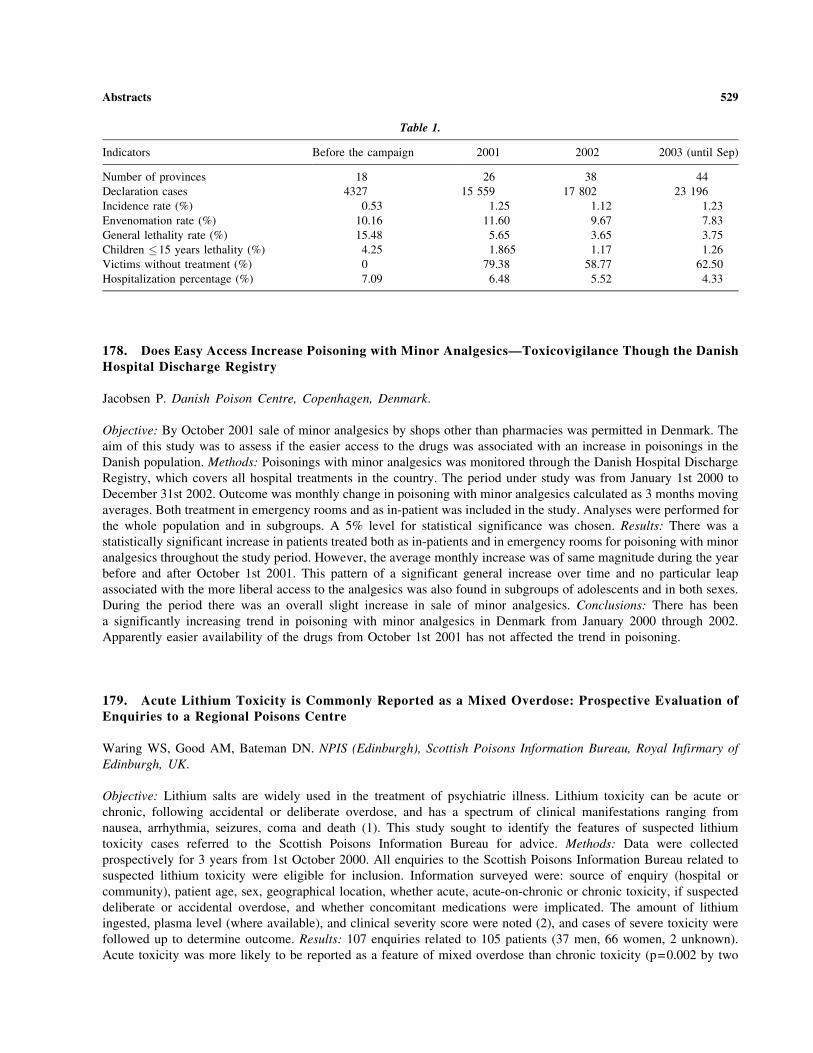
178. Does Easy Access Increase Poisoning with Minor Analgesics—Toxicovigilance Though the DanishHospital Discharge Registry
Jacobsen P. Danish Poison Centre, Copenhagen, Denmark.
Objective: By October 2001 sale of minor analgesics by shops other than pharmacies was permitted in Denmark. The
aim of this study was to assess if the easier access to the drugs was associated with an increase in poisonings in the
Danish population. Methods: Poisonings with minor analgesics was monitored through the Danish Hospital Discharge
Registry, which covers all hospital treatments in the country. The period under study was from January 1st 2000 to
December 31st 2002. Outcome was monthly change in poisoning with minor analgesics calculated as 3 months moving
averages. Both treatment in emergency rooms and as in-patient was included in the study. Analyses were performed for
the whole population and in subgroups. A 5% level for statistical significance was chosen. Results: There was a
statistically significant increase in patients treated both as in-patients and in emergency rooms for poisoning with minor
analgesics throughout the study period. However, the average monthly increase was of same magnitude during the year
before and after October 1st 2001. This pattern of a significant general increase over time and no particular leap
associated with the more liberal access to the analgesics was also found in subgroups of adolescents and in both sexes.
During the period there was an overall slight increase in sale of minor analgesics. Conclusions: There has been
a significantly increasing trend in poisoning with minor analgesics in Denmark from January 2000 through 2002.
Apparently easier availability of the drugs from October 1st 2001 has not affected the trend in poisoning.
179. Acute Lithium Toxicity is Commonly Reported as a Mixed Overdose: Prospective Evaluation ofEnquiries to a Regional Poisons Centre
Waring WS, Good AM, Bateman DN. NPIS (Edinburgh), Scottish Poisons Information Bureau, Royal Infirmary of
Edinburgh, UK.
Objective: Lithium salts are widely used in the treatment of psychiatric illness. Lithium toxicity can be acute or
chronic, following accidental or deliberate overdose, and has a spectrum of clinical manifestations ranging from
nausea, arrhythmia, seizures, coma and death (1). This study sought to identify the features of suspected lithium
toxicity cases referred to the Scottish Poisons Information Bureau for advice. Methods: Data were collected
prospectively for 3 years from 1st October 2000. All enquiries to the Scottish Poisons Information Bureau related to
suspected lithium toxicity were eligible for inclusion. Information surveyed were: source of enquiry (hospital or
community), patient age, sex, geographical location, whether acute, acute-on-chronic or chronic toxicity, if suspected
deliberate or accidental overdose, and whether concomitant medications were implicated. The amount of lithium
ingested, plasma level (where available), and clinical severity score were noted (2), and cases of severe toxicity were
followed up to determine outcome. Results: 107 enquiries related to 105 patients (37 men, 66 women, 2 unknown).
Acute toxicity was more likely to be reported as a feature of mixed overdose than chronic toxicity (p=0.002 by two
Table 1.
Indicators Before the campaign 2001 2002 2003 (until Sep)
Number of provinces 18 26 38 44
Declaration cases 4327 15 559 17 802 23 196
Incidence rate (%) 0.53 1.25 1.12 1.23
Envenomation rate (%) 10.16 11.60 9.67 7.83
General lethality rate (%) 15.48 5.65 3.65 3.75
Children �15 years lethality (%) 4.25 1.865 1.17 1.26
Victims without treatment (%) 0 79.38 58.77 62.50
Hospitalization percentage (%) 7.09 6.48 5.52 4.33
Abstracts 529
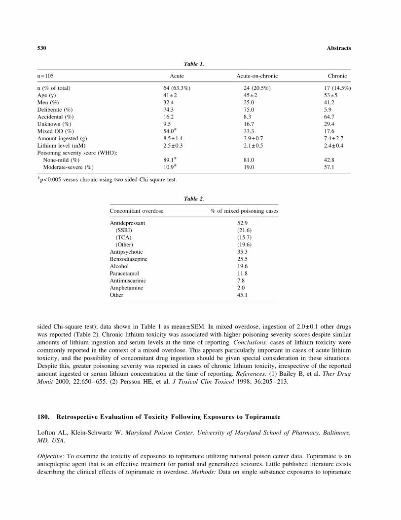
sided Chi-square test); data shown in Table 1 as mean±SEM. In mixed overdose, ingestion of 2.0±0.1 other drugs
was reported (Table 2). Chronic lithium toxicity was associated with higher poisoning severity scores despite similar
amounts of lithium ingestion and serum levels at the time of reporting. Conclusions: cases of lithium toxicity were
commonly reported in the context of a mixed overdose. This appears particularly important in cases of acute lithium
toxicity, and the possibility of concomitant drug ingestion should be given special consideration in these situations.
Despite this, greater poisoning severity was reported in cases of chronic lithium toxicity, irrespective of the reported
amount ingested or serum lithium concentration at the time of reporting. References: (1) Bailey B, et al. Ther Drug
Monit 2000; 22:650–655. (2) Persson HE, et al. J Toxicol Clin Toxicol 1998; 36:205–213.
180. Retrospective Evaluation of Toxicity Following Exposures to Topiramate
Lofton AL, Klein-Schwartz W. Maryland Poison Center, University of Maryland School of Pharmacy, Baltimore,
MD, USA.
Objective: To examine the toxicity of exposures to topiramate utilizing national poison center data. Topiramate is an
antiepileptic agent that is an effective treatment for partial and generalized seizures. Little published literature exists
describing the clinical effects of topiramate in overdose. Methods: Data on single substance exposures to topiramate
Table 1.
n=105 Acute Acute-on-chronic Chronic
n (% of total) 64 (63.3%) 24 (20.5%) 17 (14.5%)
Age (y) 41±2 45±2 53±5
Men (%) 32.4 25.0 41.2
Deliberate (%) 74.3 75.0 5.9
Accidental (%) 16.2 8.3 64.7
Unknown (%) 9.5 16.7 29.4
Mixed OD (%) 54.0* 33.3 17.6
Amount ingested (g) 8.5±1.4 3.9±0.7 7.4±2.7
Lithium level (mM) 2.5±0.3 2.1±0.5 2.4±0.4
Poisoning severity score (WHO):
None-mild (%) 89.1* 81.0 42.8
Moderate-severe (%) 10.9* 19.0 57.1
*p<0.005 versus chronic using two sided Chi-square test.
Table 2.
Concomitant overdose % of mixed poisoning cases
Antidepressant 52.9
(SSRI) (21.6)
(TCA) (15.7)
(Other) (19.6)
Antipsychotic 35.3
Benzodiazepine 25.5
Alcohol 19.6
Paracetamol 11.8
Antimuscarinic 7.8
Amphetamine 2.0
Other 45.1
530 Abstracts
reported to the American Association of Poison Control Centers Toxic Exposure Surveillance System in 2000 and
2001 were retrospectively analyzed. Statistical analyses (t-test, Chi Square) were performed using SAS (Statistical
Analysis System). Results: There were 567 cases reported that met the inclusion criteria. The majority of exposures
occurred within the age groups <5 years old (171, 30.2%) and 20 to 59 years old (213, 37.6%). Overall, the majority
of patients (62.1%) exposed to topiramate experienced no clinical effects. The most common clinical effects reported
in overdose were drowsiness/lethargy (15.5%), dizziness/vertigo (4.9%), agitation (4.9%), confusion (3.9%), nausea
(2.6%), and vomiting (2.5%). Major clinical effects reported in patients included acidosis (0.2%), coma (0.2%), and
cardiac conduction disturbance (0.2%). In children <5 years old, dose was known in 120 cases of topiramate
exposure; mean dose (94 mg, std. dev. 92) in asymptomatic patients was not significantly different from mean dose
(219 mg, std. dev. 470) in symptomatic patients (p >0.23). When known (N=4), the dose ingested in patients
experiencing major effects ranged from 200 to 12,500 mg. Ultimate medical outcome was reported as minor in
150 (26.5%) cases, moderate in 60 (10.6%), and major in 5 (0.9%) cases. No deaths were attributed to topiramate
ingestion. Conclusion: These data demonstrated that the majority of patients exposed to topiramate in overdose
experienced minor or no clinical effects. However rare, serious effects can also occur. These data will aid poison
control centers in the development of triage and management guidelines for patients exposed to topiramate in
overdose. References: Fakhoury T, Murray L, Seger D, et al. Topiramate overdose: clinical and laboratory features.
Epilepsy and Behavior 2002; 3:185–189. Traub SJ, Howland MA, Hoffman RS, et al. Neurological changes after
ingestion of topiramate (abstract). J Toxicol Clin Toxicol 2002; 40:620.
181. Acute Pesticides Poisonings in the Years 1994 – 2002 Reported to the Toxicological InformationCentre in Bratislava
Caganova B, Plackova S, Batora I, Klobusicka Z, Kresanek J. Toxicological Information Centre, Bratislava, Slovak
Republic.
Objective: The Toxicological Information Centre (TIC) in Bratislava has frequently been consulted for advice on
pesticide exposures. To obtain more information about pesticide poisonings in Slovakia, we performed a
retrospective analysis of all telephone calls to our Centre. Methods: All telephone inquiries involving pesticide
exposures were extracted from our databases for the period 1994–2002. The following data were analysed: age, sex,
intent of exposures (accidental or suicidal), substances ingested and clinical severity. All intoxications were
classified in accordance with the Poison Severity Score. Results: During the 9-year period 12 669 acute intoxications
were reported to the Slovak TIC, of which 1761 (13.9%) involved pesticides. Pesticide exposures in male (64.4%)
were more prevalent than those involving female (32.1%). Accidental poisonings were more common (76.9%) than
suicidal poisonings (20.4%). The majority of cases (55.6%) were adults. Most exposures were caused by insecticides
(54.7%), but rodenticides (20.9%), fungicides (9.4%), herbicides (9.2%) and other pesticides were also involved. Of
the insecticides, 39.5% were pyrethroids, 37.5% organophosphates and 6.1% carbamates. 81.2% of patients had
some symptoms. The majority of them developed only mild toxicity (63.8%), moderate symptoms occured in 12.4%
and severe symptoms in 4.2% of all poisonings. 15 cases (0.8%) died. Conclusion: Pesticide poisonings are still
associated with many deaths, especially among patients with organophosphate exposures. More efforts, such as
legislative control of the availability of pesticides and further innovation in therapeutic measures, are required to
reduce the serious impact of pesticide poisonings.
182. Child Poisonings Involving Anti-Inflammatories and Cough/Cold Medicines: What are the RiskFactors and Where to Next?
Shaw O, Smith NA. Department of Pharmacology and Toxicology, University of Otago, Dunedin, New Zealand.
Objective: To identify common risk factors and suggest targeted poison prevention strategies associated with child
poisonings reported to the National Poisons Centre (NPC) involving Non Steroidal Anti-Inflammatory Drugs
Abstracts 531
(NSAID and cough/cold preparations (C/Cs). Methods: Caregivers who had called the NPC in response to a
poisoning by a child aged 14 years and under between 1st January 2002 to 31st December 2002 (NSAIDs) and 1st
July 2002 to 31st December 2002 (cough/cold preparations) were invited to take part in a structured, standardized
telephone survey on the poisoning incident. Of the 185 eligible caregivers, 71 of 112 NSAIDs and 45 of 73 C/Cs
were contacted and agreed to participate. Results: Approximately 50% of the medicines were prescribed and
intended for adults in the home. Most solid doses were in blister strip packaging; 30–50% of liquid containers for
children used a child safety cap but this was not the case for the adult liquid medicines. More than half of the liquids
and topical products were (unnecessarily) kept in the refrigerator ‘‘to keep them fresh.’’ Most of the children aged
under 5 years accessed the medicine during unsupervised exploratory activities, when it was in use, not returned to
its usual storage place but still not in sight of the child. Caregivers reported that almost half of the younger children
thought the medicines were lollies, reflecting their pleasant taste and bright colours; others stated that the children
imitated adults taking the medicines. Few caregivers were aware of possible adverse effects associated with the
medicines; many reported not reading the packaging information as they were ‘‘familiar’’ with the product.
Caregivers identified first aid advice and the NPC number as useful information to include on the packaging or
medicine label. The poisoning incident caused most caregivers to enhance the storage of medicines in the home.
Conclusions: Adults prescribed NSAIDs or who purchase C/Cs comprise key target groups for poison prevention
interventions. Education at the community pharmacy level must focus on the safe use and storage of medicines,
particularly those resembling lollies. Apparent inadequacies in the strip blister packaging require investigation,
particularly as there is no current requirement to apply the NZ Standard to solid dose medicines. Clear poisons
advice and the NPC number should be included on the packaging and labeling of these medicines.
183. Reporting Hazardous Products Leads to Changes in Packaging
Marraffa JM (1), Wiener SW (2), Tarabar AF (2), Nelson LS (2), Hoffman RS (2), Stork CM (1). 1. SUNY Upstate
Medical University, CNY Poison Center; 2. New York City Poison Control Center, NY, USA.
Objective: Although many hair care products are benign, some have innate toxicity. After 2 pediatric cases of
aspiration of ‘‘PCJ Pretty and Silky’’ hair detangling spray, we notified the Consumer Product Safety Commission
(CPSC). The product contained hydrocarbons and was packaged in an attractive colorful bottle without mechanical
safety measures or precautionary labeling. As a result of reporting, the product is now sold in a child-resistant (CR)
bottle. Case Series: Case 1—A 17 month old girl presented to the ED with abrupt onset of respiratory distress and
emesis after being found with an open bottle of the product. She subsequently required intubation for respiratory
support. Chest x-ray (CXR) revealed bibasilar infiltrates and acute lung injury. After 48 hours, recalcitrant hypoxia
and hypotension ensued requiring pressors. Repeat CXR revealed pneumomediastinum and bilateral pleural
effusions. Oliguric renal failure developed, requiring peritoneal dialysis. Case 2—An 11 month old boy presented to
the ED with altered mental status and respiratory distress requiring intubation after being found with an open bottle
of the same product. A CXR revealed a right upper lobe infiltrate. Hypotension developed within 48 hours requiring
dopamine. Repeat CXR revealed bilateral infiltrates. Both patients were discharged without neurologic or respiratory
sequelae after prolonged hospital courses. Although these cases were reported to two different poison centers, routine
communication between these centers led to recognition of the hazardous nature of this product, and the cases were
reported to the CPSC. As a result, a regulation was passed requiring CR packaging for products containing greater
than or equal to 10% low-viscosity hydrocarbons. Conclusion: These cases demonstrate the value of cooperation
between poison control centers and regulatory agencies.
184. Methanol Outbreak in Norway: Role of the National Poisons Information Centre
Muan B (1), Hovda KE (2), Hunderi OH (3), Ekeland M (1), Jacobsen D (2). 1. Department for Poisons Information,
Directorate for Health and Social Affairs, Oslo; 2. Department of Acute Medicine, Ulleval University Hospital, Oslo;
3. Hospital Østfold, Fredrikstad, Norway.
532 Abstracts
Objective: The role of, and the consequence for, the National Poisons Information Centre (NPIC) in an outbreak of
methanol poisoning are described. Methods: Information from hospital journals and from the NPIC’s inquiry
database was obtained. Results: The methanol outbreak started in September 2002. 33 patients were treated for
methanol poisoning in hospitals in this period (Table 1); 5 deaths were verified. For the NPIC the outbreak resulted
in a significant increase in methanol inquiries. In the past two years the number of calls involving methanol was
52 (in 2000) and 55 (in 2001). In 2002 this number increased to 338 calls, of which 316 occurred during the last
4 months of the year. 142 of the calls to NPIC were due to real or suspected exposure to methanol. Based on the
information obtained the risk of poisoning was assessed as severe in 38, as moderate in 51 and as unlikely in 24 calls.
In the remaining calls there were either insufficient information for risk assessment or there were symptoms not
related to methanol poisoning. Discussion: The number of calls to NPIC involving methanol increased simul-
taneously and in parallel with the outbreak. NPIC’s strategy was to refer all unclear or suspicious cases to medical
care for preliminary testing of blood pH and osmolal and anion gaps. The NPIC could be further consulted for
advise regarding treatment and follow-up. The number of calls to the NPIC involving real methanol exposures is
higher than the number of patients in hospitals. This probably reflects the fact that NPIC was consulted several times
per patient. Early in the outbreak many calls from mass media were addressed. To meet public demands for
information, including demands from mass media, a summary of general information about methanol was published
on NPIC’s web-site. Conclusion: The NPIC played an important role in getting information and advice out to the
public and health service in the methanol outbreak in Norway.
185. Inquires to the Finnish Poison Information Centre Concerning Acute Poisonings in AnimalsDuring 1973 – 2002
Nyman T, Hoppu K, Kuisma P. Finnish Poison Information Centre, Helsinki University Central Hospital, Helsinki,
Finland.
Objective: Finnish Poison Information Centre (PIC) was founded in 1961, at that time primarily to give advice on
treatment and prevention of acute poisonings in children. From the early days on PIC has also received calls
concerning acute poisoning in animals and such calls seems to be increasing. The aim of the study is investigate the
development of calls related to acute poisonings in animals. Method: The yearly call statistics available for acute
animal poisoning from 1973, the first year when data on calls concerning acute poisoning of animals were
registered, were used to study trends over time. A more detailed investigation of a 40 months period (1.6.2000–
30.9.2003) was performed employing the current electronic database. Results: In 1973 only 26 calls concerned
animals, which was 0.8% of the total of 3 170 calls received. Data on calls concerning animals is missing for 1974,
and in 1975 1.2% of incoming calls were about animals. From that year on the percentage of animal calls has
Table 1. Number of patients in hospitals and calls to the NPIC as a result of the methanol outbreak.
Sept 2002 Oct 2002 Nov 2002 Dec 2002
Total
Sept–Dec 2002
Patients in hospitals with documented
methanol poisoning
18 8 3 4 33
Total number of calls involving methanol to
the NPIC
139 108 35 37 319
Calls from - hospitals/physicians 57 51 14 18 142
- public 68 51 21 19 157
- mass media 14 6 0 0 20
Calls regarding - acute exposure 56 56 15 15 142
- general information 64 43 17 19 143
- other 19 9 3 3 34
Abstracts 533
increased so that in 2002 6.1% of all inquires involved animals. (Table 1). During 1.6.2000–30.9.2002 (40 months)
we received 7 095 calls concerning animals. The great majority (98.2%) of them concerned pet animals. Dogs were
involved in 80.9% (5 744), cats in 14.6% (1 032), various exotic pets in 2.7% (193) and farm animals in 1.8% (126)
of these inquires. 88.6% (6 285) of these calls came from the general public and 11.4% came from veterinarians.
The most frequent enquiries about dogs were for medicinal products (22.7%), plants (14.3%) and pesticides
(11.0%). In cats the most common substances involved were plants (32.8%), medicinal products (16.62%),
pesticides (8.53%) and petroleum products (7.51%). Conclusion: Inquires concerning animals are increasing more
rapidly than the number of calls in our PIC. This may require reconsideration in our strategy, resources and
competence profile.
186. Amanita proxima Syndrome: 3 Cases of Acute Renal Failure Managed by the Antipoison Center ofthe Catholic University of Rome During the Last 2 Years
De Giacomo M (1), Gargano F (1), Marchione F (2), Fanghella G (2), Genovesi N (2), Ruzzi L (2). 1. Poison
Center, Intensive Care Unit, Catholic University School of Medicine, Rome; 2. Lanciano Hospital, Chieti, Italy.
Objective: To add the experience of 3 new cases on a still poor known mushroom syndrome: acute renal failure by
Amanita proxima. Amanita proxima is a mushroom eaten by mistake instead of Amanita ovoidea. The 2 mushrooms
are very similar but for the colour of the volva that is white in Amanita ovoidea and pale yellow in Amanita
proxima. In 1994 French authors (1) reported the first 5 cases of acute renal failure by Amanita proxima. In 1998
other 31 cases were reported, always by a French team (2). In 2000 the Amanita proxima was officially declared
poisonous in Italy. To our knowledge, until now, no cases were reported in Italy. Amanita proxima syndrome begins
8–12 hours after the meal with a gastroenteric syndrome followed by acute renal failure approximately 24 hours
later. 1st Case Report: a 85-year-old man was admitted to hospital 48 hours after mushrooms meal, complaining
nausea, vomiting and diarrhoea for the last 24 hours. He was anuric and his diuresis did not restore in spite of
rehydration. Laboratory assessment revealed: 13 mg/dl creatinine, 3 mg/dl total biliribin, 90 U/l GOT, and 53 U/l
GPT. The patient was supposed to have eaten Amanita ovoidea mushrooms. Hemodialysis was started. 48 hours later
the patient’s conditions improved and spontaneous urination restarted. 2nd Case Report: a 55-years-old man was
admitted to the hospital 48 hours after eating mushrooms supposed to be Ovoidea species. He was anuric. During the
previous day he had complained of vomiting, diarrhoea and poliuria. His creatinine was 10 mg/dl, Got 192 U/l, GPT
391 U/l, LDH 1896 U/l, CPK 97 U/l. Hemodialysis was started. On the following day also his mother, a 75-year-old-
woman was hospitalised because of anuria. Both underwent hemodialysis for 22 days. Diuresis and creatinine values
restored very slowly. On the 35th day the man was dismissed in good conditions, his mother was dismissed with a
creatinine value of 4.5 mg/dl to be controlled as an out-patient. References: Leray H, Canaud B, Andary C, Klouche K,
Beraud JJ, Mion C. Amanita proxima poisoning: a new cause of acute renal insufficiency. Nephrologie 1994; 15:197–
199. De Haro L, Jouglard J, Arditti J, David JM. Acute renal insufficiency caused by Amanita proxima poisoning:
experience of the Poison Center of Marseille. Nephrologie 1998; 19:21–24. Karlson-Stiber C, Persson H. Cytotoxic
fungi-an overview. Toxicon 2003; 42:339–349.
Table 1. The development of number of animal calls during 1973–2002.
Year Total number of calls Calls concerning animals %
1973 3 170 26 0.8
1975 4 467 55 1.2
1987 19 839 439 2.2
1991 27 250 862 3.2
1993 27 705 1 116 4.0
2001 38 087 2 000 5.2
2002 38 373 2 326 6.1
534 Abstracts
187. Validation of the Elisa Test for Urinary Alpha-Amanitin Analysis in Human AmanitaPhalloides Poisoning
Butera R, Coccini T, Randine G, Locatelli C, Manzo L. Pavia Poison Center, IRCCS Maugeri Foundation and
University of Pavia, Italy.
Objective: The value of the ELISA assay for urinary alpha-amanitin was examined in relation to its validity as a
diagnostic tool in patients with mushroom (Amanita phalloides) poisoning. Methods and Results: Linearity was
assessed on pooled blank urine spiked with 0, 1, 10, 20 ng/ml alpha-amanitin. The equation of the calibration plot
was y=�0.047x + 1.95, with r=0.99, where y was the Log percent bound alpha-amanitin value and x was the alpha-
amanitin concentration. The lower and higher concentrations of the linear range were used as lower and higher limits
of quantitation. Accuracy ranged from 90% at the 10 ng/ml level (best accuracy) to 75% at the 1 ng/ml (worst case).
Intra-assay precision, evaluated on replicate measurements (N=20) on three real samples from poisoned patients
resulted 16% at the 6 ng/ml level, and 15% at the 20 ng/ml level. Inter-assay precision, evaluated on real samples
(three replicates on ten runs) resulted better than 28% at all concentrations evaluated. The ELISA specificity for
alpha-amanitin was studied adding alpha-, beta-amanitin, and phalloidin (1:1:1, m/v, 1–20 ng/ml) to blank pooled
urine and comparing the results with equally spiked urine containing only alpha-amanitin. In urines containing the
three toxins the measured concentration values of alpha-amanitin were reduced by 50–65% as compared to those
detected in urines added alpha-amanitin only, indicating a masking effect of both beta-amanitin and phalloidin.
However, when tested individually, either beta-amanitin or falloidin were not measurable by this assay. Conclusion:
Validation parameters (linearity, accuracy, precision) proved the ELISA assay for urinary alpha-amanitin to be a
suitable diagnostic tool. However, further studies are needed to better define the relevance of any interference of
mushroom components other than alpha-amanitin with the determination of the actual concentrations of alpha-
amanitin in urine samples.
188. DNA Profiling: A Promising Tool for Mushroom Poisoning Diagnosis
Iturralde MJ (1), Ballesteros S (2), Ramon MF (2), Alonso A (1), Martın MP (3). 1. Seccion de Biologıa; 2. Servicio de
Informacion Toxicologica. Instituto de Toxicologıa; 3. Real Jardın Botanico, CSIC Madrid, Spain.
Introduction: Cases of mushroom poisoning are quite frequent in Spain. The process from early suspicion to final
diagnosis must proceed simultaneously along two routes - one monitoring the course of the clinical picture and the
other following the laboratory investigation. Traditional fungal study is based on morphological criteria and also in
biochemical markers. However, left-over and recovered fungal tissue are generally difficult to secure. Besides there
are only a few examples in the literature where the examination of stomach or intestinal contents were successful
and microscopic details may be either lost or compromised. The study of DNA polymorphism may be advantageous
in these cases. The rDNA gen sequences are separated by the variable internal transcribed spacer (ITS). The high
degree of polymorphisms within the ITS region has been used in the identification of fungal species and, in some
species, can be useful for separation of species into pathogenicity groups. In this work we describe our experience of
applying the study of the regions ITS-1 and ITS-2 to the resolutions of mushroom poisoning cases. Methods:
Botanical samples sent to our laboratory by judges and hospitals consisted of raw or cooked mushrooms.
Mycological identification was made using macroscopic and microscopic characteristics and searching of mushroom
toxins (amanitins, muscarine, psylocine and psylocibine). The ITS-1 and ITS-2 regions were PCR amplified and
sequenced with different primer pairs. Sequences obtained were compared with sequences located in the GenBank
(NCBI) using the BLAST search option. Results: The first results showed that, in general, genetic identification
coincided with botanical analysis or helped to identify the species. For example, a sample sent to our laboratory was
macroscopically and microscopically diagnosed as a mushroom of the Amanita muscaria/Amanita pantherina group.
A. muscaria and A. pantherina are the mainly species involved in the mycoatropinic syndrome. Poisoning cases are
accidental or intentional when the mushroom is consumed for recreational purposes. Prognosis of the poisoning is
generally minor although lethal cases with A. pantherina have been reported. After a BLAST search of the ITS-2
sequence obtained, a sequence of A. muscaria gave the higher similarity (97%). Conclusion: In medicine, molecular
Abstracts 535
methods can be used to develop diagnostic test for clinical fungus identification. A combination of the DNA data
together with morphological characteristics and biochemical characteristics will give a more complete picture of
the fungi. Moreover, the sequence data are very useful in identifying fungal material when the morphologi-
cal characteristics are ambiguous. Further development will provide more rapid and robust methods of spe-
cies identification.
189. The Incidence and Outcome of Mushroom Poisoning in Greece in Autumn 2002
Mila M, Tsamadou A, Sofidiotou V, Papathanassiou V, Valti E, Neou P. Poison Information Centre, Athens, Greece.
There are 2000 species of larger fungi in Greece. These fungi appear when the weather is damp and warm in the
season of autumn. They are often responsible of many poisonings with severe clinical picture or even for fatal
outcome. Objective: The aim of this study is to investigate the epidemiological distribution and to analyze further
more the severity of mushroom poisoning in Greece and to establish the appropriate practice for prevention and
therapy. Material and Methods: During 4 months (September to December 2002) 100 cases of mushrooms poisoning
from all over the country have been studied. Sex, age, ingestion time, onset of clinical effects, kind of clinical
effects, treatment and outcome have been registered. Results: According clinical criteria the mushrooms poisonings
by Amanita muscaria species were 57, by Amanita phalloides 39 and 4 poisonings were by undetectable mushrooms
species. Symptoms concerned especially gastrointestinal upset (94%), general (18%). In cases when onset of clinical
effects appeared within 5 hours of ingestion didn’t present any hepatic failure (Amanita muscaria). In cases when
onset of clinical effects appeared more than 6 hours up to 24 hour after ingestion only 5 cases didn’t present any
hepatic failure. In addition to hepatic failure they presented hepatorenal syndrome (22%), coagulopathies (14%),
interstitial nephritis (2%) and hepatic encephalopathy. Treatment for Amanita muscaria poisoning was symptomatic
and supportive with complete recovery. Treatment for Amanita phalloides included, except of supportive care,
antidotes, Penicillin G with simultaneously Silibilin administration for 24 hours to 7 days, dependent of clinical
picture. In those cases with antidotes administration within 1st 24 hours, 20 of 21 (95%) had mild liver enzymes
elevation. One presented renal failure and submitted in hemodialysis. In those cases with antidotes administration the
second day, 10 of 13 (76%) presented severe hepatic damage, 2 presented furthermore renal failure and submitted in
hemodialysis. One case with insufficient antidotes administration submitted in hepatic transplantation with fatal
outcome. In cases with antidotes administration the third day, 2 of the 5 (40%) presented hepatic and renal failure,
and the other 3 only (60%), hepatic failure. There was complete recovery to 97% of cases, 2 patients presented
interstitial nephritis and there was one death. Conclusion: Fungi poisonings is a substantial problem in Greece since
it is difficult to distinguish the toxic species. It is necessary people to be systematically informed about dangers of
fungi. It seems that Penicillin and Silibilin early administration, in addition to general supportive care, can help to
the complete recovery of the majority of patients.
190. Treatment of Carbon Monoxide Poisoning in Northern France. A Survey of Practice
Mathieu-Nolf M, Mathieu D, Linke JC, Durak-Carpeza C, Peucelle D. Centre antipoison, Service d’urgence et
reanimation medicale, Hopital regional universitaire; Lille, France.
Carbon monoxide is a frequent cause of poisoning which yearly induces many deaths and sequelae. Oxygen therapy is
the main part of the treatment and may be delivered as normobaric or hyperbaric. Generally accepted indications for
hyperbaric oxygen are CO induced coma, loss of consciousness and neurological abnormality. Objective: To study how
are managed CO poisoned patients in our region. Methods: During a 6 year period (1997–2002), all CO poisoned
patients admitted to hospitals in Northern France were prospectively recorded using the on-line recording system
CIGUE of the Lille Poison Centre. Circumstance of poisoning, clinical presentation, treatment and outcome were
recorded during hospitalisation. A systematic follow up was done in every case at 3 months by phone using a
questionnaire. Data are presented as percentage or mean and comparison are done by chi-square or t-test according to
536 Abstracts
the type of variables. A p value of less than 0.05 is taken as significant. Due to their therapeutic specificities, fire smoke
inhalation and CO-poisoned pregnant women were excluded from this analysis. Results: In 6 years, 3709 cases of CO
poisoning were recorded, from which 422 fire smoke inhalations and 128 pregnant women were excluded. From 3159
analysed cases, 56% were female, 44% male. Mean age was 30.5 year (16 days-94 years). Initial presentation includes
minor manifestations in 1446 (46%), neurological abnormality in 426 (13.5%), loss of consciousness in 1082 (34%),
coma in 205 (6.5%). Mean HbCO was 17.8%. During hospitalisation, 7 patients (0.2%) died. All others survived and
were discharged from hospitals. At 3 months, 6.6% of patients still complained of remaining symptoms. Hyperbaric
oxygen therapy (HBO) is performed in 1556 cases (49%), normobaric oxygen (NBO) in 1538 (49%) and 65 patients
refused treatment. According to our protocol, HBO was appropriately used in 92% of cases and NBO in 82%. Main
reasons not to use HBO were HBO contra-indication (3%) and patient request (5%). For NBO, reasons for protocol
violation were found in 5% and absent in 13%. Moreover, NBO flow rate and duration appeared to be grossly
insufficient in 57% with a significant difference between local hospitals and referral centres. Conclusion: In our region,
if HBO is generally performed in accordance with locally accepted indications, patients requiring NBO are less
well managed. Efforts have to be made in order to ensure CO poisoned patients are correctly treated when HBO is
not indicated.
191. Carbon Monoxide Poisoning in Children. A Comparison of Clinical Presentation, LaboratoryInvestigations, and Treatment in Patients Under One Year of Age and Older Children
Chiossi M, Baratta A, Debbia C, Di Marco M, Palmieri A, Rebutti I. Clinical Toxicology Unit and Emergency
Medicine Ward–Emergency Department G. Gaslini–Children’s Hospital; Genoa-Italy.
Objective: To describe epidemiological aspects and clinical features of carbon monoxide (CO) poisoning in children of
two different age groups (<1 year and 1–14 years) who were admitted into the Emergency Medicine Ward of Genoa
Children’s Hospital. Their clinical data were reported in the computerized data base of Clinical Toxicology Unit and we
chose the most significant ones in order to detect any differences in the two age groups relevant to diagnosis and
treatment. Results: 82 patients with CO poisoning are reported in the computerized data base of Clinical Toxicology
Unit. 11 children were under one year of age (13.4% of the patients) 8 of them were symptomatic (73%) while older
children (71 cases) showed a lower rate of symptomatic cases (63%). Clinical presentation is different in the two age
groups: in children <1 year the most common symptoms were cyanosis, dyspnoea, tachycardia while in older patients
symptoms were headache, somnolence, and vomiting. Particularly in older children 64% showed central nervous system
involvement which was shared by 18% of children <1year only. Carboxyhemoglobin (COHb) levels at arrival ranged
from 3–20, 3% in children <1year, while in children >1 year COHb values ranged from 2–46%. Treatment was
supportive without oxygen administration in 55 % of children <1 year (and whose COHb levels were 3.2%, 4.4%, 8.2%,
3% and <2 % in 2 cases) while normobaric oxygen was administered in 45% of them. In these patients, to whom oxygen
was administered, COHb levels were 7.8%–20.3%–10.7%–11%. In children >1 year oxygen was given to 87% of the
patients; seven of them (9.8%) underwent hyperbaric oxygen administration. COHb levels were available for 5 patients
only and they were: 18%, 27%, 41%, 18%, 46%, 29%. The outcome was favourable in all the patients without any
deaths. Conclusions: The main difference in the two groups of patients is the clinical presentation. Clinical features in
the <1 year group can be easily misunderstood for symptoms of heart disease or respiratory distress and this could lead to
a delay in the diagnosis and treatment. In children younger than 1 year, in our case series, choice of oxygen
administration seems to be influenced by COHb levels (1) rather than the appearance of symptoms. Reference: Meert
KL, Heidemann SM, Sarnaik AP. Outcome of children with carbon monoxide poisoning treated with normobaric
oxygen. J Trauma 1998; 44:149–158.
192. A Severe Methanol Poisoning Associated with Late Brain Hemorrhage: Case Report
Vieira RJ, Madureira PR, Fornazieri ME, Lalli CA, Martins LC, Pedro MN, Bucaretchi F, Toledo AS, De Capitani
EM. Emergency Infirmary and Poison Control Center, University of Campinas (UNICAMP), Brazil.
Abstracts 537
Background: the most characteristic finding in methanol intoxication is visual disturbance, CNS effects and
metabolic acidosis. Hemorrhagic and non-hemorrhagic necrosis of the putamen has been described. In Brazil
intoxication with methanol is rare and occurs mostly from ingestion of secretly distilled drinks contaminated with
methanol. We present a case of severe methanol poisoning which was treated late and developed a brain
hemorrhage by the 22nd day after ingestion. Objectives: to present a severe case of methanol poisoning with the
evolution of the CNS lesions. Case: A 20-year-old male arrived at the University Hospital at 9 a.m. on July 29,
2003, with a history of having ingested approximately 180 ml of model airplane fuel, that contains 60%
methanol, about 60 h before and about 180 ml more 36 h before. He was first treated and intubated at another
hospital before transfer to our care. On arriving, he was unconscious, Glasgow 6, with metabolic acidosis. The
arterial blood gas determination showed pH 6.919, pO2 79.5, pCO2 4.74, HCO3 9.2, lactate 13.5, SatO2 88.2%.
The chest radiography showed signs suggestive of aspiration pneumonia. The metabolic acidosis was corrected
with sodium bicarbonate and the pH kept near normal. He was treated with a loading IV dose of 1.2 mg/kg
(900 ml of a solution of 10% dextrose with 100 ml of ethanol), followed by continuous maintained IV doses of
110 mg/kg/h during 3 days. The methanol blood level on the 3rd day was 66.1 mg/dl. The patient was
maintained on mechanical ventilation for 8 days. On the second day of admission a TC showed infarct of the
basal nucleus and a perfusion brain scintilography showed hypoperfusion of basal nucleus, cingulum, frontalis
lobus, thalamus, secondary visual area, and diffuse hypoperfusion of the cerebelar cortex. After extubation he
was partially conscious with periods of mental confusion, left hemiparesis and central vision deficit. The state of
consciousness and the hemiparesis worsened around the 21st day of treatment. A magnetic resonance scan on the
24th day of treatment showed extensive compact hematoma, in organization, with an oval area 6x5x2 cm, with
center on the basal nuclei, discreet deviation of medium line with compression of the front horn of the lateral
ventricule. Comment: The CNS methanol poisoning manifestations are well known but a late haemorrhage is
not usual.
193. Diagnostic Value of Markers of Cellular Hypoxia in Experimental Cyanide Poisoning
Renard C, Borron SW, Baud FJ. *Federation de Biologie, Hopital d’instruction des armees Percy— **Reanimation
Medicale et Toxicologique, Hopital Lariboisere Inserm U26 Universite Paris VII, Paris, France.
Objectives: In patients suspected of cyanide poisoning, plasma lactate concentrations are significantly correlated
with blood cyanide concentrations. However, other markers of cellular hypoxia have also been reported. The aim
of this study was to assess among parameters of cellular hypoxia that are available to emergency physicians those
more closely correlated to blood cyanide concentrations. Materials and Methods: Four mg/kg KCN was
intraperitoneally administered to male Sprague-Dawley rats. Serial femoral venous and arterial blood samples were
collected to simultaneously determine arterial blood cyanide concentrations, blood gases as well as plasma lactate
concentrations. ABG and lactate were determined using an ABLTM
715 while blood cyanide was measured using
the method previously described by Rieders. Results are presented as mean+SEM. Correlations between blood
cyanide and plasma lactate, PvO2, SvO2 and Dav SO2 were studied using linear regression. Results: The 4 mg/kg
ip dose of KCN reliably induced a severe but non lethal poisoning with a maximal blood cyanide concentration of
2.18+0.1 mg/L 10 min after injection. The elimination half-life was 38.4+3.6 min. Maximal arterial and venous
lactate concentrations were not significantly different (11.0+2.3 and 9.6+1.1 mmol/L, respectively) but the arterial
one was observed 10 min after injection while the venous one was at 25 min. The area under the curve were not
significantly different. Impairment of blood gases occurred early with a non significant increase in PvO2 and
SvO2 and a significant but only transitory decrease in Dav SO2 at 10 min after injection. Among the various
parameters of cellular hypoxia, blood cyanide concentrations best correlated with arterial plasma lactate
concentrations. Conclusion: During the early phase of acute cyanide poisoning in rats, arterial blood cyanide
concentrations are more closely correlated to arterial plasma lactate concentrations. Thereafter, arterial and venous
lactate concentrations did not significantly differ. The decrease of cellular oxygen consumption assessed using
femoral venous PvO2 occurred early during the course of poisoning but was only transitory. These data
538 Abstracts
suggest that in addition to a decrease in oxygen consumption, other mechanisms exist to account for sustained
lactic acidosis.
194. Study of Scorpion Envenomations – Hospitalization Cases 2001 – 2002
Zemrour F (1), Soulaymani A (1), El Oufir R (2), Semlali I (2), Moukhtari A (1), Soulaymani Bencheikh R (2).
1. Toxicology and Pharmacology Laboratory, Sciences University-Kenitra; 2. Poison Centre and Pharmacovigilance
of Morocco-Rabat.
Scorpion stings are a public health problem in Morocco because of frequency, severity and socioeconomic
consequences. Aim: To analyze the mortality and morbidity of hospitalised cases of scorpion envenomation.
Methods: To analyze epidemiological, clinical, therapeutic and prognostic data, we examined hospital record cards
(age, sex, time between sting and admission (TPP), clinical symptoms: class I=localised signs, class II=general
signs, class III=distress signs, therapies applied and clinical evolution) in all provinces of Morocco from January
2001 to September 2002. The survey consisted in a follow-up of all scorpion envenomation patients who were
hospitalised. Results: During two years 2001–2002, we observed 943 hospitalised cases by scorpion envenomation,
representing 4.1% of all reports in 2001 and 1.8% in 2002 with 71% of children <15years. The majority of cases
(54%) were recorded between July and August. Clinical symptoms were rated as 4.7% Class I (Local signs: local
pain, local edema or local irritation), 81.9% class II (Systemic signs: temperature >38�C, sweating, gastrointestinal
symptoms, hypertension, priapism), and 13.4% class III (Vital distress: c hypotension, tachycardia, respiratory
failure, pulmonary edema, seizures, agitation, coma). 71% of stings occurred in children <15years.The TPP was
<1hour in 16.7%, 1–2 h in 30.6%, >2 h in 52.7%. Within the Class II group sweating occured in 43.8% and
vomiting in 50%. Within the Class III group the case fatality rate was 9.5% and most deaths occurred among
children aged less than 15 years (92% of deaths). The treatment used by medical personnel were usually many and
varied, costly and unjustified. Conclusion: The results of this study show reduction of hospitalization particularly in
mild cases (Class I).
195. Gastrotoxic Effects of Cyclooxygenase Non-Selective and Selective NSAIDs After AcuteOverdosage in Rats
Dobric S (1), Cupic V (2), Milovanovic Z (1), Jacevic V (1), Bokonjic D (1). 1. National Poison Control Centre,
Military Medical Academy 2. Faculty of Veterinary Medicine, Belgrade, Serbia & Montenegro.
Indomethacin and nimesulide are very potent non-steroidal anti-inflammatory drugs (NSAIDs). Indomethacin is a
cyclooxygenase (COX) non-selective NSAID with pronounced ulcerogenic activity, while nimesulide belongs to the
group COX-2 selective NSAIDs that have very low ulcerogenic potential. There are no data about nimesulide-
induced gastrotoxicity when given in very high doses. Accordingly, the aim of this study was to compare the
gastrotoxic effects of indomethacin and nimesulide after acute overdosage in rats. Adult male Wistar rats (200–
250 g) starving for 16–18 hours before the experiment were used. Both drugs were dissolved in DMSO and given
through the orogastric tube in a single dose of 25 mg/kg. This dose is about 8, and 25 times higher than the mean
anti-inflammatory dose (ED-50) of indomethacin and nimesulide, respectively in rats. Four hours after drug
administration the animals were sacrificed and the length, area and intensity score of gastric lesions were
determined. The pathohistological examination of the stomach and the determination of the acidity of the gastric
juice were also performed. The results demonstrated that both NSAIDs given in a dose of 25 mg/kg p.o. produced
gastric lesions in all treated animals, but they were significantly more pronounced in rats given indomethacin than
nimesulide. Indomethacin, but not nimesulide produced also significant increase of gastric acidity with no changes in
the volume of gastric juice. Our results suggest that gastrotoxic effects of the COX-2 selective NSAID nimesulide
Abstracts 539
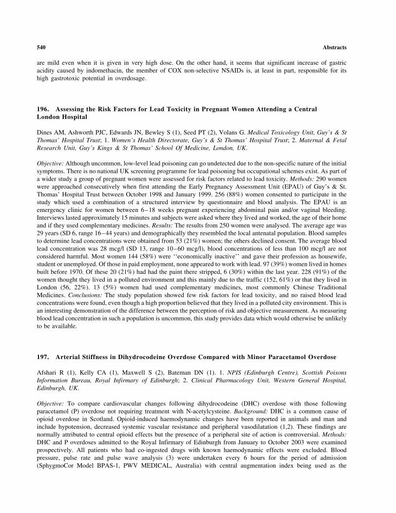
are mild even when it is given in very high dose. On the other hand, it seems that significant increase of gastric
acidity caused by indomethacin, the member of COX non-selective NSAIDs is, at least in part, responsible for its
high gastrotoxic potential in overdosage.
196. Assessing the Risk Factors for Lead Toxicity in Pregnant Women Attending a CentralLondon Hospital
Dines AM, Ashworth PJC, Edwards JN, Bewley S (1), Seed PT (2), Volans G. Medical Toxicology Unit, Guy’s & St
Thomas’ Hospital Trust; 1. Women’s Health Directorate, Guy’s & St Thomas’ Hospital Trust; 2. Maternal & Fetal
Research Unit, Guy’s Kings & St Thomas’ School Of Medicine, London, UK.
Objective: Although uncommon, low-level lead poisoning can go undetected due to the non-specific nature of the initial
symptoms. There is no national UK screening programme for lead poisoning but occupational schemes exist. As part of
a wider study a group of pregnant women were assessed for risk factors related to lead toxicity. Methods: 290 women
were approached consecutively when first attending the Early Pregnancy Assessment Unit (EPAU) of Guy’s & St.
Thomas’ Hospital Trust between October 1998 and January 1999. 256 (88%) women consented to participate in the
study which used a combination of a structured interview by questionnaire and blood analysis. The EPAU is an
emergency clinic for women between 6–18 weeks pregnant experiencing abdominal pain and/or vaginal bleeding.
Interviews lasted approximately 15 minutes and subjects were asked where they lived and worked, the age of their home
and if they used complementary medicines. Results: The results from 250 women were analysed. The average age was
29 years (SD 6, range 16–44 years) and demographically they resembled the local antenatal population. Blood samples
to determine lead concentrations were obtained from 53 (21%) women; the others declined consent. The average blood
lead concentration was 28 mcg/l (SD 13, range 10–60 mcg/l), blood concentrations of less than 100 mcg/l are not
considered harmful. Most women 144 (58%) were ‘‘economically inactive’’ and gave their profession as housewife,
student or unemployed. Of those in paid employment, none appeared to work with lead. 97 (39%) women lived in homes
built before 1970. Of these 20 (21%) had had the paint there stripped, 6 (30%) within the last year. 228 (91%) of the
women thought they lived in a polluted environment and this mainly due to the traffic (152, 61%) or that they lived in
London (56, 22%). 13 (5%) women had used complementary medicines, most commonly Chinese Traditional
Medicines. Conclusions: The study population showed few risk factors for lead toxicity, and no raised blood lead
concentrations were found, even though a high proportion believed that they lived in a polluted city environment. This is
an interesting demonstration of the difference between the perception of risk and objective measurement. As measuring
blood lead concentration in such a population is uncommon, this study provides data which would otherwise be unlikely
to be available.
197. Arterial Stiffness in Dihydrocodeine Overdose Compared with Minor Paracetamol Overdose
Afshari R (1), Kelly CA (1), Maxwell S (2), Bateman DN (1). 1. NPIS (Edinburgh Centre), Scottish Poisons
Information Bureau, Royal Infirmary of Edinburgh; 2. Clinical Pharmacology Unit, Western General Hospital,
Edinburgh, UK.
Objective: To compare cardiovascular changes following dihydrocodeine (DHC) overdose with those following
paracetamol (P) overdose not requiring treatment with N-acetylcysteine. Background: DHC is a common cause of
opioid overdose in Scotland. Opioid-induced haemodynamic changes have been reported in animals and man and
include hypotension, decreased systemic vascular resistance and peripheral vasodilatation (1,2). These findings are
normally attributed to central opioid effects but the presence of a peripheral site of action is controversial. Methods:
DHC and P overdoses admitted to the Royal Infirmary of Edinburgh from January to October 2003 were examined
prospectively. All patients who had co-ingested drugs with known haemodynamic effects were excluded. Blood
pressure, pulse rate and pulse wave analysis (3) were undertaken every 6 hours for the period of admission
(SphygmoCor Model BPAS-1, PWV MEDICAL, Australia) with central augmentation index being used as the
540 Abstracts
measure of arterial stiffness. Results: The study subjects included 10 DHC overdoses (6F) and 10 P overdoses (7F)
as controls, mean age 36.8 and 33.1 years respectively (ns). Mean systolic blood pressure (see Fig. 1), diastolic
blood pressure and pulse pressure were significantly lower in the DHC group up to 24 hours. Heart rate and
augmentation index (P=0.184, ANOVA) did not differ significantly between groups. Conclusions: DHC overdose
induces a decrease in arterial blood pressure, which diminishes over 24 hours. This effect is either related to the
central effects of opioids or an effect on systemic vascular resistance. We could not demonstrate any significant
changes in large arterial stiffness. Further studies are required to examine the potential impact of opioids on the
peripheral vasculature. These findings have clinical relevance to the management of patients with opioid poisoning.
References: 1. Czapla MA, Gozal D, Alea OA, Beckerman RC, Zadina JE. Differential cardiorespiratory effects of
endomorphin 1, endomorphin 2, DAMGO, and morphine. Am J Respir Crit Care Med 2000; 162:994–999. 2. Rosow
CE, Moss J, Philbin DM, Savarese JJ. Histamine release during morphine and fentanyl anesthesia. Anesthesiology
1982; 56:93–96. 3. Nichols WW, O’Rourke MF. McDonald’s Blood Flow in Arteries: Theoretical, Experimental and
Clinical Principles. London: Arnold, 1998.
198. Tobacco Smoking by Pregnant Woman and the Health Status of Newborn
Florek E (1), Piekoszewski W (2,3), Breborowicz GH (4), Groszek B (2). 1. Department of Toxicology, Laboratory
of Environmental Research, University of Medical Sciences, Poznan, Poland; 2. Department of Clinical Toxicology,
College of Medicine Jagiellonian University, Krakow, Poland; 3. Institute of Forensic Research, Krakow, Poland;
4. Perinatology and Gynaecology Clinic, University of Medical Sciences Poznan, Poland.
Objective: The study was designed to evaluate the effects of prenatal exposure to tobacco smoke on the foetus and
newborn. The measurement of tobacco smoke exposure biomarkers levels in biological material taken from the
mother and newborn allowed us to evaluate the influence of this exposure on the health status of the newborn
(evaluated by the physical examination). Methods: The studies was conducted on 100 volunteer pregnant women
who coming to the gynecological office around 12th –16th week of gestation. The volunteers subsequently gave
birth on the maternity ward of the Gynecological and Obstetrical Clinical Hospital. All women were divided into
two groups: (1) pregnant women smokers, (2) pregnant women non-smokers. The control group consisted of women
not exposed passively to tobacco smoke. The studies were performed in two stages. In the first stage, women around
the 12th–16th week of gestation coming to the hospital gynecological office completed a standardized questionnaire.
The women’s urine was taken for toxicological analysis. In the second stage, when the women came to hospital for
delivery, a second set of data were collected, which included the newborn’s health data. Analysis of cotinine was
carried on by means of HPLC. Results: In the tested group 26% of surveyed were smokers during their pregnancy,
and near 40.5% were exposed to passive inhalation of tobacco smoke, mainly at home. Mean value of cotinine in
urine of smokers was 947±354 ng/ml, in passive smokers was 8±11 ng/ml. The majority of smoking women were
of low education, living in small city and had low income. We found that the birth weight of newborns from
smoking mother was 355 g lower than in the non-smoking group. Conclusion: Smoking is still popular among
Figure 1.
Abstracts 541
pregnant women and only a small proportion of them stop smoking during pregnancy, however most of them
decrease the number of cigarettes smoked. The influence of smoking by pregnant woman is undeniable harmful for
foetus and newborn.
199. The Role of Prime Cultures Derived from Rat Heart Cells for Evaluating Drugs Cardiotoxicity
Scripcariu P, Petris O, Bologa C, Cotrutz C, Scripcariu O, Despa D. University of Medicine and Pharmacy ‘‘Gr. T.
Popa’’ Iasi, Romania.
Cardiomyocytes, in culture, lose some of their ‘‘in vivo’’ properties. Studies have shown that only primary
cardiomyocyte cultures are useful in toxicology. Objectives: to develop a primary culture derived from rat heart cells
in order to test the acute effects of diazepam, phenobarbital, chlorpromazine, amitriptyline and also ethanol and its
toxic metabolite, acetaldehyde. Methods: 1. We cultured three-day-old rat cardiomyocytes in 32 tubes. 2. On the
fifth day of culture we exposed the cardiomyocytes for 120 minutes to: diazepam 50 mmol/l, phenobarbital
50 mmol/l, chlorpromazine 100 mmol/l, amitriptyline 100 mmol/l, acetaldehyde 10 mmol/l and 100 mmol/l, ethanol
20 mmol/l and 200 mmol/l. 3. We evaluated the effect of these substances on cardiomyocytes using the following
criteria: A. Detachment of cardiomyocytes from the substrate. B. The morphology of cells under light microscopy.
C. The morphology of cells under electron microscopy. Results: A. There was 95% detachment of cardiomyocytes
from substrate in the diazepam, phenobarbital, chlorpromazine, amitriptyline and ethanol tubes, while in acet-
aldehyde exposure tubes the cardiomyocyte detachment was 100%. B. Under light microscopy we found no changes
to cardiomyocytes exposed to diazepam, phenobarbital, chlorpromazine, amitriptyline and ethanol. In the cardio-
myocytes exposed to acetaldehyde we noticed small cytoplasmatic vacuoles that resulted from the disappearance of
PAS positive substance (glycogen). C. Under electron microscopy we again saw no changes in cells exposed to
diazepam, phenobarbital, chlorpromazine, amitriptyline and ethanol. In the cardiomyocytes exposed to acetaldehyde
we saw swelling of the mitochondria and the formation of transit microvesicles in the endoplasmatic reticulum.
Conclusions: Our study stresses the role of primary culture from rat heart cells for the evaluation of some types
of cardiotoxicity.
200. Cocaine Induced Myocardial Ischemia Treated with Intravenous Phentolamine
Chan GM, Hoffman RS, Nelson LS. NYC Poison Control Center, New York, NY, USA.
Objective: Cocaine-induced chest pain is a common complaint in the Emergency Department (ED). The differential
diagnosis includes, but is not limited to: acute coronary syndromes (ACS), aortic dissection, pneumothorax, and
pneumomediastinum. The therapies for cocaine-induced chest pain are the same as for ACS from atherosclerotic
disease, with the exceptions of the absolute contraindication of beta-adrenergic receptor antagonists and the frequent
inclusion of benzodiazepines. However, since the data to support specific recommendations in patients with cocaine-
induced chest pain are limited, alternative therapies should be considered. We report the second case of cocaine-
induced myocardial ischemia with consistent electrocardiographic findings that resolved with the use of intravenous
phentolamine. Case Report: A 43 year-old man presented to the ED with left-sided chest pain, 1 hour after using
intranasal cocaine. The patient’s signs and symptoms were consistent with a sympathomimetic toxidrome. He was
treated with oxygen, sublingual nitroglycerin, aspirin, diazepam and morphine sulfate without resolution of
symptoms. A chest radiograph was normal and an electrocardiogram (ECG) showed sinus tachycardia (130 beats per
minute), with ST-segment depressions in leads V4–V5. Despite repeated doses of sublingual nitroglycerin, diazepam
and morphine sulfate, the patient’s symptoms and ECG findings did not resolve. After a total of 1.2mg of sublingual
nitroglycerine, 40 mg of diazepam, 10mg of morphine sulfate and the initiation of a nitroglycerin infusion, the
patient received 1mg of intravenous phentolamine at 5 minute intervals for a total dose of 3mg. Shortly after the
administration of the third dose of phentolamine, the patient’s symptoms, vital signs, and his ECG abnormalities
completely resolved. During his hospitalization, a urinalysis confirmed the use of cocaine. Serial cardiac enzymes,
542 Abstracts
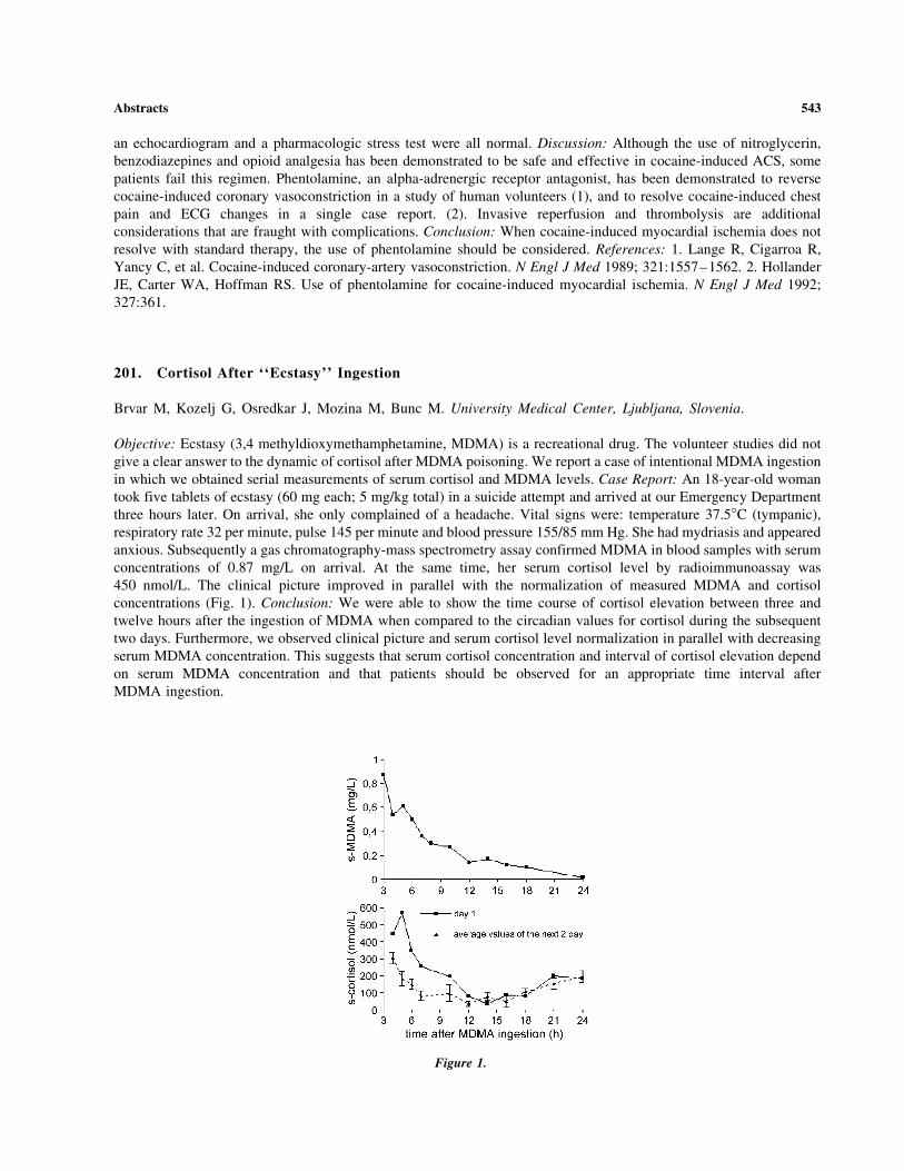
an echocardiogram and a pharmacologic stress test were all normal. Discussion: Although the use of nitroglycerin,
benzodiazepines and opioid analgesia has been demonstrated to be safe and effective in cocaine-induced ACS, some
patients fail this regimen. Phentolamine, an alpha-adrenergic receptor antagonist, has been demonstrated to reverse
cocaine-induced coronary vasoconstriction in a study of human volunteers (1), and to resolve cocaine-induced chest
pain and ECG changes in a single case report. (2). Invasive reperfusion and thrombolysis are additional
considerations that are fraught with complications. Conclusion: When cocaine-induced myocardial ischemia does not
resolve with standard therapy, the use of phentolamine should be considered. References: 1. Lange R, Cigarroa R,
Yancy C, et al. Cocaine-induced coronary-artery vasoconstriction. N Engl J Med 1989; 321:1557–1562. 2. Hollander
JE, Carter WA, Hoffman RS. Use of phentolamine for cocaine-induced myocardial ischemia. N Engl J Med 1992;
327:361.
201. Cortisol After ‘‘Ecstasy’’ Ingestion
Brvar M, Kozelj G, Osredkar J, Mozina M, Bunc M. University Medical Center, Ljubljana, Slovenia.
Objective: Ecstasy (3,4 methyldioxymethamphetamine, MDMA) is a recreational drug. The volunteer studies did not
give a clear answer to the dynamic of cortisol after MDMA poisoning. We report a case of intentional MDMA ingestion
in which we obtained serial measurements of serum cortisol and MDMA levels. Case Report: An 18-year-old woman
took five tablets of ecstasy (60 mg each; 5 mg/kg total) in a suicide attempt and arrived at our Emergency Department
three hours later. On arrival, she only complained of a headache. Vital signs were: temperature 37.5�C (tympanic),
respiratory rate 32 per minute, pulse 145 per minute and blood pressure 155/85 mm Hg. She had mydriasis and appeared
anxious. Subsequently a gas chromatography-mass spectrometry assay confirmed MDMA in blood samples with serum
concentrations of 0.87 mg/L on arrival. At the same time, her serum cortisol level by radioimmunoassay was
450 nmol/L. The clinical picture improved in parallel with the normalization of measured MDMA and cortisol
concentrations (Fig. 1). Conclusion: We were able to show the time course of cortisol elevation between three and
twelve hours after the ingestion of MDMA when compared to the circadian values for cortisol during the subsequent
two days. Furthermore, we observed clinical picture and serum cortisol level normalization in parallel with decreasing
serum MDMA concentration. This suggests that serum cortisol concentration and interval of cortisol elevation depend
on serum MDMA concentration and that patients should be observed for an appropriate time interval after
MDMA ingestion.
Figure 1.
Abstracts 543
202. Pseudolymphoma Syndrome Due to Carbamazepine: Case Report
Madureira PR, Vieira RJ, Fornazieri ME, Lalli CA, Martins LC, Pedro MN. Emergency Infirmary and Poison
Control Center, University of Campinas (UNICAMP), Brazil.
Background: pseudolymphoma syndrome (PLS) is a systemic illness characterized by onset of malaise, fever and
pharyngitis, followed by maculopapular skin rash, lymphonodenopathy, facial oedema, hepatitis, although other
organs such as the kidney, CNS or lungs may be involved, within 3 months after starting on anticonvulsants
(phenytoin, carbamazepine, phenobarbital or lamotrigini). PLS is relatively rare but can lead to death if there are
extensive skin lesions, severe hepatitis, agranulocytosis and neutropenia. The clinical presentation of PLS may serve
to confuse with lymphoma, most of the fatal cases being involved misdiagnosed and receiving inappropriate
antineoplastic chemotherapy. Objective: to present a case of pseudolymphoma syndrome with severe hepatitis and
encephalopathy due to carbamazepine. Case: A.B.S., a 16-year-old female, who arrived at the University Hospital
(UH) at 9 a.m. on September 9, 2003, with a history of malaise, daily fever, facial oedema and pharyngitis followed
by the onset of skin eruptions, pluritus, cervical lymphonodenopathy that had begun 15 days earlier. She had the
antecedent of a convulsive episode, not well characterized, 45 days before arriving at the UH, for which
carbamazepine 400 mg/day was introduced, and had been stopped one week before the admission, when she began
to present jaundice, oliguria and generalized oedema. On arriving, she was deeply icteric, with generalized oedema,
maculopapular eruptions with an exfoliative character on the whole body, and hepatosplenomegaly. Abdominal
ultrasonography showed moderate enlargement of the liver, spleen and lymphonoduli around the pancreas and liver.
Blood tests showed leuckocytosis (42,070), eosinophilia (8%), anemia (5.6 g/dL), creatinine (2.97 mg/dL), total
bilirubin (20.1 mg/dL), AST (2,656 u/L), ALT (1,375 u/L), alkaline phosphatasis (2,986 u/L), albumin (2.1 g/dL),
RNI (1.94), GGT (1,081), CK (223 u), LDH (1,393), bone marrow biopsy showed only reactive change. Six days
after admission consciousness changed with lethargy and a ‘‘flap’’ and she was treated for hepatic encephalopathy.
She remained febrile for three weeks after admission, when the clinical picture began to improve. She was
discharged well 45 days after her arrival. Comment: we presented a case of pseudolymphoma syndrome with severe
hepatitis and encephalopathy due to carbamazepine.
203. QT-Dispersion in Patients with Acute Digoxin-Intoxication
Pap Cs. Department of Emergency Medicine and Clinical Toxicology, Peterfy Hospital, 1074, Alsoerdosor 7,
Budapest, Hungary.
Objective: The repolarization of the myocardium is harmed in acute digitalis intoxication. The measurement of
QTc-dispersion is used to estimate the repolarization in homogenity. The aim of our retrospective study was to
determine the value of QTc-dispersion in patients with acute digitalis intoxication, as well as the correlation between
the magnitude of QTc-dispersion and the severity of the poisoning. Patients and Methods: Three groups were
compared. The first group (n=34, average age 56.3 ys) involved patients treated with acute digoxin overdosing
(serum levels 2.0–35.48 ng/ml, mean serum level 7.70 ng/ml) at our department during the last 8 years. Patients
having proper digoxin therapy (n=13, average age 69.5 ys, serum digoxin levels 0.5–2.0 ng/ml, mean serum level
1.41 ng/ml) and showing no signs of digitalis-toxicity were involved in the second group. The third group involved
healthy people without cardiac disease (n=25, average age 34.8 ys). On the basis of the severity of poisoning the
patients in the first group were rated by the Poison Severity Score and were classified into two subgroups: severely
(PSS 3 or death, n=14, mean serum digoxin level 14.12 ng/ml) and mildly (PSS 1–2, n=20, mean serum level
3.97 ng/ml) poisoned ones. ECG values were measured manually and the mean values of 3–3 subsequent ventricular
activity were recorded after each ECG lead. The mean of corrected QT-time and QTc-dispersion (the difference of
the shortest and longest QTc obtained from a 12 lead ECG) were determined for each patient. Results: see Table 1.
Conclusions: On the basis of our retrospective study it was established that in acute digitalis intoxication the
QTc-time did not change prominently but the value of QTc-dispersion was remarkably higher than those values
obtained from the healthy volunteers and the patients digitalized therapeutically. QTc-dispersion exceeding 75 ms
and 100 ms, considered as critical values by the literature references, occurred in digitalis intoxication in the greatest
544 Abstracts
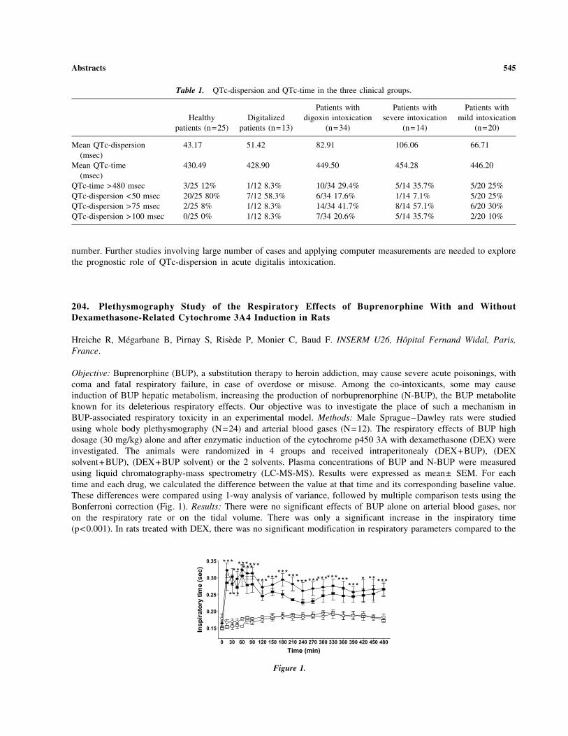
number. Further studies involving large number of cases and applying computer measurements are needed to explore
the prognostic role of QTc-dispersion in acute digitalis intoxication.
204. Plethysmography Study of the Respiratory Effects of Buprenorphine With and WithoutDexamethasone-Related Cytochrome 3A4 Induction in Rats
Hreiche R, Megarbane B, Pirnay S, Risede P, Monier C, Baud F. INSERM U26, Hopital Fernand Widal, Paris,
France.
Objective: Buprenorphine (BUP), a substitution therapy to heroin addiction, may cause severe acute poisonings, with
coma and fatal respiratory failure, in case of overdose or misuse. Among the co-intoxicants, some may cause
induction of BUP hepatic metabolism, increasing the production of norbuprenorphine (N-BUP), the BUP metabolite
known for its deleterious respiratory effects. Our objective was to investigate the place of such a mechanism in
BUP-associated respiratory toxicity in an experimental model. Methods: Male Sprague–Dawley rats were studied
using whole body plethysmography (N=24) and arterial blood gases (N=12). The respiratory effects of BUP high
dosage (30 mg/kg) alone and after enzymatic induction of the cytochrome p450 3A with dexamethasone (DEX) were
investigated. The animals were randomized in 4 groups and received intraperitonealy (DEX+BUP), (DEX
solvent+BUP), (DEX+BUP solvent) or the 2 solvents. Plasma concentrations of BUP and N-BUP were measured
using liquid chromatography-mass spectrometry (LC-MS-MS). Results were expressed as mean± SEM. For each
time and each drug, we calculated the difference between the value at that time and its corresponding baseline value.
These differences were compared using 1-way analysis of variance, followed by multiple comparison tests using the
Bonferroni correction (Fig. 1). Results: There were no significant effects of BUP alone on arterial blood gases, nor
on the respiratory rate or on the tidal volume. There was only a significant increase in the inspiratory time
(p<0.001). In rats treated with DEX, there was no significant modification in respiratory parameters compared to the
Table 1. QTc-dispersion and QTc-time in the three clinical groups.
Healthy
patients (n=25)
Digitalized
patients (n=13)
Patients with
digoxin intoxication
(n=34)
Patients with
severe intoxication
(n=14)
Patients with
mild intoxication
(n=20)
Mean QTc-dispersion
(msec)
43.17 51.42 82.91 106.06 66.71
Mean QTc-time
(msec)
430.49 428.90 449.50 454.28 446.20
QTc-time >480 msec 3/25 12% 1/12 8.3% 10/34 29.4% 5/14 35.7% 5/20 25%
QTc-dispersion <50 msec 20/25 80% 7/12 58.3% 6/34 17.6% 1/14 7.1% 5/20 25%
QTc-dispersion >75 msec 2/25 8% 1/12 8.3% 14/34 41.7% 8/14 57.1% 6/20 30%
QTc-dispersion >100 msec 0/25 0% 1/12 8.3% 7/34 20.6% 5/14 35.7% 2/20 10%
Figure 1.
Abstracts 545
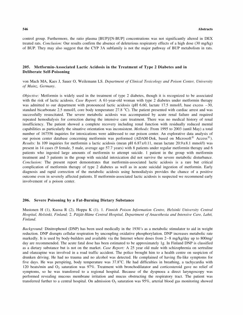
control group. Furthermore, the ratio plasma [BUP]/[N-BUP] concentrations was not significantly altered in DEX
treated rats. Conclusion: Our results confirm the absence of deleterious respiratory effects of a high dose (30 mg/kg)
of BUP. They may also suggest that the CYP 3A subfamily is not the major pathway of BUP metabolism in rats.
205. Metformin-Associated Lactic Acidosis in the Treatment of Type 2 Diabetes and inDeliberate Self-Poisoning
von Mach MA, Kaes J, Sauer O, Weilemann LS. Department of Clinical Toxicology and Poison Center, University
of Mainz, Germany.
Objective: Metformin is widely used in the treatment of type 2 diabetes, though it is recognized to be associated
with the risk of lactic acidosis. Case Report: A 61-year-old woman with type 2 diabetes under metformin therapy
was admitted to our department with pronounced lactic acidosis (pH 6.60, lactate 17.5 mmol/l, base excess –30,
standard bicarbonate 2.5 mmol/l, core body temperature 27.8 �C). The patient presented with cardiac arrest and was
successfully resuscitated. The severe metabolic acidosis was accompanied by acute renal failure and required
repeated hemodialysis for correction during the intensive care treatment. There was no medical history of renal
insufficiency. The patient showed a complete recovery including renal function with residually reduced mental
capabilities as particularly the situative orientation was inconsistent. Methods: From 1995 to 2003 (until May) a total
number of 167556 inquiries for intoxications were addressed to our poison center. An explorative data analysis of
our poison center database concerning metformin was performed (ADAM-Dok, based on Microsoft1 Access1).
Results: In 109 inquiries for metformin a lactic acidosis (mean pH 6.87±0.11, mean lactate 20.9±8.1 mmol/l) was
present in 14 cases (9 female, 5 male, average age 57.7 years) with 8 patients under regular metformin therapy and 6
patients who ingested large amounts of metformin to attempt suicide. 1 patient in the group with metformin
treatment and 3 patients in the group with suicidal intoxication did not survive the severe metabolic disturbance.
Conclusion: The present report demonstrates that metformin-associated lactic acidosis is a rare but critical
complication of metformin therapy of type 2 diabetes as well as in acute suicidal ingestion of metformin. Early
diagnosis and rapid correction of the metabolic acidosis using hemodialysis provides the chance of a positive
outcome even in severely affected patients. If metformin-associated lactic acidosis is suspected we recommend early
involvement of a poison center.
206. Severe Poisoning by a Fat-Burning Dietary Substance
Mustonen H (1), Kuosa R (2), Hoppu K (1). 1. Finnish Poison Information Centre, Helsinki University Central
Hospital, Helsinki, Finland; 2. Paijat-Hame Central Hospital, Department of Anaesthesia and Intensive Care, Lahti,
Finland.
Background: Dinitrophenol (DNP) has been used medically in the 1930´s as a metabolic stimulator to aid in weight
reduction. DNP disrupts cellular respiration by uncoupling oxidative phosphorylation. DNP increases metabolic rate
markedly. It is used by body-builders and available via the Internet where doses from 2–8 mg/kg/day up to 800mg/
day are recommended. The acute fatal dose has been estimated to be approximately 1g. In Finland DNP is classified
as a dietary substance but is not on the market. Case Report: A 25 year old male with schizophrenia on sertraline
and olanzapine was involved in a road traffic accident. The police brought him to a health centre on suspicion of
drunken driving. He had no trauma and no alcohol was detected. He complained of having flu-like symptoms for
five days. He was perspiring, body temperature was 37.8�C. He had difficulties in breathing, a tachycardia with
120 beats/min and O2 saturation was 97%. Treatment with bronchodilatator and corticosteroid gave no relief of
symptoms, so he was transferred to a regional hospital. Because of the dyspnoea a direct laryngoscopy was
performed revealing mucous membrane irritation and mucus obstructing the respiratory tract. The patient was
transferred further to a central hospital. On admission O2 saturation was 95%, arterial blood gas monitoring showed
546 Abstracts
normal oxygen level, hyperventilation, PCO2 3 Kpa. The tentative diagnosis was tracheitis and sepsis. A
tracheostomy was performed, antibiotics begun and the patient transferred to ICU. Profuse perspiration continued
and he had a body temperature of 39�C. Laboratory tests revealed a metabolic acidosis, BE-5.5, and glucose value
14.5 mmol/l. In addition to mechanical ventilation, treatment consisted of cooling, aggressive fluid administration
and alkaline diuresis. Temporary elevation of CK 19901 u/l on day 2, AST 623 u/l on day 4 occurred. CRP was
17 mg/l on admission, the highest level, ad 109 mg/l observed on day 4 when the tracheal cannula was removed.
Trachea sample revealed growth of Staphylococcus aureus and Moraxella catharalis. Thyroid function was normal.
Illicit drug screen was negative. He was removed from the respirator on day 3 and discharged on day 8 of admission.
The patient’s father provided the information of his son’s DNP use for a fat-burning purpose in connection with
bodybuilding. Afterwards the patient admitted the use. He had ordered the capsules via the Internet. The tracheal
findings were thought to be irritation due to hyperventilation secondary to metabolic acidosis. Conclusion: The
clinical picture of the patient fits with DNP poisoning.
207. Exposure to CS Gas in Pregnancy
McElhatton PR, Sidhu S, Thomas SHL. National Teratology Information Service (NTIS), RDTC, Newcastle-upon-
Tyne, UK.
Objective: To assess the fetotoxic effects of CS gas exposure in pregnancy. CS gas (tear gas, o-chlorobenzylidene
malononitrile) is a potent, rapidly incapacitating sensory irritant, particularly to the skin and eyes, dispersed as an
aerosol in riot control. There are limited data on the effects of acute exposure to CS gas during pregnancy, at the
concentrations normally used for crowd control (1,2). Method: Using standardised procedures, NTIS has provided
fetal risk assessment and collected outcome data on a prospective case series of 30 women who were exposed to CS
Gas during pregnancy. Results: The results are shown in Table 1. The mother of the infant with hypospadias and the
one who had an ETOP were exposed to CS gas during army training, but were reported as asymptomatic at the time
of exposure. Birth weight of singleton term babies was recorded in 25 live births (2 not recorded and 1 premature at
35 weeks) - Male n=15 mean weight 3693g (3199–4312) and Female n=10 mean weight 3152g (2327–3940), with
only 1 female baby classified as small for dates weighing less than 2500 g. Conclusion: Although transient maternal
symptoms of ear, nose and throat irritation were apparent in 63.3% of the exposed mothers, there was no significant
increase in adverse pregnancy outcome. There was only 1 infant with a congenital anomaly, hypospadias. It is most
unlikely that this anomaly which has a background incidence of 1 in 1000 liveborn male infants is causally
associated with CS gas exposure. Overall, these limited data indicate that in the absence of severe maternal toxicity
an increased risk of fetal toxicity is unlikely. References: 1. CS Gas TOXBASE(R). (2003, October). The Primary
Clinical Toxicology Database of the National Poisons Information Service. 2. Himsworth H, et al. Report of the
Enquiry into the Medical Toxicological Aspects of CS: Part II Enquiry into Toxicological Aspects of CS and its Use
for Civil Purposes. London HMSO 1971.
Table 1. Outcome of pregnancy following CS gas exposure.
Trimester of
exposure
Number
exposed
Acute maternal
toxicity (%)
Liveborn
normal
Liveborn
malformations
Spontaneous
abortion
Elective
termination
1st 12 6 (50) 9 1* 1+ 1+
2nd 11 9 (82) 11 – – –
3rd 7 5 (57) 7 – – –
Total 30 19 (63.3%) 27 (96.4%) 1 (3.6%) 1 (3.3%) 1 (3/3%)
*1 liveborn baby with mild hypospadias, exposed at 8 weeks (1/28=3.5% V s 2–3%).+Calculated from total exposures 1/30 (3.3% V s 10–20%+23% respectively).
Abstracts 547
208. Levofloxacin and Donepezil: A New Drug-Drug Interaction?
Mazzola JL, Bird SB, Aaron CK, Boyer EW. University of Massachusetts Medical School, Department of
Emergency Medicine, Division of Toxicology, Worcester, Massachusetts, USA. Massachusetts College of Pharmacy
and Health Sciences, Department of Pharmacy Practice, Worcester, Massachusetts, USA.
Fluoroquinolones have been associated with adverse effects such as headaches, dizziness, psychosis, and seizure.
The mechanism of fluoroquinolone-induced seizure is thought to be the result of competitive inhibition of gamma
-aminobutyric acid. In addition, co-administration of NSAIDS has been implicated in levofloxacin-associated CNS
adverse effects. Donepezil is one in a class of centrally-active acetylcholinesterase inhibitors, used in Alzheimer’s
disease. Most common neurologic adverse effects of donepezil are headache, insomnia, aphasia, ataxia, dystonia, and
seizures. Objective: A review of the English literature revealed two cases of levofloxacin-associated seizures. One of
these was in the setting of concomitant donepezil use. We report two instances of levofloxacin and donepezil-
associated seizures in one patient. Case Report: A 75 year-old male with a past medical history of seizure disorder,
renal insufficiency, and dementia presented to the emergency ward with increased seizure frequency for the past
3 nights. Levofloxacin 500 mg per day was started 7 days prior for a urinary tract infection. Other medications included
aspirin, doxazosin, folate, citalopram, gabapentin, divalproex sodium, donepezil, and ranitidine. Three months prior to
this presentation, he had increased seizure frequency shortly after initiating levofloxacin therapy for another urinary
infection. At that time, levofloxacin was discontinued and his seizures stopped. His initial vital signs on presentation
were BP 110/65 mm Hg, HR 75 bpm, T 36.5C, and RR 18 bpm. His physical exam was unremarkable. Laboratory
findings were notable for an increased BUN (25 mg/dl) and serum creatinine (1.5 mg/dl). His serum valproic acid level
was 88 mcg/ml, gabapentin 12.2 mcg/ml, and levofloxacin 3.1 mcg/ml. Using the Naranjo probability scale, the
likelihood of a levofloxacin and donepezil interaction as the source of seizure in our case was ‘‘probable.’’
Conclusions: Levofloxacin should be used in caution in patients with preexisting seizure disorder and decreased renal
function. The combination of levofloxacin and donepezil may increase further the risk of seizure. References:
O’Donnell JA, Gelone SP. Fluoroquinolones. Infect Dis Clin North Am 2000; 14:489–513. Martin SJ, Meyer JM,
Chuck SK, Jung R, Messick CP, Pendland SL. Levofloxacin and sparfloxacin: new quinolone antibiotics. Ann
Pharmacother 1998; 32:320–336. Kushner JM, Peckman HJ, Snyder CR. Seizures associated with fluoroquinolones.
Ann Pharmacother 2001; 35:1194–1198. Yagawa K. Latest industry information on the safety profile of levofloxacin
in Japan. Chemotherapy 2001; 47(S3):38–43. Naranjo CA, Busto U, Seelers EM, Sandor P, Ruiz I, Roberts EA, et al.
A method for estimating the probability of adverse drug reaction. Clin Pharmacol Ther 1981; 30:239–245.
209. Chloral Hydrate Cardiotoxicity Treated with Amiodarone
Greller HA, Hoffman RS, Nelson LS. New York City Poison Control Center, New York, NY, USA.
Objective: Myocardial sensitization following exposure to halogenated hydrocarbon compounds may produce
malignant dysrhythmias that are resistant to standard pharmacological therapies. Although amiodarone is
recommended for most ventricular dysrhythmias, its role in the treatment of toxin-induced dysrhythmias is largely
unknown. We present a patient with chloral hydrate poisoning who developed unstable ventricular tachycardia that
failed standard therapy but responded to amiodarone. Case Report: A 50 year-old man was brought to the ED after
being found by EMS with an empty bottle (30 cc) of chloral hydrate, and no other medications or drugs or abuse.
The urine toxicology screen was negative for drugs of abuse. When the patient was awake and was able to relate a
history, he confirmed the ingestion. Unfortunately, levels or other serum confirmatory markers were never obtained.
He became progressively obtunded following arrival. Vital signs were: BP 128/67 mm Hg, pulse 102/min,
respiratory rate 25/min, pulse oximeter 97% on 40% O2, and afebrile. The ECG showed a NSR at 67/min, with a
QRS of 0.084 sec and a QTc of 0.640 sec. 2 hours after arrival, he experienced polymorphic ventricular tachycardia
(PVT) associated with hypotension. After successful defibrillation (200 J, then 300 J) to sinus tachycardia and a
blood pressure of 92/41mm Hg, a lidocaine infusion was initiated. Less than one hour later, during suctioning, PVT
recurred and spontaneously converted. During a second suctioning, the patient again developed PVT, which
548 Abstracts
terminated only after a bolus of 150 mg of intravenous amiodarone. The patient was placed on an amiodarone infusion
and had no further recurrence of ventricular dysrhythmias despite recurrent suctioning. The infusion was discontinued
and a repeat ECG revealed a normal QTc. The patient was placed on oral propranolol and discharged to a psychiatric
facility. Conclusion: Ventricular dysrhythmias induced by chloral hydrate and other halogenated hydrocarbons may
respond to amiodarone. Additional studies on the safety and efficacy of amiodarone in these settings are needed.
210. Evaluation of Antivenom Therapy in Vipera palaestinae Bites
Bentur Y, Raikhlin-Eisenkraft B, Galperin M. Israel Poison Information Center, Rambam Medical Center, The
Bruce Rappaport Faculty of Medicine, Technion, Israel Institute of Technology, Haifa, Israel.
Objective: To evaluate the V. palaestinae antivenom 50ml fixed-dose regimen and to assess the need for repeated
antivenom administration as well as possible adverse effects. Methods: Retrospective review of prospectively
collected poison center data over a one-year period. One hundred and twelve patients were evaluated; 48% of these
were treated with antivenom. Antivenom treatment resulted in complete disappearance of systemic manifestations
with no relapse. Three patients required additional doses of antivenom for marked progressive local signs - one
patient had initially received 50ml of antivenom, the two others only 30ml. Anaphylaxis and serum sickness were
observed each in 3.7% of the treated patients. Conclusion: The fixed-dose regimen of 50ml V. palaestinae antivenom
is efficacious for the treatment of systemic and progressive local manifestations caused by this snake. There is
insufficient data on whether smaller doses can be successfully used for treating systemic manifestations and whether
initial larger doses are justified for marked progressive local signs (eg involvement of an entire limb). Randomised
prospective controlled studies are needed to elucidate these issues. References: 1. Bentur Y, Cahana A. Unusual
local complications of Vipera palaestinae bite. Toxicon 2003; 41:633–635. 2. Bentur Y, Zveibel F, Adler M,
Raikhlin B. Delayed administration of Vipera xanthina palaestinae antivenin. J Toxicol Clin Toxicol 1997; 35:257–
261. 3. Shemesh IY, Kristal C, Langerman L, Bourvin A. Preliminary evaluation of Vipera palaestinae snakebite
treatment in accordance to the severity of the clinical syndrome. Toxicon 1998; 36:867–873.
211. Severe Pseudohyperchloremia from an Intentional Ingestion of Sodium Bromide
Tsutaoka BT (1), Anderson IB (1), Luu L (2), Arora A (3), Olson KR (1). 1. California Poison Control System, SF
Division, Department of Clinical Pharmacy, University of California, San Francisco, California; 2. Laguna Honda
Hospital, San Francisco, California; 3. Los Robles Regional Medical Center, Thousand Oaks, California, USA.
Objective: In the United States, sodium bromide was found in many products used as sedatives and anticonvulsants
until 1975, and it continues to be used as an anticonvulsant in animals with rare use in humans for intractable
seizures. We report a case of an intentional ingestion of sodium bromide resulting in altered mental status and severe
pseudohyperchloremia. Case Report: The poison control center was contacted four days after a 48 year old woman
was admitted with altered mental status and presumed alcohol withdrawal. She had a seizure in the ED. She was
minimally responsive with spontaneous respirations. Her initial workup for medical and toxicologic causes was
negative. Admitting serum electrolytes were Na 150, K 4.2, Cl >107, total CO2 24, BUN 17, SCr 0.7. On hospital
day 5 her serum chloride was finally quantified as 168 mEq/L. The serum chloride from day 3 was 171 mEq/L. A
family member brought in an empty 400 mL bottle of sodium bromide 250 mg/mL that had been prescribed for her
dog’s seizure disorder. By this time she was beginning to awaken, and dialysis was not recommended; she was
treated with IV saline and furosemide. Her chloride level continued to decrease and her mental status fully cleared
by 14 days after admission. Serum bromide level from day 6 was reported as 170 mg/dL. Urine bromide before
saline and furosemide was 22 mg/dL, and after treatment was 87 mg/dL. The correlation of ‘‘excess’’ measured Cl
to actual serum Br was 2.4:1. Conclusion: Sodium bromide is available as a veterinary anticonvulsant, and can cause
coma and pseudohyperchloremia.
Abstracts 549
212. Lead Problems in Washington State
Robertson WO. Washington Poison Center, Seattle WA, USA.
Background: Nationally, lead is still revered as an enormous public health menace in the US. Its presence in
Venetian blinds, candy wrappers, candlewicks and sidewalk chalk prompt media mania. While clearly a valid
concern half a century ago, is it still so today? Our federal government obviously thinks so since it mandates
‘‘universal blood-lead testing’’ of all toddlers—with the loss of federal-matching Medicaid $’s for failure to comply.
Our Pacific Northwest states have repeatedly contested the need of such action - with one labeling such testing as
‘‘indisputable child abuse!’’ Consider our state’s rationale. Method: Data stem from A) Washington’s Lead
Surveillance Program’s records of all blood-lead-determinations done on any citizen since 1993 and its careful
analysis of all results from children; B) Washington’s Department of Labor and Industries which analyzes all adult
lead levels; C) all ‘‘CHARS’’ hospital discharge summaries re: lead poisoning over the past decade; and
D) Washington Poison Center’s lead problems. Results: From ’93 thru ’95, only 3.5% of 7,942 high-risk toddlers’
blood levels exceeded 10 micrograms/dL; for ’96–’98’s 12,715 toddlers, it was 2.1%; for ’99–’01’s 11,749 toddlers,
it was 1.4% and for 2002’s 7,336 toddlers, 1.2%. For adults, ’93 thru ’01 saw 94% of 43,432 reports
<25 micrograms/dL; 2002, saw >98% <25. Hospital discharge data for the decade of the ’90’s confirmed not a
single pediatric admission for lead! And, over that decade, our Poison Center only heard from the ‘‘worried well’’
and usually only after media scare tactics! Discussion: Assuming total screening costs at $70 for each child tested,
some $2.7 million was actually spent over 10 years to pin-point the 130 children with levels >20 - only one of
whom was subsequently chelated! The identification cost was $21,400 per case—which we are convinced could
have been better spent elsewhere. Conclusion: In the 60’s, our toddlers’ mean blood lead level was 34 micrograms/
dL; in the 70’s, it was 22. With the banishment of lead gasoline, 2003’s mean level is <1. Rises in autism, juvenile
delinquency and attention deficit disorders have been documented over the past years; rises in IQ have not been
corroborated. Is universal lead screening warranted? We think not.
213. Perchlorethylene Exposure in Pregnancy
McElhatton PR, Hedgley CA, Thomas SHL. National Teratology Information Service (NTIS), RDTC, Newcastle-
upon-Tyne, UK.
Objective: Assessment of the potential fetotoxic effects of perchlorethylene exposure in pregnancy. Method: Sixty
one cases of perchlorethylene exposure referred to NTIS for risk assessment were prospectively followed up using
standardised procedures (1). Results: The majority (46; 85.2%) of liveborn babies were normal. There were also
8 liveborn infants with malformations (14.8% vs 2–3% expected), 6 miscarriages (9.8% vs 10–20%) and 1 elective
termination (1.6% vs 23%). The details are shown in Table 1. Fifty-one (83.6%) babies were exposed in the first
trimester which is the most sensitive period for structural malformations. Transient maternal toxicity was reported
but there were no maternal deaths and none required admission to hospital. Conclusion: These limited data indicate
that in the absence of severe maternal toxicity the majority of liveborn babies are normal. The incidence of elective
termination of pregnancy is well within the expected range and the incidence of miscarriages is much lower than that
reported in the literature(2–4). Although the liveborn malformation rate (14.8%, 8/54) was higher than expected no
pattern of malformations was observed. Overall, this small case series has insufficient statistical power to confirm a
causal relationship between exposure to perchlorethylene and adverse pregnancy outcome, but this signal is
important, and indicates that further data are required. References: 1. Schaefer C, Vennevald F. FETIS-
documentation and follow up program for co-operation between Teratology Information Services in Europe.
Teratology 1993; 47:435. 2. Olsen J, Hemminki K, Ahlborg G, et al. Low birthweight, congenital malformations,
and spontaneous abortions among dry-cleaning workers in Scandinavia. Scandinavian Journal of Work, Environment
& Health 1990; 16:63–168. 3. Windham GC, Shusterman D, Swan SH, et al. Exposure to organic solvents and
adverse pregnancy outcome. American Journal of Industrial Medicine 1991; 20:241–259. 4. Doyle P, Roman E,
Beral V, et al. Spontaneous abortion in dry cleaning workers potentially exposed to perchlorethylene. Occupational
and Environmental Medicine 1997; 54:848–853.
550 Abstracts
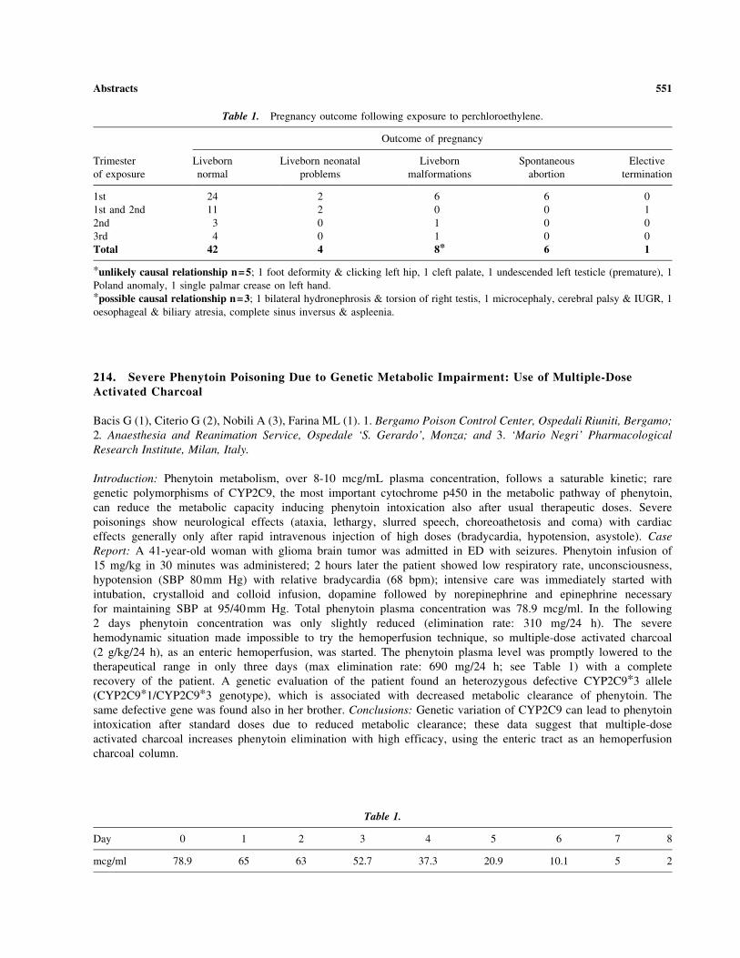
214. Severe Phenytoin Poisoning Due to Genetic Metabolic Impairment: Use of Multiple-DoseActivated Charcoal
Bacis G (1), Citerio G (2), Nobili A (3), Farina ML (1). 1. Bergamo Poison Control Center, Ospedali Riuniti, Bergamo;
2. Anaesthesia and Reanimation Service, Ospedale ‘S. Gerardo’, Monza; and 3. ‘Mario Negri’ Pharmacological
Research Institute, Milan, Italy.
Introduction: Phenytoin metabolism, over 8-10 mcg/mL plasma concentration, follows a saturable kinetic; rare
genetic polymorphisms of CYP2C9, the most important cytochrome p450 in the metabolic pathway of phenytoin,
can reduce the metabolic capacity inducing phenytoin intoxication also after usual therapeutic doses. Severe
poisonings show neurological effects (ataxia, lethargy, slurred speech, choreoathetosis and coma) with cardiac
effects generally only after rapid intravenous injection of high doses (bradycardia, hypotension, asystole). Case
Report: A 41-year-old woman with glioma brain tumor was admitted in ED with seizures. Phenytoin infusion of
15 mg/kg in 30 minutes was administered; 2 hours later the patient showed low respiratory rate, unconsciousness,
hypotension (SBP 80mm Hg) with relative bradycardia (68 bpm); intensive care was immediately started with
intubation, crystalloid and colloid infusion, dopamine followed by norepinephrine and epinephrine necessary
for maintaining SBP at 95/40mm Hg. Total phenytoin plasma concentration was 78.9 mcg/ml. In the following
2 days phenytoin concentration was only slightly reduced (elimination rate: 310 mg/24 h). The severe
hemodynamic situation made impossible to try the hemoperfusion technique, so multiple-dose activated charcoal
(2 g/kg/24 h), as an enteric hemoperfusion, was started. The phenytoin plasma level was promptly lowered to the
therapeutical range in only three days (max elimination rate: 690 mg/24 h; see Table 1) with a complete
recovery of the patient. A genetic evaluation of the patient found an heterozygous defective CYP2C9*3 allele
(CYP2C9*1/CYP2C9*3 genotype), which is associated with decreased metabolic clearance of phenytoin. The
same defective gene was found also in her brother. Conclusions: Genetic variation of CYP2C9 can lead to phenytoin
intoxication after standard doses due to reduced metabolic clearance; these data suggest that multiple-dose
activated charcoal increases phenytoin elimination with high efficacy, using the enteric tract as an hemoperfusion
charcoal column.
Table 1. Pregnancy outcome following exposure to perchloroethylene.
Outcome of pregnancy
Trimester
of exposure
Liveborn
normal
Liveborn neonatal
problems
Liveborn
malformations
Spontaneous
abortion
Elective
termination
1st 24 2 6 6 0
1st and 2nd 11 2 0 0 1
2nd 3 0 1 0 0
3rd 4 0 1 0 0
Total 42 4 8* 6 1
*unlikely causal relationship n=5; 1 foot deformity & clicking left hip, 1 cleft palate, 1 undescended left testicle (premature), 1
Poland anomaly, 1 single palmar crease on left hand.*possible causal relationship n=3; 1 bilateral hydronephrosis & torsion of right testis, 1 microcephaly, cerebral palsy & IUGR, 1
oesophageal & biliary atresia, complete sinus inversus & aspleenia.
Table 1.
Day 0 1 2 3 4 5 6 7 8
mcg/ml 78.9 65 63 52.7 37.3 20.9 10.1 5 2
Abstracts 551
215. Role of the Benzodiazepines in Buprenorphine—Associated Acute Poisonings: Place of theAntidotal Therapy
Megarbane B, Buisine A, Resiere D, Delerme S, Deye N, Baud F. Reanimation Medicale et Toxicologique, Hopital
Lariboisiere, Paris, France.
Objectives: More than one hundred anoxic deaths were attributed in France to poisonings with high dosage
buprenorphine (BUP), a substitution therapy among drug users. BUP misuse or association with psychotropic drugs,
of which benzodiazepines (BZD) was hypothesized to explain death, since the agonist-antagonist properties of BUP
are supposed to protect against respiratory depression in overdose. However, to date, the exact mechanism of death
is still not understood. Methods: We conducted a prospective study of patients admitted to our intensive care unit,
over 4 years (1998–2002), with a severe acute intoxication associating loss of consciousness, bradypnea and/or
myosis. Patients were selected if their urine toxicological screening (using a gas chromatographic method) showed
the presence of BUP in the absence of any other opioid (morphine, 6-monoacetylmorphine, codeine, codotheline,
pholcodine, methadone, and propoxyphene). A systematic screening for the psychotropic drugs and the usual illegal
drugs was performed. Clinical data, arterial blood gases and outcome analyses were collected. Results were expressed as
mean±SD. Results: Twenty-eight patients (33±1 years, 24M/4F, SAPS II: 36±4, 26/28 drug users and 4/28 suicide
attempts) were included. Among these 28 patients, 9 were depressive, 5 alcoholic, 5 HIV-infected and 6 with significant
neurological past history. All the patients declared regular consumption of psychotropic drugs, including BZD. BUP
poisoning (oral route in 19/28 cases, IV in 5/28 cases, mixed in 2/28 cases and inhalation in 2/28 cases) was responsible
for coma (Glasgow Coma Score: 8±1), myosis (28/28), and respiratory depression (respiratory rate: 12±1 /min, SpO2:
88±3%, arterial pH: 7.31±0.10, PaCO2: 7.21±0.5 kPa, and PaO2/FiO2: 41.2±4.1 kPa). 13/28 patients were intubated
and mechanically ventilated during a mean duration of 13±3 h. Complications were: aspiration pneumonia (11/28),
rhabdomyolysis (4/28) and hypotension (1/28). No partial or complete response was obtained in the 18 patients after IV
naloxone (up to 3.2 mg) injection, whereas, in the 8/12 patients who received IV flumazenil (0.2–1 mg), an improvement
in the respiratory and neurological status was observed. The 4 patients without response to flumazenil had a negative
BZD screening but a co-ingestion of phenobarbital or ethanol. Among the 8 patients who positively responded to
flumazenil, 5 had positive BZD screening and 3 others admitted, when awake to, BZD, consumption. Conclusion: BUP
is responsible of a typical opioid intoxication. Unlike naloxone, flumazenil is efficient in reversing the respiratory
depression in cases of BUP and BZD co-ingestion. BZD appear to play an essential role in the occurrence of a neuro-
respiratory toxicity during the course of BUP poisoning.
216. Risk Factors for Pneumonia in Acute Psychotropic Drug Poisoning
Vucinic S. National Poison Control Centre, Crnotravska 17, Belgrade, Serbia and Montenegro.
In many countries psychotropic drugs (anxiolytics and hypnotics, antidepressants, neuroleptics, psychostimulants etc)
are among the most common causes of death from self poisoning.. The most frequent complication is pneumonia which
results in substantial morbidity and mortality and increases the costs of treatment. The reported incidence of pneumonia
varies from 12–44%. Objective: To determine the incidence and risk factors for pneumonia in acute psychotropic drug
poisoning. Methods: During the period of the study (January 1999 to February 2002) 782 patients, admitted to the
Clinic of Emergency and Clinical Toxicology and Pharmacology, for acute drug poisoning were included in the study
and prospectively followed. A diagnosis of pneumonia was considered with: new and persistent pulmonary infiltrate on
chest radiographs; fever >38�C; leukocyte count>10000/mm3; purulent endotracheal secretions. A microorganism was
considered the etiologic agent of pneumonia only if it was obtained in blood and/or in the lower respiratory secretions,
obtained by nonbronchoscopic methods. To analyse the predisposing factors for pneumonia the following variables
were recorded: age, sex, underlying disease, intubation, H2 blockers, mechanical ventilation, coma, vasopressors,
corticosteroids, chronic alcohol intake central venous catheter (CVK), type of drug, severity of poisoning (Poisoning
Severity Score-PSS 3). The univariate analysis for pneumonia risk factors in all patients, and for each group of drugs
was done. The multivariate analysis was performed using logistic regression technique. Results: Pneumonia was found
in 94 (12.02%) of patients, 86 (91.5%) in psychotropic and 9 (8.5%) in nonpsychotropic drug poisoning (p<0.001). In
552 Abstracts
the psychotropic drug group, pneumonia was the most frequent in antidepressants (47%) and neuroleptics (16%). On
the contrary only 3.8% of patients with benzodiazepine poisoning had pneumonia. Statistically significant highest
incidence of pneumonia was registered in antidepressants (p<0.001), and smallest in benzodiazepine (p<0.001)
poisoning. Univariate analysis showed statistic significant association for following parameters: sex (p<0.05),
underlying disease (p<0.01), chronic alcohol intake (p<0.05), PSS 3 (p<0.001), coma (p<0.001), CVK (p<0.05),
vasopressors (p<0.05), corticosteroids (p<0.001), and H2 blockers (p<0.001). The multivariate analysis retained
intubation and antidepressant drug poisoning as independent risk factors for pneumonia. Conclusion: Using univariate
and multivariate analysis, risk factors for developing pneumonia were disclosed. Further consideration of the nature of
these factors may allow reduction of the incidence and morbidity of pneumonia in drug poisoning.
217. ‘‘Booty Bumping:’’ A Novel Route of Methamphetamine Abuse
Wiener SW, Hexdall A, McStay CM, Nelson LS, Hoffman RS. New York City Poison Control Center, New York
University School of Medicine, New York, NY, USA.
Objective: Methamphetamine is abused by ingestion, smoking, nasal insufflation, or injection. Rectal administration
has not been previously reported in the medical literature. We report a patient who describes ‘‘booty bumping,’’ the
rectal administration of methamphetamine, as a common practice in his community. Case Report: A 36 year-old
homosexual man with a history of HIV infection presented to the hospital complaining of skin lesions on his face
and buttocks. He said that many of his friends had the same lesions, and he was convinced that they resulted from
‘‘booty bumping.’’ He described the practice as the suspension of methamphetamine solid (crystals or powder) in
water and subsequent rectal administration using a needle-less syringe or other device. He said that while this results
in rapid onset of euphoria, many users develop skin lesions that may open and extrude crystals. Physical examination
revealed firm non-tender subcutaneous nodules without erythema. One lesion was incised, but no pus or crystals
were found. The patient was referred to the dermatology clinic for further evaluation but failed to keep his
appointment and was subsequently lost to follow-up. Discussion: Methamphetamine is bioavailable by most routes
of administration. Many users inject the drug to obtain a rapid onset of effect and high peak levels. Those who prefer
to avoid needles because of the fear of HIV, smoke the drug for similar pharmacodynamics. Others prefer nasal
insufflation (‘‘bumping’’), which provides more rapid absorption than the oral route, and is without first pass
metabolism. Rectal administration offers the same theoretical pharmacokinetic and pharmacodynamic ‘‘advantages’’
as nasal insufflation. A Medline search produced no results. An Internet search performed using the Google search
engine and the keywords ‘‘booty bumping’’ and ‘‘methamphetamine’’ found numerous references describing the
same practice, also known as ‘‘keistering.’’ There were no references to associated skin lesions. ‘‘Booty bumping’’
is often a prelude to receptive anal intercourse, and many sites warn of associated risks, including the possibility that
undissolved particles of methamphetamine may tear condoms and cause microabrasions. Methamphetamine use is
already known to increase the risk of HIV transmission through the use of needles and by leading to unsafe sexual
practices. ‘‘Booty bumping’’ may be yet another way in which methamphetamine users are at risk. Conclusion:
Rectal administration of methamphetamine, known as ‘‘booty bumping’’ or ‘‘keistering’’ is gaining popularity in the
homosexual male community and this practice may be associated with unsafe sexual activity.
218. Delayed Salicylate Toxicity Despite Negative Levels Five Hours Post-Ingestion
Chu J (1), Wiener SW (2), Tu S (1), Hahn I (1), Nelson LS (2), Hoffman RS (2). 1. St. Luke’s-Roosevelt Hospital
Center, NY; 2. NYC Poison Control Center, NY, USA.
Objective: Ingested salicylates are usually rapidly absorbed with early detectable serum levels. We describe a case of
enteric-coated aspirin ingestion with a prolonged period of delayed absorption leading to an initial series of negative
salicylate levels followed by clinically symptomatic salicylate toxicity 14.5 hours post-ingestion. Case Report: A
58 year-old woman presented three hours after ingesting a ‘‘handful’’ of enteric coated aspirin (ASA) with alcohol.
Abstracts 553
She denied nausea, abdominal pain, and tinnitus. Vital signs were: BP, 124/80 mm Hg; HR, 72/min; RR, 18/min;
temperature, 98.7�F. Her physical examination was unremarkable. She was given one dose of activated charcoal
(AC) and placed on continuous direct observation. Initial laboratory data were: ASA, <1 mg/dL, three hours post-
ingestion; alcohol, 151 mg/dL; HCO3, 26 mEq/L. A repeat ASA level five hours post-ingestion was still <1 mg/dL,
and she was transferred to psychiatry. 14.5 hours post-ingestion, she developed diaphoresis, nausea and decreased
hearing, and her repeat ASA level now 15 hours post-ingestion was 52 mg/dL. She did not receive any salicylate-
containing products while in the hospital. She was started on a continuous bicarbonate infusion and given multiple
doses of AC. Repeat ASA levels at 16.5 hours, 23.5 hours, 26.5 hours and 30 hours post-ingestion were 51 mg/dL,
44 mg/dL, 40 mg/dL, and 34 mg/dL respectively. Her nausea and hearing loss resolved at end of the second hospital
day. Discussion: Although salicylates are usually rapidly absorbed, toxicity may develop late due to delayed gastric
emptying or the formation of concretions. Enteric coated preparation are designed not to dissolve in the stomach, and
presentation is typically later than with ingestion of non-coated tablets. It is distinctly unusual, however, to have
undetectable levels as late as five hours after ingestion and subsequently develop toxicity. There is one prior report
of a negative level at four hours with subsequent toxicity in a patient who ingested a combination aspirin-
propoxyphene preparation. In another reported patient, the level did not peak until 30 hours after ingestion, although
the level was 30 mg/L (3 mg/dL) at six hours. These cases illustrate that an initial negative salicylate level does not
preclude subsequent toxicity after known salicylate ingestion, particularly with enteric-coated preparations.
Conclusion: This patient had a prolonged initial asymptomatic period with documented negative ASA levels up to
5 hours after an ingestion of enteric-coated aspirin before developing ASA toxicity 14.5 hours post-ingestion.
Patients with enteric-coated aspirin ingestions may need prolonged monitoring with serial ASA levels ever if their
initial level is negative.
219. Fibrinolytic Therapy for Cocaine-Induced Cerebrovascular Accident
Su M, Chang B, Hoffman RS. SUNY Downstate Medical Center; NYU/Bellevue Hospital Center; New York City
Poison Control Center, New York, USA.
Objective: Cocaine use is well associated with cerebrovascular accidents (CVAs). Although fibrinolytic therapy is
recommended for CVAs, its routine use for CVA remains controversial. Virtually no data support either the safety or the
efficacy of fibrinolytic therapy in cocaine-associated CVA. We present a patient with a cocaine-associated CVA who
successfully received alteplase therapy. Case Report: A 40 year-old male with no known past medical history presented
to the emergency department (ED) 30 minutes after the onset of a left-sided facial droop and left-sided hemiplegia. He
admitted to using cocaine approximately 12 hours prior to presentation. In addition to occasional cocaine use, he smoked
1 pack per day of cigarettes for 20 years. He denied taking any medications or herbal supplements. On physical
examination, he was awake, alert and appeared to be in no distress. His initial vital signs were: blood pressure,
140/90 mm Hg; pulse, 105 min�1; respirations, 16 min�1; temperature, 37�C. He had normal extraocular muscle
movements with 3–4 mm pupils bilaterally. He had a left-sided facial nerve palsy, and his motor strength was 1–2/5 in
both his left arm and leg. An electrocardiogram demonstrated sinus tachycardia with no significant ST-T wave
abnormalities. An immediate computed tomography scan without intravenous contrast was without evidence of
intracranial hemorrhage or acute infarction, but showed a possible hyperdense middle cerebral artery. A neurology
consultation was requested and the patient was believed to meet criteria for fibrinolytic therapy. Prior to the ad-
ministration of alteplase, he had transient improvement of upper and lower extremity motor strength for approximately
10 minutes, but then returned to his dense hemiplegia. The patient was given alteplase 0.9 mg/kg, 10% of the total dose
given as an intravenous bolus (IV) prior to IV infusion of the remainder over one hour. His symptoms completely
resolved over 72 hours and he was discharged home after 5 days. A urine toxicology screen positive for cocaine
metabolites, and other laboratory studies included normal electrolytes, normal complete blood count and no evidence of
a hypercoagulable state. A follow-up magnetic resonance (MR) imaging study demonstrated a small infarction in the
posterior limb of the right internal capsule. Magnetic resonance angiography of the brain, MR imaging of the neck,
bilateral carotid ultrasound and a transthoracic echocardiogram were all unremarkable. Conclusion: To our knowledge,
this is the first reported case of fibrinolytic therapy used to successfully treat a cocaine-associated CVA. Further work
is necessary to determine the safety and efficacy of fibrinolytic therapy administration for cocaine-associated CVA.
554 Abstracts
220. Renal Infarction Associated with Rizatriptan
Fulton JA, Nelson LS, Hoffman RS. New York City Poison Center, NY, USA.
Objective: Rizatriptan is used to relieve acute migraine and cluster headaches. Its mechanism of action is similar to
the other ‘‘triptans,’’ in that it reverses abnormal cerebral vasodilation through its activity as a 5-HT1B receptor
agonist. Compared to sumatriptan, it has an earlier Tmax (1 h vs. 2–2.5 h), a greater bioavailability (45% vs. 15%)
and an increased lipophilicity. Rizatriptan-induced vasoconstriction is rarely reported to result in stroke, myocardial
infarction and ischemic colitis attributed to its activity on peripheral 5-HT1B receptors. We present the first case of
renal infarction associated with two days of therapeutic rizatriptan use. Case Report: A 57 year-old man with a
history of hypertension was well controlled on valsartan and hydrochlorothiazide. He was recently diagnosed with
cluster headaches and was treated with indomethacin, prednisone, butalbital-acetaminophen-caffeine and hydro-
codone without relief. He then received two therapeutic doses of rizatriptan on each of the two days prior to
presentation. Subsequently, he presented to the emergency department complaining of nausea, vomiting and right-sided
abdominal pain. His vital signs were: blood pressure, 156/93 mm Hg; pulse, 94 min�1; respirations, 22 min�1;
temperature, 36.8�C; and room air oxygen saturation of 99%. Physical examination was positive only for right-sided
abdominal tenderness without rebound, guarding or organomegaly. His ECG showed a sinus rhythm without ischemic
changes. A complete blood count and differential, liver function studies, lipase and urinalysis were all unremarkable.
Electrolytes were only notable for a blood urea nitrogen of 8.2 mmol/L, and a serum creatinine of 83.9 umol/L. A
computerized tomography scan of the abdomen and pelvis with intravenous contrast revealed a very large wedge shaped
infarction of the right kidney. These results were confirmed by MRA and angiogram of the renal arteries. An extensive
evaluation for sources of coagulopathy included IGG, IGM and IGA Cardiolipin antibodies that were 11 GPL/mL [<13],
15 GPL/mL [<20] and 8 GPL/mL [<17], respectively. No other cause for his infarction could be identified. His renal
function remained stable and the patient recovered without any sequelae. Discussion: Vasospastic agents are often used
to treat migraine and cluster headaches. Because ergots cause numerous side-effects including vasospastic events they
have been largely replaced by ‘‘triptans’’ which have a more favorable safety profile. Despite this, ‘‘triptans’’ remain
contraindicated in patients with known or presumed vascular disease and are rarely associated with cardiac,
gastrointestinal and central nervous system infarctions. Conclusion: An acute renal infarction was associated with
therapeutic use of rizatriptan in a man with well-controlled underlying hypertension. Older patients and those with
hypertension and vascular disease should be cautioned about this potential side-effect of rizatriptan.
221. Ischemic Stroke from ‘‘Ephedra-Free’’ Dietary Supplement Containing Synephrine
Bouchard NC, Greller HA, Hoffman RS, Nelson LS. NYC Poison Control Center, New York, USA.
Objective: Due to reports of severe complications associated with the use of ephedra-containing supplements,
manufacturers have touted ‘‘ephedra-free’’ products as a safer alternative. We report an ischemic stroke associated
with an ‘‘ephedra-free’’ product that contained synephrine, a structurally related vasoactive compound. Case Report:
A 38 year-old man presented with memory loss and unsteady gait. He had no past medical history other than remote
cigarette smoking, and took no medications chronically. For a week prior to admission, he took an ‘‘ephedra-free’’
dietary supplement for weight loss. He took 1–2 tablets per day, less than the three suggested by the manufacturer.
Each tablet contained 6 mg synephrine and 200 mg caffeine alkaloids (Kola Nut Extract.) During that week, he had
several episodes of ‘‘dizziness.’’ Three days prior to presentation, he suffered a syncopal event and stopped the
supplement. On the day of presentation, he went to work (as a bus driver) and noted that he could not recall his
route. Vitals: BP, 120/72 mm Hg; pulse, 56 min�1; respirations, 15 min�1; temperature, 36�C. On examination he
had a mildly ataxic gait, could not recall his birthday, the year or the President of the USA. He also described
difficulty concentrating and was unable to perform serial 7’s. CT imaging showed subacute infarcts in the left
thalamus and left cerebellum, both in the distribution of the left posterior cerebral artery. MRI confirmed these
findings. An extensive evaluation for ischemic stroke including hematologic and coagulation studies, MRA,
echocardiogram and carotid duplex were unremarkable. His final diagnosis was vasospastic ischemic stroke. The
patient made a nearly complete recovery, left only with difficulty concentrating. Discussion: Recent public debate
Abstracts 555
has led many states to independently ban Ephedra containing products. In response to this, dietary supplement
manufacturers have begun to market ‘‘ephedra-free’’ products, boasting comparable weight loss with fewer
associated health risks. Some of these supplements contain synephrine, a sympathomimetic amine from the plant
Citrus aurantium. Synephrine shares structural similarity with other sympathomimetic drugs such as ephedrine,
amphetamine and phenylpropanolamine. Substitutions at the 4-position of the benzene ring and at the beta-carbon
suggest that synephrine may actually be a more potent alpha and beta-adrenergic agonist than ephedrine. Its clinical
effect on weight loss is uncertain, and based on the substitution of a hydroxyl group at the beta-carbon, one would
presuppose less CNS penetration. Conclusion: In an otherwise young healthy man with no defined risk factors, an
ischemic stroke was associated with the recent use of synephrine and caffeine. Consumers need to be informed of
the potential risks of ‘‘ephedra-free’’ products, even though they are advertised as ‘‘safe.’’
222. Severe Dapsone Hypersensitivity Syndrome in an Adolescent During Treatment of Leprosy
Vicente DC, Tresoldi AT, Pereira RM, Bucaretchi F. Depto de Pediatria, Centro de Controle de Intoxicacoes,
Hospital de Clınicas, Faculdade de Ciencias Medicas, Universidade Estadual de Campinas, (UNICAMP),
Campinas-SP-Brasil.
Objective: Dapsone has been widely used for treatment of leprosy, a wide variety of dermatological diseases such as
chronic bullous dermatosis, and Pneumocystis carinii prophylaxis. The side effects of dapsone are many and varied.
In addition, an uncommon and severe hypersensitivity adverse reaction, not dose-related, the ‘‘dapsone syndrome’’,
has been described since early 1950s. Case Report: A 12 y old girl was admitted 24 days after start a WHO
multidrug therapy (MDT) scheme for leprosy (dapsone, clofazimine and rifampicin). She was in a satisfactory health
condition until 6 days before, when fever, malaise, headache and rash developed. After 3 days, were noted dark
urine and jaundice, and MDT was discontinued. Upon hospital admission were noted an intense jaundice,
generalized lymphadenopathy, hepatoesplenomegaly, morbiliform rash and edema of face, ankles and hands. The
main laboratory data on admission included: hemoglobin, 8.4 g/dL; WBC, 15,710 cells/mm3, without atypical
lymphocytosis and eosinophilia; platelet count, 100,000 cells/mm3; serum aspartate aminotransferase, 1,013 IU/L
(normal, <46 IU/L); alanine aminotransferase 1,406 IU/L (normal, <38 IU/L); gamma-glutamil transpeptidase,
228 mg/dL (normal, 9–35 mg/dL); alkaline phosphatase, 672 IU/L (normal, <447 IU/L); direct bilirubin, 21.5 mg/dL
(normal, <0.2 mg/dL); indirect bilirubin, 5.6 mg/dL (normal, <0.8 mg/dL) and INR=1.49 (normal, <1.2). Following,
the clinical conditions had deteriorated, developing exfoliative dermatitis, hypotension, generalized edema, acute
renal and hepatic failure (INR=4.87), pancytopenia, intestinal bleeding and bacteremia (Enterococcus faecium),
needing adrenergic drugs, antibiotics and replacement of fluids and blood product components (RBC, platelets and
FFP). At this time, liver transplantation was considered. Ten days after admission she started to improve. Subsequent
laboratory studies revealed normal or negative results: Hepatitis A, B, C; CMV and EBV serological screens;
CD4/CD8 ratio; serum complement (C4) level; haptoglobine; rheumatoid factor and antinuclear antibody titers.
Serum tests for blood dapsone level, methemoglobin, glucose-6-phosphate dehydrogenase assay, treatment with
corticosteroids or dapsone provocation tests were not performed. She was discharged to home at day 27th.
Conclusion: This presentation could be engendered to dapsone, fulfilling most of the criteria for the syndrome
(fever, malaise, exfoliative dermatitis, jaundice with hepatic necrosis, lymphadenopathy and anemia). A concomitant
adverse reaction to rifampicin may be excluded, which is known to be hepatotoxic and nephrotoxic, since the patient
restarted supervised treatment for leprosy with clofazimine and rifampicin without any adverse effect. Physicians
should be aware to this rare and potentially fatal hypersensitivity reaction to dapsone.
223. Massive Magnesium Hydroxide Ingestion Requiring Dialysis
Gracia R, Hail S, Shepherd G. University of Texas Southwestern Medical Center and the North Texas Poison Center,
Dallas, Texas, USA.
556 Abstracts
Background: Systemic toxicity from oral magnesium hydroxide ingestion is rare. We describe a case of symptomatic
hypermagnesemia after a massive ingestion of milk of magnesia and aluminum hydroxide/magnesium hydroxide that
was successfully treated with hemodialysis. Case Report: A 55 year old female with chronic renal insufficiency,
schizophrenia, hypertension, and peptic ulcer disease presented to the emergency department after inadvertently
ingesting 540 ml of aluminum hydroxide/magnesium hydroxide (Mg(OH)2 200 mg/5 ml) and 480 ml of milk of
magnesia (Mg(OH)2 400 mg/5ml). The amount of magnesium ingested totaled 60 grams over a 2-day period. On
presentation, the patient was confused and ataxic. She had a temperature of 100.8 F, blood pressure of 72/56 mm
Hg, heart rate of 99 beats/minute, and respiratory rate of 36 breaths/minute. She was intubated for increasing
respiratory distress. On physical exam, the abdomen was distended and diffusely tender without rebound. Patellar
reflexes were absent. Significant laboratory tests included serum magnesium concentration 8.2 mg/dL, blood urea
nitrogen 66.5 mg/dL, and creatinine 4.5 mg/dL (baseline 2.1–2.5). The initial EKG showed NSR with T wave
inversions and a normal QT interval. Hemodialysis performed on 4 consecutive days finally corrected the
magnesium to 2.1 mg/dL. Aspiration pneumonia, sepsis and gastrointestinal bleeding complicated the hospital
course. All medical issues were resolved by hospital day 13. Conclusion: This case demonstrates acute severe
hypermagnesemia from oral magnesium hydroxide in a patient with chronic renal insufficiency successfully
corrected with hemodiaylsis.
224. Prolonged QTc with Massive Valproic Acid Overdose
Gracia R, Shepherd G, Rivera W, Keyes D. University of Texas Southwestern Medical Center and the North Texas
Poison Center, Dallas, Texas, USA.
Background: Cardiotoxicity is uncommonly reported in VPA overdose; the mechanism behind these cardiac effects
may involve inhibition of the delayed potassium rectifier current. This case describes a VPA ingestion resulting in a
peak level of 1626 ug/mL presenting with coma and QTc prolongation at 733 msec. Case Report: A 46 y/o female
presented following a suicide attempt with VPA about 3 hours prior to admission. Her history suggests she had not
taken other medications. On admission, the patient was unresponsive with a positive gag reflex requiring intubation.
Naloxone 2 mg IV was given with no response and activated charcoal was administered. The first VPA level was
1626 ug/mL; a corresponding QTc was 733 msec with a heart rate of 72 bpm. No cardiac arrhythmias were noted.
Aspirin, acetaminophen, and urine drug screen were all negative. Initial chemistries were all normal, except for an
elevated ammonia at 112 umol/L. Electrolytes 7 hours post ingestion revealed a metabolic acidosis: AGAP 21,
lactate 2.8 mmol/L. Initial arterial blood gases were pH 7.41 /pCO2 32 mm Hg/pO2 187 mm Hg/BE �3.7 mmol/L.
The patient received a 4-hour hemodialysis (HD) treatment 10 hours after ingestion, reducing the VPA to 336 ug/mL.
The QRS duration normalized to 401 msec commensurate with this level. Post dialysis VPA levels continued to
decrease to 56.2 ug/mL by 36 hours and she was discharged. Conclusion: This patient presented with a
hyperammonemic encephalopathy and a prolonged QTc interval associated with a markedly elevated VPA level.
Later sequelae included profound altered mental status and metabolic acidosis that corrected with the institution
of HD.
225. Effectiveness of Calcium Chloride and High Dose Insulin-Therapy in a Life ThreateningIntentional Quinidine and Verapamil Intoxication. A Case Report
Eyer F, Felgenhauer N, Zilker T. Toxicological Department—Technical University, Munich, Germany.
Objective: Intoxications with antiarrhythmic and antihypertensive drugs may eventually take a life-threatening
course. A remarkable reduction in cardiac output and dysrhythmia can make these intoxications difficult to handle.
Besides the use of inotropics, external or internal pacing may become necessary in severe cases. Adjunctive
treatment with glucose and insulin as well as use of calcium salts can play an important role in the treatment of these
intoxications. We report on a case of intentional quinidine and verapamil intoxication unresponsive to high-dose
Abstracts 557
catecholamine therapy and transjugular pacing but instantaneous stabilization after treatment with calcium chloride
and glucose-insulin was started. Case Report: A 65-year old female patient was pre-treated with a combination of
antiarrhythmic drugs due to intermittent atrial fibrillation. She intentionally took a total of 1.4 g verapamil and 4.5 g
quinidine sulfate, each of them being a lethal dosage. 6 hours after ingestion she collapsed and lost her
consciousness. Preclinical findings were coma, systemic hypotension and bradycardia with a heart rate of 20 bpm.
She was intubated and mechanically ventilated. External pacing was instituted. Administration of epinephrine and
dopamine as well as alkalisation with sodium bicarbonate was performed. On admission, blood pressure revealed 80/
40 mm Hg, heart rate was 91 bpm (with external pacing), electrocardiography (without pacing) showed widened
QRS (160 msec) with a spontaneous heart rate of 35 bpm. A transjugular pacemaker was inserted and blood pressure
declined despite of escaping dosage of epinephrine and dobutamine. Ultimately, a combination of calcium chloride
5.5% and high-dose glucose/insulin-therapy (20 g/h and 35 I.E./h, respectively) were effective to maintain mean
arterial pressure above 70 mm Hg within 1 hour. Internal pacing could be stopped 13 hours after admission,
resulting in stabilized heart rate of some 60 bpm. Mechanical ventilation was necessary until day 11 due to
pneumonia. The further course was uneventful and the patient could be transferred to a psychiatric department for
further treatment on day. Conclusion: Treatment of intoxications with calcium channel antagonists consists of
supportive care and the use of cardiotonic agents. Calcium salts should be the first-line treatment, with calcium
chloride preferable to calcium gluconate due to its three times higher calcium concentration. Glucagon,
catecholamines, high-dose regular insulin as well as phosphodiesterase inhibitors are adjunctive measurements
with beneficial use depending on the individual case. In our patient, treatment of the co-ingested class IA
antiarrhythmic quinidine and the calcium-channel blocker verapamil with sodium bicarbonate and the use of
catecholamines both turned out to be ineffective, whereas the use of calcium chloride and glucose-insulin
was successful.
226. Severe Methemoglobinemia Due to a Metoclopramide Metabolite
Wiener SW (1), Al-Chalabi M (2), O’Shaughnessy P (2), Husk G (2), Howland MA (3), Nelson LS (1), Hoffman RS
(1). 1. NYC Poison Control Center; 2. Beth Israel Medical Center; 3. St. John’s University, New York, USA.
Objective: Metoclopramide rarely causes methemoglobinemia. One report implicates a renally-eliminated metabolite
as the etiology. We report severe methemoglobinemia resulting from a single dose of metoclopramide that could not
be reproduced in vitro by adding parent drug to the patient’s blood. Case Report: A 33 year-old woman presented
with abdominal pain and vomiting. Her only past medical history was a pregnancy termination three weeks prior and
she took no medications. Vital signs were: blood pressure, 121/74 mm Hg; pulse, 98 min�1; respirations, 12 min�1;
temperature, 37.2�C. Physical examination was remarkable for cervical motion and lower abdominal tenderness with
guarding and rebound. She had no respiratory complaints or abnormalities on cardiopulmonary examination. During
her initial evaluation, she was given morphine 4 mg for analgesia, followed by 6 mg hydromorphone over 5 hours.
Antiemetics (metoclopramide 10 mg and promethazine 25 mg), and empiric metronidazole 500 mg were also
administered for presumed pelvic inflammatory disease. The surgical consult administered 0.8 mg naloxone during
his examination. No NG tube was placed, and topical anesthetic was never used. An abdominal CT scan with rectal
(gastrografin) and IV contrast (100 mL Ultraview- 300 mg I/mL iopromide) was obtained. When the patient returned
from imaging, (approximately four hours after metoclopramide administration), she was cyanotic and dyspneic.
Cardiopulmonary examination remained normal, but SpO2 was 70%. An ABG with co-oximetry showed: pH, 7.36;
pCO2, 35 mm Hg; pO2, 198 mm Hg; methemoglobin, 63.4%. Hematocrit was 40.9%. The patient was treated with
methylene blue 50 mg IV and her signs and symptoms (except abdominal pain) rapidly resolved. A repeat
methemoglobin drawn three hours later was 10.2%. One week after this episode had resolved, 0.1 mL of a 1 mcg/mL
metoclopramide solution was added to 0.9 mL of the patient’s blood and a control to make a final concentration of
0.1 mcg/mL, (a typical therapeutic level). Both samples were incubated at room temperature for 60 min.
Methemoglobins were: test sample, 0.6%; control, 0.4%. Discussion: There are only six prior cases reported of
metoclopramide-induced methemoglobinemia (three adults, three children). None of this patient’s other medications
has been reported to cause methemoglobinemia, and she had no signs or symptoms of methemoglobinemia prior to
being treated. A metabolite of metoclopramide has been suggested as the oxidant stress responsible. The fact that a
558 Abstracts
therapeutic concentration of the parent compound failed to induce methemoglobinemia in vitro is consistent with this
hypothesis. Conclusion: Although metoclopramide rarely causes methemoglobinemia, severe cases may occur in
susceptible individuals at therapeutic doses. A toxic metabolite may be responsible for the oxidant stress.
227. Fatal Lipoid Pneumonitis After Inhalation of Aloe Vera Extract
Bacis G (1), Moretti M (2), Chinaglia D (3), Farina ML (1). 1. Bergamo Poison Control Center; 2. Anaesthesia and
Reanimation Service; and 3. Pathology Department, Ospedali Riuniti, Bergamo, Italy.
Introduction: Aloe vera extracts are available as rubbery preparations (from the pericyclic cells of the leaf, contains
various anthraquinones as aloin, aloe-emodin, ethereal oil, resins and saponins) and gel (from the inner parenchymal
cells of the leaf, contains polysaccharides, vitamins, aminoacids, phosphatase, lipase, salicylic acid and steroids). The
extract has cathartic, antibacterial, anti-inflammatory effect, hemagglutinating activity and wound healing effects.
The use of Aloe herbal remedies is increasing with numerous ‘‘indications’’ such as addictions, AIDS, asthma,
autoimmune diseases, infections, cancer, poisoning, anxiety and depression, diabetes, menopause and sexual
impotency. In addition cosmetics and detergents containing Aloe vera are now highly advertised in the mass
media. We present the first case-report of fatal pneumonitis after inhalation of Aloe extract. Case Report: A 45-year-
old man with psychiatric disorders, two days before his admission in hospital, inhaled by aerosol 10 mL of the liquid
obtained with mechanical extraction from leaves of Aloe for treating thoracic pain, after a reading about its
antibacterial properties. On the admission the patient had dyspnea with hypoxiemia (PO2: 41.2 mm Hg, PCO2:
41.1 mm Hg with mask oxygen 4 L/min), leucocytosis (14,060/mcL) and neutrophia (98%), high ESR (52 mm/h),
fever and chest radiographic image showing bilateral pneumonitis with fluffy alveolar infiltrates. Treated initially
with bilevel positive airway pressure (Bi-PAP, FiO2 70%), clarithromycin, moxifloxacin, ceftriaxone, imipenem/
cilastin and methylprednisolone, after eight days, for a severe respiratory distress and pneumothorax, intubation,
mechanical ventilation (with PEEP 15 cmH2O) and chest tube insertion were needed. Also fluconazole, foscarnet
and NO (10 ppm) were added. Five days later, while the respiratory gas exchange was critical (PO2: 75 mm Hg,
PCO2: 65 mm Hg with PEEP 10 cmH2O, FiO2 90%) with pulmonary hypertension and pleural effusion, an acute
hypotensive episode (BP 66/50 mm Hg) was treated with epinephrine and then continued for maintaining adeguate
blood pressure. On the seventeenth day, the patient died with cardiogenic shock. Microbiological and serological
studies were always negative for mycobacterium, P. carinii, Legionella, C. pneumoniae, Criptococcus, Nocardia and
C. burnetii. The pathological study of lungs found a diffuse presence of macrophages with lipoid inclusions,
hemorrhages and alveolar fibrosis. Conclusions: The massive advertising and diffusion of Aloe therapy can
determine an inappropriate use of it with the risk of self-production and wrong administration, as in our fatal case.
228. A Poison Center can Perform a Greater Role in Public Health Emergencies
Bogdan GM, Seroka AM, Swanson D, Dart RC. Rocky Mountain Poison & Drug Center—Denver Health, Denver,
Colorado USA.
Background: The Colorado Health Emergency Line for the Public (COHELP) was established in 2002 by Denver
Health’s Medical Information Centers (Rocky Mountain Poison & Drug Center and Denver Health NurseLine) to
provide information during bioterrorism and other public health emergencies. COHELP has been tested for three
health events in Colorado during 2003: Smallpox vaccination program, West Nile Virus outbreak and influenza
outbreak. We provide an example of poison control centers and nurse advice lines performing a larger role in public
health emergencies beyond their usual scope of services. Objective: Characterize the ability of the COHELP service
to provide information and referrals for three major health events in Colorado during 2003. Methods: COHELP
responded to public concerns related to Smallpox from January 28 to April 26, West Nile Virus from July 22 to
Abstracts 559
October 10 and influenza from November 17 to November 23. A toll-free phone line featuring up-to-date recorded
information and referral to a web page (www.cohelp.us) for more detailed information was available 24 hours daily.
Trained information providers were available from 0700 to 2300 daily to answer caller questions, collect surveillance
data and provide referrals. Recorded information was available in English and Spanish with translation services
available for over 200 languages. Information providers began each event with Frequently Asked Question (FAQ)
scripts prepared by state health department epidemiologists about the event. These included explanations of
symptoms, treatments, and prevention measures with a mechanism to identify and answer additional FAQs within
48 hours for specific public concerns as they arose. Results: COHELP received over 18000 total calls related to three
events: 67% for West Nile Virus, 32% for influenza and 1% for Smallpox. Approximately 60% of callers only
listened to record information, with the remainder waiting to speak with information providers. The highest hourly,
daily and weekly call volumes all pertained to the influenza outbreak: 345, 2565 and 5705, respectively. Calls were
received from across Colorado, another 33 states and Canada, despite only advertising in Colorado. Only 1% of
callers required Spanish language interpreters. The most requested FAQs differed depending on the event: dead bird
reporting for West Nile Virus, symptom information for influenza and referrals for military administered smallpox
vaccinations. Conclusion: Local, state and federal public health agencies can rapidly expand their infrastructure to
increase readiness for bioterrorism and other health emergencies by partnering with poison control centers and nurse
advice lines. The state health department has utilized our capabilities, both technological and personnel, to
effectively provide information and gather surveillance data related to health events in Colorado.
229. Severe Valproic Acid Poisoning Treated with L-Carnitine and Haemodialysis. Lack ofCorrelation Between Symptoms, Serum Valproic Acid Concentrations and Ammonemia
Lavigne T (1), Meziani F (1), Kremer H (1), Castelain V (1), Bilbault P (1), Assemi P (1), Lutun P (1),
Ratomponirina C (2), Kintz P (3), Harlay ML (1), Schneider F (1), Jaeger A (1). 1. Service de Reanimation Medicale;
2. Laboratoire de Biochimie, Hopital de Hautepierre; 3. Laboratoire de Toxicologie, Universite Louis Pasteur,
Strasbourg, France.
Objective: Valproic acid (VA) overdose is usually benign. However, severe poisonings with coma, cerebral oedema,
haemodynamic instability, respiratory failure, bone marrow depression, multiorgan failure have been reported. L-
carnitine has been recommended in VA-induced hyperammonemia. Enhanced elimination by haemodialysis has also
been recommended in severe poisoning. We report a case of severe VA poisoning treated with L-carnitine
supplementation and haemodialysis. Case Report: A 17-year-old woman ingested in a suicide attempt 45 g of a slow
release form of valproic acid (VA). Admitted in hospital, the patient developed rapidly CNS disturbances and she
was transferred in the ICU. On admission (H5), examination showed: CGS=7, hypotonia, hyporeflexia, BP=140/90,
pulse rate=130/min. Serum VA concentration (SVAC) was 750 mg/L. Rapidly she developed hypoventilation with a
CGS of 3 and mydriasis which needed mechanical ventilation (H 8). CT scan showed diffuse cerebral oedema,
SVAC was 1153 mg/l and ammoniemia was 245 mmol/L. Despite treatment with L-carnitine (4 x 800 mg/kg),
induced diarrhoea, manitol infusion, a 6-hour haemodialysis (HD) followed by continuous haemodiafiltration (HDF),
the patient remained comatous and developed hepatic failure (ASAT=650 IU/L; ALAT 147 IU/L; prothrombin
level=23%; factor V=21%), renal failure (serum creatinine=307 mmol/L), rhabdomyolysis (CPK=19 030 IU/L) and
pancytopenia (Hb=7 g/L; leucocytes=1 900/mm3; platelets=16 000/mm3). At H 33, SVAC peaked at 1391 mg/L
and ammonemia at 1052 mmol/L. VA half-life was 9 hours before HD, 3 hours during HD and 12 hours during HDF.
For VA, HD clearance was 79.7 mL/min and HDF clearance was 23.9 mL/min. The calculated amount of VA
eliminated was 10.4 g during HD and 1.8 g during HDF. From H 57 to H 180, SVAC ranged between 120 and
63 mg/L and ammoniemia between 65 and 30 mmol/L. However, the patient remained comatous with a GCS ranging
between 3 and 6. The patient recovered from renal failure (H 95), from hepatic failure (H 160), and finally regained
consciousness on day 9, was extubated on day 10 and was discharged from hospital on day 19. Conclusion: In this
severe VA poisoning with multiorgan failure, L-carnitine treatment, HD and HDF (despite effective VA elimination)
had no effect on clinical outcome. Moreover, the clinical course, especially the CNS disturbances, was not correlated
with the evolution of SVAC and ammonemia. This suggests that direct toxicity of VA and hyperammonemia do not
account for all VA toxic effects and that other mechanisms are involved.
560 Abstracts
230. Indiscriminate Pediatric Transfers Following Acetaminophen Ingestions—Inadequate Use ofClinical Criteria
Perez A, Mordasky LK, McKay CA. Division of Medical Toxicology, Connecticut Children’s Medical Center,
Hartford Hospital, Hartford, Connecticut, USA.
Objective: We studied the utilization of clinical criteria by community hospitals (CH) to justify transfers to a
specialized children hospital (SCH) following acetaminophen (APAP) ingestions. Methods: Retrospective chart
review of all aromatic analgesic (ICD 965.4) poisoned children transferred from CH to a SCH between January 1998
and June 2003. We reviewed these charts for appropriate use of the Rumack–Matthew nomogram (RMN) and
adherence to King’s College Criteria (KKC) or APACHE II prior to transfer. Results: The total number of pediatric
APAP ingestions evaluated at the SCH was 181. 36 children were transferred during the study period. Four groups
were identified: Group 1—Pre 4 hour levels (n=11), Group 2—Non-toxic levels (n=3), Group 3—Post 4 hour levels
(n=18) and Group 4—Late presenters (n=4). Groups 1 and 2 (41% of transfers) demonstrate an inappropriate use of
the RMN. Group 3 and 4 transfers were not based on KKC or APACHE II scores. None of transferred patients
progressed to liver failure or subsequently met any KKC or APACHE II criteria. 14% of children developed
hepatotoxicity (3 children in group 3 and 2 of the late presenters). Conclusions: CH ED do not use clinical criteria to
justify the need for transfer of pediatric patients following acetaminophen ingestion. Transfer policies should be
established between CH and SCH.
231. The Effect of Activated Charcoal, Given as an Oral Solution, on the Elimination Half Life ofIntravenous Paracetamol
Christophersen AB (1,2), Hoegberg LCG (1,3), Kristensen K (4), Skanning PG (2), Angelo HR (3), Christensen HR
(1). Departments of 1. Clinical pharmacology; 2. Anaestesiology; 3. Clinical Biochemistry, Bispebjerg Hospital,
Copenhagen; 4. Pharmacokinetics, Novo Nordisk A/S, Maaloev, Denmark.
Objective: The possibility of activated charcoal’s (AC) ability to act through so-called gastrointestinal dialysis has
been discussed for several years. AC has been shown to enhance the elimination of certain drugs given intravenously
such as theophylline and carbamazepine. The aim of this study was to investigate whether the elimination half-life of
a drug, which does not undergo enterohepatic cycling, could be increased. Methods: A randomized cross-over study
using 18 human volunteers, consisting of 2 study days each separated by at least 4 days of wash-out. Study day
A consisted of the oral administration of 500 ml of water followed 1 hour later by 1 gram paracetamol administered
as a 15 minute IV infusion. Day A served as control. Day B the volunteers received 500 ml of orally administered
AC slurry followed 1 hour later by 1 gram of IV paracetamol as IV infusion. On both study days the volunteers
arrived after an overnight fast and were administered either water or activated charcoal slurry according to
randomization. 16 blood samples were taken at regular interval from 0 to 420 minutes from start of infusion. The
serum concentration of paracetamol was measured using HPLC. All pharmacokinetic parameters were calculated
using Winnonlin1 software and a noncompartmental model for intravenous administration. The terminal elimination
half-life for paracetamol was a primary endpoint. The secondary endpoint was the area under the curve (AUC) of
paracetamol, corrected for bodyweight (as paracetamol was given as 1 gram to all volunteers). The results were
found to have normal distribution, and Student’s t test was employed to compare the groups using a paired analysis.
Results: The elimination half-life of IV paracetamol was for day A 126 minutes (95% CI 114–138) and for day B
117 minutes (95% CI 109–126). The mean difference in half-life was 9 minutes (p=0.006, 95% CI 3–14) when AC
was administered orally, compared to when only water was given. Correspondingly, the AUC was a mean 11%
smaller (p<0.002, 95% CI 6–17%) when charcoal was administered. Conclusion: In this model, a single dose of
oral activated charcoal administered one hour before paracetamol administered intravenously, reduced the terminal
elimination half-life and the size of the AUC of paracetamol compared to control. The differences found were small
but statistically strong. The results indicate the possibility of charcoal having an effect on paracetamol that has
already been absorbed systemically, through intestinal dialysis, which has not been shown previously.
Abstracts 561
232. Is Paracetamol Poisoning Worsened by Coingestion of Cyclooxygenase Inhibitors?
Meziani F (1), Lavigne T (1), Kremer H (1), Meyer C (2), Assemi P (1), Castelain V (1), Bibault P (1), Harlay ML
(1), Lutun P (1), Jaeger A (1), Schneider F (1). 1. Service de Reanimation Medicale; 2. Service de Transplantation
Hepatique, Hopital de Hautepierre, Strasbourg, France.
Objective: Apart from acute hepatitis, other organ failures have been reported as rare complications in acute
paracetamol (PCM) poisoning. A vascular mechanism has been suggested for the occurrence of some of these
complications. We report 3 fatal cases of PCM and non steroidal anti-inflammatory drugs (NSAIDs) poisoning who
developed acute hepatitis complicated by multifocal ischemic injuries. Case Reports: Three previously healthy
women were admitted in the ICU for emergency liver transplantation after paracetamol and NSAIDs poisoning. The
patients presented severe acute liver failure with encephalopathy, anuria and signs of systemic inflammatory
response syndrome. On admission, plasma concentrations of tumor necrosis factor, interleukin 6 and endothelin were
strongly increased. During liver harvesting, despite full medical treatment including mechanical ventilation, vascular
filling and norepinephrine, clinical status worsened with persistence of shock, cutaneous vasoconstriction and
hyperlactatemia. Because of the suspicion of intra-abdominal complication of shock, an angiography of the celiac
and mesenteric artery was performed. It showed major and diffuse vasoconstriction. The 3 patients died from
refractory shock. Postmortem histological examination showed in all 3 cases multifocal ischemic areas, but without
vascular thombosis, on the stomach, small bowel and colon, and necrotizing pancreatitis. In one patient myocardial
ischemic myomalacia without coronary artery thombosis was also found. (Table 1). Discussion: under physiologic
conditions NSAIDs are not likely to have deleterious effects on vascular tone. A protective nonimmunological role
of COX has recently been advocated in the prevention of drug-induced liver injury in mice. In PCM poisoning with
SIRS, the effect of NSAIDs on vascular COX may induce or worsen vasoconstriction on small arteries. This effect
may explain the dramatic complications observed in our 3 patients.
233. Chronic Supratherapeutic Toxicity from Acetaminophen (APAP)
Dart RC, Daly FFS. Rocky Mountain Poison and Drug Center, Denver Health, Denver, CO USA.
Objective: Toxicity following repeated supratherapeutic ingestion (RSI) of APAP is an emerging cause of toxicity.
Of APAP deaths reported by the AAPCC in 2001, 31 (16%) were designated as chronic and 43 (22%) were designed
as ‘‘acute on chronic.’’ Further, of 7 pediatric APAP deaths since 1990, none were caused by acute poisoning. The
objective of this review is to systematically describe the toxic dose and clinical course of APAP RSI. Methods:
Standard systematic review techniques were used. A broad electronic search of MEDLINE (1966–Current) and
Table 1.
Patient 1 Patient 2 Patient 3
PCM (dose ingested: g) 15 30 10
NSAIDs (dose ingested) Diclofenac (0.5 g) Ketoprofen (1.5 g) Indomethacin (0.6 g)
PCM plasma level (mg/L) 41 (H 24) 144 (H 48) 26 (H 36)
ASAT (IU/L) 9232 6700 24550
ALAT (IU/L) 7125 4470 13950
Factor V (%) 4 5 2
Serum creatinine (mmol/L) 335 280 355
Lactate (mmol/L) 7.4 29.1 23.4
Mean arterial pressure (mm Hg) 58 63 53
Cardiac index (Lmin�1 m� 2) 4.65 5.85 6.8
Syst. Vasc. Resist. (dyne s�1 cm�5 m� 2) 756 601 506
Endothelin (pg/ml) (N=3.6–8.4) 129.3 162.7 152.4
562 Abstracts
EMBASE (1980–Current) in all languages and limited only to the APAP CAS registry number yielded 16,000
articles. Each article was screened and papers that reported repeated ingestion of acetaminophen (>4 g/d) were
included. Each article was abstracted by trained personnel. Results: Only one prospective study was found, involving
277 adult patients with APAP RSI. Hepatotoxicity (AST >1000 IU/L) did not develop in any of 126 patients with
normal AST at presentation. Seven of 47 (15%) patients with AST of 50–1000 IU/L at presentation developed
hepatotoxicity; one died. Six of 37 (16%) patients with AST >1,000 IU/L at presentation died or had liver
transplantation. The mean dose reported by the hepatotoxicity group was 12.6 g/d (95% CI 10.3–14.9 g).
Hepatotoxicity correlated weakly with APAP dosing and duration, but the history did not predict hepatotoxicity. No
patient with normal serum AST and APAP at presentation developed hepatotoxicity. The systematic review
identified multiple retrospective studies. Representative cases will be presented to illustrate the important
characteristics of these retrospective reports. Conclusions: Toxicity following acetaminophen RSI is not uncommon
and appears dose related. Like acute ingestion, the history does not predict toxicity. The serum AST and
acetaminophen concentrations at presentation are promising tools for the management of acetaminophen RSI.
234. Inappropriate Delay in Acetylcysteine Administration in Patients with Paracetamol OverdoseWho Subsequently Develop Severe Liver Dysfunction
Beer C, Hudson M, Smith L, Thomas SHL. Newcastle upon Tyne Hospitals NHS Trust and Wolfson Unit of Clinical
Pharmacology, University of Newcastle, Newcastle, UK.
Objective: In the United Kingdom, paracetamol poisoning is frequent and is a common cause of fulminant hepatic
failure. Early administration of the antidote acetylcysteine is very effective at preventing liver damage, however
efficacy is reduced with time, especially when treatment is given more than 8 h after overdose. Therefore starting
acetylcysteine treatment is urgent if more than 8 hours has elapsed since elapses apparently severe overdose. This study
was performed to establish how often there had been delays in commencing acetylcysteine in patients with paracetamol
overdose who developed severe liver dysfunction. Methods: The records were reviewed of all patients presenting to the
regional liver service in Newcastle between September 1996 and March 2003 with paracetamol poisoning and severe
liver dysfunction. Clinical details were sought from liver unit notes or the referring hospital, including the paracetamol
concentration at presentation, the timing of this in relation to the overdose and the timing of the start of acetylcysteine
infusion. Results: Of 172 patients with possible paracetamol poisoning referred during the period of study, paracetamol
poisoning could be verified in 160 (74 females, 86 males, mean age 33 y). Adequate data on the commencement of
acetylcysteine was available in 92. Of these, 6 had a paracetamol level below the appropriate nomogram treatment line
and a further five patients received prompt treatment within 8 hours of poisoning. For the remaining 81 patients the
median delay in administration of acetylcysteine after presentation was 1.75 h, but was more than 4 hours in 21 cases.
Conclusion: Inappropriate delays in administering acetylcysteine are common in patients with proven severe
paracetamol poisoning. Avoiding delay in starting antidotal treatment following hospital presentation may prevent an
important proportion of episodes of paracetamol induced liver dysfunction.
235. Increased Incidence of Phencyclidine Use in the Philadelphia Region Following theSeptember 11, 2001 Terrorist Attacks
Haroz R (1), Muller A (2), Greenberg M (1,3). 1. Drexel Univ. College of Medicine, Philadelphia; 2. Phila Poison
Control Center; 3. Temple Univ. School of Medicine, USA.
Background: Following the attacks of September 11, 2001, increased attention to the security of the U.S. borders and
ports of entry may have interfered with illicit drug importation. Because phencyclidine (PCP) is not generally
imported, but rather is manufactured within the U.S., it may serve as a surrogate marker for the effect of increased
port of entry security on the availability and use of illicit drugs. The purpose of this study was to determine if there
has been an increase in reported use of PCP in the Philadelphia area following September 11, 2001 that may be
Abstracts 563
related to the increase in border security. Methods: As a surrogate indicator of total PCP use, The Philadelphia
Poison Control Center (PPCC) database was queried using the search terms: phencyclidine, PCP, embalming fluid,
and ‘‘wet’’ for the period 03-01-00 through 09-10-01 and again from 09-11-01 through 03-01-03. Results: calls to
the PPCC regarding PCP use increased 53.3% during the period in question. Conclusion: There was a substantial
increase in the number of calls to the PPCC regarding PCP use in the one-year period following the September 11
terrorist attacks. It is possible that enhanced security at ports of entry has deterred illicit drug importation and thus
influenced increased use of locally produced illicit substances such as PCP.
564 Abstracts