Embed Size (px)
Citation preview
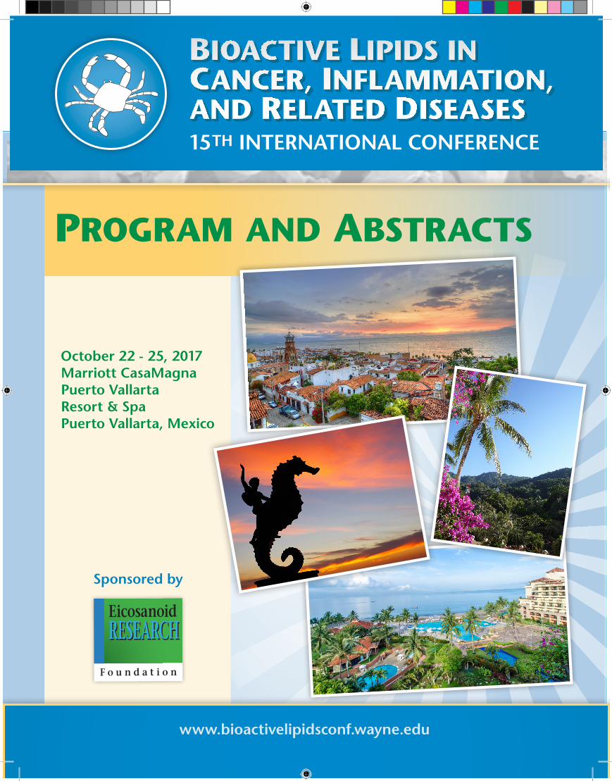
15TH INTERNATIONAL CONFERENCE
October 22 - 25, 2017Marriott CasaMagna Puerto Vallarta Resort & SpaPuerto Vallarta, Mexico
Sponsored by
www.bioactivelipidsconf.wayne.edu
Program and abstracts
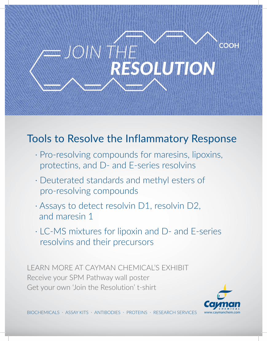
www.caymanchem.comBIOCHEMICALS · ASSAY KITS · ANTIBODIES · PROTEINS · RESEARCH SERVICES
· Pro-resolving compounds for maresins, lipoxins, protectins, and D- and E-series resolvins
· Deuterated standards and methyl esters of pro-resolving compounds
· Assays to detect resolvin D1, resolvin D2, and maresin 1
· LC-MS mixtures for lipoxin and D- and E-series resolvins and their precursors
Tools to Resolve the Inflammatory Response
LEARN MORE AT CAYMAN CHEMICAL’S EXHIBITReceive your SPM Pathway wall posterGet your own ‘Join the Resolution’ t-shirt
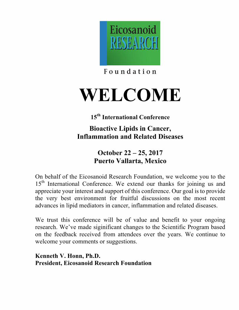
WELCOME
15th International Conference
Bioactive Lipids in Cancer, Inflammation and Related Diseases
October 22 – 25, 2017
Puerto Vallarta, Mexico
On behalf of the Eicosanoid Research Foundation, we welcome you to the 15th International Conference. We extend our thanks for joining us and appreciate your interest and support of this conference. Our goal is to provide the very best environment for fruitful discussions on the most recent advances in lipid mediators in cancer, inflammation and related diseases. We trust this conference will be of value and benefit to your ongoing research. We’ve made siginificant changes to the Scientific Program based on the feedback received from attendees over the years. We continue to welcome your comments or suggestions. Kenneth V. Honn, Ph.D. President, Eicosanoid Research Foundation
F o u n d a t i o n
RESEARCHEicosanoidRESEARCH

Training the Leading Researchers of Tomorrow by cultivating and supporting DISCOVERY, INNOVATION, COMMERCIALIZATION,and ECONOMIC GROWTH Since 1911
Untitled-3.indd 2 8/30/17 3:48 PM
OUR RESEARCH MAKES AN IMMEDIATE IMPACT.At Wayne State University, our research has no limits. Look at our new, $90 million Integrative Biosciences Center. IBio offers labs and clinical spaces that foster a team science approach, which is critical to researching health disparities affecting urban populations. Strategically located near our business incubator, TechTown, and just minutes from our School of Medicine campus, IBio allows researchers to share new discoveries and technologies directly with the community – impacting lives in Detroit, and beyond.
Discover more of our life-changing work at wayne.edu/action.
Research AD.indd 1 8/29/17 4:57 PM
ii
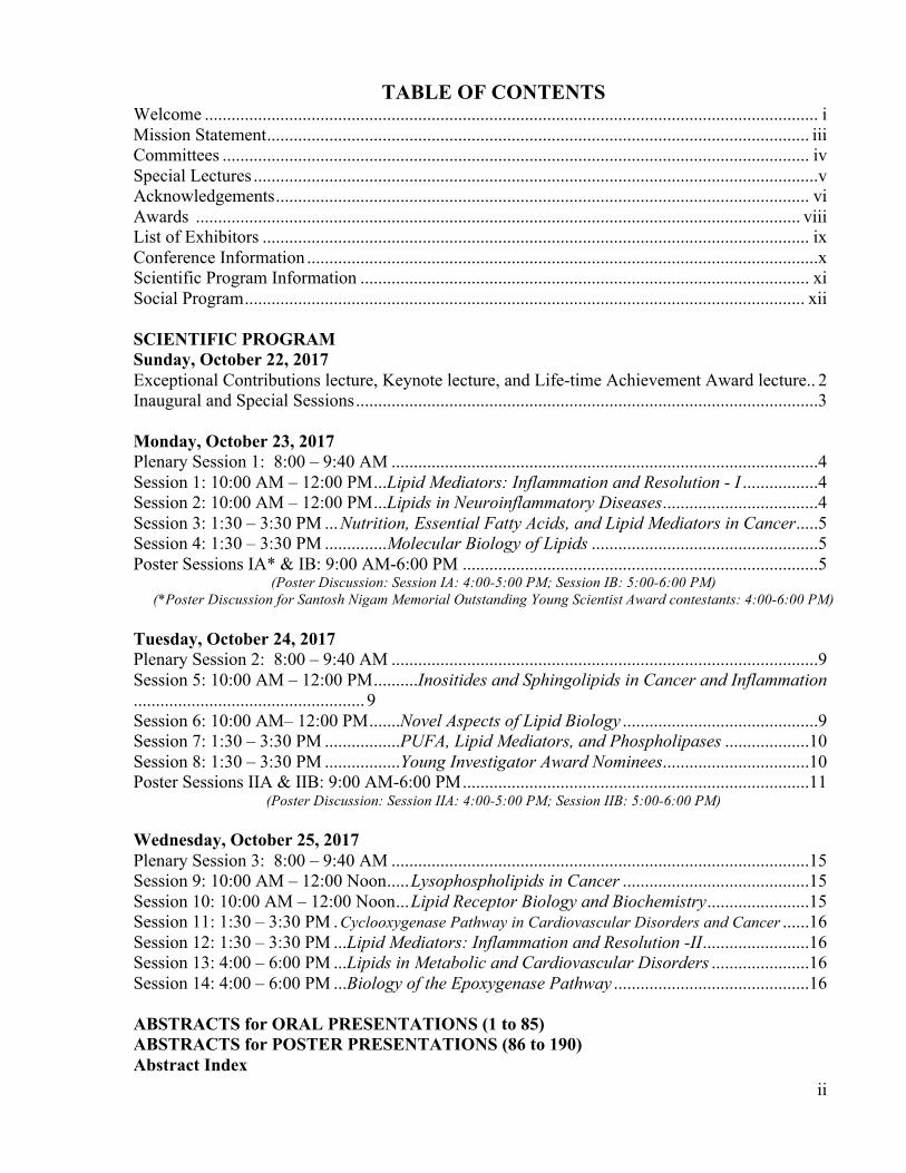
TABLE OF CONTENTS Welcome .......................................................................................................................................... i Mission Statement .......................................................................................................................... iii Committees .................................................................................................................................... iv Special Lectures ...............................................................................................................................v Acknowledgements ........................................................................................................................ vi Awards ........................................................................................................................................ viii List of Exhibitors ........................................................................................................................... ix Conference Information ...................................................................................................................x Scientific Program Information ..................................................................................................... xi Social Program .............................................................................................................................. xii SCIENTIFIC PROGRAM Sunday, October 22, 2017 Exceptional Contributions lecture, Keynote lecture, and Life-time Achievement Award lecture.. 2 Inaugural and Special Sessions ........................................................................................................3 Monday, October 23, 2017 Plenary Session 1: 8:00 – 9:40 AM ................................................................................................4 Session 1: 10:00 AM – 12:00 PM ...Lipid Mediators: Inflammation and Resolution - I .................4 Session 2: 10:00 AM – 12:00 PM ...Lipids in Neuroinflammatory Diseases ...................................4 Session 3: 1:30 – 3:30 PM ... Nutrition, Essential Fatty Acids, and Lipid Mediators in Cancer .....5 Session 4: 1:30 – 3:30 PM ..............Molecular Biology of Lipids ...................................................5 Poster Sessions IA* & IB: 9:00 AM-6:00 PM ................................................................................5
(Poster Discussion: Session IA: 4:00-5:00 PM; Session IB: 5:00-6:00 PM) (*Poster Discussion for Santosh Nigam Memorial Outstanding Young Scientist Award contestants: 4:00-6:00 PM)
Tuesday, October 24, 2017 Plenary Session 2: 8:00 – 9:40 AM ................................................................................................9 Session 5: 10:00 AM – 12:00 PM .......... Inositides and Sphingolipids in Cancer and Inflammation.................................................... 9 Session 6: 10:00 AM– 12:00 PM .......Novel Aspects of Lipid Biology ............................................9 Session 7: 1:30 – 3:30 PM .................PUFA, Lipid Mediators, and Phospholipases ...................10 Session 8: 1:30 – 3:30 PM .................Young Investigator Award Nominees .................................10 Poster Sessions IIA & IIB: 9:00 AM-6:00 PM ..............................................................................11
(Poster Discussion: Session IIA: 4:00-5:00 PM; Session IIB: 5:00-6:00 PM) Wednesday, October 25, 2017 Plenary Session 3: 8:00 – 9:40 AM ..............................................................................................15 Session 9: 10:00 AM – 12:00 Noon ..... Lysophospholipids in Cancer ..........................................15 Session 10: 10:00 AM – 12:00 Noon ... Lipid Receptor Biology and Biochemistry .......................15 Session 11: 1:30 – 3:30 PM . Cyclooxygenase Pathway in Cardiovascular Disorders and Cancer ......16 Session 12: 1:30 – 3:30 PM ...Lipid Mediators: Inflammation and Resolution -II ........................16 Session 13: 4:00 – 6:00 PM ...Lipids in Metabolic and Cardiovascular Disorders ......................16 Session 14: 4:00 – 6:00 PM ...Biology of the Epoxygenase Pathway ............................................16 ABSTRACTS for ORAL PRESENTATIONS (1 to 85) ABSTRACTS for POSTER PRESENTATIONS (86 to 190) Abstract Index
iii
MISSION STATEMENT The mission of the 15th International Conference on Bioactive Lipids in Cancer, Inflammation and Related Diseases is to provide a forum for senior and junior investigators to announce and examine their recent advancements in cutting-edge research on lipid mediators and their impact on human physiology and disease pathogenesis. The 15th International Conference will focus on new concepts in these areas that are of interest to clinicians and researchers. The Program includes presentations by leading experts in their respective fields. We are glad to offer Travel Awards to encourage the attendance of postdoctoral fellows to the conference for the third time in a row and have added Travel Awards to graduate students for the first time this year. In addition, a tradition is now established in this conference series to recognize excellence in bioactive lipids research. This will be the fourth time in this conference series, postdoctoral fellows and graduate students compete for the Santosh Nigam Memorial Outstanding Young Scientist Award and fifth time Life-time Achievement and Outstanding Achievement Awards are given to recognize the contributions of eminent scientists in the area of bioactive lipid research. Further, to encourage Young Investigators, this is the tenth meeting of the conference series in which three Awards will be presented for best abstracts on Cancer, Inflammation and Structure-Function. Thus, this meeting shall provide a superb opportunity to both new investigators and updates for those already active in this exciting area of biomedical research to ensure a memorable scientific meeting! Board of Directors, Eicosanoid Research Foundation Kenneth V. Honn, Ph.D. (President) Lawrence J. Marnett, Ph.D. (Secretary & Treasurer) Edward A. Dennis, Ph.D. (Member) Charles N. Serhan, Ph.D. (Member) Gabor J. Tigyi, M.D., Ph.D. (Member)
iv
COMMITTEES
Executive/Organizing Committee
Edward A. Dennis, Ph.D. Kenneth V. Honn, Ph.D.
Lawrence J. Marnett, Ph.D. Charles N. Serhan, Ph.D.
Gabor J. Tigyi, M.D., Ph.D.
Scientific Program Coordinator Krishna Rao Maddipati, Ph.D.
Conference Administrator Caryn Volpe
Sponsorship Committee Makoto Arita, Ph.D
Karsten Gronert, Ph.D. Michael Holinstat, Ph.D. Makoto Murakami, Ph.D. Dipak Panigrahy, M.D.
Richard P. Phipps, Ph.D. Takehiko Yokomizo, M.D., Ph.D.
v
SPECIAL LECTURES
EXCEPTIONAL CONTRIBUTIONS TO HUMAN PHYSIOLOGY AND TRANSLATIONAL MEDICINE AWARD
Jeffrey M. Drazen, M.D.
KEYNOTE ADDRESS Lewis C. Cantley, Ph.D.
LIFETIME ACHIEVEMENT AWARD
Charles N. Serhan, Ph.D.
PLENARY SPEAKERS Hiroyuki Arai, Ph.D.
Nicolas G. Bazan, M.D., Ph.D. Jesper Z. Haeggstrom, M.D., Ph.D.
Yusuf A. Hannun, M.D. Patricia Sime, M.D.
Michal L. Schwartzman, Ph.D.
INAUGURAL SESSION SPEAKERS Bruce D. Hammock, Ph.D. Jorge H. Capdevila, Ph.D.
Darryl C. Zeldin, M.D.
INVITED SPEAKERS Makoto Arita, Ph.D. Julian G. Cambronero, Ph.D. Michael S. Conte, M.D. Daniela Corda, Ph.D. Diana Escalante-Alcalde, Ph.D. Bruno Escalante, Ph.D. Miriam L. Greenberg, Ph.D. Karsten Gronert, Ph.D. Michael Holinstat, Ph.D. Jae Ho Kim, Ph.D. Krishna Rao Maddipati, Ph.D. Robert Martindale, M.D., Ph.D.
Makoto Murakami, Ph.D. Jerry Nadler, M.D. Dipak Panigrahy, M.D. Bernard Payrastre, Ph.D. Richard Phipps, Ph.D. Daniele Piomelli, Ph.D. Ambra Pozzi, Ph.D. Julia Saba, M.D., Ph.D. Takao Shimizu, M.D., Ph.D. Markus Wenk, Ph.D. Takehiko Yokomizo, M.D., Ph.D.
ACKNOWLEDGEMENTS Eicosanoid Research Foundation, along with the participants of this Conference gratefully
acknowledge the support provided by the following organizations:
Principal (Gold) Sponsors
Avanti Polar Lipids www.avantilipids.com
Cayman Chemical Company
www.caymanchem.com
Fresenius Kabi www.fresenius-kabi.com
Metagenics Institute
www.healthcareinstituteforclinicalnutrition.com
Ono Pharmaceutical Co., Ltd. www.ono.co.jp/eng
SCIEX
www.sciex.com
Solutex www.solutex.es
University of Rochester Medical Center
www.urmc.rochester.edu
Major (Silver) Sponsors
Elsevier www.elsevier.com
University of Tennessee Health Science Center
www.uthsc.edu
Wayne State University www.wayne.edu
vii
Contributors AbbVie
Ahee Jewellers
Ambiotis
ASPET
BBA - Molecular and Cell Biology of Lipids
Bruker
Chrono-Log Corporation
DIREF
EMBO
Inception Sciences
Journal of Lipid Research
Kowa
Marriott CasaMagna Puerto Vallarta Resort & Spa
Pleasant Hill Pediatrics
Prostaglandins and Other Lipid Mediators
Sato Heathcare Innovation
Springer Publishers
The Company of Biologists
Tiffany & Co.
Waters
ZENYAKU
ZONE Labs, Inc.
viii
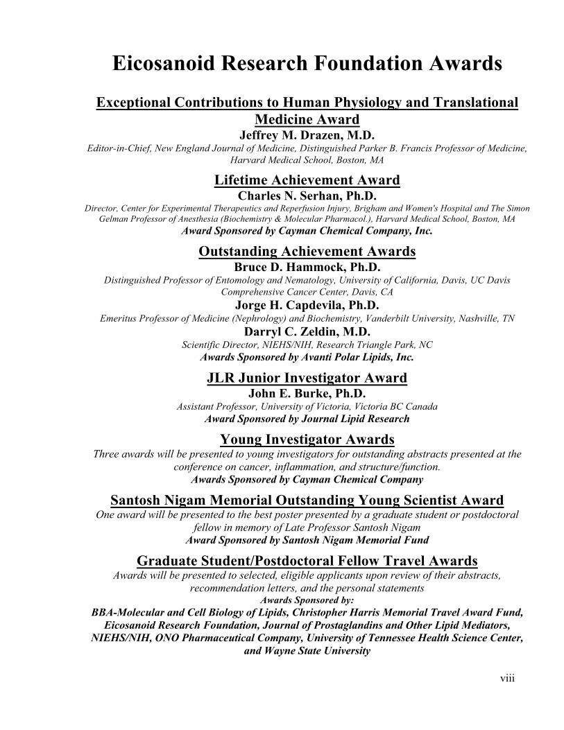
Eicosanoid Research Foundation Awards
Exceptional Contributions to Human Physiology and Translational Medicine Award
Jeffrey M. Drazen, M.D. Editor-in-Chief, New England Journal of Medicine, Distinguished Parker B. Francis Professor of Medicine,
Harvard Medical School, Boston, MA
Lifetime Achievement Award Charles N. Serhan, Ph.D.
Director, Center for Experimental Therapeutics and Reperfusion Injury, Brigham and Women's Hospital and The Simon Gelman Professor of Anesthesia (Biochemistry & Molecular Pharmacol.), Harvard Medical School, Boston, MA
Award Sponsored by Cayman Chemical Company, Inc.
Outstanding Achievement Awards Bruce D. Hammock, Ph.D.
Distinguished Professor of Entomology and Nematology, University of California, Davis, UC Davis Comprehensive Cancer Center, Davis, CA
Jorge H. Capdevila, Ph.D. Emeritus Professor of Medicine (Nephrology) and Biochemistry, Vanderbilt University, Nashville, TN
Darryl C. Zeldin, M.D. Scientific Director, NIEHS/NIH, Research Triangle Park, NC
Awards Sponsored by Avanti Polar Lipids, Inc.
JLR Junior Investigator Award John E. Burke, Ph.D.
Assistant Professor, University of Victoria, Victoria BC Canada Award Sponsored by Journal Lipid Research
Young Investigator Awards Three awards will be presented to young investigators for outstanding abstracts presented at the
conference on cancer, inflammation, and structure/function. Awards Sponsored by Cayman Chemical Company
Santosh Nigam Memorial Outstanding Young Scientist Award One award will be presented to the best poster presented by a graduate student or postdoctoral
fellow in memory of Late Professor Santosh Nigam Award Sponsored by Santosh Nigam Memorial Fund
Graduate Student/Postdoctoral Fellow Travel Awards Awards will be presented to selected, eligible applicants upon review of their abstracts,
recommendation letters, and the personal statements Awards Sponsored by:
BBA-Molecular and Cell Biology of Lipids, Christopher Harris Memorial Travel Award Fund, Eicosanoid Research Foundation, Journal of Prostaglandins and Other Lipid Mediators,
NIEHS/NIH, ONO Pharmaceutical Company, University of Tennessee Health Science Center, and Wayne State University

ix
LIST OF EXHIBITORS
Avanti Polar Lipids, Inc. www.avantilipids.com
BodyBio
www.bodybio.com
Bruker www.bruker.com
Cayman Chemical Company, Inc.
www.caymanchem.com
Chrono-Log Corporation www.chronolog.com
Metagenics Institute
www.healthcareinstituteforclinicalnutrition.com
SCIEX www.sciex.com
Solutex
www.solutex.es
Waters www.waters.com
ZONE Labs, Inc.
www.zonediet.com
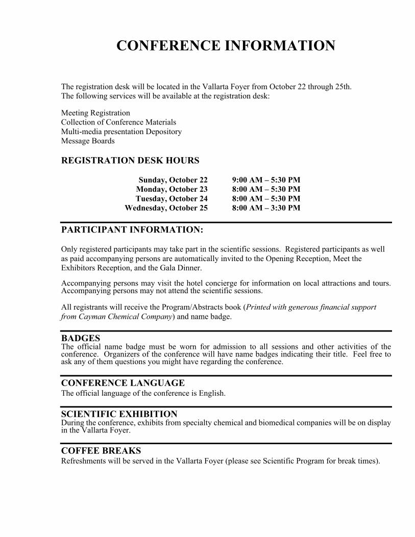
CONFERENCE INFORMATION The registration desk will be located in the Vallarta Foyer from October 22 through 25th. The following services will be available at the registration desk: Meeting Registration Collection of Conference Materials Multi-media presentation Depository Message Boards REGISTRATION DESK HOURS Sunday, October 22 9:00 AM – 5:30 PM Monday, October 23 8:00 AM – 5:30 PM Tuesday, October 24 8:00 AM – 5:30 PM Wednesday, October 25 8:00 AM – 3:30 PM PARTICIPANT INFORMATION: Only registered participants may take part in the scientific sessions. Registered participants as well as paid accompanying persons are automatically invited to the Opening Reception, Meet the Exhibitors Reception, and the Gala Dinner. Accompanying persons may visit the hotel concierge for information on local attractions and tours. Accompanying persons may not attend the scientific sessions. All registrants will receive the Program/Abstracts book (Printed with generous financial support from Cayman Chemical Company) and name badge. BADGES The official name badge must be worn for admission to all sessions and other activities of the conference. Organizers of the conference will have name badges indicating their title. Feel free to ask any of them questions you might have regarding the conference. CONFERENCE LANGUAGE The official language of the conference is English.
SCIENTIFIC EXHIBITION During the conference, exhibits from specialty chemical and biomedical companies will be on display in the Vallarta Foyer.
COFFEE BREAKS Refreshments will be served in the Vallarta Foyer (please see Scientific Program for break times).
xi
SCIENTIFIC PROGRAM INFORMATION
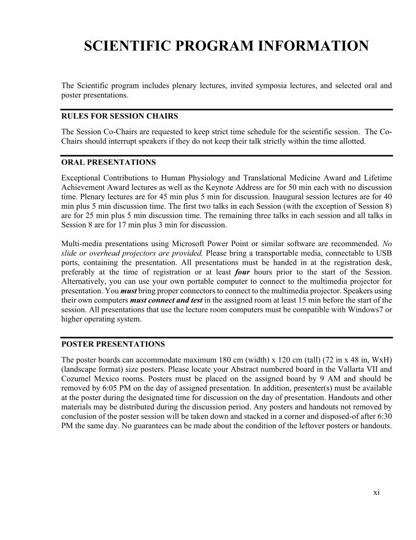
The Scientific program includes plenary lectures, invited symposia lectures, and selected oral and poster presentations. RULES FOR SESSION CHAIRS The Session Co-Chairs are requested to keep strict time schedule for the scientific session. The Co-Chairs should interrupt speakers if they do not keep their talk strictly within the time allotted. ORAL PRESENTATIONS Exceptional Contributions to Human Physiology and Translational Medicine Award and Lifetime Achievement Award lectures as well as the Keynote Address are for 50 min each with no discussion time. Plenary lectures are for 45 min plus 5 min for discussion. Inaugural session lectures are for 40 min plus 5 min discussion time. The first two talks in each Session (with the exception of Session 8) are for 25 min plus 5 min discussion time. The remaining three talks in each session and all talks in Session 8 are for 17 min plus 3 min for discussion. Multi-media presentations using Microsoft Power Point or similar software are recommended. No slide or overhead projectors are provided. Please bring a transportable media, connectable to USB ports, containing the presentation. All presentations must be handed in at the registration desk, preferably at the time of registration or at least four hours prior to the start of the Session. Alternatively, you can use your own portable computer to connect to the multimedia projector for presentation. You must bring proper connectors to connect to the multimedia projector. Speakers using their own computers must connect and test in the assigned room at least 15 min before the start of the session. All presentations that use the lecture room computers must be compatible with Windows7 or higher operating system.
POSTER PRESENTATIONS The poster boards can accommodate maximum 180 cm (width) x 120 cm (tall) (72 in x 48 in, WxH) (landscape format) size posters. Please locate your Abstract numbered board in the Vallarta VII and Cozumel Mexico rooms. Posters must be placed on the assigned board by 9 AM and should be removed by 6:05 PM on the day of assigned presentation. In addition, presenter(s) must be available at the poster during the designated time for discussion on the day of presentation. Handouts and other materials may be distributed during the discussion period. Any posters and handouts not removed by conclusion of the poster session will be taken down and stacked in a corner and disposed-of after 6:30 PM the same day. No guarantees can be made about the condition of the leftover posters or handouts.

xii
SOCIAL PROGRAM
Opening Reception SUNDAY, OCTOBER 22, 2017
Marriott CasaMagna Puerto Vallarta Resort & Spa Beach Front and Outdoor Garden Area
Cocktails and strolling dinner 7:00 - 9:00 PM
Relax, socialize and build camaraderie at the CasaMagna Marrriott's beach front surrounded by spectacular views of the Sierra Madre Mountains and Banderas Bay. The perfect setting for a memorable gatering and networking opportunity. The Opening Reception offers cocktails and a strolling dinner.
Meet the Exhibitors Reception MONDAY and TUESDAY, OCTOBER 23 & 24, 2017
Meet the Exhibitors Reception Vallarta Foyer
3:30 - 6:00 PM Familiarize yourself with the participating Exhibitors and their products and services. Connect with colleagues, make new contacts and enjoy complimentary beverages (wine & beer) in a relaxed environment.
Gala Dinner TUESDAY, OCTOBER 24, 2017
Club Regina Puerto Vallarta Outdoor Seaside Garden
(Bus Departs CasaMagna Marriott Hotel at 6:30 PM) Cocktails:
7:00 – 7:30 PM Dinner and Entertainment:
7:30 – 10:00 PM Dancing and Music:
until 11:00 PM (return transportation from Club Regina to CasaMagna: 10:15, 10:30, 10:45, & 11:00 PM)
The setting for the gala banquet at Club Regina Puerto Vallarta wraps elegance and tradition around relaxing ocean and marina views. Club Regina offers a seaside patio and tropical outdoor gardens under the umbrella of the Mexican sunset surrounded by the Sierra Madre mountains. Cocktails, dinner, rousing conversation, entertainment, dancing, along with panoramic views, the breaking rhythm of the surf…an inspiring event you won't want to miss.
Latin Dance Lessons SATURDAY, OCTOBER 21, 2017 (7 PM) and SUNDAY, OCTOBER 22, 2017 (10 AM)
Enjoy Latin Dance Lessons in preparation of a night of music and dance at the Gala Dinner. Participants must be signed up ahead of time for the lessons. Please see the registration desk for details.

An online, on-demand, resource for clinical nutrition and lifestyle medicine for all healthcare professionals
Welcome to the
Spotlights on Innovation
Spotlights on Innovation showcase speci�c therapeutic advances and innovations
All spotlights have:•Educational videos•Podcasts•Downloadable slides and research publications•Roundtable discussions•Video snippets of key opinion leader conversations

“Dedicated to Man’s fight against Disease and Pain”is our corporate philosophy.
Bringing new drugs to everyonein the world ‒ with our hopes
We will continue efforts for creating innovative drugs.For the sake of delivering drugs that truly benefit patients.
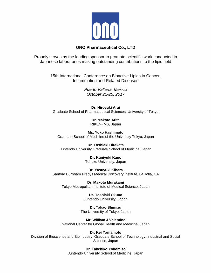
ONO Pharmaceutical Co., LTD
Proudly serves as the leading sponsor to promote scientific work conducted in Japanese laboratories making outstanding contributions to the lipid field
15th International Conference on Bioactive Lipids in Cancer, Inflammation and Related Diseases
Puerto Vallarta, Mexico October 22-25, 2017
Dr. Hiroyuki Arai Graduate School of Pharmaceutical Sciences, University of Tokyo
Dr. Makoto Arita
RIKEN-IMS, Japan
Ms. Yoko Hashimoto Graduate School of Medicine of the University Tokyo, Japan
Dr. Toshiaki Hirakata
Juntendo University Graduate School of Medicine, Japan
Dr. Kuniyuki Kano Tohoku University, Japan
Dr. Yasuyuki Kihara
Sanford Burnham Prebys Medical Discovery Institute, La Jolla, CA
Dr. Makoto Murakami Tokyo Metropolitan Institute of Medical Science, Japan
Dr. Toshiaki Okuno
Juntendo University, Japan
Dr. Takao Shimizu The University of Tokyo, Japan
Mr. William J Valentine
National Center for Global Health and Medicine, Japan
Dr. Kei Yamamoto Division of Bioscience and Bioindustry, Graduate School of Technology, Industrial and Social
Science, Japan
Dr. Takehiko Yokomizo Juntendo University School of Medicine, Japan

© 2017 Solutex Corp., LLC * These statements have not been evaluated by the Food and Drug Administration. This product is not intended to diagnose, treat, cure, or prevent any disease.
Solutex is a leading global supplier of unique,premium Omega-3 concentrates.
The most advanced combination of technologies in the Omega-3
industry allows us to satisfy demanding specifications, tailor
products to specific needs and offer unique solutions to create
differentiation in the marketplace.
Lipinova™ SPMs, a breakthrough innovation inimmuno-nutrition.
A unique, branded ingredient designed to balance the body’s natural
immune response during inflammation*, Specialized Pro-Resolving
Mediators (SPMs) are lipid mediators essential to promoting reso-
lution of the inflammatory process and the return to homeostasis*.
www.SolutexCorp.com USA | EUROPE | CANADA | ASIA
A revolutionary approach for treatingInflammatory-driven diseases.
Maresins Pharma, an emerging company based in Canada,
specializes in developing innovative drugs focused on “Resolution
Pharmacology” that are derived from maresins compounds.
Maresins compounds are among the few natural lipid modulators
that lead the resolution of inflammation.
Maresins Pharma utilizes maresins compounds as a platform for
developing formulas that address inflammation-driven diseases
with treatment needs.
1
SCIENTIFIC PROGRAM
15th International Conference on
Bioactive Lipids in Cancer, Inflammation, and Related
Diseases
October 22 – 25, 2017 Marriott CasaMagna Resort & Spa Puerto Vallarta, Mexico
2
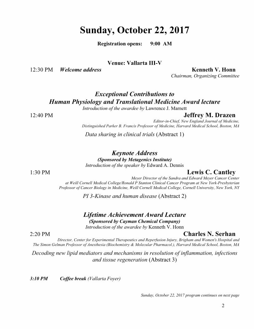
Sunday, October 22, 2017 Registration opens: 9:00 AM
Venue: Vallarta III-V
12:30 PM Welcome address Kenneth V. Honn Chairman, Organizing Committee
Exceptional Contributions to Human Physiology and Translational Medicine Award lecture
Introduction of the awardee by Lawrence J. Marnett 12:40 PM Jeffrey M. Drazen
Editor-in-Chief, New England Journal of Medicine, Distinguished Parker B. Francis Professor of Medicine, Harvard Medical School, Boston, MA
Data sharing in clinical trials (Abstract 1)
Keynote Address (Sponsored by Metagenics Institute)
Introduction of the speaker by Edward A. Dennis 1:30 PM Lewis C. Cantley
Meyer Director of the Sandra and Edward Meyer Cancer Center at Weill Cornell Medical College/Ronald P Stanton Clinical Cancer Program at New York-Presbyterian
Professor of Cancer Biology in Medicine, Weill Cornell Medical College, Cornell University, New York, NY
PI 3-Kinase and human disease (Abstract 2)
Lifetime Achievement Award Lecture (Sponsored by Cayman Chemical Company)
Introduction of the awardee by Kenneth V. Honn 2:20 PM Charles N. Serhan
Director, Center for Experimental Therapeutics and Reperfusion Injury, Brigham and Women's Hospital and The Simon Gelman Professor of Anesthesia (Biochemistry & Molecular Pharmacol.), Harvard Medical School, Boston, MA
Decoding new lipid mediators and mechanisms in resolution of inflammation, infections and tissue regeneration (Abstract 3)
3:10 PM Coffee break (Vallarta Foyer)
Sunday, October 22, 2017 program continues on next page
3
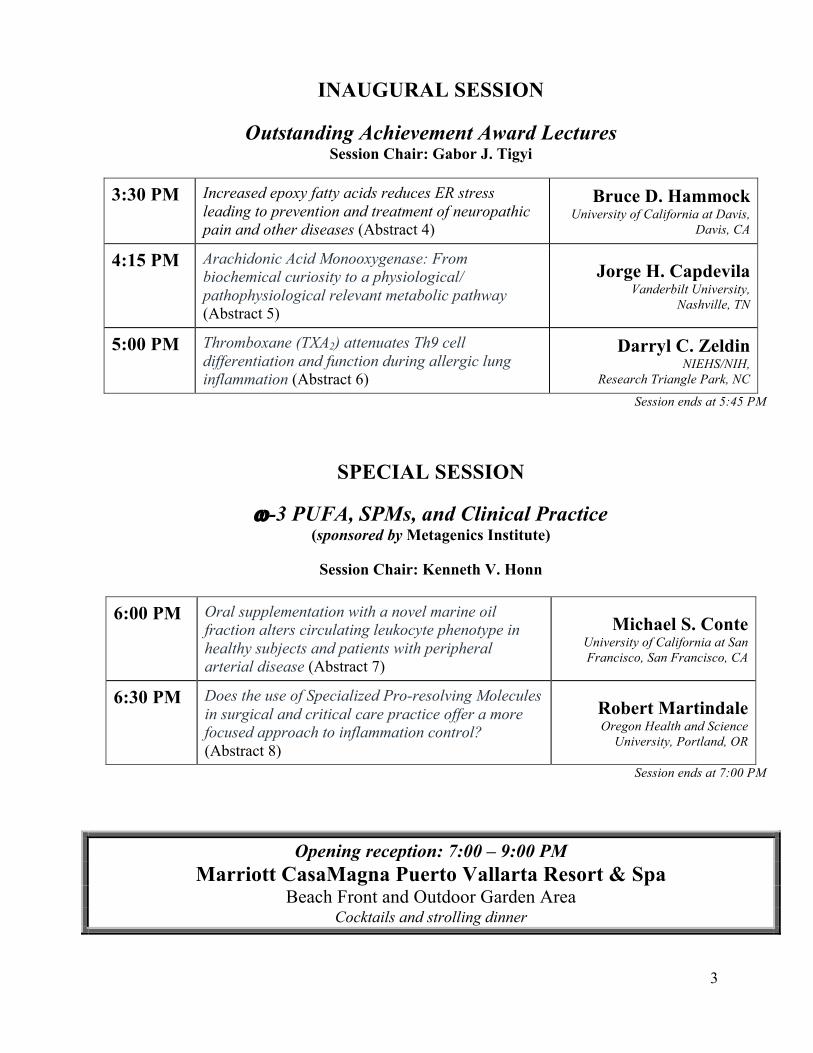
INAUGURAL SESSION
Outstanding Achievement Award Lectures Session Chair: Gabor J. Tigyi
3:30 PM Increased epoxy fatty acids reduces ER stress leading to prevention and treatment of neuropathic pain and other diseases (Abstract 4)
Bruce D. Hammock University of California at Davis,
Davis, CA
4:15 PM Arachidonic Acid Monooxygenase: From biochemical curiosity to a physiological/ pathophysiological relevant metabolic pathway (Abstract 5)
Jorge H. Capdevila Vanderbilt University,
Nashville, TN
5:00 PM Thromboxane (TXA2) attenuates Th9 cell differentiation and function during allergic lung inflammation (Abstract 6)
Darryl C. Zeldin NIEHS/NIH,
Research Triangle Park, NC
Session ends at 5:45 PM
SPECIAL SESSION
w-3 PUFA, SPMs, and Clinical Practice (sponsored by Metagenics Institute)
Session Chair: Kenneth V. Honn
6:00 PM Oral supplementation with a novel marine oil fraction alters circulating leukocyte phenotype in healthy subjects and patients with peripheral arterial disease (Abstract 7)
Michael S. Conte University of California at San Francisco, San Francisco, CA
6:30 PM Does the use of Specialized Pro-resolving Molecules in surgical and critical care practice offer a more focused approach to inflammation control? (Abstract 8)
Robert Martindale Oregon Health and Science
University, Portland, OR
Session ends at 7:00 PM
Opening reception: 7:00 – 9:00 PM Marriott CasaMagna Puerto Vallarta Resort & Spa
Beach Front and Outdoor Garden Area Cocktails and strolling dinner
4
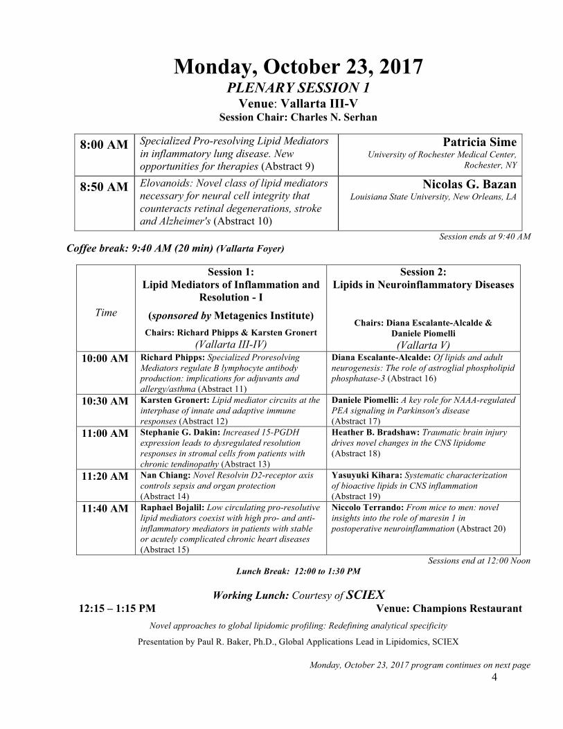
Monday, October 23, 2017 PLENARY SESSION 1
Venue: Vallarta III-V Session Chair: Charles N. Serhan
8:00 AM Specialized Pro-resolving Lipid Mediators in inflammatory lung disease. New opportunities for therapies (Abstract 9)
Patricia Sime University of Rochester Medical Center,
Rochester, NY
8:50 AM Elovanoids: Novel class of lipid mediators necessary for neural cell integrity that counteracts retinal degenerations, stroke and Alzheimer's (Abstract 10)
Nicolas G. Bazan Louisiana State University, New Orleans, LA
Session ends at 9:40 AM Coffee break: 9:40 AM (20 min) (Vallarta Foyer)
Time
Session 1: Lipid Mediators of Inflammation and
Resolution - I
(sponsored by Metagenics Institute) Chairs: Richard Phipps & Karsten Gronert
(Vallarta III-IV)
Session 2: Lipids in Neuroinflammatory Diseases
Chairs: Diana Escalante-Alcalde & Daniele Piomelli (Vallarta V)
10:00 AM Richard Phipps: Specialized Proresolving Mediators regulate B lymphocyte antibody production: implications for adjuvants and allergy/asthma (Abstract 11)
Diana Escalante-Alcalde: Of lipids and adult neurogenesis: The role of astroglial phospholipid phosphatase-3 (Abstract 16)
10:30 AM Karsten Gronert: Lipid mediator circuits at the interphase of innate and adaptive immune responses (Abstract 12)
Daniele Piomelli: A key role for NAAA-regulated PEA signaling in Parkinson's disease (Abstract 17)
11:00 AM Stephanie G. Dakin: Increased 15-PGDH expression leads to dysregulated resolution responses in stromal cells from patients with chronic tendinopathy (Abstract 13)
Heather B. Bradshaw: Traumatic brain injury drives novel changes in the CNS lipidome (Abstract 18)
11:20 AM Nan Chiang: Novel Resolvin D2-receptor axis controls sepsis and organ protection (Abstract 14)
Yasuyuki Kihara: Systematic characterization of bioactive lipids in CNS inflammation (Abstract 19)
11:40 AM Raphael Bojalil: Low circulating pro-resolutive lipid mediators coexist with high pro- and anti-inflammatory mediators in patients with stable or acutely complicated chronic heart diseases (Abstract 15)
Niccolo Terrando: From mice to men: novel insights into the role of maresin 1 in postoperative neuroinflammation (Abstract 20)
Sessions end at 12:00 Noon Lunch Break: 12:00 to 1:30 PM
Working Lunch: Courtesy of SCIEX
12:15 – 1:15 PM Venue: Champions Restaurant Novel approaches to global lipidomic profiling: Redefining analytical specificity
Presentation by Paul R. Baker, Ph.D., Global Applications Lead in Lipidomics, SCIEX
Monday, October 23, 2017 program continues on next page
5
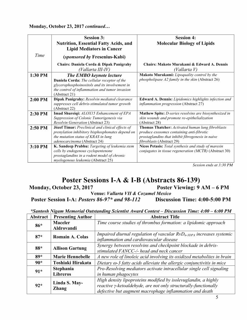
Monday, October 23, 2017 continued…
Time
Session 3: Nutrition, Essential Fatty Acids, and
Lipid Mediators in Cancer
(sponsored by Fresenius-Kabi) Chairs: Daniela Corda & Dipak Panigrahy
(Vallarta III-IV)
Session 4: Molecular Biology of Lipids
Chairs: Makoto Murakami & Edward A. Dennis
(Vallarta V) 1:30 PM The EMBO keynote lecture
Daniela Corda: The cellular receptor of the glycerophosphoinositols and its involvement in the control of inflammation and tumor invasion (Abstract 21)
Makoto Murakami: Lipoquality control by the phospholipase A2 family in the skin (Abstract 26)
2:00 PM Dipak Panigrahy: Resolvin mediated clearance suppresses cell debris-stimulated tumor growth (Abstract 22)
Edward A. Dennis: Lipidomics highlights infection and inflammation progression (Abstract 27)
2:30 PM Imad Shureiqi: ALOX15 Enhancement of EPA Suppression of Colonic Tumorigenesis via Resolvin Generation (Abstract 23)
Mathew Spite: D-series resolvins are biosynthesized in skin wounds and promote re-epithelialization (Abstract 28)
2:50 PM Józef Timar: Preclinical and clinical effects of prenylation inhibitory bisphosphonates depend on the mutation status of KRAS in lung adenocarcinoma (Abstract 24)
Thomas Thatcher: Activated human lung fibroblasts produce exosomes containing anti-fibrotic prostaglandins that inhibit fibrogenesis in naive fibroblasts (Abstract 29)
3:10 PM K. Sandeep Prabhu: Targeting of leukemia stem cells by endogenous cyclopentenone prostaglandins in a rodent model of chronic myelogenous leukemia (Abstract 25)
Nicos Petasis: Total synthesis and study of maresin conjugates in tissue regeneration (MCTR) (Abstract 30)
Session ends at 3:30 PM
Poster Sessions I-A & I-B (Abstracts 86-139)
Monday, October 23, 2017 Poster Viewing: 9 AM – 6 PM Venue: Vallarta VII & Cozumel Mexico
Poster Session I-A: Posters 86-97* and 98-112 Discussion Time: 4:00-5:00 PM
*Santosh Nigam Memorial Outstanding Scientist Award Contest – Discussion Time: 4:00 – 6:00 PM Abstract Presenting Author Abstract Title
86* Maceler Aldrovandi
Time course studies of thrombus formation: a lipidomic approach
87* Romain A. Colas Impaired diurnal regulation of vascular RvDn-3DPA increases systemic inflammation and cardiovascular disease
88* Allison Gartung Synergy between resolvins and checkpoint blockade in debris-stimulated FANCC-/- head and neck cancer
89* Marie Hennebelle A new role of linoleic acid involving its oxidized metabolites in brain 90* Toshiaki Hirakata Dietary w-3 fatty acids alleviate the allergic conjunctivitis in mice
91* Stephania Libreros
Pro-Resolving mediators activate intracellular single cell signaling in human phagocytes
92* Linda S. May-Zhang
High density lipoproteins modified by isolevuglandin, a highly reactive γ-ketoaldehyde, are not only structurally-functionally defective but augment macrophage inflammation and death
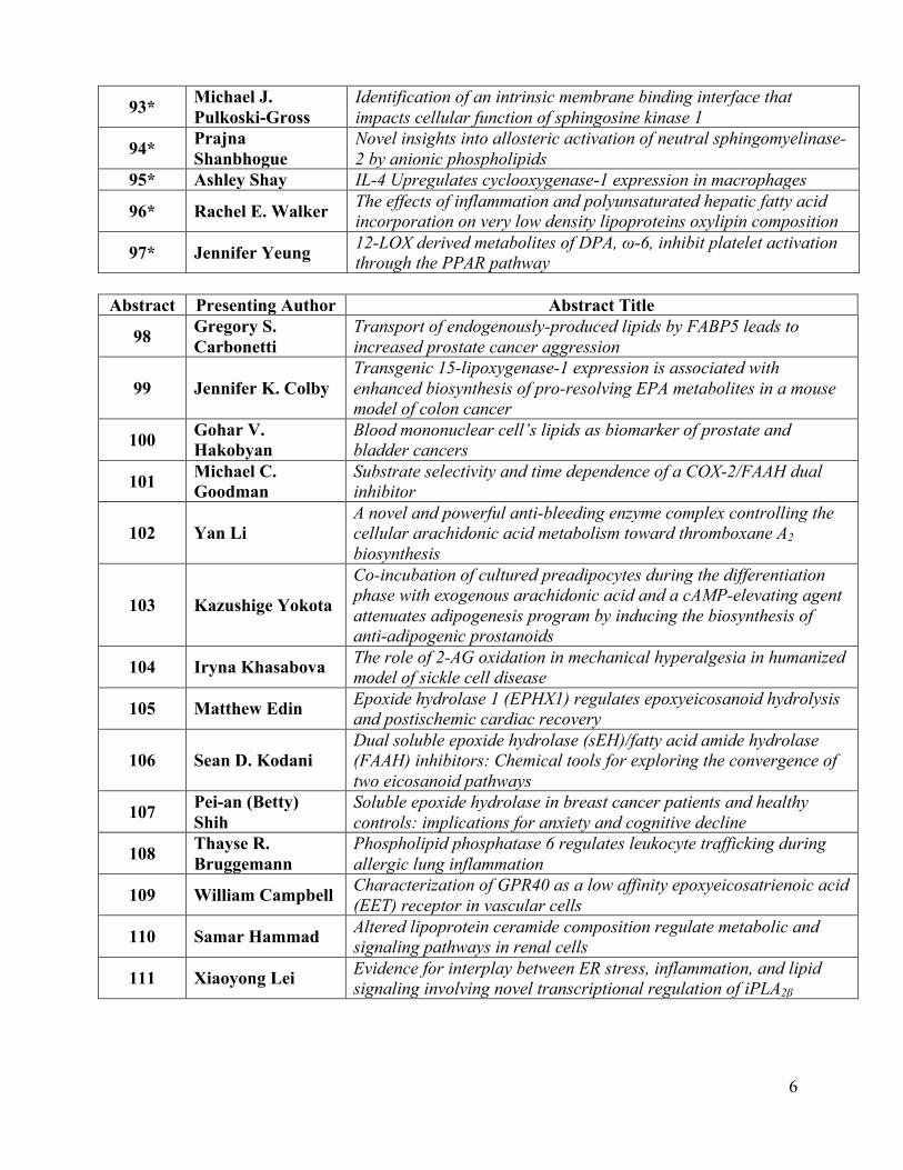
6
93* Michael J. Pulkoski-Gross
Identification of an intrinsic membrane binding interface that impacts cellular function of sphingosine kinase 1
94* Prajna Shanbhogue
Novel insights into allosteric activation of neutral sphingomyelinase-2 by anionic phospholipids
95* Ashley Shay IL-4 Upregulates cyclooxygenase-1 expression in macrophages
96* Rachel E. Walker The effects of inflammation and polyunsaturated hepatic fatty acid incorporation on very low density lipoproteins oxylipin composition
97* Jennifer Yeung 12-LOX derived metabolites of DPA, ω-6, inhibit platelet activation through the PPAR pathway
Abstract Presenting Author Abstract Title
98 Gregory S. Carbonetti
Transport of endogenously-produced lipids by FABP5 leads to increased prostate cancer aggression
99 Jennifer K. Colby Transgenic 15-lipoxygenase-1 expression is associated with enhanced biosynthesis of pro-resolving EPA metabolites in a mouse model of colon cancer
100 Gohar V. Hakobyan
Blood mononuclear cell’s lipids as biomarker of prostate and bladder cancers
101 Michael C. Goodman
Substrate selectivity and time dependence of a COX-2/FAAH dual inhibitor
102 Yan Li A novel and powerful anti-bleeding enzyme complex controlling the cellular arachidonic acid metabolism toward thromboxane A2 biosynthesis
103 Kazushige Yokota
Co-incubation of cultured preadipocytes during the differentiation phase with exogenous arachidonic acid and a cAMP-elevating agent attenuates adipogenesis program by inducing the biosynthesis of anti-adipogenic prostanoids
104 Iryna Khasabova The role of 2-AG oxidation in mechanical hyperalgesia in humanized model of sickle cell disease
105 Matthew Edin Epoxide hydrolase 1 (EPHX1) regulates epoxyeicosanoid hydrolysis and postischemic cardiac recovery
106 Sean D. Kodani Dual soluble epoxide hydrolase (sEH)/fatty acid amide hydrolase (FAAH) inhibitors: Chemical tools for exploring the convergence of two eicosanoid pathways
107 Pei-an (Betty) Shih
Soluble epoxide hydrolase in breast cancer patients and healthy controls: implications for anxiety and cognitive decline
108 Thayse R. Bruggemann
Phospholipid phosphatase 6 regulates leukocyte trafficking during allergic lung inflammation
109 William Campbell Characterization of GPR40 as a low affinity epoxyeicosatrienoic acid (EET) receptor in vascular cells
110 Samar Hammad Altered lipoprotein ceramide composition regulate metabolic and signaling pathways in renal cells
111 Xiaoyong Lei Evidence for interplay between ER stress, inflammation, and lipid signaling involving novel transcriptional regulation of iPLA2β
7
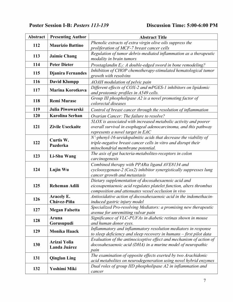
Poster Session I-B: Posters 113-139 Discussion Time: 5:00-6:00 PM Abstract Presenting Author Abstract Title
112 Maurizio Battino Phenolic extracts of extra virgin olive oils suppress the proliferation of MCF-7 breast cancer cells
113 Jaimie Chang Regulation of tumor debris-mediated inflammation as a therapeutic modality in brain tumors
114 Peter Dieter Prostaglandin E2: A double-edged sword in bone remodeling?
115 Djanira Fernandes Inhibition of CHOP chemotherapy-stimulated hematological tumor growth with resolvins
116 David Klumpp AOAH modulation of pelvic pain
117 Marina Korotkova Different effects of COX-2 and mPGES-1 inhibitors on lipidomic and proteomic profiles in A549 cells.
118 Remi Murase Group III phospholipase A2 is a novel promoting factor of colorectal diseases
119 Julia Piwowarski Control of breast cancer through the resolution of inflammation 120 Karolina Serhan Ovarian Cancer: The failure to resolve?
121 Zivile Useckaite 5LOX is associated with increased metabolic activity and poorer overall survival in esophageal adenocarcinoma, and this pathway represents a novel target in EAC
122 Curtis W. Pazderka
N’-phenyl-16-ureidopalmitic acids that decrease the viability of triple-negative breast cancer cells in vitro and disrupt their mitochondrial membrane potential.
123 Li-Shu Wang The axis of gut bacteria-metabolites-receptors in colon carcinogenesis
124 Lujin Wu Combined therapy with PPARa ligand AVE8134 and cyclooxygenase-2 (Cox2) inhibitor synergistically suppresses lung cancer growth and metastasis
125 Reheman Adili Dietary supplementation of docosahexaenoic acid and eicosapentaenoic acid regulates platelet function, alters thrombus composition and attenuates vessel occlusion in vivo
126 Aracely E. Chávez-Piña
Antioxidative action of docosahexaenoic acid in the indomethacin-induced gastric injury model
127 Megan Falsetta Specialized Pro-resolving Mediators: a promising new therapeutic avenue for unremitting vulvar pain
128 Aruna Gorusupudi
Significance of VLC-PUFAs in diabetic retinas shown in mouse and human donor eyes.
129 Monika Haack Inflammatory and inflammatory resolution mediators in response to sleep deficiency and sleep recovery in humans – first pilot data
130 Arizai Yolia Landa Juárez
Evaluation of the antinociceptive effect and mechanism of action of docosahexaenoic acid (DHA) in a murine model of neuropathic pain
131 Qinglan Ling The examination of opposite effects exerted by two Arachidonic acid metabolites on neurodegeneration using novel hybrid enzymes
132 Yoshimi Miki Dual roles of group IID phospholipase A2 in inflammation and cancer
8
Abstract Presenting Author Abstract Title
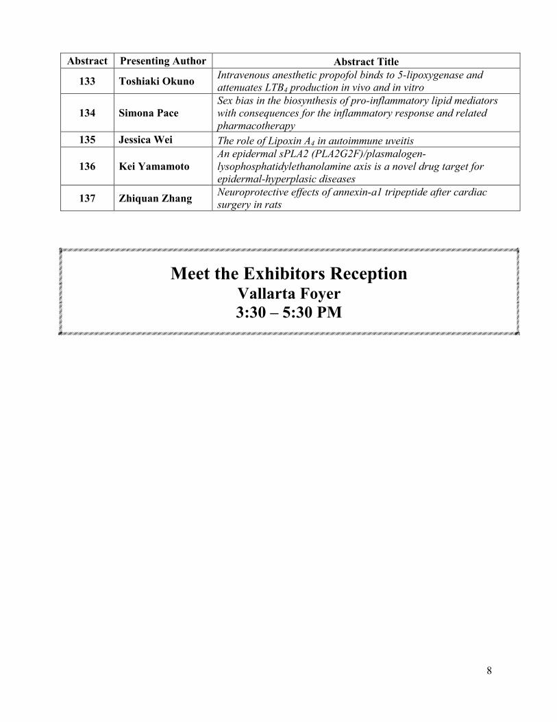
133 Toshiaki Okuno Intravenous anesthetic propofol binds to 5-lipoxygenase and attenuates LTB4 production in vivo and in vitro
134 Simona Pace Sex bias in the biosynthesis of pro-inflammatory lipid mediators with consequences for the inflammatory response and related pharmacotherapy
135 Jessica Wei The role of Lipoxin A4 in autoimmune uveitis
136 Kei Yamamoto An epidermal sPLA2 (PLA2G2F)/plasmalogen-lysophosphatidylethanolamine axis is a novel drug target for epidermal-hyperplasic diseases
137 Zhiquan Zhang Neuroprotective effects of annexin-a1 tripeptide after cardiac surgery in rats
Meet the Exhibitors Reception Vallarta Foyer 3:30 – 5:30 PM
9
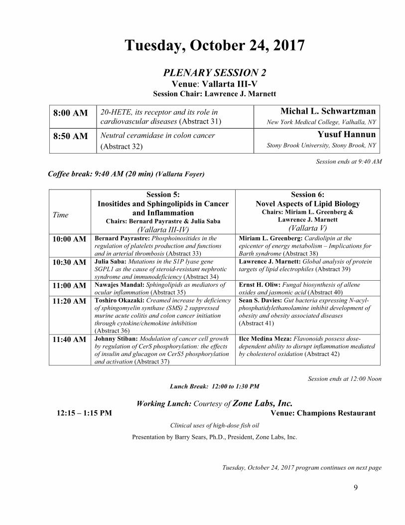
Tuesday, October 24, 2017
PLENARY SESSION 2 Venue: Vallarta III-V
Session Chair: Lawrence J. Marnett
8:00 AM 20-HETE, its receptor and its role in cardiovascular diseases (Abstract 31)
Michal L. Schwartzman New York Medical College, Valhalla, NY
8:50 AM Neutral ceramidase in colon cancer (Abstract 32)
Yusuf Hannun Stony Brook University, Stony Brook, NY
Session ends at 9:40 AM
Coffee break: 9:40 AM (20 min) (Vallarta Foyer)
Time
Session 5: Inositides and Sphingolipids in Cancer
and Inflammation Chairs: Bernard Payrastre & Julia Saba
(Vallarta III-IV)
Session 6: Novel Aspects of Lipid Biology
Chairs: Miriam L. Greenberg & Lawrence J. Marnett
(Vallarta V) 10:00 AM Bernard Payrastre: Phosphoinositides in the
regulation of platelets production and functions and in arterial thrombosis (Abstract 33)
Miriam L. Greenberg: Cardiolipin at the epicenter of energy metabolism – Implications for Barth syndrome (Abstract 38)
10:30 AM Julia Saba: Mutations in the S1P lyase gene SGPL1 as the cause of steroid-resistant nephrotic syndrome and immunodeficiency (Abstract 34)
Lawrence J. Marnett: Global analysis of protein targets of lipid electrophiles (Abstract 39)
11:00 AM Nawajes Mandal: Sphingolipids as mediators of ocular inflammation (Abstract 35)
Ernst H. Oliw: Fungal biosynthesis of allene oxides and jasmonic acid (Abstract 40)
11:20 AM Toshiro Okazaki: Creamed increase by deficiency of sphingomyelin synthase (SMS) 2 suppressed murine acute colitis and colon cancer initiation through cytokine/chemokine inhibition (Abstract 36)
Sean S. Davies: Gut bacteria expressing N-acyl-phosphatidylethanolamine inhibit development of obesity and obesity associated diseases (Abstract 41)
11:40 AM Johnny Stiban: Modulation of cancer cell growth by regulation of CerS phosphorylation: the effects of insulin and glucagon on CerS5 phosphorylation and activation (Abstract 37)
Ilce Medina Meza: Flavonoids possess dose-dependent ability to disrupt inflammation mediated by cholesterol oxidation (Abstract 42)
Session ends at 12:00 Noon
Lunch Break: 12:00 to 1:30 PM
Working Lunch: Courtesy of Zone Labs, Inc. 12:15 – 1:15 PM Venue: Champions Restaurant
Clinical uses of high-dose fish oil
Presentation by Barry Sears, Ph.D., President, Zone Labs, Inc.
Tuesday, October 24, 2017 program continues on next page
10
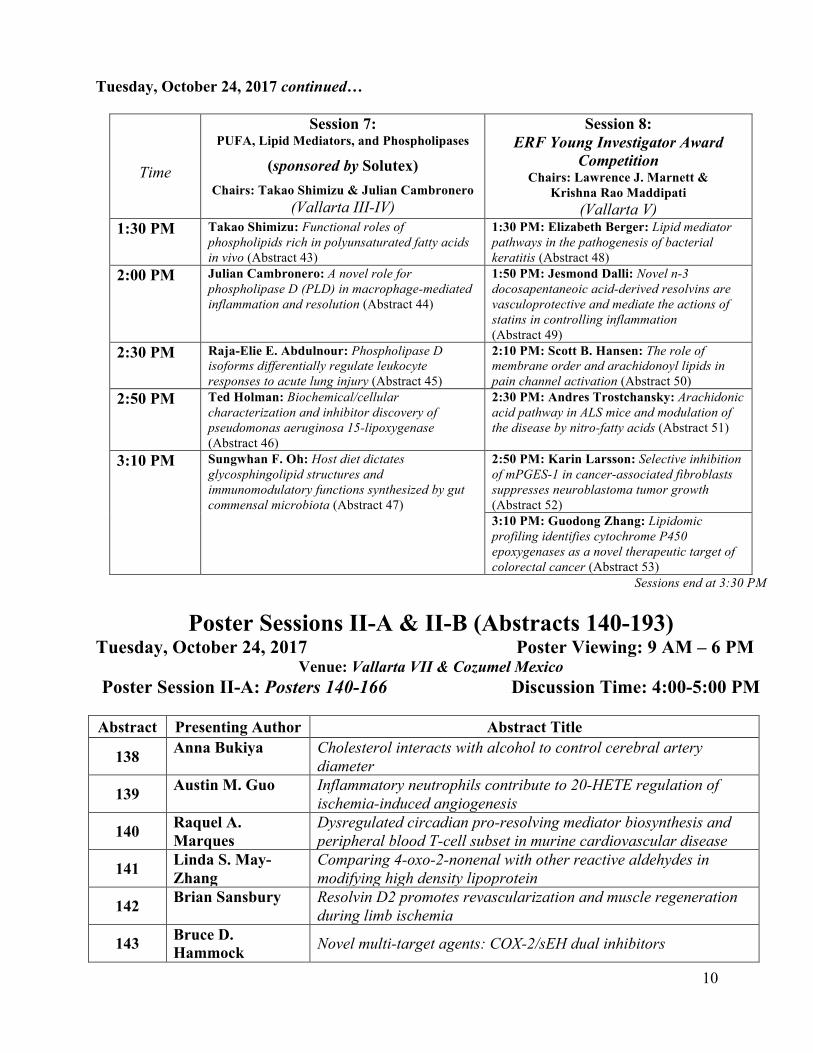
Tuesday, October 24, 2017 continued…
Time
Session 7: PUFA, Lipid Mediators, and Phospholipases
(sponsored by Solutex) Chairs: Takao Shimizu & Julian Cambronero
(Vallarta III-IV)
Session 8: ERF Young Investigator Award
Competition Chairs: Lawrence J. Marnett &
Krishna Rao Maddipati (Vallarta V)
1:30 PM Takao Shimizu: Functional roles of phospholipids rich in polyunsaturated fatty acids in vivo (Abstract 43)
1:30 PM: Elizabeth Berger: Lipid mediator pathways in the pathogenesis of bacterial keratitis (Abstract 48)
2:00 PM Julian Cambronero: A novel role for phospholipase D (PLD) in macrophage-mediated inflammation and resolution (Abstract 44)
1:50 PM: Jesmond Dalli: Novel n-3 docosapentaneoic acid-derived resolvins are vasculoprotective and mediate the actions of statins in controlling inflammation (Abstract 49)
2:30 PM Raja-Elie E. Abdulnour: Phospholipase D isoforms differentially regulate leukocyte responses to acute lung injury (Abstract 45)
2:10 PM: Scott B. Hansen: The role of membrane order and arachidonoyl lipids in pain channel activation (Abstract 50)
2:50 PM Ted Holman: Biochemical/cellular characterization and inhibitor discovery of pseudomonas aeruginosa 15-lipoxygenase (Abstract 46)
2:30 PM: Andres Trostchansky: Arachidonic acid pathway in ALS mice and modulation of the disease by nitro-fatty acids (Abstract 51)
3:10 PM Sungwhan F. Oh: Host diet dictates glycosphingolipid structures and immunomodulatory functions synthesized by gut commensal microbiota (Abstract 47)
2:50 PM: Karin Larsson: Selective inhibition of mPGES-1 in cancer-associated fibroblasts suppresses neuroblastoma tumor growth (Abstract 52) 3:10 PM: Guodong Zhang: Lipidomic profiling identifies cytochrome P450 epoxygenases as a novel therapeutic target of colorectal cancer (Abstract 53)
Sessions end at 3:30 PM
Poster Sessions II-A & II-B (Abstracts 140-193) Tuesday, October 24, 2017 Poster Viewing: 9 AM – 6 PM
Venue: Vallarta VII & Cozumel Mexico Poster Session II-A: Posters 140-166 Discussion Time: 4:00-5:00 PM
Abstract Presenting Author Abstract Title
138 Anna Bukiya Cholesterol interacts with alcohol to control cerebral artery diameter
139 Austin M. Guo Inflammatory neutrophils contribute to 20-HETE regulation of ischemia-induced angiogenesis
140 Raquel A. Marques
Dysregulated circadian pro-resolving mediator biosynthesis and peripheral blood T-cell subset in murine cardiovascular disease
141 Linda S. May-Zhang
Comparing 4-oxo-2-nonenal with other reactive aldehydes in modifying high density lipoprotein
142 Brian Sansbury Resolvin D2 promotes revascularization and muscle regeneration during limb ischemia
143 Bruce D. Hammock Novel multi-target agents: COX-2/sEH dual inhibitors
11
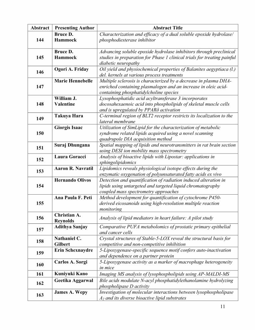
Abstract Presenting Author Abstract Title
144 Bruce D. Hammock
Characterization and efficacy of a dual soluble epoxide hydrolase/ phosphodiesterase inhibitor
145 Bruce D. Hammock
Advancing soluble epoxide hydrolase inhibitors through preclinical studies in preparation for Phase 1 clinical trials for treating painful diabetic neuropathy
146 Ogori A. Friday Oil yield and physiochemical properties of Balanites aegyptiaca (l.) del. kernels at various process treatments
147 Marie Hennebelle Multiple sclerosis is characterized by a decrease in plasma DHA-
enriched containing plasmalogen and an increase in oleic acid-containing phosphatidylcholine species
148 William J. Valentine
Lysophosphatidic acid acyltransferase 3 incorporates docosahexaenoic acid into phospholipids of skeletal muscle cells and is upregulated by PPARδ activation
149 Takuya Hara C-terminal region of BLT2 receptor restricts its localization to the lateral membrane
150 Giorgis Isaac Utilization of SimLipid for the characterization of metabolic
syndrome related lipids acquired using a novel scanning quadrupole DIA acquisition method
151 Suraj Dhungana Spatial mapping of lipids and neurotransmitters in rat brain section using DESI ion mobility mass spectrometry
152 Laura Goracci Analysis of bioactive lipids with Lipostar: applications in sphingolipidomics
153 Aaron R. Navratil Lipidomics reveals physiological isotope effects during the enzymatic oxygenation of polyunsaturated fatty acids ex vivo
154 Hernando Olivos Detection and quantification of radiation induced alteration in
lipids using untargeted and targeted liquid chromatography coupled mass spectrometry approaches
155 Ana Paula F. Peti Method development for quantification of cytochrome P450-
derived eicosanoids using high-resolution multiple reaction monitoring
156 Christian A. Reynolds Analysis of lipid mediators in heart failure: A pilot study
157 Adithya Sanjay Comparative PUFA metabolomics of prostatic primary epithelial and cancer cells
158 Nathaniel C. Gilbert
Crystal structures of Stable-5-LOX reveal the structural basis for competitive and non-competitive inhibition
159 Erin Schexnaydre 5-Lipoxygenase-specific sequence motif confers auto-inactivation and dependence on a partner protein
160 Carlos A. Sorgi 5-Lipoxygenase activity as a marker of macrophage heterogeneity in mice
161 Kuniyuki Kano Imaging MS analysis of lysophospholipids using AP-MALDI-MS
162 Geetika Aggarwal Bile acids modulate N-acyl phosphatidylethanolamine hydrolyzing phospholipase D activity
163 James A. Wepy Investigation of molecular interactions between lysophospholipase A2 and its diverse bioactive lipid substrates
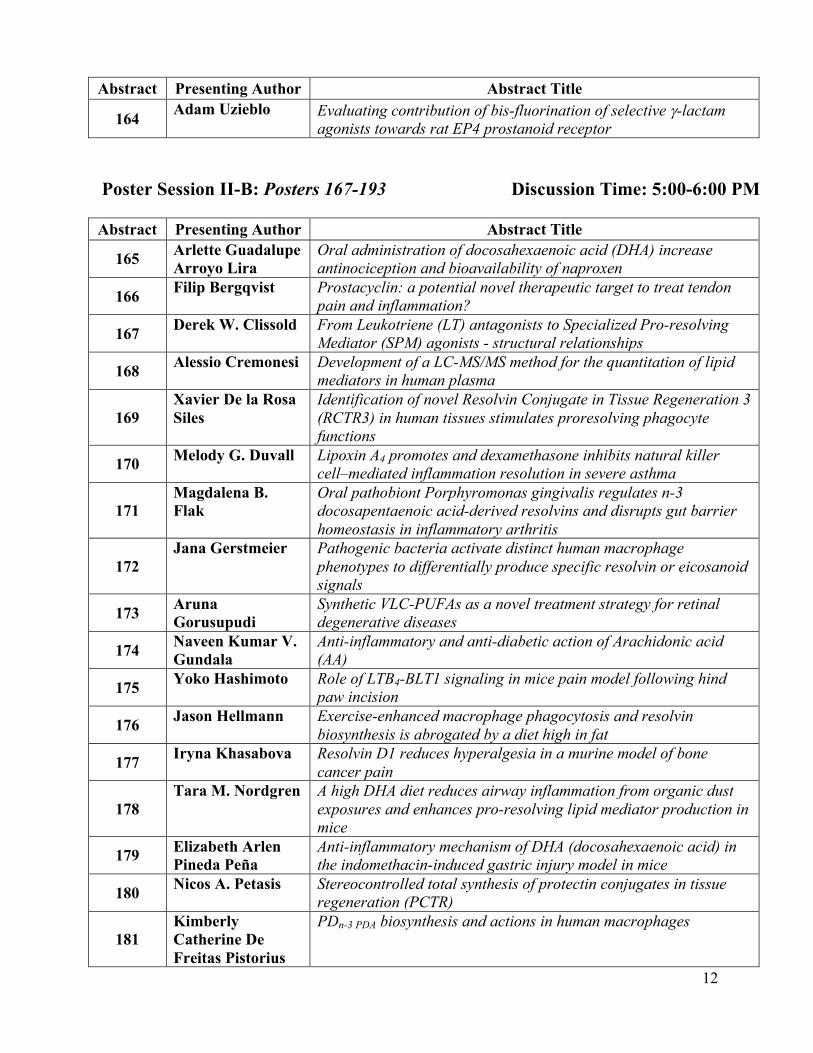
12
Abstract Presenting Author Abstract Title
164 Adam Uzieblo Evaluating contribution of bis-fluorination of selective g-lactam agonists towards rat EP4 prostanoid receptor
Poster Session II-B: Posters 167-193 Discussion Time: 5:00-6:00 PM Abstract Presenting Author Abstract Title
165 Arlette Guadalupe Arroyo Lira
Oral administration of docosahexaenoic acid (DHA) increase antinociception and bioavailability of naproxen
166 Filip Bergqvist Prostacyclin: a potential novel therapeutic target to treat tendon pain and inflammation?
167 Derek W. Clissold From Leukotriene (LT) antagonists to Specialized Pro-resolving Mediator (SPM) agonists - structural relationships
168 Alessio Cremonesi Development of a LC-MS/MS method for the quantitation of lipid mediators in human plasma
169 Xavier De la Rosa Siles
Identification of novel Resolvin Conjugate in Tissue Regeneration 3 (RCTR3) in human tissues stimulates proresolving phagocyte functions
170 Melody G. Duvall Lipoxin A4 promotes and dexamethasone inhibits natural killer cell–mediated inflammation resolution in severe asthma
171 Magdalena B. Flak
Oral pathobiont Porphyromonas gingivalis regulates n-3 docosapentaenoic acid-derived resolvins and disrupts gut barrier homeostasis in inflammatory arthritis
172 Jana Gerstmeier Pathogenic bacteria activate distinct human macrophage
phenotypes to differentially produce specific resolvin or eicosanoid signals
173 Aruna Gorusupudi
Synthetic VLC-PUFAs as a novel treatment strategy for retinal degenerative diseases
174 Naveen Kumar V. Gundala
Anti-inflammatory and anti-diabetic action of Arachidonic acid (AA)
175 Yoko Hashimoto Role of LTB4-BLT1 signaling in mice pain model following hind paw incision
176 Jason Hellmann Exercise-enhanced macrophage phagocytosis and resolvin biosynthesis is abrogated by a diet high in fat
177 Iryna Khasabova Resolvin D1 reduces hyperalgesia in a murine model of bone cancer pain
178 Tara M. Nordgren A high DHA diet reduces airway inflammation from organic dust
exposures and enhances pro-resolving lipid mediator production in mice
179 Elizabeth Arlen Pineda Peña
Anti-inflammatory mechanism of DHA (docosahexaenoic acid) in the indomethacin-induced gastric injury model in mice
180 Nicos A. Petasis Stereocontrolled total synthesis of protectin conjugates in tissue regeneration (PCTR)
181 Kimberly Catherine De Freitas Pistorius
PDn-3 PDA biosynthesis and actions in human macrophages
13
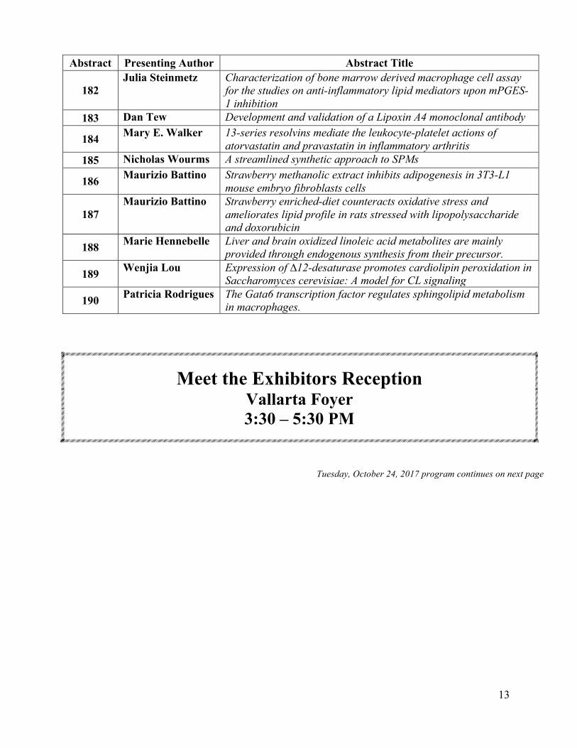
Abstract Presenting Author Abstract Title
182 Julia Steinmetz Characterization of bone marrow derived macrophage cell assay
for the studies on anti-inflammatory lipid mediators upon mPGES-1 inhibition
183 Dan Tew Development and validation of a Lipoxin A4 monoclonal antibody
184 Mary E. Walker 13-series resolvins mediate the leukocyte-platelet actions of atorvastatin and pravastatin in inflammatory arthritis
185 Nicholas Wourms A streamlined synthetic approach to SPMs
186 Maurizio Battino Strawberry methanolic extract inhibits adipogenesis in 3T3-L1 mouse embryo fibroblasts cells
187 Maurizio Battino Strawberry enriched-diet counteracts oxidative stress and
ameliorates lipid profile in rats stressed with lipopolysaccharide and doxorubicin
188 Marie Hennebelle Liver and brain oxidized linoleic acid metabolites are mainly provided through endogenous synthesis from their precursor.
189 Wenjia Lou Expression of ∆12-desaturase promotes cardiolipin peroxidation in Saccharomyces cerevisiae: A model for CL signaling
190 Patricia Rodrigues The Gata6 transcription factor regulates sphingolipid metabolism in macrophages.
Meet the Exhibitors Reception Vallarta Foyer 3:30 – 5:30 PM
Tuesday, October 24, 2017 program continues on next page
14
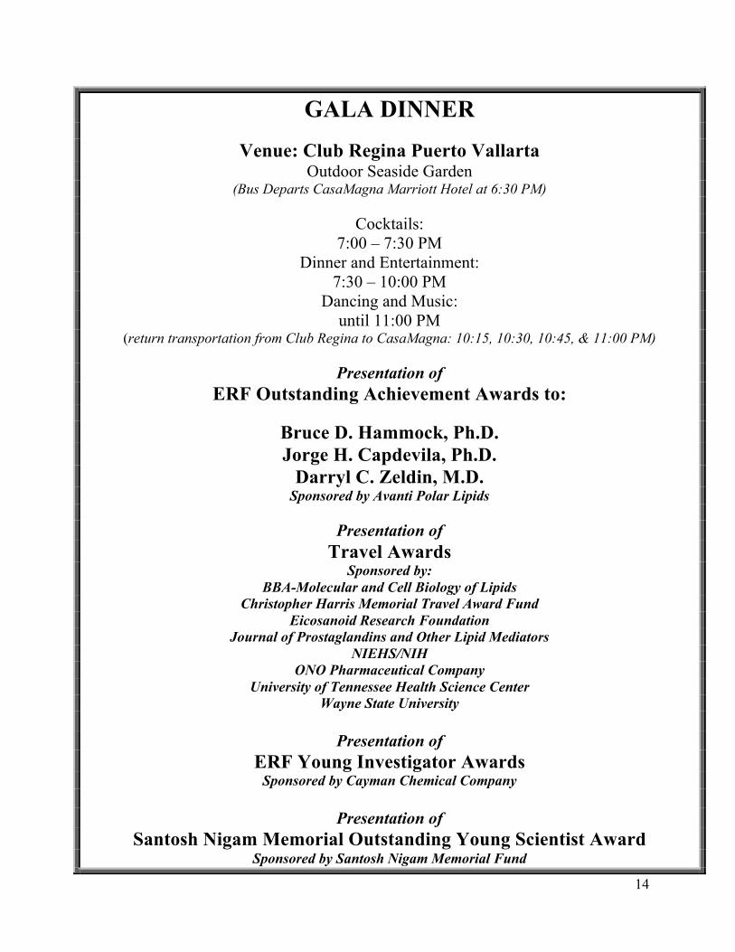
GALA DINNER
Venue: Club Regina Puerto Vallarta Outdoor Seaside Garden
(Bus Departs CasaMagna Marriott Hotel at 6:30 PM)
Cocktails: 7:00 – 7:30 PM
Dinner and Entertainment: 7:30 – 10:00 PM
Dancing and Music: until 11:00 PM
(return transportation from Club Regina to CasaMagna: 10:15, 10:30, 10:45, & 11:00 PM)
Presentation of ERF Outstanding Achievement Awards to:
Bruce D. Hammock, Ph.D. Jorge H. Capdevila, Ph.D.
Darryl C. Zeldin, M.D. Sponsored by Avanti Polar Lipids
Presentation of
Travel Awards Sponsored by:
BBA-Molecular and Cell Biology of Lipids Christopher Harris Memorial Travel Award Fund
Eicosanoid Research Foundation Journal of Prostaglandins and Other Lipid Mediators
NIEHS/NIH ONO Pharmaceutical Company
University of Tennessee Health Science Center Wayne State University
Presentation of
ERF Young Investigator Awards Sponsored by Cayman Chemical Company
Presentation of
Santosh Nigam Memorial Outstanding Young Scientist Award Sponsored by Santosh Nigam Memorial Fund
15
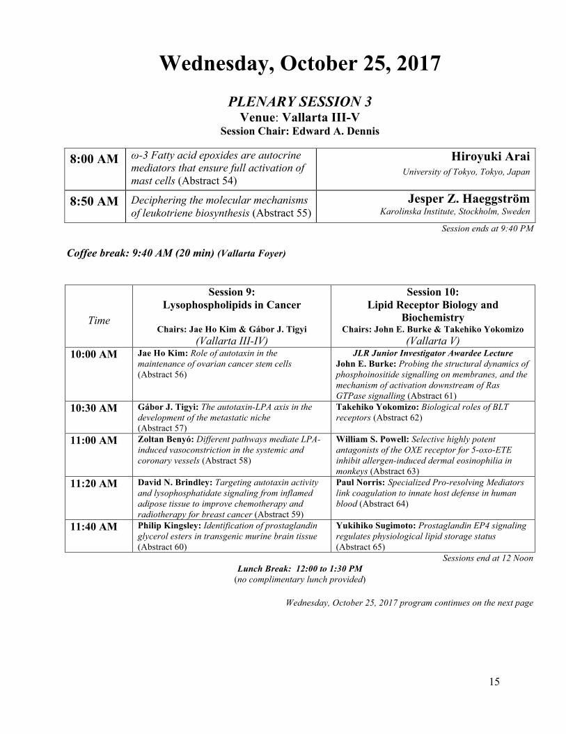
Wednesday, October 25, 2017
PLENARY SESSION 3 Venue: Vallarta III-V
Session Chair: Edward A. Dennis
8:00 AM ω-3 Fatty acid epoxides are autocrine mediators that ensure full activation of mast cells (Abstract 54)
Hiroyuki Arai University of Tokyo, Tokyo, Japan
8:50 AM Deciphering the molecular mechanisms of leukotriene biosynthesis (Abstract 55)
Jesper Z. Haeggström Karolinska Institute, Stockholm, Sweden
Session ends at 9:40 PM Coffee break: 9:40 AM (20 min) (Vallarta Foyer)
Time
Session 9: Lysophospholipids in Cancer
Chairs: Jae Ho Kim & Gábor J. Tigyi
(Vallarta III-IV)
Session 10: Lipid Receptor Biology and
Biochemistry Chairs: John E. Burke & Takehiko Yokomizo
(Vallarta V) 10:00 AM Jae Ho Kim: Role of autotaxin in the
maintenance of ovarian cancer stem cells (Abstract 56)
JLR Junior Investigator Awardee Lecture John E. Burke: Probing the structural dynamics of phosphoinositide signalling on membranes, and the mechanism of activation downstream of Ras GTPase signalling (Abstract 61)
10:30 AM Gábor J. Tigyi: The autotaxin-LPA axis in the development of the metastatic niche (Abstract 57)
Takehiko Yokomizo: Biological roles of BLT receptors (Abstract 62)
11:00 AM Zoltan Benyó: Different pathways mediate LPA-induced vasoconstriction in the systemic and coronary vessels (Abstract 58)
William S. Powell: Selective highly potent antagonists of the OXE receptor for 5-oxo-ETE inhibit allergen-induced dermal eosinophilia in monkeys (Abstract 63)
11:20 AM David N. Brindley: Targeting autotaxin activity and lysophosphatidate signaling from inflamed adipose tissue to improve chemotherapy and radiotherapy for breast cancer (Abstract 59)
Paul Norris: Specialized Pro-resolving Mediators link coagulation to innate host defense in human blood (Abstract 64)
11:40 AM Philip Kingsley: Identification of prostaglandin glycerol esters in transgenic murine brain tissue (Abstract 60)
Yukihiko Sugimoto: Prostaglandin EP4 signaling regulates physiological lipid storage status (Abstract 65)
Sessions end at 12 Noon Lunch Break: 12:00 to 1:30 PM
(no complimentary lunch provided)
Wednesday, October 25, 2017 program continues on the next page
16
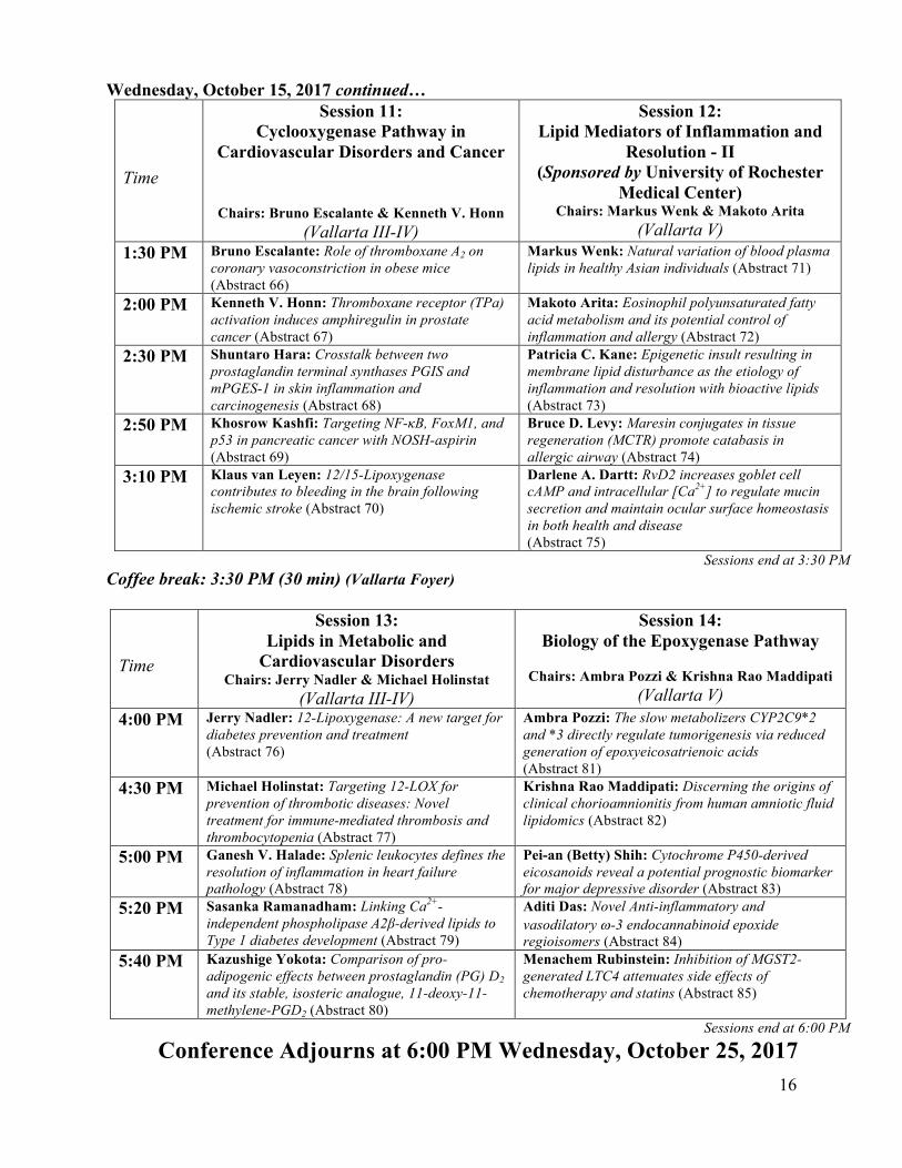
Wednesday, October 15, 2017 continued…
Time
Session 11: Cyclooxygenase Pathway in
Cardiovascular Disorders and Cancer
Chairs: Bruno Escalante & Kenneth V. Honn (Vallarta III-IV)
Session 12: Lipid Mediators of Inflammation and
Resolution - II (Sponsored by University of Rochester
Medical Center) Chairs: Markus Wenk & Makoto Arita
(Vallarta V) 1:30 PM Bruno Escalante: Role of thromboxane A2 on
coronary vasoconstriction in obese mice (Abstract 66)
Markus Wenk: Natural variation of blood plasma lipids in healthy Asian individuals (Abstract 71)
2:00 PM Kenneth V. Honn: Thromboxane receptor (TPa) activation induces amphiregulin in prostate cancer (Abstract 67)
Makoto Arita: Eosinophil polyunsaturated fatty acid metabolism and its potential control of inflammation and allergy (Abstract 72)
2:30 PM Shuntaro Hara: Crosstalk between two prostaglandin terminal synthases PGIS and mPGES-1 in skin inflammation and carcinogenesis (Abstract 68)
Patricia C. Kane: Epigenetic insult resulting in membrane lipid disturbance as the etiology of inflammation and resolution with bioactive lipids (Abstract 73)
2:50 PM Khosrow Kashfi: Targeting NF-κB, FoxM1, and p53 in pancreatic cancer with NOSH-aspirin (Abstract 69)
Bruce D. Levy: Maresin conjugates in tissue regeneration (MCTR) promote catabasis in allergic airway (Abstract 74)
3:10 PM Klaus van Leyen: 12/15-Lipoxygenase contributes to bleeding in the brain following ischemic stroke (Abstract 70)
Darlene A. Dartt: RvD2 increases goblet cell cAMP and intracellular [Ca2+] to regulate mucin secretion and maintain ocular surface homeostasis in both health and disease (Abstract 75)
Sessions end at 3:30 PM Coffee break: 3:30 PM (30 min) (Vallarta Foyer)
Time
Session 13: Lipids in Metabolic and
Cardiovascular Disorders Chairs: Jerry Nadler & Michael Holinstat
(Vallarta III-IV)
Session 14: Biology of the Epoxygenase Pathway
Chairs: Ambra Pozzi & Krishna Rao Maddipati
(Vallarta V) 4:00 PM Jerry Nadler: 12-Lipoxygenase: A new target for
diabetes prevention and treatment (Abstract 76)
Ambra Pozzi: The slow metabolizers CYP2C9*2 and *3 directly regulate tumorigenesis via reduced generation of epoxyeicosatrienoic acids (Abstract 81)
4:30 PM Michael Holinstat: Targeting 12-LOX for prevention of thrombotic diseases: Novel treatment for immune-mediated thrombosis and thrombocytopenia (Abstract 77)
Krishna Rao Maddipati: Discerning the origins of clinical chorioamnionitis from human amniotic fluid lipidomics (Abstract 82)
5:00 PM Ganesh V. Halade: Splenic leukocytes defines the resolution of inflammation in heart failure pathology (Abstract 78)
Pei-an (Betty) Shih: Cytochrome P450-derived eicosanoids reveal a potential prognostic biomarker for major depressive disorder (Abstract 83)
5:20 PM Sasanka Ramanadham: Linking Ca2+-independent phospholipase A2β-derived lipids to Type 1 diabetes development (Abstract 79)
Aditi Das: Novel Anti-inflammatory and vasodilatory w-3 endocannabinoid epoxide regioisomers (Abstract 84)
5:40 PM Kazushige Yokota: Comparison of pro-adipogenic effects between prostaglandin (PG) D2 and its stable, isosteric analogue, 11-deoxy-11-methylene-PGD2 (Abstract 80)
Menachem Rubinstein: Inhibition of MGST2-generated LTC4 attenuates side effects of chemotherapy and statins (Abstract 85)
Sessions end at 6:00 PM
Conference Adjourns at 6:00 PM Wednesday, October 25, 2017

The mission of the National Institute of Environmental Health Sciences
is to discover how the environment affects people in order to promote healthier lives.
National Institutes of Health U.S. Department of Health and Human Services
https://www.niehs.nih.gov

Stop breathing. Now read this ad.
Breath. We seldom think about it. Yet it is essential to each moment of life.At the Lung Biology and Disease Program, we have devoted nearly 50 years to helping human beings breathe easier. Whether you are researching an important lung-related topic, or considering pre- or post-doctoral education, we have the experts, facilities and experience to help.
■ One of the largest inhalation facilities in the U.S.■ The only NIH-funded respiratory pathogens disease center■ Extensive record of peer-reviewed publications■ Pre-clinical animal models of human disease■ More than 30 faculty focused on lung-related research■ Continuous NIH support since 1970
To learn more about our program, visit urmc.rochester.edu/lung-biology.
RESEARCH_LUNG_BIOLOGY_AD_8_5x11.indd 1 8/8/17 11:17 AM
15th International Conference on
Bioactive Lipids in Cancer, Inflammation, and Related Diseases
October 22-25, 2017
Puerto Vallarta, Mexico
Abstracts for
Oral Presentations

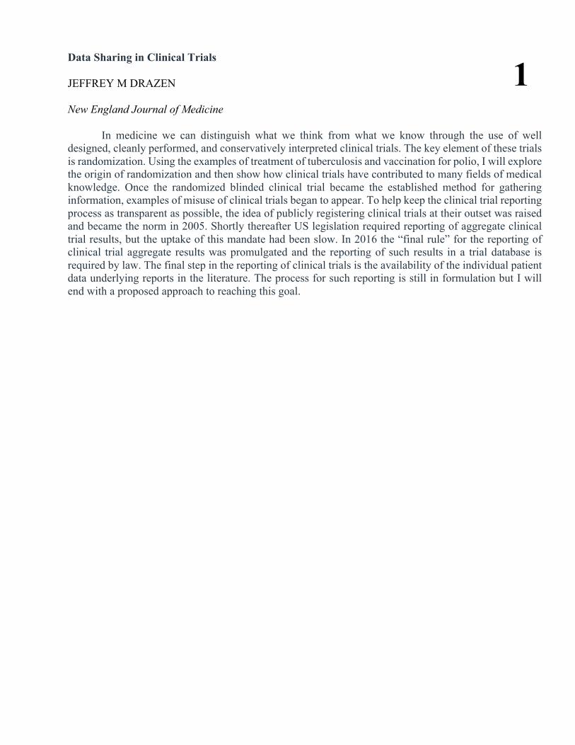
Data Sharing in Clinical Trials JEFFREY M DRAZEN New England Journal of Medicine
In medicine we can distinguish what we think from what we know through the use of well designed, cleanly performed, and conservatively interpreted clinical trials. The key element of these trials is randomization. Using the examples of treatment of tuberculosis and vaccination for polio, I will explore the origin of randomization and then show how clinical trials have contributed to many fields of medical knowledge. Once the randomized blinded clinical trial became the established method for gathering information, examples of misuse of clinical trials began to appear. To help keep the clinical trial reporting process as transparent as possible, the idea of publicly registering clinical trials at their outset was raised and became the norm in 2005. Shortly thereafter US legislation required reporting of aggregate clinical trial results, but the uptake of this mandate had been slow. In 2016 the “final rule” for the reporting of clinical trial aggregate results was promulgated and the reporting of such results in a trial database is required by law. The final step in the reporting of clinical trials is the availability of the individual patient data underlying reports in the literature. The process for such reporting is still in formulation but I will end with a proposed approach to reaching this goal.
1
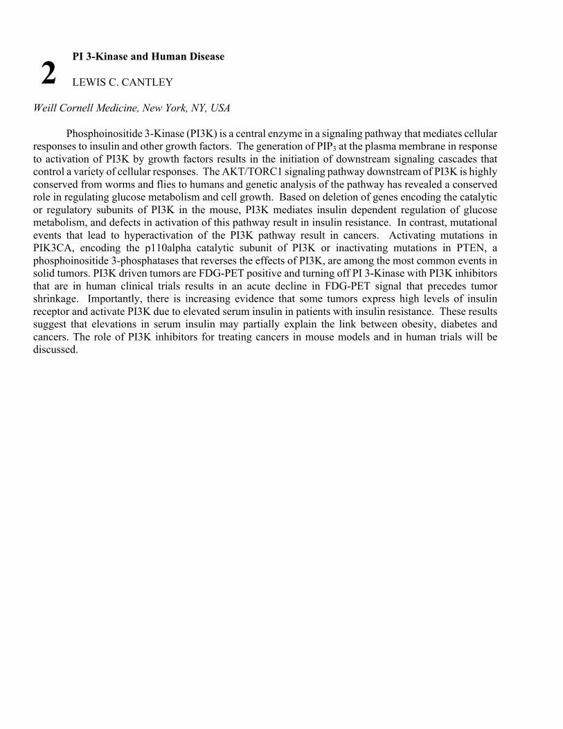
PI 3-Kinase and Human Disease LEWIS C. CANTLEY
Weill Cornell Medicine, New York, NY, USA Phosphoinositide 3-Kinase (PI3K) is a central enzyme in a signaling pathway that mediates cellular responses to insulin and other growth factors. The generation of PIP3 at the plasma membrane in response to activation of PI3K by growth factors results in the initiation of downstream signaling cascades that control a variety of cellular responses. The AKT/TORC1 signaling pathway downstream of PI3K is highly conserved from worms and flies to humans and genetic analysis of the pathway has revealed a conserved role in regulating glucose metabolism and cell growth. Based on deletion of genes encoding the catalytic or regulatory subunits of PI3K in the mouse, PI3K mediates insulin dependent regulation of glucose metabolism, and defects in activation of this pathway result in insulin resistance. In contrast, mutational events that lead to hyperactivation of the PI3K pathway result in cancers. Activating mutations in PIK3CA, encoding the p110alpha catalytic subunit of PI3K or inactivating mutations in PTEN, a phosphoinositide 3-phosphatases that reverses the effects of PI3K, are among the most common events in solid tumors. PI3K driven tumors are FDG-PET positive and turning off PI 3-Kinase with PI3K inhibitors that are in human clinical trials results in an acute decline in FDG-PET signal that precedes tumor shrinkage. Importantly, there is increasing evidence that some tumors express high levels of insulin receptor and activate PI3K due to elevated serum insulin in patients with insulin resistance. These results suggest that elevations in serum insulin may partially explain the link between obesity, diabetes and cancers. The role of PI3K inhibitors for treating cancers in mouse models and in human trials will be discussed.
2
Decoding New Lipid Mediators and Mechanisms in Resolution of Inflammation, Infections and Tissue Regeneration CHARLES N. SERHAN Center for Experimental Therapeutics and Reperfusion Injury, Department of Anesthesiology, Perioperative and Pain Medicine, Brigham and Women’s Hospital and Harvard Medical School
Uncontrolled Inflammation is involved in many widely occurring diseases. Using a systems approach with self-limited inflammatory infectious exudates to map tissues, cell traffic and identification of chemical mediators, we identified three new structurally distinct families of potent n-3 fatty acid-derived mediators (EPA, DPA and DHA) that each play an active role in resolving inflammation and infection. These bioactive metabolomes and mediator superfamily are collectively termed the specialized pro-resolving mediators (SPM) with complete structural elucidation of the new potent pro-resolving actions as well as their biosynthetic routes of production1. This presentation shall focus on our recent advances. We use LC-MS-MS mediator-metabololipidomics to profile SPM in human tissues e.g. human blood (2), breast milk (4), brain and uncovered new pathways that also stimulate tissue regeneration and bacterial clearance (3-5). Identification of SPM and novel mediators during inflammation-resolution first demonstrated that resolution is an active programmed process also of interest in aging 5 as well as many widely occurring diseases worldwide that are characterized by prolonged uncontrolled inflammation and infection1. Tissue regeneration and its relationship to resolution of infectious inflammation in model organisms permitted the structural elucidation of new mediators linking these vital processes.
SPM together with their receptors, and their biosynthesis pathways are new potent mediators of vital processes that have opened the potential for resolution physiology as well as resolution pharmacology directed to novel therapeutics that are resolution agonists and potentially 21st century treatments for a wide range of diseases.
For some recent examples please see; 1. Serhan, C.N. 2014. Pro-resolving lipid mediators are leads for resolution physiology. Nature 510:92-
101. 2. Colas, R.A., et al 2014. Identification and signature profiles for pro-resolving and inflammatory lipid
mediators in human tissue. Am J Physiol Cell Physiol 307:C39-54. 3. Dalli, J., Chiang, N. and Serhan, C.N. 2015. Elucidation of novel 13-series resolvins that increase
with atorvastatin and clear infections. Nat. Med. 21:1071-1075. 4. Dalli, J. et al. 2017 and 2016 Identification of sulfido-conjugated mediators that promote resolution
of infection and organ protection. PNAS 111:E4753-4761. 5. Dalli et al. Immunity 2017 46: 1-14. 6. Arnardottir HH, et al. Aging delays resolution of acute inflammation in mice. J Immunol. 2014
I thank the members of the Serhan lab and center past and present for their contributions to this presentation as well as our collaborators. Also, support from the National Institutes of Health (Grant Numbers R01GM38765, P01GM095467, and R01DE025020).
3
Increased Epoxy Fatty Acids Reduces ER Stress Leading to Prevention and Treatment of Neuropathic Pain and Other Diseases BRUCE D. HAMMOCK
Distinguished Professor of Entomology and Nematology, University of California, Davis, UC Davis Comprehensive Cancer Center, Davis, CA Epoxyfatty acids (EpFA) produced by cytochrome P450 enzymes are rapidly degraded by the soluble epoxide hydrolase (sEH) to polar diols with reduced biological activities. The omega-6 (EETs) and the omega-3 fatty acid (EEQs and EDPs) epoxides are the best studied. Their titer depends upon substrate concentration, rate of biosynthesis by P450s, and degradation. Plasma levels of these mediators appear to be useful biomarkers, and increasing the titers of EpFA has a variety of largely beneficial biological effects. EpFA move disease states towards homeostasis, and they appear largely anti-inflammatory and pro resolving chemical mediators. An enigma of their broad action on many pathologies can possibly be explained by EpFA reducing mitochondrial dysfunction and the resulting increase in ROS and by a reduction in the pathological endoplasmic reticulum stress response. This mitochondrial – ER stress axis appears involved in reducing severity of a variety of diseases including pathological fibrosis, liver and kidney failure, hypertension, diabetes, atrial fibrillation, inflammation of the gut and pancreas, and other disorders. Recent data indicate that EpFA will reduce and reverse pathological bone loss associated for example with periodontal disease and in the CNS reduce neuroinflammation, depression, and possibly cognitive impairment. sEHI are more powerful than NSAIDs with many inflammatory diseases including inflammatory pain. They are far more potent than gabapentin and pregabalin on nerve constriction and diabetes driven pain as well as in veterinary patients including neuropathic pain in horses. Thus, for acute and chronic pain they offer a non-NSAID, non-opiate, non-addictive intervention. sEHI synergize with and block the GI and CV side effects of NSAIDs while providing effective analgesics without problems with cognition, coordination and addiction. Genetic or pharmacological increase in EpFA production (for example with omeprazole) synergizes with sEHI. Another observation that appears to hold across biologies ranging from cardiac hypertrophy to fibrosis is that an ω-3 enriched and/or ω-6 depleted diet will usually enhance the potency of sEH inhibitors. A sEHI is being moved to the clinic for COPD and another for diabetic pain in man, and one sEHI is being developed for companion animals. These compounds show good PK-ADME, picomolar potency and a good safety profile. On a broader note our field now offers a variety of powerful probes regulating the biosynthesis, degradation, and mimicking the action of EpFA. In addition, there is hope that our field will develop a variety of ways to prevent and treat disease through life style, dietary and pharmaceutical intervention.
4
Arachidonic Acid Monooxygenase: From biochemical curiosity to a physiological/ pathophysiological relevant metabolic pathway JORGE H CAPDEVILA1, John R Falck2 1Department of Medicine, Vanderbilt University Medical School, Nashville, TN 37232; 2Department of Biochemistry, UT Southwestern Medical Center, Dallas, TX 75390
Studies of the Cytochrome P450 (P450) Arachidonic Acid (AA) Monooxygenase were initiated in 1981 with reports of roles for P450 enzymes in the epoxidation and/or ω-hydroxylation of the fatty acid to epoxyeicosatrienoic (EETs)(CYP2C epoxygenases) or 20-hydroxyeicosatetraenoic acids (20-HETE)(CYP4A 20-HETE synthases), respectively. The chemical synthesis of all four EETs and 20-HETE made possible: a) the development of techniques for their analysis and quantification in biological samples, and b) their initial in vitro functional evaluation as inhibitors of Na+ transport in kidney collecting ducts, vasoactive lipids, mediators of hormonal signaling, and regulators of protein kinases and Na+, K+, and Ca++ cellular transport (J Lipid Res. 2000; 41:163-181). These results established the P450 AA monooxygenase as a formal metabolic pathway and stimulated studies of its biological significance and biomedical relevance. The subsequent characterization of monogenic models of Cyp4a14, Cyp4a10, and Cyp2c44 dysfunction identified the participation of these enzymes in the pathophysiology of androgen (Cyp4a14) (Proc Natl Acad Sci USA. 2001; 98:5211-5216) and salt sensitive hypertension (Cyp4a10 and Cyp2c44) (J Clin Invest. 2006; 46:1696-1702, and J Biol Chem. 2014; 289:4377-4386), and physiological roles for 20-HETE in renal vasoconstriction, and for 11,12-EET in controlling the activity of the epithelial sodium channel (ENaC) and Na+ reabsorption in the distal nephron. The demonstration that ERK1/2 activation is constitutively impaired in Cyp2c44(-/-) mice and that the inhibitory, ERK1/2-catalyzed, phosphorylation of the renal epithelia sodium channel (ENaC) was also impaired (J Biol Chem, 2013; 288:5223-5231) identified a common scientific platform that could explain the seemingly unrelated biological activities attributed to the epoxygenase metabolites in cell proliferation, angiogenesis, channel activity, and blood pressure control. Extensive and continuing efforts have yet to identify an EET selective receptor capable of trans-membrane signaling. Alternatively, data showing that signaling by hormones such as epidermal and vascular endothelial growth factors are associated with changes in cell EET levels suggest roles for EETs as intracellular signaling lipids. Supported by NIDDKP01-038226 (to J.H.C. and J.R.F.) and a Robert A. Welch Foundation grant I-0011 (to J.R.F.)
5
Thromboxane (TXA2) Attenuates Th9 Cell Differentiation and Function During Allergic Lung Inflammation DARRYL C ZELDIN1, Hong Li1, J. Alyce Bradbury1, Matthew L Edin1, Artiom Gruzdev1, Matthew A Sparks2, Joan P Graves1, Samantha L Hoopes1, Laura M Degraff1, Carl D Bortner1, Thomas M Coffman2
1Division of Intramural Research, National Institute of Environmental Health Sciences, NIH, Research Triangle Park, NC 27514; 2Department of Medicine, Duke University, Durham, NC 27708
Thromboxane (TXA2) is a potent bronchoconstrictor that has been implicated in the pathogenesis of asthma. T helper type 9 (Th9) cells, a subpopulation of CD4+ T cells, also play an important role in asthma pathogenesis; however, it is unknown whether TXA2 regulates Th9 cell differentiation or function during allergic lung inflammation. We used an in vivo ovalbumin (OVA)-induced allergic inflammation model to study the role of TXA2 and its cognate receptor during allergic lung inflammation. Following OVA sensitization/exposure, the percentage of IL-9+/CD4+ T cells was significantly decreased in lungs of mice treated with carbocyclic thromboxane (cTXA2), a stable thromboxane receptor (TP) agonist. Consistent with this observation, TP-/- mice had a significantly higher percentage of Th9 cells in lung and BALF after OVA sensitization/exposure compared to WT mice. In vitro experiments showed that differentiation of naïve CD4+ T cells to Th9 cells was significantly inhibited by cTXA2. Lung F4/80+ antigen presenting cells had higher TXA2 secretion than F4/80- cells after LPS stimulation. CD4+ T cells did not produce significant amounts of TXA2, but expressed the TP receptor during Th9 cell differentiation. TXA2 activation of TP receptors increased cAMP production and enhanced phosphorylation of p38 MAPK, ERK and PI3K in CD4+ T cells during Th9 differentiation in vitro. These effects were markedly attenuated in TP-/- cells. TXA2 signaling suppressed activation of the Il9 promoter via a mechanism that involved binding of PBX1 and NFE2 transcription factors to their corresponding response elements. Thus, TXA2 acts through TP receptors to suppress Il9 promoter activation and Th9 cell differentiation and function. Intramural Research Program of the NIH, National Institute of Environmental Health Sciences (Z01 ES025043)
6
Oral supplementation with a novel marine oil fraction alters circulating leukocyte phenotype in healthy subjects and patients with peripheral arterial disease Melinda S Schaller, Mian Chen, Thomas Sorrentino, Marlene Grenon, MICHAEL SALVATORE CONTE Division of Vascular and Endovascular Surgery, Department of Surgery, University of California San Francisco, San Francisco, CA 94143
Objectives: Peripheral arterial disease (PAD) is a chronic disease characterized by high levels of systemic inflammation. Recent work suggests that the resolution of inflammation is orchestrated by specialized pro-resolving lipid mediators (SPM), largely derived from n-3 polyunsaturated fatty acids (PUFA) and enriched in marine lipid fractions. We hypothesize that PAD is associated with defective resolution, and is modifiable by increasing SPM biosynthetic pathways via oral supplementation of marine lipid fractions.
Methods: In an oral dose finding study, 10 PAD subjects and 10 healthy subjects received three escalating doses (1.25, 2.5, and 5 g/d) of a novel marine lipid supplement for 5-day periods (with washout) over 1 month. The red blood cell content of n-3 PUFA, the omega-3 index (O3I), was measured. Resolution phenotype was assessed by phagocytic activity of neutrophils (PMN) and monocytes (Mo), Mo surface markers, and Mo-derived macrophage (MDM) gene expression. Phagocytosis of fluorescently labeled E. Coli and expression of Mo surface markers were assessed by flow cytometry. MDM were generated from peripheral blood Mo via culture. MDM were treated with LPS or vehicle for 24 hours and qPCR was performed.
Results: Subjects demonstrated a significant increase in the O3I over the treatment period (5.5±0.3 to 6.8±0.2, P<0.0001). Compared to baseline, Mo phagocytosis increased (P=0.02) after treatment and correlated with increase in O3I (r=0.45, P=0.055). The increase in Mo phagocytosis was largely driven by the PAD cohort. PMN also demonstrated increased phagocytosis (P=0.003) post-supplementation. We observed decreased expression of the Mo adhesion molecule CD18 (P<0.00005) expressed on activated monocytes, and the scavenger receptors CD163 (P=0.0006) and CD36 (P=0.0001), involved in chronic inflammation and the uptake of oxidized low-density lipoproteins, respectively. Within the PAD cohort, Mo expression of intracellular adhesion molecule 1 (ICAM-1) (P=0.003) and chemokine receptor 2 (CCR2) (P=0.006), both involved with leukocyte infiltration in inflammatory states, were decreased. A decrease in MDM gene expression of inducible nitric oxide synthase (iNOS) and monocyte chemoattractant protein-1 (MCP-1), both associated with M1 phenotype, occurred with treatment. In contrast, MDM expression of the mannose receptor C Type 1 (MRC1) gene, a M2 marker, was up-regulated. In the PAD cohort, supplementation resulted in decreased interleukin-10 (IL-10) gene expression by MDM following LPS stimulation.
Conclusion: Short-term, oral supplementation with a novel marine oil fraction increased the phagocytic activity of Mo and PMN, decreased the expression of Mo surface markers associated with systemic inflammation and atherosclerosis, and promoted a resolution phenotype in MDM. Collectively these data demonstrate a basis for further studies of oral SPM supplementation on inflammation and resolution pathways in patients with PAD. We would like to thank Metagenics Inc. for research support and donation of the oral supplement.
7
Does the use of Specialized Pro-resolving Molecules in surgical and critical care practice offer a more focused approach to inflammation control? ROBERT MARTINDALE
Department of Surgery, Oregon Health and Science University, Portland Oregon 97210
The literature regarding the use of various pharmaceutical and dietary agents to limit the inflammatory and catabolic response has yielded mixed and inconsistent results. The objective of this lecture is to review the recently discovered Specialized Proresolving Molecules (SPMs) and their potential role in surgery and critical care. The SPM’s could help elucidate the discrepancies reported in the surgical and critical care literature regarding the clinical use of anti-inflammatory agents.
Numerous agents over the past decade have been reported to aid in attenuation or modulation of the inflammatory response to a catabolic insult like surgery. These agents have a wide variety of mechanisms from the classic inhibitory agents such as steroids and non-steroidal anti-inflammatory agents to the gaseous agents such as carbon monoxide (CO), hydrogen sulfide (H2S), and nitric oxide (NO). In addition adenosine has been reported to be anti-inflammatory and recently the annexins have been shown in certain models to be an anti-inflammatory mediator regulating cellular migration and the innate immune response. The omega-3 fatty acids or fish oils have been reported to inhibit inflammation and have shown benefit in animal studies as well as numerous surgery and critical care studies. The problems with the 20 and 22 carbon fish oils (EPA and DHA) has been the inconsistency in the various models. Although use of fish oil has traditionally been shown to reduce or limit the inflammatory process in the surgical and critically ill populations, a new class of endogenously produced highly active lipid mediators derived from arachidonic acid and omega-3 fatty acids (lipoxins, resolvins, protectins, and maresins) have been shown to actively enhance resolution of inflammation. These SPMs stimulate the cardinal signs of resolution of inflammation, which include the cessation of leukocytic infiltration, a countering of the effects of proinflammatory mediators, stimulation of the uptake of apoptotic neutrophils, promotion of the clearance of necrotic cellular debris, and enhancement of the host’s ability to limit microbial invasion.
By actively turning off inflammation (instead of simply attenuating its natural course), SPMs have shown more consistent effects in decreasing pain and risk of sepsis, increasing epithelialization and wound healing, promoting tissue regeneration, potentiating the effects of antibiotics, and enhancing adaptive immunity. The potential for the SPM’s to find use in surgical practice currently seems almost limitless.
8
Specialized Pro-Resolving Lipid Mediators in Inflammatory Lung Disease. New opportunities for therapies. PATRICIA SIME1, Richard P Phipps2, Thomas H Thatcher1 1Department of Medicine, University of Rochester School of Medicine, Rochester, NY, 14642.; 2Department of Environmental Medicine, University of Rochester School of Medicine, Rochester, NY, 14642
Inflammation is a protective response to toxic and external stimuli, but when left unchecked, chronic inflammation can lead to tissue damage and disease. It is now known that resolution of inflammation is an active process mediated by a recently discovered family of specialized pro-resolving lipid mediators (SPMs). These compounds are endogenously produced by inflammatory and structural cells and act to inhibit inflammation, promote resolution, and maintain normal homeostasis. Tobacco smoking is the leading preventable cause of multiple respiratory diseases including lung cancer and COPD. Tobacco smoke is a profound pro-inflammatory stimulus that promotes acute and chronic tissue inflammation in vivo and activates multiple pro-inflammatory signaling pathways as observed in vitro using various cell types as models. Importantly, chronic inflammation persists long after smoking cessation in both humans and animal models, suggesting that tobacco smoke impairs the normal mechanisms of resolution. Our research is directed at using animal and cell culture models of tobacco smoke exposure to identify how smoke dysregulates resolution mechanisms, and to investigate potential therapeutic use of SPMs in acute and chronic lung disease. Pro-resolving lipid mediators including Resolvin D1 (RvD1), RvD2, Lipoxin A4, and Maresin-1 have anti-inflammatory and pro-resolving effects on a variety of primary human lung cells including fibroblasts, epithelial cells and alveolar macrophages when stimulated with either cigarette smoke or classical pro-inflammatory stimuli such as IL-1β. These SPMs act on multiple pro-inflammatory signaling pathways including ERK and NF-κB. RvD1 profoundly inhibits lung inflammation in an acute mouse model of cigarette smoke exposure, accelerates resolution of inflammation after smoking cessation, and inhibits the develop of emphysema when given concurrently with cigarette smoke over 16 weeks. RvD1 also dampens lung inflammation and enhances clearance of Nontypeable Haemophilus influenzae (NTHi), a Gram-negative, opportunistic pathogen that frequently causes bronchitis and pneumonia in patients with underlying inflammatory lung disease, such as COPD. Taken together, our research demonstrate that pro-resolving lipid mediators are effective in therapeutically promoting a pro-resolving phenotype in human lung cells and animal models of disease through multiple anti-inflammatory and pro-resolving actions, and have tremendous potential as novel human therapeutics. Funding: NIH R01 HL120908, NIH T32-HL66988A, C. Jane Davis and C. Robert Davis Endowment
9
Elovanoids: Novel Class of Lipid Mediators Necessary for Neural Cell Integrity that Counteracts Retinal Degenerations, Stroke and Alzheimer's NICOLAS G. BAZAN
Neuroscience Center of Excellence, School of Medicine, LSU Health, New Orleans, LA, USA
The vulnerability of sight and cognition has become a major paradigm for medicine. Effective therapies are unavailable for stroke, traumatic brain injury and neurodegenerative diseases, mainly because of a gap in understanding molecular principles of neural vulnerability. Our strategy has focused on studying endogenous signaling that sustain neural cell integrity that help prevent and/or slow down onset and progression of these diseases
We identified and characterized elovanoids (ELVs): ELV-N32 and ELV-N34, as di-hydroxylated derivatives of 32:6n3 and 34:6n3, respectively. The precursors are DHA elongation products made by ELOVL4, an evolutionarily conserved enzyme selectively expressed in photoreceptor cells and in brain neurons. Mutant ELOVL4 causes juvenile macular degeneration. ELOVL4 mutations in brain lead to impaired development, neuronal dysfunction, and seizures. The structure and stereochemistry of ELV-N32 is (14Z,17Z,20R,21E,23E,25Z,27S,29Z)-20,27-dihydroxydotriaconta-14,17,21,23,25,29-hexaenoic acid and ELV-N34 is (16Z,19Z,22R,23E, 25E,27Z,29S,31Z)-22,29- dihydroxytetratriaconta-16,19,23,25,27,31-hexaenoic acid. We predict the existence of other members of this new family.
We found that ELV protects neurons undergoing either O2/glucose deprivation or N-methyl-D-aspartate receptor-mediated excitotoxicity and the brain from ischemic stroke or traumatic injury. ELVs reduced infarct, promoted cell survival, and diminished blood-brain barrier disruption when given 1h following 2h of cerebral ischemia.
Using human retinal pigment epithelial (RPE) cells undergoing uncompensated oxidative stress, we found that ELVs upregulated abundance of SIRT1, Iduna, anti-apoptotic proteins Bcl-2 and Bcl-x, and prohibitin (type-1). The prohibitins are involved in senescence, transcriptional control, cell cycle regulation, stabilizes the mitochondrial genome, modulates mitochondrial biogenesis, and the intrinsic apoptotic pathway.
We also found that Adiponectin receptor 1, independent of its cognate ligand adiponectin, is necessary for photoreceptor cell (PRC) DHA availability, and its genetic ablation shuts off ELV synthesis and leads to retinal degeneration.
ELV precursors are acyl groups at sn-1 of the phosphatidylcholine, whereas DHA (the precursor of NPD1) is at the sn-2 position of the same molecular species.
Together, our data reveal a novel pro-homeostatic and neuroprotective lipid signaling. Thus, we envision ELVs as protective sentinels, one of the very first defenses activated when cell homeostasis is threatened. The cell survival cascade and the events that sustain PRC and neuronal network integrity involve multiple checkpoints and signaling networks to counteract vulnerability against adversities. Supported by NIH: NINDS R01 NS046741, NEI R01 EY005121 and P30 GM103340
10
Specialized proresolving mediators regulate B lymphocyte antibody production: implications for adjuvants and allergy/asthma RICHARD PHIPPS, Sesquile Ramon, Nina Kim University of Rochester School of Medicine and Dentistry, Rochester NY 14642 USA
Specialized pro-resolving lipid mediators (SPMs) constitute a recently recognized class of bioactive molecules that promote the resolution of inflammation. These lipid mediators are endogenously produced and are produced by many different types of cells. However, dysregulated or insufficient production of SPMs might lead to chronic inflammatory disease. Studies have shown beneficial effects of exogenous treatment using SPMs in animal models of inflammatory diseases. SPMs influence innate and adaptive immune cells, which cooperatively promote the resolution of inflammation. Previously, our lab has reported that certain SPMs, namely resolvin D1 (RvD1) and 17- hydroxydocosahexaenoic acid (17-HDHA), promoted human B cell IgG production, (J. Immunol. Dec. 2014; 193(12):6031-40) suggesting that SPMs are novel vaccine adjuvants. However, there is an important knowledge gap regarding whether or not SPMs regulate human B cell IgE production, which is the key effector in asthma and allergy. Therefore, to investigate this we tested the effects of SPMs on induced-IgE production in B cells from healthy individuals. DHA-derived SPMs, 17-HDHA and RvD1, reduced IgE production. The suppressive effects of 17-HDHA and RvD1 were mainly mediated through blocking B cell class switching to IgE. This effect was specific to human IgE, as the SPMs did not inhibit production of IgM or IgG, and did not suppress other IL-4-upregulated genes. Next, we tested whether SPMs could also inhibit IgE production in B cells from asthmatics. Peripheral blood mononuclear cells (PBMCs) were treated with SPMs, then stimulated with an IgE-inducing cocktail or left unstimulated. Importantly, the SPMs, 17-HDHA and RvD1, strongly dampened spontaneous as well as induced-IgE production. However, unexpectedly we found that the suppressive effects of SPMs were diminished in B cells from patients who were concurrently taking high doses of oral corticosteroids. Molecular mechanisms underlying the interaction between corticosteroids and SPMs were investigated by treating B cells from asthma donors not taking oral corticosteroids in vitro. Corticosteroids blocked the inhibitory effects of SPMs on B cell class switching to IgE (JCI Insight Feb. 2017; 9(3): e88588). Taken together, our data suggest that SPMs have potential as novel therapeutics in allergic asthma by inhibiting B cell IgE production. However, oral corticosteroids may interfere with the pro-resolving activities of the SPMs. This research was supported by the Parkes Family Foundation and by NIH R21 AI103690 and T32-HL066988
11
Lipid Mediator Circuits at the Interphase of Innate and Adaptive Immune Responses KARSTEN GRONERT
Vision Science Program, Infectious Disease and Immunity Program, School of Optometry, University of California Berkeley
Lipoxins are the founding members of the specialized pro-resolving lipid mediators (SPM) and a general consensus is that their formation is specifically induced during stress and acute inflammation as counter-regulatory signals. SPM’s roles in controlling and resolving acute inflammation are well defined. Our understanding of SPM’s roles in adaptive and T cell driven immune responses and during homeostasis are less clear and evolving. We have discovered a population of resident PMN in draining lymph nodes that generate lipoxins in healthy animals. The lymph node lipoxin circuit is regulated sex-specifically and by dietary DHA. Female-specific downregulation of the resident lymph node LXA4 circuit as part of an immune response enables amplified effector T cell activation and leads to immune-driven eye disease. Sex-specific and estrogen regulation of LXA4 circuits at the site of initiating and amplifying adaptive immune responses maybe a mechanism for the striking high prevalence of female immune-driven eye disease. The draining lymph node lipoxin circuit is a key factor in regulating effector T (TH1, TH17) and T regulatory (Treg) cells and treatment with LXA4 prevents autoimmune eye disease in mice. Intrinsic lipoxin circuits are not limited to draining lymph nodes. We have identified lipoxins as neuroprotective signals that are released by astrocytes in the healthy retina, a protective signal that is abrogated in response to injury and if applied therapeutically is effective in rodent models of the common neurodegenerative disease glaucoma. Our findings provide evidence for tissue specific and resident lipoxin circuits that regulate routine T cell responses and have a role in maintaining neuronal homeostasis. Research in the Gronert Lab was supported by grants from the National Institutes of Health (EY022208, EY026082, P30EY003176) and the Sjögren’s Syndrome Foundation
12
Increased 15-PGDH expression leads to dysregulated resolution responses in stromal cells from patients with chronic tendinopathy STEPHANIE GEORGINA DAKIN1, Lucy Ly2, Romain A Colas2, Udo Oppermann3, Kim Wheway1, Bridget Watkins1, Jesmond Dalli*2, Andrew J Carr*1 1Nuffield Department of Orthopaedics, Rheumatology and Musculoskeletal Sciences, Botnar Research Centre, University of Oxford, UK; 2Lipid Mediator Unit, William Harvey Research Institute, Queen Mary University of London, UK; 3Structural Genomics Consortium, University of Oxford, UK
Tendinopathy and other musculoskeletal soft tissue diseases are a common global disease burden causing pain and prolonged disability, and an increasing component of health expenditure in ageing societies (Carr et al 2015, Global Burden of Disease Study 2015). The etiology of tendinopathy is multifactorial, encompassing effects of repetitive overuse, aging and genetic factors. Growing evidence supports the contribution of inflammation to the onset and progression of tendon disease (Dakin et al. 2015, Dakin et al. 2017), however the mechanisms underpinning the failure of inflammation to resolve in these diseased musculoskeletal soft tissues are unknown. Herein, we studied bioactive lipid mediator (LM) profiles of tendon-derived stromal cells isolated from healthy donors and patients with chronic tendinopathy. Interleukin(IL)-1#beta# treatment markedly induced prostaglandin biosynthesis in diseased compared to healthy tendon stromal cells, and up regulated the formation of several pro-resolving mediators including MaR1 and 15-epi-LXA4. Incubation of IL-1#beta# stimulated healthy tendon stromal cells with 15-epi-LXA4 or MaR1 down-regulated PGE2 and PGD2 production. When these mediators were incubated with diseased cells, we only found a modest down-regulation in prostanoid concentrations, whereas it led to significant decreases in expression of tendon inflammation markers including Interleukin-6 (IL-6) and Podoplanin. In diseased tendon cells, we also found increased 15-Prostaglandin Dehydrogenase (15-PGDH) expression as well as increased concentrations of both 15-epi-LXA4 and MaR1 further metabolites, 15-oxo-LXA4 and 14-oxo-MaR1. Inhibition of 15-PGDH using either indomethacin or SW033291 significantly reduced the further conversion of 15-epi-LXA4 and MaR1 and regulated expression of IL-6, PDPN and STAT-1. Taken together these results suggest that chronic inflammation in musculoskeletal soft tissues may result from dysregulated LM-SPM production, and that inhibition of 15-PGDH activity together with promoting resolution using SPM represents a novel therapeutic strategy to resolve chronic tendon inflammation. SGD: Oxford-UCB Prize Fellowship in Biomedical Research, Arthritis Research UK. UO: Arthritis Research UK. JD: European Research Council, Barts Charity, Wellcome Trust and Royal Society. AJC: NIHR Oxford Musculoskeletal Biomedical Research Centre
13
Novel Resolvin D2-Receptor Axis Controls Sepsis and Organ Protection NAN CHIANG, Xavier de la Rosa, Stephania Libreros, Charles N Serhan
Center for Experimental Therapeutics and Reperfusion Injury, Department of Anesthesiology, Perioperative and Pain Medicine, Brigham and Women’s Hospital and Harvard Medical School, Boston, MA
Specialized pro-resolving mediators (SPM; lipoxins, resolvins, protectins and maresins) constitute a superfamily of potent lipid mediators that are checkpoint regulators controlling key steps in resolution. Among these SPM, resolvin (Rv) D2 is a potent immunoresolvent biosynthesized during active resolution that stereoselectively stimulates resolution of acute inflammation, controls polymicrobial sepsis and reduces pain. Here, using an unbiased G protein-coupled receptor-beta-arrestin-based screening and functional sensing systems, we identified the first receptor for RvD2, namely GPR18/DRV2, that is expressed in human leukocytes including polymorphonuclear neutrophils (PMN), monocytes and macrophages. RvD2-stimulated phagocytosis of E. coli and apoptotic PMN (efferocytosis) were enhanced by DRV2 overexpression and significantly reduced by shRNA knockdown. Specific binding of RvD2 to recombinant DRV2 was confirmed using a synthetic 3H-labeled-RvD2. Scatchard analysis gave a Kd~10nM consistent with RvD2 bioactive concentration range (Chiang et al., J Exp Med, 212,1203). In both E. coli and S. aureus infections, RvD2 enhanced phagocyte clearance of bacteria and accelerated resolution. In addition, RvD2 significantly increased survival (>50%) in cecal ligation and puncture (CLP), reduced hypothermia and bacterial titers. During PMN-mediated second organ injury, RvD2 reduced PMN and tissue damage. These protective actions of RvD2 in both infections and sterile injury were diminished in DRV2 deficient mice. Using Time-of-flight mass cytometry (CyTOF), we identified RvD2-DRV2-initiated intracellular signals that enhance phosphorylation of STAT3, CREB and ERK with murine macrophages. In both murine and human macrophages, RvD2 increased intracellular cyclic AMP in DRV2-dependent manner. In addition, PKA (cyclic AMP-dependent protein kinase) and STAT3 contributed to RvD2-DRV2 enhanced phagocytosis with human macrophages. Taken together, these results provide the first evidence for a RvD2-DRV2 resolution axis that activates intracellular signaling pathways, contributing to endogenous resolution mechanisms in bacterial infections and intrinsic organ protection. Thus, SPM, their pathways and receptors provide new opportunities for the control of unwanted inflammation and infection enabling the potential for resolution pharmacology. *This work was supported in part by NIH grants R01GM38765 (CNS).
14
Low circulating pro-resolutive lipid mediators coexist with high pro- and anti-inflammatory mediators in patients with stable or acutely complicated chronic heart diseases Rashidi Springall1, Jorge Arturo Ortega-Hernandez3, Luis Fernando Pérez-Pérez1, Fausto Sánchez-Muñoz1, Julio César Arana-Martínez4, RAFAEL BOJALIL1,2 1Department of Immunology, Instituto Nacional de Cardiología Ignacio Chávez, Tlalpan 14080, Mexico City, Mexico.; 2Department of Health Care, Universidad Autónoma Metropolitana Xochimilco, Coyoacán 04960, Mexico City, Mexico.; 3Fellow of Internal Medicine, Centro Médico Nacional SXXI IMSS, Cuauhtémoc 06720, Mexico City, Mexico.; 4Fellow of Internal Medicine, Hospital 1 Octubre ISSSTE, Gustavo A. Madero 07300 , Mexico City, Mexico.
Introduction. Inflammation can be dissected in three different phases: pro-inflammatory, anti-inflammatory and pro-resolving. This last, mainly attributed to lipid mediators, is the most recently described and has not been thoroughly studied in atherosclerosis related diseases. The aim of this study was to comparatively assess circulating mediators of the three phases in two chronic heart inflammatory processes (aortic valve stenosis -AS- and stable ischemic heart disease -SIHD-) and in two acute coronary events: non-ST elevation acute coronary syndrome (NSTE-ACS), and ST elevation myocardial infarction (STEMI). Patients and methods. Patients were recruited at admittance to the Coronary Unit or the Outpatient Clinic of the Instituto Nacional de Cardiología at Mexico City. Samples were taken before a major treatment change or any procedural intervention was performed. 147 patients were included: 33 AS, 30 SIHD, 37 NSTE-ACS, 37 STEMI, and 10 with non-ischemic chest pain (NI). We measured serum levels of IL-1β, IL-6, IL-8, IL-10; ET-1, MMP-2, MMP-9, TIMP-1; LTB4, LXA4, RvD1. The study was approved by the bioethics internal commission, all patients provided written informed consent. Frequencies, medians and means were compared by the X2 test and Kruskal-Wallis 1-way ANOVA with Dunn or Bonferroni corrections for pairwise analysis. Results. Study population. 64.6% were men, 36.1% had diabetes, 59.2% hypertension and 52.7% were smokers. Gender and smoking were different across the groups (p=0.002 and 0.02): NI group had more women than AS and STEMI, and STEMI had more smokers than AS (p<0.05). Mediators. IL-1β, IL-8, MMP-2, and MMP-9 were significantly higher in all groups other than NI. LXA4 and RVD1 were lower in all patients with heart diseases (HD). NI had significantly lower levels of IL-6 (vs AS, SIHD, STEMI), IL-10 (vs AS, NSTE-ACS, STEMI), ET-1 (vs NSTE-ACS, STEMI), and TIMP-1 (vs AS). Notably RvD1 was the best marker to fully discriminate between heart diseases and NI (p<0.001), despite its reduction was less than 10%. Overall, all groups of HD patients had higher levels of both pro- and anti-inflammatory markers than NI, but lower pro-resolving molecules. Discussion. Our results suggest that independently of being stable or acutely complicated, pro-inflammatory and anti-inflammatory mechanisms are up-regulated during chronic heart diseases, whilst pro-resolving systemic mediators seem to be damped. Whether this dim levels are due to under production, extravasation, or to over-consumption, or if they are factors associated to the progression of atherosclerosis towards those diseases, or a consequence of their presence, needs to be elucidated. This study is in accordance with the concept that not only local factors but systemic inflammation and low resolution might be important in the atherosclerosis process. Indeed, the role of age, gender, and risk factors such as smoking on resolving mediators’ concentrations warrants further investigation. This study was supported by the Instituto Nacional de Cardiología Ignacio Chávez
15
Of lipids and adult neurogenesis: The role of astroglial phospholipid phosphatase-3 DIANA ESCALANTE-ALCALDE, Israel Cotzomi-Ortega, Ricardo Castro-Hernandez, Sandra Gómez-López, Valeria Martínez-Silva, Angélica Luna-Leal, José Rivera
Division of Neurosciences, Dept. of Neural Development and Physiology, Institute of Cellular Physiology, Universidad Nacional Autónoma de México, Mexico City, Mexico
Neurogenesis is the process during which new functional neurons are formed from progenitor cells. In the adult brain, this process is mainly restricted to the ventricular-subventricular zone (V-SVZ) of the lateral ventricles and dentate gyrus’ subgranular zone (SGZ) of the hippocampus. Recent single-cell RNAseq transcriptomic analyses have associated sphingolipid metabolism and/or signaling to the maintenance of neural stem cells quiescence, making these processes a key point in the regulation of neurogenesis in these niches.
The phospholipid phosphatase-3 (PLPP3) is an integral membrane enzyme with the ability of regulating the concentration and signaling activities of several bioactive lipids, including sphingosine-1-phosphate (S1P). In the adult brain, PLPP3 and some S1P G protein-coupled receptors (GPCRs) are highly expressed in neurogenic areas. This suggested that PLPP3 could participate in regulating the concentration and biological activity of this lipid in both neurogenic niches.
In this work we show that PLPP3 expresses in astroglial cells including neural stem cells (NSCs) in the V-SVZ and SGZ. Using the Cre/loxP system to conditionally inactivate Plpp3 in the neural lineage, we analyzed the consequences of the lack of PLPP3 deficiency on denate gyrus’ progenitor proliferation and differentiation both in vivo and in vitro. Our in vitro studies revealed that in the absence of PLPP3, hippocampal progenitors form fewer and smaller neurospheres than control cells. Furthermore, the number of neural progenitors incorporating BrdU was diminished and a higher proportion of neurons differentiated in mutant neurospheres with respect to their corresponding controls. In agreement with our in vitro findings, the number of BrdU labelled cells (after a 2 hrs pulse) in the SGZ of PLPP3 deficient mice was reduced when compared to control brains. Additionally, the amount of neuroblasts in the mutant hippocampi was reduced, and mutant neuroblasts displayed disrupted morphology, showing abnormal arrangement of their dendritic three and ectopia. Ablation of Plpp3 also produced a strong down-regulation of the type 1 receptor of S1P (S1P1) in hippocampus, SVZ and their derived neurospheres.
Our data indicate that PLPP3 has an important role in regulating neural progenitor cell proliferation and neuroblast differentiation in the adult murine hippocampus, probably through regulating S1P1 receptor signaling. This work was supported by CONACYT-165897 and DGAPA/PAPIIT-IN205812 e IN207015.
16
A key role for NAAA-regulated PEA signaling in Parkinson's disease DANIELE PIOMELLI Department of Anatomy and Neurobiology, University of California Irvine
Persistent neuroinflammation is an essential feature of Parkinson’s disease (PD) pathology and resident microglia are considered crucial cellular effectors in this response. Despite their potential significance as targets for disease-modifying drugs, the mechanisms leading to microglial activation in PD remain unclear. N-acylethanolamine acid amidase (NAAA) is a cysteine amidase that catalyzes the hydrolytic deactivation of palmitoylethanolamide (PEA) and other long-chain fatty acid ethanolamides – a family of lipid-derived messengers that exert profound anti-inflammatory effects both in peripheral tissues and the central nervous system. NAAA is expressed at vanishingly low levels in the healthy mouse brain. Stereotaxic injections of the neurotoxin, 6-hydroxydopamine (6-OHDA), into the striatum of wild-type mice cause within 48 h a strong but transient increase in NAAA expression in dopaminergic neurons of the substantia nigra pars compacta (SNc), followed by a profound and long-lasting (2 weeks) induction of NAAA expression in microglia of both SNc and striatum. To understand the functional significance of these findings, we injected 6-OHDA into the striatum of genetically modified mice that constitutively lack NAAA expression (NAAA-ko mice). The mice were evaluated for (i) response to the dopaminergic agonist apomorphine; (ii) dopamine neuron degeneration in the SNc; and (iii) mortality. The neurotoxic effects of 6-OHDA were markedly suppressed in NAAA-ko mice. Similar results were obtained in wild-type mice that received striatal 6-OHDA injections and were then treated once daily for three weeks with the potent and selective NAAA inhibitor ARN19702 (30 mg per kg). Using a proteomics approach, we identified the transcription factor, PGC-1, a master regulator of mitochondria biogenesis that is dysfunctional in sporadic PD, as a key target of NAAA regulation. Collectively, our results point to NAAA as a promising target for the development of disease-modifying medications for PD. Studies partially supported by grant R43 NS092123-01 from the NIH
17
Traumatic brain injury drives novel changes in the CNS lipidome HEATHER BRYTE BRADSHAW1, Emma Leishman1, Phillip Kunkler2, Joyce Hurley2
1Indiana University; 2Purdue University at Indianapolis
Traumatic brain injury (TBI) effects ~1.3 million people per year in the US, with ~50,000 resulting in deaths. Improved acute treatment techniques are contributing to sharp declines in deaths rates over the past 10 years; however, and the long-term effects of TBI are continuing to mount. There are comorbidities of TBI with Alzheimer’s, PTSD, and depression as well as cardiovascular disease and drug abuse. One hypothesis suggests that chronic inflammation with TBI changes CNS function; however, the definition of the inflammatory response is an area that needs to be re-examined. Small molecule lipids such as prostaglandins (PGs) play a crucial role in the inflammatory response; however, they also act as CNS neuromodulators. PGs are members of a large set of arachidonic acid (AA)-derived lipids that also include the endogenous cannabinoids (e.g. 2-arachidonoyl glycerol: 2-AG; and Anandamide, N-arachidonyl ethanolamine: AEA). Emerging data show that 2-AG and AEA are single members of a larger lipidome of structural analogs wherein the fatty acid (i.e. AA) is exchanged for any of the fatty acids within an organism comprising 100’s of lipids. We routinely measure over 70 of these lipids in the mammalian CNS, which are regulated by inflammation, drug use, and enzyme deletions. Here, we show dramatic changes in CNS lipids in an animal model of TBI. Male rats were anesthetized, the skull exposed, and an Impact One stereotaxic impactor (Lecia Microsystems Inc.,) with a 3-mm diameter tip was positioned over the right hemisphere adjacent to bregma and sagittal sutures. The rod tip was driven at a velocity 4.0 m/sec to a depth of 2.0 mm, which generated a mild TBI of the brain without visible damage to the overlying skull. Rats in the sham group were treated in a similar manner without cortical impact. Animals were allowed to recover and behavioral and physiological tests showed that the TBI and sham groups had significant differences in the majority of assays. Animals were sacrificed and brains collected for lipid analysis. Lipid analysis on the trigeminal ganglia, trigeminal nucleus, and the cerebellum was performed as previously described through methanolic extraction and C18 solid-phase extraction column partial purification. Lipids were analyzed using HPLC/MS/MS. Overall, a majority of the lipids on our screens were significantly changed in the cerebellum more than the other brain areas analyzed. Of those lipids that changed, the AA-derived lipids including PGs, 2-AG, and AEA were the most effected, suggesting a prolonged effect on these neuromodulators. Other lipids that were increased are TRP channel agonists, N-acyl amides, as well as activators of the CNS immune cells, microglial, the N-acyl glycine lipids. Taken together, this novel lipidomics data set provides a unique view on how TBI effects lipid signaling in the brain. This study was funded by the National Institutes of Health [DA006668 (HB) and DA024628 (EL)] and by the National Institute of Environmental Health Sciences [ES017430 (JHH)].
18
Systematic characterization of bioactive lipids in CNS inflammation YASUYUKI KIHARA Sanford Burnham Prebys Medical Discovery Institute, La Jolla, CA, USA 92037
The central nervous system (CNS) is isolated from the peripheral circulations by a physical barrier, providing a classical concept of CNS immune privilege. However, in multiple sclerosis (MS), immune cells including CD4+ T cells attack myelin sheath in the CNS, which causes demyelination and inflammation. However, the mechanism by which this happens is still unknown. Experimental autoimmune encephalomyelitis (EAE) is a well-established animal model of MS, which allows us to understand the mechanistic insights of MS pathogenesis and to study interactions between the CNS and immune system. Using this animal model, we successfully demonstrated key functions of platelet-activating factor (PAF) receptor [Kihara et al., JEM, 2005 and Kihara et al., JI, 2008], microsomal prostaglandin E2 (PGE2) synthase (mPGES-1) [Kihara et al., PNAS, 2009], and leukotriene B4 (LTB4) receptor (BLT1) [Kihara et al., BBRC, 2010] in MS/EAE, by using genetically modified mice and multi-omics technologies (lipidomics and transcriptomics).
Now, fingolimod (FTY720) is one of the best-selling disease-modifying therapies (DMTs) for MS, whose mechanisms of action (MOA) is proposed to be sequestration of peripheral lymphocytes into lymphoid organs via sphingosine 1-phosphate (S1P) receptor, S1P1. We previously identified that astrocytic S1P1 was essential for FTY720 efficacy [Choi et al., PNAS, 2011]. However, the CNS-mediated MOA of FTY720 remained unclear. Recently, by utilizing a nuclear RNA-seq technique combined with c-Fos reporter mice, we have identified a CD320, a transcobalamin-vitamin B12 complex receptor, as a novel neuroprotective factor downstream of astrocytic S1P-S1P1 signaling. Also, we found that both CD320 and vitamin B12 are essential for FTY720 efficacy as well as disease progression [Kihara et al., manuscript in submission]. Our continuous efforts unveil important roles of bioactive lipids in neuro-immune interactions. These studies were supported by the Japan Society for the Promotion of Science and Human Frontier Science Program.
19
From mice to men: novel insights into the role of maresin 1 in postoperative neuroinflammation. Ting Yang1, Krishna Rao Maddipati2, Miles Berger3, NICCOLO TERRANDO3
1Department of Medicine Division of Nephrology, Duke University Medical Center, Durham, NC, USA; 2Department of Pathology, Wayne State University, Detroit, MI, USA; 3Department of Anesthesiology, Duke University Medical Center, Durham, NC, USA
Introduction: Delirium and postoperative cognitive dysfunction are common complications without defined etiology. Using a murine model of orthopedic surgery we previously elucidated how impaired resolution of inflammation contributes to the pathogenesis of cognitive decline, including changes in synaptic plasticity, monocyte infiltration to brain via the blood-brain barrier (BBB), and subsequent memory dysfunction (FASEB J, 27(9):3564-71, 2013, Ann Neurol, 70(6):986-95, 2011). Herein we describe how a specific family of structurally distinct macrophage-derived mediators, Maresin 1 (MaR1, 7R,14S-dihydroxy-docosa-4Z,8E,10E,12Z,16Z,19Z-hexaenoic acid) (J Exp Med, 206(1):15-23, 2009), stimulate overall resolution signaling after surgery. Further, we provide evidence for the first time that endogenous human cerebrospinal fluid (CSF) MaR1 levels and other specialized pro-resolving mediators (SPMs) show dynamic changes in patients after non-cardiac surgery.
Methods: Wild-type and Ccr2RFP/+ Cx3cr1GFP/+ mice were randomly assigned as: 1) untreated control animals with analgesia, 2) surgery (an open tibial fracture of the left hind leg with intramedullary fixation) under isoflurane general anesthesia and postoperative analgesia, 3) surgery with preemptive MaR1 treatment (IP bolus, 100 ng dose/mouse), or 4) MaR1 alone. Systemic and central inflammatory markers, BBB opening/monocytes infiltration, and behavior were assessed at different time points. Human CSF was assessed by LC-MS/MS at baseline, 24 hr and 6 weeks after surgery.
Results: In mice, MaR1 treatment reduced systemic pro-inflammatory cytokines (IL-6, IL12, and CXCL1) levels at 24 hr (p<0.001, p<0.05, and p<0.01, respectively) post surgery and further enhanced the production of anti-inflammatory IL-10 (p<0.01). Centrally, MaR1 prevented BBB opening and CCR2+ve cells entering the hippocampus. Memory, as assessed by trace fear conditioning, was significantly improved in animals receiving MaR1 compared to surgery (p<0.05). In humans, CSF MaR1 levels displayed dynamic changes after anesthesia and surgery together in combination with other cytokines (IL-6 and MCP-1) and SPMs.
Conclusion: Overall, boosting endogenous levels of MaR1 may provide a safe and effective therapy to prevent monocytes infiltration in the hippocampus and limit memory impairments after surgery. Further, detection of MaR1 and other SPMs in humans CSF may serve as novel biomarkers for neurological complications, including delirium and postoperative cognitive dysfunction. CTSA grant number (UL1TR001117) and Duke Anesthesiology departmental funds.
20
The cellular receptor of the glycerophosphoinositols and its involvement in the control of inflammation and tumor invasion Alessia Varone, Mariangela Vessichelli, Manpreet Patheja, DANIELA CORDA Institute of Protein Biochemistry, National Research Council (CNR), Naples, Italy
Glycerophosphoinositols (GPIs) and arachidonic acid are produced concomitantly in cells by phospholipase A2IVα (an enzyme carrying also a lysolipase activity) acting on the membrane phosphoinositides as specific substrates. Similarly to the products of phospholipase C, the inositol phosphates, the GPIs can individually exert diverse functions in cells, with glycerophosphoinositol (GroPIns) and glycerophosphoinositol 4-phosphate (GroPIns4P) as the most active compounds (Corda et al., CMLS. 2009).
In order to define the GPIs mechanism of action, potential receptors have been identified by mass spectrometry, following pull-down of cell extracts with resin-linked GPIs. The direct binding of either GroPIns or GroPIns4P on purified proteins was used for final identification and validation. This approach has been instrumental in defining the molecular determinants of the GPIs actions.
The GPIs are known to modulate the actin cytoskeleton in fibroblasts, to induce cell proliferation in thyroid cells, and to reduce the invasive potential of tumour cell lines (Corda et al., Biochem Soc Trans. 2012). In addition, the analysis of the cytokine-dependent chemotaxis in T lymphocytes and the modulation of proliferation of lymphocytes induced by T-cell-receptor activation indicate their roles as modulators of T-cell signaling and T-cell responses (Patrussi et al., Front Immunol. 2013). More recently, GroPIns has been shown to function as mediator in the resolution of inflammation in human monocytes, where it can inhibit the lipopolysaccharide (LPS)-induced transcription of inflammatory genes through the regulation of the nuclear translocation of NFkB and binding to inflammatory gene promoters, and by inhibiting the signaling cascade initiated by the activation of Toll-Like Receptor 4 (TLR4), with decreases in the kinase cascade phosphorylation/activation. Thus, GroPIns is part of a negative feedback loop that limits pro-inflammatory and pro-thrombotic responses in LPS-stimulated human monocytes (Vessichelli et al., JBC. 2017).
With regards to tumor cell invasion and extracellular matrix (ECM) degradation, we have identified a role of the tyrosine phosphatase Shp1, a validated receptor of the GPIs (see above). We first studied Shp1 in the context of the GroPIns4P-induced membrane ruffle formation in NIH3T3 fibroblasts (Filippi et al., BBA-MCR 2008), where inhibition of the enzymatic activity of Shp1 completely abolished GroPIns4P-mediated reorganization of the actin cytoskeleton (Varone et al., 2017). We have then investigated the role of Shp1 in the GroPIns-mediated inhibition of ECM degradation (Buccione et al., EJCancer 2005) and demonstrated that in A375MM melanoma cells the GroPIns effect is suppressed when Shp1 is either inhibited or knocked-down. These studies have identified Shp1 as the cellular receptor initiating the signaling cascades controlled by the GPIs. Other elements of these cascades are under investigation. We gratefully acknowledge financial support from the Italian Association for Cancer Research (AIRC, Milan, Italy; IG10341, IG14675), the MIUR Project “FaReBio di Qualità”, PON #01-00862 and POR OcKey. A.V. and M.V. received Fellowships from FIRC, Italy.
21
Resolvin mediated clearance suppresses cell debris-stimulated tumor growth
DIPAK PANIGRAHY1,2,3
1Center for Vascular Biology Research, 2Department of Pathology, 3Cancer Center, Beth Israel Deaconess Medical Center, Harvard Medical School, Boston, MA
Cancer therapy reduces tumor burden by killing tumor cells, yet simultaneously creates tumor cell debris that may stimulate inflammation and tumor growth. Thus, conventional cancer therapy is inherently a double-edged sword. We show that tumor cells killed by chemotherapy or targeted therapy (“tumor cell debris”) stimulated primary tumor growth when co-injected with a subthreshold (non-tumorigenic) inoculum of tumor cells triggering the release of pro-inflammatory cytokines by exposing macrophages to phosphatidylserine. Debris-stimulated tumors were inhibited by anti-inflammatory and pro-resolving lipid autacoids, namely resolvin (Rv) D1, RvD2, or RvE1. These mediators specifically inhibited debris-stimulated cancer progression by enhancing clearance of debris via macrophage phagocytosis in multiple tumor types. Resolvins counter-regulated release of cytokines/chemokines, including TNFα, IL-6, IL-8, CCL4, and CCL5, by human macrophages stimulated with cell debris. These results demonstrate that enhancing endogenous clearance of tumor cell debris is a new therapeutic target that may complement cytotoxic cancer therapies.
22
ALOX15 Enhancement of EPA Suppression of Colonic Tumorigenesis via Resolvin Generation Xiangsheng Zuo, Jennifer Colby, Peiying Yang, Shen Gao, Micheline J Moussalli, IMAD SHUREIQI The University of Texas MD Anderson Cancer Center
Termination of acute inflammation requires pro-resolving mediators; failure to fully resolve acute inflammation leads to the development of chronic inflammation. While the pathogenesis of chronic inflammatory bowel disease (IBD, e.g., Crohn’s disease or ulcerative colitis) is multifactorial, incomplete resolution is believed to play a role in its establishment. IBD patients have an increased risk of developing colorectal cancer (CRC). Specialized pro-resolving mediators (SPMs) include oxidative metabolites of the polyunsaturated fatty acids docosahexaenoic acid (DHA, e.g., resolvin D1-6 (RvD1-6)) and eicosapentaenoic acid (EPA, e.g. resolvin E1-3, RvE1-3)). In both preclinical models as well as a randomized clinical trial in patients with familial adenomatous polyposis, EPA inhibited CRC tumorigenesis, however, the mechanisms for this protective action remain incompletely understood. A key enzyme in the biosynthesis of SPMs is 15-lipoxygenase-1 (ALOX15). Importantly, ALOX15 expression is frequently lost in CRC as well as other cancers, suggesting that it has tumor suppressive functions. We hypothesized that ALOX15 can modulate the antitumorigenic effects of EPA through resolvin generation. Mice with and without intestinally-targeted ALOX15 transgenic expression (ALOX15-Gut and wild-type (WT) FVB littermates) were placed on 1% EPA or control diets for 4 weeks prior to initiation of colorectal tumorigenesis via azoxymethane/ dextran sodium sulfate (AOM/DSS). Mice were maintained on the diets throughout the study. Effects due to both genotype and diet were observed. Relative to WT, tumor development was greatly reduced in ALOX15-Gut mice on either diet. The 1% EPA diet had an inhibitory effect on tumor development in both WT and ALOX15-Gut mice; this impact was greater in ALOX15-Gut mice. In WT mice fed control diet, tumor incidence was 100% and mice had an average of 8.5 ± 2.78 tumors/mouse (mean ± SEM). In contrast, WT mice on the 1% EPA diet had only 80% tumor incidence with 3.6 ± 1.74 tumors/mouse. In ALOX15-Gut mice receiving control diet, tumor incidence and multiplicity were 50% and 1 ± 0.58 tumors/mouse, while tumor incidence in those fed the EPA diet was 25%, with a multiplicity of 0.5 ± 0.5 tumors/mouse. The 1% EPA diet markedly increased EPA levels in colon and serum, demonstrating bioavailability of substrate. In addition, targeted expression of ALOX15 significantly increased colonic levels of RvE1, as well as the pathway intermediate 18-HEPE. These findings suggest that ALOX15 strongly enhances EPA suppression of CRC likely via resolvin biosynthesis. This work was partially supported by the National Cancer Institute through grant R01CA195686 and by the Cancer Prevention Research Institute of Texas through grants RP150195 to I.S
23
Preclinical and clinical effects of prenylation inhibitory bisphosphonates depend on the mutation status of KRAS in lung adenocarcinoma JÓZSEF TIMAR1, Tamás Garay1, Eszter Molnár1, József Tóvári2, Balázs Hegedűs1
12nd Department of Pathology, Semmelweis University, Budapest; 2National Institute of Oncology, Budapest
KRAS mutation is the most common genetic aberration in lung adenocarcinoma causing therapy resistance. KRAS is farnesylated and/or geranylated post-translationally promoting its membrane association. Farnesyl transferase inhibitors are barely active on KRAS driven tumors due to effective alternative prenylation pathway. Since there are no effective mutant KRAS inhibitors available, we have analysed the effect of a prenylation inhibition on KRAS mutant lung cancer cell lines in vitro. Aminobisphosphonates are one of the most effective general prenylation inhibitors beside their potent osteoclast effects. We have found that aminobisphosponate (Zometa) has antiproliferative effects in vitro exclusively in KRAS-wt human lung cancer cell lines. Data indicate, that this effect is due to the prenylation inhibition of wt-KRAS unlike the mutant form. Furthermore, prenylation inhibition caused sensitisation for chemotherapeutic agent, Cisplatin. We were able to confirm these effects in vivo in human lung adenocarcinoma xenografts. Since aminobisphosphonate (Zometa) is the most effective agent against bone metastases of solid tumors, we have analysed its effects in a clinical cohort of bone metastatic lung adenocarcinoma patients. We have found that the clinical efficacy of aminobisphosphonate therapy is dependent on the KRAS mutant status of patients where wt-KRAS tumor-carrying patients are benefited exclusively.
Aminobisphosphonate is a hidrophyllic compound and a lipophylic varriant BPH1222 was developed preserving the prenylation inbitory potential. We have tested KRAS-wt and mutant human adenocarcinoma cell lines for sensitivity to Zometa and BPH1222 in vitro in proliferation- and 3D spheroid assays. Although both type of cancer cells were sensitive to prenylation inhibition by both compound in a proliferation assay, the IC50 was significantly lower in KRAS-wt ones. More interestingly, in a longer treatment form, in 3D culture BPH1222 was proved to be significantly more effective inhibitor than Zometa in case of KRAS mutant human cancer cell lines. These data suggest that a) the prenylation inhibitor Zometa is effective exclusively in wt-KRAS human cancer cells, b) the lipophyllic variant BPH1222 can overcome the resistance to prenylation inhibition in case of KRAS mutant cancer cells, suggesting a novel modality to attac the „non-druggable” mutant KRAS. Grant support: NVKP-16-1-2016-0020.
24
Targeting of leukemia stem cells by endogenous cyclopentenone prostaglandins in a rodent model of chronic myelogenous leukemia KUMBLE SANDEEP PRABHU, Emily R Finch, Bastihalli T Diwakar, Avinash K Kudva, Ujjawal H Gandhi, Shailaja Hegde, Robert F Paulson Department of Veterinary and Biomedical Sciences, The Pennsylvania State University, University Park, PA 16802, USA
Despite the ablation of bulk leukemia cells by chemotherapy, patients are at high risk for relapse because of the persistence of leukemia stem cells (LSC). Targeting these LSCs could therefore be a potential therapy for leukemia and prevent relapse of disease. Studies in our laboratory using a murine model of chronic myelogenous leukemia (CML) where the fusion oncoprotein Bcr-Abl was expressed in stem cells, indicated increased sensitivity of LSCs to endogenous or exogenous cyclopentenone prostaglandins (CyPGs), D12-PGJ3 and 15d-PGJ3, and D12-PGJ2 and 15d-PGJ2 derived from n-3 and n-6 PUFAs, respectively. Treatment with these bioactive cyclopentenone metabolites activated the ATM-p53-Caspase3 pathway associated with oxidative stress that was only restricted to the LSCs, but not the normal hematopoietic stem cells (HSCs). Further analysis suggested these CyPGs activated the intracellular nuclear hormone receptor, peroxisome proliferator activated receptor, PPARg such that the use of an antagonist, GW9662 completely derepressed the expression of Cited2, a master regulator of LSC quiescence, mediated by Stat5a activation. In addition, the antileukemic activity of CyPGs were blocked by the use of highly specific antagonists of Crth2, a transmembrane GPCR activated by CyPGs, implicating this receptor for the first time as a viable target in cancer (leukemia). Novel pathways of apoptosis in CSCs through the activation of these intracellular and extracellular receptors for CyPGs that could be harnessed to selectively ablate these cells without affecting HSCs will be discussed. Acknowledgements: NIH RO1 CA 175576; NIH R01 DK 077152, NIH UL1 TR000127 and TL1 TR000125 from the National Center for Advancing Translational Sciences (NCATS).
25

Lipoquality control by the phospholipase A2 family in the skin MAKOTO MURAKAMI1,2
1Laboratory of Environmental and Metabolic Health Sciences, Center for Disease Biology and Integrative Medicine, Faculty of Medicine, the University of Tokyo, Tokyo 113-8655, Japan; 2AMED-CREST, Japan Agency for Medical Research and Development, Tokyo 100-0004, Japan
The phospholipase A2 (PLA2) family comprises a group of lipolytic enzymes that typically hydrolyze the sn-2 position of phospholipids to give rise to fatty acids and lysophospholipids. The mammalian genome encodes more than 30 PLA2s or related enzymes, which are classified into several subfamilies on the basis of their structures and functions. From a general viewpoint, the PLA2 family has mainly been implicated in signal transduction, producing bioactive lipid mediators derived from fatty acids and lysophospholipids. Recent evidence indicates that some if not all of the PLA2s also exert hydrolase or transacylase activities on a variety of lipids, including phospholipids, neutral lipids, and sphingolipids among others, and contribute to membrane homeostasis or energy production. Accordingly, PLA2 enzymes can be regarded as key regulators of the quality of lipids, which we herein refer to as lipoquality. Disturbance of PLA2-regulated lipoquality hampers tissue and cellular homeostasis and can be linked to various diseases. In this symposium, I will highlight several examples of the lipoquality control by the PLA2 family in the skin, as revealed by our ongoing studies using gene-manipulated mice for various PLA2s in combination with comprehensive lipidomics. This work was supported by AMED-CREST and JSPS KAKENHI.
26
Lipidomics highlights infection and inflammation progression EDWARD A DENNIS Department of Chemistry and Biochemistry and Department of Pharmacology, School of Medicine, University of California at San Diego, La Jolla, CA 92093-0601
The omics revolution began with genomics, proteomics and metabolomics, but lipidomics now dominates as the largest number of cellular metabolites are lipids, many playing critical roles in cell signaling1, and over 40,000 distinct molecular species have been identified by the LIPID MAPS Consortium (www.lipidmaps.org). We2 first used lipidomic analysis to characterize “synergistic” cellular lipid signaling of Toll-like (TLR4) and purinergic (P2X7) receptors in stimulated macrophages as models of bacterial infection and inflammation. This has led to an elucidation of the role of aspirin in enhancing anti-inflammatory lipoxin formation during cytokine and inflammasome formation and the dual role phospholipase A2 plays in lipoxin synthesis3. To elucidate infection and inflammation4, bioactive lipids in both bacterial and influenza virus infection5 as well as numerous other diseases have been profiled and in addition responses to fish oil omega-3 fatty acids6. We can now visualize based on deuterium exchange and molecular dynamics how phospholipase A2 releases eicosanoid precursors after the enzyme associates allosterically7 with membranes and extracts the phospholipid substrate or binds inhibitors8. At last, we can correlate phospholipase A2 specificity with molecular structure and physiological function using a novel lipidomics platform.
1REFLECTIONS: Liberating chiral lipid mediators, inflammatory enzymes and LIPID MAPS from biological grease. J Biol Chem, 291, 24431–24448 (2016)
2TLR-4 and sustained calcium agonists synergistically produce eicosanoids independent of protein synthesis in RAW264.7 cells. J Biol Chem, 282, 22834-47 (2007)
3Phospholipase A2 regulates eicosanoid class switching during inflammasome activation. Proc Natl Acad Sci U S A, 111, 12746-51 (2014)
4Eicosanoid storm in infection and inflammation. Nature Immunology Reviews, 15, 511-523 (2015)
5Lipidomic profiling of influenza infection identifies mediators that induce and resolve inflammation. Cell, 154, 213-27 (2013)
6Omega-3 fatty acids cause dramatic changes in TLR-4 and purinergic signaling. Proc Natl Acad Sci U S A, 109, 8517-22 (2012)
7Membranes serve as allosteric activators of phospholipase A2 enabling it to extract, bind, and hydrolyze phospholipid substrates. Proc Natl Acad Sci U S A, 112, E516-25 (2015)
8Development of potent and selective inhibitors for group VIA calcium-independent phospholipase A2 guided by molecular dynamics and structure-activity relationships. J Med Chem, 59, 4403-14 (2016) Supported by the National Institutes of Health (R01 GM020501)
27
D-series resolvins are biosynthesized in skin wounds and promote re-epithelialization. Jason Hellmann1, Brian E Sansbury1, Blenda Wong1, Xiaofeng Li1, Mansher Singh1, Kristo Nuutila2, Nan Chiang1, Elof Eriksson2, Charles N Serhan1, MATTHEW SPITE1
1Center for Experimental Therapeutics and Reperfusion Injury, Department of Anesthesiology, Perioperative and Pain Medicine, Brigham and Women’s Hospital and Harvard Medical School; 2Division of Plastic Surgery, Department of Surgery, Brigham and Women’s Hospital and Harvard Medical School, Boston, MA 02115
Upon cutaneous injury, a dynamic wound healing response is enacted to rapidly restore barrier function and tissue homeostasis and to protect the host against pathogen invasion. The inflammatory phase occurs immediately upon tissue injury and is associated with production of local mediators that regulate immunity and host defense. Resolvins are specialized pro-resolving lipid mediators (SPM) biosynthesized during acute inflammation to promote resolution in part through actions on immune cells. Their emerging roles on non-immune cells during tissue repair remain of interest. Here, utilizing targeted mass spectrometry, we found an enrichment of the D-series resolvin pathway (i.e., RvD1, RvD2 and RvD4) in skin wounds of mice. We also identified D-series resolvins and related SPM in wounds of Yorkshire pigs. Specific G-protein coupled receptors for D-series resolvins (ALX/FPR2 and GPR18/DRV2) were expressed in the epidermal layer of skin biopsies from humans and pigs. Topical application of D-series resolvins enhanced re-epithelialization of cutaneous wounds and they potently enhanced the migration of human primary epidermal keratinocytes in a receptor-dependent manner. The enhancement of keratinocyte migration by RvD2 was associated with activation of the PI3K-AKT-mTOR-S6 pathway, blockade of which prevented its pro-migratory actions. Collectively, these results assign new cellular targets and biological roles to resolvins in the tissue repair program, which could potentially inform the development of new therapeutic approaches for conditions associated with defective wound healing. We acknowledge the support of NIH grant GM095467
28
Activated human lung fibroblasts produce exosomes containing anti-fibrotic prostaglandins that inhibit fibrogenesis in naive fibroblasts THOMAS THATCHER1, Shannon H Lacy2, Collyn F Woeller2,3, Patricia J Sime1,2,3, Richard P Phipps2,3 1Department of Medicine, University of Rochester, Rochester NY 14642; 2Department of Environmental Medicine, University of Rochester, Rochester NY 14642; 3Lung Biology and Disease Program, University of Rochester, Rochester NY 14642
A critical step in the pathogenesis of organ fibrosing disease, including pulmonary fibrosis, liver fibrosis, and end stage renal disease, is the activation and differentiation of tissue fibroblasts into contractile myofibroblasts that proliferate, secrete excess extracellular matrix proteins, and form interstitial scar tissue. Fibroblasts have long been recognized as sentinel cells that produce pro-inflammatory mediators after activation with immune insults. Here, we show that primary human lung fibroblasts activated with IL-1b also produce prostaglandins and potential pro-resolving mediators, and that prostaglandin production is temporally regulated and peaks 2 days after the peak of pro-inflammatory signaling. To determine if these mediators were functional, we used contact and non-contact co-culture systems to test whether activated lung fibroblasts produce lipid mediators that can protect naive fibroblasts from a pro-fibrotic challenge. We found that IL-1β-activated lung fibroblasts inhibit transforming growth factor-beta (TGFβ)-induced myofibroblast differentiation and extracellular matrix production in both an autocrine and paracrine manner, via production of lipid mediators including PGE2, PGA2, PGJ2, and PGF2a. Interestingly, these anti-fibrotic lipids were mainly carried in extracellular vesicles including exosomes, which are increasingly recognized as a highly efficient and biologically important means of intercellular communication. Thus, activated lung fibroblasts produce exosomes that contain anti-fibrotic signals that are likely a critical pathway toward maintaining homeostasis and resisting pro-fibrotic stimuli. This work opens the way for future research into exosome-mediated intercellular signaling in lung diseases, and may inform the development of urgently needed novel therapies for fibrosing diseases in many organs. This work was supported in parts by grants from the NIH and the Greg Chandler and Guy F. Solimano Fibrosis Research Fund. SH Lacy was funded in part by the U.S. Army Medical Department. The views expressed here do not represent the views of the U.S. govt.
29
Total synthesis and study of maresin conjugates in tissue regeneration (MCTR) NICOS A. PETASIS1, Ting Fung Lam1, Robert Nshimiyimana1, Nikita A. Vlasenko1, Charles N. Serhan2
1Department of Chemistry and Loker Hydrocarbon Institute, University of Southern California, Los Angeles, CA 90089; 2Center for Experimental Therapeutics and Reperfusion Injury, Harvard Institutes of Medicine, Brigham and Women’s Hospital and Harvard Medical School, Boston, MA 02115
Recent studies on the initiation and resolution of acute inflammation provided new insights regarding the role of endogenously generated lipid mediators such as resolvins, protectins and maresins, which are enzymatically derived from docosahexaenoic acid (DHA) and act as specialized pro-resolving mediators (SPM).
Further investigations led to the discovery of a series of peptidic sulfidoconjugate derivatives that regulate tissue repair and regeneration. Among these are the maresin related derivatives, termed maresin conjugates in tissue regeneration (MCTR), which include three different types of compounds obtained from the initial enzyme-mediated conjugation of the tripeptide glutathione with the maresin epoxide, the biosynthetic precursor of maresin 1 (MaR1).
The initial glutathione sulfidoconjugate, termed MCTR1, undergoes subsequent enzyme-mediated fragmentation to form two additional derivatives termed MCTR2 and MCTR3. Although they are structurally related with the pro-inflammatory sulfido-leukotrienes LTC4, LTD4 and LTE4, the MCTRs function in the opposite manner and exert potent anti-inflammatory, pro-resolving, and tissue regenerative actions.
Herein, we describe our efforts on the stereocontrolled total synthesis of MCTR1, MCTR2, and MCTR3. Our approach is based on first synthesizing the maresin epoxide, followed by opening of the epoxide with the cysteine thiol group. Final HPLC purification gives pure products, which is confirmed by LC-MS data. During these studies, we observed that under certain conditions the MCTRs can undergo isomerization and decomposition and further studies are under way to elucidate the mechanisms that facilitate this decomposition.
Our synthetic materials contributed to the structural elucidation of the MCTRs by unambiguously confirming the stereochemistry of these lipid mediators, which are currently being used to investigate their novel biological actions related to resolution and tissue regeneration. We thank the NIH/NGMS for financial support (Grant P01-GM095467)
30
20-HETE, its Receptor and its Role in Cardiovascular Diseases MICHAL L SCHWARTZMAN1, Victor Garcia1, John R Falck2 1Departments of Pharmacology and Medicine, New York Medical College School of Medicine, Valhalla NY; 2Department of Biochemistry, University of Texas Southwestern Medical Center, Dallas, TX
20-Hydroxyeicosatetraenoic acid (20-HETE), one of the principle cytochrome P450 (CYP)-derived eicosanoids, is a potent vasoactive lipid whose vascular effects include stimulation of smooth muscle contractility, migration and proliferation, as well as endothelial cell dysfunction and inflammation. Increased levels of 20-HETE in experimental animals and in humans are associated with hypertension, stroke, myocardial infarction and vascular diseases. We identified GPR75, an orphan G-protein (Gaq/11) coupled receptor (GPCR), as a specific cellular target to which 20-HETE binds and through which it activates a signaling cascade that culminates in many of the functional outcomes attributed to 20-HETE in vitro and in vivo. GPR75 has been shown to express in a wide variety of tissues including the brain, heart and kidney. All of these tissues participate in blood pressure control and have been shown to have the capacity to produce 20-HETE. The proposal of GPR75 as a 20-HETE receptor is based on the following observations. In human endothelial cells, 20-HETE binding to GPR75 stimulated Gaq/11 protein dissociation and increased inositol phosphate (IP-1) accumulation as well as GPCR-kinase interacting protein-1 (GIT1)-GPR75 binding, which further facilitated the c-Src-mediated transactivation of endothelial EGFR. This results in downstream signaling pathways which induce angiotensin-converting enzyme (ACE) expression and endothelial dysfunction. Knockdown of GPR75 or GIT1 prevented 20-HETE-mediated EGFR phosphorylation and ACE induction. In vascular smooth muscle cells, GPR75-20-HETE pairing is associated with Gaq/11- and GIT1-mediated protein kinase C (PKC)-stimulated phosphorylation of MaxiKb, linking GPR75 activation to 20-HETE-mediated vasoconstriction. Furthermore, GPR75 knockdown in a mouse model of 20-HETE-dependent hypertension prevented blood pressure elevation and 20-HETE-mediated increases in ACE expression, endothelial dysfunction, smooth muscle contractility and vascular remodeling. The discovery of 20-HETE-GPR75 pairing provides the molecular basis for the signaling and pathophysiological functions mediated by 20-HETE in hypertension and cardiovascular diseases. National Institutes of Health Grant (HL34300) and American Heart Association
31
Neutral Ceramidase in Colon Cancer YUSUF HANNUN
Stony Brook University Cancer Center, Stony Brook, NY
Intense research over 3 decades has now resulted in appreciation of sphingolipids as an important class of cell regulatory molecules that include sphingosine, sphingosine 1-phosphate, ceramide, ceramide 1-phosphate, and several others. We now appreciate that sphingolipid metabolism constitutes a network of connected metabolites. Moreover, most enzymes of sphingolipid metabolism show very specific sub cellular localization, suggesting local metabolism and action of their substrates and products. Finally, the study of ceramide in particular shows that this is indeed a large family of closely related molecules that show structural specificity and are generated metabolically in a combinatorial fashion.
In this context, ceramidases have emerged as key enzymes in the regulation of ceramide metabolism and function. These enzymes are organized into at least 3 distinct families, depending on the optimal pH of action. Of these, our studies have focused on neutral ceramidase. This enzyme was first identified by our group and was found to be enriched in the GI tract. Our current results show that colon cancer cells become dependent on neutral ceramidase to clear ceramide and to ensure survival. Knock out mice in the enzyme show significant protection for formation and progression of cancer in the AOM model. Ongoing results are defining the mechanisms by which the enzyme participates in colon cancer survival. These results will be presented and discussed. R01 CA172517 and NIH P01-CA97132-11
32
Phosphoinositides in the regulation of platelets production and functions and in arterial thrombosis BERNARD PAYRASTRE1, Colin Valet1, Marie Levade1, Pierre-Alexandre Laurent1, Abdulrahman Mujalli1, Marie Bellio1, Gaëtan Chicanne1, Frédérique Gaits-Iacovoni1, Bart Vanhaesebroeck2, Marie-Pierre Gratacap1, Sonia Severin1 1Inserm UMR 1048, Institut of Metabolic and Cardiovascular diseases (I2MC), University of Toulouse 3, 31432 Toulouse Cedex 04, France.; 2Centre for Cell Signalling, Institute of Cancer, Queen Mary University of London, London, United Kingdom
Blood platelets are essential for normal haemostasis and are now recognized to play an important role in several other processes such as inflammation, healing or angiogenesis. However, in diseased vessels they can get recruted and activated following erosion or rupture of an atherosclerotic plaque leading to atherothrombosis and ischemic diseases which are major causes of death and disability worldwide. Platelets have a particularly active phosphoinositide metabolism generating several lipid messengers following activation. Class I, II and III phosphoinositide 3-kinases (PI 3-kinases) are expressed in platelets and the role of the different isoforms remains poorly characterized. Using pharmacological inhibitors and genetically modified mouse models we have investigated the role of class I (alpha and beta), class II (alpha) and class III (Vps34) PI 3-kinases in platelet production and activation in vitro, ex-vivo and in vivo in different models of arterial thrombosis. Class IA PI 3-kinase beta is critical for the production of the large majority of PtdInsP3 molecular species following platelet stimulation by various receptors and integrins and is essential to maintain the integrity of the platelet thrombus at high, pathological, shear rate. Our recent data suggest that class IA PI 3-kinase alpha is involved in platelet arrest on von Willebrand factor surfaces at arterial shear rate and acts as a starter for platelet activation at very low level of ITAM-mediated signaling. Class II PI 3-kinase alpha controls a pool of basal PtdIns3P and is essential for platelet membrane structure and dynamics. Finally, we will present our recent data showing a double role of Class III PI 3-kinase (Vps34) in megakaryocytes and in platelets. The relevance of targeting these different lipid kinases in the development of new antithrombotic strategies will be discussed. Acknowledgements :This work was supported by Inserm, the Fondation pour la Recherche Médicale (grant number DPC20111122988 and DEQ20170336737) and Fondation de France.
33
Mutations in the S1P lyase gene SGPL1 as the cause of steroid-resistant nephrotic syndrome and immunodeficiency Svetlana Lovric2, Hyon Yung Gee2, Babak Oskouian1, Jung H Suh1, Piming Zhao1, Joanna Lee1, Matt Sampson3, Megan Cooper4, Friedhelm Hildebrandt2, JULIE SABA1
1UCSF Benioff Children's Hospital Oakland, Oakland, CA; 2Boston Children's Hospital, Harvard Medical School, Boston, MA; 3University of Michigan School of Medicine, Ann Arbor, MI; 4Washington University, St. Louis, MO
Steroid-resistant nephrotic syndrome (SRNS) causes 15% of chronic kidney disease. A mutation in one of >40 monogenic genes can be detected in ~30% of individuals with SRNS who manifest before 25 years of age. However, in many patients the genetic etiology remains unknown. We performed whole exome sequencing to identify novel recessive causes of SRNS. In 7 families with a syndrome of SRNS and facultative ichthyosis, adrenal insufficiency, immunodeficiency, and neurological defects, we identified 9 different recessive mutations in SGPL1 encoding sphingosine-1-phosphate (S1P) lyase. All mutations resulted in reduced or absent SGPL1 protein and/or enzyme activity. Overexpression of cDNA representing mutations resulted in subcellular mislocalization of SGPL1. Furthermore, expression of wild-type human SGPL1 rescued growth of SGPL1-deficient dpl1Δ yeast strains, whereas expression of disease-associated variant proteins did not. Immunofluorescence revealed SGPL1 expression in mouse podocytes and mesangial cells. Knockdown of SGPL1 in rat mesangial cells inhibited cell migration, which was partially rescued by VPC23109, an S1P receptor antagonist. In Drosophila Sply mutants, which lack SGPL1, a phenotype reminiscent of nephrotic syndrome was observed in ‘nephrocytes’ and was rescued by wild-type Sply but not by the disease-associated variants. Together, these results indicate SGPL1 mutations as a new syndromic form of SRNS. Current studies are focused on establishing the mechanistic basis of disease, potential therapeutic interventions for affected patients and the possible role of S1P signaling in sporadic nephrotic syndrome. FH (Warren E. Grupe Professor) is supported by NIH grants DK076683, DK068306. JDS (John and Edna Beck Chair) is supported by Neptune Consortium, KMD Foundation. Mass Spectrometry Core is funded by NIH grant S10OD018070. We thank the families in this study.
34
Sphingolipids as Mediators of Ocular Inflammation NAWAJES MANDAL1,2, Sufiya Khanam1,2, Annie Chan3 1Department of Ophthalmology, Anatomy and Neurobiology, University of Tennessee Health Sciences Center (UTHSC), Memphis, TN, USA.; 2Hamilton Eye Institute, UTHSC, Memphis, TN, USA.; 3Department of Ophthalmology, University of Texas Health Sciences Center, San Antonio, TX, USA.
Purpose: Sphingolipids are essential components of every cell membrane and are especially important for development and maintenance of neural tissues. Many sphingolipid metabolites, such as ceramide (Cer), sphingosine (Sph) and sphingosine 1-phosphate (S1P) are bioactive lipids that act as second messengers to regulate cellular functions ranging from apoptosis to inflammation and neovascularization. Inflammatory retinal diseases presently account for the largest group of retinal dystrophy patients including age-related macular degeneration (AMD), diabetic retinopathy and posterior uveitis. The purpose of this study was to investigate the role of Cer in the retina by intravitrous delivery into rat eyes and evaluating the results by clinical, biochemical and molecular analyses.
Methods: Varying doses of Cer (C8) were injected into rat vitreous. Controls included isotype dihydro-Cer, vehicle (DMSO), needle puncture, or no intervention. Fundus photos, spectral domain (SD)-OCT, and fluorescein angiography were performed at 24 h, 72 h, 1 week, 2 weeks, and 4 weeks. The degree of intravitreal cellular response, retinal edema or hemorrhage, vessel tortuosity, and vitreous traction were documented. Numbers and the types of inflammatory cells were measured by flow cytometry. Retinal function was measured by ERG and structure by histology. Eyes were harvested for biochemical and molecular analyses.
Results: Intravitreal delivery of C8-Cer generated a localized dose-dependent inflammatory response with an acute phase lasting up to 72 hours after exposure, followed by extensive vitreoretinal fibrosis, especially around the optic nerve. Delivery of isotype nonactive dihydro-C8-Cer, resulted in virtually no inflammatory response. At low doses, influx of inflammatory cells occurs mainly through the optic nerve head. Increasing doses resulted in recruitment of inflammatory cells to both the posterior pole of the eye as well as through ciliary epithelium. Biochemical analyses reveal metabolic conversion of C8-Cer to Sph and S1P. Molecular analysis revealed induction of intracellular adhesion molecule (ICAM-1) and macrophage chemo attractant protein (MCP1) along with many other cytokines before and during the onset of massive inflammation.
Conclusions: We have discovered a novel role of ceramide in the retina as a mediator of inflammation. We propose that C8-Cer can be used to generate useful models for investigating posterior uveitis, and neovascular retinopathies. Grant Support: NIH Grant: EY022071, EY025256; Grants from Research to Prevent Blindness USA and Foundation Fighting Blindness USA.
35
Creamed increase by deficiency of sphingomyelin synthase (SMS) 2 suppressed murine acute colitis and colon cancer initiation through cytokine/chemokine inhibition TOSHIRO OKAZAKI1, Toshio Ohnishi2, Chieko Hashizume1, Makoto Taniguchi1, Takeo Kosaka2
1Division of Hematology/Immunology, Department of Medicine, Kanazawa Medical University; 2Division of General and Digestive Surgery, Department of Medicine, Kanazawa Medical University
Colorectal cancer (CRC) is one of the most common cancers worldwide. Although most cases of CRC are sporadic, more than 20% of patients with inflammatory bowel disease (IBD), including ulcerative colitis and Crohn’s disease, develop colitis-associated colon cancer (CAC). Thus, IBD is recognized as an important risk factor for the development of CRC. Experimental evidence suggests that the inflammatory changes create a favorable microenvironment for the initiation of CAC and tumor progression. However, the pathogenic mechanism linking IBD and CAC is not fully understood. It has been reported that the dysregulation of lipid metabolism is a crucial factor in cancer initiation and contributes to the development of CRC. Among the lipids, sphingolipids, including sphingomyelin (SM), ceramide and sphingosine-1-phosphate (S1P), are a class of lipids sharing sphingoid base as a structural backbone. Sphingolipids have been known as structural components of cell membranes for a long time, however recent studies revealed that these molecules also have important biological functions in the regulation of cell death, proliferation, migration and autophagy. Furthermore, many studies suggest that abnormalities of sphingolipid metabolism may be involved in inflammation and carcinogenesis.Sphingomyelin synthase (SMS) 2 is the synthetic enzyme of sphingomyelin (SM), which regulates the membrane fluidity and microdomain structure. SMS2 plays a role in lipopolysaccharide-induced lung injury and inflammation, but its role in inflammation-mediated tumorigenesis is unclear. We investigated the effect of SMS2 deficiency on dextran sodium sulfate (DSS)-induced murine colitis, and found suppression of DSS-induced inflammation in SMS2 deficient (SMS2-/-) mice. In SMS2-/- colon tissue DSS treatment induced a significant increase in ceramide levels with a decrease of SM levels, and DSS-up-regulated mRNA of proinflammatory cytokines and chemokines, leukocyte infiltration, and MAPK and STAT3 activation were inhibited. The transplantation of wild type bone marrow into SMS2-/- mice showed the inhibition of DSS-induced acute inflammation in the colon, suggesting that SMS2 deficiency in bone marrow-derived immune cells was not involved in the inhibition of colitis. In colon epithelial cells of SMS2-/- mice the elevated expressions of the genes related to inflammation was decreased in an early phase of DSS treatment. Finally, in an azoxymethane (AOM)/DSS-induced cancer model, SMS2 deficiency significantly decreased the tumor incidence, but not the tumor size in the colon. Our results showed that SMS2 deficiency inhibited DSS-induced murine colitis and subsequent initiation of colitis-associated colon cancer (CAC) through the inhibition of colon epithelial cells-mediated inflammation. Therefore, the inhibition of SMS2 may be a potential therapeutic target for human colitis and CAC. This work was partly supported by the Takeda Science Foundation and grants from the Strategic Research Foundation Grant-Aided Project for Private Universities (grant number S1201004) from the Ministry of Education, Culture, Sports, Science and Technology.
36
Modulation of cancer cell growth by regulation of CerS phosphorylation: the effects of insulin and glucagon on CerS5 phosphorylation and activation JOHNNY STIBAN Department of Biology and Biochemistry, Birzeit University, Palestine
Changes of ceramide levels in cells affect their viability. Elevation of intracellular ceramide concentrations can occur via the de novo synthesis pathway or catabolism of complex sphingolipids. This elevation will eventually cause intrinsic apoptosis by permeabilization of the mitochondrial outer membranes and the release of proapoptotic proteins into the cytosol. While being crucial for determining the fate of the cell, the key enzyme family of the de novo pathway, the ceramide synthases (CerS) have been shown to be activated by phosphorylation. Here, we confirm that CerS are phosphorylated in vivo and dephosphorylation leads to a significant loss of catalytic activity. Moreover, in cultured human cancer cells, glucagon leads to the phosphorylation and activation of CerS5 while insulin leads to the opposite effects. Furthermore, treatment of cancer cells in vitro with glucagon induced significant cellular mortality via apoptosis while insulin treatment showed no change compared to controls. Cytochrome c was released into the cytosol after glucagon treatment. This implies that the nutritional state of the organism plays an important role in determining key steps toward the initiation of apoptosis, primarily by enhancing the flux through ceramide producing pathways and producing more ceramide. These results confirm the integral role ceramides play in the intertwined networks of cancer biology. I acknowledge the receipt of multiple research grants from the office of the dean of graduate studies at Birzeit University for the completion of this work.
37
Cardiolipin at the epicenter of energy metabolism – Implications for Barth syndrome Yiran Li, Wenjia Lou, Vaishnavi Raja, Christian A Reynolds, MIRIAM L GREENBERG
Department of Biological Sciences, Wayne State University, Detroit, MI
Cardiolipin (CL) is the signature phospholipid of mitochondrial membranes, where it is synthesized locally and plays a critical role in mitochondrial function and biogenesis. The importance of CL in human health is underscored by the finding that perturbation of CL biosynthesis causes the severe genetic disorder Barth syndrome. In light of its association with the respiratory apparatus, the role of CL in mitochondrial bioenergetics was not unexpected. Interestingly, however, accumulating evidence indicates that CL is also required for cellular functions that are not directly associated with oxidative phosphorylation, including apoptosis, mitophagy, MAPK signaling, and vacuolar function.
We have recently shown that CL deficiency in yeast cells results in perturbation of energy metabolism, including defects in the TCA cycle and decreased synthesis of acetyl CoA. We now report that the role of CL in energy metabolism is conserved. CL-deficient mammalian cells exhibit decreased synthesis of acetyl CoA as a result of decreased activity of pyruvate dehydrogenase. Cells compensate by up-regulating pyruvate carboxylase to generate TCA cycle intermediates. These findings suggest avenues for metabolite supplementation in Barth syndrome and other disorders in which CL metabolism is perturbed. The work was funded in part by National Institutes of Health grant R01 HL117880 and grants from the Barth Syndrome Foundation, Barth Syndrome Foundation of Canada, and Association Barth France.
38
Global Analysis of Protein Targets of Lipid Electrophiles LARRY MARNETT1, Will Beavers1, Philip Kingsley1, James Galligan1, Keri Talman2, Ned Porter2, Alex Brown3 1Biochemistry Department, Vanderbilt University School of Medicine, Nashville TN, 37232; 2Chemistry Department, Vanderbilt University; 3Pharmacology Department, Vanderbilt University
Oxidation of polyunsaturated fatty acids generates a multitude of products including lipid electrophiles. These reactive species form stable adducts with DNA and protein that induce mutations, cause toxicity or induce immune responses. Characterization of protein targets of reactive electrophiles is challenging because of the differential reactivity of individual amino acids and the multiplicity of structures of electrophile-protein adducts. We have developed a global strategy for identifying the protein targets of reactive lipid electrophiles using alkyne-tagged electrophiles and click chemistry to attach biotin linkers to adducted proteins post-hoc. Quantifying peptide counts from digests of streptavidin-enriched protein mixtures identifies the most reactive proteins among the many that bear adducts. Bioinformatic analysis of these data sets and integration with microarray or RNA-seq data sets from treated cells enables construction of networks relating protein modification, signal transduction and transcriptional activation/inhibition. This identified key pathways of cellular response to lipid electrophiles including protein targets triggering transcriptional changes.
The above analysis provides a comprehensive overview of the chemical biology of protein modification by exogenously added electrophiles. Parallel experiments were performed in which cells were treated with ω-alkynyl-linoleic or ω-alkynyl-arachidonic acids and allowed to incorporat into cellular lipid pools. Treatment with the lipopolysaccharide mimetic, KDO2-lipid A (KLA) triggered lipid mobilization and fatty acid oxidation. Alkynylated proteins were conjugated to biotin and concentrated using streptavidin-based methodologies. Digestion and proteomic analysis provided a profile of proteins adducted by endogenously generated lipid electrophiles. Interestingly, the major targets of modification by endogenously generated lipid electrophiles were different than those adducted by exogenously added lipid electrophiles. In fact, mitochondrial membrane proteins are the major target for modification by endogenously generated lipid electrophiles but are rarely modified by exogenously added lipid electrophiles.
An important class of targets of modification by both exogenously added and endogenously generated lipid electrophiles are histones, the major structural proteins of chromatin and important sites of epigenetic regulation. We have developed a quantitative method for analysis of arginine and lysine modification (QuaRKMod), which reveals many modifications beyond acetyllysine and trimethyllysine including modifications by the lipid electrophile, 4-oxo-nonenal. QuaRKMod is also capable of identifying unknown histone modifications and we have used it to detect and identify histone adducts derived from intermediates of central pathways of intermediary metabolism. These adducts may link metabolism and epigenetic regulation. Supported by research grants from the National Institutes of Health.
39
Fungal biosynthesis of allene oxides and jasmonic acid ERNST H OLIW1, Mats Hamberg2
1Division of Biochemical Pharmacology, Department of Pharmaceutical Biosciences, Uppsala University, SE 75124, Uppsala, Sweden; 2Department of Medical Biochemistry and Biophysics, Karolinska Institutet, SE 171 77 Stockholm, Sweden
Plant and human pathogenic fungi form oxylipins as secondary metabolites. The most intriguing pathway leads to biosynthesis of large amounts jasmonic acid (JA) and the JA-isoleucine conjugate (JA-Ile). Plants form JA from 18:3n-3 by 13S-lipoxygenase (13S-LOX), allene oxide synthase (AOS; CYP74), and allene oxide cyclase (AOC), and this pathway now appears to be present in fungi.
Previous work shows that fungi can form oxylipins by iron and manganese containing lipoxygenases (Fe- and MnLOX) and by dioxygenase-cytochrome P450 fusion enzymes (DOX-CYP). These DOX domains form hydroperoxides with R or with S stereochemistry at C-8, C-9, or C-10. These hydroperoxides can be transformed by the CYP domains to 1,2- or 1,4-diols, epoxy alcohols, or to four different allene oxides. AOS of these fusions enzymes belong to different subfamilies of P450 and show little homology to AOS (CYP74) of plants. They do not transform 13-hydroperoxy metabolites of 18:2n-6 and 18:3n-3 to allene oxides.
JA and JA-Ile biosynthesis has been reported in the Fusarium family, and recently in F. oxysporum f sp tulipae. We chose to investigate JA and JA-Ile biosynthesis in this strain. Shaking cultures of F. oxysporum f. sp. tulipae released over 200 mg jasmonates (mainly JA-Ile) per liter medium.
Nitrogen powder of the mycelia expressed 10R-dioxygenase-epoxy alcohol synthase (10R-DOX-EAS) activities. We identified this enzyme by comparison with the catalytic properties of the recombinant 10R-DOX-EAS of F. oxysporum. We could not detect 13S-LOX activities in these cell-free preparations of F. oxysporum f sp tulipae.
Incubation of mycelia in phosphate buffer with [17,17,18,18,18-2H5]18:3n-3 for 1 h led to biosynthesis of a [2H5]12-oxo-13-hydroxy-9Z,15Z-octadecadienoic acid (α-ketol), [2H5]12-oxo-10,15Z-phytodienoic acid (12-OPDA), [2H5]13-keto- and [2H5]13S-hydroxyoctadecatrienoic acids. The α-ketol consisted of 90% of the 13R stereoisomer, suggesting its formation by non-enzymatic hydrolysis of an allene oxide with 13S configuration. Labeled and unlabeled 12-OPDA in a ratio from 0.4:1 up to 47:1 were observed following incubation of 0.1 mM [2H5]18:3n-3 with mycelia from liquid cultures of different ages, whereas 10 times higher concentration of [2H5]13S-HPOTrE was required to detect biosynthesis of [2H5]12-OPDA. The fungus likely forms this 13S-hydroperoxide by its FeLOX with 13S-LOX activity, previously identified by recombinant expression. A CYP or catalase-related hydroperoxidase then likely form the allene oxide. Action of the so far unidentified AOC activity provides 9S,13S-12-OPDA, which is further converted into the main JA metabolite JA-Ile by double bond reduction, three steps of β-oxidation, and conjugation with Ile.
We conclude that F. oxysporum, in analogy with plants, forms jasmonates from 13S-hydroperoxyoctadecatrienoic acid with an allene oxide and 12-OPDA as key intermediates. This work was supported by Vetenskapsrådet (K2013-67X-06523-31-3), The Knut and Alice Wallenberg Foundation (Grant KAW 2004.0123), and Uppsala University.
40
Gut bacteria expressing N-acyl-phosphatidylethanolamine inhibit development of obesity and obesity associated diseases. SEAN STEPHEN DAVIES, Zhongyi Chen, Linda S May-Zhang, Noura Dosoky, Andrew Feigley, Valery Yermalitsky Department of Pharmacology, Vanderbilt University, Nashville, TN
Obesity is a major risk for chronic diseases including type 2 diabetes, cardiovascular disease, cancer, and neurodegenerative disease and more than one third of Americans are obese. Current treatments for obesity are often inadequate, so that novel treatment approaches are needed. We hypothesize that gut bacteria engineered to biosynthesize N-acyl-phosphatidylethanolamides (NAPEs) and N-acyl-ethanolamides (NAEs) will be a sustainable approach to prevent and treat obesity. In healthy individuals, NAPEs are synthesized by intestinal NAPE acyltransferases (N-AT) in response to food intake and are then hydrolyzed to N-acyl-ethanolamides (NAEs) by NAPE hydrolyzing phospholipase D (NAPE-PLD). These NAEs act as satiety factors to reduce food intake and increase basal metabolic rate. Biosynthesis of NAPE and NAE in response to food intake is impaired in obese individuals. We previously showed that administering the gut commensal bacteria E. coli Nissle 1917 (EcN) engineered to synthesize NAPEs (pNAPE-EcN) inhibited development of obesity in wild-type C57BL6 mice fed a high fat diet and that active NAPE-PLD was required for the anti-obesity effects of pNAPE-EcN. To determine if colonized pNAPE-EcN provided sustained protection against obesity, we administered pNAPE-EcN for two weeks to mice fed a high fat diet. One group of mice was pre-treated with a cocktail of antibiotics to determine if this led to a more sustained effect. We found that administration of pNAPE-EcN for two weeks was sufficient to inhibit the development of obesity for at least 4 weeks after ending pNAPE-EcN administration. Two week administration of the pNAPE-EcN markedly increased sensitivity to the satiety hormone cholecystokinin (CCK). Fecal NAPE and plasma NAE levels remained elevated in mice administered pNAPE-EcN even four weeks after ending pNAPE-EcN administration. Pre-treatment with antibiotic did not further increase inhibition of obesity. In contrast, antibiotic treatment after the 2 weeks of pNAPE-EcN administration completely ablated the sustained inhibition of obesity, demonstrating that the sustained effect required colonized pNAPE-EcN. To test whether pNAPE-EcN could prevent development of obesity related disease, we administered pNAPE-EcN to LDLR-/- mice fed a Western diet and found that pNAPE-EcN significantly reduced markers of non-alcoholic fatty liver disease (NAFLD) as well as the extent of necrosis in atherosclerotic lesions compared to vehicle treated mice. In summary, the NAPE expressing bacteria pNAPE-EcN provides sustained protection against obesity and its associated diseases. NIH/NCCIH grant AT07830
41
Flavonoids possess dose-dependent ability to disrupt inflammation mediated by cholesterol oxidation ILCE MEDINA MEZA1, Matthew Schweiss1, Carlo Barnaba2
1Department of Biosystems and Agricultural Engineering, Michigan State University, East Lansing, MI. USA; 2Department of Biophysics and Chemistry, University of Michigan, Ann Arbor, MI, USA
Chronic inflammation mediated by cholesterol oxidation (CO) is widely thought to raise cardiovascular disease (CVD) risk by exacerbating mechanisms that produce atherosclerotic plaques. The mechanism of regulation of oxidative stress, cholesterol oxidation, inflammation, CVD is most likely further complicated in disease processes. Polyphenols from Montmorency tart cherries have been associated with a wide spectrum of health benefits. Despite the presence of a vast amount of literature on food intake and inflammation, no studies have investigated the cellular and molecular mechanisms beyond the observable positive effects.
We investigated the antioxidant properties of Montmorency tart cherries against CO using in vitro model systems of CVD, such as, biomimetic liposomes and HUVEC culture cells. Oxidation assays were conducted by addition of AAPH radical generator (2,2’-azobis(2-amidinopropane) 0.5-2 mM to a large unilamelar vesicles (LUVs) suspension in physiological conditions. Different concentrations of flavonoids (2-100 µM) were evaluated.
Results demonstrated that flavonoids, may have a dose-dependent ability to disrupt the inflammation cycle, specifically targeting CO. The kinetics associated with the formation of cholesterol hydroperoxides were delayed up 180 min, with subsequent decrease on the accumulation of 7-ketocholesterol (7-Keto) and 25-hydroxycholesterol (25-OH), which are biomarkers of CO and thus inflammation. We speculate that differences between different compounds are attributable to their ability to penetrate lipid membrane and scavenge ROS. Then, in vitro experiments carried on HUVEC cells, monitoring several inflammation markers (IL 1β, IL6 and TNF-α). Our results show that flavonoids can exert their action also in HUVEC cells. These systemic effects along with anti-oxidant and anti-inflammatory activities of flavonoids may contribute to the attenuation of CVD. Authors wants to acknowledge to the Michigan Cherry Research Committee for provide the sample using in this study.
42
Functional roles of phospholipids rich in polyunsaturated fatty acids in vivo TAKAO SHIMIZU1, 2 1Department of Biochemistry and Moleculalr Biology, Faculty of Medicine, The University of Tokyo, Tokyo 113-0033; 2Department of Lipid Signaling, National Center for Global Health and Medicine, Tokyo 162-8655
Polyunsaturated fatty acids play important roles in health and disease, either as an efficient energy source, components of biological membranes, and/or precursor of bioactive lipid mediators. During the identification of enzymes involved in phospholipid diversity and sn-1 vs sn-2 asymmetry, we found that LPCAT3 (an enzyme in Lands cycle) and LPAAT3 (an enzyme in de novo pathway) exhibit critical roles in producing arachidonic acid-rich membrane and DHA-rich membrane, respectively. By conditional knockout of enzymes in vivo and CRISPR/Cas 9 system in cells, we found that enzymes are necessary for proper functions of tissues which express these enzymes highly and specifically.
LPCAT3 knockout mice showed neonatal lethality due to the excess accumulation of triglyceride and destruction of cellular functions in intestine and liver. Although the exact mechanism underlying lipid accumulation and cell necrosis is unclear, the phenotypes are independent of eicosanoid procuction. By in vitro liposome analyses, we propose that arachidonate-containing phospholipids in ER membrane are indispensable for MTPP functions and TG transformation to lipoproteins.
LPAAT3 knockout mice exhibited severe male infertility, retinal dysfunctions and muscle wasting, possibly due to the lack of DHA in phosphatidylcholine and phosphatidylethanolamine. Again, in vitro liposome assays as well as molecular dynamic simulation suggest that DHA-containing membrane is highly flexible and fluid for proper development and functions of sperm, photoreceptors and myofibriles.
Refs. 1. Shimizu, T. Ann. Rev. Pharmacol. Toxicol. 49, 123-150, 2009 (review) 2. Hishikawa, D. et al. J. Lipid Med. 55, 799-807, 2014 (review) 3. Harayama, T. et al. Cell Metabolism 20, 295-305, 2014 (mechanism of diversity) 4. Hashidate-Yoshida, T. et al. eLife 4, e06328, 2015 (arachidonate) 5. Hishikawa-Iizuka, Y. et al. J. Biol. Chem. in press, 2017 (DHA in testis) 6. Shindou, H. et al. J. Biol. Chem. in press, 2017 (DHA in retina)
The work supported by Grant of JSPS, Mext, and AMED-CREST, Takeda Science Foundation, Japan. The laboratory at the University of Tokyo is supported by Shimadzu Co., Ltd, and NCGM laboratory is partly supported by ONO Pharamaceutical Co., Japan.
43
A novel role for phospholipase D (PLD) in macrophage-mediated inflammation and resolution. JULIAN CAMBRONERO1,2, Charles Serhan2, Ramya Ganesan1,2
1CETRI, Harvard Medical School, Boston, MA; 2Wright State University School of Medicine, Dayton, OH
Macrophages are central to the inflammatory response and its ability to resolve effectively. They are complex cells that adopt a range of subtypes depending on the tissue type and stimulus that they find themselves under. This flexibility allows them to play multiple, sometimes opposing, roles in inflammation and tissue repair. Their central role in the inflammatory process is reflected in macrophage dysfunction being implicated in chronic inflammation and poorly healing wounds. Acute inflammatory response induces an increase in prostaglandins and leukotrienes and leads to chronic inflammation, which is inhibited by resolvins. Additionally, resolvins play a crucial role in wound healing. During inflammation, leukocytes release cytokines, exacerbate inflammation and damage tissues, while neutrophils produce oxygen radicals that worsen the initial inflammation. Atherosclerosis is an inflammatory disease caused by accumulation of foam cells derived from macrophages on blood vessel walls. It is a significant health problem and a major contributor to cardiovascular disease (CVD), which accounts for one in three deaths in the U.S., and continues to rise globally. Deleterious inflammation is a primary feature of breast cancer. Accumulating evidence demonstrates that macrophages, the most abundant leukocyte population in mammary tumors, have a critical role at each stage of cancer progression. Such tumor-associated macrophages facilitate neoplastic transformation, tumor immune evasion and the subsequent metastatic cascade. Phospholipase D (PLD) is a cell membrane remodeling and signaling protein implicated in the pathology of chronic inflammation. As PLD is also central to macrophage cell migration, we present novel data on the molecular basis of PLD’s involvement and regulation in macrophage-initiated inflammation (atherosclerosis) and resolution and macrophage polarization and signaling. We have found that PLD is associated with signaling proteins and positively affects cell movement, phagocytosis and NADPH-initiated release of Reactive Oxygen Species (ROS). We have also found a novel way of inducing macrophage (MØ) class-switch (polarization) by PLD overexpression. PLD induces a macrophage M1 to M2 class-switch that accelerates resolution of inflammation and limits damage to blood vessels and affected tissues during atherosclerosis. We also investigated a new molecular pathway for MØ class-switch (M1-to-M2) by overexpressed PLD resulting in pro-inflammation by bacterial phagocytosis or inflammation resolution by efferocytosis. Directly related to phenotype reprogramming, we also provide data on a novel role of PLD2 in foam cell formation during atherosclerosis and phagocytosis of cholesterol. In summary, we have elucidated new and novel roles of PLD in macrophage class-switch during inflammation and resolution that can also be extrapolated into tumor environment as well as atherosclerosis progression. Supported from grants HL05665314 from the National Institutes of Health (NIH) and 13GRNT17230097 from the American Heart Association (AHA) to JGC.
44
Phospholipase D isoforms differentially regulate leukocyte responses to acute lung injury RAJA-ELIE E ABDULNOUR1, Judie A Howrylak2, David N Douda1, Karen M Henkles3, Taylor E Miller3, Laura E Fredenburgh1, Rebecca M Baron1, Julian Gomez-Cambronero3,4, Bruce D Levy1,4 1Division of Pulmonary and Critical Care Medicine, Brigham and Women’s Hospital, Harvard Medical School, Boston, MA, USA; 2Division of Pulmonary Allergy and Critical Care Medicine, Penn State Hershey Medical Center, Hersey, PA, USA; 3Department of Biochemistry and Molecular Biology, Wright State University, Dayton, OH, USA; 4Center for Experimental Therapeutics and Reperfusion Injury, Brigham and Women’s Hospital, Harvard Medical School, Boston, MA, USA
Phospholipase D (PLD) isoforms play important roles in cellular responses to tissue injury that are critical to acute inflammatory diseases, such as the Acute Respiratory Distress Syndrome (ARDS).
We investigated the expression of PLD isoforms and related phospholipid phosphatases in patients with ARDS, and their roles in a murine model of self-limited acute lung injury (ALI).
Gene expression microarray analysis on whole blood obtained from patients that met clinical criteria for ARDS and clinically-matched controls (No-ARDS) demonstrated that PLD1 gene expression was increased in patients with ARDS relative to No-ARDS and correlated with survival. In contrast, decreased PLD2 expression was associated with mortality. In a murine model of self-resolving ALI, lung Pld1 expression increased and Pld2 expression decreased 24 hrs after intra-bronchial acid. Total lung PLD activity was increased 24 hrs after injury. Pld1-/- mice demonstrated impaired alveolar barrier function and increased tissue injury relative to WT and Pld2-/- mice, whereas Pld2-/- mice demonstrated increased recruitment of neutrophils and macrophages, and decreased tissue injury. PLD1 gene expression knock-down in human leukocytes was associated with decreased neutrophil phagocytosis, while ROS production and phagocytosis decreased in M2-macrophages. PLD2 gene expression knock-down increased neutrophil and M2-macrophage transmigration, and increased M2-macrophage phagocytosis.
These results have uncovered regulation of PLD isoforms after ALI/ARDS, and the opposing effects of selective isoform knock-down on host responses and tissue injury. These findings support distinct spatiotemporal roles phospholipase D isoforms that may inform new treatment opportunities for ARDS. We thank Troy Carlo, Ho Pan Sham, and Guangli Zhu for technical advice and assistance. This research was supported in part by the US NIH K08-HL130540 (R.E.A.), R01-HL056653 (J.G), and P01-GM095467 (B.D.L.), and by the AHA 13GRNT17230097 (J.G.)
45
Biochemical/Cellular Characterization and Inhibitor discovery of Pseudomonas aeruginosa 15-Lipoxygenase TED HOLMAN1, Abiola Ogunsola2, J. Brian Jameson1, Adam Yasgar3, Becca A Flitter2, Cody J. Freedman1, Jefferey A Melvin2, Jason V.M.H. Nuguyen1, David J. Maloney3, Ajit Jadhav3, Anton Simeonov3, Jennifer Bomberger2
1Department of Chemistry, UC Santa Cruz, CA 95064; 2Department of Microbiology and Molecular Genetics, U. of Pittsburgh, PA 15219; 3National Center for Advancing Translational Sciences, NIH, 9800 Med. Center Dr. Bethesda MD 20892
Pseudomonas aeruginosa is an opportunistic pathogen which can causes nosocomial and chronic infections in immunocompromised patients. P. aeruginosa secretes a lipoxygenase, LoxA, but the biological role of this enzyme is currently unknown. LoxA has poor sequence similarity to both soybean LOX-1 (s15-LOX-1) and human 15-LOX-1, 37% and 39%, respectively and yet has comparably fast kinetics to s15-LOX-1 (kcat = 181+/-6 s-1 and kcat/KM = 16+/-2 µM-1s-1 at pH 6.5). LoxA is capable of efficiently catalyzing the peroxidation of a broad range of free fatty acid (FA) substrates (e.g. AA and LA) with high positional specificity, indicating a 15-LOX. Its mechanism is through a hydrogen-atom abstraction (kinetic isotope effect (KIE) greater than 30) and yet LoxA is a poor catalyst against phosphoester-FAs, suggesting that LoxA is not involved in membrane decomposition. LoxA also does not react with 5- or 15-HETEs, indicating poor involvement in lipoxin production. A LOX high-throughput screen of the LOPAC library yielded a variety of low micromolar inhibitors; however, none selectively targeted LoxA over the human LOX isozymes. With respect to cellular activity, LoxA expression is increased when P. aeruginosa transitions to a biofilm mode of growth, but LoxA is not required for biofilm growth on abiotic surfaces. However, LoxA does appear to be required for biofilm growth in association with the host airway epithelium, suggesting a role for LoxA in mediating bacterial-host interactions during colonization. We thank Robert Shanks (U. Pittsburgh) and Herbert Schweizer (U. Florida) for plasmids for cloning in P. aeruginosa. NCATS acknowledges Yuhong Wang and Hongmao Sun for data analysis and Paul Shinn and the compound management team. NIH for funding.
46
Host diet dictates glycosphingolipid structures and immunomodulatory functions synthesized by gut commensal microbiota SUNGWHAN F OH1,2, Heebum Song3, Deniz Erturk-Hasdemir1, Hyunsoo Kim3, Seung Bum Park3, Dennis L Kasper1 1Department of Microbiology and Immunobiology, Harvard Medical School, Boston, MA 02115; 2Division of Gastroenterology, Hepatology and Endoscopy, Brigham and Women's Hospital, Boston, MA 02115; 3Department of Chemistry, Seoul National University, Seoul, Republic of Korea
Mammalian gut is a complex ecosystem where the host, microbiota and the diet meet and communicate each other hence one of the most, if not the most, important organ for the microbiome research. The interaction among these three components play crucial roles in the metabolic and immunological regulation of the host, however, it is still in needs of mechanistic and molecular-level investigations. By using microbiological, immunological and LC-MS/MS based lipidomics platforms, we have identified alpha-galactosylceramides from the human gut commensal Bacteroides fragilis (BfaGCs), which have unique immunomodulatory functions to the host invariant natural killer T cells (iNKTs) (An et al., Cell 2014). Structure-activity relationship study with synthetic ligands revealed that a branched structure in sphingosine chain of BfaGCs is important for the CD1d-mediated iNKT activation. Strikingly, this branching of BfaGC is clearly dictated by the branched-chain amino acids (BCAA), such as leucine and valine, available to the bacteria. Deprivation of BCAA in the bacterial culture media or host diet in monocolonized animals reduces the production of branched BfaGCs. These results show that the nutrition taken by the host directly affect the production of endobiotic mediators from symbiotic microbes which feeds back the host immune system, showing direct interrelation of three key compartments of the gut. This work was supported by NIDDK K01(DK102771, SFOH) and F. Hoffmann-La Roche Ltd (DLK). We appreciate technical supports of Tsering Yanostang, Charlie C. Lee and Jason Daugherty.
47
Lipid Mediator Pathways in the Pathogenesis of Bacterial Keratitis Thomas Carion1, Matthew Greenwood2, Karsten Gronert2, ELIZABETH BERGER1
1Department of Anatomy & Cell Biology, Wayne State University School of Medicine, Detroit, MI 48201; 2School of Optometry, University of California, Berkeley, Berkeley, California
Purpose: While lipoxygenase activity has been reported in the cornea, the role of lipid mediators, notably specialized pro-resolving mediators (SPMs), during bacterial keratitis remains largely unknown. The current study investigates how SPMs contribute to the development of resistant (BALB/c) and susceptible (C57BL/6, B6) phenotypes after P. aeruginosa (PA)-induced ocular infection. In addition, formyl peptide receptor 2 (FPR2) is a promiscuous transmembrane protein belonging to the GPCR family. Ligands include pro-resolving lipids (LXA4, RvD1), annexin 1 and pro-inflammatory serum amyloid A (SAA) and cathelicidin LL-37. Depending on the ligand, different downstream signal transduction pathways can be activated. As such, the role of FPR2 in disease pathogenesis of bacterial keratitis is also examined. Furthermore, vasoactive intestinal peptide (VIP) is used as a potential treatment to augment the aforementioned pro-resolving pathways.
Methods: Bacterial keratitis was induced in resistant BALB/c and susceptible B6 mice using PA ATCC strain 19660 +/- VIP treatment (10-9 M). In vitro studies included BALB/c- and B6-derived PMN and macrophages stimulated with PA. Lipidomic analysis, real-time RT-PCR, ELISA, Western blot, IHC, flow, functional assays and an FPR2 antagonist were used to assess biosynthetic pathways associated with SPM production and the FPR2 pathway.
Results: Significant increases in PUFA (ω-3, ω-6)-derived lipid mediators – AA, EPA and DHA – were detected in B6 vs BALB/c mice post-infection. Results also showed a predominance of 5-LOX and COX-2 pathway activation in B6 mice. In contrast, BALB/c mice demonstrated a more effective balance between 5-LOX/12-LOX/15-LOX enzymatic pathways. Use of 15-LOX KO mice showed a pivotal role for this enzyme regarding bacterial clearance, inflammatory cell infiltration and apoptosis. Further, differences in the FPR2 pathway were observed between the two mouse strains, as indicated (in vivo and in vitro) by receptor expression and FPR2 ligand levels. In addition, preliminary studies indicate that the neuropeptide, VIP, increases pro-resolving 12-/15-LOX and FPR2, while decreasing LL-37 and SAA levels.
Conclusions: Using a model of bacterial keratitis, our results suggest that an imbalance of the LOX enzymatic pathways contribute to susceptibility as observed in B6 mice, which have been characterized in large part by an overwhelming persistence of activated PMNs. As the first study to explore the role of FPR2 in the pathogenesis of bacterial keratitis, it appears a SAA/LL-37:FPR2 interaction contributes to susceptibility observed in B6 mice; while RvD1:FPR2 interaction in BALB/c, promotes resolution. VIP modulates the FPR2 axis towards resolution vs inflammation in B6 mice, which could offer a therapeutic point of intervention for enhancing upstream and downstream pro-resolving FPR2 signal transduction. Support: NIH Grant R01EY023226, Research to Prevent Blindness, NIH Grant P30EY004068
48
Novel n-3 Docosapentaneoic Acid-derived Resolvins Are Vasculoprotective and Mediate the Actions of Statins in Controlling Inflammation. JESMOND DALLI Lipid Mediator Unit, William Harvey Research Institute, Barts and the London School of Medicine and Dentistry, Queen Mary University of London Understanding processes occurring within the vasculature may shed light onto events leading to both vascular as well as peripheral tissue disease. We recently found that during both sterile and infectious inflammation n-3 docospaentaenoic acid is precursor to two novel autacoid families termed as D-series resolvins (RvDn-3 DPA) (Sci Rep. 2013;3:1940) and 13-series resolvins (RvT) (Nat Med. 2015 Sep;21(9):1071-5). These mediators carry potent leukocyte and endothelial directed actions counter-regulating the production of inflammatory mediators including prostaglandins and leukotrienes. Using liquid chromatography-tandem mass spectrometry based lipid mediator profiling we found that RvDn-3 DPA are diurnally regulated in the plasma of healthy volunteers. These changes in peripheral blood RvDn-3 DPA correlated with a circadian regulation of peripheral blood leukocyte and platelet activation. The diurnal regulation of RvDn-3 DPA was found to be lost in patients with cardiovascular disease (CVD) that also displayed increased platelet and leukocyte activation. RvT were also found to display potent actions in the circulation, regulating systemic inflammation via downregulating the levels of inflammation initiating factors including PAI-1 and inflammasome components, and the reregulation of circulating prostacyclin during bacterial infections (Nat Med. 2015 Sep;21(9):1071-5). In addition, we found that these autacoids were responsible for mediating the host protective actions of statins during both sterile and infectious inflammation. Atorvastatin, pravastatin and simvastatin differentially upregulated RvT biosynthesis, an action that correlated with their ability to regulate both local and systemic inflammation in inflammatory arthritis (FASEB J. 2017 Aug;31(8):3636-3648). Furthermore, inhibition of cyclooxygenase 2, the initiating enzyme in the RvT biosynthetic pathway, reversed the protective actions of both atorvastatin and pravastatin in inflammatory arthritis (FASEB J. 2017 Aug;31(8):3636-3648). Together these findings establish the vasculoprotective actions of n-3 docospentaenoic acid derived resolvins. They also identify novel biomarkers in assessing vascular inflammation and the effectiveness of statins at regulating local and systemic inflammation. This work was supported by the Wellcome Trust and the Royal Society (Grant number: 107613/Z/15/Z), the European Research Council (Grant number: 677542), and the Barts Charity (Grant number: MGU0343).
49
The role of membrane order and arachidonoyl lipids in pain channel activation. SCOTT B HANSEN, E Nicholas Petersen, Cerrone Cabanos
Department of Molecular Medicine, The Scripps Research Institute, Jupiter, FL 33458
Pain channels critically regulate mechanosensation and force transduction. Inflammation dramatically increases the sensitivity of pain fibers to mechanical stimulation—likely through pain channels. The molecular basis for pain channel activation and regulation is still poorly understood. We propose a novel mechanism that pain thresholds are set by anionic lipids in the plasma membrane, including arachidonoyl containing lipids. We show the esterification of arachidonic acids (AA) into phospholipids governs the generation, localization, and potency of anionic signaling lipids relevant TREK-1 pain channels. Pharmacological inspections of anionic lipids bound to TREK show ligand like interaction of the lipids with the ion channels. Furthermore, using super resolution imaging, we show ordered micro domains regulate TREK-1 by sequestering an activating lipase, phospholipase D (PLD), away from the channel and its substrate phosphatidylcholine. Release of PLD from lipid micro domains activates PLD through substrate presentation. Disruption of micro domains by mechanical force or anesthetics releases PLD from the micro domain allowing the enzyme to mix with its substrate near TREK-1. Our data establishes bioactive lipids as the central signals regulating the excitability of cells and ion channels are their direct downstream targets. This work was supported by a Director’s New Innovator Award to S.B.H. (1DP2NS087943-01) from the National Institutes of Health and a graduate fellowship from the Joseph B. Scheller & Rita P. Scheller Charitable Foundation to E.N.P.
50
Arachidonic acid pathway in ALS mice and modulation of the disease by nitro-fatty acids ANDRES TROSTCHANSKY1, Mauricio Mastroggiovani1, Laura Martinez2, Bruce A Freeman3, Patricia Cassina2, Homero Rubbo1 1Department of Biochemistry, Facultad de Medicina, Universidad de la República, Montevideo, Uruguay; 2Department of Histology, Facultad de Medicina, Universidad de la República, Montevideo, Uruguay; 3Department of Pharmacology and Chemical Biology, University of Pittsburgh, USA
The lack of current treatments for Amyotrophic Lateral Sclerosis (ALS) highlights the absence of a comprehensive understanding of the biological mechanisms that occur during the process of neurodegeneration. A blood-based biomarker could act as a screening tool to identify at-risk individuals. We are focusing on small-to-medium molecules as final products of inflammatory processes associated with the onset of the disease. Analysis is focused in products derived from arachidonic acid (AA) metabolization by the lipoxygenase (LOX) and prostaglandin endoperoxide H synthase pathways. Preliminary results on G93ASOD mice showed different behavior of LOX-derived products: an increase in 12-HETE levels, when clinical symptoms appeared was observed without changes in wt mice. In addition, 5-HETE levels decreased faster in G93ASOD mice than in controls. This is in agreement with reports about changes of 12/15-LOX expression in central nervous system, proposing that 12-HETE may be involved in oxidative damage in brain. Prostaglandins E2 and D2 play key roles in pathophysiological processes in brain, including modulation of synaptic plasticity and neuro inflammation. When analyzed, both products exhibited similar behaviors in both wt and G93ASOD mice. No changes in prostaglandins levels were observed in the plasma of transgenic mice while wt levels were greater at day 120. In addition, preliminary analysis of plasma samples from both wt and G93ASOD mice showed quantitative differences in lipid-derived products, which correlated with disease onset and progression.
To analyze a potential pharmacological modulation of the progress of the disease, mice were treated with the anti-inflammatory signaling mediators’ nitro-fatty acids. Administration of nitro-oleic acid (NO2-OA) 16 mg/kg, s/c three times a week, to G93ASOD female mice, significantly improved grip strength and reduced weight loss compared to vehicle treated animals. Improved grip strength data were associated to increased heme oxygenase 1 expression in the spinal cord. HO-1 co-localized with reactive astrocytes in the spinal cord of symptomatic mice. Furthermore, significant levels of NO2-OA were detected in brains and spinal cords from G93ASOD mice treated with this nitroalkene. This is the first demonstration that nitro-fatty acid can cross the brain barrier while systemic administration can be effective to reach the central nervous system and induce neuroprotective mechanisms. Finally, changes in AA-derived products levels were observed as well as different expression and activity of LOX in both brain and spinal cord from G93ASOD animals treated with nitro-fatty acids. Overall, we are proposing key mediators of AA-derived pathways as novel footprints of ALS onset and progression with the aim to determine molecules associated with ALS and their underlying biology. A.T., M.M. and H.R. were supported by Comisión Sectorial de Investigación Científica (CSIC, Grupo 536)-Uruguay. L.M. and P.C. were supported by Comisión Sectorial de Investigación Científica (CSIC, Grupo 1104)-Uruguay.
51
Selective inhibition of mPGES-1 in cancer-associated fibroblasts suppresses neuroblastoma tumor growth Anna Kock1,3, KARIN LARSSON2,3, Filip Bergqvist2, Nina Eissler1, Lotta Elfman1, Joan Raouf2, Marina Korotkova2, John Inge Johnsen1, Per-Johan Jakobsson2,3, Per Kogner1,3
1Childhood Cancer Research Unit, Dep. of Children’s and Women’s Health, Karolinska Institutet, Karolinska University Hospital, Stockholm, Sweden; 2Rheumatology Unit, Dep. of Medicine, Solna, Karolinska Institutet, Karolinska University Hospital, SE-171 76 Stockholm, Sweden; 3These authors contributed equally to this work.
Neuroblastoma is a childhood tumor with aggressive high-risk subsets, e.g. 11q-deletion and MYCN-amplification. Clinical outcome for children with high-risk metastatic neuroblastoma is still poor even with extensive treatment including chemotherapy, surgery and irradiation. Prostaglandin E2 (PGE2)-driven inflammation promotes tumor growth, immune suppression, angiogenesis, metastasis and resistance to established cancer therapies. In neuroblastoma, cancer-associated fibroblasts (CAFs) residing in the tumor microenvironment are the primary source of prostaglandin E synthase-1 (mPGES-1), responsible for PGE2 synthesis. We propose that targeting PGE2 production in CAFs in the neuroblastoma tumor microenvironment will reduce tumor progression.
To elucidate if mPGES-1 and PGE2 contribute to tumor progression two neuroblastoma mouse models were utilized, the transgenic MYCN-driven model and a xenograft model using an 11q-deleted cell line. In the transgenic model, starting from the age of 4.5 weeks, the mPGES-1 inhibitor, Compound III (CIII), was administered with daily i.p. injections. In the xenograft model, treatment was started either from tumor take or from the time of tumor cells inoculation.
By targeting mPGES-1 activity with the small molecule inhibitor, CIII, we could block CAF-derived PGE2 production in tumors. This selective inhibition of PGE2 resulted in significantly reduced growth of established tumors in both xenograft and transgenic models compared to controls. Early initiated treatment with CIII delayed development of large macroscopic xenograft tumors (≥1 ml). In the fully immune competent transgenic mouse model, pharmacological mPGES-1 inhibition resulted in reduced angiogenesis, induced a favorable shift in the M1/M2 macrophage ratio supporting host immune response and reduced infiltration of PDGFRβ expressing CAFs. An in vitro migration assay also revealed inhibition of tumor cell induced migration of fibroblast in the presence of CIII or an EP4 antagonist.
In this study we show that selective inhibition of prostaglandin E2 biosynthesis and its role in the crosstalk between cells of the microenvironment reduce tumor progression. This novel non-toxic single drug treatment targeting non-malignant cells in the stroma may constitute a novel clinical therapeutic approach to improve survival and quality of life of children with neuroblastoma. This work was supported by grants from the Swedish Childhood Cancer Foundation, The Swedish Cancer Society and Radiumhemmets forskningsfonder.
52
Lipidomic profiling identifies cytochrome P450 epoxygenases as a novel therapeutic target of colorectal cancer Weicang Wang1, Jun Yang2, Matthew L Edin3, Hang Xiao1, Jun-Yan Liu4, Bruce D Hammock2, Darryl C Zeldin3, GUODONG ZHANG1 1Department of Food Science, University of Massachusetts, Amherst, MA, USA; 2Department of Entomology and Comprehensive Cancer Center, University of California, Davis, CA, USA; 3Division of Intramural Research, National Institute of Environmental Health Sciences, National Institutes of Health, Research Triangle Park, NC, USA; 4Center for Nephrology and Clinical Metabolomics, Division of Nephrology and Rheumatology, Shanghai Tenth People's Hospital, Tongji University School of Medicine, Shanghai, PR China
In United States, there are ~130,000 new cases and ~50,000 deaths caused by colorectal cancer (CRC) every year, making CRC a serious health problem. There is an urgent need to identify novel therapeutic target of CRC, in order to develop targeted strategies for cancer prevention and/or treatment. Eicosanoids and associated lipid mediators (LMs) play central roles in regulating inflammation and carcinogenesis. While most previous studies of LMs in CRC have only analyzed single or limited number of LMs, few systematic studies have been carried out. Here we used a LC-MS/MS-based lipidomics, which can analyze >100 bioactive LMs produced by cyclooxygenase (COX), lipoxygenase (LOX), and cytochrome P450 (CYP) enzymes, to profile CRC in animal model and human patients. In a well-established azoxymethane (AOM)/dextran sulfate sodium (DSS)-induced CRC model in C57BL/6 mice, LC-MS/MS showed that CYP-derived fatty acid epoxides were the only LMs which were significantly increased in both plasma and colon tissues of tumor-bearing mice. RT-PCR and immunoblotting analysis demonstrated that the expressions of CYP epoxygenases (including CYP2C38, 2C39, 2C50, 2C65, 2C70, and 2J6) were significantly increased in colon tumors. Consistent with the animal results, LC-MS/MS showed that the plasma levels of CYP-derived fatty acid epoxides were significantly increased in patients clinically diagnosed with CRC. To study the functional roles of CYP enzymes, we treated mice with SKF-525A and clotrimazole, which are two different inhibitors of CYP enzymes involved in biosynthesis of fatty acid epoxides, and found that these CYP inhibitors suppressed AOM/DSS-induced colorectal cancer in mice. To elucidate the specific LM involved in the pro-CRC effects of CYP, we treated mice with epoxyoctadecenoic acids (EpOMEs) which are metabolites of linoleic acid produced by CYP, and found that EpOMEs promoted development of colitis in mice. Together, our studies showed that the previously unappreciated CYP epoxygenase pathway plays critical roles in promoting progression of CRC, in part through formation of pro-inflammatory EpOMEs. These results suggest that the circulating levels of CYP-derived fatty acid epoxides could serve as novel diagnostic biomarkers of CRC, and the CYP enzymes could be promising therapeutic targets of CRC. This work was supported by USDA NIFA 2016-67017-24423. In addition, the research is supported in part by NIEHS R01 ES002710 and Superfund Research Program P42 ES04699.
53
ω-3 fatty acid epoxides are autocrine mediators that ensure full activation of mast cells HIROYUKI ARAI
Graduate School of Pharmaceutical Sciences, University of Tokyo
Allergic diseases such as atopic dermatitis and asthma are becoming very common in developed countries. Association of an antigen (Ag) with IgE on the mast cell surface causes cross-linking of high affinity IgE receptor (FcεRI), leading to exocytosis of granule-associated mediators and proteases (called degranulation) into the extracellular environment 1. Activated mast cells also newly synthesize various bioactive substances such as cytokines, chemokines, growth factors and lipid mediators.
Mast cells produce arachidonic acid (AA, an ω6 fatty acid)-derived lipid mediators (called eicosanoids) as effector molecules that act in autocrine and paracrine fashions. Cysteinyl leukotrienes (LTs), including LTC4 and its derivatives LTD4 and LTE4, stimulate contraction of airway smooth muscle and facilitate type 2 immune responses, whereas prostaglandin D2 (PGD2) and LTB4 affect the migration or activation of various immune cells. Recently, oxygenated products derived from ω3 fatty acids, primarily eicosapentaenoic acid (EPA) and docosahexaenoic acid (DHA), have attracted much attention as a new class of lipid mediators such as resolvins, protectins and maresins, which have pro-resolving functions 9. Phospholipase A2 (PLA2) lies upstream of these lipid mediators by supplying their fatty acid precursors from membrane phospholipids. To date, more than 30 enzymes have been shown to possess PLA2 or PLA2-related activity in mammals, but only some of them have been firmly established to be linked to the production of lipid mediators. Although a wide variety of oxygenated fatty acids can be synthesized from fatty acid precursors by sequential actions of phospholipases and fatty acid oxygenases, studies of lipid mediators related to mast cells and thereby allergic responses have so far mostly focused on a limited class of ω6 AA-derived eicosanoids. ω3 fatty acids are reported to have the capacity to attenuate allergic symptoms, yet the biological importance of ω3 fatty acid-derived lipid mediators in mast cells remains poorly understood.
In the present study, we performed a comprehensive lipidomics analysis of oxygenated metabolites of AA, EPA and DHA produced by mast cells, and identified particular epoxygenated metabolites of ω3 fatty acids (ω3 epoxides) as a new class of lipid mediators that unexpectedly enhanced mast cell activation and anaphylaxis. The ω3 epoxides exerted their actions through a previously unrecognized mechanism involving PPARγ as well as its downstream Src-inhibitory protein, whose function in mast cells had been entirely unknown. We also identified an ω3 epoxide-producing PLA2 subtype as type II platelet-activating factor acetylhydrolase (PAF-AH2), which could be a druggable target to combat allergic diseases. This work was supported by Grant-in-aid for for Scientific Research (S) (Grant number 20370045), by Science and Technology Research Promotion Program for Agriculture, Forestry, Fisheries, and Food Industry and by AMED-CREST, AMED
54
Deciphering the molecular mechanisms of leukotriene biosynthesis Alena Stsiapanava, Joseph Brock, Madhuranayaki Thulasingam, Shabbir Ahmad, Thea Kleinschmidt, Agnes Rinaldo-Matthis, JESPER Z HAEGGSTRÖM Department of Medical Biochemistry and Biophysics, Division of Chemistry II, Karolinska Institutet, S-17177 Stockholm, Sweden
The leukotrienes (LTs) are lipid mediators with powerful immune modulatory and proinflammatory properties. When formed in excess these compounds play a pathogenic role in several acute and chronic inflammatory diseases. The biosynthetic machinery catalyzing formation of leukotrienes is composed of five soluble and membrane bound proteins/enzymes, viz. phospholipase A2, 5-lipoxygenase, FLAP, LTA4 hydrolase, and LTC4 synthase assembled as a functional complex at the ER and perinuclear membrane. Our laboratory has focused on studies of the terminal synthases of the leukotriene cascade. Here, recent insights to our understanding of the molecular and functional properties of these enzymes will be presented along with a discussion of their potential value as therapeutic targets. Swedish Research Council, Novo Nordisk Foundation, and Karolinska Institutet
55
Role of Autotaxin in the Maintenance of Ovarian Cancer Stem Cells JAE HO KIM
Department of Physiology, Pusan National University School of Medicine, Yangsan, 50612, Republic of Korea
Ovarian cancer shows high mortality due to development of resistance to chemotherapy and relapse. Cancer stem cells (CSCs) have been suggested to be a major contributor in developing drug resistance and relapse in ovarian cancer. In this study, we isolated CSCs through sphere culture of A2780, SKOV3, OVCAR3 epithelial ovarian cancer cells and primary ovarian cancer cells from patients. We identified heat-stable factors secreted from ovarian CSCs stimulated migration and proliferation of CSCs. Mass spectrometry and ELISA analysis revealed that lysophosphatidic acid (LPA) was significantly elevated in CSC culture media compared with non-CSC culture media. Treatment of CSCs with LPA resulted in augmented CSC characteristics such as sphere-forming ability, resistance to anticancer drugs, tumorigenic potential in xenograft transplantation, and high expression of CSC-associated genes, including OCT4, SOX2, and aldehyde dehydrogenase 1. Treatment of CSCs with LPA receptor 1-specific inhibitors or silencing of LPA receptor 1 expression abrogated the LPA-stimulated CSC properties. Autotaxin, an LPA-producing enzyme, is highly secreted from ovarian CSCs, and pharmacological inhibition or knockdown of autotaxin markedly attenuated the LPA-producing, tumorigenic, and drug resistance potentials of CSCs. Clinicopathological analysis showed a significant survival disadvantage of patients with positive staining of autotaxin. In addition, we further identified that AKT1 activity was upregulated in ovarian CSCs through an LPA-dependent mechanism and silencing of AKT1 expression led to suppression of CSC characteristics. These results suggest that autotaxin-LPA-LPA receptor 1-AKT1 signaling axis is critical for maintaining CSC characteristics through an autocrine loop and provide a novel therapeutic target for ovarian CSCs. This research was supported by the programs of the National Research Foundation of Korea (NRF) funded by the Ministry of Education, Science and Technology (NRF-2010-0020274).
56
The Autotaxin-LPA Axis in the Development of the Metastatic Niche GÁBOR J. TIGYI1, Andrea Balogh1, Sue Chin Lee1, Mitul Bavaria1, Bryan Kuo1, Derek Norman1, Junming Yue2, Louisa Balazs2, Souvik Benrjee3, Duane D. Miller3 1Department of Physiology, University of Tennessee Health Science Center Memphis, USA; 2Department of Pathology, University of Tennessee Health Science Center Memphis, USA; 3Department of Pharmaceutical Sciences, University of Tennessee Health Science Center Memphis, USA
Autotaxin (ATX, ENPP2), a lysophospholipase D that generates lysophosphatidic acid (LPA) in biological fluids, is among the 40 most upregulated genes in metastatic cancers. In therapy-resistant breast cancer stem cells ATX is the second most upregulated gene, whereas lipid phosphate phosphatase 3, the enzyme that breaks down LPA, is the most downregulated gene. These observations point to the role of ATX-LPA signaling in aggressive carcinomas. We found that soluble factors released by aggressive tumor cells upregulate ATX in cancer-associated fibroblasts. These soluble mediators upregulate ATX expression via the inhibition of P53. Inhibition of P53-P21 in turn activates the CDK/cyclin-Rb axis leading to ENPP-mediated transcriptional upregulation of ATX expression. Based on our observations we have developed and tested several small molecule ATX inhibitors that have sensitized therapy resistant cancer stem cells to radiation- and paclitaxel-induced apoptosis. Supported by NCI grant CA-092160 and the Van Vleet Endowment.
57
Different pathways mediate LPA-induced vasoconstriction in the systemic and coronary vessels Zsuzsanna Miklós1, Éva Ruisanchez1, Margit Kerék1, Dina Wafa1, Andrea Balogh1,2, Péter T. Dancs1, Krisztina E. Vén1, Jerold Chun3, Gábor J. Tigyi2, ZOLTAN BENYÓ1
1Institute of Clinical Experimental Research, Semmelweis University, Budapest, Hungary; 2Department of Physiology, University of Tennessee Health Science Center, Memphis, TN 38163; 3Sanford Burnham Prebys Medical Discovery Institute, La Jolla, CA 92037
Lysophosphatidic acid (LPA) has been reported recently to induce endothelium-independent contraction of the murine aorta mediated by LPA1-coupled release of thromboxane A2 and activation of thromboxane prostanoid (TP) receptors (FASEB J 31: 1547-1555, 2017). In this study, however, 18:1 LPA had remarkably smaller vasoconstrictive activity compared to the LPA1-3 receptor agonist VPC31143 indicating that it may not be the main endogenous vasoactive LPA species. Platelet activation at sites of vascular injury or plaque rupture yields mostly polyunsaturated LPA species which are reportedly upregulated in the plasma of patients with acute coronary syndrome (Arterioscler Thromb Vasc Biol 35, 463-470, 2015). In the present study we compared the activity of saturated and unsaturated LPA species on the vascular tone and determined their potential effects on the coronary blood flow.
Vasoconstrictor effects of different LPA species were measured with myography in de-endothelialized murine thoracic aortae. When applied at the resting tone, 16:0, 17:0 and 18:0 as well as 18:1 LPA elicited no or moderate changes of the vascular tone. In contrast, the 18:2 and 18:3 LPA-s evoked strong vasoconstriction. All constrictor responses to the different LPA species were diminished in vessels of LPA1-KO mice.
The effect of LPA species and VPC31143 on coronary flow (CF) were assessed in isolated murine hearts mounted in a Langendorff apparatus and perfused at constant pressure. In contrast to isolated aortae, 18:1 LPA markedly reduced CF even in the presence of intact endothelium, which was accompanied by a reduction of the left ventricular developed pressure (LVDevP) indicating diminished contractile performance. Interestingly, the effect of 18:2 LPA and VPC31143 were not significantly different from those of 18:1 LPA. Finally, 18:1 LPA-induced changes of CF and LVDevP remained unaltered in hearts of LPA1- and TP-KO mice.
In conclusion, polyunsaturated LPA species appear to have increased vasoconstrictor activity in murine aorta, which is mediated by the LPA1 receptor. In addition, a strong vasoconstrictor effect of 18:1 and 18:2 LPA as well as the LPA1-3 receptor agonist VPC31143 were demonstrated in the coronary circulation. However, the signaling pathways of the coronary constriction are different from those described in the aorta as do not involve LPA1 or TP receptors. As atherosclerotic plaques of the coronaries are rich in polyunsaturated fatty acids, this effect may have utmost relevance in acute coronary events associated with plaque rupture. Supported by the National Research, Development and Innovation Office of Hungary (OTKA K-112964 and NVKP 16-1-2016-0042).
58
Targeting autotaxin activity and lysophosphatidate signaling from inflamed adipose tissue to improve chemotherapy and radiotherapy for breast cancer DAVID N. BRINDLEY, Guanmin Meng, Zelei Yang, Ganesh Venkatraman, Raie T Bekele, Matthew MGK Benesch Department of Biochemistry, University of Alberta, Edmonton, AB, Canada
Autotaxin (ATX) is a secreted enzyme that produces most of the extracellular lysophosphatidate (LPA), which signals through six G-protein coupled receptors. LPA signaling is important wound healing where inflammatory cytokines increase ATX production. Inflammation resolves when the wound is healed resulting is decreased levels of inflammatory cytokines. This removes the stimulus for ATX production and facilitates the normal feedback inhibition of ATX transcription by LPA. Signaling by LPA becomes maladaptive in chronic inflammatory conditions, including arthritis, fibrosis, inflammatory bowel disease, hepatitis and cancers (wounds that do not heal). LPA promotes tumor growth, angiogenesis, immune evasion and metastasis.
Adipose tissue is a major site of autotaxin production, especially when it is inflamed. Breast cancer cells produce little autotaxin, which instead is secreted by adjacent adipose tissue in response to inflammatory cytokines produce by the tumor. This causes a feed-forward cycle of inflammation since LPA stimulates the production cyclooxygenase-2 and secretion of more inflammatory cytokines and chemokines. Therefore, inhibiting autotaxin activity decreases the concentrations of >16 inflammatory mediators in adipose tissue adjacent to the breast tumor, thereby decreasing tumor growth and metastasis in mice. LPA also decreases the efficacies of taxanes, doxorubicin and tamoxifen in killing breast cancer cells partly by increasing Nrf2 expression. This increases the transcription of multidrug-resistance transporters and anti-oxidant proteins that counteract chemotherapy. Therefore, inhibiting autotaxin activity synergistically increases the efficacy of doxorubicin in decreasing breast tumor growth and metastasis in mice. Breast cancer patients commonly have their tumors removed surgically and this is followed by exposing the whole breast to 25 fractions of ~2 Gy of radiation to eradicate remaining cancer cells. The resulting tissue damage should set up an inflammatory response. This was the case since exposing human breast adipose tissue in culture to 0.25 to 5.0 Gy increased the expressions of autotaxin, LPA1 and LPA2 receptors, cyclooxygenase-2 and multiple inflammatory mediators downstream of DNA damage. Increased LPA signaling is known to protect cells against radiation-induced damage. Therefore, radiation-induced increases in LPA signaling should decrease the efficacy of further fractions of radiotherapy and as well as contributing to radiation-induced fibrosis. We are now studying how blocking LPA signaling can be used to improve the efficacy radiotherapy for breast cancer and decrease the side effects. Targeting LPA signaling from the tumor environment has the advantage that it should be effective independently of specific mutations in cancer cells. This work was supported by grants from the Canadian Breast Cancer Foundation, the Canadian Cancer Society Research Institute and the Women and Children’s Health Research Institute of the University of Alberta.
59
Identification of Prostaglandin Glycerol Esters in Transgenic Murine Brain Tissue PHILIP KINGSLEY1,2, Amanda Morgan5, Andrew Gaulden5, Lawrence J Marnett1,2,3,4, Sachin Patel5
1A.B. Hancock Jr. Memorial Laboratory for Cancer Research; 2Department of Biochemistry; 3Department of Chemistry; 4Department of Pharmacology ; 5Department of Psychiatry and Behavioral Sciences
The cyclo-oxygenase isoforms 1 and 2 incorporate 2 molecules of oxygen into arachidonic acid, generating PGH2. This compound is acted upon by the various PG Synthases, generating the 5 canonical prostanoids; prostacyclin, PGF2a, PGE2, PGD2 and thromboxane A2. Yu (J. Biol. Chem. 1997, 272, 21181-86) and Kozak (J. Biol. Chem. 2000, 275, 33744-49) reported that the endocannabinoids (and arachidonic acid analogs) anandamide (AEA) and 2‑ arachidonyl glycerol (2‑ AG) were also substrates for COX-2; resulting in ethanolamide (PG-EA) and glycerol (PG-G) analogs of the major prostanoids (with the exception of thromboxane). While there are many reports of the production of these non-canonical COX-2 products in vitro, direct observation in vivo has been elusive. Here, using a transgenic mouse model overexpressing human COX-2 (hCOX-2) we report that treatment with the MAG Lipase inhibitor JZL-184 generates PGE2-G, PGD2G and PGF2a-G at pmol/g levels in the brain.
The analytes were identified and quantified by LC-MS/MS analysis. Three selected reaction monitoring (SRM) transitions were used for each analyte; one transition was used for quantification and the other two served as identifying transitions. Additionally, CID spectra of authentic PG-G standards obtained via LC‑ MS was identical to the CID spectra of the nominal PG-Gs purified from animal tissue. Analytes were quantified via stable isotope dilution against stably labeled internal standards. Vanderbilt Mass Spectrometery Resource Center
60
Probing the structural dynamics of phosphoinositide signalling on membranes, and the mechanism of activation downstream of Ras GTPase signalling Manoj K Rathinaswamy, Meredith L Jenkins, Braden D Siempelkamp, Gillian L Dornan, Jacob A McPhail, JOHN E BURKE Department of Biochemistry and Microbiology, 270 Petch Building, University of Victoria, Victoria BC Canada V8W 2Y2
Lipid phosphoinositides are essential regulators of many cellular processes, including growth, proliferation, membrane trafficking, and cytokinesis. The generation of lipid phosphoinositides through the action of lipid kinases and phosphatases must be tightly regulated. The misregulation of these enzymes are frequently involved in numerous human diseases, including cancer, viral infection, and inflammation. I will discuss our work examining the regulation of phosphatidylinositol 4 kinases (PI4K) and phosphoinositide 3-kinases (PI3K), and the molecular basis of how they are regulated by both host proteins and viral proteins in disease. Both of these enzymes are critically linked to a variety of human diseases, with the PI4K enzymes playing key roles in mediating viral infection, and the class IA PI3Ks being some of the most frequently mutated genes in all of human cancer, along with being mutated in patients with crippling primary immunodeficiency disorders.
I will describe our synergistic approach of using X-ray crystallography, Hydrogen deuterium exchange mass spectrometry (HDX-MS), and functional biochemical assays to probe enzyme structure, dynamics, and function both in solution and on membrane surfaces. A key focus will be on the development of novel biophysical tools to examine membrane-signalling complexes in their native lipid environment, and how disease linked mutations change interactions with membrane surfaces and mediate pathogenic signal transduction processes.
Our research has specifically focused on examining the PI4K isoform PI4KB and the class IA family of PI3Ks. For the PI4KB enzyme we will describe our HDX-MS approach to crystallise the protein complex of PI4KB with the GTPase Rab11 (Burke et al. Science 2014), as well as how HDX-MS was used to characterise the complex of PI4KIII with an ACBD3-viral protein complex that mediates viral infection (McPhail et al. Structure 2017). For the class IA PI3Ks we will describe our work examining primary immunodeficiency mutants in PIK3CD and PIK3R1 and how HDX-MS was used to define the molecular basis for how these mutants mediate disease (Dornan et al. PNAS 2017). Finally, we will discuss our work using HDX-MS to characterise the membrane signalling complex of PI3K with the potent oncogene Ras on membranes (Siempelkamp et al JBC 2017), and how this approach has provided unique insight into isoform-specific signalling of the class IA PI3Ks, as well as provided a novel biochemical framework for the development of novel anti-cancer therapeutics targeting Ras signalling. This work is supported by grants from the Canadian Institute of Health Research (CIHR open operating grant FRN 142393, and a New investigator award), and the National Science and Engineering Research Council (Discovery grant NSERC-2014-05218).
61
Biological roles of BLT receptors TAKEHIKO YOKOMIZO
Department of Biochemistry, Juntendo University Graduate School of Medicine, Tokyo 113-8421, Japan
BLT receptors (BLT1 and BLT2) share a significant homology, but play distinct roles in vivo. BLT1 (Nature 387, p620-24 (1997)) is a highly selective receptor for leukotriene B4 (LTB4) and expressed in leukocytes such as granulocytes, eosinophils, differentiated T cells, and certain subsets of macrophages and dendritic cells. LTB4/BLT1 axis stimulates chemotaxis of these cells, and accelerates inflammatory diseases. BLT1-deficinet mice exhibit attenuated phenotypes in murine models of peritonitis, bronchial asthma, rheumatoid arthritis, osteoporosis, and atherogenesis. In contrast, BLT2 (J. Exp. Med. 192, p421-32 (2000)) is a promiscuous receptor for oxidized fatty acids including 12-hydroxyheptadecatrienoic acid (12-HHT, J. Exp. Med. 205, p759-66 (2008)), LTB4, 12- and 15-hydroxyeiosatetraenoic acids (HETEs). We have clarified various physiological and pathological roles of BLT receptors by analyzing the phenotypes of mice deficient in BLT1 or BLT2.
BLT2 is expressed in the intestinal epithelial cells and protects inflammatory colitis (FASEB J. 24, p4678-90 (2010)). We also found that BLT2 is expressed in skin keratinocytes and accelerates wound healing (J. Exp. Med. 211, p1063-78 (2014)) and maintains skin barrier function (FASEB J. 30, p933-47 (2016)). Based on the finding that BLT2 is expressed in mouse lung, we explored the role of 12-HHT/BLT2 using a murine model of acute lung injury caused by pneumolysin (PLY). Intratracheal injection of PLY caused lethal acute lung injury with massive vascular leakage and bronchoconstriction in mice deficient in BLT2 or mice that had been treated with COX inhibitors that inhibits 12-HHT production. Large amounts of cysteinyl leukotrienes were detected in PLY-treated lung, and pretreatment with an antagonist for CysLT1 receptor ameliorated PLY-dependent acute lung injury both in wild type and BLT2-deficient mice. BLT2 and CysLT1 receptors were detected in pulmonary endothelial cells in mice. Thus, 12-HHT/BLT2 axis is important in attenuating the PLY-induced acute lung injury by inhibiting CysLT1 signaling. Increased mortality rate of pneumococcus-infected patients by COX inhibitors is presumably due to the reduced production of 12-HHT, and CysLT1 antagonists currently used as anti-asthmatic drugs will be beneficial to treat patients with pneumococcal pneumonia (Sci. Rep. 34560 (2016)). This work was supported by MEXT/JSPS KAKENHI Grant Numbers, 22116001, 22116002, 15H05901, 15H05904, 15H04708, 15K08316, 15K19032, 22860223, 24590386, and 25460374.
62
Selective highly potent antagonists of the OXE receptor for 5-oxo-ETE inhibit allergen-induced dermal eosinophilia in monkeys WILLIAM S POWELL1, Shishir Chourey2, Chintam N Reddy2, Qiuji Ye2, Chantal Cossette1, Sylvie Gravel1, Lisa A Miller3, Joshua Rokach2 1Meakins-Christie Laboratories, Centre for Translational Biology, McGill University Health Centre, 1001 Decarie Blvd, Montreal, QC H4A 3J1, Canada; 2Claude Pepper Institute and Department of Chemistry, Florida Institute of Technology, 150 West University Boulevard, Melbourne, FL 32901-6982, USA; 3California National Primate Research Center, University of California, Davis, USA
5-Oxo-ETE is a product of the 5-LO pathway formed by the oxidation of 5S-HETE by the highly selective enzyme 5-hydroxyeicosanoid dehydrogenase. It is the most powerful chemoattractant for human eosinophils among lipid mediators and acts via the selective OXE receptor that is highly expressed on these cells. This receptor is an attractive therapeutic target for eosinophilic diseases and for this reason, we initiated a program to develop selective synthetic OXE receptor antagonists, none of which previously existed. We synthesized indole-based compounds containing substituents mimicking the initial hydrophilic and the terminal hydrophobic regions of 5-oxo-ETE, and screened them for antagonist activity by measuring their ability to block 5-oxo-ETE-induced calcium mobilization in neutrophils. The most potent of these “1st generation” antagonists were chiral compounds, with the S-enantiomers (230 and 264) displaying in vitro antagonist potencies of ~10 nM. Because there is no rodent ortholog of the OXE receptor we conducted in vivo studies in cynomolgus monkeys that showed high blood levels of both antagonists following oral administration, which dropped to much lower levels by 8 h. In a pilot experiment (n=3), we found that administration of 230 (3 x 30 mg/kg) by oral gavage to cynomolgus monkeys appeared to inhibit dermal eosinophil infiltration induced by intradermal injection of either 5-oxo-ETE or Ascaris suum, to which these monkeys had acquired a natural sensitivity. Examination of the metabolism of 230 and 264 revealed that the hexyl group that is present in the 2-position of the indole in both compounds is the target of cytochrome P450-mediated hydroxylation, both in vitro (monkey liver microsomes) and in vivo (monkey plasma levels). Because of the sharp drop in the plasma levels of 230 and 264 after reaching maximal levels by 1 h, we used several approaches to attempt to minimize their metabolism and thereby improve their bioavailability in vivo. This culminated in the identification of two “2nd generation” antagonists (SF230 and SCF264) with in vitro potencies in the low to mid picomolar range. These compounds are less susceptible to P450-catalyzed oxidation by liver microsomes compared to 230 and 264 and exhibited much more sustained plasma levels following administration to cynomolgus monkeys. We investigated the effects of oral administration of SCP264 (2 x 5 mg/kg) on the infiltration of eosinophils into the skin of rhesus monkeys (n=6) that had been sensitized by repeated subcutaneous injection of house dust mite allergen (HDMA). SCP264 blocked eosinophilia in response to intradermal injection of 5-oxo-ETE (p< 0.001) and inhibited HDMA-induced dermal eosinophilia by about 50% (p<0.002). This is the first demonstration of a pathophysiological role for 5-oxo-ETE in vivo and suggests that SCP264 might be a novel therapeutic approach for the treatment of eosinophilic diseases such as atopic dermatitis, asthma, and allergic rhinitis. Supported by the Canadian Institutes of Health Research (WSP: Grants MOP-6254 and PP2-133388), the American Asthma Foundation (JR: Grant 12-0049), and the National Heart, Lung, and Blood Institute (JR: Grant R01HL081873) and by AmorChem (Montreal, QC).
63
Specialized Pro-resolving Mediators Link Coagulation to Innate Host Defense in Human Blood PAUL NORRIS, Stephania Libreros, Nan Chiang, Charles N Serhan
Department of Anesthesiology, Perioperative and Pain Medicine, Brigham and Women’s Hospital and Harvard Medical School, Boston, MA 02115
Coagulation of blood is a protective response that prevents excessive bleeding on injury of blood vessels. Coagulation mechanisms related to resolution of inflammation and infection remain uncharted. To determine whether coagulation led to host-protective actions by lipid mediators (LM), we used a new metabololipidomic-based profiling approach with human whole blood (WB) during coagulation. We identified temporal clusters of endogenously produced pro-thrombotic and pro-inflammatory lipid mediators (eicosanoids) as well as specialized pro-resolving mediators (SPMs). In addition to classic eicosanoids, a specific SPM cluster was identified consisting of resolvin E1, resolvin D1, resolvin D5, lipoxin B4, and maresin 1 each at bioactive [0.1-1.0 nM] concentrations. Removal of adenosine dramatically enhanced SPM production (p<0.05) as well as unmasked the biosynthesis of resolvin D3, resolvin D4, and resolvin D6. Celecoxib and indomethacin did not block production of clot-driven SPMs. Unbiased mass cytometry demonstrated that the SPM cluster (resolvin E1, resolvin D1, resolvin D5, lipoxin B4, and maresin 1) produced in human blood targets leukocytes, directly activating signal transduction pathways at the single-cell level. These included pERK1/2 and pCREB in neutrophils and CD14+ monocytes. In human WB, this SPM cluster (at 0.1 nM) enhanced E. coli containment via phagocytosis by leukocytes and bacterial killing (at 1 nM). These results demonstrate a novel pro-resolving lipid mediator circuit that includes endogenous molecular brakes and accelerators, which promotes host defense in human tissue. Moreover, these temporal LM-SPM clusters can now provide accessible metabolomic profiles for precision and personalized medicine. This work was supported, in part, by the NIH (grant numbers R01GM38765, R01GM38765-29S1, and P01GM095467).
64
Prostaglandin EP4 siganling regulates physiological lipid storage status YUKIHIKO SUGIMOTO1, 2, Tomoaki Inazumi1, 2, Shuh Narumiya3 1Grad. Sch. Pharmaceut. Sci., Kumamoto Univ.; 2Core Res. for Evolutionary Sci. Tech., Japan Agency for Med. Res. Dev.; 3Grad. Sch. Med., Kyoto Univ.
Adipose tissue is important not only for energy storage but also as an endocrine organ that regulates energy homeostasis and insulin sensitivity by secreting adipokines such as adiponectin and leptin. Excessive lipid accumulation in adipose tissue results in an imbalance in the secretion of adipokines, leading to diabetes and other metabolic disorders. Therefore, understanding of molecular mechanisms underlying physiological regulation of adipocyte function is an important issue both in biological and clinical aspects. Prostaglandins (PGs) are the arachidonate metabolites synthesized by the action of cyclooxygenase (COX) as a rate-limiting enzyme. It has been shown that several PGs regulate adipocyte differentiation or lipolysis in cell culture system. Indeed, we previously identified that PGE2 suppresses adipocyte differentiation from 3T3-L1 preadipocytes via EP4 receptor. However, the physiological roles of EP4 receptor in adipocyte differentiation or function remain to be determined. To elucidate the roles of endogenous PG on adipocyte differentiation, we first employed an adipocyte differentiation system from mouse embryonic fibroblasts, and found that PGE2-EP4 signaling suppresses adipocyte differentiation in an autocrine manner. In this presentation, we would like to show the phenotypes regarding adipocyte development and insulin response of EP4KO mice and discuss on the physiological role of EP4 signaling in the maintenance of adipose homeostasis, also in humans.
65
Role of Thromboxane A2 on coronary vasoconstriction in obese mice
Lesvia-Karina Alvarado-Coutiño*, Amelia Rios, BRUNO ESCALANTE
Centro de Investigación y de Estudios Avanzados del IPN, Unidad Monterrey Apodaca Nuevo Leon Mexico
Obesity has been associated with cardivascular disseases development. Recently we have published an increased vasconstrictor response in the coronary circulation of obese mice, we suggested that nitric oxide is involved in this vascular response. However, we observed that coronary vasoconstriction was also indomethacin dependent suggesting a role for the arachidonic acid metabolism in the increased vasocnstriction. Thus, we explored the hypothesis that increased vasocnstrictor prostaglandins may be responsible for the increased vascular tone in the obese mice. We used C57/BL6 (6 weeks old mice), with a high fat diet for the following 8 weeks. After the diet period hearts were isolated in Langendorff preparation and coronary vasocosntriction was evaluated by mesurement perfusion pressure. Arachidonic acid administration (30 µg), increased perfiusion pressure in both control and obese mice , however responses were higher in the obese mice, the effect was blocked by cyclooxygenase and thromboxane synthase inhibition. Coronary perfusate from the obese mice showed increased Thromboxane A2 release when compared to the normal diet mice. Furthermore cycoloxygenase and thromboxane synthase protein expression were higher in herat tissue from obese mice as compared to the normal diet mice.
Our data suggest that thromboxane A2 synthesis is increased in obese mice and is mediating increased coronary vasoconstrictor responses. Increased thomboxane A2 synthesis seems to be associated to increased thromboxane A2 synthase protein expression.
66
Thromboxane receptor (TPa) activation induces amphiregulin in prostate cancer KENNETH V. HONN1, Prasanna Ekambaram1, Anthony Ashton2, Stephanie C. Tucker1 1Wayne State University, Detroit, MI; 2University of Sydney, St Leonards, NSW, Australia Inhibition of the thromboxane synthase (TXS) has previously been shown by us to control tumor growth. The TXS enzymatic product, thromboxane A2 (TXA2) is a major arachidonic acid metabolite that signals through TXA2 receptors (TP) to induce platelet aggregation and smooth muscle contraction. Two isoforms exist in humans, namely TP-alpha (TPa) and TP-beta (TPb), which have common and distinct signaling pathways. This study investigated the functional role of TPa in prostate cancer (PCa) progression and metastasis. Our data indicate that human PCa cell lines express TPα, and that expression of TPα is higher in PCa cells compared to cultured normal prostate epithelial cells, such as RWPE1. Furthermore, expression of TPα was elevated in clinical PCa specimens when compared with normal tissue, where expression was higher in tumor tissues with Gleason scores of 7 and above. DU145 cells that have higher endogenous levels of TPα compared to other cell lines, up regulated expression of the growth factor Amphiregulin (AREG) and its receptor, EGFR, in response to the TXA2 mimetic, IBOP. Introduction of an expression plasmid encoding TPα into another PCa cell line, PC3, led to a similar phenotype in response to IBOP, thereby confirming a link between TPα and over expression of AREG and EGFR. Furthermore, increased AREG expression mediated by TPα involved AMP activated protein kinase (AMPK) signaling. PC3-TPα cells treated with IBOP were also highly invasive compared to PC3-Neo cells, and TPα-mediated invasiveness was affected by pre-treatment with an EGFR inhibitor. In a subcutaneous xenograft animal model, mice exhibited greater tumor growth and increased neovascularization when injected with PC3-TPα compared to vector controls. Activation of TPα receptor in PCa cells increases the level of growth factors such as AREG and its receptor EGFR, potentially through AMPK, thereby implicating TPα in prostate cancer progression.
67
Crosstalk between two prostaglandin terminal synthases PGIS and mPGES-1 in skin inflammation and carcinogenesis SHUNTARO HARA1, Yuka Sasaki1, Chieko Yokoyama2
1Showa University School of Pharmacy, Tokyo, Japan; 2Kanagawa Institute of Technology, Atsugi, Japan
Prostacyclin synthase (PGIS) and microsomal prostaglandin E synthase-1 (mPGES-1) are prostaglandin (PG) terminal synthases that function downstream of inducible cyclooxygenase (COX-2) in the PGI2 and PGE2 biosynthetic pathway, respectively. In the present study, we investigated the crosstalk between these two PG terminal synthases in skin pathophysiology using these knockout (KO) mice.
We first investigated the involvement of mPGES-1 and PGIS in two-stage skin carcinogenesis model, in which 7,12-dimethylbenz[a]anthracene (DMBA) was used as an initiator and 12-O-tetradecanoylphorbol-13-acetate (TPA) as a promoter. Topical application of DMBA and TPA induced papillomata in almost all of the treated wild-type (WT) mice, but mPGES-1 deficiency significantly reduced the tumor incidence at 20 weeks of tumor induction. On the other hand, PGIS deletion did not affect tumor incidence in this model. Timing of the appearance of papillomata was not also affected. We further found that double gene deletion of PGIS and mPGES-1 restored a degree of skin carcinogenesis in mPGES-1 KO mice. These results suggested that mPGES-1-derived PGE2 exacerbates skin carcinogenesis, while PGIS-derived PGI2 suppresses it.
We next subjected PGIS or mPGES-1 KO mice and littermate WT mice to contact hypersensitivity (CHS). 5 days after the sensitization of dinitrofluorobenzene (DNFB), the ears were challenged with DNFB application and then ear thickness was sequentially measured. As the results, we found that both PGIS and mPGES-1 KO mice exhibited a significantly decreased severity of ear swelling. Histological examination of the ears revealed that leukocyte infiltration and edema in the dermis were suppressed in both genotypes. Furthermore, the expression level of IFNγ was increased by DNFB treatment in WT mice and mPGES-1 KO mice, but its expression was not induced in PGIS KO mice. These results suggested that PGIS-derived PGI2 promotes Th1 differentiation and then, unlike in the case of carcinogenesis, act cooperatively with mPGES-1-derived PGE2 to promote DNFB-induced CHS. This work was supported by grants-in-aid for scientific research from the Japanese Ministry of Education, Culture, Sports, Science and Technology and the Japanese Society for the Promotion of Science.
68
Targeting NF-κB, FoxM1, and p53 in pancreatic cancer with NOSH-aspirin KHOSROW KASHFI Department of Molecular, Cellular and Biomedical Sciences, City University of New York School of Medicine, New York, USA
Background: Pancreatic cancer is a lethal malignancy for which current therapies prove minimally effective. Work in cancer biology, epidemiology and preclinical models has shown that NSAIDs in general and aspirin in particular can significantly affect the development of several types of cancer, including pancreatic cancer. However, the side effects of NSAIDs preclude widespread use. In response, we developed NOSH-aspirin, a hybrid molecule that releases nitric oxide (NO) and hydrogen sulfide (H2S), two relevant gasotransmitters. Largely devoid of cytotoxicity, NOSH-aspirin is 100,000 to 250,000-fold more potent than aspirin in inhibiting colon cancer cell growth over a 24-72h-time period. Here we report our results on the effects of NOSH-aspirin on human pancreatic cancer cell kinetics and in a xenograft model of human pancreatic cancer. Methods: NOSH-aspirin was synthesized and purified by us. Cell lines: MIA PaCa2 and BxPC3 human pancreatic cancer, ACBRI 515, human normal pancreatic epithelial. Xenografts: Male athymic nude mice (N=10) implanted s.c. in the right flank with MIA PaCa2 cells (2 x10⁶), after 10 days the mice were randomly divided into 2 groups (N=5/gp) and gavaged with NOSH-aspirin (100 mg/kg/d) or vehicle. Tumor volume and animal wt were recorded every 3 days. After 3 weeks of treatment, mice were sacrificed, tumors excised, weighed, and fixed in 10% buffered formalin for IHC studies. Results: NOSH-aspirin’s IC50 in nM at 24h for cell growth inhibition were MIAPaCa2 (52 ± 4), BxPC3 (62 ± 5), ACBRI 515 (30,000 ± 2000), aspirin (>5,000,000). NOSH-aspirin’s effects on cancer cell growth appear to be cyclo-oxygenase (COX) independent as MIA PaCa2 cells are COX null while BxPC3 cells express both COX-1 and COX-2. The cell growth inhibitory effects of NOSH-aspirin were due to a dose-dependent induction of apoptosis and cell cycle arrest (G0/G1), leading to reductions in cell proliferation. Notably, NOSH-aspirin affects pancreatic cancer cells more than it does pancreatic normal cells. In xenografts, NOSH-aspirin had no effect on the weight of the mice with no overt signs of toxicity. Tumor volume was reduced as a function of treatment time. Tumor mass for control and NOSH-aspirin-treated mice at the end of the protocol were 2.47 ± 0.24 g and 0.62 ± 0.25 g respectively, (75% reduction, P=0.0031). NOSH-aspirin inhibited growth of these cancer cell xenografts as a result of reduced proliferation (decreased PCNA expression), and induction of apoptosis (increased number of TUNEL positive cells). NF-κB, and FoxM1 activated in untreated xenografts was significantly inhibited by NOSH-aspirin, whereas p53 expression, which was low in untreated xenografts was significantly induced. Conclusions: NOSH-aspirin preferentially affects cancer cells, targeting parameters important in determining cellular mass, and inflammation, and merits further evaluation. Supported in part by NIH grant R24 DA018055 and Avicenna Pharmaceuticals, Inc.
69
12/15-Lipoxygenase contributes to bleeding in the brain following ischemic stroke KLAUS VAN LEYEN1, Yi Zheng1, Yu Liu1, Kazim Yigitkanli1, Hulya Karatas1, Oliver T. Bruns2, Moungi G. Bawendi2, Theodore R. Holman3
1Neuroprotection Research Laboratory, Massachusetts General Hospital, Harvard Medical School, Charlestown, MA, USA; 2Department of Chemistry, Massachusetts Institute of Technology, Cambridge, MA, USA ; 3Department of Chemistry and Biochemistry, University of California at Santa Cruz, Santa Cruz, CA, USA
Objectives: Neurovascular failure leading to increased hemorrhage is a major complication of ischemic stroke. Perhaps best known as a major side effect of thrombolytic therapy with tissue plasminogen activator (tPA), hemorrhagic transformation (HT) also occurs frequently in severe strokes, and in stroke patients on oral anticoagulants. Focusing on 12/15-lipoxygenase (12/15-LOX), we investigated the hypothesis that key mediators of neurovascular injury are common to different triggers.
Methods: Ischemia was induced in mice using a filament 24h after warfarin exposure, or through thrombosis by ferric chloride. tPA was infused 3h after onset of ischemia, with or without 12/15-LOX inhibitor. Hemoglobin was determined photometrically in brain homogenates, and by measuring hemorrhage areas in brain sections. Immunohistochemistry was used to detect 12/15-LOX and associated oxidized polyunsaturated fatty acids. Early hemorrhage was detected by intravenous injection of fluorescent quantum nanodots and whole body infrared imaging. Brain endothelial cells were grown on transwell filters and subjected to oxygen/glucose deprivation.
Results: 12/15-LOX was increased in all HT models. Warfarin pretreatment resulted in reproducible anticoagulation and significant HT, which occurred early after reperfusion following 3h MCAO. Lipoxygenase inhibition protected against HT even after normalization to infarct size, and 12/15-LOX knockout mice suffered less HT than wild-type mice. HT following tPA was similarly reduced by 12/15-LOX inhibition. Endothelial cell barrier function was protected against OGD by lipoxygenase inhibition.
Conclusions: Despite different triggers - in the presence or absence of anticoagulant, with or without tPA treatment - 12/15-LOX is activated in all models of HT studied. In addition to its benefits in infarct size reduction, 12/15-LOX inhibition may independently reduce HT by protecting the vasculature. We thank Dr. Joseph Witztum for generously providing the MDA2 antibody. Funding from the National Institues of Health (R01 NS049430 and R21 NS087165 to KvL) and the American Heart Association (17GRNT33460100 to KvL) is gratefully acknowledged.
70
Natural Variation of Blood Plasma Lipids in Healthy Asian Individuals MARKUS WENK Department of Biochemistry, National University of Singapore
Many unanswered questions remain as to the impact of genome natural variation in healthy populations. Biological variation can also be studied by assessing metabolic profiles within (within-subject variation) and across (between-subject variation) individuals. Numerous endogenous and exogenous factors contribute to biological variation and affect homeostatically controlled metabolic profiles. The influence of ethnicity remains a largely unknown contributor to between-subject variation. It is imperative to understand natural variations in basal lipid profiles across healthy individuals before we can study pathological conditions. This study aims to explore biological variation of glycerophospholipids, sphingolipids and sterols in human plasma from approximately 360 healthy Singaporeans, with an equal split across the major ethnic groups in Singapore (Chinese, Malay and Indian). Work in our laboratory is supported by grants from the National University of Singapore via the Life Sciences Institute (LSI), the National Research Foundation (NRFI2015-05) and a BMRC-SERC joint grant (BMRC-SERC 112 148 0006) from the Agency for Science
71
Eosinophil polyunsaturated fatty acid metabolism and its potential control of inflammation and allergy MAKOTO ARITA1,2,3
1Laboratory of Metabolomics, RIKEN Center for Integrative Medical Sciences, Yokohama, Japan; 2Division of Physiological Chemistry and Metabolism, Keio University Faculty of Pharmacy, Tokyo, Japan; 3Graduate School of Medical Life Science, Yokohama City University, Yokohama, Japan
Polyunsaturated fatty acids (PUFAs) exhibit a range of biological effects, many of which are mediated through the formation and actions of their bioactive metabolites. It is well appreciated that dietary PUFA balance affects inflammation and/or allergic diseases, and recent advances in LC-MS/MS-based mediator lipidomics have revealed a potential link between PUFA metabolism and biological phenotypes.
Eosinophils serve as innate immune cells by producing pro-inflammatory mediators such as leukotrienes and platelet activating factor, but eosinophil also express relatively high level of 12/15-lipoxygenase (12/15-LOX) and produce 12/15-LOX-derived mediators. Therefore imbalanced fatty acid metabolism in eosinophils could be involved in disease pathogenesis. We previously reported that eosinophils promoted the resolution of acute peritonitis by affecting macrophage phenotypes in 12/15-LOX-dependent manner. Also clinical study revealed imbalanced PUFA metabolism in eosinophils isolated from allergic patients as compared to healthy subjects. Emerging roles of eosinophils and eosinophil-derived lipid mediators in controlling inflammatory and/or allergic responses will be discussed. Supported by JSPS KAKENHI Grant Numbers JP15H05897, JP15H05898
72
Epigenetic Insult resulting in Membrane Lipid Disturbance as the Etiology of Inflammation and Resolution with Bioactive Lipids PATRICIA C KANE1, Shideh Pouria2, Annette L Cartaxo3, Damien Downing4, Kristine Gedoric5, Edward Kane6, Thomas M Wnorowski7, Sheryl L Leventhal8, Meinrad Milz9, Mark O Speight10, Tania Dempsey11, Katrin Bieber12 7NeuroLipid Research Foundation, Millville, New Jersey, 08332 USA; 2Kings College & The Grace Clinic, London, UK; 3Atlantic / Morristown Hospital, Morristown, New Jersey, USA; 4Private Practice London, UK; 5Private Practice Morristown, New Jersey & Assistant Professor, Rutgers New Jersey Medical School, USA; 8Private Practice, Valley Cottage, New York, USA; 9Private Practice, Bad Grönenbach, Germany ; 10Private Practice, Charlotte, North Carolina, USA; 11Private Practice, New York, USA; 12Private Practice, Bad Grönenbach, Germany
Epigenetic insult results in disruption of organelle interplay, reflected in both aberrant lipids (ceramides, lipid rafts, VLCFAs, sphingomyelin, oxidized lipids & cholesterol) and proteins (misfolded, aggregated, unfolded) throughout the cellular components (ER, mitochondrion, peroxisome, cytosol, membranes). Very long chain fatty acids, which form ceramides, or lipid rafts, are primary proinflammatory lipid mediators that activate such pathways in macrophages. These phagocytes are likewise driven by an increase in the saturated fatty acid, palmitic acid. Disease progression and inflammation are hallmarked by a sharp increase in VLCFAs in red blood cells, and by a rise in sphingomyelin in the outer leaflet of the cellular membrane, accompanied by a decline in phosphatidylcholine levels. Moreover, increased palmitic acid, elevation of oxidized phospholipids in cellular and organelle membranes, impaired peroxisomal and mitochondrial respiration, and altered gene expression following epigenetic insult contribute to harmful deviations from the norm. Primary therapeutic targets of inflammatory diseases include deranged neural, cellular and organelle membranes, and the integrated phospholipids, cardiolipin and phosphatidylcholine. Here, we use exogenous phospholipid therapy to clear denigrating DNA complexes that arise from epigenetic antecedents, thereby optimizing membrane function. Therapeutic modalities include targeted bioactive lipids with PC, PE, PI, SR3 oil, linoleic acid, gamma linoleic acid,, stimulation of resolvins/protectins with butyrate and TUDCA, and a membrane-stabilizing diet. In capturing visual images of distorted phospholipid membranes, we have linked DNA adducts that alter gene expression to aberrations in lipid metabolism and resultant cellular dysfunction, noting irregularities in phospholipid structure that are characteristic of the presenting diagnosis and symptoms. Our phospholipid protocol has yielded marked clinical improvement in subjects following 3 months of a targeted regime, corresponding with normalization in red blood cell lipid analysis, in cardiolipin homeostasis, in epigenetic status and in cellular structure, as seen in images of the subjects’ membrane leaflets. To optimize membrane architecture, we address appropriate balance, fluidity and content of phospholipids, that being crucial to optimal metabolic function. Stabilization of organelle membranes and cell leaflets, of the nuclear envelope and cardiolipin, and of DNA adducts is the new therapeutic goal to address epigenetic-induced inflammatory and neurological disorders in the presence of neurometabolic involvement. Red cell fatty acid analysis by Ann Moser Peroxisomal Diseases Laboratory, Kennedy Krieger Institute, Baltimore, MD
73
Maresin conjugates in tissue regeneration (MCTR) promote catabasis in allergic airway BRUCE DAVID LEVY1, Raja-Elie E. Abdulnour1, Paul Norris2, Ian Riley2, Alexander Tavares1, Yan Bai1, Xingbin Ai1, Nicos A Petasis3, Charles N Serhan2
1Pulmonary and Critical Care Division, Department of Medicine, Brigham and Women's Hospital and Harvard Medical School, Boston, MA 02115, USA.; 2Center for Experimental Therapeutics and Reperfusion Injury, Department of Anesthesiology, Perioperative and Pain Medicine, Brigham and Women's Hospital and Harvard Medical School; 3Department of Chemistry, Loker Hydrocarbon Institute, University of Southern California, Los Angeles, CA 90089, USA.
Bioactive peptido-lipid mediators are produced in the lung in health and in common and important lung diseases, such as asthma. Lipid mediator metabololipidomic profiling of murine lungs collected during allergic airway inflammation demonstrated the presence of maresin conjugates in tissue regeneration (MCTRs). The MCTRs increased and cysteinyl leukotrienes decreased during resolution of the allergic airway responses. MCTRs were also produced by healthy human precision-cut lung sections in culture with significantly higher levels of MCTR1 relative to cysteinyl leukotrienes. In addition, human lung rapidly converted exogenous MCTR1 to MCTR2 and MCTR3 consistent with pathway engagement in this tissue. In murine allergic airway inflammation, exogenous MCTR1 and MCTR2 (10 ng, iv) decreased BALF eosinophil numbers and airway hyperresponsiveness to methacholine. In human lung sections, both MCTR2 and MCTR3 decreased LTD4-induced airway contraction. These results indicate that MCTRs are generated in mammalian lungs with increased levels during lung catabasis, and that MCTRs promoted resolution of allergic inflammation and were airway active to decrease airway hyperresponsiveness and counter-regulate LTD4-induced airway contraction. Together, these findings suggest that in addition to the cysteinyl leukotrienes, there are important roles for peptido-lipid mediators of the SC-SPM family in the regulation of phlogistic lung responses. NIGMS PO1 GM09546
74

RvD2 Increases Goblet Cell cAMP and Intracellular [Ca2+] to Regulate Mucin Secretion and Maintain Ocular Surface Homeostasis in Both Health and Disease DARLENE ANN DARTT1,2,5,6, Nora Botten1,2,5,6, Robin R Hodges1,2,5, Dayu Li1,2,5, Jeffrey A Bair1,2,5, Marie A Shatos1,2,5, Tor P Utheim1,2,5,6,7, Charles N Serhan3,4,5 1Schepens Eye Research Institute/Massachusetts Eye and Ear; 2Department of Ophthalmology; 3CET&RI, Department of Anesthesiology, Perioperative and Pain Medicine; 4Brigham and Womens Hospital; 5Harvard Medical School, Boston, MA; 6Department of Oral Biology, Faculty of Dentistry, University of Oslo, Oslo, Norway; 7Department of Medical Biochemistry, Oslo University Hospital, Oslo, Norway
Purpose: Specialized substances of resolution (SPMs) function to maintain tissue homeostasis in health and actively resolve pathological inflammatory processes. The purpose of this study was to determine if an SPM of the resolvin (Rv) family, RvD2, regulates conjunctival goblet cell (GC) function and explore potential cellular signaling pathways.
Methods: Rat conjunctival GCs were grown from tissue explants and first passage cells were used. Presence of the RvD2 receptor GPR18 was determined by RT-PCR. To measure intracellular [Ca[sup[2+] ([Ca2+]i) cells were incubated with the calcium indicator dye fura2 and [Ca2+]imeasured. Cellular cAMP levels were measured by ELISA and high molecular weight glycoconjugate (HMWG) secretion by an enzyme-linked lectin assay. RvD2 (10-11 to 10-8M) was added alone or inhibitors were added 30 min prior to RvD2 and [Ca2+]i quantified. RvD1, VIP (increases cAMP), or the cholinergic agonist carbachol (increases [Ca2+]i) were used as positive controls.
Results: Message for the RvD2 receptor GPR18 was expressed at the correct base pair number in GCs. RvD2 significantly increased [Ca2+]i to the same magnitude as RvD1 with 10-8 M giving the maximal response for both Rvs. RvD1 and RvD2 desensitized each other’s [Ca2+]i response indicating use of different receptors. RvD2 from 10-11 to 10-8 M stimulated HMWG secretion in a concentration-dependent manner. RvD2 at 10-8 M increased cAMP levels. Use of BAPTA to block the increase in [Ca2+]i and the protein kinase A inhibitor H-89 each blocked RvD2 stimulated HMWG secretion, but not the RvD1 stimulated increase in [Ca2+]i. Increasing cAMP with forskolin that activates adenylyl cyclase, 8-bromocAMP, and IBMX that prevents breakdown of cAMP each significantly increased [Ca2+]i. Removal of extracellular Ca2+ and inhibition of the IP3 receptor with 2-APB blocked the RvD2 stimulated increase in [Ca2+]i. Addition of RvD2 30 min prior to a cholinergic agonist or histamine blocked their increase in [Ca2+]i. Use of the PKA inhibitor blocked this inhibition.
Conclusions: In health RvD2 stimulates conjunctival goblet cell secretion via cAMP/PKC and Ca2+ to maintain ocular surface mucin levels. In inflammatory diseases such as allergic conjunctivitis, RvD2 activates PKA to counter-regulate cholinergic muscarinic and histamine receptors to inhibit oversecretion of mucin and return these levels to normal. Supported by The Norwegian Research Council (NB), National Institutes of Health grants EY019470 to DAD, R01GM038765 to CNS and P30 EY00379035 (core grant).
75
12-Lipoxygenase: A New Target for Diabetes Prevention and Treatment JERRY NADLER1, Theodore R. Holman2, David A. Taylor-Fishwick3, Raghu G. Mirmira4,5,6, David J. Maloney7, Diane K. Luci7, Yumi Imai8
1Department of Internal Medicine, Eastern Virginia Medical School, Norfolk, Virginia, USA; 2Department of Chemistry and Biochemistry, University of California Santa Cruz, Santa Cruz, CA USA; 3Department of Microbiology & Molecular Cell Biology, Eastern Virginia Medical School, Norfolk, Virginia, USA; 4Department of Pediatrics, IU Center for Diabetes and Metabolic Disease, Indiana University School of Medicine, Indiana, USA; 5Depts of Biochemistry and Molecular Biology, Medicine, & Cellular and Integrative Physiology, IU Center for Diabetes and Metabolic Disease, Indiana University School of Med; 6Indiana Biosciences Research, Institute Indianapolis, Indiana, USA; 7National Center for Advancing Translational Sciences, National Institutes of Health, Rockville, Maryland, USA; 8Department of Internal Medicine, Fraternal Order of Eagles Diabetes Research Center, The University of Iowa Carver College of Medicine, Iowa City, Iowa, USA
Pancreatic islet inflammation is a key contributor to loss of functional beta cell mass in both Type 1 and Type 2 diabetes. Inflammatory cytokines and immune cell infiltration lead to pathways that damage beta cell viability. Key therapeutic targets leading to new drug-based strategies to prevent the loss of functional beta cell mass have not been identified. Activation of 12-lipoxygenase (12LO) participates as a key link between cytokine mediated inflammation and beta cell damage. 12LO is an oxygenase for arachidonic acid (AA) and other fatty acids leading to pro-inflammatory lipid formation. 12LO is expressed in rodent and human islets and is upregulated by of metabolic and cytokine stress. 12LO derived lipids of AA can impair beta cell function or lead to loss of human beta cell viability. Both rodent and human models of Type 1 and Type 2 diabetes show increased expression of 12LO in the islet. Deletion of 12LO protects mice against hyperglycemia induced with low dose streptozotocin. In the NOD mouse, 12LO deletion confers protection from Type 1 diabetes and also reduces immune cell activation. 12LO activation also participates in high fat induced insulin resistance and adipose tissue inflammation. Deletion of 12LO significantly inhibits the development of insulin resistance in mice fed a high fat diet. Products of 12LO lead to ER stress, oxidative stress and generation of pro-inflammatory cytokines. In human islets obtained from type 2 diabetic donors, 12LO is upregulated and is associated with early islet dysfunction and increased migration of CD45+ leukocytes. Studies to date have not shown any adverse effects associated with a targeted genetic deletion of 12LO in rodent models. Historically, the field has been limited by the lack of selective 12LO inhibitors. Recent efforts in collaboration with the NIH-National Center for Advancing Translation Sciences has led to the discovery of new chemical matter with the lead compound ML355 having submicromolar potency for 12-LO inhibition and selectivity relative to other LO enzymes. ML355 also has favorable in vitro absorption, distribution, metabolism and elimination (ADME) and in vivo pharmacokinetic profile, thus has a potential for clinical application. 12LO expression is low in healthy control islets. In contrast, expression increases dramatically in pre-diabetic and T1D and T2D samples. In addition, use of a 12LO inhibitor could provide a novel intervention to prevent diabetes complications. Evidence clearly indicates a key role of this 12LO in eye, kidney, and nerve complications of diabetes. The presentation will focus on the role of 12LO in diabetes development and opportunity to use 12LO inhibitors for diabetes treatment. Juvenile Diabetes Research Foundation; National Institute of Health R01-HL112605; R01-DK105588
76
Targeting 12-LOX for prevention of thrombotic diseases: Novel treatment for immune-mediated thrombosis and thrombocytopenia MICHAEL HOLINSTAT1,2 1Department of Pharmacology, University of Michigan, Ann Arbor, MI 48109; 2Department of Internal Medicine, Division of Cardiovascular Medicine, University of Michigan, Ann Arbor, MI 48109
Bioactive metabolites have been shown to play both pro- and anti-regulatory roles in platelet function resulting in modulation of hemostasis and thrombosis. While much is known about COX-1 regulation and the role of its free fatty acid metabolites in regulation of the platelet, less is known about how 12-LOX and its fatty acid eicosanoids mediate these essential functions. Nearly 33% of deaths annually are associated with cardiovascular disease and platelet activation is essential to arteriothrombotic clots leading to myocardial infarction and stroke. Therefore a greater understanding of the role of 12-LOX in this process is needed and may represent a novel target for prevention of thrombosis. Our group in collaboration with a number of research groups including medicinal chemists from the NIH and Theodore Holman at UCSC, have developed a highly selective 12-LOX inhibitor to target 12-LOX in the platelet and determine its potential role in platelet activation and thrombotic risk. Here, we show for the first time the in vivo utility of inhibiting 12-LOX. In human platelets run through a parallel plate system at arterial shear, treatment with the 12-LOX inhibitor ML355 was shown to be more effective at decreasing platelet adhesion to collagen compared to aspirin. In vivo, platelet accumulation at the site of injury in a number of thrombotic models in the mouse was prevented in the presence of ML355. Importantly, bleeding, a common side effect of platelet inhibition, was not affected, supporting 12-LOX as an important enzyme in regulation of hemostasis and thrombosis in vivo (Adili et al. Arterioscler Thromb Vasc Biol 2017,). These observations, coupled to the earlier observation by our group that inhibition or ablation of 12-LOX was effective in preventing immune-meditated thrombosis in human platelets and mouse models (Yeung et al. Blood 2014), raised the question of whether inhibition of 12-LOX might be a viable treatment of immune-mediated thrombocytopenia and thrombosis (ITTs). To address this question, transgenic mice expressing human immune receptor FcγRIIa but not ALOX12, were retro-orbitally injected with a fluorescent antibody for the platelet receptor α-GPIX to induce ITT-like symptoms. Blood was collected at several time points to assess platelet count and the mice were sacrificed after 4 hours to determine the degree of thrombosis in vascular beds such as the lungs. While induction of ITT resulted in over 80% platelet loss within an hour and significant thrombosis in the lungs within 4 hours, animals lacking 12-LOX showed protection from both of these pathologies. Treatment of FcγRIIa mice with ML355 prior to antibody injection similarly protected the mice from ITT-like symptoms supporting a potentially clinical role for targeting 12-LOX in prevention of thrombotic events. Hence, targeting 12-LOX with ML355 demonstrates that 12-LOX is a viable antiplatelet target for ITT syndromes. This study was funded in part by the National Institutes of Health grants R01 GM105671 and HL114405.
77
Splenic Leukocytes Defines the Resolution of Inflammation in Heart Failure Pathology GANESH V HALADE1, Paul C Norris2, Vasundhara Kain1, Charles N Serhan2, Kevin A Ingle1
1Division of Cardiovascular Disease, Department of Medicine, The University of Alabama at Birmingham, AL 35233; 2Center for Experimental Therapeutics and Reperfusion Injury, Brigham and Women's Hospital, Department of Anesthesiology, Perioperative and Pain Medicine, Boston, MA 02115
Background: After myocardial infarction (MI), leukocytes infiltration is prominent in the infarcted heart; however the specific role of leukocytes in inflammation and resolution of inflammation is unclear. Therefore, MI-induced innate inflammatory and resolving responses were measured in the heart failure pathology.
Hypothesis: To define innate and resolving response, we aimed to quantitate time-dependent generation of specialized pro-resolving mediators (SPMs) and leukocyte trafficking in the left ventricle (LV) and spleen post-MI.
Methods: We determined the leukocyte kinetics of left ventricle (LV) and spleen by flow cytometry and quantitated resolution metabolome using mass spectrometry in C57BL/6 mice after permanent coronary ligation.
Results: Lipid mediators metabololipidomic analyses revealed that spleen not only serves as a leukocyte reservoir but also participates actively in SPMs production post-MI. Leukocytes (CD11b+Ly6G+CD206+) turnover at d1 post-MI is linked to an increase in levels of SPMs in the infarcted LV with a marked increase of resolvin (Rv)D1, RvD3-6, maresin (MaR)1, lipoxins (LX) and protectin D1 compared to d0 naïve controls and d5 post-MI. Before MI, SPMs such as RvD5, RvD6, protectin (PD)1, AT-PD1, MaR1, LXA4, aspirin-triggered (AT)-LXA4, AT-LXB4, were higher in spleen than in LV, indicating that splenic leukocytes mobilize SPMs to activate the healing program in LV post-MI. Lipoxygenase (LOX -5, -12 and -15) were preferentially activated in LV, while cyclooxygenase (COX-1 and -2) in the spleen post-MI. Comprehensive leukocyte analyses post-MI revealed the self-regulating means of pro-inflammatory and resolving neutrophils (CD11b+Ly6G+CD206+) and macrophages (CD11b+Ly6ChiCD206+) in a time dependent manner in the infarcted LV but to a lesser extent in the spleen. Thus, the splenic storage of SPMs, the generation of LOX-mediated SPMs in the infarcted LV and COX-mediated prostanoids in the spleen facilitates the myocardial healing post-MI. In addition, we found that macrophages secretome is the major regulator in SPMs generation determined by using a clodronate-induced macrophage depletion model. Thus, abundance of SPMs in the spleen before MI and amplification of SPMs in the infarcted LV within 24 hours play important roles in resolving post-MI inflammation.
Conclusions: Pre-MI abundance of SPMs in the spleen and amplification of SPMs in the infarcted LV post-MI within 24 hours confirms that splenic leukocytes generated SPMs determine the murine myocardium-healing. Thus, the acute phase coincides with the resolving response post-MI and demands investigation of the SPMs role in heart failure pathology. This work was supported by National Institutes of Health [AT006704 and HL132989] to G.V.H. and [P01-GM095467 and GM038765-30] to C.N.S.
78
Linking Ca2+-Independent Phospholipase A2β-Derived Lipids to Type 1 Diabetes Development Alexander J Nelson1,2, Quoc K Hunyh3, Margaret A Park3, Charles E Chalfant3, SASANKA RAMANADHAM1,2 1Department of Cell, Developmental, and Integrative Biology, University of Alabama at Birmingham, Birmingham, AL 35294; 2Comprehensive Diabetes Center, University of Alabama at Birmingham, University of Alabama at Birmingham, Birmingham, AL 35294; 3Department of Biochemistry and Molecular Biology, Virginia Commonwealth University-School of Medicine, Richmond, VA 23298
Type 1 diabetes (T1D) is a consequence of autoimmune destruction of β-cells, involving activation of cellular immunity leading to leukocyte infiltration of islets. An understudied area is the role lipid signals play in this process. We find that the Ca2+-independent phospholipase A2β (iPLA2β) is induced under a diabetic milieu and the mitigation of iPLA2β attenuates β-cell death. The iPLA2β, a member of the PLA2 family, hydrolyzes the sn-2 substituent from glycerophospholipid substrates to yield a free fatty acid, which can be metabolized to bioactive lipids. Macrophages participate in autoimmune-mediated destruction of β-cells and we reported that iPLA2β activation favors M1 pro-inflammatory phenotype, suggesting that iPLA2β-derived lipids (iDLs) contribute to inflammatory responses promoting β-cell death. Here, we utilized peritoneal macrophages from spontaneous diabetes-prone NOD (non-obese diabetic) mice to assess relevant lipid pools impacted by iPLA2β activation during the course of T1D development (4-15 weeks of age). Macrophages were treated with DMSO (vehicle), IFNγ+LPS (to induce M1 polarization), or IL-4 (to induce M2 polarization). At 16h, the media and cell pellet were collected and processed for assessment of eicosanoid class of bioactive lipids via UPLC ESI-MS/MS. These analyses revealed that pro-inflammatory lipids in spontaneous diabetes-prone NOD during the pre-diabetic phase were higher both basally and following activation, relative to spontaneous diabetes-resistant C57BL/6J. Interestingly, their abundances increased up to 8 weeks and decreased by 15 weeks, correlating with onset and development of insulitis. Similar analyses revealed that production of several pro-inflammatory and anti-inflammatory lipids was decreased and increased, respectively, from macrophages and islets of NOD.iPLA2β-/+, relative to age-matched NOD-WT, and this correlated with reduced T1D incidence in the NOD.iPLA2β-/+. Intriguingly, T1D incidence was exacerbated in the NOD.RIP.iPLA2β.Tg (overexpressing iPLA2β selectively in β-cells), suggesting that iDLs generated by immune- and β-cells contribute to the onset of T1D. These findings raise the possibility that iPLA2β and/or iDLs are candidates for targeting to counter T1D development. Work supported by funding from CDIB, IFF, VA (BX001792 (CEC) and 13F-RCS-002 (CEC), NIH R01-DK-69455 (SR) and R01 DK110292 (SR), R01 HL125353 (CEC), U01 HD087198 (CEC), RR031535 (CEC), NH1C06-RR17393 (VCU), NCI Cancer Center (P30 CA016059).
79
Comparison of pro-adipogenic effects between prostaglandin (PG) D2 and its stable, isosteric analogue, 11-deoxy-11-methylene-PGD2 Pinky K. Syeda1, Shahidur M. Rahman1, Michael N. N. Nartey1, Mazharul M. I. Chowdhury1, Kohji Nishimura2, Mitsuo Jisaka1, Fumiaki Shono3, KAZUSHIGE YOKOTA1
1Department of Life Science and Biotechnology, Shimane University, Matsue, Shimane 690-8504, Japan; 2Center for Integrated Research in Science, Shimane University, Matsue, Shimane 690-8504, Japan; 3Department of Clinical Pharmacy, Tokushima Bunri University, Tokushima City, Tokushima 770-8514, Japan
Prostaglandin (PG) D2 is synthesized by the arachidonate cyclooxygenase (COX) pathway. PGD2 is relatively unstable and dehydrated non-enzymatically into PGJ2 derivatives serving as activators of peroxisome proliferator-activated receptor (PPARγ), a nuclear hormone receptor. 11-Deoxy-11-methylene-PGD2 (11d-11m-PGD2 ) is a novel, chemically stable, isosteric analogue of PGD2 in which the 11-keto group is replaced by an exocyclic methylene. We have previously shown that this stable analogue is useful as a hapten mimic for the preparation of monoclonal antibody specific for PGD2 . This study was undertaken to compare pro-adipogenic effects between PGD2 and 11d-11m-PGD2 in cultured preadipogenic 3T3-L1 cells after the maturation phase of adipocytes. 11d-11m-PGD2 was found to be significantly more potent than natural PGD2 to stimulate the storage of fats suppressed in the presence of indomethacin, a COX inhibitor. These effects were found to be due to the up-regulation of adipogenesis as evident with higher gene expression levels of adipogenesis markers. The transcript analysis revealed the enhanced gene expression of two subtypes of cell-surface membrane receptors for PGD2 , namely DP1 and CRTH2 together with lipocalin-type PGD synthase during the maturation phase. Specific agonists for DP1, CRTH2, and PPARγ were effective to rescue adipogenesis attenuated by indomethacin. The action of PGD2 was appreciably suppressed by specific antagonists for DP1 and PPARγ, but not by that for CRTH2. By contrast, the effect of 11d-11m-PGD2 was preferentially interfered by a specific antagonist for CRTH2 while other inhibitors had less inhibitory effects. These results suggest that 11d-11m-PGD2 could primarily exert its pro-adipogenic action through the CRTH2 receptor. This work was supported by JSPS KAKENHI Grant Numbers 25450128, 25-03091, and 26-04092.
80
The slow metabolizers CYP2C9*2 and *3 directly regulate tumorigenesis via reduced generation of epoxyeicosatrienoic acids Lindsay N Sausville3, Mahesha Gangadhariah1, Manuel Chiusa1, Shaojun Mei1, Shouzuo Wei2, Jorge H Capdevila1, Roy Zent1,4, Frederick P Guengerich2, Scott M Williams3, AMBRA POZZI1,4 1Vanderbilt University School of Medicine, Nashville, TN; 2Vanderbilt University, Nashville, TN; 3Case Western Reserve University, Cleveland, OH; 4Department of Veterans Affairs, Nashville, TN
CYP2C9 is an enzyme involved in the metabolism of xenobiotics and catalyzes the oxidation of arachidonic acid (AA) to epoxyeicosatrienoic acids (EETs), powerful pro-angiogenic lipids. CYP2C9 is expressed in the vasculature of human tumors and increased expression of this enzyme, together with elevated EET levels, is associated with aggressiveness of human cancer. Inhibition of CYP2C9 expression reduces EET biosynthesis and endothelial cell function, suggesting that preventing and/or decreasing CYP2C9-derived EET biosynthesis might be beneficial in reducing cancer growth and progression.
Two common polymorphic variants, CYP2C9*2 and CYP2C9*3, are associated with significantly reduced enzyme activity. Because individuals carrying these variants metabolize drugs slower than individuals carrying wild-type CYP2C9*1, their response to drug treatment make them more protected or more at risk of disease. However, the role of these two variants in EET biosynthesis has not been explored.
The goal of this study was to determine if CYP2C9*2 and CYP2C9*3 metabolize AA less efficiently than CYP2C9*1 and if they play a direct protective role in cancer development via reduced and/or impaired EET biosynthesis. We show that purified CYP2C9*2 and CYP2C9*3 have attenuated catalytic efficiency in producing EETs due to impaired reduction of these two variants by NADPH-P450 reductase. Furthermore, endothelial cells expressing these two variants show reduced proliferation and migration compared to cells expressing CYP2C9*1. In vivo studies showed that tumor cells expressing CYP2C9*2 and CYP2C9*3 produce significantly lower levels of EETs and develop fewer, smaller, and less vascularized tumors than cells expressing CYP2C9*1. Finally, we provide evidence that the loss-of-function CYP2C9 SNPs rs1799853 (CYP2C9*2) and rs1057910 (CYP2C9*3) are associated with improved survival in non-small cell lung cases.
Thus, we propose that decreased biogenesis of pro-angiogenic EETs represents a novel mechanism whereby the polymorphic CYPC9*2 and CYP2C*3 play a direct protective role in cancer development. This work was in part supported by the Veterans Affairs Merit Reviews 1I01BX002025-01 (AP) and 1I01BX002196-01 (RZ); the National Institutes of Health grants DK095761 (AP), PO1-DK38226 (JHC, JDI, AP), DK069221 (RZ), DK083187 (R.Z), DK038227 (JRF); DK081662
81
Discerning the origins of clinical chiroamnionitis from human amniotic fluid lipidomics KRISHNARAO MADDIPATI1, Roberto Romero2, Tinnakorn Chaiworapongsa2, Piya Chaemsaithong2, Senlin Zhou1, Xanghui Xu2, Adi L. Tarca2, Juan Pedro Kusanovic2,3, Hernan Munoz2,4, Nikolina Docheva2, Kenneth V. Honn1
1Wayne State University, Detroit, MI; 2Perinatology Research Branch, NICHD/NIH; 3Pontificia Catholic University of Chile and Sotero del Rio Hospital, Santiago; 4University of Chile, Santiago
Clinical Chorioamnionitis is an inflammatory condition of the amniotic cavity diagnosed by maternal fever, maternal or fetal tachycardia, leukocytosis, uterine tenderness, and foul-smelling amniotic fluid. It is long believed that microbial infection of amniotic cavity (MIAC) is responsible for this inflammatory response and prophylactic antibiotic treatment is the standard of care. However, recent evidence from culture and molecular methods show that nearly 60% of the patients with clinical chorioamnionitis had no detectable MIAC. Additionally, about 24% of the patients do not have elevated levels of IL-6 in the amniotic fluid (the cytokine used for the characterization of intra-amniotic inflammation). This represents an unnecessary exposure of the fetus to antibiotics in 64,000-155,000 cases per year in the US alone.
Lipid mediators derived from polyunsaturated fatty acids (PUFA) play an important role in reproductive biology, especially, in parturition. In human amniotic fluid, significantly elevated levels of eicosanoids (PGE2 and PGF2α) as well as the epoxy PUFA precede the onset of labor (FASEB J, 2014. 28: 4835). Despite the high concentration (µM-mM) of the inflammatory mediators such as PGE2 in the amniotic fluid, spontaneous labor at term does not lead to fever, tachycardia or any other signs of inflammation either in the mother or the fetus. The presence of anti-inflammatory eicosanoids, the epoxy fatty acids, in the amniotic fluid at term labor, suggests that they may mitigate inflammatory signaling mechanisms emanating from the prostaglandins to serve a physiological function, parturition. Indeed, a comparison of the fatty acyl lipidomic profiles of human amniotic fluid at term from spontaneous labor with those from patients with clinical chorioamnionitis without MIAC suggests such a diversion of the signaling pathways (J. Lipid Res., 2016. 57: 1906). While there was no difference in the concentration of ‘inflammatory’ lipid mediators such as PGE2 in human amniotic fluid between normal subjects and patients with clinical chorioamnionitis, the levels of epoxy fatty acids are significantly lower or absent in clinical chorioamnionitis without MIAC. Similar differences were observed in patients with clinical chorioamnionitis without MIAC or elevated inflammatory cytokine IL-6 (<2.6 ng/ml). This suggests that the inflammatory condition of clinical chorioamnionitis in the absence of infection is a result of lower levels of anti-inflammatory epoxy fatty acids to mitigate the inflammatory signaling mechanisms of prostaglandins during parturition. Further, LTB4 is exclusively present in the human amniotic fluid of patients with demonstrable MIAC (FASEB J., 2016. 30: 3296). Thus, LTB4 could serve as a biomarker of microbial invasion of the amniotic cavity and can be measured in minutes using mass spectrometry instead of days for culture methods to assist patients by the bedside. Supported in part by Perinatology Research Branch (NICHD/NIH), Federal funds from NICHD, NIH under Contract No. HSN275201300006C, NCRR (NIH), and Perinatal Virtual Discovery Grant from Wayne State University, Detroit.
82
Cytochrome P450-derived eicosanoids reveal a potential prognostic biomarker for major depressive disorder Jun Yang1, Christophe Morisseau1, Chadi Calarge2, PEI-AN (BETTY) SHIH3 1Department of Entomology and Nematology, University of California, Davis; 2Department of Psychiatry, Baylor College of Medicine; 3Department of Psychiatry, University of California, San Diego
Major depressive disorder (MDD) is common and debilitating. While systemic inflammation is associated with depressive symptoms, little is known about the role of inflammatory resolution in MDD. Eicosanoids are bioactive lipid mediators produced by the oxidative metabolism of polyunsaturated fatty acids. Soluble epoxide hydrolase (sEH) is a key enzyme that regulates inflammatory cascade of cytochrome P450 eicosanoids. To gain insights into the role of inflammatory resolution in MDD, eicosanoids markers were studied in adolescent females having completed a 2-year naturalistic longitudinal MDD study.
A latent class analysis generated MDD trajectory classifier for all subjects. Data from the baseline and the final study visits of six females from the “high chronicity trajectory group” (HCG) and six females from the “low chronicity trajectory group” (LCG) were used for analysis. MDD severity was measured using Inventory of Depressive Symptomatology [IDS] and Beck Depression Inventory [BDI]. MDD chronicity was assessed as the proportion of weeks with a depression severity rating meeting DSM criteria for episode. Forty eicosanoid markers were measured using the GC/MS and LC/MS/MS systems. Seven diol-eicosanoid:epoxy-eicosanoid ratios were calculated as proxy markers of in vivo sEH activity. Nonparametric test statistics, principal component analysis, and partial least squares discriminant analysis were used.
At the baseline visit, no significant difference was found in MDD severity or factors known to influence MDD trajectory between HCG and LCG. At the final visit, HCG had significantly worse depression scores (IDS: 29 vs 6.5, p=0.0005; BDI: 24.5 vs 1.33, p=0.002) and chronicity (88.7% versus 17.7%, p<0.0001) compared to LCG. 13-HODE, 9.10-DiHOME, 15-oxo-ETE, and 14.15-EpETrE were associated with HCG (p-values <0.05). PLS-DA further demonstrated 9-HODE and 15.16-DiHODE to discriminate HCG from LCG. All 7 baseline diol-eicosanoid:epoxy-eicosanoid ratios (proxy markers of in vivo sEH activity) were elevated in HCG compared to LCG (p-values: 0.008 – 0.39). The sEH proxy markers were positively correlated with final visit MDD severity (Rho: +0.2 to +0.72) and chronicity score (Rho: +0.21 to +0.76). At the final visit, the top 5 eicosanoids most significantly discriminating HCG from LCG were all diol-eicosanoids, the by-products of sEH catalyzed reactions.
sEH is known as a regulator of inflammatory resolution. We have previously shown that patients with remitted anorexia nervosa have higher levels of sEH compared to healthy controls, implicating sEH in neuropsychiatric disorders. This study is the first report linking high sEH to adverse longitudinal outcome of MDD. A number of eicosanoids were differentially expressed in adolescent females with high chronicity MDD. Longitudinal data further support that sEH plays a role in the outcomes of MDD, warranting further research for a prognostic biomarker. NIH R01MH106781 and UCSD Senate Grant PSTX26H (Shih)
83
Novel Anti-inflammatory and Vasodilatory Omega-3 Endocannabinoid Epoxide Regioisomers ADITI DAS1, Josephine E Watson1, Daniel R McDougle1
1Department of Comparative Biosciences; 2Department of Biochemistry; 3Division of Nutritional Sciences; 4Beckman Institute for Advanced Science and Technology
The human body contains endocannabinoids that elicit similar psychoactive and anti-nociceptive effects to phytocannabinoids in cannabis. Herein we report on the endogenous production of a previously unknown class of ω-3 PUFA–derived endocannabinoid epoxides that originate from the crosstalk between endocannabinoid and cytochrome P450 (CYP) epoxygenase metabolic pathways. The ω-3 endocannabinoid epoxides are derived from docosahexaenoic acid (DHA) and eicosapentaenoic acid (EPA) to form epoxyeicosatetraenoic acid-ethanolamide (EEQ-EA) and epoxydocosapentaenoic acid-ethanolamide (EDP-EA), respectively. Both EEQ-EAs and EDP-EAs are endogenously present in rat brain and peripheral organs as determined via targeted lipidomics methods. These metabolites were directly produced by direct epoxygenation of the ω-3 endocannabinoids, docosahexanoyl ethanolamide (DHEA) and eicosapentaenoyl ethanolamide (EPEA) by activated BV-2 microglial cells, and by human CYP2J2 epoxygenase. Neuroinflammation studies revealed that the terminal epoxides 17,18-EEQ-EA and 19,20-EDP-EA dose-dependently abated proinflammatory IL-6 cytokines while increasing anti-inflammatory IL-10 cytokines, in part through cannabinoid receptor-2 and PPAR gamma activation. Furthermore, the ω-3 endocannabinoid epoxides 17,18-EEQ-EA and 19,20-EDP-EA exerted antiangiogenic effects in human microvascular endothelial cells (HMVEC) and vasodilatory actions on bovine coronary arteries and reciprocally regulated platelet aggregation in washed human platelets. Taken together, the ω-3 endocannabinoid epoxides’ physiological effects are mediated through both endocannabinoid and epoxyeicosanoid signaling pathways. Furthermore, we examined the anti-inflammatory and anti-apoptotic role of the six different regioisomers of EDP-EA (19,20, 16,17-, 13,14-, 10,11-, 7,8- & 3,4) that showed wide range of activity towards cannabinoid receptors 1 & 2. In summary, the ω-3 endocannabinoid epoxides are found at concentrations comparable to those of other endocannabinoids and are expected to play critical roles during inflammation in vivo. Thus, the identification of these metabolites may aid in the development of therapeutics for neuroinflammatory and cerebrovascular diseases. American Heart Association Scientist Development grant 15SDG25760064, National Institute of Health (NIH) R01 grant 1R01GM115584-01A1 and NIH R03 grant 1R03DA042365-01A1
84
Inhibition of MGST2-generated LTC4 attenuates side effects of chemotherapy and statins Efrat Dvash, Adi Rogel, MENACHEM RUBINSTEIN Department of Molecular genetics, The Weizmann Institute, Rehovot 7610001, Israel
The ubiquitously expressed MAPEG family member MGST2 was shown to catalyze the biosynthesis of LTC4 by conjugating LTA4 with glutathione in vitro, however, its physiological role has not been established. We have recently elucidated its physiological function by showing that endoplasmic reticulum (ER) stress and various chemotherapeutic agents (doxorubicin, 5-FU, vincristine, bortezomib) induce MGST2-based biosynthesis of LTC4 in cells of non-hematopoietic lineage. In analogy with its isoenzyme LTC4S, MGST2-based biosynthesis of LTC4 was initiated by stress-triggered co-localization of cPLA2, 5-LO, FLAP and MGST2 at the nuclear envelope. ER stress and chemotherapy-triggered nuclear translocation of the two LTC4 receptors, CysLT1 and CysLT2, as well. Acting in an intracrine manner, LTC4 then elicited nuclear translocation of NADPH oxidase 4 (NOX4), resulting in oxidative stress, oxidative DNA damage, and nuclear dsDNA breaks. Mgst2 deficiency, RNAi, and LTC4 receptor antagonists abolished ER stress- and chemotherapy-instigated NOX4 translocation, and oxidative DNA damage in cell cultures and in mouse kidneys. Cell death and mouse morbidity were also significantly attenuated. Our finding that doxorubicin-triggered dsDNA breaks were prevented by LTC4 receptor antagonists revealed a missing component in the mechanism of doxorubicin anti neoplastic action. Tumor cells of hematopoietic lineage do not express MGST2, and indeed, LTC4 inhibitors did not affect their susceptibility to chemotherapy. We therefore proposed that LTC4 inhibitors, commonly used for the treatment of asthma, may alleviate chemotherapy-associated morbidities when used in hematopoietic malignancies. Statins, which serve as the primary therapy for hypercholesterolemia, trigger muscle toxicity and pain in 25% of the patients, requiring dose reduction, which compromises treatment efficacy, and/or reducing drug compliance. We found that statins activate the MGST2-LTC4 pathway in cultured mouse myocytes. Inhibition of LTC4 biosynthesis using zileuton, as well as CysLT2 receptor antagonists, attenuated statin-triggered oxidative stress and subsequent cytotoxicity. Furthermore, Mgst2 deficiency attenuated muscle atrophy in mice without compromising statin efficacy. Studies by other groups demonstrated that various LTC4 inhibitors attenuated disease progression in models of neurodegenerative diseases, nephropathies and myocardial infarct. Therefore, approved LTC4 inhibitors may find use in a broad range of human morbidities. Supported by Grant No. 425/12 from the Israel Science Foundation.
85

Answers for Science. Knowledge for Life.™
Thousands of lipids.
One name to
remember: Lipidyzer™
LIPIDYZER™ PLATFORM FOR LIPID ANALYSIS
Finally all-in-one lipid analysis is within your reach
The new Lipidyzer™ Platform enables you to rapidly and accurately differentiate
and quantitate over a thousand lipid species, and translates your data into knowledge
with ease and efficiency.
The Lipidyzer Platform is the only all-in-one, comprehensive solution that empowers you to
perform next generation lipid analysis—without any specialized mass spec experience.
Unlike current methodologies with cumbersome sample preparation and fragmented procedures,
the Lipidyzer Platform streamlines and simplifies the workflow. Developed by SCIEX and powered
by Metabolon®, the Lipidyzer Platform provides comprehensive data analysis and expert scientific
interpretation so you can derive biological significance and research disease relevance with confidence.
Next generation lipidomics at your fingertips. Learn more at sciex.com/lipidyzer
AB Sciex is operating as SCIEX. ©2015 AB Sciex. For Research Use Only. Not for use in diagnostic procedures. The trademarks mentioned herein are the property of AB Sciex Pte. Ltd. or the respective owners. Metabolon is a registered trademark of Metabolon, Inc. AB SCIEX™ is being used under license. RUO-MKT-13-2191-A.

15th International Conference on
Bioactive Lipids in Cancer, Inflammation, and Related Diseases
October 22-25, 2017
Puerto Vallarta, Mexico
Abstracts for
Poster Presentations

Time course studies of thrombus formation: a lipidomic approach MACELER ALDROVANDI, David Slatter, Keith Allen-Redpath, Peter R Collins, Valerie B
O’Donnell Systems Immunity Research Institute, School of Medicine, Cardiff University, UK.
Blood coagulation is a rapid and localized process of the human body designed to prevent excess bleeding and essential for physiological hemostasis. Thromboxane and prostaglandins are well-known modulators of thrombus formation, however, the generation of recently discovered procoagulant phospholipids via lipoxygenase (LOX) and cyclooxygenase (COX), termed enzymatically-oxidized phospholipids (eoxPLs), during clot formation, is unknown. Here, we reveal, using a lipidomic approach, temporal formation of numerous eoxPLs and oxidized fatty acids during ex vivo clotting (0 - 240 minutes) of human blood. Freshly drawn blood, collected with sodium citrate and corn trypsin inhibitor, was allowed to clot via the extrinsic pathway (tissue factor (TF)-dependent activation of blood coagulation), mimicking physiological conditions. Lipidomics revealed significant time dependent changes, including generation of over 90 different eoxPLs. These comprised phosphatidylethanolamines (PE) and phosphatidylcholine (PC) containing oxidized moieties that included hydroxy-eicosatetraenoic acid (HETE), hydroxy docosahexaenoic acid (HDoHE), hydroxy octadecadienoic acid (HODE) and dioxolane A3 (DXA3) as well as oxo-valeroyl and oxo-nonanyl-containing PLs. In a separate analysis, over 40 oxidized fatty acids generated upon thrombus formation were identified in serum. Our lipidomics results demonstrated a temporal generation of specific families of eoxPLs based on the order of cell activation. Platelet COX-1-derived lipids, including DXA3-PEs, DXA3, and TXB2, were generated following 2 - 5 minutes, while, platelet 12-LOX-derived products, including free and esterified 12-HETE, appeared considerably later, at 60 minutes. In contrast, shorter chain/truncated oxPLs were present at baseline but rapidly disappeared, following 2 minutes stimulation. This is due to their localization in the lipoprotein compartment, which is not associated with the clot itself. Levels of 15 or 5-HETE-PEs formed by eosinophils or neutrophils increased within 1 min activation. These findings demonstrate the temporal formation of procoagulant eoxPLs by blood cells that initiate the extrinsic pathway and reveal a complex coordination of cell activation during this process. The relative importance of different cellular eoxPL species to the effective function of a human clot remains to be determined. This research was funded by the Wellcome Trust and European Research Council.
Impaired diurnal regulation of vascular RvDn-3 DPA increases systemic inflammation and cardiovascular disease ROMAIN ALEXANDRE COLAS1, Patricia R
Souza1, Mary E Walker1, Maudrian Burton2, Jesmond Dalli1 1William Harvey Research Institute, Barts and the London School of Medicine, Queen Mary University of London, Charterhouse Square London, United Kingdom. EC1M 6BQ; 2William Harvey Research Institute, NIHR Cardiovascular Biomedical Research Unit at Barts, Queen Mary University of London, Charterhouse Square London, United Kingdom. EC1M 6BQ
Circadian mechanisms are central to regulating host responses. Recent studies uncovered a novel family of mediators termed as specialized pro-resolving mediators (SPM) that terminate inflammation without interfering with the immune response. Little is known on their circadian regulation. Using lipid mediator profiling and healthy volunteers herein, we found diurnal changes in n-3 docosapentaenoic acid-derived D-series resolvins (RvDn-3
DPA) that peaked during the early morning hours. Lipid mediator profiling of plasma from patients at risk of myocardial infarct demonstrated reductions in RvDn-3 DPA that were associated with increased activation of peripheral blood platelets and leukocytes. Incubation of patient peripheral blood with RvD2n-3 DPA and RvD5n-3 DPA significantly reduced cellular activation. Furthermore, administration of RvD5n-3
DPA to Apolipoprotein E deficient mice significantly reduced platelet-leukocyte aggregates and vascular disease. These results demonstrate that peripheral blood SPM are diurnally regulated in humans and dysregulations in these pathways may lead to cardiovascular disease. Wellcome Trust (107613/Z/15/Z), European Research Council (677542), MRC Advance Course Masters (MR/J015741/1) and the Barts Charity (MGU0343) to JD
86 87
Synergy between resolvins and checkpoint blockade in debris-stimulated FANCC-/- head and neck cancer ALLISON GARTUNG1,2, Molly M Gilligan1,2,
Marion Brusadelli3, Laura E Hays4, Susanne Wells4, Sui Huang5, Mark W Kieran6, Charles N Serhan7, Dipak Panigrahy1,2 1Center for Vascular Biology Research, Beth Israel Deaconess Medical School, Boston, MA; 2Department of Pathology, Beth Israel Deaconess Medical School, Boston, MA; 3Cancer and Blood Diseases Institute, Cincinnati Children’s Hospital Medical Center, Cincinnati, OH; 4Fanconi Anemia Research Fund, Eugene, OR; 5Institute for Systems Biology, Seattle, WA; 6Division of Pediatric Oncology, Dana-Farber Cancer Institute, Harvard Medical School, Boston, MA; 7Center for Experimental Therapeutics and Reperfusion Injury, Brigham and Women’s Hospital and Harvard Medical School, Boston, MA
Background: Fanconi anemia (FA) patients have a high incidence of malignancies including aggressive head and neck cancer. Bone marrow transplantation, a life-saving therapy for FA patients, requires chemotherapy such as fludarabine to promote engraftment. However, chemotherapy can increase the risk of cancer post-transplant in FA patients compared to non-FA patients. The mechanisms for this therapy-associated increased risk of malignancy remain poorly understood. Moreover, few to no transplantable FA tumor models have been characterized to date. While FA patients exhibit excessive inflammatory cytokines such as TNFα, a new direction has emerged in inflammation research with the discovery of a novel genus of endogenous pro-resolving and anti-inflammatory omega-3 fatty acid-derived lipid autacoid mediators, such as resolvins, that promote the clearance of cellular debris by macrophage phagocytosis, resulting in reduced localized inflammatory cytokines. As immunotherapy response can be predicted by DNA repair deficiencies that occur in FA, we hypothesize that the stimulation of resolution of inflammation and checkpoint blockade would inhibit FANCC-/- tumor growth. Results: Utilizing FANCC-/- and wild type (WT) head and neck tumor cells, we have developed a novel transplantable debris-stimulated FA tumor model applicable to many cancer types. FANCC-/- tumors exhibited accelerated growth and increased proliferation compared to WT. FANCC-/- tumor growth was altered in RAG1 KO mice vs WT mice, suggesting that the adaptive immune system may play a critical role in FANCC-/- tumor growth. Fludarabine-generated debris also shortened survival in an orthotopic FANCC-/- model in which tumor cells were implanted directly into the tongue. Systemically administered fludarabine stimulated FA tumor dormancy escape. Cytokine array screening of conditioned media from Raw 264.7 macrophages revealed an increase in pro-inflammatory and pro-tumorigenic cytokine release by macrophages co-cultured with fludarabine-generated FANCC-/- tumor cell debris, including TNFα, CCL2, G-CSF, and CXCL2 compared to macrophages alone. The immune checkpoint proteins PD-1, PD-L1, and CTLA4 were expressed in FANCC-/- head and neck tumors. Administration of immune checkpoint inhibitors (e.g. anti-PD-1) and resolvins (e.g. Resolvin (Rv) D3, RvD4, RvD5, or RvE1) alone or in combination suppressed FANCC-/- subcutaneous and orthotopic tumor growth and prolonged survival without toxicity at nanogram/day doses. Conclusions: We have established a robust transplantable FANCC-/- head and neck tumor model whereby various novel therapeutic approaches can be easily evaluated. We demonstrate that fludarabine-generated tumor cell debris can stimulate tumor growth by the release of pro-inflammatory cytokines. Resolvins and immune checkpoint inhibitors (e.g. anti-PD-1) suppress primary FANCC-/- head and neck tumors, providing a rationale for the treatment of FA cancers including head and neck cancer. This work was supported by the National Cancer Institute grant RO1 CA170549-02 (D.P. and C.N.S.); Stop and Shop Pediatric Brain Tumor Fund (M.W.K.), CJ Buckley Pediatric Brain Tumor Fund (M.W.K.); ALSF (D.P. and M.W.K.); and BIDMC start-up.
A new role of linoleic acid involving its oxidized metabolites in brain MARIE HENNEBELLE1, Sunjay Sethi2, Adam H Metherel3, Zhichao Zhang1, Liang
Zhang4, Richard P Bazinet3, Jun Yang5, Bruce D Hammock5, Pamela J Lein2, Ameer Y Taha1 1Department of Food Science and Technology, University of California, Davis, CA, USA; 2Department of Molecular Biosciences, University of California, Davis, CA, USA; 3Department of Nutritional Sciences, Faculty of Medicine, University of Toronto, ON, Canada; 4Krembil Research Institute, University Health Network and Department of Medicine (Neurology), University of Toronto, ON, Canada; 5Department of Entomology and Nematology, University of California, CA, USA
Background: Linoleic acid (LA, 18:2 n-6), the most abundant polyunsaturated fatty acid in US diet, accounts for 7% of daily caloric intake (17 g/day). LA is a precursor to oxidized linoleic acid metabolites (OxLAMs), whose regulation and function in the brain are unknown. Objective: The goal of this study is to investigate the role of OxLAMs in brain. Specifically, we tested the hypotheses that 1) ischemia stimulates the synthesis of OxLAMs in brain, and that 2) these OxLAMs regulate brain signaling and axonal growth. Methods: The effect of ischemia was determined by measuring brain OxLAM concentrations with liquid chromatography coupled to tandem mass spectrometry (LC-MS/MS) in rat brains subjected to head-focused microwave fixation (control group) or CO2 asphyxiation for 2 minutes (ischemic group). Using extracellular recordings and primary cortical neuron-glia co-cultures, we assessed the effects of 13-hydroxyocatadecadienoic acid (13-HODE), the most abundant OxLAMs in rat brain, on hippocampal neurotransmission and cortical axonal growth, respectively. Results: OxLAM concentrations were significantly higher in ischemic brains compared to controls. In particular, LA-derived hydroxy-metabolites were 2-fold higher in cortex and brain stem and epoxy-metabolites were 2- to 5-fold higher in hippocampus and cerebellum of ischemic rats relative to controls. 13-HODE increased hippocampal somatic paired-pulse facilitation and cortical axonal growth, while its fatty acid precursor, LA, did not alter facilitation or axonal morphology. Conclusion: Brain OxLAMs produced in response to ischemic brain injury regulate neurotransmission and neuronal morphology. This study reveals a new role of LA oxidized metabolites in brain. This study was funded by UC Davis College of Agriculture and Environmental Sciences and by Graduate Women in Science (GWIS) Nell-Mondy Fellowship.
88 89
Dietary omega-3 fatty acids alleviate the allergic conjunctivitis in mice TOSHIAKI HIRAKATA1,2, Hyeon-Cheol Lee2, Mai Ohba2, Toshiaki Okuno2, Akira Matsuda1,
Akira Murakami1, Takehiko Yokomizo2 1Department of Ophthalmology, Juntendo University Graduate School of Medicine, Hongo 2-1-1, Bunkyo-ku, Tokyo, Japan; 2Department of Biochemistry, Juntendo University Graduate School of Medicine, Hongo 2-1-1, Bunkyo-ku, Tokyo, Japan
Purpose: Allergic conjunctivitis is one of the most common diseases of ocular surface. Previous studies showed that omega-3 fatty acids have anti-allergic and anti-inflammatory properties. It is unknown, however, whether omega-3 fatty acids have any therapeutic effect on allergic conjunctivitis. Thus, we evaluated the effectiveness of feeding of omega-3 fatty acids using a mouse model of allergic conjunctivitis.
Methods: BALB/c mice were fed either an omega-6 rich control diet (4% soybean oil) or an omega-3 rich diet (4% linseed oil) for one month. The mice were sensitized twice with Ragweed pollen (RW) in alum adjuvant and challenged with RW in eye drops. Clinical score and scratching behavior was evaluated and eosinophil infiltration into the conjunctiva was counted. Levels of serum immunoglobulin E (IgE) was measured and the expressions of Th2 cytokines and chemokines in the conjunctival tissues were quantified by RT-PCR. Eicosanoid profiling in the conjunctiva was performed using HPLC-ESI/MS/MS.
Results: Omega-3 fed mice showed a lower clinical score and a decrease in the number of scratching behavior. Total serum IgE levels were similar between omega-3 diet mice and omega-6 diet mice. Numbers of infiltrated eosinophils in conjunctiva were significantly lower in omega-3 diet mice than in omega-6 diet mice. The expression levels of Il4, Il13, Ccl5 and Ccl11 were indistinguishable between omega-3 fed mice and omega-6 fed mice. The contents of inflammatory eicosanoids such as prostaglandin D2 and leukotriene B4 were decreased in the conjunctiva of omega-3 fed mice.
Conclusions: We revealed that dietary omega-3 fatty acids alleviate allergic conjunctivitis through suppression of eicosanoid signalings. Dietary omega-3 fatty acids would serve as a new therapeutic treatment against allergic conjunctivitis. This work was supported by MEXT/JSPS KAKENHI Grant Numbers, 15H05901, 15H05904, 15H04708, 15H06600, 15K08316, 16K08596, and a grant of institute for Environmental and Gender-Specific Medicine.
Pro-Resolving mediators activate intracellular single cell signaling in human phagocytes STEPHANIA LIBREROS1,2, Paul C. Norris1,2,
Nan Chiang1,2, Charles N. Serhan1,2 1Center for Experimental Therapeutics and Reperfusion Injury, Department of Anaesthesiology, Perioperative and Pain Medicine, Brigham and Women’s Hospital; 2Harvard Medical School, Boston MA 02115
Phagocytes play a pivotal role during the acute inflammatory process by controlling host defense mechanisms, via temporal regulation of classical pro-inflammatory eicosanoids and the production specialized pro-resolving mediators (SPMs) that include resolvins, protecting and maresins. SPM are sub-nanomolar potent, stereoselective agonists that promote microbial clearance, and containment, while enhancing host survival by accelerating resolution mechanisms of inflammation as thus serve as immunoresolvents. Here, we present evidence that SPM evoke intracellular signaling pathways during host defense and coagulation at a single cell level using state-of-the-art time of flight mass cytometry (CyTOF) in human leukocytes. Coagulation is a host protective response to a barrier breach that can occur during infection. Using a new metabololipidomic-based profiling, we identified a cluster of pro-resolving mediators in human blood clots. This cluster consists of resolvin E1 (RvE1), resolvin D1 (RvD1), resolvin D5 (RvD5), lipoxin B4 (LXB4), and maresin 1 (MaR1) each at bioactive [0.1-1 nM] concentrations. Nineteen phenotypic surface makers and eight phosphoepitopes intracellular proteins were analyzed using CyTOF in human whole blood samples incubated with or without SPM (RvE1, RvD1, RvD5, LXB4 and MaR1) cluster. High dimensional visualization t-Stochastic Neighbor Embedding (viSNE) unsupervised clustering analysis demonstrated that the SPM cluster produced in human whole blood targets leukocytes, directly activating signal transduction pathways at the single-cell level. In CD15+ neutrophils and CD14+ classical monocytes, we found that SPM cluster induces the abundances of phosphorylation of extracellular signal–regulated kinases 1 and 2 (pERK1/2) cyclic adenosine monophosphate response element– binding protein (pCREB), p38 mitogen-activated protein kinase (MAPK), ribosomal protein S6 and serine/threonine-specific protein kinase (AKT) at a single cell level. In addition, we found that RvE1 specifically induces pERK1/2 in CD16+ non-classical monocytes and plasmacytoid dendritic cells (pDC). RvD5 and LXB4 substantially increased the abundance of pS6 in CD20+ B cells. The SPMs, RvE1, RvD1, RvD5, MaR1, and LXB4, did not stimulate the phosphorylation of nuclear factor κB (NF-κB) or the signal transducer and activator of transcription (STAT) family members STAT3 and STAT5. In whole blood, this SPM cluster enhances containment and phagocytosis of Escherichia coli (E. coli) by neutrophils (CD66b+ cells) at concentrations as low as 100 pM. The SPM cluster also enhanced the phagocytosis of by classical monocytes (CD14+ cells) in whole blood at 1 nM as measured by flow cytometry. These studies provide direct evidence of the molecular and intracellular signals activated by physiological concentrations of SPM at a single cell level to promote host defense mechanisms and control resolution. This work was supported by the NIH (grant numbers R01GM38765, R01GM38765-29S1, and P01GM095467).
90 91
High density lipoproteins modified by isolevuglandin, a highly reactive γ-ketoaldehyde, are not only structurally-functionally defective but augment
macrophage inflammation and death LINDA S MAY-ZHANG1, Valery Yermalitsky1, Tiffany Pleasent2, Mark S Borja3, Jiansheng Huang4, W. Gray Jerome5, Patricia G Yancey4, MacRae F Linton1,4, Sean S Davies1 1Department of Pharmacology, Vanderbilt University, Nashville, TN; 2Meharry Medical College, Nashville, TN; 3Children’s Hospital Oakland Research Institute, Oakland, California; 4Department of Medicine, Division of Cardiovascular Medicine, Vanderbilt Medical Center, Nashville, TN; 5Department of Pathology, Vanderbilt Medical Center, Nashville, TN
Increasing evidence suggests that loss of high density lipoprotein (HDL) function, rather than low levels of HDL-cholesterol, increase risk for cardiovascular disease (CVD). In CVD, increased oxidative stress generates reactive lipid dicarbonyl species that alter HDL function. Isolevuglandins (IsoLGs), generated in parallel to isoprostanes, are extremely reactive lipid dicarbonyls that react with lysine residues of proteins to form monoadducts and crosslinks. IsoLG-protein adducts are elevated in atherosclerosis. Recently, we observed a significant reduction of lesions in atherosclerosis-prone mice treated with small molecule dicarbonyl scavengers, implicating IsoLG and related dicarbonyls in atherogenesis. The effect of IsoLG on HDL function is not known. We hypothesized that IsoLG modification (particularly of the main structural protein, apoA-I) deleteriously alters HDL function. We sought to determine the effect of IsoLG on HDL structure-function, and whether pentylpyridoxamine (PPM), a highly effective dicarbonyl scavenger, can preserve HDL function.
HDL exposed to myeloperoxidase (MPO) had elevated IsoLG-lysine adducts (5.7 ng/mg protein) compared to unexposed HDL (0.5 ng/mg protein). Preincubation with PPM reduced IsoLG-lysine adducts by 67% (1.9 ng/mg protein), while its inactive analogue pentapyridoxine (PPO) did not (5.8 ng/mg protein). MPO oxidation of HDL crosslinked apoA-I, while PPM dose-dependently reduced crosslinking, suggesting that IsoLG contributes to MPO-mediated crosslinking. Direct addition of IsoLG produced apoA-I and apoA-II crosslinks beginning at 0.3 molar equivalents IsoLG per mol apoA-I (0.3 eq.), while succinylaldehyde and 4-hydroxynonenal (HNE) required at least 10 and 30 eq. for apoA-I crosslinking. Addition of 10 eq. IsoLG to HDL resulted in >50% of HDL lysines modified, while 10 eq. succinylaldehyde or 4-hydroxynonenal (HNE) resulted in only 14% and 2%, respectively. IsoLG-induced protein crosslinking correlated with increased HDL size, generating a new HDL subpopulation of 16-23 nm size. Addition of 0.1 eq IsoLG ablated HDL’s ability to inhibit LPS-stimulated cytokine expression by macrophages, and in fact increased IL-1β expression by 9-fold (P<0.001). These effects were not seen in response to HNE- or succinaldehyde. 1 eq. IsoLG decreased HDL mediated 3H-cholesterol efflux from macrophages by 16%, which corresponded to a decrease in HDL apoA-I exchange from 47.4% to only 24.8% (P<0.01). This suggests that IsoLG modification inhibits apoA-I from disassociating from HDL to interact with ABCA1. Interestingly, HDL modified with over 1 eq. IsoLG induced macrophage death within 4 hours in cells treated with LPS and within 8 hours with LPS-naïve foam cells. The various structural-functional effects were partially rescued with PPM scavenging. In summary, IsoLG modification of HDL is a novel pathway linking oxidative stress with HDL dysfunction, augmented inflammation, and cell death. We acknowledge Dr. Kasey Vickers (Vanderbilt Medical Center) for providing the purified human HDL, and Dr. Amarnath Venkataraman (Vanderbilt University) for providing the small molecule scavengers.
Identification of an Intrinsic Membrane Binding Interface that Impacts Cellular Function of Sphingosine Kinase 1 MICHAEL JOHN PULKOSKI-GROSS1,2,
Meredith L. Jenkins3, Jean-Philip Truman2, Mohamed F. Salama4, Christopher J. Clarke2, John E. Burke3, Yusuf A. Hannun2, Lina M. Obeid2,5 1Department of Pharmacological Sciences, Stony Brook University, Stony Brook, NY, USA; 2Department of Medicine and, The Stony Brook Cancer Center, Stony Brook University, Stony Brook, NY, USA; 3Department of Biochemistry and Microbiology, University of Victoria, Victoria, BC, Canada; 4Department of Biochemistry, Faculty of Veterinary Medicine, Mansoura University, Mansoura, Egypt; 5Northport Veterans Affairs Medical Center, Northport, NY, USA
Sphingosine Kinase 1 (SK1) is a key enzyme which regulates cellular growth and inflammation via the production of sphingosine-1-phosphate (S1P). In order for SK1 to produce S1P, it must access sphingosine via direct interaction with membranes. Anionic phospholipids comprising cellular membranes have been shown to interact with and activate SK1. However, the molecular mechanism underlying direct interaction between SK1 and the membrane is unclear. Here, we identify a positively charged site on SK1 which is responsible for binding to phosphatidic acid (PA). Furthermore, we demonstrate, using hydrogen deuterium exchange mass spectrometry that this positively charged site in conjunction with a hydrophobic site on SK1 form a single contiguous membrane binding interface. These sites are required to mediate the interaction between SK1 and membrane phospholipids. HCT116 cells lacking SK1 were generated using CRISPR/Cas9 technology and validated to test the effect of these mutants in cells. Disruption of each individual site, or both sites of the interaction surface combined, resulted in reduced membrane association in cells and decreased cellular SK1 activity. SK1 dependent signaling processes, including cell invasion and endocytosis, were abolished by mutation of SK1’s membrane interaction interface. Taken together, these results demonstrate that SK1 has an intrinsic ability to bind to membranes and that this binding is critical for the proper functioning of SK1. The authors would like to thank the Lipidomics Facility of Stony Brook University for lipid analyses. The authors would also like to thank Janet Allopenna for her expertise and assistance with cloning.
92 93
Novel insights into allosteric activation of neutral sphingomyelinase 2 by anionic phospholipids PRAJNA SHANBHOGUE1,3, Michael V
Airola1,3, Kip E Guja2,3, Rohan Maini3, Miguel Garcia-Diaz2, Yusuf A Hannun1 1Department of Medicine and the Stony Brook Cancer Center, Stony Brook University, New York, USA; 2Department of Pharmacological Sciences, Stony Brook University, New York, USA; 3Department of Biochemistry and Cell Biology, Stony Brook University, New York, USA
The predominant pathway for ceramide production in cells is through catalysis of sphingomyelin by phosphodiesterases such as neutral sphingomyelinase 2 (nSMase2), making it an important player in the crucial pathways required for cell survival and death. Biochemical studies on the multi-domain protein, nSMase2, have shown that it requires a neutral pH environment, Mg2+ and the presence of anionic phospholipids such as phosphotidylserine (PS) and phosphatidic acid (PA) to fully activate the enzyme. We have employed biochemical and structural methods such as the yeast two-hybrid system and small-angle X-ray scattering to demonstrate that anionic phospholipids regulate the function of nSMase2 through inter-domain interactions, thereby inducing conformational changes in the nSMase2 catalytic domain that fully activate the enzyme. We have also identified key residues that were previously unknown that are required to mediate this conformational change. Our results have helped pave the way for further studies, most notably screening for small molecules that activate or inhibit nSMase2 activity and thus modulate key cell signaling functions that are relevant in cancer and neurodegenerative conditions. We thank the X25 and LiX beamlines at Brookhaven National Laboratory for access to their facilities. This work was supported by grants from the National Institutes of Health R37GM043825 (to Y.A.H.), R01GM100021 (to M.G.D), F32GM100679 (to M.V.A.).
IL-4 Upregulates Cyclooxygenase-1 Expression in Macrophages ASHLEY SHAY1, Bastihalli T Diwakar1, Bo-Jhih Guan2, Vivek Narayan3, Joseph F Urban
Jr4, K Sandeep Prabhu1 1Department of Veterinary and Biomedical Sciences, 115 Henning Building, The Pennsylvania State University, University Park, PA 16802; 2Department of Genetics and Genome Sciences, Case Western Reserve University, Cleveland, OH 44106; 3Department of Cellular and Molecular Medicine, Lerner Research Institute, Cleveland Clinic, Cleveland, OH, 44195; 4U.S. Department of Agriculture, Agriculture Research Service, Beltsville Human Nutrition Research Center, Diet, Genomics, and Immunology Laboratory, Beltsville, MD, 20705
Macrophages utilize various cell surface receptors to sense their environment and elicit a polarized response. Based on the stimuli, macrophages can be polarized to either a M1 (classically-activated) pro-inflammatory phenotype or a M2 (alternatively-activated) anti-inflammatory phenotype that corresponds to two ends of a spectrum with many intermediate phenotypes. Interleukin (IL)-4 or IL-13 released by T-helper type 2 (Th2) cell responses during allergies, asthma, or parasitic infection (as seen during helminth Nippostrongylus brasiliensis infection), activate the IL-4 receptor on macrophages to drive M2 polarization. This phenotype is associated with the expression of potent pro-resolving mediators, such as the prostaglandin (PG) D2–derived cyclopentenone metabolite, 15d-PGJ2, produced by the cyclooxygenase (Ptgs; Cox) pathway. Interestingly, Cox-2, which is a highly inducible gene regulated by diverse stimuli via mechanisms involving transcriptional and translational control of expression, was significantly downregulated by IL-4 in bone marrow-derived macrophages, highlighting the importance of Cox-1 in a Th2 environment, as seen during helminth infections. This phenomenon not only challenges the dogma that Cox-1 is only developmentally regulated, but also demonstrates a novel mechanism in which IL-4–dependent regulation of Cox-1 involves the activation of the feline sarcoma oncogene kinase-Akt-mechanistic target of rapamycin complex (mTORC) axis. The presence of terminal oligopyrimidine (TOP) sequences in the vicinity of the transcription start site in the 5’ UTR of both murine and human PTGS-1 mRNA further supported the involvement of mTORC. However, since Cox-1 does not behave as a bona fide TOP sequence with regard to polysome-dependent control, it is likely that mTORC2 may affect Cox-1 protein expression. However, the downstream targets of mTORC2 are unknown and further work is needed to delineate the functional differences between mTORC1 and mTORC2 in regulating Cox-1 protein expression. That said, mTORC signaling appears to play a role in the accumulation of Cox-1 protein over the course of IL-4 stimulation suggesting other mechanisms potentially involving protein stability. Activation of AMP-activated protein kinase (Ampk) by metformin, inhibition of mTORC by torin 1, or CRISPR/Cas9-mediated genetic knockout of tuberous sclerosis complex-2 blocked the IL-4-dependent expression of Cox-1 and the ability of macrophages to polarize to M2. However, use of 15d-PGJ2 partially rescued the effects of Ampk activation in-vivo, suggesting the importance of Cox-1 in macrophage polarization as well as N.brasiliensis clearance. In summary, these findings suggest a new paradigm where IL-4–dependent upregulation of Cox-1 expression may play a key role in tissue homeostasis and wound healing during Th2-mediated immune responses, such as helminth infections. These studies were funded in part by PHS grants from the National Institute of Health (DK077152 and Office of Dietary Supplements) and USDA Hatch Funds (#4605) to KSP. AES was partially funded by T32 Training Grant (#5T32AI074551-05).
94 95
The effects of inflammation and polyunsaturated hepatic fatty acid incorporation on very low density lipoproteins oxylipin composition
RACHEL E WALKER1, Olga V Savinova2, Theresa L Pedersen3, John W Newman4, Gregory C Shearer1 1Department of Nutritional Sciences, Pennsylvania State University, Univeristy Park, PA; 2New York Institute of Technology College of Osteopathic Medicine, Old Westbury, NY; 3Advanced Analytics, Davis, CA; 4USDA ARS Western Human Nutrition Res Ctr, Davis, CA
OBJECTIVE: We have previously demonstrated that oxylipins circulate in Very Low Density Lipoprotein (VLDL), but it is unknown if they are biologically regulated. In humans, VLDL are synthesized only in the liver, so the inflammatory status of the hepatocyte could change the oxylipin profile delivered by VLDL to tissues. Our objectives were to 1) investigate if hepatocytes incorporate oxylipins into VLDL, 2) determine if incorporation is modulated by inflammation or inhibition of soluble epoxide hydrolase (sEH), and 3) determine if the oxylipin concentration in VLDL is dependent on the rate of polyunsaturated fatty acid (PUFA) VLDL incorporation (VLDL-I) or under independent biological control.
APPROACH AND RESULTS: Isolated perfused livers from rats pre-treated with lipopolysaccharide (LPS, 10 mg/kg ip) or saline were prepared. AUDA (10 µM) was used to inhibit sEH during perfusions for 180 minutes (Control, N=3; LPS, N=4; AUDA, N=3; LPS+AUDA, N=4). Labeled linoleic acid (LA) (d4) and palmitic acid (PA) (d2), a fatty acid (FA) primarily incorporated into triglycerides, were included in the perfusate. 14 time-dependent samples were collected, and VLDL fractions were analyzed for FA and oxylipin content. Oxylipins derived from α-linolenic acid (ALA), linoleic acid (LA), arachidonic acid (AA), eicosapentaenoic acid (EPA), and docosahexaenoic acid (DHA) were detected. FA tracer data was used to calculate hepatic uptake (HU) and VLDL-I rates by constructing parallel kinetic models of PA and LA, with LA rates used as a surrogate for PUFA kinetics. Concentrations (nM) are reported as mean [± 95% CI]. Tracer enrichment of LA epoxides was greater than alcohols: 2.0% [1.4, 2.8] for 12(13)-EpOME versus 1.5% [1.1, 2.1] for 13-HODE (P<0.0001), and AUDA increased only epoxide enrichment by 24% [6, 291] relative to placebo (P=0.008). Total abundance of only DHA epoxides increased with AUDA (4.00 [2.95, 5.43]) compared to vehicle (2.01 [1.48, 2.73]) (p=0.005) with no LPS effect. EPA epoxides decreased with LPS (0.32 [0.23, 0.44]) compared to saline (0.71 [0.49, 1.03]) (p=0.004) with no AUDA effect. Only omega-6 alcohol concentrations were dependent on PUFA VLDL-I (LA (p=0.01), AA (p=0.03)). PUFA HU was associated with the concentrations of epoxides (LA (p=0.03), AA (p=0.02), ALA (p=0.008)) and alcohols (LA (p<0.0001), AA (p=0.0003), EPA (p=0.05), DHA (p=0.002)). This effect was altered by LPS for ALA epoxides (p=0.03) and several alcohols (LA (p=0.002), AA (p=0.02), EPA (p=0.04), DHA (p=0.02)).
CONCLUSIONS: Perfused livers use precursor PUFAs to incorporate epoxides and alcohols into VLDL, and inflammation has significant effects on their concentrations. LPS decreased EPA-derived epoxides, and sEH inhibition increased DHA-derived epoxides. Hepatic PUFA kinetics affect oxylipin concentrations of omega-6- and omega-3-derived oxylipins differently. Omega-3-derived oxylipin levels in VLDL are independent of PUFA VLDL-I, suggesting biological control.
12-LOX derived metabolites of DPA, ω-6, inhibit platelet activation through the PPAR pathway JENNIFER YEUNG1, Anet Szatkowski1,
Angela Chen1, Madeline Jackson1, Theodore R Holman2, Michael Holinstat1,3 1Department of Pharmacology, University of Michigan, Ann Arbor, MI 48109; 2Department of Chemistry and Biochemistry, University of California Santa Cruz, Santa Cruz, CA 95064; 3Department of Internal Medicine, Division of Cardiovascular Medicine, University of Michigan, Ann Arbor, MI 48109
Platelet-mediated thrombosis is the primary underlying mechanism leading to cardiovascular life-threatening clinical events. Regulating excessive platelet reactivity is an essential aspect of antithrombotic therapy. A number of anti-platelet drugs have been developed to target specific signaling pathways or endpoints involved in platelet activation. Despite the effectiveness of current anti-platelet therapies, uncontrolled thrombosis or bleeding complications still persist, warranting the development of novel anti-thrombotic strategies. Previously, we had presented that 12-HETrE, an oxylipin generated by 12-lipoxygenase (12-LOX) oxidation of ω-6 polyunsaturated fatty acid (PUFA), dihomo-γ-linolenic acid (DGLA), could be an approach to modulate platelet reactivity through the prostacyclin receptor. Here, we demonstrate that the elongated form of DGLA, docosapentaenoic acid (DPA; 22:5n-6) can also be endogenously oxidized by platelet 12-LOX to generate two distinct oxylipins, 11-oxy-DPA and 14-oxy-DPA to regulate platelet reactivity. Both 11- and 14-oxy-DPA potently inhibited platelet aggregation as well as adhesion on collagen-coated flow chamber under venous and arterial shear. In support of the ex vivo findings, laser-induced thrombus formation (platelet and fibrin) in the cremaster vessel of wild-type mice intravenously administered with either 11- and 14-oxy-DPA were attenuated compared to vehicle control. Interestingly, both 11- and 14-oxy-DPA inhibited platelet activation in a Gαs independent manner. Instead, 11-oxy-DPA and 14-oxy-DPA exerted their anti-platelet effects through the peroxisome proliferated-activated receptor (PPAR), by which pharmacological antagonism of the PPARs reversed the ability of the metabolites to inhibit platelet aggregation. This is the first study to demonstrate the novel metabolites of ω-6DPA generated by 12-LOX to also have a role in regulating platelet activation through the PPAR pathway. Funding was provided by NHLBI (GM105671) and NRSA (HL12948).
96 97
Transport of Endogenously-Produced Lipids by FABP5 Leads to Increased Prostate Cancer Aggression GREGORY SCOTT CARBONETTI1,2,
Xiaoxue Peng1, Tessa Wilpshaar1,2, Jessie Kroonen1,2, Simon D'Oelsnitz2, Martin Kaczocha1,2 1Department of Anesthesiology, Stony Brook University, Stony Brook, NY, 11794, USA; 2Department of Biochemistry and Cell Biology, Stony Brook University, Stony Brook, NY, 11794, USA
Introduction: The 5-year relative survival rate of men in the United States diagnosed with distantly metastasized prostate cancer is 28%. Fatty acid binding proteins promiscuously bind long-chain fatty acids and facilitate their transport to diverse cellular compartments, including the nucleus. Upregulation of fatty acid binding protein 5 (FABP5) increases the migratory and invasive potential (aggression) of prostate carcinomas and is associated with shorter survival time of prostate cancer patients. Previous work indicates that overexpression of monoacylglycerol lipase (MAGL) and fatty acid synthase (FASN), enzymes that produce endogenous free fatty acids, increase prostate cancer aggression. Because FABP5 translocates free fatty acids into the nucleus and MAGL/FASN activities produce free fatty acids, it is possible that FABP5 binds to these free fatty acids and facilitates their nuclear entry to exert pro-tumorigenic effects. Here we test the central hypothesis that FABP5 is required to facilitate the transport of MAGL/FASN-generated lipids to promote an aggressive phenotype in human prostate carcinomas.
Methods: Lentiviral vectors were constructed to manipulate the expression of FABP5, MAGL, and/or FASN, which were introduced into LNCaP and PC3 prostate cancer cell lines. Migration and invasion assays were employed to examine phenotypic alterations in cellular aggression. In vivo phenotypic alterations were observed following orthotopic implantation of PC3 cells overexpressing and/or lacking FABP5, MAGL, and FASN.
Results: Overexpression of MAGL or FASN in the weakly aggressive LNCaP cells (which do not express FABP5) does not promote an aggressive phenotype in vitro. However, concomitant overexpression of FABP5 and MAGL/FASN significantly increases aggression (greater than overexpression of FABP5 alone). This effect was dependent upon nuclear entry of FABP5 because a cytoplasmically-restricted FABP5 was unable to increase cancer cell aggression regardless of MAGL/FASN expression. Similarly, overexpression of MAGL in the highly aggressive PC3 cell line (which express FABP5) promotes an aggressive phenotype, whereas knockdown of FABP5 expression in these cells results in a significant decrease in aggression both in vitro and in vivo.
Conclusions: The abilities of FASN and MAGL to increase prostate cancer aggression are dependent upon the presence and activity of FABP5. This ongoing work seeks to directly address the mechanistic role of FABP5 in the increased aggression of prostate carcinoma both in vitro and in vivo. This work was funded by NIH grants DA035949 and 4T32GM008468-15
Transgenic 15-lipoxygenase-1 expression is associated with enhanced biosynthesis of pro-resolving EPA metabolites in a mouse model of colon cancer
JENNIFER K COLBY1, Xiangsheng Zuo1, Peiying Yang2, Bo Wei2, Shen Gao1, Imad Shureiqi1 1Department of Gastrointestinal Medical Oncology, University of Texas M.D. Anderson Cancer Center, Houston, TX 77030; 2Department of Palliative, Rehabilitation, and Integrative Medicine, University of Texas M.D. Anderson Cancer Center, Houston, TX 77030
Chronic inflammation greatly enhances the development of colorectal cancer (CRC). Research in mouse models and clinical trials in patients with familial adenomatous polyposis has shown that the omega-3 fatty acid eicosapentaenoic acid (EPA) can protect against tumorigenesis. The mechanisms responsible for this protective effect remain incompletely understood, but likely involve the ability of EPA metabolites to reduce inflammation and promote resolution. EPA is metabolized to bioactive mediators by lipoxygenases, aspirin-acetylated cyclooxygenase-2 (COX-2), and cytochrome p450 enzymes. Importantly, earlier work has demonstrated a tumor-suppressive role for 15-lipoxygenase-1 (ALOX15) in CRC. Transgenic mice expressing human ALOX15 in intestinal epithelium (ALOX15-Gut mice) are protected from CR tumorigenesis. Here, we present data on EPA metabolite generation in ALOX15-Gut mice or wild-type (WT) controls subjected to colorectal carcinogenesis and maintained on different EPA-enriched diets (free fatty acid (FFA), ethyl ester (EE) or triglyceride (TG)). The concentration of EPA was 1% in all diets discussed. Mice fed EPA diets had reduced tumor burden, and the effect was more dramatic in ALOX15-Gut mice. Colon levels of the EPA-derivative resolvin E1 (RvE1) and its precursor, 18- hydroxyeicosapentaenoic acid (18-HEPE) were analyzed by LC-MS/MS; data are reported as mean ng/mg protein ± SEM. 18-HEPE and RvE1 were increased in mice receiving the 1% EPA-FFA diet. Levels of 18-HEPE were 0.23 ± 0.06 (WT) versus 0.47 ± 0.11 (ALOX15-Gut mice); RvE1 in ALOX15-Gut mice was 2.4 ± 0.78 ng/mg protein, and was not detected in WT. Similar results were obtained with diet containing 1% Lovaza®, a pharmaceutical grade fish oil preparation of EPA and DHA ethyl esters (O3AEE). In WT mice on 1% O3AEE diet, the 18-HEPE level was 0.46 ± 0.05ng/mg protein, and was increased ~2–fold in ALOX15-Gut mice (0.82 ± 0.31 ng/mg protein). RvE1 was present at 1.98 ± 0.77ng/mg protein in transgenic mice, but was undetectable in WT and ALOX15-gut mice on control diet and WT mice on O3AEE diet. Even more striking changes in 18-HEPE and RvE1 were seen with diets containing highly purified EPA-EE or -TG. With EPA-EE, 18-HEPE was 4.5-fold higher in ALOX15-gut mice relative to WT (0.96 ± 0.15 ng/mg protein (WT); 4.41 ± 0.70 (ALOX15-Gut mice)). On EPA-TG diet, levels of 18-HEPE were 0.3 ± 0.05 (WT) vs. 3.03 ± 0.82ng/mg protein (ALOX15-Gut mice). RvE1 was detected at low levels in WT and ALOX15-Gut mice on control diet (0.36 ± 0.05 vs. 0.48 ± 0.08ng/mg protein). On EPA-EE diet, WT mice had 0.42 ± 0.1 while ALOX15-mice had 9.24 ± 1.55 ng RvE1/mg protein. Similar results were observed on the EPA-TG diet, with ALOX15-Gut mice having ~7.5-fold greater RvE1 levels. Our studies demonstrate that 15-LOX-1 consistently and significantly contributes to the biosynthesis of EPA oxidative derivatives in various experimental systems, and may be beneficial in preventing colorectal cancer. This work was partially supported by grants from the National Cancer Institute (R01CA195686) and the Cancer Prevention Research Institute of Texas (RP150195) to I.S.
98 99
Blood Mononuclear Cell’s Lipids as Biomarker of Prostate and Bladder Cancers GOHAR VLADIMIR HAKOBYAN1,
Kristine S. Harutyunyan1, Knarik A. Alexanyan2, Yuri V. Tadevosyan1 1Institute of Molecular Biology, National Academy of Sciences, Yerevan, Armenia; 2National Center of Oncology after V.Fanarjyan, MH RA, Yerevan, Armenia
The involvement of cell plasma membrane lipids in the regulatory mechanisms of various important membrane-associated processes is well documented. Plasma membrane lipids are key elements in numerous processes and are regulated reciprocally with proteins. More specifically, phospholipids (PL) are acting as sensors to control alterations in the physiological and/or pathological states of cells. It is well known that patients with different forms of cancer exhibit a poorly functioning immune system. As an easily accessible cellular pool of the blood, researchers have focused on peripheral blood mononuclear cells (PBMCs) to study the mechanisms for cancer development and for biomarker discovery.
The aim of this study was to investigate the quantitative changes in the phospholipid (PL) content of PBMC plasma membrane fractions in healthy volunteers and patients with prostate (PrC) and bladder cancer (BlC). Using the lipidomic approaches we have identified abnormal changes in certain PL-amount in PBMC of PrC and BlC patients compared to norm. Data obtained indicate the existence of perturbations in the lyso- and amino-PLs homeostasis and might be involved in the development of cancer. In contrast to the absolute amounts, the relative amounts of examined PLs in PrC and BlC cells were not changed. Thus, in pathological conditions the optimal relations of various PLs are mainly maintained in the membrane, which is crucial for preserving the morphofunctional integrity of the cells.
We conclude that quantitative alterations of PLs have been associated to diseases and specific PLs may be involved in the onset and evolution of these cancers. Especially, lysophosphatidylcholine and phosphatidyl-ethanolamine fractions in PBMC membrane can be used as prospective blood-based biomarkers for early detection and evaluation of malignancy as well as for elaboration of new, personalized modes for the treatment of disease. These data will be useful as a starting point to define possible PLs as prospective biomarkers as well as to discovery of new and valuable tools cancer screening program. The authors acknowlegde support from the ANSEF Program Grant.
Substrate Selectivity and Time Dependence of a COX-2/FAAH dual inhibitor MICHAEL CHRISTOPHER
GOODMAN1, 3, Shu Xu2, 3, Surajit Banerjee4, Carol Rouzer3, Kebreab Ghebreselasie2, Marco Migliore5, Daniele Piomelli5,
6, Lawrence Marnett1, 2, 3 1Department of Chemistry, Vanderbilt University, Nashville, TN 37235; 2Department of Biochemistry, Vanderbilt University, Nashville, TN 37232; 3Vanderbilt Institute of Chemical Biology, Vanderbilt University, Nashville, TN 37232; 4Northeastern Collaborative Access Team, Argonne National Laboratory, Argonne, IL 60439; 5Department of Drug Discovery and Development, Istituto Italiano di Tecnologia, Genoa, Italy; 6Departments of Anatomy and Neurobiology, Pharmacology and Biological Chemistry, University of California Irvine, Irvine, CA 92697
The cyclooxygenase enzymes (COX-1 and COX-2) oxygenate arachidonic acid (AA) to prostaglandin H2 (PGH2), an intermediate in prostanoid biosynthesis. In addition, COX-2 oxygenates neutral derivatives of AA, including the endocannabinoids 2-arachidonoylglycerol (2-AG) and arachidonoylethanolamide (anandamide, AEA). These substrates are converted to PGH2-glycerol and PGH2-ethanolamide, respectively. An additional enzyme, fatty acid amide hydrolase (FAAH), catalyzes the hydrolysis of AEA into AA and ethanolamide. Thus, both COX-2 and FAAH may contribute to AEA degradation, and both enzymes are overexpressed in diseases arising from chronic inflammation.
A role for COX-2 in inflammation is supported by the fact that it is a primary target of the action of nonsteroidal anti-inflammatory drugs (NSAIDs). FAAH may also play a pro-inflammatory role, as anti-inflammatory effects have been attributed to AEA. Thus, dual inhibitors of FAAH and COX-2 have been proposed as potential agents with superior anti-inflammatory properties compared to traditional NSAIDs. Recently, compounds that target both COX-1 and COX-2 as well as FAAH with high potency and oral availability have been reported. One compound from this class, ARN2508, decreases intestinal inflammation and protects the gastrointestinal tract from NSAID-induced toxicity in vivo (Sasso, O., et al. (2015) FASEB, 29, 2616-2627). ARN2508 is an analogue of flurbiprofen, a member of the 2-arylpropionic acid class of NSAIDs.
ARN2508 is more potent for inhibition of COX-2-dependent 2-AG oxygenation than AA-oxygenation. Thus, it can be classified as a substrate-selective inhibitor. We determined the X-ray crystal complex of COX-2 with ARN2508 at 2.2 Å. The flurbiprofen moiety projects near the entrance of the cyclooxygenase active site and the alkyl tail is inserted deep into the top channel between helices 6 and 17 which are lined with hydrophobic amino acid residues. This is the first time we have observed any COX inhibitor utilizing this region of the active site for protein-inhibitor interactions. Inhibition kinetics are highly dependent on the configuration of the -methyl group of the flurbiprofen core of ARN2508, as (S)-ARN2508 is more potent than (R)-ARN2508 for both AA and 2-AG oxygenation by COX-2. Furthermore, inhibition of COX-2 by (S)-ARN2508 is time-dependent, whereas this is not the case for (R)-ARN2508. The kinetics for each enantiomer of ARN2508 are similar to the enantiomers of flurbiprofen, but the additional carbamate and alkyl chain substituents require more time for favorable binding in the active site of COX-2. Thus, a longer pre-incubation time of ARN2508 is required to achieve similar potency as flurbiprofen. ARN2508 is the prototype of a new class of dual function NSAIDs that inhibit AEA degradation by COX-2 and FAAH simultaneously. NIH R01 CA089450 (LJM) NIH R01 GM030910 (RNA), NIH R01 DA012413 (DP)
100 101
A novel and powerful anti-bleeding enzyme complex controlling the cellular arachidonic acid metabolism toward thromboxane A2 biosynthesis
YAN LI, Qunying Li, Qinglan Ling, Ke-he Ruan The Center for Experimental Therapeutics and Pharmacoinformatics, Department of Pharmacological and Pharmaceutical Sciences, College of Pharmacy, University of Houston, Houston, TX
Arachidonic acid (AA) is a very important cellular bioactive lipid, which could be metabolized by the cyclooxygenase 1 and 2 (COX-1 and COX-2) into various prostaglandins, including prostacyclin (PGI2 ), prostaglandin E2 (PGE2 ) and thromboxane A2 (TXA2 ). In the bleeding injury, TXA2 mediates the platelet aggregation and vasoconstriction, but PGI2 and PGE2 counter the effects of TXA2 . Thus PGI2 , PGE2 and TXA2 are directly involved in maintaining hemostasis. Therefore, we proposed that in bleeding emergency, especially in life threatening bleeding, effectively convert AA to TXA2 simultaneously limiting the conversion to PGI2 and PGE2 on the bleeding site can increase the anti-bleeding efficacy. In this study, we constructed structure-based engineering and cloning of single-chain enzyme complex (SCEC), COX-1-10-aa-TXAS, by linking the C-terminus of COX-1 to the N-terminus of the TXAS forming an enzyme complex within a single polypeptide chain. This SCEC can inhibit AA to be metabolized to PGI2 and PGE2 , and dramatically and rapidly increase the production of TXA2 . Due to its triple effects of increasing the anti-bleeding TXA2 and inhibition of the bleeding prostanoids PGI2 and PGE2, this novel SCEC exhibited a strong anti-bleeding effect, both in vitro and in vivo. What’s more, based on our animal study, the recombinant enzyme complex does not need the exogenous substrate to activate the anti-bleeding effect, the endogenous AA released to the bleeding site is pretty enough to stop bleeding. Thus, the novel SCEC has a great potential to be developed into a powerful enzymatic anti-bleeding reagent in the future. This work was supported by NIH Grants (RO1 HL56712 and HL79389 for KHR), and AHA grant 10GRNT4470042 and 14GRNT20380687 for KHR.
Co-incubation of cultured preadipocytes during the differentiation phase with exogenous arachidonic acid and a cAMP-elevating agent attenuates adipogenesis
program by inducing the biosynthesis of anti-adipogenic prostanoids Ferdous Khan1, Michael N. N. Nartey1, Pinky K. Syeda1, Shahidur M. Rahman1, Mazharul M. I. Chowdhury1, Kohji Nishimura2, Mitsuo Jisaka1, Fumiaki Shono3, KAZUSHIGE YOKOTA1 1Department of Life Science and Biotechnology, Shimane University, Matsue, Shimane 690-8504, Japan; 2Center for Integrated Research in Science, Shimane University, Matsue, Shimane 690-8504, Japan; 3Department of Clinical Pharmacy, Tokushima Bunri University, Tokushima City, Tokushima 770-8514, Japan
Several types of prostaglandins (PGs) are biosynthesized and involved in the control of adipogenesis at different manners depending on the class of PG, culture condition, and life stage. Confluent cultured 3T3-L1 preadipocytes have been usually pretreated for 2 days with the differentiation medium supplemented with dexamethasone, insulin, and 3-isobutyl-1-methylxanthine (IBMX), a cAMP-elevating agent, before being cultured in the maturation medium with insulin to generate mature adipocytes. IBMX in the differentiation medium is known to promote adipogenesis program of cultured preadipoctyes. However, pretreatment of cultured preadipocytes with exogenous arachidonic acid (AA) together with IBMX during the differentiation phase significantly attenuated the storage of fats after the maturation phase of adipocytes. The addition of H-89, a cAMP-dependent protein kinase A (PKA), or cyclooxygenase (COX) inhibitors in the differentiation medium rescued the adipogenesis suppressed by AA and IBMX, indicating the involvement of PKA and endogenous prostanoids in the suppression of adipogenesis program. The gene expression analysis revealed that the co-incubation with AA and IBMX caused the sustained expression of COX-2, membrane-bound PGE synthase-1 and PGF synthase, which was associated with the enhanced synthesis of PGE2 and PGF2α serving as anti-adipogenic factors. These results indicate the specific role of endogenous anti-adipogenic prostanoids in the attenuating effect of AA on adipogenesis program during the differentiation phase of cultured preadipocytes in the presence of IBMX. This work was supported by JSPS KAKENHI Grant Numbers 25450128, 25-03091, and 26-04092.
102 103
The Role of 2-AG Oxidation in Mechanical Hyperalgesia in Humanized Model of Sickle Cell Disease IRYNA KHASABOVA1, Amy Kim1,
Kalpna Gupta2, Virginia Seybold1, Donald Simone1 1Department of Diagnostic and Biological Sciences, University of Minnesota, Minneapolis, MN, 55455; 2Division of Hematology, Oncology and Transplantation, University of Minnesota, Minneapolis, MN, 55455
Sickle Cell Disease (SCD) is an inherited red blood cell disorder accompanied by intense ongoing and episodic pain. Unfortunately, treatment of pain in SCD remains challenging because the underlying mechanisms are unclear. HbSS-BERK mice that express human sickle hemoglobin and reproduce symptoms of human SCD were used to test the contribution of 2-arachidonylglycerol (2-AG) downstream signaling to the development of mechanical hyperalgesia. Mechanical hyperalgesia was assessed by quantifying the withdrawal response to a von Frey monofilament (3.9 mN bending force) applied to the plantar surface of the hind paw. The level of 2-AG in dorsal root ganglia (DRG) was determined by HPLC-MS. Quantitative RT-PCR was conducted to determine levels of mRNA of monoacylglycerol lipase (MGL), the primary enzyme that hydrolyzes 2-AG. The expression of cycloxygenase-2 (COX-2) protein in DRGs was measured by Western blot.
Mechanical hyperalgesia in HbSS mice was associated with a decreased level of 2-AG in DRGs. The decrease in 2-AG was likely independent of a change in MGL because mRNA for MGL in DRGs from HbSS mice was not different from control mice (HbAA-BERK). It is more likely that COX-2 contributed to the reduction in 2-AG since COX-2 protein was elevated in DRGs of HbSS mice. Considering that COX-2 oxygenates 2-AG to form PGE2-G, we investigated the contribution of PGE2-G to the development of mechanical hyperalgesia. A single intraplantar injection of PGE2-G (1 µg/10 µl, i.pl.) produced mechanical hyperalgesia in the affected paw of naive mice, suggesting a peripheral mechanism of action. Importantly, this effect was not reduced by co-injection of a pool of PGE2 receptor antagonists. R-flurbiprofen, which preferentially blocks the glycerol binding site of COX-2 over the arachidonic acid binding site, was used to test the contribution of oxidation of 2-AG by COX-2 to hyperalgesia in SCD. Injection of R-flurbiprofen (30 µg/10 µl, i.pl.) to sickle mice suppressed mechanical hyperalgesia in the injected paw of HbSS mice. Furthermore, systemic administration of R-Flurbiprofen reduced mechanical and cold hyperalgesia in HbSS mice. We conclude that increased expression of COX-2 in DRG neurons of HbSS mice resulted in the oxidation of 2-AG and production of the pronociceptive mediator PGE2-G. Thus, modulation of 2-AG oxidation by blocking the glycerol binding site of COX-2 is a promising strategy for the management of pain in SCD. Acknowledgements: Funded by NIH NHLBI (Grant. No. 9RO1HL135895-16)
Epoxide Hydrolase 1 (EPHX1) Regulates Epoxyeicosanoid Hydrolysis and Postischemic Cardiac Recovery MATTHEW EDIN1, Behin Gholipour
Hamedani2, Artiom Gruzdev1, Fred Lih1, Annette Orjuela Leon2, Joan Graves1, Alyce Bradbury1, Laura DeGraff1, Micheal Arand2, Darryl Zeldin1 1National Institute of Environmental Health Sciences/National Institutes of Health, 111 T.W. Alexander Drive, Research Triangle Park, NC, 27709; 2Institute of Pharmacology and Toxicology, University of Zurich, Winterthurerstrasse 190, 8057 Zurich, Switzerland
Stimuli such as inflammation or hypoxia induce production of epoxyeicosatrienoic acids (EETs) from arachidonic acid (AA) by cytochrome P450 epoxygenases. EETs have cardioprotective, vasodilatory, mitogenic and anti-inflammatory effects which are diminished by hydrolysis to biologically less active diols (low rate of EET metabolism that idihydroxyeicosatrienoic acids, DHETs). Previously, in vitro assays suggested that soluble epoxide hydrolase/epoxide hydrolase 2 (sEH/EPHX2) is the predominant enzyme responsible for EET hydrolysis. Pharmacological inhibition of sEH has been proposed as a possible novel treatment for a variety of human diseases. Herein we present data that suggests both mEH and sEH significantly contribute to EET hydrolysis in vivo. mEH-/- mice did not have significantly lower plasma DHET levels. Similar to previous studies, sEH-/- mice had 38, 44, and 67% reduction in plasma 8,9-, 11,12-, and 14,15-DHET, respectively. mEH-/-/sEH-/- mouse plasma had 100, 99, and 96% reduction those respective DHETs, which indicates a substantial role for mEH in EET hydrolysis in vivo. Kinetic assays in liver lysates suggest that mEH is a poor scavenger of cytoplasmic EETs but participates in a coupled reaction with P450s hydrolyzing EETs to DHETs during slow basal EET production. Our data suggests that mEH has a relatively s surpassed during high rates of EET formation. mEH appears to play a significant role in EET hydrolysis and function in the heart. Production of DHETs by mEH-/-/sEH-/- hearts was significantly lower than that of WT or sEH-/- hearts. In addition, recovery of heart function after ischemia was significantly higher in mEH-/-/sEH-/- (71%) compared to sEH-
/- (51%) or WT (31%) hearts. Together this work describes a new paradigm for EET metabolism, it suggests that mEH-/-
/sEH-/- mice represent an ideal model for the assessment of EET-mediated effects, and offers the possibility that dual inhibition of mEH and sEH may offer synergistic therapeutic effects for novel treatment of acute human pathologies such as myocardial infarction.
104 105
Dual soluble epoxide hydrolase (sEH)/fatty acid amide hydrolase (FAAH) inhibitors: Chemical Tools for Exploring the Convergence of Two Eicosanoid
Pathways SEAN DAVID KODANI, Karen Wagner, Sung Hee Hwang, Christophe Morisseau, Bruce D Hammock Department of Entomology and Nematology, University of California, Davis
Soluble epoxide hydrolase (sEH) and fatty acid amide hydrolase (FAAH) are key enzymes in the arachidonic acid cascade. sEH regulates the activity of epoxy fatty acids, lipid mediators with potent anti-inflammatory properties, by hydrolyzing them to their inactive diols. In comparison, FAAH regulates acyl ethanolamides, lipids that include the endocannabinoid arachidonoyl ethanolamide (AEA) and the sleep regulating compound oleamide, through hydrolysis to their inactive acid form. It has been previously shown the concurrent inhibition of these two pathways is synergistic at reducing pain; however, the mechanism is poorly understood. Harnessing this synergy may be therapeutically beneficial towards forms of pain that are difficult to treat and towards other diseases including cardiovascular disease and obesity. To supplement studies investigating the mechanism and application of this synergy, we have recently developed several series of dual inhibitors that have a variety of potencies, mechanisms and potential experimental uses.
In this poster presentation, we will overview the different types of dual sEH/FAAH inhibitors we have recently developed in our laboratory. Our initial approach resulted in potent reversible inhibitors (FAAH IC50= 24 nM, sEH IC50 = 3 nM) that are only potent on human enzyme and not the other species tested. Thus, these inhibitors are useful for human cell culture studies but not in vivo rodent experiments. Subsequent approaches yielded more potent inhibitors (FAAH IC50= 7 nM, sEH IC50 = 8 nM) that have improved rodent potency. We will present the in vivo characterization in addition to the biochemical characterization for the improved inhibitors, including pharmacokinetics, target engagement and alterations in the relevant lipid profiles. This work was supported by the National Institute of Environmental Health Sciences grant R01 ES002710 and P42 ES004699. SDK was supported by a NIGMS-funded Pharmacology Training Program (T32GM099608).
Soluble epoxide hydrolase in breast cancer patients and healthy controls: implications for anxiety and cognitive decline
Christophe Morisseau1, Jun Yang1, Marbella Cervantes2, Frank Penedo2, Lei Wang2, PEI-AN (BETTY) SHIH3 1Department of Entomology and Nematology, University of California, Davis; 2Department of Psychiatry, Northwestern University; 3Department of Psychiatry, University of California, San Diego
Cancer patients receiving chemotherapy often report increased difficulties in cognition and mood. Inflammation is associated with neurodegeneration, hence dysregulation in inflammation may lead to cognitive decline and related traits. This study examined relationships of ex vivo activity of soluble epoxide hydrolase (sEH), a key enzyme with inflammation resolution properties, with measures of cognition, depression, and anxiety in female breast cancer patients receiving chemotherapy and healthy control women. We assessed the longitudinal changes in cognition, depression, and anxiety in both groups and elucidated associations of sEH activity with these changes to identify inflammation-mediated effects. Data on bioactive lipid mediators that are impacted by sEH will also be presented at the meeting.
Seven female breast cancer patients and seven age-, sex-, education- matched healthy controls were included in the study. Psychopathology traits and ex vivo sEH activity were measured at both baseline and the follow-up visits (post-chemo treatment visit for breast cancer patients, mean follow-up days: 202 vs. 219) in all subjects. Cognitive function was measured by Verbal Fluency, Ray’s Auditory Verbal Learning Test (RAVLT), WMS-III Letter Number Sequencing (LNS), WMS-III Spatial Span (SS), Trail Making A & B, and WAIS-IV Digit Symbol Coding (DSC). Depression was measured by Inventory for Depressive Symptomatology (IDS) and anxiety was measured by Hamilton Anxiety Scale (HAMA). Longitudinal change of clinical traits and sEH activity was assessed by the difference of the two timepoints. Nonparametric test statistics were used to assess the strength of associations.
At the baseline, sEH activity trended lower in cancer patients compared to controls (11.14 vs 15.53, p=0.09). Combining all data, the strength of association increased (11.46 vs. 14.61, p=0.05). Longitudinal change of sEH activity appeared to be higher in cancer patients compared to controls (+0.98 vs -3.3, NS). Longitudinal changes of cognition measures were not significantly different between the two groups. Severity in depression and anxiety were significantly increased in patients (+3.43 vs -0.33, p=0.04 and +2.29 vs. -0.57, p=0.03, respectively). Higher longitudinal sEH activity was associated with increased longitudinal anxiety in controls (Rho= 0.8944, p=0.04). Longitudinal sEH activity was inversely associated with SS Forward score (Rho= -0.703, p=0.015) and positively associated with Trailmaking B score (Rho= 0.6027, p=0.04) in all subjects.
Breast cancer patients demonstrated a number of adverse longitudinal traits in depression and anxiety compared to control women. This is the first study showing a decreased sEH activity in breast cancer patients. The significant association of longitudinal change of sEH activity with poorer outcomes in anxiety and cognition suggests that dysregulation of inflammation, marked by aberrant sEH activity, may contribute to these traits. NIH R01MH106781 and UCSD Senate Grant PSTX26H (Shih)
106 107
Phospholipid Phosphatase 6 Regulates Leukocyte Trafficking During Allergic Lung Inflammation THAYSE R. BRUGGEMANN, Nandini
Krishnamoorthy, Raja E. Abdulnour, Bruce D. Levy Pulmonary and Critical Care Medicine, Brigham and Women’s Hospital and Harvard Medical School, Boston, MA, USA
RATIONALE: Allergic inflammation is central to the pathogenesis of asthma, a chronic inflammatory condition that is a global public health burden. A recently characterized leukocyte intracellular enzyme termed phospholipid phosphatase 6 (PLPP6) can transduce pivotal signals for cell activation in response to leukotrienes or cell inactivation in response to lipoxins. In response to pro-inflammatory stimuli, PLPP6 converts presqualene diphosphate (PSDP) into its monophosphate form (PSMP) with phospholipase D activation and superoxide anion generation. PLPP6 is inactivated by lipoxins with inhibition of NADPH oxidase assembly. In addition to PSDP, PLPP6 can also recognize other phosphorylated lipid substrates to regulate cell function more broadly. Here, we generated mice deficient in Plpp6 (Plpp6 -/-) to determine in vivo actions for this interesting lipid phosphate phosphatase.
METHODS: Plpp6-/- or wilt type (WT) control mice were subjected to a model of house dust mite (HDM)-induced allergic airway responses. Mice were sensitized to HDM with three daily instillations (HDM 25 µg, intranasal). After a four-day rest, mice were allergen challenged (HDM 25 µg, intranasal) every 24 hours for eight consecutive days. Maximal inflammation was 24 hours after the last challenge at which time mice were euthanized for immunophenotyping the lung and mediastinal lymph nodes by FACS.
RESULTS: Relative to WT, Plpp6-/- mice had less allergic lung inflammation with lower numbers of eosinophils and neutrophils. In addition, there were decreased numbers of monocyte resident dendritic cells (MoRDCs), exudative macrophages (exMac), and natural killer cells (NK) in lungs from Plpp6-/- mice. In contrast, lung draining mediastinal lymph nodes from Plpp6-/- mice had a significant increase in CD103+ and plasmacytoid dendritic cells (pDCs) and significantly increased numbers of CD4+ and CD8+ T cells. Of note, Plpp6-/- mice had decreased allergen sensitization with lower levels of serum total IgE comparing to WT.
CONCLUSION: Plpp6 deficiency decreased tissue inflammatory responses to HDM, and increased the clearance of allergic inflammatory effectors, such as dendritic cells and lymphocytes to tissue draining lymph nodes. These results highlight an integrative role of Plpp6 signaling in vivo to coordinate allergic responses, and represent a target for specialized pro-resolving mediators for tissue protection. This study was supported by NHLBI RO1-HL122531.
Characterization of GPR40 as a Low Affinity Epoxyeicosatrienoic Acid (EET) Receptor in Vascular Cells Sang-Kyu Park1, Anja Herrnreiter1, Sandra
L Pfister1, John R Falck2, WILLIAM CAMPBELL1 1Department of Pharmacology, Medical College of Wisconsin, Milwaukee, WI 53226; 2Department of Biochemistry, University of Texas Southwestern Medical Center, Dallas, TX 75390
Endothelium-derived EETs have a number of vascular actions including dilating arteries, promoting angiogenesis and reducing inflammation. These actions may be mediated by high and low affinity G protein-coupled receptors. GPR40 is activated by long chain free fatty acids and EETs. We investigated the role of GPR40 and its signaling mechanisms in some vascular actions of EETs. 14,15-EET, 11,12-EET and the GPR40 agonist GW9508 increased intracellular calcium concentration in HEK293 cells overexpressing human GPR40 (EC50= 0.66 ± 0.13 µM, 0.98 ± 0.09 µM and 19 ± 0.37 nM, respectively) but were without effect in non-transfected HEK293 cells. 8,9-EET, 11,12-DHET and 14,15-DHET were less active. The cis and trans epoxides of 11,12- and 14,15-EET had similar activities. GW1100 (10 µM), the GPR40 antagonist, blocked these increases in calcium by EETs and GW9508. Using PCR and immunoblotting, GPR40 expression was detected in human and bovine endothelial cells (ECs), smooth muscle cells and arteries. 11,12-EET caused concentration-related relaxations of U46619-preconstricted bovine coronary arteries; however, these relaxations were not altered by GW1100 (EC50 = 0.46 ± 0.14 µM vs 0.63 ± 0.37 µM; control vs GW1100). Iberitoxin inhibited but did not eliminate the relaxations to 11,12-EET. The iberiotoxin-resistant relaxations to 11,12-EET were also not altered by GW1100. In human umbilical vein ECs, 11,12-EET (1 µM) increased MAP kinase phosphorylation of ERK and increased the expression of connexin-43 (Cx43) and cyclooxygenase-2 (COX-2). GW1100 and the MAP kinase inhibitor U0126 (1 µM) inhibited 11,12-EET-mediated ERK phosphorylation and Cx43 and COX-2 increases. In addition, the NFκB inhibitor andrographolide (40 µM) also inhibited the 11,12-EET-mediated COX-2 increase in ECs. The combination of the specific soluble epoxide hydrolase (sEH) inhibitor, EH1555 (1 µM) with 11,12-EET (1 µM) increased the expression of COX-2 more than treatment of 11,12-EET alone (1 µM) in ECs, suggesting that sEH inhibition enhances the effect of 11,12-EET on COX-2 regulation. These results indicate that GPR40 is a low affinity EET receptor that is expressed in vascular cells and arteries. GPR40 does not mediate EET-induced vasorelaxation; however, 11,12-EET through GPR40 increases Cx43 and COX-2 expression in ECs via ERK phosphorylation. Thus, Cx43 and COX-2 play important roles in cell to cell gap junctional communication and prostaglandin production to mediate some of vascular actions of EETs. These studies were supported from a grant from the National Heart, Lung and Blood Institute (HL-83297 and HL-111392), the Ralph and Marian Falk Medical Research Trust Bank of America, N.A., Trustee Grant and Robert A. Welch Foundation (I-0011).
108 109
Altered lipoprotein ceramide composition regulate metabolic and signaling pathways in renal cells SAMAR HAMMAD1, Waleed O. Twal1,
Ehtesham Arif2, Andrea J. Semler3, Richard L. Klein3,4, Deepak Nihalani2 1Department of Regenerative Medicine and Cell Biology, Medical University of South Carolina, Charleston, SC 29425; 2Division of Nephrology, Department of Medicine, Medical University of South Carolina, Charleston, SC 29425; 3Division of Endocrinology, Metabolism, and Medical Genetics, Department of Medicine, Medical University of South Carolina, Charleston, SC 29425; 4Research Service, Ralph H. Johnson Department of Veterans Affairs Medical Center, Charleston, SC 29401
Sphingolipids are bioactive lipids found associated with cellular membranes and plasma lipoproteins, and their synthesis and degradation are tightly regulated to maintain homeostasis. We have previously analyzed the sphingolipid profile of plasma samples obtained from Type 1 diabetic patients, with normal albumin excretion rate at their entry into the Diabetes Control and Complications Trial and determined that low plasma concentrations of very long-chain ceramide species predict the development of nephropathy (Klein and Hammad et al., Metabolism. 2014; 63:1287). We also have previously reported that in healthy subjects HDL2 has lower sphingosine 1-phosphate (S1P) content but higher levels of very long-chain ceramide species than HDL3 (Hammad et al., J Lipid Res. 2010; 51:3074). In this study, we tested the hypothesis that the altered sphingolipid content of circulating lipoproteins in diabetes can alter membrane composition of renal cells perturbing their metabolism and cell functionality. The data show that HDL3 from diabetic patients stimulated more albumin transfer compared to non-diabetic HDL3, whereas HDL2 from diabetic and non-diabetic subjects had minor effects on albumin transfer in cultured human podocytes. This could have resulted either from an increased S1P content in the HDL3 from diabetic patients and/or from a lower content of very long-chain ceramides in the HDL3 lipoprotein. To demonstrate that the ceramide content of lipoproteins elicits changes in cell signaling which may adversely affect podocyte proliferation and albumin transport, we induced human podocytes with lipoproteins from healthy subjects enriched in vitro with C16 ceramide. We examined the effect on the mTOR signaling pathway, which regulates expression of slit diaphragm proteins and cytoskeleton structure in podocytes. The data show that levels of the majority of the pathway molecules measured were significantly downregulated in response to C16 ceramide-enriched HDL compared to relevant controls: HDL alone or HDL incubated with OH-ceramide, a skin ceramide not found in the circulation. Interestingly, the insulin receptor (IR), IGFR1, and AKT were not affected by the C16 ceramide-enriched HDL treatment, which suggests that the documented role of the glucose-affected AMPK pathway may not be directly involved in podocyte response to ceramide-enriched lipoproteins. To determine whether C16 ceramide-enriched lipoproteins regulate gene expression in human podocytes, we treated cultured podocytes with human lipoproteins with and without C16 ceramide enrichment. Analyses of the RNA-Seq data demonstrate differential expression of genes regulating the glycosphingolipid metabolism, the sphingiolipd signaling, and the mTOR signaling pathways. Our investigations of lipoprotein-associated sphingolipids in regulating metabolic and signaling pathways should foster novel strategies for intervention with diabetic nephropathy. R01DK099177, R01DK087956, P30 CA138313, P30GM103342, P20 RR017677, MUSC Hollings Cancer Center Genomics & MUSC Bioinformatics Cores, and VA Merit Review Program. Dr. J. Barth (MUSC). Contents do not represent the views of Department of VA or US Government
Evidence for Interplay Between ER Stress, Inflammation, and Lipid Signaling Involving Novel Transcriptional Regulation of iPLA2b
XIAOYONG LEI1,2, William D Hancock1,2, Susan E Nozell3, Sasanks Ramanadham1,2 1Department of Cell, Developmental, and Integrative Biology, University of Alabama at Birmingham, Birmingham, AL 35294, USA; 2Comprehensive Diabetes Center, University of Alabama at Birmingham, Birmingham, AL 35294, USA; 3Department of Radiation Oncology, University of Alabama at Birmingham, Birmingham, AL 35294, USA
Type 1 diabetes (T1D) is a consequence of autoimmune destruction of β-cells. We previously reported that ER stress and cytokine-mediated β-cell apoptosis are associated with induction and increased activity of Ca2+-independent phospholipase A2β (iPLA2β), which leads to accumulations in arachidonic acid and lysophospholipids, and generation of pro-inflammatory eicosanoids in the β-cells. Pro-inflammatory cytokines are key contributors to T1D development and among their effects include ER stress induction. We recently reported that selective inhibition of iPLA2β reduces T1D incidence in spontaneous diabetes-prone non-obese diabetic (NOD) mice. These findings strengthen a role for iPLA2β-derived lipids in T1D development. Here, we addressed the link between iPLA2β activation and inflammation-mediated ER stress, by assessing INS-1 β-cells treated with inflammatory cytokines (IL-1β + IFNγ). Following 16h of treatment, the cells were processed for immunoblotting, TUNEL, and ChIP analyses. We report that (a) cytokine-induced ER stress, iPLA2β, and β-cell apoptosis are mitigated by inhibition of ER stress, (b) inhibition of iPLA2β, but not iPLA2γ or cPLA2, restores ER homeostasis and reduces β-cell apoptosis, (c) inhibition of COX2 or 12-LO reduces β-cell death, (d) inhibition of either ER stress or iPLA2β prevents nuclear accumulation of inflammatory factor NF-κB, (e) inhibition of NF-κB activation prevents iPLA2β induction, and (f) iPLA2β is subject to transcriptional regulation by NF-κB and STAT1. These findings reveal a complex interplay between ER stress and inflammation, linked by iPLA2β-derived lipid signaling, contributes to β-cell death leading to T1D. Further, they reveal a previously unidentified transcriptional regulation of PLA2G6 by inflammatory factors. They also raise the possibility that similar processes are important in the development of other autoimmune and inflammation diseases. Work supported by funding from: the CDIB Department, Iacocca Family Foundation, the National Institutes of Health (R01-DK-69455 (SR), R01 DK110292 (SR), and R01-CA138517 to SEN), and the American Diabetes Association to SR (1-09-RA-147).
110 111
Phenolic extracts of extra virgin olive oils suppress the proliferation of MCF-7 breast cancer cells MAURIZIO BATTINO1,4, Francesca
Giampieri1, Tamara Y Forbes-Hernandez1,3, Massimiliano Gasparrini1, Sadia Afrin1, Danila Cianciosi1, Alfonso Varela-Lopez1, Luis Dzul-Lopez3, Patricia Reboredo-Rodriguez1,2 1Department of Clinical Sciences, Faculty of Medicine, Polytechnic University of Marche, via Ranieri 65, Ancona 60131, Italy; 2Departamento de Química Analítica y Alimentaria, Grupo de Nutrición y Bromatología, Universida de de Vigo, Ourense 32004, Spain; 3Area de Nutrición y Salud, Universidad Internacional Iberoamericana (UNINI), Campeche 24040, Mexico; 4Centre for Nutrition & Health, Universidad Europea del Atlantico (UEA), Santander 39011, Spain
The interest of Galician oil producers (NW Spain) in recovering the ancient autochthonous olive varieties Brava and Mansa has increased substantially in recent years. Previous studies have been developed to determine the chemical parameters and sensory analysis to classify olive oils according to EU Regulation 2568/91 and subsequent amendments. Recently their healthy properties are beginning to be studied.
In the present work, Brava and Mansa GalicianExtra Virgin Olive Oils (EVOOs) were analyzed to determine the o-diphenolic and total phenolic content (TPC), the bitterness index (K225) and the total antioxidant capacity (TAC). Phenolic extracts of Brava EVOO, which showed the highest values of all the studied parameters, were selected for treating MCF-7 breast cancer cells.
Cell viability was evaluated by MTT assay and cell motility has been evaluated determining the colony formation assay and the wounding assay. The alterations in the expressionof antioxidant enzymes in response to the treatment were further assessed by measuring changes in superoxide dismutase (SOD), catalase (CAT) and glutathione peroxidase (GPX).
The results showed that phenolic extracts from Brava EVOO significantly reduced MCF-7 cells proliferation in a concentration and time-dependent manner. Moreover, they suppressed the ability of MCF-7 cells motility as it has been observed in the colony formation assay and the wounding assay. Finally, phenolic extracts from Brava EVOOmodulated the antioxidant enzymes activity at different concentrations. The support of UNINI (Universidad Internacional Iberoamericana), Campeche, Mexico and of Fundacion Pablo Garcia, Campeche, Mexico is gratefully acknowledged
Regulation of tumor debris-mediated inflammation as a therapeutic modality in brain tumors JAIMIE CHANG1,2, Chantal A
Barksdale1,2, Nicholas Kieran1,2, Megan L Sulciner1,2, Romain A Colas3, Sui Huang4, Charles N Serhan3, Mark W Kieran5, Dipak Panigrahy1,2 1Center for Vascular Biology Research, Beth Israel Deaconess Medical Center, Boston, MA 02115; 2Department of Pathology, Beth Israel Deaconess Medical Center, Boston, MA 02115; 3Center for Experimental Therapeutics and Reperfusion Injury, Brigham and Women's Hospital, Boston, MA 20115; 4Institute for Systems Biology, Seattle, WA 98109; 5Divison of Pediatric Oncology, Dana Farber Cancer Institute, Boston, MA
Background: Brain cancer is the leading cause of cancer-related deaths in children.
Current brain tumor therapy, including chemotherapy and targeted therapy, induces cell death resulting in apoptotic and necrotic tumor cells (tumor cell debris), which has been assumed to be inert or inhibitory to tumor growth. However, tumor cell debris produced by radiation therapy has been shown to accelerate tumor progression through the inflammatory response. Inflammation is regulated by endogenous specialized pro-resolving lipid mediators (SPMs) such as resolvins and protectins, which are present in the brain and CSF. Several neurodegenerative diseases have been correlated with reduced levels of SPMs, but the role of SPMs in brain cancer has not been studied. We hypothesize that protectins and resolvins represent a novel modality in treatment of brain tumors through their anti-inflammatory and pro-resolving properties by promoting tumor cell debris clearance via microglial/macrophage phagocytosis.
Results: We first confirmed the generation of brain tumor debris by the cytotoxic chemotherapeutic agent cisplatin, and the targeted therapeutic agents JQ-1 or dabrafenib via flow cytometry. Cisplatin- and JQ-1-generated medulloblastoma debris stimulated subcutaneous and orthotopic primary brain tumor growth in a dose-dependent manner. In parallel, when exposed to therapy-generated debris, macrophages and microglia secreted increased levels of pro-inflammatory cytokines in a dose-dependent manner. Relative to mice bearing dormant tumors, mice with medulloblastoma xenografts exhibited lower levels of endogenous resolvins. Similarly, orthotopic medulloblastoma-bearing mice also exhibited reduced endogenous resolvins compared to mice that received sham injections. We found that protectins and resolvins stimulate macrophage and microglial proliferation and phagocytosis of therapy-generated brain tumor cell debris. Furthermore, the SPMs counter-regulated the pro-inflammatory cytokine/chemokine release by macrophages and microglia exposed to tumor cell debris. Finally, we found that systemic administration of protectins or resolvins inhibited subcutaneous and orthototopic medulloblastoma and glioblastoma tumor growth and prolonged survival.
Conclusions: Our results suggest that therapeutic enhancement of endogenous resolution via protectins and resolvins represents a new modality to complement current brain cancer treatments. This work was supported by the National Cancer Institute grant RO1 CA170549-02 (D.P. and C.N.S.); Stop and Shop Pediatric Brain Tumor Fund (M.W.K.), CJ Buckley Pediatric Brain Tumor Fund (M.W.K.); ALSF (D.P. and M.W.K.); BIDMC start-up.
112 113

Prostaglandin E2: A double-edged sword in bone remodeling? PETER DIETER, Katrin Grahl, Ute Hempel, Anne Lutter, Caro Noack, Sabine
Schulze Institute of Physiological Chemistry, Carl Gustav Carus Faculty of Medicine, Dresden University of Technology, Fiedlerstrasse 42, D-01307 Dresden, Germany
Prostaglandin (PG) E2 is a multifunctional regulator of bone remodeling and has been described in the literature to affect bone formation by osteoblasts but also bone resorption by osteoclasts. Data from literature are often contradictory and do not give a conclusive answer if PGE2 supports bone formation or bone resorption. The data depend also on experimental conditions (time and duration PGE2 addition/exposure) and are also different for different species like mice and humans.
Here we show the effect of PGE2 exposure a) on the differentiaton of human mesenchymal stromal cells (hMSC) into osteoblasts (OB) and the activity of differentiated osteoblasts and b) on the differentiaton of human haematopoietic stem cells (hHSC) into osteoclasts (OC) and the activity of differentiated osteoclasts.
We show that hMSC/differentiated osteoblasts show little endogenous PGE2 formation and express all four EP receptors.
Exogenous PGE2 inhibits the differentiation of hMSC into osteoblasts and enhances the expression of EP2- and EP4- receptors during differentiation but has no effect on the activity of differentiated osteoblasts. In contrast, exogenous PGE2 supports the differentiation of hMSC into adipocytes.
We show that hHSC/differentiated osteoclasts show little endogenous PGE2 formation and express all four EP receptors.
Exogenous PGE2 inhibits the differentiation of hHSC into osteoclasts and enhances the expression of EP2 and EP4 receptors during differentiation but has no effect on the activity of differentiated osteoclasts.
Our data show that both, hMSC and hHSC produce little amounts of PGE2. Exogenous PGE2 inhibits both, the differentiation of hMSC into osteoblasts and the differentiation of hHSC into osteoclasts, most probably mediated by EP2 and EP4 receptors. In contrast, the differentiation of hMSC into adipocytes was supported by exogenous PGE2. Exogenous PGE2 has no effect on the activity of differentiated osteoblasts and differentiated osteoclasts. Altogether, these data indicate that PGE2 interferes in both, bone formation by osteoblasts and bone resorption by osteoclasts. We acknowledge Carolin Preissler and Petra Mitzscherling for extraordinary technical assistance.
Inhibition of CHOP Chemotherapy-stimulated Hematological Tumor Growth with Resolvins DJANIRA FERNANDES1,2, Molly M
Gilligan1,2, Mark W Kieran3, Sui Huang4, Charles N Serhan5, Dipak Panigrahy1,2 1Center of Vascular Biology Research; 2Department of Pathology, Beth Israel Deaconess Medical School, Boston, MA; 3Division of Pediatric Oncology, Dana-Farber Cancer Institute, Harvard Medical School, Boston, MA; 4Institute for Systems Biology, Seattle, WA; 5Center for Experimental Therapeutics and Reperfusion Injury, Brigham and Women’s Hospital and Harvard Medical School, Boston, MA.
Introduction: Current therapeutic approaches for non-Hodgkin lymphoma, such as CHOP chemotherapy (cyclophosphamide, doxorubicin, vincristine, prednisone), reduce tumor burden by inducing tumor cell death thereby creating tumor cell debris. However, the accumulation of dead/dying tumor cells, or tumor cell debris, can stimulate inflammation in the tumor microenvironment, making cytotoxic therapies a double-edged sword. A new direction in inflammation research has emerged with the discovery of a novel genus of endogenous anti-inflammatory and pro-resolving lipid-autacoid mediators derived from omega-3 polyunsaturated fatty acids, including resolvins, which have potent novel inflammation clearing (‘pro-resolution’) activity without being immunosuppressive. Steroids such as prednisone may lead to an increased risk of developing certain cancers. We hypothesize that steroid-generated debris may stimulate tumor growth via pro-inflammatory cytokines. We further hypothesize that (1) resolvins represent a novel modality in cancer treatment by enhancing endogenous clearance of tumor debris by macrophage phagocytosis. (2) If apoptotic debris stimulates lymphoma tumor growth, phosphatidylserine (PS), which is presented on the surface of apoptotic cells, could be a molecular mediator. Thus, blocking PS on therapy-generated debris may represent a novel modality in cancer treatment by inhibiting debris-stimulated cancer growth. Results: Flow cytometry confirmed CHOP chemotherapy generated apoptotic and necrotic cell death in a dose- and time-dependent manner. Vincristine-generated EL4 debris stimulated the growth of EL4 tumors by over 100-fold. Similarly, mafosfamide and prednisolone-generated lymphoma (EL4) debris stimulated primary tumor growth. Co-injection of phosphatidylserine (PS) liposomes stimulated EL4 tumor growth in a dose-dependent manner in comparison to phosphatidylcholine (PC) liposomes utilized as a control. Blocking PS in the cell debris using recombinant annexin V protein or an anti-PS antibody inhibited debris-stimulated tumor growth. Mafosfamide-, doxorubicin-, vincristine-, prednisolone-, and CHOP (combination of all four drugs)-generated tumor cell debris stimulated pro-inflammatory cytokine production by macrophages. Resolvins inhibited debris-stimulated tumor growth via stimulating macrophage phagocytosis of chemotherapy-induced tumor cell debris. Conclusions: The clearance of CHOP therapy-induced tumor cell debris via resolvins provides a novel approach to hematological cancer therapy. Understanding the mechanisms including PS-dependent debris-stimulated tumor growth is critical to improving hematological cancer treatment. This work was supported by the National Cancer Institute grant RO1 CA170549-02 (D.P. and C.N.S.); Stop and Shop Pediatric Brain Tumor Fund (M.W.K.), CJ Buckley Pediatric Brain Tumor Fund (M.W.K.); ALSF (D.P. and M.W.K.); BIDMC start-up
114 115
AOAH Modulation of Pelvic Pain DAVID KLUMPP1,2, Wenbin Yang1, Charles Rudick1, Anthony Schaeffer1 1Department of Urology, Feinberg School of
Medicine, Northwestern University, Chicago, IL 60611; 2Microbiology-Immunology, Feinberg School of Medicine, Northwestern University, Chicago, IL 60611
Interstitial cystitis/bladder pain syndrome is a debilitating condition of chronic pelvic pain with unknown etiology. To identify novel loci modulating pelvic pain, we performed QTL genetic analyses in a murine neurogenic cystitis model, and we identified a SNP near Aoah, the gene encoding acyloxyacyl hydrolase. We found that AOAH-deficient mice have elevated pelvic pain responses, and AOAH immunoreactivity was detected along the bladder-brain axis. Pilot metabolomic analyses identified arachidonic acid (AA) as significantly elevated in the sacral spinal cord of AOAH-deficient mice, suggesting AA is a substrate for AOAH. Spinal cord lipidomics revealed increased arachidonic acid-containing phosphatidylcholine in AOAH-deficient mice and concomitant decreased phosphatidylethanolamine, consistent with decreased CoA-independent transferase activity (CoIT). Recombinant AOAH protein exhibited CoIT activity that required residues in the putative CoIT domain. In spinal cords, AOAH deficiency was also associated with elevated arachidonic acid and PGE2, and pelvic pain was reduced in AOAH-deficient mice by a PGE2 receptor antagonist. Together, these findings suggest that AOAH represents a long-sought arachidonic acid CoIT and thereby modulates CNS pain pathways. This work was supported by National Institute of Diabetes and Digestive and Kidney Diseases awards R01 DK066112S1 (D.J. K.) and MAPP U01 DK82342 (D.J.K. and A.J.S.).
Different effects of COX-2 and mPGES-1 inhibitors on lipidomic and proteomic profiles in A549 cells. MARINA KOROTKOVA1, Filip
Bergqvist1, Joan Raouf1, Helena Idborg1, Elena Ossipova1, Antonio Checa2, Payam Emami4, Johan Lengqvist1, Karin Liden3, Petter Englund3, Kim Kultima4, Craig E. Wheelock2, Per-Johan Jakobsson1 1Rheumatology Unit, Department of Medicine, Solna, Karolinska Institutet, Karolinska University Hospital, SE-171 76 Stockholm, Sweden; 2Department of Medical Biochemistry and Biophysics, Bioanalytical Chemistry Research Laboratory in Inflammatory Metabolomics, Karolinska Institutet, Stockholm, Sweden.; 3Department of Analytical Chemistry, Stockholm University, Stockholm, Sweden; 4Department of Medical Sciences, Cancer Pharmacology and Computational Medicine, Uppsala University, Uppsala, Sweden.
Microsomal prostaglandin E synthase-1 (mPGES-1) is the terminal enzyme in the induced PGE2 biosynthesis and well-recognized target for the development of potent and safe anti-inflammatory drugs. Targeting mPGES-1 activity is protective in experimental models of inflammatory diseases and cancer while avoiding the adverse effects associated with COX inhibitors. Although it is clear that targeting mPGES-1 or COX-1/2 alters the eicosanoid profiles differently, little is known about the effects of those inhibitors on the metabolism of other lipids.
Here we compared effects of mPGES-1 and COX-2 inhibitors on lipid metabolism in human lung cancer cell line A549. IL-1β stimulated A549 cells were treated with mPGES-1 inhibitor Compound III (C3) or COX-2 inhibitor NS-398, and lipid profiles were analyzed by the targeted approach using the LC-MS/MS. FA composition of total lipids extracted from A549 cells before and after treatment with the inhibitors was determined using gas chromatography with flame ionization detector.
The levels of several phospholipids and sphingolipids were differently affected by the inhibitors. Treatment with C3 led to the accumulation of sphinganine and dihydroceramide(16:0) suggesting induction of cell apoptosis whereas NS-398 did not have this effect. In addition, NS-398 increased the levels of two lysophosphatidylcholines, LPC(16:1) and LPC(18:1) while C3 did not change the levels of those lipid species. Both inhibitors antagonized the IL-1β induced shift of sphingomyelins to hexosylceramides. FA composition of total lipids was not affected either by C3 or NS-398. We have also performed mass spectrometry-based proteomic analysis of IL-1b-induced A549 cells treated with C3 or NS-398 and found that C3 induced a pro-apoptotic/anti-proliferative signature in the cells. These results bring novel information about mPGES-1 inhibitors as therapeutic strategy in chronic inflammatory diseases and cancer.
116 117
Group III phospholipase A2 is a novel promoting factor of colorectal diseases REMI MURASE1,2, Yoshitaka Taketomi1,2, Moe Saito2, Yoshimi Miki1,2, Makoto
Murakami1,2,3 1Center for Disease Biology and Integrative Medicine, Graduate School of Medicine, The University of Tokyo, Tokyo 113-8655, Japan; 2Lipid Metabolism Project, Tokyo Metropolitan Institute of Medical Science Tokyo 156-8506, Japan; 3AMED-CREST, Tokyo 100-0004, Japan
Lipid mediators play pivotal roles in colon cancer and colitis, and multiple phospholipase A2 (PLA2) enzymes, which lie upstream of the lipid mediator-biosynthetic pathways, have been implicated in these diseases. Clinical and biochemical evidence suggests that group III secreted PLA2 (sPLA2-III) is associated with colorectal cancer, although its precise role remains obscure. In C57BL/6 mice, sPLA2-III is highly expressed in the colorectal epithelium. In a colon carcinogenesis model induced by azoxymethane (AOM), tumor development was significantly attenuated in sPLA2-III-null (Pla2g3-/-) mice compared to littermate Pla2g3+/+ mice. In a model of dextran sulfate (DSS)-induced colitis, Pla2g3+/+ mice displayed body weight loss, elevation of clinical score and collapse of the colon epithelium, whereas Pla2g3-/- mice were protected from these symptoms throughout the experimental period. Furthermore, Pla2g3-/- mice also displayed a trend toward improvement of a colitis-associated cancer model induced by DSS+AOM, implying that the amelioration of colonic inflammation by sPLA2-III ablation may underlie the protective effect against colon cancer. Lipidomics analysis of the colon revealed significant reduction of pro-inflammatory/pro-tumorigenic lysophospholipids in DSS-treated Pla2g3-/- mice. Additionally, under the steady-state conditions, there was unusual elevation of colon-protective fatty acids and their oxygenated metabolites in Pla2g3-/- colon. Thus, our results show that sPLA2-III is a promoting factor of colorectal diseases and point to this atypical sPLA2 as a novel druggable target. This work was supported by AMED-CREST and JSPS KAKENHI (JP15H05905 and JP16H02613).
Control of Breast Cancer through the Resolution of Inflammation JULIA PIWOWARSKI1,2, Allison Gartung1,2, Donna R Vatnick1,2, Kristen A
Lehner1,2, Jenna Laidley1,2, Vikas Sukhatme3, Mark W Kieran4, Sui Huang5, Charles N Serhan6 1Center for Vascular Biology Research, 2Department of Pathology, 3Department of Medicine, Beth Israel Deaconess Medical Center, Harvard Medical School, Boston, MA; 4Division of Pediatric Oncology, Dana Farber Cancer Institute, Harvard Medical School, Boston, MA; 5Institute for System Biology, Seattle, WA; 6Center for Experimental Therapeutics and Reperfusion Injury, Brigham and Women's Hospital, Harvard Medical School, Boston, MA
Background: Tumor recurrence remains the principal cause of therapy failure in breast cancer patients. Breast cancer therapy currently focuses on inducing maximal cancer cell apoptosis/death via chemotherapy, anti-hormone therapy, and/or radiation. However, the resulting apoptotic/necrotic cell debris may stimulate the growth of the surviving cancer cells. Chemotherapy-induced inflammation is one of the main contributors to chemo-resistance in breast cancer. A key inflammatory cell type is tumor associated macrophages, which comprise up to 50% of the breast tumor mass, and are critical for the phagocytosis of cellular debris. To stimulate the natural debris-clearing process, we utilized endogenous specialized pro-resolving mediators (SPMs), specifically maresins, which terminates inflammation through stimulation of resolution. Maresins are biosynthesized by human macrophages from endogenous docosahexaenoic acid. We hypothesize that maresins represent a novel modality in breast cancer treatment by resolving inflammation and enhancing endogenous clearance of tumor cell debris by macrophage phagocytosis. Results: Breast tumor and non-tumor cells (e.g. fibroblasts and endothelial cells) were treated with cytotoxic chemotherapeutic agents (eribulin) or anti-hormone therapy (tamoxifen and fulvestrant) to generate dead cells (apoptotic cells, necrotic cells and cell fragments), referred to as “cellular debris”. Cellular debris potently stimulated primary estrogen-receptor positive and estrogen receptor-negative breast tumor growth via a pro-inflammatory cytokine storm and loss of the endogenous anti-inflammatory cytokine interleukin-1 receptor antagonist (IL-1Ra). Systemic administration of tamoxifen stimulated the growth of a subthreshold inoculum of living breast cancer cells via induction of cell death. Maresin 1 (MaR1) specifically inhibited debris-stimulated breast cancer tumor growth in wild type (WT) but not IL-1Ra knockout (KO) mice. MaR1 promoted the clearance of tumor debris by stimulating macrophage phagocytosis of tumor cell debris and stimulating IL-1Ra. MaR1 also counter-regulated debris-stimulated TNFα production by macrophages. Conclusions: Eliminating tumor cell debris with maresins is a novel strategy for the primary treatment and prevention of metastatic breast cancer. This work was supported by the National Cancer Institute grant RO1 CA170549-02 (D.P. and C.N.S.); Stop and Shop Pediatric Brain Tumor Fund (M.W.K.), CJ Buckley Pediatric Brain Tumor Fund (M.W.K.); ALSF (D.P. and M.W.K.); BIDMC start-up
118 119
Ovarian Cancer: The Failure to Resolve? KAROLINA SERHAN1,2, Allison Gartung1,2, Megan Sulciner1,2, Sui Huang3, Mark W Kieran4, Charles N Serhan5, Dipak
Panigrahy1,2 1Center for Vascular Biology Research, 2Department of Pathology, Beth Israel Deaconess Medical Center, Harvard Medical School, Boston, MA; 3Institute for Systems Biology, Seattle, WA; 4Division of Pediatric Oncology, Dana Farber Cancer Institute, Harvard Medical School, Boston, MA; 5Center for Experimental Therapeutics and Reperfusion Injury, Brigham And Women’s Hospital and Harvard Medical School, Boston, MA
Background: Ovarian cancer is the most lethal gynecological cancer as the survival rates for the advanced stage of the disease remain poor. Chemotherapy and radiation can induce a pro-metastatic tumor microenvironment via upregulation of systemic markers of inflammation and pro-tumorigenic cytokines including TNFα, IL-6, and IL-8. Ovarian cancer therapy induces maximal cancer cell apoptosis/death via chemotherapy, anti-angiogenic therapy, and/or radiation. However, the resulting apoptotic/necrotic cell debris may stimulate the surviving cancer cells and lead to resistance to therapy. To stimulate the natural debris-clearing process, we utilized endogenous specialized pro-resolving mediators (SPMs), specifically resolvins, which function to terminate inflammation through stimulation of resolution. In addition, resolvins inhibit angiogenesis, which is required for tumor growth and metastasis. Failure of resolution contributes to the pathogenesis of non-neoplastic inflammatory diseases. However, the role of resolvins and endogenous resolution mechanisms in ovarian cancer has not been studied. We hypothesize: (1) the failure of resolution of inflammation can lead to ovarian cancer progression; (2) resolvins may inhibit ovarian tumor progression by counter-regulating pro-inflammatory cytokines and clearing therapy-generated cellular debris. Results: Ovarian tumor (e.g. ID8) and non-tumor cells (e.g. fibroblasts and endothelial cells) were treated with cytotoxic chemotherapeutic agents, anti-angiogenic therapy, vascular targeting agents, or PARP inhibitors to generate dead cells (apoptotic cells, necrotic cells and cell fragments), referred to as “cellular debris”. Flow cytometry analysis of ovarian tumor cells treated with chemotherapy agents cisplatin, carboplatin, paclitaxel, or combination of carboplatin and paclitaxel, revealed significant cell death. Chemotherapy-generated ovarian tumor cell debris triggers a series of macrophage-derived cytokines which stimulate intraperitoneal tumor growth and ascites that is unresponsive to chemotherapy. Resolvins inhibited primary ovarian tumor growth and metastasis via stimulation of macrophage phagocytosis of apoptotic ovarian tumor cells and counter-regulating pro-inflammatory cytokines. Conclusions: The resolvin pathway, or the enhancement of endogenous resolution processes, offers an entirely novel approach for controlling debris-associated inflammation to complement current ovarian cancer therapy to prevent tumor recurrence. This work was supported by the National Cancer Institute grant RO1 CA170549-02 (D.P. and C.N.S.); Stop and Shop Pediatric Brain Tumor Fund (M.W.K.), CJ Buckley Pediatric Brain Tumor Fund (M.W.K.); ALSF (D.P. and M.W.K.); and BIDMC start-up.
5LOX is associated with increased metabolic activity and poorer overall survival in esophageal adenocarcinoma, and this pathway represents a novel
target in EAC ZIVILE USECKAITE1, Vinod Malik1, Joseph D Murphy1, Ciaran Johnston2, David J Maloney3, John V Reynolds1, Graham P Pidgeon1 1Department of Surgery, Trinity Translational Medicine Institute, St. James's Hospital, Dublin, Ireland.; 2Department of Radiology, St. James's Hospital, Dublin, Ireland.; 3Chemical Genomics Centre, NIH, Bethesda, Maryland, USA.
Background: Esophageal adenocarcinoma (EAC) is the fastest growing cancer in the Western world. Standard treatment regimen includes neoadjuvant chemoradiotherapy (CRT), however many patients do not benefit from such treatment. The involvement of 5LOX in cancer progression is now widely supported by a growing body of literature and an increase of 5LOX activity has been shown to be associated with a number of cancers. Eicosanoid signaling via lipoxygenases has been implicated in inflammation and cancer, and targeting this pathway may have therapeutic value in many cancers.
Aim: To investigate the link between 5LOX expression and disease outcome in EAC and to delineate the efficacy of targeting 5LOX using a novel inhibitor.
Methods: Metabolic tumor activity (SUVmax) for each EAC patient was calculated measuring fludeoxyglucose (18F-FDG) uptake by the tumor during diagnostic PET-CT scan. 5LOX expression was assessed in the same cohort by immunohistochemistry in tissue microarrays. LTB4 and VEGF levels were assessed by ELISA. The efficacy of a novel inhibitor of 5LOX (NIH4), was determined using Proseek® Multiplex Inflammation I and Oncology II 96x96 multiplex OAC immunoassays, utilizing treatment-naive EAC tumor biopsies.
Results: IHC data revealed that high epithelial 5LOX expression was associated with early stage of the disease (higher expression in the invasive edge of tumor, p<0.05) and more aggressive phenotype (association with nodal involvement, p<0.05). High epithelial 5LOX expression was also associated with high metabolic tumor activity (SUVmax, p<0.05) and poor 5-year disease-specific survival (p<0.05). There was a significant positive correlation between LTB4 and VEGF levels in serum and tumor conditioned medium. NIH4 treatment (novel inhibitor of 5LOX) significantly decreased a number of growth factors, cytokines, pro-inflammatory and pro-tumorigenic factors, including VEGFA (p<0.01), TGF-alpha (p<0.001), SCF (p<0.05), MCP3 (p<0.01), WISP1 (p<0.01), SCF (p<0.05), CTSV (p<0.001), ESM1 (p<0.05) and CYR61 (p<0.01) relative to vehicle control.
Conclusion: 5LOX is associated with more aggressive cancer type and poor EAC-specific survival. Targeting 5LOX may be a therapeutic strategy in the treatment of EAC. This work was supported by The Health Research Board, Ireland.
120 121
N’-phenyl-16-ureidopalmitic acids that decrease the viability of triple-negative breast cancer cells in vitro and disrupt their mitochondrial membrane potential.
CURTIS WILLIAM PAZDERKA1, Tristan Rawling2, Michael Murray3, Kirsi Bourget3, Nooshin Koolaji3, Hassan Choucair3, Tasya Salian2, Natalie Mazurek2 1Discipline of Pharmacy, Graduate School of Health, University of Technology Sydney, Ultimo, NSW, 2007, Australia; 2School of Mathematical and Physical Sciences, Faculty of Science, University of Technology Sydney, Ultimo, NSW, 2007, Australia; 3Discipline of Pharmacology, School of Medical Sciences, Sydney Medical School, University of Sydney, NSW, 2006, Australia
Triple-negative breast cancer (TNBC) is an aggressive and metastatic cancer with a poor prognosis (Molecular Oncology 2010, 4 (3), 209-229). Palmitic acid induces apoptosis in the MDA-MB-231 TNBC cell line by affecting mitochondrial function and cardiolipin synthesis (J. Biol. Chem. 2003, 278 (34), 31861-31870). Our group has previously developed compound 29, which is comprised structurally of palmitic acid (PA) and an aryl urea head group, as an agent that impaired mitochondrial function in MDA-MB-231 cells and induced apoptotic cell death. The substitution pattern on the aryl ring appears critical to activity. 29 bears strongly electron withdrawing substituents (4-chloro-3-trifluoromethyl) whilst other urea analogues bearing electron donating substituents were not active. The aim of this study was to prepare a diverse analogue library, and assess their antiproliferative activities to define a structure-activity relationship.
A series of N’-phenyl-16-ureidopalmitic acids substituted with electron withdrawing and donating groups, both hydrophilic and hydrophobic, were prepared in 2 steps. The urea groups were synthesised using either commercially available aryl isocyanates or anilines and carbonyldiimidazole. Antiproliferative activity against MDA-MB-231 cells was measured using the ATP assay. Analogues bearing 3-chloro-5-trifluoromethyl (NM3), 4-nitro-3-trifluoromethyl (TS08), 3,4-bis(trifluoromethyl) (NM2), 3-chloro-5-pentafluorosulfanyl (CP22), 4-trifluoromethylsulfonyl (CP21) and 3-chloro-4-trifluoromethyl (CP19) all decreased cell viability to a greater extent than 29, whilst 5-nitro-3-trifluoromethyl (NM1) had a similar effect. The capacity to collapse the mitochondrial membrane potential of the cells was assessed by the JC-1 assay. The analogues that decreased cell proliferation also disrupted the mitochondria. NM3 (EC50 3.0±0.4 µM) and 29 (EC50 3.3±1.1 µM) were the most disruptive; followed by NM1, CP22 and TS08 (EC50 5-7.3 µM).
All active analogues had strongly electron withdrawing substituents on the ring (combined Hammett constant ≥ 0.66). It also emerged that substituent hydrophobicity was important because substitution with the strongly electron withdrawing but hydrophilic SO2Me group produced an inactive analogue. This research is supported by an Australian Government Research Training Program (RTP) Scholarship.
The axis of gut bacteria-metabolites-receptors in colon carcinogenesis Pan Pan1, Vy Lam2, Nita Salzman3, James White4, Steven Stirdivant5, Kiyoko
Oshima6, Yi-Wen Huang7, LI-SHU WANG1 1Division of Hematology and Oncology, Department of Medicine, 2Division of Cardiothoracic Surgery, 3Department of Pediatrics/Gastroenterology, 6Department of Pathology, 7Department of Obstetrics and Gynecology, Medical College of Wisconsin, Milwaukee, WI, USA.; 4Resphera Biosciences, Baltimore, MD, USA.; 5Metabolon, Inc., Durham, NC, USA.
Gut dysbiosis associates with many diseases, including colorectal cancer. Some natural compounds and foodstuff could alter the diversity and composition of gut microbiome, potentially benefiting our health. Our previous studies demonstrated that black raspberries (BRBs) are chemopreventive against colorectal cancer. To investigate the effects of whole BRBs and their fiber fraction on gut microbiota, we fed F-344 rats a control AIN-76A diet or the control diet supplemented with 5% BRBs or the fiber fraction from 5% BRBs for 6 weeks. Feces were collected at the baseline and at weeks 3 and 6, and bacterial sequencing was analyzed using 16S rRNA. We observed distinct patters of gut microbiota after different diets. Beta diversity analysis showed that baseline (week 0) samples were segregated from post-treatment (weeks 3 and 6) samples within each dietary group, suggesting that both the whole BRBs and the fiber fraction induced time-dependent changes in the bacterial diversity. Gut bacteria can ferment dietary fibers into short-chain fatty acids (SCFAs), which activate free fatty acid receptor 2 (FFAR2). We showed that BRBs suppressed the proliferation of colon cancer cells by activating FFAR2, whereas loss of FFAR2 signaling promoted colon carcinogenesis in ApcMin/+ mice, a mouse model of colon cancer. Thus, we further investigated if FFAR2 signaling affects the gut microbiome and bacteria-produced metabolites. Principal coordinate analyses (PCA) showed different clusters between ApcMin/+ and ApcMin/+-FFAR2-/- mice. Relative abundance of bacteria at family level showed increased levels of Bacteroidaceae, Sphingobacteriaceae and Porphyromonadaceae, and decreased levels of Ruminococcaceae and Bifidobacteriaceae in both ApcMin/+ mice and ApcMin/+-FFAR2-/- mice compared to wild-type (WT) mice. Decreased Bifidobacterium has been reported in human colon cancer tissues, consisting with our results in mice. More importantly, we observed that the levels of Flavobacteriaceae and Verrucomicrobiaceae were further increased in ApcMin/+-FFAR2-/- mice compared with ApcMin/+ mice, which could contribute to FFAR2 deficiency-promoted colon carcinogenesis in ApcMin/+-FFAR2-/- mice. In addition, gut bacteria could deconjugate a significant portion of the primary bile acids, and structurally modify them into secondary bile acids, which have been shown to promote colon carcinogenesis. We observed significantly increased levels of secondary bile acids, including deoxycholate, 6-beta-hydroxylithocholate, and 7-ketolithocholate in ApcMin/+-FFAR2-/- mice compared to ApcMin/+ mice. Deoxycholate has been demonstrated to promote colon carcinogenesis by 165.1% in ApcMin/+ mice, suggesting that increased secondary bile acids could directly contribute to FFAR2 deficiency-promoted colon cancer development. Collectively, our results suggest the axis of gut bacteria-metabolites-receptors can greatly influence colon carcinogenesis. This project was supported in part by a NIH grant 5 R01 CA148818 (to L.-S. Wang) and an American Cancer Society Research Scholar Grant RSG-13-138-01-CNE (to L.-S. Wang).
122 123
Combined therapy with PPARa ligand AVE8134 and cyclooxygenase-2 (Cox2) inhibitor synergistically suppresses lung cancer growth and metastasis
LUJIN WU, Wei Wang, Chen Chen, Peihua Wang, Daowen Wang Division of Cardiology, Department of Internal Medicine, Tongji Hospital, Tongji Medical College, Huazhong University of Science and Technology, Wuhan 430030, China
AIM: Globally, lung cancer (LC) is one of leading killers of human health for its high morbidity and mortality, highlighting an urgent need for better therapies. Peroxisomal proliferator-activated nuclear receptor-α (PPARα), known as a key nuclear transcription factor involving in lipid metabolism and transport, and energy homeostasis, had been also implicated in cell proliferation, angiogenesis by downregulating cytochrome P450 epoxygenases (CYP2c44). However, the effect and potential mechanism of the novel PPARα ligand AVE8134 on growth and progression of LC still remain unclear. METHODS: Subcutaneous tumor mice with TC-1 tumor cells (~2×10⁶ cells) injected into the shaved left flank of C57BL/6 mice and orthotopic model of LC with A549-GFP cells (~1×10⁶ cells) into the lung parenchyma of athymic nude mice were treated with PPARα ligands AVE8134 (0.025% in drinking water) or Wyeth-14643 (0.025% in drinking water). Then, tumor sizes and metastasis were evaluated by morphology and histology and the metabolites of arachidonic acid (AA) were detected by LC-MS/MS. Moreover, Cyp2c44 knockout (Cyp2c44 KO) mouse and cyclooxygenase (Cox)-2-selective inhibitor rofecoxib were used to explore the mechanisms in later experiments. RESULTS: Interestingly, although both PPARα ligands inhibited primary tumor growth and tumor angiogenesis, AVE8134 showed a weaker significance. Moreover, Wyeth-14643 treatment rather than AVE8134 significantly inhibited lung cancer metastasis. Lung cancer orthotopic model presented the similar trend. AA metabolites analysis showed that both drugs not only reduced the production of cytochrome P450 epoxygenases-derived epoxyeicosatrienoic acids (EETs) but also increased cyclooxygenase–derived 11-Hydroxyeicosatetraenoic acids (11-HETE) dozens of times in tumor tissue and plasma. Excitingly, 11-HETE administration significantly promoted migration, proliferation and tube formation of human umbilical vein endothelial cells (HUVEC) via activating AKT and extracellular signal-regulated kinase (ERK) pathways in a dose-dependent manner and deteriorated TC-1-bearing mice. These results implied that increased Cox-2-derived 11-HETE antagonized the effect of PPARα-Cyp2c44-EETs axis in AVE8134-treated tumor-bearing mice. Although Cyp2c44 deficiency decreased primary tumor growth and vascularization, AVE8134-treated Cyp2c44 KO mice partially abolished these changes. Dual inhibition COX-2 by rofecoxib and Cyp2c44 by AVE8134 synergistically inhibits primary tumor growth and metastasis by suppressing tumor angiogenesis in vivo and vitro. CONCLUSION: In this study, we found that the degree of inhibition of PPARα ligands on LC growth and metastasis depended on the ratio of EETs to 11-HETE, both having pro-tumorigenesis and -angiogenesis activity. Thus, combined therapy with AVE8134 and Cox-2 inhibitor may be a potential therapeutic strategy for cancer therapy. We thank Jorge H. Capdevila, Ph.D. and Darryl C. Zeldin, M.D. for kindly providing PPARα ligand AVE8134 and Cyp2c44 knockout mice.
Dietary supplementation of docosahexaenoic acid and eicosapenta-enoic acid regulates platelet function, alters thrombus composition and
attenuates vessel occlusion in vivo RAYMOND ADILI1, Ellen M. M. Voigt2, Jordan L. Bormann2, Kaitlynn N. Foss2, Luke J. Hurley2, Evan S. Meyer2, Amber J. Veldman2, Katherine A. Mast1, Joshua L. West1, Sidney W. Whiteheart3, Mark K. Larson2, Michael Holinstat1 1Department of Pharmacology, University of Michigan, Ann Arbor, MI 48109; 2Department of Biology, Augustana University, Sioux Falls, SD 57197; 3Department of Molecular and Cellular Biochemistry, University of Kentucky, Lexington, KY 40536
Background: Dietary supplementation with omega-3 (ω-3) polyunsaturated fatty acids (PUFAs) have been widely used to decrease the risk of cardiovascular disease and prevent thrombotic complications. However, the cardioprotective benefits of ω-3 PUFA including their antiplatelet effects remains controversial due to the lack of mechanistic and in vivo evidence. Here we study the effect of ω-3 PUFAs on platelets in vitro and in vivo thrombosis.
Aims: To determine the effect of docosahexaenoic acid (DHA) and eicosapentaenoic acid (EPA), the commonly used ω-3 dietary supplements from fish oil, on human platelet function in vitro and further assess their effect on thrombus formation and vessel occlusion in mice following DHA/EPA dietary supplementation.
Methods: DHA and EPA treated human platelet were activated with a combination of convulxin (CVX) and thrombin receptor agonist peptide (TRAP) or by thromboxane A2 receptor agonist U46619 and adenosine diphosphate (ADP), which respectively represent the platelets at the core and shell regions of thrombi in vivo. Platelet function was assessed in vitro including thrombin generation by flow cytometer, microscopy, and platelet aggregometry. Thrombus formation was assessed in mice following 6-week of 7.5% DHA/EPA-enriched diet using laser- and FeCl3-induced thrombosis models.
Results: Ex vivo addition of DHA/EPA to human platelets resulted in a mild inhibition of platelet function as determined by a decrease in integrin activation, α-granule release, dense granule secretion and aggregation compared to control platelets. Additionally, the kinetics of platelet-mediated thrombin generation were impaired following exogenous treatment with DHA/EPA in human platelets as well as platelets isolated from mice on a 6-week DHA/EPA diet. Platelet accumulation and fibrin formation in thrombi were attenuated in the DHA/EPA-fed mice compared to controls in a laser-induced cremaster arteriole thrombosis model. Intriguingly, the level of P-selectin positive platelets that accumulated at the thrombus core was similar between the two groups of mice as observed under confocal intravital microscopy. Lastly, vessel occlusion was significantly delayed in mice on a DHA/EPA diet in a FeCI3-induced carotid artery thrombosis model.
Conclusion: These comprehensive in vitro and in vivo studies clearly demonstrate that DHA/EPA supplementation through food enrichment attenuates thrombus formation by decreasing the size of the thrombus shell, without impairing the P-selectin positive platelet core of the growing thrombus. Interestingly, DHA/EPA had minimal effects on agonist-induced secretion, aggregation, and adhesion. However, DHA/EPA was observed to modulate platelet-mediated thrombin generation in vitro. Taken together, this study supports a role for ω-3 PUFAs supplementation as a means to achieve cardioprotective effects by attenuating platelet function resulting in modulation of thrombus formation and vessel occlusion. This study is supported grant from the National Institutes of Health Office of Dietary Supplement to MH and an Institutional Development Award (IDeA) from the National Institute of General Medical Sciences of the National Institutesof Health to ML, LH, EM
124 125
Antioxidative action of docosahexaenoic acid in the indomethacin-induced gastric injury model Yoalli Martínez-Pérez1, Marina Galicia-
Moreno2, Liliana Favari-Perozzi3, Pablo Muriel3, ARACELY EVANGELINA CHÁVEZ-PIÑA1 1Laboratorio de Farmacología. Programa Institucional en Biomedicina Molecular. Escuela Nacional de Medicina y Homeopatía, Instituto Politécnico Nacional. México.; 2Departamento de Farmacología, Facultad de Medicina Mexicali, Universidad Autónoma de Baja California (UABC). México; 3Departamento de Farmacología, Centro de Investigación y de Estudios Avanzados del IPN (CINVESTAV). México.
Non-steroidal anti-inflammatory drugs (NSAIDs) such as indomethacin, generate gastric injury due to prostaglandin inhibition and the production of reactive oxygen species (ROS) which is associated with the activation of inflammatory process in damaged gastrointestinal tissue. Docosahexaenoic acid (DHA), an omega-3 polyunsaturated fatty acid, has shown gastroprotective effect; however, the molecular mechanism underlying this effect has not been fully explained. As a result, the aim of this study was to examine the anti-oxidative action of DHA in a mouse model of indomethacin-induced gastric injury. Oral administration of DHA (100 mg/kg) reduced the indomethacin-induced gastric hemorrhagic lesions. DHA treatment was accompanied by a decrease of leukocyte infiltration and we observed improvement of antioxidant defenses by the increase of superoxide dismutase (SOD), glutathione levels (GSH), heme-oxygenase (HO-1), and also the decrease of some markers of oxidative damage in lipids and proteins such as malondialdehyde (MDA) and carbonyl proteins. These results suggest the antioxidative action of DHA in its gastroprotective effect SIP IPN 20170176
Specialized Pro-resolving Mediators: a promising new therapeutic avenue for unremitting vulvar pain MEGAN FALSETTA1, David C Foster1,
Mitchell A Linder1, Adrienne D Bonham1, Kenneth V Honn2, Krishna Rao Maddipati2, Richard P Phipps1 1University of Rochester Medical Center, Departments of Obstetrics and Gynecology and Environmental Medicine; 2Wayne State University, Department of Pathology
Localized provoked vulvodynia (LPV) is the most common cause of longstanding dyspareunia (painful sexual intercourse) in premenopausal women, characterized by pain to light touch limited to the vulvar vestibule surrounding the vaginal opening. The devastating impact of LPV includes sexual dysfunction, infertility, depression, and even suicide. Yet, LPV etiology is unclear, and no effective medical therapy exists. We discovered that the vulvar vestibule expresses a unique inflammatory profile involving the elevated production of pro pain and proinflammatory mediators (e.g. prostaglandin E2; PGE2 and interleukin-6; IL 6) by fibroblast strains isolated from these sites. Furthermore, elevated proinflammatory mediator release correlates with pain profiles in women; fibroblasts producing high levels of these mediators can be isolated from patients at sites with intense, quantifiable pain. Therefore, effective therapeutics for LPV would ideally reduce proinflammatory signaling, while preserving the natural ability of these cells to respond to harmful proinflammatory stimuli. We focused on specialized pro-resolving mediators (SPMs), lipids derived from omega-3 and omega-6 fatty acids, naturally produced by the human body to promote bacterial clearance, reduce pain, and accelerate wound healing. Using our in vitro model, we identified at least 4 SPMs highly effective in reducing IL-6 and PGE2 production in cells when administered prior to inflammatory stimulation. Furthermore, at least two SPMs are highly effective in reducing IL-6 and PGE2 in already activated cells, suggesting they could be effective throughout the entire disease process. In addition, we found that supplementing fibroblasts with SPM precursor, docosahexaenoic acid, also reduced proinflammatory signaling and supported the production of SPMs, detected by targeted lipidomic analysis. Our results suggest that SPMs or their precursors may be effective LPV therapies. Therefore, we developed a reproducible C57BL/6 mouse model of LPV to assess therapeutic intervention against vulvar pain (the first of its kind). Our model couples real-time proinflammatory mediator quantification with mechanical pain testing via an electronic von Frey to monitor pain and inflammation over time. We were able to establish stable allodynia in mice, lasting months. During allodynia induction, we found that pain thresholds decreased, while PGE2 levels increased within collected vulvovaginal fluids, consistent our in vitro findings. We then treated mice daily with a topical SPM preparation, which increased pain thresholds, while suppressing PGE2 levels. Our in vitro and in vivo findings suggest that topical application of SPMs can reduce vulvar pain and inflammation and would represent an ideal therapy for LPV. This research was supported by NIH-NICHD R01 HD069313.
126 127
Significance of VLC-PUFAs in diabetic retinas shown in mouse and human donor eyes. ARUNA GORUSUPUDI, Fu-Yen Chang,
Santosh Muddana, Gregory S Hageman, Paul S Bernstein Department of Opthamology and Visual sciences, John A Moran Eye Center, University of Utah, Salt lake city, UT, 84132
A new class of non-dietary polyunsaturated fatty acids (C>26), the very long-chain polyunsaturated fatty acids (VLC-PUFAs) have been identified in vertebrate retina. Defects of VLC-PUFA synthesis secondary to ELOVL4 mutations are associated with dominant Stargardt disease (STGD3), and we have previously shown that AMD affected eyes have significantly lower levels of VLC-PUFAs in their retinas and unusually low n-3/n-6 ratios that are indicative of chronic inflammation. Fatty acid metabolism is greatly affected in hyperglycemia and dyslipidemia, which are the causative factors for diabetic retinopathy. Earlier studies have shown a decrease in LC-PUFAs and elongases (ELOVL4 and ELOVL2) in diabetic retinas. We hypothesize that VLC-PUFA levels and n-3/n-6 VLC-PUFA ratios are also affected in diabetic retinas.
In the present study, we analyzed fatty acids in human donor and mouse eyes. Human donor retinal punches and their respective serum were collected from Utah Lions Eye Bank and used for fatty acid analysis. Heterozygous Ins2Akita mice, which develop insulin dependent diabetes and control (WT) mice were used for this study. Mouse eyes were harvested, retinas were separated from RPE and used for VLC-PUFA analysis. Using a standardized method, fatty acid methyl esters were extracted and then analyzed by GC-MS (electron ionization mode). Two methods (A and B) were adopted; method A was used to analyze the LC-PUFAs, while method B was used to analyze C24- C36 VLC-PUFAs.
The VLC-PUFA levels (%) in diabetic human retinas (n=7) (0.75 ± 0.11) decreased in comparison to age-matched control donor retinas (n=21) (1.09 ± 0.2). The n-3/n-6 VLC-PUFA ratios also decreased in diabetic human donor retinas (1.57 ± 0.1) in comparison to age-matched control donor retinas (1.77 ± 0.1). Serum EPA/AA ratios, which serve as biomarkers for n-3/n-6 retinal VLC-PUFAs were also reduced in diabetic human retinas when compared to age matched control retinas. In the retinas of diabetic mice, the VLC-PUFA levels (0.786 ± 0.18) were reduced in contrast to WT mice (2.27 ± 0.05) and retinal n-3/n-6 VLC-PUFA ratios were elevated in WT mice (8.99 ± 0.35) in comparison to diabetic mice (3.87 ± 0.5).
The present study shows that diabetes leads to decreased serum n-3/n-6 LC-PUFA, retinal n-3/n-6 VLC-PUFA ratios, and retinal VLC-PUFA levels, which inturn could lead to retinopathy. Diet is known to play an important role in altering the levels and ratios of n-3/n-6 LC-PUFAs in serum which in turn influence the n3/n6 VLC-PUFA ratios and levels in retina with possible beneficial effects on macular physiology and protection against degeneration. Knights Templar Career support Grant Research to Prevent Blindness
Inflammatory and inflammatory resolution mediators in response to sleep deficiency and sleep recovery in humans – first pilot data
MONIKA HAACK1, Norah Simpson2, Huan Yang1, Moussa Diolombi1, Alisha Wilkins1, Janet Mullington1 1Beth Israel Deaconess Medical Center & Harvard Medical School; 2Stanford University
Introduction: Epidemiological and experimental studies have shown that sleep deficiency, such as occurring in form of sleep restriction or sleep disruption, leads to dysregulation of inflammatory mediators, thereby increasing risks of many diseases characterized by low-grade inflammation, including cardiovascular, metabolic, pain disorders, as well as some forms of cancer. Furthermore, recent findings suggest that inflammatory upregulation in response to sleep deficiency may not resolve completely after recovery sleep, suggesting that mediators promoting inflammatory resolution may be affected by sleep deficiency as well.
Using an experimental model mimicking a typical week of restricted sleep during the week days and catching-up sleep on weekend days, we explored, for the first time, whether inflammatory resolution mediators are affected by this sleep pattern. In addition, we tested whether aspirin, known for its unique ability to promote production of inflammatory resolution mediators, can blunt the inflammatory response to sleep deficiency.
Methods: Healthy participants underwent a sleep restriction protocol (N=15), with 5 days of restricted sleep (4 hours of sleep/night) or a sleep disruption protocol (N=7) with 2 days of disrupted sleep (hourly 20-min awakenings with a total sleep time of 4 hours/night), both protocols followed by recovery sleep of 8 hours of sleep/night. In the disruption protocol, low-dose aspirin (81mg/day) was administered daily for 14 days prior to the exposure to sleep disruption. Blood was sampled at baseline, last day of sleep restriction or disruption, and after recovery sleep. Morning whole blood was processed for analysis of interleukin (IL)-6 expression by monocytes using flow cytometry; plasma was used to assay inflammatory resolution lipid mediators through LC-MS/MS, as well as IL-6 using ELISA. Both protocols were carried out in the controlled environment of the Clinical Research Center.
Results: Il-6 expression by monocytes (stimulated and spontaneous) increased during sleep restriction, and did not fully return to baseline after two nights of recovery sleep (N=15, p<0.05 for condition effect). Resolvin (Rv) E3 decreased during sleep restriction, with a further decrease during recovery sleep (N=2). Administration of low-dose aspirin prior to sleep disruption blunted the inflammatory IL-6 response to sleep disruption (N=3).
Conclusion These are first preliminary data suggesting that sleep deficiency not only affects inflammatory, but also inflammatory resolution mediators, without complete return to baseline after a couple of nights of recovery sleep. Low-dose aspirin appears to lower the inflammatory response to sleep disruption, suggesting the involvement of inflammatory resolution mediators.
Depending on further investigations and findings, targeting inflammatory resolution pathways may provide a novel, non-behavioral approach in limiting the inflammatory consequences resulting from sleep deficiency. Support: NIH/NHLBI R01 HL105544, NIH/UL1 RR02758 and M01-RR-01032 from the National Center for Research Resources to the Harvard Clinical and Translational Science Center.
128 129
Evaluation of the Antinociceptive Effect and Mechanism of Action of Docosahexaenoic Acid (DHA) in a Murine Model of Neuropathic Pain
ARIZAI YOLIA LANDA JUÁREZ, Aracely Evagelina Chávez Piña Doctorado en Ciencias en Biotecnología, Laboratorio de Farmacología, Escuela Nacional de Medicina y Homeopatía, Instituto Politécnico Nacional, México
Docosahexaenoic acid (DHA) is an omega-3 polyunsaturated fatty acid (PUFA) that has shown gastroprotective, cardioprotective, neuroprotective, anti-inflammatory and antinociceptive effects in different models. However, its action mechanism is still not well-defined. The aim of this study is to evaluate the probable antinociceptive, antiallodynic effect and antioxidative activity of DHA. Main Methods: Female Wistar rats were injected with streptozotocin (STZ; 50 mg/kg, i.p.) to induce experimental diabetes. After 4 or 6 weeks’ rats were treated with peripheral administration of DHA (562 µg/paw) or gabapentin (GBP) (1778 µg/paw). Antinociceptive effect was measured in the 1% formalin test and tactile allodynia was tested with von Frey filaments. Oxide nitric (ON), reduced glutathione (GSH) activity, lipoperoxidation and reactive oxygen species (ROS) in spinal cord. Key findings: Local peripheral administration of DHA produced dose-dependent antinociceptive effect in the first and second phase in the formalin test (p<0.05). DHA and gabapentin produced antiallodynic effect (p<0.05). Significance: DHA generate local antinociceptive and antiallodynic effect in a murine model of diabetic neuropathic pain. Keywords: Docosahexaenoic Acid; Nociception; Diabetic neuropathic pain; Antioxidants. Projects SIP-1838
The examination of opposite effects exerted by two Arachidonic acid metabolites on neurodegeneration using novel hybrid enzymes
QINGLAN LING, Ke-He Ruan The Center for Experimental Therapeutics and Pharmacoinformatics, Department of Pharmacological and Pharmaceutical Sciences, College of Pharmacy, University of Houston
Neuroinflammation is the hallmark of Alzheimer’s disease (AD), one of major neurodegenerative diseases. Several studies proposed that non-steroidal anti-inflammatory drugs (NSAIDs) could be used early to modulate neuroinflammation, and prevent AD. However, so far, NSAIDs showed limited success. Our study intended to answer the question why NSAIDs could not prevent neurodegeneration in AD. We proposed that during the course of neuroinflammation, two major arachidonic acid metabolites exerted opposite consequences on the neurons. Prostaglandin E2 (PGE2) induces neurodegeneration, and prostacyclin (PGI2) leads to neuroprotection. We tested our hypothesis in vitro using two novel hybrid enzymes, COX-2-10aa-mPGES-1 and COX-1-10aa-PGIS. The enzymes are able to redirect the metabolism of arachidonic acid specific to prostaglandin E2 and prostacyclin, respectively. We transfected the cDNA of the two hybrid enzymes into to HT-22 cell, a well-established mouse hippocampal neuronal cell line, and established two stable cell lines. The transfected HT-22 cells were challenged with amyloid β (Aβ) peptide (amino acid 25-35), and it was observed that only COX-1-10aa-PGIS transfected cells were resistant to Aβ-induced neurotoxicity. Furthermore, we cultured primary hippocampal neurons from postnatal day 0-1 transgenic mice (over expressing COX-1-10aa-PGIS), and challenged with Aβ peptide (amino acid sequence 25-35). We found that the cultured primary PGI2 producing-neurons were resistant to the Aβ 25-35-induced cellular damages, and these effects were blocked by aspirin, which inhibits COX-1 and COX-2 activity. Additionally, WT primary hippocampal neurons treated with Iloprost, a synthetic analogue of prostacyclin, promoted neuroregeneration against Aβ peptide (amino acid 1-42)-induced neurotoxicity. However, this effect was abolished by Ro1138452, a prostacyclin receptor (IP receptor) antagonist, which suggested that the protective effects exerted by prostacyclin were through IP receptor. This was further demonstrated in PGI2 producing-neurons. These neurons were resistant to Aβ 1-42-induced neurotoxicity, but could not maintain a normal differentiation even under a concentration of Ro1138452 that exhibited no toxic effects in WT neurons. Together, our study suggested that PGE2 exacerbates and PGI2 protects neurons against Aβ-induced neurotoxicity. This might give an explanation for the failure of NSAIDs in the treatment of AD, as well as clues on a novel therapeutic target of AD. This work was supported by NIH Grants (RO1 HL56712 and HL79389 for KHR), and AHA grant 10GRNT4470042 and 14GRNT20380687 for KHR.
130 131
Dual roles of group IID phospholipase A2 in inflammation and cancer YOSHIMI MIKI1, Yoshitaka Taketomi1,2, Kei Yamamoto1,2,3, Makoto Murakami1,2,4
1Center for Disease Biology and Integrative Medicine, Graduate School of Medicine, The University of Tokyo, Tokyo 113-8655, Japan; 2Lipid Metabolism Project, Tokyo Metropolitan Institute of Medical Science, Tokyo 156-8506, Japan; 3Graduate School of Technology, Industrial and Social Science, Tokushima University, Tokushima 770-8513, Japan; 4PRIME, Japan Agency for Medical Research and Development, Tokyo 100-0004, Japan; 5AMED-CREST, Japan Agency for Medical Research and Development, Tokyo 100-0004, Japan
Group IID secreted phospholipase A2 (sPLA2-IID) is abundantly expressed in dendritic cells in lymphoid tissues. Herein, we show that psoriasis (Th17-type immune response) and contact dermatitis (Th1-type immune response) were exacerbated in Pla2g2d-/- mice, whereas they were ameliorated in Pla2g2d-overexpressing transgenic mice, relative to littermate wild-type mice. These phenotypes were associated with concomitant alterations in the tissue levels of ω3 polyunsaturated fatty acid (PUFA) metabolites, which had the capacity to reduce the expression of pro-inflammatory and Th1/Th17-type cytokines in dendritic cells or lymph node cells. Thus, sPLA2-IID resolves the harmful immune responses by controlling the steady-state levels of anti-inflammatory ω3 lipids in lymphoid tissues. However, in the context of cancer, Pla2g2d-/- mice showed a marked attenuation of skin carcinogenesis, likely because of the augmented anti-tumor immunity with the reduction of tumor-promoting M2-like macrophages and the reciprocal increase of tumor-suppressing M1-like macrophages and CD8+ T cells. Altogether, these results underscore a general role of sPLA2-IID as an immunosuppressive sPLA2 that allows the microenvironmental lipid balance toward an anti-inflammatory state, exerting beneficial or detrimental impact depending upon distinct pathophysiological contexts in inflammation and cancer. This work was supported by AMED-CREST and JSPS KAKENHI (JP15H05905, JP16H02613, JP26860051).
Intravenous anesthetic propofol binds to 5-lipoxygenase and attenuates LTB4 production in vivo and in vitro TOSHIAKI OKUNO1, Sophia
Koutsogiannaki2,3, Mai Ohba1, Matthew Chamberlain2, Weiming Bu4, Fu-Yan Lin5, Roderic G Eckenhoff4, Takehiko Yokomizo1, Koichi Yuki2,3 1Department of Biochemistry, Juntendo University School of Medicine, Tokyo, Japan; 2Department of Anesthesiology, Perioperative and Pain Medicine, Cardiac Anesthesia Division, Boston Children’s Hospital, Boston, MA, 02115, USA; 3Department of Anaesthesia, Harvard Medical School; 4Department of Anesthesiology & Critical Care, University of Pennsylvania Perelman School of Medicine; 5Program in Cellular and Molecular Medicine, Boston Children’s Hospital
Propofol is an intravenous anesthetic that produces its anesthetic effect largely via gamma-aminobutyric acid A (GABAA) receptor in the central nervous system, and also reduces N-formyl-methionyl-leucyl-phenylalanine (fMLP)-induced neutrophil respiratory burst. Because fMLP-stimulated neutrophils produce leukotriene B4 (LTB4), we examined the effect of propofol on LTB4 production in vivo and in vitro. Cecal ligation and puncture surgery was performed in mice with or without propofol exposure. We found that propofol attenuated the production of 5-lipoxygenase (5-LOX) related arachidonic acid (AA) derivatives in the peritoneal fluid. Also in the in vitro experiments using fMLP-stimulated neutrophils and 5-LOX transfected human embryonic kidney cells, we found that propofol attenuated the production of 5-LOX related AA derivatives. Based on these results, we hypothesized that propofol would directly affect 5-LOX function. Using meta-azi-propofol (AziPm), we photolabeled ‘stable 5-LOX protein’, which was previously used to solve the X-ray crystallographic structure of 5-LOX, and examined the binding site(s) of propofol on 5-LOX. Two propofol binding pockets were identified near the active site of 5-LOX. Alanine scanning mutagenesis was performed for the residues of 5-LOX in the vicinity of propofol and we evaluated the functional role of these pockets in LTB4 production. We demonstrated that these pockets were functionally important for 5-LOX activity. These two pockets can be used to explore a novel 5-LOX inhibitor in the future. This study was in part supported by NIH GM101345, GM118277 (K.Y.), P01GM055876 (R.G.E), MEXT/JSPS KAKENHI (15H05901, 15H05904, and 15H04708) (T.Y.), (16K08596, 15KK0320) (T.O.).
132 133
Sex bias in the biosynthesis of pro-inflammatory lipid mediators with consequences for the inflammatory response and related pharmacotherapy
SIMONA PACE1, Carlo Pergola1, Antonietta Rossi2, Friederike Dehm1, Jana Gerstmeier1, Fabiana Troisi1, Helmut Pein1, Anja M Schaible1, Stefan Laufer3, Thorsten Maier4, Olof Radmark5, Bengt Samuelsson5, Andreas Koeberle1, Lidia Sautebin2, Oliver Werz1 1Dept. of Pharmaceutical/Medicinal Chemistry, Institute of Pharmacy, Friedrich-Schiller-University Jena, D-07743 Jena ,Germany; 2Dept. of Pharmacy, University of Naples Federico II, I-80131 Naples, Italy; 3Dept. of Medicinal Chemistry, Pharmaceutical Institute, University Tuebingen, D-72076 Tuebingen, Germany; 4Dept.of Biomedicine & Center for study and prevention of Neurodegenerative Inflammation (NEURODIN), The Victor Albeck Building DK-8000 Aarhus C ,Denmark; 5Dept. of Medical Biochemistry and Biophysics, Division of Physiological Chemistry II, Karolinska Institutet, Solna SE-17177 Stockholm, Sweden Institutional affiliati
Sex has emerged as key variable in the incidence, severity, and development of diseases associated with the immune system, particularly inflammation, with implications for both disease progression and therapy. Pro-inflammatory eicosanoids such as leukotrienes (LTs) and prostaglandins (PGs) are bioactive lipid mediators (LM) produced from arachidonic acid (AA) by the actions of 5-lipoxygenase (5-LO) and cyclooxygenase (COX), respectively. They are mainly formed in innate immune cells during inflammation and their biosynthesis is affected by sex. Thus, we previously demonstrated lower LT biosynthesis in males during acute inflammation in zymosan-induced peritonitis in mice (Rossi A. et al., Pharmacol. Res., 2014) and in isolated human neutrophils and monocytes as well as in mouse peritoneal macrophages from male donors, due to altered activation of the key enzyme 5-LO by testosterone. Interestingly, we found a converse sex dimorphism in PG production in neutrophils during acute inflammation (higher in males) under conditions where LT production is reduced at sites of injury (Pace S. et al., Sci. Rep., 2017). Here, we reveal that testosterone impedes the agonist-induced, tight interaction of 5-LO with its 5-LO-activating protein (FLAP) at the nuclear membrane of human and murine leukocytes which explains the suppressed 5-LO product biosynthesis. Moreover, we present pre-clinical in vivo and in vitro evidences for a sex bias in the effectiveness of clinically relevant inhibitors of LT biosynthesis, i.e., various FLAP inhibitors and “novel-type” 5-LO inhibitors that are superior in females (Pace et al., J. Clin. Invest., 2017). These data imply that women might benefit more from anti-LT therapy than men, and our findings strongly suggest that the therapy with LT-modifiers should be evaluated with respect to sex. This work was supported by the Deutsche Forschungsgemeinschaft, SFB 1127 (“ChemBioSys) and the State of Thuringa, ProExcellence Initiative 2 (“RegenerAging”).
The Role of Lipoxin A4 in Autoimmune Uveitis JESSICA WEI1, Karsten Gronert1,2,3 1Vision Science Program, University of
California, Berkeley; 2Infectious Disease and Immunity Program; 3School of Optometry
The prevalence of autoimmune diseases has been steadily increasing in developed countries in the past decade, with annual healthcare cost at $100 billion. Current treatments for autoimmunity are systemic immune suppression or cytokine manipulation, which often causes opportunistic infections and tumor development as cytokines have broad physiological functions. While most research focuses how cytokines affect autoimmune diseases, little is known about the role of eicosanoids in autoimmunity. Using the experimental autoimmune uveitis (EAU) model, we sought to delineate the lipid profile during uveitis pathogenesis, and investigated whether the endogenous LXA4 circuit regulates the adaptive immune response during EAU progression.
Expression of 5-lipoxygenase (5-LOX), 12/15-lipoxygenase (12/15-LOX) and LXA4 receptor gene expression was assessed in the retina, choroid, lymph to determine the role of LXA4 circuit during healthy homeostasis and EAU. Endogenous LXA4 formation in tissues and isolated effector cells was measured by liquid chromatography- tandem mass spectrometry (LC-MS/MS). Immune cell infiltration, identification and retinal inflammation were assessed by flow cytometry, immunohistochemistry and clinical scoring.
5-LOX and 12/15-LOX are dramatically upregulated during EAU in the choroid and retina respectively, LXA4 receptor FPR2 expression was also upregulated in the eye during disease but downregulated in the lymph nodes indicating novel site- and cell-specific actions of the LXA4 circuit in the eye and lymph nodes. Endogenous formation of LXA4 was dramatically abrogated in the lymph nodes at onset of disease and during peak inflammation (n=6, p= 0.0194, p=0.0368 respectively). More importantly, treatment with LXA4 prevented EAU progression and development.
Findings suggest that the LXA4 circuit in the eye and lymph nodes is essential in maintaining healthy homeostasis and balanced T-cell response, and is a key factor for the progression and development of EAU. This endogenous protective and immune regulatory circuit can be amplified therapeutically and thus is a potential target of interest for treating autoimmune diseases. We thank the Rachel Caspi lab for training on the EAU model, as well as helpful discussions. Funding sources: EY026082 and P30EY003176
134 135
An epidermal sPLA2 (PLA2G2F)/ plasmalogen-lysophosphatidylethanol-
amine axis is a novel drug target for epidermal-hyperplasic diseases.
KEI YAMAMOTO1,3, Shota Fujino1, Yoshimi Miki2, Makoto Murakami2,4 1Division of Bioscience and Biotechnology, Graduate School of Technology, Industrial and Social Science, Tokushima University, Japan; 2Center for Disease Biology and Integrative Medicine, Graduate School of Medicine, The University of Tokyo, Japan; 3PRIME, Japan; 4AMED-CREST, Japan
Epidermal lipids play important roles in skin homeostasis. We report that plasmalogen-lysophosphatidylethanolamine (P-LPE), preferentially produced by a secreted phospholipase A2 (PLA2G2F) expressed in the suprabasal epidermis, promotes epidermal hyperplasia. Pla2g2f-/- mice had a fragile stratum corneum and are strikingly protected from psoriasis, contact dermatitis, and skin cancer. Conversely, Pla2g2f- transgenic mice displayed psoriasis-like epidermal hyperplasia. Primary keratinocytes from Pla2g2f-/- mice showed defective differentiation and activation. PLA2G2F was induced by calcium or IL-22, a psoriatic cytokine, in keratinocytes and preferentially hydrolyzed ethanolamine plasmalogen secreted from keratinocytes to give rise to P-LPE, which restored defective activation of Pla2g2f-/- keratinocytes both in vitro and in vivo. Furthermore, topical application of recombinant phospholipase D (LyPls-PLD) from Thermocrispum, a lysoplasmalogen-specific hydrolase, prevented psoriasis by reducing skin P-LPE levels. Overall, our results highlight P-LPE as a previously unrecognized bioactive lysophospholipid and point to the PLA2G2F/P-LPE axis as a novel drug target for epidermal-hyperplasic diseases.
REFERENCE Yamamoto, K., Miki, Y., Sato, M., Taketomi, Y.,
Nishito, Y., Taya, C., Muramatsu, K., Ikeda, K., Nakanishi, H., Taguchi, R., Kambe, N., Kabashima, K., Lambeau, G., Gelb, M.H., and Murakami, M. The role of group IIF secreted phospholipase A2 in epidermal homeostasis and hyperplasia. J. Exp. Med. 212, 1901-1919 (2015)
Yamamoto, K., Miki, Y., Sato, H., Nishito, Y., Gelb, M.H., Taketomi, Y., and Murakami, M. Expression and function of group IIE phospholipase A2 in mouse skin. J. Biol. Chem. 291, 15602-15613 (2016) This work was supported by JSPS-KAKENHI Grant Numbers JP26461671, JP16H01372 (to K. Y.), JP16K18882 (to Y. M.), JP15H05905 and JP16H02613 (to M. M.), and PRIME (to K. Y.) and AMED-CREST (to M. M.) from the Agency for Medical Research and Development.
Neuroprotective effects of annexin-a1 tripeptide after cardiac surgery in rats ZHIQUAN ZHANG1, Qing Ma1, Bijal Shah2, G. Burkhard Mackensen3, Donald C
Lo2, Joseph P Mathew1, Mihai V Podgreanu1, Niccolo Terrando1 1Department of Anesthesiology, Duke University Medical Center, Durham, NC; 2Department of Neurobiology, Duke University Medical Center, Durham, NC; 3Department of Anesthesiology and Pain Medicine, University of Washington Medical Center, Seattle, WA
Background: Resolution agonists, including lipid mediators and peptides such as annexin a1 (ANXA1) are providing novel approaches to treat several inflammatory conditions (Trends Pharmacol Sci 36, 737-755, 2015; FASEB J 31, 1273-1288 2017). Surgical trauma exerts a significant burden on the immune system that can affect and impair multiple organs. Perioperative cerebral injury after cardiac surgery is associated with significant adverse neurological outcomes such as delirium and postoperative cognitive dysfunction (Lancet 368, 694-703, 2006). Using a clinically relevant rat model of cardiopulmonary bypass (CPB) with deep hypothermic circulatory arrest (DHCA), we tested the pro-resolving effects of a novel bioactive ANXA1 tripeptide (ANXA1sp) (Cancer Res 70. 2379-2388, 2010) on neuroinflammation and cognition.
Methods: Male rats underwent 2 hour CPB and 1 hour DHCA at 18°C, and received either vehicle (1% DMSO in saline, iv) or ANXA1sp (1 mg/kg, iv) followed by timed reperfusion ranging up to postoperative day 7 (n=5 to 10/group). Immortalized murine microglial cell line BV2 were treated with vehicle or ANXA1sp and subjected to 2 hour oxygen-glucose deprivation (OGD) followed by 3, 6, and 24 hour reoxygenation. Microglial morphology (Iba1), neuronal apoptosis (TUNEL), necrosis (acid fuchsin-celestine blue), NF-kB activity (EMSA-based ELISA), cytokine production and leukocyte extravasation (MPO) were assessed in tissue and cell culture.
Results: Rats exposed to CPB/DHCA had evident neuroinflammation in different brain areas. However, in ANXA1sp-treated rats, microglial activation and cell death (apoptosis and necrosis) were reduced at 24 hours and 7 days after surgery. This was associated with a reduction in key pro-inflammatory cytokines and inhibition of NF-B activation, both systemically and in the brain. Treated rats also had improved neurological outcomes, including neurological scores and shorter latency in the Morris Water maze. In vitro studies using BV2 cells showed similar anti-inflammatory effects of ANXA1sp on TNF-α, NF-B activation, and cell death.
Conclusions: Our findings provide pre-clinical evidence that treatment with a novel pro-resolving ANXA1 tripeptide is neuroprotective after cardiac surgery in rats by attenuating neuroinflammation and may prevent postoperative neurologic complications. Acknowledgements: This work was supported by grants from the American Heart Association 11BGIA (ZZ), 2009 SCA-IARS Mid-Career Grant (QM), NIH R01 HL092071 (MVP), NIH/NINDS NS098323 (DL), and the Department of Anesthesiology, Duke University Medical Center.
136 137
Cholesterol interacts with alcohol to control cerebral artery diameter Kelsey Cleland, Shivantika Bisen, Olga Seleverstov, Maria Simakova, ANNA
BUKIYA Dept. Pharmacology, College of Medicine, University of Tennessee Health Science Center, USA
Binge drinking represents the major form of excessive alcohol (ethanol) consumption in the US. Episodic (such as binge) drinking results in blood alcohol levels of 18 to 80 mM and leads to an alcohol-induced cerebral artery constriction (AICAC). AICAC was shown to arise from ethanol-induced inhibition of large-conductance, calcium/voltage-gated potassium (BK) channels in the vascular smooth muscle. Using a rat model, we have recently shown that a high-cholesterol (CLR) diet (2% CLR ad libitum for up to 23 weeks) protects against AICAC whether evoked in vivo or in vitro. This protection is endothelium-independent, and is removed upon CLR removal from the arterial vasculature using in vivo administration of atorvastatin (10 mg/kg daily supplementation of high-CLR diet) or in vitro CLR depletion with methyl-beta-cyclodextrin. The molecular mechanisms that underlie CLR-alcohol interaction remain poorly studied.
Here, we used concentration-response curves to show that CLR enrichment of arteries diminishes alcohol potency, but does not affect the efficacy of this drug. Remarkably, enrichment of arteries with enantiomeric CLR (ent-CLR) or coprostanol also blunts cerebral artery constriction in response to ethanol. Ethanol-induced BK channel inhibition in inside-out patches excised from freshly isolated cerebral artery myocytes is abolished in animals on high-CLR diet and can be restored by in vitro removal of excessive CLR. In contrast, ethanol-induced BK channel inhibition is undistinguishable between rats on high-CLR diet supplemented by atorvastatin and rats on normal chaw. The decrease in ethanol-induced BK channel inhibition can be achieved by in vitro enrichment of the myocytes with either CLR or ent-CLR. Yet, the effect is only achieved when myocytes are subjected to CLR-enriching treatment prior to membrane patch excision. Therefore, CLR-alcohol interactions are likely to involve a third-party molecular player. To study the role of BK beta1 subunit in CLR-driven protection against ethanol-induced BK channel inhibition and resulting vasoconstriction, we used myocytes and middle cerebral arteries of BK beta1 (KCNMB1) knockout mice. While BK beta1 subunits are critical for ethanol-induced BK channel inhibition and resulting vasoconstriction, the blunting of AICAC by in vitro CLR enrichment was observed in arteries and myocyte membrane patches from BK beta1 (KCNMB1) knockout mice. Thus, BK beta1 subunits are not needed for CLR protection against ethanol effect on BK channel function and cerebral artery diameter.
In synthesis, CLR-alcohol interaction is enabled at the level of BK pore-forming alpha subunit and likely requires a third-party molecular player with lax structural requirements for steroid ligands. Support: R37 AA11560 (AD), R01 AA023764 (AB).
Inflammatory Neutrophils contribute to 20-HETE Regulation of Ischemia-induced Angiogenesis Samantha Tang1, John C Falck2, Michal L
Schwartzman1, AUSTIN MENG GUO1 1Department of Pharmacology, New York Medical College; 2Department of Biochemistry, University of Texas Southwestern Medical Center
Objective: 20-Hydroxyeicosatetraenoic acid (20-HETE), an important bioactive lipid metabolite, has recently been identified as a novel contributor of angiogenesis, secondary to ischemia. The current study is to examine the cellular and molecular contributions of inflammation in 20-HETE regulation of ischemia-induced neovascularization.
Methods: Mouse ischemia hindlimb angiogenesis model using immuno-competent Balb/C, immuno-deficient SCID, and Balb/C mice with neutrophil or macrophage/monocyte depletion by Ly-6G neutralizing antibody or Clodronate Liposomes were established by femoral artery ligation. Laser Doppler perfusion imaging (LDPI) was used to visualize and quantify blood perfusion over a time course of 21 days in these animals. Ischemia-induced angiogenesis was analyzed by microvessel density (MVD) counts (capillary to fiber ratio). LC/MS/MS analysis was performed to quantitate 20-HETE production in hindlimb ischemic gracilis muscle.
Results: Quantification of the compensatory blood perfusion recovery post-ischemia by LDPI showed that immune-competent Balb/C mice displayed a normal course of the compensatory angiogenic response, whereas immune-deficient SCID mice showed a significantly decreased response. The 20-HETE synthesis inhibitor, DDMS, inhibited the compensatory response in Balb/C mice, but had no inhibitory effect in SCID mice. MVD analysis confirmed that SCID mice had a 45% decrease in ischemia-induced angiogenesis post-ischemia (1.5±0.2) compared to Balb/C mice (2.65±0.32). Additionally, ischemia markedly increased 20-HETE production in the ischemic gracilis muscle of Balb/C mice by 5-fold (6±2 pg/mg in non-ischemic contralateral control limb vs 32±5 pg/mg in ischemic limb), while 20-HETE levels in the ischemic gracilis muscle of SCID mice showed no change post-ischemia. Furthermore, specific depletion of neutrophils but not macrophages/monocytes in immuno-competent Balb/C mice completely blunted ischemia-induced 20-HETE production (9.2±1.1 vs 35±4.5 pg/mg) and is associated with a marked decrease in ischemia-induced angiogenesis.
Conclusion: Inflammatory neutrophils contribute to ischemia-induced 20-HETE increases in vivo, thus, playing a crucial role in the regulation of ischemia-induced angiogenic response. American Heart Association Scientist Development Grant 11SDG6870004, American Heart Association Grants-in-Aid 17GRNT33430003
138 139
Dysregulated circadian pro-resolving mediator biosynthesis and peripheral blood T-cell subset in murine cardiovascular disease
RAQUEL ALEXANDRA MACHADO MARQUES, Mary E Walker, Romain A Colas, Jesmond Dalli William Harvey Research Institute, Barts and The London School of Medicine and Dentistry, Queen Mary University of London, Charterhouse Square, London, UK, EC1M 6BQ
Mounting evidence demonstrates that dysregulation in diurnal mechanisms is central to the development and exacerbation of many diseases that afflict western societies. T-cell subsets are implicated in the onset and progression of cardiovascular disease with mechanisms leading to their dysregulated responses remaining of interest. In the present studies, we found that western diet led to the dysregulation of the diurnal levels of peripheral blood regulatory T-cells and effector cells circadian in both wild-type (WT) and mice deficient in Apolipoprotein E (ApoE-/-). Western diet also led to the development of early lesions in the aortic branch of ApoE-/- and increased peripheral blood platelet leukocyte aggregates. Using lipid mediator profiling, we also found a dysregulation in the diurnal regulation of both plasma and aortic tissue pro-resolving lipid mediator concentrations. In WT mice, western diet led to a down regulation of a number of SPM including the n-3 docosapentaenoic acid resolvins RvDn-3 DPA as well as upregulation of inflammation initiated by eicosanoids prostaglandin E2 and Thromboxane B2, the further metabolite of the pro-thrombotic mediator TxA2. This dysregulation in peripheral blood mediators was further exacerbated in ApoE-/- mice. Administration of RvD5n-3 DPA, one of the mediators found to be dysregulated in ApoE-/- mice peripheral blood, reduced both the expression IL-17A and IFNγ in CD4+ T-cells as well as the number of these cells in circulation. RvD5n-3 DPA also downregulated the expression of RORγt on circulating CD4+ cells, numbers of circulating neutrophil-platelet aggregates and decreased the occurrence of early lesions in ApoE-/- mice fed western diet. Together these findings suggest that impaired diurnal regulation of peripheral blood lipid mediators leads to the dysregulation of T-cell responses contributing to cardiovascular disease development. The European Research Council (grant no: 677542), the Barts Charity (grant no: MGU0343) and Wellcome Trust and the Royal Society (grant 107613/Z/15/Z) to J.D.
Comparing 4-oxo-2-nonenal with other reactive aldehydes in modifying high density lipoprotein LINDA S MAY-ZHANG1, Mark S Borja2,
Tiffany Pleasent3, Amarnath Venkataraman4, Huan Tao5, Patricia G Yancey5, MacRae F Linton1,5, Sean S Davies1 1Department of Pharmacology, Vanderbilt University, Nashville, TN; 2Children’s Hospital Oakland Research Institute, Oakland, California; 3Meharry Medical College, Nashville, TN; 4Department of Pathology, Microbiology, and Immunology, Vanderbilt University, Nashville, TN; 5Department of Medicine, Division of Cardiovascular Medicine, Vanderbilt Medical Center, Nashville, TN
The Framingham study established an inverse relationship between high density lipoprotein (HDL) cholesterol and cardiovascular disease risk. Numerous population and animal studies have confirmed the anti-atherogenic properties of HDL. However, high levels of HDL are not always protective in patients, which suggests that not all HDL are functional in preventing atherosclerosis. Recent studies show that some HDL is dysfunctional or even proinflammatory. One mechanism that may cause HDL dysfunction is its modification by reactive lipid aldehydes generated during oxidative stress. We have developed small molecule therapeutics that target reactive lipid aldehydes to prevent their reaction with proteins and other biomolecules. Recently, we discovered that these scavengers can significantly reduce atherosclerosis in animal models, suggesting a major role of lipid aldehydes in atherogenesis.
Hydroxy-2-nonenal (HNE) is a well-studied product of lipid peroxidation that causes HDL dysfunction. Generated in parallel to HNE is 4-oxo-2-nonenal (ONE), which is less studied potentially due to its far greater reactivity. The potential consequences of ONE modification to HDL function is not known. We hypothesize that ONE modifies HDL to a greater extent and with a greater consequence for its function than HNE. In this study, we examine the effects of ONE on crosslinking HDL proteins and altering HDL functions. We compare these effects to another γ-ketoaldehyde, isolevuglandin (IsoLG or isoketal), which we recently found potently crosslinks HDL proteins and renders HDL pro-inflammatory (May-Zhang 2017, Abstract). In addition, we examine the scavenging ability of small molecules in preventing HDL from modification.
We find that ONE crosslinked apolipoprotein (apo) A-I on HDL at a concentration of only 3 ONE molecules for every 10 apoA-I proteins. This concentration is 100 fold lower than HNE but comparable to IsoLG. ONE-mediated crosslinking of HDL proteins preferentially produces a 39 kDa band on SDS-PAGE, likely an apoA-I/apoA-II heterodimer. Similar to IsoLG, ONE dramatically decreases the ability of apoA-I to exchange among HDLs, from 46.5±5.6% to only 18.4±3.1%. In contrast, ONE-modified HDL only partially inhibits the ability of HDL to protect against the inflammatory response of macrophages (as shown in TNFa and IL-1β mRNA expression), but do not seem to render HDL pro-inflammatory. Effects of ONE-modification on HDL-mediated cholesterol efflux are currently being investigated. While small molecules including salicylamine (SAM) and pentylpyridoxamine (PPM) are shown to partially prevent IsoLG-induced HDL crosslinking, we find that that PPM nearly completely blocks ONE induced crosslinking, while SAM is less efficacious. Our study is first to show that ONE causes HDL dysfunction, and demonstrates that not all modified HDLs result in the same “dysfunction”. We also demonstrate the use of PPM in preferentially scavenging ONE in biological systems. We acknowledge Dr. Kasey Vickers (Vanderbilt Medical Center) for providing the purified human HDL.
140 141
Resolvin D2 promotes revascularization and muscle regeneration during limb ischemia. BRIAN SANSBURY1, Michael J. Zhang2,
Jason Hellmann1, James F. Baker2, Luping Guo2, Daniel Conklin2, Aruni Bhatnagar2, Matthew Spite1 1Center for Experimental Therpaeutics and Reperfusion Injury, Department of Anesthesiology, Perioperative and Pain Medicine, Brigham and Women’s Hospital and Harvard Medical School; 2Division of Cardiovascular Medicine, Department of Medicine, University of Louisville, Louisville, KY
Resolvins are lipid mediators generated by leukocytes during the resolution phase of inflammation. They have been shown to promote resolution of inflammation through direct actions on immune cells and to decrease leukocyte-mediated tissue injury during ischemia-reperfusion. Here, we found that resolvins are generated by leukocytes during ischemia and that they signal tissue revascularization and regeneration. In mice undergoing hind limb ischemia (HLI) induced by permanent femoral artery ligation, resolvin D2 (RvD2) was generated in bone marrow and skeletal muscle and isolated monocytes produced RvD2 in a 12/15-lipoxygenase-dependent manner. Consistent with its inflammation-resolving actions, local delivery of RvD2 decreased neutrophil infiltration into skeletal muscle and reduced plasma levels of TNF-α and GM-CSF, while increasing IL-10 and IL-4. RvD2 enhanced perfusion recovery in HLI, as assessed by laser Doppler imaging, and micro-CT scans of Microfil-casted limb vasculature revealed greater volume in RvD2-treated limbs. Rapid restoration of perfusion and visual evidence of tortuous arterioles suggests that arteriogenesis is the primary mechanism of RvD2-enhanced recovery. These actions were associated with increased myocyte regeneration in ischemic muscle. Importantly, RvD2 did not stimulate angiogenesis in vivo. RvD2 promoted endothelial cell migration in a Rac-dependent manner, via its receptor, GPR18/DRV2, without affecting proliferation. These results indicate that monocyte-derived resolvins stimulate arteriogenic revascularization during HLI and demonstrate that resolvins are a novel class of mediators that both resolve inflammation and promote arteriogenesis. This work was supported in part by NIH grants HL136044 (Dr. Sansbury), HL106173, GM095467 (Dr. Spite) and GM103492 (Drs. Bhatnagar, Spite, and Conklin).
Novel multi-target agents: COX-2/sEH dual inhibitors Sung Hee Hwang1, Jun Yan Liu1, Karen Wagner1, Guodong Zhang1, Christophe
Morisseau1, John Imig2, BRUCE D HAMMOCK1 1Department of Entomology &Nematology, and UCD Comprehensive Cancer Center, University of California, Davis, CA, USA; 2Department of Pharmacology and Toxicology, Medical College of Wisconsin, Milwaukee, WI, USA.
There is an increasing demand to develop agents that concurrently modulate multiple targets, aiming for enhancement of efficacy and improvement of safety compared to drugs that target only a single target. Cyclooxygenase-2 selective inhibitors or coxibs are a class of drugs that are effective for treating inflammation, pain, and cancers. However, their mechanism-based adverse effects limit their chronic use. Meanwhile soluble epoxide hydrolase is involved in the metabolism of endogenously derived fatty acid epoxides by cytochrome P450 (CYP) in the arachidonic cascade. These CYP metabolites of arachidonic acid are known modulators of blood pressure, inflammation, and pain. Therefore, co-inhibition of both enzymes showed synergistic effect in several animal studies. Interestingly the synergism was maximized by combining both actions into a single molecule, which was more active than higher combined doses of a coxib and a sEH inhibitor. However, bioavailability of these compounds is limited. Therefore, studies have been conducted to improve their water solubility and metabolic stability, while retaining potencies for both sEH and COX-2, and thus improving the oral bioavailability. Here, structure-activity relationships and lead optimization of these novel COX-2/sEH dual inhibitors and their in vivo efficacy in several disease models will be presented. This work was supported in part by NIEHS grant R01ES02710, NIEHS Superfund Basic Research Program grant P42 ES04699, and NIHLB grant HL059699, National Institute of Neurological Disorders and Stroke (NINDS) U54 NS079202. NIH 5T32DC008072-05.
142 143
Characterization and efficacy of a dual soluble epoxide hydrolase/phosphodi-esterase inhibitor. Karen M. Wagner1, Rene Bloecher1,
Gopireddy R. Reddy2, Christophe Morriseau1, BRUCE D HAMMOCK1 1Department of Entomology and Nematology, and UCD Comprehensive Cancer Center, University of California, Davis; 2Department of Pharmacology, University of California, Davis
The soluble epoxide hydrolase (sEH) is the main regulator of epoxy fatty-acids (EpFA) transforming them in to the less active diols. Inhibitors of sEH block this activity and maintain levels of EpFA in vivo which has demonstrated potent analgesia in several preclinical models of acute and chronic pain. Despite this sEH inhibitors have no observable action in the absence of a pain state (Life Sci., 2006, 79, 2311-9). This led to the hypothesis that an additional factor was present in a painful condition that was required for the antinociceptive action of the EpFA (Proc. Natl. Acad. Sci., USA, 2011, 108, 5093-7). The factor investigated was cyclic adenosine monophosphate (cAMP) and experiments demonstrated that not only that this was active but that increasing cAMP with phosphodiesterase inhibitors also increased EpFA as well. Phosphodiesterase enzymes hydrolyze the cyclic nucleotide into adenosine monophosphate altering its secondary messenger capacity. This led to the design and synthesis of several dual inhibitors of sEH and phosphodiesterase.
Multi-target ligands are designed to improve safety and efficacy in treatment of diseases. The use of combinations of drugs cannot assumed to be safe given drug-drug interactions, additionally the prediction of pharmacodynamic and pharmacokinetic relationships is less complicated with a single entity. The multi-target ligands are selected to improve therapeutic efficacy by hitting multiple targets in physiological disorders. Thus, increasing the EpFA by halting their degradation and at the same time potentiating their activity with an increase in cAMP was the goal with synthesis of the dual inhibitor. We present the biochemical characterization as well as the in vivo efficacy for the selected dual inhibitor of sEH and PDE enzymes. The IC50 of RBH61 for sEH determined on the sEH human recombinant enzyme is 2.1 ± 0.5 nM. This is paired with a IC50 in the PDE assay of 8.1 ± 0.05 nM in human embryonic kidney (HEK293) cells. Further evaluation in vivo in a model of inflammatory pain in rats demonstrates the antinociceptive efficacy of the dual inhibitor. This work was supported, in part, by National Institutes of Health grants: NIEHS R01 ES002710 and NIEHS/Superfund Research Program P42 ES004699 and Grants NIH 5T32DC008072-05 and 4T32HL086350-09 (to K.W.).
Advancing Soluble Epoxide Hydrolase Inhibitors Through Preclinical Studies in Preparation for Phase 1 Clinical Trials for Treating Painful Diabetic
Neuropathy Cindy B McReynolds1, William Schmidt2, Karen Wagner1, BRUCE D HAMMOCK1 1Department of Entomology &Nematology, and UCD Comprehensive Cancer Center, University of California, Davis, CA, USA; 2EicOsis, LLC, Davis, CA, 95618 USA
Soluble epoxide hydrolase (sEH) inhibitors function to stabilize biologically active lipid metabolites responsible for regulating homeostatic biological processes. The presence of these beneficial lipid metabolites in tissues and their metabolism by sEH into less active diols was first reported 30 years ago, and many subsequent advances were facilitated by several academic laboratories, principally The University of California at Davis. These efforts led to the identification of a large chemical library of potent sEH inhibitors. Researchers correlated enzyme potency with in vivo efficacy using Ki and target occupancy to identify lead compounds that were then tested for efficacy in a variety of models and identified sEH inhibitors as a potential and promising therapy for human pathologies including but not limited to pain, seizure, chronic obstructive pulmonary disorder, and arthritis. Consequently, the research was positioned for translation into clinical development. EicOsis was formed and incorporated to license the technology from the University of California and navigate through the ‘Valley of Death’ – a term coined to describe the funding challenges between basic research and proof of efficacy in humans. In order to move through this stage, EicOsis successfully used creative strategies that capitalized on a small business’ ability to move quickly and efficiently, and leveraged existing academic collaborations to advance lead optimization in a start-up environment.
Physical characterization of early lead compounds found they had poor solubility and a high melting point. Slight modifications in the structure significantly lowered the melting point and improved solubility, resulting in new IP. These new compounds were then tested either in-house or with collaborators in traditional safety, PK and pain efficacy studies, as well as assays that differentiated peripherally acting compounds from those acting on the central nervous system. Ultimately, these strategies led to the identification of a peripherally restricted lead candidate and back-up that is both peripherally and centrally active. These compounds have an efficacy and safety profile in rodents exceeding that of FDA approved neuropathic pain drugs, opioids, and NSAIDs, without adverse effects on the central nervous system, cardiovascular system, or GI system. Preclinical data also indicate there is unlikely to be addiction liability. This work was supported in part by NIEHS grant R01ES02710, NIEHS Superfund Basic Research Program grant P42 ES04699, R44ES025598 and NINDS grant UH2NS094258
144 145
Oil Yield and Physiochemical Properties of Balanites aegyptiaca (l.) del. Kernels at Various Process Treatments OGORI AKAMA FRIDAY
Department of Home Science, Faculty of Agriculture, Federal University, Gashua, Nigeria
The oil yield and physiochemical properties of Balanites aegyptiaca oil were investigated. Balanites fruits were source from different locations within Yobe State, Nigeria. Fruits collected were crushed and the kernel extracted and dried. The kernels were subjected to three (3) different treatments of toasting only, boiling and toasting and soaking overnight at ambient temperature and toasting. Toasted groundnut seeds were used as the fourth treatment and served as the control. The oil from the milled flour was mechanically expelled using centrifugal screw which is semi-automated for oil expelling from entrapped increase surface area. The oil yields from the roasted, boiled and soaked Balanites kernels were 50%, 26% and 21% crude oil yield respectively while the control (groundnut seed oil) had 23%. Physiochemical analysis of the oil samples from the varied process treatments of Balanites kernel revealed the moisture content of 0.05–0.59%, acid value of 4.44- 7.854mg/KOH/g, saponfication value of 193.54 –198.81mg/KOH/g, iodine values of 64 – 80wijis, peroxide value of 4.40 – 61.20meq/kg, unsaponifiable matter of 0.0044 – 0.0070mg/KOH/g, free fatty acid of 1.085 – 4.22mgKOH/g, viscosity of 45 – 48.0mpa/s and refractive index of 1.502 – 1.8 at 36 ˚C. The results revealed that Balanites seed when boiled and soaked had better physiochemical properties than groundnut oil, however with low oil yield. This could provide good quality oil to food applications, pharmaceutical and for bioenergy generation industry. The authors acknowledge the grant received from The Tertiary Education Trust Fund (TetFund) through the Federal University, Gashua, Nigeria which made it possible to carry out this work.
Multiple sclerosis is characterized by a decrease in plasma DHA-enriched containing plasmalogen and an increase in oleic acid-containing phosphatidyl-
choline species. MARIE HENNEBELLE1, Yurika Otoki1,2, Duncan Sylvestre1, Kiyotaka Nakagawa2, Walter Swardfager3, Richard I Aviv4, Ameer Y Taha1 1Department of Food Science and Technology, College of Agriculture and Environmental Sciences, University of California, Davis, CA, USA; 2Graduate school of Agricultural Science, Tohoku University, Sendai, Miyagi, Japan; 3Department of Pharmacology & Toxicology, University of Toronto and Hurvitz Brain Sciences Program, Sunnybrook Research Institute , Toronto, ON, Canada; 4Department of medical imaging, University of Toronto and Division Neuroradiology, Sunnybrook Research Institute, Toronto, ON, Canada
Background: Multiple sclerosis (MS) is an autoimmune neuroinflammatory disorder characterized by episodes of demyelination that cause motor and cognitive impairments. Plasmalogens are a unique class of phospholipids that make up 40-60% of the myelin sheath surrounding neurons. Studies have shown that plasmalogen concentrations are reduced in post-mortem brains of MS patients. Objective: To test the hypothesis that circulating plasmalogen concentrations are altered in MS. Methods: Plasmalogen and diacyl-phospholipid species were measured in plasma of 29 relapsing-remitting MS patients and 18 unaffected controls with liquid chromatography coupled to tandem mass-spectrometry (LC-MS/MS). Results: Concentration of 1-(1Z-octadecenyl)-2-docosahexaenoyl-sn-glycero-3-phosphoethanolamine, a plasmalogen phospholipid containing omega-3 docosahexaenoic acid (DHA), was significantly reduced by 28% in the MS group compared to controls (t=3.243; df=45; p=0.0022). 1-Stearoyl-2-oleoyl-sn-glycero-3-phosphocholine, a diacyl-phospholipid containing oleic acid (18:1 n-9), was increased by 11% in the MS group compared to controls (t=2.316; df=45; p=0.0252). No differences in other plasmalogen and diacyl-phospholipid species were observed. Conclusion: The selective reduction in DHA-enriched ethanolamine plasmalogen and increase in oleic acid-containing phosphatidylcholine suggests their possible involvement in MS pathology and potential as biomarkers. This study was funded by the National Multiple Sclerosis Society.
146 147
Lysophosphatidic acid acyltransferase 3 incorporates docosahexaenoic acid into phospholipids of skeletal muscle cells and is upregulated by PPARδ activation
WILLIAM VALENTINE1, Hideo Shindou1,2,3, Suzumi M Tokuoka4, Daisuke Hishikawa1, Yoshihiro Kita4,5, Takao Shimizu1,4 1Department of Lipid Signaling, National Center for Global Health and Medicine, Shinjuku-ku, Tokyo 162-8655, Japan; 2Japan Agency for Medical Research and Development (AMED), Chiyoda-ku, Tokyo 100-0004, Japan; 3Department of Lipid Science, Graduate School of Medicine, The University of Tokyo, Bunkyo-ku, Tokyo 113-0033, Japan; 4Department of Lipidomics, 5Life Sciences Core Facility, Graduate School of Medicine, The University of Tokyo, Bunkyo-ku, Tokyo 113-0033, Japan
Skeletal muscle is highly adaptable and can adjust its metabolism to meet metabolic demands. Adaption of skeletal muscle to endurance exercise involves a switch of glycolytic to oxidative fiber types and is associated with whole body metabolic improvements that may counteract obesity and obesity-related diseases. Although the precise mechanisms and biological functions are unknown, increased docosahexaenoic acid (DHA; C22:6n-3) content in phosphatidylcholine (PC) and phosphatidylethanolamine (PE) also occurs during adaptation to regimes of endurance training and correlates with enhanced oxidative fiber content and oxidative metabolic capacity of the skeletal muscle.
Four lysophosphatidic acid acyltransferases (LPAATs) and a fifth candidate enzyme have been identified that function to incorporate fatty acids including DHA into phospholipids during the Kennedy (de novo) pathway of phospholipid biosynthesis. In order to determine whether LPAATs may incorporate DHA into skeletal muscle phospholipids, we measured changes in LPAAT expressions and fatty acid compositions of PC and PE during in vitro differentiation of primary myoblasts. LPAAT3 was upregulated during myoblast differentiation and functioned to incorporate DHA into PC and PE. Adaptation of skeletal muscle to exercise is transcriptionally regulated, and the AMPK and PPARδ pathways are known to cooperatively promote endurance and increase oxidative metabolic capacity of skeletal muscle. We found that application of either the PPARδ agonist GW1516 or the AMPK activator AICAR enhanced LPAAT3 expression during myoblast differentiation, and combination treatment led to further increases in expression as well as enhanced DHA incorporation into PC and PE. These results indicate LPAAT3 is upregulated by exercise-activated cellular pathways and suggest LPAAT3 may also function to increase phospholipid-DHA content in endurance-trained muscle. Identification of the enzymes and mechanisms that regulate DHA metabolism in skeletal muscle will help elucidate the broad health effects of DHA. Department of Lipidomics, Graduate School of Medicine, University of Tokyo, is financially supported by SHIMADZU CORP. Department of Lipid Signaling, National Center for Global Health and Medicine, is financially supported by ONO PHARMACEUTICAL CO., LTD.
C-terminal region of BLT2 receptor restricts its localization to the lateral membrane TAKUYA HARA1,2, Kazuko Saeki1,
Takehiko Yokomizo1 1Department of Biochemistry, Juntendo University School of Medicine, Tokyo, Japan; 2Kowa Company Ltd., Japan
Leukotriene B4 (LTB4) receptor type 2 (BLT2), a G-protein coupled receptor for 12(S)-hydroxyheptadeca-5Z,8E,10E-trienoic acid (12-HHT) and LTB4, is mainly expressed in epithelial cells. We have reported that BLT2 plays important roles in cell migration, barrier function, and wound healing processes and found that BLT2 is localized at the lateral membrane. However, the mechanism and physiological role of BLT2 localization are unknown.
As BLT1 is localized at both apical and lateral membrane, we generated chimeric receptor by domain swapping between BLT2 and BLT1 to determine the responsible region of BLT2 localization. We found that a chimeric receptor containing BLT2 C-terminal domain and BLT1 N-terminal domain (BLT1N/2C) localized at only the lateral membrane. Next, we evaluated the epithelial barrier function of the chimeric BLT-expressing MDCK cells by measuring transepithelial electric resistance (TER), which was increased in BLT1N/2C -expressing MDCK cells. These results suggest that C-terminal domain of BLT2 is important for its localization at the lateral membrane and for epithelial barrier function.
It was difficult to identify BLT2 interacting proteins by biochemical methods because BLT2 expression level is very low even in BLT2-overexpressing cells. However, we found that BLT1N/2C showed higher expression than wild type BLT2 in transfected cells. In order to identify BLT2 interacting proteins that determine BLT2 localization, we are performing co-immunoprecipitation experiments using BLT1N/2C. This work was supported by MEXT/JSPS KAKENHI Grant Numbers, JP15H0590, JP15H05904, JP15H04708, and JP15K08316.
148 149
Utilization of SimLipid for the Characterization of Metabolic Syndrome Related Lipids Acquired Using a Novel Scanning Quadrupole DIA Acquisition
Method GIORGIS ISAAC1, Lee A Gethings2, Sanjib Meitei3, Johannes P.C. Vissers2, David Heywood2, Jose Castro-Perez1, James I. Langridge2, Rob Plumb1 1Waters Corporation, Milford, MA; 2Waters Corporation, Wilmslow, United Kingdom; 3Premier Biosoft, Palo Alto, CA
Lipidomics has become a rapidly increasing area of research over recent years with a focus on its use and application for disease processes including metabolic syndrome disorders, cancer and cardiovascular disease for example. Obesity is known to initiate inflammation, which in turn can lead to type 2 diabetes. The exact mechanism as to how this occurs is however not well understood. Here, we describe a lipidomic approach to reveal molecular factors that may be involved in obesity and diabetes. Data acquired using a novel scanning quadrupole DIA method and processed through SimLipid provides a list of curated lipids, which can be used to identify multi-factorial disease associated components and pathways.
Plasma samples from three biological states of varying phenotype (control, diabetic and obese) were used with each group consisting of plasma from six individuals. Extracts were prepared and LC separated and data acquired using a data independent acquisition method (SONAR), whereby the quadrupole (MS1) was continuously scanned between m/z 400 to 1000, with a quadrupole transmission width of approximately 10 Da. The data were processed and searched using SimLipid with compound databases, providing comprehensive characterisation. Quantitative analysis was performed using Skyline with the results indicating increased selectivity afforded by the scanning quadrupole.
Plasma samples were treated with isopropanol and centrifuged for protein precipitation. The lipid containing layer was collected and diluted to adjust the water content prior to analysis. Label-free LC-MS data were acquired in positive and negative ion electrospray mode with an oa-QTof platform using a scanning quadrupole data independent analysis (DIA) acquisition workflow. Raw data were processed and database searched using SimLipid. Subsequent compound identifications were matched to features using the in-built database, which comprised of lipids and in-silico MS/MS characteristic ions. The results were scored based on a proprietary algorithm, which also allowed for isobarics being distinguished. Identifications were curated on the basis of mass accuracy (<5 ppm) for both precursor and product ions. Unsupervised MVA of the resulting data showed clear distinction between cohorts. OPLS-DA was used to filter for features of significant correlation and covariance. Further correction of the data was performed for isotopic overlap prior to targeted quantification using Skyline, which indicated differential expression of specific lipid classes including fatty acids, phosphatidylcholines, triglycerides and phosphatidylserines between the three cohorts. SONAR-based analysis indicates that the scanning quadrupole DIA enables over an order of magnitude more specificity than a static quadrupole operated with the same resolution and it was found that a quadrupole transmission window of approximately 10 Da provided optimum identifications.
Spatial Mapping of Lipids and Neurotransmitters in Rat Brain Section Using DESI Ion Mobility Mass Spectrometry
Anthony J Midey1, Hernando J Olivos1, Michael A Batey2, Emmanuelle Claude2, Bindesh Shrestha1, SURAJ DHUNGANA1 1Waters Corporation, Milford, MA, USA; 2Waters Corporation, Wimslow, United Kingdom
Spatial mapping of small molecules, such as neurotransmitters, alongside lipids, can increase our understanding of biological functions of those molecules within the brain. Mass spectrometry imaging (MSI) can be used to map distribution of molecules from any flat surfaces, including tissue sections. Desorption Electrospray Ionization (DESI) is an ambient ionization technique that can spatially profile the distribution of molecules in research tissue samples. Here we present the utility of DESI imaging to simultaneously detect lipids and neurotransmitters directly in brain tissue samples. These results are for research use only and not for use in diagnostic procedures.
Rat brain was harvested and flash-frozen in liquid nitrogen before cryosectioning. Coronal tissue sections (8 microns thick) were mounted on regular glass microscope slides, vacuum dried, and analyzed without any further sample preparation. The DESI imaging platform coupled with a high definition mass spectrometer (HDMS) with ion mobility separation was employed to obtain ion intensities of small molecules and lipids over the entire tissue. DESI-MSI data were collected and processed on a high definition mass spectrometer with ion mobility separation (SYNAPT HDMS G2-Si, QToF) using High Definition Imaging (HDI) 1.4 software with MassLynx 4.1 data acquisition control. DESI acquisitions were performed using methanol and water as a DESI spray solvent.
The ambient nature of DESI allowed for MS imaging without any matrix application or extensive sample preparation steps. Molecular maps were processed by High Definition Imaging (HDI) 1.4 and overlaid with an optical image of the tissue to co-register the molecular distribution based on the anatomical features of the brain, such as the corpus callosum. Small molecules such as amino acids and neurotransmitters were simultaneously detected along with lipids. Spatial correlation between detected metabolites and lipids were explored using analysis based on Pearson product-moment correlation coefficient and hierarchical clustering analysis. Molecular identification was aided using high mass accuracy database searches against LipidMaps and HMDB. Mass accuracy of DESI-MSI analysis was improved by the elimination of systematic mass drift, using either in-line lock mass, other endogenous ions, or background ions. In addition to the accurate mass and high-fidelity isotopic distribution, collisional cross sections (CCS) or drift time data obtained during ion mobility separation was used to improve confidence in detected molecules. Ion mobility separation prior to the MS was leveraged to increase coverage of the molecular species from tissue, as well as improving identification of molecules. This preliminary work indicated the utility of DESI imaging for clearly distinguishing localized metabolites and lipids to provide insights for neuromolecular research. Waters Corporation R&D for financial support.
150 151
Analysis of bioactive lipids with Lipostar: applications in sphingolipidomics LAURA GORACCI1, Paolo Tiberi2, Gabriele Cruciani1
1Department of Chemistry, Biology and Biotechnology, University of Perugia, Perugia, Italy; 2Molecular Discovery Ltd., Centennial Park, Borehamwood, Hertfordshire, United Kingdom
Mass spectrometry technologies and high-throughput experimental protocols for lipidomics are rapidly advancing, moving their applications towards early research stages. In early-stage studies, untargeted lipidomics should be preferred, as the obtained results can subsequently drive a targeted approach. In addition, searching for possible impairments of bioactive lipids can represent a valuable strategy to identify molecular pathways and to define the potential induced effects. However, the massive amount of data generated by an untargeted approach requires specific tools for data handling and interpretation, and indeed a number of bioinformatic and chemoinformatic software have been developed in the last decade. Recently, we described Lipostar, a platform independent software tailored for untargeted lipidomics (Goracci et al. Analytical Chemistry, 2017, 89, 6257−6264). In the lipid identification process, Lipostar combines the commonly used database search approach with the direct interpretation of MS/MS spectra based on fragmentation rules, to facilitate identification of those lipids that are not yet included in the database. Furthermore, the Lipostar identification tool is trainable by the user and an additional module, named Lipid builder, has been recently added to allow the user to generate novel lipid databases, including also isotope-labeled or radio-labeled species. In the present study, Lipostar was tested on a sphingolipidomic case study. Rat brain slices were labeled for 8, 12, 16 days with d7-sphingosine. In addition, the brain slices were treated with a ceramide synthase inhibitor (Fumonisin B1) for 4 or 8 days. The first aim of the study was to perform a flux analysis to automatically identify the deuterated lipid-species produced and to hypothesize the involved lipid pathways. The second aim was to understand the dynamic lipid changes induced by Fumonisin B1. Concerning the flux analysis, a database of d7-lipids was built using the Lipid builder tool, and the automatic identification with Lipostar lead to a list of about 20 possible deuterated species. An expected accumulation of d7-sphingosine and a decrease of d7-ceramides and d7-sphingomyelins were observed in the presence of the ceramide synthase inhibitor. In addition, potential d7-glycerophosphospholipids were also detected after 8-days treatment with Fumonisin B1, suggesting the activation of d7-sphingosine-1-phosphate conversion. The study of the dynamic lipid change revealed a significant increase of sphinganine and 1-deoxysphinganine upon Fumonisin B1 treatment, confirming literature results (Zitomer et al., J. Biol. Chem. 2009, 284, 4786-4795). In addition N,N-dimethylsphinganine was also detected in the presence of the ceramide synthase inhibitor already upon 4-days treatment.
Lipidomics reveals physiological isotope effects during the enzymatic oxygenation of polyunsaturated fatty acids ex vivo. AARON ROBERT NAVRATIL1, Mikhail
S Shchepinov2, Edward A Dennis1 1The Departments of Chemistry & Biochemistry and Pharmacology, University of California San Diego, School of Medicine, La Jolla, CA 92093, USA; 2Retrotope, Inc, 4300 El Camino Real, Suite 201, Los Altos, CA 94022, USA
Arachidonic acid (AA, 20:4) is an omega-6 polyunsaturated fatty acid (PUFA) and the main precursor to the class of lipid mediators known as eicosanoids. The enzymes that catalyze the oxygenation of AA begin by abstracting hydrogen from one of three bis-allylic carbons within 1,4-cis,cis-diene units. Replacing bis-allylic hydrogen atoms with deuterium has been shown to strongly slow the rate of enzymatic oxygenation of linoleic acid by soybean lipoxygenase, resulting in a kinetic isotope effect (KIE). Yet, experimental determination of the KIE during oxygenation of AA by mammalian cyclooxygenase (COX) and lipoxygenase (LOX) has revealed far lower values. All prior studies investigating the KIE of PUFA oxygenation have relied on in vitro systems using purified enzymes and were limited by availability of deuterated PUFAs. Here we demonstrate the use of macrophages as an ex vivo model system to study the physiological kinetic isotope effect (pKIE) during enzymatic AA oxygenation by living cells using a newly synthesized library of deuterated AA isotopologues. By extending sensitive lipidomic UPLC-MS/MS approaches to simultaneously quantify native and deuterated lipid products, we were able to measure the magnitude of the pKIE for COX and LOX oxygenation of AA by macrophages. This work provides the first demonstration that hydrogen(s) present at carbon 10 (C10) of AA play an important role in the catalysis of prostaglandin and thromboxane synthesis. Furthermore, we discovered that deuteration of C10 also promotes the formation of the resolving lipid mediator lipoxin B4 by activated macrophages, possibly by interfering with AA cyclization and shunting AA toward LOX metabolism under physiological conditions.
152 153
Detection and quantification of radiation induced alteration in lipids using untargeted and targeted liquid chromatography coupled mass
spectrometry approaches HERNANDO OLIVOS1, Evagelia C Laiakis2, Steven K Lai1, Albert J Fornace Jr2, Suraj Dhungana1 1Waters Corporation, Milford, MA, USA ; 2Georgetown University Medical Center, Washington DC, USA
Increased research in radiation metabolomics has led to the identification of alterations in lipid metabolites indicative of plasma membrane alterations while also verifying with new methods the already described levels of increased apoptosis. These lipids can be utilized as biomarkers for biodosimetry in cases of accidental or intentional exposures, and as such aid in the determination of appropriate triage and medical management of exposed individuals as a biodosimetric methodology. Additionally, they can provide insights in cellular functions at the point of assessment. Serum samples from irradiated mice (sham, 8 Gy, 13Gy) spanning the hematopoietic and gastrointestinal syndromes were screened for changes in lipid profiles with particular focus on sphingolipids and phosphosphingolipids.
Serum lipids extracts were chromatographically separated on a Waters ACQUITY Ultra Performance Liquid Chromatography (UPLC) system. Untargeted lipid profiling was performed on a high resolution Xevo G2-XS QTof mass spectrometer using SONAR, a novel quadrupole scanning data independent acquisition (DIA) method. SONAR allowed untargeted profiling of lipids belonging to different classes: ceramides, dihydroceramides, sphingomyelins, sphingosines, sphinganines, and glucosylceramides. Rapidly scanning quadrupole allows for the generation of both qualitative and quantitative data. The selectivity of this approach generates clean MS/MS fragment ion information that increases the confidence during identification, which has been a limiting factor of lipidomic studies. Furthermore, a targeted quantification of pre-determined sets of sphingolipids and phosphosphingolipids was performed using multiple reaction monitoring (MRM) approach on a Xevo-TQ-XS tandem quadrupole mass spectrometer.
Lipids quantification using the two analytical approaches are compared and discussed for their method performance. The ceramide to sphingomyelin ratio was specifically calculated as an indicator of apoptosis and/or proliferation. Finally, study sample analyses with these technologies were performed on a select time course of three separate time points (4, 24, and 48 hours) to identify time and dose responsive changes these important lipid metabolites. This work was funded by the National Institutes of Health (National Institute of Allergy and Infectious Diseases) grant U19 AI067773 (P.I. David J. Brenner, performed as part of Columbia University Center for Medical Countermeasures against Radiation).
Method Development for Quantification of Cytochrome P450-derived Eicosanoids Using High-Resolution Multiple Reaction Monitoring
ANA PAULA FERRANTI PETI, Morgana Kelly Borges Prado, Lúcia Helena Faccioli Center of Excellence in Quantification and Identification of Lipids (Ceqil), DACTB - FCFRP, Universdade de São Paulo - Ribeirão Preto/SP - Brazil
Cytochrome P450 (CYP450)-mediated eicosanoid (EIC) metabolism must be associated with fundamental physiological and pathophysiological roles for homeostasis and pathological events, and its metabolites should be identified in different tissues, fluids or others biological matrices. Cytochrome P450s metabolize arachidonic acid to hydroxyeicosatetraenoic acids (HETEs), epoxyeicosatrienoic acids (EETs) and dihydroxyeicosatrienoic acids (DHETs). These HETEs, EETs and DHETs are essential lipid mediators associated with several diseases. Consequently, using lipidomics approach to identify and quantify these metabolites is crucial for understand the chemistry involved in physiological responses. For this, a high-resolution multiple reaction monitoring (MRMHR) method was developed for the quantification of HETEs, EETs, DiHETs and DiHETEs in biological samples. The analytes monitored were: 5-HETE, 12-HETE, 15-HETE, 16-HETE, 17-HETE, 18-HETE, 19-HETE, 20-HETE, 5,6-EET, 8,9-EET, 11,12-EET, 14,15-EET, 5,6-DiHETE, 5,6-DiHET, 11,12-DiHET and 14,15- DiHET. For this purpose, a method was developed by us based in a binary gradient with acetonitrile and water acidified with 0.1% of formic acid, Ascentis Express C18 column (10 cm x 4.6 mm, 2.7 µm) and flow rate of 0.4 mL‧ min-1. The method was validated and applied for quantification of CYP450-derived EICs in lung, kidney and liver from male mice from the strain 129sv. The results show different concentrations of HETEs, EETs and DiHETs in each tissue. For example, in liver we found 2.1 ng per mg of tissue of 11,12-DiHET, while in lung the concentration was 20.6 ng per mg of tissue of 14,15-EET and 10.7 ng per mg of tissue of 19-HETE. In conclusion, the MRMHR method developed by us was appropriated to separate and quantify the EICs derived from CY450 pathway. The method is appropriated for screening of CYP450 metabolites in several biological samples (Blanksby, S J.; Mitchell, T.W. Ann Rev Anal Chem, 3 (2010) 433-465; Kroetz, D. L.; Zeldin, D. C. Curr Opin Lipidol, 13:3 (2002) 273-283; Martín-Venegas, R.; Casillas, R.; Jáuregui, O.; Moreno, J. J. Journal of Pharmaceutical and Biomedical Analysis 56 (2011) 976–982; M. Anal Chem, 87:10 (2015) 5036-5040). We thank FAPESP (State of Sao Paulo Research Foundation) and CNPq for financial support and fellowships.
154 155
Analysis of Lipid Mediators in Heart Failure: A Pilot Study CHRISTIAN ANDREW REYNOLDS, Phillip D Levy, Krishna Rao Maddipati
Wayne State University School of Medicine, Detroit, MI 48201, USA
Heart failure affects approximately 6.6 million patients in the U.S. and half of all patients with heart failure will die within 5 years of diagnosis. The significant public health burden of heart failure underscores the current need for improved treatment strategies and methods of identifying patients at high risk for disease progression. Normally, the heart generates roughly 85% of its ATP via mitochondrial fatty acid oxidation. The metabolism of the failing heart is profoundly compromised such that the high energy demands of the heart cannot be satisfied by fatty acid utilization. Within the heart, lipids serve not only as fuel substrates but also as signaling molecules and apoptotic factors. While lipid biology is clearly important in the failing heart, to date no dedicated metabolomic investigations into the lipidome of the failing heart have been performed.
Analysis of the sphingolipid and fatty acid lipidome was performed in plasma of 45 male subjects diagnosed with heart failure with reduced ejection fraction. Age and sex-matched patients with preserved ejection fraction were also enrolled. Omics-based LC-MS analysis of bioactive lipid mediators was performed at the Wayne State University lipidomics core facility. Using a QTrap5500 mass spectrometer (AB Sciex, Singapore) we preformed multireaction monitoring (MRM) to detect unique molecular ion–daughter ion combinations. The data were collected using Analyst 1.6 software (AB Sciex) and the MRM transition chromatograms were quantitated by MultiQuant software (AB Sciex). The internal standard signals in each chromatogram were used for normalization of overall recovery as well as relative quantitation of each analyte. All lipid mediators quantified were positively identified by comparing HPLC retention times with authentic standards (Cayman Chemicals, Ann Arbor, MI, USA) and specific parent–daughter ion combinations as well as mass spectra obtained from information dependent acquisition.
This pilot study constitutes the first omics-based investigation of the heart failure lipidome. Given the relationship between perturbed fatty acid oxidation and heart failure, there clearly exists a potential role of accumulating lipids and lipotoxic intermediates in contributing to cardiac dysfunction in these patients. For this reason, comprehensive lipidomic analysis is an attractive tool for use in developing improved pharmacologic interventions and biomarkers, as well as for elucidating novel patho-mechanisms underlying heart failure. The work was funded by research support from the Wayne State University School of Medicine Department of Emergency Medicine and the Wayne State University lipidomics core facility.
Comparative PUFA metabolomics of prostatic primary epithelial and cancer cells ADITHYA SANJAY1, Yinlong Cai2,
Senlin Zhou2,3, Krishna Rao Maddipati2,3 1Troy High School, Troy, MI; 2Department of Pathology, 3Lipidomics Core Facility, Wayne State University, Detroit, MI 48202 Metabolism of essential polyunsaturated fatty acids (PUFA) leads to the biosynthesis of lipid mediators with both physiological and pathophysiological consequences. Inflammatory, anti-inflammatory, and/or pro-resolution lipid mediators result from the PUFA metabolism, depending on the physiological context. An imbalance in the pro- and anti-inflammatory lipid mediators in addition to insufficiency of pro-resolution lipid mediators is a likely contributor to chronic inflammatory state of cancer tissue. To assess the relative contribution of these lipid mediators to the inherent inflammatory state of cancer tissues, we evaluated their biosynthesis in commonly studied prostate cancer cells (PC3, PC3M, LnCaP, and Du145) of different metastatic origin and compared to normal primary prostatic epithelial cells (PrEc and RWPE). Since many of the cancer cells exhibit enhance expression of PUFA metabolizing enzymes, we also used PC3M cell lines transfected with lipoxygenases to evaluate their role in product profile. Essential PUFA most abundant in diet and/or nutritional supplements such as linoleic, arachidonic, eicosapentaenoic, and docosahexaenoic acids were used as substrates to evaluate their relative metabolism by these cells. The PUFA metabolome for each cell type-PUFA combination was analyzed by LC-MS. Data generated from this study serves as a benchmark for PUFA metabolism experiments in prostate cancer studies.
156 157
Crystal structures of Stable-5-LOX reveal the structural basis for competitive and non-competitive inhibition NATHANIEL C GILBERT, Erin E
Schexnaydre, Marcia E Newcomer Department of Biological Sciences, Louisiana State University, Baton Rouge, LA 70803
Leukotrienes (LT) are lipid mediators of the inflammatory response linked to asthma, atherosclerosis and many other inflammatory maladies. The synthesis of leukotriene A4 (LTA4), the initiating compound of the LT biosynthetic pathway, is catalyzed by a two-step reaction by 5-lipoxygenase (5-LOX). Upon Ca2+ stimulation, 5-LOX translocates to the nuclear membrane where the substrate, arachidonic acid (AA) and it helper protein, 5-lipoxygenase activating protein (FLAP) reside. The first step of catalysis is the lipoxygenase reaction where molecular oxygen is added to the lipid in a regio- and stereo-specific manner; hence 5-LOX produces 5-hydroperoxyeicosatetraenoic acid (5-HPETE). FLAP is essential for robust completion of the second step of the reaction: the transformation of 5-HPETE to LTA4. Consequently, FLAP is a target for novel therapeutics. Our previous work with an engineered form of human 5-LOX, Stable-5-LOX, resulted in a high-resolution crystal structure. However, this model represents an inactive conformation with a closed active site. In our current studies, we have utilized inhibitors and AA as chemical tools to probe the molecular determinants for 5-LOX catalysis and inhibition. We were able to perturb the closed structure of Stable-5-LOX and refine an open conformation more akin to other “open” lipoxygenase structures. We were also successful in solving a crystal structure of Stable-5-LOX bound to the competitive inhibitor nordihydroguaiaretic acid (NDGA). These open conformation models come with some caveats, as multiple sections of peptide around the active site appear disordered.
In addition, we report the crystal structure of the non-competitive inhibitor 3-Acetyl-11-Keto-beta Boswellic Acid (AKBA), a natural product found in frankincense, bound to Stable-5-LOX. AKBA was found to be wedged between the membrane-binding and catalytic domains of 5-LOX in a novel allosteric binding site located ~30 Å from the active site iron. Binding of AKBA shifts the regio-specificity of 5-LOX to produce 12-hydroperoxyeicosatetraenoic (12-HPETE) at the expense of 5-HPETE formation in the absence of FLAP. In the presence of FLAP, AKBA effectively blocks the production of any lipid-hydroperoxy product and most importantly, LTA4. Thus, we have identified an allosteric site in the 5-LOX crystal structure that may provide a more tractable approach to isoform-specific 5-LOX inhibition design, given the conformational complexity of the active site. AHA16GRNT31000010, NIH R01HL107887
5-Lipoxygenase-Specific Sequence Motif Confers Auto-Inactivation and Dependence on a Partner Protein. ERIN SCHEXNAYDRE1, Jana
Gerstmeier2, Ulrike Garscha2, Oliver Werz2, Marcia Newcomer1 1Department of Biological Sciences, Louisiana State University, Baton Rouge, Louisiana 70803; 2Chair of Pharmaceutical/Medicinal Chemistry, Institute of Pharmacy, Friedrich-Schiller-University, 07743, Jena, Germany
Leukotrienes (LT) are lipid mediators of the inflammatory response that play key roles in diseases such as asthma and atherosclerosis. Leukotriene A4 (LTA4) is synthesized from arachidonic acid (AA) by 5-lipoxygenase (5-LOX), a membrane associated enzyme, with the help of 5-lipoxygenase activating protein (FLAP), a nuclear transmembrane protein. In lipoxygenases the main chain carboxylate of the C-terminus is part of the catalytic center as it is a ligand for the non-heme iron. A lysine-rich sequence 20 amino acids upstream of the terminus, unique to 5-LOX, suggests disruption of salt links and π-cation interactions present in other lipoxygenases might destabilize 5-LOX and contribute to its atypically short half-life, a consequence of both turnover- and non-turnover- dependent auto-inactivation. A “stabilized” 5-LOX mutant, in which KKK:655-657 is replaced by ENL, was transfected into HEK293 cells in the absence and presence of FLAP. Immunofluorescence studies indicate that the mutant traffics to the membrane and co-localizes with FLAP, as is observed for the wild-type protein. Substitution of KKK with ENL leads to a ~20-fold increase in levels of 5-LOX products in cells expressing the mutant, despite a rapid turnover-dependent decay of the enzyme. In contrast, co-expression of the mutant with FLAP leads to wild-type levels of total 5-LOX products. Under these conditions the cellular activities of the mutant and WT are equivalent, but the levels of the mutant protein detected are stable while those for the wild-type decline. Thus when the activities of the wild-type and mutant enzymes are roughly equivalent, elevated levels of mutant enzyme are observed in the cell. Our data on 5-LOX product analysis and enzyme levels combine to suggest that the lysine-rich tripeptide motif suppresses a robust 5-LOX activity, and that this destabilizing effect must be compensated for by the presence of a partner protein for effective LT biosynthesis. This work was funded by NIH HL 107887.
158 159
5-lipoxygenase activity as a marker of macrophage heterogeneity in mice. CARLOS ARTERIO SORGI1, Simona Zarini2, Sarah A Martin2, Raphael L
Sanchez1, Rodrigo F Scandiuzzi1, Miguel A Gijón2, Carlos Guijas3, Nicolas Flamand4, Robert C Murphy2, Lucia Helena Faccioli1 1Departamento de Análises Clínicas, Toxicológicas e Bromatológicas, Faculdade de Ciências Farmacêuticas de Ribeirão Preto, Universidade de São Paulo, SP - Brazil 14040-903.; 2Department of Pharmacology, University of Colorado Denver, Aurora, CO 80045, EUA; 3Scripps Center for Metabolomics, The Scripps Research Institute, La Jolla, CA 92037, EUA; 4Centre de Recherche de l'Institut Universitaire de Cardiologie et de Pneumologie de Québec, Faculté de Médecine, Université Laval, Quebec City, Quebec G1V 4G5, Canada
Mononuclear phagocytes play a major role in inflammation by eliminating pathogens and producing soluble mediators, as well as function in tumour immunology. The diversified origins of macrophages (MAs) are well described in mouse, wherein it is assumed that the resident tissue MAs are originate from an embryonic precursor and are maintained by self-renewal. However, under inflammatory conditions, infiltrating monocytes from bone marrow progenitors are also found that can differentiate into inflammatory MAs. Further plasticity is displayed through MAs ability to be polarized in cell culture microenvironment. M1 and M2 phenotype not only distinguished about the function of MAs in the body, but also by different expressions of receptors and enzymes related to cell metabolism. However, the detailed regulation of eicosanoid production and its involvement in MA biology remains imprecise. Using a lipidomics approach, we demonstrated that eicosanoid production profiles between bone marrow-derived (BMDM) and peritoneal macrophages (PM) differed drastically, particularly with an absence of 5-Lipoxygenase (LO)-derivate metabolites in BMDM. Although Alox5 expression and the presence of 5-LO protein in BMDMs was observed, the absence of leukotriene (LTs) production reflected an impairment in 5-LO activity, which could be triggered by addition of exogenous arachidonic acid (AA). Also, we demonstrated an up-regulated time-dependent mRNA expression for eicosanoids enzymes metabolism in polarized MAs, such as 5-LO, FLAP, LTA4H in both M1 and M2, and LTC4-synthase on M2. For prostaglandins (PG) pathway enzymes, we observed a later up-regulation for PGD2-synthase and COX-2 mRNA in M1 and M2 macrophage, but more prominent in M1. Indeed, M1 and M2 phenotypes produced distinguished amounts of TXB2, PGE2, and PGD2. In addition, the lysophospholipid acyltransferase activity was different between those polarized MAs. Together, our results showed the discrepancy of MAs subtypes lipid mediators’ metabolism. Understanding the interactions between cellular metabolism and MAs functions, in physiological and pathological situations, may offer novel therapies for MA-associated diseases. CAPES, CNPq and grant #2014/07125-6 from São Paulo Research Foundation (FAPESP).
Imaging MS analysis of lysophospholipids using AP-MALDI-MS KUNIYUKI KANO1,2, Daisuke Saigusa2,3, Junken Aoki1,2
1Lab. of Mol. Cell. Biochem., Grad. Sch. of Pharm. Sci., Tohoku Univ., Japan.; 2AMED-CREST, Japan.; 3Dept. of Integrative Genomics, Tohoku Medical Megabank Organization, Tohoku Univ., Japan
Lysophospholipids (LPLs), such as lysophosphatidic acid (LPA) and lysophosphatidylserine (LysoPS), are produced via specific pathway and act as a lipid mediator thorough their specific receptors. To know their pathophysiological significance, lipidomics analysis using mass spectrometry (MS) has been developed. In recent years, imaging MS (IMS) has emerged as a tool for detecting the spatial localization of phospholipids (PLs). However, MS imaging of endogenous LPLs has been considered difficult due to their low abundance in comparison to other PLs. In this study, we tried to improve IMS workflow to visualize endogenous LPLs. MS imaging was carried out using the AP-SMALDI10 imaging source (TransMIT) coupled to an orbital trapping MS Q-Exactive (Thermo Fisher Scientific). First, we tested a variety of matrices for ionization of LPLs and found that many LPLs became detectable by para-Nitroaniline (p-NA), which is known to be applicable in the positive and negative ion modes. We optimized p-NA usage by automated matrix sprayer Suncollect (SunChrom). Using the optimum condition, it was possible to analyze at a spatial resolution under 10 µm. Next, to enhance the sensitivity of LPLs detection, we investigated sample preparation protocol. Washing the tissue cryosection with volatile buffer, such as ammonium formate, significantly increased signal intensity of LPLs without perturbing their distribution in tissue. To demonstrate the significance of the IMS method, we analyzed mouse myocardial infarction model, because we previously reported that LPLs with polyunsaturated fatty acids were elevated in the plasma of patients. Interestingly, our IMS analysis revealed that LPLs with docosahexaenoic acid were specifically produced in the infarct region after ischemia. Taken together, the IMS methods can analyze the same tissue section in both the positive and negative ion mode at a high spatial resolution. This approach also allows to visualize endogenous LPLs clearly. The present work was supported partly by AMED-CREST (Japan Agency for Medical Research and Development, Core Research for Evolutional Science and Technology)
160 161
Bile acids modulate N-acyl phosphatidyl-ethanolamine hydrolyzing phospholipase D activity GEETIKA AGGARWAL1, Paige N
Vinson2, David Weaver1, Sean S Davies1 1Department of Pharmacology, Vanderbilt University, Nashville TN 37232, United States; 2Department of Biochemistry, Vanderbilt Institute of Chemical Biology, Nashville TN 37232, United States
N-acyl-phosphatidylethanolamide (NAPE)-hydrolyzing phospholipase D (NAPE-PLD) is the final biosynthetic enzyme for the formation of N-acyl-ethanolamides (NAEs), a family of bioactive lipids that play an important role in regulating feeding behavior and metabolism. Deletion of NAPE-PLD from adipose tissue results in increased obesity and obese individuals have reduced intestinal synthesis of NAEs in response to food intake. The mechanisms underlying changes in NAE biosynthesis during obesity are unclear but could be the result of impaired NAPE-PLD activity. If so, then restoring normal NAPE-PLD activity may help in the treatment of obesity. Recent reports found that the secondary bile acid deoxycholic acid (DCA) increased in vitro NAPE-PLD activity, while lithocholic acid (LCA) markedly inhibited activity. Because changes in diet and the gut microbiota can alter an individual’s bile acid profile, we hypothesize that bile acid profiles that reduces NAPE-PLD activity and NAE biosynthesis contribute to positive energy balance and obesity. To identify bile acids that modulated NAPE-PLD activity, we tested 24 bile acids for their effects on NAPE-PLD activity in vitro using recombinant NAPE-PLD and a fluorescent NAPE analog. We found that in addition to LCA, several other bile acids such as chenodeoxycholic acid (CDCA), glyco-lithocholic acid (gLCA), tauro-lithocholic acid (tLCA), alpha-muricholic acid (αMCA), and beta-muricholic acid (βMCA) are also the potent NAPE-PLD inhibitors. None of the 24 bile acids enhanced NAPE-PLD activity at the concentrations tested. Of interest, elevated CDCA levels have previously been associated with obesity and insulin resistance. These results suggest that an individual’s bile acid profile could play a role in regulating NAPE-PLD activity and NAE biosynthesis. Future studies will seek to determine if reducing concentrations of inhibitory bile acids increase NAPE-PLD activity in vivo as well as to identify small molecule activators of NAPE-PLD that can potentially reverse the reduced NAPE-PLD activity found in obese individuals.
Investigation of Molecular Interactions Between Lysophospholipase A2 and its Diverse Bioactive Lipid Substrates JAMES ANTHONY WEPY1, Michael C
Goodman1, Shu Xu2, Keri A Tallman1, James J Galligan2, Lawrence J Marnett1,2,3 1Department of Chemistry, Vanderbilt University, Nashville, TN, 37235; 2Department of Biochemistry, Vanderbilt University, Nashville, TN, 37232; 3Vanderbilt Institute of Chemical Biology, Vanderbilt University, Nashville, TN, 37232
Lysophospholipase A2 (LYPLA2) is a serine hydrolase responsible for the hydrolysis of three separate classes of bioactive lipid substrates, including lysophospholipids, prostaglandin glyceryl esters (PGG), and the S-palmitoylcysteine post-translational modification of proteins. This diverse range of substrates suggests a regulatory role of LYPLA2 in a number of physiological processes, including inflammatory signaling, vasoregulation, and intracellular protein trafficking. To understand how LYPLA2 selectively interacts with each of its substrates, we expressed and purified recombinant protein. Utilizing novel mass spectrometric approaches, we evaluated in vitro enzymatic activity toward each type of lipid substrate and elucidated how they compete for the active site of LYPLA2. To understand the structural interactions between this protein and its substrates, we employed x-ray crystallography resulting in the first reported crystal structure of LYPLA2. These structural investigations reveal multiple potential substrate-binding sites as well as a mobile loop containing a Ser phosphorylation site enveloping the catalytic triad of LYPLA2. Site-directed mutagenesis was employed to investigate these structural features and sites of post-translational modification. Mutations of amino acids near the active Ser were mutated to large Trp residues, resulting in steric hindrance of lipid substrate binding at proposed binding sites. Additionally, phosphorylated Ser was mutated to Asp and Ala to mimic permanently phosphorylated and unphosphorylated states, respectively, resulting in differential hydrolytic activity toward specific substrates. Furthermore, a LYPLA2-/- neuroblastoma cell model was developed using CRISPR-Cas9 technology to investigate the activity of this protein toward each type of substrate in a cellular setting. Acyl-protein thioesterase activity in these cells was assessed using alkynyl-palmitic acid and click chemistry to label palmitoylated proteins. These data suggest modest increases in global protein palmitoylation compared to that of wild-type cells. Here we describe the first kinetic and cellular analyses of this enzyme’s ability to hydrolyze a range of PGG, lysophospholipid, and lipoprotein substrates. Collectively, these data provide the first structural interpretation of the molecular interactions of LYPLA2 with its bioactive lipid substrates. Center in Molecular Toxicology Predoctoral Training Grant (NIEHS T32 ES007028), National Institute Of General Medical Sciences Fellowship (F31GM120879), National Institute of Health Research Grant (GM15431)
162 163
Evaluating contribution of bis-fluorination of selective g-lactam agonists towards rat EP4 prostanoid receptor ADAM UZIEBLO1, Melissa C Holt1, Rana
Sidhu1, Chi S Ho1, Bradlee D Germain1, James B Kramer1, Andrei Kornilov1, Joseph M Colombo1, Fred L Ciske1, Thomas A Owen2, Jeffrey K Johnson1, James P O'Malley2, Maria Ines Morano1, Stephen D Barrett1, Adam J Stein1 1Cayman Chemical Company, Ann Arbor, MI 48108; 2Myometrics, LLC, New London, CT 0632
The EP4 prostanoid receptor is one of four G protein-coupled receptors (GPCRs) that mediate the actions of prostaglandin E2 (PGE2). EP4 is widely distributed in the body and plays various physiologic and pathophysiologic roles. In addition to classical inflammatory actions on immune cells, EP4 is related to carcinogenesis, cardiac hypertrophy, vasodilation, vascular remodeling, bone remodeling, gastrointestinal homeostasis, renal function, and female reproductive function. Thus, the diverse EP4-mediated effects of PGE2 point to the need to identify novel small molecule EP4 agonists, both to further elucidate the function of this receptor subtype and for use as therapeutics. We have prepared a novel series of substituted gamma-lactam (pyrrolidinone) derivates that mimic the carbocyclic prostaglandin scaffold structure and are potent, highly-selective EP4 agonists. Among them, two of Cayman’s EP4 agonists, KMN-80 and KMN-159, only differ by difluoro disubstitution alpha to the lactam ring carbonyl group. In the present work these compounds were assessed against the Rattus norvegicus (rat) EP4 homolog. EP4 shares overlapping functional roles with other EP receptors. In particular, both EP4 and EP2 are coupled to G protein-dependent pathways through Gαs, activating adenylate cyclase and inducing synthesis of intracellular cAMP. The biological function of the novel compound series was screened against the rat EP4 receptor using Cayman's EP4 Receptor (rat) Reporter Assay Kit (Item No. 600350), a luminescent cell-based cAMP response element reporter assay. Compounds were counter screened for selectivity against Cayman’s EP2 Receptor (rat) Reporter Assay Kit (Item No 600340). KMN-80 and KMN-159 are both potent EP4 agonists with EC50 values in the picomolar range and higher than 50,000-fold selectivity against EP2. Interestingly, bis-fluorination increased potency more than 5-fold within the SAR study. Compounds were evaluated further by docking onto the rat EP4 and EP2 receptor model using Schrödinger. The two fluoro groups in KMN-159 occupy hydrophobic space left unoccupied by the non-fluoro analog KMN-80, and the fluoro di-substitution flattens the lactam 5-membeted ring, diminishing the strain on the sp2 ring nitrogen while providing an entropic advantage. These stereoisomerically pure compounds represent a novel set of EP4 receptor-selective agonists featuring a lactam core, a fully saturated heptanoic acid α-chain, and a unique alkyne ω-chain. Bis-fluorination alpha to the lactam ring carbonyl group further improves biological activation of the EP4 receptor.
US and European patents granted and pending. Funds provided by Intramural Research at Cayman Chemical.
Oral administration of docosahexaenoic acid (DHA) increase antinociception and bioavailability of naproxen ARLETTE GUADALUPE ARROYO LIRA
Doctorado en Ciencias en Biotecnología, Laboratorio de Farmacología, ENMH, Instituto Politécnico Nacional, México.
Background: Naproxen is a traditional nonselective nonsteroidal anti-inflammatory drug (NSAID) prescribed in the management of pain and inflammatory diseases like arthritis. Nevertheless, like other traditional nonselective NSAIDs cause gastric damage and other side effects. In other hand, the use of combination of analgesic whit natural compounds for pain relief is a widespread practice intended to achieve one or more therapeutic goals; for example, improving efficacy without increasing side effects. Docosahexaenoic acid (DHA) is an omega-3 long-chain polyunsaturated fatty acid (n-3 PUFA), its antinociceptive, anti-inflammatory and gastro-protective effect has been demonstrated. The aims of this work were to evaluate the effects of docosahexaenoic acid (DHA) on the nociception and pharmacokinetics of naproxen in rats, as well as to determine the gastric safety resulting from this combination compared with the sole administration of naproxen.
Methods: Female Wistar rats were orally (p.o.) administered DHA (56.23-316.23 mg/kg, p.o.), naproxen (10-300 mg/kg, p.o.) or the DHA-naproxen mixture at fixed-ratio combination 1:3 (6.80-54.43 mg/kg, p.o.). The antinociceptive effect was evaluated through the 1% formalin test. The gastric injury was determined 3 h after naproxen administration. An isobolographic analysis was performed to characterize the antinociceptive interactions between DHA and naproxen. To determine the possibility of pharmacokinetic interaction, the oral bioavailability of naproxen was evaluated in presence and absence of DHA.
Results: The theoretical ED30 values (Zadd) for the combination (54.42±7.49 mg/kg, p.o.) differed significantly from the experimental ED30 values (Zexp) (33.44 ± 1.90 mg/kg, p.o.) (p<0.05). The isobolographic analysis showed that the combination exhibited super-additive interaction. In the other hand, the oral administration of DHA increased significantly the pharmacokinetic parameter AUC0-t of naproxen (p<0.05). Furthermore, the gastric damage induced by naproxen was abolished when this drug was combined with DHA.
Conclusions: These data suggest that the oral administration of DHA-naproxen combination induces gastric safety and super-additive antinociceptive effect. Grant sponsor: Consejo Nacional de Ciencia y Tecnología, Grant number: Project CONACyT 178027; Grant sponsor: Instituto Politécnico Nacional, Grant number: SIP 20171838; Arroyo-Lira Arlette Guadalupe is a CONACyT fellow, Grant number: 269377.
164 165
Prostacyclin: a potential novel therapeutic target to treat tendon pain and inflammation? FILIP BERGQVIST1, Andrew J Carr2, Kim
Wheway2, Bridget Watkins2, Udo Oppermann2, Per-Johan Jakobsson1, Stephanie G Dakin2 1Rheumatology Unit, Department of Medicine, Solna, Karolinska Instituet, Karolinska University Hospital, SE-17176, Stockholm, Sweden; 2Nuffield Department of Orthopaedics, Rheumatology and Musculoskeletal Sciences, Botnar Research Centre, University of Oxford, Nuffield Orthopaedic Centre, Headington, OX3 7LD, United Kingdom
Inflammation in tendon disease is a significant healthcare burden whereby patients experience pain and disability. As with other chronic inflammatory diseases, there is an unmet clinical need and requirement for novel treatments that address the underlying disease biology. Herein, we studied tissues and cells isolated from biopsies of healthy and diseased human tendons to investigate the role of prostaglandins in these inflamed tissues. Immunostaining of diseased tendon tissues showed increased numbers of macrophages (CD68+ cells), increased expression of COX-1 and COX-2, prostacyclin synthase (PTGIS), the prostacyclin receptor (PTGIR) and microsomal prostaglandin E synthase (mPGES)-1. PTGIS co-localised with Podoplanin, a marker of stromal fibroblast activation and nociceptive neuromodulator NMDAR-1. Stimulation of cultures of tendon cells with IL-1β induced prostaglandin E2 (PGE2) in both healthy and diseased tendon cells. The same treatment potently induced the prostacyclin (PGI2) metabolite 6-keto PGF1α in diseased compared to healthy tendon cells. Incubation of IL-1β treated tendon cells with the selective mPGES-1 inhibitor Compound III reduced PGE2 and increased 6-keto PGF1α in both healthy and diseased tendon cells. Conversely COX blockade with either NS-398 or Naproxen completely inhibited both PGE2 and 6-keto PGF1α production from these cells. Increased expression of PTGIS and PTGIR in diseased tendons and increased production of 6-keto PGF1α by diseased tendon cells suggests that prostacyclin may contribute to the pathogenesis of tendon inflammation, and play a potential role in pain associated with disease. Targeting the prostacyclin pathway presents a novel potential therapeutic strategy to modulate inflammation and pain in tendon disease.
From Leukotriene (LT) Antagonists to Specialized Pro-resolving Mediator (SPM) Agonists - Structural Relationships DEREK WYNDHAM CLISSOLD
LipidEv Ltd, Science & Technology Centre, Reading RG6 6BZ, UK
Following the discovery in the late 1970s of the leukotrienes and their identification as important mediators in the pathogenesis of asthma an immense R & D programme was undertaken by the pharma industry and academia to develop anti-LT agents. This endeavour culminated in the now established receptor antagonists zafirlukast and montelukast and the biosynthesis inhibitor zileuton.
While these heterocyclic compounds do not structually resemble the native LT molecules a great deal of the early research did focus on LT mimetics such as Lilly's LTD4, LTE4 antagonist LY170680 (sulukast) and this along with several other LT-mimetics developed by other companies reached late stage clinical development.
Similar utilisation of the pro-inflammatory Leukotriene B4 structure as a template for the development of antagonists has taken place since its discovery soon after the peptido-LTs LTC4, D4 and E4 in the late 1970s
The groundbreaking discoveries of Dr. Charles Serhan in the mid 1980s of the arachidonate derived lipoxins (LXA4, LXB4) which in marked contrast to the LTs possess highly potent anti-inflammatory activity has led to the development of LXA4 structural mimetic agonists that have particularly featured, in common with the LT mimetic antagonists, the use of stabilising aryl ring pharmacophores as key structural features.
Following on from the discovery of the Lipoxins Dr. Serhan at Harvard has in recent years unveiled the revolutionary discovery of the omega-3 derived potent bioactive mediators superfamily coined the specialized pro-resolving mediators (SPM). These super families of omega-3 derived endogenous mediators (resolvins, protectins and maresins and including the omega-6 derived lipoxins) each stimulate and promote resolution of inflammation and infection, clearance of microbes, reduce pain and promote tissue regeneration via novel mechanisms. The remarkable varied actions and potency of these unique molecules will with some certainty lead to new pharmaceuticals for treatment of many of the widely occurring chronic inflammatory based diseases such as cardiovascular and neurodegenerative diseases, obesity, diabetes, asthma, arthritis and periodontal disease. The SPM possess evolutional conserved structural motifs that can be exploited as pharmacophores.
Some of the chemistry that was exploited earlier to synthesise the structurally related leukotrienes and also for LT and LX antagonist mimetics may potentially be utilised, at least in part for certain of the key native SPMs and more importantly to suggest structural leads for the development of stabilised SPM agonist mimetic drugs of the future.
166 167
Development of a LC-MS/MS method for the quantitation of lipid mediators in human plasma ALESSIO CREMONESI1, Martin
Hersberger1,2 1Division of Clinical Chemistry and Biochemistry, Children’s Research Center, University Children’s Hospital Zurich, Switzerland; 2Zurich Centre for Integrative Human Physiology, University of Zurich, Zurich, Switzerland
Lipid mediators are known to play an important role during the development, maintenance and resolution of inflammation. They are derived either from arachidonic acid (AA) or from the essential fatty acids eicosapentaenoic acid (EPA), docosapentaenoic acid (DPA) and docosahexaenoic acid (DHA) and they can be subdivided according to their pro- or anti-inflammatory functions. Pro-inflammatory lipid mediators like prostaglandins and leukotrienes are derived from the common precursor AA. Anti-inflammatory and pro-resolving lipid mediators include metabolites of AA, such as the lipoxins, metabolites of DPA, such as T-series resolvins, and derivarives of DHA, like D-series resolvins, protectins and maresins. In addition, E-series resolvins are derived from EPA.
In light of their importance in the regulation of the inflammatory response, it is very important to accurately measure the concentrations of these lipids in human tissue and plasma. However, these compounds are analytically very challenging because of (1) their structural similarities, (2) their relative hydrophobicity and poor ionization efficiency, (3) their chemical instability both in plasma as well as in organic solutions and (4) because of their low abundance. In order to measure these compounds in an accurate and precise way, a solid phase extraction method was developed to extract 24 different lipid mediators with an extraction yield of >70%. In order to maximize the chromatographic separation, extracts were separated by UPLC using a 150 x 2.1 mm CSH C18 column (Waters) and analyzed with a TripleQuad 6500 (AB Sciex) equipped with a SelexIon for further ion mobility separation. Data will be presented about the development and performance of the established LC-MS/MS method as well as its potential clinical applications.
Identification of novel Resolvin Conjugate in Tissue Regeneration 3 (RCTR3) in human tissues stimulates proresolving phagocyte functions
XAVIER DE LA ROSA SILES1, Paul C. Norris1, Nan Chiang1, Ana R. Rodriguez2, Bernd W. Spur2, Charles N. Serhan1 1Center for Experimental Therapeutics and Reperfusion Injury, Department of Anesthesiology, Brigham and Women’s Hospital and Harvard Medical School, Boston, MA 02115; 2Department of Cell Biology, Rowan University – SOM, Stratford, NJ 08084
Resolvin conjugates in tissue regeneration (RCTR) are a new group of chemical signals that coordinate host responses to accelerate resolution of inflammation and infection via enhancing efferocytosis, organ protection, and tissue regeneration. Here, we identified a new member of the resolvin conjugates denoted RCTR3 (8R-cysteinyl,7S,17S-dihydroxy-4Z,9E,11E,13Z,15E,19Z-docosahexaenoic acid). This third member, RCTR3 was identified using LC-MS-MS based profiling metabololipidomics in human brain, lymph node, bone marrow, and spleen. After addition of Staphylococcus aureus to human spleens, we obtained endogenous production of RCTR1, RCTR2, and RCTR3. The addition of deuterium labeled-DHA substrate (d5-DHA) increased production of RCTRs and their precursor by human spleens. We matched each [RCTR1 (8R-glutathionyl 7S,17S-dihydroxy-4Z,9E,11E,13Z,15E,19Z-docosahexaenoic acid), RCTR2 (8R-cysteinylglycinyl,7S,17S-dihydroxy-4Z,9E,11E,13Z,15E,19Z-docosahexaenoic acid, and RCTR3] unique stereochemical assignments and actions using materials prepared by total organic synthesis. Synthetic RCTRs confirmed their ability to stimulate human macrophage phagocytosis of Escherichia coli (E.coli), and apoptotic neutrophils giving a rank order potencies of RCTR3>RCTR2>RCTR1. Both RCTR2 and RCTR3 significantly reduced neutrophil infiltration into the lungs following hind limb ischemia reperfusion, and reduced eicosanoid amounts (LTB4, TxB2), where RCTR3 was found to be the most potent of the RCTRs. Using in vitro chemotaxis with human PMN, all three RCTRs limited migration of human neutrophils towards a potent chemoattractant gradient of LTB4 where RCTR3 gave the greatest action (rank order of potency RCTR3>RCTR2>RCTR1). Each RCTR dose-dependently (1-100nM) accelerated tissue regeneration in planaria shortening tissue regeneration index (TRI50) ~0.5 days at the optimal concentration of 1nM. RCTRs were less potent than either MCTRs or PCTRs in promoting tissue regeneration. Taken together these results identify a new RCTR and establish the complete stereochemistry and rank order potencies for RCTR1, RCTR2 and RCTR3. In addition, RCTRs are produced in human organs, exert potent anti-inflammatory and pro-resolving actions with human leukocytes. This work was supported in part by NIH grant P01GM095467 (CNS).
168 169
Lipoxin A4 Promotes and Dexamethasone Inhibits Natural Killer Cell–Mediated Inflammation Resolution in Severe Asthma
MELODY G. DUVALL1,2, Manuela Cernadas1, Isabell Ricklefs1, Nandini Krishnamoorthy1, Roisin Reilly1, Elliot Israel1, Bruce D. Levy1 1Pulmonary and Critical Care Medicine Division, Department of Medicine, Brigham and Women’s Hospital, Harvard Medical School, Boston, MA 02115, USA.; 2Division of Critical Care Medicine, Department of Anesthesiology, Perioperative and Pain Medicine, Boston Children’s Hospital, Harvard Medical School, Boston, MA 02115, USA
Background: Natural killer (NK) cells have dual roles to both promote inflammation in host defense and also resolve inflammation by inducing apoptosis of activated T cells and granulocytes. NK cells are targets for modulation by lipoxin A4 (LXA4) through surface expression of cognate ALX/FPR2 receptors and LXA4 increases NK cell-mediated granulocyte apoptosis in human non-severe asthma. Severe asthma is characterized by chronic airway inflammation and low airway LXA4 levels and does not respond to corticosteroid therapy. We hypothesized that the resolution functions of NK cells may be defective in severe asthma and contribute to persistent airway inflammation.
Methods and Results: Patients were recruited into the Severe Asthma Research Program-3 and bronchoalveolar lavage (BAL) cells were analyzed by flow cytometry. Compared with healthy individuals (n = 21), patients with asthma (n = 53) had fewer BAL NK cells. Patients with severe asthma (n = 29) had a marked increase in the ratios of BAL CD4+ T cells to NK cells and neutrophils to NK cells. BAL NK cells in severe asthma were skewed toward the cytotoxic CD56dim subset, which inversely correlated with lung function, and had lower surface expression of ALX/FPR2 relative to healthy individuals or those with non-severe asthma. Ex vivo, LXA4-exposed peripheral blood NK cells killed target cells similarly to vehicle-exposed cells. In contrast, dexamethasone-exposed NK cells released less cytotoxic mediators and lysis of target cells was reduced by approximately 40% relative to vehicle. The addition of LXA4 to dexamethasone exposure blunted the steroid-mediated suppression of target cell lysis by approximately 50% in NK cells from healthy individuals but not asthmatic patients. In work in progress using immunofluorescent microscopy, dexamethasone appears to prevent the polarization of NK cell lytic granules towards the immune synapse with target cells, which may impair targeted delivery of cytotoxic effectors to the target cell.
Conclusions: Together, our findings indicate that the immunology of the severe asthma airway is characterized by decreased NK cell cytotoxicity with increased numbers of target leukocytes, which is exacerbated by corticosteroids that further disable NK cell function. The partial inhibition of dexamethasone by LXA4 in NK cells from healthy individuals, but not asthmatic patients, may reflect a lower availability of ALX/FPR2 receptors on CD56dim NK cells in asthma which combined with low airway levels of endogenous LXA4 may predispose patients with severe asthma to being less able to overcome the immunosuppressive effects of corticosteroids on NK cells. This study was conducted with the support of grants U10HL109172 (BDL and EI) and K12HD047349 (MGD).
Oral Pathobiont Porphyromonas gingivalis regulates n-3 Docosapentaenoic Acid-derived Resolvins and Disrupts Gut Barrier
Homeostasis in Inflammatory Arthritis MAGDALENA BARBARA FLAK1, Estefanía Muñoz Atienza2, Michael A Curtis2, Jesmond Dalli3, Costantino Pitzalis1 1Centre for Experimental Medicine & Rheumatology, William Harvey Research Institute, QMUL, London; 2Institute of Dentistry, QMUL, London; 3Lipid Mediator Metabololipidomics Unit, William Harvey Research Institute, QMUL, London
Rheumatoid Arthritis (RA) is a chronic inflammatory disease characterised by joint inflammation, cartilage- and bone-destruction. The underlying causes are predicted to be both host-intrinsic as well as environmental. Although associations between RA and oral and intestinal microbial dysbiosis exist, a causative role of microbiota in arthritic inflammation and the underlying pathological pathways remain of interest.
In the present study we investigated the role of RA-associated oral microbe Porphyromonas gingivalis in regulating gut and joint tissue lipid mediator profiles and the correlation of these lipid mediator profiles with disease activity and gut barrier homeostasis in murine inflammatory arthritis. Inoculation of mice with P. gingivalis dysregulated both gut and tissue LM-SPM profiles including those of the recently described n-3 docospentaenopic acid derived resolvins (RvDn-3 DPA). In addition, in joints from mice inoculated with P. gingivalis we found elevated joint disease activity and an increase in levels of pro-inflammatory LM including the potent vasoactive prostaglandin (PG) E2 and the leukocyte chemoattractant leukotriene (LT) B4. This was accompanied gut microbial dysbiosis and impaired gut barrier function. Of note, administration of RvD5n-3 DPA, one of the mediators found to be dysregulated by P. gingivalis, significantly reduced disease severity and restored gut barrier function in arthritic mice inoculated with P. gingivalis. Taken together these results demonstrate that pathobionts may contribute to the aetiology of inflammatory arthritis by reducing the local levels of RvDn-3 DPA leading to exacerbated joint inflammation as well as to loss of intestinal homeostasis. Arthritis Research UK (grant no.: 21134) to M.B.F., C.P., E.M.A., M.A.C. The European Research Council (grant no: 677542), the Barts Charity (grant no: MGU0343) and Wellcome Trust and the Royal Society (grant 107613/Z/15/Z) to J.D.
170 171
Pathogenic bacteria activate distinct human macrophage phenotypes to differentially produce specific resolvin or eicosanoid signals
JANA GERSTMEIER1, Oliver Werz1, Stephania Libreros2, Xavier De la Rosa2, Markus Werner1, Paul Norris2, Nan Chiang2, Charles N. Serhan2 1Department of Pharmaceutical/Medicinal Chemistry, Institute of Pharmacy, Friedrich-Schiller-University, 07743 Jena, Germany; 2Harvard Institutes of Medicine, Brigham and Women's Hospital and Harvard Medical School, Boston, Massachusetts 02115, USA
Inflammation and its active resolution phase are host-protective responses to pathogens, toxins or injury, orchestrated by specialized pro-resolving mediators (SPM) such as lipoxins (LX), resolvins (Rv), protectins, and maresins. Pro-inflammatory eicosanoids (prostaglandins and leukotrienes) and specialized pro-resolving mediators (SPM) are temporally regulated during bacterial infections. Despite the well-recognized fundamental and perpetual combat of macrophages with bacteria, consequent induction of lipid mediator (LM) formation in bacteria-challenged human macrophages remained elusive. We show here that pathogenic bacteria markedly activate LM pathways in macrophages, a pathophysiological relevant condition that leads to pronounced LM levels from endogenous substrate. Unique LM signatures are produced by distinct human macrophage phenotypes in response to pathogenic bacteria. Thus, polarization towards M1 gave consistent and substantial expression of 5-LOX and FLAP, which correlated to the temporal pro-inflammatory LM profile elicited by E. coli (leukotriene B4 and prostaglandin E2). On the other hand, in IL-4-polarized M2, expression of 5-LOX was similar to M1 but FLAP was strongly impaired; the latter may explain markedly lower 5-LOX/FLAP-dependent LTB4 levels in M2 and suggests that FLAP might be dispensable for resolution of inflammation. 15-LOX-1 expression started after 24 - 48 h of polarization and increased up to 72 h, along with induced expression of the M2-related marker CD163 and substantial formation of 15-LOX-1-derived SPM (RvD2, RvD5 and maresin1) upon E. coli challenge. With E. coli exposure of M2 macrophages, 5-LOX and 15-LOX-1 translocated to different subcellular locales and RvD5 proved more potent than LTB4 in enhancing macrophage phagocytosis. We suggest that LTB4 plays a crucial role in neutrophil chemotaxis at the initiation phase of inflammation, with minor importance for phagocytosis by macrophages during the resolution phase, a function assigned to RvD5. Together, we show that macrophage polarization dramatically changes the LM biosynthetic pathways in response to pathogenic exposure, and we elucidated a molecular basis for the divergence between LTB4 versus SPM biosynthesis that directly impacts macrophage host defense. This work was supported by the Deutsche Forschungsgemeinschaft (SFB1127, ChemBioSys) and in part by grants from National Institutes of Health (NIH) NIGMS P01GM095467, and Jana Gerstmeier received a Carl-Zeiss Post-doc stipend.
Synthetic VLC-PUFAs as a Novel treatment strategy for Retinal Degenerative diseases ARUNA GORUSUPUDI1, Fu-Yen
Chang1, Rameshu Rallabandi2, Jon D Rainier2, Paul S Bernstein1 1Department of Opthamology and Visual sciences, University of Utah, Salt lake city, UT, 84132; 2Department of Chemistry, University of Utah, Salt lake city, UT, 84112
A new class of non-dietary very long chain-polyunsaturated fatty acids (VLC-PUFAs) with chain lengths greater than 24 carbons has been identified in the vertebrate retina and in just a few other tissues such as testes. They enhance membrane fluidity and help to maintain the highly curved membrane disks of the photoreceptor outer segments, but their high degree of unsaturation renders these lipids susceptible to oxidative damage. These fatty acids cannot be synthesized de novo in vertebrates and are rarely consumed in normal diets. They are synthesized in vivo from specific precursors such as eicosapentaenoic acid (EPA, 20:5n-3), and arachidonic acid (AA, 20:4n-6) through the action of an enzyme known as ELOVL4.
Due in part to technical difficulties in measuring retinal VLC-PUFA levels (<2% of total fatty acids), little attention had been paid to ELOVL4 and VLC-PUFA function until the discovery that genetic defects in ELOVL4 underlie the retinal pathology in Stargardt Type 3 (STGD3), early-onset blinding disease with many symptoms that mirror dry age-related macular degeneration. Previously, we observed that AMD affected eyes have a significant decrease in VLC-PUFA levels and n-3/n-6 ratios which are indicative of chronic inflammation. In spite of the evidence which suggests a requirement of VLC-PUFAs for various developmental and physiological processes in the vertebrate retina, VLC-PUFAs are not currently available for supplementation studies because until now there has been no known method for synthesizing them in sufficient quantity required for these studies. We, therefore initiated the chemical synthesis of VLC-PUFA compounds to study their absorption and delivery to the retina.
In the present study, we confirmed the synthesis of 32:6 (n-3) from DHA ethyl ester using NMR, LC-MS and GC-MS. To measure uptake and metabolism of orally administered VLC-PUFAs in 8 weeks old WT (C57B6L) mice, synthetic VLC-PUFAs (500mg/kg/day) were mixed in safflower oil and gavage fed. At 2, 4, 6, 12 and 24 hour time points after feeding, 4 mice were sacrificed to collect serum, red blood cells, brain, liver, and retina. All the organs were used for extraction and analysis of LC and VLC-PUFAs using a standardized method. Fatty acid methyl esters were extracted and then analyzed by GC-MS (electron ionization mode). Two methods (A and B) were adopted; method A was used to analyze the LC-PUFAs, while method B was used to analyze C24- C36 VLC-PUFAs.
We could detect synthetic VLC-PUFAs in both serum and retina. We observed that our synthetic VLC-PUFAs had been absorbed into serum (6h, <1% of total fatty acids) and delivered to the retina, as confirmed by GC-MS. We did not detect any VLC-PUFAs in liver. The present study shows that synthetic VLC-PUFAs can be used as an alternative for biosynthetic VLC-PUFAs in the treatment of STGD3 and AMD-like degenerative diseases. This has been the first study to synthesize VLC-PUFAs chemically and deliver them to the retina. Knights Templar Career support Grant Research to Prevent Blindness
172 173
Anti-inflammatory and anti-diabetic action of Arachidonic acid (AA) NAVEEN KUMAR VENKATA GUNDALA1, Narasimha Das Undurti1,2
1BioScience Research Centre, Department of Medicine, Gayatri Vidya Parishad Hospital, GVP College of Engineering Campus, Visakhapatnam-530048, India; 2UND Life Sciences, 2020 S 360th St, # K-202, Federal Way, WA 98003, USA.
Background: Diabetes mellitus 1 and 2 are the common inflammatory disorders in recent times. Type 1 DM is due to the autoimmune mediated destruction of pancreatic beta cells (PNAS, 2014, 111, 12157-62). On the other hand consumption of high-fat diet or energy rich diet and sedentary life style results in low-grade chronic systemic inflammation, this subsequently develops obesity and type 2 diabetes mellitus (Diabet. Med., 2006, 23, 156–163). AA (20:4, n6), by the action of COX and LOX enzymes, plays a vital role in the regulation of inflammation. The current study evaluated the action of AA against streptozotocin-induced type 1 and type 2 DM (Ind. J. Med. Res. 2011, 134, 320-329; Prostaglandins, Leukotrienes and essential fatty acids 1995, 52, 387-91).
Methodology: Type 1 Diabetes Mellitus (DM) was induced by 45mg/kg body weight STZ and type 2 Diabetes mellitus was by 175mg/kg body weight Nicotinamide with 15mins interval followed by 65mg/kg body weight STZ in male Wistar rats. 10µg/animal of AA was given continously for 7 days and weekly once till the end of study. Blood glucose, body weight and plasma TNFα and Insulin levels were measured during the experiment. The genes (Nf-kb, IKB, PDX, and Lipocalin2) and protein (Nrf2, COX2, GLUT2, and iNOS) expression studies and anti oxidant assay were performed in pancreas and adipose.
Results: AA significantly (p<0.05) restored blood glucose levels, body weight, blood TNFα and Insulin levels to normal in both type 1 and type 2 DM animals. AA also restored the expression of genes Nf-kB, IKB and PDX1 and proteins Nrf2, COX2, iNOS and GLUT2 in pancreatic tissue and of genes Nf-kB, IKB and Lipocalin 2 in adipose tissue to normal.
Conclusion: Based on the results of the present study, it is evident that AA has both anti-inflammatory and anti-diabetic actions. This work was performed at (BSRC), Visakhapatnam, India. UND conceived and designed the experiments along with the idea proposal, and contributed reagents and materials. NKVG performed the experiments. NKVG and UND analyzed and interpreted the data.
Role of LTB4-BLT1 signaling in mice pain model following hind paw incision YOKO HASHIMOTO1, Miho Asahara1, Yoshitsugu Yamada1, Takehiko
Yokomizo2, Takao Shimizu3 1Department of Anesthesiology, Faculty of Medicine, The University of Tokyo, Tokyo, Japan; 2Department of Biochemistry, Juntendo University Graduate School of Medicine, Tokyo, Japan; 3Department of Lipid Signaling Project, Research Institute, National Center for Global Health and Medicine, Tokyo, Japan
Background: Leukotriene B4 (LTB4) is a potent lipid chemoattractant for neutrophils. Although many studies have reported that LTB4 -BLT1 signaling is involved in inflammation, little is known about the LTB4-BLT1 signaling in pain sensation. We previously reported that BLT1KO mice exhibited reduced acute pain responses following intraplantar injection of formalin (Asahara, et al. Mol Pain.2015;11:11). These nociceptive effects could be attributed partially to the recruitment of neutrophils. To elucidate the role of LTB4-BLT1 signaling in pain sensation and neutrophil infiltration, we used the pain model by hind paw incision that mimics the body surface surgery.
Methods: The hind paw incisional model in mice was established according to the previous study. Briefly, mice were anesthetized and a 5-mm longitudinal incision was made sufficiently deep to divide deep tissue including the plantaris muscle on the right hind paw. LTB4 concentrations in hind paw tissue of wild-type mice were measured using mass spectrometry (LS-MS/MS) analysis at different time points. Mechanical nociceptive thresholds were determined using von Frey filaments according to a modification of the ‘up-down’ method as described previously. The numbers of infiltrated neutrophil in the right hind paw was counted using flow cytometry. For this, paw tissue was minced, digested, filtered and the cells were incubated with anti-Gr-1 PEcy5.5 antibody and anti-CD11b FITC antibody. Neutrophils were considered as CD11b- and Gr-1-double positive cells (CD11b+Gr-1 +cell). The percentage of CD11b+Gr-1 +cell was compared at each time point. Cytokine contents in hind paw were also measured by cytomeric bead array method.
Results: LTB4 concentrations in hind paw of wild-type mice were significantly increased 3hr after incision. BLT1KO mice showed significantly reduced mechanical thresholds of ipsilateral side of hind paw, on 2 hrs, day1, day2, day3, and day4 after incision compared with the wild type mice. The percentage of infiltrated neutrophils peaked on day1 after incision in both types of mice. In BLT1KO mice, a reduced number of infiltrated neutrophils was observed compared with WT mice on day1, day2, and day3 after incision. Concentration of TNFa and IL-1b of hind paw increased 3hr. after incision and it was significantly lower in BLT1KO mice on day1.
Conclusions: These results substantiated that reduced local infiltration of neutrophil is responsible for the attenuated pain behavior of BLT1KO mice in the early phase of incisional pain. Blockage of BLT1 on local wound site might be effective to control the neutrophil recruitment and reduce pain from surgical incision. This work was supported by grant from the Ministry of Education, Culture, Sports, Science and technology of Japan.
174 175
Exercise-enhanced macrophage phagocytosis and resolvin biosynthesis is abrogated by a diet high in fat Jing-Juan Zheng1, Ernesto P. Calderin1,
Lindsey A. McNally1, Brian E. Sansbury2, Matthew Spite2, Aruni Bhatnagar1, Bradford G. Hill1, JASON HELLMANN1 1Diabetes and Obesity Center, Div. of Cardiovascular Medicine, University of Louisville School of Medicine; 2Center for Experimental Therapeutics and Reperfusion Injury, Department of Anesthesiology, Perioperative and Pain Medicine, Brigham and Women's Hospital and Harvard Medical School
Regular exercise prevents the development and progression of chronic inflammatory diseases including obesity, insulin resistance and diabetes. Although dietary modifications and exercise are effective treatment strategies for diabetes, the molecular mechanisms by which they prevent or mitigate disease are unclear. We provide data that exercise (Exe) enhances resolution of acute inflammation in a diet-dependent manner by promoting macrophage (Mɸ) phagocytosis and by augmenting resolvin D1 (RvD1) levels. Mice adapted to a 4-week treadmill exercise regimen displayed 1.48-fold higher Mɸ phagocytotic activity compared with sedentary controls (Sed) (n=3), and demonstrated earlier neutrophilic clearance (ΔRi ~7 h) during peritonitis. Peritonitis cell extracts from exercise-adapted mice showed higher Alox5 expression (48 h: Sed 0.77±0.15 vs. Exe 1.67±0.27 relative expression, n=4-5), Alox15 expression (48 h: Sed 0.97±0.15 vs. Exe 1.49±.16 relative expression, n=8-9) and RvD1 (48 h: 28.1±1.5 vs. 48.0±8.5 pg/mL, n=6) levels. Replacement of fetal bovine serum (FBS) with plasma from exercise-adapted mice stimulated naïve bone marrow macrophage (BMM) phagocytosis; conversely, plasma from sedentary mice diminished phagocytosis (FBS 100.0±11.1% vs. Sed 68.6±7.1% vs. Exe 135.9±6.0%, n=5-8). Interestingly, BMM treated with plasma from mice fed a high-fat diet (HFD; 60% kcal fat) during exercise showed no improvement in phagocytosis. Because RvD1 mitigates inflammatory tone in obesity, we questioned how exercise and HFD affect RvD1 biosynthesis in adipose tissue. In mice fed normal chow, exercise resulted in higher adipose tissue RvD1 (Sed 156.7±12.0 vs. Exe 223.0±24.7 pg/mL, n=5-7) and increased anti-inflammatory Mɸ (F4/80+CD301+; Sed 17.5±3.2 vs. Exe 40.9±4.5 %F4/80+, n=4-6); whereas HFD prevented these exercise-mediated effects. These results suggest that an exercise stimulated plasmatic factor enhances resolution of inflammation. Our findings put forth the concept that poor dietary choices can block the beneficial pro-resolving effects of exercise. Supported by NIH grants GM103492, HL106173 and ADA grant 1-16-JDF-041
Resolvin D1 reduces hyperalgesia in a murine model of bone cancer pain IRYNA KHASABOVA, Sergey Khasabov, Don Simone
Department of Diagnostic and Biological Sciences, University of Minnesota, Minneapolis, MN 55455
Pain is the most persistent and incapacitating symptom associated with bone metastases. In light of the adverse side effects of opioids, we examined the antihyperalgesic efficacy of Resolvin D1 (RvD1), the inflammation-resolving derivative of ω-3 PUFAs. Tumors were generated in mice by injection of mouse NCTC clone 2472 fibrosarcoma cells unilaterally into and around the calcaneus bone. Mechanical hyperalgesia was defined as an increase in the frequency of withdrawal evoked by a von Frey monofilament (force) applied to the plantar surface of the hind paws. The levels of prostaglandins (PGs) and endocannabinoids (eCBs) in dorsal root ganglia (DRG) and spinal cord were determined by HPLC-MS. Our results show that an imbalance in levels of pronociceptive lipid mediators (PGD2, PGE2 PGF2α, and PGI2 and antinociceptive lipid mediators (anandamide (AEA) and 2-arachidonoylglycerol (2-AG)) contributes to hyperalgesia in tumor bearing mice. Tumor-evoked hyperalgesia was accompanied by an 11-fold increase in levels of PGs in spinal cord and more than a 5-fold increase in DRGs while eCBs were unchanged in spinal cord and decreased 24% in DRGs. Intrathecal (0.0001-3 ng/0.5 µl) and subcutaneous administrations of RvD1 (80-200 ng/10 µl) decreased hyperalgesia in tumor bearing mice. The antihyperalgesic effect of RvD1 was associated with a reduction in PGs and a parallel increase in eCBs in the spinal cord and DRG. RvD1 decreased levels of PGs by inhibiting activity of COX-2. In summary, these results suggest that 1) a disproportion in the levels of PGs and eCBs contributes to bone cancer pain, and 2) RvD1 could be a promising strategy for the treatment of cancer pain. Acknowledgements: Funded by the Department of Diagnostic & Biological Sciences, University of Minnesota.
176 177
A high DHA diet reduces airway inflammation from organic dust exposures and enhances pro-resolving lipid mediator production in mice
TARA M NORDGREN1, Art Heires1, Jacqueline Pavlik1, Michelle Baack2,3, Tricia Larsen2, Jane Devasure1, Elizabeth Berry4, Dawn Katafiasz1, Joseph Sisson1, Kristina Bailey1,5, Debra J Romberger1,5 1Pulmonary, Critical Care, Sleep and Allergy Division, University of Nebraska Medical Center, Omaha, NE 68198, USA; 2Children’s Health Research Center, Sanford Research, Sioux Falls, SD 57104, USA; 3Division of Neonatology, University of South Dakota—Sanford School of Medicine, Sioux Falls, SD 57104, USA; 4Pediatrics Department, University of Nebraska Medical Center, Omaha, NE 68198, USA; 5VA Nebraska-Western Iowa Healthcare System, Omaha, NE 68105, USA
Agricultural organic dust exposures elicit harmful lung inflammation, and exposed individuals are at increased risk for chronic lung diseases including chronic bronchitis and obstructive pulmonary disease. Omega-3 fatty acids serve as substrates for the biosynthesis of specialized pro-resolving lipid mediators (SPM) that coordinate resolution and recovery from inflammation. We have previously identified that the docosahexaenoic acid (DHA)-derived SPM maresin-1 reduces airway inflammation in a murine model of acute and repetitive exposure to extracts of organic dusts (DE) taken from swine confinement facilities. Building on these studies, we sought to assess how dietary intake of DHA alters SPM levels and inflammatory processes associated with repetitive DE exposure. In these investigations, mice were treated with a high DHA or control (no DHA) diet for four weeks prior to initiating three weeks of daily intranasal DE or saline instillations. At 5 hours following the final DE intranasal challenge, whole blood was collected, bronchoalveolar lavage was performed, and lungs were inflated and fixed for histological analyses. Through these analyses, we identified that blood levels of DHA were significantly elevated in mice fed the high DHA diet compared to control mice. In DHA diet-fed mice challenged with DE, bronchoalveolar lavage levels of the pro-inflammatory cytokine TNF-α were significantly decreased (p = 0.005) while neutrophil airway influx also trended downward (p = 0.076). In addition, DHA diet-fed mice challenged with DE had significantly increased lavage DHA-derived SPM RvD1 (p = 0.003) and RvD2 (p=0.012) levels and exhibited reductions in lung inflammatory pathology compared to control diet-fed mice. Taken together, these studies indicate that DHA dietary supplementation leads to increased substrate for the biosynthesis of SPM following repetitive exposure to DE and mitigates the inflammatory physiology associated with these exposures. These findings warrant additional investigations to assess the potential of omega-3 fatty acid supplementation and SPM in promoting inflammation recovery and disease prevention in individuals that are chronically exposed to organic dusts. Funding in support of this work was provided by NIEHS K99ES025819 (TMN).
Anti-inflammatory mechanism of DHA (docosahexaenoic acid) in the indomethacin-induced gastric injury model in mice
ELIZABETH ARLEN PINEDA PEÑA, Aracely E Chávez-Piña Laboratorio de Farmacología, Escuela Nacional de Medicina y Homeopatía (ENMyH), Instituto Politécnico Nacional (IPN), México City, C.P. 07320, México. [email protected]
Introduction. Nonsteroidal anti-inflammatory drugs (NSAIDs) such as indomethacin are widely used as analgesic and anti-inflammatory drugs for treatment of inflammatory conditions; however, it has been reported that the use of NSAIDs generates severe adverse effects in the gastrointestinal tract (GI), these side-effects limit their clinical use. Besides the prostaglandin inhibition, gastric tissue damage is generated by the induction of proinflammatory cytokines expression, neutrophil infiltration and microvascular injury. Docosahexaenoic acid (DHA, 22:6n-3), an omega-3 long-chain Polyunsaturated Fatty Acid (PUFA), has been shown gastroprotective effect against indomethacin-induced gastric damage, but the complete mechanism involved in this effect have not been fully explained. The aim of this study was to evaluate the effect of DHA in some molecules implicated in the inflammatory pathway of indomethacin-induced gastric injury.
Methods. Balb/c male mice received oral administration of DHA (3, 10, 30 and 100 mg/kg), and 2 h later, gastric damage was induced by a single oral dose of indomethacin (30 mg/kg). Five hours later, mice were anesthetized and euthanized in a CO2 chamber. The stomach was removed and fully extended, for a macroscopic analysis of the total gastric lesion area (mm2) for each mouse. A sample of gastric mucosa was taken and the leukocyte infiltration by measuring myeloperoxidase (MPO), leukotriene B4 (LTB4) and histology study and alpha tumor necrosis factor (TNF-a), Interleukin- 1 beta (IL-1β), Resolvin D1 (RvD1) levels and the Free Fatty Acid Receptor 4 (FFAR 4) expression on gastric mucosa were assessed.
Results and Discussion. Our results showed that DHA treatment induces gastroprotective effect against indomethacin-induced gastric injury. DHA pre-treatment exhibits the ability to reduce the leukocyte infiltration in gastric tissue through the modulation of MPO, LTB4 and the decreasing of TNF-a molecule. In addition, DHA leads the production of RvD1 and the expression of Free Fatty Acid Receptor 4 (FFAR 4) in gastric tissue. Our results suggest that DHA modulates neutrophil infiltration induced by indomethacin administration and some pro-inflammatory molecules.
In conclusion, DHA pre-treatment showed gastroprotective effect against indomethacin-induced gastric injury effect through the modulation of leukocyte infiltration and others molecules implicated in inflammation process, through FFAR 4 expression and the production of RvD1. therefore it may be a therapeutic resource to limit NSAIDS side-effects in gastrointestinal tract. The authors acknowledge the support provided by the National Council for Science and Technology (Project CONACyT 178027) and SIP-IPN 20170176. Elizabeth Arlen Pineda-Peña is a CONACyT fellow (Grant number 252829).
178 179
Stereocontrolled total synthesis of protectin conjugates in tissue regeneration (PCTR) NICOS A. PETASIS1, Robert
Nshimiyimana1, Ting Fung Lam1, Nikita A. Vlasenko1, Charles N. Serhan2 1Department of Chemistry and Loker Hydrocarbon Institute, University of Southern California, Los Angeles, CA 90089; 2Center for Experimental Therapeutics and Reperfusion Injury, Harvard Institutes of Medicine, Brigham and Women’s Hospital and Harvard Medical School, Boston, MA 02115
Enzyme-mediated oxygenation of docosahexaenoic acid (DHA) leads to the local release of several endogenous series of stereochemically defined lipid mediators, including resolvins, protectins and maresins, which are potent specialized pro-resolving mediators (SPM), which promote the resolution of inflammation and tissue protection.
Recent investigations related to the role of DHA-derived mediators, led to the identification of a new series of peptidic sulfidoconjugates related to protectin D1, termed protectin conjugates in tissue regeneration (PCTR). These include three different types of compounds originating with the initial enzyme-mediated opening of the protectin epoxide by the thiol group of the tripeptide glutathione.
Enzyme-mediated fragmentation of the initial protectin / glutathione sulfidoconjugate, termed PCTR1, leads to two additional derivatives termed PCTR2 and PCTR3. Structurally, each of these compounds features a distinct docosahexaenoate backbone that includes a sulfido-conjugated triene containing specific Z/E geometry and defined stereocenters.
In order to contribute to the elucidation of the complete stereochemistry of the PCTRs and to further investigate their biological actions, we developed a stereocontrolled total synthesis approach that affords stereochemically pure compounds. Herein, we describe our efforts involving the stereocontrolled total synthesis of the protectin 16S, 17S-epoxide precursor, as well as the stereocontrolled total synthesis of PCTR1, PCTR2, and PCTR3. These synthetic materials are useful for unambiguously affirming the stereochemistry of these lipid mediators, and for the detailed investigation of their biological actions related to tissue regeneration and tissue protection. We thank the NIH for financial support (Grant P01-GM095467)
PDn-3 DPA biosynthesis and actions in human macrophages KIMBERLY CATHERINE DE FREITAS PISTORIUS1, Patricia R Souza1, Karoline
G Primdahl2, Romain A Colas1, Marius Aursnes2, Trond V Hansen2, Jesmond Dalli1 1William Harvey Research Institute, Barts and The London School of Medicine and Dentistry, Queen Mary University of London, Charterhouse Square, London, UK, EC1M 6BQ; 2School of Pharmacy, Department of Pharmaceutical Chemistry, University of Oslo, P.O. Box 1068 Blindern, N-0316 Oslo, Norway
Clearance of apoptotic cells, termed as efferocytosis, is essential for the maintenance of homeostais and is a key step in the resolution of inflammation. Macrophages are central in orchestrating the clearance of apoptotic cells and cellular debris during inflammation, with the mechanism(s) regulating this process remaining of interest. Herein, using lipid mediator profiling we investigated the n-3 docosapentanoic acid-derived protectin (PDn-3 DPA) biosynthetic pathway as well as the role of these mediators in regulating both human macrophage phenotype and efferocytosis. Inhibition of human 15-lypoxygenase (LOX), the initiating enzyme in the PDn-3 DPA biosynthetic pathway, led to a significant reduction in several macrophage lineage markers including CD206 and CD163 as well as a reduction in macrophage efferocytosis. Using acid methanol trapping we obtained evidence for the formation of an allyllic epoxide in the PDn-3 DPA biosynthetic pathway in human macrophages, a reaction of that was catalysed by h15-LOX. The role of this intermediate was confirmed using material obtained using stereo-controlled synthesis, were incubation of enantiomerically pure 16S, 17S-epoxy-7Z,10Z,12E,14E,19Z-docosapentaenoic acid (16S,17S-epoxy-PDn-3 DPA) with human macrophages yielded PD1n-3
DPA and PD2n-3 DPA, a step that was catalysed by macrophage epoxide hydrolases, since inhibition of enzymatic activity in these cells by either incubation at 100oC or addition of epoxide hydrolase inhibitors significantly reduced the conversion of this intermediate to the bioactive products. Of note incubation of human macrophages with either 16S,17S-epoxy-PDn-3 DPA or PD1n-3 DPA restored the expression of several of the lineage markers downregulated by 15-LOX inhibition including CD206 and CD64 as well as rescued their ability to uptake apoptotic cells. Taken together these results establish the PDn-3 DPA biosynthetic pathway in human macrophages and its role in regulating macrophage resolution responses. This work was funded by The School of Pharmacy, University for scholarship to K. G. P, The Norwegian Research Council for grants to T.V.H and M.A. The European Research Council, the Barts Charity and Wellcome Trust and the Royal Society to J.D.
180 181
Characterization of bone marrow derived macrophage cell assay for the studies on anti-inflammatory lipid mediators upon mPGES-1 inhibition.
JULIA STEINMETZ1, Filip Bergqvist1, Helena Idborg1, Johan Lengqvist2, Marina Korotkova1, Per-Johan Jakobsson1 1Unit of Rheumatology, Department of Medicine, Solna, Karolinska Institutet, SE-171 76 Stockholm, Sweden; 2Department of Oncology-Pathology, Solna, Karolinska Institutet, SE-171 76 Stockholm, Sweden
15-deoxy-D12,14-prostaglandin J2 (15dPGJ2) is a naturally occurring degradation product of PGD2. It consists of a highly reactive polyunsaturated carbonyl moiety that can form adducts with thiol-containing biomolecules like Glutathione (GSH) or cysteine residues on proteins such as Keap1 or NFkB via Michael addition and thereby exert anti-inflammatory effects in vivo. The depletion of microsomal prostaglandin E synthase 1 (mPGES-1), a novel key target of anti-inflammatory drugs might lead besides the reduction of pro-inflammatory PGE2 levels, to the activation of anti-inflammatory pathways, directing excessive PGH2 into the PGD2/15dPGJ2 pathway or generating anti-inflammatory mediators from altered fatty acid metabolism.
However the production and anti-inflammatory mechanisms of 15dPGJ2 are not clearly understood, due to the lack of appropriate model systems and detectability with current methods. Here we aim to develop a suitable method for the detection of 15dPGJ2 and GSH-metabolites. Moreover, we aim to characterize 15dPGJ2 production and functions and investigate whether mPGES1 inhibition has protective effects via a shunting towards anti-inflammatory lipid mediators.
We have developed a multiple reaction monitoring (MRM) method based on ultra-high performance liquid chromatography tandem mass spectrometry to analyze 15dPGJ2-GSH conjugates in supernatants of bone marrow derived macrophages (BMDM) from wild type and mPGES1 KO mice as well as BMDM treated with mPGES-1 inhibitors. We synthesized 15dPGJ2-GSH as well as its reduced form, L-Cysteine-15dPGJ2 for MRM method optimization.
Our preliminary data demonstrated the metabolism of 15dPGJ2 via the conjugation with GSH, resulting in L-Cysteine-15dPGJ2 as the major conjugate after 24h in bone marrow derived macrophages. Further we observed that BMDM produce predominantly PGD2 and PGE2 in a 2:1 ratio at 8 to 16 hours after inflammatory stimuli and the mPGES-1 deletion results in a further increased production of PGD2. This data confirms that this cell assay is optimal for the studies on potential anti-inflammatory PGD2 metabolites and the effects of novel candidate mPGES-1 inhibitors on those.
Development and validation of a Lipoxin A4 monoclonal antibody DAN TEW Immunoassay Department, Cayman
Chemical Company, Ann Arbor, MI 48108
Lipoxin A4 is a specialized pro-resolving mediator (SPM) derived from arachidonic acid via double 5- and 12/15-lipoxygenase-catalyzed reactions. Historically LxA4 has been difficult to measure, typically requiring tedious purification procedures and advanced LC-MS/MS methodologies. Since its discovery in 1984, a very limited number of analytical tools have been available to the researcher for measurement of LxA4. Here we describe the development and initial characterization of the first LxA4 monoclonal antibody with potential application to ELISA quantitation and sample immunoaffinity enrichment technologies.
182 183
13-series resolvins mediate the leukocyte-platelet actions of atorvastatin and pravastatin in inflammatory arthritis MARY ELIZABETH WALKER, Patricia
R. Souza, Romain A. Colas, Jesmond Dalli Lipid Mediator Unit, Centre for Biochemical Pharmacology, William Harvey Research Institute, Barts and the London School of Medicine, Queen Mary University of London, London, Unite
Rheumatoid arthritis is characterised by excessive local inflammation and progressive joint damage, and is associated with increased cardiovascular risk. Statins are therapeutics for patients with cardiovascular disease and exert beneficial actions in rheumatoid arthritis. The mechanism(s) underlying these beneficial actions remain of interest. In the present study, we found that administration of clinically relevant statins—atorvastatin or pravastatin—to mice during inflammatory arthritis upregulated systemic and tissue amounts of a novel family of pro-resolving mediators, termed 13-series resolvins (RvT), and significantly reduced joint disease. Of note, administration of simvastatin did not significantly upregulate RvT or reduce joint inflammation. We also found that atorvastatin and pravastatin each reduced systemic leukocyte activation, including platelet-monocyte aggregates. These statins decreased neutrophil trafficking to the joint as well as joint monocyte and macrophage numbers. Atorvastatin and pravastatin produced significant reductions in expression of CD11b and major histocompatibility complex class II on both monocytes and monocyte-derived macrophages in joints. Administration of an inhibitor to cyclooxygenase-2, the initiating enzyme in the RvT pathway, reversed the protective actions of these statins on both joint and systemic inflammation. These results suggest a role for atorvastatin and pravastatin-driven RvT production in reducing local and vascular inflammation, and may be used as a tool for measuring the anti-inflammatory actions of statins. This work was supported by funding from the European Research Council, a Sir Henry Dale Fellowship jointly funded by the Wellcome Trust and the Royal Society, the Barts Charity, and in part, the Medical Research Council Advance Course Masters.
A Streamlined Synthetic Approach to SPMs NICHOLAS WOURMS Cayman Chemical Company, Ann Arbor, MI
Specialized pro-resolving lipid mediators (SPMs)
are a class of compounds that are biosynthesized from polyunsaturated fatty acids (PUFAs) and exhibit pro-resolving capabilities. Although the subset Lipoxins were initially discovered nearly 35 years ago, the role of SPMs in the progression and resolution of inflammation is still being explored and new SPMs are being discovered to this day.
SPMs are key in numerous anti-inflammatory processes that occur within the body, including host defense, pain, organ protection, and tissue remodeling. SPMs are all derived from free PUFAs released at the onset of inflammation including arachidonic acid and the omega-3 fatty acids: eicosapentaenoic acid, docosahexaenoic acid (DHA), and docosapentaenoic acid. This results in several structural similarities in the SPMs that can be exploited to create a scalable and streamlined synthetic approach to multiple derivatives. In fact, the research community has been able to synthesize 20 SPMs from six commercially available starting materials. By manipulation of these six main building blocks, one can synthesize a major portion of any of the SPMs and their derivatives. Two examples of these integral pieces are outlined below.
2-Deoxy-D-ribose is commercially available from numerous sources and is inexpensive. This synthon gives the preferred syn diol and allows bidirectional carbon-carbon coupling. This allows the stereochemistry of the alcohols to be set early in the synthesis and avoid any enantiomeric enrichments or purifications, making the synthesis scalable and cost effective. Payne rearrangement allows for synthesis of the epoxides necessary to make the PCTRs and MCTRs.
Glycidol is another building block used in the synthesis of multiple SPMs. Although glycidol is made synthetically, enriched versions are commercially available and affordable. Utilizing glycidol in synthesis provides isomerically pure alcohols while avoiding chiral reductions and difficult separations; this helps keep cost down while boosting scalability.
Resolvin D1 (RvD1) is one of the many SPMs derived from DHA. Using both 2-deoxy-D-ribose and enantiomerically enriched glycidol one can synthesize RvD1 with fixed stereochemistry of the hydroxyl groups. Because of this, RvD1 can be synthesized in both higher yields and in larger quantities than if we used an enantiomerically impure reagent. Specific to the D-class of Resolvins is the use of 1-butyne. 1-butyne is used to complete the w chain and is a versatile piece in the synthesis. 1-butyne-d5 is commercially available and is a cost-effective way of labeling any of the D-class Resolvins.
184 185
Strawberry methanolic extract inhibits adipogenesis in 3T3-L1 mouse embryo fibroblasts cells MAURIZIO BATTINO1, Francesca
Giampieri1, Massimiliano Gasparrini1, Sadia Afrin1, Patricia Reboredo-Rodriguez1, Danila Cianciosi1, Alfonso Varela-Lopez1, Luis Dzul-Lopez2, Tamara Y Forbes-Hernandez1,2 1Dipartimento di Scienze Cliniche Specialistiche, Università Politecnica delle Marche, 60131, Ancona, Italy ; 2Area de Nutrición y Salud, Universidad Internacional Iberoamericana (UNINI), Campeche 24040, Mexico
Obesity - a complex disorder involving an excessive amount of body fat- is one of the major problems of the 21st century worldwide. It is characterized by an expansion of adipose tissue mass resulting from increased adipocytes number (hyperplasia) and/or size (hypertrophy). The excessive accumulation of terminally differentiated adipocytes is associated with high triglyceride levels and with a general impairment of catabolic pathways. Scientific evidence has demonstrated that strawberry´s bioactive compounds are capable to prevent the development of dyslipidemia and obesity in different animal models, to decrease circulating levels of C-reactive protein, lipid peroxidation and LDL- cholesterol making this fruit a good candidate to inhibit the adipogenesis process. The main objective of the present work was to evaluate the effect of a strawberry methanolic extract on 3T3-L1 pre-adipocytes differentiation. For such purpose confluent pre-adipocytes were induced to differentiate in the presence or absence of different concentrations of the strawberry extract. 10 days later, total lipid accumulation was quantified by the Oil Red O staining method and the total cholesterol, LDL-cholesterol and triglycerides levels were determined by enzymatic colorimetric assays. The expression of proteins associated with adipogenesis and fatty acids oxidation was also evaluated. The results demonstrated that strawberry methanolic extract significantly reduced 3T3-L1 pre-adipocytes differentiation, lipid accumulation and down-regulated the expression of the fatty acid binding protein (FABP4), the acetyl-CoA carboxylase (ACC) and the sterol regulatory element-binding protein (SREBP1). Likewise, it stimulated the AMP- activated protein kinase (AMPK), the sirtuin 1(SIRT1) and the liver kinase B1 (LKB1) in a dose-dependent manner suggesting that the anti-adipogenic effects of strawberry extract are mediated by the activation of the AMPK pathway. The support of UNINI (Universidad Internacional Iberoamericana), Campeche, Mexico and of Fundacion Pablo Garcia, Campeche, Mexico is gratefully acknowledged
Strawberry enriched-diet counteracts oxidative stress and ameliorates lipid profile in rats stressed with lipopolysaccharide and doxorubicin
MAURIZIO BATTINO1,6, Francesca Giampieri1, Tamara Y Forbes-Hernandez1,2, Sadia Afrin1, Danila Cianciosi1, Patricia Reboredo-Rodriguez1,3, Alfonso Varela-Lopez1,4, Luis Dzul-Lopez2, Maria-Tersa Ariza1,5, Massimiliano Gasparrini1 1Dipartimento di Scienze Cliniche Specialistiche, Università Politecnica delle Marche, Ancona, Italy, ; 2Area de Nutrición y Salud, UniversidadInternacional Iberoamericana, Campeche, Mexico; 3Departamento de Quimica Analıtica y Alimentaria, Grupo de Nutricion y Bromatologıa, Universidade de Vigo, Ourense, Spain, ; 4Department of Physiology, Institute of Nutrition and Food Technology ‘‘José Mataix”, Biomedical Research Centre, University of Granada, Spain, ; 5Instituto Andaluz de Investigación y Formación Agraria y Pesquera, Consejería de Agricultura, Pesca y Desarrollo Rural, Junta de Andalucía, IFAPA de Churriana, Malaga, Spain; 6Centre for Nutrition & Health, Universidad Europea del Atlantico, Santander, Spain
Oxidative stress represents a common denominator in the pathogenesis of most chronic diseases: it is connected to the production by all aerobic organisms of reactive oxygen species (ROS) including free radicals (World Allergy Organ J. 2012, 5: 9–19). In the last years, many epidemiological studies showed that ROS production, which negatively affects lipid profile and also increase lipid peroxidation, can be efficiently counteracted through a diet particularly rich in fruits and vegetables (Food Funct. 2015, 6:1386-98). In this context strawberry fruits, thanks to their high content in vitamin C, folateand phytochemicals, represent one of the most important source of bioactive compounds which assign to this berry essential properties against oxidative stress, inflammation, cancer, CVD and neurodegenerative diseases (Crit Rev Food Sci Nutr. 2016, 56:S46-59). In our studies we investigated the effect of a two months diet enriched with strawberry fruit against lipopolysaccharide (LPS) and doxorubicin (DOX) induced-stress, in young Wistar male rats. The plasma levels of HDL, LDL, total cholesterol and triglycerides, such as the production of ROS and the lipid oxidative damage markers (thiobarbituric acids and hydroperoxides), both in plasma and liver tissues, were evaluated in order to detect any possible changes in lipid profile of the animals. The results confirmed a reduction of physiological oxidative damage in rats, especially in those subjected to LPS or DOX injection and fed with strawberry diet. Strawberry supplementation in fact enhanced the altered lipid profile level in plasma, induced by LPS and DOX injection, decreasing the content of, LDL, total cholesterol and triglicerydes and increasing the levels of HDL, restoring values similar to rats fed with normal diet. The favourable effect of strawberries was also found in liver, where, as for plasma, a decrease in ROS, thiobarbituric acids and hydroperoxides production was detected. The results obtained in the present study confirm the in vivopotential health benefit of strawberry fruit against oxidative and inflammatory disorders; however additional studies are necessary to identify the bioactive compounds which play a fundamental role against oxidative stress and by which mechanisms strawberry fruit and their phytochemicals (anthocyanins and polyphenols) can improve antioxidant defences and ameliorate lipid profile. The support of UNINI (Universidad Internacional Iberoamericana), Campeche, Mexico and of Fundacion Pablo Garcia, Campeche, Mexico is gratefully acknowledged.
186 187
Liver and brain oxidized linoleic acid metabolites are mainly provided through endogenous synthesis from their precursor.
MARIE HENNEBELLE1, Chris Ramsden2, Susanne Schuster3, Irina Kirpich4, Craig McClain4, Casey Johnson3, Ariel Feldstein3, Ameer Y Taha1 1Department of Food Science and Technology, College of Agriculture and Environmental Sciences, University of California, Davis, CA, USA; 2National Institute on Alcohol Abuse and Alcoholism, National Institutes of Health, Bethesda, Maryland, USA; 3Department of Pediatrics, University of California, San Diego, USA; 4School of Medicine, University of Louisville, Louisville, KY
Background: Linoleic acid (LA, 18:2 n-6), an omega-6 fatty acid, is the most abundant polyunsaturated fatty acid in the US diet. It is a precursor to oxidized linoleic acid metabolites (OxLAMs) that are present in processed foods or endogenously made in the body. OxLAMs have been implicated in pain and cardiovascular disease. They are abundant in plasma and tissues, but it is not known whether their concentrations are regulated by dietary OxLAM intake or endogenous synthesis from LA. Objective: To determine whether liver and brain OxLAM levels are modulated by dietary intakes of their fatty acid precursor, LA, or OxLAMs themselves. Methods: Mice (n=8 per group) were fed a low LA diet (4.3% energy), a high LA diet (17% energy) or a low LA diet (4.3% energy) enriched with OxLAMs derived from heated corn oil for 8 weeks. Liver and brain OxLAMs were quantified by liquid chromatography-tandem mass spectrometry (LC-MS/MS). Results: Liver and brain OxLAM concentrations, particularly epoxy- and hydroxy- metabolites, were 2 times higher in mice fed the high LA diet compared to mice fed the low LA diet and the low LA diet enriched with OxLAMs. Conclusion: Liver and brain OxLAM concentrations are governed by endogenous synthesis from dietary LA, rather than direct incorporation from diet. This study was funded by UC Davis College of Agriculture and Environmental Sciences.
Expression of ∆12-desaturase promotes cardiolipin peroxidation in Saccharomyces cerevisiae: A model for CL signaling
WENJIA LOU1, Hsiu-Chi Ting2, Christian A. Reynolds1, Yulia Y. Tyurina2, Wenxi Yu1, Zhuqing Liang1, Detcho A. Stoyanovsky2, Joel S. Greenberg2, Hülya Bayir2, Valerian E. Kagan2, Miriam L. Greenberg1 1Department of Biological Sciences, Wayne State University, Detroit, MI 48202; 2Environmental and Occupational Health, University of Pittsburgh, Pittsburgh, PA 15219
Cardiolipin (CL) is a unique phospholipid that is localized almost exclusively within the mitochondrial membranes where it is synthesized and plays important roles in mitochondrial function. Accumulating evidence suggests that CL peroxidation serves as a source of mitochondrial generated signaling molecules. We have previously shown that yeast cells can incorporate oxidizable polyunsaturated fatty acids into CL. In order to exploit the powerful yeast model to study CL peroxidation, we expressed the H. brasiliensis Δ12-desaturase gene in yeast, which then synthesized CL species containing polyunsaturated fatty acid. The lipidomic analysis confirmed the presence of polyunsaturated fatty acids (PUFA) in CL and other phospholipid species. Multiple CL-hydroperoxides and CL-dihydroperoxides were readily detected. Although no severe functional defects were detected in these cells at optimum growth condition, increased sensitivity to peroxidation reagent and elevated temperature were observed. Interestingly, oxidized species of phosphatidylcholine (PC) and phosphatidylethanolamine (PE), which are abundant in mitochondria, were present. In contrast, oxidized species of phosphatidylserine (PS), which is not abundant in mitochondria, were not detected, suggesting that lipid peroxidation occurs predominantly in this organelle. This study indicates that oxidation of mitochondrial CL may have arisen early in evolution, and provides a framework for future studies to define the mechanisms underlying CL peroxidation and its cellular significance. This work was supported by grants from the Barth Syndrome Foundation, Wayne State University Thomas C. Rumble University Fellowship (to W. L.), and RO1 HL117880 from the National Institutes of Health (to M. L. G).
188 189
The Gata6 transcription factor regulates sphingolipid metabolism in macrophages. PATRICIA RODRIGUES, Marcela Rosas, Sven Meckelmann, Christine Hinz, Anne
O’Connor, Natacha Ipseiz, Philip Taylor, Valerie B O’Donnell Systems Immunity Research Institute and Division of Infection and Immunity, School of Medicine, Cardiff University, Cardiff, CF14 4XN, UK.
The transcription factor Gata6 was recently recognised as a master regulator of the phenotype and function of peritoneal resident macrophages, whose deficiency results in dysregulated proliferative renewal during homeostasis and altered inflammatory responses, associated with delays in resolution.
Herein, we show using microarray analysis that mice with a myeloid deficiency of Gata6 (Gata6-KOmye mice) have significant changes in genes associated with lipid metabolism, in particular sphingolipids. Two of the most notable alterations were the downregulation of genes responsible for the degradation of glucosylceramides and sphingomyelins (Gba2 and Smpd1). To characterise the metabolic defect, we compared the lipidome of GATA6-KOmye and wild-type (WT) peritoneal resident macrophages (pResMf) using a high-resolution LC-MS based global lipidomics approach. Sphingolipids, showed a high percentage of changes (20 – 25 % of all lipids levels were altered significantly) with a marked increase in many in the GATA6-KOmye pResM. Sphingolipids are important constituents of the plasma membrane in eukaryotes and, as second messengers, modulate apoptosis, cell proliferation and differentiation. Putative identification showed that the most significantly-increased molecular species were long chain GlcCer and sphingomyelins. This correlated with downregulation of Gba2 and Smpd1, observed during the microarray analysis. To determine whether these gene changes are linked to the altered lipid levels, I am currently supressing these pathways using shRNA approaches. Accumulation of sphingolipid metabolites in tissues is implicated in a plethora of health complications such as development of neurological dysfunctions, atherosclerosis, diabetes, and heart failure.
In summary, our results demonstrate that Gata6 deficiency may at least in part regulate macrophage phenotypes through altering sphingolipid signalling. Thus, our studies propose GATA6 as a new lipid-regulating transcription factor. This research was funded by the European Research Council
190

Presenting Author – Abstract Index
Abdulnour ···························· 45 Adili ································· 125 Aggarwal ···························· 162 Aldrovandi ···························· 86 Arai ···································· 54 Arita ··································· 72 Arroyo Lira ························· 165 Battino ··················· 112, 186, 187 Bazan ·································· 10 Benyó ·································· 79 Berger ································· 48 Bergqvist ···························· 166 Bojalil ································· 15 Bradshaw ····························· 18 Brindley ······························· 78 Bruggemann ························ 108 Bukiya ······························· 138 Burke ·································· 61 Cambronero ·························· 44 Campbell ···························· 109 Cantley ································· 2 Capdevila ······························ 5 Carbonetti ····························· 98 Chang ································ 113 Chávez-Piña ························ 126 Chiang ································· 14 Clissold ······························ 167 Colas ··································· 87 Colby ·································· 99 Conte ··································· 7 Corda ·································· 26 Cremonesi ·························· 168 Dakin ·································· 13 Dalli ··································· 49 Dartt ··································· 75 Das ····································· 84 Davies ································· 41 De la Rosa Siles ···················· 169 Dennis ································· 22 Dhungana ··························· 151 Dieter ································ 114 Drazen ·································· 1 Duvall ······························· 170 Edin ·································· 105 Escalante-Acosta ····················· 66 Escalante-Alcalde ···················· 16 Falsetta ······························ 127
Fernandes ··························· 115 Flak ·································· 171 Friday ································ 146 Gartung ································ 88 Gerstmeier ·························· 172 Gilbert ······························· 158 Goodman ···························· 101 Goracci ······························ 152 Gorusupudi ··················· 128, 173 Greenberg ····························· 38 Gronert ································ 12 Gundala ····························· 174 Guo ·································· 139 Haack ································ 129 Haeggstrom ··························· 55 Hakobyan ··························· 100 Halade ································· 46 Hammad ···························· 110 Hammock ············ 4, 143, 144, 145 Hannun ································ 32 Hansen ································ 50 Hara ···································· 68 Hara ·································· 149 Hashimoto ·························· 175 Hellmann ···························· 176 Hennebelle ················ 89, 147, 188 Hirakata ······························· 90 Holinstat ······························ 57 Holman ································ 58 Honn ··································· 67 Isaac ································· 150 Kane ··································· 73 Kano ································· 161 Kashfi ································· 69 Khasabova ···················· 104, 177 Kihara ································· 19 Kim ···································· 76 Kingsley ······························· 80 Klumpp ······························ 116 Kodani ······························· 106 Korotkova ··························· 117 Landa Juárez ························ 130 Larsson ································ 52 Lei ··································· 111 Levy ··································· 74 Li ····································· 102 Libreros ······························· 91
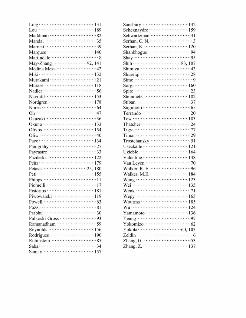
Ling ·································· 131 Lou ··································· 189 Maddipati ····························· 82 Mandal ································ 35 Marnett ································ 39 Marques ····························· 140 Martindale ····························· 8 May-Zhang ····················· 92, 141 Medina Meza ························· 42 Miki ·································· 132 Murakami ····························· 21 Murase ······························ 118 Nadler ································· 56 Navratil ······························ 153 Nordgren ···························· 178 Norris ·································· 64 Oh ······································ 47 Okazaki ······························· 36 Okuno ······························· 133 Olivos ······························· 154 Oliw ··································· 40 Pace ·································· 134 Panigrahy ····························· 27 Payrastre ······························ 33 Pazderka ···························· 122 Peña ·································· 179 Petasis ··························· 25, 180 Peti ··································· 155 Phipps ································· 11 Piomelli ······························· 17 Pistorius ····························· 181 Piwowarski ························· 119 Powell ································· 63 Pozzi ··································· 81 Prabhu ································· 30 Pulkoski-Gross ······················· 93 Ramanadham ························· 59 Reynolds ···························· 156 Rodrigues ··························· 190 Rubinstein ···························· 85 Saba ···································· 34 Sanjay ······························· 157
Sansbury ···························· 142 Schexnaydre ························ 159 Schwartzman ························· 31 Serhan, C. N. ·························· 3 Serhan, K. ··························· 120 Shanbhogue ··························· 94 Shay ··································· 95 Shih ······························ 83, 107 Shimizu ······························· 43 Shureiqi ······························· 28 Sime ···································· 9 Sorgi ································· 160 Spite ··································· 23 Steinmetz ··························· 182 Stiban ·································· 37 Sugimoto ······························ 65 Terrando ······························ 20 Tew ·································· 183 Thatcher ······························· 24 Tigyi ··································· 77 Timar ·································· 29 Trostchansky ························· 51 Useckaite ···························· 121 Uzieblo ······························ 164 Valentine ···························· 148 Van Leyen ···························· 70 Walker, R. E. ························· 96 Walker, M.E. ······················· 184 Wang ································ 123 Wei ·································· 135 Wenk ·································· 71 Wepy ································ 163 Wourms ····························· 185 Wu ··································· 124 Yamamoto ·························· 136 Yeung ································· 97 Yokomizo ····························· 62 Yokota ··························· 60, 103 Zeldin ··································· 6 Zhang, G. ····························· 53 Zhang, Z. ···························· 137

This year, we celebrate 50 years of the good stuff.
This year, we celebrate 50 years of Avanti.Thank you to our loyal customers for allowing
Avanti to provide you with the highest purity lipids available.
More than Lipidswww.avantilipids.com
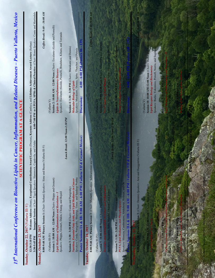
Lake
of t
he c
loud
s, P
orcu
pine
Mou
ntai
ns, O
nton
agon
, MI,
US
A –
Pho
to C
ourte
sy o
f KR
Mad
dipa
ti
15th
Inte
rnat
iona
l Con
fere
nce
on B
ioac
tive
Lipi
ds in
Can
cer,
Infla
mm
atio
n, a
nd R
elat
ed D
isea
ses –
Pue
rto
Val
lart
a, M
exic
o SC
IEN
TIF
IC P
RO
GR
AM
AT
A G
LA
NC
E
Sund
ay, O
ctob
er 2
2, 2
017
(Val
larta
III-
V)
12:3
0-3:
10 P
M
Wel
com
e A
ddre
ss (H
onn)
, Exc
eptio
nal C
ontr
ibut
ions
Aw
ard
Lec
ture
(Dra
zen)
, Key
note
Add
ress
(Can
tley)
, and
Life
time
Ach
ieve
men
t Aw
ard
Lec
ture
(Ser
han)
3:
30-5
:45
PM
Inau
gura
l Ses
sion
: (C
hair
: Tig
yi) S
peak
ers:
Ham
moc
k, C
apde
vila
, and
Zel
din
6:00
-7:0
0 PM
w-3
PU
FA, S
PMs &
Clin
ical
Pra
ctic
e: (C
hair
: Hon
n) S
peak
ers:
Con
te a
nd M
artin
dale
Mon
day,
Oct
ober
23,
201
7 8:
00-9
:40
AM
Ple
nary
Ses
sion
1: (
Cha
ir: S
erha
n) S
peak
ers:
Sim
e an
d B
azan
(Val
larta
III-
V)
Cof
fee
Bre
ak: 9
:40
– 10
:00
AM
(Val
larta
III-
IV)
(Val
larta
V)
Sess
ion
1: 1
0:00
AM
– 1
2:00
Noo
n (C
hair
s: P
hipp
s and
Gro
nert)
Se
ssio
n 2:
10:
00 A
M –
12:
00 N
oon
(Cha
irs:
Esc
alan
te-A
lcal
de a
nd P
iom
elli)
Li
pid
Med
iato
rs o
f Inf
lam
mat
ion
and
Res
olut
ion
- I
Lipi
ds in
Neu
roin
flam
mat
ory
Dis
ease
s Sp
eake
rs: P
hipp
s, G
rone
rt, D
akin
, Chi
ang,
and
Boj
alil
Spea
kers
: Esc
alan
te-A
lcal
de, P
iom
elli,
Bra
dsha
w, K
ihar
a, a
nd T
erra
ndo
Lunc
h B
reak
: 12:
00 N
oon-
1:30
PM
Se
ssio
n 3:
1:3
0 - 3
:30
PM (C
hair
s: C
orda
and
Pan
igra
hy)
Sess
ion
4: 1
:30
- 3:3
0 PM
(Cha
irs:
Mur
akam
i and
Den
nis)
N
utri
tion,
Ess
entia
l Fat
ty A
cids
, and
Lip
id M
edia
tors
in C
ance
r M
olec
ular
Bio
logy
of L
ipid
s Sp
eake
rs: C
orda
, Pan
igra
hy, S
hure
iqi,
Tim
ar, a
nd S
ande
ep P
rabh
u Sp
eake
rs: M
urak
ami,
Den
nis,
Spite
, Tha
tche
r, an
d Pe
tasi
s
Po
ster
Ses
sion
s IA
& IB
: 9:0
0 A
M -
6:00
PM
(Val
lart
a V
II &
Coz
umel
Mex
ico)
D
iscu
ssio
n:
4:00
- 6:
00 P
M (P
oste
rs 8
6 - 1
39)
Tue
sday
, Oct
ober
24,
201
7 8:
00-9
:40
AM
Ple
nary
Ses
sion
2: (
Cha
ir: M
arne
tt) S
peak
ers:
Sch
war
tzm
an a
nd H
annu
n (V
alla
rta II
I-V
) C
offe
e B
reak
: 9:4
0-10
:00
AM
(Val
larta
III-
IV)
(Val
larta
V)
Sess
ion
5: 1
0:00
-12:
00 N
oon
(Cha
irs:
Pay
rast
re an
d Sa
ba)
Sess
ion
6: 1
0:00
-12:
00 N
oon
(Cha
irs:
Gre
enbe
rg a
nd M
arne
tt)
Inos
itide
s and
Sph
ingo
lipid
s in
Can
cer a
nd In
flam
mat
ion
Nov
el A
spec
ts o
f Lip
id B
iolo
gy
Spea
kers
: Pay
rast
re, S
aba,
Man
dal,
Oka
zaki
, and
Stib
an
Spea
kers
: Gre
enbe
rg, M
arne
tt, O
liw, D
avie
s, an
d M
edin
a M
eza
Lunc
h B
reak
: 12:
00 N
oon-
1:30
PM
Se
ssio
n 7:
1:3
0 - 3
:30
PM (C
hair
s: S
him
izu
and
Cam
bron
ero)
Se
ssio
n 8:
1:3
0 - 3
:30
PM (C
hair
s: M
arne
tt an
d M
addi
pati)
PU
FA
, Lip
id M
edia
tors
, and
Pho
spho
lipas
es
Youn
g In
vest
igat
or A
war
d C
ompe
titio
n Sp
eake
rs: S
him
izu,
Cam
bron
ero,
Abd
ulno
ur, H
olm
an, a
nd O
h Sp
eake
rs: B
erge
r, D
alli,
Han
sen,
Tro
stch
ansk
y, L
arss
on, a
nd Z
hang
Po
ster
Ses
sion
s IIA
& II
B: 9
:00
AM
- 6:
00 P
M (V
alla
rta
VII
& C
ozum
el M
exic
o))
Dis
cuss
ion:
4:
00 -
6:00
PM
(Pos
ters
140
- 19
3)
Wed
nesd
ay, O
ctob
er 2
5, 2
017
8:00
-9:4
0 A
M P
lena
ry S
essi
on 3
: (C
hair
: Den
nis)
Spe
aker
s: A
rai a
nd H
aegg
strö
m (V
alla
rta II
I-V
) C
offe
e B
reak
: 9:4
0-10
:00
AM
(Val
larta
III-
IV)
(Val
larta
V)
Sess
ion
9: 1
0:00
AM
- 12:
00 N
oon
(Cha
irs:
Kim
and
Tig
yi)
Sess
ion
10: 1
0:00
AM
-12:
00 N
oon
(Cha
irs:
Bur
ke a
nd Y
okom
izo)
Ly
soph
osph
olip
ids i
n C
ance
r Li
pid
Rec
epto
r Bio
logy
and
Bio
chem
istr
y Sp
eake
rs: K
im, T
igyi
, Ben
yó, B
rindl
ey, a
nd K
ings
ley
Spea
kers
: Bur
ke, Y
okom
izo,
Pow
ell,
Nor
ris, a
nd S
ugim
oto
Lunc
h B
reak
: 12:
00 N
oon-
1:30
PM
Se
ssio
n 11
: 1:3
0-3:
30 P
M (C
hair
s: E
scal
ante
and
Hon
n)
Sess
ion
12: 1
:30-
3:30
PM
(Cha
irs:
Wen
k an
d A
rita)
C
yclo
oxyg
enas
e Pa
thw
ay in
Car
diov
ascu
lar D
isor
ders
and
Can
cer
Lipi
d M
edia
tors
of I
nfla
mm
atio
n an
d R
esol
utio
n - I
I Sp
eake
rs: E
scal
ante
, Hon
n, H
ara,
Kas
hfi,
and
van
Leye
n Sp
eake
rs: W
enk,
Arit
a, K
ane,
Lev
y, a
nd D
artt
C
offe
e B
reak
: 3:0
0-3:
30 P
M
Sess
ion
13: 4
:00-
6:00
PM
(Cha
irs:
Nad
ler a
nd H
olin
stat
) Se
ssio
n 14
: 4:0
0-6:
00 P
M (C
hair
s: P
ozzi
and
Mad
dipa
ti)
Lipi
ds in
Met
abol
ic a
nd C
ardi
ovas
cula
r Dis
orde
rs
Bio
logy
of t
he E
poxy
gena
se P
athw
ay
Spea
kers
: Nad
ler,
Hol
inst
at, H
alad
e, R
aman
adha
m, a
nd Y
okot
a Sp
eake
rs: P
ozzi
, Mad
dipa
ti, S
hih,
Das
, and
Rub
inst
ein
Con
fere
nce
Adj
ourn
s at 6
:00
PM