Embed Size (px)
Citation preview
ABSTRACTS
EMSOS 2014 – 27th Annual Meeting of the European Musculo-Skeletal Oncology Society (E.M.S.O.S.) 15th Symposium EMSOS Nurse Group, May 21-23, 2014 – Vienna, Austria
1
EMSOS 2014 27th Annual Meeting of the European Musculo-Skeletal Oncology Society (E.M.S.O.S.)
BOOK OF ABSTRACTS Editors: Reinhard Windhager, Philipp Funovics
ABSTRACTS
EMSOS 2014 – 27th Annual Meeting of the European Musculo-Skeletal Oncology Society (E.M.S.O.S.) 15th Symposium EMSOS Nurse Group, May 21-23, 2014 – Vienna, Austria
2
Scientific Board: Reinhard Windhager, Vienna, Austria Philipp Funovics, Vienna, Austria Peter Ambros, Vienna, Austria Oskar Aszmann, Vienna, Austria Örjan Berlin, Göteborg, Sweden David Biau, Paris, France Thomas Brodovic, Vienna, Austria Rodolfo Capanna, Firenze, Italy Simon Carter, London, United Kingdom José Manuel Pinto Silva Casanova, Coimbra, Portugal Richard Crevenna, Vienna, Austria Karin Dieckmann, Vienna, Austria Sander Dijkstra, Leiden, Netherlands Davide Donati, Bologna, Italy Nicola Fabbri, New York, United States Stefano Ferrari, Rizzoli, Italy Ramses Forsyth, Gent, Belgium Georg Fraberger, Vienna, Austria Mark Gebhardt, Boston, United States Georg Gosheger, Münster, Germany Robert Grimer, Birmingham, United Kingdom Kirsten Hall, Oslo, Norway John Healey, New York, United States Leo Karger, Vienna, Austria Heinrich Kovar, Vienna, Austria Wolfgang J. Köstler, Vienna, Austria Ruth Ladenstein, Vienna, Austria Andreas Leithner, Graz, Austria Marco Manfrini, Bologna, Italy Iris-Melanie Nöbauer, Vienna, Austria Joannis Panotopoulos, Vienna, Austria Piero Picci, Bologna, Italy Richard Pötter, Vienna, Austria Pietro Ruggieri, Rizzoli, Italy Miguel San Julian, Navarra, Spain Miklós Szendrői, Budapest, Hungary Carmen Trost, Vienna, Austria Hiroyuki Tsuchiya, Kanazawa, Japan Per-Ulf Tunn, Berlin, Germany Oleg Vyrva, Kharkiv, Ukraine Jeremy Whelan, London, United Kingdom Duncan Whitwell, Oxford, United Kingdom
Annotations In the following we are publishing the abstracts as submitted by the authors. Missing presentation numbers do represent talks with no abstract submitted as per date of production or papers that have been withdrawn by the authors. The Editors
ABSTRACTS
EMSOS 2014 – 27th Annual Meeting of the European Musculo-Skeletal Oncology Society (E.M.S.O.S.) 15th Symposium EMSOS Nurse Group, May 21-23, 2014 – Vienna, Austria
3
TABLE OF CONTENTS ORAL PRESENTATIONS ........................................................................................................................................................................................... 4
INVITED ABSTRACTS .......................................................................................................................................................................................... 4 TRAINING DAY T01 Radiology ........................................................................................................................................................................ 4 TRAINING DAY T02 Surgery / Perioperative Management .............................................................................................................................. 4 TRAINING DAY T03 Radiotherapy .................................................................................................................................................................. 5 TRAINING DAY T04 Oncology ........................................................................................................................................................................ 6 Campanacci Lecture ........................................................................................................................................................................................ 6
SUBMITTED ABSTRACTS ................................................................................................................................................................................... 6 SESSION 1 Osteosarcoma/Ewing’s sarcoma - What’s new in targets and innovative therapies? (Part I) ......................................................... 6 SESSION 2 Advanced STS - What are our novel options in personalised medicine? ....................................................................................... 9 SESSION 3 Osteosarcoma/Ewing’s sarcoma - What’s new in targets and innovative therapies? (Part II) ...................................................... 12 SESSION 4 Surgical options in the very young - Grower or biological? .......................................................................................................... 13 SESSION 5 Spine and Pelvis ......................................................................................................................................................................... 16 SESSION 6 Imaging and Interventional radiology - When and which? ........................................................................................................... 18 SESSION 7 Experimental models in sarcoma research - What’s on the horizon? .......................................................................................... 20 SESSION 8 Adolescents and young adults - The same or different than others? ........................................................................................... 23 SYMPOSIUM S02 Amputation - Are there new devices? ............................................................................................................................... 26 SESSION 9 Chordoma .................................................................................................................................................................................. 27 SESSION 10 Infection control in modular prostheses - How to push the boundaries? .................................................................................... 29 SESSION 11 Endoprosthetic reconstruction .................................................................................................................................................. 31 SESSION 12 Free Papers ............................................................................................................................................................................. 34 NURSE DAY N01 The patient ........................................................................................................................................................................ 36 NURSE DAY N02 Communication ................................................................................................................................................................. 37 NURSE DAY N03 Contacts............................................................................................................................................................................ 38 NURSE DAY N04 Psycho oncology ............................................................................................................................................................... 39
POSTER PRESENTATIONS .................................................................................................................................................................................... 40 P01 Osteosarcoma/Ewing’s sarcoma - What’s new in targets and innovative therapies? ............................................................................... 40 P02 Advanced STS - What are our novel options in personalised medicine? ................................................................................................. 44 P04 Surgical options in the very young - Grower or biological?/Interventional radiology - When and which? .................................................. 50 P05 Spine and Pelvis ..................................................................................................................................................................................... 50 P06 Imaging and Interventional radiology - When and which? ....................................................................................................................... 54 P07 Experimental models in sarcoma research - What’s on the horizon? ...................................................................................................... 56 P08 Adolescents and young adults - The same or different than others? ....................................................................................................... 59 PS02 Amputation - Are there new devices? ................................................................................................................................................... 60 P09 Chordoma ............................................................................................................................................................................................... 61 P10 Infection control in modular prostheses - How to push the boundaries?/Protons and heavy ions - What is the benefit? .......................... 62 P11 Endoprosthetic reconstruction ................................................................................................................................................................. 64 P12 Free Papers ............................................................................................................................................................................................ 70 PN01 Poster Nurse Day ................................................................................................................................................................................. 77
AUTHOR INDEX ...................................................................................................................................................................................................... 78 KEYWORD INDEX ................................................................................................................................................................................................... 85
ABSTRACTS
EMSOS 2014 – 27th Annual Meeting of the European Musculo-Skeletal Oncology Society (E.M.S.O.S.) 15th Symposium EMSOS Nurse Group, May 21-23, 2014 – Vienna, Austria
4
ORAL PRESENTATIONS
INVITED ABSTRACTS
TRAINING DAY T01 Radiology T01.01 New guidelines of the ESSR I. Nöbauer-Huhmann; Medical University of Vienna, Vienna, Austria.
Soft tissue tumours are rare. Soft tissue sarcomas only account for approximately 1% of soft tissue tumours in adults. However, in those sarcoma patients, an early correct diagnosis with subsequent appropriate treatment is crucial for the clinical outcome. Therefore, regularly updated guidelines for an optimized diagnostic strategy are mandatory. In patients with a suspected soft tissue tumour, ultrasound_by an experienced sonographer_is considered the appropriate initial imaging modality. Ultrasound helps to select those patients in need of further radiological diagnostic refinement. Often, especially in deep and large lesions, additional projection radiographs (or, in complex regions, CT) serve to detect osseous involvement and/or matrix calcifications. With ultrasound, it is possible to determine purely cystic or definitely benign lesions; ultrasound criteria will be discussed. All patients with indeterminate or likely malignant lesions should undergo MRI, the criteria for which will be discussed. These patients should be referred to a tumour reference center, with a multidisciplinary team that includes experienced radiologists, pathologists, surgeons, medical oncologists, and radiation therapists. All lesions that are indeterminate or likely malignant on MRI should be biopsied. Biopsy can be performed by image-guidance. Guidelines for diagnostic biopsies will also be discussed. In patients with histologically proven sarcoma, whole-body staging must be performed. With the results available, further treatment should be planned by the multidisciplinary tumour board. T01.02 Modern functional MR techniques for MSK Tumors C. S. P. van Rijswijk; Department for Radiology, Leiden Medical University Medical Center, Leiden, Netherlands.
Modern functional MR techniques are increasingly being used to monitor response to therapy in Bone and Soft tissue tumors, often predicting the success of therapy before conventionale measurements such as tumor dimensions are changed. Dynamic contrast-enhanced (DCE) MR imaging and Diffuision-weighted (DW) MR imaging have shown potential for improving the early assessment of tumour response to therapy by identification and quantification of the proportion of therapy-induced necrosis and residual viable tumor and the assessment of residual tumour after the end of therapy. This may have an impact on prognosis, modification of neoadjuvant (presurgical) treatment protocols, timing and planning of surgery, planning of radiation therapy, and selection of postoperative radiation therapy and/or chemotherapy regimens. This presentation focusses on the technique and evaluation of functional MR techniques including, fast DCE-MR imaging and DW-MR imaging. The definite role of these techniques is still under research and in most hospital not part of the routine clincal MR protocol.
T01.03 Emerging technologies: The role of PET-MRT and PET-CT M. Henninger, M. Eiber; Klinikum rechts der Isar, Technische Universität München, München, Germany.
Soft tissue and bone tumors comprise a multitude of entities with different grades of malignancy, biological behavior, and therapeutic options. Treatment strategies rely on initial staging evaluation and follow-up. Therefore diag4nostic imaging plays an important role in patient management. Initial assessment of bone sarcomas recommend conventional plain radiography combined with Magnetic resonance imaging. For detection of distant metastases mainly bone scintigraphy and plain radiography or CT of the chest are used. To assess soft tissue sarcomas, MRI of the tumour, plain radiography to exclude boneinvolvement and CT of the lungs are recommended. In the last years positron emission tomography (PET) as noninvasive tool to assess cancer biology is gaining increasing importance in staging those tumors, detecting relapse, or differentiating scar from residual tumors. Numerous studies have evaluated potential advantages of imaging sarcoma with PET. Although PET is not included in the guidelines yet, hybrid imaging modalities as PET-MRI and PET-CT combine morphologic and methabolic informations and are expected to play an central role in staging and follow-up of soft tissue and bone tumors in the future.
TRAINING DAY T02 Surgery / Perioperative Management T02.02 New concepts for the management of implant-associated infections A. Trampuz; Charité - University Medicine Berlin, Berlin, Germany.
Management of implant-associated infections includes adequate diagnosis and a combined surgical and antibiotic management. Goal is the eradication of biofilms to cure the infection, relieve pain and re-gain the full function. If the following principles are followed, a cure rate of >90% is usual. Diagnosis. Preoperative diagnosis includes aspiration of the synovial fluid for microbiology (Gram stain and culture) and cytology (cell count and differential). During surgery, multiple tissue biopsies should be sampled around the prosthesis (no swabs) for microbiology and histopathology. Sonication of the removed device improves the diagnostic sensitivity. Novel diagnostic tests include PCR, which can detect also non-growing microorganisms, e.g. after previous antimicrobial treatment. Surgical management. In acute infections, implant can be cured with retention of the implant, if the symptoms of infection are lasting <3 weeks, the prosthesis is stable and the infecting pathogen is susceptible to anti-biofilm antibiotics. In these cases, immediate debridement with change of all removable parts (e.g. inlay) is required. In chronic infection, the implant is exchanged in 1-step (if the soft tissue is not compromised) or 2-step procedure. The drainages should be kept in place as short as possible (max. 3-5 days). Large soft tissue defects require coverage with a flap. Antibiotic management. First 2 weeks usually intravenous antibiotics are administered, followed by 10 weeks of oral antibiotics, active against biofilms (i.e. rifampin in staphylococci). Common mistakes are too short duration of treatment, too low antibiotic dose (insufficient concentration in bone) or oral antibiotics with insufficient bioavailability or inactivity against biofilms.
ABSTRACTS
EMSOS 2014 – 27th Annual Meeting of the European Musculo-Skeletal Oncology Society (E.M.S.O.S.) 15th Symposium EMSOS Nurse Group, May 21-23, 2014 – Vienna, Austria
5
T02.03 Role of embolization in musculoskeletal oncology P. Ruggieri, A. Angelini, E. Pala, E. Rimondi, G. Rossi; University of Bologna, Istituto Ortopedico Rizzoli, Bologna, Italy.
Introduction. Main indications for selective arterial embolization (SAE) are definitive treatment of benign vascular lesions, reducing surgical bleeding (p.e. sacral giant cell tumors), palliation in inoperable tumors. We present the Rizzoli Institute experience for SAE in musculoskeletal tumors: 1) therapeutic or palliative treatment of primary and metastatic bone tumors; 2) therapeutic effect on hemangiomas; 3) palliative treatment of advanced bone sarcomas, 4) metastatic bone disease and 5) bone metastases from renal carcinoma. Methods. 1) 365 oncologic patients were treated with 454 SAE procedures. 2) therapeutic SAEs in 31 patients with bone (15) and soft-tissue (16) hemangiomas 3) palliative SAEs in 43 patients with advanced bone sarcomas. 4) 309 palliative SAEs in 243 patients with metastases 5) palliative SAE in 107 patients with bone metastases from renal cell carcinoma. Results. 1) A total of 419 SAEs (93%) were successful with a clinical response of 97%. 2) nine patients with bone and 10 with soft-tissue hemangiomas experienced complete pain relief, whereas the others require repeat embolization. 3) complete pain relief and >50% reduction in analgesic use was experienced by 36 patients with highly hypervascular sarcomas 4) a complete occlusion of metastatic blood supply has been obtained in all 309 embolizations. 5) a clinical response was achieved in 157 (96%) and no response in six embolisations of sacroiliac metastases. Conclusions. We recommend preoperative embolization for selected highly vascular tumors; therapeutic SEA for vascular benign lesions (hemangiomas or selected aneurysmal bone cysts); palliative SEA for advanced sarcomas or metastatic tumors of variable histology. References Rossi et al. Embolisation of bone metastases from renal cancer. Radiol Med. 2013 Mar;118(2):291-302. Mavrogenis et al. The role of embolization for hemangiomas. Musculoskelet Surg. 2012 Aug;96(2):125-35. Rossi et al. Selective arterial embolisation for bone tumours: experience of 454 cases. Radiol Med. 2011 Aug;116(5):793-808. Rossi et al. Selective embolization with N-butyl cyanoacrylate for metastatic bone disease. J Vasc Interv Radiol. 2011 Apr;22(4):462-70. T02.04 Rehabilitation of sarcoma patients F. J. Hornicek; Center for Sarcoma and Connective Tissue Oncology, Massachusetts General Hospital, Boston, MA, United States.
The rehabilitation in sarcoma patients following surgery depends on the type of surgery received. Amputations are done less frequently than decades ago and limb salvage procedures are more common now. Rehabilitation in both groups of patients is not only physical but can also be psychologically challenging. With advances in medical treatment patients live longer and their bone or soft tissue reconstruction needs to accommodate their life span when possible. There is a significant interest in the quality of life for these patients and whether there is a difference in their outcomes base upon the management. There is a focus to achieve the highest functional status after management of the sarcoma patient for their aggressive disease type.
T02.05 Risk of pathological fracture J. Healey; Orthopaedic Service Memorial, Soan-Kettering Cancer Center, New York, NY, United States.
Bone metastases are morbid and the major source of cancer pain. Modern therapies for metastatic carcinoma are turning metastatic cancer into a chronic disease. They seem less effective against bone than soft tissue disease. Thus surgical management of bone manifestations remains critical. Bisphosphonates and RANKL inhibition can prevent bone events including fracture. These treatments may effectively push the pathologic fracture curve to the right, yet the pathology can still resurface and result in fracture. Anti-antigenic factors that are important adjuncts to modern therapies can have devastating effects on wound healing. This can be a significant problem if a fracture occurs in the midst of chemotherapy. Metastatic bone lesions and pathological fractures must be considered in the context of the patient’s prognosis and life expectancy. New strategies to predict survival have been developed and validated worldwide. “PathFx” will be released as an internet tool to help predict patient survival and select appropriate surgery. Prevention is best. This can be primary administration of bisphosphonates, or secondary oncological intervention. Radiotherapy effectively relieves pain with if the bone retains mechanical integrity. If not, surgery is necessary. We need a better method to assess bone strength effectively and assess fracture risk than the currently-accepted Mirels classification. Quantification of bone mineral content and structural properties of the bone are helpful in predicting fracture. Even more dramatic is the ability of PET scan combined with computerized tomography to predict fracture. This will enable prophylactic surgical treatment of the lesions in the most effective fashion.
TRAINING DAY T03 Radiotherapy T03.02 Proton therapy in sarcoma M. Krengli1,2,3, C. Pisani2, V. Vitolo3, P. Fossati3, M. R. Fiore3, A. Iannalfi3, B. Vischioni3, M. Ciocca3, R. Orecchia3,4; 12University of "Piemonte Orientale", Novara, Italy, 3CNAO, Pavia, Italy, 4UNiversity of Milan, Milan, Italy.
Radiotherapy has a well established role in the treatment of sarcoma. Protons, thanks to their physical characteristics, can be of advantage in particular tumour locations nearby critical organs such as spine and skull base. Moreover, sarcoma presenting in paediatric age are suitable for proton therapy in relation to lower dose deposition in healthy tissues compared to conventional radiotherapy. Literature studies come from institutions with expertise in proton therapy and in sarcoma management. As an example, some of them are reported in this abstract. Fifty patients were treated at Massachusetts General Hospital for spine sarcomas to a maximum total dose of 77.4 GyE (DeLaney 2009). The 5-year local control and overall survival rates were 78% and 87% respectively. At the same institution, 375 chordoma and 246 chondrosarcoma of the base of skull were treated by protons obtaining a 10-year local control rates of 54% and 98% respectively (Munzenrider 1999). Sixteen children with inoperable soft tissue sarcoma were treated at Paul Scherrer Institute with spot-scanning or intensity-modulated proton therapy to a median dose of 50 GyE between 1996 and 2007 obtaining a disease free survival of 75% at 18 months (Timmermann 2007). At CNAO, 77 patients affected by sarcoma of the head and neck and trunk were treated by protons and carbon ions from 2011 through 2013. Results are under evaluation.These studies and others show that proton therapy is feasible and can be conveniently used in clinical trials to spare critical organs and/or to escalate the tumour dose.
ABSTRACTS
EMSOS 2014 – 27th Annual Meeting of the European Musculo-Skeletal Oncology Society (E.M.S.O.S.) 15th Symposium EMSOS Nurse Group, May 21-23, 2014 – Vienna, Austria
6
TRAINING DAY T04 Oncology T04.01 Oncological issues in adolescents and young adults T. Brodowicz1,2; 1Comprehensive Cancer Center – Musculoskeletal Tumors, Medical University of Vienna – General Hospital, Vienna, Austria, 2Department of Internal Medicine I / Oncology, Medical University of Vienna – General Hospital, Vienna, Austria.
The most common sarcomas in childhood are osteosarcoma, sarcoma of the Ewing-family and rhabdomyosarcoma. As these and other subtypes of sarcoma also occur in adulthood, both pediatric oncologists and medical oncologist treat these tumor entities. The presentation will discuss differences between pediatric and adult sarcomas in terms of pathology, clinical outcome and management. In addition, topics like pharmacokinetics of the standard systemic drugs applied in children vs. adults, psychsocial factors, fertility preservation and clinical trial enrolment will be presented. T04.02 Controversies in osteosarcoma S. Bielack; Olgahospital - Klinikum Stuttgart, Stuttgart, Germany.
Osteosarcoma is the most frequent bone cancer of children and adolescents. Unless treatment includes both surgery and multiagent chemotherapy, the development of fatal lung or, less frequently, bone metastases is the rule. Staging investigations must therefore not only include imaging of the primary tumor (X-ray, magnetic resonance imaging), but also of the chest (X-ray, computed tomography) and skeleton (bone scintigraphy). The interpretation of small, nonspecific findings upon chest CT remains challenging. Induction chemotherapy is usually initiated after biopsy and prior to definitive surgery. High-dose methotrexate, doxorubicin, cisplatin, and with some limitations ifosfamide are considered the most active agents. Most fully grown patients receive limb-salvage surgery with endoprosthetic or biological reconstruction. Surgery of osteosarcomas in growing bones is associated with additional difficulties. Expandable endoprostheses, biologic reconstruction techniques including rotation-plasty, and amputation may all be discussed. The extent of histologic response to preoperative chemotherapy observed in the resected specimen carries important prognostic implications. Predicting tumor response preoperatively may be attempted with a variety of imaging modalities, but none is totally specific. While multiple attempts to improve the prognosis of poor responders to preoperative chemotherapy by postoperative treatment modifications have been made, none has been convincing. The results of the only large scale randomized trial assessing this question, EURAMOS-1, are pending. Further studies are required to identify whether the addition of biologic or targeted therapies to chemotherapy will unequivocally improve outcomes. Long-term follow-up is necessary in order to assess potential late-effects of treatment and to search for late recurrences.
Campanacci Lecture C01.01 Decision making in orthopaedic oncology R.Grimer Royal Orthopaedic Hospital, Birmingham, United Kingdom.
The aim of all of us involved in orthopaedic oncology is do what we believe is best for the patient. But do we always know what that is? Evidence based medicine is generally considered to be the optimum method of delivering healthcare, but there is remarkably little high level evidence for much of orthopaedic oncology. This presentation explores some of the evidence base for orthopaedic oncology, identifying areas of uncertainty and how decisions are made. The cost effectiveness of some aspects of management are also investigated. The need for convincing randomised controlled trial evidence and cost effectiveness for some of current practice is compelling.
SUBMITTED ABSTRACTS
SESSION 1 Osteosarcoma/Ewing’s sarcoma - What’s new in targets and innovative therapies? (Part I) 01.01 The role of megatherapy (MGT) and stem cell transplantation (SCT) in high risk Ewing tumors: More than 30 years of EBMT activity R. Ladenstein1, D. Valteau-Couanet2, E. Glogova3, H. Juergens4, S. Burdach5, J. Michon6, H. Van den Berg7, I. Lewis8, I. Yaniv9, C. Peters10; 1St. Anna Kinderkrebsforschung e.V. and St. Anna Kinderspital, Vienna, Austria, 2Institut Gustave Roussy, Villejuif, France, 3St. Anna Kinderkrebsforschung e.V., Vienna, Austria, 4Universitätsklinikum Münster, Münster, Germany, 5Klinik und Poliklinik für Kinder und Jugendmedizin, Kinderklinik München Schwabing, München, Germany, 6Institut Curie, Paris, France, 7University of Twente, Enschede, Netherlands, 8NHS St. James's Hospital, Leeds, United Kingdom, 9Schneider Children's Medical Center, Petach Tikvah, Israel, 10St. Anna Kinderspital, Vienna, Austria.
Purpose: EBMT registry data on ET helps to explore indications and outcomes. Patients and Methods: Since 1980, 3695 patients (pts) with ET (2186 males) were registered (142 centers/24 countries). MGT indications were primary multifocal &high-risk local disease (2411pts) or relapse (719pts). Median age is 15 years (yrs); range, 1 to 65; 2568pts <18yrs. The median survival time is 0.5 yrs after allogeneic (70 pts) and 2.8yrs after autologous (A)SCTs. Peripheral blood stem cells were used in 3143 pts. Results: The 5-year overall survival rates (%) are: 44% with ASCT (3521pts) (49% for primary treatments [for localized disease: 53%; for multifocal 41%]; 31% after relapse) and 12% for alloSCT (p<0.001). Age has significant impact: 48% for ≤10yrs, 43% for >10 to ≤ 18yrs and 38% >18yrs (p<0.001). The preSCT remission status is of importance (ASCT only): 58% in CR1 (1343pts), 40% in PR (836pts), only 20% in stable disease (144pts) or primary refractory (146pts); (p<0.001). CR2 results in 46%; all others do significantly worse with< 20% (p<.000). During primary treatment TBI regimens are inferior to non-TBI MGT with 38% vs. 50% (p<0.026). A significant influence of MGT type > year 2000 is observed: busulphan based (684 pts) 60%, melphalan based (148 pts) 37%,
ABSTRACTS
EMSOS 2014 – 27th Annual Meeting of the European Musculo-Skeletal Oncology Society (E.M.S.O.S.) 15th Symposium EMSOS Nurse Group, May 21-23, 2014 – Vienna, Austria
7
treosulfan based (95pts) 19% and others (133pts): 55% (p<0.01). A Cox proportional hazards regression model identified age, response status, stem cell source and MGT regimens as independent risk factors. Conclusions:EBMT Ewing data shows improved results in high-risk pts and favours busulphan based MGT. 01.02 A retrospective analysis of bone marrow aspirations in Ewing sarcomas: are they still necessary? B. Breitegger1, L. A. Holzer1, C. Urban2, A. Leithner1; 1Department of Orthopaedic Surgery, Medical University of Graz, Graz, Austria, 2Division of Haemato-Oncology, Department of Pediatrics, Medical University of Graz, Graz, Austria.
Background and Objectives: According to the 2012 ESMO clinical practice guidelines bone marrow aspirations and biopsies (BMAB) are mandatory in staging of Ewing sarcomas (EWS). However, the prognostic value of positive tested bone marrow (BM) is discussed controversially. Therefore the aim of this study was to retrospectively review BM samples from patients with EWS at our institution and to review published literature. Materials and Methods: 55 patients (34 male, 21 female, median age 20.9) with diagnosed EWS between 2000-2013 were identified. In 29 of 55 patients BM samples (19 male, 10 female, median age 15.3) were available and retrospectively reviewed. During the last 13 years BM-samples have also been sent to the national EUROEWING study center for PCR / FISH analysis but results have not been provided since and have therefore not influenced therapeutic decision making. Furthermore a PubMed-search was performed to identify relevant published articles. Results: All 29 bone marrow samples of our patients were tested negative by immunohistochemistry. 21 studies (published between 1995-2013, including 3-140 EWS patients) were identified by searching PubMed. Three studies correlated BM positivity with a poorer outcome (2/3 in non-metastatic patients), one study excluded a correlation of positivity and early relapse and two studies suggested the elimination of BM examination the staging process of patients with localized disease. In 14 studies no clear conclusion could have been drawn whether positive tested BM has any influence on prognosis. Conclusion: Currently, it seems that there is still no conclusive proof whether BMAB in EWS are necessary. 01.03 Subsequent neoplasms after treatment for Ewing sarcoma and osteosarcoma in childhood - single institution experience J. Kruseova, M. Ganevova, A. Lukš, J. Starý; Child hematooncology dept., Prague 5, Czech Republic.
Background: Today more than 80% of children with cancer can be cured. Longer term, the development of subsequent neoplasms (SN) is one of the most serious late effects. The purpose of this study was to determine which SN occur most frequently in patients who have survived Ewing sarcoma and osteosarcoma. Procedures: from 1979 to 2012 we, in the Late Effects Clinic in Prague, began monitoring some 4,500 child cancer survivors. Unfortunately, various factors, notably the split of Czechoslovakia into two countries, reduced the number of our visitors to 2502 solid tumour survivors currently. 93 had had Ewing sarcoma and 63 osteosarcoma. Results: In total, we diagnosed 119 second tumours in 93 patients. Cumulative incidence was 4,8 % at 25 years. Ewing sarcoma patients had the third highest occurrence of SN ( CI 6,5% 25 years) . The median time to SN was 2,85 years. Three patients had MDS, one AML, three carcinoma of the thyroid, one embryonal rabdomyosarcoma. In osteosarcoma the CI was 6,3% over 25 years. Median time to SN was 6,46 years. Two had AML, one AIHA, one patient, with Li-Fraumeni syndrome, developed three SN - two soft tissue sarcoma and a chondrosarcoma. Conclusions: The most common SN for survivors
of Ewing sarcoma and osteosarcoma are hematologic malignancies. Additional research, seeking, in all likelihood, genetic causes is required to identify patients most at risk and prepare effective cures. 01.04 Presentation, treatment, and prognosis of recurrent osteosarcoma: The EUropean RELapsed OsteoSarcoma Registry (EURELOS) B. Sorg1, B. Kempf-Bielack1, M. Kevric1, K. Sundby Hall2, S. Smeland2, E. Marchesi3, S. Ferrari3, S. Bielack1; 1Cooperative OsteoSarcoma Study Group (COSS), Klinikum Stuttgart, Stuttgart, Germany, 2Scandinavian Sarcoma Group (SSG), Oslo, Norway, 3Italian Sarcoma Group (ISG), Bologna, Italy.
Background: While a significant number of osteosarcomas relapse following treatment, prospectively gathered data concerning relapse presentation and outcomes is scarce. In order to fill this void, three multi-institutional osteosarcoma groups representing 10 European countries formed the EUropean RELapsed OsteoSarcoma Registry (EURELOS). Material and methods: EURELOS registers ISG, SSG, and COSS patients who develop recurrent disease after having been made disease-free from high-grade osteosarcoma by surgery during 1st-line treatment. Tumor and treatment-related variables are collected according to a predefined dataset. Results: Between 06/05 and 12/13, 490 patients were registered: 319 male, 171 female; 447 primary extremity tumor, 43 axial; 397 initially localized, 91 metastatic (2?). Median interval from diagnostic biopsy to 1st relapse was 1.66 years (range: 0.2- 22.7). First suspicion of relapse: symptoms 111, routine imaging 314, laboratory 1, (64?). 384 recurrences were metastatic, 66 local and 38 combined (2?). Metastases involved lungs 373, bones 84 and/or other sites 30. After a median follow-up of 1.3 years (range: 0.0 - 9.4) from 1st relapse, 2-/5-year overall survival (OS) was 49%/29%. 237 patients were alive and 151 patients were in 2nd (108) or later (43) CR at last follow-up. No primary metastases, good histologic response to 1st line chemotherapy, relapse detection by imaging, disease-free interval >median, solitary lesion at recurrence and a local relapse without metastases were factors which correlated with superior OS in survival analyses. Conclusions: International collaboration in relapsed osteosarcoma is feasible. The results resemble those of earlier, retrospective analyses and add novel information. Data from EURELOS may be used to help define standards of care. 01.05 Gemcitibine (G) and docetaxel (D) in relapsed and unresectable high-grade osteosarcoma after failure of standard multimodal therapy E. Palmerini1, R. L. Jones2, A. Paioli3, E. Marchesi3, M. Cesari3, L. Coccoli4, S. M. Pollack2, D. Vanel3, P. Picci3, S. Ferrari3; 1Istituto Ortopedico Rizzoli, Bologna, Italy, 2Medical Oncology, University of Washington/ Fred Hutchinson Cancer Research Center, Seattle, WA, United States, 3Istituto Ortopedico Rizzoli, Bo, Italy, 4Oncoematologia Pediatrica, Pisa, Italy.
Background: The prognosis of relapsed and unresectable high-grade osteosarcoma is dismal and unchanged over the last decades. Increased survival for metastatic soft tissue sarcomas have been demonstrated with the combination G+D compared to G alone (Maki R., JCO 2007). Thus, we explored G+D activity in patients (pts) with relapsed/unresectable osteosarcoma. Method: Pts, progressing after standard treatment, were eligible to receive G 900 mg/m2 day 1, 8 + D 75 mg/m2 day 8, every 21 days, until progression or unacceptable toxicity. The primary end point was progression-free survival (PFS) at 4 months. Secondary objectives were Overall Survival (OS), tumor response, defined as complete response (CR) or partial response (SD) or stable disease (SD) lasting at least 6 months.
ABSTRACTS
EMSOS 2014 – 27th Annual Meeting of the European Musculo-Skeletal Oncology Society (E.M.S.O.S.) 15th Symposium EMSOS Nurse Group, May 21-23, 2014 – Vienna, Austria
8
RESULTS: 36 pts were enrolled. The median age was 22 (10-77). 24 (67%) of pts in 2nd or 3rd line. Histology: 25 pts classic osteosarcoma, 11 high grade spindle cell sarcoma (HGS). 30 pts were evaluable for response as defined by RECIST (2 pts off study for D allergic reaction, 2 pts still being treated, 2 pts with no measurable lesions). 4-month PFS was 40% (4moPFS for classic osteosarcoma: 50% vs. 4moPFS for HGS 9%, p =.06). PFS and OS at 6 months were 25 and 74%, respectively. Tumor responses: CR: 0/30, PR: 4/30 (13%), SD 13/30 (43.5%), Progressive Diseases (PD) 13/30 (43.5%); CR/PR/SD lasting ≥ 6 months: 8/30 (27%). CONCLUSIONS: G+D demonstrated activity in pre-treated relapsed high grade classic osteosarcoma. This combination should be included in therapeutic armamentarium of metastatic classic osteosarcoma. 01.06 Focal areas of grade 3 in low-grade osteosarcoma. Still low-grade or high-grade osteosarcoma? A. Righi, A. Paioli, E. Palmerini, E. Marchesi, D. Donati, P. Coluccino, M. Cesari, M. Gambarotti, P. Picci, S. Ferrari; Istituto Ortopedico Rizzoli, Bologna, Italy.
Foci of grade-3 are sometime detected in low-grade central osteosarcoma. The aim of this study was to retrospectively evaluate the clinical outcome in patients that after a first diagnosis of low-grade osteosarcoma, were upgraded to grade-3 due to the detection of areas of grade-3 in the resected specimen. A search was performed in the Bone Tumor Center data base of our Institute and 36 patients with a definitive diagnosis of grade-2-3 osteosarcoma were retrieved. Median age was 38 (5-70). 30 patients had extremity location. All were resected with adequate margins. Post-operative chemotherapy was given in 25 (69%) patients. With a median FU of 88 months, the 5-yrs OS and EFS were 85% and 68%. After a histological revision, patients were grouped in G2 focal3 (<25% G3areas) 16 patients and G2-3 (20 patients, >25% G3areas). 5-yrs OS and EFS were in G2 focal3 100 % and 93%, in G2-3 75 % and 50% (p=0.008 and 0.02 respectively). In the G2 focal3, 6/16 patients did not receive chemo; and only one of them died of unrelated causes. In the chemo group (10/16), 1 had lung met and he is presently CR2. In the G2-3 group, 5/20 patients did not receive chemo, 3 had recurrence and 1 died of disease, 15/20 patients received chemo, 7 had recurrence and 6 died of disease. The advantage of chemotherapy in patients with low-grade Osteosarcoma and small foci of grade 3 (<25% of tumor map) is not apparent in this study. 01.07 Chemotherapy induced necrosis as a prognostic marker in osteosarcoma - Do we need to raise the bar? A. Puri, A. Gulia, S. Bhanupriya; Tata Memorial Centre, Mumbai, India.
Aim: Study co relation between chemotherapy induced percentage necrosis and overall survival (OS). Materials: 192 consecutive patients of non metastatic osteosarcoma were analysed. Patients underwent appropriate surgical resection after receiving neoadjuvant chemotherapy. Excised specimen was analysed for chemotherapy induced percentage necrosis. Patients were divided based on the percentage necrosis as <90 %, 90 – 99 % and 100%. Results: Necrosis was available in 184 patients. 77 had < 90 % necrosis, 63 had 90 – 99 % necrosis and 44 had 100 % necrosis. 187 of these patients were available for follow up. Currently 85 patients are alive (follow up range 31 to 88 months, median 49 months). The OS of all patients was 47 % at 5 years. There was no difference in OS in groups when traditional cut-off “< / > 90 %” necrosis was used (46 % and 32 % for < 90 % necrosis and > 90 %
necrosis respectively - p = 0.139). When we changed the cut-off to “< / = 100 %” necrosis OS was 41 % and 73 % for < 100 % necrosis and = 100 % necrosis respectively (p = 0.001). Conclusion: Our data suggests that the traditional cut off “< / > 90 %” necrosis may not be a true representation of poor and good responders. It may be better to stratify patients as < / = 100 % necrosis, both for prognosis and in trials evaluating post surgery chemotherapy change. 01.08 Intramedullary Extension In Periosteal Osteosarcoma - Does It Portend Aggressive Biology? A. Puri, A. Gulia, S. Desai; Tata Memorial Centre, Mumbai, India.
AIM: To evaluate if intramedullary extension in periosteal osteosarcoma was indicative of more aggressive biological behaviour in terms of poorer overall survival. METHODS: A retrospective analysis of 18 cases of periosteal osteosarcoma treated between January 2001 and December 2010 was carried out. There were 12 males and 6 females. The mean age at presentation was 16.3 years (range 5-26 years). Tibia and femur were the most common sites (seen in 8 patients each). Sixteen of 18 patients received chemotherapy, 16 had limb sparing resection, one had an amputation and one had rotationplasty. RESULTS: Surgical margins were free in all patients. On histopathology, intramedullary involvement was found in 7 patients (44%). All patients were available for follow up. The median follow up was 61 months (range 18- 130 months). Pulmonary metastasis subsequently occurred in 4 cases (22%). Intramedullary involvement was seen in 3 of these 4 cases. Fourteen patients are currently alive and continuously disease free. The median follow up of survivors was 82 months (30-130 months). Disease free survival at 5 years was 77.8% and overall survival was 83.3%. Patients without marrow involvement had a better overall survival at 5 years as compared to patients with marrow involvement (90% vs 75%), p = 0.23. CONCLUSION: Intramedullary involvement may suggest more aggressive disease biology in these intermediate grade tumors. The difference in our study was not statistically significant but this could be a reflection of the small numbers. 01.09 Diaphyseal osteosarcomas - Do they have distinct features from metaphyseal osteosarcoma? S. Iwata, T. Nakamura, C. L. Gaston, S. R. Carter, R. M. Tillman, A. Abudu, L. Jays, R. J. Grimer; Royal Orthopaedic Hospital, Birmingham, United Kingdom.
Objective: Osteosarcoma occurring in the diaphysis is an unusual subtype and its distinct biological behavior from metaphyseal osteosarcoma remains unclear. The aim of this study was to clarify the clinical features and outcomes of diaphyseal osteosarcoma. Methods: Patients with newly-diagnosed high-grade osteosarcoma occurring in the long bone were eligible for this retrospective study. Clinicopathological information was collected from our database and compared among 36 diaphyseal, 405 proximal and 519 distal metaphyseal, and 14 whole bone osteosarcoma patients. Additionally, case-control study matching by age, gender, site, and metastatic condition at diagnosis with 1:3 ratio of 36 diaphyseal to 108 metaphyseal osteosarcomas patients was also conducted. Results: Five-year overall survival and metastasis-free survival of the three groups including diaphyseal, metaphyseal, and whole bone osteosarcoma patients showed significantly different (P=.029 and P=.013, respectively), although there is no difference for any factors between proximal and distal metaphyseal osteosarcoma patients. Case-control study resulted that patients with diaphyseal osteosarcomas had a tumour which was significantly larger tumour size (mean 13.5 cm vs 10 cm, P=.026), and demonstrated higher pathologic fracture rate (28% vs 12%, P=.033), superior 5-year
ABSTRACTS
EMSOS 2014 – 27th Annual Meeting of the European Musculo-Skeletal Oncology Society (E.M.S.O.S.) 15th Symposium EMSOS Nurse Group, May 21-23, 2014 – Vienna, Austria
9
metastasis-free survival (74% vs 40%, P =.0068), and slightly better 5-year overall survival (68% vs 46%, P =.074). Prognostic factor analysis showed that a pathologic fracture significantly decreased the survival of the patients with diaphyseal osteosarcoma. Conclusion: The current study showed that diaphyseal osteosarcoma has distinct clinical features from metaphyseal osteosarcoma having an increased risk of pathologic fractures but with favorable survival outcome. 01.10 Chest X-ray or Computed Tomography for the follow-up in patients with osteosarcoma? A. Paioli, E. Palmerini, E. Marchesi, A. Nobile, D. Vanel, P. Picci, C. Raspanti, M. Cesari, S. Ferrari; Istituto Ortopedico Rizzoli, Bologna, Italy.
A formally validated follow-up policy in bone sarcomas is not available. The aim of this study was to evaluate whether the technique of surveillance of lung metastases (Chest X-ray or CT) influenced the post relapse survival (PRS) in osteosarcoma. Rate of second complete remission (CR2) and PRS were the end-points of the study. Patients aged up to 40 years with extremity osteosarcoma treated at the Rizzoli Hospital from 1986 to 2009 were identified. The clinical charts of 178 patients who had only lung metastases as first pattern of recurrence were reviewed. The median relapse-free interval was 22 months. In 102 (57%) patients metastases were detected by chest X-ray, CT in 73 (41%) and in 3 by symptoms. A CR2 was achieved in 75% of patients, the median PRS was 20 months (1-280). None of the patients without CR2 survived. The 3-year PRS was 40% (95%CI 32%-47%) The rate of CR2 was 66% with chest X-ray, 89% with CT, 1 of the 3 symptomatic patients had CR2 (p=0.0005). The 3-year PRS was 33% (95%CI 23%-42%)when metastases were detected by chest X-ray and it was 56% (95% CI 43%-68%) by CT (p< 0.001).In patients with osteosarcoma of the extremity, a follow-up strategy based on chest CT rather than chest x-ray allows a higher rate of CR2 and higher probability of PRS. A change over the years in the surgical strategy of treatment of lung metastases cannot be excluded as a possible factor influencing the results of this analysis.
SESSION 2 Advanced STS - What are our novel options in personalised medicine? 02.01 Impact of margin status and local recurrence on primary high-grade soft tissue sarcoma of the extremities: a retrospective single Institution review G. Bianchi1, E. Sebastiani1, A. Righi2, D. Donati1; 1Istituto Ortopedico Rizzoli - Oncological Department, Bologna, Italy, 2Istituto Ortopedico Rizzoli - Pathology Department, Bologna, Italy.
Introduction: The impact of local-recurrence and surgical-margin status on survival in extremity soft-tissue sarcomas remains to be clearly defined. Aim of our study was to determine the prognostic relevance of positive resection margins and local recurrence for extremity soft-tissue sarcomas. Materials and Methods: Outcomes for local recurrence, distant metastases and overall survival were analyzed according to clinical, pathological and treatment variables in 501 localized patients treated in our Institute from 1990 to 2005, using Kaplan-Meier-method and Cox-regression.
Results: The overall survival was 55.7%. At latest follow up (235 months), 1% of the patients were alive with disease, 46% were alive without evidence of disease or had died of other causes. Lung metastases occurred in 14% of the patients, bone metastases in 3%, soft tissue metastases in 3%, lymph-nodes in 1%. Surgical-margins were negative in 79% and positive in 21%. Local-recurrence rate was 17%. In univariate analysis, margin status was not associated with overall survival (p=0.36) or disease specific survival (p=0.176). Local recurrence was significantly associated with overall survival (hazard ratio, 1.695 [95% confidence interval, 1.24 to 2.31]) and disease free survival (hazard ratio 1.71 [95% confidence interval, 1.14 to 2.59]) after multivariate Cox-regression. Conclusions: Local-recurrence influence the disease specific survival and overall survival in patients affected by localized high grade sarcomas. Surgical margins do not influence disease specific survival or overall survival. 02.02 Local recurrences after resection of soft tissue sarcomas: a multifactorial analysis V. Kainhofer, J. Szkandera, B. Liegl-Atzwanger, W. Maurer-Ertl, A. Gerger, A. Leithner; Medical University of Graz, Graz, Austria.
Background: Patients with soft tissue sarcoma (STS) being treated following the standardized guidelines, i.e. resection and adjuvant radiation therapy for certain cases, can still not be guaranteed to remain free from local recurrence (LR) of the sarcoma at resection site. This study was designed to analyse possible factors for local recurrence. Methods: This retrospective study included 412 patients, who were diagnosed with STS and underwent resection between 1998 and 2013. Analysis included histological reports, surgery report and follow up for recurrence and survival of each patient with focus on the factors resection margin, grading, size, depth of the tumor and age and gender of the patient. Applied statistical methods were the Kaplan-Meier survival curve analysis, Log rank test, Cox proportional hazards model. Results: Statistical analysis revealed that the factors resection margin (R1/2 HR 3.417, p<0.001; 6.6% LR with R0 vs. 21.9% LR with R1/R2) and age of the patient (≥50yrs. at operation HR 4.087 for LR, p=0.020) could reach valid significance levels. The width of resection margins (125 cases) did not show significant differences (0.1 – 0.5cm vs. 0.6 – 1.0cm vs. 1.1 – 2cm vs. >2cm; p>0.29). The factors grading (high grade HR 2.279, p=0.088), size (≥5 cm HR 1.679, p=0.224), depth (deep HR 1.012, p=0.974) of tumor and gender (p>0.40) of the patient did not show significant differences concerning LR. Conclusions: Negative resection margin (R0) is a crucial factor for low local recurrence rates, while the minimal width of resection margins does not seem to influence local recurrence. 02.03 High-grade soft tissue sarcomas of the extremities: evaluating the influence of surgical margins on mid-term overall and recurrence free survival J. J. Willeumier1, M. Fiocco1, R. Nout1, S. Dijkstra1, W. Aston2, R. Pollock2, H. H. Hartgrink1, M. A. van de Sande1; 1LUMC, Leiden, Netherlands, 2Royal National Orthopaedic Hospital, Stanmore, United Kingdom.
Influence of resection-margins on overall-survival of high-grade soft tissue sarcomas remains controversial. Further knowledge concerning this controversy is relevant for decisions regarding the aggressiveness of local treatment. The aim of this study is to define the influence of known prognostic-factors on local recurrence and
ABSTRACTS
EMSOS 2014 – 27th Annual Meeting of the European Musculo-Skeletal Oncology Society (E.M.S.O.S.) 15th Symposium EMSOS Nurse Group, May 21-23, 2014 – Vienna, Austria
10
overall survival for patients with high-grade, extremity soft tissue sarcomas. Materials and Methods A retrospective analysis of patients surgically treated between 2000 and 2007 at our institutions for four types of primary, high-grade, extremity soft tissue sarcomas was performed. The prognostic influence of margin status and other clinicopathologic characteristics was analysed by employing a cause-specific multivariate Cox regression model in the context of a competing risks model. Results A total of 138 patients with high-grade extremity soft-tissue-sarcoma were identified. Tumor size proved a consistent adverse prognostic factor for local recurrence, distant metastasis and overall survival. In addition, this study confirmed the significance of resection margins for local control. Margins were however of no significant influence on developing distant metastasis or overall survival. Controversially, the occurrence of local recurrence did have a significant impact on overall survival. Discussion The results of this study indirectly support the hypothesis that high-grade soft tissue sarcomas encompass highly aggressive tumor biology, resulting in a high risk for metastasizing haematologically on the one hand and an increased risk for positive-margin resection and local recurrence on the other hand. This implies that tumor characteristics, i.e. tumor size and growth pattern play a critical role in the decision-making for local and systemic treatment. 02.04 Epithelioid sarcomas: how important is loco-regional control? A. Pradhan, R. J. Grimer, A. Abudu, S. R. Carter, R. M. Tillman, L. Jeys; Royal Orthopaedic Hospital, Birmingham, United Kingdom.
Background: Epithelioid sarcomas are rare soft tissue sarcomas commonly seen in the upper extremity. Many years ago the senior author was advised that ‘immediate amputation of the whole limb is needed to offer a chance of cure’. We assessed outcomes in our patients to see if this advice may have been correct. Methods The centre database was used to identify patients with epithelioid sarcomas between 1985 to 2012. Patient, tumour, treatment and outcome data was collected. Results: 22 patients were managed for a primary epithelioid sarcoma. The three most common sites involved were the hand (36%), forearm (27%) and the thigh (18%). There were 17 males and 5 females with a mean age of 32.8 years. 50% of the sarcomas had inadvertent excision carried out elsewhere. Limb salvage surgery was carried out in 16 patients (73%) and limb amputation in 6 (27%). The overall risk of local recurrence was 5.6%, with risk increased in younger patients and those who had undergone an inadvertent biopsy. The event free survival rate at five and ten years was 50%. Survival was shown to be lower in young patients, those who had undergone an inadvertent biopsy and more radical surgery. Regional (skip) metastases rose in 18% of the patients. Conclusions: This series has shown that the risk of local recurrence is not influenced by the type of surgery. We could not prove that immediate amputation was likely to effect overall survival. Larger series are needed to identify the optimum management of this rare tumour. 02.05 Surgical treatment of atypical lipomatous tumour of the extremity location by marginal R1 planned resection. F. Gouin1, P. Anract2, A. Babinet2, F. Larousserie2, A. Moreau1, P. Rosset3, G. De Pinieux3, D. Waast1, S. Briand1; 1University Hospital Hôtel Dieu, Nantes, France, 2University Hospital Cochin, Paris, France, 3University Hospital, Tours, France.
Atypical lipomatous tumor (ALT) (formely named well-differentiated liposarcoma) of the extremity is one of the more frequent neoplasm of soft tissue. Recent advance in molecular diagnostic of ATL has
highlighted the sensitivity of MDM2 amplification in these tumors and the correlation with clinical behaviour. There is still debate about the more appropriate treatment of this subtype of soft tissue tumour, namely how wide must be the resection and the place of radiotherapy. Seventy patients with primary extremity located lipomatous tumour have been included in this retrospective study. All tumours were histologically proved ALT, on the basis of morphological, immunohistological and molecular criteria. They were all managed surgically by marginal resection alone (conservative surgery with no sacrifice of important anatomic structure) without adjuvant radiotherapy. Thirty one patients were male and 39 females. Median age was 63 years. Sixty two tumours were located proximally on the limb. Three patients recurred locally, respectively 10 months, 5 and 7 years after resection. The former recurred as a dedifferentiated liposarcoma, as the 2 laters as the same ALT. Thus, the disease free survival was 94.3% at 7 years. These results show that a planned R1 marginal resection without adjuvant radiotherapy is an adequate conservative treatment of molecular proved ALT, with an acceptable rate of local recurrence. Nevertheless a long lasting follow up is necessary for this subtype of soft tissue tumour due to the risk known of late local recurrences. 02.06 Improved long-term survival with pre-operative chemotherapy for high grade sarcoma L. J. Johnson1, M. Clayer1, K. Patterson2; 1Royal Adelaide Hospital, Adelaide, Australia, 2Queen Elizabeth Hospital, Adelaide, Australia.
High grade soft tissue sarcomas have mortality risk from metastasis, despite good local control, of approximately 50%. Systemic treatment is controversial. This prospective study reports the long term results of pre-operative chemotherapy on long term survival. Consecutive patients presenting with high grade, localised soft tissue sarcoma were included in the study. Those physically fit to undergo MAID chemotherapy and no prior surgery were offered pre-operative chemotherapy. The comparison group consisted of those that did not have chemotherapy due to poor physical fitness or had had an operation prior to presentation. All patients achieved wide margins with subsequent surgery. Sixty three patients were enrolled, 36 received chemotherapy and 27 did not. Two patients were excluded due to adverse chemotherapy reactions, ceasing after only 3 cycles. Two patients were excluded as they did not complete surgical treatment. Seven patients were excluded due to protocol deviations. The remaining 52 patients had a mean length of follow up of 58 months (range 4-134 months). The 5 year survival was 91% for those that received chemotherapy compared to 73% for those that did not (p = 0.03). The 10 year metastasis free survival rate was 69% versus 49% respectively (p = 0.06). Late metastasis was a feature of those who had chemotherapy. Excluding those with malignancies smaller than 5 cm at presentation, however, demonstrated a 10 year survival rate of 69% versus 37% respectively (p = 0.02). This study has shown chemotherapy given pre-operatively reduces the mortality rate but late metastasis is still a problem.
ABSTRACTS
EMSOS 2014 – 27th Annual Meeting of the European Musculo-Skeletal Oncology Society (E.M.S.O.S.) 15th Symposium EMSOS Nurse Group, May 21-23, 2014 – Vienna, Austria
11
02.07 Prognostic relevance of the mitotic count and the amount of viable tumor after neoadjuvant treatment for primary, localized, high-grade soft tissue sarcoma D. Andreou1, M. Werner2, D. Pink3, F. Traub4, M. Schuler5, B. Jobke4, P. Reichardt4, P. Tunn4; 1Münster University Hospital, Münster, Germany, 2HELIOS Klinikum Emil von Behring, Berlin, Germany, 3HELIOS Klinikum Bad Saarow, Bad Saarow, Germany, 4HELIOS Klinikum Berlin-Buch, Berlin, Germany, 5University Hospital Carl Gustav Carus Dresden, Dresden, Germany.
Background: We sought to evaluate whether mitotic count (MC) and the amount of viable tumor (VT) following neoadjuvant treatment for primary, localized, high-grade soft tissue sarcoma correlate with prognosis. Methods: Retrospective analysis of 118 patients who underwent neoadjuvant regional (isolated limb perfusion - ILP, n=61) or systemic chemotherapy (SC, n=57) followed by surgical resection with curative intent between 2001 and 2011. Results: Median follow-up was 56 months for surviving patients. The amount of VT after SC was significantly associated with disease-specific survival (DSS) and event-free survival (EFS). Patients with <10% VT had a DSS of 94% at 5 years, compared to 61% for patients with ≥10% VT (P=0.033); EFS was 75%, compared to 48% (P=0.030). The amount of VT after ILP had no correlation with DSS or EFS. Patients with a MC ≥20/10 high power fields (HPF) after SC had a significantly lower DSS (33% vs. 84% at 5 years, P<0.001) and EFS (40% vs. 63%, P=0.019) than patients with a MC <20/10 HPF. Following ILP the survival benefit was more pronounced for patients with a MC <10/10 HPF, who had a significantly higher DSS (73% vs. 39% at 5 years, P<0.001) and EFS (68% vs. 25%, P=0.002) than patients with a MC ≥10/10 HPF. Conclusions: The MC and the amount of VT after neoadjuvant therapy appear to correlate with prognosis. If these results are validated prospectively, they could provide a rational for the design of neoadjuvant treatment modification/escalation studies, analog to the EURAMOS-1 and EURO-B.O.S.S. trials for bone sarcomas. 02.08 Are there any soft tissue features which could help accurately differentiate between a malignant or benign lesion? A review of GP referrals to the London Sarcoma Service J. Woodford1, R. Saqib2, P. Gikas1, J. Jagiello1, J. Skinner1, T. Briggs1, W. Aston1, R. Pollock1; 1Royal National Orthopaedic Hospital, London, United Kingdom, 2Stepping Hill Hospital, Stockport, United Kingdom.
Introduction: GPs are able to refer patients who they suspect may have a soft tissue sarcoma to the London Sarcoma Service. A proforma must be completed and specific soft tissue features must be recorded; deep to fascia, >5cm, increasing in size, painful, fixed, immobile. The aim of our study was to assess whether there was a strong correlation between a feature and the likelihood of malignancy. Methods: Every GP referral within a six month period from August 2012 for suspected sarcoma was included. For each proforma, it was noted whether the any soft tissue sarcoma features were identified or not. Clinic letters were used to identify a final diagnosis. Results: A total of 295 patients were identified. 130 patients were male, 165 female. 199 patients had benign pathology, 41 had malignancies and 55 had no final diagnosis. Of the final malignancy patients GP referrals 50% > 5cm, 30% immobile, 75% increasing in size, 40% painful, 50% deep to fascia and 45% fixed. Patients with final benign pathology GP referrals 62% >5cm, 8% immobile, 62% increasing in size, 48% painful, 29% deep to fascia and 31% fixed. Conclusion: Deep to fascia, immobile and increasing in size were the three features that were more common in malignant compared to benign pathology. However, no particular feature was significant in predicting the likelihood of malignancy.
02.09 Personalized molecular signature and outcomes of extraskeletal myxoid chondrosarcoma C. Chandrahanayingyong1,2, T. Hickernell1, J. Compton1, L. Song1, F. Remotti1, V. Murti1, F. Y. Lee1, C. Hahn1; 1Columbia University, New York, NY, United States, 2Faculty Medicine Siriraj Hospital, Bangkok, Thailand.
Background & Methods: Extraskeletal myxoid chondrosarcoma (EMC) is a poorly defined sarcoma of uncertain cellular origin despite its name suggestive of chondrosarcoma. Clinical, histopathological, cytogenetic (FISH), micro-RNA and molecular data (RCR & RT-PCR) of sixteen patients were reviewed with respect to personalized molecular and oncologic outcomes. Mean follow-up time was 6.7 years (range: 2.4-20.7). Tumor tissues were examined for expression of chondrogenic, neurogenic, and epithelial markers. Results: There were thirteen classical (less cellular and more myxoid) and three high-grade (high-cellular and less myxoid) subtypes according to WHO classifications. Most of tumors were stained positively for mesenchymal cell marker (vimentin), an early immature chondrogenic transcription factor (SOX-9) and neurogenic lineage marker (neuromedin B). Type II collagen, a mature chondrogenic phenotype, was positive in only one case. Synaptophysin was positive in 44% and was associated with local recurrence (p=0.0135). Epithelial markers were negative in all cases. EWSR1-related translocations were identified in twelve of sixteen cases. Local recurrence rate was 44% Metastasis involved lung (four cases), bone (two cases), muscles (two cases), and lymphatic system (two cases). Overall survival rate at five, ten and fifteen years were 73.5%, 55.3% and 33.8% respectively. Compared to other sarcomas, micro RNA-10b, 21, 30a, 199a, & 99a were up-regulated (>1.5 fold) and micro RNA-891a, 642a, 5584, 1269a, & 4426 were down-regulated (<0.5). Among these, mir10b was associated with metastasis and high-grade pathologic type. Summary: Presentation with metastasis, high-grade pathology subtype, synaptophysin positivity, and miR10b was associated with poor clinical outcome and requires whole-body surveillance. 02.10 EMSOS study on mesenchymal chondrosarcoma: prognostic factors and outcome A. Frezza1,2, M. Cesari3, C. Esler4, D. Baumhoer5, A. Leithner6, D. Biau7, C. Gerrand8, A. Gronchi9, R. Grimer10, J. Whelan1; 1University College of London Hospital, London, United Kingdom, 2University Campus Bio-Medico, Rome, Italy, 3Istituto Ortopedico Rizzoli di Bologna, Bologna, Italy, 4Leicester Royal Infirmary, Leicester, United Kingdom, 5Bone Tumor Reference Centre at the Institute of Pathology, University Hospital Basel, Basel, Switzerland, 6Medical University of Graz, Graz, Austria, 7Hospital Cochin, Paris, France, 8Freeman Hospital, Newcastle, United Kingdom, 9Istituto Nazionale Tumori, Milano, Italy, 10The Royal Orthopaedic Hospital, Birmingham, United Kingdom.
Background. Mesenchymal chondrosarcoma (MCS) is an exceptionally uncommon sarcoma with little evidence to support treatment recommendations. The EMSOS collaborated to produce a dataset of 69 patients to report on prognostic factors and outcome. Results. Median age was 30 years (range: 13-80), male/female ratio 1.3. Twenty-eight lesions primarily affected soft tissues. Primary site included extremities (54%), trunk (36%) and head and neck (10%). Twelve patients had metastases at diagnosis. Median follow-up was 14.9 years (range: 1-27). Fifty-six of 57 patients with localised disease underwent surgery. Thirty-one received doxorubicin-based chemotherapy. Thirty-six patients are alive, 23 progression-free (10 recurred locally, 30 developed metastases). Median progression-free survival (PFS) and overall survival (OS) were 7 (95%CI: 3.5-10.4) and 14 years (95%CI: 9.5-18.4) respectively. Patients receiving chemotherapy showed a significantly lower rate of metastatic progression (35% versus 73%; P=0.005) and improvement in PFS (10 versus 3 years; P= 0.02). Clear resection margins predicted less frequent local recurrence (4% versus 24%; P=0.05) with a favourable trend in PFS (13 versus 4 years; P=0.2). Primary site and
ABSTRACTS
EMSOS 2014 – 27th Annual Meeting of the European Musculo-Skeletal Oncology Society (E.M.S.O.S.) 15th Symposium EMSOS Nurse Group, May 21-23, 2014 – Vienna, Austria
12
origin did not predict survival. The median OS for patients with metastases at presentation was 1.4 years (95% CI: 1.1-2.1; P<0.0001). Conclusions. Prognosis in MCS varies considerably. Metastatic disease at diagnosis has the strongest impact on survival. Radical surgery remains crucial for localised disease, although chemotherapy may reduce the rate of metastatic progression and improve survival.
SESSION 3 Osteosarcoma/Ewing’s sarcoma - What’s new in targets and innovative therapies? (Part II) 03.01 Functional and molecular characterization of B Cells and plasmocytes in osteosarcoma J. M. Casanova1,2,3, M. T. Abreu4, M. S. Carneiro4; 1Coimbra University Hospital Center, Coimbra, Portugal, 2CIMAGO, Coimbra, Portugal, 3Coimbra Medical College, Coimbra, Portugal, 4Center for Neurosciences and Cell Biology, Coimbra, Portugal.
The role of B cells in the natural immune response against osteosarcoma (OS), is unknown. Assessing the functional phenotypes of circulating B cell might provide new insights into whether there are any changes that can be used as potential biomarkers for disease evolution and response to therapy. The functional phenotypes of circulating B cells were determined in 15 patients with diagnosed osteosarcoma and naïve for any therapy and the results were compared to those of 14 age-matched healthy controls (HC) were analyzed by 7 color flow-cytometry. Differences between groups were calculated using the non-parametric Mann-Whitney test, and were considered as significant for p<0.05. OS patients had significantly more circulating double-negative (IgD-CD27-) memory B cells than HC (OS: 6.0%±0.7 vs HC: 3.3%±0.4, p<0.01). In OS patients these cells -similarly to what was observed in the total B cell pool (MFI CD126: OS=400.5±49.3 vs HC=269.5±58.8; p<0.05)- have increased expression of IL-6R (CD126). The frequency of B cells expressing apoptosis-related Fas/CD95 was increased in OS patients when compared to HC (OS=19.8%±3.0 vs HC=12.5%±1.8; p<0.05). In recent studies activated memory B cells increase CD95-expression, therefore we suggest that in OS patients the memory B cell pool (comprises the antibody producing cells) is abnormally activated. OS patients had less circulating regulatory (CD1dhiIL-10hiTGFβ+) B cells (OS: 14.1%±6.7 vs HC:28.6%±8.4; p<0.05) which was accompanied by a decrease in the expression of the anti-inflammatory cytokine TGFβ (MFI TGFβ: OS=202.6±35.5 vs HC=488.8±118.8; p<0.05). Our results indicate major shifts in functional B cell subsets suggesting their direct involvement in OS. 03.02 Targeting osteosarcoma with the gallium complex KP46 T. Schöfl1,2; 1Department of Orthopaedics, Medical University Vienna, Vienna, Austria, 2Institute for Cancer Research, Medical University Vienna, Vienna, Austria.
Osteosarcoma (OS) is the most frequent primary malignant bone tumor in children. Although long-term survival over the last 30 years could be improved from 20% to almost 80% through neo-adjuvant chemotherapy in combination with surgery, drug resistance and metastatic progression are still limiting factors for cure. Thus, novel therapeutic approaches are urgently needed. KP46, an orally available gallium complex in early clinical development, is preferentially accumulating in the bone. Consequently we investigated, whether KP46 is active against
human OS-cells and started to dissect the underlying mode of action. KP46 exerted strong anti-cancer effects against four human OS-cell lines with IC50-values between 0.5 and 1.5µM (72h drug exposure, MTT-assay). Two different mechanisms seemed to underlie these profound effects: 1. rapid cell deterioration within 24 hours lacking classical features of apoptosis like caspase activation and chromatin condensation; 2. cell cycle arrest followed by classical apoptosis induction after longer drug exposure times (>48h). The relative contribution of these two effects was depending on the applied KP46 dose (higher dose favoring rapid cell deterioration) and cell density (higher density favoring cytostasis, followed by apoptosis induction). Rapid cell deterioration caused by KP46 was preceded by cell body retraction and loss of cell adhesion. Accordingly a destabilization of the two main focal adhesion proteins integrin b1 and talin paralleled KP46-induced cell detachment (Western blot analysis). Moreover, a synergistic effect of KP-46 with drugs from the EURAMOS therapy protocol was observed. Summarizing, our preliminary data suggest KP46 might be a promising new agent in OS therapy. 03.03 pERK1/2 targeting as a novel molecular adjuvant therapy in osteosarcoma: Molecular mechanisms of pERK1/2 & 14-3-3 protein regulation of apoptosis and preclinical results in vivo F. Y. Lee, K. Kim, A. Hsu, J. Compton, J. Caldwell, A. Martinez, N. Patel, J. KIM, H. Lee, S. Nizami; Columbia University, New York, NY, United States.
There is no cure for aggressive metastatic or multifocal osteosarcoma. Our hypothesis is inhibition of MAPK/ERK pathway, a well-known cell proliferation and survival signalling process, potentiates conventional cytotoxic agents in osteosarcoma-bearing mice. In addition, we further define the molecular mechanisms in vitro. We transplanted GFP-LUC-tagged 143B osteosarcoma cells into tibia in nude mice. Mice were randomly assigned to treatment with doxorubicin, PD98059 (p-ERK1/2 inhibitor), or both (N=15/each group). Outcome was assessed by monitoring bioluminescence and survival. Cell death, survival, and anti-apoptotic protein expression were examined in vitro. Targeting ERK1/2 with PD98059 resulted in prolonged survival in comparison with vehicle-treated control mice (p=0.02). Standalone doxorubicin treatment yielded similar animal mortality (p=0.017). Combined PD98059 and doxorubicin treatment further prolonged survival (p = 0.0023). ERK1/2 blockage increased the expression of proapoptotic proteins such as Bim and increased cell death in 143B and SaOS2 osteosarcoma cells. Doxorubicin treatment increased the expression of Bcl-2, an anti-apoptotic protein, but this upregulation was blocked by combined treatment with PD98059, suggesting a role for ERK1/2 in conferring drug resistance. This means that pERK inhibition prevents drug-resistance and promotes cancer cell death in two independent pathways. Additionally, 14-3-3ε gene silencing resulted in downregulation of Bim expression after PD98059 treatment. These data indicate that pERK pathways promote osteosarcoma cell survival through Bcl-2 and inhibit Bim anti-apoptotic protein. pERK inhibition is a powerful molecular adjuvant therapy reversing the drug resistance. A new combinatorial therapy using conventional cytotoxic drugs and kinase inhibitors will most likely improve the outcome of patients.
ABSTRACTS
EMSOS 2014 – 27th Annual Meeting of the European Musculo-Skeletal Oncology Society (E.M.S.O.S.) 15th Symposium EMSOS Nurse Group, May 21-23, 2014 – Vienna, Austria
13
03.04 VCN01 and pediatric osteosarcoma. Preliminary results of in vitro and in vivo orthotopic osteosarcoma models M. San-Julian1, N. Martinez1, M. Alonso1, E. Xipell1, F. Lecanda1, A. Rodriguez-García2, R. Alemany3, M. Cascallo3, M. Zalacain1, A. Patiño-Garcia1; 1University of Navarra, Pamplona, Spain, 2Institut catalá dóncoligia, L´hospitalet de Llobregat, Spain, 3VCN Biosciences, San Cugat del Vallés, Spain.
Background: VCN01 is a conditionally replicative oncolytic adenovirus, selective for replication in pRb pathway-defective tumor cells. It expresses PH20 hyaluronidase. Hyaluronan is an important element of the extracellular matrix (ECM). We aimed to test if VCN01 would result in an enhanced anti-osteosarcoma effect in vitro and in vivo in an in-house murine orthotopic osteosarcoma model. Materials/methods: Osteosarcoma cell lines, established from osteosarcoma patients with metastatic disease: 531MII, 678R, 588M, and 595M and the commercial cell line 143B, were used. Results: VCN01 displayed a robust cytopathic effect (IC50 ranging from 11 to 30 MOIs) Replication phenotype was dose dependant, as shown by early and late viral protein expression. The parallel analysis of fiber and hyaluronidase showed an increased expression of hyaluronidase with increasing viral titer. Injection of 500,000 531MII cells in the tibia resulted in development of tumors in >80% of athymic nude mice. Animals were randomized as controls (PBS) or treated with VCN01 at a dose of 107 or 108 pfu. Animals were weighed and tumors measured every week. 18-FDG-PET was performed. The 108 pfu dose significantly reduced the tumor burden (p=0.02 and p=0.03, respectively) compared to untreated mice. Conclusions: Oncolytic adenovirus VCN01 is able to infect, replicate in and eliminate primary osteosarcoma-derived cell lines. We have developed a valid orthotopic osteosarcoma model to validate the in vivo oncolytic activity of VCN01. This strong antitumor effect might be mediated by the expression of hyaluronidase that, in turn, improves the spread of the virus in this tumor, by destruction ECM. 03.05 Genomic alterations in biopsy samples predict the chemosensitivity of pediatric osteosarcoma S. Iwata, T. Yonemoto, H. Kageyama, Y. Hagiwara, H. Kamoda, S. Yokoi, H. Nagase, A. Nakagawara, T. Ishii, M. Ohira; Chiba Cancer Center, Chiba, Japan.
PURPOSE: To construct a prediction system of chemosensitivity of preoperative chemotherapy for pediatric OS patients, array CGH and NGS technologies were applied. METHODS: Pre-therapeutic biopsy tumor samples of 30 pediatric conventional OS patients who had homogeneous clinicopathological- and therapeutic- background were analyzed. Patients were divided into three groups (A:MAP-sensitive, B:IFO-sensitive and C:resistant to both) according to the histological response to chemotherapy. Twenty-two samples were used as a learning set for searching candidate markers and 8 samples for independent analysis as a validation set. Genomic DNAs were used for CGH analysis with Agilent 44k DNA microarray. Target sequencing by using the Ion Torrent AmpliSeq Comprehensive Cancer Panel were also conducted for 10 tumor/non-tumor pairs. RESULTS: Differential aberration analyses of the copy number data resulted in selection of 8 putative genomic markers for classifying into A, B and C groups with differential chemosensitivity. Scoring system to predict chemosensitivity of each tumor was developed with these genomic markers and subsequent validation analysis indicated that the scoring system was remarkably consistent to the patient properties. Target re-sequencing of 409 cancer-related genes by using Ion Proton sequencer identified 98 candidate non-synonymous single nucleotide variations (SNVs) (average 10 SNVs per tumor), however, significantly recurrent mutations in C group have not been observed so far. CONCLUSION: Our results indicated that copy number changes in OS biopsy samples could be good predictors of response to
preoperative chemotherapy. These signatures as well as global mutation information may be useful for the future genomics-based personalized chemotherapy for OS patients. 03.06 Comparative Genomic Hybridization (CGH) array technology in a homogeneous cohort pediatric osteosarcoma identified new genomic pathways involved in tumor resistance to chemotherapy C. Lasthaus1, E. Sohier2, B. Job2, N. Gaspar2, C. Philippe2, L. Marcellin1, D. Guenot3, N. Entz-Werle1; 1CHRU Strasbourg, Strasbourg, France, 2Institut Gustave Roussy, Strasbourg, France, 3EA3430, Strasbourg, France.
The genetic pathways involved in pediatric osteosarcomas are still poorly understood. There is a real need to go further and identify the molecular events in the multistep progression of those bone cancers but also in the tumor resistance to standard treatment. Those osteosarcomas are characterized by an unstable karyotype which typically has a heterogeneous pattern of complex structural and numerical chromosomal abnormalities. To help us in this large DNA scale screening, a Comparative Genomic Hybridization array technology was used to investigate the copy number alterations. Materiel and methods: 67 DNAs from diagnostic biopsies were collected. The patients were homogeneously treated in the OS94 and OS05 protocols. A clinical database was associated to this DNA collection to obtain DNA/clinical correlations. DNA was hybridized on Agilent 4x44K Whole Human Genome microarrays, according to the manufacturer’s instructions. Data were extracted from scanned images using the Feature Extraction software from Agilent and quality control report was generated according to Agilent methodology. The GISTIC 2.0 software was used to analyze the CGHarray results. Results: The unsupervised analysis determine 3 subgroups of patients. Two groups were highly rearranged, especially involving genes of the osteogenesis pathway, the neuronal and muscle developmental pathways. The response to chemotherapy was linked to genes involved in osseous metabolism and resorption, whereas the chemoresistance was preferentially linked to intra-tumor hypoxia and cell metabolism. Surprisingly, genes of normal neuronal, muscle and osseous development were involved suggesting the role of their deregulation in the undifferentiated malignant osteoblast, supporting the fact of their mesenchymal properties.
SESSION 4 Surgical options in the very young - Grower or biological? 04.01 Does feet length discrepancy after resection of bone tumor in children reflects a limb global growth disturbance? E. J. Mascard1, N. Gaspar1, C. Glorion2, S. Pannier2, P. Wicart2; 1Institut Gustave Roussy, Villejuif Cedex, France, 2Hospital Necker, Paris, France.
We observed in patients with growing prostheses a discrepancy in foot length after surgery. The purpose of our study was to describe a new complication of bone tumor surgery in children. 48 patients operated-on for a bone tumor of the lower limb, were reviewed (Patients with radiotherapy, bilateral surgery, neurological impairment, foot tumours or lost to follow-up were excluded). There were 26 boys and 22 girls aged 6 to 15 (m. 9.5). Diagnoses were 32 osteosarcomas, 16 Ewing’s, in 32 femur, 11 tibia, and 5 pelvis. All patients received peri-operative chemotherapy. Reconstruction was achieved with a prosthesis in 28 cases, a vascularized fibula transfer in 14, an arthrodesis in 4 and an intercalary graft in 2. All patients
ABSTRACTS
EMSOS 2014 – 27th Annual Meeting of the European Musculo-Skeletal Oncology Society (E.M.S.O.S.) 15th Symposium EMSOS Nurse Group, May 21-23, 2014 – Vienna, Austria
14
were prospectively reviewed yearly and size, body weight and both feet length were recorded. Results were assessed with a 11.5 mean FU (3 to 27). Average final limb length discrepancy was 2.5 cm (0 to 5). Feet length discrepancy ranged from -0.5 to 3 cm. There was a correlation between feet and limb length discrepancy (p=0.0267). There was no correlation with age at time of surgery, but patients with a growing prosthesis and a femur resection had more feet-length discrepancy (p<0,04). Feet length is reflecting a global growth disturbance of the limb. Our study showed that resection of a bone tumour in a child will induce growth impairment not only linked to the resection of the growth cartilage. Further studies have to be achieved to explain the mechanism of that growth impairment. 04.02 A "bio"expandable Endoprosthesis in malignant Bone Tumors in Children - First Clinical Results R. Baumgart; ZEM-Germany, Munich, Germany.
Introduction: Expandable endoprostheses offer better options in children after resection of malignant bone tumors to compensate limb length discrepancies at maturity. Especially if there is a high demand of lengthening, the proportion of the residual bone shaft to prosthesis and therewith the long term stability becomes worse. With new types of prostheses the residual bone and not the body of the prosthesis can be enabled to grow using the method of callus distraction, diminishing host bone - endoprosthetic lever arm forces. Methods: In 8 patients (6x femur, 2x tibia), mean age 16,3 years the resulting limb length discrepancy after resection of a malignant bone tumor (6x Osteosarcoma, 2x Ewingsarcoma) was corrected at maturity. The stem of the initial prosthesis was exchanged to a motorized distraction nail (Fitbone) the residual bone was osteotomized and lengthened 1mm per day. In 2 patients the lengthening was done in 2 steps. The medium lengthening distance was 76mm. Results: In all patients lengthening was completed successfully. In 2 patients overlengthening was done to reduce the size of the later definitive prosthesis. In 5 patients meanwhile a coated stem could be implanted to allow bone ingrowth. In 1 case between lengthening intervals a breakage of the nail occurred, so the second step was done earlier. Conclusion: Bioexpandable endoprostheses offer new perspectives of bone growth to achieve equal leg length even after resection of malignant bone tumors. Due to the fact that the lever arm forces are reduced by lengthening, better long term results can be expected. 04.03 20 years’ experience with stem fixation in pre-puberal children. with distal femur mega-prostheses. Better cemented or press-fit ? M. Manfrini, M. Colangeli, L. Campanacci, T. Frisoni, D. Donati; Istituto Ortopedico Rizzoli, Bologna, Italy.
Megaprostheses after distal femur resection in sarcoma patients became popular in the 1980s .Stem fixation is achieved by cemented or uncemented stems but scarse data have been produced about this issue in pediatric patients. The authors review their 20years’ experience in pre-puberal children using both uncemented or cemented stems Method: Radiographic evolution of femoral stems in 88 children below 13 years of age (range 5-12), with sarcomas of the distal femur, that had a megaprosthesis implanted in the period 1984-2013 was analized..Uncemented stems were used in 53 patients (median age 11) with modular implants: three designs were used along the period. Cemented custom-made stems were implanted in 32 children (median age 8) with different custom-made megaprostheses: in 25 cases the canal was over-reamed with a complete mantle of cement all around the stem, in 7 cases a polished stem with HA collar was inserted with minimal reaming and
cementation. In 3 children the femur was short and a segment of femur allograft was used to elongate the canal in a composite fashion. Diaphyseal remodelling around the stem was evaluated during follow-up modifying the Capanna system for KMFTR implants. Results: Intense cortical atrophy with bone lysis became evident in 46 uncemented stem (86%) and in 21 heavily cemented stem (84%) while no atrophic changes appeared in the minimally cemented cases with no loosening and excellent clinical results Discussion: Minimally cemented stem seem to allow better stress transmission to cortex in young children preserving the bone stock and minimizing the loosening 04.04 The Effect Of Hydroxyapatite Coated Collars On Long-Term Survival Of Extendable Cemented Distal Femoral Endoprostheses In The Immature Skeleton S. Khan, M. Coathup, P. Gikas, L. Johnston, J. Skinner, R. Pollock, T. W. R. Briggs, W. Aston, G. Blunn; Royal National Orthopaedic Hospital, London, United Kingdom.
Introduction: Distal femoral replacement is mainstay treatment for distal femoral tumours in paediatric patients. Asceptic loosening is known to be a major cause for failure in these implants. This study assesses the effect of HA coated collars on asceptic loosening in extendable distal femoral replacement prosthesis in the immature skeleton. Methods: All paediatric patients undergoing distal femoral replacement with extendable prosthesis were retrospectively reviewed between 1980-2003. 24 patients underwent distal femoral replacement with extendable prosthesis without a HA coated collar. This cohort was compared to 18 patients who were treated with an extendable prosthesis with a HA coated collar between 2001-03.. Radiographs taken at last follow-up were analysed for loosening. Results: A total of 32 patients were reviewed. Thirty-one patients were treated for primary osteosarcoma and one for a histiocytoma. Average follow up in patients without a collar was 10.1yrs (6-18) and 8.1 yrs in those with a collar (4-11). Three patients in the non-collared cohort underwent revision compared to one in the collared group. One patient in each cohort died within 5 years of surgery. Mean loosening score at last follow up for the non collared group was 11.2 compared to 2.5 for the collared group with a p value of <.05 (mann whitney-u). Conclusions: Aseptic loosening is a major cause of failure for distal femoral replacement prosthesis. Our data demonstrates that HA coated collars significantly reduce long term loosening of distal femoral endoprostheses in the immature skeleton and advocate its use in all prosthesis. 04.05 Use of Non-hinged Endoprosthesis Can Provide Stable Knee Joint and Preserved Adjacent Uninvolved Physeal Growth after Tumor Resection in Children T. Ji, W. Guo, R. Yang, X. Tang; Musculoskeletal Tumor Center, People's Hospital, Peking University, Beijing, China.
The non-hinged endoprosthetic device was implanted in 7 pediatric patients with stage IIB osteosarcoma. The prosthesis consists of a femoral component, including segmental defect body and cemented stem, and a tibial component with design of a non-hinged base plate, small-diameter press-fit stem and derotation fins. A posterior stabilizing polyethylene component is fixed on the tibial component. Implantation of the tibial component requires penetration of adjacent uninvolved bone, through the growth plate, by the press-fit stem. The cases were prospectively followed up with focus on growth rate of adjacent uninvolved bone in the salvaged limb, joint stability and length discrepancy.There were two girls and five boys with an average age at the time of primary surgery of 10.0 years. All the tumor was located in the distal femur. The average followup was
ABSTRACTS
EMSOS 2014 – 27th Annual Meeting of the European Musculo-Skeletal Oncology Society (E.M.S.O.S.) 15th Symposium EMSOS Nurse Group, May 21-23, 2014 – Vienna, Austria
15
32.7 months. All the patients were alive. There were one local recurrence and the patient received local resection. Pulmonary metastasis was observed in one patient. The adjacent uninvolved bone in the salvaged limb grew by an average of 2.7 cm, and the equivalent bone in unoperated contralateral limb grew by an average of 2.9 cm. The mean MSTS 93 score was 88.9. One recurrent dislocation was observed in one patient and the prosthesis was revised to a rotating hinged distal femur. 04.06 Summing up the results of endoprosthesis for children with bone sarcomas: the East-European Sarcoma Group (EESG) D. Nisichenko, M. Kubirov, A. Dzampaev, D. Khestanov, M. D. Aliev; RONC N.N.Blokhin, Moscow, Russian Federation.
Goal: analyze outcomes of tumor endoprosthesis for upper and lower limbs in primary and secondary implantations. Methods: 268 children were treated from 2000 to 2012. We performed 291 operations: 265 primary and 26 revisions. Boys - 54% and girls- 46%. Osteosarcoma 74%, Ewing saroma-19%. Average age - 12.6 years (min - 3.5, max. 18 yrs). We shared patients for two groups: standard zones (n=254) of affection and accordingly, typical kinds of endoprosthesis and second group that includes rare kinds of endoprosthesis. As far as those operations took a rare place, we cannot achieve reliable evidence. We used endoprosthesis made by Wright (USA), W.Link (Germany), Implantcast (Germany), ProSpon (Chech Republic), Stanmore (England). RESULTS Average follow up - 39 months. Min - 2 days (as a result of thrombosis), maximum monitoring time - 159 months (more then 13 years). Femur involvement was 144/268 (53%): distal femur 114/144 (79%), proximal femur 11/144 (7,8%), total femur-17/144 (11.8%) and others. Tibia involvement was 80/268 (30%): proximal part- 92.5%, diaphysis - 5%, distal tibia 2/80 (2.5%). Total humer was in 14/41 (34%) cases. Two-year overall survival was 65%. Infectious complication we observed in 44/251 (15%) cases: distal femur - 5.6%, proximal tibia - 20.5%, total femur - 17.3%. Aseptic loosing we observed in 52/291 (18%) cases. Local relapse - 22/268 (8%). CONCLUSIONS: For making limb salvage operations for children with bone sarcoma do not yet reached skeletal immature we need to plan operations carefully and consider age of patient and possible further reoperations and disease prognosis. 04.07 Joint Preservation Limb Sparing Surgery in Children with Malignant Bone Tumors: The Biological Solution S. Kenan; North Shore Long Island medical center, Garden City, NY, United States.
Introduction: The purpose of the study to analyze long term oncological and functional outcomes of joint preservation limb sparing surgery by transepiphyseal transmetaphyseal resection about the knee joint in children with malignant bone tumors. Material and methods: From 1990-2012, twelve patients malignant bone tumors, age 5 to 18 years were treated. Location 8-distal femur, 4-proximal tibia. All received chemotherapy. Ten patients transmetaphyseal and two transepiphyseal resection. Combined allograft vascularized fibular graft performed in five distal femur. Results: Follow-up period 2 to 23 years. All remained disease free. No immediate complications. Two patients, tibial allograft stress fracture treated successfully by improved fixation. Two other patients with distal femur, allograft fracture required revision using vascularized fibular graft. One patient, distal femur nonunion underwent resection replacement endoprosthesis. All continued growth without significant limb length discrepancy. All patients retained their limb, regained full active range of motion.
Discussion: Better understanding of the biological behavior, improved radiographic imaging, more effective chemotherapy has enabled to accurately assess malignant tumor extension preoperatively. These advances made it possible to come closer to tumor with adequate surgical margins. Patients in whom the tumor is away from the growth plate, transmetaphyseal resection could be performed successfully, saving the knee joint and growth plate allowing continues growth, minimizing limb length discrepancy. This biological solution is effective alternative to mega-prothesis. All patients retained their limb, regained full active range of motion of the knee. Returned to normal lifestyle activity. Based on M.S.T.S functional evaluation, excellent results were achieved in 10 patients. 04.08 Non-vascularized fibulae for reconstruction of bone defects following tumor resection at the extremities - a reliable biological treatment method? U. Lenze, B. M. Speth, F. Hefti, A. H. Krieg; Department for Childrens Orthopedics, Universitäts-Kinderspital beider Basel, Basel, Switzerland.
Due to new treatment options limb salvage procedures have become the standard for patients with bone or soft tissue tumors. In pediatric patients, beside tumor endoprostheses which sometimes bare the handicap of various revision surgeries, especially biologic reconstruction methods are frequently used. Aim of this study was to analyse the results of non-vascularized fibulae reconstructions following tumor of the extremities. Patients who were treated for a musculoskeletal tumor of the extremities and achieved reconstruction with non-vascularized fibulae between 1976 and 2012 were identified. All patients were retrospectively analyzed with regard to consolidation, hypertrophy, function, relapse and complications. In total 34 patients (18 male; 16 female) with a mean age of 24 years (range 5,5 - 68,4 years) were identified. In total 31 patients had a bone tumor and 3 a soft tissue tumor. The median follow up was 6,8 years, (2,0 - 26,7 years). Primary union was achieved in 90% of reconstructions and a median period of 6 months, whereas significant hypertrophy was found in 50% of all graft junctions. Complete or partial remodeling at the donor site was found in 70%. We observed 3 infections and 9 fatigue fractures, whereas only 3 needed treatment. At the donor site the complication rate was 15%. Non-vascularized fibulae are a valuable and underestimated alternative to other limb sparing reconstruction methods at the extremities. Compared to vascularized autografts the use of non-vascularized fibulae is a cheap and technically less demanding procedure, which in principle allows a complete remodelling at the donor site. 04.09 Massive osteoarticular allograft reconstruction after bone tumor resection of the knee in pediatric patients D. A. Campanacci, F. Totti, G. Beltrami, G. Scoccianti, D. Matera, F. Frenos, R. Capanna; Department of Orthopaedic Oncology, Azienda Ospedaliera Universitaria Careggi, Firenze, Italy.
Osteoarticular allografts (OA) represent a reconstructive option after bone tumor resection around the knee in growing children. The major advantage is the chance to preserve the growth plate of the remaining bone, but the disadvantage is the high failure rate eventually requiring definitive prosthetic replacement at skeletal maturity. We reviewed 23 patients who underwent OA reconstructions of the distal femur (16) or proximal tibia (7). Average age at surgery was 11 years (7-17). The diagnosis was osteosarcoma (20) and Ewing sarcoma (3). At an average follow-up of 97 months (12-167), 18 patients (78%) were continuosly disease free. We observed 10 allograft failures requiring prosthetic replacement: 8 fractures (5 proximal tibia, 3 distal femur) and 2 articular degeneration (2 distal femur). At last
ABSTRACTS
EMSOS 2014 – 27th Annual Meeting of the European Musculo-Skeletal Oncology Society (E.M.S.O.S.) 15th Symposium EMSOS Nurse Group, May 21-23, 2014 – Vienna, Austria
16
follow-up 13 allografts (56%) were still in place. Overall allograft survival was 74.3% at five and 45% at ten years. In distal femur, allograft survival was 85.1% at five and 63.8% at ten years. In proximal tibia, allograft survival was 51.4% at five years. Average limb shortening was 3,5cm (2-5) in patients with the allograft still in place and 1,8cm (0-4) in patients after prosthetic replacement. Average MSTS functional score in 16 evaluable patients was 25 (11-29). In conclusion, OA reconstruction of the knee in pediatric age can be considered a temporary solution with the aim to limit limb length discrepancy. Proximal tibial allografts showed a higher failure rate than distal femur. Allograft-prosthesis composite might improve implant durability and reduce the risk of failure.
SESSION 5 Spine and Pelvis 05.01 A novel scoring system for predicting poor survival in primary spinal tumors A. Lazary, Z. Szövérfi, Á. Bozsódi, I. Klemencsics, P. Éltes, P. Varga; National Center for Spinal Disorders, Budapest, Hungary.
BACKGROUND: Surgical and oncological management of primary spinal tumors has changed significantly over the last few decades but the prognosis is still poor. There is very limited scientific data about the prognosis affecting factors. The objective of the present study was to study pre-operative factors associated with poor survival in primary spinal tumors. METHODS: The study included 323 surgically treated primary spinal tumor patients. Patients were randomly divided into a training cohort (n = 273) and a validation cohort (n = 50). Pre -operative factors were analyzed using Cox proportional hazards models. Based on the mortality related variables, a simple scoring system of mortality was created, and three groups of patients were identified. Internal validation was performed assessing the discrimination and explained variation of the model in the validation cohort. RESULTS: Patient age, spinal region, tumor grade, spinal pain, motor deficit and myelopathy/cauda syndrome were significantly associated with poor survival in the multivariate analysis (p<0.001, R^2=0.799). Based on these variables, we developed the Primary Spinal Tumor Mortality Score (PSTMS), where an eight-point scale was divided into three categories (low, medium and high mortality). The three PSTMS categories were significantly associated with the overall survival (p<0.001, R^2 =0.811, c=0.82). The models performance remained similarly high in the validation cohort (R^2 =0.831, c=0.81). CONCLUSION: Six predictive variables for mortality in primary spinal tumors were identified. Using these six variables, an easy-to-use scoring system was developed which can be applied to the estimation of postoperative survival in all types of primary spinal tumor patients. 05.02 Evaluation of prognostic scoring systems for spinal bone metastases - A Retrospective multi-center study on 1379 patients L. Bollen1, C. Wibmer2, Y. Van der Linden1, W. Pondaag1, M. Fiocco1, W. Peul1, A. Leithner2, S. Dijkstra1; 1LUMC, Leiden, Netherlands, 2Medizinische Universität Graz, Graz, Austria.
Objectives: Models to aid in the decision making process for the treatment of spinal bone metastases (SBM) have been developed by Tomita, Tokuhashi, Van der Linden, Bauer, Rades and Bollen. The aim of this study was to assess the validity and predictive accuracy of these six models designed to estimate survival of patients suffering from SBM.
Methods: Included were patients treated for SBM between 2000 and 2010 in this international multi-center retrospective study (n=1379). Medical records were reviewed for all items needed to use the models. Survival time was calculated as the difference between start of treatment for SBM and date of death. Survival curves were estimated using the Kaplan-Meier method and accuracy was assessed with the c-statistic. For the lowest predictive group of each model, survival rate at two months was assessed. Results: Median follow-up was 6.7 years (95%CI 5.6-7.7) with a minimum of 2.3 years and a maximum of 12.3 years. The overall median survival was 5.1 months (95%CI 4.6-5.6). The most common primary tumors were breast (n=388, 28%), lung (n=318, 23%) and prostate cancer (n=259, 19%). The Tomita, Tokuhashi, Bauer modified and Van der Linden models performed similar with a c-statistic of 0.64 and a 2-month accuracy of 42%. The Rades model (c-statistic 0.58) and Bollen model (c-statistic 0.69) had a similar 2-month accuracy of 53%. Conclusion: The newer Rades and Bollen models provide the most solid distinction between poor and very poor survival in this study. However, improvements are still warranted to increase the predictive accuracy. 05.03 Ewing's sarcoma of the spine: prognostic variables for recurrence and survival in surgically treated patients P. P. Varga1, M. S. Dirks2, L. D. Rhines2, S. Boriani3, A. Luzzati4, M. G. Fehlings5,6, C. G. Fisher7,8, M. B. Dekutoski9, R. P. Williams10, Z. L. Gokaslan11; 1National Center for Spinal Disorders and Buda Health Center, Budapest, Hungary, 2University of Texas, M.D. Anderson Cancer Center, Houston, TX, United States, 3Rizzoli Institute, Bologna, Italy, 4Istituto Ortopedico Galeazzi, Milan, Italy, 5University of Toronto, Toronto, ON, Canada, 6Toronto Western Hospital, Toronto, ON, Canada, 7University of British Columbia, Vancouver, BC, Canada, 8Vancouver Coastal Health, Vancouver, BC, Canada, 9The CORE Institute, Sun City West, AZ, United States, 10Princess Alexandra Hospital, Brisbane, Australia, 11Johns Hopkins University School of Medicine, Baltimore, MD, United States.
Primary Ewing’s sarcoma of the spine is a challenging tumor to treat because of a high rate of local recurrence. The optimum role of surgery in the treatment of spinal Ewing’s sarcoma is not clearly defined. The objective of this study is to analyze factors relevant to local control and survival in surgically treated patients diagnosed with primary Ewing’s sarcoma of the spine. This study is a retrospective review of multi-institutional data from the AO Spine Knowledge Forum Multicenter Primary Spinal Tumor Database. Patients who underwent surgery for primary Ewing’s sarcoma of the spine were included. The primary outcomes assessed were 5-yr survival and local recurrence. Multiple variables including the Enneking appropriateness of the surgery were evaluated for relationships to the primary outcomes. An Enneking appropriate procedure for Ewing’s sarcoma is an enbloc resection with negative margins. Group differences were evaluated by Pearson's chi-squared test and significant when p≤0.05. Fifty-nine patients met inclusion criteria; 31 treated Enneking appropriately and 24 treated Enneking inappropriately. The 5-year survival was 74% (n=23) for patients with an Enneking appropriate procedure versus 46% (n=11) for patients with an Enneking inappropriate procedure (p=0.03). Local recurrence occurred in 20% (n=6) of patients with an Enneking appropriate procedure vs. 50% (n=11) of patients with an Enneking inappropriate procedure (p=0.02). Primary spinal Ewing’s sarcoma requires multidisciplinary treatment. An Enneking appropriate surgical procedure is associated with better local control and longer survival. An Enneking appropriate procedure, when feasible, should be the surgical treatment of choice for primary spinal Ewing’s sarcoma.
ABSTRACTS
EMSOS 2014 – 27th Annual Meeting of the European Musculo-Skeletal Oncology Society (E.M.S.O.S.) 15th Symposium EMSOS Nurse Group, May 21-23, 2014 – Vienna, Austria
17
05.04 Outcomes of surgically treated pelvic sarcomas - the ROH experience J. H. H. Chan, L. Jeys, R. J. Grimer; Royal Orthopaedic Hospital, Birmingham, United Kingdom.
Introduction and Aim: The management of pelvic sarcomas involves challenging surgery with high complication rates, difficulty in achieving local control and historically poor outcomes. The aim of this study was to review the outcomes of surgically treated pelvic sarcomas in a supra-regional centre. Method: Retrospective review of the ROH Oncology database between January 1 2008 and December 31 2012. Data was collected regarding demographics, tumour type, surgical outcomes and survival. Results: There were a total of 333 pelvic sarcoma referrals of which 109 underwent surgery. The mean age at presentation was 53 years and the mean length of follow up was 29.6 months. The most common tumours were: chondrosarcoma (39 cases), chordoma (20 cases), metastases (18 cases). 54 patients had resection alone, 32 had prosthetic reconstruction, 18 had hindquarter amputation, 5 had excision, irradiation and reimplantation. Only 24 cases had a resection with a wide margin. Infective complications were found in 25 patients. There were 6 thromboembolic events. 7 patients had dislocations of their prostheses. 29 patients developed a local recurrence and 22 developed metastases. 14 patients were lost to follow up and 30 patients died with a mean survival of 16.5 months. Conclusions: Our results suggest that the overall outcome from these tumours remain poor with high complication rates and high rates of local recurrence and progression to metastatic disease. 05.05 Periacetabular pelvic resection for bone tumors: an analysis of 99 cases P. Ruggieri, A. Angelini, E. Pala, T. Calabrò, G. Trovarelli, M. Maraldi, I. Piraino, E. Kon, M. Marcacci; University of Bologna, Istituto Ortopedico Rizzoli, Bologna, Italy.
Introduction. Purposes of this retrospective study were 1) to assess the outcome and local recurrence rate after limb-salvage surgery with reconstruction for periacetabular bone tumors, 2) to analyze complications and their relationship with type of reconstruction. Methods: We analyzed 99 patients : 25 had type II resections, 23 type I-II, 40 type II-III and 14 type I-II-III. Reconstruction consisted of prosthetic composite allografts in 64 cases, with allograft only in 11 cases, with prosthesis only in 10 cases, with saddle prosthesis in 13 cases and arthrodesis in one case. Results: Margins were wide in 90 cases (17 focally contaminated), marginal in 4 and intralesional in 5. At a mean of 6.5 years, oncologic outcome showed: 55 patients continuously NED, 7 NED after treatment of relapse, 10 AWD, 23 DWD and 4 dead of other causes. Survival was 76% and 67% at 5 and 10 years respectively. Local recurrence rate was 23.2% (23 patients) and metastasis rate was 30% (30 patients). Average MSTS score was 21/30. Deep infection was observed in 22 cases (22%) at median followup of 1.7 months. No statistical difference was found between reconstructions with/without allograft (p=0.089). Reconstruction with saddle prosthesis had the worst survival to infection compared with other reconstructions. In 13 cases, finally external hemipelvectomy was performed. Conclusions: Favourable oncologic and functional outcome can be achieved with conservative surgery in selected patients with pelvic bone tumors. Infection is a major complication. The use of allografts did not increase risk of infection. External hemipelvectomy is rarely needed.
05.06 Reconstruction with the LUMiC prosthesis following periacetabular tumor resection: early to mid-term clinical results M. P. A. Bus1, S. Sellevold2, P. C. Jutte3, J. A. M. Bramer4, M. A. J. van de Sande1, P. D. S. Dijkstra1; 1Leiden University Medical Center, Leiden, Netherlands, 2Oslo University Hospital, Oslo, Norway, 3University Medical Center Groningen, Groningen, Netherlands, 4Academic Medical Center, Amsterdam, Netherlands.
Background: Reconstructing a functional limb after periacetabular tumor resection is among the most challenging procedures in orthopaedic oncology. While the optimal technique is still subject of debate, complication rates remain high. Our study aim was to evaluate the early to mid-term clinical results of the LUMiC prosthesis, a novel modular device for endoprosthetic pelvic reconstruction. Methods: We retrospectively evaluated 31 patients, with a median age of 53 years, who underwent type-2 internal hemipelvectomy. Twenty-seven had primary neoplasms, predominantly chondrosarcoma (n=18), four were treated for metastatic carcinoma. Six LUMiCs were implanted after failure of other reconstructions. Tripolar cups were used in nine patients. The mean follow-up was 27 months (2-66) for the 25 patients alive at final review, and 10 months (0-24) for the deceased patients. Results: Complications were observed in 21 patients. Eighteen patients underwent at least one reoperation. Frequent complications were infection (n=12) and dislocation (n=8, two of which required cup revision). No dislocations occurred in patients with tripolar cups. Four failures occurred, two of which required hindquarter amputation (both deep infection), two were managed with revision of the LUMiC (one infection, one aseptic loosening). Local recurrences occurred in four (three of which were alive with disease at final follow-up). Conclusion: Infection remains a major concern after internal hemipelvectomy and subsequent endoprosthetic reconstruction. Nevertheless, the LUMiC survived in the vast majority of patients and the mechanical complication rate was acceptable. Although longer follow-up is needed, our preliminary results suggest that the LUMiC is a reliable prosthesis for pelvic reconstruction. 05.07 Functional results and complications of ice cream cone reconstruction of the pelvis in tumor surgery at 38 months mean follow-up A. Gerometta1, S. Briand2, A. Babinet1, D. Biau1, F. Gouin2, P. Anract1; 1Cochin Hospital, Paris, France, 2Nantes Hospital, Nantes, France.
Introduction: During resection of periacetabular tumors, reconstruction is difficult because we must found a reliable anchoring structure for the acetabular part. Since 2003, we use acetabular reconstruction with iliac anchor pad called "ice cream cones". The purpose of this study was to evaluate the results. Materials and methods: The bi-center retrospective study (2003-2012) included 21 patients with primary tumors of the pelvis or proximal femur before joint invasion treated by resection followed by reconstruction with Ice cream cone. The pathological diagnosis was 8 chondrosarcoma, 8 Ewing's sarcoma, 4 osteosarcoma and 1 leiomyosarcoma. The average follow-up time was 38.1 months (6-122) with a median of 32 months and 18 patients had a minimum follow-up of one year. An assessment of functional outcome of these implants was performed by two scores MSTS and TESS, in 14 patients. Results: Two patients died of their disease , 17 are alive without disease , 2 with disease. The functional outcome was found an average MSTS score of 17/30 (6-26) with a median of 14. Nine patients (42.8 %) underwent reoperation for postoperative complications, with an average of 24 months. Three patients (14.3%) had a removal of the prosthesis. Among these complications, 6 patients (28.6 %) had an infection, 2 acute and 4 chronic. Three patients had a dislocation.
ABSTRACTS
EMSOS 2014 – 27th Annual Meeting of the European Musculo-Skeletal Oncology Society (E.M.S.O.S.) 15th Symposium EMSOS Nurse Group, May 21-23, 2014 – Vienna, Austria
18
Conclusion: The morbidity of this reconstruction technique is comparable to other techniques. Functional outcomes appear to be equivalent to the technique of Puget. It is simpler and can be used in case of invasion joint. 05.08 Prognostic factors of pelvic chondrosarcomas - What is an appropriate surgery for high- or low-grade tumour? S. Iwata, T. Frisoni, F. Giardina, K. Goulding, S. R. Carter, R. M. Tillman, A. Abudu, L. Jeys, R. J. Grimer; Royal Orthopaedic Hospital, Birmingham, United Kingdom.
Objective: The pelvis is the most frequent location for chondrosarcoma. Large tumour volumes and the structural complexity of the pelvis can lead to an incomplete tumour resection. To determine which tumor- and treatment-related factors were associated with the outcomes of patients with pelvic chondrosarcoma, we investigated prognostic factor analysis. Patients and Methods: A total of 126 consecutive patients with pelvic chondrosarcoma treated between 1976 and 2011 were identified with a median age at diagnosis of 46 years. The numbers of the patients underwent amputation, excision, and curettage was 20, 98, and six, respectively. Fifty-two patients were histologically diagnosed as grade 1, 47 were grade 2, and 25 were grade 3. Thirty-two patients had a wide margin at final resection, 41 a marginal margin, and 43 an intralesional margin. Each tumour- and treatment-related factor was analyzed for disease specific survival (DSS), local failure-free survival (LFFS) and distant failure-free survival (DFFS). Results: The median follow-up was 9.8 years for all surviving patients. Multivariate analysis demonstrated that older age at diagnosis, grade 2-3 tumour grade, and local failure were correlated with decreased DSS. Older age at diagnosis and grade 2-3 tumour grades were also correlated with DFFS. After stratifying for tumour grade, marginal or intralesional surgical margin correlated with inferior DSS and DFFS in the patients with grade 2-3 tumours. Patients with low-grade tumours managed with intralesional resection showed inferior LFFS. Conclusion: Surgical margin, independent of tumour grade, was a prognostic factor for survival in patients with pelvic chondrosarcoma. 05.09 Osteosarcoma of the sacrum: an analysis of 20 patients P. Ruggieri, A. Angelini, E. Pala, T. Calabrò, G. Trovarelli, M. Maraldi, G. Drago, E. Kon, M. Marcacci; University of Bologna, Istituto Ortopedico Rizzoli, Bologna, Italy.
Introduction: Specific experiences about treatment of sacral osteosarcomas have never been report. Patients require systemic chemotherapy plus surgery when feasible. Objective of this study was to evaluate the role of surgical treatment in sacral osteosarcomas. Methods: We retrospectively reviewed 20 patients with sacral osteosarcoma treated in our institute (1985-2012). Six patients received surgery plus chemotherapy (wide margins in 5 cases, focally intralesional in one), 13 were treated with chemotherapy and in 6 cases radiotherapy was addicted. One patient refused hemipelvectomy and was treated with chemotherapy only. Four patients had metastases at diagnosis and the others had stage IIB osteosarcoma. Result: Overall survival was 25% and 5% at 5 and 10 years respectively. At a mean follow up of 40 months 2 were NED (10%), 16 DWD (80%) and 2 AWD (10%). Three out of six patients who underwent surgery developed local recurrence at a mean of 2.4 years. Twelve patients developed metastases during followup. Overall survival of patients treated with chemotherapy plus/without surgery was not statistically significant. On univariate analysis, none
of the followed factors influenced significantly survival: patients that complaints of 3 months or less before initial presentation, osteoblastic subtype and age at presentation. Selection bias in treatment and the small number of cases may confound the result of analysis. Conclusions: Our analysis confirms poor survival for patients with sacral osteosarcoma compared with other primary locations. Prognosis remained poor despite modern multimodality treatment regimens. Oncologic outcome of patients who underwent surgery is similar to that treated only with chemotherapy.
SESSION 6 Imaging and Interventional radiology - When and which? 06.01 Bioengineering Contributions in the Management of Musculoskeletal Tumor - Past, Present, and Future E. Y. S. Chao; Mayo Clinic & Johns Hopkins University, Orange, CA, United States.
The early bioengineering and material sciences contributions to the musculoskeletal tumor field were primary in the design and application of custom implants used in bone and joint defect reconstruction after en bloc resection of the tumor. Later, it evolved to the refinement of these mega-prosthesis in terms of interchangeable modular components, the use of Ti alloy, incorporating porous or HA coating to minimize aseptic loosening, direct tendon attachment to the metal implant, and the growing prosthesis, which preceded the total joint replacement field by a significant margin. These advancements were made possible by close collaboration between the surgeons and bioengineers. During the same period, designing internal fixation devices for segmental autograft or allograft to bride the skeletal defect combining with imaged-based preoperative planning in joint alignment, implant selection, and graft sizing were actively pursued. As imaging and 3D composite model reconstruction technology advanced, “Digital Bone Bank” and computer-aided limb salvage surgery planning and execution became reality. More recently, the use of compression fixation principle and 3D printing technique were introduced in prosthesis design and fabrication but their significant increase in cost has not yet justified by strong evidence of improved clinical results. Finally, the past and present works related to prosthesis are still unable to ascertain the improvement in limb/joint function and reconstruction durability. Re-implantation of the resected bone treated by alcohol pasteurization, radiation, and autoclave had been tried to facilitate easier and more durable reconstruction but their results were variable. Several forms of physical energy and delivery method became matured to promise improved local tumor ablation without en bloc resection. The most common energy sources are radiation and heat (hyperthermia) while the latter also includes the use of extremely low temperature (hypothermia). These energy sources when properly applied can achieve effective tumor as well as normal cell death, thus leaving behind the intact but totally devitalized construct for easier defect reconstruction with greater propensity for normal tissue regeneration. These energies are usually delivered percutaneous using probes but radiation and heat from microwave may be transmitted non-invasively using the tomographic principle. Every energy and delivery method has its advantages and disadvantages as well as potential side effects. This brief presentation will discuss these issues with emphasis on the microwave technology. The majority of the problems associated with each energy source could be resolved but the use of microwave hyperthermia has several distinctive clinical, biological and engineering advantages making it more attractive. Finally, using computer-aided pretreatment planning and intra-operative navigation in controlling heat dispersion throughout the tumor mass with sufficient safety margin will further accentuate the benefits of this
ABSTRACTS
EMSOS 2014 – 27th Annual Meeting of the European Musculo-Skeletal Oncology Society (E.M.S.O.S.) 15th Symposium EMSOS Nurse Group, May 21-23, 2014 – Vienna, Austria
19
technique in patient care. This exciting bioengineering technology is expected to make a major impact on limb sparing surgery especially for aggressive benign and malignant tumor in the spine and pelvis. 06.02 The Success Rate of Image Guided Needle Biopsy for Musculoskeletal Tumours K. B. Ferguson, J. McGlynn, M. Jane, A. Mahendra; Glasgow Royal Infirmary, Glasgow, United Kingdom.
Introduction: Needle guided biopsy of a suspected musculoskeletal malignancy has become increasingly popular as an effective modality for diagnosis. Our aim was to determine accuracy and success rates of the image guided biopsies performed by our service. Methods: A retrospective review of the Bone and Soft Tissue Sarcoma service database was performed to identify all patients who underwent diagnostic biopsy and to identify the results. A biopsy was deemed successful if a sample of the target lesion was sampled at biopsy. The successful biopsies were then classified as diagnostic or non-diagnostic if the diagnosis could be reached from the sampled tissue. Results: 465 of the 1181 new referrals to the Bone and Soft Tissue Sarcoma service in a 4 year period underwent biopsy. 75% (350) were image guided biopsies - 60% (281) ultrasound and 15% (69) CT guided. The rate of successful ultrasound guided biopsy was 94.7% and the rate of a successful diagnostic biopsy was 93.6%. CT guided biopsies were successful in 95.7% and were both successful and diagnostic in 79.7%. Discussion: The rate of a successful diagnostic ultrasound biopsy within our institution reflects the reported rate within the literature. The rate of a successful diagnostic CT guided biopsy is lower however is also consistent with that reported within the literature. Lipomatous and cartilaginous lesions are associated with a more difficult histological diagnosis on biopsy alone which is consistent with our findings. For this reason our institution has stopped performing routine image guided biopsies on these lesions. 06.03 Preoperative embolization in hypervascular metastatic or primary bone tumors Y. Chung, H. Gil, M. Joo; Seoul St. Mary’s Hospital, College of Medicine, The Catholic University of Korea, Seoul, Korea, Republic of.
Introduction: In hypervascular metastatic or primary bone tumor surgeries, massive intra-operative bleeding and transfusion can be a serious problem. We evaluated the effect of preoperative embolization in hypervascular metastatic or primary bone tumors. Methods: Since April 2007, we performed preoperative embolization and tumor resection in 26 hypervascular metastatic or primary bone tumors. Amount of intra-operative bleeding, transfusion and operation time were evaluated. Mean age was 54.9 years. The main primary tumors were hepatocellular ca(8), renal cell ca(4) and colon ca(3). The main locations of lesions were pelvis(10), spine(6), proximal humerus(4) and proximal femur(4). Wide resections were performed in 7 and curettage and cementations with/without internal fixation or intralesional excision in 19. The the mean number of vessel embolized was 4.0. Results: After exclusion of two patients with extraordinary distribution, average intra-operative bleeding was 1808(400~5000) ml, average transfusion amount was 1428(250~3720) ml and average operation time was 279(60~685) minutes. Tumors located in pelvis or spine had more bleeding than those of appendicular structures (2187 ml/1178 ml, p<0.05) Curettage or intralesional excision group showed similar amount of intraoperative bleeding with wide resection group (p>0.05), which might be reflect the effect of preoperative embolization. According to the primary cancers, there was no statistical difference on intraoperative bleeding or
transfusion amounts. All 26 patients were hemodynamically stable during tumor resection or curettage. There was no embolization related complication. Conclusions: Preoperative embolization before tumor resection and/or reconstruction facilitated the safe surgical treatment even in hypervascular metastatic or primary bone tumors. 06.04 Aneurysmal bone cysts of the spine: treatment options and considerations S. Boriani1, S. L. Lo2, V. Puvanesarajah2, C. G. Fisher3, L. D. Rhines4, N. M. Germscheid5, D. Chou6, R. P. Williams7, C. Bettegowda2, Z. L. Gokaslan2; 1Rizzoli Institute, Bologna, Italy, 2Johns Hopkins University School of Medicine, Baltimore, MD, United States, 3University of British Columbia and Vancouver Coastal Health, Vancouver, BC, Canada, 4MD Anderson Cancer Center, The University of Texas, Houston, TX, United States, 5AOSpine International, Davos, Switzerland, 6University of California San Francisco, San Francisco, CA, United States, 7Princess Alexandra Hospital, Brisbane, Australia.
Aneurysmal bone cysts (ABCs) are benign bone lesions that can present in the spine. Such lesions are traditionally treated with curettage or other intralesional techniques. Because ABCs can be locally aggressive, intralesional resection can be incomplete and result in recurrence. This has led to increased use of novel techniques, including selective arterial embolization (SAE). This study aims to: 1) compare outcomes based on extent of surgical resection, and 2) compare the efficacy of SAE versus surgical resection. Clinical data pertaining to 71 cases of spinal ABCs were ambispectively collected from nine institutions. Twenty-two spinal ABCs were treated with surgery, 32 received preoperative embolization and surgery, and 17 were treated with SAE. Most tumors were classified as Enneking Stage 2 (n=29, 41%) and Stage 3 (n=29, 41%). Local recurrence and survival were investigated and a significant difference was not observed between treatment groups. However, all three local recurrences occurred following surgical resection. Surgical resection was further categorized based on Enneking appropriateness. Recurrences only occurred following intralesional Enneking inappropriate (EI) resections (P=0.10), a classification that characterized 47% of all surgical resections. Furthermore, 56% of intralesional resections were EI, compared to only 10% of en bloc resections (P=0.01). Although SAE treatment did not result in any local recurrences, 35% involved more than five embolization procedures. Spinal ABCs can be effectively treated with intralesional resection, en bloc resection, or SAE. Preoperative embolization should be considered before intralesional resection to limit intraoperative bleeding. Treatment plans must be guided by lesion characteristics and clinical presentation. 06.05 Radiofrequency ablation of chondroblastoma: long-term clinical and imaging outcomes C. Xie1, L. R. Arnold2, F. L. Howells2, L. Jeys1, S. James1; 1Royal Orthopaedic Hospital, Birmingham, United Kingdom, 2School of Medicine, University of Birmingham, Birmingham, United Kingdom.
Introduction: The standard treatment of chondroblastoma is surgical curettage. In recent years, a series of cases were treated successfully by percutaneous radiofrequency ablation (RFA). Due to the rarity of the condition and recent introduction of this technique, there is limited evidence of the long-term results. The aim of this study was to investigate the long-term clinical and imaging outcomes of patients with chondroblastoma treated by RFA. Method: Retrospective analysis of clinical and imaging follow-ups of 25 consecutive patients treated with RFA at our institution from 2005 to present day. Patients were reviewed in clinic within one month of procedure, then every 3-6 months and yearly unless became symptomatic for up to three years. All patients had magnetic resonance imaging (MRI) at follow-up.
ABSTRACTS
EMSOS 2014 – 27th Annual Meeting of the European Musculo-Skeletal Oncology Society (E.M.S.O.S.) 15th Symposium EMSOS Nurse Group, May 21-23, 2014 – Vienna, Austria
20
Results: Fifteen male and ten female patients were treated (mean age 14.3 years). Pre-procedure MRI examinations confirmed osteolytic lesions with surrounding oedema (size range 1.1-3.2 cm; mean 2.1 cm). All patients reported improvement in symptoms within one week of RFA and became completely asymptomatic within four months. Corresponding serial MRI examinations confirmed progressive resolution of oedema and inflammation with consolidation of the cavity. Three patients’ symptoms returned at 16, 22 and 24 months after RFA. MRI and biopsy confirmed local recurrence in these patients. Conclusion: RFA can be a valuable alternative to surgery in selected cases of chondroblastoma. Our study demonstrated that RFA provided significant improvement in symptoms and imaging findings, but long-term follow-up is required for timely detection of recurrences. 06.06 Is the linea aspera an adequate rotational landmark? An MRI based anatomical study G. Reple1, A. Feydy1, A. Babinet1, P. Anract1, D. J. Biau1,2; 1Hopital Cochin, Paris, France, 2INSERM U1153, Paris, France.
Introduction: Adequate rotational alignement is mandatory when implanting a knee megaprosthesis. However, after the resection, traditional rotational landmarks are not assessible and the linea aspera (LA) is used as a landmark to position the femoral implant. We designed a study to evaluate whether the LA was an adequate landmark. Methods: Fifty femurs on 27 subjects were analysed. All sujects underwent a full length MR of the femur with axial, coronal, and sagittal views. The angle R between the LA and the posterior condylar plane was determined on serial axial views from about 10cm above the articular joint surface to the lesser trochanter. Linear polynomial regression models were used to determine the association between the angle R and the distance from the knee articular surface; mixed effect regression models were computed to determine if the angle Q was different between patients. Results: There was great variation in the R angle as the distance from the articular surface increased: the median R angle was 98 degrees (Q1-Q3: 90 - 103); the minium was 47 and the maximum recorded was 120. The R angle increased until about a distance at 60% of the length of the femur (articular surface to lesser trochanter), then it decreased dramatically. There was statistically significant variation between subjects. Conclusion: The linea aspera is not a repoducible rotational landmark. Preoperative planning should include a measure of the R angle at the level of resection to restore rotation during surgery. 06.07 Value of Dynamic Magnetic Resonance Imaging in Preoperative Evaluation of Pediatric Osteosarcoma I. Zaki1, R. Farghaly1, I. Gouda2, A. El Ghoneimy3, M. El Sherbiny4, R. Soliman5, M. Zamzam6; 1Children's Cancer Hospital Egypt, Department of Radiology, Cairo, Egypt, 2Children's Cancer Hospital Egypt, Department of Surgical Pathology, Cairo, Egypt, 3Children's Cancer Hospital Egypt, Department of Orthopedic Oncology, Cairo, Egypt, 4Children's Cancer Hospital Egypt, Department of Orthopedics, Cairo, Egypt, 5Children's Cancer Hospital Egypt, Department of Research, Cairo, Egypt, 6Children's Cancer Hospital Egypt, Department of Pediatric Oncology, Cairo, Egypt.
Background: Histologic response to neoadjuvant chemotherapy is an essential prognostic criterion in osteosarcoma. Non-invasive assessment of the effect of neoadjuvant chemotherapy may influence timing and method of definitive surgery. Aim: This study evaluated the use of Dynamic Contrast Enhanced-MRI (DCE-MRI) in
preoperative estimation of residual viable tumor in Osteosarcoma following chemotherapy. Patients and Methods: We conducted a prospective study from July 2011 till April 2013 of 50 pediatric patients with a mean age of 12.8 years diagnosed as High-grade osteosarcoma in long bones at Children Cancer Hospital Egypt. Conventional and dynamic MRI performed before surgery were compared with histopathological assessment of necrosis, where area of largest diameter was cross-sectioned. For DCE-MRI, 3 regions of interest (ROIs) were chosen and signal intensity for each ROI was plotted against time. Signal intensity values and curve pattern for each ROI were compared to necrosis percent of corresponding areas on the resected specimens. Results: DCE-MRI showed sensitivity of 92.5% and specificity of 96% with positive predictive value of 92% and negative predictive value of 96%. The regions which enhanced brightly and/or reached near maximum enhancement rapidly were correlated with viable tumor regions. Negative correlation was found between signal intensity values and percent of necrosis (r= -0.785, p-value=0.001). Conclusion: DCE-MRI can be used preoperatively as a sensitive, specific, and non-invasive method for detection of viability and necrosis within osteosarcoma. Comparative studies of initial and preoperative DCE-MRI with histopathological correlation is recommended to establish a cut-off value for good and poor responders.
SESSION 7 Experimental models in sarcoma research - What’s on the horizon? 07.01 A metastatic mouse model for Ewing´s Sarcoma R. H. Moriggl1, J. Tahereh1, E. Tomazou2, M. Mikula3, J. Pencik1, M. Schlederer1, L. Kenner1, M. Kauer2, H. Kovar2; 1Ludwig Boltzmann Institute for Cancer Research, Vienna, Austria, 2Children´s Cancer Research Institute, Vienna, Austria, 3Medical University Vienna, Vienna, Austria.
Ewing Sarcoma is a malignant bone tumor in children, which originates from mesenchymal stem cells (MSC). It is driven by the EWS/FLI1 (EF) chimeric oncogene, toxic to many cell types, but tolerated in MSC. We expressed EF from MSC stage on using Prx1Cre. Bone formation was blocked at an early chondrocytic stage and mice lacked limb bones, had reduced calvaria, craniofacial abnormalities and polydactyly. EF expression blocked bone differentiation at the pre-hypertrophic chondrocyte stage due to blocked TGF-β and enhanced Hedgehog signaling. Upon EF expression after birth using a tamoxifen inducible Cre system, mice developed solid tumors, mainly different types of sarcomas including Ewing´s like sarcomas, Rhabdomyosarcoma like or Liposarcoma like tumors. The histo-pathology resembled a Ewing´s like sarcoma phenotype in the majority of tumors. Importantly, a EWS/FLI1 signature was seen similar to a patient-derived Ewing sarcoma gene expression pattern. Mice with induced tumors presented also several tumors/metastasis in different organs like liver, bone, muscle or adrenal gland. Thus, EWS/FLI1 can drive oncogenesis, but the induction of tumors required juvenile mice. We propose that this is the first transgenic mouse model that could resemble a faithful model for a solid EWS/FLI1 induced tumor which could mimic human Ewing´s Sarcoma.
ABSTRACTS
EMSOS 2014 – 27th Annual Meeting of the European Musculo-Skeletal Oncology Society (E.M.S.O.S.) 15th Symposium EMSOS Nurse Group, May 21-23, 2014 – Vienna, Austria
21
07.02 The role of lysine demethylases KDM5 and KDM6 in the Ewings sarcoma family of tumours E. S. Hookway1, P. Cain1, D. Brotherton1,2, N. A. Athanasou1,3, U. Oppermann1,2; 1Nuffield Department of Orthopaedics, Rheumatology and Musculoskeletal Sciences, University of Oxford, Oxford, United Kingdom, 2Structural Genomics Consortium, Oxford, United Kingdom, 3Oxford University Hospitals NHS Trust, Oxford, United Kingdom.
Background: The lysine demethylase (KDM) super-family of enzymes remove methyl marks from methyl-lysine residues found on histones, a process aberrantly regulated in many cancers. This study investigates the role of KDMs in Ewings Sarcoma Family of Tumours (ESFT) using GSK-J4, a specific inhibitor of KDM5B, KDM6A and KDM6B. Methods: Eight ESFT cell lines were treated with GSK-J4 and viability at 72 hours measured using the CellTiter assay. Cell cycle analysis was performed by flow cytometry using fixed cells stained with propidium iodide. DNA replication was measured by incorporation of bromodeoxyuridine (BrdU). Transcription was assessed using a microarray in two cell lines and qPCR used to confirm and quantify the response across all lines. Knockdown studies were performed using Locked Nucleic Acids (LNAs). Results: GSK-J4 inhibited proliferation with an EC50 in the range of 200nM(±100nM) in SK-N-MC to 13.6μM (±2.5μM) in RD-ES. In TC71, 36h treatment with GSK-J4 caused cell-cycle arrest, increasing the G1 population from 46% to 83%, and inhibited DNA synthesis, reducing BrDU incorporation in 30 minutes from 43% to 0.7%. A consistent transcriptional response to GSK-J4 was seen across all 8 cell lines with a large, transient increase in the expression of the metallothionein family and increased expression of Activating Transcription Factor 4 target genes such as DDIT3 and INHBE. Knockdown of the KDMs with LNAs reproduced the transcriptional response seen with GSK-J4. Conclusion: Inhibition of KDM5B and KDM6 is antiproliferative in ESFT and induces a characteristic transcriptional response. KDMs represent a novel potential therapeutic target in ESFT. 07.03 Chromothripsis and other Genomic Aberrations in Pediatric Ewing Tumors and Osteosarcomas P. F. Ambros1, L. Kager2, R. Ladenstein1, U. Pötschger3, G. Amann4, S. Lang4, R. Windhager5, W. Holter6, I. M. Ambros3; 1CCRI, Children's Cancer Research Institute, St. Anna Kinderkrebsforschung; Dept. of Pediatrics, Medical University of Vienna, Vienna, Austria, Vienna, Austria, 2St. Anna Kinderspital, Vienna, Austria, Vienna, Austria, 3CCRI, Children's Cancer Research Institute, St. Anna Kinderkrebsforschung, Vienna, Austria, 4Clinical Institute for Pathology, Medical University of Vienna, Vienna, Austria, Vienna, Austria, 5Department of Orthopedics, Medical University of Vienna, Vienna, Austria, Vienna, Austria, 6CCRI, Children's Cancer Research Institute, St. Anna Kinderkrebsforschung; St. Anna Kinderspital; Dept. of Pediatrics, Medical University of Vienna, Vienna, Austria, Vienna, Austria.
Introduction: Despite that fact that during the last decade a number of comprehensive publications on the molecular background of bone tumors and soft tissue sarcomas have been published, there are still a number of open questions. One of the most burning questions concerns the genomic phenomenon described as ‘chromothripsis’, a form of local hypermutation. Chromothripsis is defined as tens to hundreds of genomic rearrangements involving one (whole or parts thereof) or a small number of chromosomes. Besides the initial description, no detailed study on the frequency and also on the clinical impact of this very peculiar genomic aberration exists with regard to pediatric osteosarcomas and Ewing tumors. Patients and method: a detailed genomic profile of 14 Ewing tumors and 6 osteosarcomas was obtained by applying an ultra-high density SNParray. Results: 3/5 patients with initial metastatic Ewing tumor showed at least one hypermutated region affecting chromosome
arms 9p, 11q and 22q involving CDKN2A or EWSR1. In addition, deletion of the CDKN2A gene and/or +1q were observed. However, only 1/9 patients with initial non-metastatic disease showed a focal hypermutated region, four tumors showed gains of whole chromosomes and two showed awcUPDs (acquired whole chromosome uniparental disomy). Strikingly, six out of six osteosarcomas showed a large number of clustered rearrangements affecting specific chromosomal regions with up to 10 chromosomes being affected by this local hypermutation, resulting in up to 8 copies of the involved chromosomal region. Discussion: by applying this ultra-high resolution array technique, we found an extraordinary high rate of chromothripsis in pediatric osteosarcomas and, interestingly, Ewing tumors, which so far are believed to have no or only very few chromosomal changes besides the EWSR1 rearrangement, show chromothripsis in a substantial number of metastatic tumors and only rarely in local tumors. 07.04 Receptor activator of nuclear factor kappaB (RANK) expression is a prognostic factor in human osteosarcoma K. Trieb1, I. Sulzbacher2, B. Kubista3; 1Department of Orthopedics, Wels, Austria, 2Department of Pathology, Vienna, Austria, 3Department of Orthopedics, Vienna, Austria.
Introduction: The receptor activator of nuclear factor kappαβ (RANK), a member of the tumor necrosis factor family, is activated by its ligand and regulates the differentiation of osteoclasts and dendritic cells. It is the aim of this study to investigate the prognostic and predictive value of RANK expression in human osteosarcoma. Materials and Methods: The expression of RANK was examined immunohistochemically in biopsies of 43 patients (mean age 25.7 years) with high grade osteosarcoma and the results were correlated with histologic response to chemotherapy, disease free and overall survival. Tumors with more than 40% positive osteosarcoma cells were scored positive. Results: In 8 of 43 (18%) osteosarcoma specimens RANK expression could be dedected, the rest were negative. RANK expression showed a statistically significant correlation with overall survival of patients. 7/8 patients with RANK expressing tumours died, whereas only one in the negative group (88% in RANK positive tumours versus 37%; p<0.05). No significant difference was found when comparing RANK expression status with response to chemotherapy; 50% had a poor and 50% had a good response in RANK positive and 30,3% had a good and 69,7% had a bad response in RANK negative osteosarcomas. The appearance of metastases did not correlate with RANK expression status (37,5% metastases in RANK positive tumours versus 28,6% in negative). Discussion: In conclusion our findings suggest that RANK is likely to provide an additional prognostic information for clinical purposes in osteosarcoma patients at the time of diagnosis. 07.05 Differential distribution of trace elements in human osteosarcoma - a SR µ-XRF analysis J. G. Hofstaetter1, B. Pemmer2, A. Roschger3, R. Simon4, S. Lang5, M. Dominkus6, R. Windhager7, P. Roschger3, K. Klaushofer3, C. Streli2; 1LBI of Osteology, Dept.of Orthopaedics, Medical University of Vienna, Orthopaedic Hospital Vienna Speising, Vienna, Austria, 2ATI, Technical University Vienna, Vienna, Austria, 3LBI of Osteology, Vienna, Austria, 4Karlsruhe Institute of Technology, Karlsruhe, Germany, 5Medical University of Vienna, Vienna, Austria, 6Medical University of Vienna, Orthopaedic Hospital Vienna Speising, Vienna, Austria, 7Dept.of Orthopaedics, Medical University of Vienna, Vienna, Austria.
In recent studies, significant changes in minor and trace elements were found in various types of cancer. Currently there are no data with regard to musculoskeletal tumors. The aim of this study was to
ABSTRACTS
EMSOS 2014 – 27th Annual Meeting of the European Musculo-Skeletal Oncology Society (E.M.S.O.S.) 15th Symposium EMSOS Nurse Group, May 21-23, 2014 – Vienna, Austria
22
characterize the spatial distribution of the trace elements zinc (Zn), strontium (Sr), iron (Fe) and calcium (Ca) in osteosarcoma using a high resolution Synchrotron Radiation induced confocal micro x-ray fluorescence analysis (SR µ-XRF). Six samples of high grade osteosarcoma of the knee joint were obtained following surgical resection and following neoadjuvant chemotherapy according to the EURAMOS protocol. Various regions of the tumor were investigated and adjacent normal healthy bone tissue was used as an internal control. Undecalcified samples were also examined by quantitative Backscattered Electron Imaging using a pixel resolution of 1µm. Measurements were performed at the FLUO beamline at ANKA using a beam size of 15x12 µm2 and a depth resolution of 20 µm at Au-Lα, with primary excitation energy of 17 keV. Our measurements revealed significant differences in the accumulation of trace elements between healthy bone and tumor tissue. Zinc levels were on average 6-fold higher in tumor tissue than normal bone and Fe levels up to 147-fold higher. Furthermore the Ca content of mineralized tumor tissues was higher than in normal bone. These findings of differential accumulation of trace elements in normal and malignant bone samples may lead to new insights into basic tumor biology of osteosarcoma. 07.06 A multicentric study of osteosarcoma of the mandible : diagnostic and prognostic factors. MDM2 overexpression distinguishes the fibroblasitc osteosarcoma subtype from begnign fibroosseous lésions ans is associated, with Ezrin, to a poor survival A. Gomez-Brouchet; CHU Rangueil, Toulouse, France.
OBJECTIVE: The aim of the study was to identify immunohistochemical diagnosis and prognostic factors in osteosarcomas of the mandible (MOS). METHODS: Biopsies of 52 centrally-reviewed high/intermediate grade MOS and 25 benign fibro-osseous lesions of the jaw (10 fibrous dysplasia and 15 non-ossifying fibroma) were collected at institutions of the GSF-GETO, Rare Cancer Network, GETTEC/REFCOR and SFCE. Immunohistochemical staining for MDM2, Ezrin, Galectin 1 (GAL1), CD 34 and MIB1 were performed and correlated with clinical, histological data, response to chemotherapy and survival. RESULTS: 58.3% were male, median age: 39.3 years. MOS were classified as chondroblastic (47.2%), osteoblastic (27.8%) and fibroblastic (35.0%). Neoadjuvant chemotherapy was carried out in 71.4% and 36/52 patients had surgery. Median of follow up: 70.9 months 95%CI[19.4,131.3]. No correlation was found for all markers with clinical data and response to treatment. GAL 1 immunostaining was diffuse and strong (> 50% of cells) in 61% of cases. Vessels were abundant in 17 cases. The MIB1 status was heterogeneous. Ezrin immunostaining was most present in female (p<0.02) and was diffuse and strong (48 % of cases). Ezrin overexpression was associated with a poorer overall survival (p< 0.037). MDM2 immunoreactivity was present in 12/29 cases of MOS independently of histological subtype. All 25 benign fibro-osseous lesions were negative for MDM2. Patients with MDM2 overexpression tended to have a poor prognosis. CONCLUSION: This immunohistological retrospective multicentric study is the largest conducted on MOS. Ezrine is a poor prognosis factor. MDM2 could be used to distinguish between osteosarcoma and benign fibrous osseous lesions.
07.07 Hedgehog pathway inhibitors as potential anticancer agents in rhabdomyosarcoma K. A. Boehme1, S. Schweiss1, R. Handgretinger2, T. Kluba3, S. B. Schleicher2; 1Laboratory of Cell Biology, Department of Orthopaedic Surgery, University Hospital Tuebingen, Tuebingen, Germany, 2Department of Hematology and Oncology, Children’s Hospital Tuebingen, Tuebingen, Germany, 3Department of Orthopaedic Surgery, University Hospital Tuebingen, Tuebingen, Germany.
Background: Rhabdomyosarcoma is the most common pediatric soft tissue sarcoma, accounting for 5-7% of all malignancies in children and adolescents. Although improvements in overall survival have been made, new treatment options are needed, particularly for patients with metastatic or recurrent disease. Aberrant Hedgehog (Hh) signaling is one of the major pathways in cancer associated with tumor formation, metastasis and chemoresistance, making it an attractive target for therapeutic intervention. Methods: Experiments were performed with different rhabdomyosarcoma cell lines, two of which were established from primary resections at the University Hospital Tuebingen. To investigate the effects of Hh pathway inhibition on cell viability and proliferation, cell lines were treated with increasing concentrations of SMO inhibitors (cyclopamine, itraconazole) or GLI inhibitors (arsenic trioxide, GANT61), respectively and monitored by dynamic cell-electrode impedance response (xCELLigence system, Roche) or MTT assay. Apoptosis was assessed in terms of cell morphology, nuclear fragmentation and by flow cytometry. Results: Rhabdomyosarcoma cell lines showed a time- and dose-dependent decrease of proliferation and induction of apoptosis with varying sensitivity to different inhibitors. Combination of Hh antagonists resulted in synergistic pathway blockage at concentrations that were partially inhibitory for the individual agents. Inhibition of Hh signaling also influenced rhabdomyosarcoma clonal growth, leading to a decrease in number of cell colonies. Except for cyclopamine, viability of primary skeletal muscle cells was scarcely affected by inhibitor doses inducing maximal response in rhabdomyosarcoma cells. Conclusion: These data indicate that inhibition of the Hh pathway may be a promising strategy for the treatment of rhabdomyosarcoma. 07.08 Ex vivo expansion and activation of natural killer cells from sarcoma patients for autologous adoptive immunotherapy S. B. Schleicher1, M. Alkhaled1, V. Heininger1, T. Kluba2, D. Campana3, R. Handgretinger1, M. Pfeiffer1; 1Department of Hematology and Oncology, Children’s Hospital Tuebingen, Tuebingen, Germany, 22Department of Orthopaedic Surgery, University Hospital Tuebingen, Tuebingen, Germany, 33Department of Pediatrics, Center for Translational Medicine, National University of Singapore, Singapore, Singapore.
Background: New therapeutic strategies bypassing resistance to chemotherapy or radiation treatment are needed, particularly for sarcoma patients with metastatic or recurrent disease. Natural killer (NK) cells are a major component of the innate immune system due to their ability to excert direct cytotoxicity against malignant cells. However, the implementation of autologous NK cell immunotherapy has been limited by the small number of NK cells in peripheral blood, difficulties with large-scale production of GMP-compliant cytotoxic NK cells, autologous inhibitory receptor-ligand interactions and decreased functionality of NK cells in cancer patients. Methods: Peripheral blood mononuclear cells (PBMC) from patients with different sarcoma subtypes were co-cultured with irradiated, genetically engineered K562-mb15-41BBL cells in the presence of IL-2 for 14 days. Expansion efficiency, NK receptor repertoire and the expression of NK cell ligands on autologous sarcoma cell lines were investigated by flow cytometry. Cytolytic activity of NK cells was tested using the EuTDA cytotoxicity assay based on time-resolved fluorometry.
ABSTRACTS
EMSOS 2014 – 27th Annual Meeting of the European Musculo-Skeletal Oncology Society (E.M.S.O.S.) 15th Symposium EMSOS Nurse Group, May 21-23, 2014 – Vienna, Austria
23
Results: Ex vivo expansion of PBMC from sarcoma patients allowed the generation of large numbers of NK cells exhibiting enhanced activation characteristics and high cytolytic activity against autologous sarcoma cell lines, yet retaining tolerance towards normal body cells. Moreover, this method has been adapted to large-scale clinical-grade conditions, generating a sufficient quantity of highly cytotoxic NK cells for immunotherapy. Conclusion: Based on these data, adoptive transfer of ex vivo expanded and activated autologous NK cells deserves further clinical evaluation as a possible new treatment option for sarcomas. 07.09 Prognostic significance of T gene SNP s2305089 in individuals with spinal column chordoma P. P. Varga1, C. G. Fisher2, L. D. Rhines3, Z. L. Gokaslan4, S. Boriani5, Á. Lazary1, W. Wang3, A. Bozsodi1, S. Yip2, C. Bettegowda4; 1National Center for Spinal Disorders and Buda Health Center, Budapest, Hungary, 2University of British Columbia and Vancouver Coastal Health, Vancouver, BC, Canada, 3MD Anderson Cancer Center, The University of Texas, Houston, TX, United States, 4Johns Hopkins University School of Medicine, Baltimore, MD, United States, 5Rizzoli Institute, Bologna, Italy.
Chordoma is the most common primary spinal tumor with a median survival of 6-7 years, with high recurrence rates. Radical surgical resection remains the mainstay of treatment, with radiation often utilized to enhance local disease control. There are no approved chemotherapeutic agents for chordomas. Recently, Pillay and colleagues found that the single nucleotide polymorphism (SNP) rs2305089 in the T gene was associated with chordoma development. The variant A allele at rs2305089 results in a p.GLy177Asp alteration in brachyury, the protein encoded by the T gene. Brachyury has been implicated in the pathogenesis of chordomas and is highly expressed in most of these tumors. Given the rarity of this disease, with an incidence of 0.08 per 100,000 individuals, determining the clinical significance of this SNP has been challenging. In order to address whether the rs2305089 SNP was associated with altered prognosis in individuals with spinal chordomas, we performed a retrospective analysis on 82 chordomas from 5 different high volume centers in Europe and North America. Each sample was linked to well-curated clinical outcomes data. The A variant at rs2305089 was present in 74 of 82 cases analyzed. In our cohort, there were no differences in gender, age, location, ethnicity, histologic subtype or time to local recurrence based on the presence of the A variant. However, overall survival was significantly worse (log-rank=0.003) in individuals who lacked the A variant. To our knowledge, this is the first time the rs2305089 SNP has been implicated in the prognosis and survival of individuals with chordoma. 07.10 Cartilaginous lesions of unknown malignant potential (CLUMP): A different clinical entity from Grade 1 chondrosarcomas? C. L. Gaston, H. Wafa, V. P. Sumathi, H. Douis, K. Reddy, A. T. Abudu, S. R. Carter, L. M. Jeys, R. M. Tillman, R. J. Grimer; Royal Orthopaedic Hospital NHS Foundation Trust, Birmingham, United Kingdom.
In the 2013 WHO Classification of Tumours of Soft Tissue and Bone, atypical cartilaginous tumours are grouped together with grade 1 chondrosarcomas. Our unit makes a distinction between grade 1 chondrosarcomas, which we believe have true malignant potential and require complete surgical excision, versus that of CLUMPs (cartilaginous lesions of unknown malignant potential) that are greater than 5cm in length, histologically composed of well-differentiated cartilage, and without histologic or radiologic evidence of invasive growth.
A retrospective review of long bone enchondromas, atypical enchondromas, atypical chondromas, low grade chondrosarcoma, grade 1 chondrosarcoma, and CLUMPs treated at our institution was done looking at clinical outcomes (treatment strategy, local recurrence, metastases). 481 patients were treated from 1980 to 2012 (chondrosarcoma n= 95, CLUMP n = 144, enchondroma n = 242). 70% of CLUMPs did not undergo surgery and were observed. Local recurrence rate of grade 1 chondrosarcomas treated with detailed curettage was 31.8% versus 6.8% for CLUMPs. One patient with CLUMP was treated with curettage and recurred after 26 years as a chondrosarcoma. 10-year disease-specific survival for grade 1 chondrosarcomas was 92.7% with all but 1 deaths developing metastasis after local recurrence. There were no chondrosarcoma related deaths in patients who had CLUMPs or enchondromas. CLUMPs are distinct clinical entities from grade 1 chondrosarcomas. Although there is a small risk of transformation into chondrosarcomas, majority of CLUMPs can be treated with a careful observational strategy. The role of functional imaging and molecular sub-typing in differentiating CLUMPs from grade 1 chondrosarcomas needs further investigations.
SESSION 8 Adolescents and young adults - The same or different than others? 08.01 Quality of life, physical and psychical status and contentment of patients with soft tissue and bone sarcomas S. M. Sachsenmaier, T. Kluba; Orthopädische Uniklinik, Tübingen, Germany.
Background: Extremity soft tissue and bone sarcoma are a rare heterogeneous group of malignancies. There is little information about psycho-emotional status and impact on quality of life after diagnosis and under treatment. The aim of the survey was to define a risk profile for additional psychooncological care. Methods: Our study is based on a survey containing 71 items about the individual emotional, social, psychical and physical situation after sarcoma diagnosis. Patients with sarcoma surgically treated at our department, were included. 66 patients returned the completed questionnaire within a time period of 4 months. Data were collected and analyzed anonymously using JMP SAS. Results: Included patients were between 40 and 91 years old. 45 patients were employed before the diagnosis, only 20 patients returned to work after the treatment. Partnerships changed mostly among childless couples. A change of sexual life was described by female patients. Psychooncological treatment was accepted mostly by female patients, by patients with higher education level and by married patients compared to singles and divorcee. Emotional stability and confidence in future after treatment was associated with strong familiar background, with numerous consultation of psychooncological service and was correlated to gender and physical condition. We found a correlation between current quality of life and physical condition. Conclusion: With our questionnaire risk factors for poor emotional outcome after therapy were disclosed, such as higher age, social isolation, female gender and poor physical status. Emotional situation could be improved by offering earlier psychooncological support and by detailed information about the disease.
ABSTRACTS
EMSOS 2014 – 27th Annual Meeting of the European Musculo-Skeletal Oncology Society (E.M.S.O.S.) 15th Symposium EMSOS Nurse Group, May 21-23, 2014 – Vienna, Austria
24
08.02 Consensus on the measurement of Quality of Life in Sarcoma and Soft Tissue Tumours H. Richards, J. Torres, P. Wilson, P. Cornes; Frenchay Hospital, Bristol, United Kingdom.
Introduction: Benign soft tissue tumours outnumber malignant sarcomas by at least 100 to 1. For most patients attending a soft tissue tumour or sarcoma service, higher quality of life will be the aim. The UK National Institute for Clinical Excellence (NICE) mandates measuring quality of life (QOL) a key outcome measure for sarcoma treatment. This study reports our systematic structured review to identify a common QOL measure for use as benchmark and prospective assessment. We assessed the pathology database to what proportion of patients had benign tumours, for which QOL would be the only meaningful outcome measure. Methods: We looked at all referrals to our sarcoma MDT over four months to identify the proportion of patients diagnosed with malignancy. A literature search using the MeSH search programme of the PubMed database was carried out using the terms ("Quality of Life"[Mesh]) AND "Sarcoma"[Mesh]. Results: 162 of 216 (75%) patients referred in July-October 2013 were diagnosed as having benign disease. Literature search revealed 227 papers; 29 reporting QOL outcomes for clinical trials or case series of patients with soft tissue tumours and reported 16 different QOL scoring systems. Conclusion: The majority of patients referred have a non-malignant diagnosis and, therefore, a threat to their QOL not OS. Only for a minority would OS represent a meaningful outcome measure. There’s no consensus for reporting QOL outcomes in published reports on soft tissue tumours to date (Jan 2014). To fulfil NICE guidance and improve service we need to decide a common QOL tool. 08.03 Age-related toxicity and prognosis in bone sarcomas, a pooled analysis in patients with Osteosarcoma and Ewing sarcoma A. Paioli, E. Marchesi, A. Nobile, R. Boschi, E. Palmerini, M. Cesari, P. Picci, S. Ferrari; Istituto Ortopedico Rizzoli, Bologna, Italy.
Primary bone sarcomas have a peak of incidence in the second decade of life. The objective of this study was to test whether the age affects the response to primary chemotherapy, survival and chemotherapy toxicity in patients with non metastatic bone sarcomas. A pooled analysis of 544 patients enrolled in ISG/OS-1 (244 patients) and ISG/SSG 3 (300 patients) trials for osteosarcoma (OS) and Ewing sarcoma (ES) was performed. According to the presumed pubertal status, patients were grouped in: children (male 3-12, female 3-11 years); adolescent (male 13-17, female 12-16 years); and adult (male 18-40, female 17-40 years). No differences were observed between OS and ES in terms of age group distribution. In ES, the response rate was significantly (p=0.001) different in adults (32%) adolescents (55%) and children (64%) with a probability of 5-year survival of 68%, 77%, 82% respectively (p=0.054). No age-related differences of response and survival were reported in OS. In spite of the different chemotherapy regimen used, a similar toxicity profile was observed in OS and ES patients, with adolescent and adult groups having a significant lower rate of G4 leukopenia (57%, 61%), febrile neutropenia (15%, 15%) and blood transfusions (19%,17%) compared to the children (71%, 22%, 29%). This analysis shows that a relation between age, hematological toxicity, response rate and prognosis can be expected in ES, but not in osteosarcoma. The impact of pubertal status on pharmacokinetics of the antineoplastic agents is worth of further investigation.
08.04 Total hip arthroplasty in late failures after acetabular reconstructions in children and adolescent pelvic bone tumour: preliminary results E. J. Mascard1, G. Missenard1, n. Gaspar1, P. Wicart2; 1Institut Gustave Roussy, Villejuif Cedex, France, 2Hospital Necker, Paris, France.
Acetabular resection and reconstruction have many complications leading some to perform hip transposition in complex type II resections. The purpose of our study was to report the result of acetabular resection in children and the possibility of secondary total hip arthroplasty after failure of a primary reconstruction. 32 patients, 18 males and 14 females aged 4 to 19 (mean 14.5) underwent a complete acetabular resection for a pelvic bone tumour. Histology were 20 Ewing, 9 ostéosarcoma, 2 chondrosarcomas and one undifferenciated sarcoma. In 14 patients a type II +III resection was performed, in 9 a type I+II, in 7 a type I+II+IV and in 2 a type I+II+III. Reconstruction was achieved by hip arthrodesis in 12 cases, an osteoarticular pelvic allograft in 7, hip arthoplasty with allograft in 7 cases and different prosthetic devices in the remaining cases. All patients had reconstruction of the pelvis defect. Patients were retrospectively reviewed with a 13.1 year mean FU. 16 patients were in remission, 2 had an evolutive disease, 14 were deceased. Five patients had a local recurrence and 2 had a radio induced second tumour. Four patients had complete failure of their primary reconstruction, e.g. 25% of the surviving patients. All were revised by total hip replacement allowing good anatomical and acceptable functional results.The feasibility of secondary total hip replacement after failure of a primary reconstruction in acetabular pelvic tumours advocates for immediate reconstruction of type II resections instead of doing simple hip transposition in children. 08.05 Femoral head decompression and bone graft injection for treatment of cancer patients with AVN, techniques and outcome A. M. Shehadeh; King Hussein Cancer Center, Amman, Jordan.
Background: Avascular necrosis (AVN) of the femoral head is a pathologic process resulting from interruption of blood supply to bone. It can result from chemotherapeutic agents used for treatment of different cancer especially leukemia. Material and Methods: 20 patients and 25 Femoral head AVN,(Ficat stage I to early III) were treated using the Core decompression kit followed by injection with bone graft material (Hydroset), 8 hips were stage III, 16 stage II and 4 stage I. All cases were done as day case surgeries with average operative time of 25 mins. Results: 20 hips had almost complete pain relief at mean follow up of 3 years, 5 hips the pain persisted, all patients who had clinical response show radiological stabilization of the disease stage . The mean Haris hip score for all patient prior to surgery was 52, and after surgery was 85. Conclusion; Femur head decompression using the Core decompression kit followed by bone substitute injection resulted in long term pain relief and prevention of AVN progression in 80% of patients, review of literature did not show a similar success as in our series. Core decompression when appropriately done, is safe, simple and effective way for pain relief and prevention of femur head AVN progression. 08.06 Adolescents and children spine tumors. Retrospective analysis of 128 cases from 1990 to 2012 S. Colangeli, A. Gasbarrini, R. Ghermandi, S. Boriani; Rizzoli Orthopaedic Institute, Bologna, Italy.
Introduction: The treatment of primary bone tumors of the spine is demanding before the growth spur. Materials and methods: A retrospective review of 128 patients younger than 16 years of age, affected by tumor or pseudotumoral
ABSTRACTS
EMSOS 2014 – 27th Annual Meeting of the European Musculo-Skeletal Oncology Society (E.M.S.O.S.) 15th Symposium EMSOS Nurse Group, May 21-23, 2014 – Vienna, Austria
25
lesions of the spine treated from 1990 to 2012 was perfomed. The average age of patients was 12 (min 3 max 16 years of age). The medium follow up was 36 months (min 12 max 100). The most frequent malignant tumor was Ewing sarcoma (9 cases, 7%). Osteoblastomas and Osteoid osteomas were the most frequent benign (overall 44 cases, 34%). Epidemiologial data are provided from the whole series, while from these 53 cases criteria for diagnosis and treatment can be offered based on homogeneous strategy. Results: Ewing sarcoma survival and local recurrence were related to an adeguate surgical exicision. No deaths and local recurrence were observed in the cases treated by en bloc resection with wide margins. In Osteoid osteoma no recurrence were found. Osteoblastoma can occur as active, with the same prognosis as OO, or as aggressive, requiring enbloc resection. Local recurrence was found in 2 cases after intralesional surgery. Before the age of 12 years 2 cases present a scoliosis after 6 months of surgery After the age of 12 years no mechanical complications were found Conclusion: The surgical treatment of bone tumors in children and adolescent should be primarily dedicated to local and systemic control. Deformity can occur after short fusions and should be accordingly treated once achieved the tumor control. 08.07 Evaluation of neoadjuvant chemotherapy effects using imaging modalities Y. Chung, M. Joo, H. Gil; Seoul St. Mary’s Hospital, College of Medicine, The Catholic University of Korea, Seoul, Korea, Republic of.
Introduction: Preoperative chemotherapy response evaluation using noninvasive imaging modalities has advantages. This study is to evaluate the correlation between the findings and values from imaging modalities of plain radiograph and FDG PETCT and the histologic response to neoadjuvant chemotheraly in the patients with osteosarcomas. Methods: Since Jan 2010, a total of fourteen patients with osteosarcoma were enrolled. Plain radiograph and FDG PETCT were performed before and after neoadjuvant chemotherapy. Radiologic findings were categorized. The values and reduction rates of SUVmax, SUVavg and metabolic tumor volume (MTV) after neoagjuvant chemotherapy were measured. The responses to neoadjuvant chemotherapy were graded as good (grade III and IV) and poor (grade I and II). The correlations of histologic response and the values were statistically analyzed. Results: Any correlations between the findings and values from both imaging modalities and the histologic responses were not proved statistically. The mean reduction rate of SUVmax values after neoadjuvant chemotherapy was much higher in good responders (63%) than in poor ones (28%), which was not statistically significant though (p=0.071). The radiologic response category, reduction rate of SUVavg, MTV and the values of SUVmax, SUVavg and MTV did not show any correlations to histologic response rates Conclusion: Radiologic finding was not reliable for the assessment of neoadjuvant chemotherapy effect. We believe that histologic response and the values from FDG PETCT, such as the reduction rate of SUVmax after neoadjuvant chemotherapy, were probably correlated. Larger-scale study, therefore, should be mandatory. 08.08 Extremity giant cell tumors of the bone in children K. I. A. Reddy, H. Wafa, C. L. Gaston, R. J. Grimer, L. M. Jeys, A. T. Abudu, S. R. Carter, R. M. Tillman, S. Vaiyapuri; Royal Orthopaedic Hospital, Birmingham, United Kingdom.
Background: Giant cell tumors (GCT) of bone are unusual lesions in children with very few series in literature on these tumors in
children. The purpose of the study was to characterize the incidence of GCT in skeletally immature (age ≤ 16 years) at presentation. Methods: Between 1984 and 2013 we treated a total of 688 patients for histologically confirmed GCT. 24 patients with extremity GCT were included in the study. The treatment was aimed at symptom control with preference for intralesional curettage with or without adjuvants. We evaluated local control rates needing a second procedure, radiological characteristics and any factors that may predict recurrence. Results: There were 22 (92%) females with the tumour most commonly located around the knee in 16 (67). With the exception of two patients, all lesions were nearer to skeletal maturity (age 13-16). No patient developed pulmonary metastasis and local recurrences (LR) were observed in 5 (21%). All LR developed within 18 months. Two patients underwent an amputation. Radiologically, tumours were epimetaphyseal or epimetadiaphyseal or metadiaphyseal. Conclusions: The overall incidence of GCT of bone in children in our series is 3.5% and there was significant predilection to affect female gender. “An intralesional procedure was successful in 79% in effecting cure and was comparable to success rates of similar treatments in adults. Radiological they are not always limited to epiphyses in children. 08.09 Diffuse-type giant cell tumor of the knee and hip - First Facebook-based functional outcome and quality of life study after arthroscopic or open synovectomy in 71 patients L. van der Heijden, Facebook PVNS research group, M. A. J. van de Sande; Leiden University Medical Center, Leiden, Netherlands.
Introduction: We set out to correlate functional outcome and quality of life (QOL) to patient characteristics, surgical procures and oncological outcome in patients with diffuse-type giant cell tumor (Dt-GCT) or pigmented villonodular synovitis (PVNS) of the knee or hip. Methods: Through a patient initiated questionnaire on the PVNS-patient-group on Facebook (1,112 members) we retrospectively included 71 patients (58 females) that underwent arthroscopic (n=39) or open (n=32) synovectomy in hospitals throughout the world (1982-2012). Mean age was 32 years (SD12). Sixty-three patients had Dt-GCT about the knee (51 diffuse;12 localized) and eight about the hip (6 diffuse;2 localized). Mean follow-up after index surgery was 70 months (12-374). Functional outcome and QOL were assessed with range of motion (ROM), KOOS/HOOS, TESS and SF-36. Statistics were performed with Chi-squared, Mann-Whitney-U and Logistic-regression. Results: For localized disease, recurrence rate was 0% (0/6) after open and 25% (2/8) after arthroscopic synovectomy (p=0.31). For diffuse disease, recurrence rate was 65% (17/26) after open and 77% (24/31) after arthroscopic synovectomy (p=0.24). Diffuse disease increased recurrence risk (OR=17; 95%CI=3.3-88; p=0.001). Mean number of surgeries was 2.6 (1-9). Total hip arthroplasty was required in 5/8 and total knee arthroplasty in 8/51 (p=0.004). Mean functional and QOL results did not differ for arthroscopic or open synovectomy: ROM 129vs.131 degrees (p=0.65), KOOS 49vs.57 (p=0.26), HOOS 62vs.53 (p=0.56), TESS 78vs.82 (p=0.85), SF-36 60vs.62 (p=0.56). Local recurrence impaired TESS (OR=2.4; 95%CI=0.09-4.6; p=0.042) and ROM (OR=1.7;95%CI=0.31-3.0; p=0.016). Conclusion: Recurrence risk and functional outcome and ROM were negatively influenced by diffuse disease and arthroscopic synovectomy.
ABSTRACTS
EMSOS 2014 – 27th Annual Meeting of the European Musculo-Skeletal Oncology Society (E.M.S.O.S.) 15th Symposium EMSOS Nurse Group, May 21-23, 2014 – Vienna, Austria
26
08.10 Whole body MRI in the diagnosis and management of Chronic Recurrent Multifocal Osteomyelitis (CRMO): A case series of 31 patients M. T. Brown, S. Keller, P. D. Gikas, B. Jacobs, R. Keen, R. Hargunani, A. Saifuddin, W. Aston, T. W. R. Briggs; Royal National Orthopaedic Hospital, Stanmore, United Kingdom.
Introduction: Chronic recurrent multifocal osteomyelitis (CRMO) is a benign and non-infective autoinflammatory bone disorder characterised by multiple and recurrent inflammatory bone lesions. Commonly diagnosed in children and young adults, patients classically present with insidious onset bone pain. No diagnostic criteria exist and management protocols differ between institutions. We manage CRMO with non-steroidal anti-inflammatories (NSAIDs) +/- bisphosphonates, with anti-TNF therapy and surgical excision reserved for severe and refractory cases. Objectives: To assess the role of whole body MRI (WB-MRI) in the diagnosis and management of patients with CRMO. Methods: Retrospective review of CRMO diagnoses since 2008 at a specialist orthopaedic and metabolic bone disease hospital. Cases were identified from electronic patient records, and clinical information was collated from radiology and histopathology records and individual case notes. Results: 72 new CRMO diagnoses were identified since 2008. Thirty-one had one or more WB-MRI [mean age 21 years (range 6-53); 10 males, 21 females]. The number of WB-MRI per case ranged from 1 to 5 [median 1.6]. WB-MRI identified multifocal lesions in 25 patients. The clavicle, tibia and femur were most frequently involved. All cases were managed with oral or intravenous bisphosphonates, with none requiring anti-TNF agents or surgical resection. Conclusions: In the absence of specific diagnostic criteria, WB-MRI in combination with clinical assessment can aid in the prompt diagnosis of CRMO and help monitor response to treatment. WB-MRI avoids the morbidity of bone biopsy and has almost entirely replaced bone biopsy in the diagnosis of CRMO at our institution.
SYMPOSIUM S02 Amputation - Are there new devices? S02.01 Bionic Reconstruction of the Upper Extremity O. C. Aszmann, S. Salminger, A. Sturma, H. Oppel, C. Hofer Department of Surgery, Division of Plastic & Reconstructive Surgery, Vienna, Austria.
Massive soft tissue damage or loss due to malignancies, infection, electrocution injuries, acute compartment syndrome with consequent Volkmann’s Contracture, severe brachial plexopathies and many other mutilations of the arm and hand may lead to a more or less functionless extremity. These defects pose a major reconstructive challenge and currently there are only a few therapeutic options with very moderate outcome. Depending on the cause of injury either the neurological deficit, the actual loss of functional tissues or the poor trophic state of the hand sometimes do not allow meaningful biological reconstruction. We have now developed different strategies that combine complex technical systems with sophisticated surgical techniques to create novel neurological landscapes so that patients can interact with complex mechatronic devices in an intuitive and natural way. For this purpose we selectively transfer nerves that have lost their targets to free functional muscles transplants in the forearm to provide a new neurological surface to express lost hand function. These muscles then act as bioamplifiers of peripheral nerve signals that can power specific movements of a prosthetic device with several degrees of freedom. Here we present our experience with this new concept of “bionic reconstruction” in various scenarios of challenging upper extremity defects.
S02.02 An osseointegrated percutaneous prosthetic system for the treatment of patients with transfemoral amputation: Results from the OPRA technique Ö. Berlin, R. Brånemark, P. Bergh, B. Gunterberg, K. Hagberg, B. Rydevik; Dept of Orthopaedics, Gothenburg, Sweden.
Introduction: Transfemoral amputations (TFA) due to trauma or tumor surgery often cause problems with conventional socket prostheses. In 1999 the prospective OPRA study (Osseointegrated Prosthesis for Rehabilitation of Amputees) was initiated using standardized surgery, equipment and rehabilitation program. Methods: First a titanium screw (fixture) is inserted into the remaining skeleton (S1 operation). Six months later a second implant (abutment) is inserted into the first, allowing it to penetrate the skin (S2 operation). Evaluation included Q-TFA/SF-36 questionaires. Results: Between1999 and 2007, 51 patients with 55 TFAs were consecutively enrolled and followed for two years. Four implants have been removed due to loosening (3) or infection (1). One patient was lost to follow-up, two were excluded. The cumulative implant survival was 92 % at two years. The patients had an average of one superficial infection every two years, successfully treated conservatively in all cases, except in one where an implant revision was required. There were 6 deep infections in 4 patients. All but one were successfully treated by conservative means. Four patients had 9 mechanichal complications (bent or fractured implant parts) and 3 skeletal fractures occurred. All function-scores were significantly improved (p<0.001) and prosthetic problems were reduced (p<0.001). Conclusions: A standardized surgical technique combined with a strict graded rehabilitation program is of importance for the promising results. The amputee no longer has skin ulcers, pain when loading, and problems with stump volume changes. All these changes lead to a significantly improved quality of life for the individual with a transfemoral amputation. http://www.bjj.boneandjoint.org.uk/cgi/content/abstract/96-B/1/106. S02.03 Functional, Pain and Quality of Life outcomes after Amputation for Musculoskeletal tumours - A National Survey in England C. H. Gerrand1, S. Furtado1, S. Murray1, T. Briggs2, S. Hudson2, J. Fulton2, L. Russell3, R. Grimer3, V. Wren4, P. Cool5; 1Freeman Hospital, Newcastle Upon Tyne, United Kingdom, 2RNOH Stanmore, London, United Kingdom, 3ROH, Birmingham, United Kingdom, 4Oxford University Hospitals, Oxford, United States Minor Outlying Islands, 5RJAH, Oswestry, United Kingdom.
Aims: To investigate patient reported outcomes after amputation for musculoskeletal tumours in England. Specific objectives: 1. Describe patient-reported outcomes including physical functioning, pain and quality of life for adults (>18 years). 2. Compare outcomes by amputation level. 3. Compare with published series. Methods: This cross-sectional national audit involved the five commissioned centres for bone tumour surgery in England. Funding was secured under QIDIS scheme to improve outcomes for sarcoma amputees. A survey instrument was developed following literature review, patient and clinician consultation and piloting. This comprised measures of physical functioning (Toronto Extremity Salvage Score (TESS)), quality of life (Quality of Life- Cancer Survivors (QoL-CS) and pain (Brief Pain Inventory (BPI)). A postal survey was sent including one reminder. Results: 105 of 251 patients responded (42%). 100 were adults, with mean age 56.6 years (19-91) and 65 (62%) were male. 22% hemipelvectomy, 9% hip disarticulation, 35% transfemoral, 1% knee disarticulation, 30% transtibial, 2% minor, one rotationplasty.
ABSTRACTS
EMSOS 2014 – 27th Annual Meeting of the European Musculo-Skeletal Oncology Society (E.M.S.O.S.) 15th Symposium EMSOS Nurse Group, May 21-23, 2014 – Vienna, Austria
27
TESS significantly differed by amputation level, with poorer scores at higher levels (P=0.000) (Figure.1). Total QOL-CS and physical, psychological, social and spiritual component scores did not vary significantly by level (0.555) (Figure.2). Total scores appeared lower than published series. Pain severity and interference scores were relatively high, but did not vary significantly by level (Figure 3, 4). Conclusion: This national survey confirms the impact of amputation level on physical functioning in this population, but not on quality of life and pain measures. Outcomes appear worse than in published series which may reflect service provision. S02.04 Amputation or excision for osteosarcoma of the proximal fibula. Which is better? C. L. Gaston, K. Reddy, A. T. Abudu, S. R. Carter, L. M. Jeys, R. M. Tillman, R. J. Grimer; Royal Orthopaedic Hospital NHS Foundation Trust, Birmingham, United Kingdom.
Introduction: The proximal fibula is affected in about 5% of all cases of osteosarcoma. The tumours frequently extend out in all directions involving many compartments and frequently abutting critical structures. Despite chemotherapy, surgical excision can rarely be achieved with wide margins. We investigated whether the surgical decision to attempt limb salvage produced a different oncologic to amputation. Methods: A retrospective review of all patients with primary, non metastatic osteosarcoma of the proximal fibula treated at one centre. Results: 45 patients (32 male; 13 female) were treated. Their median age was 15 and tumour size 8cm. 16 had amputation and 29 excision of the fibula. All patients with amputation had radical margins whist of those with excision, 3 had intralesional margins, 15 marginal and 11 wide. The risk of LR was nil with amputation and nil with LSS with a good response to chemotherapy (no matter what the margins) but was 23% with excision with marginal or worse margins and a poor response to chemotherapy. 2 of the 3 patients who developed LR had synchronous metastases, the third remains disease free following amputation. Survival was related to % necrosis on univariate and multivariate Cox analysis. If post-chemotherapy necrosis was >90% then survival was 95% at ten years compared to 43% with poor necrosis. Conclusions: Osteosarcoma of the proximal fibula poses challenges for limb salvage. If there is a good response to chemotherapy then LSS is justified, if not and wide margins cannot be achieved, then amputation may be preferable. S02.05 Classic and modified Tikhoff-Linberg shoulder resection: a review of a single institution experience P. Ruggieri, G. Trovarelli, T. Calabrò, E. Pala, A. Angelini, M. Maraldi, I. Piraino, M. Marcacci; University of Bologna, Istituto Ortopedico Rizzoli, Bologna, Italy.
Background: For patients with a soft tissue or bone sarcomas of the shoulder girdle, the Tikhoff-Linberg resection might be an alternative to interscapulothoracic amputation. Aim was to evaluate complications, oncologic and functional results after classic (31 cases) or modified (21 cases) Tikhoff-Linberg procedure. Methods: We retrospectively reviewed 52 patients during a 25-year period. The diagnosis was osteosarcoma in 16 patients, chondrosarcomas in 19, metastasis in 6, synovial sarcoma in 2 and other histotypes (angiosarcoma, chondroblastoma, fibrosarcoma, pleomorphic sarcoma, aggressive fibromatosis, lymphoma, liposarcoma, giant cell tumor, Ewing’s sarcoma) in 1 each. Stage was: IA in 1 case, IIB in 42, III in 9. Tumors involved: proximal humerus in 32 cases, scapula in 16, soft tissue surrounding shoulder in 4. For reconstruction of the proximal humerus, a cemented (37) or
uncemented (15) shoulder megaprosthesis was implanted. Surgical margins were wide in 46 cases, contaminated in 2, marginal in 3 and intralesional in one. Results: At a mean follow up of 5.83 years, overall survival was 60% and 51% at 5 and 10 years respectively. Twenty-six patients were NED, 5 NED after treatment of relapse, 3 AWD and 23 DWD. According to Henderson et al, major complications were: Type 1 in one case, Type 3 in three, Type 4 in five, and Type 5 in seven. The mean MSTS score was 75%. Conclusions: Despite an overall complication rate of 25%, the Tikhoff-Linberg procedure proved to be a valuable surgical procedure for extended tumors of the shoulder girdle for functional and oncologic outcome.
SESSION 9 Chordoma 09.01 Mobile spine chordoma. Results of 166 patients from the AOSpine Primary Tumor Knowledge Forum P. Varga1, Z. L. Gokaslan2, D. M. Sciubba2, J. Wolinsky2, C. Bettegowda2, A. Luzzati3, L. Rhines4, C. Fisher5, M. Dekutoski6, S. Boriani7; 1National Center for Spinal Disorders, Budapest, Hungary, 2Johns Hopkins University School of Medicine, Baltimore, MD, United States, 3IRCCS Istituto Orthopedico Galeazzi, Milan, Italy, 4University of Texas MD Anderson Cancer Center, Houston, TX, United States, 5University of British Columbia, Vancouver, BC, Canada, 6Mayo Clinic, Rochester, MN, United States, 7Rizzoli Orthopaedic Institute, Bologna, Italy.
Background: The most common locations for chordomas are the clivus and sacrum, however cervical, thoracic and lumbar chordomas are not uncommon. While multiple studies have investigated the outcomes following en bloc resection of chordomas in the sacrum, there have been few large scale studies of patients with chordoma of the mobile spine. Methods: This study reviewed the survival and recurrence for 166 patients across multiple international centers. Demographic data including tumor recurrence and patient age were also reviewed. The effect of aggressive surgical resection on survival and recurrence was examined. The study was supported by the AOSpine Foundation. Results: Fifty-five patients (33%) were female, 111 patients were male (67%). The mean age at diagnosis was 58 ± 14 years (range 17-86 years). Median survival after surgery was 6.9 years for all patients. Median survival was statistically significantly different by extent of surgical resection on Log-rank testing (p=0.04): palliative resection 5.3 years, intralesional resection 4.0 years, en bloc wide or marginal margins 8.2 years, en bloc resection with intralesional transgression 5.2 years. Enneking inappropriate resection was associated with increased risk of recurrence (p < 0.00005) and death (p = 0.043). Conclusions: The extent of surgical resection plays a major role in outcomes for patients with chordoma of the mobile spine. Median survival after surgery in this patient cohort was 6.9 years. En bloc resection for mobile spine chordoma decreases local tumor recurrence and increases overall survival and progression-free survival.
ABSTRACTS
EMSOS 2014 – 27th Annual Meeting of the European Musculo-Skeletal Oncology Society (E.M.S.O.S.) 15th Symposium EMSOS Nurse Group, May 21-23, 2014 – Vienna, Austria
28
09.02 The prognosis and long-term outcome of a case series of 60 patients with chordoma C. Xie1, N. Whalley2, K. Adasonla2, L. Jeys1; 1Royal Orthopaedic Hospital, Birmingham, United Kingdom, 2School of Medicine, University of Birmingham, Birmingham, United Kingdom.
Introduction: Chordomas are rare malignant bone neoplasms. The insidious development of the condition usually results in late diagnosis. Often the tumour has invaded nearby structures, which makes surgical management challenging. The aim of this study was to evaluate the long-term outcome in terms of disease recurrence at our institution. Method: Retrospective review of 60 consecutive patients treated surgically from 1990 to present day. Pre-operative clinical and imaging findings, margination, and yearly follow-up assessments were evaluated. Results: 54 sacral, 3 spinal, and 3 non-axial chordomas were treated surgically (tumour size 4-15 cm; mean 8.7 cm). Surgical margins were wide in 13, marginal in 38 and intralesional in 9 patients. 6 (46%) patients with wide margins developed recurrence after 5 years. The rest (7, 54%) showed no evidence of recurrence. Patients with marginal margins, 20 (53%) had local progression from 5 months to 13 years after surgery (average - 3 years 4 months). 8 (21%) patients are without recurrence after 2 years since surgery. 10 (26%) underwent surgery within past year and showed no local progression. 2 (22%) patients with intralesional margins showed no recurrence after 10 years. The remaining patient (7, 78%) developed recurrence from 3 months to 5 years (average - 2 years 4 months). Overall disease recurrence is 55% (33/60). The management of locally recurrent disease will be discussed. Conclusion: Complete excisions of chordomas with clear margins are difficult to achieve given the large tumour burden at time of diagnosis. Inadequate margins contributes to the high recurrence of chordomas 09.03 C-reactive Protein as a Prognostic Factor in Patients with Chordoma of Lumbar Spine and Sacrum - a retrospective single Center Study G. M. Hobusch, F. Bodner, S. Walzer, R. Marculescu, P. T. Funovics, I. Sulzbacher, R. Windhager, J. Panotopoulos; Medical University of Vienna, Vienna, Austria.
Background: Chordoma is one of the most frequent primary malignant bone tumor of the spine and sacrum. Pre-operative serum levels of C-reactive protein (CRP) are associated with disease progression and survival in different kind of tumors. Patient&Methods: CRP (mg/dl) and clinical and oncological parameters in association with survival and recurrence were preoperatively assessed of 21 chordoma patients (15 males, 6 females) [ mean age 67 years (SD±16; range: 20-85 years); minimum FU 2 years, mean FU 7 years (SD±5; range 2-19 years). Results: Twelve patients died of disease 2.8 years (median 2.6 years; range: 0.5-6.7 years) after diagnosis. Fourteen patients (67%) developed recurrent disease. The mean pre-operative serum CRP level was 1.12 mg/dl (SD 1.16 mg/dl). Low CRP was a significant predictor of better survival in univariate analysis (p=0.01*). A pre-operative CRP level of >1.0 mg/dl was significantly associated with lower survival rate than a CRP level <1.0 mg/dl (p=0.012*). The estimated 2-year, 5-year- and 10 year survival of patients with pre-operative CRP values <1.0 mg/dl was 93%, 86% and 86% with <1.0 mg/dl 67% 17% and 17% respectively. Conclusions: Recurrence in Chordoma patients is high. Pre-operative CRP levels are a prognostic factor for disease-specific survival in patients with chordoma of the lumbar spine and sacrum.
09.04 Long-term outcomes after resection of sacral chordomas: 5 year follow-up from a specialist centre L. E. Johnston, M. Faimali, S. Khan, P. Gikas, E. Staals, J. Skinner, S. Cannon, T. W. R. Briggs, W. Aston, R. Pollock; Royal National Orthopaedic Hospital, London, United Kingdom.
Chordomas are rare tumours of notochord remnants and comprise the vast majority of primary sacral bone tumours. They have a propensity to develop insidiously, present at an advanced stage and are minimally responsive to traditional adjuvant therapies. They are primarily managed with resection but this is associated with high morbidity. Retrospective review of our centre’s histological databases identified 21 patients who underwent excision of a sacral chordoma between 2003 and 2008. Medical records, imaging and histology were examined for each patient. We analysed associated morbidity, mortality and risk of recurrence at 5 years. 21 patients were identified (13 male, 8 female) with an average of 59 years (22-80 years). Complications occurred in 86% of patients. Wound complications occurred in 11 patients. Late complications occurred in 7 patients. The difference in recurrence rate (60% versus 50%, p=0.70), metastatic spread (20% versus 12.5%, p= 0.68) and 5 year mortality (54.5% versus 50%, p=0.83) between patients with positive and negative resection margins respectively was not significantly different. Local recurrence or metastases were identified at an average of 20 months (range 1-55 months) post-operatively. Overall 5 year mortality was 52.4%. The average number of months between surgery and death was 23 (3-55 months). Resection of sacral chordomas is associated with significant morbidity. Post-operative radiotherapy is recommended however our data does not suggest improved survival in these patients. Although achieving clear excision margins is paramount, our data does not suggest statistically significant differences in recurrence or mortality between patients with positive and negative margins. 09.05 Surgical treatment of sacral chordoma: prognostic variables for local recurrence and overall survival P. Varga1, Z. Szövérfi1, Z. Gokaslan2, C. Fisher3, S. Boriani4, M. Dekutoski5, D. Chou6, N. Quraishi7, M. Fehlings8, L. Rhines9; 1National Center for Spinal Disorders, Budapest, Hungary, 2Johns Hopkins University School of Medicine, Baltimore, MD, United States, 3University of British Columbia, Vancouver, BC, Canada, 4Rizzoli Institute, Bologna, Italy, 5The CORE Institute, Arizona, AZ, United States, 6University of California, San Fransisco, CA, United States, 7Queens Medical Centre, Nottingham, United Kingdom, 8Toronto Western Hospital, Toronto, ON, Canada, 9MD Anderson Cancer Center, Houston, TX, United States.
Background: Sacral chordomas (SC) are rare, locally invasive, malignant neoplasms. Despite surgical resection, adjuvant therapies, local recurrence (LR) is common and survival is poor. The objective of this study was to identify factors that have an impact on the overall (OS) and local recurrence-free survival (LRFS) of patients with SC. Methods: Utilizing the AOSpine Knowledge Forum Tumor multicenter ambispective database, surgically treated SC cases were identified. Cox regression modeling was used to assess the effect of several pre-, peri-, and postoperative variables on OS and LRFS. Results: A total 167 patients with surgically treated SC were identified. The male/female ratio was 98/69 with a mean age of 57 (SD=15) years at the time of surgery (18-89 years). The LR was 35% (n=57), death occurred in 30% of patients (n=50) during the study period (5 days to 16.2 years). The median OS was 6 years post-surgery, and LRFS was 4 years. In the univariate analysis, age (p<0.001) and preoperative motor deficit (p=0.003) were significantly associated with poor OS, and nerve root sacrifice showed a trend towards significance (p=0.088). Previous tumor surgery at the same site (p=0.002), intralesional resection (p<0.001), and tumor volume (p=0.030), were significantly associated with LR. In the multivariate
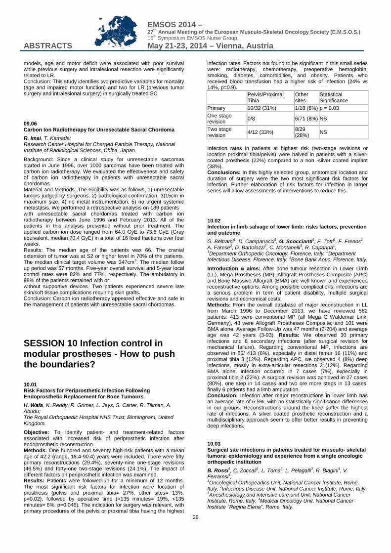
ABSTRACTS
EMSOS 2014 – 27th Annual Meeting of the European Musculo-Skeletal Oncology Society (E.M.S.O.S.) 15th Symposium EMSOS Nurse Group, May 21-23, 2014 – Vienna, Austria
29
models, age and motor deficit were associated with poor survival while previous surgery and intralesional resection were significantly related to LR. Conclusion: This study identifies two predictive variables for mortality (age and impaired motor function) and two for LR (previous tumor surgery and intralesional surgery) in surgically treated SC. 09.06 Carbon Ion Radiotherapy for Unresectable Sacral Chordoma R. Imai, T. Kamada; Research Center Hospital for Charged Particle Therapy, National Institute of Radiological Sciences, Chiba, Japan.
Background: Since a clinical study for unresectable sarcomas started in June 1996, over 1000 sarcomas have been treated with carbon ion radiotherapy. We evaluated the effectiveness and safety of carbon ion radiotherapy in patients with unresectable sacral chordomas. Material and Methods: The eligibility was as follows; 1) unresectable tumors judged by surgeons, 2) pathological confirmation, 3)15cm in maximum size, 4) no metal instrumentation, 5) no urgent systemic metastasis. We performed a retrospective analysis on 189 patients with unresectable sacral chordomas treated with carbon ion radiotherapy between June 1996 and February 2013. All of the patients in this analysis presented without prior treatment. The applied carbon ion dose ranged from 64.0 GyE to 73.6 GyE (Gray equivalent, median 70.4 GyE) in a total of 16 fixed fractions over four weeks. Results: The median age of the patients was 66. The cranial extension of tumor was at S2 or higher level in 70% of the patients. The median clinical target volume was 347cm3. The median follow up period was 57 months. Five-year overall survival and 5-year local control rates were 82% and 77%, respectively. The ambulatory in 98% of the patients remained with or without supportive devices. Two patients experienced severe late skin/soft tissue complications requiring skin grafts. Conclusion: Carbon ion radiotherapy appeared effective and safe in the management of patients with unresectable sacral chordomas.
SESSION 10 Infection control in modular prostheses - How to push the boundaries? 10.01 Risk Factors for Periprosthetic Infection Following Endoprosthetic Replacement for Bone Tumours H. Wafa, K. Reddy, R. Grimer, L. Jeys, S. Carter, R. Tillman, A. Abudu; The Royal Orthopaedic Hospital NHS Trust, Birmingham, United Kingdom.
Objective: To identify patient- and treatment-related factors associated with increased risk of periprosthetic infection after endoprosthetic reconstruction. Methods: One hundred and seventy high-risk patients with a mean age of 42.2 (range, 18.4-90.4) years were included. There were fifty primary reconstructions (29.4%), seventy-nine one-stage revisions (46.5%) and forty-one two-stage revisions (24.1%). The impact of different factors on periprosthetic infection was examined. Results: Patients were followed-up for a minimum of 12 months. The most significant risk factors for infection were location of prosthesis (pelvis and proximal tibia= 27%, other sites= 13%, p=0.02), followed by operative time (>135 minutes= 19%, <135 minutes= 6%, p=0.046). The indication for surgery was relevant, with primary procedures of the pelvis or proximal tibia having the highest
infection rates. Factors not found to be significant in this small series were: radiotherapy, chemotherapy, preoperative hemoglobin, smoking, diabetes, comorbidities, and obesity. Patients who received blood transfusion had a higher risk of infection (24% vs 14%, p=0.9).
Pelvis/Proximal Tibia
Other sites
Statistical Significance
Primary 10/32 (31%) 1/18 (6%) p = 0.03 One stage revision 0/8 6/71 (8%) NS
Two stage revision 4/12 (33%) 8/29
(28%) NS
Infection rates in patients at highest risk (two-stage revisions or location proximal tibia/pelvis) were halved in patients with a silver-coated prosthesis (22%) compared to a non -silver coated implant (38%). Conclusions: In this highly selected group, anatomical location and duration of surgery were the two most significant risk factors for infection. Further elaboration of risk factors for infection in larger series will allow assessments of interventions to reduce this. 10.02 Infection in limb salvage of lower limb: risks factors, prevention and outcome G. Beltrami1, D. Campanacci1, G. Scoccianti1, F. Totti1, F. Frenos1, A. Farese2, D. Bartolozzi2, C. Montanelli3, R. Capanna1; 1Department Orthopedic Oncology, Florence, Italy, 2Department Infectious Disease, Florence, Italy, 3Bone Bank Aouc, Florence, Italy.
Introduction & aims: After bone tumour resection in Lower Limb (LL), Mega Prostheses (MP), Allograft Prostheses Composite (APC) and Bone Massive Allograft (BMA) are well known and experienced reconstructive options. Among possible complications, infections are a serious problem in term of patient disability, multiple surgical revisions and economical costs. Methods: From the overall database of major reconstruction in LL from March 1996 to December 2013, we have reviewed 562 patients: 413 were conventional MP (all Mega C Waldemar Link, Germany), 48 were Allograft Prostheses Composite, and 101 were BMA alone. Average Follow-Up was 47 months (2-204) and average age was 42 years (3-93). Results: We observed 30 primary infections and 8 secondary infections (after surgical revision for mechanical failure). Regarding conventional MP, infections are observed in 25/ 413 (6%), expecially in distal femur 16 (11%) and proximal tibia 3 (12%). Regarding APC, we observed 4 (8%) deep infections, mostly in extra-articular resections 2 (12%). Regarding BMA alone, infection occurred in 7 cases (7%), expecially in proximal tibia 2 (22%). A surgical revision was achieved in 27 cases (80%), one step in 14 cases and two ore more steps in 13 cases; finally 6 patients had a limb amputation. Conclusion: Infection after major recostructions in lower limb has an average rate of 6.5%, with no statistically significance differences in our groups. Reconstructions around the knee suffer the highest rate of infections. A silver coated prosthetic reconstruction and a multidisciplinary approach seem to offer better results in preventing deep infections. 10.03 Surgical site infections in patients treated for musculo- skeletal tumors: epidemiology and experience from a single oncologic orthopedic institution B. Rossi1, C. Zoccali1, L. Toma2, L. Pelagalli3, R. Biagini1, V. Ferraresi4; 1Oncological Orthopeadics Unit, National Cancer Institute, Rome, Italy, 2Infectious Disease Unit, National Cancer Institute, Rome, Italy, 3Anesthesiology and intensive care unit Unit, National Cancer Institute, Rome, Italy, 4Medical Oncology Unit, National Cancer Institute "Regina Elena", Rome, Italy.
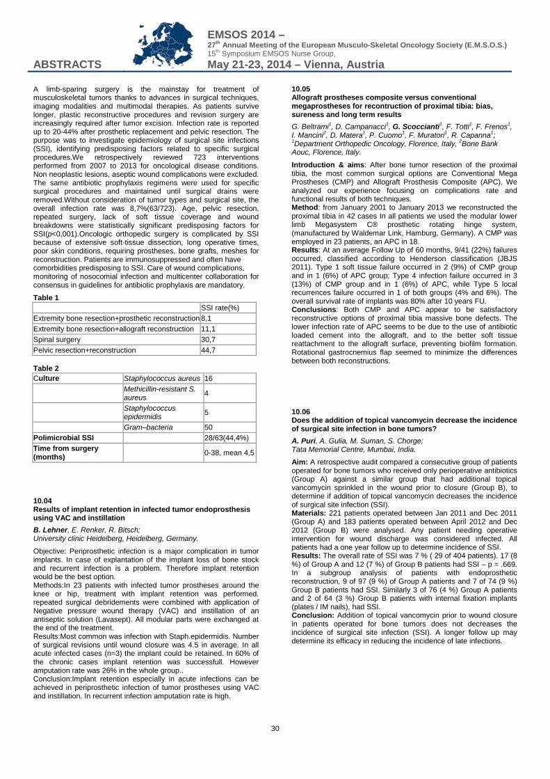
ABSTRACTS
EMSOS 2014 – 27th Annual Meeting of the European Musculo-Skeletal Oncology Society (E.M.S.O.S.) 15th Symposium EMSOS Nurse Group, May 21-23, 2014 – Vienna, Austria
30
A limb-sparing surgery is the mainstay for treatment of musculoskeletal tumors thanks to advances in surgical techniques, imaging modalities and multimodal therapies. As patients survive longer, plastic reconstructive procedures and revision surgery are increasingly required after tumor excision. Infection rate is reported up to 20-44% after prosthetic replacement and pelvic resection. The purpose was to investigate epidemiology of surgical site infections (SSI), identifying predisposing factors related to specific surgical procedures.We retrospectively reviewed 723 interventions performed from 2007 to 2013 for oncological disease conditions. Non neoplastic lesions, aseptic wound complications were excluded. The same antibiotic prophylaxis regimens were used for specific surgical procedures and maintained until surgical drains were removed.Without consideration of tumor types and surgical site, the overall infection rate was 8,7%(63/723). Age, pelvic resection, repeated surgery, lack of soft tissue coverage and wound breakdowns were statistically significant predisposing factors for SSI(p<0,001).Oncologic orthopedic surgery is complicated by SSI because of extensive soft-tissue dissection, long operative times, poor skin conditions, requiring prostheses, bone grafts, meshes for reconstruction. Patients are immunosuppressed and often have comorbidities predisposing to SSI. Care of wound complications, monitoring of nosocomial infection and multicenter collaboration for consensus in guidelines for antibiotic prophylaxis are mandatory.
Table 1
SSI rate(%) Extremity bone resection+prosthetic reconstruction 8,1 Extremity bone resection+allograft reconstruction 11,1 Spinal surgery 30,7 Pelvic resection+reconstruction 44,7 Table 2 Culture Staphylococcus aureus 16
Methicillin-resistant S. aureus 4
Staphylococcus epidermidis 5
Gram–bacteria 50 Polimicrobial SSI 28/63(44,4%) Time from surgery (months) 0-38, mean 4,5
10.04 Results of implant retention in infected tumor endoprosthesis using VAC and instillation B. Lehner, E. Renker, R. Bitsch; University clinic Heidelberg, Heidelberg, Germany.
Objective: Periprosthetic infection is a major complication in tumor implants. In case of explantation of the implant loss of bone stock and recurrent infection is a problem. Therefore implant retention would be the best option. Methods:In 23 patients with infected tumor prostheses around the knee or hip, treatment with implant retention was performed. repeated surgical debridements were combined with application of Negative pressure wound therapy (VAC) and instillation of an antiseptic solution (Lavasept). All modular parts were exchanged at the end of the treatment. Results:Most common was infection with Staph.epidermidis. Number of surgical revisions until wound closure was 4.5 in average. In all acute infected cases (n=3) the implant could be retained. In 60% of the chronic cases implant retention was successfull. However amputation rate was 26% in the whole group.. Conclusion:Implant retention especially in acute infections can be achieved in periprosthetic infection of tumor prostheses using VAC and instillation. In recurrent infection amputation rate is high.
10.05 Allograft prostheses composite versus conventional megaprostheses for recontruction of proximal tibia: bias, sureness and long term results G. Beltrami1, D. Campanacci1, G. Scoccianti1, F. Totti1, F. Frenos1, I. Mancini2, D. Matera1, P. Cuomo1, F. Muratori1, R. Capanna1; 1Department Orthopedic Oncology, Florence, Italy, 2Bone Bank Aouc, Florence, Italy.
Introduction & aims: After bone tumor resection of the proximal tibia, the most common surgical options are Conventional Mega Prostheses (CMP) and Allograft Prosthesis Composite (APC). We analyzed our experience focusing on complications rate and functional results of both techniques. Method: from January 2001 to January 2013 we reconstructed the proximal tibia in 42 cases In all patients we used the modular lower limb Megasystem C® prosthetic rotating hinge system, (manufactured by Waldemar Link, Hamburg, Germany). A CMP was employed in 23 patients, an APC in 18. Results: At an average Follow Up of 60 months, 9/41 (22%) failures occurred, classified according to Henderson classification (JBJS 2011). Type 1 soft tissue failure occurred in 2 (9%) of CMP group and in 1 (6%) of APC group; Type 4 infection failure occurred in 3 (13%) of CMP group and in 1 (6%) of APC, while Type 5 local recurrences failure occurred in 1 of both groups (4% and 6%). The overall survival rate of implants was 80% after 10 years FU. Conclusions: Both CMP and APC appear to be satisfactory reconstructive options of proximal tibia massive bone defects. The lower infection rate of APC seems to be due to the use of antibiotic loaded cement into the allograft, and to the better soft tissue reattachment to the allograft surface, preventing biofilm formation. Rotational gastrocnemius flap seemed to minimize the differences between both reconstructions. 10.06 Does the addition of topical vancomycin decrease the incidence of surgical site infection in bone tumors? A. Puri, A. Gulia, M. Suman, S. Chorge; Tata Memorial Centre, Mumbai, India.
Aim: A retrospective audit compared a consecutive group of patients operated for bone tumors who received only perioperative antibiotics (Group A) against a similar group that had additional topical vancomycin sprinkled in the wound prior to closure (Group B), to determine if addition of topical vancomycin decreases the incidence of surgical site infection (SSI). Materials: 221 patients operated between Jan 2011 and Dec 2011 (Group A) and 183 patients operated between April 2012 and Dec 2012 (Group B) were analysed. Any patient needing operative intervention for wound discharge was considered infected. All patients had a one year follow up to determine incidence of SSI. Results: The overall rate of SSI was 7 % ( 29 of 404 patients). 17 (8 %) of Group A and 12 (7 %) of Group B patients had SSI – p = .669. In a subgroup analysis of patients with endoprosthetic reconstruction, 9 of 97 (9 %) of Group A patients and 7 of 74 (9 %) Group B patients had SSI. Similarly 3 of 76 (4 %) Group A patients and 2 of 64 (3 %) Group B patients with internal fixation implants (plates / IM nails), had SSI. Conclusion: Addition of topical vancomycin prior to wound closure in patients operated for bone tumors does not decreases the incidence of surgical site infection (SSI). A longer follow up may determine its efficacy in reducing the incidence of late infections.
ABSTRACTS
EMSOS 2014 – 27th Annual Meeting of the European Musculo-Skeletal Oncology Society (E.M.S.O.S.) 15th Symposium EMSOS Nurse Group, May 21-23, 2014 – Vienna, Austria
31
10.07 The Role of Fibrin Sealant in Major Joint Reconstruction and Orthopaedic Tumour Surgery J. S. Palmer, Y. Ghani, P. Gikas, M. Cooper, R. Pollock; Royal National Orthopaedic Hospital, London, United Kingdom.
Major joint reconstruction and orthopaedic tumour surgery frequently involve extensive surgical dissection and the creation of large surgical wounds. Blood loss is a cause of significant morbidity in patients undergoing such procedures and bleeding at the surgical site can increase the risk of wound complications. A retrospective analysis of hospital records was performed to investigate the effect of a topical fibrin sealant (Evicel®) on post-operative blood loss and wound healing. The case notes of 41 patients who had received Evicel® intra-operatively were compared to a control group of 26 patients who underwent similar major orthopaedic surgical procedures. Our results demonstrate a significantly increased drain output (P=0.03), reduced blood transfusion requirement (P=0.015) and fewer major wound complications (P=0.045) in those patients who received Evicel® compared to the control group. There was no significant difference between Haemoglobin drop before and after surgery (P=0.09). This study demonstrates positive outcomes from the use of Evicel® in major joint reconstruction and orthopaedic tumour surgery. Its potential to reduce the need for post-operative blood transfusions and wound complications is encouraging. Reducing returns to theatre for wound complications has significant economic benefits for the hospital and health benefits for the patient. Whilst there is conflict in the literature over the use of fibrin sealants in primary arthroplasty we feel further investigation into its use for major joint reconstruction and orthopaedic tumour surgery is warranted. 10.08 Comparison of two alternative wound closure methods for tumor arthroplasty of the hip W. H. Hettwer, P. F. Horstmann, T. A. Grum-Schwensen, M. M. Petersen; Musculoskeletal tumor section, Department of orthopaedic surgery, Rigshospitalet, Copenhagen university hospital, Copenhagen, Denmark.
Introduction: Persistent wound drainage (PWD) is a known risk factor for periprosthetic joint infection. The optimal wound closure method, after tumor arthroplasty of the hip, is unknown. Methods: We prospectively recorded duration of PWD, administration of antibiotics and date of discharge in twenty-two patients who underwent tumor resection and endoprosthetic reconstruction of the proximal femur in our department in 2013 and had wound closure with a combination of intradermal suture, Steristrips and an occlusive skin adhesive. This was compared to standard wound closure with skin staples in an equally sized control group (n=22), matched for tumor type and extent of bone resection, with similar data collected either prospectively for patients operated in 2013 (n=6), or retrospectively for those operated in 2012 (n=16). Statistical evaluation: Student’s T-test for unpaired data. Results: Compared to the control group, we observed a significant reduction in most parameters in our prospective study group. Mean duration of PWD was reduced from 6.5 days to 3.4 days (p<0.0005), mean duration of post-operative administration of antibiotics was reduced from 6.5 days to 4.0 days (p=0.005) and mean hospital stay was reduced from mean 7.9 days to 6.2 days (p=0.07). While highly prevalent in the control group (n=7, 32%), PWD over 7 days was completely absent in the study group. Conclusions: A simple change in the wound closure routine showed a significant reduction in wound drainage and postoperative antibiotic administration. Further randomised prospective studies are warranted.
10.09 Successful disinfection of femoral head allograft using High Hydrostatic Pressure M. A. van de Sande, J. V. Bovee, E. Kuijper; LUMC, Leiden, Netherlands.
The current gold standard for sterilization allografts considered for tissue reconstruction, is associated with deterioration of mechanical, physical and biological properties of the implant. We evaluated application of High-Hydrostatic-Pressure(HHP) to sterilize bone grafts. 4 femoral heads were divided into 5 parts each, of which 16 were contaminated (duplicate) in a sterile plastic bag containing 100 ml of PBS with 105 CFU/ml of Staphylococcus-epidermidis, Bacillus-cereus, Pseudomonas-aeruginosa and Candida-albicans, respectively. Of each duplicate, one sample was untreated and stored similarly. The remaining 4 parts were included as sterile-control and non-infected-control. The 8 parts in plastic bags were vacuum-sealed and underwent HHP at 600 MPa. After HHP, serial dilutions were made and cultured on selective-media and into enrichment-media to recover low amounts of microorganism and spores. Cultures were incubated for 48 hours at 37C. The colony forming units of the inoculum were corrected for the cultured colonies from the untreated contaminated bags. Three complete femoral heads were treated with 0, 300 and 600 MPa HHP respectively for histological evaluation. None of the sterile control bone fragments contained microorganisms. The measured colony counts correlated excellent with the expected colony count. All HHP treated bone fragments did not grow on culture plates or enrichment media. Histological examination showed that the bone structure was unchanged by the HHP, while in treated specimens the cells were less viable . Sterilizing bone grafts by high hydrostatic pressure was successful and remains a promising technique with the possible advantage of retaining biomechanical properties of allograft tissue.
SESSION 11 Endoprosthetic reconstruction 11.01 Results of Mutars modular tumor prostheses: the experience of two Centers P. Ruggieri1, E. Pala1, A. Angelini1, T. Calabrò1, G. Trovarelli1, M. Maraldi1, I. Piraino1, M. Marcacci1, V. Ippolito2; 1University of Bologna, Istituto Ortopedico Rizzoli, Bologna, Italy, 2Orthopedic Oncologic, Ospedali Civili, Brescia, Italy.
Background: The current investigation includes a retrospective review of the experience of two Institutions on Mutars prostheses for bone tumors to assess the incidence and mode of failures. Methods: Between 2000 and 2013, 111 Mutars modular tumor prostheses were implanted: 38 upper limb and 73 lower limb reconstructions, 96 primary implants and 15 revision implants. Causes of endoprosthetic failure were classified according to Henderson et al. in 5 types. Functional results were analyzed according to the MSTS system, Kaplan-Meier curves of implant survival defined comparing primaries and revisions. Results: At last follow up, 73.9% of patients (82/111) had retained their implant. The overall failure rate in our series was 26.1% (29/111): 3 Type 1 failures, 6 Type 2 failures, 3 Type 3 failures, 12 Type 4 and 7 Type 5 failures. The overall implant survival to all types of failure was 55% and 33% at 5 and 10 years respectively with no significant difference between upper and lower limb (p = 0.22) and between primary and revision implants (p=0.69). Functional results, assessed according to MSTS system, were evaluated in 81 of 111 patients, the average score was 23.4. Conclusion: The most common cause of failure was infection, but the overall implant survival at long term and functional results were
ABSTRACTS
EMSOS 2014 – 27th Annual Meeting of the European Musculo-Skeletal Oncology Society (E.M.S.O.S.) 15th Symposium EMSOS Nurse Group, May 21-23, 2014 – Vienna, Austria
32
satisfactory. Revision procedures may be necessary for patients with good oncologic prognosis, and is need for continuous improvements in reconstruction techniques and implant design. However, the anatomic diversities in failure modes indicate that changes must be site-specific and failure-mode specific. 11.02 Good mechanical mid term outcome of the GMRS tumourprosthesis; analysis of 213 cases L. Elferink1, P. C. Jutte1, R. E. Stewart1, M. Stevens1, R. Windhager2, P. Funovics3; 1University Medical Center Groningen, Groningen, Netherlands, 2University of Vienna, Vienna, Austria, 3University of Vienna, Vienna, Netherlands.
The Global Modular Replacement System (GMRS) is a relatively new megaprosthesis (2004), used in the treatment of oncologic and revision patients. It is the successor of the Howmedica Modular Replacement System (HMRS). There have been no extensive reports in the literature about its performance. This study reports the mid-term mechanical outcome of the GMRS. A multicenter study was executed on 213 patients who received a GMRS with at least one-year follow-up. Forty cases were lost to follow-up, leaving 174 for analysis. Data on demographics, prosthesis components, complications and failures were collected. Kaplan-Meier statistic analyses were used to determine the survival of the prosthesis. Mechanical performance was good; there were 5 cases of aseptic loosening (2.9%) and no prosthesis breakage. Compared to results of the HMRS this can be considered an improvement. Overall prosthesis survival was 93.7%. Limb-salvage rate was 94.8%. The most common complications were fissures during implantation (8.7%) and infections (15.0%). The midterm performance of the GMRS is good, it is a reliable megaprosthesis, having a low incidence of mechanical failure. Although prosthesis survival is similar to HMRS results, the GMRS shows a decrease incidence of polyethylene wear, aseptic loosening and prosthesis breakage. 11.03 Prosthetic Reconstruction of the Limbs after Tumor Resection: 12 Years Follow Up in Ukraine. Two Institutions Experience O. Vyrva1, A. Diedkov2, G. Klymnyuk2; 1Sytenko Institute of Spine and Joint Pathology, Kharkiv, Ukraine, 2National Cancer Institute, Kiev, Ukraine.
This is 12 years clinical experience report of two Ukrainian Institutes with using of customized and modular tumor prosthesis. The authors present a review of 628 consecutive cases of prosthetic reconstruction performed during the last 12 years (2011-2013). All patients underwent prosthetic reconstruction with the GMRS (USA) - 51, MUTARS (Germany) - 47, INMED (Ukraine) - 384 and SIMEKS (Ukraine) - 146. The diagnosis was primary malignant tumors in 459 cases and metastases in 116 cases. For 53 the prosthesis were implanted as revision of failed prosthesis. Complications were reported and analyzed. X-ray, CT were reviewed and pertinent information achieved for each patient. Functional results were assessed according to MSTS score. The most frequent complication was implant infection which occurred in 14,2% of patients. The mechanical failure of the morse taper prosthetic body occurred in 13,5% requiring a surgical revision. Aseptic loosening was seen in 10,9%. At final follow up 88% of the evaluable patients presented a satisfactory functional result (excellent or good following MSTS) 74,6 ± 17,0 % for the lower and 67,6 ± 9,0 % for the upper limbs. The customized Ukrainian prosthesis systems are chipper then analogs and have individual advantages for Ukrainian patient population. The preliminary data of the presented series of patients showed satisfactory functional results which need to be confirmed by
a long term results. Modular systems seem definitely promising and reviewed design and material justifyits use in primary oncological replacements and complex revision of prosthetic failures with massive bone loss. 11.04 Proximal Femur Resection for Metastatic Lesions and Replacement by LIMA Prostheses: 30 years - Experience of one single surgeon in over 250 cases V. Ippolito; Oncologia Ortopedica, 1°Tr., Spedali Civili, Brescia, Italy.
Bone metastases are the most frequent tumor lesions of the proximal femur requiring surgery and, also for perithrocanteric and subtrochanteric lesions, resection and reconstruction by a modular prosthesis is the best option. Among metastatic patients undergoing proximal femur resection, almost half of them will not survive longer than 18 months. This implies 2 consequences: postoperative weight-bearing should not be delayed and the implant must remain stable even in case of local tumor-progression, therefore, only cemented prostheses should be implanted in metastatic lesions. The second consequence is related to the worldwide need for cost-containment: such a poor prognosis does not justify the cost of the expensive prostheses used for Primary tumors and using a “lower cost” solution makes a lot of sense in these patients. Following this concepts, the Author presents his own experience with the LIMA resection prostheses, starting in the early ‘80s, on over 250 cases which he treated personally. LIMA Resection Prostheses, now at the 3rd generation, are a Titanium-Made Modular system for the reconstruction of proximal femur and proximal humerus. Their technology is probably less sophisticated than the most diffused prostheses for Primary Tumors, but their cost is significantly lower and they represent an excellent solution for these patients. 11.05 Bipolar hip and proximal femur replacement using a modular prosthesis for neoplasms V. I. Sakellariou, V. Kontogeorgakos, O. Savvidou, N. A. Stavropoulos, A. F. Mavrogenis, P. J. Papagelopoulos; 1st Department of Orthopaedics, University of Athens, ATTIKON University General Hospital, Chaidari, Greece.
Introduction: Limb salvage has become an important alternative to amputation in the treatment of bone neoplasms. Modular endoprostheses offer off-the-shelf availability and can be adapted to most reconstructive situations. Aim: To evaluate the functional results of patients who were treated with modular prosthetic replacement for bone tumors of the proximal femur. Material & Methods: Forty patients (22 males, 18 females; mean age 51 years; range 18 to 76 years) who underwent wide resection and cemented bipolar hip and proximal femur replacement between 2003 and 2009 were retrospectively reviewed. Forteen patients had metastatic, and 26 patients had primary tumors. Results: The mean follow-up was 36 months (range 8 to 86 months). Postoperative complications were seen in 10 patients (25%), including delayed wound healing (4), drainage (4), dislocation (2). No patient needed revision surgery. No amputation was needed. Local recurrences were developed in three patients at 3, 8 and 12 months post surgery. Four patients died, three patients developed distant metastases, and thirteen patients were tumor-free. All the patients were able to ambulate without walking assistance 3 months postoperatively. The mean MSTS score was 62,9% (range 50% to 90%) in survivors, which was 74.5% (range 60% to 90%) for primary tumors, and 46.8% (range 30% to 75%) for metastatic tumors (p<0.001). Conclusions: Reconstruction with cemented modular endoprostheses is an appropriate surgical alternative in the
ABSTRACTS
EMSOS 2014 – 27th Annual Meeting of the European Musculo-Skeletal Oncology Society (E.M.S.O.S.) 15th Symposium EMSOS Nurse Group, May 21-23, 2014 – Vienna, Austria
33
treatment of segmental defects after resection of proximal femoral neoplasms. They are associated with intraoperative versatility, low incidence of implant-related complications and acceptable function particularly in primary tumors. 11.06 Extra-articular resection for malignant tumors of pelvis and proximal femur involving hip joint W. Guo, X. Tang, Y. Yang, T. Ji; Muskuloskeletal tumor center, Peking University People’s Hospital, Beijing, China.
Objective Very few reports specifically address the surgical technique of extra-articular resection of hip joint for patients with malignancy. The purpose of this study was to describe the surgical technique and evaluate the clinical outcome of this procedure. Methods Eleven patients with malignant tumors of pelvis or proximal femur involving hip joint were reviewed. There were eight males and three females with a mean age of 42.2 years. The tumors originated from pelvis in 3 patients, proximal femur in 7 patients, and soft-tissue of thigh in 1 patient. The pathologic diagnosis included 2 chondrosarcomas, 7 osteosarcomas, 1 Ewing sarcoma, and 1 soft-tissue sarcoma. All patients received extra-articular resection of hip. The bony defect was reconstructed by modular hemipelvic and proximal femoral prostheses. Results Eight patients got wide or marginal margins and three got intralesional margins. All patients were followed up for 10 to 37 months with a mean time of 19.7 months. Finally, four patients died of disease. Local recurrence occurred in 4 patients while lung metastasis was detected in 3 patients. The two-year disease-free survival rate was 71%. The complications included dislocation in one patient and deep infection in another patient. The postoperative MSTS 93 scores was 53% to 89% with a mean of 65.6%. Conclusions The extra-articular resection is a safe and effective surgical procedure for malignant tumors of pelvis and proximal femur involving hip joint. Using modular hemipelvic and proximal femur prostheses for the reconstruction of hip joint can achieve reasonable function with a low complication rate. 11.07 Gait analysis helps to understand failures in constrained tumor knee prostheses B. Lehner, A. Ayache, S. Jäger, R. Bitsch; University clinic Heidelberg, Orthopaedic department, Heidelberg, Germany.
Objectives: Mechanical failure as one of the biggest challenges of constrained tumor knee prostheses.. In this study we examined by gait analysis patients who had a replacement of a constrained knee prostheses because of mechanical failure or aseptic loosening. Methods: The gait analysis was performed on a set of 8 digital Real- Time Hawk cameras. In this study we examined 24 Patients after a revision of a constrained knee prosthesis.We analyzed the mean values and the range of motion of the differences of gait between the healthy and the operated limb and related these to the particular phase of gait. Results: All gait analyses showed significant differences between healthy and operated limbs in all gait phases. We see a significant hyperextension in the operated knee, which is increasing in the progress of the stance phase. During Mid stance and Terminal stance we observed a significant alternating load in medio- lateral direction . All through the stance phase we see a significant alternating load in anterior- posterior direction . Torque forces could be shown to be significantly higher in the operated knee. During the segment Loading response the hinge moment in proximal- distal direction is significantly greater in the operated knee.
Conclusion: Through 3D-gait analysis we could show the kinematic and biomechanical characteristics of constrained tumor knee prostheses and loads during movement could be measured in vivo. Hyperextension and bending moments as well as very high torque forces seem to be the most influential factors for mechanical failure. 11.08 Reversed shoulder endoprosthesis in patients with malignant tumours of the proximal humerus - report of four cases Y. Bakhshai1, A. Baur-Melnyck2, T. Knösel3, V. Jansson1, H. Dürr1; 1Department of Orthopedic Surgery, Munich, Germany, 2Institute for Clinical Radiology, Munich, Germany, 3Institute of Pathology, Munich, Germany.
Purpose: We investigated the functional and radiological outcome as well as complications in 4 patients with a reversed shoulder endoprosthesis after resection of the proximal humerus due to a malignant tumour. Methods: Between 2007 and 2013 four reversed shoulder endoprostheses were implanted in 2 male and 2 female patients after resection of the proximal humerus with preservation of the deltoid muscle and the axillary nerve. The average age was 55 years (SD 21.7; range 25-74). The resected bone had an average size of 8.8 cm (SD 3.4; range 6-13). The functional score was evaluated according to the range of motion and the functional score for the upper extremity according to the Musculoskeletal Tumor Society (MSTS). The mean follow up was 5.8 months (SD 3.6; range 1-9). Results: One patient had 2 luxations of the prosthesis (immediately after surgery). She had revision with a Trevira tube and showed no further luxations. In the other 3 patients there were no complications postoperatively. We saw no loosening. The mean active abduction was 130°, active elevation was 133°. The patient with the luxations was still wearing the gilchrist at latest follow up. The mean MSTS Score was 88.3% (SD 12.9, range 70-100). Conclusion: Reconstruction with a reversed shoulder endoprosthesis is an excellent and safe procedure for patients with a tumour of the proximal humerus if the deltoid muscle and the axillary nerve can be preserved. 11.09 Soft tissue reconstruction with LARS® tumor band improves shoulder function after intra-articular resection and prosthetic replacement of proximal humerus X. Tang, W. Guo, R. Yang, S. Tang, T. Ji, Y. Zhang; Muskuloskeletal tumor center, Peking University People’s Hospital, Beijing, China.
Objective We used a synthetic mesh to wrap the prosthesis of proximal humerus for soft tissue reconstruction, and asked whether these patients had better functional scores, wider ranges of motion (ROM) of the shoulder, and more stable joints compared with patients who underwent prosthetic reconstruction alone. Methods We reviewed 29 patients with tumors in proximal humerus, who underwent intra-articular resection and endoprosthetic reconstruction. The patients were divided into two groups: patient with soft tissue reconstruction using a LARS® tumor band (14 patients) and without soft tissue reconstruction using a LARS® tumor band (15 patients). The data from two groups were compared. Results The two groups did not significantly differ in terms of age, gender, follow-up time, length of humeral resection, blood loss volume, and incidence of complications. The mean MSTS score for patients without LARS® reconstruction was 19.87±3.44 points (66%), whereas that for patients with LARS® reconstruction was 23.71 ± 2.09 points (79%) (p=0.001). Patients with LARS® reconstruction had better ROM on mean active forward flexion (77°, p=0.020), abduction (68°, p<0.001), and external rotation (73°, p<0.001) than patients without LARS® reconstruction. Proximal migration of the prosthesis was observed in one third of patients in
ABSTRACTS
EMSOS 2014 – 27th Annual Meeting of the European Musculo-Skeletal Oncology Society (E.M.S.O.S.) 15th Symposium EMSOS Nurse Group, May 21-23, 2014 – Vienna, Austria
34
the group without LARS® reconstruction. In the group with LARS® reconstruction, all patients had stable shoulder joints (p=0.042). Conclusions Using a LARS® tumor band for soft tissue reconstruction in patients who undergo intra-articular resection and endoprosthetic replacement of the proximal humerus achieve better functional scores, wider active ROM of shoulder, and more stable joints.
SESSION 12 Free Papers 12.01 Chondrosarcoma and lymph node metastases B. Uhl1, J. Friesenbichler1, N. M. Woltsche1, B. Liegl-Atzwanger2, P. Tunn3, A. Leithner1; 1Univ. Klinik für Orthopädie und Orthopädische Chirurgie, Graz, Austria, 2Institut für Pathologie, Graz, Austria, 3HELIOS Klinikum Berlin-Buch, Berlin, Germany.
Introduction: Whereas in osteosarcoma the prevalence of lymph node metastasis is investigated and described with an incidence of less than 4% in recent literature, there are hardly publications found on lymph node metastases in chondrosarcoma. The aim of this two-center retrospective study was to calculate the incidence and analyse data for possible predilections and prognostic factors. Patients and methods: Data on 330 chondrosarcoma patients (center 1 198 patients, 78 G1, 74 G2, 46 G3; center 2 132 patients, x G1, y G2, z G3) was retrospectively analysed for reports on lymph node metastasis. In seven out of 330 patients (2.1%) lymph node metastases were reported (four G2 and three G3). Results: In three cases lymph nodes were found at first diagnosis, in two cases lymph nodes were affected four months after initial diagnosis and in two cases metastases were detected 11 and 40 months after initial diagnosis. All metastases were found in close proximity of the primary tumor site and the mean age was 64.7 years. Three patients died one, five and 23 months after diagnosis, two had no evidence of disease 36 and 48 months after operation and one with local recurrence and pulmonary metastases was still alive 15 months after diagnosis. One patient was lost to follow-up. Discussion: Surprisingly high grade chondrosarcoma patients with lymph node metastases seem not to have the same bad prognosis as those with pulmonary metastases. Therefore, a curative therapeutical intent should be chosen despite presence of lymph node metastases. 12.02 Clinical characteristics, therapeutic regimen and outcome of periosteal chondrosarcoma in the Netherlands L. M. Goedhart1, J. J. W. Ploegmakers1, H. M. Kroon2, E. C. H. Zwartkruis2, P. C. Jutte1; 1University Medical Center Groningen, Groningen, Netherlands, 2Leiden University Medical Center, Leiden, Netherlands.
The purpose of this study is to describe the clinical characteristics and therapy of a large series of patients with periosteal chondrosarcoma. Through the archive of the Netherlands Committee on Bone Tumours 36 patients were selected. Patient demographics, clinical characteristics, radiologic features, therapeutic regimen and follow-up were retrospectively described. Gender, age, size, location, histologic grading and treatment were compared in terms of survival. This series of periosteal chondrosarcoma showed a slight preference for males (61%), with the distal femur (33%) and proximal humerus (33%) as predominant sites. Clinically, periosteal chondrosarcoma presented most commonly with pain (44%). The metaphysis was predominantly affected (47%). Histological evaluation showed grade I chondrosarcoma in 50% of cases. In one case pulmonary metastases were reported after initial intralesional resection, another case resulted in death due to local recurrence and
possible pulmonary and skin metastases after contaminated resection. In conclusion we believe that it is important not to under diagnose these lesions, because incomplete resection results in local recurrence and even metastases. Staging for metastatic disease is recommended in grade II lesions or higher. It is recommended to have an adequate diagnosis including contrast-enhanced MRI of the tumour with histological confirmation, followed by en-bloc excision. 12.03 Intralesional treatment with phenol adjuvant for atypical cartiliginous tumour in the long bones: the Groningen experience in 79 patients J. Gerbers, E. Dierselhuis, P. Jutte; University Medical Center Groningen, Groningen, Netherlands.
Introduction: Chondrosarcoma grade I was renamed atypical cartilaginous tumour (ACT). Over the last decades, treatment has shifted from resection to curettage. Purpose of our study was to present an overview of our experience, with an emphasis on oncological outcome and complications. Patients and methods: A retrospective study was designed. Patients with ACT in the long bones treated with intralesional surgery and phenol as adjuvant were included. Patients treated with neo-adjuvant RFA or for recurrence were excluded. Reconstructions were done with PMMA or bonegrafts. Endpoints were local recurrence, residue and occurrence of complications. Tumour volume was approximately calculated. Follow-up was performed using MRI and radiographs. Results: A total of 79 patients were analyzed, with a mean follow-up of 36 months (range 2-85). A total of 73/79 patients (92.5%) had a disease free survival. In six patients, residual tumour was suspected, in two patients (2.5%) this was tested and confirmed. There were neither deaths due to disease, nor patients with metastases. Postoperative fracture occurred in 9/79 patients (11%). Other complications were infection (n=2), necrosis (n=1) and fissure (n=1). Mean calculated tumour volume was 32.3 cm3. Conclusion and perspectives: Low-grade chondroid tumours in the long bones can be effectively treated by curettage with adjuvant phenolisation. We found no local recurrence, although in six patients residual tumour was suspected, this was confirmed in two patients. The most frequent complication was a post-operative fracture. Consequently, a more aggressive osteosynthesis strategy was started. Studies with only radiographic follow-up might under-report on residue- and over-report on recurrence-rate. 12.04 Vascularized fibula autograft as salvage technique for complications of intercalary allograft reconstructions in bone tumors M. M. De Paolis, M. Manfrini, A. Biazzo, M. Romantini, N. Ali, C. Romagnoli, D. M. Donati; Rizzoli, Bologna, Italy.
Intercalary allograft reconstruction following bone tumor resection can be complicated by several problems such as nonunion, allograft fractures and infections. The authors report their experience using the vascularized fibula autograft (VFA) as salvage technique for failure of intercalary allograft reconstructions From 1995 to 2011, in 29 patients, 21 male and 8 female, with an average age of 25 years, VFA was used as salvage technique for the treatment of reconstructions’ failures. Causes of failures were mechanical (86%) or infection (14%). Twenty-five cases were free vascularized fibula and 4 were a pedicle grafts. In 25 cases reconstruction was obtained by massive bone allograft associated with VFA, in 4 with VFA alone. Implant outcome was assessed on serial radiology in all cases, with minimum follow-up of 24 months. Functional results were evaluated according MSTS score The consolidation at the bone-host interface was achieved in 24 cases at 6-12 months after surgery (83%). We report 5 cases of nonunion
ABSTRACTS
EMSOS 2014 – 27th Annual Meeting of the European Musculo-Skeletal Oncology Society (E.M.S.O.S.) 15th Symposium EMSOS Nurse Group, May 21-23, 2014 – Vienna, Austria
35
treated with new synthesis and autografts (3) and with new synthesis and cortical bone grafts (2), 1 infection treated with debridement and external fixator, 1 case of varus deformity of the proximal tibia treated with corrective osteotomy, 1 allograft fracture treated with new massive bone graft + endomedullary nail and 1 plate breakage treated with new synthesis. Functional results were satisfactory in 80% of patients. VFA is a valid salvage technique with a low complication rate and a high percentage of success and it ensures adequate mechanical and biological support. 12.05 Frozen hotdog update : Cryopreserved shell and vascular fibula combination for bone defect reconstruction H. Ozger1, B. Alpan2, N. Valiyev1; 1Istanbul University Istanbul Faculty of Medicine, ISTANBUL, Turkey, 2Acibadem Maslak Hospital, ISTANBUL, Turkey.
Biological reconstruction is the treatment of choice for bridging intercalary defects. Combination methods are preferred to provide biological and mechanical support simultaneously. We prefer to combine liquid-nitrogen treated tumor-bearing bone with free vascular fibula graft. Thirty six patients with a mean age of 14,1 years (5-38) underwent curative resection of malignant bone tumor followed by biological reconstruction with "frozen hotdog" technique. Patients were followed up for a mean period of 36,7 (6-96) months. The tumor was located in femur in 26 patients, tibia in 9 and humerus in 1. Bilateral fibula grafts were harvested in one patient to span whole femur. Fibula graft was harvested with vascular proximal epiphysis in one patient for intra-articular reconstruction. Plates were used for fixation in 33 patients, intramedullary nails in 2 and no implant was used in one case. Mean length of bony defect was 16.6 cm (9.0-26.0). Mean fibular graft length was 18.5 cm (10.0-25.0 cm). Complete union and full weight bearing was achieved in 16 patients. The mean time to detect radiological union at an osteotomy site was 8 months (6-12). Local recurrence was observed in 4 patients. Although problems related to nonunion or delayed union can be observed, vascularity of the fibular graft is the main determinant of successful treatment in the long term. Marginal safety is crtical for the oncologic outcome of these extreme reconstructions. 12.06 Functional and oncological outcome of condyle sparing resections for malignant proximal tibial tumours W. A. Ebeid, W. Abousenna; Cairo University, Cairo, Egypt.
In metaphyseal tumours of the proximal tibia, the resection cut may be so close to the joint leaving just a centimeter or less of the condyles. This would raise the question whether doing an intercalary resection preserving minimal amount of the condyle would offer a better functional outcome than intraarticular resections and the usual reconstructive modalities. The aim of this study was to evaluate the functional and oncological outcome of condyle sparing resections. 26 patients diagnosed with malignant tumours of proximal tibia were included in this study. Their average age was 17 years. Their pathological diagnosis was Ewing sarcoma (10 patients), osteosarcoma (5), parosteal osteosarcoma (4), adamantinoma (3), osteofibrous dysplasia (2), chondrosarcoma (2). Surgical resection with a wide margin was done in all patients. The proximal resection cut was <1cm below the joint line in 10 patients, 1 - 2cm below the joint line in 16 patients. The average size of the resultant defect was 15 cm. Reconstruction was done by pedicled vascularized fibular graft in 20 patients and recycling in 6 patients. The average follow up period was 64 month. The average MSTS functional score was 26. Local recurrence occurred in 3 patients and chest metastases in 2 patients. Complications included fixation
failure in 2 patients, stress fracture in 2 patients, foot drop in 2 patients, leg length discrepancy in 3 patients. Complications were all treated and did not affect limb survivorship. Condyle sparing resections are oncologically safe and offer an acceptable function meanwhile preserving the patient’s joint. 12.07 Is tibiofibular joint resection necessary to achieve wide margins in extraarticular knee resection for sarcomas? M. M. Gilg, C. Wibmer, A. Leithner; Medical University of Graz, Graz, Austria.
Introduction: Sarcomas within the knee joint require extraarticular resection to achieve wide margins. Differing opinions exist whether the superior tibiofibular joint (STFJ) is part of the knee joint and has to be removed when performing extraarticular resection of the knee. We conducted a literature review analysing the frequency of a communication between the tibiofemoral joint (TFJ) and the STFJ as well as studies about local recurrence rates (LRR) in extraarticular knee resection. Methods: A Pubmed based literature review using the terms “tibiofibular”, “joint”, “knee” and “anatomy” was conducted, detecting four cadaveric (12- 20 specimens) and one in vivo study (17 patients) between 1990 and 2013. Six studies analysing extraarticular knee resection in sarcomas involving the knee joint (9- 59 patients) between 2000 and 2013 were extracted. Results: Cadaveric studies detected a communication between the TFJ and STFJ in 10%- 64%. Direct arthrography with physical loading and delayed imaging verified a communication in all 17 patients. Regarding the extent of extraarticular knee resection two institutions did resect the STFJ (LRR 3-8%), two did not (LRR 0-9%), one decided depending on tumour location (LRR 14%) and in one study the extent was not clearly defined (LRR 11%). Conclusion: Literature provides evidence that the frequency of a communication between the TFJ and the STFJ is up to 100 %. Therefore the resection of the STFJ in patients with sarcomas involving the knee joint would be indicated, although it is not clear whether resection of the STFJ results in lower local recurrence rates. 12.08 Giant cell tumors of the sacrum - A nationwide study on midterm results in 26 patients after intralesional excision L. van der Heijden1, M. A. J. van de Sande1, I. C. M. van der Geest2, H. W. B. Schreuder2, B. J. van Royen3, P. C. Jutte4, J. A. M. Bramer5, F. C. Öner6, H. M. J. A. Kroon1, P. D. S. Dijkstra1; 1Leiden University Medical Center, Leiden, Netherlands, 2Radboud University Medical Center, Nijmegen, Netherlands, 3VU University Medical Center, Amsterdam, Netherlands, 4University Medical Center Groningen, Groningen, Netherlands, 5Academic Medical Center, Amsterdam, Netherlands, 6University Medical Center Utrecht, Utrecht, Netherlands.
Introduction: Only 2-8% of all giant cell tumors (GCT) are located in the sacrum. Surgical treatment is hampered by tumor size, soft tissue extension, spinal instability and involvement of sacral nerve roots. Optimal treatment is debatable. Aim of this study was to evaluate recurrences, complications and functional outcome after intralesional excision of sacral GCT. Methods: In this retrospective nationwide study, via the Netherlands Committee on Bone Tumors, all 26 patients who underwent surgery for sacral GCT were included (1990-2010). Median follow-up was 98 months (range 6-229). All patients underwent intralesional excision; 21 with local adjuvants, five radiotherapy, three IFN-α and one bisphosphonates. Functional outcome was assessed with use of Musculoskeletal Tumor Society (MSTS) score. Statistics were performed using Kaplan-Meier, log-rank, Cox regression, Mann-Whitney-U. Results: Recurrence rate was 14/26 after median 13 months (range 3-139), and was highest after isolated curettage (4/5; p=0.035). Soft tissue masses >10cm increased the risk of recurrence (HR=3.3,
ABSTRACTS
EMSOS 2014 – 27th Annual Meeting of the European Musculo-Skeletal Oncology Society (E.M.S.O.S.) 15th Symposium EMSOS Nurse Group, May 21-23, 2014 – Vienna, Austria
36
95%CI=0.81-13, p=0.09). Complications were reported in 12/26 patients and included various neurological deficits (n=15), massive intraoperative hemorrhage (n=4), infection (n=3), hardware failure (n=1), radiation-induced sarcoma (n=1) and radiation-induced menopause (n=1). Mean MSTS was higher in patients without complications (27 vs. 21; p=0.024). Conclusion: The risk of recurrence was high after curettage for sacral GCT, especially without use of any (local) adjuvants. Complications were common and impaired functional outcome. This indicates that local or systemic adjuvant treatment is desired to obtain immediate local control, as this would likely result in reduced recurrence and complication risks and superior functional outcome. 12.09 Tumors of the hand and wrist: a single institution experience P. Ruggieri, A. Angelini, E. Pala, T. Calabrò, G. Trovarelli, M. Maraldi, S. Zaffagnini, M. Marcacci; University of Bologna, Istituto Ortopedico Rizzoli, Bologna, Italy.
Introduction: Aim of this study was to evaluate incidence, histologic features and treatment strategy of the tumors involving hand and wrist. Methods: Between 1900 and 2007, 1491 tumors of the hand or the wrist were retrospectively analyzed. Imaging included radiographs in all patients, and CT or MRI when feasible. All histologic slides were reviewed. The lesions arose from soft tissue in 12 cases, and from bone in the remaining cases. Results: Benign or pseudotumoral lesions were 1372 (92%): multiple chondromas (499), enchondroma (362), giant cell tumor (141), osteoid osteoma (71), solitary osteochondroma (57),“brown tumors” in primary hyperparathyroidism (54), aneurysmal bone cyst (50), reparative giant cell granuloma (26), periosteal chondroma (21), and other more rare entities. Malignant lesions were 119 (8%): metastatic carcinoma (28), osteosarcoma (29), chondrosarcoma (26), Ewing’s sarcoma (17), and other more rare entities. Localizations were phalanges (756; 50%), metacarpal region (369; 25%), and the wrist (366; 25%). Benign lesions were generally treated with curettage +/- bone grafting. Giant cell tumors recurred frequently after curettage. For malignant lesions, chemotherapy is required pre- and postoperatively, and especially when the tumor involved the phalanges, partial or total amputation may be required. Conclusions: Malignant tumors are relatively rare, but we re-emphasize the need to maintain a high level of attention, even when diagnosis seems straightforward. Whenever there is a doubt on the lesion, a biopsy should be done before proceeding with definitive surgery. In spite of the complex anatomy, adherence to proper oncologic principles most often will lead to a satisfactory outcome.
NURSE DAY N01 The patient N01.01 Care for Adolescents and Young Adults: Improving patients’ lives together P. Veldman -Goossen; Radboud university medical centre, Department of orthopaedic surgery, Nijmegen, Netherlands.
AYA stands for Adolescents and Young Adults, based upon the population between 18 and 35 years old who have or have had cancer. Every year 2200 new AYA’s in the Netherlands hear that they have cancer. The AYA-age involves many facets, such as starting a new study, searching for a job, buying a house, starting a family. Because of the low incidence of cancer at “AYA-age”, AYA’s are somewhat isolated. The Radboud University Medical Centre in Nijmegen (the Netherlands) created an AYA centred healthcare vision, and the
implementation of this vision. The aim is to improve care and treatment of AYA’s, supported by research and education, by co-creation and co-design of AYA’s and dedicated professionals. This complementary co-creation has already resulted in: 1. A hospital wide integrated and AYA centered care concept 2. The adolescent and young adult with cancer outpatient clinic 3. The young adult hangout spot in the hospital 4. The online community AYA4 for young adults with cancer 5. Enhancement of research 6. The embedding of age related and patient participation issues in education of health care professionals The aim for the future is to develop, together with patients and care professionals, comprehensive and age-specific centred care for young adults with cancer and their caregivers. In order to sustainably improve the life expectancy and quality of life of AYAs in the Netherlands information and knowledge should be shared. N01.02 Procedure of informed assent/consent for clinical trials in pediatric Hemato-Oncology: the adolescents’ and parents’ point of view V. Van de Velde1, E. De Strooper1, A. Morez1, C. Dhooge2; 1Pediatric Hemato-Oncology and Stemcelltransplant Unit, Gent, Belgium, 2Pediatrie - Hemato-Oncologie en Stamceltransplantatie, Gent, Belgium.
Objective: Clinical trials require an assent by the adolescents to participate. Few literature data are available on the experiences expressed by adolescents. A study has been conducted to explore the personal views of adolescents and their parents in order to optimize this consent procedure. Methods: Adolescents (n=10) and their parents (n=15) completed a questionnaire and were interviewed. Qualitative data were coded using NVIVO 8. Quantitative data were analyzed by the statistic program SPSS. Results: All adolescents (>13 years) want to be informed about the trial. The majority of them want to co-decide. They want this from an average of 10 years (SD=2,88) while parents indicate an average of 12,5 years (SD=2,30). Adolescents believe that their decision should be accompanied by consultations with parents and physicians. Parents are in favour of a unilateral decision only made by themselves (significant difference between adolescents and mothers, p=0,008) or by a physician. Adolescents and parents experience mostly positive emotions. Important preconditions for the adolescents were: presence of their parents and structured and clear approach by the physician during the IC interview. The majority of the adolescents wanted information: as much as possible, adapted to their developmental level. There was a significant difference regarding the amount of information: boys wanted more information than girls (p=0,033). Both adolescents and parents are positive about participating in clinical trials: it’s important to improve cancer treatment and it may help others. Reported suggestions for improvement: no coincidence between IC and the diagnostic interview, more written/oral information for adolescents.Conclusion: Despite this small sample, the results give us an insight into the adolescents’ and parents’ point of view. These results are a first step towards the adaptation and the improvement of the departmental guidelines and towards the adjustment of the written information for adolescents about their participation in clinical trials. N01.03 Retrospective review of rehabilitation and functional outcomes of patients with primary malignant bone sarcomas in England S. A. Hudson; Royal National Orthopaedic Hospital Trust, Stanmore, United Kingdom.
Background: This Quality Improvement Development and Innovation Scheme project was commissioned by National Specialised Commissioning Group to review post-operative rehabilitation
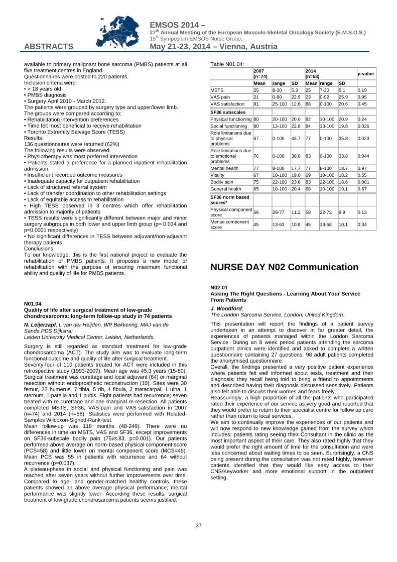
ABSTRACTS
EMSOS 2014 – 27th Annual Meeting of the European Musculo-Skeletal Oncology Society (E.M.S.O.S.) 15th Symposium EMSOS Nurse Group, May 21-23, 2014 – Vienna, Austria
37
available to primary malignant bone sarcoma (PMBS) patients at all five treatment centres in England. Questionnaires were posted to 220 patients. Inclusion criteria were: • > 18 years old • PMBS diagnosis • Surgery April 2010 - March 2012. The patients were grouped by surgery type and upper/lower limb. The groups were compared according to: • Rehabilitation intervention preferences • Time felt most beneficial to receive rehabilitation • Toronto Extremity Salvage Score (TESS) Results: 136 questionnaires were returned (62%) The following results were observed: • Physiotherapy was most preferred intervention • Patients stated a preference for a planned inpatient rehabilitation admission. • Insufficient recorded outcome measures • Inadequate capacity for outpatient rehabilitation • Lack of structured referral system • Lack of transfer coordination to other rehabilitation settings • Lack of equitable access to rehabilitation • High TESS observed in 3 centres which offer rehabilitation admission to majority of patients • TESS results were significantly different between major and minor surgery subgroups in both lower and upper limb group (p= 0.034 and p=0.0001 respectively) • No significant differences in TESS between adjuvant/non-adjuvant therapy patients Conclusions: To our knowledge, this is the first national project to evaluate the rehabilitation of PMBS patients. It proposes a new model of rehabilitation with the purpose of ensuring maximum functional ability and quality of life for PMBS patients. N01.04 Quality of life after surgical treatment of low-grade chondrosarcoma: long-term follow-up study in 74 patients N. Leijerzapf, L van der Heijden, WP Bekkering, MAJ van de Sande,PDS Dijkstra; Leiden University Medical Center, Leiden, Netherlands.
Surgery is still regarded as standard treatment for low-grade chondrosarcoma (ACT). The study aim was to evaluate long-term functional outcome and quality of life after surgical treatment. Seventy-four of 110 patients treated for ACT were included in this retrospective study (1993-2007). Mean age was 45.3 years (15-80). Surgical treatment was curettage and local adjuvant (64) or marginal resection without endoprosthetic reconstruction (10). Sites were 30 femur, 22 humerus, 7 tibia, 5 rib, 4 fibula, 2 metacarpal, 1 ulna, 1 sternum, 1 patella and 1 pubis. Eight patients had recurrence; seven treated with re-curettage and one marginal re-resection. All patients completed MSTS, SF36, VAS-pain and VAS-satisfaction in 2007 (n=74) and 2014 (n=58). Statistics were performed with Related-Samples Wilcoxon-Signed-Rank-test. Mean follow-up was 118 months (48-249). There were no differences in time on MSTS, VAS and SF36, except improvements on SF36-subscale bodily pain (75vs.83, p=0.001). Our patients performed above average on norm-based physical component score (PCS=58) and little lower on mental component score (MCS=45). Mean PCS was 55 in patients with recurrence and 64 without recurrence (p=0.037). A plateau-phase in social and physical functioning and pain was reached after seven years without further improvements over time. Compared to age- and gender-matched healthy controls, these patients showed an above average physical performance; mental performance was slightly lower. According these results, surgical treatment of low-grade chondrosarcoma patients seems justified.
Table N01.04:
2007 (n=74)
2014 (n=58) p-value
Mean range SD Mean range SD MSTS 25 8-30 5.3 25 7-30 5.1 0.19 VAS pain 21 0-80 22.9 23 0-92 25.9 0.95 VAS satisfaction 91 25-100 12.6 88 0-100 20.6 0.45 SF36 subscales Physical functioning 80 20-100 20.0 82 10-100 20.9 0.24 Social functioning 80 13-100 22.8 84 13-100 19.8 0.026 Role limitations due to physical problems
67 0-100 43.7 77 0-100 35.8 0.023
Role limitations due to emotional problems
76 0-100 38.0 83 0-100 33.8 0.044
Mental health 77 8-100 17.7 77 8-100 18.7 0.97 Vitality 67 10-100 19.0 69 10-100 18.2 0.55 Bodily pain 75 22-100 23.6 83 22-100 18.6 0.001 General health 65 10-100 20.4 68 10-100 19.1 0.67 SF36 norm based scores* Physical component score 56 29-77 11.2 58 22-73 9.9 0.13
Mental component score 45 13-63 10.8 45 13-58 10.1 0.34
NURSE DAY N02 Communication N02.01 Asking The Right Questions - Learning About Your Service From Patients J. Woodford; The London Sarcoma Service, London, United Kingdom.
This presentation will report the findings of a patient survey undertaken in an attempt to discover in far greater detail, the experiences of patients managed within the London Sarcoma Service. During an 8 week period patients attending the sarcoma outpatient clinics were identified and asked to complete a written questionnaire containing 27 questions. 98 adult patients completed the anonymised questionnaire. Overall, the findings presented a very positive patient experience where patients felt well informed about tests, treatment and their diagnosis; they recall being told to bring a friend to appointments and described having their diagnosis discussed sensitively. Patients also felt able to discuss their worries and fears freely. Reassuringly, a high proportion of all the patients who participated rated their experience of our service as very good and reported that they would prefer to return to their specialist centre for follow up care rather than return to local services. We aim to continually improve the experiences of our patients and will now respond to new knowledge gained from the survey which includes; patients rating seeing their Consultant in the clinic as the most important aspect of their care. They also rated highly that they would prefer the right amount of time for the consultation and were less concerned about waiting times to be seen. Surprisingly, a CNS being present during the consultation was not rated highly, however patients identified that they would like easy access to their CNS/Keyworker and more emotional support in the outpatient setting.
ABSTRACTS
EMSOS 2014 – 27th Annual Meeting of the European Musculo-Skeletal Oncology Society (E.M.S.O.S.) 15th Symposium EMSOS Nurse Group, May 21-23, 2014 – Vienna, Austria
38
N02.03 What should nurses do? - Communication in the oncology outcome C. Trost; General Hospital of Vienna, Department of Orthopedics, Vienna, Austria.
What is communication? In the system theory according to Luhman communication develops by selecting: • Information • Message and • Understanding Communicative contexts for the invalid treatment according to Vogd (2011): 1) The sick body 2) Communication on the level of emotion 3) Communicative anchorage points on artefacts 4) The organisation (here : hospital) has the means to influence communication by making formal decisions. 5) Network. The actor status is determined as communicative and its value is determined by the assessment of other participants. 6) Smell. Smell releases emotions. These facts interact with the medicine-patient-communication. Especially in the patient outcome the communication is difficult, because of less time, plenty of interruptions and examinations etc. To boot too much information in a short time. Especially regarding the oncological outcome. What should nurses do? • Nurses should support the patient: • Take the anxiety by asking questions • Ask if all points are clear • Avoid interruptions
NURSE DAY N03 Contacts N03.01 Cancer Rehabilitation in Austria R. Crevenna; Department of Physical Medicine and Rehabilitation, Medical University of Vienna, Vienna, Austria.
In Austria, cancer rehabilitation is an important issue in the management of cancer patients. Today, in Austria there are about 500 beds for inpatient rehabilitation. An outpatient clinic for cancer rehabilitation was developed fifteen years ago at the Department of Physical Medicine and Rehabilitation of the Medical University of Vienna. Two further outpatient rehabilitation concepts are at the beginning in Upper Austria. Rehabilitation concepts for cancer patients have to be individually tailored depending on their individual needs. They have to include specific nutrition programs, psychotherapy (so called “psychooncology”), and different options and modalities from the field of Physical Medicine and Rehabilitation, which may help these patients to improve their functional health, and independence in daily activities, and to maintain social participation. These individual tailored rehabilitation plans typically include medical exercise programs, neuromuscular electrical stimulation (as an passive option to exercise), nutrition, lymph massage, breathing therapy, physiotherapy, occupational therapy, breathing therapy, different forms of massage, electrotherapy, and other physical modalities, but also biofeedback and drug treatment for pain. At the Medical University of Vienna, challenging and complex cases of cancer patients are presented and discussed with the goal to plan rehabilitation, within the worldwide first official tumour board for cancer rehabilitation. Regular physical activity is an active option which has been shown to be an important part in the treatment and rehabilitation of cancer patients. Exercise - individually prescribed - has been described to improve functional health of cancer patients
with benefits for physical performance, mental health, quality of life, participation, and - in some types of cancer - survival. However, not all cancer patients need the same form of regular physical activity. Nevertheless, most of cancer patients should start as soon as possible to be physically active, but under the supervision of specialized physicians and within their individual “right” setting. N03.02 Sexuality and cancer G. Fraberger; General Hospital of Vienna, Department of Orthopedics, Vienna, Austria.
Love and sexuality can be described as “Sexual expression and is a fundamental human need, a near-universal experience, a keystone of intimate human relationships and a prerequisite of procreation”. (Johnson, Wadsworth, Wellings & Field, 1994). As one’s sexual life is important in reflecting the re-integration, consequently its evaluation is important. It has been said that the brain is the ultimate sexual organ: the seat of sexual urges, thoughts, sensations, inhibitions, and behaviours (Griffith & Lemberg, p.71, 1993). Whenever cancer is related to fear, pain, suffering, loss of control and a painful death, thoughts about the future can take away any motivation to participate actively in social activities leading to love and sex. Forgetting fundamental needs has a fatal effect on a patient‘s motivation to survive. We present various difficulties depending on the time of diagnosis (immediate vs. long-term reaction). Information is crucial in order to cope with the disease. N03.03 Triangle: Study Nurse - Investigator - Patient - Information C. Trost; General Hospital of Vienna, Department of Orthopedics, Vienna, Austria.
The occupation of a Study Nurse is based on the Good Clinical Practice (GCP) and national laws (eg the Medical Act). The maintask is the organization and administration of clinical trials. These include providing support and follow-up for patients, the organization of diagnostic material, sample shipment, investigational drugs and the documentation. A very important task is to talk with the patient about his health status and how he or she feels. To document this and talk with the investigator, this information is important for the study, and how we can help the patient. Especially, in oncological clinical studies. • Does the patient need an analysis? • Or other support? So the very important job of a Study Nurse is to talk with the patients and give the essential information to the investigator. In order for a clinical study to be successful many necessary points have to be determined: Soft skills such as communication, knowledge of GCP, teamwork of all persons involved.
ABSTRACTS
EMSOS 2014 – 27th Annual Meeting of the European Musculo-Skeletal Oncology Society (E.M.S.O.S.) 15th Symposium EMSOS Nurse Group, May 21-23, 2014 – Vienna, Austria
39
NURSE DAY N04 Psycho oncology N04.01 Psychosocial consequences and pain after amputation: an Austrian long term survey C. M. Armbruster, G. Fraberger, R. Windhager; Department for Orthopaedic Surgery, General Hospital, Vienna, Austria.
After amputation, several pain and mental phenomena as i.e. phantom limb pain (PLP) and major depressive episodes are being observed (Kern et al., 2009, Seidel et al., 2006). As a consequence of amputation and pain sensation, patients´ quality of life is often reduced (Willrich et al., 2005). This can result in long term mental disorders as depression or substance abuse. Therefore, a survey has been done where data about amputees was being collected at the Department of Orthopedics (MUV). The questions included i.e. type and reason of amputation, pain, amputation management, psycho-social and demographic data, quality of life, social activities and drug abuse. The survey started in 2003 and is containing patient data until beginning of 2013. The survey is focusing mainly on physical phenomena in amputees like PLP and stump pain and on the interrelated psychological constructs. The survey shall emphasize the consequences of amputation in patients´ daily routine and quality of life. First outcomes show PLP in a significant majority of amputees after surgery. Furthermore, significant correlations between amputation and an increased depression rate such as substance abuse can be shown. N04.02 Evaluating Patient Satisfaction in Orthopedic Oncology L. Doornbos, I. Egberst-Vree, N. Tiemes, P. Jutte; UMCGroningen, Groningen, Netherlands.
Background: The orthopedic oncology is a growing branch of orthopedics in our centre, the UMCG. All patients that have a benign or malignant bone disease are seen in 4 centers in the Netherlands. Often the diagnosis is still uncertain at first contact. This orthopedic oncology patient goes through an intensive process. The seriousness of the disease and insecurity of the patients requires an accurate and tight communication and organization. Since 2006 is structurally nursing support added to cancer care. Aim: The patient satisfaction (the views of patients about the care provided) is measured. The results are being used to further improve care and to obtain insight in patients needs. Method: A questionnaire was developed that covers the entire care process. The questions are divided into prehospital care, inpatient care and outpatient care. The Core Questionnaire Patient Satisfaction in Academic hospitals (KPAZ) is used as basis, supplemented with additional questions focused on the orthopedic oncology patients. The results were benchmarked with all orthopedics departments in the national research. The results are a reference for evaluation after a 5 years. Result: We have chosen to focus on aspects which the patients were satisfied or dissatisfied about. The analysis of the results shows strong and weak points. The e-mail service was highly appreciated as well as our patient information. Following the results of the measurement some improvements were made.
N04.03 Psychooncology in clinical practice G. Fraberger; General Hospital of Vienna, Department of Orthopedics, Vienna, Austria.
Psycho-oncology is a special part of psychology in the field of oncological diseases . It deals with interdisciplinary study and practice at the intersection of lifestyle, psychology and oncology. The need to include psychological aspects in addition to the medical treatment comes from a high rate of emotional stress, depression, anxiety und fatigue in cancer patients. Depending on the kind of cancer around 25 % of patients suffer from psychological problems like a manifest depression or an anxiety disorder. This does not only affect the patient’s QOL but also increases the mortality rate. In orthopaedic patients psychological difficulties can arise for two different reasons: 1. cancer related questions like surviving, suffering, and living healthy, 2. questions related to the challenge after orthopaedic surgery affecting people’s physical functioning and thus their range of movement as well as their body image. Whenever surgical treatment is necessary patients start questionning their personal independency as well as mobility and their activities of daily life. The underlying problem of these functional related aspects of cancer but also of the question of the chance of surviving or of finding a healthy behaviour and life style seems to be that people think and act future orientated. Best known is the level of stress for patients with breast cancer. For patients with bone cancer information about emotional burden und psychosocial difficulties are rae. We present our experience in dealing with stress and compliance in the daily life. Psychooncology thus focuses on aspects of cancer that go beyond medical treatment and include lifestyle, psychological and social aspe cts of cancer.
ABSTRACTS
EMSOS 2014 – 27th Annual Meeting of the European Musculo-Skeletal Oncology Society (E.M.S.O.S.) 15th Symposium EMSOS Nurse Group, May 21-23, 2014 – Vienna, Austria
40
POSTER PRESENTATIONS
P01 Osteosarcoma/Ewing’s sarcoma - What’s new in targets and innovative therapies? P01.01 Extraskeletal osteosarcoma: a review of 10 patients treated with multimodal therapy D. Campanacci1, F. Frenos1, A. Vilardi2, G. Beltrami1, D. Matera1, A. Franchi1, R. Capanna1; 1Azienda Ospedaliera Universitaria Careggi, Firenze, Italy, 2Università di Messina, Messina, Italy.
BACKGROUND: Extraskeletal osteosarcoma is a rare high grade soft tissue neoplasm and a more aggressive biologic behavior than classic osteosarcoma has been described. In this study we analyzed the clinical outcome of our series of patients treated with different associations of surgery, chemotherapy and radiationtherapy. METHODS: We analyzed 10 patients affected by extraskeletal osteosarcoma treated from 1998 to 2013. They were 8 males and 2 females with a mean age at diagnosis of 51 years (18-82). The lower limb was involved in 6 cases and upper limb in 4. All patients but one were treated by limb sparing surgery. Surgical margins were wide in 5 patients and marginal in 5. Two patients received only surgery. One patient received adjuvant chemotherapy and 2 patients adjuvant radiationtherapy. In 5 cases surgery was associated to chemotherapy and radiationtherapy (3 brachytherapy). RESULTS: At an average follow up of 44 months (3-170), 4 patients were continuously disease free, 2 had no evidence of disease after treatment of local relapse, 1 was alive with disease, 1 was dead of disease and 2 dead of another cause. Local recurrence occurred in 2 cases treated by new excisions. Two cases developed metastases. No surgical complications was observed. CONCLUSIONS: In conclusion, in our experience, extraskeletal osteosarcoma affected an adult population and adequate surgical margins could be achieved in 50% of cases. Multimodal treatment combining surgery, radiationtherapy and chemotherapy showed a low morbidity allowing disease control in half of the patients. P01.02 Primary Malignant Bone Tumors - 25 Years Review I. M. Balaco, C. M. Alves, P. S. Cardoso, T. P. Ling, G. Matos; Hospital Pediatrico Coimbra, Coimbra, Portugal.
Objective: The authors present a series of primary malignant bone tumors of the pediatric population, treated in the same institution during last 25 years. They analyze and correlate variables that seem to have predictive value in the prognosis of these patients. Material and Methods: Retrospective analysis of a series of primary malignat bone tumors, with a minimum follow-up of 3,5 year, divided in two groups according to the diagnosis (Osteosarcoma and Ewing Sarcoma). Overall survival was correlated for each group with the gender, age, diagnosis, anatomic location, Enneking classification, chemotherapy protocol, surgical margins and pathology results. Results: 68 primary malignant bone tumors, 39 Osteosarcomas and 29 Ewing Sarcomas. Anatomic location and Enneking classification have a predictive value for the prognosis in the two groups. Chemotherapy protocol and pathology results only have significance in the Osteosarcoma group. Overall survival curve showed a decrease to 73,5% at 2 years and to 63,2% at 5 years. Overall survival for Osteosarcomas diminishes to 73,3% and stabilizes after 2 years, for Ewing Sarcomas the curve diminishes progressively to 46,7% until 5 years, and stays steady after. Discussion: Primary bone tumors represent about 13% of the malignant tumors in the pediatric population. Chemotherapy
protocols nowadays permit limb salvage surgery in almost all cases. Peripheral high grade Osteosarcoma, with no evidence of metastasis, still has best prognosis. Conclusion: The current series shows the overall survival results similar to literature. Achieving free surgical margins and high percentage of tumor necrosis guaranties the best results in Osteosarcoma group. P01.03 Agricultural land usage and bone cancer: is there a link? Small-area analyses of osteosarcoma and Ewing sarcoma diagnosed in 0-49 year olds in Great Britain, 1985-2009 K. Blakey1, R. Feltbower2, R. Parslow2, P. James1, C. Stiller3, P. Norman2, D. Garthwaite4, J. Hughes5, C. H. Gerrand6, R. McNally7; 1Newcastle University, Newcastle Upon Tyne, United Kingdom, 2University of Leeds, Leeds, United Kingdom, 3University of Oxford, Newcastle Upon Tyne, United Kingdom, 4Food and Environment Research Agency, York, United Kingdom, 5Science and Advice for Scottish Agriculture, Edinburgh, United Kingdom, 6Freeman Hospital, Newcastle Upon Tyne, United Kingdom, 7University of Newcastle, Newcastle Upon Tyne, United Kingdom.
Aim: This population-based study analysed putative associations between Ewing sarcoma (ES) and osteosarcoma incidence and agricultural land usage. Methods: All osteosarcoma and ES cases aged 0-49 years diagnosed in Great Britain during 1985-2009 were included. Pesticide data were a proxy for agricultural land usage and population density an urban/rural indicator. Negative binomial regression was used to examine small area relationships between incidence rates and pesticide levels. The models adjusted for gender, age and deprivation. The logarithm of the ‘at risk’ population was used as an offset. Results: There were 2562 osteosarcoma cases aged 0-49; 820 aged 0-14; 1262 aged 15-29 and 480 aged 30-49 years, with overall age-standardised incidence rate of 2.84 per million persons per year (95% confidence interval [CI] 2.73-2.95). For ES there were 1711 cases aged 0-49; 670 aged 0-14; 822 aged 15-29 and 219 aged 30-49 years, with overall age-standardised incidence rate of 1.97 per million persons per year (95% CI 1.88-2.07). After adjustment for gender, age and deprivation, pesticide usage was not found to have any significant effect on the incidence of osteosarcoma or ES. For osteosarcoma, the relative risk (RR) for one kilogram per hectare increase in pesticide level = 0.987 (95% CI 0.954-1.022) and for ES, RR = 1.011 (95% CI 0.967-1.057). Conclusion: No association with land usage and ES or osteosarcoma was found. Other agricultural related factors could explain the higher incidence found in less urbanized areas. Further research should investigate other activities such as livestock farming. P01.04 Period detection of metastases as a prognostic factor in metastatic osteosarcoma recurrence A. Diedkov, P. Kovalchuk, S. Bojchuk, V. Kostyuk; National cancer institut, Kiev, Ukraine.
Background: Approximately 35% of patients with osteosarcoma have lung metastasis. Usage of second line chemotherapy and the possibility of surgical removal of metastasis brings a positive result. Goal: To determine the dependence of the period of development of lung metastases and treatment outcomes. Methods: 272 patients with localized osteosarcoma, treated between 2003 and 2012 were identified. 97 patients had pulmonary metastasis after the treatment. Metastases were detected between 6 and 38 months after the treatment (median - 22.3 months). All patients received second line chemotherapy. 37 (38.1%) patients underwent surgical treatment (metastasectomy with full metastasis removal). Other 60 (61.8%) patients were considered as inoperable
ABSTRACTS
EMSOS 2014 – 27th Annual Meeting of the European Musculo-Skeletal Oncology Society (E.M.S.O.S.) 15th Symposium EMSOS Nurse Group, May 21-23, 2014 – Vienna, Austria
41
and underwent palliative radiation therapy or symptomatic treatment in case of disease progression. The patients were divided into 2 groups depending on metastasis development terms. First group contained 42 (43.3 %) patients with metastases that developed during first year after treatment. Second group included 55 (46.7 %) patients with metastases that developed later than first years after treatment. Results: Only 9 of 42 patients (21.4%) of the first group and 28 of 55 patients (50.9 %) of the second group have been surgically treated. In other cases was disease progression. 5-years overall survival for first group was 5.4%, while in second group it was 20.6% (p<0.05). Was also significant difference of survival rate among surgically treated patients: 22.2% and 39.3% in group 1 and 2 respectively (p>0.05). Conclusion: Lung metastasis development in the first year after treatment is a negative factor for the long-term results. P01.05 Does Methotrexate has a real advantage in chemotherapeutic protocols for Osteosarcoma? Analysis of 79 patients of non metastatic extremity Osteosarcoma A. M. Shehadeh; King Hussein Cancer Center, Amman, Jordan.
Background: Osteosarcoma (OS) is the most common non-hematologic primary malignant bone tumor. Altholugh Methotrexate is included in all pediatric osteosarcoma treatment protocols, but there is little evidence in literature about its benefit. The objective of this study is to show if it is beneficial to include Methotrexate these protocols, and tumor necrosis impact on outcome. Methods: Retrospective Chart review of 79 patients diagnosed with non-metastatic osteosarcoma of the extremities, from Jan 2003 to Dec 2011.twelve patients didn’t receive neoadjuvant chemotherapy were excluded from the analysis,63 patients received Multiagent chemotherapy, 28 patients(group I) received Cisplatinum and Doxorubicin alone, while 35 patients (group II) received Cisplatinum, Doxorubicin and Methotrexate. Tumor necrosis was estimated in all resection specimen by the same pathologists team, stage and surgical margins were matching in both groups, we compared both groups in terms of: tumor necrosis(TN),rate of limb salvage ,and 5 year overall survival(OS). Results: 17 patients (25%) only had necrosis >90%, TN>90% was seen in 8 patients in group I and 9 patients in group II (p- value=0.915),rate of limb salvage was 72%,60% (p-value=0.28),5 year OS was 68%,71% (p-value=0.91) for group I and II respectively. 5 year OS for patients with TN>90% ,TN<90% was 92%,63% (p-value=0.018) respectively. Conclusion: Tumor necrosis remains an independent factor in prediction of survival in Osteosarcoma. Adding Methotrexate to chemotherapy regimen did not result in improved tumor necrosis, and 5 year overall survival. Given the cost of this agent, its value in chemotherapy protocols for pediatric Osteosarcoma should be further evaluated by randomized control trials. P01.06 Improving survival of children with Osteosarcoma: An International Cooperation approach S. D. Asaftei1, M. Boffano2, A. Linari3, E. Maldi3, R. Piana2, F. Fagioli1; 1Pediatrics Oncology - AO Città della Salute e della Scienza di Torino - OIRM Hospital, Torino, Italy, 2S.C. Oncologic and Reconstructive Orthopaedics - AO Città della Salute e della Scienza di Torino - CTO Hospital, Torino, Italy, 3Pathology Department - AO Città della Salute e della Scienza di Torino - OIRM/S.Anna Hospital, Torino, Italy.
Introduction: In low-income countries, pediatric cancers are one of the leading causes of death in children. This paper presents a successful therapeutic strategy developed for Osteosarcoma patients diagnosed in Kyrghizstan where overall survival is very low.
Methods: A retrospective analysis was made in 9 patients with Osteosarcoma patients who received surgery and adjuvant chemotherapy in an Italian reference centre between 2010 and 2013. Range age was 5,6-16,8 years (average 12,4). Histotype were: osteoblastic (7), chondroblastic (1), telangiectasic (1). Eight tumours were localized while one patient had lung metastasis. 4/9 (44%) were identified as having a positive test for anti-HCV antibodies. Patients were treated initially in Kyrghizstan with two to five cycles with Adriamycin 75mg/mq and Cisplatin 120 mg/mq or Ifosfamide 14g/mq and Etoposide 500mg/mq. Postoperatively chemotherapy was administrated and consisted in: 5 cycles of Methotrexate 12g/mq, 3 cycles of Adriamycin 75mg/mq, 3 cycles of Cisplatin 120 mg/mq and 3 cycles of Ifosfamide 10g/mq. Results: Six out of nine patients (67%) underwent limb salvage, while three (33%) had amputation. Chemotherapy induced necrosis was good in 33%. Five of nine patients who received cytotoxic chemotherapy developed febrile neutropenia and one transaminases elevation. 2/9 patients (22%) relapsed during the treatment and 7/9 (78%) are alive free of disease at an average follow-up 27,6 months (range 2-40). Conclusion: Through the complex cooperation between a volunteers organization and oncological and orthopaedics medical team it is feasible to conduct an aggressive and successful front-line osteosarcoma treatment at two institution in countries with different levels of resources. P01.07 Outcome of Adolescent patients with High grade osteosarcoma E. A. H. Moussa1, M. Zamzam2, R. Soliman3, I. Gouda4, A. El Ghoneimy5, M. El Sherbiny5, I. Zaky6, L. Gaber7; 1The Children's Cancer Hospital-Egypt- Department of Pediatric Oncology CCHE, Cairo, Egypt, 2The Children's Cancer Hospital-Egypt - Department of Pediatric Oncology, Cairo, Egypt, 3The Children's Cancer Hospital-Egypt - Department of Research, Cairo, Egypt, 4The Children's Cancer Hospital-Egypt- Department of surgical Pathology, Cairo, Egypt, 5The Children's Cancer Hospital-Egypt-Department of Orthopedic Oncology, Cairo, Egypt, 6The Children's Cancer Hospital-Egypt-Department of Radiology, Cairo, Egypt, 7The Children's Cancer Hospital-Egypt-Department of Research, Cairo, Egypt.
Introduction: Survival data for osteosarcoma are limited in adolescents. Our aim was to study the outcome and prognosis of patients with Osteosarcoma in the adolescent age group and compare them with younger age. Patients & Methods: Patients, aged 1 - 18 years presented to CCHE, from 2007- 2012, with the diagnosis of high grade Osteosarcoma were included in the study. The impact of age, disease extent and histologic response on survival was evaluated. Results: One-hundred fifty eight patients (82 males, and 76 females), were reviewed. Of them, 122 patients (77.2%) were in adolescent age group (10-18y). Their median age was 14.3 year, 34% of them were initially metastatic, and 32.6% had good response to neo-adjuvant chemotherapy. Ninety patients out of adolescent age group were eligible for survival analysis. The 3-year OS at a median follow-up period of 27.5 months, in patients with localized and metastatic disease were 75.3%, 48.3% respectively (p=0.002), while EFS was 59.2%,13.9% respectively (p<0.001). On comparing EFS (p=0.914) and OS (p=0.584) in children below 10 years and adolescents (10-18y) it was statistically insignificant. In multivariate analysis comparing 3 factors age group(<10yand >10years) , disease extent and response to neo-adjuvant chemotherapy, it showed that extent of disease was the only significant prognostic factor on both OS and EFS (p<0.001; HR=3.698) Conclusion: The outcome of Osteosarcoma in adolescent age group was similar to younger age . Presence of metastatic disease at diagnosis was the only significant prognostic factor.
ABSTRACTS
EMSOS 2014 – 27th Annual Meeting of the European Musculo-Skeletal Oncology Society (E.M.S.O.S.) 15th Symposium EMSOS Nurse Group, May 21-23, 2014 – Vienna, Austria
42
P01.08 Trust the radiologist, pathologist or your sixth sense: Atypical osteosarcoma? H. Ozger1, B. Alpan2, A. Salduz1, L. Eralp1; 1Istanbul University Istanbul Faculty of Medicine, Istanbul, Turkey, 2Acibadem Maslak Hospital, Istanbul, Turkey.
The diagnosis and treatment of conventional osteosarcoma is a well-established concept. However, osteosarcoma variants with atypical features pose a great challenge for the sarcoma surgeon We retrospectively reviewed 13 osteosarcoma cases (F/M: 7/6), which presented between 1995-2013 and lacked characteristic osteosarcoma radiology and histology. The mean age of the patients was 26,7 (5,5- 41) and the mean follow-up was 22,8 months (4-72). The lesion was located in distal radius in 3 patients, femur diaphysis in 3, distal femur in 2, proximal humerus in 2, distal tibia in 1, proximal tibia in 1 and periacetabular region in 1. The lesions ranged from permeative subtle lesions to mixed lytic-sclerotic lesions with cortical expansion in radiographs. Extracortical soft tissue component was not prominent in MRI. Definitive surgical treatment was wide resection. The pathology report mentioned osteoblastoma-like features in 4, fibrous-dysplasia like features in 3, osteoclastic cells in 4, fibroblastic cells in 3 patients. Osteosarcoma could be confirmed with open biopsy in 11 patients and trucut biopsy in only 2 patients. Seven patients showed no evidence of disease while four were metastatic, one died of disease and one was lost to follow-up. Osteosarcoma variants are confusing in two main ways. One of the scenarios is a slow-progressing lesion resembling fibrous dysplasia or desmoid tumor. The other is a rapidly progressing or recurring lesion with benign aggressive appearance like osteoblastoma, giant cell tumor or aneurysmal bone cyst. The surgeon must interpret radiological, pathological and clinical findings with a high index of suspicion before planning treatment. P01.09 Impact of RANK signaling on survival and chemotherapy response in osteosarcoma Z. Bago-Horvath1, K. Schmid2, F. Rössler1, K. Nagy-Bojarszky1, P. Funovics3, I. Sulzbacher1; 1Clinical Institute of Pathology, Medical University of Vienna, Vienna, Austria, 2Department of Anatomy II, University Hospital Hamburg-Eppendorf, Hamburg, Germany, 3Department of Orthopaedics, Medical University of Vienna, Vienna, Austria.
Background: The receptor activator of NF-κB (RANK) signaling pathway directs physiological and pathological bone remodelling and represents a promising target for the therapy of bone-related tumors. However, there are few studies investigating the prognostic and predictive role of RANK and ist ligand RANKL in osteosarcoma. In our study we evaluated the impact of the expression of RANK and its ligand (RANKL) on survival and response to neoadjuvant chemotherapy in this tumor entity. Patients and methods: Expression of RANK and RANKL was examined by immunohistochemistry in formalin-fixed and paraffin embedded (FFPE) human tumor samples of 91 osteosarcoma patients. Stainings were performed on biopsy material obtained prior to neoadjuvant chemotherapy. Staining results were correlated with clinicopathological parameters using Pearson´s Chi-square test and patient survival applying the Kaplan-Meier method. Results: Sixty three osteosarcomas (69.2%) showed expression of RANK. RANK expression was significantly associated with shorter disease free survival (p=0.031). We further observed worse response to chemotherapy in RANK expressing tumors, which was not statistically significant (p=0.099). RANKL expression was detected in only eight cases (8.8%) and was significantly more frequent in osteosarcoma of the lower extremity than in any other location. RANKL expression did not exhibit statistically significant correlation with disease free or osteosarcoma-specific survival. Conclusions: We identified RANK expression as a negative prognostic factor regarding disease free survival in osteosarcoma. Moreover, our study provides first evidence that RANK might modulate response of human osteosarcoma to neoadjuvant
chemotherapy. Therefore, RANK signaling cascade is likely to provide a novel alternative to targeted therapy of osteosarcoma. P01.10 Preclinical evidence of positive effect of L-MTP-PE alone or combined with zoledronic acid in osteosarcoma K. Biteau, J. Taurelle, R. Guiho, J. Chesneau, J. Amiaud, N. Corradini, D. Heymann, F. Redini; INSERM UMR957, Nantes, France.
Background: Zoledronic Acid (ZA, zometa®), a potent inhibitor of bone resorption is currently evaluated in phase III clinical trials in Europe for the treatment of malignant primary bone tumors. The beneficial effect of the liposomal form of MuramylTriPeptide-Phosphatidyl Ethanolamine (MTP-PE, MEPACT®), activating the macrophage population in tumors, has also proved its efficacy in osteosarcoma. The objective of our study was to evaluate the safety of the combination of zoledronic acid and liposomal mifamurtide in pre-clinical models of osteosarcoma before transfer to patients. Methods: Two protocols were developed in mouse syngenic models of osteosarcoma: (1) 1 or 2.5 mg/kg MEPACT alone in primary tumor progression and pulmonary metastasis dissemination (experimental model induced by paratibial injection of murine osteosrcoma cells), (2) the potential interference of MEPACT on ZA induced effect on osteosarcoma. These effects were evaluated at clinical, radiological (bone microarchitecture by microCT analysis), biological and histological levels. Results: MEPACT alone induced slight but not significant inhibitory effect on primary osteosarcoma growth. However, it significantly inhibits spontaneous (lung metastasis dissemination from primary bone tumor) and experimental (lung colonization after intraveinous injection of osteosarcoma cells) metastases at pulmonary site. Combinatory studies of MEPACT associated with zoledronic acid showed no significant interference on specific effect on primary bone tumor growth. Conclusions: In mouse, MEPACT alone has a potent inhibitory effect on lung metastasis development, probably due to high macrophage infiltration in the lung parenchyma. Preliminary data did not evidence any interference of MEPACT with ZA potential therapeutic activity in preclinical models of osteosarcoma. P01.11 Preclinical evidence of craniofacial adverse effect of zoledronic acid in newborn mice: potential consequences in pediatric osteosarcoma and Ewing’s sarcoma patients F. Redini1, F. Lezot1, J. Chesneau1, S. Battaglia1, R. Brion1, J. Farges2, G. Lescaille3, B. Castaneda3, C. Chaussain4, P. Marec-Berard5; 1INSERM UMR957, Nantes, France, 2IGFL, CNRS UMR-5242, Lyon, France, 3Service odontologie Hôpital Pitié-Salpétrière, Paris, France, 4Service d'odontologie Hôpital Bretonneau, Paris, France, 5Institut d'hémato-oncologie pédiatrique, Lyon, France.
Background: Oncologic doses of zoledronic acid (ZOL) are currently evaluated in phase III clinical trials in Europe for the treatment of malignant primary bone tumors in children and adolescents. The impact of such an intensive treatment on the craniofacial skeleton growth is a critical question in the context of patients with actively growing skeleton. Methods: Two protocols adapted from pediatric treatments were developed for newborn mice (5 or 10 injections of ZOL 50 µg/kg every two days). Their impact on skull bones and teeth growth was analyzed by micro-CT and histology up to 3 months after the last injection. In parallel, panoramic radiographs of pediatric patients from the French OS2006 trial (chemotherapy +/- Zometa®) were analyzed for potential orofacial consequences. Results: In mouse, ZOL administrations induced a transient delay of skull bone growth and an irreversible delay in incisor, first molar eruption and root elongation. Root histogenesis was severely
ABSTRACTS
EMSOS 2014 – 27th Annual Meeting of the European Musculo-Skeletal Oncology Society (E.M.S.O.S.) 15th Symposium EMSOS Nurse Group, May 21-23, 2014 – Vienna, Austria
43
impacted for all molars and massive odontogenic tumor-like structures were observed in lower incisors. Panoramic radiograph analysis of 23 pediatric patients treated by chemotherapy + Zometa® in the OS2006 protocol showed not significant increase of tooth eruption delay comparatively to 21 patients treated by chemotherapy alone. Conclusions: In mouse, oncologic doses of ZOL irreversibly disturbed teeth eruption and elongation, and delayed skull bone formation. In human, ZOL treatment may impact the permanent teeth eruption. These observations are crucial for the follow-up of pediatric patients with bone tumors (osteosarcoma, Ewing’s sarcoma) treated with Zometa® in several European and American protocols. P01.12 Zoledronic acid inhibits pulmonary metastasis dissemination in a preclinical model of Ewing’s sarcoma via inhibition of cell migration G. A. Odri, P. Kim, F. Lamoureux, D. Heymann, F. Gouin, F. Redini; UMR 957, Faculté de médecine, Nantes, France.
Ewing’s sarcoma (ES) is the second most frequent primitive malignant bone tumor in adolescents with a very poor prognosis for high risk patients, mainly when lung metastases are detected (overall survival <15% at 5 years). Zoledronic acid (ZA), a potent inhibitor of bone resorption was shown to inhibit ES cells growth in vitro, and ES primary tumor growth in vivo in a mouse model developed in bone site. The aim of this study was to determine the effect of ZA on ES cell invasion and metastatic properties. In vitro, invasion assays were performed in Boyden’s chambers with Matrigel, and Matrix Metalloproteinase (MMP) activity in ES cells supernatant was analyzed by zymography. In vivo, ZA effect (50 µg/kg, 3x/week) was studied in a mouse ES model of spontaneous pulmonary metastases induced by orthotopic tumor cell injection in the tibia. ZA induced inhibition of ES cell invasion, due to down regulation of MMP-2 and -9 expression and activities. In vivo, ZA inhibits the dissemination of spontaneous lung metastases from a primary ES tumor but had no effect on the growth of established lung metastases. These results suggest that ZA could be used early in the treatment of ES to inhibit bone tumor growth but also to prevent the early metastatic events to the lungs. P01.13 En-bloc Excision, Extra-corporeal Irradiation & Re-implantation in Limb Salvage Surgery For Bone Sarcoma: the Stanmore Experience L. J. Johnson, A. Kiszely, P. Gikas, J. Bowen, R. Pollock, T. Briggs; Royal National Orthopaedic Hospital, Stanmore, London, United Kingdom.
The outcome of 10 patients treated with en-bloc excision, extra-corporeal irradiation (gamma sterilisation) & re-implantation for primary bone malignancies (mostly Ewing’s sarcoma and osteosarcoma), between 2008 & 2013, is discussed. A 90 Gy kill dose to the resected bone was used in all cases. 5 males and 5 females with a mean age 10.8 years (range 4 - 17 years) at time of surgery, had a mean follow-up of 35 months (range 10-62 months). There were 3 ilia, 1 proximal femur, 1 femoral shaft, 4 distal femora, and 1 distal tibia managed with this technique. The resected, irradiated bone was used in combination with fixation or prosthetic reconstruction in all cases. All patients were treated with neoadjuvant chemotherapy and 30% with post-operative radiotherapy. A 30% revision rate is seen; plate revision in 2 cases plus 1 proximal femur requiring revision to PFR. 90% graft retention has been observed. Painful non-union of the osteotomy site was the reason for all revisions. Distal osteotomy sites united faster than proximal (mean of 3.6 months vs. 7 months). No infections have been observed. No local recurrence has been observed. 1 patient had
progression of disease, presenting as lung metastases. 1 patient died from causes unrelated 16 months following surgery. Functional results are discussed. Our experience of this technique is that it is a durable and oncologically safe procedure providing an alternative to joint sacrifice or massive endoprosthetic reconstruction & remains a viable technique. P01.14 Re-implantation of intercalary bone segments in Ewing's sarcoma treated with Liquid Nitrogen Y. Gortzak, A. Sternheim, S. Dadia, Y. Kollender, J. Bickels; Tel-Aviv Soursky Medical Center, Tel Aviv, Israel.
Here we summarize our experience with six patients treated for Ewing's Sarcoma of the femur that have undergone intercalary resection, cryoablation and re-implantation of the involved bone. Six patients with non-metastatic Ewing sarcoma of the femoral diaphysis, age 12.5 (range 6-18), underwent wide resection. The resected bone was stripped of soft tissue and the medulla was curetted out, followed by immersion in liquid nitrogen for 20 minutes, followed by 15 minutes in sterile water. The bone segment was filled with cement and fixation was achieved by plates, intramedullary nails or a combination of these and local bone graft. Weight bearing was initiated 3-4 months post surgery and patients progressed to FWB according to clinical inspection and repeat imaging showing consolidation of the osteotomy sites. 5 patients are alive and one has died at 24 months of follow up (range 4-49). One patient had positive margins (without LR) and necrosis was above 90% in all patients. There have been no local recurrences and all osteotomy sites have healed. Two patients needed one and one needed two additional procedures in order to achieve osteotomy site consolidation. No deep infections occurred and one fracture healed. All patients ambulate without support and have satisfactory function. Re-implantation of weight bearing long bone segments in Ewing's Sarcoma after cryoablation with liquid nitrogen is an attractive option for reconstruction, while it has a perfect fit and is readily available. Union of the osteotomy sites will be achieved as long as proper osteo-synthesis rules are followed. P01.15 A Simple Method of Reconstruction of the Posterior Distal Femoral Cortex Following Wide Excision of Parosteal Sarkoma Located in the Popliteal Region M. V. M. Hiz, G. Kaynak, L. Aslan, A. Arya; Istanbul University Cerrahpasa Faculty of Medicine Department of Orthopaedics and Traumatology, Istanbul, Turkey.
Introduction: Parosteal sarcoma has a predilection of location of the posterior wall of distal femur. Reconstruction of posterior wall defect following wide excision causes difficulties because of proximity of popliteal neurovascular structures. A simple reconstruction method of spongiosaplasty with cancellous allograft and bracing resulted in corticalization of the graft was reported. Patients and method: 3 patients(2 female, 1 male) with parosteal sarcoma with a mean age of 21 years with popliteal parosteal sarcoma were treated with wide excision. Mean length of dissected specimen was 8cm,frozen section of medullary cavity revealed safe margin, amount of circumferential loss was less than one half of the femoral shaft diameter. The defect was packed by Tantoplast freeze dried allograft chips. Thin layer of Bone wax was applied to hold the grafts in defect. Tumors were resected by “S” shaped popliteal posterior incision. Above knee brace were applied after operation. The patients were permitted partial weight bearing at the end of first month. A gradual flexion was applied and maximum ROM gained at the end of third month. CT scan that was taken at sixth month, began to show corticalization of cancellous graft. Mean follow-up duration is 10.6 years. Patients had total repair of posterior cortex.
ABSTRACTS
EMSOS 2014 – 27th Annual Meeting of the European Musculo-Skeletal Oncology Society (E.M.S.O.S.) 15th Symposium EMSOS Nurse Group, May 21-23, 2014 – Vienna, Austria
44
Serial X-ray taken every year revealed thickening of reconstruct. No patients developed local recurrence and restriction of daily living and knee joint ROM . Conclusion: Defects up to 10 cm of the posterior wall of distal femur could be simply reconstructed by spongiosaplasty after wide excision of parosteal sarcoma.
P02 Advanced STS - What are our novel options in personalised medicine? P02.01 A retrospective review to assess the impact of benign pathology on the London Sarcoma Service J. Woodford1, R. Saqib2, P. Gikas1, J. Jagiello1, J. Skinner1, T. Briggs1, W. Aston1, R. Pollock1; 1Royal National Orthopaedic Hospital, London, United Kingdom, 2Stepping Hill Hospital, Stockport, United Kingdom.
Introduction: GPs are able to refer patients they suspect may have a soft tissue sarcoma to the London Sarcoma Service using a proforma. Clinical details, soft tissue features and prior imaging should be attached. Patients who are diagnosed with benign pathology could potentially complete their treatment at their local orthopaedic centre, allowing for improved efficiency. Our aim was to assess the number of benign and malignant pathology referred under the 2 week wait and those who underwent treatment for benign pathology. Methods: Every GP referral within a six month period from August 2012 for suspected sarcoma was included. Prior imaging, final diagnosis, number of clinic events and whether patients had treatment were noted using referral proformas and clinic letters. Results: 295 patients were identified. 199 patients had benign pathology, 41 had malignancies and 55 had no final diagnosis. 220 patients had imaging prior to referral. Of these, 52 had x-rays, 80 had ultrasound, 71 MRIs, 2 had CTs, 12 had ≥2 imaging tests and 3 had ≥3 imaging tests. Of the patients with benign pathology, 148 patients had 1 clinic event, 40 had 2 clinic events, 9 patients had 3 clinic events and 2 patients had 4. 140 patients with benign pathology had treatment. Conclusion: Over 2/3 of patients referred to the sarcoma service had a final benign diagnosis. Many of these patients could receive treatment in their local orthopaedic unit, leading to improved efficiency of the sarcoma service, reduced costs and better care for patients with malignant pathology. P02.02 Soft tissue sarcoma abutting the bone, does it carry a worse prognosis, and what surgery is the most appropriate? A. M. Shehadeh; King Hussein Cancer Center, Amman, Jordan.
Background: The incidence, surgical treatment and effect on overall survival and recurrence of bone invading/abutting soft tissue sarcoma, still poorly described in the literature. Objectives: To present an institutional experience regarding; surgical treatment and outcome of soft tissue sarcoma abuts the bone. Material / Methods: From July 2006 -Dec. 2013, 125 patients with wide local/compartment resection, at KHCC. Twenty five patients (20%) the tumor were abutting the bone, 22 patients as first presentation and 3 as recurrent disease, age 15-65 year, Median age 49 years. Tumor location includes: extremity 22, one case pelvic and one case chest wall, 16/25 patients received bone surface burring and adjuvant radiation;9/25 patients in whom signs of cortical invasion and early destruction seen in MRI or more than 50% circumferential bone involvement was observed, we resect the adjacent cortex en-
bloc with the tumor and prophylactic bone fixation, followed by adjuvant radiation. Results: At mean follow up of 30 months, (10-58), 4 patients died due to metastatic disease, 2 patient developed metastatic disease and still on palliative care, and 3 patients developed local recurrence (12%).One patient developed radiation related femur fracture. 5 years event free survival was 53% and overall survival 76%. Conclusion: This is a small group retrospective pilot study; the results show that STS abutting bone probably do not lead to worse outcome, bone surface burring or uni-cortical resection is sufficient, and no need to do bi-cortical bone resection. Multicenter cooperation is needed to recruit more patients to have statistically significant number. P02.03 Prognostic significance of pre-treatment haemoglobin level in patients with high-grade soft tissue sarcomas M. Jagodic; Institute of Oncology, Ljubljana, Slovenia.
Objectives: Lower level of haemoglobin (Hb) has been recognized as a prognostic factor in various cancers. To date we are almost lack of information about its prognostic significance in soft tissue sarcoma. The primary aim of this study was to assess the prognostic value of pre-treatment Hb in high-grade soft tissue sarcoma patients. Material and methods: A total of 68 patients were retrospectively analysed. All patients underwent surgical resection and adjuvant radiation and/or chemotherapy when indicated. Median follow-up was 43 months. Hb levels were routinely measured by an autoanalyzer before start of treatment. Hb and classical prognostic factors were correlated with disease-free survival (DFS) in an uni-variate and multi-variate statistical model. Results: Mean pre-treatment Hb was 137.85 g/l (range 70-170 g/l, median 138 g/l). We found a value of Hb 135 g/l to be an optimal threshold to identify if patients were at risk from disease progression. 26 (38%) patients had Hb levels below the defined cut-off. In univariate analysis of DFS, Hb, histological grade, tumor depth, stage, age of patients and surgical margins were statistically significant prognostic factors. The three-year DFS was only 34% in patients with Hb values ≤135 and 66% in patients with Hb values >135 g/l. In multivariate analysis of DFS, Hb (p=0.03), age of patients (p=0.029) and surgical margins (p=0.037) retained an independent significant influence on DFS. Conclusion: Pre-treatment Hb is an independent prognostic factor for DFS in patients with high-grade soft tissue sarcomas and deserves further research on a large scale basis. P02.04 The lymphocyte/monocyte ratio predicts poor clinical outcome and improves the predictive accuracy in patients with soft tissue sarcomas J. Szkandera1,2, A. Gerger1,2, B. Liegl-Atzwanger1,3, G. Absenger1,2, M. Stotz1,2, J. Friesenbichler1,4, T. Stojakovic1,5, K. Eberhard1,6, A. Leithner1,4, M. Pichler1,2; 1Medical University of Graz, Graz, Austria, 2Division of Clinical Oncology, Department of Medicine, Graz, Austria, 3Institute of Pathology, Graz, Austria, 4Department of Orthopaedic Surgery, Graz, Austria, 5Clinical Institute of Medical and Chemical Laboratory Diagnostics, Graz, Austria, 6Research Facility for Biostatistics, Center for Medical Research, Graz, Austria.
Increasing evidence indicates the involvement of inflammation and coagulation in cancer progression and metastases. Inflammatory biomarkers hold great promise for improving the predictive ability of existing prognostic tools in cancer patients. In the present study, we investigated several inflammatory indices with regard to their prognostic relevance for predicting clinical outcome in soft tissue sarcoma (STS) patients. Three hundred forty STS patients were divided into a training set (n=170) and a validation set (n=170). Besides well-established clinico-pathological prognostic factors, we
ABSTRACTS
EMSOS 2014 – 27th Annual Meeting of the European Musculo-Skeletal Oncology Society (E.M.S.O.S.) 15th Symposium EMSOS Nurse Group, May 21-23, 2014 – Vienna, Austria
45
evaluated the prognostic value of the neutrophil/lymphocyte (N/L) ratio, the lymphocyte/monocyte (L/M) ratio and the platelet/lymphocyte (P/L) ratio using Kaplan-Meier curves and univariate as well as multivariate Cox regression models. Additionally, we developed a nomogram by supplementing the L/M ratio to the well-established Kattan nomogram and evaluated the predictive accuracy of this novel nomogram by applying calibration and Harrell’s concordance index (c-index). In multivariate analysis, a low L/M ratio was significantly associated with decreased CSS and DFS (HR=0.41, 95%CI=0.18-0.97, p=0.043; HR=0.39, 95%CI=0.16-0.91, p=0.031, respectively) in the training set. Using the validation set for confirmation, we found also in multivariate analysis an independent value for CSS (HR=0.33, 95%CI=0.12-0.90, p=0.03) and for DFS (HR=0.36, 95%CI=0.16-0.79, p=0.01). The estimated c-index was 0.74 using the original Kattan nomogram and 0.78 when the L/M ratio was added. Our study reports for the first time that the pre-operative L/M ratio represents a novel independent prognostic factor for prediction the clinical outcome in STS patients. This easily determinable biomarker might be helpful in improved individual risk assessment. P02.05 Are changes in tumor size associated with survival in soft tissue sarcoma patients undergoing neoadjuvant regional or systemic chemotherapy? D. Andreou1, B. Jobke2, M. Werner3, D. Pink4, P. Reichardt2, P. Tunn2; 1Münster University Hospital, Münster, Germany, 2HELIOS Klinikum Berlin-Buch, Berlin, Germany, 3HELIOS Klinikum Emil von Behring, Berlin, Germany, 4HELIOS Klinikum Bad Saarow, Bad Saarow, Germany.
Objectives: To evaluate the prognostic relevance of changes in tumor size following neoadjuvant isolated limb perfusion (ILP) and systemic chemotherapy (SC) for locally advanced soft tissue sarcomas (STS). Methods: The files of 93 patients with primary, locally advanced, high-grade, non-metastatic STS who underwent neoadjuvant ILP (n=47) or SC (n=46) followed by surgical resection between 2001 and 2011 were retrospectively analyzed. Median follow-up was 55 months. Results: We found no significant differences in changes in tumor size (p=0.467) and volume (p=0.898) between patients who underwent ILP and SC. Patients with ≥30% decrease in tumor diameter had a strong trend for an improved OS compared to patients with <30% decrease (94% vs. 62% at 5 years, p=0.051), but the differences in MFS were not significant (74% vs. 64%, p=0.249). Assessment of response according to tumor volume provided comparable results. In patients with an initial tumor size of ≥10cm, a decrease of ≥30% in tumor diameter was associated with a significantly improved OS (100% vs. 44% at 5 years, p=0.031) and a trend for an improved MFS (72% vs. 44%, p=0.058), while a decrease of ≥65% in tumor volume was only associated with a trend for an improved OS (p=0.068) but not MFS (p=0.166). Conclusions: A decrease of ≥30% in tumor diameter following neoadjuvant ILP or SC appears to be associated with an improved outcome, especially in the subgroup of patients with an initial tumor size ≥10cm. The additional assessment of changes in tumor volume does not appear to be of prognostic relevance. P02.06 Prospects for the use of isolated hyperthermic perfusion chemotherapy in the treatment of locally advanced pelvic tumors T. Kharatishvili, D. Martynkov, E. Musaev, A. Fedenko; RCRC NN Blochin RAMS, Moscow, Russian Federation.
Isolated pelvic perfusion technique allows to create a high concentration of chemotherapy in the pelvic cavity and is used for locally advanced soft tissue sarcoma, recurrent locally advanced
colorectal cancer or gynecological cancer pathology, often after combined treatment, including chemotherapy and radiation therapy. Pelvic perfusion with melphalan, especially in combination with tumor necrosis factor, provides a objective response level, comparable with the results of isolated limb perfusion. Pelvic perfusion technique continues to improve. It is necessary to create maximum isolation of pelvic region, to use good vascular access, to select the most effective combination of chemotherapy dose and mode of administration. A numerous studies to identify opportunities for pelvic perfusion in the treatment of sarcomas and carcinomas of the pelvis continue in foreign clinics. Seems promising introduction and improvement of this method in the leading clinics of Russian Federation. Studies were initiated in the RCRC RAMS. P02.07 Functional outcome after isolated limb perfusion T. Kharatishvili, D. Martynkov, Y. Buydenok, B. Narkevich, B. Bokhyan, A. Fedenko, N. Petrachenko, M. Aliev; RCRC NN Blochin RAMS, Moscow, Russian Federation.
Objective: To estimate a functional outcome after ILP. Materials and methods: In RCRC in 2010-2013 were treated 32 patients (32 ILP`s). Female - 25 (78,1 %), male - 7 (21,9 %). Mean age 49±16,7 years, range 21-79 years. Patients underwent ILP via the femoral (n = 30) and axillary (n = 2) approach. Melanoma - 23 cases (stage IIIB, IIIC), soft tissue sarcoma - 9 cases (large, recurrent or multiple). Leakage was <7% (mean 1-2%). Perfusion at mild hyperthermia. Assessment of the degree of toxicity by Wieberdink scale. Evaluation of systemic toxicity was conducted by NCI-CTC. Functional outcome was evaluated on the MSTS Score. The data were collected before perfusion and after 1 and 6 month after. Results: Median follow-up - 18 months (range 1 to 45 months). OR was recorded in 20 (86,9%) patients with melanoma, CR - 6 (26%), PR - 14 (60,9%), SD - 3 patient. OR was recorded in 7 (77,8%) patients with sarcoma, CR - 2 (22,2%), PR - 5 (55,5%), SD - 2 patient. In our study, there was no local toxicity above level 2 (moderate hyperemia and edema). None of the patients had systemic toxicity. MSTS score before perfusion was at 87 ± 11,4%. After 1 month 84 ± 12,7%. After 6 month 83 ± 11,1%. Conclusion: Implementation of high dose regional chemotherapy is possible without significant local and systemic side effects. On MSTS scale was not observed marked reduction in functional capabilities of patients after perfusion. Limb salvage rate 96.9%. P02.08 Safety and efficacy of pazopanib in advanced desmoplastic small round cell tumour: retrospective analysis of a multi-institutional experience A. Frezza1, C. Benson2, I. Judson2, S. Litiere3, S. Marreaud3, S. Sleijfer4, J. Blay5, R. Dewji6, W. van der Graaf7, L. Hayward8; 1Medical Oncology, Università Campus Bio-Medico, Rome, Italy, 2Sarcoma Unit, Royal Marsden Hospital, London, United Kingdom, 3EORTC HQ, Brussels, Belgium, 4Medical Oncology, Erasmus MC Cancer Institute, Rotterdam, Netherlands, 5Medical Oncology, Centre Leon Berard, Lyon, France, 6GlaxoSmithKline, Oncology, Uxbridge, United Kingdom, 7Radboud University Medical Center, Nijmegen, Netherlands, 8Medical Oncology, NHS Lothians, Edinburgh, Italy.
Background: We retrospectively reviewed data from nine pre-treated patients with metastatic desmoplastic small round cell tumour (DSRCT) treated with pazopanib. Patients and methods: Three patients received pazopanib within the EORTC phase II 62043, 3 in the EORTC phase III 62072, 3 in the context of UK named patient program. Results: Median age was 30 years (21-40), all males. At the time of treatment start, 4 patients (44%) had ECOG PS 0, 4 (44%) PS 1, 1 (11%) PS 2. Best response was partial response (PR) in 2/9 (22%)
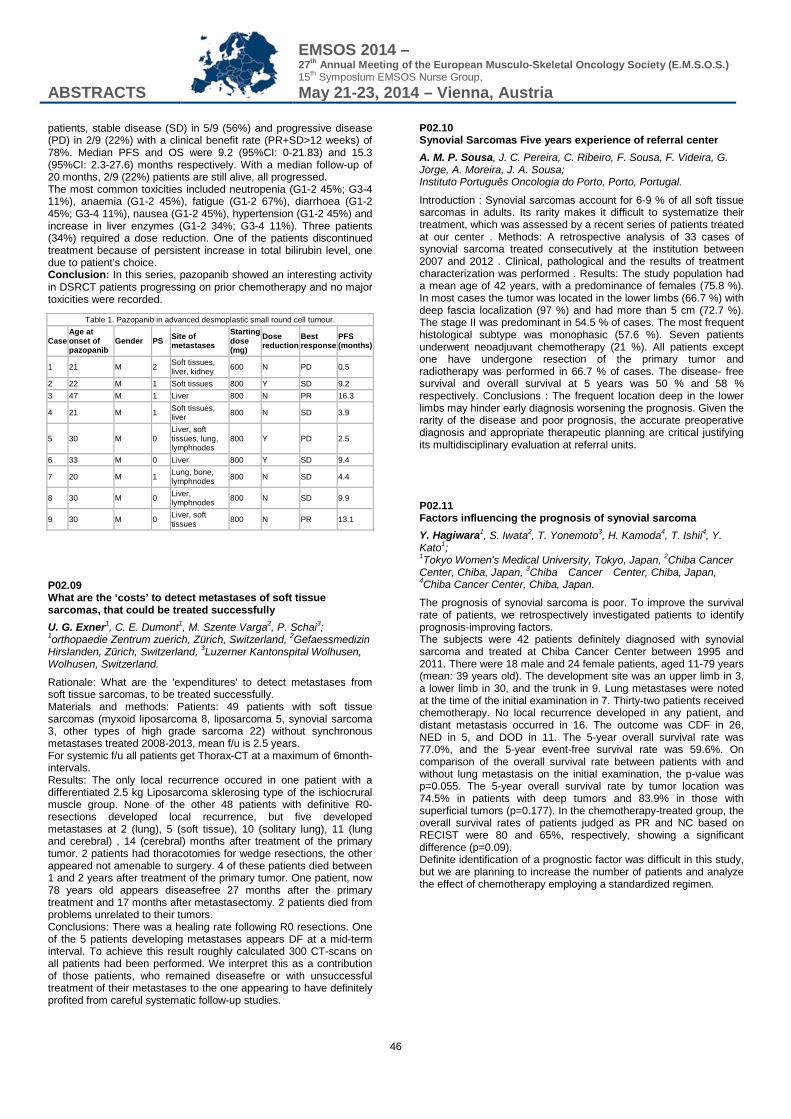
ABSTRACTS
EMSOS 2014 – 27th Annual Meeting of the European Musculo-Skeletal Oncology Society (E.M.S.O.S.) 15th Symposium EMSOS Nurse Group, May 21-23, 2014 – Vienna, Austria
46
patients, stable disease (SD) in 5/9 (56%) and progressive disease (PD) in 2/9 (22%) with a clinical benefit rate (PR+SD>12 weeks) of 78%. Median PFS and OS were 9.2 (95%CI: 0-21.83) and 15.3 (95%CI: 2.3-27.6) months respectively. With a median follow-up of 20 months, 2/9 (22%) patients are still alive, all progressed. The most common toxicities included neutropenia (G1-2 45%; G3-4 11%), anaemia (G1-2 45%), fatigue (G1-2 67%), diarrhoea (G1-2 45%; G3-4 11%), nausea (G1-2 45%), hypertension (G1-2 45%) and increase in liver enzymes (G1-2 34%; G3-4 11%). Three patients (34%) required a dose reduction. One of the patients discontinued treatment because of persistent increase in total bilirubin level, one due to patient’s choice. Conclusion: In this series, pazopanib showed an interesting activity in DSRCT patients progressing on prior chemotherapy and no major toxicities were recorded.
Table 1. Pazopanib in advanced desmoplastic small round cell tumour.
Case Age at onset of pazopanib
Gender PS Site of metastases
Starting dose (mg)
Dose reduction
Best response
PFS (months)
1 21 M 2 Soft tissues, liver, kidney 600 N PD 0.5
2 22 M 1 Soft tissues 800 Y SD 9.2 3 47 M 1 Liver 800 N PR 16.3
4 21 M 1 Soft tissues, liver 800 N SD 3.9
5 30 M 0 Liver, soft tissues, lung, lymphnodes
800 Y PD 2.5
6 33 M 0 Liver 800 Y SD 9.4
7 20 M 1 Lung, bone, lymphnodes 800 N SD 4.4
8 30 M 0 Liver, lymphnodes 800 N SD 9.9
9 30 M 0 Liver, soft tissues 800 N PR 13.1
P02.09 What are the ‘costs’ to detect metastases of soft tissue sarcomas, that could be treated successfully U. G. Exner1, C. E. Dumont1, M. Szente Varga2, P. Schai3; 1orthopaedie Zentrum zuerich, Zürich, Switzerland, 2Gefaessmedizin Hirslanden, Zürich, Switzerland, 3Luzerner Kantonspital Wolhusen, Wolhusen, Switzerland.
Rationale: What are the 'expenditures' to detect metastases from soft tissue sarcomas, to be treated successfully. Materials and methods: Patients: 49 patients with soft tissue sarcomas (myxoid liposarcoma 8, liposarcoma 5, synovial sarcoma 3, other types of high grade sarcoma 22) without synchronous metastases treated 2008-2013, mean f/u is 2.5 years. For systemic f/u all patients get Thorax-CT at a maximum of 6month-intervals. Results: The only local recurrence occured in one patient with a differentiated 2.5 kg Liposarcoma sklerosing type of the ischiocrural muscle group. None of the other 48 patients with definitive R0-resections developed local recurrence, but five developed metastases at 2 (lung), 5 (soft tissue), 10 (solitary lung), 11 (lung and cerebral) , 14 (cerebral) months after treatment of the primary tumor. 2 patients had thoracotomies for wedge resections, the other appeared not amenable to surgery. 4 of these patients died between 1 and 2 years after treatment of the primary tumor. One patient, now 78 years old appears diseasefree 27 months after the primary treatment and 17 months after metastasectomy. 2 patients died from problems unrelated to their tumors. Conclusions: There was a healing rate following R0 resections. One of the 5 patients developing metastases appears DF at a mid-term interval. To achieve this result roughly calculated 300 CT-scans on all patients had been performed. We interpret this as a contribution of those patients, who remained diseasefre or with unsuccessful treatment of their metastases to the one appearing to have definitely profited from careful systematic follow-up studies.
P02.10 Synovial Sarcomas Five years experience of referral center A. M. P. Sousa, J. C. Pereira, C. Ribeiro, F. Sousa, F. Videira, G. Jorge, A. Moreira, J. A. Sousa; Instituto Português Oncologia do Porto, Porto, Portugal.
Introduction : Synovial sarcomas account for 6-9 % of all soft tissue sarcomas in adults. Its rarity makes it difficult to systematize their treatment, which was assessed by a recent series of patients treated at our center . Methods: A retrospective analysis of 33 cases of synovial sarcoma treated consecutively at the institution between 2007 and 2012 . Clinical, pathological and the results of treatment characterization was performed . Results: The study population had a mean age of 42 years, with a predominance of females (75.8 %). In most cases the tumor was located in the lower limbs (66.7 %) with deep fascia localization (97 %) and had more than 5 cm (72.7 %). The stage II was predominant in 54.5 % of cases. The most frequent histological subtype was monophasic (57.6 %). Seven patients underwent neoadjuvant chemotherapy (21 %). All patients except one have undergone resection of the primary tumor and radiotherapy was performed in 66.7 % of cases. The disease- free survival and overall survival at 5 years was 50 % and 58 % respectively. Conclusions : The frequent location deep in the lower limbs may hinder early diagnosis worsening the prognosis. Given the rarity of the disease and poor prognosis, the accurate preoperative diagnosis and appropriate therapeutic planning are critical justifying its multidisciplinary evaluation at referral units. P02.11 Factors influencing the prognosis of synovial sarcoma Y. Hagiwara1, S. Iwata2, T. Yonemoto3, H. Kamoda4, T. Ishii4, Y. Kato1; 1Tokyo Women's Medical University, Tokyo, Japan, 2Chiba Cancer Center, Chiba, Japan, 3Chiba Cancer Center, Chiba, Japan, 4Chiba Cancer Center, Chiba, Japan.
The prognosis of synovial sarcoma is poor. To improve the survival rate of patients, we retrospectively investigated patients to identify prognosis-improving factors. The subjects were 42 patients definitely diagnosed with synovial sarcoma and treated at Chiba Cancer Center between 1995 and 2011. There were 18 male and 24 female patients, aged 11-79 years (mean: 39 years old). The development site was an upper limb in 3, a lower limb in 30, and the trunk in 9. Lung metastases were noted at the time of the initial examination in 7. Thirty-two patients received chemotherapy. No local recurrence developed in any patient, and distant metastasis occurred in 16. The outcome was CDF in 26, NED in 5, and DOD in 11. The 5-year overall survival rate was 77.0%, and the 5-year event-free survival rate was 59.6%. On comparison of the overall survival rate between patients with and without lung metastasis on the initial examination, the p-value was p=0.055. The 5-year overall survival rate by tumor location was 74.5% in patients with deep tumors and 83.9% in those with superficial tumors (p=0.177). In the chemotherapy-treated group, the overall survival rates of patients judged as PR and NC based on RECIST were 80 and 65%, respectively, showing a significant difference (p=0.09). Definite identification of a prognostic factor was difficult in this study, but we are planning to increase the number of patients and analyze the effect of chemotherapy employing a standardized regimen.
ABSTRACTS
EMSOS 2014 – 27th Annual Meeting of the European Musculo-Skeletal Oncology Society (E.M.S.O.S.) 15th Symposium EMSOS Nurse Group, May 21-23, 2014 – Vienna, Austria
47
P02.12 The treatment of children with synovial sarcoma R. Y. Maxim, A. Dzampaev, V. Polyakov; Institute of Pediatric Oncology and Hematology, Moscow, Russian Federation.
Introduction: to improve the results of the treatment of synovial sarcoma. Methods: 48 children and adolescents at the mean age of 10,5±3,7 years with synovial sarcoma were treated between 1990 and 2013 years. The analyzable group of patients was disintegrated to the group of historical control (29 patients) and the investigation group (19 patients). Histologically, 20 patients (69,0%) had the biphasic, 8 (27,6 %) had the monophasic, and 1 (3,4%) of them had the poorly differentiated pattern. The most often affected area was the area of the lower extremity - 14 (48,3 %) cases. One child was diagnosed with synovial sarcoma of abdominal cavity. According to the staging systems adopted, the size > 5 cm was reported in 22 (75,9 %) cases. In the group of historical control the average tumor size was 49,1 sm³. 15 (51,7 %) patients had relapse of disease. 11 patients (37,9 %) had distant metastases. The scheme of the treatment included: 8 courses of chemotherapy; the harvesting and preservation of the stem cells after the stimulation of the haemophoesis by G-CSF. Results: the partial effect was registered by most of the patients - 80%. We observed 1 case of progression of the disease during inductive CT. The treatment included chemotherapy, radiotherapy and surgical operation. 2-year disease-free survival was 66,1±11,3 %, overall 5-year survival -75,6±10,6%. Conclusion: the intensification of the chemotherapy increases the survival of the patients with synovial sarcoma. P02.13 Extraskeletal osteosarcoma: A single institution experience A. G. Robinson, C. L. Gaston, R. J. Grimer, S. R. Carter, L. M. Jeys, K. Reddy, N. Murukesh, V. Agarwal, J. Sherriff, V. P. Sumathi; Royal Orthopaedic Hospital NHS Foundation Trust, Birmingham, United Kingdom.
Extraskeletal osteosarcoma is a rare high grade neoplasm associated with a poor prognosis. We present our institution’s experience. A retrospective review of 25 histologically confirmed cases of extraskeletal osteosarcoma surgically treated between 1997 and 2008 in our institution was performed. Minimum follow-up was 48 months for those still alive. Mean age at diagnosis was 57 years (range, 13 - 90 years) with a male to female ratio of 2:1. Eight patients (32%) presented after inadvertent excision prior to referral and the most common site was the lower limb (11 cases in the thigh, 3 cases in the knee, 1 in the leg). Local recurrence occurred in 6 patients (24%) at a mean time of 9 months (range 2 - 18 months). Five patients (20%) had pulmonary metastases at presentation. 15 patients (60%) developed progressive metastatic disease after surgery at a mean time of 12.8 months (range 2 - 64 months) with the majority being pulmonary metastases. The 2- and 5- year overall survival rate was 40% and 32%, with a median follow up of 17 months (range 2 - 190 months). The 6 patients who had chemotherapy (either neoadjuvant or post-operative) usually with a soft tissue sarcoma protocol with Dox/Ifos or Adria/Ifos are the only ones still alive at last follow-up. Our experience confirms the aggressive nature of extraskeletal osteosarcoma which demonstrates a high propensity to metastasise and a poor prognosis. Soft tissue sarcoma chemotherapy protocols seem to have a role but larger clinical studies are needed.
P02.14 Outcome of Rhabdomyosarcoma In Adolescence: A CCHE Experience E. A. Moussa, E. M. El Nadi, H. El Zomor, A. Younes, H. Taha, N. EL Kinaai, M. S. Zagloul, M. El Wekeel, R. Labib, M. Tantawi; The Children's Cancer Hospital-Egypt, Cairo, Egypt.
Background: RMS occurs in young children, has a second age peak in adolescence, then incidence declines afterwards. Despite survival improvements, outcome for older children and young adults remains decimal. Our aim was to study outcome of older children and adolescents treated at our center. Patients and Methods: At a mean follow up period of 29.4 months between july 2007 and December 2012 , 32 children aged 10 to 18 years were diagnosed with rhabdomyosarcoma at CCHE. They were 12 females and twenty males. Results: Out of the 32 children, 15 patients had alveolar, 14 emberyonal, one botryoid, and 2 had spindle cell tumor. Seven patients had intracranial extension at presentation, while 22 presented with tumors at unfavorable sites, and 12 had metastases at presentation. Twelve patients were high risk, 17 were intermediate risk and 3 were low risk. Local control by radiation was done in 20 patients, surgery alone in 3 patients and both radiation and surgery in 8 patients. One patient had no local control. Twelve patients attained C.R, 4 showed P.R and 3 patients had no response. Disease progression was seen in 13 patients. Overall Survival was 59.1% and failure free survival was 40.4%. Presence of metastases showed inferior survival results (p=0.01). Risk stratification showed significant results for 5-years failure free survival. Pathological subtypes were insignificant regarding survival outcome. Conclusion: Outcome of adolescent group showed inferior results. Increasing knowledge about biological characteristics in older children and adolescence can help in standardizing treatment for this age group. P02.15 Usual atypia in imaging of myxoid liposarcomas : a trick and a standard of gravity J. C. Mattei, M. Lemeur, P. Souteyrand, C. Chagnaud, G. Curvale, A. Rochwerger; AP-HM Conception Hospital, Marseille, France.
Introduction: myxoids liposarcomas imaging diagnosis depends on differentiation degree (from well-differentiated pure myxoid to strong cellular undifferentiated liposarcoma). They are potentially aggressive tumours, and need specialized-centre handling. Recurrence rate is 1/3 (risk factors: age, tumour size, grading, resection margins and round cell percentage). Materials and methods: From 2004 to 2011, 20 patients (10 men and women) had a myxoid liposarcoma resection. Usual datas were collected and recurrence/survival rates were calculated. Particular attention was given to initial imaging and how the diagnosis was established. Results: Initial symptomatology was a mass of rapidly increasing volume. Only 14 of 20 patients were initially handled in specialized centres, 15 had an MRI in the first evaluation (4 of them led directly to the myxoid liposarcoma diagnosis). Eleven tumours had a round cell component (6 of them had a > 5% round cells percentage), which all locally recidivated. Four of these 6 patients were initially addressed to unspecialized centres and 3 of them developed metastasis. Two of them died. Discussion: Major prognostic factors are round cell percentage and unspecialized centre handling. Typical MRI presentation matches low grade less aggressive, purely myxoid tumours. Round-cell rate is correlated with differentiation and tumour aggressiveness. Initial MRI might be tricky is these cases because fatty and myxoid components might be in minority. Conclusion: Myxoid liposarcoma are untypical and aggressive. MRI might be unusual and must help to biopsy performing. Pathological examination only will confirm the diagnosis, though some typical imaging findings do exist, corresponding to low grade, well-differentiated tumours.
ABSTRACTS
EMSOS 2014 – 27th Annual Meeting of the European Musculo-Skeletal Oncology Society (E.M.S.O.S.) 15th Symposium EMSOS Nurse Group, May 21-23, 2014 – Vienna, Austria
48
P02.16 Clinical and Radiological Presentation of Primary Bone Leiomyosarcoma: Case series M. Bergovec1,2, K. Crnogaca1, M. Smerdelj1,2, K. Potocki1,2, A. Bonevski3, S. Seiwerth1,2; 1Clinical Hospital Centre Zagreb, Zagreb, Croatia, 2Zagreb Univesity School of Medicine, Zagreb, Croatia, 3Children's Hospital Zagreb, Zagreb, Croatia.
Primary leiomyosarcoma of bone is a very aggresive tumor with high incidence of metastases and poor prognosis. Aim of this study is to report about four cases treated in our Department for primary leiomyosarcoma of bone: radiological-pathological correlation, results of treatment and mid-term survival. We present case series of four patients who were treated in our Department from the year of 2009 till 2012, with minimal follow-up of one year. There were 2 males and 2 females with a mean age of 39.3 years (range 25 to 49). Tumors were located in distal femur (N=2), acetabulum and proximal humerus. Plain radiography, MRI, CT and bone scans were obtained prior to open biopsy. Radiological appearance was very various in this series: tumor presented either as locally destructive, osteolitic, located intramedullary, with no soft-tissue component, or as a sclerotic intramedulary tumor, or as similar to osteochondroma - connected with root to metaphysis origin. All specimens showed smooth muscle differentiation, immunohistochemically positive for actin and vimentin, but negative for desmin. Patients were treated with chemotherapy and wide surgical resection. One patient died for local recurrence and pulmonary metastases 24 months after operation. In one patient (Enneking III) pulmonary metastases regressed after chemotherapy. The other two patients classified as Enneking Ia and IIb show no signs of local recurrence or metastases at their latest controls. Primary leiomyosacromas of bone show various radiological appearances. In our series it seems that Enneking staging and presence of metastasis at diagnosis could serve as a prognostic factor. P02.17 Primary Leiomyosarcoma of Bone - review of the literature and own series T. Sununu1, N. Woltsche2, A. Leithner2; 1Orthopedic Department, Hospital St. Johann in Tirol, St. Johann in Tirol, Austria, 2Orthopedic Department, University of Graz, Graz, Austria.
Introduction: Primary Leiomyosarcoma of bone is a very rare condition. Only about 100 cases are reported in the literature. The disease mostly affects elderly people, sex distribution is equal. Most authors suggest treatment in the same way as Osteosarcoma including poly-chemotherapy according to recent study protocols. Recent data suggest that 5-year-survival may be slightly better than in Osteosarcoma. Patients and Methods: In our center we identified 5 patients diagnosed and treated with primary Leiomyosarcoma of bone, their age ranging from 33 to 89 years, 4 male, 1 female. The median age at time of diagnosis was 65,7. All patients underwent complete staging and biopsy. 3 patients already presented with metastases at time of diagnosis. Wide resection was performed in three patients achieving clear margins. One patient died 5days, one 2 years after surgery. One patient received radio- and chemotherapy and no surgery for disseminated disease. Results: In our small series 2 patients are still followed up about a year after diagnosis, one of those, the only one presenting with a low grade G1 tumor, disease-free, the other one with stable metastatic disease. Discussion: Primary Leiomyosarcoma of bone is a rare condition. More data collection is necessary to clearly diminish a difference to other malignant bone tumor entities. Wide resection with clear margins combined with chemotherapy following an Osteosarcoma protocol should be applied.
P02.18 Primary intraosseus leiomyosarcoma of the distal femur: a case report M. Besalduch1, A. Combalía2, A. Peiró1, L. Trullols1, I. Gracia1; 1Hospital de la Santa Creu i Sant Pau, Barcelona, Spain, 2Hospital Clínic i Provincial de Barcelona, Barcelona, Spain.
Leiomyosarcoma is a frequent malignant mesenchymal neoplasm, but it is rarely found in bones (<0’7% of all primary malignant bone tumours). It has uncommon radiographic characteristics, making diagnosis challenging. A 33 year-old man consulted for a 5 month right knee pain. He underwent simple radiography, computed tomography and magnetic resonance, which reported an osteolitic 10’5cm mass on the distal femur, with bone and soft tissue involvement. A trocar biopsy was performed. Histologically, an intermediate grade (2/3) fusocellular sarcoma with smooth muscular differentiation was described, being the differential diagnoses between leiomyosarcoma and fibroblastic osteosarcoma. The staging study showed no systemic dissemination. Considering the age of the patient, the risk of a pathological fracture and in order not to modify the histological analysis, it was decided to perform surgery first. We carried out an en bloc resection and reconstruction with an arthroplasty (METS modular implant, Stanmore implants). The final histological study identified a high grade (3/3) fusocellular sarcoma with smooth muscle differentiation. Focal soft tissue surgical margins were affected. The patient underwent chemotherapy and radiation. Leiomyosarcomas rarely occur in bones. The median age is 46 years (9 to 88). The most frequent location is distal femur, followed by proximal tibia. Diagnosis is difficult because of the lack of suspicion in cases of lytic bone lesions without matrix production. Treatment is based on surgery; the chemotherapy regime yet to be stablished. Prognosis is poor and seems to be related with the stage at diagnosis. It appears to be slightly better than in osteosarcomas. P02.19 Extraskeletal soft tissue sarcoma or ossified low-grade liposarcama Case report and review of the literature J. Bruns1, C. Habermann2, M. Werner3; 1Dept. Orthop. Surgery, AGAPLESION-Diakonieklinikum, Hamburg, Germany, 2Dept. of Radiology, Marienkrankenhaus Hamburg, Hamburg, Germany, 3Dept. Orthop. Pathology, Stiftung Oskar-Helene-Heim, MVZ des HELIOS Klinikums Emil von Behring, Berlin, Germany.
Osteosarcomas located in the vertebral column are a rarity. An extraosseous soft tissue osteosarcoma (ESOS) located in the paravetebral muscle is very uncommon. Case report: A 58-years old woman was admitted owing to a soft tissue mass located in the paravertebral muscles of the thoracic spine. Clinically, the patient exhibited a tumor mass in the paravertebral muscles of the thoracic spine. No neurological deficits were detected. Imaging exhibited a solid tumor surrounded by a layer of lipoid soft tissue within the extensor muscles of the spine. No contact to any osseous structure could be detected. To define the character of the tumor an incisional biopsy was performed. Histology revealed a low-grade osteosarcoma. Chemotherapy and radiotherapy were not indicated. We performed a resection of the tumor within wide margins. Intraoperatively, a small lamella of the transversal processus as well of the underlying ribs were cut off to achieve wide margins in the region. Postoperatively, the patient recovered without any surgical problems. For at least 6 weeks postop. she felt local pain owing to the muscular dysbalance. Discussion: Most of ESOS are located in the soft tissue of the extremities but can also occur in almost all anatomic regions and organs. The majority of reports are case reports. Only to papers mentioned a reasonable number of patients suffering from ESOS (10/20 Pat.). Most of the cases are high-grade tumors and had been treated surgically with additional chemotherapy. For low-grade ESOS local resection within wide margins seems to be the adequate surgical procedure
ABSTRACTS
EMSOS 2014 – 27th Annual Meeting of the European Musculo-Skeletal Oncology Society (E.M.S.O.S.) 15th Symposium EMSOS Nurse Group, May 21-23, 2014 – Vienna, Austria
49
P02.20 Alveolar soft part sarcoma of the extremity: Case report and literature review K. Ata; King Abdullah University Hospital, Irbid, Jordan.
Introduction: Alveolar soft part sarcoma (ASPS), a rare soft tissue sarcoma in children and adolescents, carries a poor prognosis. ASPS is an aggressive tumor of controversial histogenesis that, unlike other soft tissue sarcomas, tends to metastasize to the brain. Case presentation: A 9-year-old boy presented to our outpatient clinic in April 2009 with a chief complaint of a large painless mass in the left thigh whose size had increased significantly over the past 10 months. After staging the tumor we performed open biopsy; the diagnosis was ASPS and he underwent wide local excision. In the course of 4 -year follow-up by clinical and imaging studies there was no evidence of early tumor recurrence or metastasis. Conclusion: Complete surgical resection is the treatment of choice in patients with ASPS. P02.21 Angiosarcoma over chronic lymphedema of the upper extremity (Stewart-Treves syndrome): a case report M. Besalduch, A. Peiró, M. Gómez, L. Trullols, I. Gracia; Hospital de la Santa Creu i Sant Pau, Barcelona, Spain.
The Stewart-Treves syndrome, is a rare and fatal cutaneous tumour which arises over chronic lymphedema, as a consequence of mastectomy and axillary lymph node dissection. Although the patient with breast cancer may be cured with surgery and adjuvant therapies, this secondary malignancy worsens the patient’s outcome. An 81 year-old woman underwent a right breast tumorectomy and node dissection with adjuvant radiation. She gradually developed lymphedema of the right arm. 9 years later she grew multiple ecchymotic nodules on the volar and posterior aspect of the upper extremity. Some of these lesions ulcered and bled easily, and were extremely painful. The biopsy reported an epithelioid angiosarcoma. Due to the extension of the tumour (assessed by magnetic resonance) and the age of the patient, it was decided to perform a forequarter amputation. The surgical margins showed no residual sarcoma. Unfortunately, during the follow-up, the patient presented new cutaneous metastases on the anterior chest wall. She was still alive in the last visit, 5 months after being diagnosed of the Stewart-Treves syndrome. This pathology occurs in 0’5% of patients surviving 5 or more yeas after radical mastectomy and predominantly affects women, between the fifth and seventh decade of life. The etiology is still unclear but is associated with chronic lymphedema. This tumour often appears as innocuous red bumps. Clinicians should be responsible to biopsy this lesions. Several treatment options should be considered, depending on the size and stage of the disease. Prognosis is poor, with survival raging from 19-31 months. P02.22 Myxoid liposarcoma associated with Mazabraud's Syndrome: a case Report F. Nakatani, N. Setsu, Y. Tanzawa, T. Fujiwara, K. Ogura, D. Kubota, E. Kobayashi, U. Yamaguchi, A. Kawai, H. Chuman; National Cancer Center Hospital, Tokyo, Japan.
Objective: Mazabraud's syndrome is the rare association between benign intramuscular myxoma and fibrous dysplasia of bone. Here we present the first case report of a 55-year-old male who developed myxoid liposarcoma associated with Mazabraud's syndrome. Materials and Methods: A 55-year-old male who presented swelling in the right distal thigh referred to our outpatient clinic. CT scan and MRI revealed the bone lesions in his right acetabular and proximal femur coupled with two separated tumors in his distal quadriceps;
one was a large multi-nodular tumor sited mainly in the vastus medialis of his right quadriceps and presented heterogeneous enhancement of gadolinium, the other was a small tumor which located separately in the vastus lateralis of the same limb without any enhancement with gadolinium. The large tumor was diagnosed as myxoid liposarcoma with core needle biopsy. Result: After preoperative radiotherapy of the medial large soft tissue tumor, we have performed resection of the soft tissue tumors and needle biopsies of the bone lesions (both acetabular and proximal femur). After resection, the large tumor was pathologically diagnosed as myxoid liposarcoma which was genetically proved to have TLS gene rearrangement. On the contrary, the small lateral soft tissue tumor was diagnosed as a benign intramuscular myxoma without any gene rearrangement. The bone lesions were revealed to be fibrous dysplasia. Therefore, we have concluded that the patient with Mazabraud’s syndrome developed myxoid liposarcoma in the same limb. Conclusion: To our knowledge, this is the first report of myxoid liposarcoma associated with Mazabraud’s syndrome. P02.23 Malignant psammomatous melanotic schwannoma: a case report of a rare tumour D. Hirzberger1, B. Liegl-Atzwanger2, W. Maurer-Ertl1, J. Friesenbichler1, U. Anegg3, A. Leithner1; 1Department of Orthopaedic Surgery, Medical University of Graz, Graz, Austria, 2Institute of Pathology, Medical University of Graz, Graz, Austria, 3Division of Thoracic and Hyperbaric Surgery, Department of Surgery, Medical University of Graz, Graz, Austria.
Background: Psammomatous melanotic schwannoma (PMS) is a rare melanin-producing neuroectodermal tumour originating from the Schwann cells of the nerve sheath. PMS affects males and females equally and generally occurs in young adults. Although usually benign, 10% show malignant clinical behaviour. About half of the PMS occur in the Carney complex setting, an autosomal dominantly inherited syndrome comprising spotty skin pigmentation, myxomas, endocrine tumours, and schwannomas. Case presentation: A 20-year-old male with 5-year history of recurrent painful expansion in the right 8th-rib region was referred to our department for biopsy. CT scan disclosed a 6 x 5 x 5 cm contrast enhancing chest wall mass in the epigastrium left of the lower thorax aperture. Histological examination of biopsy material revealed a morphologically malignant psammomatous melanotic schwannoma. Wide en-bloc resection of the tumour was performed. Patient´s investigation for features of Carney's complex showed only a testicular microlithiasis in ultrasound. The last follow-up six months after resection showed no evidence of disease. Discussion: PMS may occur anywhere in the central and peripheral nervous system, but is most frequently found in the gastrointestinal tract and paraspinal sympathetic chain. Differential diagnosis includes malignant melanoma, pigmented meningioma and neurofibroma, rhabdomyosarcoma, clear cell soft tissue sarcoma, melanotic medulloblastoma, ganglioneuroblastoma, ectomesenchymoma (triton tumour), neurotrophic melanoma, and melanotic neuroendocrine carcinomas and carcinoids. The treatment of choice recommended for PMS is total excision with tumour-free margins and a minimum 5-year follow-up period. P02.24 Fetal rhabdomyoma of the upper extremity in a 31-year-old patient: a case report G. Hauer1, F. Gollowitsch2, L. A. Holzer1, A. Leithner1; 1Department of Orthopaedic Surgery, Medical University of Graz, Graz, Austria, 2Department of Pathology, Medical University of Graz, Austria, Graz, Austria.
Introduction: Fetal Rhabdomyomas are very rare benign mesenchymal tumours primarily in the head and neck. This tumour exhibits immature skeletal muscle differentiation. The patients’
ABSTRACTS
EMSOS 2014 – 27th Annual Meeting of the European Musculo-Skeletal Oncology Society (E.M.S.O.S.) 15th Symposium EMSOS Nurse Group, May 21-23, 2014 – Vienna, Austria
50
median age is 4 years. Surgical resection is the recommended treatment. We report a unique case of a 31-year-old male patient with a fetal rhabdomyoma located in the left distal upper arm. Case Report: The patient presented with a painless soft tissue tumour on his upper arm, which he had since 2 months. Sonography was inconclusive, so a MRI was done. The tumour showed a size of 3 x 1.5 x 1.4 cm in the MRI. MRI led to the differential diagnosis of a Schwannoma and an excisional biopsy was performed. The histological specimen showed interlacing broad fascicles of spindled muscle cells and partially big rounded cells and no nuclear atypia. Based on immunohistochemistry the diagnosis of a fetal rhabdomyoma, intermediate (juvenile) subtype was made. Resection areas were free of tumour and therefore no further treatment was necessary. Discussion: The combination of the location and age make this case special. Extremities are usually not affected and only one similar case of fetal rhabdomyoma in the upper extremity has been reported yet. The patient’s age is far over the median age. One should be aware of this skeletal muscle tumour and should be considered as a differential diagnosis.
P04 Surgical options in the very young - Grower or biological?/Interventional radiology - When and which? P04.01 Experimental Modeling of Damage of Growing Zones of Knee Bones for Children O. Vyrva, V. Baev; Sytenko Institute of Spine and Joint Pathology, Kharkiv, Ukraine.
Growth numbers of children with primary bone tumors in Ukraine and high cost of expandable prosthesis force us to look for new ways for children limb salvage surgeries. Experimental research on growing animals was done for possible planning of modular tumor replacement of the children growing knee. This is study of influence damage of bone growing zones at growing rats having simulated clinical situation with knee prosthetic replacement. We have used white rats-males 3 month old selected for 7 serial groups on 5 animals. Endoprosthesis of distal hip and proximal tibia have been custom made for rats especially. Surgeries for bone defects replacement with endoprosthesis and without were performed for distal femur, proximal tibia and both segments at the same time. Postoperative observation till the moment euthanasia was spent with use clinical, osteometry and radiological methods. It had been observed some general tendencies for animals. At carrying out radiologic and osteometry tests it is report the animals that have one segment surgery have had an interfacing segment longer than similar to not operated extremity. Experimental by we have proved possibility endoprosthetic replacement children with bone tumors of the knee. The knee unipolar endoprostheses are the method of selection for bone defects replacement at children who is growing, like a method of temporary replacement until the growth process will completed.
P04.02 Stanmore modular endoprosthesis replacements following bone tumor resection M. Gallo1, G. Balato2, G. Colella2, F. Fazioli1; 1INT "G.Pascale", Napoli, Italy, 2A.O.U. "Federico II", Napoli, Italy.
Endoprosthetic replacement following bone tumor resection may be required to treat primary bone tumors or destructive metastases either with impending or established pathological fracture. Modular prostheses can be adapted to most reconstructive situations for this purpose. We have assessed the clinical and functional outcome of using METS (Stanmore Implants Worldwide) modular tumour prosthesis following a tumor resection. Between January 2012 and July 2013 we treated 30 patients (16 Male, 14 Female) with high-grade primary malignant tumours or bone metastasis by resection of the lesion and replacement with an extendible endoprosthesis. The tumors were located at the distal femur in eighteen patients, at the proximal femur in ten patients and at the proximal humerus in only two patients. The mean age of the patients was 35 years (7 to 69). The mean follow-up was 8.9 months (6 to 12 months). At final follow-up the survival of the implants was 100%. The mean Karnofsky index increase from 40% pre-operatively to 80% post-operatively. There were no cases of aseptic loosening or early infection of the implant. In our experience the Stanmore endoprosthesis following a bone tumor excision provides good functional outcome without compromising patient survival, and represents an effective alternative to amputation in patients with primary or secondary malignant bone tumors.
P05 Spine and Pelvis P05.01 Short term outcomes of invasive surgical treatment for symptomatic spinal bone metastases S. M. C. Welters1, L. Bollen1, D. J. Lobatto2, W. Pondaag1, G. C. W. Ruiter2, M. Arts2, W. C. Peul1,2, S. P. D. Dijkstra1; 1Leiden University Medical Center, Leiden, Netherlands, 2Medical Center Haaglanden, The Hague, Netherlands.
Objectives: Surgery for spinal bone metastases (SBM) implies possible adverse complications because of its invasiveness and the individuals’ possible unfavorable condition. Surgical treatment should only be executed when the expected gain in quality of life outweighs the risks. The goal of this study was to specify the short term clinical outcomes for a cohort of patients surgically treated for SBM. Methods: All patients (n=123) who were surgically treated from 2000-2012 for SBM were included in this retrospective multi-center cohort study. Surgical treatment was divided into two groups; limited decompressive surgery (n=68) and corpectomies (n=55). Analysis was performed using the Kaplan-Meier method and log rank tests. Results: Median follow-up was 36.1 months (95%CI 29.7-42.5) and median survival was 10.6 months (95%CI 6.3-14.9). The mean age was 59.3±10.8 years. The most prevalent primary tumors were those of breast (n=29; 23.6%), lung (n=24; 19.5%) and kidney (n=19; 15.4%). A postoperative neurological improvement was seen in 32.2% of the cases. Analysis showed a median hospitalization period of 7 days (95%CI 4.8-9.2) for the decompressions and 13 days (95% CI 9.9-16.1) for the corpectomies (p=0.03). Overall 30-day complication rate was 33.3% with a relative risk of 27.9% for the limited decompressive surgery and 40.7% for the corpectomies. Conclusion: Surgical treatment for symptomatic SBM has a lower complication rate and shorter duration of hospitalization for limited decompressive surgery in comparison to the corpectomies but both are reasonably effective. This should be taken into account when deciding on the type of treatment for each individual patient.
ABSTRACTS
EMSOS 2014 – 27th Annual Meeting of the European Musculo-Skeletal Oncology Society (E.M.S.O.S.) 15th Symposium EMSOS Nurse Group, May 21-23, 2014 – Vienna, Austria
51
P05.02 Spinal bone metastases from breast cancer - differences in survival for histopathologic subtypes L. Bollen1, C. Wibmer2, M. Wang3, Y. Van der Linden1, C. Bünger3, A. Jensen3, A. Leithner2, G. Bratschitsch4, W. Pondaag1, S. Dijkstra1; 1LUMC, Leiden, Netherlands, 2Medizinische Universität Graz, Graz, Austria, 3Aarhus University, Aarhus, Denmark, 4Medizinische Universität Graz, Graz, Denmark.
Objectives: Existing prognostic systems generally consider breast cancer a positive predictive factor for survival in patients with spinal bone metastases (SBM), but do not take the different subtypes based on estrogen (ER), progesterone (PR) and human epidermal growth factor 2 (HER2) receptors into account. The aim of this international multi-center study was to analyze whether receptor status influences survival, when the disease has progressed to SBM. Methods: Retrospectively 110 patients treated for SBM from breast cancer between 2005 and 2012 were included . Patients were subdivided into four categories: Luminal A, Luminal B, HER2 and Triple negative according to the histopathologic ER, PR and HER2 status. Survival time was calculated from start of treatment for SBM . Median follow-up was 3.7 years (minimum 0.6, maximum 5.9 years) Analysis was performed using the Kaplan-Meier method, univariate log-rank tests and Cox-regression models. Results: Overall median survival was 18.0 months (95%CI 12.3-23.6). Similar survival times were seen for the Luminal A, B and HER2 categories. These were therefore pooled into one category: hormone positive (77% of all patients). A median survival of 22.5 months (95%CI 18.0-26.9) for the Hormone positive category and 6.7 months (95%CI 2.4-10.9) for the Triple negative category (p<0.001)was found. The corresponding hazard ratio was 2.2 (95%CI 1.4-3.7, p=0.002). Conclusion: Analyzing a dataset after the implementation of trastuzumab for HER2 positive malignancies, we found a markedly shorter survival for Triple negative breast cancer patients with SBM, than for hormone or HER2 positive phenotypes. Models estimating survival should be adjusted accordingly. P05.03 Giant cell tumor of the mobile spine - A multicenter study on midterm results in 18 patients after surgical excision L. van der Heijden1, A. C. Heineken1, L. Bollen1, I. C. M. van der Geest2, H. W. B. Schreuder2, B. J. van Royen3, P. D. S. Dijkstra1; 1Leiden University Medical Center, Leiden, Netherlands, 2Radboud University Medical Center, Nijmegen, Netherlands, 3VU University Medical Center, Amsterdam, Netherlands.
Introduction: Only 1-4% of giant cell tumors (GCT) are located in the mobile spine. Surgical treatment is hampered by soft tissue extension, spinal instability and involvement of spinal nerve roots. Aim of this study was to evaluate recurrences, complications and functional outcome after surgical excision of spinal GCT. Methods: In this retrospective multicenter study, all 18 patients that underwent surgery for spinal GCT were included (1993-2010). Median follow-up was 50 months (range 18-225). Twelve patients underwent en bloc resection, six intralesional excision (three with liquid nitrogen and two with PMMA). Two patients had radiotherapy, two denosumab. In 13 patients, posterior stabilization was performed. Functional results were evaluated with use of Musculoskeletal Tumor Society (MSTS) scores. Statistics were performed using Chi-squared, Mann-Whitney U and Cox regression. Results: Recurrence rate was 6/18 after mean 12 months (range 4-31); 3/6 after intralesional excision and 3/12 after resection (p=0.11). No individual risk factors for recurrence could be identified. One patient deceased from malignant transformation after radiotherapy (after 68 months). Complications occurred in 9/18 patients (3/6 after intralesional excision and 6/12 after resection) and included neurological deficits (n=4) and infection, hepatic dysfunction due to systemic therapy, non-union due to hematoma and postoperative hemothorax (each, n=1). Mean MSTS was 24 (range 15-29) and did not differ between curettage and resection (p=0.53).
Discussion: The recurrence risk was acceptable after en bloc resection but high after curettage for spinal GCT. Complications occurred equally after intralesional excision and en bloc resection. Functional outcome was good in all living patients. P05.04 Long-term outcomes in primary, spinal osteochondroma: A multicenter study of 27 patients P. Varga1, M. Macki2, M. Bydon2, D. M. Sciubba2, J. Wolinsky2, S. Boriani3, J. J. Reynolds4, L. D. Rhines5, C. G. Fisher6, Z. L. Gokaslan2; 1National Center for Spinal Disorders and Buda Health Center, Budapest, Hungary, 2Johns Hopkins University School of Medicine, Baltimore, MD, United States, 3Rizzoli Institute, Bologna, Italy, 4Oxford Univesity Hospitals NHS Trust, Oxford, United Kingdom, 5University of Texas, M.D. Anderson Cancer Center, Houston, TX, United States, 6University of British Columbia and Vancouver Coastal Health, Vancouver, BC, Canada.
Introduction: Clinical outcomes in patients with primary spinal osteochondromas are limited to small series and sporadic case reports. We present one of the first, long-term investigations on 27 spinal osteochondromas. Methods: An international, multicenter, ambispective study on primary spinal osteochondroma was performed. Perioperative prognostic variables, including age, tumor size, spinal level, and surgical resection were analyzed in reference to long-term local recurrence and survival. Tumor resections were compared using Enneking appropriate (EA) or Enneking inappropriate (EI) surgical margins. Results: There were 27 patients diagnosed at an average age of 36 years. Twenty-two were found in the mobile spine (cervical, thoracic, lumbar) and five in the fixed spine (sacrum). Twenty-three cases (85%) were benign tumors (Stage1-Stage3) while three (11%) exhibited malignant changes (Stage Ia-IIb). Sixteen patients (59%) were treated with en bloc: wide or marginal resections, nine (33%) with intralesional, and one (4%) with palliative. Twenty-four operations (92%) followed EA margins. None received adjuvant therapy. Three patients (11%) experienced recurrences: two in the fixed spine and one in the mobile spine. All three recurrences occurred in latent S1 tumors following en bloc: wide or marginal resection. Overall survival averaged 85%. The four observed deaths were not as a direct result of the osteochondroma. Conclusion: Based on our findings, most patients underwent an en bloc resection and were treated as EA. Despite a Stage 1 classification, all three recurrences occurred in these groups. Therefore, although benign in character, osteochondromas still require careful management and thorough follow-up. P05.05 Osteoblastoma of the sacrum: an analysis of 18 cases P. Ruggieri1, A. Angelini1, K. Huch2, E. Pala1, T. Calabrò1, G. Trovarelli1, M. Maraldi1, S. Zaffagnini1, M. Marcacci1; 1University of Bologna, Istituto Ortopedico Rizzoli, Bologna, Italy, 2University of Ulm, Department of Orthopaedic Surgery, Ulm, Germany.
Introduction. Aims of this retrospective study were 1) to provide a comprehensive review of the clinical and radiographic characteristics, treatment and outcome in a series of patients with sacral osteoblastoma 2) to evaluate local recurrence rate and 3) to identify prognostic factors. Methods. We analyzed 18 patients treated from 1980 to 2010 (16 males, 2 females). Thirteen tumors were stage 2 and 5 stage 3. Mean tumor volume was 64 cm3. Nine patients had preoperative evaluation without MRI. Five patients had a previous intralesional surgery. Treatment consisted of intralesional surgery (16 cases), intralesional surgery plus radiotherapy (one case) and wide resection (one case). Local adjuvants used were phenol (7 patients),
ABSTRACTS
EMSOS 2014 – 27th Annual Meeting of the European Musculo-Skeletal Oncology Society (E.M.S.O.S.) 15th Symposium EMSOS Nurse Group, May 21-23, 2014 – Vienna, Austria
52
cryotherapy with “ice ball” technique (1 case). Embolizations were performed in 7 patients. Results. At a mean of 8.4 years, 15 patients (83%) remained disease-free while three patients had local recurrence (17%). Survival to local recurrence was 87% at 5 years and 74% at 10 years. Two patients had postoperative complications (wound dehiscence and deep infection). No statistical difference was found between patients that received or not local adjuvants (p=1254), older or younger than 20 years (p=0.970), stage 2 or 3 (p=0.826), evaluated preoperatively with or without MRI (p=0.160), primarily treated versus patients with previous intralesional surgery elsewhere (p=0.131). Conclusions. Local recurrence rate in our series was lower than local recurrence reported in literature on all sites (about 24%). Local adjuvants do not seem to reduce the risk of local recurrence when combined with intralesional surgery. Preoperative embolization is recommended. P05.06 Metalwork failure after Total Sacral Resection L. E. Johnston, M. Faimali, S. Khan, P. D. Gikas, A. Olivier, J. Jagiello, W. Aston, J. Skinner, R. Pollock, T. W. R. Briggs; Royal National Orthopaedic Hospital, London, United Kingdom.
Tumours requiring Total Sacral Resection (TSR) necessitate iliolumbar reconstruction. We seek to analyse the risk of metalwork failure in patients who underwent TSR for tumours between 2004 and 2008 at our tertiary referral centre. Patients were identified retrospectively from our database. Patients with less than 5 years follow-up, or who had a partial sacral resection were excluded. Information regarding time to failure, mechanism of failure and further management was obtained from medical records. 9 patients were identified (4 male, 5 female) who fulfilled the criteria for inclusion. 5 had a diagnosis of Conventional Chordoma, 2 Giant Cell Tumour, 1 Osteosarcoma and 1 Myxopapillary Ependymoma. Average age at surgery was 44 years (18-68). 6 patients suffered metalwork failure within 5 years of surgery, one of whom has been excluded due to incomplete data. All of the patients were mobilising at the time of failure; one with a rollator frame and the remainder with crutches. Average time between surgery and metalwork failure was 21 months (range 10-29). Three developed aseptic loosening identified on imaging. Two presented with symptoms relating to broken metalwork, one of whom demonstrated evidence of infection. All of the patients underwent revision surgery. Metalwork failure is a considerable risk associated with Total Sacral Resection for tumour. Iliolumbar reconstruction results in significant forces at the lumbosacral junction. Failure typically occurs within two years of initial surgery. There is no evidence at present to suggest a relationship between infection and metalwork failure in this group. P05.07 Isolated cervical Langerhans Cell Hystiocytosis with neurological deficit and potential cervical spine instability: recuperation without surgical intervention T. Rod Fleury, G. De Coulon, A. Dhouib Chargui, M. Ansari, P. Lascombes; University Hospitals of Geneva, Genève, Switzerland.
We present the case of an eleven-year-old boy who consulted our hospital for non-traumatic left cervical pain accompanied by progressive weakness and paresthesia in the left C5 nerve root region. Radiological workup comprising of conventional imaging, CT and MRI scans showed an osteolysis of the entire left side of the C4 vertebra and a 3cm-diameter soft-tissue mass protruding into the cervical canal. There was neither medullary compression nor cervical spine malalignement at the time of investigation. Biopsy of the lesion revealed a Langerhans Cell Hystiocytosis. Results of the extensive tumor workup confirmed a unifocal lesion.
The osteolysis of C4 included the articular facets which rose concern about potential instability. Therefore, a protective Philadelphia-type rigid cervical collar was worn for 5 months. Medical treatment included chemotherapy according to the HL2010 protocol. Surgical stabilization and reconstruction were reserved as secondary procedures in case of persistent cervical spine instability. The treatment was well tolerated and no complications were encountered. At the one year follow-up, the patient was symptom-free and had recovered normal sensibility and strength in the C5 nerve root region. Radiological, there was full re-ossification of the left side of the C4 vertebra. There were no persistent side effects from the chemotherapy treatment. Non-surgical treatment of this case of cervical LHC with neurological impairment and potential cervical spine instability proofed to be effective allowing for an optimal outcome. Furthermore, it confirms the good results previously obtained under the same treatment protocol in isolated LHC of other bones of the skeleton. 1 P05.08 Using of computer guided navigation system in resction of pelvic bone tumors E. Sushentsov, E. Sofronov, S. Schipakhin, K. Borzov, M. Aliev; N.N.Blokhin Cancer Research Center, Moscow, Russian Federation.
Introduction: Despite the advances of medical, surgical and radiation oncology local recurrences are very often in pelvic tumors due to difficulties of radical resection and achieving of negative margin. Aim: to analyze results of resection of pelvic bone tumors using image- guided computer navigation system. Material and methods: Since 2010 twenty nine resections were performed in patients with pelvic bone tumors with assistance of image- guided computer navigation system. There were 17 male and 12 female. Average age was 58 years (range26-74). In 19 cases tumors were located in pelvic bones, in 5 cases - in sacrum, in 5 cases - in sacroiliac joint. The most commonly found tumor was chondrosarcoma (19 cases). Three renal cancer mts , three chordoma, three Ewing sarcoma and one osteosarcoma were also found. Preoperative MRI and CT and 3D-views of the bone were performed each patient. Reconstructed images allowed pre-operative surgical planning. The plane of tumor resection was defined and marked along the margin of the planned resection. Results: The mean time for intra-operative navigation procedure was 23 min (range12-47). The mean registration error was 1.1 mm (range 0,7-1,6mm). There were no intra-operative complication. Histological examination of all resected specimens showed a negative tumor margin. All patients were followed up from 6 to 48 mounts. There no recurrence in all cases, four patients died from distant metastasis. Conclusion: Resection of pelvic bone tumors using computer-guided navigation system allows to accurately determine lines of resection comparing with preoperative planning and to achieve negative margins. P05.09 Functional and radiological outcome after partial internal hemipelvectomy and reconstruction with a non-vascularized fibula graft Y. Bakhshai1, A. Baur-Melnyck2, T. Knösel3, J. Rauch4, V. Jansson1, H. Dürr1; 1Department of Orthopedic Surgery, Munich, Germany, 2Institute for Clinical Radiology, Munich, Germany, 3Institute of Pathology, Munich, Germany, 4Department of Radiotherapy and Radiation Oncology, Munich, Germany.
We evaluated the functional and radiological outcome after biological reconstruction of pelvic ring defects using non-vascularized fibula grafts after resection of malignant bone tumours.
ABSTRACTS
EMSOS 2014 – 27th Annual Meeting of the European Musculo-Skeletal Oncology Society (E.M.S.O.S.) 15th Symposium EMSOS Nurse Group, May 21-23, 2014 – Vienna, Austria
53
Between 2008-2013 13 patients (7f:6m) were treated with partial internal hemipelvectomy of a malignancy. Two patients had neoadjuvant radiotherapy. All patients had a P1 resection of the pelvis, 9 patients also had a partial/total P4 resection. All patients received two grafts from one fibula. The functional score was evaluated according to the score of the Musculoskeletal Tumor Society (MSTS, 0-100%). The mean follow up (FU) was 22.5 months (SD 21.3, range 0-59). Four patients died during FU due to malignant disease. Graft dislocation or loosening of the osteosynthesis was seen in only 3 cases. Pelvic shortening was at an average of 1.3 cm (SD 1.1, range 0.09-3.11) at the latest FU. Seven patients had to undergo revision surgery, 2 because of graft problems. One of those patients had had preoperative radiation. Two patients suffered from a peroneal nerve palsy. Recurrence of the disease was seen in 3 patients. The mean MSTS was 67.4% (SD 29.7, range 23-100). For one patient no functional FU was available. A FU of more than 12 months was available for 8 patients, for those the mean MSTS was 78.5%, only one <50%. At the latest FU all survivors were able to walk without crutches. Non-vascularized fibula grafts are a very good biological graft for reconstructing the pelvic ring. The complication rate is low and the functional outcome is very good. P05.10 Long-term results of reconstruction with pelvic allografts after wide resection of pelvic sarcomas M. Ayvaz, S. Bekmez, M. Tokgozoglu; Hacettepe University Faculty of Medicine Department of Orthopaedics and Traumatology, Ankara, Turkey.
Introduction & aims: Main advantage of allograft reconstruction after resection of a pelvic tumor is restoration of the complex pelvic anatomy. However high complication rates had been reported in the literature. The aim of the study is to evaluate the long-term results of the allograft reconstruction after wide resection of malignant pelvic tumors. Method: We retrospectively evaluated nine patients treated with pelvic resection and structural fresh-frozen pelvic allograft reconstruction. Functional results, complications and survival of the patients and the allografts were evaluated. Results: At a mean follow up of 79 months, three patients were dead. Major complications were detected in eight of the nine patients. Infection (four of the nine patients) and allograft resorption (three of the nine patients) were the most common causes of failure. The cumulative survival of the patients was 66.7 percent at 70 months. However allograft survival was only 26.7 percent at 60 months. Mean MSTS score was 69. Conclusions: In conclusion, high complication rates with massive allograft reconstruction after pelvic resections directing our opinion to other reconstruction options. P05.11 Reconstruction of primary and metastatic periacetabular tumors V. I. Sakellariou, A. F. Mavrogenis, V. Kontogeorgakos, O. Savvidou, N. A. Stavropoulos, P. J. Papagelopoulos; 1st Department of Orthopaedics, University of Athens, ATTIKON University General Hospital, Chaidari, Greece.
Introduction: Periacetabular tumors, primary and metastatic, can be painful and disabling. Operative treatment is indicated for patients who fail to respond adequately to adjuvant therapy. Aim: To evaluate the functional and oncological outcome of acetabular reconstruction for primary or metastatic tumors. Material & Methods: Twelve patients with tumors around acetabulum were treated surgically between 2006 and July 2008 in our department. There were 5 males and 8 females with a mean age 54,3 years (range 42 to 75 years). Three patients were diagnosed
with chondrosarcoma, 2 with osteosarcoma, 2 with multiple myeloma, 3 with aneurysmal bone cyst and 2 with metastatic breast cancer. Three cases were reconstructed with bulk allograft, 6 with curettage and cemented fixation of anti-protrusion cages and 3 with total hip replacement after curettage of the lesion and filling with morselized grafts. Results: The mean follow-up was 24 months (range 14 to 48 months). Two patients died at mean 12 months post reconstruction (range 8 to 16 months). Two patients had local recurrence. Eight patients were free of disease. Nine of the twelve patients who could not walk preoperatively regained the ability to walk. One case had hip dislocation and one more was revised due to early failure of implant fixation. The mean MSTS score was 59,8% (range 40% to 90%) in survivors. The overall patient survival was 83.3% at 24 month follow-up. Conclusions: Reconstruction of the acetabulum after primary or metastatic tumors, although technically demanding, is associated with good overall functional and oncological outcome. P05.12 Iliac Stem Prosthesis (McMinn) reconstruction of the pelvis after bone tumors resection: middle term results from a single institute P. Daolio, R. Zorzi, A. Giraldi, S. Bastoni, F. Giardina, E. Marini; Istituto Ortopedico Gaetano Pini, Milano, Italy.
Endoprosthetic replacement of the pelvis is one of the most challenging types of limb-salvage surgery. We retrospectively evaluated 26 patients treated between 1998 and 2013 at a single specialist bone tumour centre in Milan (Italy) with pelvic resection and reconstruction with McMinn acetabular prosthesis (Waldemar Link, Hamburg, Germany). The indications for treatment included primary bone tumours in 19 patients and metastatic disease in 6, and one implant was inserted following failure of a total hip replacement. Most of the patients had a P2+P3 resection as classified by Enneking and Dunham. In 3 cases we used an allograft-prosthetic composite. Soft tissue reconstruction was performed using tube. After the surgery patients spent a 8 weeks period of hip brace immobilization. Minimum follow-up required to evaluate outcome was 12 months. Mean follow-up period was 56,5 months (min 3 - max 192). Complications occurred in 7 patients (26,9%), of which infection was the most common, affecting 4 patients (15,3%) followed by dislocation (2 - 7,7%) and loosening (2 - 7,7%). Two patients (8%) developed a local recurrence after the treatment. In our experience outcome is related to extension of bone and muscular resection, soft tissue reconstruction and post-operative management. P05.13 Cemented cups with an acetabular reinforcement ring provide excellent long-term fixation after pelvic irradiation A. Felden1, A. Babinet1, S. Kreps2, M. Housset2, M. Hamadouche1, C. Bourdet1, P. Anract1, D. J. Biau1; 1Hopital Cochin, Paris, France, 2Hopital Européen Georges Pompidou, Paris, France.
Introduction: The treatment of radiation injuries to the hip with standard hip replacement has demonstrated poor results. We analysed implant survival and patient function in a series of patients who underwent cemented cup fixation reinforced with an acetabular ring after pelvic irradiation. Method: This is a retrospective study of 39 hips. The main reason for treatment was pathologic fractures (n=10), avascular necrosis (n=14), and osteoarthritis (n=15). The median age was 70 years old. Pelvic irradiation was for the treatment of a regional tumor in 33 cases and metastasis in 6 other cases. The median dose of radiation was 50 Gy. The median interval between radiation and symptoms was 24 months. All implants were cemented and all acetabular cups had a reinforcement ring. The main outcome criteria was the
ABSTRACTS
EMSOS 2014 – 27th Annual Meeting of the European Musculo-Skeletal Oncology Society (E.M.S.O.S.) 15th Symposium EMSOS Nurse Group, May 21-23, 2014 – Vienna, Austria
54
cumulative probability of implant failure with revision for mechanical reason as the endpoint. Results: The cumulative probability of revision for mechanical reason at 1, 2, 5, and 10 years was 0%, 0%, 3% (95 % CI : 0-14) and 3% (95 % CI : 0-14). Four patients had a revision : one patient for aseptic loosening and three patients for septic loosening. At last followup, the median postoperative PMA was 15. Patient survival at 1, 2, 5, and 10 years was 87% (77-98), 71% (58-87), 52% (37-72) and 41% (26-65). Conclusion: Cemented cups with an acetabular reinforcement ring provides good long-term fixation after pelvic irradiation with only 3 % probability of revision at 10 years for mechanical reason. P05.14 Iliac bone cysts adjacent to sacroiliac joint: an unusual cause of sacroiliac joint pain M. Ayvaz, S. Bekmez, M. Tokgozoglu; Hacettepe University Faculty of Medicine Department of Orthopaedics and Traumatology, Ankara, Turkey.
Introduction & aims: Sacroiliac joint pain is common in the population. Benign cystic bone lesions has not been defined as an etiologic factor. The aim of this study is to describe iliac bone cysts adjacent to sacroiliac joint as a rare cause of sacroiliac joint related pain. Methods: Eight patients with iliac bone cysts in pelvis zone 1 and with a minimum two years follow-up were included. Detailed radiologic examination was performed with magnetic resonance imaging or computed tomography. Extended curettage + adjuvant phenol/thermal cauterization + grafting with autograft/allograft was performed in all patients. Patients were analyzed for age, side and size of the lesion, tumor stage, histologic findings, complications, clinical and radiological outcomes. Results: Female to male ratio was 3/5. Mean age was 41.7 years (27-54). Average lesion width was 6.7 cm (4-11). Three patients were grade 2 (active) and five patients were grade 3 (agressive) according to Musculoskeletal Tumor Society grading system. Three patients which are radiologically stage 2 had simple bone cysts and five patients which are radiologically stage 3 had aneurysmal bone cysts in histologic examination. Complications were superficial wound infection in one patient and hypoesthesia around incision in two patients. Radiologically no recurrence was detected at an average 32 months (25-40) of follow-up. Conclusions: Tumor-like benign cystic bone lesions should be kept in mind in the differential diagnosis of sacroiliac joint related pain. Such lesions can be managed effectively and safely with expanded intralesional curretage, local adjuvant therapy and grafting. P05.15 Extrahepatic manifestation of Echinococcus granulosus presenting as a symptomatic pelvic mass: a rare finding and potential differential diagnosis to pelvic neoplasms S. Scheipl1, R. Windhager2, B. Liegl1, A. Leithner1, P. Schedlbauer1, R. Krause1, J. Friesenbichler1, T. Valentin1; 1Medical University of Graz, Graz, Austria, 2Medical University of Vienna, Vienna, Austria.
Introduction: Cystic echinococcosis is a wide-spread zoonosis caused by the tapeworm Echinococcus granulosus. Ingestion of eggs is followed by growth of hydatid cysts in intermediate hosts, particularly in the liver and the lungs. Bone involvement is rare (0.2-4%). Up to 60% of infections may be asymptomatic. Clinical presentation depends on localization and size of the cysts. Case-Report: A 53 year-old, Turkish female presented with pain in the left gluteal region and lumboischialgia. She had previously been operated for tumorous pelvic cysts before external biopsy confirmed cystic echinococcosis. This was followed by surgical cyst exstirpation. MRI at presentation revealed a symptomatic praesacral relapse-cyst involving the neuroforamina S1/2; serologic antibody-
testing confirmed reactivation of echinococcosis. Other organ manifestations were excluded. During preoperative antihelmintic treatment with albendazole the symptoms ceased and CT confirmed subtotal reduction of the symptomatic cyst. Thus, PAIR (puncture, aspiration, injection, reaspiration) intervention was put off. The patient is receiving continuous antihelmintic treatment and is free of symptoms after 48 months. Discussion: Even though bone involvement in echinococcosis is rare, the outlined case indicates that - particularly in patients originating from hyperendemic areas such as Turkey - echinococcosis should be considered as differential diagnosis for bone and soft tissue tumours. Adaequate diagnosis including serology, antihelmintic therapy and treatment monitoring in collaboration with a unit for infectious diseases is crucial. PAIR technique provides an alternative to surgery in selected cases.
P06 Imaging and Interventional radiology - When and which? P06.01 Use Of Radiofrequency Thermoablation In Surgical Treatment Of Patients With Impending Pathological Fractures In Metastatic Lesions Of The Proximal Femur, 13 years experience P. Grigorev, P. Zasulskiy, I. Mikailov, D. Ptashnicov; Vreden Russian Reserch Institute of Traumatology and Orthopedics, St. Petersburg, Russian Federation.
We evaluated the efficiency of radiofrequency ablation in the prophylactic fixation of the proximal femur with the impending pathologic fracture against the background of metastatic lesions. Thirty two patients (22 females, 10 males) with metastatic lesion of the proximal femur were treated using this method in our department between 2000 and 2013. The patients' age ranged from 45 to 84 years. Most metastatic lesions were lytic. The study group included 17 (53,13%) patients who had applied radiofrequency thermoablation in combination with intramedullary fixation. The control group consisted of 15 (46,88%) surgery patients who underwent intramedullary fixation without radiofrequency thermoablation. For control the results we used VAC, MSTS and ISOLS scores. We compared the early and mid-term outcomes of treatment in both groups and found statistically significant difference in pain intensity postoperatively. The maximum difference was observed in the study group, where the level of pain was 3.2 points lower than patients in the control group (p <0,05). Also the study group showed lower incidence of continued dissemination of metastatic lesions 7%, versus 18% of patients in the control group (p <0,05). The study found that the radio radiofrequency thermoablation is effective method in addition to prophylactic fixation of the lytic metastatic process with risk of the impending pathologic fracture. P06.02 Cryotherapy in metastatic renal cell carcinoma B. G. P. Lindeque; University of Colorado School of Medicine, Aurora, CO, United States.
The purpose of the study was to evaluate the incidence of local recurrence and complications of modern cryoprobe cryotherapy in metastatic renal cell carcinoma. A retrospective review was done on prospectively collected data in the Bone Tumor Registry of the University of Colorado. We report on 11 cases done over a period from 2008 to present. The sites include pelvis, humerus, femur, tibia, and calcaneus. The method includes using multiple cryoprobes from Healthtronics after MRI mapping of the tumor. The number of probes varied in
ABSTRACTS
EMSOS 2014 – 27th Annual Meeting of the European Musculo-Skeletal Oncology Society (E.M.S.O.S.) 15th Symposium EMSOS Nurse Group, May 21-23, 2014 – Vienna, Austria
55
accordance with the size of the tumor. One to two cycles cryotherapy were used. All but one patient received either cementation or IM fixation or prosthesis of the site. Results: Two patients died. Six patients have advanced disease. Three are lost to follow up. None of the patients developed a local recurrence of the tumor or a pathological fracture during follow up. One patient suffered a temporary radial nerve palsy. Conclusion: Modern cryoprobe treatment of renal cell carcinoma is safe if used in conjunction with cementation or fixation. The procedure outlives the patient. P06.03 Cryoablation in the management soft tissue and bone metastases N. Gallusser, H. A. Rüdiger, E. Ngassom, I. Letovanec, F. Becce, M. Pracht, P. Bize, S. Cherix; University Hospital CHUV, Lausanne, Switzerland.
Background: Overall survival of cancer patients improved over past decades. Palliative treatment of metastates is important to improve life quality. In an attempt to optimize local control and limit local morbidity, we introduced cryoablation of bone and soft tissue metastases into our practice. Cryoablation consists in freezing (-40°) lesions under CT/US control. It may be combined with fixation of (impending) pathologic fracture and/or cementation of bone metastases. We report preliminary results of cryoabaltion of soft tissue and bone metastases. Methods: Eight patients suffering from bone and soft tissue metastases treated with cryoablation were retrospectively reviewed to assess impact on pain and local control. Tumor progression was assessed with radiographs, MRI or PET-CT. Pain was evaluated pre-operatively, immediately after the procedure and in periodic oncologic medical visit. Results: No complications were recorded. Average follow-up was 5.5 month (1-15). Two patients died of the disease (25%). Five patients (62.5%) declared significant pain reduction immediately after the intervention (24-48h). The remaining patients with unrelieved pain had undergone simultaneous fixation of impending fractures. Six out of 8 patients reported satisfactory pain control at last visit. Four patients (50%) had stable disease at last radiological examination; the remaining four (50%) displayed progressive local disease. Conclusion: Percutaneous cryoablation seems promising for selected patients with soft tissue and bone metastases. It allows for better pain control and transitory local control of the disease. More studies are necessary to confirm the potential benefits of percutaneous cryoablation in local control and life quality. P06.04 Preliminary results of cryoablation in the management of locoregional recurrence of sacral chordoma N. Gallusser, H. A. Rüdiger, E. Ngassom, I. Letovanec, M. Pracht, F. Becce, M. Matter, P. Bize, S. Cherix; University Hospital CHUV, Lausanne, Switzerland.
Background: Chordoma is a low grade malignant bone tumor of the spinal column, with a predilection for sacrum or skull base. Wide resection remains the mainstay of treatment. While distant metastases are rare, this radio- and chemoresistant tumor has a high rate of local recurrence after resection. Patients usually die after a long course from loco-regional complication related to tumor progression. Cryoablation is a minimally invasive procedure consisting in freezing (-40°C) lesions under CT/US control. The purpose of this study is to evaluate our experience with cryoablation for local recurrences of chordoma. Methods: Four patients with locoregional recurrent sacral chordoma treated with cryoablation were retrospectively reviewed.
Local control was assessed with CT and MRI. Pain was evaluated from medical charts before, immediately after the procedure and at every oncologic medical visit. Results: No patient suffered from complications related to the procedure. Median follow-up was 10 month (4-21). All patients remained alive. However, one patient had stable disease at last radiological examination, whereas three were in progress after initial stability. Three out of four patients reported significant pain reduction immediately after intervention (24-48h), persisting at further evaluations. Conclusion: Cryoablation may be a valuable modality in the palliative management of sacral chordoma. More studies are necessary to better define the indication for this treatment and to evaluate its role in local control of the progressive disease. P06.05 Is Radiological Imaging in Malign Fibula Tumors Sufficient for Surgical Planning? N. Dabak, H. Gocer, A. Cıraklı; Ondukuz mayıs university, Samsun, Turkey.
Approximately 2.5% of malign bone tumors are seen in fibula. Most of these tumors are in fibula 1/3 proximal side. In this study, radiographyXR), computerized tomographyCT) and magnetic resonance imagingMR) measurements and intraoperative frozen results in malign tumors were compared and their reliabilities were assessed. **4cases with fibula located malign bone tumors were included 1-10yM(male). fibula 1/3proximal Ewing sarcoma(ES), XR:60mm, CT:105mm, MR:115mm, surgical excision size: 250mm. 2-13yF(female) fibula 1/3middle ES, XR:90mm, CT:125mm, MR:170mm. Surgical excision size: 270mm. 3-17yM fibula 1/3proximal ES, XR:70mm, CT: 120mm, MR:145mm. Surgical excision size: 240mm. 4-52yM fibula 1/3distal chondrosarcoma XR;60mm, CT:68mm, MR:73mm. Surgical excision size: 103mm. Fibula has an important contribution to adjacent joint stability. Since tumor dissemination is more in fibula proximal area because of its rich vascular and muscular structure, care must be taken in surgical treatment. In bone tumors,10mm-30mm more extraction in addition to tumor margins is recommended. Our cases, although the area with the tumor was measured to be 125mm with MR imaging, since the tumor was found to be positive in intraoperative frozen, repetitive resections were made and no-tumor area was reached with a resection of about 215 mm. In addition to few studies in literature about fibula located malign tumors, the relationship between MR and surgical margins is not pointed out. Fibula can have misdirected us radiologically because of its long, thin and spiral anatomy. Besides the insufficient number of our cases, it should be taken into consideration that radiological imaging methods can be misleading in fibula located malign tumors.
ABSTRACTS
EMSOS 2014 – 27th Annual Meeting of the European Musculo-Skeletal Oncology Society (E.M.S.O.S.) 15th Symposium EMSOS Nurse Group, May 21-23, 2014 – Vienna, Austria
56
P07 Experimental models in sarcoma research - What’s on the horizon? P07.01 Establishment and characterization of the novel myxofibrosarcoma cell line MUG-Myx1 B. Lohberger1, N. Stuendl1, B. Liegl-Atzwanger2, A. Leithner1, B. Rinner3; 1Department of Orthopaedic Surgery, Medical University of Graz, Graz, Austria, 2Institute of Pathology, Medical University of Graz, Graz, Austria, 3Center for Medical Research, Medical University of Graz, Graz, Austria.
Introduction: Myxofibrosarcoma comprises a spectrum of malignant neoplasms with prominent myxoid stroma, cellular pleomorphism, and a distinct curvilinear vascular pattern. Permanent cell lines derived from primary sarcomas offer the opportunity to study functional alterations in sarcoma biology. Results: After the establishment of the novel myxofibrosarcoma cell lines MUG-Myx1, cells were characterized using short tandem repeat (STR) analysis, copy number variation (CNV), and genotype/loss-of-heterozygosity (LOH) analysis. The frozen primary parental tumour tissue and the MUG-Myx1 cell line showed the same STR profile at the markers D3S1358, TH01, D21S11, D18S51, Penta E, D5S818, D13S317, D7S820, D16S539, CSF1PO, Penta D, Amelogenin, D8S1179, TPOX, and FGY. Typically, myxofibrosarcoma gain and/or amplification was mapped to 7p21.3-q31.1, q31.1-q31.33, q33-q36.2, p21.3, p21.2, p14.1-q11.23, q31.33-q33, p21.2-p14.1, q11.23-q21.3, q36.2-q36.3, which, respectively are known to harbour tumour-associated genes, including TIF, BRAF, MLL3, SMO, and MET. Typically an LOH for myxofibrosarcoma on chr5 q21 was found. The tumourigenicity of MUG-Myx1 was proven in NOD/SCID mice. Additionally, we isolated for the first time a stem-like cell population with high enzymatic activity of aldehyde dehydrogenase 1 (ALDH1high) from myxofibrosarcoma cells using the Aldefluor® assay. ALDH1high cells showed an upregulation of the ABC transporter ABCB1 and ABCG2; higher c-Myc, E-cadherin and SOX-2 expression; and a higher potential for tumourigenicity and proliferation levels. Conclusions: The new myxofibrosarcoma cell line MUG-Myx1 was established to enrich the bank of publicly available cell lines, with respect to providing comprehensive genetic and epigenetic characterization. Furthermore, because of their tumourigenicity, the cell line is also suitable for in vivo experiments. P07.02 Glucosamine sulphate reduces the expression of matrixmetalloproteinases as a mediator of invasion in osteosarcoma cells in vitro F. Pohlig, J. Ulrich, U. Lenze, J. Schauwecker, F. Lenze, R. von Eisenhart-Rothe; Department of Orthopaedic Surgery, Klinikum rechts der Isar, Technical University Munich, Munich, Germany.
Background: Inivasiveness of osteosarcoma cells is closely linked to increased expression and activation of matrixmetalloproteinases (MMP). Inhibition of MMPs can reduce the risk of metastases. In osteoarthritis a decrease of MMP-expression by glucosamine sulphate could already be shown. Aim of this study was the downregulation of MMP-2, -3 and -9 by glucosamine sulphate in osteosarcoma cells in vitro. Methods: Commercially available osteosarcoma cell lines (SaOS-2 and MG63) were cultivated and seeded in well plates. After 24h glucosamine sulphate in different concentrations (10, 50, 100ug/ml) was added. Cells of the control were cultivated in regular medium. After 42h cells were harvested and RNA was isolated. RNA levels of MMP-2, -3, and -9 were relatively quantified by realtime-PCR. An
additional quantification was performed by ELISA. Furthermore, cell viability was determined colorimetrically by WST-Assay. Results: Our data show a significant but not dose-dependent reduction of MMP-3 by glucosamine sulphate in MG63 and SaOS-2 cells. Similiar, but statistically insignificant results could be achieved for MMP-9. Again, this effect was not dose-dependant. MMP-2 expression was not substantially influenced by glucosamine sulphate in our study. The results for MMP-3 and -9 could be confirmed on protein-level by ELISA. Cell viability was not influenced by glucosamine sulphate at the present concentration. Conclusion: In our study we could show a reduction of MMP-3 and -9 in osteosarcoma cells by glucosamine sulphate on RNA- and protein-level. These results suggest a reduced invasiveness of the stimulated cells leading to a reduced risk of metastases in osteosarcomas. P07.03 Osteosarcoma-specific micro RNA (miRNA) levels in blood plasma of patients and healthy persons N. E. Kushlinskii1, Y. S. Timofeev1, A. N. Shirshova2, M. A. Gordukova3, M. L. Philipenko2, E. S. Gershtein1, E. Senjapova1; 1Russian N.N.Blokhin Cancer Research Center RAMS, Moscow, Russian Federation, 2Institute of Chemical Biology and Fundamental Medicine SB RAS, Novosibirsk, Russian Federation, 39th G.N.Speranskii Municipal Children’s Hospital, Moscow, Russian Federation.
Objectives: About 200 miRNAs have been found in several investigations to be differentially expressed in osteosarcoma tissues and/or cell cultures, but only one study was devoted to the search of diagnostically valuable circulating miRNAs and a 3-plasma miRNA signature was proposed as a novel biomarker for osteosarcoma (Ouyang et al. Med Oncol. 2013 Mar;30(1):340.). The aim of our pilot study was to evaluate plasma miRNA profile of osteosarcoma patients in order to reveal potential prognostic or predictive biomarkers. Methods: Blood plasma of 10 primary osteosarcoma patients and 7 practically healthy age-matched persons was studied. 9 miRNAs with increased tumor expression and minimal normal plasma levels were chosen for investigation based on literature data: hsa-miR-7, -miR-9, -miR-32, -miR-137, -miR-149, -miR-542-5p, -miR-542-3p, -miR-450a, -miR-455. Micro RNA was isolated from plasma by miRNeasy Serum/Plasma Kit, then tailored and subjected to reverse transcription (miScript II RT Kit). Selected microRNAs were quantified by miScript SYBR Green PCR Assay. The level of cel-miR-39 miRNA (Spike-In Control), hsa-miR-93 and U43 RNA was used for normalization of the results obtained. Results and conclusion: 2 of 9 miRNAs studied - -miR-450a and hsa-miR-455 were detected in blood plasma of osteosarcoma patients and their levels were statistically significantly (p = 0.002) elevated as compared to control persons. Further studies of these miRNAs are needed for evaluation of their potential diagnostic value. P07.04 Neuropilin 2 is a Novel Target Gene of Wnt Signaling Pathway Regulating Angiogenesis, Metastasis and Growth of Osteosarcoma T. Ji1,2, Y. Guo2, C. H. Lin3, R. Eskander4, B. H. Hoang2; 1Musculoskeletal Tumor Center, People's Hospital, Peking University, Beijing, China, 2Orthopedic Surgery, University of California Irvine Medical Center, Orange, CA, United States, 3Department of Oncology, CHOC Children's Hospital, Orange, CA, United States, 4Department of Obsterics and Gynecology, University of California Irvine Medical Center, Orange, CA, United States.
Multivascularity of osteosarcoma (OS) rendered the anti-angiogenesis is a promising therapeutics. Neuropilin 2 (NRP2), a co receptor of vascular endothelial growth factor (VEGF), has been implicated in tumor angiogenesis, growth, and metastasis of many
ABSTRACTS
EMSOS 2014 – 27th Annual Meeting of the European Musculo-Skeletal Oncology Society (E.M.S.O.S.) 15th Symposium EMSOS Nurse Group, May 21-23, 2014 – Vienna, Austria
57
cancers.By microtissue array, the expression of NRP2 is implicated in metastasis and poor prognosis of osteosarcoma patients. The in vitro data showed NRP2 knockdown has an inhibitory effect on OS tumor cell growth, both anchorage dependent and independent way. Xenograft model showed silencing of NRP2 expression in OS cells has an extraordinary inhibitory effect. Both migration and invasion were significantly impaired in two NRP2 knockdown OS cell lines in vitro. Again, a lung metastasis mice model was used to verify the anti metastasis effect after NRP2 silencing and the results showed metastasis was inhibited. The blood vessels and capillaries in the xenograft samples were evaluated by immunohistochemistry staining for CD31. The capillaries in NRP2 knockdown tumors were rarely seen compared with the intact capillaries in control tumors. In order to explore the mechanism underlying the anti-angiogenesis effect, we conducted the co-culture of tumor cells and HUVEC cells in both contact and non contact way. The ability of recruiting HUVEC cells by OS cells were dramatically inhibited by NRP2 knockdown. Also NRP2 silencing caused the tumor cells to behave less involved in tube formation. ChIP assay revealed TCF4, wnt effector, bound all the five binding sites in NRP2 promoter region.The finding suggested that anti-NRP2 might be beneficial for the treatment of osteosarcoma. P07.05 Genome-wide gene dose alterations in osteosarcoma models with enhanced metastatic potential B. Kubista1, L. Mayr1, R. Windhager1, W. Berger2; 1Dep. Orthopedics, MUW, Vienna, Austria, 2Div. of applied and experimental oncology, MUW, Vienna, Austria.
Background: Osteosarcoma is the most common primary bone tumor in children and adolescents. The prognosis of patients with lung metastases is very poor. Therefore, the better characterization of genomic and cell biological alterations that support the metastatic potential is of high importance, to improve patient prognosis. Methods/Results: We used the human osteosarcoma cell-line U2OS to establish isogenic models for enhanced metastasis. U2OS cells were intravenously injected in SCID mice and a novel cell-line (U2OS/M1) established from lung metastasis. This procedure was repeated with U2OS/M1 to obtain a second cell-line (U2OS/M2). Especially, U2OS/M1 grew with enhanced aggressiveness and formed more rapid and frequent lung metastases in vivo as compared to the parental cell-line. Wound healing analyses showed a higher cell migration potential and thus enhanced wound closure for the metastatic cell-lines. Array-CGH analyses revealed a DNA dose gain at chromosome 11p13 in the metastatic models harboring the gene for CD44 already reported to promote osteosarcoma growth and metastasis. Additionally, we detected gains on chromosome 7p12, the locus of EGFR, in all cell-lines. Combination of gefitinib and chemotherapeutics (doxorubicin, cisplatin) led to significantly reduced colony formation when compared to standard chemotherapy. Furthermore, cell survival and migratory activity was synergistically reduced by EGFR blockade in combination with the chemotherapeutic drugs for the metastatic cell-lines. Conclusion: In this study, we have established isogenic cell models with altered metastatic potential and investigated genome-wide gene dose alterations. The concerned genes are currently further investigated as potential therapeutic targets (e.g. EGFR inhibitors) using overexpression or knock-down strategies.
P07.06 Prognostic value of beta1 integrin and YAP/TAZ expression in conventional osteosarcomas C. Bouvier1, N. Macagno1, H. Nguyen1, A. Loundou1, C. Jiguet-Jiglaire1, J. Gentet2, J. Jouve3, A. Rochwerger4, S. Salas1; 1UMR911, AMU, Marseille, France, 2Pediatric Oncology Department Timone Hospital, Marseille, France, 3Pediatric Orthopedic Surgery Timone Hospital, Marseille, France, 4Adult orthopedic Surgery, La Conception Hospital, Marseille, France.
Beta1 integrin has been recently reported to promote metastasis in osteosarcomas in vitro. YAP/TAZ transcription coactivators are key downstream effectors of the mammalian Hippo pathway and could have oncogenic properties though no data exist concerning bone sarcomas. We performed automate immunohistochemistry on Tissue microarrays of 69 biopsies prior to chemotherapy. Location of the staining and semiquantitative analysis (ImmunoReactive Score) were done and correlated to clinicopathological data. Beta1 integrin expression was found in the cytoplasm of most tumours: 54/59. In 33 cases membranous staining was also seen. For YAP/TAZ, 27/45 cases were positive. 14 cases showed cytoplasmic positivity while 13 additional cases showed a nuclear staining. No link was found with the response to chemotherapy. In univariate analysis IRS of YAP/TAZ was correlated with Progression Free Survival (PFS) (p=0.038) and overall survival (OS) (p=0.005). Beta1 integrin membranous immunostaining was also pejorative for OS (p=0.045). In multivariate analysis IRS and nuclear location for YAP/TAZ were independent prognostic factors for PFS (p=0.013; p=0.035). Study on a large cohort (patients enrolled in OS06 trial) will be performed to confirm the prognostic value of these markers. P07.07 IGF system in bone neoplasms N. E. Kushlinskii, Y. S. Timofeev, I. V. Boulytcheva, E. S. Gershtein, I. N. Kuznetsov, Y. N. Solovyev, M. D. Aliev, E. A. Solovykh, A. A. Alferov; Russian N.N.Blokhin Cancer Research Center RAMS, Moscow, Russian Federation.
Objectives: Bone tumors comprise a variable group of oncologic diseases affecting musculo-skeletal apparatus. Insulin growth factors (IGF) system is one of the important components of bone tumor pathogenesis. The aim of this work was to study the role of serum IGF-I, IGF-II, IGF-binding protein (IGFBP)-1, and IGFBP-3 in bone neoplasms, and to evaluate association between IGF1.rs7956547 polymorphism and bone tumor development. Materials and methods: 207 bone tumor patients (osteosarcoma, Ewing sarcoma, chondrosarcoma, MFH, GCBT, benign neoplasms) aged 14 - 69 years and 140 practically healthy persons were enclosed. The studied markers’ levels were measured in blood serum with standard ELISA Assay Kits (DSL Inc, USA). Polymorphism’s detection was performed by minisequencing with subsequent mass-spectrometric analysis (MALDI-TOF). Results: Serum IGF-I levels in malignant bone tumor patients were higher than in those with benign neoplasms, and the latter were higher than in control group. IGF-II level in malignant bone tumor group were higher than both in benign tumor patients, and in healthy persons. The highest IGF-I levels were revealed in osteosarcoma and Ewing sarcoma patients. The highest IGFBP-3 was found in benign bone tumor patients, and the lowest – in healthy persons. Some associations were found between serum levels of IGF system components and tumor histological structure, treatment effect, and patients’ survival. It was also demonstrated that C allel in IGF1.rs7956547 polymorphism is associated with increased risk of bone tumors development. Conclusion. Involvement of IGF signaling system in bone tumor development on several levels was demonstrated.
ABSTRACTS
EMSOS 2014 – 27th Annual Meeting of the European Musculo-Skeletal Oncology Society (E.M.S.O.S.) 15th Symposium EMSOS Nurse Group, May 21-23, 2014 – Vienna, Austria
58
P07.08 Osteoprotegrin (OPG) - receptor activator of NF-kappaB (RANK) - RANK ligand (RANKL) signaling system components in blood serum of patients with various primary bone neoplasms N. E. Kushlinskii, E. S. Gershtein, Y. Timofeev, I. Babkina, I. Boulytcheva, I. Kuznetsov, Y. Solovyev; Russian N.N.Blokhin Cancer Research Center RAMS, Moscow, Russian Federation.
Objectives: OPG/RANK/RANKL ligand-receptor system directly regulates osteoclast differentiation and osteolysisis. It modulates cancer cell migration controlling bone metastases development. Much less is known about its role in primary bone neoplasms, though anti-RANKL antibodies have been already successfully used as targeted therapy for some bone tumors. The aim of the study was to measure OPG/RANK/RANKL levels in primary bone tumor patients’ serum, and to assess their associations with inflammatory cytokines and tumor characteristics, and possible clinical value. Methods: 67 primary bone tumor patients (14 osteosarcoma, 19 chondrosarcoma, 8 chordoma, 11 benign, 15 borderline giant cell bone tumor - GCBT) were involved. Control group comprised 17 persons. OPG, sRANKL, sRANK, ILs-1β, 6, 8, 16 serum levels were measured by ELISA. Results: OPG, RANK, IL-6 and 8 serum levels in bone tumor patients’ were significantly higher than in control, IL-16 and sRANKL did not differ from control. Significant positive associations of OPG with sRANK, IL-6, and IL-8 were found. sRANK, IL-6 and IL-8 were significantly higher in malignant and borderline bone tumor patients than in control and benign tumor groups, OPG was increased in all tumor groups in relation to control. The highest sRANKL and sRANKL/OPG ratio were revealed in GCBT patients. Conclusions: RANK/RANKL/OPG serum levels in patients with GCBT characterized by active bone destruction are markedly increased as compared not only to control and benign bone tumor patients, but also to those with bone sarcomas. Hence, these proteins can be regarded as promising serologic markers and therapeutic targets in this rare disease. P07.09 Investigating the NAD metabolome in Ewing Sarcoma C. N. Mutz1, J. Ban1, S. Niedan1, M. O. Kauer1, D. N. T. Aryee1,2, D. Fuchs3, A. Heitger1,2, H. Kovar1,2; 1Children's Cancer Research Institute Vienna, Vienna, Austria, 2Department of Pediatrics, Medical University Vienna, Vienna, Austria, 3Division of Biological Chemistry, Innsbruck Medical University, Innsbruck, Austria.
Ewing Sarcoma (ES) is the second most common bone cancer in children and adolescents with a high metastatic potential. ES is driven by the fusion oncogene EWS-FLI1 encoding an aberrant transcription factor. Recently, ES was shown to be sensitive to PARP1 inhibitors. PARP1 uses NAD+ as substrate and was demonstrated to co-regulate EWS-FLI1. Another major NAD+ consumer is SIRT1 which we observed to be highly expressed in ES metastases, validated with immunohistochemistry. PARP1 and SIRT1 play pivotal roles in coupling cellular metabolism to transcriptional gene regulation and stress response. Severe cell stress such as DNA damage leads to massive activation of PARP1, thus resulting in depletion of cellular NAD+. Usually, NAD+ is regenerated via NAMPT or LDHA (Warburg effect), but can also be synthesized de novo from tryptophan. Interestingly, the knockdown of EWS-FLI1 in ES cells increases TDO and suppresses KMO expression, both being involved in tryptophan and NAD+ metabolism. Consequently, cellular tryptophan consumption rises, which might indicate a regulatory function of EWS-FLI1 in the maintenance of cellular NAD+ pools. Additionally, we demonstrate sensitivity of ES cells to IFNϒ resulting in the induction of the tryptophan metabolizing enzyme IDO and of the downstream immune-suppressive metabolite kynurenine. We are currently studying in how far these changes in tryptophan metabolism affect cellular NAD+ pools, SIRT1 and PARP1 activities in ES. This study aims to better understand the role of EWS-FLI1 in
the control of SIRT1 and PARP1 regulated gene transcription and the consequences for PARP1 and/or SIRT1 directed therapies of ES. P07.10 Chromatin dynamics reveal multiple modes of transcriptional regulation in Ewing sarcoma cells E. M. Tomazou1, N. Sheffield2, M. Schuster2, A. Schönegger2, M. Kauer1, C. Bock2, H. Kovar1,3; 1Children's Cancer Research Institute, Vienna, Austria, 2CeMM Research Center for Molecular Medicine of the Austrian Academy of Sciences, Vienna, Austria, 3Department of Pediatrics, Medical University of Vienna, Vienna, Austria.
Epigenetic marks such as DNA methylation and histone modifications are frequently deregulated in cancer. Epigenetic aberrations are prevalent even in paediatric cancers that are driven by well-defined genetic events, suggesting that epigenetic defects constitute an emerging hallmark of paediatric cancers and a promising new target for drug development. To define the epigenetic basis of Ewing sarcoma, we performed comprehensive epigenome sequencing in a cellular model that provides precise temporal control of the EWS-FLI1 fusion gene, which is the main driver of Ewing sarcoma. Specifically, we established reference epigenome maps comprising 10 different marks for the A673 cell line in the presence and absence of EWS-FLI1. Large-scale integrative analysis of this dataset gave rise to the following observations: -EWS-FLI1 associates with open chromatin at promoters and enhancer elements -EWS-FLI1 regulates gene clusters via distinct epigenetic mechanisms -Epigenetic drugs targeting distinct epigenetic mechanisms can rewire the transcriptional network of EWS-FLI1 expressing cells -EWS-FLI1 binding of super-enhancers provides a mechanism of oncogene addiction In summary, the comprehensive nature of our dataset constitutes a unique resource for the Ewing sarcoma community, and it allowed us to conduct a systematic dissection of the interplay between EWS-FLI1 and the epigenome. These results shed light on the functional relevance of the epigenetic deregulation in Ewing sarcoma cells, and they have the potential to provide a rational basis for testing the therapeutic utility of epigenetic drugs in preclinical models. P07.11 The influence of EWS-FLI1 on the Rho/actin/MRTF circuit in Ewing Sarcoma A. M. Katschnig1, R. Schwentner1, S. Niedan1, M. Kauer1, E. Lawlor2, D. N. T. Aryee1,3, H. Kovar1,3; 1St.Anna Kinderkebsforschung/ CCRI, Vienna, Austria, 2Department of Pediatrics, University of Michigan, Ann Arbor, MI, United States, 3Department of Pediatrics, Medical University Vienna, Vienna, Austria.
Ewing sarcoma (ES) is the second most common bone tumor in children and adolescents. The disease is characterized by the expression of the aberrant transcription factor EWS-FLI1, which is a fusion gene product originating from the chromosomal translocation t(11;22)(q24;q12). Despite good treatment outcomes for patients with localized ES, about 25% of patients already harbor metastatic disease upon presentation and are prevalently faced with poor overall survival. Metastasis is a complex process, however, remodeling of the actin cytoskeleton is the first step for a cell in order to acquire a migratory phenotype. The major regulator of the actin cytoskeleton is the Rho family of GTPases, which regulate cell adhesion, motility and migration via the transcription factor SRF and its cofactors MRTFA/B. Pharmacologic inhibition of the Rho axis is a promising new strategy in the prevention of cancer metastasis. However it has been previously shown that EWS-FLI1 can substitute
ABSTRACTS
EMSOS 2014 – 27th Annual Meeting of the European Musculo-Skeletal Oncology Society (E.M.S.O.S.) 15th Symposium EMSOS Nurse Group, May 21-23, 2014 – Vienna, Austria
59
for a MRTF competing class of SRF cofactors. In ES the transcriptional SRF/MRTF axis might therefore be uncoupled from upstream Rho signaling potentially impeding pharmacologic intervention. Osteosarcoma, a bone cancer lacking EWS-FLI1, might however be susceptible to treatment with Rho inhibitors. By gene expression profiling, we show that several putative MRTF target genes are repressed in the presence of EWS-FLI1 in ES cell lines and are strongly induced upon EWS-FLI1 knockdown. We want to unravel the role of EWS-FLI1 in the regulation of early metastastic steps and its influence on the Rho/actin/MRTF axis to predict Rho inhibitor responsiveness in ES opposed to OS. P07.12 The morphological features of variants of giant cell tumor structure M. Zaytseva, P. Zasulskiy; Vreden Russian Research Institute of Traumatology and Orthopedics, St.Petersburg, Russian Federation.
In most cases, giant cell tumor of the bone has a rather favorable course, but it possesses ability to recurrence and metastasis. In other words, the tumor has properties of malignant neoplasms. In international histological classification of bone tumors Giant cell tumor is seen as a potentially malignant. Despite numerous attempts to reveal the histological signs of GKO that have predictive value of microscopic criteria to judge about the possibility of malignant course of the tumor process. The structure of giant cell tumor subdivided on two variants: with a typical histological structure and with signs of malignancy. However, such a gradation of the tumor process does not satisfy the practicing surgeons facing the task of determining the volume of operative intervention and the strategy for further observation and treatment of patients. The purpose. Compare histological structure gigant cell tumor with clinico-roentgenological data and long-term results of treatment of patients. Determine the morphological criteria suitable for judgments about the nature of the flow of the tumor process have prognostic value. The morphological criteria of giant cell tumor and the different ways of the run of the disease were designated as the result of collation between the histopathologic feature with the clinicoradiographic dates and the long-term results of treatment of 101 of patients. These findings promote a better collaboration of pathologist and surgeons. In future these results could be used for creating an algorithm of determination of the surgery extent and treatment approach. P07.13 p63 expression in Giant Cell-Rich Tumors of Bone. A multi-centre retrospective study of 291 cases C. Bouvier1, A. Maues de Paula2, A. Vasiljevic3, R. Giorgi4, A. Gomez-Brouchet5, S. Aubert6, X. Leroy6, H. Duval7, G. De Pinieux8; 1UMR911, AMU, Marseille, France, 2service d'Anatomie Pathologique Hopital la Timone, Marseille, France, 3Centre de Pathologie EST, Bron, France, 4service de santé Publique et d'information Médicale hôpital Timone, Marseille, France, 5service d'Anatomie Pathologique Hopital Rangueil, Toulouse, France, 6Pôle Biologie-Pathologie parc Eurasanté CHRU, Lille, France, 7service d'Anatomie Pathologique CHU Pontchaillou, Rennes, France, 8service d'Anatomie Pathologique Hopital Trousseau, Tours, France.
Giant Cell-RichTumours of Bone (GCRTB) encompass several different benign or malignant lesions. Accurate diagnosis of these bone tumors relies on clinical data, radiological and pathological data. However there are some overlapping features. An immunohistochemical marker would be useful to increase the reliability of diagnosis. We have searched for the diagnosis value of p63 expression by immunohistochemistry in 291 GCRTB. Except for non-ossyfing fibromas, all the benign giant cell-rich tumours of bone
we have tested showed p63 expression at least in few cells but we observed that a positivity of more than 50% of mononuclear cells was highly suggestive of the diagnosis of giant cell tumour of bone rather than chondroblastoma, aneuvrysmal cyst, reparative granuloma, non-ossyfing fibroma and brown tumour. In contrast, 19 “giant cell-rich” primitive sarcomas of bone were most often negative or showed a few positive cells. Our results show that the use of anti-p63 antibody may be a useful tool that helps in the differential diagnosis of GCRTB. P07.14 Criteriae of transformation of Chondromatosis into chondrosarcoma J. C. Mattei, M. Lemeur, P. Souteyrand, M. C. Guzian, P. Lafforgue, G. Curvale, A. Rochwerger; AP-HM Conception Hospital, Marseille, France.
INTRODUCTION: This transformation of a hip chondromatosis into chondrodrosarcoma of a patient followed during 15 years is vey rare. However it still has to be considered when there is a clinical presentation modification of chondromatosis. CASE PRESENTATION: A 64-year-old female was diagnosed on typical imaging with a hip synovial chondromatosis during rickettsiosis evaluation with no further therapeutics needed (clinical and imaging stability). Pain and joint motion limitation justified a resection-biopsy 8 years later and chondromatosis diagnosis was pathologically confirmed. Pain recurrence led to total hip arthroplasty with large synovial resection and unchanged pathology. Scan-guided then surgical biopsies were then performed because of proliferation and pain increase. Analysis confirmed the evolution into a grade-2 chondrosarcoma. DISCUSSION: Pathological proofs of such transformation are hard to establish and highlight the necessity of looking for other signs of malignancy (clinics worsening , osseous invasion on plain radiographs and/or medullary affection on MRI). Cochin Hospital (Paris) offered 3 compulsory criteriae to diagnose malignant transformation : chondromatosis has to be diagnosed before chondrosarcoma ; pathology must be related to the same anatomical site and there must be pathological coexistence of both diagnosis on the same sample. Treatment is usually wide resection or amputation. CONCLUSION: However extremely rare, this transformation has to be detected to perform an adapted treatment. Quick clinics deterioration and/or a bone invasion must be an alert. The risk of mixing both pathologies up requires to gather clinical, imaging and pathological arguments to assess malignancy, which could lead to large resection or amputation.
P08 Adolescents and young adults - The same or different than others? P08.01 The importance of a correct chemotherapy dosage in the treatment of sarcomas of bone and soft tissue I. Balalaikin; Institute of Pediatric Oncology and Hematology N. N. Blokhin, Moscow, Russian Federation.
Introduction: in the course of treatment of bone and soft tissue sarcomas chemotherapy of no less than 10 courses has a leading part. Materials and methods: from 2010 to 2013 we observed 297 patients aged from 6 months to 17 years with sarcomas of bone and soft tissues of different locations who underwent combined treatment (chemotherapy, surgery and, if necessary, radiotherapy). The chemotherapy consisted of at least 5 non-adjuvant courses, surgical
ABSTRACTS
EMSOS 2014 – 27th Annual Meeting of the European Musculo-Skeletal Oncology Society (E.M.S.O.S.) 15th Symposium EMSOS Nurse Group, May 21-23, 2014 – Vienna, Austria
60
stage and at least 5 courses of adjuvant therapy. Using standard protocols, including Doxorubicin, Cyclophosphamide, Ifosfamide, Methotrexate in standard dosages. However, not less than 50% cases the doses were significantly minimized because of doctors’ fear of side effects. The underdosage was nowhere recorded and was performed on the basis of oral instructions to nurses, so it has turned impossible to show exact figure of the violation . According to unofficial admissions of nurses in some cases, patients under the guise of the chemotherapy drugs were administered normal saline. Results: in almost all cases, the underdosage led to progression of the disease on the early stages . Inconsistency of wards to sanitary epidemic standards in 40% of cases led to the development of severe infectious complications in patients. Conclusion: can we trust the results of treatment in a country where health centers extortion for hospitalization - a common phenomenon? Has the secret underdosage affected the results of treatment? Publishing these data, we want to draw the world's attention to the state of medicine in Russia. P08.02 Analysis of the skull bone tumors in children and adolescents D. V. Rogozhin1,2, I. V. Boulytcheva1, A. P. Ektova1,2, M. V. Bolotin1, D. M. Konovalov1,2, A. G. Talalaev1,2, V. Y. Roshin2, A. V. Lopatin1; 1Russian Childrens Clinical Hospital, Moscow, Russian Federation, 2Rogachevs Federal Research Center of Pediatric Hematology, Oncology and Immunology, Moscow, Russian Federation.
We have analyzed 57 cases of the skull bone tumors in children and adolescents for five years. Among them, 17 were diagnosed as fibrous dysplasia (FD), 11 juvenile ossifying fibroma (JOF), 12 giant cell lesion (GCL), 4 aneurysmal bone cyst (ABC), 8 odontogenic tumor (ameloblastomas, keratocystic odontogenic tumor, odontogenic fibroma), 3 osteomas and 2 melanotic progonomas. Patients age was from 2 months to 17 years. The clinical, radiographic, histopathologic findings, including wide spectrum of immunohistochemical markers (CD34, S-100 Protein, SMA, CD10, Ki-67, EMA) have been done. The main difficulties appeared in the differential diagnosis of GCL, FD and ABC (especially solid variant). We have identified a group of GCL (2 cases of cherubism, and 10 unspecified GCL). This group of tumors characterized by aggressive behavior and were more frequent relapses (2 cases - 20%). Interesting that JOF show overlapping pathologic features with fibrous dysplasia, but locally aggressive behavior was observed in majority of cases. JOF should not be confused with psammomatoid memingeoma (EMA positive). The skull bone tumors in children and adolescents are very rare. FD, JOF, GCL and ABC are more common, which make difficulties during the differential diagnosis inbetween. Integrated application of different methods appeared to be very helpful. P08.03 Features of central venous catheterization in patients with Askin tumor R. Y. Maxim, V. Polyakov; Institute of Pediatric Oncology and Hematology, Moscow, Russian Federation.
Background: Treatment of Askin tumor in children in the first stage involves holding at least five cycles of chemotherapy requiring the central vein catheterization in the first hours after the diagnosis. Materials and Methods: From 2010 to 2013, we observed 15 patients with Askin tumor at the age of 6 to 17 years. In 7 (46.6%) of these tumor-induced processes that use mediastinal shift, sprouting dome diaphragm, pushed aside and squeezed the subclavian artery and vein. These patients were performed catheterization of the internal jugular vein on the affected side after the preliminary layout with ultrasound. As a solution to close the catheters between their uses, we use a product containing taurolidine, prevents the formation of biofilm on the inner surface of the catheter. After the 2
courses of chemotherapy (including combinations of Doxorubicin, Vincristine, Cyclophosphamide and Etoposide with Ifosfamide) the significant regression process and the stabilization of the state were achieved providing these patients with long-term vascular access. Results: no cases of hemo-pneumothorax, trauma adjacent common carotid artery and other anatomical structures, catheter-related infections were observed. In 3 (20%) patients developed catheter thrombosis, which was successfully resolved by adding to it 3 ml of urokinase with exposure of 15 minutes. Conclusion: the internal jugular vein catheterization in patients with contraindications to implantation of subcutaneous venous ports and a high risk of complications when trying to subclavian vein allows to initiate neoadjuvant chemotherapy in minor time. With virtually no risk of complications that can delay the performance of significant treatment. P08.04 Conformal pediatric radiotherapy of bone and soft tissue sarcoma in Serbia J. Z. Bokun1, I. Miskovic1, M. Nikitovic2; 1Institute for Oncology and Radiology of Serbia, Belgrade, Serbia, 2Institute for Oncology and Radiology of Serbia, School of Medecine, University of Belgrade,, Belgrade, Serbia.
Purpose: Institute for Oncology and Radiology of Serbia has a 30 years long tradition in pediatric radiotherapy and oncology. With 60 to 70 children for radiotherapy treatment per year Institute is the main center for pediatric radiotherapy in country. From 2006 we provide equipment for conformal radiotherapy and start with new radiotherapy techniques. Equipment is: three linear accelerator with MLC, with different energy and 3D planning system ( XiO). Methods: From December 2006 to December 2012 radiotherapy was performed for 387 children with malignancy. There were 172 ( 44,4 %) boys and 215 (55,6 %) girls age from 1 to 18 years. Radiotherapy was performed for 54 pts. (14 % ) with bone tumors and for 38 pts. (9, 82 % ) with soft tissue sarcoma. Ewing sarcoma was diagnosed in 45 pts. (83, 3% ).Just 9 pts. ( 16,7%) with osteosarcoma received radiotherapy and most of them (5 pts.) had palliative radiotherapy. Rabdomyosarcoma was diagnosed in 22 pts.( 57,9%). Nonrabdomyosarcoma was less frequent and raditoherapy received 16 pts. ( 42,1%). Results: Conformal radiotherapy was performed using 3D planning system ( XiO). Since 2006 to 2010 yr. 83,3 % pts.with soft tissue sarcoma and 72,4% with bone tumors had conformal radiotherapy treatment. From 2010 to 2012 yr. almost all patients with soft tissue and bone tumors, had conformal radiotherapy treatment. Conclusion: With further education of all in radiotherapy team and with better possibilities to use radiotherapy equipment we continually improve conformal radiotherapy with aim to start with advance radiotherapy techniques.
PS02 Amputation - Are there new devices? PS02.01 Service Provision after Amputation for Musculoskeletal Tumours - A National Audit Project C. H. Gerrand1, S. Furtado1, S. Murray1, T. Briggs2, S. Hudson2, J. Fulton2, L. Russell3, R. Grimer3, V. Wren4, P. Cool5; 1Freeman Hospital, Newcastle Upon Tyne, United Kingdom, 2RNOH Stanmore, London, United Kingdom, 3ROH, Birmingham, United Kingdom, 4Oxford University Hospitals, Oxford, United Kingdom, 5RJAH, Oswestry, United Kingdom.
Aims: To investigate national variation in limb fitting services for amputees after musculoskeletal tumours.
ABSTRACTS
EMSOS 2014 – 27th Annual Meeting of the European Musculo-Skeletal Oncology Society (E.M.S.O.S.) 15th Symposium EMSOS Nurse Group, May 21-23, 2014 – Vienna, Austria
61
Specific objectives: 1. Describe the current provision of limb fitting services. 2. Compare services against national standards/guidelines. 3. Make recommendations about service improvement. Methods: A survey instrument was developed following literature review, patient and clinician consultation and piloting. This comprised a service evaluation based on service standards as well as PROMs. A postal survey was sent from each centre, including one reminder. Results: 105 responses from 251 patients (42%). The number of limb fitting centres accessed by patients from each centre varied considerably. Many patients received care falling short of national standards in areas including preamputation counselling (68% vs standard of 100%), information provision, meeting an individual with similar amputations before surgery (26% vs standard of 100%), psychological support and falls management. Patients were seen sooner in centres with a limb fitting service on site. Many patients rely on being driven, ambulance and public transport to access services. Significant variation in TESS scores between centres likely reflected variation in amputation levels . Patients treated in centres with limb fitting on site tended to have higher TESS scores. In general survivorship outcomes (TESS, QOL-CS) appear lower than international published comparators. Conclusion: This project demonstrates significant variation in limb fitting services for sarcoma patients. Areas requiring improvement include information provision, preamputation counselling, psychological support, and falls management. Clinicians should be aware limb fitting services are highly variable, and this can impact on patient outcomes. PS02.02 The role of amputation surgery in recurrent soft tissue sarcomas of extremities D. Martynkov, T. Kharatishvili, B. Bohyan, M. Aliev; RCRC NN Blochin RAMS, Moscow, Russian Federation.
Objective: To analyze the frequency and reasons for amputation. Materials and methods : 232 patients were treated in RCRC RAMS in 2002-2012 years. Surgical treatment - 232 patients, amputation rate - 15,5%. In these 36 patients: men-17 (47,2%), women- 19(52,8%), mean age 45,1±15,9 years, range 19-77 years. Histological type: synovial sarcoma - 13(36,1%) , MFH-11(30,6%) , MPNST-5(13,9%), liposarcoma-5(13,9%), leiomyosarcoma-2(5,5%). Size: <5 cm - 7(19,5%), 5-10cm - 13(36,1%) >10 cm - 16(44,4 %). More than 20 % of the tumors were multinodular. Grade: G3-30(83,4%), G2-6(16,6%). Time to recurrence: up to 6 months - 16(44,4%), 6 to 12 months - 13(36,1%), 1 to 2 years - 3(8,4%), over 2 years - 4(11,1%). Until relapse, patients received the following treatments: surgery - 22(61,1%), surgery + radiotherapy - 7(19,4%), surgery+chemotherapy - 4(11,1%), surgery+ chemotherapy+ radiotherapy - 3(8,4%). This initial treatment was carried out in the RCRC - 4 patients, no referral center - 32 patients. Results: Treatment for recurrent tumor was: surgical - 6(16,7%), surgery+radiotherapy - 4(11,1%) , surgery+chemotherapy - 21(58,3%) , surgery+ chemotherapy+ radiotherapy - 5(13,9%). Recurrence rate in amputation group - 3/36(8,3%), limb salvage group - 53/196(27%). Overall 3y-5y survival rate - 56,3±11,2 and 48,2±12,2 months, 63,8±5,0 and 50,8±5,6 months, respectively. Conclusions: In amputation group recurrence rate is lower than in limb salvage. However, the overall 3 and 5-year survival in the limb salvage group is higher. MSTS score was lower in the amputation group.
PS02.03 Adverse events and radiologic outcome in upper arm amputees treated with bone-anchored amputation prosthesis according to the OPRA implant system G. Tsikandylakis, R. Brånemark, Ö. Berlin; Dept of Orthopaedics, Gothenburg, Sweden.
Introduction: The OPRA (Osseointegrated Prosthesis for Rehabilitation of Amputees) implant provides direct anchorage of arm prosthesis to the residual humerus and has been used in upper arm amputees in Sweden since 1995 with a two-stage surgical procedure (S1 and S2) with 6 months interval. Results on adverse events and radiologic outcome of this novel method are still lacking. Method: Eighteen transhumeral amputees with 20 implants were enrolled. Medical charts and plain radiograms were retrospectively reviewed. The median follow up was 7.8 years (2-19). Results: The 2- and 5-year implant survival was 85% and 82% respectively. The most common adverse event was superficial infection of the skin penetration site (15 instances in 5 patients), followed by skin reactions of the skin penetration site (8 instances), incomplete fracture at S1 surgery (8 instances), defect bony canal at S2 surgery (3 instances), avascular skin flap necrosis (3 instances) and one deep implant infection. The most common radiologic finding was proximal trabecular buttressing (10 of 20 implants) followed by endosteal bone resorption and cancellization (7 of 20), cortical thinning (5 of 20) and distal bone resorption (3 of 20). Conclusions: The OPRA implant system presented a high survival rate and an acceptable frequency of infectious complications which makes it an attractive alternative to conventional socket prostheses in patients with transhumeral amputation.
P09 Chordoma P09.01 Surgical Treatment of Sacral Chordomas V. A. Kontogeorgakos1, A. Mavrogenis1, N. A. Stavropoulos1, K. Soultanis1, P. Patapis2, P. J. Papagelopoulos1; 1ATTIKON University Hospital of Athens, Department of Orthopaedic Surgery, Athens, Greece, 2ATTIKON University Hospital of Athens, Department of General Surgery, Athens, Greece.
Introduction: Sacral chordomas are rare malignant tumors. The cornerstone of treatment is radical surgical resection. However sacrectomies necessitates nerve root sacrifice, while resection to clear margins is not always an easy issue. Material and Methods: We retrospectively reviewed 13 patients with sacral chordoma, treated between 2008-2011. Three patients were referred for local tumor recurrence. There were 7 male and 6 female with a mean age of 63 years Results: Mean tumor largest diameter was 14 cm. The mean duration of operation was 7.5 (3-18 hours). Eight underwent an anterior and posterior approach. In 4 patients the surgical margins were contaminated. Spinopelvic reconstruction was performed in 4 patients (31%), while 8 (62%) underwent below the S1 sacrectomy. Two rotational rectus abdominis flap and 2 V-Y glouteal advancement flaps were performed in 4 patients for wound closure. The most common postoperative complication was deep infection in 6 of the 13 patients (46%). The most common microbe was Clepsiella. All infections were treated with debridement and IV antibiotics. In 4 patients, vacuum assisted wound closure (VAC) was used for at least 6 weeks. One patient died at one month post-op due to septicemia. Three patients died (2 patients with recurrent disease) within 2 years because of metastatic disease, while 6 other patients are disease free at 2 year follow up. Conclusion: Sacrectomies are challenging surgical procedures. Infection is a frequent complication and difficult to managed. However a reasonable oncological outcome is anticipated in midterm follow up.
ABSTRACTS
EMSOS 2014 – 27th Annual Meeting of the European Musculo-Skeletal Oncology Society (E.M.S.O.S.) 15th Symposium EMSOS Nurse Group, May 21-23, 2014 – Vienna, Austria
62
P09.02 Tumor heterogeneity in chordoma cells B. Rinner1, A. El-Heliebi2, K. Wagner1, D. Kolb1, J. Feichtinger3, G. Thallinger1, F. Quehenberger4, B. Liegl2, A. Leithner5, T. Kroneis6; 1Center for Medical Research, Medical University of Graz, Graz, Austria, 2Institute of Pathology, Medical University of Graz, Graz, Austria, 3Institute for Genomics and Bioinformatics, Graz University of Technology, Graz, Austria, 4Institute for Medical Infromatics, Statistics and Documentation, Medical University of Graz, Graz, Austria, 5Department of Orthopedic Surgery, Medical University of Graz, Graz, Austria, 6Institute of Cell Biology, Histology & Embryology, Medical University of Graz, Graz, Austria.
The typical morphology of chordoma presents with a typical morphology of lobulated myxoid tumor tissue with cords, strands and nests of tumor cells. The population of cells consists of small non-vacuolated cells, intermediate cells with a wide range of vacuolization and large heavily vacuolated (physaliferous) cells. To date analysis was only performed on bulk tumor mass because of its rare incidence and lack of suited model systems. We intended to clarify whether the observed cell types are derived from genetically distinct clones or represent different phenotypes. Furthermore, we aimed at elucidating the differences between small non-vacuolated and large physaliferous cells on the genomic and transcriptomic level. Candidate genes involved in chordoma cell development were tested by RT36 qPCR in MUG-Chor1 and U-CH1, two independent chordoma cell lines. UCHL3, coding for an ubiquitin hydrolase, was found to be over-expressed in the large physaliferous cell phenotype of MUG-Chor1 and U-CH1 cells. The mannosyltransferase ALG11 and the phosphatase subunit PPP2CB were found to be up regulated in large physaliferous MUG-Chor1 cells showing a similar trend in U-CH1 cells. TMEM144, an orphan 10-transmembrane family receptor, yielded contradictory data as cDNA microarray analysis showed up- but RT-qPCR data down-regulation in large physaliferous MUG-Chor1 cells Isolation of few but morphologically identical cells allowed us to overcome the limitations of bulk analysis in chordoma research. We identified the different chordoma cell phenotypes to be part of a developmental process and discovered new genes linked to chordoma celldevelopment representing potential targets for further research in chordoma biology
P10 Infection control in modular prostheses - How to push the boundaries?/Protons and heavy ions - What is the benefit? P10.01 Novel Biomarkers to Detect Infection in Orthopedic Revision Arthroplasty M. Glehr1, J. Friesenbichler1, G. A. Bernhardt1, R. Windhager2, G. Hofmann3, J. Szkandera3, A. Avian4, A. Leithner1; 1Universityclinic of Orthopedic Surgery, Medical University of Graz, Graz, Austria, 2Universityclinic of Orthopedic Surgery, Medical University of Vienna, Vienna, Austria, 3Department of Internal Medicine, Division of Oncology, Medical University of Graz, Graz, Austria, 4Institute for Medical Informatics, Statistics & Documentation, Graz, Austria.
Background: In orthopedic revision surgery periprosthetic joint infection (PJI) is one of the most challenging complications in orthopedic revision surgery. In the diagnostic process for detecting PJI, one of the most important steps remains the analysis of laboratory infection biomarkers. Questions/purposes: We investigated the sensitivity and specificity of the biomarkers procalcitonin (PCT), interleukin 6 (IL-6), and interferon α (IFN-α) as compared to conventional biomarkers (C-reactive protein [CRP], leukocyte level) for PJI associated with orthopaedic revision surgery.
Methods: We prospectively included and analyzed 84 patients (124 orthopaedic revision operations). The blood parameters of interest were PCT, IL-6, IFN-α, leukocyte level, and CRP. Samples were taken preoperatively and on the first, third, and seventh postoperative days. The sensitivity and specificity of these biomarkers were then calculated. Results: Considering the preoperative values of 124 operations, PCT, IL-6, CRP, and leukocyte level correlated with PJI, while IFN-α did not. A PCT cut-off level of 0.35 ng/mL revealed a sensitivity of 80% and a specificity of 37%. An IL-6 cut-off level of 2.55 pg/mL had a sensitivity of 92% and a specificity of 59%. Conclusions: In this study PCT and IL-6 were helpful for detecting PJI in orthopedic revision surgery, although CRP was generally the superior test. PCT and IL-6 may be considered adjuvant tests where the diagnosis of PJI is in doubt. This study showed, besides conventional biomarkers such as CRP and leukocyte level, PCT and IL-6 were helpful for detecting infections associated with orthopaedic revision surgery. P10.02 Prevention of infectious complications in children with bone tumors after the arthroplasty R. Y. Maxim, A. Dzampaev, V. Polyakov; Institute of Pediatric Oncology and Hematology, Moscow, Russian Federation.
Background: The treatment of bone tumors requires numerous courses of chemotherapy. Materials and Methods: From 2008 to 2013 we observed 175 children with bone tumors of extremities (aged 3 years to 17 years). Limb arthroplasty was performed in 167 patients (95.4%): in 2008 - 24 patients, in 2009 - 34, in 2010 - 28, in 2011 - 44, in 2013 - 37. We have used venous ports since 2010 and implanted them in 80 (45.2%) patients with limb bone sarcomas: in 2010 - 5 (17.8%) patients, in 2011 - 39 (88.6%), in 2012 -36 (97.2%). Subclavian catheters were implanted in 96 (54.8%) patients. Results: Infectious complications developed in 18 patients with limb endoprosthesis (10.8%). There were 3 infected implants (12.5%) in 2008, 5 (14.7%) - in 2009, 3 (10.7%) - in 2010, 4 (9.0%) - in 2011, 3 (8.1%) - in 2012. Two-step re-arthoplasty was performed in 11 (61.1%) patients, conservative treatment helped to keep the implants in 7 patients (38.8%). In this early - developed within 3 months after the operation - infectious complications occurred in 64.3% of patients, delayed - from 3 months up to 2 years - 24.1%, and late - in 11.6%. Catheter-related bloodstream infection developed in 28 (29.1%) patients with subclavian catheters. The most common cause of catheter-related infections - S. epidermidis (71,8%) and S. aureus (18,2%). Conclusion: The introduction of implantable venous port-systems for the treatment of child patients with bone tumors has significantly reduced the number of limb prostheses. P10.03 Infection of total femoral prosthesis following bone tumor resection, treated with negative pressure associated with instillation: clinical and therapeutic considerations. M. Gallo1, G. Colella2, E. Cardone1, F. Fazioli1; 1INT "G.Pascale", Napoli, Italy, 2A.O.U. "Federico II", Napoli, Italy.
A critical clinical case of infected prosthesis, particularly interesting because of the therapeutic strategy adopted and the brilliant response to the therapy is hereby presented. A young female with severe osteosarcoma of the femur leading to a total femoral resection and reconstruction with total prosthesis presented after about 3 years from the surgery, with a massive infection of both the prosthesis and the surrounding soft tissues, together with a cutaneous fistula. Although multiple courses of broad-spectrum antibiotics were administered to cope with the abscess, no final result could be achieved. The surgical strategy
ABSTRACTS
EMSOS 2014 – 27th Annual Meeting of the European Musculo-Skeletal Oncology Society (E.M.S.O.S.) 15th Symposium EMSOS Nurse Group, May 21-23, 2014 – Vienna, Austria
63
chosen included the prosthesis removal and a temporary antibiotic-loaded spacer. Negative pressure therapy with instillation (NPWTi) was simultaneously applied, to treat infected tissues and cleanse the wound bed. Dressing changes and microbiological swabs were performed every three days. The treatment lasted for 40 days and, once reached the complete negativity of the swabs, a total femoral implant was performed, which was retained by the patient. The treatment with NPWTi showed excellent results. The patient was safeguarded from both limb amputation and resulting disability. Despite the procedural costs, the preservation of limb function, extremely important due to the patient’s young age, shows positive risk-benefit-ratio and significant economic benefit to the NHS. This therapeutic strategy appears particularly advantageous due to the results obtained in such complex cases as prosthesis infection, where solutions are never straightforward. P10.04 Silver-coated megaprostheses in patients at high risk for infection: evaluation of clinical results and silver ion levels in body fluids in a series of 22 patients G. Scoccianti, D. A. Campanacci, G. Beltrami, F. Frenos, R. Capanna; Orthopaedic Oncology - AOU Careggi, Firenze, Italy.
Introduction: Deep infection is one of the most frequent and harmful complications in megaprostheses. Silver coating was recently introduced for orthopedic and other internal implants to reduce infection rate due to silver antibacterial effect. Materials and methods From 2010 in infected or high-risk patients we began to use a modified MegaC System megaprosthesis with an innovative surface modification producing a peripheral silver-added layer of titanium alloy (‘Porag’). From June 2010 to December 2013 22 PorAg implants were implanted (10 proximal femur, 8 distal femur, 1 total femur, 3 knee arthrodesis) after infection following previous prosthesis (9) or fracture (6) or in patients at risk for local or general conditions (7). Results: Average follow-up was 19 months (1 to 41). No occurrence or recurrence of infection was detected so far. Urine and blood levels of silver were monitored. At one year from surgery levels of 0,1 to 1,5 µg/L were detected in urine and levels of 0.24 to 3.9 µg/L in blood. Higher levels were usually detected in the first months after surgery. No local or systemic side effects related to silver were detectable. Conclusion: A very satisfying early result was found for infection control with no septic complications in a high risk group of patients, mostly affected by previous infections. The absence of side-effects and the circulating silver levels detected confirm in our experience the safety of silver-coated prostheses. Longer follow-up is needed both to confirm long-term results and to monitor silver levels in body fluids on the long term. P10.05 Prevention of catheter-related infections in patients with tumors of the musculoskeletal system R. Y. Maxim, V. Polyakov; Institute of Pediatric Oncology and Hematology, Moscow, Russian Federation.
Background: The treatment of musculoskeletal tumors in children requires numerous courses of chemotherapy. Materials and Methods: From 2008 to 2013 we observed 281 patients with tumors of the musculoskeletal system aged 6 months to 17 years, for 147 (52.3%) of which implanted venous port systems were used and for 134 (47.6%) with subclavian catheters. Estimated criteria: the development of catheter-related bloodstream infections and cases of catheter thrombosis. In cases of thrombosis, we injected the system with a 25,000 IU dose of urokinase with an
exposure of 15 minutes. To seal the catheter between the usages, we used solution containing taurolidin. Results: Periportal tissue infection was observed in 3 cases (2.0%) of the patients with implanted venous ports, while the children with subclavian catheters puncture site infection was noted in 89 cases (66.4%). No catheter-related bloodstream infections were noted at children with venous ports. Thrombosis of venous ports was observed in 7 cases (4.7%), which caused by incorrect exploitation. The development of catheter-related bloodstream infections was noted in 18 cases (13.4%) at children with subclavian catheters. Subclavian catheter thrombosis was observed in 47 cases (35.0%). The treatment of complications caused in exploitation of a subclavian catheter required its replacement in 29 cases (21.6%), with the necessity of another general anesthesia. Conclusion: The use of taurolidin solution to close the venous system prevents infection. The treatment of catheter-related infections is more effective with a combination of taurolidin and urokinase. The local use of a gel containing taurolidin at endoprosthesis infecting is possible. P10.06 Severe groin wound dehiscence management with negative pressure therapy after metastatic lymph nodes excision and extra-anatomical transobturator bypass: a case report E. Sebastiani1, G. Bianchi1, F. Montanari2, M. Mirelli3, D. Donati1; 1Istituto Ortopedico Rizzoli - Oncological Department, Bologna, Italy, 2Ospedale Bellaria - Plastic Surgery Department, Bologna, Italy, 3Policlinico Sant'Orsola Malpighi - Vascular Unit, Bologna, Italy.
Introduction: Groin soft tissue tumour surgery represent a challenging problem because of higher risk of wound complications, particularly after excision of large and deeply seated tumours or in patients with other risk factors (old-age, diabetes, previous radiotherapy, lymphedema). Wound complications may include deep infection, vascular impairment and may conduce to secondary amputation. Materials and methods: We report the case of a 71-years old patient with a previously irradiated metastatic carcinoma of the inguinal lymph nodes, with a bleeding ulceration/chronic infection and with vascular bundle involvement usually requiring amputation. Since the presence of lung metastasis and implication of contralateral lymph nodes, a limb salvage procedure was preferred and the patient underwent en-bloc lymp-nodes excision, abdominal fasciocutaneous flap transposition and transobturator by-pass. Full wound slough occurred in the early post-operative period and secondary amputation was considered; however, the choice of a heterotopic by-pass avoided vessels infection and early negative pressure therapy (NPT) management was undertaken associated with adequate antibiotics administration. After twelve weeks of NPT and three surgical revisions, the wound healed, the CT angiogram at 6 months showed complete by-pass revascularization and the patient recovered limb function. Conclusion: Large and deeply seated groin tumours requiring surgical excision present higher risk of wound complications and secondary amputation. Good skin coverage is always recommended (miocutaneous or fasciocutaneous flaps) and in case of femoral artery by-pass, heterotopic by-pass should be considered (transobturator). Finally, in case of dehiscence, the use of NPT, even if with long healing time, can define wound healing and prevent amputation.
ABSTRACTS
EMSOS 2014 – 27th Annual Meeting of the European Musculo-Skeletal Oncology Society (E.M.S.O.S.) 15th Symposium EMSOS Nurse Group, May 21-23, 2014 – Vienna, Austria
64
P11 Endoprosthetic reconstruction P11.01 Prosthetic reconstruction for the osteosarcoma of the extremities: a single Institution experience. P. Ruggieri1, E. Pala1, A. Angelini1, T. Calabrò1, G. Trovarelli1, M. Maraldi1, S. Zaffagnini1, D. G. Letson2, M. Marcacci1; 1University of Bologna, Istituto Ortopedico Rizzoli, Bologna, Italy, 2Moffitt Cancer Center, Tampa, FL, United States.
Background: aims of this study were to retrospectively analyze results of a modular reconstructive tumor prosthesis for the upper and lower limb after resection of ostesosarcoma of the extremities. Methods: between 2000 and 2010, 266 modular prostheses were implanted: 43 in the upper limb and 223 in the lower limb. Most frequent prosthses used were: 111 GMRS, 94 HMRS, 33 MRS. Functional results were assessed with MSTS system and implants survival were analyzed with Kaplan-Meier curves. Results: At a mean oncologic follow up of 4.6 years, 141 patients were continuously NED, 58 NED after treatment of relapse, 3 AWD, 64 DWD. Local recurrence occurred in 5.6% of cases. The overall survival of pts with osteosarcoma was 70% at 10 yrs. Major complication occurred in 17% of cases at a mean of 2.4 years. Infection occurred in 27 cases, all in lower limb reconstruction. Aseptic loosening occurred in 6.7%: 4 in the upper limb and 14 in the lower limb. Implant survival to all major complication was 81% at 5 yrs and 69% at 10 yrs with no significant difference between upper and lower limb (p = 0.155). Breakage of prosthetic components did never occur. The mean MSTS scores for upper limb reconstruction was 23.8 and for the lower limb was 25.1. Conclusion: Survival of pts with osteosarcoma of the extremities was good. Reconstruction with modular prostheses after resection of osteosarcoma in upper and lower limb showed satisfactory functional results and relatively low rate of complication. P11.02 Prosthetic reconstruction for Ewing’s sarcoma of the extremities: a single institution experience P. Ruggieri, E. Pala, T. Calabrò, A. Angelini, G. Trovarelli, M. Maraldi, S. Zaffagnini, M. Marcacci; University of Bologna, Istituto Ortopedico Rizzoli, Bologna, Italy.
Background: aims of this study were to retrospectively analyze results of a modular reconstructive tumor prosthesis for the upper and lower limb after resection of Ewing’s sarcoma of the extremities. Methods: between 2000 and 2010, 42 modular prostheses were implanted: 12 in the upper limb and 30 in the lower limb. Sites of reconstruction included: 14 distal femur, 4 proximal tibia, 8 proximal femur, 4 total femur, 7 proximal humerus, 5 distal humerus. Most frequent prostheses used were: 17 GMRS, 15 HMRS, 7 MRS. Functional results (MSTS system) were analyzed and Kaplan-Meier curves of implant survival defined comparing primaries and revisions. Results: At a mean oncologic follow up of 3.4 years (range, 1 month to 10 years), 25 patients were NED, 3 AWD, 17 DWD and 2 lost at follow up. Local recurrence occurred in 2.3% of cases. The overall survival of pts with osteosarcoma was 70% at 10 yrs. Major complication occurred in 9.5% (4 in 42 cases). Infection occurred in 2 cases (4.7%), all in lower limb reconstruction. Aseptic loosening occurred in 2.4% (1 case). Breakage of prosthetic components did never occur. The mean MSTS scores in 28 evaluated pts was 25 (range, 13-30). Conclusion: Survival of pts with osteosarcoma of the extremities was good. Reconstruction with modular prostheses after resection of osteosarcoma in upper and lower limb showed satisfactory functional results and relatively low rate of complication.
P11.03 Lower limb reconstruction in malignant tumours: review of the case series of a reference centre M. Boffano1, E. Boux1, S. Marone1, U. Albertini1, A. Monticelli1, D. Ciclamini2, A. Ferro1, P. Tos2, B. Battiston2, R. Piana1; 1S.C. Oncologic and Reconstructive Orthopaedics - AO Città della Salute e della Scienza di Torino - CTO Hospital, Torino, Italy, 2Microsurgery Unit - AO Città della Salute e della Scienza di Torino - CTO Hospital, Torino, Italy.
Aim: Bone and soft tissue sarcomas of the lower limb often request soft tissue reconstruction with flaps to improve surgical margins and residual function in limb salvage surgery. The aim of the study is to evaluate the different reconstructive options available. Methods: 79 individuals (age range 12-87, average 57,3) with bone and soft tissue tumours underwent reconstructive interventions after tumour resection at an Italian reference centre in the period 1998-2013. Type of flaps, overall survival, complication rate, local recurrence (LR) rate were analyzed. Quality of life (QoL) and functional outcome were evaluated in 44 patients. Results: Type of flaps, follow up and QoL/functional scores are showed in detail in the tables 1-2. There were 17 functional and 62 coverage flaps. The specific complication rate was 12.7% (10/79, 1 requested amputation). LR rate was 13.9% (11/79, 4 requested amputation). Discussion Reconstructive techniques with local or free flaps are fundamental to obtain free margins and consequently improve overall limb survival in locally diffuse disease. QoL seems not to be affected in these patients. Different options are available and need to be considered after resection. A guideline for reconstructive options in limb salvage surgery should be considered by the main orthopaedic oncology centres.
Table 1 - Flaps Flaps N° gastrocnemius 20 propeller 6 latissimus dorsi 5 parascapolar 4 sural 1 gracile 1 saphenous 1 groin 1 pedicled random-vascularization 40
Table 2 - Follow up and functional scores Oncologic follow up N° NED 36 NED after LR 11 DOD 16 DOC 3 Lost to follow up 12 PD 1 Mean scores score MSTS 72 TESS 77.6 LLCS 85.7 SF-36 (PCS) 42.8 SF-36 (MCS) 49.1 EQ-5D 72.7 EQ-VAS 74.1 ECOG <1
ABSTRACTS
EMSOS 2014 – 27th Annual Meeting of the European Musculo-Skeletal Oncology Society (E.M.S.O.S.) 15th Symposium EMSOS Nurse Group, May 21-23, 2014 – Vienna, Austria
65
P11.04 The outcome of limb salvage surgery in developing country, KHCC experience A. M. Shehadeh; King Hussein Cancer Center, Amman, Jordan.
Background: Limb salvage surgery (LSS) became the standard surgical treatment for bone sarcomas since the late 1970s; however, LSS has high cost and numerous complications that make it less applicable in developing countries. Objectives: To Show that LSS in developing countries, can be compared to developed countries, when; team work, expert surgeon and enough resources are available. Methods: Since July 2006, a multidisciplinary team of sarcoma was established. This team consisted of pediatric and medical oncologists, radiation oncologists, radiologist, nurse coordinator and a full-time orthopedic oncology surgeon. The team was supported by a service for physical therapy. Clinical practice guidelines were established and a special protocol for rehabilitation following surgery was applied. One hundred and nineteen patients with malignant or benign aggressive bone tumors presented at the study period, 17 patients received primary amputation, 102 patients received LSS (86 % of all patients) included in our analysis, with mean follow up of 34 months (range, 6-70 months). Tumors were located in the extremities (n=92), in the scapula (n=4) and the pelvis (n=6). Results: At 3 yr median follow up, local control was achieved in 87% of patients , 82% of patients has no complications,8% developed infection,96% of limbs survived, MSTS functional score=87%. Conclusions: Our early results are encouraging. Patients with sarcoma are managed better within a multidisciplinary team that is familiar with highly specialized procedures including LSS. The early outcomes of our cases are comparable to that in developed countries in term of local control and prosthesis related complications. P11.05 Limb-Sparing Surgery in the Complex Treatment of Osteogenic Sarcoma of the Long Bones (13 years’ experience) P. Zasulskiy; Vreden Russian Research Institute of Traumatology and Orthopedics, St.Petersburg, Russian Federation.
From 1999 to 2012 we treated 55 patients with long bone osteogenic sarcoma using 6 chemotherapy cycles (doxorubicin and cisplatin) and limb-sparing surgery. The patients’ age varied from 10 to 36 years with the mean age of 14,9. In 43 (78,2%) cases the tumor was located in the knee; the elbow and wrist joint were affected in 1 case (1,8%) each; ankle joint, hip joint and shoulder joint in 2 cases (3,6%) each; dyaphyseal localization of the tumor was observed in 4 cases (7,2%). All these patients underwent limb-sparing surgery i.e. radical tumr removal and defect plastic (endoprosthesis in 49 (89,1%) of patients, bone plastic in 6 - 10,1%). The length of the follow-up varies from 9 months to 12 years. Two (3,6%) patients had a local recurrence of the tumor; 15 (27,3%) patient had lung metastases in terms from 6 months to 4 years after finishing the combined treatment. The overall 5-year survivability in patients treated by the EORTC 80931 protocol combined with limb-sparing surgery was 63% with the highest risk for tumor progressing during the first 2 years after the treatment. Short-term functional results were estimated as good in 38 cases (69,1%), satisfactory in 12 (21,8%) cases; in 5 patients (9,1%) the result was unsatisfactory due to inflammatory complications and need for revision surgery.
P11.06 Preliminary results of limb-sparing surgery with modular megasystem C® tumor prosthesis M. Ayvaz, S. Bekmez, M. Tokgozoglu; Hacettepe University Faculty of Medicine Department of Orthopaedics and Traumatology, Ankara, Turkey.
Introduction & aims: The aim of the study is to introduce the preliminary surgical outcome of a new modular tumor resection prosthesis system in the malignant lower extremity musculoskeletal tumors. Method: In between 2008 and 2013, 37 patients with a primary or metastatic lower extremity malignant bone tumor were included. Megasystem C® (Waldemar Link, Hamburg, Germany) tumor resection prosthesis system was used in reconstruction. Patient demographics, type and locatization of tumor, follow-up time, survival analyze of extremity and patients, complications and functional scores were evaluated. Results: Male/female ratio was 25/12. Mean age at the surgery was 35. 10 patients had carcinoma metastatis, 2 patiens had soft tissue sarcoma ad 25 patients had primary bone tumor (12 osteosarcoma, 5 Ewing sarcoma, 3 chondrosarcoma, 5 giant-cell tumor). 14 patients had proximal femoral, 3 patient had femoral diaphyseal, 14 patients had distal femoral and 6 patients had proximal tibial tumors. At a mean follow up of 27 months, there was a %100 of extremity survival. Revision-free prosthesis survival was 79%. Mean MSTS score was 77%. There was no significant difference in functional scores for different tumor localizations. There were 20 complications in 18 patients. Most common complications are deep infection (8), skin problems (6), mechanical complications of prosthesis (5). Distant metastasis was detected in 2 patients. Complication rates were higher in the distal femoral tumors. Conclusions: In conclusion, Megasystem C® modular system have good preliminary results in reconstruction after wide resection of lower extremity malignant tumors. P11.07 The use of modular megaprostheses in revision surgery after failed osteosynthesis in patients with bone metastases J. Hardes, A. Streitbuerger, G. Gosheger, M. Nottrott, D. Andreou, S. Bockholt, M. Henrichs; Department of orthopaedic surgery, University Hospital Muenster, Muenster, Germany.
Intramedullary nailing, compound osteosynthesis and cemented arthroplasty are the standard procedures for metastastatic bone lesions of the extremities. In some cases the reconstruction does not outlive the patient. Here we present a series of 36 patients ( 15 female, 21 male, mean age 67 years) with failed osteosynthesis in which we achieved limb salavage by reconstruction with modular megaprostheses. Most common primary tumors were renal cell carcinoma (53%) breast (19%) and lung cancer (11%). The osteosynthetic reconstructions consisted of nails (44%), plates (17%), compound osteosynthesis (17%) and others. 16 patients received adjuvant radiotherapy. The mean period till reconstruction failure was 26 months (2,2 - 151,3). Reason for failure was local tumor progression in all cases. The most frequent megaprostheses were proximal Femur replacements in 21 cases, followed by proximal humerus (n= 5), distal femur (n=4) and others (n=6). The mean follow-up is 19 months ( 1-107). Complications included aseptic loosening (n=1), wound healing disturbance (n=1), recurrent dislocation of a proximal femur (n=2), a haematoma (n=1) and infection (n=1). Limb salvage was achieved in all but one case. Perlonged survival as well as a bad response to adjuvant treatment options after intralesional surgery are main risk factors for reconstruction failure. In these cases the use of modular megaprostheses is a suitable option for reconstruction. Our results suggest that the primary implantation of megaprostheses in case of bone metastases should be considered more often in a selected group of patients.
ABSTRACTS
EMSOS 2014 – 27th Annual Meeting of the European Musculo-Skeletal Oncology Society (E.M.S.O.S.) 15th Symposium EMSOS Nurse Group, May 21-23, 2014 – Vienna, Austria
66
P11.08 Early loosening of cementless Tumor-Endoprostheses in irradiated bone U. G. Exner1, T. Collen2, P. Schai3; 1orthopaedie Zentrum zuerich, Zürich, Switzerland, 2Radioonkologie Luzerner Kantonspital, Luzern, Switzerland, 3Luzernern Kantonspital Wolhusen, Wolhusen, Switzerland.
Rationale: In three consecutive patients with prior irradiation of the bone cementless fixation resulted in early loosening. With cemented revision stable fixation was achieved. Patients: 1. At 15 years an embryonal rhanbdomyosarcoma with invasion of the hip was treated by neo-adjuvant radiotherapy 52 Gy and resection including part of the acetabulum. At 29 years femoral head necrosis developed. Reconstruction was performed with an uncemented Pedestal cup. Loosening was evident 21 months postoperatively and revised with a cemented LUMIC. 2. At 75 years a high grade sarcoma of the Vastus lateralis invading the knee joint was treated with neo-adjuvant radiotherapy 50 Gy and extraarticular en bloc resection of the knee joint. Two weeks later the uncemented tibial shaft showed loosening with cutting anteriorly. At 2 years f/u after cemented revision the patient is diseasefree with stable reconstruction. 3. At 43 years an epitheloid leiomyosarcoma of the peroneal muscle group was resected en bloc followed by radiotherapy with 66 Gy and chemotherapy. No local recurrence, but at 16 months lung metastases and biopsy proven bone metastasis in the distal femur. An extraarticular resection was performed. The tibial component loosened after 2months. The cemented revision remained stable until the patient died from systemic metastases 6 months after the last revision. Conlusion: Fixation of endoprostheses in irradiated bone may differ in trabecular bone and cortical bone. Experience after irrradiation surely is limited. In our series of endoprostheses following irradiation all cementless fixations in trabecular bone became loose quickly, while the cemented revision remained stable. P11.09 Biomechanical Study of Extracortical Triplates Fixation for Tumor Prosthesis O. Vyrva, V. Burlaka, D. Mikhanovsky, I. Shevchenko; Sytenko Institute of Spine and Joint Pathology, Kharkiv, Ukraine.
The most common complications for tumor prosthesis replacements are prosthetic stem and bone periprosthetic fractures. Maximal peak of load is on base of the endoprosthesis intramedullary stem. It was to create mathematical models for stress load calculation of different external and internal fixing prosthesis elementts and to compare stability of fixation for different types of bone forms. Using the finite elements method we have studied of mechanical properties of the "endoprosthesis - bone" system. Three types of bone with round, oval and triangle forms with biplate, triplate and cylindrical tube form of extracortical fixation were analised. Study was performed for tension and bend loading. Maximal tension was in the proximal part of the stem in contact zones with bones and on external bone surface in contact places with extracortical plates. Another maximal strength of loading was in external parts of the system with round form of bone and cylindrical extracortical tube of endoprosthesis. One more maximal strength of loading was in external parts of the system with oval form of bone and biplate extracortical fixation of the endoprosthesis. Last one maximal strength of loading was in external parts of the system with the triangle form of the bone and triplate extracortical fixation of the endoprosthesis. Endoprostheses with combined type fixation have even load on all elements of connection. Endoprosthesis with triplates external fixation with bone of triangle form is most optimal system. This system is most approximate to natural bone.
P11.10 Joint Tissue Contact Changes for Ceramic Coating Implants (Experimental Study) O. Vyrva, V. Burlaka, R. Malik, Y. Golovina; Sytenko Institute of Spine and Joint Pathology, Kharkiv, Ukraine.
Ceramic is very inertly and have good osteointegrative effect. These properties to do enable to use ceramic for coating on metal implants for better fixation. We have to studied cartilage and bone changes in contact place with ceramic coating metal implants. It was used 20 nonlinear white rats by 4 groups during 5 month: I - intact rats, II - animals with luxation and reposition of the femoral head, III - animals with titanium oxide coating implants, IV - animals with ceramic coating implants. The defect of femur head was replaced customized implant. We have defined the surface of the head and joint capsule of intact rats and control group were not changed. In animals with implanted samples the joint capsule was hypertrophy. The thickness of joint cartilage in groups with implants was significantly thinner than intact and control groups. Degenerative changes of joint cartilage in the group of intact animals (I) were absent. In the control group (II) 60% of the rats had stage I degenerative changes. In the group III 80% of the animals had stage II, and 20% - III stage degenerative changes of joint tissue. The animals of group IV with ceramic coating implanted samples the 40% of rats had I stage degenerative changes of the joint cartilage and 60% - II stage the same changes. It has shown degenerative changes in the joint cartilage in contact with the surface of the implants with ceramic coating was significantly less than same implants with titanium oxide coating. P11.11 Long term outcomes of proximal femoral replacements with or without acetabular resurfacing S. Khan, M. Faimali, L. Johnston, P. Gikas, J. Jagiello, A. Oliver, J. Skinner, T. Briggs, W. Aston, R. Pollock; Royal National Orthopaedic Hospital, London, United Kingdom.
Introduction: Endoprosthetic replacement of the proximal femur is common in the management of bone tumours and failed revision arthroplasty. This study seeks to compare those patients undergoing acetabular resurfacing at the time of femoral replacement with those patients where the native acetabulum was preserved. Methods: All proximal femoral replacements from 2004 to 2009 with a five year follow up were included. Case files were interrogated to identify those that had either revision surgery or dislocation of the hip. Results: 54 patients met the inclusion criteria with an average age of 51 (13 failed revision arthroplasty, 25 for tumours and 1 for osteomyelitis). In 35 patients the acetabulum was resurfaced at surgery, in 19 patients the acetabulum was preserved. 19 of 35 patients (54%) undergoing immediate resurfacing of the acetabulum had tumours. In patients who did not undergo acetabular resurfacing at surgery 3 sustained dislocations (2 had surgery for failed revision arthroplasty and 1 tumour resection). There were no dislocations in the acetabular resurfacing cohort p=0.0034. Eleven of the 54 patients underwent revision surgery (20.4%) although none of these were for recurrent dislocation. 8 of the 19 patients (42.1%) that had a native acetabulum or a pre-existing cup went on to have revision surgery in comparison with 3 (8.5%) revisions performed in those undergoing acetabular resurfacing at the time of surgery p= 0.02. 83% of revisions were for acetabular wear p<0.05. Conclusions: Revision and dislocation rates for proximal femoral replacement are significantly lower in those who undergo resurfacing of the acetabulum.
ABSTRACTS
EMSOS 2014 – 27th Annual Meeting of the European Musculo-Skeletal Oncology Society (E.M.S.O.S.) 15th Symposium EMSOS Nurse Group, May 21-23, 2014 – Vienna, Austria
67
P11.12 Proximal femur replacements about primary bone tumors by means of revision endoprostheses, 15 years experience I. Mikailov, D. Ptashnicov, P. Zasulskiy, P. Grigorev; Vreden Russian Reserch Institute of Traumatology and Orthopedics, St. Petersburg, Russian Federation.
The purpose of the study was to evaluate medium-term results of treatment of patients with primary tumors of the proximal femur, by means of revision endoprostheses. Between 1997 and 2012, 76 patients underwent primary limb preservation with use of revision implant systems in the primary total hip arthroplasty. The patients' age ranged from 24 to 78 years. Distribution by nosology is the following: chondrosarcoma 23 (30,2%), GCT 25 (32,9%), osteosarcoma 4 (5,3%), fibrous dysplasia 10 (13,2%), other malignant tumors 14 (18,4%). The size of the defect of the proximal femur after removal of the tumor varied from 6 to18 cm. Clinical and radiographic outcome of treatment was assessed by the ISOLS system. Clinical evaluation was performed in 68 patients. We received the following functional outcomes: excellent 24 (35,3%), good 41 (60,2 %), satisfactory 3 (4,5%). X-ray evaluation was performed in 62 patients. The results were as follows: excellent 17 (27,4% ), good 32 (51,7 %) , satisfactory 10 (16,1%), unsatisfactory 3 (4,8%). We observed the following complications: dislocation of the endoprosthesis 6 (7,9%), recurrence of the tumor at 5 (6,6 %), infectious 4 (5,3%) and there was 1 (1,2%) case of the fracture of the implant . Overall complication rate was 21%. We applied the revision implant systems of hip in cases of tumors of the proximal femur, and the analysis of medium-term results showed mostly excellent and good results in 95,5%. Therefore we consider these implants give good functional outcome without compromising the oncological treatment component. P11.13 A new prosthetic model of proximal femur after resection for bone metastases: preliminary results N. Salducca, A. Rita, B. Rossi, C. Zoccali, L. Favale, R. Biagini; Istituto Regina Elena, Roma, Italy.
Prosthetic replacement of the proximal femur may be required to treat primary bone tumors or destructive metastases either with impending or established pathological fracture. We have assessed the clinical and functional outcome of using INTEGRA (Groupe Lépine) modular tumor prosthesis to reconstruct the proximal femur in 10 patients between Feb. 2013 and Dec. 2013 There were 7 females and 3 males with a mean age of 69.6 years (range 55-81 years). In all patients the procedure was undertaken for metastases and four patients presented with a pathological fracture. In one case occured failed fixation of previous pathological fracture. The length of resection was between 3 and 14 centimeters. All the prosthesis were cemented. In 3 patients we performed also an acetabular replacement. Functional analysis according to MSTS was performed. Two patients died due to the oncological disease. No patient needed revision surgery for dislocation. None had implant breakage, loosening or fracture. One infection occurred but no revision was performed and the patient was treated conservatively with antibiotics. We conclude that the Integra modular prosthesis is an excellent and a low cost device to reconstruct proximal femur because of its versatility, expecially in patients who need small resection (3-4 cm) where it’s possible to spare the lesser trochanter and the insertion of ileopsoas muscles. It’s very simple to use this prosthesis because of a low number of components. A low incidence of complications occurred and an acceptable function resulted in patients with metastatic tumors and pathological fractures.
P11.14 Results of 70 cemented fixed hinge megaprosthesis of the distal femur after resection of a primary bone tumor C. Heyberger, A. Babinet, P. Boudou-Rouquette, F. Larousserie, P. Anract, D. J. Biau; Hopital Cochin, Paris, France.
Introduction: Mechanical failure is one of the main causes of revision after reconstruction with a megaprosthesis of the distal femoral resection. Although most centres use rotating hinges, we kept to the fixed hinge design. We present the medium to long term results of a retrospective case series of fixed hinge cemented megaprostheses. Methods:Seventy patients were included. For 52 patients, this reconstruction was a primary (at the time of resection) and for 18 it was a revision (history of previous resection). The main outcome criterion was the cumulative probability of revision of the implants for any reason. Function and quality of life was assessed at maximum follow-up regardless of the status of the implants. Results: The cumulative incidences of revision for any reason were 9.8% (3.9-18.8), 13.2% (6.1-23.1) et 20.7% (10.7-33.1) at two, five and 10 years respectively. The cumulative incidences of revision for mechanical reason were 6.6% (2.1-14.8), 6.6% (2.1-14.8) and 14.1% (5.9-25.8) at similar follow-up times. Eight patients underwent a revision of one or more of the components for mechanical reason; three patients were revised for infection. Patient overall survival was 64% (52-79) at 10 years. At last follow-up, the median TESS score was 78% (IQR : 64-87), the median MSTS was 77% (66-91), and the physical component of the SF36 was 44 (36-50) and the mental component of the SF36 was 48 (33-54). Conclusion: Custom made cemented fixed hinge megaprosthesis of the distal femur provide good mechanical mid to long term results; infection remains a challenge. P11.15 Limb reconstruction with knee mega-prosthesis in patients with distal femur primary tumours: gait analysis and alignment evaluation M. Boffano1, C. Bertolo1, V. Agostini2, M. Carlone3, M. Knaflitz2, L. Marcantonio3, G. Massazza3,4, R. Piana1; 1S.C. Oncologic and Reconstructive Orthopaedics AO Città della Salute e della Scienza di Torino - CTO Hospital, Torino, Italy, 2Department of Electronics and Telecommunication - Polytechnic University of Turin, Torino, Italy, 3S.C. Physical Medicine and Rehabilitation - AO Città della Salute e della Scienza di Torino - S. Giovanni Battista Hospital, Torino, Italy, 4University of Turin, Torino, Italy.
Introduction: The aim of this study was the functional evaluation and lower limb alignment assessment of patients with a modular knee prosthesis after distal femur resection for primary bone tumour. Materials and methods: 15 patients affected by distal femur tumor and treated with a megaprosthesis implant (6 females and 9 males, mean age: 41 years, range: 15-74 years) and 15 controls were recruited for the study. For each subject the function evaluation included an instrumented gait analysis, while only patients underwent a teleradiography and a latero-lateral X-ray projection of the knee. Results: The duration of the stance phase of gait was shortened in the prosthetic limb with respect to the contralateral limb (57.5±3.6 % gait cycle vs. 60.9±4.8 % gait cycle, P = 0.01), with a correspondent increase of the swing phase. The prosthetic limb also showed an altered knee joint kinematic curve during gait, with a flexion deficit at load response with respect to the contralateral limb (4.5±3.6° vs. 13.4±5.0°, P = 0.000003). Abnormal timing in the muscle activation intervals were observed for tibialis anterior, gastrocnemius lateralis and rectus femoris of the prosthetic limb. The prosthetic limb was found to be misaligned with respect to the contralateral limb, both for the femorotibial (P<0.05) and the ankle joints (P<0.05). Conclusions: Gait analysis performed widely in reference centres could lead to a change in the design of megaprostheses to improve the function and prevent degenerative changes in not involved joint. A multicentric expertise is mandatory.
ABSTRACTS
EMSOS 2014 – 27th Annual Meeting of the European Musculo-Skeletal Oncology Society (E.M.S.O.S.) 15th Symposium EMSOS Nurse Group, May 21-23, 2014 – Vienna, Austria
68
P11.16 Megaprosthesis for primary tumours of the proximal tibia A. P. Kiszely, J. Bowen, S. Keller, P. Gikas, J. Jagiello, R. Pollock, J. Skinner, T. Briggs; RNOH, London, United Kingdom.
Background: The proximal tibia is the second most common location of primary bone tumours after the distal femur. Proximal tibial replacements using megaprostheses offer the possibility of limb salvage surgery, but are associated with high complication rates.. Long term survival, at 5 and 10 years is reported as between 45% to 63%. Material and methods: 87 patients who underwent proximal tibial replacement between 1986 and 2007, following excision of primary tumours, were included in this retrospective case series. All patient notes were reviewed, and information was drawn from operation notes, inpatient entries and clinic letters. Results: The most common tumour was osteosarcoma which represented 52% of cases. This was followed by giant cell tumour and chondrosarcoma, both of which represented 16% and 15% respectively. Median follow up from surgery was 14 years. Overall 5 year survival rate was 76%, and 10 year survival was 65%. Limb salvage was achieved in 88% of case, with parity between malignant and benign cases. Revision rates were 15%, the majority of these (92%) was for infection with the remainder for tumour recurrence. Further surgery was required in an additional 19% of cases, most commonly for exchange of bushings. Conclusions: For tumours of the proximal tibia requiring surgery, limb salvage is achievable in a high proportion of cases. There are however high rates of complication, chief among which is infection, and revision is required in a significant proportion of patients. P11.17 Complications In Patients After Oncological Knee Replacement P. Zasulskiy, D. Ptashnicov, P. Grigorev, I. Mikailov; Vreden Russian Research Institute of Traumatology and Orthopedics, St.Petersburg, Russian Federation.
The follow-up includes 161 cases of knee oncology endoprosthesis performed from 1993 to 2012. All patints were divided into 2 groups. The 1st group - 66 patients with individually made implants; the second group - 95 patients (59%) with modular prostheses. In 74 (45,9%) cases we had to perform revision surgery. In 67 cases (90,5%) the primary implant removal was needed. All complications were divided into 5 types (tabl.). Type I includes soft tissue problems in the area of the operation. In all cases the primary implant was saved. Type II includes the cases of aseptic instability arisen in terms from 1,2 to 9 years after primary surgery. Type III - the implant destruction registered in terms from 1,3 to 10 years after the primary operation. All patients underwent a revision knee arthroplasty. Type IV - infectious complications which arised in terms from 3 weeks to 5,6 years. In 8 patients a two-stage revision arthroplasty was performed, but in 6 (75%) patients the infectious process was untractable and therefore the knee joint function was impossible to reestablish, in 5 (62,5%) of them needed amputation of the affected limb and one patient (12,5%) underwent knee arthrodesis. Type V -tumor progression in terms from 1,2 to 5,7 years; all these patients underwent limb amputation.
Distribution of patients according to the type of complications
I group I group II group II group All com plications
All com plications
All com plications
Type com plications
abs. %* abs %* abs %* %**
I type 11 1,4 5 6,7 6 8,1 3,7 II type 11 14,9 3 4,1 13 17,6 8,1 III type 20 27,0 3 4,1 23 31,1 14,3 IV type 8 10,8 11 14,9 19 25,7 11,8 V type 4 5,4 8 8,6 12 16,2 7,5 Summary 44 59,5 30 32,3 74 100 45,9
note:
* - percentage of all patients with com plications
** - share of all operated patients
P11.18 Short and Middle-Term Results of Knee Joint Replacement with Modular Eddoprothesis in Maligant Tumors of Knee P. Zasulskiy, D. Ptashnicov, P. Grigorev, I. Mikailov; Vreden Russian Research Institute of Traumatology and Orthopedics, St.Petersburg, Russian Federation.
The Abstracts presents the experience of use in clinical practice in patients with malignant tumors of the knee modular endoprosthesis (Global Modular Replacement System (GMRS-"Stryker") and Oncology Salvage System (OSS-"Biomet"). Also the results of analysis of these implant systems in primary total knee replacement in 63 patients. The purpose - to evaluate the short- and medium-term results of treatment patients with knee tumors using modular endoprostheses GMRS (Stryker) and OSS (Biomet). Matherial and methods. The article presents the experience of use in clinical practice in patients with malignant tumors of the knee modular endoprosthesis (Global Modular Replacement System (Stryker) and Oncology Salvage System (Biomet). Distribution of patients according to nosological forms: malignant GKO - 36 (57,1%), chondrosarcoma - 13 (20,6%); osteogenic sarcoma - 12 (19.0%) and malignant fibrous histiocitoma - 2 (3,2%) observations. In all cases, the diagnosis was confirmed histologically before the operation. Patients with osteogenic sarcoma and malignant fibrous histiocitoma before the operation was carried out neoadjuvant polychemotherapy. Results. Overall 5-year survival of patients of the study group was 87.3%, 5-year disease-free survival - 92.1%. Average follow-up was 4 years. Immediate and medium-term good and satisfactory results were achieved orthopedic in 51 (80.9%) patients; complications were noted in 5 (8.8%) cases. P11.19 Five years experience in ankle joint replacement in tumor lesions of distal tibia M. D. Aliev, V. A. Sokolovsky, A. V. Sokolovsky; N.N. Blokhin Russian Cancer Research Center, Moscow, Russian Federation.
Introduction: The aim of the current study was to analyze 5-year oncological and functional results at patients with primary malignant and benign bone tumors of the distal tibia after resection en block with the subsequent replacement of defect by ankle joint megaprostheses.Methods: Within of five years from 2008 to 2013, to sixteen patients with primary malignant and aggressively benign bone tumors of the distal tibia executed seventeen operations-16 primary endoprosthetics and--1 revision. In the studied group was included 11 men, 5 women with a mean age of 36,2. Among the treated patients 4 had benign bone tumors, 12 primary malignant. The mean duration of follow-up after the operation was 26,3months. Reconstruction of the distal tibia defect in 8 patients was carried out using oncological modular endoprostheses of ankle joint composing from TiAl6V4,CoCrMo and in 8 patients of titanium. Results: Common free of recurrence surviving during 5-years was 75%. Progressing of the primary disease was revealed in 30% in the form of occurrence of the remote metastasis in lungs. The mean functional result has been assessed by the MSTS system was78%. During the whole period of supervision no patient of six had complications such as periprostheses infection, fracture of endoprosthesis. In one patient, 8 months after the primary surgery was performed revision operation due to instability of endoprosthesis talus component. Conclusion: Achieving good oncologic and functional result requires strict indications, techniques of reconstructive surgery of the ankle, careful selection of patients for the effects of ongoing conservative treatment and cancer prognosis.
ABSTRACTS
EMSOS 2014 – 27th Annual Meeting of the European Musculo-Skeletal Oncology Society (E.M.S.O.S.) 15th Symposium EMSOS Nurse Group, May 21-23, 2014 – Vienna, Austria
69
P11.20 Irradiation, reimplantation and reconstruction with reverse prosthesis for scapular tumour U. Albertini1, M. Boffano1, E. Boux1, S. Marone1, F. Moretto2, U. Ricardi2, A. Linari3, A. Comandone4, R. Piana1; 1S.C. Oncologic and Reconstructive Orthopaedics AO Città della Salute e della Scienza di Torino - CTO Hospital, Torino, Italy, 2Department of Radiotherapy AO Città della Salute e della Scienza di Torino - Molinette Hospital, Torino, Italy, 3Department of Pathology AO Città della Salute e della Scienza di Torino - OIRM- S. Anna Hospital, Torino, Italy, 4Department of Oncology - Gradenigo Hospital, Torino, Italy.
Aim: Malignant tumours involving the scapula are rare and their treatment is often challenging with poor functional outcome. The aim is to describe the technique of irradiation and reimplantation of the scapula and reconstruction of the shoulder joint with a reverse modular prosthesis. Case report: Lady, 57 years old, Ewing’s sarcoma of the soft tissue of the posterior shoulder. Acute Lymphoblastic Leukemia treated with chemotherapy and total body radiotherapy 25 years in advance. Neoadjuvant chemotherapy was performed with good response but the tumour was still involving the supraspinatus and infraspinatus muscle, the shoulder joint leaning on the scapular blade. Extraarticular resection of the shoulder joint with the whole scapula. The tumour was removed from the scapula on the bench and the scapula was sent for external beam radiotherapy in water (65 Gy). After radiotherapy the scapula was reimplanted with soft tissue reconstruction both on the autograft and a Trevira tube and the implantation of a reverse modular prostheses. After one year she developed lung metastases but she is still free from local recurrence (ECOG 2), the function of the shoulder is fair (TESS 80); active range of motion: anterior flexion 0-60°, external rotation 40°, abduction 80°, full movement at the elbow. Discussion: This surgical solution for the reconstruction should be considered particularly in patients who cannot wait for a custom made prosthesis or a proper allograft being available. The good functional result and the surgical feasibility confirm the reconstructive option of irradiation and reimplantation after malignant tumour involving the scapula. P11.21 Total humerus resection and reconstruction for bone tumors: a single institution experience P. Ruggieri, T. Calabrò, E. Pala, A. Angelini, G. Trovarelli, M. Maraldi, I. Piraino, M. Marcacci; University of Bologna, Istituto Ortopedico Rizzoli, Bologna, Italy.
Background: For bone tumor with massive involvement of humerus, total humerus resection and reconstruction with prosthesis alone or allograft and prosthesis might be an alternative to amputation. Aim was to evaluate complications, oncologic and functional results in a single institution experience. Methods: We retrospectively reviewed 13 patients treat from 1993 to 2010. There were 9 female and 4 male, with mean age of 37 years (range, 7-78 years). The diagnosis was osteosarcoma in 5 patients, chondrosarcomas in 4, Ewing’s sarcoma in 2, metastasis in 1 and Gorham-Stout disease in 1. Twelve prosthesis were implanted after resection of total humerus, only in 1 case after local recurrence in patient treated with primary proximal humerus prosthesis for renal metastasis. The reconstruction of the humerus was obtained with prosthesis in 8 patients and with composite prosthesis in 5 patients. Results: At a mean follow up of 62 months (range, 4-238 months), overall survival was 70 % at 5 and 10 years. Eight patients were NED, 1 AWD and 4 DWD. According to Henderson et al, major complications were: neurovascular deficit (Type 1) in one case after the surgery and local recurrence (Type 5) in one case at 27 months. The mean MSTS score was 22 (range, 17-27). Conclusions: After total humerus resection, reconstruction with prosthesis alone or with allograft might be an alternative to amputation with satisfactory functional and oncologic results.
P11.22 The use of LARS tube in Stabilization of Proximal Humeral Endoprostheses N. A. Stavropoulos1, H. Sawan2, R. E. Turcotte2; 11st Department of Orthopaedics, ATTIKON University Hospital, Athens Medical School, Chaidari,Athens, Greece, 2Department of Orthopaedic Surgery, McGill University Health Centre,, Montreal, QC, Canada.
Background: Maintaining shoulder joint stability following endoprosthetic replacement following resection of proximal humerus remains challenging. Purpose: Review results following the use of the LARS® tube to enhance stabilization and identify potential complications. Patients and Methods: Nineteen patients who underwent proximal humeral replacement since 2004 using LARS® synthetic fabric identified through prospective database were included. Medical files and radiographs also reviewed. Results: Ten patients had sarcoma while 9 suffered metastasis. Median age was 64 years old. The mean follow up was 25 months. Median length of resection was 110mm (range 60-210mm). The prosthesis mean end position displayed some migration antero-superiorly (p=0.012). There were no statistical significant correlations between the head size (p=0.87), length of the implant body (p= 0,949)or length of resection (p= 0.125) with the head position at last follow up. One unstable and symptomatic shoulder required reoperation. The only other complication noted was a superficial wound dehiscence. There were no infection, aseptic loosening or metal failure. No adverse tissue reactions were noted. There was a tendency for end-function to correlate with pre-operative MSTS score. Conclusion: The LARS® augmentation insured stability of the shoulder following endoprosthetic reconstruction in most. This construct provided for reasonable and predictable results. No foreign body complications were identified. P11.23 Surgical considerations for reconstruction of massive shoulder defects using allograft-reverse shoulder prosthesis composites F. Y. Lee1, J. Compton1, J. Caldwell1, C. chandrahanayingyong2, P. Kaiser1, A. Martinez1, C. Lazzari1; 1Columbia Universtiy, New York, NY, United States, 2Faculty Medicine Siriraj Hospital, Bangkok, Thailand.
Introduction: Resection of aggressive tumors in the proximal humerus and glenoid is associated with loss of rotator cuff function and proximal humerus with attached muscles. Conventional reconstruction includes resection arthrodesis, osteoarticular allograft replacement, and prosthesis-allograft composite reconstruction. Reverse shoulder arthroplasty reshapes the biomechanical anatomy but results following oncologic reconstructions are not well known. Methods: Five patients with aggressive proximal humerus tumors underwent reverse shoulder arthroplasties following massive resection of proximal humerus (Average 22 cm ranging 15-31 cm) rotator cuff, deltoid (average resection of 45%), and associated musculo-tendinous structures. The mean age was 24 years. Diagnoses included 4 malignant tumors and one giant cell tumor s/p failed proximal humerus allograft-hemiarthroplasty. Patients were followed for an average duration of 15 months. Results: Resection types were Type I (resection of proximal to the deltoid insertion) and Type II (distal to deltoid resection). All patients underwent allograft-reverse shoulder composite arthroplasty. One patient required a through-the-glenoid extra-articular resection. Two cases required a pectoralis major flap. There were no cases of dislocation, proximal migration of humeral prosthesis, inferior glenoid notching, neuropathy, non-union, tumor recurrence, or infection. For those who retained deltoid more than 70%, average active abduction and forward elevation were 90 degrees and 90 degrees respectively. Further assisted abduction and elevation provided more mobility. Regardless of deltoid preservation, all patients were pleasantly able to internally rotate the shoulder unlike patients with resection arthrodesis.
ABSTRACTS
EMSOS 2014 – 27th Annual Meeting of the European Musculo-Skeletal Oncology Society (E.M.S.O.S.) 15th Symposium EMSOS Nurse Group, May 21-23, 2014 – Vienna, Austria
70
Discussion & Conclusion: Reverse shoulder arthroplasty provides a new option for preserving shoulder function following massive resection in the shoulder joint. P11.24 Functional Outcomes of the Modular Reverse Prosthetic Reconstruction in the Treatment of Proximal Humerus Tumors M. F. Guven1, H. Botanlioglu1, L. Aslan1, G. Kaynak1, H. Kesmezacar2, R. Erginer1, M. Babacan1; 1Istanbul University Cerrahpasa Faculty of Medicine Department of Orthopaedics and Traumatology, Istanbul, Turkey, 2Kaktus Health Clinic, Istanbul, Turkey.
PURPOSE: To evaluate early functional results of local resection and modular reverse prosthetic reconstruction for tumoral lesions of the proximal humerus. METHOD: Nine patients were included to our retrospective study that had undergone tumor resection and reverse modular prosthetic reconstruction in our hospital between 2010 and 2013. Malawer Type I resection were performed for all of the patients by the same surgeon with deltopectoral approach. Rotator cuff was reattached to implant by nonabsorbable sutures. Postoperative mean follow-up time was 20 months. Range of motion while standing, Visual Analog Scale (VAS), Disabilities of the Arm, Shoulder and Hand (DASH), Musculoskeletal Tumor Society rating scale (MSTS), and Constant scores were obtained. AP and true AP X-rays of the shoulder and scapular-Y graphies were used for radiographic evaluation. RESULTS: The mean MSTS score was %63, the mean VAS score was 2.2, the mean DASH score was 44.2, and the mean Constant score was %40.6 for the last visits. The mean values for the range of motion the shoulder girdle were; 84.4° for active abduction, 94.4° for active forward flexion and 6.1° for active external rotation. During the follow-up, 3 patients (1 patient for early loosening of humeral component because of short stem, 2 patients for humeral component implant failure and periprosthetic fracture) were undergone revision surgery. Although short-term results revealed that range of motion of the shoulder were higher in our study group when we compared to the modular prosthesis in the literature, there was not any significant difference for the functional results.
P12 Free Papers P12.01 Malignant bone tumours involving the knee joint: What are the outcomes of extra-articular resections? M. Shahid, L. Gaston, T. Purvis, K. Heron, F. Wu, C. Graci, N. Albergo, L. Jeys, R. Grimer; Royal Orthopaedic Hospital, Birmingham, United Kingdom.
Joint involvement in long bone sarcomas is a difficult surgical challenge.Careful evaluation for any intra-articular extension of a tumour is mandatory as an extra-articular resection would be indicated for complete surgical clearance. We report on our experience of treating malignant bone tumours in and around the knee joint and look at the different surgical options including extra-articular resections. A retrospective review of 90 patients surgically treated for malignant bone tumours with knee joint involvement from 1996 - 2013 was performed. Oncologic and functional outcomes were recorded based on the type of surgical treatment. Mean age and follow up was 32 years and 42 months reespectively. Amputation was performed in 10 patients. In 24 patients, an extra-articular resection was planned but an intra-operative mini-arthrotomy changed the surgeon’s decision to instead perform an intra-articular resection (IAR). An extra-articular resection with
endoprosthetic reconstruction (EAR) was performed in 53 patients. Local recurrence occurred in 23% of EAR, 12% of IAR, and none in those treated with ablative surgery. Mean MSTS functional score was 25.2 for EAR versus 26.4 for IAR. Major complications arose in 43% of EAR versus 25% of IAR with deep infection occurring in 15% of EAR compared to 8% of IAR. Over-all 5 year survival was 50.5% for patients treated with EAR versus 58.7% for IAR. Extra-articular resection is an alternative to amputation for bone tumours of the distal femur and proximal tibia with intra-articular extension into the knee. Complication rates are higher as compared to routine intra-articular resections. P12.02 Vascularized fibular grafts for reconstruction of bone defects caused by tumor resections Y. Chung, H. Gil, M. Joo; Seoul St. Mary’s Hospital, College of Medicine, The Catholic University of Korea, Seoul, Korea, Republic of.
Introduction: Reconstruction of large bone defects with viable structural bones has many advantages. We evaluated the outcomes of surgical reconstructions using vascularized fibular grafts (VFGs) for bone defects caused by tumor resections. Methods: Since 1998 May, 23 patients with malignant or locally aggressive bone tumors were managed VFGs with/without recycled autografts. The common locations were proximal humerus(7), distal femur(4) and proximal tibia(3). In 10 patients recycled autografts were combined with VFGs and in 13 patients, only VFGs which included two osteoarticular grafts and two ipsilateral fibular transfers were grafted. Average length of fibula was 16.1 cm. The bone union, degree of hypertrophy of transferred fibula, functional outcome and reconstruction-related complications were evaluated. The mean follow up period was 54.7(12~144) months. Results: Bone unions were obtained in 12 out of 17 metaphyseal junctions at mean 5.3 month and in 19 out of 23 diaphyseal junctions at mean 8.5 month after surgery. Union times of intramedullary VFGs (5 months) were shorter than those of onlay VFG (7.3 months). Nine nonunions/delayed unions were associated with loss of fixation(3), metallic failure(3) or too short VFG to bridging(3). An average 160% hypertrophy occurred in 14 fibulas. Hypertrophies have occurred in upper extremity(154%) as well. The mean functional score was 75%. Conclusions: VFGs combined with/without recycled autografts facilitated bone union at host-VFGs-recycled bone junctions. The fibulas were hypertrophied at an average 160%. Even with VFGs, the failure of stable fixation, insufficient length to bridge both junctions or premature load-bearing resulted in high rate of non-unions/delayed unions. P12.03 Extracorporal irradiation and reimplantation of resected bone tumours - Outcome in 14 patients Y. Bakhshai1, A. Baur-Melnyck2, T. Knösel3, J. Rauch4, V. Jansson1, H. Dürr1; 1Department of Orthopedic Surgery, Munich, Germany, 2Institute for Clinical Radiology, Munich, Germany, 3Institute of Pathology, Munich, Germany, 4Department of Radiotherapy and Radiation Oncology, Munich, Germany.
We present our results of wide en bloc resection of a bone tumour, extracorporal irradiation with 300 Gy and reimplantation of the tumour bearing bone segment with or without non/vascularized fibula grafts. Between 1999 and 2013 14 patients (9 male, 5 female) were treated. One female patient had 2 separate lesions operated on with this technique in a single procedure. The mean age was 45 years (SD 26.7; range 9-83). Tumour localisation was femur (8), tibia (5), calcaneus (1) and scapula (1). Eight patients received additional bone grafting with vascularized (6) or non-vascularized (3) fibula, the
ABSTRACTS
EMSOS 2014 – 27th Annual Meeting of the European Musculo-Skeletal Oncology Society (E.M.S.O.S.) 15th Symposium EMSOS Nurse Group, May 21-23, 2014 – Vienna, Austria
71
patient with 2 lesions received one of each. The functional outcome was evaluated according to the Score of the Musculoskeletal Tumor Society (MSTS, 0-100%). The mean follow up was 42 months (SD 38.3, range 2-105), 2 patients were lost in FU, 4 died. Nine patients needed revision surgery, 1 because of a traumatic graft dislocation, 1 because of plate failure. In total we saw 2 plate failures of the osteosynthesis. Two patients suffered from a peroneal nerve palsy. There was no graft fracture but 3 loosenings in the FU. One patient had a local recurrence not involving the graft. The mean MSTS score was 82.5% (SD 15.7, range 57-100). At the latest FU 8 of the survivors were able to walk completely without crutches. Intraoperatively extracorporal irradiated bone grafts are a valuable alternative to reconstruct tumour defects in selected cases preserving good function at a low complication rate. P12.04 Hemicortical allograft reconstructions following resection of primary bone tumors: a retrospective multicenter study M. P. A. Bus1, J. A. M. Bramer2, G. R. Schaap2, A. H. M. Taminiau1, H. W. B. Schreuder3, P. C. Jutte4, I. C. M. van der Geest3, M. A. J. van de Sande1, P. D. S. Dijkstra1; 1Leiden University Medical Center, Leiden, Netherlands, 2Academic Medical Center, Amsterdam, Netherlands, 3Radboud University Nijmegen Medical Center, Nijmegen, Netherlands, 4University Medical Center Groningen, Groningen, Netherlands.
Background: Major advances in imaging and surgical techniques gave rise to the idea that segmental resections may not always be necessary to adequately treat primary bone tumors. Our study aim was to evaluate surgical complications, oncological safety and risk factors for failure for hemicortical resections and subsequent inlay allograft reconstructions. Methods: In this retrospective multicenter study, we identified all patients with hemicortical allograft reconstructions after resection of primary tumors of the long bones between 1989-2011. Minimum follow-up was 24 months. Results: 95 patients (median age, 26 years; mean follow-up, 8.3 years) were evaluated. Predominant diagnoses were adamantinoma (30%) and parosteal osteosarcoma (26%). Femur (48%) and tibia (42%) were most frequently affected. Median graft length was 8 cm (range, 2-20) and in most patients, <25 (42%) or 25-50 (41%) percent of the cortical circumference was resected. Complications occurred in 46%, most often fracture of remaining cortices (19%) and nonunion (7%). Complication rates were higher in reconstructions comprising >25% of the cortex (p<0.01). Reoperations were performed in 34%, 43% of which within one year after index surgery. Nine percent failed, most often because of infection (3%) or recurrences (3%). Recurrences developed in 6% (requiring ablative surgery in 3%), metastases in 5%. At final follow-up, 94% was disease-free. Conclusions: Hemicortical resection of bone tumors is associated with considerable complication and recurrence rates. Nevertheless, limb salvage was achieved in 97% and therefore, it appears to be a safe procedure after careful staging and meticulous resection. In selected cases, hemicortical allograft reconstructions are well justifiable. P12.05 Time Needed for Bone Ingrow into Allografts in Meta-Diaphysiseal Region M. Bergovec, M. Smerdelj, A. Bonevski; Clinical Hospital Centre Zagreb, Zagreb, Croatia.
Reconstruction of bone defect after resection of tumors can be performed with allografts. Literature is limited regarding healing / bone overgrowth time, and thus protocols for weight-bearing and physical therapy. In this study we included all patients treated in our Department, between 1999 and 2012, with minimal follow-up of 12 months. In
total, we found 23 patients in whom femur (n=14), humerus (n=5), and tibia (n=4) were reconstructed with bone allograft. Operations were performed for either primary bone tumors (osteosarcoma, n=10; periosteal osteosarcoma n=3, Ewing sarcoma n=3, chondrosarcoma n=3, other n=2), or metastasis (n=2). Resection length was at average 15cm (range: 9 to 24cm). Fixation of the graft was performed with plate and screws or intramedulary nail alone in 9 patients each, while in combination in 3 patients. In two cases reconstruction was performed with endoprosthesis-allograft composite. Host bone - allograft junction union occurred after an average of 4 months (range 2 to 12 months). We found no correlation between healing time and fixation method, length of resection, or other patients’ individual characteristics. Complications were registered in 56% of patients, all occurred during the first 18 months. In total, we performed 48 surgical procedures in our cohort. After primary postoperative high risk of complications, it seems that biological spacers work well in mid-term follow-up. Host bone - allograft junction union does not depend on any of the parameters we included in this study. Each patient should be followed individually, and weight bearing and physical therapy determined according to radiology findings. P12.06 Ilizarov bone transport for reconstruction following intercalary resection of primary bone malignancy N. Jacobs, M. Sutherland, D. Whitwell, D. A. Stubbs, M. A. McNally; Nuffield Orthopaedic Hospital, Oxford, United Kingdom.
Objectives: To present our case series and literature review of Ilizarov bone transport (IBT) for primary reconstruction of long bone defects following intercalary resection of primary bone malignancy (PBM). Methods: A systematic literature review of IBT reconstruction following PBM resection was performed. All cases of IBT reconstruction for PBM at our institution were reviewed. Outcome was determined retrospectively from case notes and radiology review. Patients were contacted to define final status. Results: There are few cases reported in the literature of IBT for primary reconstruction following resection of PBM. Most reports relate to benign tumours or reconstruction of secondary deformities and non-union following alternative reconstructions. We have treated 7 patients using IBT following PBM resection (4 tibiae, 3 femora). Mean age was 42.1 years (23-48). Histologic diagnoses were 3 chondrosarcoma, 2 malignant fibrous histiocytoma, 1 adamantinoma and 1 malignant intraosseous nerve sheath tumour. All patients had isolated bone lesions without metastases and were assessed through the hospital sarcoma board. Mean bone defect after resection was 13.1cm (10-17). Mean frame time was 13.6 months (5-23). Mean follow-up was 46 months (15-137). Complications included pin infection, docking site non-union, premature corticotomy union, soft-tissue infection and minor varus deformity. There was one local recurrence of tumour five months post-resection, necessitating through hip disarticulation. The other cases remain tumour-free with united, well-aligned bones and acceptable long-term function. Conclusions: PBM is rare and poses a major reconstructive challenge. IBT provides an effective biologic reconstruction option in select cases, and good outcomes can be achieved. P12.07 Bone defect filling methods and reoperation rates after curretage of benign bone cysts B. Mavčič; University Medical Center Ljubljana, Department of Orthopaedic Surgery, Ljubljana, Slovenia.
Curretage of benign bone cysts often requires bone defect reconstruction, but there are no clear guidelines as to which type of bone filling should be used in a particular case. The aim of our work
ABSTRACTS
EMSOS 2014 – 27th Annual Meeting of the European Musculo-Skeletal Oncology Society (E.M.S.O.S.) 15th Symposium EMSOS Nurse Group, May 21-23, 2014 – Vienna, Austria
72
was to find out the recurrence rate after curretage of benign bone cysts depending on the specific diagnosis and the bone defect filling method. In a retrospective review of patient documentation we identified 202 patients with benign bone cyst curretage procedures performed at our institution in a 10-year period (from January 1, 2004 to December 31, 2013). Gigantocellular tumors: 4 cases had spongioplasty (reoperation 75%) and 24 cases had bone cement filling or en-bloc resection (reoperation 8%). Pediatric solitary bone cysts: 5 cases had spongioplasty (reoperation 20%) and 30 cases were treated with drainage screws without bone defect filling (reoperation 70%). Enchondromas: 33 cases had spongioplasty or bone substitute (reoperation 3%) and 6 cases were filled with bone cement (reoperation 0%). Aneurismal bone cysts were treated with spongioplasty or bone substitute (reoperation 18%). The remaining 67 degenerative cysts or fibrous dysplasia were filled with spongioplasty (32 cases), bone substitute (11 cases), bone cement (4 cases) or were left empty (20 cases) with 5% reoperation rate . We conclude very high reoperation rates were identified in patients with gigantocellular tumor + spongioplasty and in pediatric population with solitary bone cysts + drainage screws. Treatment of gigantocellular tumors shows the trend of using bone cement as the primary choice for bone defect filling. P12.08 Shoulder girdle resection, modifications in the surgical techniques and introduction of a new classification system A. M. Shehadeh; King Hussein Cancer Center, Amman, Jordan.
Background: Surgical techniques for resection of tumors at proximal humerus and scapula has been described in literature along with different classification systems, however , these techniques have not been revised for a while and the classification systems didn’t respect the difference between bone and soft tissue tumors, or humerus vs scapula locations. Material and Methods: The authors operated on 24 patients with shoulder girdle resection , all are bone tumors, Ewings sarcoma (n=10), Osteosarcoma (n=4), Metastatic tumors(n=4),GCT (n=3),Chondrosarcoma(n=3).we assigned a separate classification to humerus and scapula resection, since surgical techniques, mechanics and reconstruction is totally different for the both sites. Resection of the humerus classified into: Type I to Type IV, A: is added to the type when the majority of Deltoid is preserved, and B: when it is sacrificed. And we classify the scapula resection into: Type I to Type III. Also we modified the surgical techniques for both intra articular and extra articular humerus resection., Endoprosthesis was used in 20 patients for reconstruction, osteoarticular allograft was used in 2 patients, and Tichoff Lindberg technique for 2 patients. Results: At 30 month mean follow up period, 2patients developed local recurrence(osteosarcoma n=1, Ewing Sarcoma n=1),and 2 patients infection, one patient stem loosening, the average MSTS functional score for all patient was 83%. Conclusion: The new classification system is realistic, easy to recalled and applicable to all patients, the modification of surgical techniques saved structured which were unnecessarily resected, and kept the integrity of more muscular tissue for better function. P12.09 Investigation of Characteristic of Malignant Tumors in Hands and Feet Y. Z. Inoue; Seirei Hamamatsu General Hosp., Hamamatsu, Japan.
Purpose: Investigation of characteristics of malignancies in hands and feet Materials and Methods Malignancies in hands and feet treated in our hospital during past 20 years: 50 tumors of 49 patients, 27 in hands and 23 in feet. The period between onset and
first visit, treatment, background of inappropriate treatment, prognosis were examined. Results: 1. The mean period (months) between onset and first visit: 33.5 in whole 50 tumors, 32.8 in skin cancers and 37.4 in sarcomas 2. The 34 patients were diagnosed and treated in dermatology department. The 16 patients were done in orthopedic surgery. 3. The previous inappropriate treatments were done 12% by dermatologists and 50% by general or orthopedic surgeons. 4. The operations in our hospital were 8 amputations and 2 limb salvages for sarcomas, wide excision and skin graft for skin cancers except at thumbs, fingers and toes. 5. Adjuvant therapies were chemotherapy for three melanomas with metastasis, chemotherapy and radiation for one melanoma with metastasis and four sarcomas, radiation for two sarcomas. 6. Six skin cancers and two sarcomas died of disease. Ten patients died of the other diseases. Conclusion: The prediagnostic period was long and inappropriate treatment risk was high because the malignancies in hands and feet are uncommon. The number of skin cancers was four times of sarcomas and easy to look directly, so proficient dermatologists could reach correct diagnoses. On the contrarily, surgeons are not accustomed to sarcomas in that area. This was the main reason why they treated inappropriately. P12.10 Chondrosarcoma of long bones. Grade remains a remarkable predictor of outcome. A review of 39 cases F. Machacek, N. Haffner, P. Ritschl; Gersthof Orthopedic Hospital, Wien, Austria.
Chondrosarcoma forms a heterogeneous group of tumors with a wide variety in clinical course. In addition to parameters like anatomical location and size prognosis mainly depends on histological grading. We reviewed all cases of chondrosarcoma of long bones treated during the last 20 years. We identified 39 patients with a mean age of 50 years (8-77), the main location being femur (22) and humerus (10). The majority of cases were low grade sarcomas (G1: 26, G2: 3, G3: 10). In contrast to tumor size soft tissue extension was a reliable marker for tumors of higher grade. The type of treatment depended primarily on histological grading. 24/26 grade 1 tumors were treated with intralesional resection (curettage and homologous bone grafting). Grade 2 and 3 tumors underwent wide (10) or marginal (2) resection, including one case of hemipelvectomy and three cases of total femur replacement. One case of disseminated chondrosarcoma refused amputation and received palliative chemotherapy and radiation therapy alone. 3 patients with grade 1 tumor underwent revision surgery for a local recurrence but no distant metastases developed. In contrast to this favorable outcome 9 out of 10 patients with grade 3 tumors succumbed to their disease. 4 patients developed a synchronous local recurrence in addition to distant metastases. The high mortality of grade 3 tumors in this series is partly caused by the rate of dedifferentiated chondrosarcoma (6), however grade proofed to be the most reliable predictor of outcome. P12.11 Locally recurrent chondrosarcoma - Does tumour upgrading affect the outcome? S. Iwata, F. Giardina, T. Frisoni, K. Reddy, V. P. Sumathi, S. R. Carter, R. M. Tillman, A. Abudu, L. Jeys, R. J. Grimer; Royal Orthopaedic Hospital, Birmingham, United Kingdom.
Objective: To clarify the outcomes and prognostic factors of the patients with locally recurrent chondrosarcoma, especially from the viewpoint of local treatment and the alteration of tumour grade after the recurrence. Patients and Methods: 115 locally recurrent chondrosarcoma patients who had treated at our institute for primary tumour with
ABSTRACTS
EMSOS 2014 – 27th Annual Meeting of the European Musculo-Skeletal Oncology Society (E.M.S.O.S.) 15th Symposium EMSOS Nurse Group, May 21-23, 2014 – Vienna, Austria
73
more than 2 years follow-up after local recurrence (LR) were subjected to present study. Clinicopathological and therapeutic information were retrospectively corrected from the database of our institute. Attempted local treatment was stratified by curative intent, palliative intent, or inoperable. Tumour grade was determined as 4-tier grading, although patients diagnosed as a dedifferentiated chondrosarcoma for primary tumour were excluded from this study. Results: Median follow-up from the initial LR was 38 months and five-year disease-specific survival of the periods was 43%. Median interval from the primary lesion to initial LR was 19 months, which significantly related to the metastatic status at diagnosis and surgical margin. Metastasis at the time of LR, higher tumour grade of recurrent tumour, and intralesional surgical margin were significant poor prognostic factors for the survival after LR of the patients with grade 2-3 tumour. Recurrence within 1 year and higher tumour grade were also poor prognostic factors for metastasis-free survival after LR. Patients were clearly stratified for disease-specific survivals by categorizing tumour grade alteration within primary and recurrent tumours. Conclusion: Curative-intent surgery for locally recurrent chondrosarcoma improves the outcome, although metastatic status and tumour grade alteration should take into account to make a treatment strategy. P12.12 Risk of pathological fractures after intralesional curettage with local adjuvant therapy of low grade central chondrosarcoma of the distal femur C. H. W. Heusdens1, M. A. J. van de Sande2, P. W. van Egmond3, P. D. S. Dijkstra2; 1Universitair Ziekenhuis Antwerpen/ Leids Universitair Medisch Centrum, Edegem, Belgium, 2Leids Universitair Medisch Centrum, Leiden, Netherlands, 3Spaarne Ziekenhuis, Hoofddorp, Netherlands.
BACKGROUND: The common treatment of low-grade chondrosarcoma (Atypical Cartilaginous Tumour) lesions of bone is intralesional curettage with allogenic bone grafting. Despite protective weight bearing during the first six weeks after curettage of the distal femur, “spontaneous” fractures are common. The aim of this study is to assess the risk rate and risk factors for a fracture. METHODS: Between 1996 and 2011, 97 consecutive patients with indication of a low grade central chondrosarcoma in the distal femur were treated intralesional with curettage, application of phenol and ethanol, and bone-grafting. RESULTS: Of the ninety-seven patients, fourteen had a fracture. All the fractures were in the diaphysis (n=64), none in the metaphysis (n=33). This leads to a fracture risk for the whole group of 14,4 %, for the diaphysis group alone the risk is 21,8 %. We performed two different type of cortical windows to approach the cartilaginous tumour. In the “stress riser” group (n=65) the windows were created with a jigsaw, a drill and a chisel or gauge. There were 13 (19,4%) fractures. In the “none stress riser” group the windows were created with a mill, this group had 1 (3,1%) fracture. CONCLUSIONS: Despite protective weight bearing during the first six weeks after intralesional curettage of low grade chondrosarcoma of the diaphysis of the femur, pathological fractures are common. Patients should be informed about this risk. Approach through an oval window without cortical stress risers reduces the fracture risk. P12.13 Chondromyxoid fibroma-factors influencing local recurrence L. Mills1, M. Conwill1, K. I. A. Reddy2, H. Wafa2, C. L. Gaston2, L. M. Jeys2, R. J. Grimer2, A. T. Abudu2, S. R. Carter2, R. M. Tillman2; 1University of Birmingham, Birmingham, United Kingdom, 2Royal Orthopaedic Hospital, Birmingham, United Kingdom.
Background: Chondromyxoid Fibroma (CMF) is a primary benign cartilaginous lesion of bone accounting for less than 1% of the lesions.
Methods: Between January 1990 and December 2012 time 47 cases have been registered on our prospective oncology database. We excluded patients with incomplete data set (10). In remaining 37 patients- demographic data, treatment modalities, local recurrence patterns were all collated and analyzed. Kaplan-Meier survival analysis was carried out on recurrence free survival. Results: The median age was 17 years (range 4 to 67). The commonest site was around the knee accounting for 16 cases (43%). The primary treatment modalities included curettage (27/37), curettage with adjuvants-bone grafting (1/37) or cement (1/37), amputation (1/37), excisions (5/37) and embolization (1/37). The over all recurrence rate was 21% (8/37). The recurrence free five year survival was 78.3%. Three patients had multiple recurrences (>2 procedures). Soft tissue LR was found in 2 patients in relation to previous biopsy or surgical scar. Mean time to LR was 14.75 months (range 4-27). Conclusion: We present a single center experience of this benign tumour that poses management concerns in view of risk of LR. Soft tissue LR is not unusual and biopsy tract and previous surgical scar need special consideration in recurrent disease. P12.14 Malignant Proximal Fibula Tumours: long term outcomes from a tertiary referral centre M. Faimali, S. Khan, L. E. Johnston, P. D. Gikas, E. Staals, A. Olivier, J. Skinner, R. C. Pollock, W. J. Aston, T. W. Briggs; Royal National Orthopaedic Hospital, London, United Kingdom.
Introduction: Resection of tumours from the proximal fibula are challenging due to the close relation of the common peroneal nerve and anterior tibial artery. We sought to analyse the morbidity, mortality and risk of recurrence associated with operative management of such tumours at a specialist centre. Methods: We retrospectively identified 44 patients (19 males to 25 females) with confirmed malignant tumours of the proximal fibula. The mean age was 29.7 years (range 8-79). The most common histological diagnosis was osteosarcoma (n=21) followed by chondrosarcoma (n=12) and Ewing’s sarcoma (n=9). 2 patients had other histologies. Results: Limb salvage surgery was performed in 37 cases, below knee amputation in 2 and above knee amputation in 5. Clear margins were achieved in 89% of patients (n=39). Post-operative complications occurred in 8 cases (6 wound infections, refashioning of the amputation stump in 1 and the development of a sinus in another). 20 patients experienced foot drop post-operatively due to planned common peroneal nerve sacrifice and 2 complained of symptomatic knee instability. Mean follow-up was for 6.3 years (range 5-14 years). At 10 years Kaplan-Meier survival estimates for patients with osteosarcoma, Ewing’s sarcoma and chondrosarcoma were 69%, 78% and 100% respectively. Conclusion: Only 2.5% of primary bone tumours are located in the proximal fibula of which approximately half are malignant. They have a propensity to directly invade muscle, and coupled with thin cortices become extra-compartmental earlier in their course. We advocate prompt referral to a specialist centre, with the expertise to manage these complex tumours. P12.15 Proximal fibular tumors: 17 cases review P. de la Dehesa Cueto-Felgueroso, L. Trullols Tarrago, A. Peiró Ibañez, I. Gracia Alegría, J. Mollau Garcia; Hospital Santa Creu i Sant Pau, Barcelona, Spain.
Of primary bone tumors, only 2.5% are located in the fibula. Patients with malignant or benign aggressive tumors in proximal fibula require surgery , with the underlying risk of neurological injury Popliteal Sciatic External (PSE) or knee instability at the expense of the Lateral Collateral Knee Complex (CKC).
ABSTRACTS
EMSOS 2014 – 27th Annual Meeting of the European Musculo-Skeletal Oncology Society (E.M.S.O.S.) 15th Symposium EMSOS Nurse Group, May 21-23, 2014 – Vienna, Austria
74
Our aim is to review the proximal fibula tumors treated in our center, determinate postoperative sequelae and the type of reconstruction performed. We conducted a rerospective analysis of patients undergoing surgery of tumors in proximal fibula between 1986-2012 . Depending on the type of tumor was performed either surgical technique. In exophytic benign tumors without infiltration of surrounding structures simply resection was performed, in those endomedulares aggressive benign tumors curettage of the lesion and filling with allograft cancellous bone was performed, in malignant or aggressive benign tumors that comprimised adjacent structures block resection was performed, and CKC reconstruction was conducted by two different techniques : First, direct reintegration of soft tissue to tibia with staples, in all patients treated until 2011. On the other hand, reconstruction with Achilles plasty with calcaneus bone table in later treaties. The most common postoperative sequelae was paralysis of CPE. In one case there was damage to the posterior tibial artery. There was one reoperation for extrusion material. No patient had serious postoperative unstable knee. A case had local recurrence and another died 10 years postoperative . P12.16 Adamantinoma and Osteofibrous dysplasia of lone bones- A single center analysis of 15 cases S. Puchner1, J. Panotopoulus1, R. Varga1, S. Lang2, I. Nöbauer-Huhmann3, R. Windhager1, P. T. Funovics1; 1Department of Orthopedics, Medical University of Vienna, Vienna General Hospital, Vienna, Austria, 2Department of Pathology, Medical University of Vienna, Vienna General Hospital, Vienna, Austria, 3Department of Radiology, Medical University of Vienna, Vienna General Hospital, Vienna, Austria.
Osteofibrous dysplasia (OFD) is a rare, benign, lesion that typically is seen in children. Adamantinoma (AD) is also a rare, low-grade malignant primary bone tumor. In most cases is occurs in the tibia of adolescents and young adults. Initial symptoms of these lesions are often indolent and unspecific and the etiology of the tumors is still a matter of debate, while international literature is limited. By database analysis of the Vienna Bone and Soft Tissue Tumor Registry, we retrospectively identified 3 patients with OFD and 12 patients with AD of long bones treated between 1980 and 2013. This included 7 males and 8 females with a mean age of 28 years (range, 4-62 years). The average time of follow-up was 11 years (1-30 years). The tumor was located in the tibia in 14 patients and the femur in 1 patient. Resection and transposition of the fibula was performed in 5 patients, curettage in 4 patients, en-bloc resection and stabilization in 1 patient, endoprosthetic reconstruction in 1 patient and amputation in 1 patient. In 3 patients no further resection of the tumor was performed after biopsy. One patient with Adamantinoma was diagnosed with metastasis 6 years post surgery, while 6 patients with AD and 1 patient with OFD suffered from local recurrence at an average of 5 years post surgery. No patient died of the underlying disease. The overall prognosis of AD and OFD is good, yet local recurrence rates are high. Large-scale studies on these rare entities will be required to identify successful and internationally standardized treatment regimens. P12.17 Experience with the application of denosumab in treatment of giant cell tumor A. Diedkov, P. Kovalchuk; National cancer institut, Kiev, Ukraine.
Background: The reason of application of denosumab (monoclonal antibody blocking RANKL/RANK bond) in our clinic were cases of advanced giant cell tumor (GCT) of pelvic bones when surgical treatment was associated with serious disability of patients.
Methods: Study included 8 patients with GCT who were treated from 2012. All patients had locally widespread tumor or pathologic fracture. The tumor localized in pelvic bones in 4 cases, distal femur - 3 cases and ulna - 1 case. Age range of patients was from 19 to 52 years. Mean follow-up was 13.5 months. Рatients received 6 injection denosumab (120 mg). The efficiency was assessed by CT scans (volume and density of tumor). Results: All 8 patients reported a decrease in pain intensity after 2 week of therapy. In all cases were detected calcification of tumor area with clear tumor borders on CT scan. 4 patients underwent curettage with plastic by polymethylmethacrylate, in 3 cases tumors cells were not detected. One patient (with widespread tumor in pelvic bones and 100% necrosis) had local recurrence in 11 months after surgery. 3 patients with total lesion of sacrum (2 persons) and distal femur lesion (1 person) refused of any surgical treatment and currently in remission during 18, 14 and 8 months respectively. Conclusion: Administration of denosumab in patients with advanced or complicated GCT of bone helps to avoid disabling operations or endoprosthesis and allows to achieve remission and to increase the quality of life. Study should be continued in order to make final conclusions. P12.18 Giant cell tumor of bone treatment with cryosurgery N. Dabak, H. Gocer, A. Cirakli; Ondukuz mayıs university, Samsun, Turkey.
Giant cell tumor accounts for 20% of benign bone tumors. The most common treatment is the complete removal of the tumor tissue while maintaining the bone integrity. Chemical agents are used primarily to reduce recurrence and local recurrence rate is reported to be 20-60% and more severe surgical procedures are required. In this paper, cases undergoing liquid nitrogen cryosurgery were discussed with literature. Forty-one patients diagnosed with giant cell tumor of bone and underwent cryosurgery with liquid nitrogen between 2005-2013 were examined. Cases were examined with respect to age, sex, tumor location and recurrence rate. The data obtained were analyzed statistically. The mean age of the patients was 33 years, the mean follow up period was 43 months. 22 were female and 19 were male. 9 cases G-I, 25 cases G-II and 7 cases G-III according to Campanacci’s classification. Tumor was located in femur(n=15), tibia(n=11), radius(n=5), pelvis(n=4), fibula(n=3), metatarsus(n=2) and phalanges of the hand (n=1). A total of five recurrences, one in femur and tibia(n=1), three in radius and one in pelvis were observed. Cryosurgery is a useful application for the prevention of local recurrence in the treatment of giant cell tumor of bone. Only 5 cases (12.2%) had recurrence. Cryosurgery offers an excellent curettage by maintaining the soft tissue in femur and tibia. Complete removal of the lesions in the pelvic region and distal radius with soft tissue mass can not be achieved with cryosurgery. The risk of iatrogenic fracture, soft tissue necrosis and infection increases after cryosurgery. P12.19 Management and long term outcomes of pigmented villonodular synovitis of the hand at a tertiary sarcoma centre S. Khan, M. Faimali, L. Johnston, P. Gikas, A. Olivier, J. Skinner, R. Pollock, W. Aston, T. W. R. Briggs; Royal National Orthopaedic Hospital, London, United Kingdom.
Introduction: Pigmented Villonodularsynovitis (PVNS) is a rare inflammatory disorder of the synovium, bursa and tendon sheath. This study evaluated the long term outcomes and morbidity associated with operative management of PVNS of the hand. Methods: Histological databases were retrospectively interrogated. All patients between 2003-2008 with confirmed PVNS of the hand were included in the study.
ABSTRACTS
EMSOS 2014 – 27th Annual Meeting of the European Musculo-Skeletal Oncology Society (E.M.S.O.S.) 15th Symposium EMSOS Nurse Group, May 21-23, 2014 – Vienna, Austria
75
Results: 15 patients were identified with PVNS of the hand. 10/15 (67%) patients had growths over the digits and 4/15 (26%) involved the thumb with two of these involving the IPJ. 6/10 (60%) of cases with digital involvement arose from a joint (4 PIPJ & 2 MCPJ). Nodular growth was the most common cause for referral. Average length of symptoms prior to presentation was 2.4 years (6 months - 5 years). 6/15(40%) of cases had pre-operative MR scans with 100% radiological and histological correlation. Marginal excision was the operative intervention of choice. There was no evidence of bony destruction in any cases. 4/15(26.7%) patients developed a temporary neuropraxia. 4/15 (26.7%) had recurrence at 5 years of which 3/10 had amputations P=0.008. One amputation was due to digital artery injury 2 due to recurrence. All patients reported stiffness post-operatively. No functional deficit was recorded. Conclusions: MR imaging is useful in radiological confirmation of PVNS and is both sensitive and specific making routine biopsy unnecessary. PVNS joint destruction appears rare in such patients although excision carries a high morbidity and risk of recurrence. Those with recurrence are significantly more likely to undergo amputation. P12.20 Case presentation: A rare cause for an osteolytic lesion in the distal radius N. A. Haffner, F. Machacek, P. Ritschl; Orthopaedic Hospital Gersthof, Vienna, Austria.
A 20 year old caucasian girl presented with an osteolysis of the left distal radius. The lesion had been diagnosed on x-ray after she had reported recurrent pain and swellings of her wrist over the past year. At the time of presentation she felt no particular pain and clinical examination showed a free range of motion. However a subtle swelling and sensitivity to pressure of the distal radius were present. The radiographs showed a homogenous well-defined lytic process of the central epi- and metaphysis without a sclerotic margin. The cortex was not affected. Radiographic appearance, anatomical location and age suggested the diagnosis of a giant cell tumor, but medical history yielded another possibility: The patient had suffered from recurrent sinusitis with lymphadenopathy oft the head and neck region in the past. One year ago, biopsy of an affected lymph node had revealed the diagnosis of Rosai-Dorfman disease (RDD). This rare disorder usually affects lymph nodes and is characterized by the proliferation of histiocytes with massive lymphadenopathy; however a 2-10% rate of bone involvement has been reported. We performed an intralesional curettage with homologues bone grafting. The further course was uneventful. Histological as well as immunohistochemical analysis confirmed a skeletal manifestation of RDD. Twelve months after surgery the patient is free of pain and radiological follow up shows no signs of recurrence. RDD is a rare disease, but potential bone involvement should be included in the differential diagnoses of lytic epi- and metaphyseal lesions. P12.21 Surgical treatment of a large desmoid tumour of the supraclavicular region: a case report C. Steiger1, J. Y. Beaulieu2, G. La Scala3, A. L. Rougemont-Pidoux4, A. Dhouib Chargui5, P. Lascombes1; 1Hôpitaux Universitaires de Genève, Sérvice d'orthopédie pédiatrique, Genève, Switzerland, 2Hôpitaux Universitaires de Genève, Unité de chirurgie de la main, Genève, Switzerland, 3Hôpitaux Universitaires de Genève, Chirurgie pédiatrique, Genève, Switzerland, 4Hôpitaux Universitaires de Genève, Pathologie clinique, Genève, Switzerland, 5Hôpitaux Universitaires de Genève, Service de Radiologie, Genève, Switzerland.
We present a case of a nine-year-old boy with Klippel-Feil syndrome, cervical myelomeningocele and clavicular agenesis, who consulted our hospital for a large and painful cervical lesion. The boy
had previously undergone surgery in the affected supraclavicular region in his country of origin, but unfortunately no detailed information concerning the surgery could be obtained. MRI and CT scan confirmed an 8cm-diameter solid mass. Biopsy of the lesion revealed a desmoid tumour. As the local recurrence rate is very high after resection of these tumours, a conservative symptomatic treatment was started. Follow-up MRIs after three and six months showed a rapid increase in size of the tumour (to 12cm in diameter), causing severe pain. Because of symptom progression, surgical treatment was indicated. To achieve complete tumour removal and best long-term results, a wide margin resection was planned. A multidisciplinary surgical team carried out the resection of the tumour, requiring partial resection of the brachial plexus (C5, C6 roots), resection of the humeral head and of the scapula. A latissimus dorsi with skin paddle rotation flap was used to cover the post-resection soft tissue defect. Histopathology confirmed complete tumour removal with > 5mm tumour-free margins. Patient recovery was uneventful with return to normal daily activities within one month. The three months follow-up MRI showed no evidence of tumour recurrence. A multidisciplinary approach to this difficult tumour allowed for an optimal outcome for the patient. P12.22 Intraarticular liposarcoma of the knee. En bloc extraarticular resection and reconstrucción with megaprothesis and extensor apparatus allograft A. Valcárcel, J. Puertas, C. Fernández, I. De la Fuente, J. Martínez, J. Navarro, J. Berna, J. Parra, J. Carrillo, J. Martínez; Hospital Universitario Virgen de la Arrixaca, El Palmar (Murcia), Spain.
Introduction: Extension of a tumor into a joint poses a challenge because posible contamination of the sinovial fluid by tumor cells. We present an en bloc extraarticular resection of the whole knee, including the entire extensor apparatus and the reconstruction with a femorotibial megaprosthesis combined with a whole extensor apparatus. Patients and Methods: Patient, 29 y.o, with nonspecific knee pain and MRI with meniscopaty. Given the persistence of pain was decided a diagnostic arthroscopy ( not pathological findings). After surgery, the patient had repetead synovial effusion and fever with negative microbiology. A new MRI show an intra-articular knee mass dependent Hoffa fat. Biopsy under ultrasound: malignant spindle cell tumor of high grade. We permormed an extensive surgical approach with a true en bloc extraarticular resection of the whole knee, including the entire extensor apparatus, performing the reconstruction with a knee megaprosthesis combined with a whole extensor apparatus and coverage with gastrocnemius flap. Pathologic confirmation: Pleomorphic liposarcoma. Chemotherapy and radiotherapy was used like adyuvant treatment. Results: Histologic examination of the resected specimen showed wide surgical margins. No local recurrence was observed after two years. Flexion was 110º and passive extensión complete, and active extensor lag was 30º. Discussion: We present an intra-articular liposarcoma of the knee with intraarticular spread after a diagnostic arthroscopy. This case is a limit indication for the limb salvage. The strategy we used for this challenging case let us and adequate excision with an optimal reconstruction with less morbidity than amputation or arthrodesis.
ABSTRACTS
EMSOS 2014 – 27th Annual Meeting of the European Musculo-Skeletal Oncology Society (E.M.S.O.S.) 15th Symposium EMSOS Nurse Group, May 21-23, 2014 – Vienna, Austria
76
P12.23 bone metastases from gastrointestinal stromal tumor (GIST): a case report P. Wilhelmer, J. Friesenbichler, B. Liegl-Atzwanger, A. Leithner; Medical University of Graz, Graz, Austria.
Introduction: Gastrointestinal stromal tumors (GISTs) most commonly metastasize to liver and the peritoneum but rarely to the bones. Herein, we report an uncommon case of a 79-year-old male presenting with bone lesions nine years after initial diagnosis of a GIST. Case presentation: In 2002, a 79-year-old male was diagnosed with a GIST in the duodenum. Immunohistochemically the tumor cells expressed KIT (CD117) whereas CK, S100, Desmin, Melan A and CD31 were negative. The tumor was classified as high risk GIST. He had undergone a Whipple surgery. In 2003 a partial resection of the liver was performed because of liver metastases. In 2011 the patient was admitted to our department because of paraplegia of the lower extremities. Imaging showed multiple lesions located in the vertebrae and a compression of the spinal cord. To reduce the pressure on the spinal cord, a laminectomy was performed and a biopsy was taken. The histological features and staining pattern of the tumor cells were consistent with a GIST metastasis. Radiotherapy was planned, but the patient died a few days after the detection of the bone metastases due to progression of disease. Discussion: Bone metastases from GISTs are extremely rare but if they occur they can cause severe pain and may develop skeletal-related events like pathological fractures, paraplegia, paraesthesia etc. associated with severe morbidity. As bone metastases often occur years after the initial diagnosis, therefore in the follow-up examinations should be paid particular attention on detection bone metastases especially in patients with long-term prognosis. P12.24 Benign fibrous histiocytoma of the sternum and HNPCC: a case report and review of literature F. Hofer1, J. Friesenbichler1, B. Liegl-Atzwanger2, A. Leithner1; 1Department of Orthopaedic Surgery, Medical University of Graz, Graz, Austria, 2Institute of Pathology, Medical University of Graz, Graz, Austria.
Background: In 2000 it has been shown genetically that in rare cases, the at this time so-called malignant fibrous histiocytoma (MFH) may be part of the hereditary non-polyposis colorectal cancer (HNPCC) syndrome. Herein, we present a case of an aggressive, recurrent benign fibrous histiocytoma (BFH) of the sternum in a patient with an HNPCC syndrome. Material and Methods: The clinical data, including the genetic testing, was evaluated. Furthermore, a systematic literature review of PubMed and OvidSP using the key words “benign fibrous histiocytoma and sternum”, “primary and benign and neoplasms and sternum” and “hnpcc and benign fibrous histiocytoma” was performed. Results: We present a 21-year-old female patient diagnosed with a recurring benign fibrous histiocytoma of the sternum, resulting in a wide resection of the manubrium sterni and a 9 years later diagnosed HNPCC. The performed genetic screening showed a high-grade microsatellite instability and a loss of mismatch repair genes MLH1 and PMS2. Discussion: To the best of author’s knowledge, there are no other cases of a benign fibrous histiocytoma of the sternum reported in the literature. At the moment, whether BFH presents another tumor in the spectrum of the HNPCC remains speculative but the authors encourage further research. Conclusion: The benign fibrous histiocytoma of the sternum is an exceptional rare tumor. Furthermore, there might be a genetical connection to the hereditary non-polyposis colorectal cancer syndrome.
P12.25 Metastasized, malignant solitary fibrous tumour of the pleura (SFTP): case report and review of the literature J. Friesenbichler, W. Maurer-Ertl, L. Holzer, P. Sadoghi, B. Liegl-Atzwanger, A. Leithner; Medical University of Graz, Graz, Austria.
Introduction: Solitary fibrous tumours of the pleura (SFTP) are rare. Most of them have benign clinical behaviour, whereas the malignant variant may lead to death. Case Presentation: Herein we present the case of a 73-year-old male patient with a soft tissue tumour in the left quadriceps. In 2010, three years before this presentation, a wedge lobectomy of the right lung had been performed due to a malignant solitary fibrous tumour of the pleura (SFTP). In 2013 an open biopsy was performed and revealed a mesenchymal tumour with hyper- and hypocellular areas. There was an increased mitotic activity with 6 mitosis/10 HPF and immunohistochemistry was positive for STAT6 and CD34. Five percent of the tumour cells were positive for Ki-67, therefore indicating a metastasis of the malignant solitary fibrous tumour. However, due to the limited therapeutic options to cure the patient, a wide resection was performed. Computed tomography of the chest showed an expansion in the right lower field of the lung, which was also shown in the PET scan. Therefore, it was presumed to be a relapse of the SFTP, which was resected by the thoracic surgeons. Discussion: The number of reported malignant SFTP is very small, therefore it is hard to offer a real overview. Reported recurrence rates vary between 14% to 63% and metastasis may occur in up to 13%. The complete surgical resection remains the mainstay of cure because there is no established systemic therapy. Further, a long term follow-up is needed. P12.26 Malignant transformation of an aneurysmal bone cyst A. Linari1, M. Boffano2, S. Marone2, E. Boux2, U. Albertini2, A. Ferro2, E. Maldi1, R. Piana2; 1Department of Pathology - AO Città della Salute e della Scienza di Torino - OIRM/S. Anna Hospital, Torino, Italy, 2S.C. Oncologic and Reconstructive Orthopaedics - AO Città della Salute e della Scienza di Torino - CTO Hospital, Torino, Italy.
Aim: Aneurysmal bone cyst is a benign condition and, to date, no malignant transformation was described. The aim is to describe an unusual case of malignant transformation of an aneurismal bone cyst (ABC) after a local recurrence. Case report: Lady, 30 years old. No comorbidities. Diagnosis of ABC of the proximal tibia on an incisional biopsy. Curettage, local adjuvant treatment and homologous bone graft were performed. Local recurrence was diagnosed occasionally at the periodic follow up after ten months. The patient referred no pain with full range of motion. Local treatment was repeated and the bone gap was filled with bone graft and cement. Final pathology on the local recurrence reported an osteoblastic osteosarcoma. Tibial resection with wide margins and reconstruction with a proximal tibia endoprosthetic replacement were performed. Pathology on the resected specimen confirmed the diagnosis. No metastases. National board of pathologists with international expertise reviewed pathology and confirmed the previous and the present diagnosis. Discussion: To our knowledge no malignant transformation of an ABC was ever described. This clinical case reports a possible explanation of malignant tumour pathogenesis. Further discussion among pathologists from different reference centres could improve the comprehension of this transformation.
ABSTRACTS
EMSOS 2014 – 27th Annual Meeting of the European Musculo-Skeletal Oncology Society (E.M.S.O.S.) 15th Symposium EMSOS Nurse Group, May 21-23, 2014 – Vienna, Austria
77
P12.27 Distal femoral pseudotumour in Congenital Stuart-Prower factor deficiency A. Valcárcel, J. Puertas, F. García-Candel, Á. Cepero, C. Salcedo, J. Berna, J. Parra, J. Navarro, P. Martínez Victorio, M. Medina; Hospital Universitario Virgen de la Arrixaca, El Palmar (Murcia), Spain.
Introduction: Congenital deficiency of factor X (Stuart-Prower) is a rare bleeding disorder due to a reduced activity factor X. Transmission is autosomal recessive. We present a rare case of pseudotumor of distal femur in a patient with congenital factor X deficiency Materials: Patient 60 y.o whit mechanical pain in knee and right thigh without trauma. A physical examination revealed full knee articular balance. Mass of hard consistency is palpated on the posterior distal femur. Rx:expansive lytic lesion at distal femur. MRI:hyperintense formation of expansive aspect intraosseous distal third shaft and distal femoral condyles. Analysis: Prothrombin time: 46.1 sec (10.5-15.8). Prothrombin activity 18 (65-100). INR 4(1-1.5). Partial thromboplastin time 62(22-35). With the prolongation of prothrombin and partial thromboplastin times; we requested factors determining: Factor II 84 U/dL (60-140). Factora VII 100U/dL (60-140). Factor V 94 U/dL (60-140). X Factor 2 U/dL (60-140). Factor VIII 73 U/dL(60-140). Factor IX 109 U/dL(60-140). Echo-guided biopsy: Blood with traces of hemosiderin. In conclusion was diagnosed like a pseudotumor by congenital deficiency Stuart Factor. Results: After the biopsy soft tissue mass improved decreasing and completely balance knee. Discussion: Congenital deficiency of factor X is a bleeding disorder due to a reduced factor X antigen and characterized by hemorragic manifestations. The severity correlated with the level of FX. In this patient, the only symptom was the presence of pseudotumor. The treatment varies from immobilization, radiation or emptying and sealed with fibrin resin. In this case the evacuation improved after biopsy therefore remains periodic observation P12.28 Pathological fracture of the distal humerus due to a textiloma: a case report M. Maier, G. Bratschitsch, J. Friesenbichler, A. Leithner, L. A. Holzer; Department of Orthopaedic Surgery, Medical University of Graz, Graz, Austria.
Introduction: A textiloma is a tumor composed of cotton matrix or synthetic fibres surrounded by granulomatous tissue most frequently resulting from forgotten surgical gauzes. We present a case of a pathological fracture of the distal humerus resulting from a textiloma. Case Report: A 73-year-old male patient presented with a pathological fracture of the right distal humerus. Medical history revealed a B-cell chronic lymphatic leukaemia and a bursectomy of the olecranon bursa 30 years ago. Conventional X-ray and CT-scan showed a pathological fracture resulting from a suspect osteolytic lesion with a maximum size of 8,2 cm. The suspect lesion was embolized preoperatively as a metastasis of a B-cell chronic lymphatic leukaemia was hypothesized. An open biopsy was performed. A metastasis of a B-cell chronic lymphatic leukaemia was excluded in the frozen section analysis, but another primary tumor could not be excluded. A bone scintigraphy showed a positive tracer uptake at the right distal humerus without any other pathological tracer uptake in the skeleton. Definitive surgery was performed. A well-defined textiloma resulting from a forgotten surgical gauze in the bursectomy 30 years ago was removed from beneath the triceps brachii muscle. Definitive histopathological confirmed the diagnosis. The fracture was stabilized with a compound osteosynthesis. At the last follow up 2 months after surgery the patient did well clinically and X-rays of the distal humerus showed an orthograde position of the compound osteosynthesis. Conclusion: The diagnosis of textiloma can be very challenging as radiological imaging can be misinterpreted as malignant process.
PN01 Poster Nurse Day PN01.01 Complementary methods of care: Aromatherapy with essential oils in the pediatric oncology S. Berthold; Department of Oncology, Olgahospital, Stuttgart, Germany.
Introduction: In our work in pediatric oncology we are confronted daily with the accompanying symptoms and side effects of chemotherapy. These are,just to name a few, nausea, constipation, pain and not to forget the fears faced by our patients. Aim: Our aim was to integrate a complementary method to make the side effects a little more bearable,without interfering with the effectiveness of therapy.Three years ago we started the experiment to insert aromatherapy with essential oils. Effects and methods: The gaseous molecules of essential oils are small and fat soluble,so they can get quickly through the skin and the mucosa into the bloodstream and even overcome the blood-brain barrier.So they are effective on the nervous system,the skin and mucosa and have various effects like acting antiseptical,antiphlogistical,antiemetical,just to name a few. In our department we mainly use the essential oils with fragrance lamps and liniments. The most commonly used oils are bergamot, mandarin or fennel-seed oil for rubbing. Conclusion: As an accompanying measure aromatherapy is very well received by patients.Almost all of them feel very comfortable.In palliative care it is very enriching , and relatives as well as nurses, feel actively involved in the nursing process. We now start the treatment of oral mucositis with buckthorn oil,so I hope I can tell more about the results in May. PN01.02 Dynamic changes to the consent process P. Large; Nuffield Orthopaedic Centre, Headington, Oxford, United Kingdom.
Informed’ and ‘Broad’ models of consents are standard, regulated and ethically approved processes in the acquisition of human tissue collection for research studies today. Integral in the process of obtaining ‘freely given’ Informed or Broad consents are the contractual relationships between donor participants and researchers. These contracts have been accused, by some, of being rather paternalistic and seen as a one way relationship, with the benefits favouring the researcher ahead of the donor. Current literature also raises a debate by asking if consent models are ‘fit for purpose’, especially in relation to biobanking and the advances in biomedical research? Recently a group of scientists, clinicians and academics have been in discussion over ‘broadening’ further the consent profile by offering donor participants an interactive ‘research relationship’ with researchers. Developing technologies, using an IT interface concept, are being proposed, with donors having access to an IT interactive web based research platform that offers a range of services, information, and ‘personal research e-accounts’. This poster retraces some of the historical events leading up to legislation relating to human tissue donation for research and the Informed consent process. It reviews the current consent processes, ethical arguments, and explores the 21st century proposal for a ‘Dynamic Consent’ model.
ABSTRACTS
EMSOS 2014 – 27th Annual Meeting of the European Musculo-Skeletal Oncology Society (E.M.S.O.S.) 15th Symposium EMSOS Nurse Group, May 21-23, 2014 – Vienna, Austria
78
AUTHOR INDEX A Abousenna, W.: 12.06 Abreu, M. T.: 03.01 Absenger, G.: P02.04 Abudu, A. T.: 01.09, 02.04, 05.08, 07.10, 08.08, 10.01, P12.11, P12.13, S02.04 Adasonla, K.: 09.02 Agarwal, V.: P02.13 Agostini, V.: P11.15 Albergo, N.: P12.01 Albertini, U.: P11.03, P11.20, P12.26 Alemany, R.: 03.04 Alferov, A. A.: P07.07 Ali, N.: 12.04 Aliev, M. D.: 04.06, P02.07, P05.08, P07.07, P11.19, PS02.02 Alkhaled, M.: 07.08 Alonso, M.: 03.04 Alpan, B.: 12.05, P01.08 Alves, C. M.: P01.02 Amann, G.: 07.03 Ambros, I. M.: 07.03 Ambros, P. F.: 07.03 Amiaud, J.: P01.10 Andreou, D.: 02.07, P02.05, P11.07 Anegg, U.: P02.23 Angelini, A.: 05.05, 05.09, 11.01, 12.09, P05.05, P11.01, P11.02, P11.21, S02.05, T02.03 Anract, P.: 02.05, 05.07, 06.06, P05.13, P11.14 Ansari, M.: P05.07 Armbruster, C. M.: N04.01 Arnold, L. R.: 06.05 Arts, M.: P05.01 Arya, A.: P01.15 Aryee, D. N. T.: P07.09, P07.11 Asaftei, S. D.: P01.06 Aslan, L.: P01.15, P11.24 Aston, W.: 02.03, 02.08, 04.04, 08.10, 09.04, P02.01, P05.06, P11.11, P12.14, P12.19 Aszmann, O.: S02.01 Ata, K.: P02.20 Athanasou, N. A.: 07.02 Aubert, S.: P07.13 Avian, A.: P10.01 Ayache, A.: 11.07 Ayvaz, M.: P05.10, P05.14, P11.06 B Babacan, M.: P11.24 Babinet, A.: 02.05, 05.07, 06.06, P05.13, P11.14 Babkina, I.: P07.08 Baev, V.: P04.01 Bago-Horvath, Z.: P01.09 Bakhshai, Y.: 11.08, P05.09, P12.03 Balaco, I. M.: P01.02 Balalaikin, I.: P08.01 Balato, G.: P04.02 Ban, J.: P07.09 Bartolozzi, D.: 10.02 Bastoni, S.: P05.12 Battaglia, S.: P01.11 Battiston, B.: P11.03 Baumgart, R.: 04.02 Baumhoer, D.: 02.10 Baur-Melnyck, A.: 11.08, P05.09, P12.03 Beaulieu, J. Y.: P12.21 Becce, F.: P06.03, P06.04 Bekkering, W. P.: N01.04 Bekmez, S.: P05.10, P05.14, P11.06 Beltrami, G.: 04.09, 10.02, 10.05, P01.01, P10.04 Benson, C.: P02.08
Berger, W.: P07.05 Bergh, P.: S02.02 Bergovec, M.: P02.16, P12.05 Berlin, Ö.: PS02.03, S02.02 Berna, J.: P12.22, P12.27 Bernhardt, G. A.: P10.01 Berthold, S.: PN01.01 Bertolo, C.: P11.15 Besalduch, M.: P02.18, P02.21 Bettegowda, C.: 06.04, 07.09, 09.01 Bhanupriya, S.: 01.07 Biagini, R.: 10.03, P11.13 Bianchi, G.: 02.01, P10.06 Biau, D. J.: 06.06, P05.13, P11.14 Biau, D.: 02.10, 05.07 Biazzo, A.: 12.04 Bickels, J.: P01.14 Bielack, S.: 01.04, T04.02 Biteau, K.: P01.10 Bitsch, R.: 10.04, 11.07 Bize, P.: P06.03, P06.04 Blakey, K.: P01.03 Blay, J.-Y.: P02.08 Blunn, G.: 04.04 Bock, C.: P07.10 Bockholt, S.: P11.07 Bodner, F.: 09.03 Boehme, K. A.: 07.07 Boffano, M.: P01.06, P11.03, P11.15, P11.20, P12.26 Bohyan, B.: PS02.02 Bojchuk, S.: P01.04 Bokhyan, B.: P02.07 Bokun, J. Z.: P08.04 Bollen, L.: 05.02, P05.01, P05.02, P05.03 Bolotin, M. V.: P08.02 Bonevski, A.: P02.16, P12.05 Boriani, S.: 05.03, 06.04, 07.09, 08.06, 09.01, 09.05, P05.04 Borzov, K.: P05.08 Boschi, R.: 08.03 Botanlioglu, H.: P11.24 Boudou-Rouquette, P.: P11.14 Boulytcheva, I. V.: P07.07, P07.08, P08.02 Bourdet, C.: P05.13 Bouvier, C.: P07.06, P07.13 Boux, E.: P11.03, P11.20, P12.26 Bovee, J. V.: 10.09 Bowen, J.: P01.13, P11.16 Bozsódi, Á.: 05.01, 07.09 Bramer, J. A. M.: 05.06, 12.08, P12.04 Brånemark, R.: PS02.03, S02.02 Bratschitsch, G.: P05.02, P12.28 Breitegger, B.: 01.02 Briand, S.: 02.05, 05.07 Briggs, T. W. R.: 02.08, 04.04, 08.10, 09.04, P01.13, P02.01, P05.06, P11.11, P11.16, P12.14, P12.19, PS02.01, S02.03 Brion, R.: P01.11 Brodowicz, T.: T04.01 Brotherton, D.: 07.02 Brown, M. T.: 08.10 Bruns, J.: P02.19 Bünger, C.: P05.02 Burdach, S.: 01.01 Burlaka, V.: P11.09, P11.10 Bus, M. P. A.: 05.06, P12.04 Buydenok, Y.: P02.07 Bydon, M.: P05.04 C Cain, P.: 07.02 Calabrò, T.: 05.05, 05.09, 11.01, 12.09, P05.05, P11.01, P11.02, P11.21, S02.05 Caldwell, J.: 03.03, P11.23 Campana, D.: 07.08 Campanacci, D.: 04.09, 10.02, 10.05, P01.0, P10.04 Campanacci, L.: 04.03
ABSTRACTS
EMSOS 2014 – 27th Annual Meeting of the European Musculo-Skeletal Oncology Society (E.M.S.O.S.) 15th Symposium EMSOS Nurse Group, May 21-23, 2014 – Vienna, Austria
79
Cannon, S.: 09.04 Capanna, R.: 04.09, 10.02, 10.05, P01.01, P10.04 Cardone, E.: P10.03 Cardoso, P. S.: P01.02 Carlone, M.: P11.15 Carneiro, M. S.: 03.01 Carrillo, J.: P12.22 Carter, S. R.: 01.09, 02.04, 05.08, 07.10, 08.08, P02.13, P12.11, P12.13, S02.04 Carter, S.: 10.01 Casanova, J. M.: 03.01 Cascallo, M.: 03.04 Castaneda, B.: P01.11 Cepero, Á.: P12.27 Cesari, M.: 01.05, 01.06, 01.10, 02.10, 08.03 Chagnaud, C.: P02.15 Chan, J. H. H.: 05.04 Chandrahanayingyong, C.: 02.09, P11.23 Chao, E. Y. S.: 06.01 Chaussain, C.: P01.11 Cherix, S.: P06.03, P06.04 Chesneau, J.: P01.10, P01.11 Chorge, S.: 10.06 Chou, D.: 06.04, 09.05 Chuman, H.: P02.22 Chung, Y.-G.: 06.03, 08.07, P12.02 Ciclamini, D.: P11.03 Ciocca, M.: T03.02 Cıraklı, A.: P06.05 Cirakli, A.: P12.18 Clayer, M.: 02.06 Coathup, M.: 04.04 Coccoli, L.: 01.05 Colangeli, M.: 04.03 Colangeli, S.: 08.06 Colella, G.: P04.02, P10.03 Collen, T.: P11.08 Coluccino, P.: 01.06 Comandone, A.: P11.20 Combalía, A.: P02.18 Combs, S. E.: T03.03 Compton, J.: 02.09, 03.03, P11.23 Conwill, M.: P12.13 Cool, P.: PS02.01, S02.03 Cooper, M.: 10.07 Cornes, P.: 08.02 Corradini, N.: P01.10 Crevenna, R.: N03.01 Crnogaca, K.: P02.16 Cuomo, P.: 10.05 Curvale, G.: P02.15, P07.14 D Dabak, N.: P06.05, P12.18 Dadia, S.: P01.14 Daolio, P.: P05.12 De Coulon, G.: P05.07 de la Dehesa Cueto-Felgueroso, P.: P12.15 De la Fuente, I.: P12.22 De Paolis, M. M.: 12.04 De Pinieux, G.: 02.05, P07.13 De Strooper, E.: N01.02 Dekutoski, M. B.: 05.03, 09.01, 09.05 Desai, S.: 01.08 Dewji, R.: P02.08 Dhooge, C.: N01.02 Dhouib Chargui, A.: P05.07, P12.21 Dieckmann, K.: T03.01 Diedkov, A.: 11.03, P01.04, P12.17 Dierselhuis, E.: 12.03 Dijkstra, P. D. S.: 05.06, 12.08, N01.04. P05.03, P12.04, P12.12 Dijkstra, S.: 02.03, P05.01, 05.02, P05.02 Dirks, M. S.: 05.03 Dominkus, M.: 07.05 Donati, D.: 01.06, 02.01, 04.03, 12.04, P10.06
Doornbos, L.: N04.02 Douis, H.: 07.10 Drago, G.: 05.09 Dumont, C. E.: P02.09 Dürr, H.: 11.08, P05.09, P12.03 Duval, H.: P07.13 Dzampaev, A.: 04.06, P02.12, P10.02 E Ebeid, W. A.: 12.06 Eberhard, K.: P02.04 Egberst-Vree, I.: N04.02 Eiber, M.: T01.03 Ektova, A. P.: P08.02 El Ghoneimy, A.: 06.07, P01.07 EL Kinaai, N.: P02.14 El Nadi, E. M.: P02.14 El Sherbiny, M.: 06.07, P01.07 El Wekeel, M.: P02.14 El Zomor, H.: P02.14 Elferink, L.: 11.02 El-Heliebi, A.: P09.02 Éltes, P.: 05.01 Entz-Werle, N.: 03.06 Eralp, L.: P01.08 Erginer, R.: P11.24 Eskander, R.: P07.04 Esler, C.: 02.10 Exner, U. G.: P02.09, P11.08 F Fagioli, F.: P01.06 Faimali, M.: 09.04, P05.06, P11.11, P12.14, P12.19 Farese, A.: 10.02 Farges, J.-C.: P01.11 Farghaly, R.: 06.07 Favale, L.: P11.13 Fazioli, F.: P04.02, P10.03 Fedenko, A.: P02.06, P02.07 Fehlings, M. G.: 05.03, 09.05 Feichtinger, J.: P09.02 Felden, A.: P05.13 Feltbower, R.: P01.03 Ferguson, K. B.: 06.02 Fernández, C.: P12.22 Ferraresi, V.: 10.03 Ferrari, S.: 01.04, 01.05, 01.06, 01.10, 08.03 Ferro, A.: P11.03, P12.26 Feydy, A.: 06.06 Fiocco, M.: 02.03, 05.02 Fiore, M. R.: T03.02 Fisher, C. G.: 05.03, 06.04, 07.09, 09.01, 09.05, P05.04 Fossati, P.: T03.02 Fraberger, G.: N03.02, N04.01, N04.03 Franchi, A.: P01.01 Frenos, F.: 04.09, 10.02, 10.05, P01.01, P10.04 Frezza, A.: 02.10, P02.08 Friesenbichler, J.: 12.01, P02.04, P05.15, P10.01, P12.23, P12.24, P12.25, P02.23, P12.28 Frisoni, T.: 04.03, 05.08, P12.11 Fuchs, D.: P07.09 Fujiwara, T.: P02.22 Fulton, J.: PS02.01, S02.03 Funovics, P. T.: 09.03, 11.02, P01.09, P12.16 Furtado, S.: PS02.01, S02.03 G Gaber, L.: P01.07 Gallo, M.: P04.02, P10.03 Gallusser, N.: P06.03, P06.04 Gambarotti, M.: 01.06 Ganevova, M.: 01.03 García-Candel, F.: P12.27 Garthwaite, D.: P01.03 Gasbarrini, A.: 08.06
ABSTRACTS
EMSOS 2014 – 27th Annual Meeting of the European Musculo-Skeletal Oncology Society (E.M.S.O.S.) 15th Symposium EMSOS Nurse Group, May 21-23, 2014 – Vienna, Austria
80
Gaspar, N.: 03.06, 04.01, 08.04 Gaston, C. L.: 01.09, 07.10, 08.08, P02.13, P12.13, S02.04 Gaston, L.: P12.01 Gentet, J.-C.: P07.06 Gerbers, J.: 12.03 Gerger, A.: 02.02, P02.04 Germscheid, N. M.: 06.04 Gerometta, A.: 05.07 Gerrand, C. H.: P01.03, PS02.01, S02.03 Gerrand, C.: 02.10 Gershtein, E. S.: P07.03, P07.07 Gershtein, E. S.: P07.08 Ghani, Y.: 10.07 Ghermandi, R.: 08.06 Giardina, F.: 05.08, P05.12, P12.11 Gikas, P. D.: 02.08, 04.04, 08.10, 09.04, 10.07, P01.13, P02.01, P05.06, P11.11, P11.16, P12.14, P12.19 Gil, H.-J.: 06.03, 08.07, P12.02 Gilg, M. M.: 12.07 Giorgi, R.: P07.13 Giraldi, A.: P05.12 Glehr, M.: P10.01 Glogova, E.: 01.01 Glorion, C.: 04.01 Gocer, H.: P06.05, P12.18 Goedhart, L. M.: 12.02 Gokaslan, Z. L.: 05.03, 06.04, 07.09, 09.01, 09.05, P05.04 Gollowitsch, F.: P02.24 Golovina, Y.: P11.10 Gómez, M.: P02.21 Gomez-Brouchet, A.: 07.06, P07.13 Gordukova, M. A.: P07.03 Gortzak, Y.: P01.14 Gosheger, G.: P11.07 Gouda, I.: 06.07, P01.07 Gouin, F.: 02.05, 05.07, P01.12 Goulding, K.: 05.08 Graci, C.: P12.01 Gracia Alegría, I.: P12.15 Gracia, I.: P02.18, P02.21 Grigorev, P.: P06.01, P11.12, P11.17, P11.18 Grimer, R. J.: 01.09, 02.04, 02.10, 05.04, 05.08, 07.10, 08.08, 10.01, C01.01, P02.13, P12.01, P12.11, P12.13, PS02.01, S02.03, S02.04 Gronchi, A.: 02.10 Grum-Schwensen, T. A.: 10.08 Guenot, D.: 03.06 Guiho, R.: P01.10 Gulia, A.: 01.07, 01.08, 10.06 Gunterberg, B.: S02.02 Guo, W.: 04.05, 11.06, 11.09 Guo, Y.: P07.04 Guven, M. F.: P11.24 Guzian, M. C.: P07.14 H Habermann, C.: P02.19 Haffner, N.: P12.10, P12.20 Hagberg, K.: S02.02 Hagiwara, Y.: 03.05, P02.11 Hahn, C.: 02.09 Hamadouche, M.: P05.13 Handgretinger, R.: 07.07, 07.08 Hardes, J.: P11.07 Hargunani, R.: 08.10 Hartgrink, H. H.: 02.03 Hauer, G.: P02.24 Hayward, L.: P02.08 Healey, J.: T02.05 Hefti, F.: 04.08 Heineken, A. C.: P05.03 Heininger, V.: 07.08 Heitger, A.: P07.09 Henninger, M.: T01.03 Henrichs, M.-P.: P11.07 Heron, K.: P12.01
Hettwer, W. H.: 10.08 Heusdens, C. H. W.: P12.12 Heyberger, C.: P11.14 Heymann, D.: P01.10, P01.12 Hickernell, T.: 02.09 Hirzberger, D.: P02.23 Hiz, M. V. M.: P01.15 Hoang, B. H.: P07.04 Hobusch, G. M.: 09.03 Hofer, C.: S02.01 Hofer, F.: P12.24 Hofmann, G.: P10.01 Hofstaetter, J. G.: 07.05 Holter, W.: 07.03 Holzer, L. A.: 01.02, P02.24, P12.25, P12.28 Hookway, E. S.: 07.02 Hornicek, F. J.: T02.04 Horstmann, P. F.: 10.08 Housset, M.: P05.13 Howells, F. L.: 06.05 Hsu, A.: 03.03 Huch, K.: P05.05 Hudson, S.: N01.03, PS02.01, S02.03 Hughes, J.: P01.03 I Iannalfi, A.: T03.02 Imai, R.: 09.06 Inoue, Y. Z.: P12.09 Ippolito, V.: 11.01, 11.04 Ishii, T.: 03.05, P02.11 Iwata, S.: 01.09, 03.05, 05.08, P02.11, P12.11 J Jacobs, B.: 08.10 Jacobs, N.: P12.06 Jäger, S.: 11.07 Jagiello, J.: 02.08, P02.01, P05.06, P11.11, P11.16 Jagodic, M.: P02.03 James, P.: P01.03 James, S.: 06.05 Jane, M.: 06.02 Jansson, V.: 11.08, P05.09, P12.03 Jays, L.: 01.09 Jensen, A.: P05.02 Jeys, L. M.: 02.04, 05.04, 05.08, 06.05, 07.10, 08.08, 09.02, 10.01, P02.13, P12.01, P12.11, P12.13, S02.04 Ji, T.: 04.05, 11.06, 11.09, P07.04 Jiguet-Jiglaire, C.: P07.06 Job, B.: 03.06 Jobke, B.: 02.07, P02.05 Johnson, L. J.: 02.06, P01.13 Johnston, L. E.: 09.04, P05.06, P12.14 Johnston, L.: 04.04, P11.11, P12.19 Jones, R. L.: 01.05 Joo, M.-W.: 06.03, 08.07, P12.02 Jorge, G.: P02.10 Jouve, J.: P07.06 Judson, I.: P02.08 Juergens, H.: 01.01 Jutte, P. C.: 05.06, 11.02, 12.02, 12.03, 12.08, N04.02, P12.04 K Kager, L.: 07.03 Kageyama, H.: 03.05 Kainhofer, V.: 02.02 Kaiser, P.: P11.23 Kamada, T.: 09.06 Kamoda, H.: 03.05, P02.11 Kato, Y.: P02.11 Katschnig, A. M.: P07.11 Kauer, M. O.: 07.01, P07.09, P07.10, P07.11 Kawai, A.: P02.22 Kaynak, G.: P01.15, P11.24 Keen, R.: 08.10
ABSTRACTS
EMSOS 2014 – 27th Annual Meeting of the European Musculo-Skeletal Oncology Society (E.M.S.O.S.) 15th Symposium EMSOS Nurse Group, May 21-23, 2014 – Vienna, Austria
81
Keller, S.: 08.10, P11.16 Kempf-Bielack, B.: 01.04 Kenan, S.: 04.07 Kenner, L.: 07.01 Kesmezacar, H.: P11.24 Kevric, M.: 01.04 Khan, S.: 04.04, 09.04, P05.06, P11.11, P12.14, P12.19 Kharatishvili, T.: P02.06, P02.07, PS02.02 Khestanov, D.: 04.06 Kim, J.: 03.03 Kim, K.: 03.03 Kim, P.-P.: P01.12 Kiszely, A. P.: P01.13, P11.16 Klaushofer, K.: 07.05 Klemencsics, I.: 05.01 Kluba, T.: 07.07, 07.08, 08.01 Klymnyuk, G.: 11.03 Knaflitz, M.: P11.15 Knösel, T.: 11.08, P05.09, P12.03 Kobayashi, E.: P02.22 Kolb, D.: P09.02 Kollender, Y.: P01.14 Kon, E.: 05.05, 05.09 Konovalov, D. M.: P08.02 Kontogeorgakos, V.: 11.05, P05.11, P09.01 Kostyuk, V.: P01.04 Kovalchuk, P.: P01.04, P12.17 Kovar, H.: 07.01, P07.09, P07.10, P07.11 Krause, R.: P05.15 Krengli, M.: T03.02 Kreps, S.: P05.13 Krieg, A. H.: 04.08 Kroneis, T.: P09.02 Kroon, H. M.: 12.02, 12.08 Kruseova, J.: 01.03 Kubirov, M.: 04.06 Kubista, B.: 07.04, P07.05 Kubota, D.: P02.22 Kuijper, E.: 10.09 Kushlinskii, N. E.: P07.03, P07.07, P07.08 Kuznetsov, I.: P07.07, P07.08 L La Scala, G.: P12.21 Labib, R.: P02.14 Ladenstein, R.: 01.01, 07.03 Lafforgue, P.: P07.14 Lamoureux, F.: P01.12 Lang, S.: 07.03, 07.05, P12.16 Large, P.: PN01.02 Larousserie, F.: 02.05, P11.14 Lascombes, P.: P05.07, P12.21 Lasthaus, C.: 03.06 Lawlor, E.: P07.11 Lazary, A.: 05.01, 07.09 Lazzari, C.: P11.23 Lecanda, F.: 03.04 Lee, F. Y.: 02.09, 03.03, P11.23 Lee, H.: 03.03 Lehner, B.: 10.04, 11.07 Leijerzapf, N.: N01.04 Leithner, A.: 01.02, 02.02, 02.10, 05.02, 12.01, 12.07, P02.04, P02.17, P02.23, P02.24, P05.02, P05.15, P07.01, P09.02, P10.01, P12.23, P12.24, P12.25, P12.28 Lemeur, M.: P02.15, P07.14 Lenze, F.: P07.02 Lenze, U.: 04.08, P07.02 Leroy, X.: P07.13 Lescaille, G.: P01.11 Letovanec, I.: P06.03, P06.04 Letson, D. G.: P11.01 Lewis, I.: 01.01 Lezot, F.: P01.11 Liegl, B.: P05.15, P09.02
Liegl-Atzwanger, B.: 02.02, 12.01, P02.04, P02.23, P07.01, P12.23, P12.24, P12.25 Lin, C. H.: P07.04 Linari, A.: P01.06, P11.20, P12.26 Lindeque, B. G. P.: P06.02 Ling, T. P.: P01.02 Litiere, S.: P02.08 Lo, S.-F. L.: 06.04 Lobatto, D. J.: P05.01 Lohberger, B.: P07.01 Lopatin, A. V.: P08.02 Loundou, A.: P07.06 Lukš, A.: 01.03 Luzzati, A.: 05.03, 09.01 M Macagno, N.: P07.06 Machacek, F.: P12.10, P12.20 Macki, M.: P05.04 Mahendra, A.: 06.02 Maier, M.: P12.28 Maldi, E.: P01.06, P12.26 Malik, R.: P11.10 Mancini, I.: 10.05 Manfrini, M.: 04.03, 12.04 Maraldi, M.: 05.05, 05.09, 11.01, 12.09, P05.05, P11.01, P11.02, P11.21, S02.05 Marcacci, M.: 05.05, 05.09, 11.01, 12.09, P05.05, P11.01, P11.02, P11.21, S02.05 Marcantonio, L.: P11.15 Marcellin, L.: 03.06 Marchesi, E.: 01.04, 01.05, 01.06, 01.10, 08.03 Marculescu, R.: 09.03 Marec-Berard, P.: P01.11 Marini, E.: P05.12 Marone, S.: P11.03, P11.20, P12.26 Marreaud, S.: P02.08 Martínez Victorio, P.: P12.27 Martinez, A.: 03.03, P11.23 Martínez, J.: P12.22 Martinez, N.: 03.04 Martynkov, D.: P02.06, P02.07, PS02.02 Mascard, E. J.: 04.01, 08.04 Massazza, G.: P11.15 Matera, D.: 04.09, 10.05, P01.01 Matos, G.: P01.02 Mattei, J.-C. C.: P02.15, P07.14 Matter, M.: P06.04 Maues de Paula, A.: P07.13 Maurer-Ertl, W.: 02.02, P02.23, P12.25 Mavčič, B.: P12.07 Mavrogenis, A. F.: 11.05, P05.11, P09.01 Maxim, R. Y.: P02.12, P08.03, P10.02, P10.05 Mayr, L.: P07.05 McGlynn, J.: 06.02 McNally, M. A.: P12.06 McNally, R.: P01.03 Medina, M.: P12.27 Michon, J.: 01.01 Mikailov, I.: P06.01, P11.12, P11.17, P11.18 Mikhanovsky, D.: P11.09 Mikula, M.: 07.01 Mills, L.: P12.13 Mirelli, M.: P10.06 Miskovic, I.: P08.04 Missenard, G.: 08.04 Mollau Garcia, J.: P12.15 Montanari, F.: P10.06 Montanelli, C.: 10.02 Monticelli, A.: P11.03 Moreau, A.: 02.05 Moreira, A.: P02.10 Moretto, F.: P11.20 Morez, A.: N01.02 Moriggl, R. H.: 07.01
ABSTRACTS
EMSOS 2014 – 27th Annual Meeting of the European Musculo-Skeletal Oncology Society (E.M.S.O.S.) 15th Symposium EMSOS Nurse Group, May 21-23, 2014 – Vienna, Austria
82
Moussa, E. A. H.: P01.07, P02.14 Muratori, F.: 10.05 Murray, S.: PS02.01, S02.03 Murti, V.: 02.09 Murukesh, N.: P02.13 Musaev, E.: P02.06 Mutz, C. N.: P07.09 N Nagase, H.: 03.05 Nagy-Bojarszky, K.: P01.09 Nakagawara, A.: 03.05 Nakamura, T.: 01.09 Nakatani, F.: P02.22 Narkevich, B.: P02.07 Navarro, J.: P12.22, P12.27 Ngassom, E.: P06.03, P06.04 Nguyen, H.-Q.: P07.06 Niedan, S.: P07.09, P07.11 Nikitovic, M.: P08.04 Nisichenko, D.: 04.06 Nizami, S.: 03.03 Nöbauer-Huhmann, I.: P12.16, T01.01 Nobile, A.: 01.10, 08.03 Norman, P.: P01.03 Nottrott, M.: P11.07 Nout, R.: 02.03 O Odri, G. A.: P01.12 Ogura, K.: P02.22 Ohira, M.: 03.05 Olivier, A.: P05.06, P11.11, P12.14, P12.19 Öner, F. C.: 12.08 Oppel, H.: S02.01 Oppermann, U.: 07.02 Orecchia, R.: T03.02 Ozger, H.: 12.05, P01.08 P Pabinger, I.: T02.01 Paioli, A.: 01.05, 01.06, 01.10, 08.03 Pala, E.: 05.05, 05.09, 11.01, 12.09, P05.05, P11.01, P11.02, P11.21, S02.05, T02.03 Palmer, J. S.: 10.07 Palmerini, E.: 01.05, 01.06, 01.10, 08.03 Pannier, S.: 04.01 Panotopoulos, J.: 09.03, P12.16 Papagelopoulos, P. J.: 11.05, P05.11, P09.01 Parra, J.: P12.22, P12.27 Parslow, R.: P01.03 Patapis, P.: P09.01 Patel, N.: 03.03 Patiño-Garcia, A.: 03.04 Patterson, K.: 02.06 Peiró Ibañez, A.: P12.15 Peiró, A.: P02.18, P02.21 Pelagalli, L.: 10.03 Pemmer, B.: 07.05 Pencik, J.: 07.01 Penninger, J.: E01.01 Pereira, J. C.: P02.10 Peters, C.: 01.01 Petersen, M. M.: 10.08 Petrachenko, N.: P02.07 Peul, W. C.: P05.01 Peul, W.: 05.02 Pfeiffer, M.: 07.08 Philipenko, M. L.: P07.03 Philippe, C.: 03.06 Piana, R.: P01.06, P11.03, P11.15, P11.20, P12.26 Picci, P.: 01.05, 01.06, 01.10, 08.03 Pichler, M.: P02.04 Pink, D.: 02.07, P02.05 Piraino, I.: 05.05, 11.01, P11.21, S02.05
Pisani, C.: T03.02 Ploegmakers, J. J. W.: 12.02 Pohlig, F.: P07.02 Pollack, S. M.: 01.05 Pollock, R.: 02.03, 02.08, 04.04, 09.04, 10.07, P01.13, P02.01, P05.06, P11.11, P11.16, P12.14, P12.19 Polyakov, V.: P02.12, P08.03, P10.02, P10.05 Pondaag, W.: 05.02, P05.01, P05.02 Potocki, K.: P02.16 Pötschger, U.: 07.03 Pracht, M.: P06.03, P06.04 Pradhan, A.: 02.04 Ptashnicov, D.: P06.01, P11.12, P11.17, P11.18 Puchner, S.: P12.16 Puertas, J.: P12.22, P12.27 Puri, A.: 01.07, 01.08, 10.06 Purvis, T.: P12.01 Puvanesarajah, V.: 06.04 Q Quehenberger, F.: P09.02 Quraishi, N.: 09.05 R Raspanti, C.: 01.10 Rauch, J.: P05.09, P12.03 Reddy, K.: 07.10, 08.08, 10.01, P02.13, P12.11, P12.13, S02.04 Redini, F.: P01.10, P01.11, P01.12 Reichardt, P.: 02.07, P02.05 Remotti, F.: 02.09 Renker, E.: 10.04 Reple, G.: 06.06 Reynolds, J. J.: P05.04 Rhines, L. D.: 05.03, 06.04, 07.09, 09.01, 09.05, P05.04 Ribeiro, C.: P02.10 Ricardi, U.: P11.20 Richards, H.: 08.02 Righi, A.: 01.06, 02.01 Rimondi, E.: T02.03 Rinner, B.: P07.01, P09.02 Rita, A.: P11.13 Ritschl, P.: P12.10, P12.20 Robinson, A. G.: P02.13 Rochwerger, A.: P02.15, P07.06, P07.14 Rod Fleury, T.: P05.07 Rodriguez-García, A.: 03.04 Rogozhin, D. V.: P08.02 Romagnoli, C.: 12.04 Romantini, M.: 12.04 Roschger, A.: 07.05 Roschger, P.: 07.05 Roshin, V. Y.: P08.02 Rosset, P.: 02.05 Rossi, B.: 10.03, P11.13 Rossi, G.: T02.03 Rössler, F.: P01.09 Rougemont-Pidoux, A. L.: P12.21 Rüdiger, H. A.: P06.03, P06.04 Ruggieri, P.: 05.05, 05.09, 11.01, 12.09, P05.05, P11.01, P11.02, P11.21, S02.05, T02.03 Ruiter, G. C. W.: P05.01 Russell, L.: PS02.01, S02.03 Rydevik, B.: S02.02 S Sachsenmaier, S. M.: 08.01 Sadoghi, P.: P12.25 Saifuddin, A.: 08.10 Sakellariou, V. I.: 11.05, P05.11 Salas, S.: P07.06 Salcedo, C.: P12.27 Salducca, N.: P11.13 Salduz, A.: P01.08 Salminger, S.: S02.01 San-Julian, M.: 03.04
ABSTRACTS
EMSOS 2014 – 27th Annual Meeting of the European Musculo-Skeletal Oncology Society (E.M.S.O.S.) 15th Symposium EMSOS Nurse Group, May 21-23, 2014 – Vienna, Austria
83
Saqib, R.: 02.08, P02.01 Savvidou, O.: 11.05, P05.11 Sawan, H.: P11.22 Schaap, G. R.: P12.04 Schai, P.: P02.09, P11.08 Schauwecker, J.: P07.02 Schedlbauer, P.: P05.15 Scheipl, S.: P05.15 Schipakhin, S.: P05.08 Schlederer, M.: 07.01 Schleicher, S. B.: 07.07, 07.08 Schmid, K.: P01.09 Schöfl, T.: 03.02 Schönegger, A.: P07.10 Schreuder, H. W. B.: 12.08, P05.03, P12.04 Schuler, M.: 02.07 Schuster, M.: P07.10 Schweiss, S.: 07.07 Schwentner, R.: P07.11 Sciubba, D. M.: 09.01, P05.04 Scoccianti, G.: 04.09, 10.02, 10.05, P10.04 Sebastiani, E.: 02.01, P10.06 Seiwerth, S.: P02.16 Sellevold, S.: 05.06 Senjapova, E.: P07.03 Setsu, N.: P02.22 Shahid, M.: P12.01 Sheffield, N.: P07.10 Shehadeh, A. M.: 08.05, P01.05, P02.02, P11.04, P12.08 Sherriff, J.: P02.13 Shevchenko, I.: P11.09 Shirshova, A. N.: P07.03 Simon, R.: 07.05 Skinner, J.: 02.08, 04.04, 09.04, P02.01, P05.06, P11.16, P12.14, P12.19 Skinner, J.: P11.11 Sleijfer, S.: P02.08 Smeland, S.: 01.04 Smerdelj, M.: P02.16, P12.05 Sofronov, E.: P05.08 Sohier, E.: 03.06 Sokolovsky, A. V.: P11.19, P11.19 Soliman, R.: 06.07, P01.07 Solovyev, Y. N.: P07.07, P07.08 Solovykh, E. A.: P07.07 Song, L.: 02.09 Sorg, B.: 01.04 Soultanis, K.: P09.01 Sousa, A. M. P.: P02.10 Sousa, F.: P02.10 Sousa, J. A.: P02.10 Souteyrand, P.: P02.15, P07.14 Speth, B. M.: 04.08 Staals, E.: 09.04, P12.14 Starý, J.: 01.03 Stavropoulos, N. A.: 11.05, P05.11, P09.01, P11.22 Steiger, C.: P12.21 Sternheim, A.: P01.14 Stevens, M.: 11.02 Stewart, R. E.: 11.02 Stiller, C.: P01.03 Stojakovic, T.: P02.04 Stotz, M.: P02.04 Streitbuerger, A.: P11.07 Streli, C.: 07.05 Stubbs, D. A.: P12.06 Stuendl, N.: P07.01 Sturma, A.: S02.01 Sulzbacher, I.: 07.04, 09.03, P01.09 Suman, M.: 10.06 Sumathi, V. P.: 07.10, P02.13, P12.11 Sundby Hall, K.: 01.04 Sununu, T.: P02.17 Sushentsov, E.: P05.08 Sutherland, M.: P12.06
Szente Varga, M.: P02.09 Szkandera, J.: 02.02, P02.04, P10.01 Szövérfi, Z.: 05.01, 09.05 T Taha, H.: P02.14 Tahereh, J.: 07.01 Talalaev, A. G.: P08.02 Taminiau, A. H. M.: P12.04 Tang, S.: 11.09 Tang, X.: 04.05, 11.06, 11.09 Tantawi, M.: P02.14 Tanzawa, Y.: P02.22 Taurelle, J.: P01.10 Thallinger, G.: P09.02 Tiemes, N.: N04.02 Tillman, R. M.: 01.09, 02.04, 05.08, 07.10, 08.08, 10.01, P12.11, P12.13, S02.04 Timofeev, Y. S.: P07.03, P07.07, P07.08 Tokgozoglu, M.: P05.10, P05.14, P11.06 Toma, L.: 10.03 Tomazou, E. M.: 07.01, P07.10 Torres, J.: 08.02 Tos, P.: P11.03 Totti, F.: 04.09, 10.02, 10.05 Trampuz, A.: T02.02 Traub, F.: 02.07 Trieb, K.: 07.04 Trost, C.: N02.03, N03.03 Trovarelli, G.: 05.05, 05.09, 11.01, 12.09, P05.05, P11.01, P11.02, P11.21, S02.05 Trullols, L.: P02.18, P02.21, P12.15 Tsikandylakis, G.: PS02.03 Tunn, P.-U.: 02.07, 12.01, P02.05 Turcotte, R. E.: P11.22 U Uhl, B.: 12.01 Ulrich, J.: P07.02 Urban, C.: 01.02 V Vaiyapuri, S.: 08.08 Valcárcel, A.: P12.22, P12.27 Valentin, T.: P05.15 Valiyev, N.: 12.05 Valteau-Couanet, D.: 01.01 van de Sande, M. A. J.: 02.03, 05.06, 08.09, 10.09, 12.08, N01.04, P12.04, P12.12 Van de Velde, V.: N01.02 Van den Berg, H.: 01.01 van der Geest, I. C. M.: 12.08, P05.03, P12.04 van der Graaf, W.: P02.08 van der Heijden, L.: 08.09, 12.08, N01.04, P05.03 Van der Linden, Y.: 05.02, P05.02 van Egmond, P. W.: P12.12 van Rijswijk, C. S. P.: T01.02 van Royen, B. J.: 12.08, P05.03 Vanel, D.: 01.05, 01.10 Varga, P. P.: 05.03, 07.09 Varga, P.: 05.01, 09.01, 09.05, P05.04 Varga, R.: P12.16 Vasiljevic, A.: P07.13 Veldman-Goossen, P.: N01.01 Videira, F.: P02.10 Vilardi, A.: P01.01 Vischioni, B.: T03.02 Vitolo, V.: T03.02 von Eisenhart-Rothe, R.: P07.02 Vyrva, O.: 11.03, P04.01, P11.09, P11.10 W Waast, D.: 02.05 Wafa, H.: 07.10, 08.08, 10.01, P12.13 Wagner, K.: P09.02
ABSTRACTS
EMSOS 2014 – 27th Annual Meeting of the European Musculo-Skeletal Oncology Society (E.M.S.O.S.) 15th Symposium EMSOS Nurse Group, May 21-23, 2014 – Vienna, Austria
84
Walzer, S.: 09.03 Wang, M.: P05.02 Wang, W.-L.: 07.09 Welters, S. M. C.: P05.01 Werner, M.: 02.07, P02.05, P02.19 Whalley, N.: 09.02 Whelan, J.: 02.10, T04.03 Whitwell, D.: P12.06 Wibmer, C.: 05.02, 12.07, P05.02 Wicart, P.: 04.01, 08.04 Wilhelmer, P.: P12.23 Willeumier, J. J.: 02.03 Williams, R. P.: 05.03, 06.04 Wilson, P.: 08.02 Windhager, R.: 07.03, 07.05, 09.03, 11.02, N04.01, P05.15, P07.05, P10.01, P12.16 Wolinsky, J.-P.: 09.01, P05.04 Woltsche, N. M.: 12.01, P02.17 Woodford, J.: 02.08, N02.01, P02.01 Wren, V.: PS02.01, S02.03 Wu, F.: P12.01 X Xie, C.: 06.05, 09.02 Xipell, E.: 03.04 Y Yamaguchi, U.: P02.22 Yang, R.: 04.05, 11.09 Yang, Y.: 11.06 Yaniv, I.: 01.01 Yip, S.: 07.09 Yokoi, S.: 03.05 Yonemoto, T.: 03.05, P02.11 Younes, A.: P02.14 Z Zaffagnini, S.: 12.09, P05.05, P11.01, P11.02 Zagloul, M. S.: P02.14 Zaki, I.: 06.07 Zaky, I.: P01.07 Zalacain, M.: 03.04 Zamzam, M.: 06.07, P01.07 Zasulskiy, P.: P06.01, P07.12, P11.05, P11.12, P11.17, P11.18 Zaytseva, M.: P07.12 Zhang, Y.: 11.09 Zoccali, C.: 10.03, P11.13 Zorzi, R.: P05.12 Zwartkruis, E. C. H.: 12.02
ABSTRACTS
EMSOS 2014 – 27th Annual Meeting of the European Musculo-Skeletal Oncology Society (E.M.S.O.S.) 15th Symposium EMSOS Nurse Group, May 21-23, 2014 – Vienna, Austria
85
KEYWORD INDEX A abutting the bone: P02.02 Acetabular: P11.11 ACT: 12.03 Adamantinoma: P12.16 Adolescence: P02.14 Adolescents: N01.01, P01.07, T04.01 adult: P02.24 Advanced disease: 01.05 ALDH: P07.01 ALLOGRAFT PROSTHESES COMPOSITE: 10.05 allograft: 10.09, P05.10, P12.04 alveolar soft part sarcoma: P02.20 Ampuation: PS02.01 amputation: 02.04, N04.01, P12.01, PS02.02, S02.02, S02.03 Amputations: T02.04 aneurismal bone cyst: P12.26 aneurysmal bone cyst: 06.04 Aneurysmal: P05.14 angiosarcoma: P02.21 antibiotic: 10.06 aromatherapy: PN01.01 Array-CGH: P07.05 Askin timord: P08.03 atypical enchondroma: 07.10 Austria: N03.01 AVN: 08.05 B B cells and plasmocytes: 03.01 Benifit: P01.05 Benign fibrous histiocytoma: P12.24 Benign tumors: 12.09 Benign: 02.08, P02.01 beta1 integrin: P07.06 Bioengineering: 06.01 Biofilm: T02.02 biologic reconstruction: 04.08 biological reconstruction: 12.05 Biological Spacer: P12.05 Bionic Reconstruction: S02.01 biopsy: 06.02 blocked bone differentiation: 07.01 blood plasma: P07.03 bone and soft tissue tumour: P05.15 Bone cyst: P05.14 Bone cysts: P12.07 Bone defect reconstruction: P12.07 Bone grafting: P01.13 bone marrow aspiration biopsy: 01.02 bone metastases: P11.07, P12.23 Bone reconstruction: P12.02 Bone sarcoma: 08.03 bone transport: P12.06 bone tumor: bone tumors: 04.01, 12.04, P04.02, P07.07, P08.04, P11.12, P11.13, P11.17, P11.18, T02.03 bone tumour: P12.03 Bone Tumours: 10.01 Breastcancer: P05.02 broadening: PN01.02 Busulfan: 01.01 C callus distraction: 04.02 Cancer Rehabilitation: N03.01 Cancer: 08.05, N03.02 carbon ion radiotherapy: 09.06 Care: N01.01 Carney complex: P02.23
case report: P02.22, P12.24 catheter associated bloodstream infections: P08.03, P10.05 CD117 positive: P12.23 cell heterogeneity: P09.02 cementless fixation: P11.08 ceramic coating: P11.10 Cervical: P05.07 Chemotherapy response: 03.05 Chemotherapy: 01.06, 01.07, 01.08, 02.06, 05.09, P01.06, P02.12, P08.01, P08.03, P10.02, P10.05, T04.02 Chest CT: 01.10 child: 04.01 children: 01.03, 04.03, 04.06, 08.06, P08.02 Chondromatosis: P07.14 chondromblastoma: 06.05 Chondromyxoid fibroma: P12.13 chondrosarcoma (ACT): P12.12 Chondrosarcoma: 05.05, 05.08, 12.01, N01.04, P07.14, P12.10, P12.11 Chordoma: 07.09, 09.01, 09.03, 09.04, 09.05, 09.06, P06.04, P09.02 Chordomas: 09.02 Chromothripsis: 07.03 clinical trials: C01.01, N01.02 Clump: 07.10 Collar: 04.04 communication: N02.03 complementary methods: PN01.01 computer guided navigation: P05.08 condyle sparing: 12.06 conformal radiotherapy: P08.04 consent processes: PN01.02 Conservative surgery: P11.01, P11.02 Conservative treatment: P05.07 Conventional Megaprostheses: 10.05 Cost effectiveness: C01.01 costs: P02.09 craniofacial growth: P01.11 CRMO: 08.10 CRP: 09.03 cryoablation: P06.03, P06.04 cryosurgery: P12.18 cryotherapy: P06.02 cure: T02.02 curettage: 12.03 D decision making: C01.01 denosumab: P12.17 desinfection: 10.09 Desmoid tumour: P12.21 Desmoplastic small round cell tumour: P02.08 developing country: P11.04 diagnosis: 01.02, 08.10, P01.08, P02.23 diagnostic imaging: T01.01 Diaphysis: 01.09 differential diagnosis: P05.15 Diffuse-type giant cell tumor: 08.09 distal femur reconstruction: 04.03 Dynamic MRI: 06.07 E echinococcosis: P05.15 elderly patient: P02.17 Embolization: P05.05 en bloc resection: 06.04 endoprosthesis: 04.05, 04.06 Endoprosthetic Replacement: 10.01, P04.02 Epidemiology: 10.03, P01.03 epigenetic treatment: P07.10 Epigenetics: 07.02 epigenomics: P07.10 epi-metaphyseal: P12.20 epiphyseal preservation: 04.05 Epithelioid sarcoma: 02.04 Evaluation of neoadjuvant chemotherapy effect: 08.07
ABSTRACTS
EMSOS 2014 – 27th Annual Meeting of the European Musculo-Skeletal Oncology Society (E.M.S.O.S.) 15th Symposium EMSOS Nurse Group, May 21-23, 2014 – Vienna, Austria
86
Ewing sarcoma and osteosarcoma: 01.03 Ewing sarcoma: 01.02, P01.12, P07.10, P07.11 Ewing tumor: 07.03 Ewing: P01.02 Ewing's Sarcoma: 05.03, 07.01, 07.02, P01.03, P01.14, P11.02, P11.20 EWS/FLI1: 07.01 EWS-FLI1: P07.09, P07.11 expandable prostheses: 04.02 experimental model: P11.10 experimental modeling: P04.01 Extendible: 04.04 extensive surgical treatment: P12.21 extraarticular resections: P12.01 extraarticular: 12.07 Extracorporal irradiation: P12.03 Extracorporeal Irradiation: P01.13 extracortical fixation: P11.09 extraosseous: P02.13 extraskeletal osteosarcoma: P02.19 extraskeletal: P01.01, P02.13 Extremities: 11.01 extremity sarcoma: P02.20 F factor x: P12.27 failure: P11.14 Failures: 11.01 Features: 02.08 Femoral: 04.04, P11.11 Femur: 11.05, P02.18, P12.12 fetal rhabdomyoma: P02.24 Fibrin sealant: 10.07 Fibula: P12.15, S02.04 Follow-up: 01.10, 06.05, 11.02 fracture: P12.12, T02.05 frozen: P06.05 function: P11.03 Functional outcomes: S02.03 functional results: P11.19 functional: P02.07, PS02.02 G Gait analysis: 11.07, P11.15 gallium: 03.02 Gemcitabine and taxotere: 01.05 Genomics: 03.05 giant cell rich tumours of bone: P07.13 giant cell tumor of bone: P12.18 giant cell tumor: 12.08, P05.03, P07.12, P12.17 Giant cell tumours: 08.08 GIST: P12.23 glucosamine: P07.02 Good Clinical Practice (GCP): N03.03 grade: P12.10 groin tumors: P10.06 growing zone: P04.01 growth: 04.01 H haemoglobin: P02.03 Haemostasis: 10.07 hand and foot: P12.09 Hand: P12.19 Healing: P12.05 hedgehog: 07.07 hemicortical: P12.04 hemipelvectomy: 05.06 Hereditary non-polyposis colorectal cancer: P12.24 HHP: 10.09 High Risk Ewing Tumours: 01.01 hip replacement: P11.12 Histological response: 06.07 human tissue donation: PN01.02 humerus: P12.28
Hypervascular tumor: 06.03 hypoxic pathways: 03.06 I ice cream cone: 05.07 IGF1.rs7956547 polymorphism: P07.07 iliolumbar reconstruction: P05.06 Ilizarov: P12.06 image guided biopsy: 06.02 Imaging modality: 08.07 imaging: P02.15 Immunochemistry: P02.16 immunohistochemistry: P07.13, P12.25 immunotherapy: 07.08 Implant retention: 10.04 implantable venous ports: P10.02 inappropriate treatment: P12.09 Incidence: 12.01 infection of endorpothesis: P10.02 Infection Parameters: P10.01 INFECTION: 10.02, 10.06, P10.03, P10.04 Infections: 10.03 inflammatory biomarkers: P02.04 informed assent/consent: N01.02 inhibitors: 07.07 insuline-like growth factors system: P07.07 intercalary allograft: 12.04 Intercalry allograft: 04.07 interdisciplinary study: N04.03 international cooperation: P01.06 intraarticular resections: P12.01 intralesional excision: 12.08 Intralesional surgery: P05.05 intraosseous: P02.18 irradiated bone: P11.08 irradiation-reimplantation: P11.20 J joint degenerative changes: P11.10 Joints Replacement In Oncology: P11.05 K KMO: P07.09 knee bones: P04.01 knee megaprosthesis: 06.06 knee replacement: P11.17, P11.18 knee tumor prosthesis: 11.07 Knee: 04.09, 12.07, P11.14 KP46: 03.02 L Langerhans Cell Hystiocytosis: P05.07 LARS tube: P11.22 Leiomyosarcoma: P02.16, P02.18 Leiomyo-sarcoma: P02.17 lengthening: 04.02 life quality of sarcoma patients: 08.01 Limb Salvage Surgery: 06.01, P04.02, P11.04 limb salvage: 01.08, 04.05, 10.06, 11.06, 12.06, P01.13, P11.06, S02.04 Limb-salvage: P12.14 lipomatous tumour: 02.05 liposarcoma: P02.15, P02.19, P12.22 Liquid nitrogen: P01.14 L-MTP-PE: P01.10 local control: P06.03 local recurrence: 02.01, 02.02, 02.05, P12.11 loco-regional control: 02.04 locoregional progression: P06.04 lone bones: P12.16 long bones: P12.10 long-term outcome: 09.02 loosening: P11.08 low grade chondrosarcoma: 07.10 lower limb function: P11.15
ABSTRACTS
EMSOS 2014 – 27th Annual Meeting of the European Musculo-Skeletal Oncology Society (E.M.S.O.S.) 15th Symposium EMSOS Nurse Group, May 21-23, 2014 – Vienna, Austria
87
Lower Limb: 10.02 Low-grade: 01.06 Lymph node metastases: 12.01 lymphedema: P02.21 Lysine Demethylase: 07.02 M Major Orthopaedic Surgery: 10.07 Major Reconstruction: 10.02 Malign Fibula Tumor: P06.05 Malignancy: P12.14 malignant solitary fibrous tumour: P12.25 malignant tumor: 11.03, P12.09 Malignant tumors: 12.09 Malignant: 02.08 mandible: 07.06 Margins: 02.03 Massive Bone Allograft: P12.05 Massive Shoulder Defects: P11.23 mathematical modeling: P11.09 matrix mineralisation: 07.05 matrixmetalloproteinase: P07.02 mazabraud's syndrome: P02.22 McMinn: P05.12 MDM2 ezrin: 07.06 mechanical complication: 08.04 mechanical failure: 11.07 Mechanical outcome: 11.02 mega-prostheses: 04.03 megaprostheses: P10.04, P11.15 Megaprosthesis: 11.02, P11.14, P11.16 megaprothesis: P12.22 Mental Disorders: N04.01 mesenchymal chondrosarcoma: 02.10 metalwork failure: P05.06 Metastases: 05.02, 11.04, P01.12, P02.09, P05.02, P11.13 metastasis: 01.08, P01.04, P06.02 metastatic lesions of the proximal femur: P06.01 Metastatic or primary bone tumor: 06.03 metastatic potential: P07.05 Metastatic: P05.11 Methotrexate: P01.05 micro RNA: P07.03 microarray analysis: P09.02 microRNA: 02.09 microsurgery: P11.03 Microwave Hyperthermia: 06.01 mobile spine: 09.01 Model: N01.03 Models: 05.02 modular megaprostheses: P11.07 modular oncology prosthesis: P11.18 modular prostheses: P11.13 modular prosthesis: P11.24 molecular characterization: 03.01 Molecular Targets: 03.03 morphological study: P07.12 MR techniques: T01.02 MRI: 08.10 MSK tumors: T01.02 musculo-sceletal-tumors: P10.05 myxofibrosarcoma: P07.01 Myxoid liposarcoma: P02.22 myxoid: P02.15 N natural killer cells: 07.08 NBCA: T02.03 necrosis: 01.07 negative pressure therapy: P10.03, P10.06 neoadjuvant treatment: 02.07 Neoplasm: 11.06, 11.09 neuropilin 2: P07.04 new classification: P12.08 new strategies: 03.04
non-vascularized fibula graft: P05.09 non-vascularized fibula: 04.08 Novel treatment options: P02.08 Nurse-Investigator-Patient-Information: N03.03 O oncological clinical studies: N03.03 oncological diseases: N04.03 Oncological Issues: T04.01 oncology outcome: N02.03 oncolytic adenovirus: 03.04 OPRA: PS02.03, S02.02 orthopeadic oncology: N04.02 osseointegration: PS02.03, S02.02 Osteoarticular allograft: 04.09 osteochondroma: P05.04 Osteofibrous dysplasia: P12.16 osteolysis: P12.20 Osteoprotegrin: P07.08 Osteosarcoma: 01.04, 01.05, 01.06, 01.09, 01.10, 03.01, 03.02, 03.03, 03.04, 03.05, 03.06, 05.09, 07.03, 07.04, 07.05, 07.06, 08.07, P01.01, P01.02, P01.03, P01.04, P01.05, P01.06, P01.07, P01.08, P01.09, P01.10, P01.11, P02.13, P07.02, P07.03, P07.04, P07.05, P07.06, P08.01, P11.01, P11.05, P12.26, S02.04, T04.02 osteosynthesis failure: P11.07 outcome: 02.10, 05.04, 07.04, 08.02, N01.03, P01.07, P02.02, P02.14, P11.04, T03.02 outcomes: 09.01, P05.01, P12.13 overview: 08.08 P p63: P07.13 Paediatric age: 04.09 pain control: P06.03 Palliative treatments: T02.03 Parosteal: P01.15 PARP1: P07.09 partial internal hemipelvectomy: P05.09 pathogenesis: P12.26 pathologic fracture: 01.09, P06.01 pathological fracture: P12.28 pathological: T02.05 Patient Experience: N02.01 patient guidance: N04.02 Patient Questionnaire: N02.01 patient satisfaction: N04.02 Pazopanib: P02.08 Pediatric Hemato-Oncology: N01.02 pediatric oncology: P02.12, PN01.01 Pediatric Osteosarcoma: 06.07 Pediatric population: P01.02 pelvic bone tumor: P05.08 pelvic tumor: 08.04, P05.12 Pelvic: 05.04, P02.06 Pelvis: 05.05, 05.07, 05.08, 11.06, P05.10 perfusion: P02.06, P02.07 periacetabular: 05.06, P05.11 Periosteal chondrosarcoma: 12.02 Periprosthetic infection: 10.01 Periprosthetic Joint Infection: P10.01 pERK1/2: 03.03 PET-CT: T01.03 PET-MRI: T01.03 Phantom Limb Pain: N04.01 phenol: 12.03 Phenotypic markers: 02.09 Pigmented villonodular synovitis: 08.09 planing: 06.06 pleomorphic: P12.22 Potential risks: P11.22 prediction: 05.01, T02.05 Preoperative embolization: 06.03 primary bone tumors: P07.08 primary spinal tumor: 05.03, P05.04 Primary: P05.11
ABSTRACTS
EMSOS 2014 – 27th Annual Meeting of the European Musculo-Skeletal Oncology Society (E.M.S.O.S.) 15th Symposium EMSOS Nurse Group, May 21-23, 2014 – Vienna, Austria
88
Procalcitonin: P10.01 prognosis: 01.07, 07.09, 08.03, P02.03, P02.04, P02.05, P02.11 Prognostic factor: 05.08, P01.04, P01.09 prognostic factors: 02.10, P02.11 prognostic relevance: 09.03 Prostheses: 11.04 prosthesis: 11.01, P11.01, P11.02, P11.21 prosthetic replacement: 11.03 Proton therapy: T03.02 Proximal femur: 11.04 Proximal fibula: P12.14 proximal humerus: 11.08, 11.09, P11.24 PROXIMAL TIBIA RECONSTRUCTION: 10.05 proximal tibia: 12.06 Proximal: 11.05, P12.15 pseudotumor: P12.27 psychooncology: N03.01, N04.03 PVNS: P12.19 Q Quality of Care: N02.01 quality of life: 08.02, 08.09, N01.04 questionnaire: 08.01 R radiation: P05.13 radiofrequency ablation: 06.05, P06.01 Radiology: P02.16 rank: 07.04, P01.09 RCRC: P02.06 Receptor activator of NF-kappaB (RANK)/RANK ligand: P07.08 reconstruction of the ankle joint: P11.19 Reconstruction: 05.05, 05.06, 05.07, 11.05, 11.09, P05.12, P11.23, P12.04 reconstructive techniques: P11.03 recurrence: 09.02, 09.04, N01.04, P05.05, P12.13 recycled bone: 12.05 Registry: 01.04 Rehabilitation: N01.03, T02.04 reimplantation: P01.14, P12.03 Relapse: 01.04 relationships: N03.02 renal cell carcinoma: P06.02 Reoperation rate: P12.07 resection margin: 02.02 resection: 12.07 response assessment: 02.07 Resurfacing: P11.11 retrospective analysis: P02.10 Reverse Shoulder Arthroplasty: P11.23 reverse: P11.24 reversed shoulder endoprosthesis: 11.08 revision arthroplasty: P11.17 revision endoprostheses: P11.12 rhabdomyosarcoma: 07.07, P02.14 Rho: P07.11 risk factors: 08.01 Rosai-Dorfman disease: P12.20 rotation: 06.06 S Sacral chondromas: P09.01 Sacral resection: P05.06 Sacrectomy: 09.04, P09.01 Sacroiliac joint: P05.14 sacrum: 09.05, 09.06, 12.08 sarcoma patients: T02.04 sarcoma: 04.06, 04.08, 07.08, 08.02, P01.01, P01.15, P02.01, P02.03, P02.07, P02.10, P05.10, P11.06, P11.16, P12.06, PS02.01, S02.03, T01.03, T03.02 schwannoma: P02.23 selective arterial embolization: 06.04 Service: P02.01 Services: PS02.01 Sexuality: N03.02
shoulder girdle: P12.08 shoulder reconstruction: P11.20 Shoulder: S02.05 silver coating: P10.04 skeletally immature: 08.08 Skin adhesives: 10.08 skull: P08.02 soft tissue sarcoma in children and adolescents: P02.20 Soft tissue sarcoma: 02.01, 02.02, 02.05, 02.06, 02.07, 02.09, P02.02, P02.04, P02.05, P02.09, P02.17, P02.19, P08.04 soft tissue tumors: P08.01, T01.01 sonication: T02.02 spinal bone metastases: P05.01 spinal tumor: 05.01 Spinal: 05.02, P05.02 Spine: 08.06, P05.03 Spongiosaplasty: P01.15 Stabilization of humeral head replacement: P11.22 steroid induced: 08.05 Stewart-Treves: P02.21 STR analysis: P07.01 STS: 02.03 Stuart: P12.27 subsequent neoplasms: 01.03 success rates biopsy: 06.02 supraclavicular region: P12.21 Surface lesion: 12.02 surgerical treatment: P05.01 surgery: 05.03, P05.04, T04.02 Surgical margin: P06.05 surgical margins: 02.01 Surgical procedures: 10.03 surgical techniques: P12.08 Surgical treatment sacral tumors: P09.01 surgical treatment: P07.12 Survival: 02.03, 02.06, 05.01, 05.04, 09.05, P05.13, PS02.02 synovial sarcoma: P02.11, P02.12 synovial: P02.10 T T gene SNP: 07.09 textiloma: P12.28 The Limb-Salvation Surgical: P11.05 Therapy: 12.02 Tibia: P11.16 Tikhoff-Linberg: S02.05 total hip arthroplasty: 08.04 total hip replacement: P05.13 Total humerus: P11.21 Toxicity: 08.03 trace elements: 07.05 transepiphyseal resection: 04.07 transformation: P07.14 transhumeral amputation: PS02.03 Transmetaphyseal resection: 04.07 transobturator by-pass: P10.06 treatment outcome: P05.08 Treatment: 05.09, 12.09, P12.17, P12.18, P12.25 Treosulfan: 01.01 Tumor prosthesis infection: 10.04 Tumor prosthesis: 10.08, P10.03, P11.06, P11.09 tumor resection: 11.03, P12.02 tumor resistance: 03.06 tumor size: P02.05 Tumor: P12.15, S02.05 tumors of distal tibia: P11.19 Tumors: 08.06, P08.02, P11.21 Tumour grade: P12.11 tumour resection: P05.09 tumour: 11.08 U upper extremity: P02.24, S02.01
ABSTRACTS
EMSOS 2014 – 27th Annual Meeting of the European Musculo-Skeletal Oncology Society (E.M.S.O.S.) 15th Symposium EMSOS Nurse Group, May 21-23, 2014 – Vienna, Austria
89
V VAC and instillation: 10.04 variant: P01.08 vascular fibula: 12.05 vascularized fibula autograft: 12.04 Vascularized fibular graft: P12.02 Vertebra: P05.03 villonodular: P12.19 Vogd: N02.03 W wnt pathway: P07.04 Wound drainage: 10.08 Y YAP/TAZ: P07.06 Young Adults: N01.01, T04.01 Z zoledronic acid: P01.10, P01.11, P01.12