Embed Size (px)
Citation preview
r Human Brain Mapping 31:438–447 (2010) r
Abnormal Hippocampal Shape in Offenders withPsychopathy
Marina Boccardi,1 Rossana Ganzola,1 Roberta Rossi,2 Francesca Sabattoli,1
Mikko P. Laakso,3,4 Eila Repo-Tiihonen,5 Olli Vaurio,5 Mervi Kononen,4,6
Hannu J. Aronen,7 Paul M. Thompson,8 Giovanni B. Frisoni,1,9,10* and JariTiihonen5,11*
1LENITEM Laboratory of Epidemiology, Neuroimaging, and Telemedicine, IRCCS San Giovanni diDio-Fatebenefratelli, Brescia, Italy
2Psychosocial Rehabilitation Unit, IRCCS S. Giovanni di Dio-Fatebenefratelli, Brescia, Italy3Department of Neurology, Kuopio University Hospital, Kuopio, Finland
4Department of Clinical Radiology, Kuopio University Hospital, Kuopio, Finland5Department of Forensic Psychiatry, University of Kuopio and Niuvanniemi Hospital, Kuopio, Finland
6Department of Clinical Neurophysiology, Kuopio University Hospital, Finland7Department of Radiology, University of Turku, Finland
8Laboratory of NeuroImaging, Brain Mapping Division, Department of Neurology, UCLA School ofMedicine, Los Angeles, California
9Psychogeriatric Ward, IRCCS San Giovanni di Dio-Fatebenefratelli, Brescia, Italy10AFaR Associazione Fatebenefratelli per la Ricerca, Rome, Italy
11Department of Clinical Physiology, Kuopio University Hospital, Kuopio, Finland
r r
Abstract: Posterior hippocampal volumes correlate negatively with the severity of psychopathy, butlocal morphological features are unknown. The aim of this study was to investigate hippocampal mor-phology in habitually violent offenders having psychopathy. Manual tracings of hippocampi frommagnetic resonance images of 26 offenders (age: 32.5 � 8.4), with different degrees of psychopathy(12 high, 14 medium psychopathy based on the Psychopathy Checklist Revised), and 25 healthy con-trols (age: 34.6 � 10.8) were used for statistical modelling of local changes with a surface-based radialdistance mapping method. Both offenders and controls had similar hippocampal volume and asymme-try ratios. Local analysis showed that the high psychopathy group had a significant depression alongthe longitudinal hippocampal axis, on both the dorsal and ventral aspects, when compared with thehealthy controls and the medium psychopathy group. The opposite comparison revealed abnormalenlargement of the lateral borders in both the right and left hippocampi of both high and medium psy-chopathy groups versus controls, throughout CA1, CA2-3 and the subicular regions. These enlarge-ment and reduction effects survived statistical correction for multiple comparisons in the main contrast(26 offenders vs. 25 controls) and in most subgroup comparisons. A statistical check excluded apossible confounding effect from amphetamine and polysubstance abuse. These results indicate that
Additional Supporting Information may be found in the onlineversion of this article.
*Correspondence to: Giovanni B. Frisoni, LENITEM, IRCCS S.Gio-vanni di Dio-Fatebenefratelli, 25100 Brescia, Italy. E-mail: [email protected]; or Jari Tiihonen, Department of ForensicPsychiatry, University of Kuopio, Niuvanniemi Hospital, FI-70240Kuopio, Finland. E-mail: [email protected]
Received for publication 12 January 2009; Revised 9 July 2009;Accepted 10 July 2009
DOI: 10.1002/hbm.20877Published online 28 August 2009 in Wiley InterScience (www.interscience.wiley.com).
VC 2009 Wiley-Liss, Inc.

habitually violent offenders exhibit a specific abnormal hippocampal morphology, in the absence oftotal gray matter volume changes, that may relate to different autonomic modulation and abnormalfear-conditioning. Hum Brain Mapp 31:438–447, 2010. VC 2009 Wiley-Liss, Inc.
Keywords: antisocial personality disorder ASPD; psychopathy; neuroimaging; MRI; hippocampus;radial mapping
r r
INTRODUCTION
Psychopathy is a severe alteration of emotional life thatfeatures a profound lack of empathy for other people’sfeelings, violation of social norms to obtain short termgains, and a failure to capitalize on experience, particularlypunishment, to correct one’s own behavior and thus bene-fit in the long run [Hare, 2006; Sommer et al., 2006]. Thesyndrome becomes apparent in early childhood develop-ment, and is frequently observed in people who are guiltyof violent behavior, accompanied by substance abuse, andrefractory to treatment [Hare, 2006]. Psychopathy overlapspartially with the DSM-IV-R diagnosis of antisocial per-sonality disorder (ASPD). However, this only defines thephenotype of aberrant social behavior, and not onlyapplies to offenders with psychopathy but also to personswith reversible, treatment-responsive aberrant behaviorthat may be due to a particular social context or situation.Deficits in classical conditioning, particularly fear condi-tioning [Birbaumer et al., 2005], in the processing of emo-tional (mainly negative) stimuli [Sommer et al., 2006], andin response inhibition tasks may be considered as centralcomponents of the clinical syndrome [Pridmore et al.,2005]. Together, these features lead to the core features ofimpulsive conduct and an inability to take into accountthe long-term consequences of one’s actions. Results fromfunctional MRI have consistently identified abnormalitiesin the limbic system of such cases, and offenders exhibitabnormally low affect-related activity in regions that arecrucially involved in emotion processing, such as theamygdala, hippocampus, striatum, and cingulate gyrus[Birbaumer et al., 2005; Kiehl et al., 2001].
Among brain structures, the relevance of the hippocam-pus for emotional and adaptive behavior is underesti-mated. The hippocampus is involved in the acquisitionand retrieval of fear conditioning [Burman et al., 2006;Giovannini, 2006; Tsetsenis et al., 2007], and in the noci-ceptive aspects of behavior and memory. The hippocam-pus belongs, among others, to the serotoninergic system, akey circuit regulating fear conditioning and impulsivebehavior [Cardinal, 2006; Deakin, 2003; van Goozen andFairchild, 2006]. Despite a decreased activation in func-tional imaging studies [Kiehl et al., 2001], global hippo-campal volumes among antisocial or psychopathic subjectsdo not seem to differ significantly from controls [Barkatakiet al., 2006; Laakso et al., 2001; Raine et al., 2004].
Nonetheless, a more detailed morphological investigationdenoted a negative correlation between the posterior hip-pocampal volume and severity of psychopathy in a sampleof offenders [Laakso et al., 2001]. Consistent with a possi-ble posterior hippocampal involvement, deficits in spatialperformance have been detected in individuals with ASPD[Raine et al., 2003] and, at a very young age, also in con-duct disorder [Raine et al., 2002].
To further investigate a putative hippocampal involve-ment in psychopathy, we used a technique that allows forthe detection of local changes throughout the hippocampalsurface in a group of well-characterized, habitually violentoffenders that had different severities of psychopathy, andwho were also free of other significant past or currentmajor mental disorders.
METHODS
Subjects
A total of 26 violent offenders and 25 healthy Cauca-sian Finnish men were included, and have already beendescribed in detail [Tiihonen et al., 2008]. Offenders wereconsecutively admitted to a forensic psychiatric hospitalfor pretrial assessment, and had no history or diagnosisof psychosis, Cluster A personality disorder, or braindamage. All offenders had been charged with violentoffences, had a history of recurrent violent acts, and pre-vious criminal convictions. Diagnoses were made by asenior forensic psychiatrist using multiple sources ofinformation. All offenders fulfilled criteria for both DSM-IV ASPD and ICD-10 dissocial personality. All met theDSM-IV and ICD 10 criteria for alcohol abuse with earlyonset, corresponding to Cloninger Type 2 alcoholism[Cloninger, 1987], and had no access to alcohol for3–6 months prior to the brain scan and no access to illicitdrugs for 1–7 weeks prior to the brain scan. Fifteenoffenders had used amphetamine at least once. Eleven ofthe offenders were free of psychotropic medication, and15 were taking either a benzodiazepine, an antidepres-sant, or an antipsychotic medication (i.e., small dosesused as an anxiolytic or hypnotic).
The Psychopathy Checklist Revised (PCL-R) ratingswere used to assess psychopathy [Hare, 1991]. The pretrialpsychiatric assessment consisted of thorough physical,
r 439 r
r Hippocampus in Psychopathy r

neuropsychological, and psychiatric evaluations, includingthe completion of the Wechsler Adult Intelligence Scale-Revised [Wechsler, 1981], and a structural MRI scan.
Controls were recruited among university students, hos-pital staff, and skilled workers, and were free of current orpast substance abuse and mental disorders, which weredetermined on the basis of an unstructured interview.Psychological testing was not used to assess controls. Allparticipants signed an informed consent, and additionaldata from the offenders were obtained retrospectivelyfrom hospital files, after approval by the ethical committeeof the Kuopio University Hospital.
Magnetic Resonance Imaging
The participants were scanned with a 1.0 T Impact MRIscanner (Siemens; Erlangen, Germany) using a standardhead coil and a tilted T1-weighted coronal 3D gradientecho sequence (magnetization prepared rapid acquisitiongradient echo: TR 10 ms, TE 4 ms, TI 250 ms, flip angle12�, FOV 250 mm, matrix 256 � 192, 1 acquisition).The three-dimensional spatial resolution was 2.0 mm � 1.3mm � 0.97 mm.
Image Processing
MR images were registered to a customized templateusing the Statistical Parametric Mapping software (http://www.fil.ion.ucl.ac.uk/spm/software/spm2/), after man-ually setting the anterior commissure as the origin of thestereotactic space, reorienting along the AC-PC line, andremoval of voxels below the cerebellum with the MRIcrosoftware (www.psychology.nottingham.ac.uk/staff/cr1/mricro.html). A 12-parameters affine transformation wasused to register each image to a customized template cre-ated from the MRI of all the subjects included in thestudy. The images were resampled to an isotropic voxel of1 mm. The hippocampi of offenders and controls weremanually traced by a single tracer, who was blind to thediagnosis and followed a validated protocol (Pruessneret al., 2000). Tracings were carried out on contiguouscoronal brain sections with the Segmentation softwaredeveloped by the LONI (Laboratory of NeuroImaging)at the University of California at Los Angeles (http://www.loni.ucla.edu/ICBM/ICBM_ResSoftware.html#seg3).At the same time, the entire 3D MRI of the same subjectwas entered into the interactive segmentation softwareDisplay (Brain Imaging Center -Montreal NeurologicalInstitute: http://www.bic.mni.mcgill.ca/software), toallow simultaneous orthogonal views, to assist in the iden-tification of the hippocampal boundaries, especially forsome slices where the coronal section does not provideoptimal information. Reliability measures were previouslyobtained on an independent sample of 20 controls (intra-rater reliability: 0.94, inter-rater reliability: 0.89). Hippo-campal volumes were obtained and corrected for brainsize, and retained for subsequent statistical analyses[Thompson et al., 2004].
Radial Distance Mapping (RDM)
Three-dimensional parametric surface mesh modelswere generated from the manually segmented hippocam-pal tracings [Narr et al., 2004; Thompson et al., 2004](see Fig. 1). Slices were resampled and resliced to the 150uniformly spaced levels that were required for preciselymatching the sequential points in the standard meshmodel. Mathematical details for the interpolation of thehippocampal shape between image slices are presentedelsewhere [Thompson et al., 1996]. Essentially, the surfacewas more highly sampled than the original images, as thehippocampus is found in different numbers of sectionsacross subjects [Thompson et al., 1996]. Therefore, a stand-ard sampling frequency of 150 sections was used. At theslice level, the points lying on the contour of the tracingwere uniformly spaced into 100 homologous points. On
Figure 1.
Stages of data processing; from the manual tracing of the hippo-
campus on coronal MRI images to the three-dimensional recon-
struction of the hippocampal shape, where percent changes and
statistical differences are mapped in color codes. Adapted from
Apostolova et al., 2006 and Thompson et al., 2004 [Color figure
can be viewed in the online issue, which is available at
www.interscience.wiley.com.]
r 440 r
r Boccardi et al. r
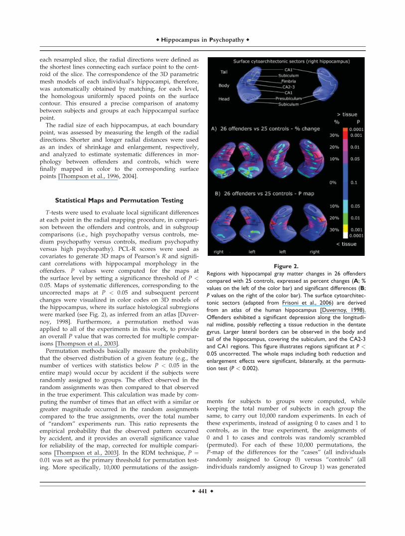
each resampled slice, the radial directions were defined asthe shortest lines connecting each surface point to the cent-roid of the slice. The correspondence of the 3D parametricmesh models of each individual’s hippocampi, therefore,was automatically obtained by matching, for each level,the homologous uniformly spaced points on the surfacecontour. This ensured a precise comparison of anatomybetween subjects and groups at each hippocampal surfacepoint.
The radial size of each hippocampus, at each boundarypoint, was assessed by measuring the length of the radialdirections. Shorter and longer radial distances were usedas an index of shrinkage and enlargement, respectively,and analyzed to estimate systematic differences in mor-phology between offenders and controls, which werefinally mapped in color to the corresponding surfacepoints [Thompson et al., 1996, 2004].
Statistical Maps and Permutation Testing
T-tests were used to evaluate local significant differencesat each point in the radial mapping procedure, in compari-son between the offenders and controls, and in subgroupcomparisons (i.e., high psychopathy versus controls, me-dium psychopathy versus controls, medium psychopathyversus high psychopathy). PCL-R scores were used ascovariates to generate 3D maps of Pearson’s R and signifi-cant correlations with hippocampal morphology in theoffenders. P values were computed for the maps atthe surface level by setting a significance threshold of P <0.05. Maps of systematic differences, corresponding to theuncorrected maps at P < 0.05 and subsequent percentchanges were visualized in color codes on 3D models ofthe hippocampus, where its surface histological subregionswere marked (see Fig. 2), as inferred from an atlas [Duver-noy, 1998]. Furthermore, a permutation method wasapplied to all of the experiments in this work, to providean overall P value that was corrected for multiple compar-isons [Thompson et al., 2003].
Permutation methods basically measure the probabilitythat the observed distribution of a given feature (e.g., thenumber of vertices with statistics below P < 0.05 in theentire map) would occur by accident if the subjects wererandomly assigned to groups. The effect observed in therandom assignments was then compared to that observedin the true experiment. This calculation was made by com-puting the number of times that an effect with a similar orgreater magnitude occurred in the random assignmentscompared to the true assignments, over the total numberof ‘‘random’’ experiments run. This ratio represents theempirical probability that the observed pattern occurredby accident, and it provides an overall significance valuefor reliability of the map, corrected for multiple compari-sons [Thompson et al., 2003]. In the RDM technique, P ¼0.01 was set as the primary threshold for permutation test-ing. More specifically, 10,000 permutations of the assign-
ments for subjects to groups were computed, whilekeeping the total number of subjects in each group thesame, to carry out 10,000 random experiments. In each ofthese experiments, instead of assigning 0 to cases and 1 tocontrols, as in the true experiment, the assignments of0 and 1 to cases and controls was randomly scrambled(permuted). For each of these 10,000 permutations, theP-map of the differences for the ‘‘cases’’ (all individualsrandomly assigned to Group 0) versus ‘‘controls’’ (allindividuals randomly assigned to Group 1) was generated
Figure 2.
Regions with hippocampal gray matter changes in 26 offenders
compared with 25 controls, expressed as percent changes (A; %
values on the left of the color bar) and significant differences (B;
P values on the right of the color bar). The surface cytoarchitec-
tonic sectors (adapted from Frisoni et al., 2006) are derived
from an atlas of the human hippocampus [Duvernoy, 1998).
Offenders exhibited a significant depression along the longitudi-
nal midline, possibly reflecting a tissue reduction in the dentate
gyrus. Larger lateral borders can be observed in the body and
tail of the hippocampus, covering the subiculum, and the CA2-3
and CA1 regions. This figure illustrates regions significant at P <0.05 uncorrected. The whole maps including both reduction and
enlargement effects were significant, bilaterally, at the permuta-
tion test (P < 0.002).
r 441 r
r Hippocampus in Psychopathy r

point by point along the whole 3D hippocampal meshmodel, and a new P-map was obtained for each randomexperiment. Subsequently, the number of supra-threshold(i.e., significant) voxels was computed and comparedbetween each random experiment and the true experi-ment. In the whole set of the 10,000 experiments, the totalnumber of times that the supra-threshold count was equalor higher than that observed in the true experiment wasdivided by 10,000 (i.e., the number of random experimentscarried out), and this estimated the probability that a map,with an amount of significant local differences greaterthan or equal to that observed in the true experiment,could be obtained by chance.
We note that this use of the supra-threshold volume ofstatistics is analogous to set-level inference in functionalimaging [Frackowiak et al., 2003]. Other types of permuta-tion tests are possible, examining the distribution of the sizeof the largest supra-threshold cluster, or the peak height(maximum statistic), etc. In general, we choose the supra-threshold volume statistic as it allows the detection of dif-fuse effects that occur weakly across the surface of a struc-ture so long as they are present at larger number of voxelsthat would be expected by chance alone. This is very simi-lar to analysis that control the false discovery rate [Hoch-berg and Benjamini, 1990; Genovese et al., 2002], where thesupra-threshold counts are analyzed using a histogram.
RESULTS
Offenders and controls were of similar age. Alcoholdependence or other substance abuse was common amongoffenders (100%), and absent in controls (0%; P < 0.001).Besides alcohol, polysubstance abuse was present in 77%
of cases and 0% of controls (P < 0.001). IQ scores were notavailable for control subjects, but, in the offendergroup, they were typically on the lower range of normality(Table I).
Twelve of the 26 violent offenders obtained a score of30 or higher on PCL-R featuring psychopathy [Hare, 1991],with a mean value of 34.6 and a standard deviation of 3.1.The other 14 had medium severity scores (25.9 � 2.8). Incomparison between the high and medium psychopathygroups, no significant differences were observed for age,duration of alcohol abuse, amphetamine use, or currentuse of psychotropic medication, but the prevalence of pol-ysubstance abuse was higher in the subgroup with highpsychopathy scores (Table II).
The hippocampal volumes, after correction for brain size,were similar between offenders and controls (Table I), andbetween the two subgroups of high and medium psychopa-thy. Paired t-tests between left and right hippocampidenoted a similar degree of significant difference inboth offenders and controls (controls: P ¼ 0.008, offenders:P ¼ 0.007), consistently with the physiological volumetricasymmetry. When the asymmetry ratio (right/left) wascomputed and directly compared between offenders andcontrols, no significant differences were observed withthe t-test (offenders: 0.97 � 0.05; controls: 0.96 � 0.07, P ¼0.532).
In the hippocampal surface maps, the offender sampledisplayed a complex pattern of both enlargement andreduction, covering most cytoarchitectonic sectors of thehippocampus, as inferred from an atlas [Duvernoy, 1998;Fig. 2]. More specifically, a bilateral depression (Fig. 2,green-yellow color range) was observed along the longitu-dinal axis of the body and tail, in the dorsal and ventralaspects. On the anterior hippocampi, a grey matter
TABLE I. Sociodemographic and clinical features of the healthy men and the violent
offenders
Controls(N ¼ 25) Violent offenders (N ¼ 26) P-value
Age, years 34.6 � 10.8 32.5 � 8.4 0.438Alcohol dependence, % 0 100% —Age onset of alcohol abuse — 13.6 � 2.9 —Polisubstance abuse, % 0 77% —Total intracranial volume (cm3) 1,707 � 117 1,654 � 108 0.102Hippocampus (mm3)Right 3,258 � 395 3,361 � 316 0.306Left 3,389 � 378 3,460 � 353 0.491
Hare’s Factor 1 — 10.8 � 3.5 —Hare’s Factor 2 — 16.6 � 1.7 —IQ — 91.5 � 9.0 —
Hippocampal volumes are normalized by total intracranial volumes. Values denote mean � SD.P values refer to Student’s t and Fisher’s exact tests. Total hippocampal volumes are normalizedand scaled to the ICBM (International Consortium Brain Mapping) standard space. Factor 1 is‘‘Arrogant Deceitful Interpersonal Conduct and Deficient Affective Experience’’, and Factor 2‘‘Impulsive, Irresponsible Behavioral Style.’’ The IQ of the offenders was assessed using the Wechs-ler Adult Intelligence Scale—Revised [Wechsler, 1981].
r 442 r
r Boccardi et al. r
reduction amounting to over 20% was observed, mappingto the CA1 sector of the offenders’ hippocampi. When themedium and high PCL-R subgroups were directly com-pared to each other, or with controls separately, this find-ing was significant only for the medium PCL-R group,while the tissue reduction was more prominent in thegroup with high psychopathy along the longitudinal axis(Fig. 3B: green-yellow color range).
The opposite contrast showed an abnormal enlargementof the lateral borders (Fig. 2, red color range), in both lat-eral aspects throughout the right and left hippocampi ofoffenders, covering the CA1, CA2-3, and subicular regions.This feature distinguished both offender groups with me-dium and high PCL-R scores (Fig. 3A,B, red color range).
Mean differences between offenders and controls inregions with hippocampal gray matter changes thatexceeded 15% were generally significant at P < 0.05 (Figs.2B and 3). The corrected P value for the whole enlargementand reduction map of the main comparison, contrasting alloffenders versus controls (see Fig. 2) was P < 0.002 for boththe left and right hippocampi, as computed by permutationtesting. The results of the comparisons of subgroups versuscontrols (see Fig. 3) also survived correction for multiplecomparisons with the permutation test (Medium psychopa-thy: left hippocampus, P < 0.008; right, P < 0.030; high psy-chopathy: left, P < 0.046). However, the P map of theexperiment ‘‘medium vs. high psychopathy’’ did not survivecorrection for multiple comparisons.
Overall, the hippocampal’s deviation from the normalsingle convex morphology, observed in offenders, suggestsa tendency to appear as a double convex structure in coro-nal sections, as illustrated by Figure 4.
The correlation analysis of local hippocampal morphol-ogy with PCL-R scores carried out in the offendersshowed both positive and negative correlation values (seeFig. 5), but did not survive the permutation test. Thesubgroup with polysubstance (i.e., alcohol plus any othersubstance) abuse (n ¼ 20) exhibited a less strong, butqualitatively similar pattern of correlation values as nonus-ers (n ¼ 6), and analogous findings were found in the corre-lation analyses in amphetamine users (n ¼ 15) andnonusers (n ¼ 11) (see Supporting Information Fig. 5Suppl:http://www.centroalzheimer.it/public/Figure5Suppl_CorrAmphetamine.doc). To further check for the possible effectof substance abuse, users and nonusers of polysubstancesand of amphetamine were subsequently compared withcontrols separately (Fig. 6; Supporting Information Fig.6Suppl at: http://www.centroalzheimer.it/public/Figure6-Suppl_pMaps-directComp-SubstAbuse.doc). These P maps,that resembled those observed in the main comparisons, alsosurvived correction for multiple comparisons in the contrasts:‘‘polysubstance users vs. controls’’ (left, P < 0.002, right, P <0.003), ‘‘amphetamine users vs. controls’’ (left, P < 0.003,right, P < 0.02), ‘‘amphetamine nonusers vs. controls’’ (right,P < 0.02).
DISCUSSION
In this work, hippocampal volume and morphology ofhabitually violent offenders with different severities ofpsychopathy were compared to normal controls. Size com-parisons denoted a similar overall volume and asymmetrypattern, but surface maps detected a peculiar distribution
TABLE II. Comparisons of sociodemographic and clinical characteristics of the
offenders with high and those with medium psychopathy
High psychopathy(N ¼ 12)
Medium psychopathy(N ¼ 14) P-value
Mean age (in years) 33.0 � 8.6 32.1 � 8.5 0.800Duration of alcohol abuse (in years) 19.6 � 10.0 18.3 � 8.9 0.725Amphetamine use 75% 43% 0.130Polysubstance abuse 100% 57% 0.017Mean global IQ score 94.7 � 8.6 88.7 � 8.8 0.095Current psychotropic medication 67% 50% 0.453Mean score PCL-R total 34.6 � 3.1 25.9 � 2.8 <0.001Hare’s Factor 1 14.0 � 2.1 8.1 � 1.5 <0.001Hare’s Factor 2 17.2 � 1.8 16.0 � 1.6 0.062Cooke’s Factor 1 6.03 � 1.74 1.90 � 1.12 <0.001Cooke’s Factor 2 7.83 � 0.58 6.14 � 1.10 <0.001Cooke’s Factor 3 9.67 � 1.15 9.71 � 0.47 0.889
Values denote mean � SD or percent proportions. P values refer to Student’s t and Fisher’s exacttests. The IQ of the offenders was assessed using the Wechsler Adult Intelligence Scale—Revised[Wechsler, 1981]. Hare’s Factor 1 is ‘‘Arrogant Deceitful Interpersonal Conduct and Deficient Affec-tive Experience’’, and Hare’s Factor 2 ‘‘Impulsive, Irresponsible Behavioral Style’’. Cooke’s Factor 1is ‘‘Arrogant and deceitful interpersonal style’’, Cooke’s Factor 2 ‘‘Deficient affective experience’’and Cooke’s Factor 3 ‘‘Impulsive, irresponsible behavioral style’’ [Cooke and Michie, 2001].
r 443 r
r Hippocampus in Psychopathy r
of alterations in the offenders, consisting of an extensiveenlargement of lateral borders in the hippocampal bodyand tail, with a depression along the midline longitudinalaxis. In the coronal plane, the hippocampus tended toappear in a double convex shape. This structural patternwas similar on both right and left sides, was more pro-nounced in the subgroup with greater severity of psy-chopathy, and could not be attributed to any of thestudied confounders (i.e., amphetamine or polysubstanceabuse).
Our data on total hippocampal volume are consistentwith previous findings that did not observe global volu-metric differences in antisocial or psychopathy subjects[Barkataki et al., 2006; Laakso et al., 2001; Raine et al.,2004]. A negative correlation between tissue volumes inposterior sections and PCL-R scores [Laakso et al., 2001]was described in a sample of antisocial offenders. The cor-relation analysis with PCL-R scores in our offender groupidentified predominant negative correlations with the pos-terior hippocampal sectors in both the subgroups with and
without polysubstance abuse. Therefore, this findingshould be interpreted in the context of changes relative tothe normal morphology of controls, consisting of reductionbut also of enlargement effects.
The RDM technique allows a fine reconstruction for the3D shape of the hippocampus, but it is not possible toidentify for sure which cytoarchitectonic regions are spe-cifically involved. Atlas-based inferences are typically usedto interpret such results [Fig. 2, top panel; Csernanskyet al., 2005; Duvernoy, 1998; Frisoni et al., 2006] and thusformulate more precise experimental hypotheses for futurestudies. In this case, the depressed line in the hippocampiof the psychopathic offenders may reflect lesser tissue inthe underlying dentate gyrus, which contains CA4 neu-rons, and is located along the midline of the hippocampalcore [see also the cytoarchitectonic map in Csernanskyet al., 2005]. The putative involvement of this anomaly isbiologically plausible, as CA4 neurons are responsible forvisceral sensory and autonomic response [Mollace et al.,2005], via different kinds of neurotransmission. Among
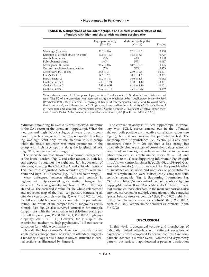
Figure 3.
Regions with hippocampal gray matter changes in offenders with
medium psychopathy (mean PCL-R ¼ 25.9 � 2.8, N ¼ 14) (A)
and with high psychopathy (mean PCL-R ¼ 34.6 � 3.1, N ¼ 12)
(B) compared to controls (N ¼ 25), and in offenders with me-
dium psychopathy compared to offenders with high psychopathy
(C), expressed as percent differences (left panel; % values on
the left of the colour bar) and P values (right panel; P values on
the right of the color bar). The right panel illustrates regions sig-
nificant at P < 0.05 uncorrected. The permutation test, indicat-
ing significance corrected for multiple comparisons, was
significant in the comparison ‘‘medium psychopathy versus con-
trols’’ (A) (left hippocampus: P ¼ 0.008; right hippocampus: P ¼0.030) and in the comparison ‘‘high psychopathy versus controls’’
(B) (left hippocampus: P ¼ 0.046). [Color figure can be viewed
in the online issue, which is available at www.interscience.
wiley.com.]
r 444 r
r Boccardi et al. r

these, the 5-HT system is associated with nociception[Echeverry et al., 2002], aggressive behavior [van Goozenand Fairchild, 2006], impulsivity [Cardinal, 2006; Masakiet al., 2006], drug addiction, and threat avoidance [Deakin,2003] that are all key features in psychopathy [Birbaumeret al., 2005]. As to the lateral expansions in the body andtail of the hippocampus, it is known that the CA3 regionis also involved in the processing of emotional and vis-ceral input [Mollace et al., 2005], and in contextual fear-conditioning [Daumas et al., 2007]. Together with otherevidence from animal research [Butkevich et al., 2003;Tsetsenis et al., 2007], these data are compatible with theclinical syndrome of psychopathy. However, these pointsshould be kept more as a basis for further experimentalhypotheses.
All offenders had both problems with alcohol or otherforms of substance abuse, and most of them had usedamphetamine, while these conditions were absent in con-trols. Our effort to isolate a pattern of hippocampal altera-tions that were independent of substance abuse eventuallyresulted in the analyses of subgroups for polysubstance oramphetamine abuse. In all of these analyses, the same hip-pocampal morphology was replicated, indicating that theobserved anomalies in hippocampal morphology were asso-ciated with psychopathy, rather than with substance abuse.Nonetheless, this was a statistical check: further experi-ments are needed to investigate the effect of psychopathy inlarger groups, stratified by kind and dosages of substances.
In spite of these positive findings, it should be acknowl-edged that sample size should be larger to ensure confi-dence, especially when analyzing subgroups, and in the
need to correct for multiple comparisons. Moreover, theRDM technique also has some acknowledged limitations.First, we can only indirectly infer observations about innerstructures from the surface shape, as the hippocampalmorphology is reconstructed from the tracings of the hip-pocampal contour in the coronal plane. Second, as in thevoxel-based analyses, the RDM technique can detect a sig-nificant change only when most cases display a differencein exactly the same points, compared to controls. There-fore, even important changes, such as those observedalong the midline longitudinal axis of the hippocampus,which we suppose to reflect the dentate gyrus’ morphol-ogy, actually turn out to be significant only in those fewpoints where the local change has exactly the same
Figure 5.
Correlation of localhippocampal gray matter with PCL-R scores,
expressed as a map of Pearson’s R values ranging from �1 to 1
(upper line) and of p values (lower line) in offenders with no sub-
stance abuse other than alcohol (A; n ¼ 6) and in offenders with
polysubstance abuse (B; n ¼ 20). The subgroup with no polysub-
stance abuse exhibited a stronger but similar correlation map to
that observed in the subgroup with polysubstance abuse. P maps
relate to uncorrected P; correlation experiments did not survive
correction for multiple comparisons. [Color figure can be viewed in
the online issue, which is available at www.interscience.wiley.com.]
Figure 4.
Coronal view of the hippocampal head (z ¼ 117), body (z ¼ 100),
and tail (z ¼ 90) in a control (left) and an offender with high
psychopathy (PCL-R score > 30) (right). In the body and tail,
larger lateral borders, and a steeper dip in the middle of the upper
border can be observed in the original coronal slices. [Color figure
can be viewed in the online issue, which is available at
www.interscience.wiley.com.]
r 445 r
r Hippocampus in Psychopathy r

location in most subjects. Regarding the potential con-founding variables that we could not rule out by subjectselection, such as substance abuse, the statistical analysesdid show that their effect did not influence the pattern ofanomalies associated with psychopathy. Nonetheless, wecould estimate only the presence versus absence of abuse,and not the exact amounts used during the whole lifetime.Finally, autopsy information on the cytoarchitectonic hip-
pocampal regions involved in psychopathy is required todefinitively elucidate the exact cellular correlates of theseMR findings.
ACKNOWLEDGMENTS
The authors thank Michela Pievani for providing help inthe revision of the manuscript, and Dr. James Callawayfor additional comments and revising in English.
REFERENCES
Apostolova LG, Dutton RA, Dinov ID, Hayashi KM, Toga AW,Cummings JL, Thompson PM (2006): Conversion of mild cog-nitive impairment to Alzheimer disease predicted by hippo-campal atrophy maps. Arch Neurol 63:693–699.
Barkataki I, Kumari V, Das M, Taylor P, Sharma T (2006): Volu-metric structural brain abnormalities in men with schizophre-nia or antisocial personality disorder. Behav Brain Res169:239–247.
Birbaumer N, Veit R, Lotze M, Erb M, Hermann C, Grodd W,Flor H (2005): Deficient fear conditioning in psychopathy: Afunctional magnetic resonance imaging study. Arch Gen Psy-chiatry 62:799–805.
Burman MA, Starr MJ, Gewirtz JC (2006): Dissociable effects ofhippocampus lesions on expression of fear and trace fear con-ditioning memories in rats. Hippocampus 16:103–113.
Butkevich IP, Khozhai LI, Mikhailenko VA, Otellin VA (2003):Decreased serotonin level during pregnancy alters morphologi-cal and functional characteristics of tonic nociceptive system injuvenile offspring of the rat. Reprod Biol Endocrinol 1:96.
Cardinal RN (2006): Neural systems implicated in delayed andprobabilistic reinforcement. Neural Netw 19:1277–1301.
Cloninger CR (1987): A systematic method for clinical descriptionand classification of personality variants. A proposal. ArchGen Psychiatry 44:573–588.
Cooke DJ, Michie C (2001): Refining the construct of psychopathy:Towards a hierarchical model. Psychol Assess 13:171–188.
Csernansky JG, Wang L, Swank J, Miller JP, Gado M, McKeel D,Miller MI, Morris JC (2005): Preclinical detection of Alzhei-mer’s disease: Hippocampal shape and volume predict demen-tia onset in the elderly. Neuroimage 25:783–792.
Daumas S, Betourne A, Halley H, Wolfer DP, Lipp HP,Lassalle JM, Frances B (2007): Transient activation of the CA3Kappa opioid system in the dorsal hippocampus modulatescomplex memory processing in mice. Neurobiol Learn Mem88:94–103.
Deakin JF (2003): Depression and antisocial personality disorder:Two contrasting disorders of 5HT function. J Neural TransmSuppl 64:79–93.
Duvernoy HM (1998): The Human Hippocampus. FunctionalAnatomy, Vascularization, and Serial Sections With MRI. Ber-lin: Springer.
Echeverry MB, Guimaraes FS, Oliveira MA, do Prado WA, DelBel EA (2002): Delayed stress-induced antinociceptive effect ofnitric oxide synthase inhibition in the dentate gyrus of rats.Pharmacol Biochem Behav 74:49–156.
Frackowiak RSJ, Friston KJ, Frith C, Dolan R, Friston KJ, Price CJ,Zeki S, Ashburner J, Penny WD (2003): Human Brain Function,2nd ed. San Diego (CA): Academic Press.
Figure 6.
Regions with hippocampal gray matter changes in subgroups of
users (A; n ¼ 20) and nonusers (B; n ¼ 6) of polysubstances
(i.e., any substance other than alcohol) and amphetamine (users,
C; n ¼ 15; nonusers D; n ¼ 11), compared to controls (n ¼25). Maps denote percent enlargement (red range) or reduction
(light blue-green-yellow range) effects. Regions with tissue
change greater than 20% were usually significant at P < 0.05
(see Fig. 6Suppl at http://www.centroalzheimer.it/public/Figure6
Suppl_pMaps-directComp-SubstAbuse.doc). The permutation
test, indicating significance corrected for multiple comparisons,
was significant in the comparisons ‘‘polysubstance users versus
controls’’ (left: P ¼ 0.002; right: P ¼ 0.003), ‘‘amphetamine users
versus controls’’ (left: P ¼ 0.003; right: P ¼ 0.029), ‘‘ampheta-
mine nonusers versus controls’’ (right: P ¼ 0.02). [Color figure
can be viewed in the online issue, which is available at
www.interscience.wiley.com.]
r 446 r
r Boccardi et al. r
Frisoni GB, Sabattoli F, Lee AD, Dutton RA, Toga AW, ThompsonPM (2006): In vivo neuropathology of the hippocampal forma-tion in AD: A radial mapping MR-based study. Neuroimage32:104–110.
Genovese CR, Lazar NA, Nichols T (2002): Thresholding of statis-tical maps in functional neuroimaging using the false discov-ery rate. Neuroimage 15:870–878.
Giovannini MG (2006): The role of the extracellular signal-regu-lated kinase pathway in memory encoding. Rev Neurosci 17:619–634.
Hare RD (1991): The Hare Psychopathy Checklist—Revised. To-ronto: Multi-Health Systems.
Hare RD (2006): Psychopathy: A clinical and forensic overview.Psychiatr Clin North Am 29:709–724.
Hochberg Y, Benjamini Y (1990): More powerful procedures formultiple significance testing. Stat Med 9:811–818.
Kiehl KA, Smith AM, Hare RD, Mendrek A, Forster BB, Brink J,Liddle PF (2001): Limbic abnormalities in affective processingby criminal psychopaths as revealed by functional magneticresonance imaging. Biol Psychiatry 50:677–684.
Laakso MP, Vaurio O, Koivisto E, Savolainen L, Eronen M, Aro-nen HJ, Hakola P, Repo E, Soininen H, Tiihonen J (2001): Psy-chopathy and the posterior hippocampus. Behav Brain Res118:187–193.
Masaki D, Yokoyama C, Kinoshita S, Tsuchida H, Nakatomi Y,Yoshimoto K, Fukui K (2006): Relationship between limbic andcortical 5-HT neurotransmission and acquisition and reversallearning in a go/no-go task in rats. Psychopharmacology (Berl)189:249–258.
Mollace V, Muscoli C, Masini E, Cuzzocrea S, Salvemini D (2005):Modulation of prostaglandin biosynthesis by nitric oxide andnitric oxide donors. Pharmacol Rev 57:217–252.
Narr KL, Thompson PM, Szeszko P, Robinson D, Jang S, WoodsRP, Kim S, Hayashi KM, Asunction D, Toga AW, Bilder RM(2004): Regional specificity of hippocampal volume reductionsin first-episode schizophrenia. Neuroimage 21:1563–1575.
Pridmore S, Chambers A, McArthur M (2005): Neuroimaging inpsychopathy. Aust N Z J Psychiatry 39:856–865.
Pruessner JC, Li LM, Serles W, Pruessner M, Collins DL, KabaniN, Lupien S, Evans AC (2000): Volumetry of hippocampus andamygdala with high-resolution MRI and three-dimensional
analysis software: Minimizing the discrepancies between labo-ratories. Cereb Cortex 10:433–442.
Raine A, Yaralian PS, Reynolds C, Venables PH, MednickSA (2002): Spatial but not verbal cognitive deficits at age 3 yearsin persistently antisocial individuals. Dev Psychopathol 14:25–44.
Raine A, Lencz T, Taylor K, Hellige JB, Bihrle S, Lacasse L, LeeM, Ishikawa S, Colletti P (2003): Corpus callosum abnormal-ities in psychopathic antisocial individuals. Arch Gen Psychia-try 60:1134–1142.
Raine A, Ishikawa SS, Arce E, Lencz T, Knuth KH, Bihrle S,LaCasse L, Colletti P (2004): Hippocampal structural asymme-try in unsuccessful psychopaths. Biol Psychiatry 55:185–191.
Sommer M, Hajak G, Dohnel K, Schwerdtner J, Meinhardt J, Mul-ler JL (2006): Integration of emotion and cognition in patientswith psychopathy. Prog Brain Res 156:457–466.
Thompson PM, Schwartz C, Toga AW (1996): High-resolution ran-dom mesh algorithms for creating a probabilistic 3D surfaceatlas of the human brain. Neuroimage 3:19–34.
Thompson PM, Hayashi KM, de Zubicaray G, Janke AL, Rose SE,Semple J, Herman D, Hong MS, Dittmer SS, Doddrell DM,Toga AW (2003): Dynamics of gray matter loss in Alzheimer’sdisease. J Neurosci 23:994–1005.
Thompson PM, Hayashi KM, De Zubicaray GI, Janke AL,Rose SE, Semple J, Hong MS, Herman DH, Gravano D,Doddrell DM, Toga AW (2004): Mapping hippocampaland ventricular change in Alzheimer disease. Neuroimage22:1754–1766.
Tiihonen J, Rossi R, Laakso MP, Hodgins S, Testa C, Perez J,Repo-Tiihonen E, Vaurio O, Soininen H, Aronen HJ, KononenM, Thompson PM, Frisoni GB (2008): Brain anatomy of persis-tent violent offenders: More rather than less. Psychiatry Res163:201–212.
Tsetsenis T, Ma XH, Lo Iacono L, Beck SG, Gross C (2007): Sup-pression of conditioning to ambiguous cues by pharmacoge-netic inhibition of the dentate gyrus. Nat Neurosci 10:896–902.
van Goozen SH, Fairchild G (2006): Neuroendocrine and neuro-transmitter correlates in children with antisocial behavior.Horm Behav 2006:647–654.
Wechsler D (1981): Adult Intelligence Scale. S. Antonio, TX, USA:Psychological Corporation.
r 447 r
r Hippocampus in Psychopathy r