Embed Size (px)
Citation preview
Citation Skawinski T Kuziak P
Kloskowski J Borczyk B
Phylogenetic Diversity of Ossification
Patterns in the Avian Vertebral
Column A Review and New Data
from the Domestic Pigeon and Two
Species of Grebes Biology 2022 11
180 httpsdoiorg103390
biology11020180
Academic Editors Susan
C Chapman and Dana J Rashid
Received 30 November 2021
Accepted 21 January 2022
Published 24 January 2022
Publisherrsquos Note MDPI stays neutral
with regard to jurisdictional claims in
published maps and institutional affil-
iations
Copyright copy 2022 by the authors
Licensee MDPI Basel Switzerland
This article is an open access article
distributed under the terms and
conditions of the Creative Commons
Attribution (CC BY) license (https
creativecommonsorglicensesby
40)
biology
Article
Phylogenetic Diversity of Ossification Patterns in the AvianVertebral Column A Review and New Data from the DomesticPigeon and Two Species of GrebesTomasz Skawinski 12 Piotr Kuziak 2 Janusz Kloskowski 3 and Bartosz Borczyk 2
1 Department of Palaeozoology Faculty of Biological Sciences University of Wrocław Sienkiewicza 2150-335 Wrocław Poland
2 Department of Evolutionary Biology and Conservation of Vertebrates Faculty of Biological SciencesUniversity of Wrocław Sienkiewicza 21 50-335 Wrocław Poland 326967uwredupl (PK)bartoszborczykuwredupl (BB)
3 Department of Zoology Institute of Zoology Poznan University of Life Sciences Wojska Polskiego 71C60-625 Poznan Poland januszkluppoznanpl
Correspondence tomaszskawinskiuwredupl
Simple Summary There are still many unknowns in the development of the skeleton in birdsTraditionally the neck vertebrae were considered to be the first ossifying elements in the spine Laterstudies have shown that this is not always the case In some species the thoracic vertebrae ossifyeven before them Evolutionary analyses indicate that ancestrally the spine starts ossifying from twodifferent sites one located in the neck the other in the thorax However the Neoaves a group thatincludes all living birds except the palaeognaths landfowl and waterfowl are very poorly studiedIn this article we review the information about ossification patterns of the spine in birds We alsodescribe its development in three neoavians the pigeon and two grebes In the pigeon the neckvertebrae were the first to ossify but in the grebe the thoracic vertebrae ossified earlier Our analysesconfirm the ancestral presence of two sites from which the ossification of the spine starts in birds
Abstract Despite many decades of studies our knowledge of skeletal development in birds is limitedin many aspects One of them is the development of the vertebral column For many years it waswidely believed that the column ossifies anteroposteriorly However later studies indicated thatsuch a pattern is not universal in birds and in many groups the ossification starts in the thoracicrather than cervical region Recent analyses suggest that two loci located in the cervical and thoracicvertebrae were ancestrally present in birds However the data on skeletal development are veryscarce in the Neoaves a clade that includes approximately 95 of extant species We review theavailable information about the vertebral column development in birds and describe the ossificationpattern in three neoavians the domestic pigeon (Columba livia domestica) the great crested grebe(Podiceps cristatus) and the red-necked grebe (Podiceps grisegena) In P cristatus the vertebral columnstarts ossifying in the thoracic region The second locus is present in the cervical vertebrae In thepigeon the cervical vertebrae ossify before the thoracics but both the thoracic and cervical loci arepresent Our ancestral state reconstructions confirm that both these loci were ancestrally present inbirds but the thoracic locus was later lost in psittacopasserans and at least some galloanserans
Keywords axial skeleton birds evo-devo ossification sequences skeletogenesis
1 Introduction
The vertebral column is a defining feature of vertebrates It consists of a series ofsegmental units the vertebrae which play a crucial role in vertebrate biology such asprotecting the spinal cord or providing muscle attachment sites for numerous musclesinvolved in locomotion The vertebra itself is usually a complex structure composed of
Biology 2022 11 180 httpsdoiorg103390biology11020180 httpswwwmdpicomjournalbiology
Biology 2022 11 180 2 of 20
several distinct parts the body arches and a few projections such as spinous or transverseprocesses (eg [1]) The vertebral column is divided into several regions the cervicalthoracic lumbar sacral and caudal which differ morphologically (eg [2]) There isalso a great interspecific variation in the number of vertebrae that constitute the columnHowever the number of vertebrae is constrained at relatively low numbers in mammalswhile in sauropsids (reptiles and birds) these numbers are usually higher and much lessconservative [34] In addition the vertebral column is strongly modified in extant birdsas an adaptation to active flight many of the individual vertebrae fuse (usually afterhatching [5ndash8]) to form compound structuresmdashthe notarium synsacrum and pygostyle(eg [2679]) Although the morphology and number of vertebrae can tell us a lot aboutdevelopmental processes which act on the vertebral column (eg [31011]) the data on thesequence in which it ossifies are still incomplete for many groups
The ossification sequencesmdashthe order in which individual bones appear in develop-mentmdashare one of the most basic yet important studies in developmental biology Theygive us not only information about the development of a given species but also allow us tocompare different species thus placing these sequences in an evolutionary context Theyalso help us interpret fossil embryos and reconstruct ancestral developmental patterns andheterochronic events (eg [12ndash15]) With over 10000 extant species the birds are one of themost species-rich lineages of tetrapods They exhibit great ecological diversity and occupynumerous different niches and habitats Their life histories are also very diversemdashsomebirds such as galliforms (Galliformes) including the well-studied chicken and quail areprecocial ie have independent hatchlings while many others are altricial with embryo-like hatchlings Numerous species occupy intermediate positions in this precocial-altricialspectrum (eg [16]) Despite this huge diversity the temporal order of bone ossificationin birds has been described as remarkably conservative with only small differences be-tween species widely divergent ecologically and phylogenetically [16] In particular thedevelopment of the vertebral column has been repeatedly cited as predominantly or evenuniversally proceeding from anterior to posterior in birds [1216ndash18] Although furtherstudies indicated a greater variation in this respect among birds (eg [19ndash22]) ossificationpatterns other than the anteroposterior are regarded as exceptions [23] Recently there hasbeen an increase in knowledge on the genetic and developmental bases of the regionalisa-tion of the vertebral column in birds (eg [101124]) However despite the long history ofstudies on the development of the avian skeleton (eg [2526]) the data on the ossificationsequences are still scarce and limited mostly to precocial birds in particular palaeognathsand galloanserans while the information about neoavians concerns only a few species(eg [13]) This is probably related to the fact that a much greater proportion of neoaviansrepresents altricials and in many altricial species (although this is far from being universal)the vertebral column starts ossifying only after hatching (eg [161819]) In general thepostnatal skeletal ontogeny of birds is very poorly known (eg [8]) so it hampers ourunderstanding of the development and evolution of the vertebral column in many groupsof birds In this article we attempt to summarise currently available information about theossification patterns in extant birds and supplement it with new data from two basal neoa-vian lineages the altricial (lsquoaltricial 1prime according to Starck [16]) pigeons (Columbiformes)and precocial (lsquoprecocial 3prime in the classification by Starck [16] which means that they arerelatively little precocial) grebes (Podicipediformes) (relationships of the species discussedin the text are shown in Figure 1) We focused on the following three different processes(1) body ossification (2) vertebral arch ossification and (3) vertebral arch fusion with aparticular reference to the first two
Biology 2022 11 180 3 of 20Biology 2022 11 x FOR PEER REVIEW 3 of 21
Figure 1 Phylogenetic relationships of the species discussed in the text The topology of the den-drogram follows the lsquoconsensus phylogeny of birdsrsquo from Braun and Kimball [27] while the inter-relationships of the main clades follow primarily Prum et al [28] and Kuhl et al [29] Codes 1mdashNeornithes 2mdashPalaeognathae 3mdashNeognathae 4mdashGalloanserae 5mdashGalliformes 6mdashAnseriformes 7mdashNeoaves 8mdashCharadriiformes 9mdashPsittacopasserae
2 Materials and Methods 21 Literature Review
The information given below are summarised in Table 1 and Figures 2ndash4 The exact individual age of the studied birds was often unknown in the publications discussed be-low (as well as in the specimens described in the lsquoResultsrsquo) so the timing of the observed events is often not directly comparable but we were interested primarily in the sequence in which these events appear during the ontogeny which can be compared even without information about the individual age of the specimens
Table 1 A summary of the ossification sequences of the vertebral bodies and arches in extant birds
Species Sequence References
Struthio camelus Vertebral bodies thoracic synsacral rarr cervical rarr caudal rarr py-gostyle Vertebral arches cervical rarr thoracic rarr synsacral
[30]
Rhea americana Vertebral bodies thoracic rarr cervical rarr synsacral Vertebral arches cervical rarr thoracic rarr synsacral [30]
Dromaius novaehollandiae Vertebral bodies cervical thoracic rarr synsacral rarr caudal rarr py-gostyle Vertebral arches cervical rarr thoracic rarr synsacral
[30]
Eudromia elegans Vertebral bodies thoracic synsacral rarr cervical Vertebral arches cervical rarr thoracic rarr synsacral [30]
Meleagris gallopavo Vertebral bodies cervical rarr thoracic rarr synsacral rarr caudal Vertebral arches cervical rarr thoracic rarr synsacral [2131]
Galllus domesticus Vertebral bodies cervical rarr thoracic rarr synsacral rarr caudal [1721]
Figure 1 Phylogenetic relationships of the species discussed in the text The topology of thedendrogram follows the lsquoconsensus phylogeny of birdsrsquo from Braun and Kimball [27] while theinterrelationships of the main clades follow primarily Prum et al [28] and Kuhl et al [29] Codes1mdashNeornithes 2mdashPalaeognathae 3mdashNeognathae 4mdashGalloanserae 5mdashGalliformes 6mdashAnseriformes7mdashNeoaves 8mdashCharadriiformes 9mdashPsittacopasserae
2 Materials and Methods21 Literature Review
The information given below are summarised in Table 1 and Figures 2ndash4 The exactindividual age of the studied birds was often unknown in the publications discussed below(as well as in the specimens described in the lsquoResultsrsquo) so the timing of the observed eventsis often not directly comparable but we were interested primarily in the sequence in whichthese events appear during the ontogeny which can be compared even without informationabout the individual age of the specimens
Biology 2022 11 x FOR PEER REVIEW 4 of 21
Vertebral arches cervical rarr thoracic rarr synsacral
Coturnix japonica Vertebral bodies cervical rarr thoracic rarr synsacral rarr caudal Vertebral arches cervical rarr thoracic rarr synsacral [213233]
Synoicus chinensis Vertebral bodies anterior cervical thoracic rarr mid- and posterior cervical synsacral rarr caudal Vertebral arches cervical rarr thoracic rarr synsacral
[34]
Anas platyrhynchos Vertebral bodies cervical rarr thoracic synsacral rarr caudal rarr py-gostyle Vertebral arches cervical rarr thoracic rarr synsacral
[1220]
Cairina moschata Vertebral bodies cervical thoracic synsacral rarr caudal rarr py-gostyle Vertebral arches cervical rarr thoracic
[20]
Somateria mollissima Vertebral bodies cervical rarr thoracic synsacral rarr caudal rarr py-gostyle Vertebral arches cervical rarr thoracic rarr synsacral
[20]
Podiceps cristatus Vertebral bodies thoracic rarr synsacral rarr cervical rarr caudal rarr pygostyle Vertebral arches cervical rarr thoracic rarr synsacral
this work
Columba livia domestica Vertebral bodies cervical rarr thoracic synsacral rarr caudal Vertebral arches cervical rarr thoracic this work
Sterna hirundo Vertebral bodies cervical thoracic synsacral rarr caudal Vertebral arches cervical rarr thoracic or thoracic rarr cervical [22]
Stercorarius skua Vertebral bodies cervical thoracic rarr synsacral rarr caudal Vertebral arches cervical rarr thoracic [2235]
Chroicocephalus ridibundus Vertebral bodies cervical rarr thoracic rarr synsacral rarr caudal Vertebral arches cervical rarr thoracic [223637]
Larus canus Vertebral bodies cervical thoracic synsacral rarr caudal Vertebral arches thoracic rarr cervical [2237]
Myiopsitta monachus Vertebral bodies cervical rarr thoracic rarr synsacral caudal [14] Taeniopygia guttata Vertebral bodies cervical rarr thoracic rarr synsacral caudal [12] Corvus frugilegus Vertebral bodies cervical rarr thoracic rarr synsacral rarr caudal [36]
Figure 2 A schematic summary of the ossification patterns of the vertebral bodies in birds (AB)Blue indicates cervical loci while orange indicates thoracic loci The bird silhouette was taken fromPhyloPic and is available in public domain
Biology 2022 11 180 4 of 20
Table 1 A summary of the ossification sequences of the vertebral bodies and arches in extant birds
Species Sequence References
Struthio camelus Vertebral bodies thoracic synsacral rarr cervical rarr caudal rarr pygostyleVertebral arches cervical rarr thoracic rarr synsacral [30]
Rhea americana Vertebral bodies thoracic rarr cervical rarr synsacralVertebral arches cervical rarr thoracic rarr synsacral [30]
Dromaius novaehollandiae Vertebral bodies cervical thoracic rarr synsacral rarr caudal rarr pygostyleVertebral arches cervical rarr thoracic rarr synsacral [30]
Eudromia elegans Vertebral bodies thoracic synsacral rarr cervicalVertebral arches cervical rarr thoracic rarr synsacral [30]
Meleagris gallopavo Vertebral bodies cervical rarr thoracic rarr synsacral rarr caudalVertebral arches cervical rarr thoracic rarr synsacral [2131]
Galllus domesticus Vertebral bodies cervical rarr thoracic rarr synsacral rarr caudalVertebral arches cervical rarr thoracic rarr synsacral [1721]
Coturnix japonica Vertebral bodies cervical rarr thoracic rarr synsacral rarr caudalVertebral arches cervical rarr thoracic rarr synsacral [213233]
Synoicus chinensisVertebral bodies anterior cervical thoracic rarr mid- and posterior cervicalsynsacral rarr caudalVertebral arches cervical rarr thoracic rarr synsacral
[34]
Anas platyrhynchos Vertebral bodies cervical rarr thoracic synsacral rarr caudal rarr pygostyleVertebral arches cervical rarr thoracic rarr synsacral [1220]
Cairina moschata Vertebral bodies cervical thoracic synsacral rarr caudal rarr pygostyleVertebral arches cervical rarr thoracic [20]
Somateria mollissima Vertebral bodies cervical rarr thoracic synsacral rarr caudal rarr pygostyleVertebral arches cervical rarr thoracic rarr synsacral [20]
Podiceps cristatus Vertebral bodies thoracic rarr synsacral rarr cervical rarr caudal rarr pygostyleVertebral arches cervical rarr thoracic rarr synsacral this work
Columba livia domestica Vertebral bodies cervical rarr thoracic synsacral rarr caudalVertebral arches cervical rarr thoracic this work
Sterna hirundo Vertebral bodies cervical thoracic synsacral rarr caudalVertebral arches cervical rarr thoracic or thoracic rarr cervical [22]
Stercorarius skua Vertebral bodies cervical thoracic rarr synsacral rarr caudalVertebral arches cervical rarr thoracic [2235]
Chroicocephalus ridibundus Vertebral bodies cervical rarr thoracic rarr synsacral rarr caudalVertebral arches cervical rarr thoracic [223637]
Larus canus Vertebral bodies cervical thoracic synsacral rarr caudalVertebral arches thoracic rarr cervical [2237]
Myiopsitta monachus Vertebral bodies cervical rarr thoracic rarr synsacral caudal [14]
Taeniopygia guttata Vertebral bodies cervical rarr thoracic rarr synsacral caudal [12]
Corvus frugilegus Vertebral bodies cervical rarr thoracic rarr synsacral rarr caudal [36]
Biology 2022 11 180 5 of 20
Biology 2022 11 x FOR PEER REVIEW 5 of 21
Figure 2 A schematic summary of the ossification patterns of the vertebral bodies in birds (AB) Blue indicates cervical loci while orange indicates thoracic loci The bird silhouette was taken from PhyloPic and is available in public domain
Figure 3 A schematic summary of the developmental patterns of the vertebral arches in birds (AndashD) Ossification (appearance) of the vertebral arches Blue indicates cervical loci orangemdashthoracic loci red mdashsynsacral loci purplemdashcaudal loci
Figure 3 A schematic summary of the developmental patterns of the vertebral arches in birds (AndashD)Ossification (appearance) of the vertebral arches Blue indicates cervical loci orangemdashthoracic lociredmdashsynsacral loci purplemdashcaudal loci
Biology 2022 11 x FOR PEER REVIEW 6 of 21
Figure 4 A schematic summary of developmental patterns in the vertebral arch fusion (AndashC) Blue indicates cervical loci orangemdashthoracic loci
211 Vertebral Bodies (Corpora Vertebrae) In palaeognathous birds the ossification of the vertebral bodies (and the vertebral
column in general) usually starts with the thoracic vertebrae This is the condition ob-served in most of the hitherto studied species including both the flying tinamous (Tin-amidae) and secondarily flightless ratites In the ostrich (Struthio camelus) the bodies of the thoracic and preacetabular synsacral (which belong to the lumbar region) vertebrae are the first to ossify usually in 23-day-old embryos but in some individuals slightly later In 25-day-old embryos the cervical vertebrae ossify but the ossification of the first two cervicals the atlas and axis is delayed and starts in 28-day-old embryos The free caudal vertebrae start to ossify in 30-day-old embryos (although there is some variability in this respect) and finally the bodies of the vertebrae that form the pygostyle ossify in 37-day-old embryos [30] A very similar condition is observed in the elegant crested tinamou (Eudromia elegans) The bodies of the posterior thoracics and anterior synsacrals (lumbars) are the first to appear in 11-day-old embryos From there the ossification spreads anteri-orly and posteriorly and in 14-day-old embryos all cervical and synsacral bodies had be-gun ossifying [30] In the greater rhea (Rhea americana) the bodies of the thoracic vertebrae 3ndash6 are the first to ossify in stage 37 (according to Hamburger and Hamilton [38]) embryos In late stage 38 the cervical vertebrae start ossifying In 22-day-old embryos (stage 40+) all thoracic vertebrae are ossified The emu (Dromaius novaehollandiae) differs from other studied palaeognaths in the earlier onset of ossification in the cervical vertebrae bodies In stage 38 embryos the ossification starts from two loci (sensu Verriegravere et al [13]) one located in the cervical vertebrae 2ndash4 (C2ndashC4) while the second one contains thoracic ver-tebrae located posteriorly to the thoracic 2 (T2) A few synsacral bodies located under an-terior parts of the ilia are also ossified In the following stage (39) all cervicals except the atlas as well as all thoracics and synsacrals are ossified In stage 40+ embryos the free
Figure 4 A schematic summary of developmental patterns in the vertebral arch fusion (AndashC) Blueindicates cervical loci orangemdashthoracic loci
Biology 2022 11 180 6 of 20
211 Vertebral Bodies (Corpora Vertebrae)
In palaeognathous birds the ossification of the vertebral bodies (and the vertebralcolumn in general) usually starts with the thoracic vertebrae This is the condition observedin most of the hitherto studied species including both the flying tinamous (Tinamidae)and secondarily flightless ratites In the ostrich (Struthio camelus) the bodies of the thoracicand preacetabular synsacral (which belong to the lumbar region) vertebrae are the first toossify usually in 23-day-old embryos but in some individuals slightly later In 25-day-oldembryos the cervical vertebrae ossify but the ossification of the first two cervicals the atlasand axis is delayed and starts in 28-day-old embryos The free caudal vertebrae start toossify in 30-day-old embryos (although there is some variability in this respect) and finallythe bodies of the vertebrae that form the pygostyle ossify in 37-day-old embryos [30] Avery similar condition is observed in the elegant crested tinamou (Eudromia elegans) Thebodies of the posterior thoracics and anterior synsacrals (lumbars) are the first to appear in11-day-old embryos From there the ossification spreads anteriorly and posteriorly andin 14-day-old embryos all cervical and synsacral bodies had begun ossifying [30] In thegreater rhea (Rhea americana) the bodies of the thoracic vertebrae 3ndash6 are the first to ossifyin stage 37 (according to Hamburger and Hamilton [38]) embryos In late stage 38 thecervical vertebrae start ossifying In 22-day-old embryos (stage 40+) all thoracic vertebraeare ossified The emu (Dromaius novaehollandiae) differs from other studied palaeognathsin the earlier onset of ossification in the cervical vertebrae bodies In stage 38 embryosthe ossification starts from two loci (sensu Verriegravere et al [13]) one located in the cervicalvertebrae 2ndash4 (C2ndashC4) while the second one contains thoracic vertebrae located posteriorlyto the thoracic 2 (T2) A few synsacral bodies located under anterior parts of the ilia are alsoossified In the following stage (39) all cervicals except the atlas as well as all thoracics andsynsacrals are ossified In stage 40+ embryos the free caudals and lastly the pygostylestart ossifying [30] In his study on the anatomy and development of the kiwi Parker [25]described a specimen (ldquostage Grdquo) of the little spotted kiwi (Apteryx owenii) in which thebodies of almost all cervical (except the atlas) thoracic lumbar and sacral vertebrae hadalready begun their ossification while the caudals were still cartilaginous It is howeverunclear whether all these vertebrae indeed ossify nearly simultaneously or whether it is anartefact of an insufficient number of studied embryos
The galloanserans differ from palaeognaths in that they usually conform to the lsquotypicalrsquoanteroposterior gradient of ossification In the turkey (Meleagris gallopavo) the ossificationseems to proceed in a strictly anterior to posterior order with the most anterior cervical ver-tebrae ossifying at stage 38 The thoracic bodies start ossifying at stage 40+ and are followedby the ossification centres in the synsacral (lumbar) vertebrae Lastly ossifications appearin the caudal vertebrae [2131] The chicken (Gallus domesticus) is similar to the turkey in theorder of ossification of the vertebral centra in general the cervical bodies are followed bythe thoracic then synsacral and finally caudal vertebrae [18] However in contrast to theturkey but similarly to most palaeognaths the ossification of the anteriormost cervicals isdelayed [1721] The ossification of the most posterior cervical precedes the more anteriorlylocated cervical vertebrae from there the ossification proceeds posteriorly [21] so the lastcervical belongs to the thoracic locus (sensu Verriegravere et al [13]) The ossification of thevertebral column in the Japanese quail (Coturnix japonica) seems to be more similar to thechicken than to the turkey The mid-cervical vertebrae (6ndash9) ossify first and the ossificationspreads both anteriorly and posteriorly [213233] A similar condition is present in thethoracic vertebrae in which mid-thoracics appear first and the ossification proceeds bothanteriorly and posteriorly Anterior synsacrals ossify next followed by posterior synsacralsthen by free caudals and lastly by the pygostyle [3233] Interestingly Blom and Lilja [19]described a strictly anteroposterior order of ossification in C japonica which may suggestan intraspecific variability in the ossification patterns A closely related blue-breastedquail (Synoicus chinensis) differs from the Japanese quail in that the ossification starts withanterior cervicals and all thoracic vertebrae (11-day-old embryos) They are followed bythe remaining cervicals and anterior synsacrals (12-day-old) mid-synsacrals (13-day-old)
Biology 2022 11 180 7 of 20
posterior synsacrals and anterior caudals (14-day-old) and finally by the remaining caudals(17-day-old) [34] In the Muscovy duck (Cairina moschata) all precaudal vertebrae ossifysimultaneously (stage 39) without a gradient and are followed closely by the first threecaudal bodies [20] In the common eider (Somateria mollissima) the cervical bodies follow ananteroposterior gradient Posterior cervicals are worse ossified than the anterior thoracicswhich suggests that the ossification proceeds from two loci Ten anterior synsacrals alsoossify at the same stage (38) In stage 39 the remaining synsacrals ossify and are followed(stage 40) by the caudal vertebrae [20] The vertebral bodies ossify in an anteroposteriordirection in the mallard (Anas platyrhynchos) The cervicals appear first (stage 37) and arefollowed by thoracics and approximately half of synsacrals (stage 39) In stage 40 freecaudal bodies ossify then the remaining synsacrals and finally the anterior components ofthe pygostyle [20]
The neoavians which represent approximately 95 of all extant species of birds [27]are relatively the most poorly studied avian clade In the common tern (Sterna hirundo) theossification of the vertebral column starts with the thoracic bodies The ossification proceedsposteriorly from the first thoracic Later the ossifications in the cervicals appear Anteriorsynsacral bodies ossify approximately at the same time with the more posteriorly locatedones ossifying later The caudal bodies appear last [22] In the great skua (Stercorariusskua) the cervical and thoracic bodies ossify approximately at the same time and arefollowed closely by the synsacral bodies The bodies of the caudals appear last [35] Inthe black-headed gull (Chroicocephalus ridibundus) the cervical and thoracic bodies ossifynearly simultaneously [2237] but Rogulska [36] described that they precede the thoracicswhich may indicate an intraspecific variation or an insufficient sampling in the work ofSchumacher and Wolff [37] In the common gull (Larus canus) the thoracic bodies weredescribed to ossify slightly before the cervicals [2237] In both these species the remainingvertebrae were not described so they must ossify later The monk parakeet (Myiopsittamonachus) exhibits a typical anteroposterior gradient of ossification The cervical vertebraeossify first (although the appearance of the atlas is delayed) the thoracic vertebrae secondlyand the synsacral and caudal vertebrae lastly [14] There is some intraspecific variation inthe timing of the ossification of the vertebral column in the specimens of the zebra finch(Taeniopygia guttata) studied by Mitgutsch et al [12] but in most of them the directionof ossification proceeded from anterior to posterior with the cervical vertebrae ossifyingfirst thoracics secondly and synsacrals and caudals thirdly [12] An anteroposterior orderof ossification was also described for the rook (Corvus frugilegus) in which the cervicalvertebrae appear first during the development [36]
212 Vertebral Arches (Arcus Vertebrae)
The ossification of the vertebral arches usually takes place after the appearance of thebodies of the respective vertebrae The exception is the atlas in which the arches ossifybefore the body This condition is present in many species of phylogenetically distantspecies (eg [173033]) so probably represents an ancestral condition Curiously in theEurasian reed warbler (Acrocephalus scirpaceus) the atlantal body was described to be absentand the atlas is composed only of two arches which fuse dorsally and ventrally [8]
The ossification of the vertebral arches is much less variable in birds than the ossifica-tion patterns of the bodies Usually it much more closely conforms to the anteroposteriororder even if the bodies ossify with a different pattern In Struthio camelus the cervicalarches ossify first (in 28-day-old embryos) with the atlantal and axial arches being delayedand are completely ossified in 31-day-old embryos The thoracic arches ossify at the sametime or slightly later The synsacral arches ossify next usually in 36-day-old embryos buttheir ossification may be slightly delayed in some individuals [30] Rhea americana andDromaius novaehollandiae follow a similar pattern with the cervical arches ossifying first(in stage 40+ 22-day-old embryos in the former species and at stage 39 in the latter) InD novaehollandiae the atlas and axis ossify first the remaining cervicals and thoracicsappear next (stage 40+ 36-day-old embryos) The ossification of the synsacral arches is
Biology 2022 11 180 8 of 20
variable but takes place much later (approximately in 41-day-old embryos) In R americanaa clear order of ossification in cervical arches was not described and the atlas does not seemto be better ossified than the remaining cervicals Thoracic arches begin ossifying later(stage 40+ 28-day-old embryos) and are followed by the synsacral arches (approximately30-day-old embryos) [30] In Eudromia elegans the arches begin ossifying simultaneouslywith the bodies in the cervicals There are two loci within the cervical series one located inthe atlas and the other in C4 and more posteriorly located vertebrae (14-day-old embryos)The thoracic arches ossify slightly later (15-day-old embryos) [30]
In Gallus domesticus the vertebral arches ossify first in the cervicals (C2ndashC9) in 14-day-old embryos The ossification in the atlas is delayed and takes place approximately twodays later In 19-day-old embryos the arches are ossified also in thoracic and synsacralvertebrae [17] The caudal arches ossify last [21] In Meleagris gallopavo the cervical archesare also the first to appear (at stage 40+ 18-day-old embryos) simultaneously with thearches of the thoracic vertebrae located anteriorly to the notarium In 20-day-old embryosall thoracic arches are ossified The caudal arches ossify in 23-day-old embryos [21] InSynoicus chinensis the vertebral arches of the cervicals and anterior thoracics are ossified in12-day-old embryos They are followed by the ossifications in mid-thoracics (13-day-old)posterior thoracics anterior synsacrals (lumbosacrals) and anterior caudals (14-day-old)mid- and posterior synsacrals and mid-caudals (15-day-old) and finally posterior caudals(17-day-old) [34] There are several loci in vertebral arches in C japonica The atlantal archesare the first to ossify (in 10-day-old embryos) and are followed by ossifications in the axisand cervicals 6ndash9 (11-day-old embryos) then by cervicals 3ndash5 thoracic 1 and synsacrals1ndash3 (12-day-old embryos) then by cervicals 10ndash13 thoracics 2ndash6 and synsacrals 4ndash7 (13-day-old embryos) then by cervicals 14ndash15 synsacrals 8ndash12 and caudals 1ndash6 (14-day-oldembryos) and lastly by four vertebrae that form the pygostyle (15-day-old embryos) [33]In Anas platyrhynchos the vertebral arches ossify in a typical anteroposterior sequence(at stage 40+) The cervical arches ossify first anterior thoracics (T1ndashT2) come next andare followed by the remaining thoracics then by synsacral arches and finally by caudalarches [20] In Somateria mollissima the sequence is very similar the only difference is thatanterior thoracics (T1ndashT2) ossify simultaneously with cervicals (stage 39) Cairina moschataalso develops very similarly The cervical arches appear first (stage 40+) preceding theossification of the anteriormost thoracic arch (T1) In contrast to previous two species thecaudal arches ossify slightly before the synsacral arches [20]
In Sterna hirundo the ossification of the vertebral arches starts variably either withthe cervicals or with the thoracics In the cervicals the locus appears to be present inmid-cervicals and the ossification proceeds both anteriorly and posteriorly In thoracicvertebrae the more posteriorly located arches are more poorly ossified which suggestsand anteroposterior order of ossification in the thoracics Later the synsacrals and theanteriormost two caudals ossify [22] The knowledge of other charadriiforms is incompletebut the ossification of the vertebral arches seems to start with the cervicals in Stercorariusskua and Chroicocephalus ridibundus [22] In Myiopsitta monachus the cervical arches ossifyfirst (stage 40+) and are followed by ossification of the synsacral and caudal vertebrae (afterhatching) The ossification of the thoracic arches was not described but it probably takesplace after cervical arches [14] Although the ossification of the arches was not explicitlydescribed for Taeniopygia guttata by Mitgutsch et al [12] it was stated that ldquomore anteriorparts of the axial skeleton ossify before the more caudal groupsrdquo [12]
213 Vertebral Arch Fusion
The knowledge of the timing and sequence of fusion of the vertebral arches is veryincomplete in amniotes in general and in birds in particular [13] In Sterna hirundo the locusis present in mid-cervical vertebrae and the arches proceed to fuse bidirectionally [19] Thesame pattern is present in Chroicocephalus ridibundus [8] Verriegravere et al [13] also describedthe presence of such locus in cervical vertebrae in Phasianus The situation is more complexin the Eurasian reed warbler (Acrocephalus scirpaceus) with the presence of several such
Biology 2022 11 180 9 of 20
loci The vertebral arches start to fuse soon after hatching In an approximately 1-day-oldhatchling the arches are fused in both the anterior cervical and posterior cervicals Theossification starts with anterior part of the arch The other locus is present in the thoracicvertebrae with the arches of T4 and T5 being fused (the fusion is more advanced in thelatter vertebra) In T6 the arches contact each other anteriorly but did not yet start tofuse [8]
22 Skeletal Development
We studied the development of the vertebral column in three species of neoavians thegreat crested grebe (Podiceps cristatus) the red-necked grebe (Podiceps grisegena) and thehoming pigeon a variety of the domestic pigeon (Columba livia domestica) The sample ofP cristatus consisted of 12 perinatal specimens Seven of these specimens represent lateembryos with unknown date and place of collection They were stored in 70 ethanolThe remaining five perinates were collected in between 1995 and 2004 in eastern Polandand later frozen Because all of them were collected in the field their exact individualage is not known However all embryos belong to 39+ stage according to the Hamburgerand Hamilton [38] staging table for the chicken (Gallus domesticus) The specimens weredescribed from the least ossified to the most ossified For example ldquoossification state1rdquo is the least ossified of the studied specimens The ossification states are not exactlycomparable between species so the ldquoossification state 1rdquo in P cristatus represents a differentstage than ldquoossification state 1rdquo in P grisegena The sample of P grisegena consisted of18 perinatal specimens of unknown exact individual age collected in the vicinity of Lublin(eastern Poland) in 1995ndash2013 and later frozen The sample of C livia domestica consisted of5 neonatal specimens collected in approximately 24 h intervals ordered from the youngestto the oldest All these specimens are currently in the collection of the Department ofEvolutionary Biology and Conservation of Vertebrates University of Wrocław (IZK)
Double-staining of the specimens followed the procedure described by Dingerkus andUhler [39] with slight modifications In brief the specimens were stained for the presenceof cartilage in alcian blue solution (with a reduced amount of glacial acetic acid and shorterperiod of staining than in the original procedure to minimise the risk of decalcification)digested in pancreatin solution stained for the presence of calcifications with alizarincleared in a growing series of potassium hydroxide-glycerin solutions and finally stored in99 glycerin with the addition of thymol The first uptake of alizarin (ie bone turningreddish) was regarded as the onset of ossification The identification and nomenclatureof anatomical structures follow primarily Baumel and Witmer [9] but using mostly theEnglish equivalents rather than original Latin terms
23 Ancestral State Reconstructions
We attempted to reconstruct the evolutionary history of the ossification patternsof the vertebral bodies in 21 species of birds (their relationships are shown in Figure 1which includes also Podiceps grisegena and Acrocephalus scirpaceus which were not analysed)The higher-level relationships of birds are still contentious (see review in [27]) so thephylogenetic hypothesis used in this article is based on the lsquoconsensus phylogeny ofbirdsrsquo from Braun and Kimball [27] while the interrelationships of the main clades followPrum et al [28] and Kuhl et al [29] Our goal was to reconstruct the number of loci in thevertebral column from which the bodies ossify (as did Verriegravere et al [13] using a smallersample of bird species) Because the ossification patterns of vertebral arch ossificationwere much less variable and the knowledge of vertebral arch fusion is very incompletewe did not provide reconstructions for these characters The analyses were conductedin Mesquite 361 [40] using the adopted phylogenetic framework (Figure 1) as a basisWe made the ancestral state reconstructions using maximum parsimony and maximumlikelihood (Mesquitersquos current probability model) We made two separate analyses usingmaximum likelihoodmdashin the first all branches of the phylogenetic tree were given anequal length (=1) while in the second the length of branches was adjusted to reflect the
Biology 2022 11 180 10 of 20
estimated divergence dates between the species (Table S1) We used primarily the datesfrom Kuhl et al [29] to calibrate the deep divisions in the avian tree of life However thatstudy was concentrated on higher-level relationships and many species used in our studywere not included Therefore many divergence dates for species that were missing fromKuhl et al [29] were taken from TimeTree [41] The estimated divergence dates from Kuhlet al [29] are usually higher than in another recent comprehensive genetic analysis [28] orinferences based on the fossil record [42] but our tests indicate that such relatively smalldifferences have a negligible effect on the obtained results
3 Results31 Development of the Vertebral Column in Podiceps cristatus
Ossification state 1 The osteogenesis of the vertebral column starts with the ossifica-tion of the bodies in five thoracic vertebrae (T3ndashT7) (Figure 5)
Biology 2022 11 x FOR PEER REVIEW 11 of 21
Figure 5 Ossification pattern of the vertebral column in Podiceps cristatus (A) A schematic drawing of a single vertebra showing the body and the vertebral arch (BndashI) Ossifications present in P cris-tatus specimens from state 1 (A) to state 8 (I) Grey colour indicates the presence of bone while white indicates cartilage
Ossification state 2 The ossification proceeds both anteriorly and posteriorly In total bodies of seven thoracic (T2ndashT8) and first four synsacral (S1ndashS4) vertebrae are ossified
Ossification state 3 The ossification further spreads anteroposteriorly The bodies of all thoracic (T1ndashT9) all synsacral two posteriormost cervical vertebrae (C17ndashC18) and first caudal vertebrae are ossified (in total 20 vertebrae) Additionally the bodies of cervical vertebrae C2ndashC5 ossify the ossification in the axis is very small (Figures 6 and 7)
Figure 5 Ossification pattern of the vertebral column in Podiceps cristatus (A) A schematic drawingof a single vertebra showing the body and the vertebral arch (BndashI) Ossifications present in P cristatusspecimens from state 1 (A) to state 8 (I) Grey colour indicates the presence of bone while whiteindicates cartilage
Ossification state 2 The ossification proceeds both anteriorly and posteriorly In totalbodies of seven thoracic (T2ndashT8) and first four synsacral (S1ndashS4) vertebrae are ossified
Ossification state 3 The ossification further spreads anteroposteriorly The bodies ofall thoracic (T1ndashT9) all synsacral two posteriormost cervical vertebrae (C17ndashC18) and firstcaudal vertebrae are ossified (in total 20 vertebrae) Additionally the bodies of cervicalvertebrae C2ndashC5 ossify the ossification in the axis is very small (Figures 6 and 7)
Biology 2022 11 180 11 of 20Biology 2022 11 x FOR PEER REVIEW 12 of 21
Figure 6 Ossification of the vertebral column in a state 3 specimen of Podiceps cristatus The thoracic locus (with the ossification spreading into cervical and lumbosacral (synsacral) vertebrae) is seen on the photograph Scale bar = 5 mm tvmdashthoracic vertebrae
Figure 7 Ossification of the vertebral column in a state 3 specimen of Podiceps cristatus The cervical locus is seen on the photograph Scale bar = 5 mm cramdashcranium cvmdashcervical vertebrae
Ossification state 4 There are ossifications in the first seven cervical vertebrae (C1ndashC7) but in the atlas only the vertebral arches (unfused) are present In the first four post-atlantal vertebrae (C2ndashC5) the vertebral arches begin to ossify The bodies are present in the posterior cervical vertebrae (C14ndashC18) all thoracic and almost all synsacral vertebrae (in the studied specimen the anterior caudals were not yet ossified even though they were present in a specimen representing previous stage)
Ossification state 5 The bodies of all vertebrae from the first cervical to first caudal vertebrae are ossified though the mid-cervicals are very poorly ossified The vertebral arches are present in the first six cervical vertebrae (C1ndashC6) but the most posteriorly lo-cated are much more poorly developed
Figure 6 Ossification of the vertebral column in a state 3 specimen of Podiceps cristatus The thoraciclocus (with the ossification spreading into cervical and lumbosacral (synsacral) vertebrae) is seen onthe photograph Scale bar = 5 mm tvmdashthoracic vertebrae
Biology 2022 11 x FOR PEER REVIEW 12 of 21
Figure 6 Ossification of the vertebral column in a state 3 specimen of Podiceps cristatus The thoracic locus (with the ossification spreading into cervical and lumbosacral (synsacral) vertebrae) is seen on the photograph Scale bar = 5 mm tvmdashthoracic vertebrae
Figure 7 Ossification of the vertebral column in a state 3 specimen of Podiceps cristatus The cervical locus is seen on the photograph Scale bar = 5 mm cramdashcranium cvmdashcervical vertebrae
Ossification state 4 There are ossifications in the first seven cervical vertebrae (C1ndashC7) but in the atlas only the vertebral arches (unfused) are present In the first four post-atlantal vertebrae (C2ndashC5) the vertebral arches begin to ossify The bodies are present in the posterior cervical vertebrae (C14ndashC18) all thoracic and almost all synsacral vertebrae (in the studied specimen the anterior caudals were not yet ossified even though they were present in a specimen representing previous stage)
Ossification state 5 The bodies of all vertebrae from the first cervical to first caudal vertebrae are ossified though the mid-cervicals are very poorly ossified The vertebral arches are present in the first six cervical vertebrae (C1ndashC6) but the most posteriorly lo-cated are much more poorly developed
Figure 7 Ossification of the vertebral column in a state 3 specimen of Podiceps cristatus The cervicallocus is seen on the photograph Scale bar = 5 mm cramdashcranium cvmdashcervical vertebrae
Ossification state 4 There are ossifications in the first seven cervical vertebrae (C1ndashC7)but in the atlas only the vertebral arches (unfused) are present In the first four post-atlantal vertebrae (C2ndashC5) the vertebral arches begin to ossify The bodies are present inthe posterior cervical vertebrae (C14ndashC18) all thoracic and almost all synsacral vertebrae(in the studied specimen the anterior caudals were not yet ossified even though they werepresent in a specimen representing previous stage)
Ossification state 5 The bodies of all vertebrae from the first cervical to first caudalvertebrae are ossified though the mid-cervicals are very poorly ossified The vertebralarches are present in the first six cervical vertebrae (C1ndashC6) but the most posteriorly locatedare much more poorly developed
Biology 2022 11 180 12 of 20
Ossification state 6 The vertebral arches are present in all cervical vertebrae thoughthe more posterior ones are poorly ossified Very small costal processes are visible in thevertebrae C3ndashC6 They are not fused to the body
Ossification state 7 The ossification of the vertebral column extends further posteriorlyThe vertebral arches are well developed but still not fused to the body in all cervicalvertebrae (C1ndashC18) In contrast to the specimen representing previous stage the costalprocesses were not ossified in the specimen from stage 7 demonstrating the presence ofsome variation
Ossification state 8 The vertebral arches are fused in all cervicals and thoracics
32 Development of the Vertebral Column in Podiceps grisegena
Ossification state 1 The bodies are well ossified in all cervical thoracic synsacral andmost caudal vertebrae The vertebral arches are well developed in the cervical and thoracicvertebrae In the cervicals the arches are already fused but in more posteriorly locatedvertebrae they are still separate which indicates an anteroposterior order of the arch fusion(Figure 8)
Biology 2022 11 x FOR PEER REVIEW 13 of 21
Ossification state 6 The vertebral arches are present in all cervical vertebrae though the more posterior ones are poorly ossified Very small costal processes are visible in the vertebrae C3ndashC6 They are not fused to the body
Ossification state 7 The ossification of the vertebral column extends further posteri-orly The vertebral arches are well developed but still not fused to the body in all cervical vertebrae (C1ndashC18) In contrast to the specimen representing previous stage the costal processes were not ossified in the specimen from stage 7 demonstrating the presence of some variation
Ossification state 8 The vertebral arches are fused in all cervicals and thoracics
32 Development of the Vertebral Column in Podiceps grisegena Ossification state 1 The bodies are well ossified in all cervical thoracic synsacral and
most caudal vertebrae The vertebral arches are well developed in the cervical and tho-racic vertebrae In the cervicals the arches are already fused but in more posteriorly lo-cated vertebrae they are still separate which indicates an anteroposterior order of the arch fusion (Figure 8)
Ossification state 2 The vertebral arches fuse in the remaining thoracic vertebrae
Figure 8 The neck and thorax of a neonatal Podiceps grisegena The vertebral arches are fused in cervical vertebrae but still separate in the thoracic vertebrae Scale bar = 5 mm cramdashcranium cvmdashcervical vertebrae fumdashfurcula
33 Development of the Vertebral Column in Columba livia domestica Hatchling The bodies of the cervical vertebrae C3ndashC5 ossify from two lateral ossifi-
cations There is only a single ossification in the axis The ossifications in the axis and C3 are more pronounced than in the remaining vertebrae (Figures 9 and 10)
Figure 8 The neck and thorax of a neonatal Podiceps grisegena The vertebral arches are fused incervical vertebrae but still separate in the thoracic vertebrae Scale bar = 5 mm cramdashcraniumcvmdashcervical vertebrae fumdashfurcula
Ossification state 2 The vertebral arches fuse in the remaining thoracic vertebrae
33 Development of the Vertebral Column in Columba livia domestica
Hatchling The bodies of the cervical vertebrae C3ndashC5 ossify from two lateral ossifica-tions There is only a single ossification in the axis The ossifications in the axis and C3 aremore pronounced than in the remaining vertebrae (Figures 9 and 10)
1-day-old The bodies are present in the cervical vertebrae C6ndashC10 but the moreposteriorly located vertebrae are more poorly ossified (Figure 11)
2-day-old There are ossifications in the bodies and vertebral arches in all cervicalvertebrae The degree of ossification decreases in more posteriorly located vertebrae InC8ndashC12 the vertebral arches remain partly cartilaginous The cervical vertebral archesremain separate in all vertebrae In C3ndashC8 the caudal zygapophyses are more pronouncedIn all cervical vertebrae except C12 the ossification centres for future costal processesappear ventrally to their corresponding vertebrae The bodies are present in all thoracicvertebrae The bodies of T3ndashT5 are slightly better ossified which may indicate that theossification starts there and proceeds bidirectionally In T1 the vertebral arches are ossifiedIn T2ndashT6 the transverse processes begin to ossify There are ossifications in the bodies ofthe synsacral vertebrae (S1ndashS10) The more posteriorly located ones are less well ossified(Figure 12)
Biology 2022 11 180 13 of 20
Biology 2022 11 x FOR PEER REVIEW 13 of 21
Ossification state 6 The vertebral arches are present in all cervical vertebrae though the more posterior ones are poorly ossified Very small costal processes are visible in the vertebrae C3ndashC6 They are not fused to the body
Ossification state 7 The ossification of the vertebral column extends further posteri-orly The vertebral arches are well developed but still not fused to the body in all cervical vertebrae (C1ndashC18) In contrast to the specimen representing previous stage the costal processes were not ossified in the specimen from stage 7 demonstrating the presence of some variation
Ossification state 8 The vertebral arches are fused in all cervicals and thoracics
32 Development of the Vertebral Column in Podiceps grisegena Ossification state 1 The bodies are well ossified in all cervical thoracic synsacral and
most caudal vertebrae The vertebral arches are well developed in the cervical and tho-racic vertebrae In the cervicals the arches are already fused but in more posteriorly lo-cated vertebrae they are still separate which indicates an anteroposterior order of the arch fusion (Figure 8)
Ossification state 2 The vertebral arches fuse in the remaining thoracic vertebrae
Figure 8 The neck and thorax of a neonatal Podiceps grisegena The vertebral arches are fused in cervical vertebrae but still separate in the thoracic vertebrae Scale bar = 5 mm cramdashcranium cvmdashcervical vertebrae fumdashfurcula
33 Development of the Vertebral Column in Columba livia domestica Hatchling The bodies of the cervical vertebrae C3ndashC5 ossify from two lateral ossifi-
cations There is only a single ossification in the axis The ossifications in the axis and C3 are more pronounced than in the remaining vertebrae (Figures 9 and 10)
Figure 9 Ossification pattern of the vertebral column in Columba livia (AndashC) Ossifications present instudied specimens from specimens at the time of hatching (A) to 3 days old (C) Dark grey indicatesthe presence of bone while white indicates cartilage The vertebrae that are missing in the specimenare highlighted by light grey See Figure 5A for the legend
Biology 2022 11 x FOR PEER REVIEW 14 of 21
Figure 9 Ossification pattern of the vertebral column in Columba livia (AndashC) Ossifications present in studied specimens from specimens at the time of hatching (A) to 3 days old (C) Dark grey indi-cates the presence of bone while white indicates cartilage The vertebrae that are missing in the specimen are highlighted by light grey See Figure 5A for the legend
Figure 10 The state of ossification in a hatchling Columba livia domestica (A) Ventral view (B) Dorsal view Scale bar = 1 cm cvmdashcervical vertebrae
1-day-old The bodies are present in the cervical vertebrae C6ndashC10 but the more pos-teriorly located vertebrae are more poorly ossified (Figure 11)
Figure 11 The state of ossification in a 1-day-old Columba livia domestica in lateral view Scale bar = 1 cm vamdashvertebral arches vbmdashvertebral bodies
2-day-old There are ossifications in the bodies and vertebral arches in all cervical vertebrae The degree of ossification decreases in more posteriorly located vertebrae In C8ndashC12 the vertebral arches remain partly cartilaginous The cervical vertebral arches re-main separate in all vertebrae In C3ndashC8 the caudal zygapophyses are more pronounced
Figure 10 The state of ossification in a hatchling Columba livia domestica (A) Ventral view (B) Dorsalview Scale bar = 1 cm cvmdashcervical vertebrae
Biology 2022 11 180 14 of 20
Biology 2022 11 x FOR PEER REVIEW 14 of 21
Figure 9 Ossification pattern of the vertebral column in Columba livia (AndashC) Ossifications present in studied specimens from specimens at the time of hatching (A) to 3 days old (C) Dark grey indi-cates the presence of bone while white indicates cartilage The vertebrae that are missing in the specimen are highlighted by light grey See Figure 5A for the legend
Figure 10 The state of ossification in a hatchling Columba livia domestica (A) Ventral view (B) Dorsal view Scale bar = 1 cm cvmdashcervical vertebrae
1-day-old The bodies are present in the cervical vertebrae C6ndashC10 but the more pos-teriorly located vertebrae are more poorly ossified (Figure 11)
Figure 11 The state of ossification in a 1-day-old Columba livia domestica in lateral view Scale bar = 1 cm vamdashvertebral arches vbmdashvertebral bodies
2-day-old There are ossifications in the bodies and vertebral arches in all cervical vertebrae The degree of ossification decreases in more posteriorly located vertebrae In C8ndashC12 the vertebral arches remain partly cartilaginous The cervical vertebral arches re-main separate in all vertebrae In C3ndashC8 the caudal zygapophyses are more pronounced
Figure 11 The state of ossification in a 1-day-old Columba livia domestica in lateral view Scale bar =1 cm vamdashvertebral arches vbmdashvertebral bodies
Biology 2022 11 x FOR PEER REVIEW 15 of 21
In all cervical vertebrae except C12 the ossification centres for future costal processes ap-pear ventrally to their corresponding vertebrae The bodies are present in all thoracic ver-tebrae The bodies of T3ndashT5 are slightly better ossified which may indicate that the ossi-fication starts there and proceeds bidirectionally In T1 the vertebral arches are ossified In T2ndashT6 the transverse processes begin to ossify There are ossifications in the bodies of the synsacral vertebrae (S1ndashS10) The more posteriorly located ones are less well ossified (Figure 12)
Figure 12 The state of ossification in a 2-day-old Columba livia domestica (A) Lateral view (B) Dorsal view Scale bar = 1 cm cvmdashcervical vertebrae tvmdashthoracic vertebrae lsvmdashlumbosacral (synsacral) vertebrae
4-day-old In the cervical vertebrae the vertebral arches begin to fuse which indi-cates an anteroposterior order of arch fusion
34 Ancestral State Reconstructions Ancestral state reconstruction using the maximum parsimony suggests that the pres-
ence of two loci of the vertebral bodies ossification (one located in the cervical the other in the thoracic vertebrae) is ancestral for most major avian cladesmdashNeornithes Palaeog-nathae Neognathae and Neoaves (Figure 13) The thoracic locus seems to be lost in Psit-tacopasserae and possibly in Galloanserae (with either a few independent losses or a sin-gle loss and a few secondary re-evolutions) Almost the same result was also obtained in maximum likelihood analysis using equal branch lengths (=1) (Figure 14A Table S2) The analysis conducted on the temporally calibrated tree also gave similar results however it reconstructed the thoracic locus to be more likely ancestrally present in Galloanserae Gal-liformes and Psittacopasserae unlike the previous analyses (Figure 14B) The estimated probabilities for these clades are relatively low so it is best to consider the results as am-biguous (Table S2)
Figure 12 The state of ossification in a 2-day-old Columba livia domestica (A) Lateral view (B) Dorsal viewScale bar = 1 cm cvmdashcervical vertebrae tvmdashthoracic vertebrae lsvmdashlumbosacral (synsacral) vertebrae
4-day-old In the cervical vertebrae the vertebral arches begin to fuse which indicatesan anteroposterior order of arch fusion
34 Ancestral State Reconstructions
Ancestral state reconstruction using the maximum parsimony suggests that the pres-ence of two loci of the vertebral bodies ossification (one located in the cervical the other inthe thoracic vertebrae) is ancestral for most major avian cladesmdashNeornithes PalaeognathaeNeognathae and Neoaves (Figure 13) The thoracic locus seems to be lost in Psittacopasseraeand possibly in Galloanserae (with either a few independent losses or a single loss anda few secondary re-evolutions) Almost the same result was also obtained in maximumlikelihood analysis using equal branch lengths (=1) (Figure 14A Table S2) The analysis con-ducted on the temporally calibrated tree also gave similar results however it reconstructedthe thoracic locus to be more likely ancestrally present in Galloanserae Galliformes andPsittacopasserae unlike the previous analyses (Figure 14B) The estimated probabilities forthese clades are relatively low so it is best to consider the results as ambiguous (Table S2)
Biology 2022 11 180 15 of 20Biology 2022 11 x FOR PEER REVIEW 16 of 21
Figure 13 Parsimony-based ancestral state reconstructions of the ossification patterns of the verte-bral body ossification Black indicates the presence of a thoracic locus while white indicates the lack thereof
Figure 14 Maximum likelihood-based ancestral state reconstructions of the ossification patterns of the vertebral body ossification Black indicates the presence of a thoracic locus while white indicates the lack thereof (A) Reconstructions based on the tree with equal branch length (=1) (B) Recon-structions based on the temporally calibrated tree See Table S2 for the exact proportional likelihood values
4 Discussion The anteroposterior order of the ossification in the vertebral column has long been
thought to be ancestral for amniotes [1323] Newer analyses confirm this assumption but indicate a somewhat more complex picture Ancestrally the vertebral bodies start ossify-ing from two loci in the columnmdasha cervical one and a thoracic one (from which the ossi-fication proceeds bidirectionally) The vertebral arches more closely conform to the an-teroposterior sequence with the ossification starting from one cervical locus in the major-ity of hitherto studied species [13] The strictly anteroposterior order of ossification of the
Figure 13 Parsimony-based ancestral state reconstructions of the ossification patterns of the ver-tebral body ossification Black indicates the presence of a thoracic locus while white indicates thelack thereof
Biology 2022 11 x FOR PEER REVIEW 16 of 21
Figure 13 Parsimony-based ancestral state reconstructions of the ossification patterns of the verte-bral body ossification Black indicates the presence of a thoracic locus while white indicates the lack thereof
Figure 14 Maximum likelihood-based ancestral state reconstructions of the ossification patterns of the vertebral body ossification Black indicates the presence of a thoracic locus while white indicates the lack thereof (A) Reconstructions based on the tree with equal branch length (=1) (B) Recon-structions based on the temporally calibrated tree See Table S2 for the exact proportional likelihood values
4 Discussion The anteroposterior order of the ossification in the vertebral column has long been
thought to be ancestral for amniotes [1323] Newer analyses confirm this assumption but indicate a somewhat more complex picture Ancestrally the vertebral bodies start ossify-ing from two loci in the columnmdasha cervical one and a thoracic one (from which the ossi-fication proceeds bidirectionally) The vertebral arches more closely conform to the an-teroposterior sequence with the ossification starting from one cervical locus in the major-ity of hitherto studied species [13] The strictly anteroposterior order of ossification of the
Figure 14 Maximum likelihood-based ancestral state reconstructions of the ossification patternsof the vertebral body ossification Black indicates the presence of a thoracic locus while whiteindicates the lack thereof (A) Reconstructions based on the tree with equal branch length (=1)(B) Reconstructions based on the temporally calibrated tree See Table S2 for the exact proportionallikelihood values
4 Discussion
The anteroposterior order of the ossification in the vertebral column has long beenthought to be ancestral for amniotes [1323] Newer analyses confirm this assumptionbut indicate a somewhat more complex picture Ancestrally the vertebral bodies startossifying from two loci in the columnmdasha cervical one and a thoracic one (from whichthe ossification proceeds bidirectionally) The vertebral arches more closely conform tothe anteroposterior sequence with the ossification starting from one cervical locus in the
Biology 2022 11 180 16 of 20
majority of hitherto studied species [13] The strictly anteroposterior order of ossification ofthe vertebral bodies and arches is present in hitherto studied crocodylians the extant sistergroup of birdsmdashtwo alligatorids Alligator mississippiensis [43] and Melanosuchus niger [44]Unfortunately our knowledge of the skeletogenesis of the vertebral column in fossil birds isvery incomplete Many embryonic perinatal and juvenile specimens are known in enantior-nitheans (Enantiornithes) (eg [45ndash48]) and a few in ornithuromorphs [4950] Howeverenantiornitheans were a clade of superprecocial birds and they had a very well-ossifiedskeleton at the time of hatching The skeleton presumably like in other precocial birds(eg [1618]) ossified rapidly already during embryonic development These specimensgive us much information about the evolution of avian developmental sequences andfor example the timing of neurocentral suture closure [45] but they represent relativelyadvanced developmental stages and are not informative regarding the order of ossificationof the vertebral column The same situation is also present with non-avian theropods Theembryos are known for several clades of maniraptorans closely related to birds such astherizinosauroids [51] oviraptorosaurs (eg [1552ndash54]) and troodontids [55] Howeverthe exact ossification sequence of the vertebral column cannot be deduced because theknown specimens are not complete enough or represent near-hatching stages when thevertebrae (at least most of them) are already ossified Additionally embryos are very smalland delicate fossils and it is usually very difficult to determine whether for example thelack of some vertebrae represents their genuine absence or merely an incompleteness ofthe specimen Therefore the evolutionary history of the ossification patterns of the avianvertebral column must be reconstructed based on extant taxa alone (at least for now)
It was long thought that birds universally exhibit the typical anteroposterior pat-tern [1618] In particular Starck [18] studied perinatal specimens of a wide range of birdsand in none of them he found that ossification of posterior vertebrae preceded the ossifi-cation of more anteriorly located vertebrae However his study sample was rather small(at most five specimens per species in most cases only one or two) and included perinatesrather than embryos so it left out the embryonic skeletal development This was the proba-ble reason for this misinterpretation Further studies using larger samples indicated thatthe cervical vertebrae are not necessarily the first to ossify in birds (see lsquoLiterature Reviewrsquo)The vertebral column in Podiceps cristatus was stated to ossify in a typical anteroposteriorsequence by Schinz and Zangerl [17] but their sample included only five specimens Ourdata suggest that their conclusion about the development of the axial skeleton was probablya sampling artefact The vertebral bodies ossify from two loci in P cristatusmdashthe cervicaland thoracic The same condition is also present in palaeognathous birds [30] Althoughmany parts of the phylogeny of Neoaves are still unresolved the Podicipediformes seemto occupy a basal position within this clade (see review in Braun and Kimball [27]) Itis thus possible that the ossification pattern observed in Palaeognathae and P cristatusrepresents an ancestral state and the lsquotypicalrsquo anteroposterior order of ossification present ingalloanserans [2021] and many neoavian lineages [121436] are convergent The vertebralcolumn in the pigeon ossifies with a pattern like the one observed in the grebe with twoossification loci (cervical and thoracic) The cervical vertebrae however ossify before thethoracics so it more closely conforms to the typical anteroposterior order Our ancestralstate reconstructions support the presence of two ossification loci in the vertebral columnas an ancestral state for all major avian cladesmdashNeornithes Palaeognathae Neognathaeand Neoaves They thus confirm the result of Verriegravere et al [13] which was focused onamniotes in general
Within extant birds there is noticeable variation in both the number of ossification loci(one or two) and the direction of ossification in the vertebral column However there seemsto be no clear correlation between these characteristics and eco-developmental strategiesprecociality or altriciality For example in the precocial palaeognaths the ossificationof the vertebral column starts in the thoracic region while the cervical locus appearslater [30] In two of the three members of the Anseriformes studied so far which are alsoprecocial the ossification starts from a single cervical locus and proceeds posteriorly [20]
Biology 2022 11 180 17 of 20
The same pattern of ossification as in anseriforms has also been observed in highly altricialparrots and passerines [1214] Therefore it seems that the number of loci and direction ofossification does not have an adaptive significance and the observed variation is a result ofthe phylogeny
Interestingly in a previous article on the skeletal development of the pigeon [14] itwas described that the vertebral column (specifically the cervical vertebrae) starts ossifyingalready during embryonic development In our sample the hatchling had poorly ossifiedbodies only in the most anteriorly located cervicals These differences between our sampleand the specimens studied by Schinz and Zangerl [17] possibly suggest the variation intiming of ossification in the pigeon This possibility cannot be excluded as the pigeonis a highly variable species (eg [56]) and needs to be tested by studying the skeletaldevelopment in different breeds The variation in trait growth though not in the sequenceof development has recently been demonstrated for several chicken breeds [57]
The regionalisation of the vertebral column results primarily from the Hox gene ex-pression [245859] These are regulatory genes that are expressed along the anteroposteriorbody axis Cumulative effects of the expression of different Hox genes are the main deter-minants of the regionalisation of the vertebral column For example the transition betweencervical and thoracic vertebrae which is determined by the position of the pectoral girdledepends on Hoxb4 and Hoxb9 which regulate the activity of Tbx5 the forelimb initiationgene [59] However the expression of homeotic genes may differ depending on the activityof regulatory genes [60] or developmental origin of a given adult structure eg fromprimaxial or abaxial mesoderm [3] The ossification sequences are thus influenced byHox gene expression Interestingly we observed that in P cristatus the ossifications arenot necessarily strictly correlated with the boundaries of vertebral column regions Theossification of the bodies first developed in a locus located in anterior thoracic vertebraeand then spread posteriorly but also anteriorly into the cervical vertebrae This suggeststhat the boundaries between different axial regions are not necessarily lsquoobstaclesrsquo for thespreading ossifications (irrespective of the direction of ossification ie from anterior toposterior or from posterior to anterior) Further studies are needed to fully understandthe relationships between homeotic genes and the direction and sequence of ossificationin birds
5 Conclusions
The ossification patterns of the vertebral column exhibit a relatively large variation inbirds In most currently studied species the ossification of the vertebral bodies starts fromtwo loci one located in cervical vertebrae (from which it proceeds anteroposteriorly) theother in the thoracic vertebrae (from which it proceeds bidirectionally) The thoracic locuswas probably lost at least two or three times as it is absent in psittaciforms passeriformsanseriforms and at least one galliform species The vertebral arches start ossifying from asingle cervical locus in the majority of hitherto studied species The order of fusion betweenthe arches was described in only a few species In three of them (the galliform Phasianus spand the charadriiforms Sterna hirundo and Chroicocephalus ridibundus) the fusion appears toproceed bidirectionally from a single locus located in mid-cervical vertebrae In Podicepsgrisegena and Columba livia there is a single locus also located in the cervicals but in thepasseriform Acrocephalus scirpaceus there are three loci of fusion between the arches (locatedin anterior and posterior cervicals and mid-thoracics) which indicates a greater but verypoorly explored variation within birds
Supplementary Materials The following are available online at httpswwwmdpicomarticle103390biology11020180s1 Data S1 Data used for ancestral state reconstructions provided asa NEXUS file Table S1 Estimated divergence dates for given clades of the avian phylogenetictree for the temporallycalibrated ancestral state reconstructions Table S2 Results of ancestral statereconstructions for major avian clades represented by at least three species in our dataset The valuesreported in brackets in maximum likelihood analyses are proportional likelihoods AbbreviationsCLmdashcervical locus TLmdashthoracic locus
Biology 2022 11 180 18 of 20
Author Contributions Conceptualization TS methodology TS software TS validation TSPK JK and BB formal analysis TS investigation TS PK and BB resources TS PK JK andBB data curation TS PK JK and BB writingmdashoriginal draft preparation TS writingmdashreviewand editing TS PK JK and BB visualization TS PK and BB supervision TS and BBproject administration TS and BB funding acquisition TS All authors have read and agreed to thepublished version of the manuscript
Funding Part of this research was funded by the University of Wrocław (grants number 0420231617and 0420256618) to TS
Institutional Review Board Statement Not applicablemdashthe specimens used in this study come frommuseum collections
Informed Consent Statement Not applicable
Data Availability Statement All data generated by this study are available in this manuscript andthe accompanying Supplementary Materials
Acknowledgments We are very grateful to three anonymous referees for their helpful comments onthe manuscript
Conflicts of Interest The authors declare no conflict of interest
References1 Fleming A Kishida MG Kimmel CB Keynes RJ Building the backbone The development and evolution of vertebral
patterning Development 2015 142 1733ndash1744 [CrossRef]2 Mallo M Of necks trunks and tails Axial skeletal diversity among vertebrates Diversity 2021 13 289 [CrossRef]3 Hautier L Weisbecker V Saacutenchez-Villagra MR Goswami A Asher RJ Skeletal development in sloths and the evolution of
mammalian vertebral patterning Proc Natl Acad Sci USA 2010 107 18903ndash18908 [CrossRef] [PubMed]4 Muumlller J Scheyer TM Head JJ Barrett PM Werneburg I Ericson PGP Pol D Saacutenchez-Villagra MR Homeotic effects
somitogenesis and the evolution of vertebral numbers in recent and fossil amniotes Proc Natl Acad Sci USA 2010 1072118ndash2123 [CrossRef] [PubMed]
5 Hogg DA Fusions occurring in the postcranial skeleton of the domestic fowl J Anat 1982 135 501ndash512 [PubMed]6 Rashid DJ Chapman SC Larsson HCE Organ CL Bebin A-G Merzdorf CS Bradley R Horner JR From dinosaurs
to birds A tail of evolution EvoDevo 2014 5 25 [CrossRef] [PubMed]7 Rashid DJ Surya K Chiappe LM Carroll N Garrett KL Varghese B Bailleul A OrsquoConnor JK Chapman SC
Horner JR Avian tail ontogeny pygostyle formation and interpretation of juvenile Mesozoic specimens Sci Rep 2018 8 9014[CrossRef] [PubMed]
8 Skawinski T Borczyk B Hałupka L Postnatal ossification sequences in Acrocephalus scirpaceus and Chroicocephalus ridibundus(Aves Neognathae) The precocialndashaltricial spectrum and evolution of compound bones in birds J Anat 2021 238 349ndash364[CrossRef]
9 Baumel JJ Witmer LM Osteologia In Handbook of Avian Anatomy Nomina Anatomica Avium 2nd ed Baumel JJ King ASBreazile JE Evans HE Vanden Berge JC Eds Nuttall Ornithological Club Cambridge MA USA 1993 pp 45ndash132
10 Boumlhmer C Rauhut OWM Woumlrheide G Correlation between Hox code and vertebral morphology in archosaurs Proc R SocB Biol Sci 2015 282 20150077 [CrossRef]
11 Boumlhmer C Rauhut OWM Woumlrheide G New insights into the vertebral Hox code of archosaurs Evol Dev 2015 17 258ndash269[CrossRef]
12 Mitgutsch C Wimmer C Saacutenchez-Villagra MR Hahnloser R Schneider RA Timing of ossification in duck quail and zebrafinch Intraspecific variation heterochronies and life history evolution Zoolog Sci 2011 28 491ndash500 [CrossRef] [PubMed]
13 Verriegravere A Froumlbisch NB Froumlbisch J Regionalization constraint and the ancestral ossification patterns in the vertebral columnof amniotes bioRxiv 2021 [CrossRef]
14 Carril J Tambussi CP Skeletogenesis of Myiopsitta monachus (Psittaciformes) and sequence heterochronies in Aves Evol Dev2017 19 17ndash28 [CrossRef]
15 Weishampel DB Fastovsky DE Watabe M Varricchio D Jackson F Tsogtbaatar K Barsbold R New oviraptorid embryosfrom Bugin-Tsav Nemegt Formation (Upper Cretaceous) Mongolia with insights into their habitat and growth J VertebrPaleontol 2008 28 1110ndash1119 [CrossRef]
16 Starck JM Evolution of avian ontogenies In Current Ornithology Powers DM Ed Plenum Press New York NY USA 1993Volume 10 pp 275ndash366 [CrossRef]
17 Schinz HR Zangerl R Beitraumlge zur Osteogenese des Knochensystems beim Haushuhn bei der Haustaube und beim Hauben-steissfuss Eine vergleichend osteologische Studie Denkschr Schweiz Naturforsch Ges 1937 72 117ndash165 (In German)
Biology 2022 11 180 19 of 20
18 Starck JM Comparative morphology and cytokinetics of skeletal growth in hatchlings of altricial and precocial birds Zool Anz1996 235 53ndash75
19 Blom J Lilja C A comparative study of growth skeletal development and eggshell composition in some species of birds J Zool2004 262 361ndash369 [CrossRef]
20 Maxwell EE Ossification sequence of the avian order Anseriformes with comparison to other precocial birds J Morphol 2008269 1095ndash1113 [CrossRef]
21 Maxwell EE Comparative embryonic development of the skeleton of the domestic turkey (Meleagris gallopavo) and othergalliform birds Zoology 2008 111 242ndash257 [CrossRef]
22 Maxwell EE Harrison LB Ossification sequence of the common tern (Sterna hirundo) and its implications for the interrelation-ships of the Lari (Aves Charadriiformes) J Morphol 2008 269 1056ndash1072 [CrossRef]
23 Maxwell EE Evolution of Avian Ossification Sequences PhD Thesis McGill University Montreal QC Canada 200824 Bui HNN Larsson HCE Development and evolution of regionalization within the avian axial column Zool J Linn Soc
2021 191 302ndash321 [CrossRef]25 Parker TJ Observations on the anatomy and development of Apteryx Philos Trans R Soc 1891 182 25ndash134 [CrossRef]26 Piiper J On the evolution of the vertebral column in birds illustrated by its development in Larus and Struthio Philos Trans R
Soc B 1928 216 285ndash351 [CrossRef]27 Braun EL Kimball RT Data types and the phylogeny of Neoaves Birds 2021 2 1ndash22 [CrossRef]28 Prum RO Berv JS Dornburg A Field DJ Townsend JP Moriarty Lemmon E Lemmon AR A comprehensive phylogeny
of birds (Aves) using targeted next-generation DNA sequencing Nature 2015 526 569ndash573 [CrossRef] [PubMed]29 Kuhl H Frankl-Vilches C Bakker A Mayr G Nikolaus G Boerno ST Klages S Timmermann B Gahr M An unbiased
molecular approach using 3rsquo-UTRs resolves the avian family-level tree of life Mol Biol Evol 2021 38 108ndash127 [CrossRef][PubMed]
30 Maxwell EE Larsson HCE Comparative ossification sequence and skeletal development of the postcranium of palaeognathousbirds (Aves Palaeognathae) Zool J Linn Soc 2009 157 169ndash196 [CrossRef]
31 Atalgin SH Kuumlrtuumll I A morphological study of skeletal development in turkey during the pre-hatching stage Anat HistolEmbryol 2009 38 23ndash30 [CrossRef]
32 Nakane Y Tsudzuki M Development of the skeleton in Japanese quail embryos Dev Growth Differ 1999 41 523ndash534 [CrossRef]33 Pourlis AF Antonopoulos J The ossification of the vertebral column thorax and sternum in the quail (Coturnix coturnix
japonica) Vet Res Forum 2019 10 1ndash7 [CrossRef]34 Nakamura Y Nakane Y Tsudzuki M Skeletal development in blue-breasted quail embryos Anim Sci J 2019 90 353ndash365
[CrossRef] [PubMed]35 Maillard J Recherches embryologiques sur Catharacta skua Bruumlnn (pteacuterylose et ossification) Rev Suisse Zool 1948 55 1ndash114
(In French)36 Rogulska T Differences in the process of ossification during the embryonic development of the chick (Gallus domesticus L) rook
(Corvus frugilegus L) and black-headed gull (Larus ridibundus L) Zool Pol 1962 12 223ndash23337 Schumacher G-H Wolff E Zur vergleichenden Osteogenese von Gallus domesticus L Larus ridibundus L und Larus canus L II
Zeitliches Erscheinen der Ossifikationen bei Larus ridibundus L und Larus canus L Gegenbaurs Morphol Jahrb 1966 110 620ndash635(In German)
38 Hamburger V Hamilton HL A series of normal stages in the development of the chick embryo J Morphol 1951 88 49ndash92[CrossRef] [PubMed]
39 Dingerkus G Uhler LD Enzyme clearing of alcian blue stained whole small vertebrates for demonstration of cartilage StainTechnol 1977 52 229ndash232 [CrossRef]
40 Maddison W Maddison D Mesquite Version 361 (Build 927) Available online httpswwwmesquiteprojectorg (accessed on19 January 2022)
41 Kumar S Stecher G Suleski M Hedges SB TimeTree A resource for timelines timetrees and divergence dates Mol BiolEvol 2017 34 1812ndash1819 [CrossRef]
42 Mayr G The origins of crown group birds Molecules and fossils Palaeontology 2014 57 231ndash242 [CrossRef]43 Rieppel O Studies on skeleton formation in reptiles v Patterns of ossification in the skeleton of Alligator mississippiensis DAUDIN
(Reptilia Crocodylia) Zool J Linn Soc 1993 109 301ndash325 [CrossRef]44 Vieira LG Lima FC Mendonccedila SHST Menezes LT Hirano LQL Santos ALQ Ontogeny of the postcranial axial
skeleton of Melanosuchus niger (Crocodylia Alligatoridae) Anat Rec 2018 301 607ndash623 [CrossRef]45 Elzanowski A Embryonic bird skeletons from the Late Cretaceous of Mongolia Palaeontol Pol 1981 42 147ndash17946 Zheng X Wang X OrsquoConnor J Zhou Z Insight into the early evolution of the avian sternum from juvenile enantiornithines
Nat Commun 2012 3 1116 [CrossRef] [PubMed]47 OrsquoConnor JK Zheng XT Sullivan C Chuong CM Wang XL Li A Zhang XM Zhou ZH Evolution and functional
significance of derived sternal ossification patterns in ornithothoracine birds J Evol Biol 2015 28 1550ndash1567 [CrossRef][PubMed]
Biology 2022 11 180 20 of 20
48 Knoll F Chiappe LM Sanchez S Garwood RJ Edwards NP Wogelius RA Sellers WI Manning PL Ortega FSerrano FJ et al A diminutive perinate European Enantiornithes reveals an asynchronous ossification pattern in early birdsNat Commun 2018 9 937 [CrossRef]
49 Balanoff AM Rowe T Osteological description of an embryonic skeleton of the extinct elephant bird Aepyornis (PalaeognathaeRatitae) J Vertebr Paleontol 2007 27 1ndash53 [CrossRef]
50 Foth C Wang S Spindler F Lin Y Yang R A juvenile specimen of Archaeorhynchus sheds new light on the ontogeny of basaleuornithines Front Earth Sci 2021 9 604520 [CrossRef]
51 Kundraacutet M Cruickshank ARI Manning TW Nudds J Embryos of therizinosauroid theropods from the Upper Cretaceousof China Diagnosis and analysis of ossification patterns Acta Zool 2008 89 231ndash251 [CrossRef]
52 Pu H Zelenitsky DK Luuml J Currie PJ Carpenter K Xu L Koppelhus EB Jia S Xiao L Chuang H et al Perinate andeggs of a giant caenagnathid dinosaur from the Late Cretaceous of central China Nat Commun 2017 8 14952 [CrossRef]
53 Bi S Amiot R Peyre de Fabregravegues C Pittman M Lamanna MC Yu Y Yu C Yang T Zhang S Zhao Q et al Anoviraptorid preserved atop an embryo-bearing egg clutch sheds light on the reproductive biology of non-avialan theropoddinosaurs Sci Bull 2021 66 947ndash954 [CrossRef]
54 Xing L Niu K Ma W Zelenitsky DK Yang T-R Brusatte SL An exquisitely preserved in-ovo theropod dinosaur embryosheds light on avian-like prehatching postures iScience 2022 25 103516 [CrossRef]
55 Varricchio DJ Horner JR Jackson FD Embryos and eggs for the Cretaceous theropod dinosaur Troodon formosus J VertebrPaleontol 2002 22 564ndash576 [CrossRef]
56 Shapiro MD Kronenberg Z Li C Domyan ET Pan H Campbell M Tan H Huff CD Hu H Vickrey AI et alGenomic diversity and evolution of the head crest in the rock pigeon Science 2013 339 1063ndash1067 [CrossRef] [PubMed]
57 Nuacutentildeez-Leoacuten D Cordero GA Schlindwein X Jensen P Stoeckli E Saacutenchez-Villagra MR Werneburg I Shifts in growthbut not differentiation foreshadow the formation of exaggerated forms under chicken domestication Proc R Soc B Biol Sci2021 288 20210392 [CrossRef] [PubMed]
58 Head JJ Polly PD Evolution of the snake body form reveals homoplasy in amniote Hox gene function Nature 2015 520 86ndash89[CrossRef] [PubMed]
59 Moreau C Caldarelli P Rocancourt D Roussel J Denans N Pourquise O Gros J Timed collinear activation of Hox genesduring gastrulation controls the avian forelimb position Curr Biol 2019 29 35ndash50 [CrossRef]
60 Hautier L Charles C Asher RJ Gaunt SJ Ossification sequence and genetic patterning in the mouse axial skeleton J ExpZool B Mol Dev Evol 2014 322 631ndash642 [CrossRef]
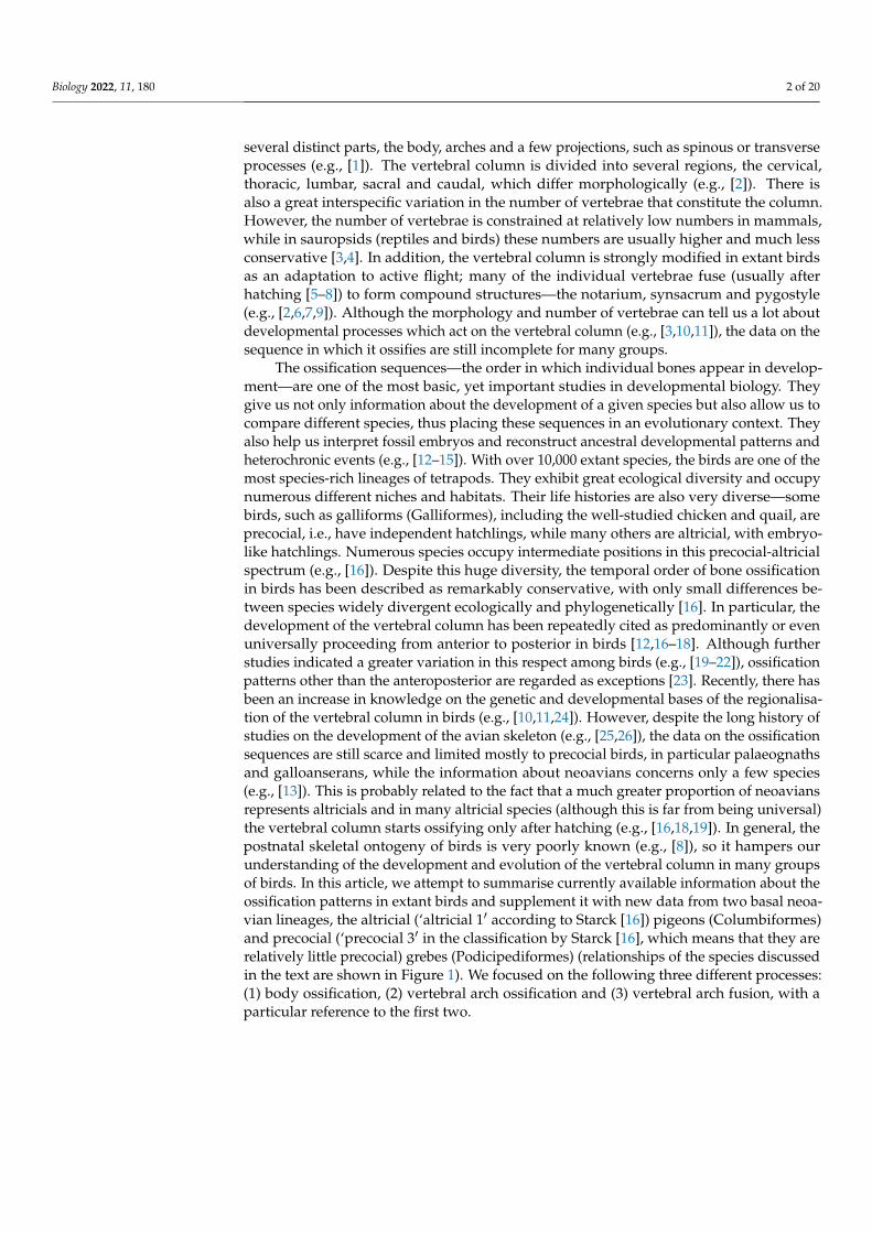
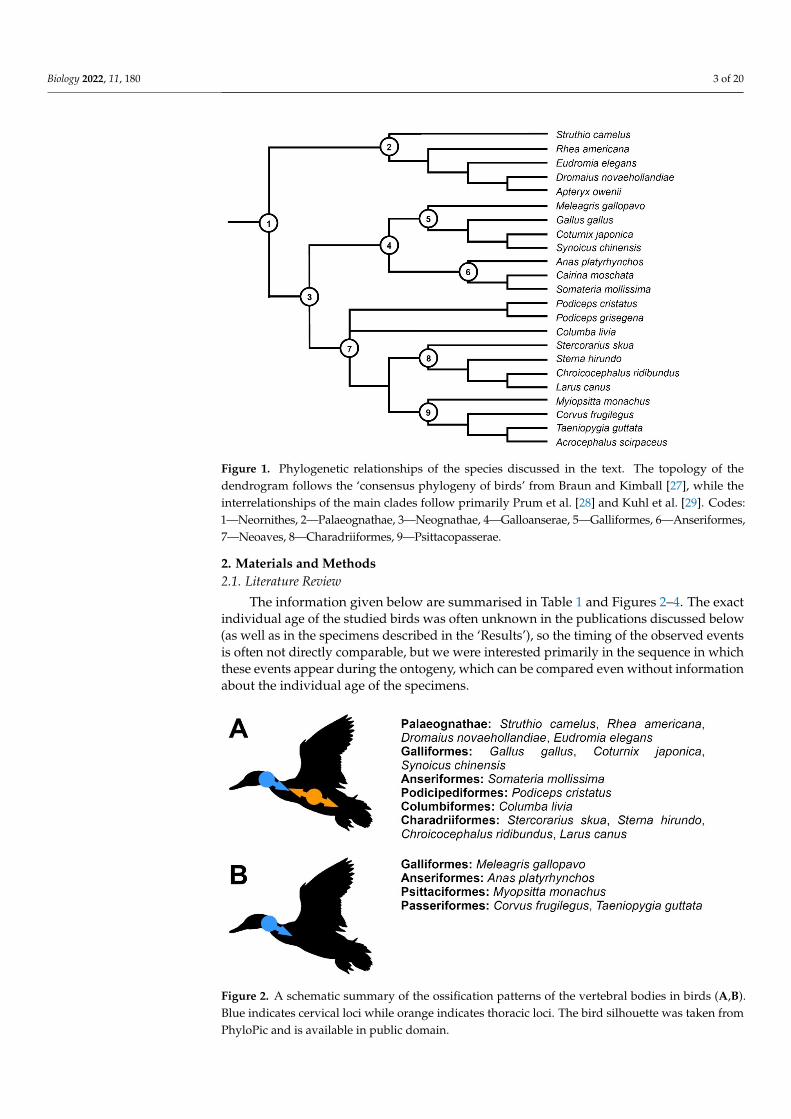
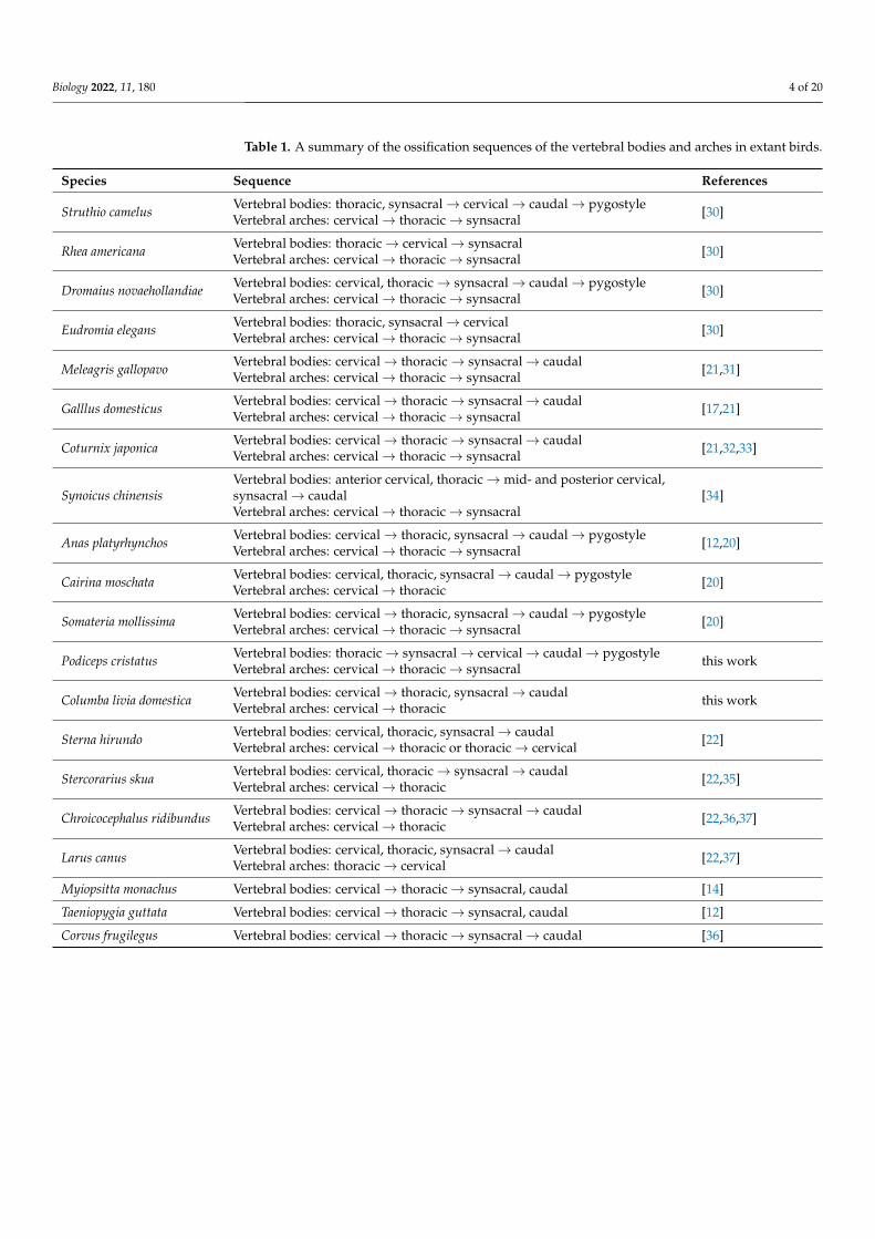
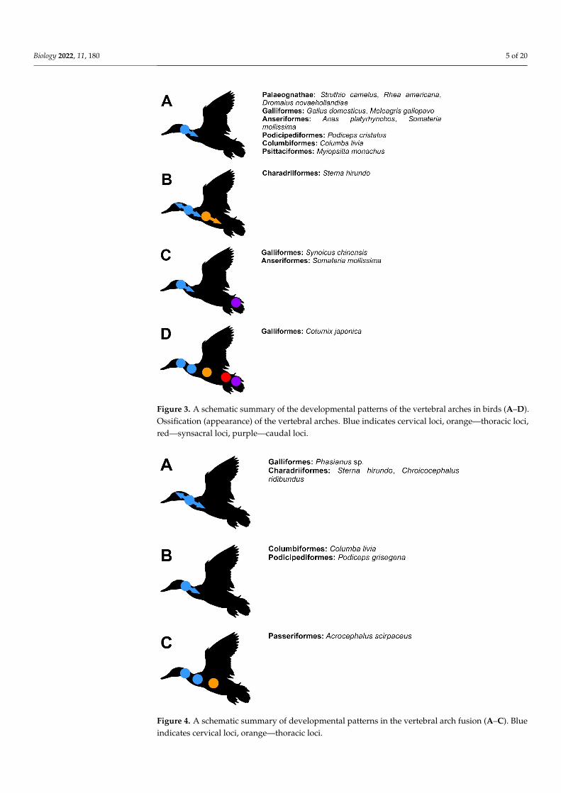
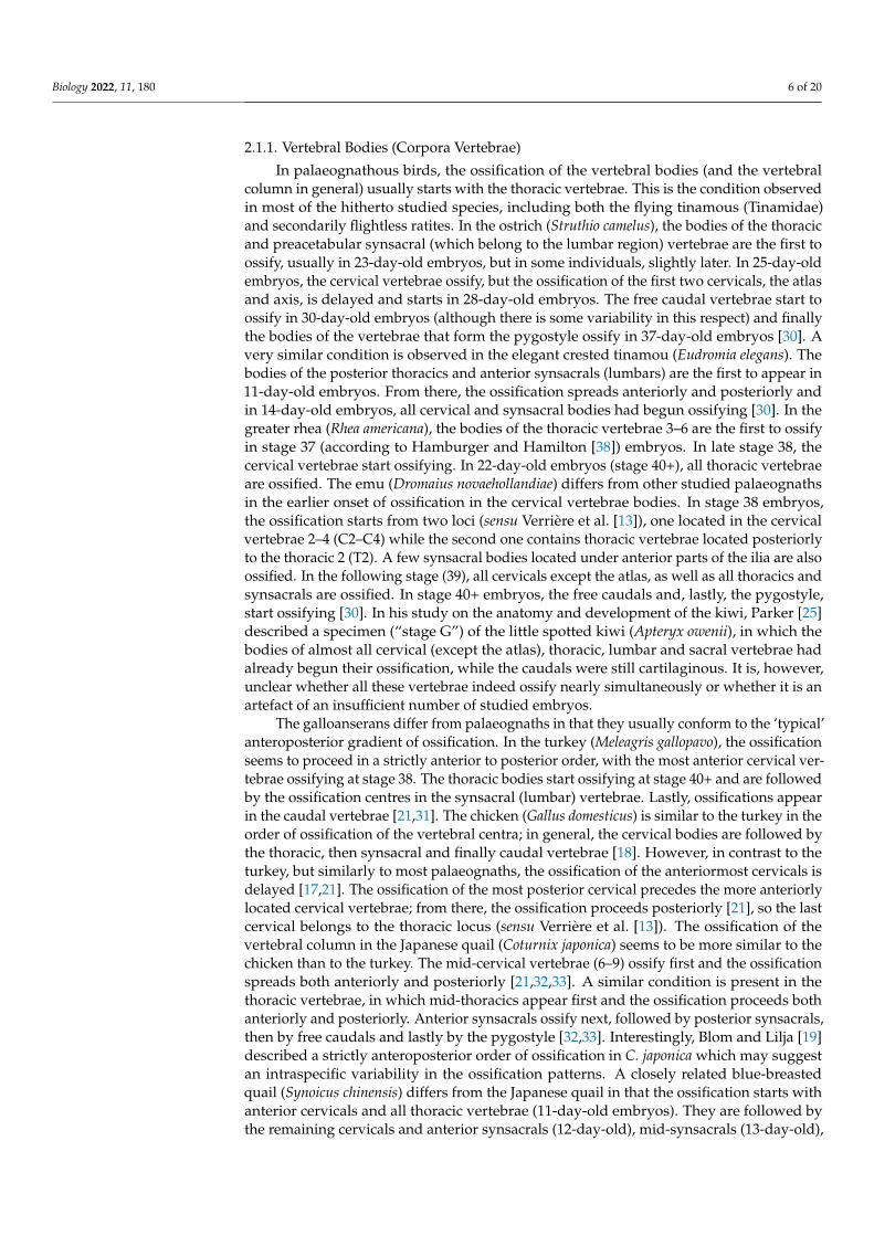
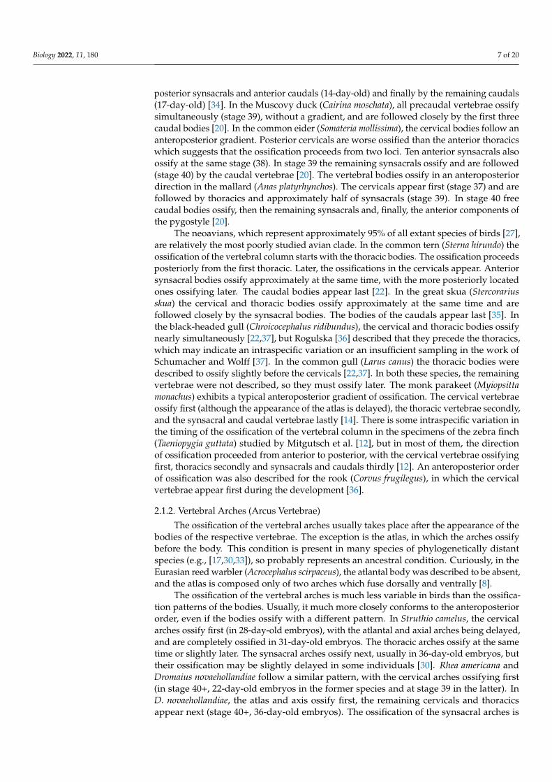
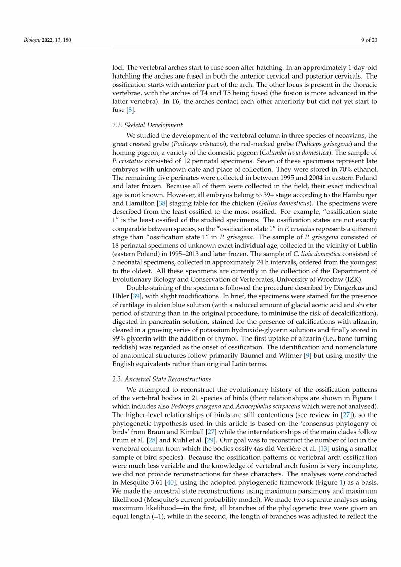
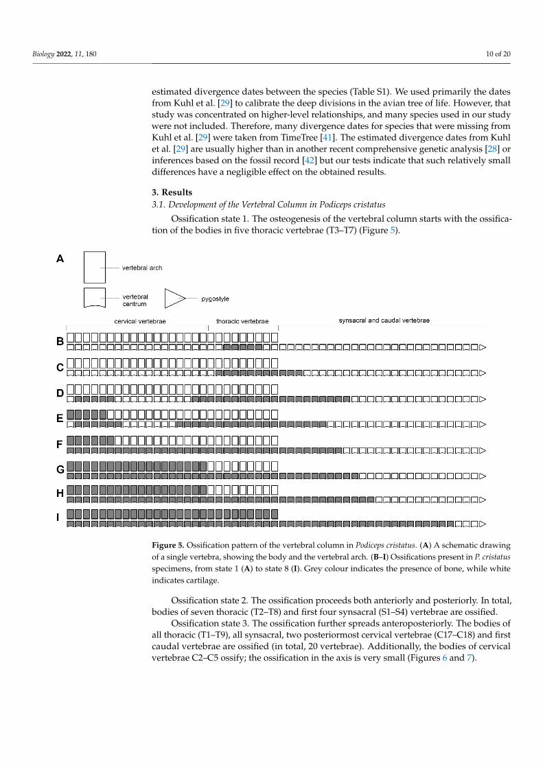
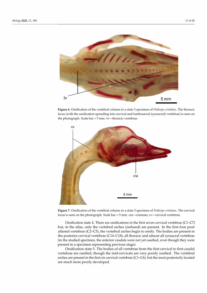
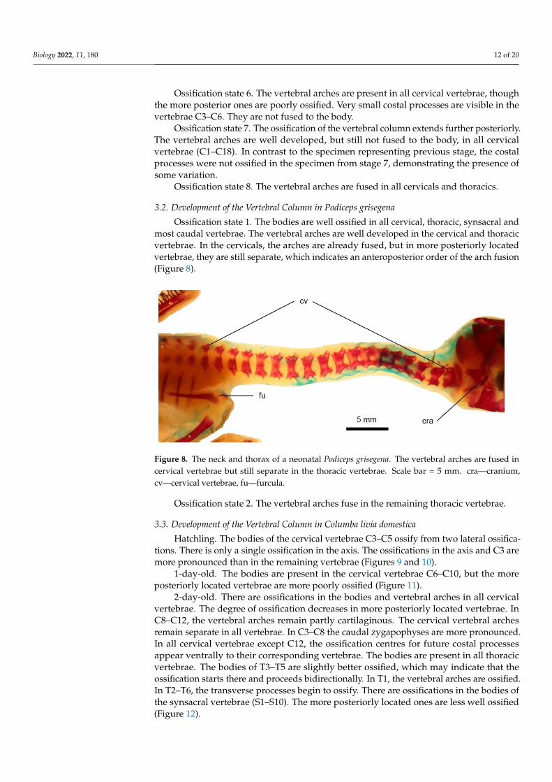
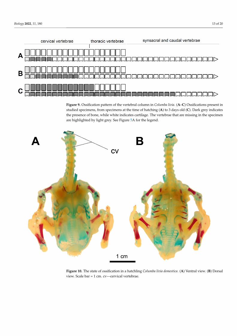
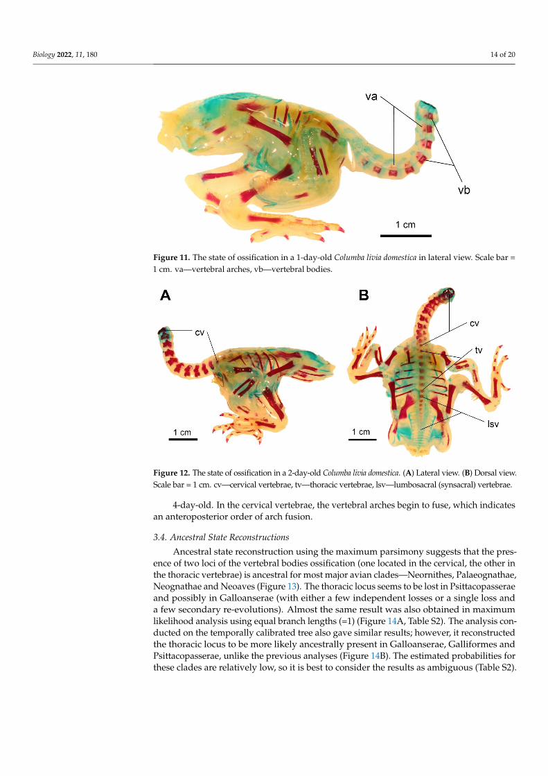
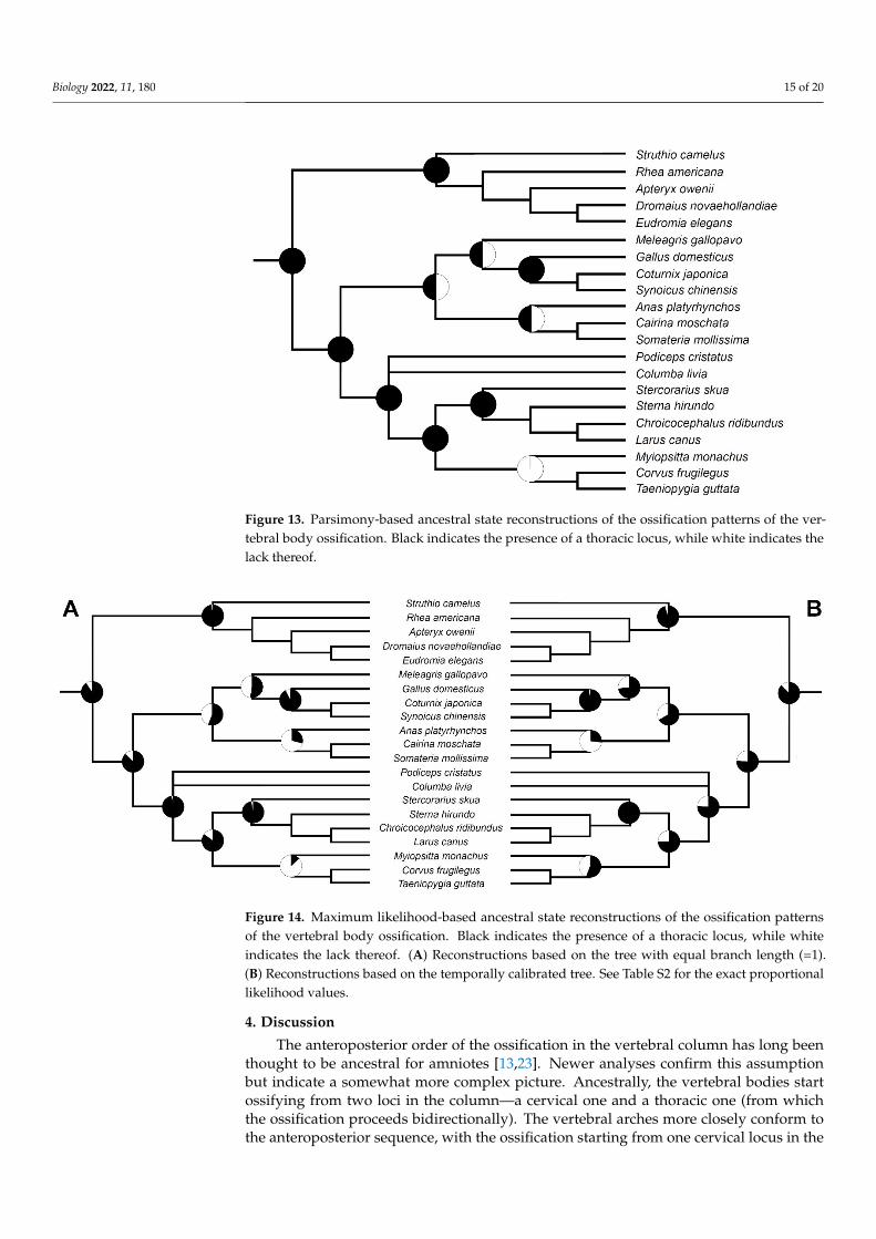
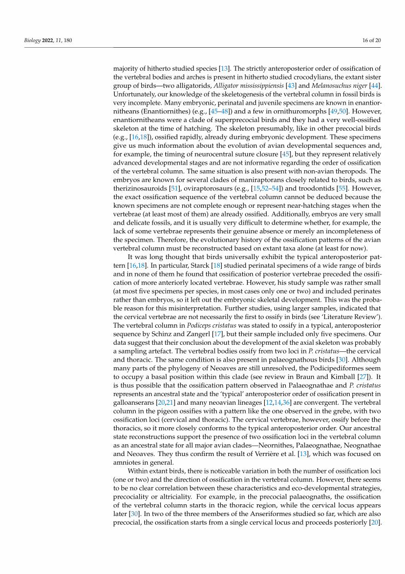


