Embed Size (px)
Citation preview
A Prospective, Randomized Evaluation of Acellular Human DermalMatrix Augmentation for Arthroscopic Rotator Cuff Repair
F. Alan Barber, M.D., F.A.C.S., Joseph P. Burns, M.D., Allen Deutsch, M.D.,Marc R. Labbé, M.D., and Robert B. Litchfield, M.D., F.R.C.S.C
Purpose: To prospectively evaluate the safety and effectiveness of arthroscopic acellular human dermalmatrix augmentation of large rotator cuff tear repairs. Methods: A prospective, institutional reviewboard–approved, multicenter series of patients undergoing arthroscopic repair of 2-tendon rotator cufftears measuring greater than 3 cm were randomized by sealed envelopes opened at the time of surgery toarthroscopic single-row rotator cuff repair with GraftJacket acellular human dermal matrix (WrightMedical Technology, Arlington, TN) augmentation (group 1) or without augmentation (group 2). Preop-erative and postoperative functional outcome assessments were obtained by use of the American Shoulderand Elbow Surgeons (ASES), Constant, and University of California, Los Angeles scales. Gadolinium-enhanced magnetic resonance imaging (MRI) evaluation of these repairs was obtained at a mean of 14.5months (range, 12 to 24 months). Adverse events were recorded. Results: There were 22 patients in group1 and 20 in group 2 with a mean age of 56 years. The mean follow-up was 24 months (range, 12 to 38months). The ASES score improved from 48.5 to 98.9 in group 1 and from 46.0 to 94.8 in group 2. Thescores in group 1 were statistically better than those in group 2 (P � .035). The Constant score improvedfrom 41.0 to 91.9 in group 1 and from 45.8 to 85.3 in group 2. The scores in group 1 were statisticallybetter than those in group 2 (P � .008). The University of California, Los Angeles score improved from13.3 to 28.2 in group 1 and from 15.9 to 28.3 in group 2 (P � .43). Gadolinium-enhanced MRI scansshowed intact cuffs in 85% of repairs in group 1 and 40% in group 2 (P � .01). No adverse events wereattributed to the presence of the matrix grafts. Conclusions: Acellular human dermal matrix augmentationof large (�3 cm) cuff tears involving 2 tendons showed better ASES and Constant scores and morefrequent intact cuffs as determined by gadolinium-enhanced MRI. Intact repairs were found in 85% of theaugmented group and 40% of the nonaugmented group (P � .01). No adverse events related to theacellular human dermal matrix were observed. Level of Evidence: Level II, lesser-quality randomizedcontrolled trial.
irai
mttdnfia“
From the Plano Orthopedic Sports Medicine and Spine Center(F.A.B.), Plano, Texas; Southern California Orthopedic Institute (J.P.B.),Van Nuys, California; Kelsey-Seybold Clinic of Orthopaedics (A.D.),Houston, Texas; Sports Medicine & Arthroscopic Surgery, Bone & JointClinic of South Houston (M.R.L.), Houston, Texas, U.S.A.; and FowlerKennedy Sport Medicine Clinic, University of Western Ontario (R.B.L.),London, Ontario, Canada.
Supported by grants from Wright Medical Technology. Theauthors have received research or institutional support fromWright Medical Technology.
Received January 21, 2011; accepted June 29, 2011.Address correspondence to F. Alan Barber, M.D., F.A.C.S.,
Plano Orthopedic and Sports Medicine Center, 5228 W PlanoPkwy, Plano, TX 75093, U.S.A.
© 2012 by the Arthroscopy Association of North America
m0749-8063/1162/$36.00doi:10.1016/j.arthro.2011.06.038
8 Arthroscopy: The Journal of Arthroscopic and Related
Rotator cuff tears are increasingly common withadvancing age and lead to both pain and disabil-
ty. Surgery is an option for patients who do notespond to a nonoperative program. Neither open norrthroscopic rotator cuff repairs have uniform successn preventing retears.
Arthroscopic repair offers advantages of decreasedorbidity, better visualization, and patient accep-
ance. However, a significant percentage of tears failo completely heal or are found to have persistentefects in the tendon at follow-up. The degenerativeature of the rotator cuff tendon may contribute to thisnding. In addition, the tension on the repaired tendonfter it is reattached to the greater tuberosity for adouble-row” repair or even a basic single-row repair
ay reduce the likelihood of complete healing.Surgery, Vol 28, No 1 (January), 2012: pp 8-15
smubdtha
taacgltacsrgcprmlelBr
9ACELLULAR HUMAN DERMAL MATRIX AUGMENTATION
Every effort should be made to enhance the poten-tial for complete healing after rotator cuff repair.However, despite the use of the best technique, thequality of cuff tissue is often so poor that biologichealing is compromised, resulting in rotator cuff tissueretearing or incomplete healing at rates ranging from40% to over 90%.1-4 There is no uniformly acceptedurgical solution to this problem because even theost advanced biomechanical constructs have been
nable to improve what is usually a compromisediologic environment. Many techniques have beenevised to enhance the repair of large and degenera-ive tears including the recent emphasis on biologicealing enhancements, such as platelet-rich plasmand marrow aspirates.
The incorporation of biologic tissue scaffolds intohe rotator cuff is another biologic approach. Bothutograft and allograft materials have been used tougment or replace irreparable portions of the rotatoruff tendon. An acellular human dermal matrix allo-raft (GraftJacket; Wright Medical Technology, Ar-ington, TN) is currently available for tendon augmen-ation. This graft tissue is processed to render itcellular and therefore less immunogenic while theollagen extracellular matrix is left intact to providetrength and a scaffold into which new host tissue canegenerate. GraftJacket matrix is a human skin allo-raft processed to remove the epidermis and dermalells and was chosen for this study because theseroperties facilitate incorporation of the matrix andeduce any rejection response. The 3-dimensional hu-an dermal tissue forms a scaffold from native col-
agen structure that contains blood vessel channels andssential biochemical components that enhance cellu-ar repopulation and revascularization during healing.iomechanical testing has shown GraftJacket’s supe-
ior strength to xenograft and synthetic alternatives,5,6
and clinical reports support its use in the shoulder.7-9
The purpose of this study was to prospectivelyevaluate the safety and effectiveness of arthroscopicacellular human dermal matrix augmentation of largerotator cuff tear repairs. The hypothesis was that anarthroscopically applied soft-tissue allograft augmen-tation would both reinforce and enhance the healing ofa significant rotator cuff repair, resulting in fewerretears.
METHODS
A randomized, prospective, multicenter clinicalstudy of a consecutive series of patients undergoing
large (�3 cm), 2-tendon arthroscopic rotator cuff re-pair was undertaken. Patients were prospectively ran-domized into 2 groups by means of sealed envelopesopened at the time of surgery after assessment of thesize and reparability of the rotator cuff tendon: group1 included rotator cuff repairs with an acellular humandermal matrix augmentation, and group 2 (controlgroup) comprised repairs without any augmentation.Patients were excluded at the time of surgery if any ofthe exclusion criteria were found to be present or if theinclusion criteria were not met. This was determinedbefore the envelope was opened. This multicenterstudy was conducted in accordance with the approvedresearch protocol, good clinical practice guidelines,and applicable local regulatory requirements and laws,and institutional review board approval was obtained.Preoperative and postoperative functional outcome as-sessments were obtained with the Constant, AmericanShoulder and Elbow Surgeons (ASES), and Univer-sity of California, Los Angeles (UCLA) scores. Post-operative patient evaluations including range of mo-tion, strength, and standard shoulder tests were obtainedin conjunction with the subjective questions associatedwith the functional outcome scores mentioned previ-ously at 6 and 12 months after surgery and annuallythereafter.
Magnetic resonance imaging (MRI) arthrogramevaluations with gadolinium enhancement of theserepairs were obtained at 12 or 24 months in 1.5-Tscanners. The MRI interpretations were performed at4 separate study sites by an independent radiologist ateach site who was blinded to the patients’ treatmentassignments. The method of MRI setup and amountand brand of gadolinium injected were left to thediscretion of the radiologists and not controlled.
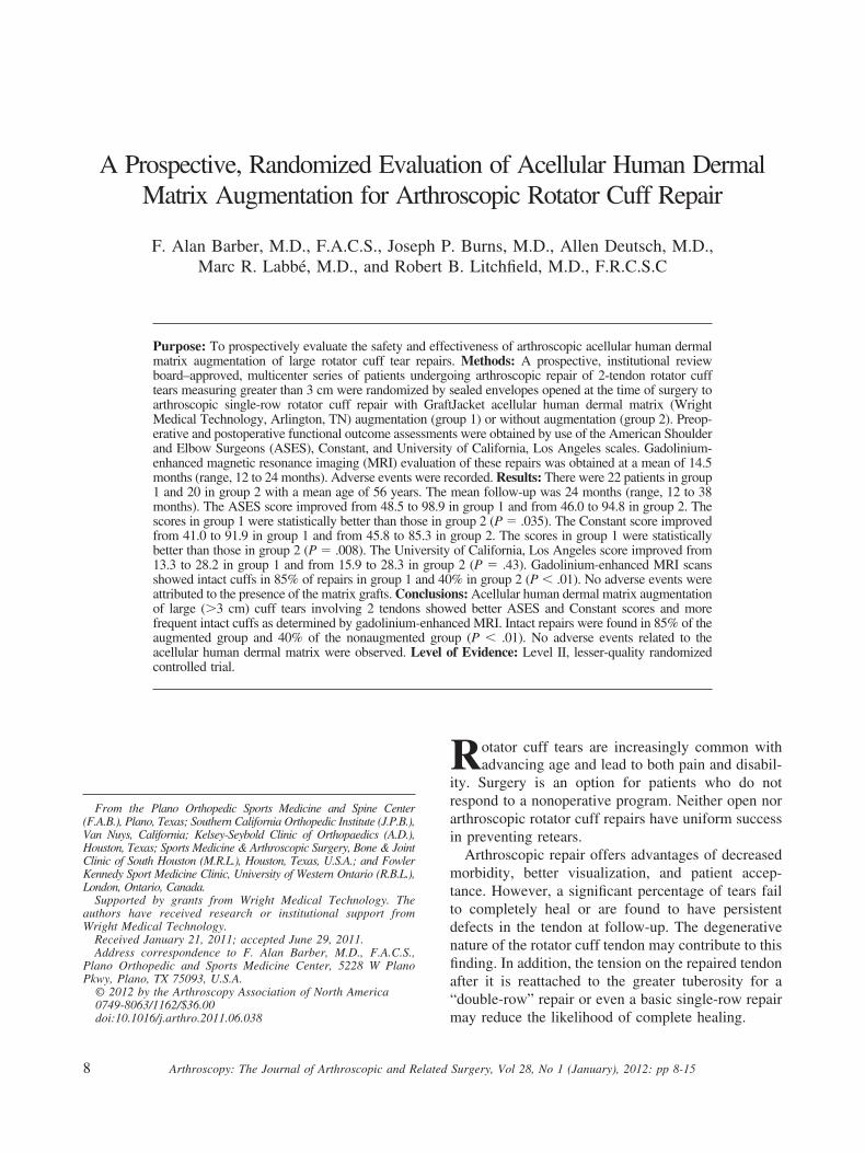
Inclusion criteria were patients aged between 18and 75 years with large rotator cuff tears measuring atleast 3 cm in width and with 2-tendon involvementthat could be repaired arthroscopically (Fig 1). Goodpreoperative movement of the nonoperative arm (de-fined as shoulder elevation �90°), the ability to per-form postoperative exercises, and the ability to read,understand, and complete the patient-reported out-come forms in English were required. Workers’ Com-pensation patients were allowed.
Exclusion criteria were irreparable massive rotatorcuff tears measuring more than 5 cm; subscapularistendon disruptions; revision surgery; inflammatory orautoimmune diseases; evidence of active infection,cancer, or highly communicable diseases; smokers;and patients for whom there was no reasonable expec-tation that they would be able to participate in the
protocol-required postoperative follow-up examina-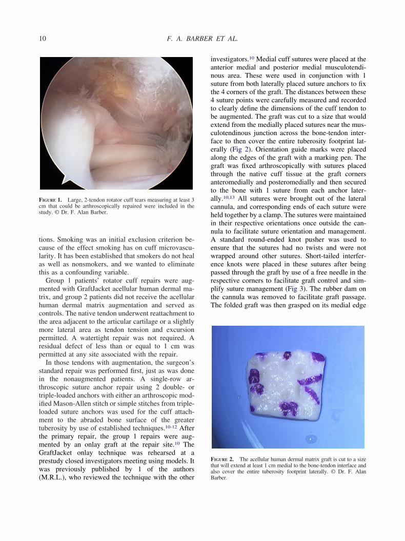
Gpw(
i
t
10 F. A. BARBER ET AL.
tions. Smoking was an initial exclusion criterion be-cause of the effect smoking has on cuff microvascu-larity. It has been established that smokers do not healas well as nonsmokers, and we wanted to eliminatethis as a confounding variable.
Group 1 patients’ rotator cuff repairs were aug-mented with GraftJacket acellular human dermal ma-trix, and group 2 patients did not receive the acellularhuman dermal matrix augmentation and served ascontrols. The native tendon underwent reattachment tothe area adjacent to the articular cartilage or a slightlymore lateral area as tendon tension and excursionpermitted. A watertight repair was not required. Aresidual defect of less than or equal to 1 cm waspermitted at any site associated with the repair.
In those tendons with augmentation, the surgeon’sstandard repair was performed first, just as was donein the nonaugmented patients. A single-row ar-throscopic suture anchor repair using 2 double- ortriple-loaded anchors with either an arthroscopic mod-ified Mason-Allen stitch or simple stitches from triple-loaded suture anchors was used for the cuff attach-ment to the abraded bone surface of the greatertuberosity by use of established techniques.10-12 Afterthe primary repair, the group 1 repairs were aug-mented by an onlay graft at the repair site.10 The
raftJacket onlay technique was rehearsed at arestudy closed investigators meeting using models. Itas previously published by 1 of the authors
FIGURE 1. Large, 2-tendon rotator cuff tears measuring at least 3cm that could be arthroscopically repaired were included in thestudy. © Dr. F. Alan Barber.
M.R.L.), who reviewed the technique with the otheraB
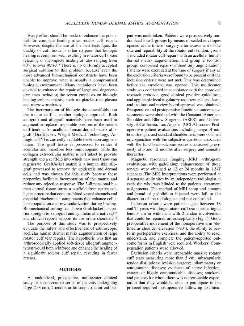
nvestigators.10 Medial cuff sutures were placed at theanterior medial and posterior medial musculotendi-nous area. These were used in conjunction with 1suture from both laterally placed suture anchors to fixthe 4 corners of the graft. The distances between these4 suture points were carefully measured and recordedto clearly define the dimensions of the cuff tendon tobe augmented. The graft was cut to a size that wouldextend from the medially placed sutures near the mus-culotendinous junction across the bone-tendon inter-face to then cover the entire tuberosity footprint lat-erally (Fig 2). Orientation guide marks were placedalong the edges of the graft with a marking pen. Thegraft was fixed arthroscopically with sutures placedthrough the native cuff tissue at the graft cornersanteromedially and posteromedially and then securedto the bone with 1 suture from each anchor later-ally.10,13 All sutures were brought out of the lateralcannula, and corresponding ends of each suture wereheld together by a clamp. The sutures were maintainedin their respective orientations once outside the can-nula to facilitate suture orientation and management.A standard round-ended knot pusher was used toensure that the sutures had no twists and were notwrapped around other sutures. Short-tailed interfer-ence knots were placed in these sutures after beingpassed through the graft by use of a free needle in therespective corners to facilitate graft control and sim-plify suture management (Fig 3). The rubber dam onthe cannula was removed to facilitate graft passage.The folded graft was then grasped on its medial edge
FIGURE 2. The acellular human dermal matrix graft is cut to a sizehat will extend at least 1 cm medial to the bone-tendon interface and
lso cover the entire tuberosity footprint laterally. © Dr. F. Alanarber.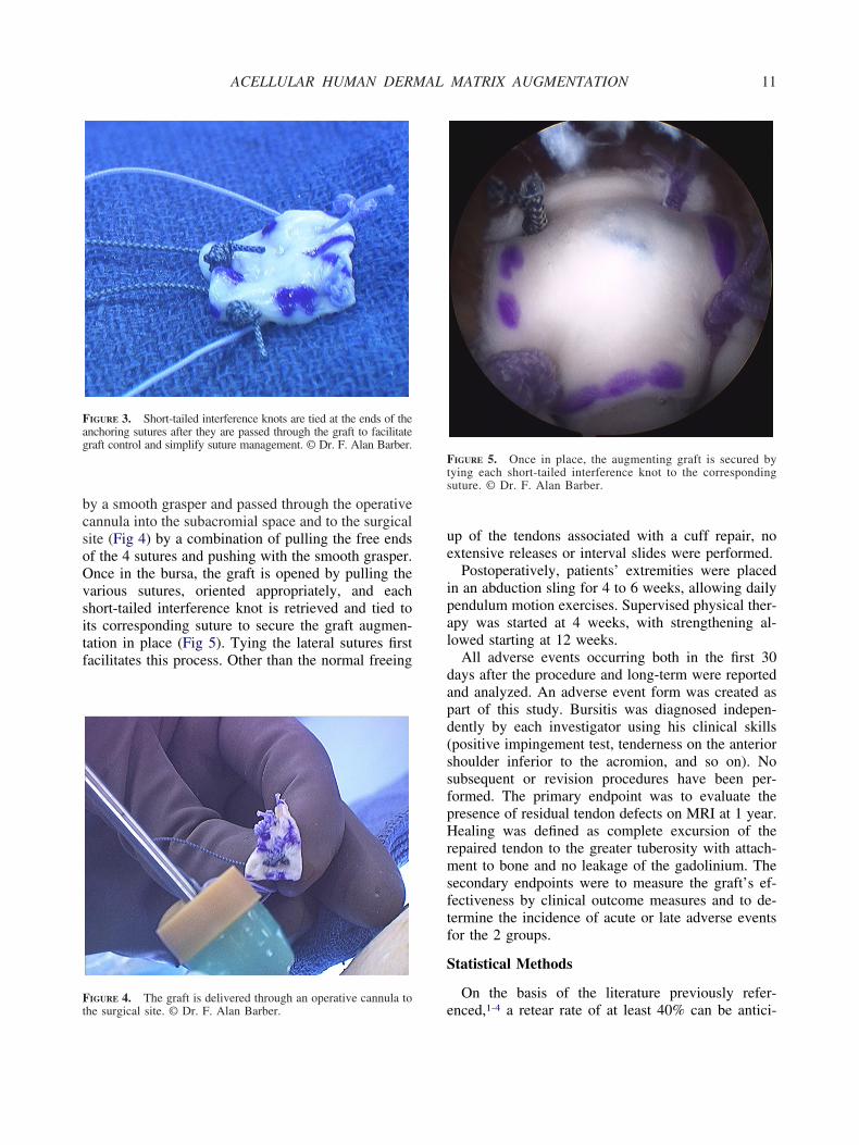
11ACELLULAR HUMAN DERMAL MATRIX AUGMENTATION
by a smooth grasper and passed through the operativecannula into the subacromial space and to the surgicalsite (Fig 4) by a combination of pulling the free endsof the 4 sutures and pushing with the smooth grasper.Once in the bursa, the graft is opened by pulling thevarious sutures, oriented appropriately, and eachshort-tailed interference knot is retrieved and tied toits corresponding suture to secure the graft augmen-tation in place (Fig 5). Tying the lateral sutures firstfacilitates this process. Other than the normal freeing
FIGURE 3. Short-tailed interference knots are tied at the ends of theanchoring sutures after they are passed through the graft to facilitategraft control and simplify suture management. © Dr. F. Alan Barber.
FIGURE 4. The graft is delivered through an operative cannula to
the surgical site. © Dr. F. Alan Barber.up of the tendons associated with a cuff repair, noextensive releases or interval slides were performed.
Postoperatively, patients’ extremities were placedin an abduction sling for 4 to 6 weeks, allowing dailypendulum motion exercises. Supervised physical ther-apy was started at 4 weeks, with strengthening al-lowed starting at 12 weeks.
All adverse events occurring both in the first 30days after the procedure and long-term were reportedand analyzed. An adverse event form was created aspart of this study. Bursitis was diagnosed indepen-dently by each investigator using his clinical skills(positive impingement test, tenderness on the anteriorshoulder inferior to the acromion, and so on). Nosubsequent or revision procedures have been per-formed. The primary endpoint was to evaluate thepresence of residual tendon defects on MRI at 1 year.Healing was defined as complete excursion of therepaired tendon to the greater tuberosity with attach-ment to bone and no leakage of the gadolinium. Thesecondary endpoints were to measure the graft’s ef-fectiveness by clinical outcome measures and to de-termine the incidence of acute or late adverse eventsfor the 2 groups.
Statistical Methods
On the basis of the literature previously refer-
FIGURE 5. Once in place, the augmenting graft is secured bytying each short-tailed interference knot to the correspondingsuture. © Dr. F. Alan Barber.
enced,1-4 a retear rate of at least 40% can be antici-
s1.
gmw6t
flw
dGtfittp
8 of 22
SMBBBL
12 F. A. BARBER ET AL.
pated for single-row repairs. Reducing this rate by halfwould be clinically meaningful. An SD of 15% withingroups was allowed. Power analysis indicated that 20patients in each group would provide sufficient powerto identify statistical significance in healing rates be-tween the 2 groups. Preoperative and postoperativeclinical outcome measures as well the presence ofpersistent cuff defects between groups were evaluatedby use of Student t tests. Statistical significance wasplaced at P � .05.
RESULTS
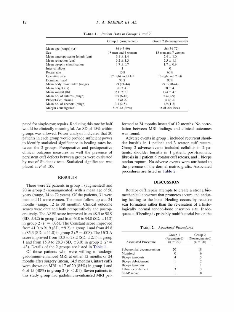
There were 22 patients in group 1 (augmented) and20 in group 2 (nonaugmented) with a mean age of 56years (range, 34 to 72 years). Of the patients, 31 weremen and 11 were women. The mean follow-up was 24months (range, 12 to 38 months). Clinical outcomescores were obtained both preoperatively and postop-eratively. The ASES score improved from 48.5 to 98.9(SD, �4.2) in group 1 and from 46.0 to 94.8 (SD, �14.2)in group 2 (P � .035). The Constant score improvedfrom 41.0 to 91.9 (SD, �9.2) in group 1 and from 45.8to 85.3 (SD, �11.0) in group 2 (P � .008). The UCLAcore improved from 13.3 to 28.2 (SD, �2.1) in groupand from 15.9 to 28.3 (SD, �3.0) in group 2 (P �
43). Details of the 2 groups are listed in Table 1.Of those patients who were willing to undergo
adolinium-enhanced MRI at either 12 months or 24onths after surgery (mean, 14.5 months), intact cuffsere shown on MRI in 17 of 20 (85%) in group 1 andof 15 (40%) in group 2 (P � .01). Seven patients in
TABLE 1. Patient
Gro
Mean age (range) (yr)Sex 18 mMean anteroposterior length (cm)Mean retraction (cm)Mean atrophy classificationInterval slidesRetear rateOperative side 17Dominant handMean body mass index (range)Mean height (in)Mean weight (lb)Mean no. of sutures (range)Platelet-rich plasmaMean no. of anchors (range)Margin convergence
his study group had gadolinium-enhanced MRI per-S
ormed at 24 months instead of 12 months. No corre-ation between MRI findings and clinical outcomesas found.Adverse events in group 1 included recurrent shoul-
er bursitis in 1 patient and 3 rotator cuff retears.roup 2 adverse events included cellulitis in 2 pa-
ients, shoulder bursitis in 1 patient, post-traumaticbrosis in 1 patient, 9 rotator cuff retears, and 1 biceps
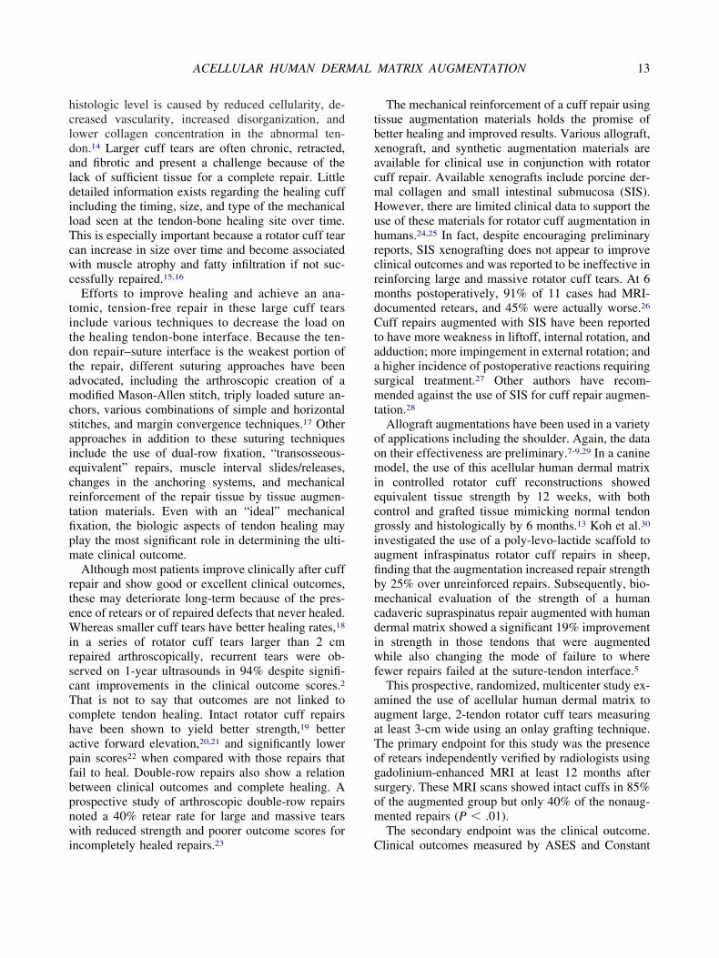
endon rupture. No adverse events were attributed tohe presence of the dermal matrix grafts. Associatedrocedures are listed in Table 2.
DISCUSSION
Rotator cuff repair attempts to create a strong bio-mechanical construct that promotes secure and endur-ing healing to the bone. Healing occurs by reactivescar formation rather than the re-creation of a histo-logically normal tendon-bone insertion site. Inade-quate cuff healing is probably multifactorial but on the
in Groups 1 and 2
ugmented) Group 2 (Nonaugmented)
3-69) 56 (34-72)d 4 women 13 men and 7 women
1.4 2.4 � 1.01.3 2.5 � 1.10.7 1.7 � 0.9
0% 60%
and 5 left 13 right and 7 left% 90%1-44) 29.7 (20-44)
4 68 � 431 194 � 47
-16) 5.4 (2-9)f 22 4 of 20-5) 1.9 (1-3)(36%) 5 of 20 (25%)
TABLE 2. Associated Procedures
Associated Procedure
Group 1(Augmented)
(n � 22)
Group 2(Nonaugmented)
(n � 20)
ubacromial decompression 20 18umford 0 6iceps tenodesis 4 5iceps debridement 1 2iceps tenotomy 1 1abral debridement 3 3
Data
up 1 (A
56 (4en an3.1 �3.2 �1.7 �
115
right91
29 (270 �
200 �9.5 (6
7 o3.3 (2
LAP repair 1 0
aldilTcwc
aiecrtfipm
rteW
fbpnwi
miecg
13ACELLULAR HUMAN DERMAL MATRIX AUGMENTATION
histologic level is caused by reduced cellularity, de-creased vascularity, increased disorganization, andlower collagen concentration in the abnormal ten-don.14 Larger cuff tears are often chronic, retracted,nd fibrotic and present a challenge because of theack of sufficient tissue for a complete repair. Littleetailed information exists regarding the healing cuffncluding the timing, size, and type of the mechanicaload seen at the tendon-bone healing site over time.his is especially important because a rotator cuff tearan increase in size over time and become associatedith muscle atrophy and fatty infiltration if not suc-
essfully repaired.15,16
Efforts to improve healing and achieve an ana-tomic, tension-free repair in these large cuff tearsinclude various techniques to decrease the load onthe healing tendon-bone interface. Because the ten-don repair–suture interface is the weakest portion ofthe repair, different suturing approaches have beenadvocated, including the arthroscopic creation of amodified Mason-Allen stitch, triply loaded suture an-chors, various combinations of simple and horizontalstitches, and margin convergence techniques.17 Otherpproaches in addition to these suturing techniquesnclude the use of dual-row fixation, “transosseous-quivalent” repairs, muscle interval slides/releases,hanges in the anchoring systems, and mechanicaleinforcement of the repair tissue by tissue augmen-ation materials. Even with an “ideal” mechanicalxation, the biologic aspects of tendon healing maylay the most significant role in determining the ulti-ate clinical outcome.Although most patients improve clinically after cuff
epair and show good or excellent clinical outcomes,hese may deteriorate long-term because of the pres-nce of retears or of repaired defects that never healed.hereas smaller cuff tears have better healing rates,18
in a series of rotator cuff tears larger than 2 cmrepaired arthroscopically, recurrent tears were ob-served on 1-year ultrasounds in 94% despite signifi-cant improvements in the clinical outcome scores.2
That is not to say that outcomes are not linked tocomplete tendon healing. Intact rotator cuff repairshave been shown to yield better strength,19 betteractive forward elevation,20,21 and significantly lowerpain scores22 when compared with those repairs thatail to heal. Double-row repairs also show a relationetween clinical outcomes and complete healing. Arospective study of arthroscopic double-row repairsoted a 40% retear rate for large and massive tearsith reduced strength and poorer outcome scores for
ncompletely healed repairs.23
The mechanical reinforcement of a cuff repair usingtissue augmentation materials holds the promise ofbetter healing and improved results. Various allograft,xenograft, and synthetic augmentation materials areavailable for clinical use in conjunction with rotatorcuff repair. Available xenografts include porcine der-mal collagen and small intestinal submucosa (SIS).However, there are limited clinical data to support theuse of these materials for rotator cuff augmentation inhumans.24,25 In fact, despite encouraging preliminaryreports, SIS xenografting does not appear to improveclinical outcomes and was reported to be ineffective inreinforcing large and massive rotator cuff tears. At 6months postoperatively, 91% of 11 cases had MRI-documented retears, and 45% were actually worse.26
Cuff repairs augmented with SIS have been reportedto have more weakness in liftoff, internal rotation, andadduction; more impingement in external rotation; anda higher incidence of postoperative reactions requiringsurgical treatment.27 Other authors have recom-mended against the use of SIS for cuff repair augmen-tation.28
Allograft augmentations have been used in a varietyof applications including the shoulder. Again, the dataon their effectiveness are preliminary.7-9,29 In a canine
odel, the use of this acellular human dermal matrixn controlled rotator cuff reconstructions showedquivalent tissue strength by 12 weeks, with bothontrol and grafted tissue mimicking normal tendonrossly and histologically by 6 months.13 Koh et al.30
investigated the use of a poly-levo-lactide scaffold toaugment infraspinatus rotator cuff repairs in sheep,finding that the augmentation increased repair strengthby 25% over unreinforced repairs. Subsequently, bio-mechanical evaluation of the strength of a humancadaveric supraspinatus repair augmented with humandermal matrix showed a significant 19% improvementin strength in those tendons that were augmentedwhile also changing the mode of failure to wherefewer repairs failed at the suture-tendon interface.5
This prospective, randomized, multicenter study ex-amined the use of acellular human dermal matrix toaugment large, 2-tendon rotator cuff tears measuringat least 3-cm wide using an onlay grafting technique.The primary endpoint for this study was the presenceof retears independently verified by radiologists usinggadolinium-enhanced MRI at least 12 months aftersurgery. These MRI scans showed intact cuffs in 85%of the augmented group but only 40% of the nonaug-mented repairs (P � .01).
The secondary endpoint was the clinical outcome.
Clinical outcomes measured by ASES and Constant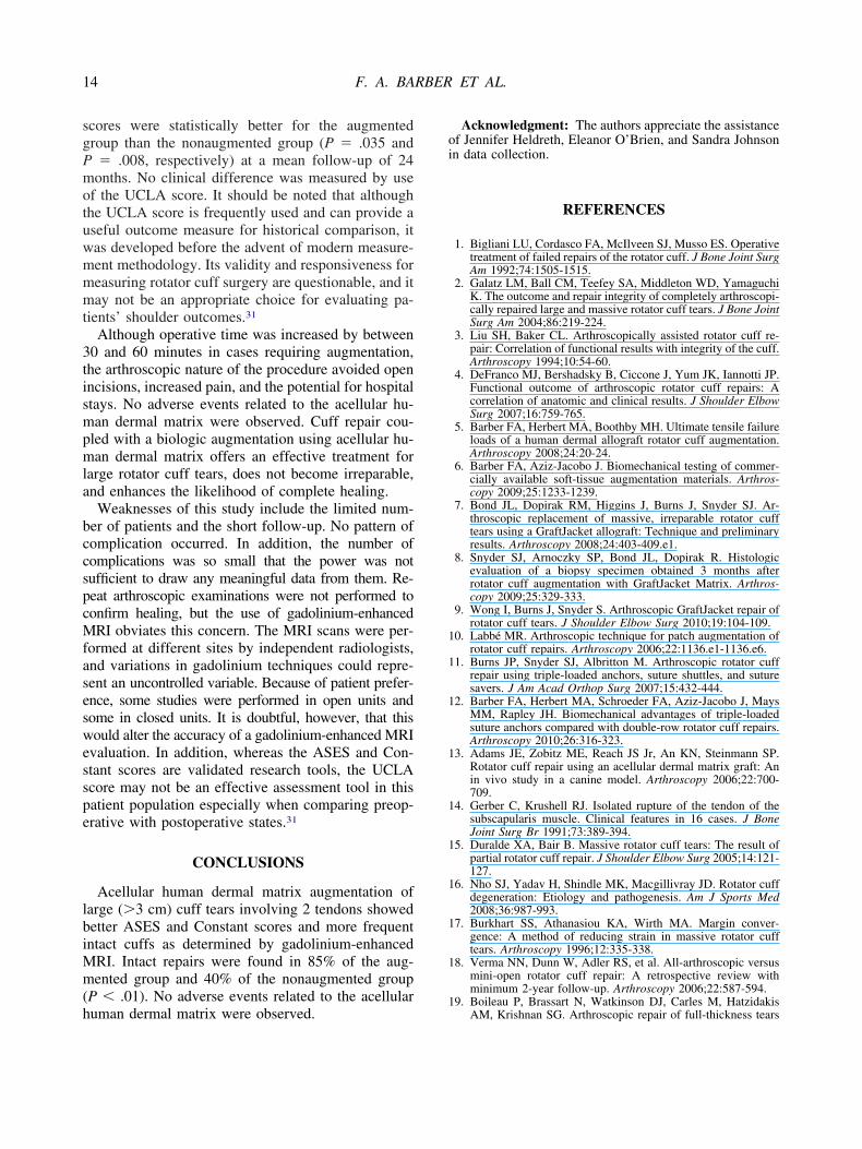
motuwmmmt
1
14 F. A. BARBER ET AL.
scores were statistically better for the augmentedgroup than the nonaugmented group (P � .035 andP � .008, respectively) at a mean follow-up of 24
onths. No clinical difference was measured by usef the UCLA score. It should be noted that althoughhe UCLA score is frequently used and can provide aseful outcome measure for historical comparison, itas developed before the advent of modern measure-ent methodology. Its validity and responsiveness foreasuring rotator cuff surgery are questionable, and itay not be an appropriate choice for evaluating pa-
ients’ shoulder outcomes.31
Although operative time was increased by between30 and 60 minutes in cases requiring augmentation,the arthroscopic nature of the procedure avoided openincisions, increased pain, and the potential for hospitalstays. No adverse events related to the acellular hu-man dermal matrix were observed. Cuff repair cou-pled with a biologic augmentation using acellular hu-man dermal matrix offers an effective treatment forlarge rotator cuff tears, does not become irreparable,and enhances the likelihood of complete healing.
Weaknesses of this study include the limited num-ber of patients and the short follow-up. No pattern ofcomplication occurred. In addition, the number ofcomplications was so small that the power was notsufficient to draw any meaningful data from them. Re-peat arthroscopic examinations were not performed toconfirm healing, but the use of gadolinium-enhancedMRI obviates this concern. The MRI scans were per-formed at different sites by independent radiologists,and variations in gadolinium techniques could repre-sent an uncontrolled variable. Because of patient prefer-ence, some studies were performed in open units andsome in closed units. It is doubtful, however, that thiswould alter the accuracy of a gadolinium-enhanced MRIevaluation. In addition, whereas the ASES and Con-stant scores are validated research tools, the UCLAscore may not be an effective assessment tool in thispatient population especially when comparing preop-erative with postoperative states.31
CONCLUSIONS
Acellular human dermal matrix augmentation oflarge (�3 cm) cuff tears involving 2 tendons showedbetter ASES and Constant scores and more frequentintact cuffs as determined by gadolinium-enhancedMRI. Intact repairs were found in 85% of the aug-mented group and 40% of the nonaugmented group(P � .01). No adverse events related to the acellular
human dermal matrix were observed.Acknowledgment: The authors appreciate the assistanceof Jennifer Heldreth, Eleanor O’Brien, and Sandra Johnsonin data collection.
REFERENCES
1. Bigliani LU, Cordasco FA, McIlveen SJ, Musso ES. Operativetreatment of failed repairs of the rotator cuff. J Bone Joint SurgAm 1992;74:1505-1515.
2. Galatz LM, Ball CM, Teefey SA, Middleton WD, YamaguchiK. The outcome and repair integrity of completely arthroscopi-cally repaired large and massive rotator cuff tears. J Bone JointSurg Am 2004;86:219-224.
3. Liu SH, Baker CL. Arthroscopically assisted rotator cuff re-pair: Correlation of functional results with integrity of the cuff.Arthroscopy 1994;10:54-60.
4. DeFranco MJ, Bershadsky B, Ciccone J, Yum JK, Iannotti JP.Functional outcome of arthroscopic rotator cuff repairs: Acorrelation of anatomic and clinical results. J Shoulder ElbowSurg 2007;16:759-765.
5. Barber FA, Herbert MA, Boothby MH. Ultimate tensile failureloads of a human dermal allograft rotator cuff augmentation.Arthroscopy 2008;24:20-24.
6. Barber FA, Aziz-Jacobo J. Biomechanical testing of commer-cially available soft-tissue augmentation materials. Arthros-copy 2009;25:1233-1239.
7. Bond JL, Dopirak RM, Higgins J, Burns J, Snyder SJ. Ar-throscopic replacement of massive, irreparable rotator cufftears using a GraftJacket allograft: Technique and preliminaryresults. Arthroscopy 2008;24:403-409.e1.
8. Snyder SJ, Arnoczky SP, Bond JL, Dopirak R. Histologicevaluation of a biopsy specimen obtained 3 months afterrotator cuff augmentation with GraftJacket Matrix. Arthros-copy 2009;25:329-333.
9. Wong I, Burns J, Snyder S. Arthroscopic GraftJacket repair ofrotator cuff tears. J Shoulder Elbow Surg 2010;19:104-109.
0. Labbé MR. Arthroscopic technique for patch augmentation ofrotator cuff repairs. Arthroscopy 2006;22:1136.e1-1136.e6.
11. Burns JP, Snyder SJ, Albritton M. Arthroscopic rotator cuffrepair using triple-loaded anchors, suture shuttles, and suturesavers. J Am Acad Orthop Surg 2007;15:432-444.
12. Barber FA, Herbert MA, Schroeder FA, Aziz-Jacobo J, MaysMM, Rapley JH. Biomechanical advantages of triple-loadedsuture anchors compared with double-row rotator cuff repairs.Arthroscopy 2010;26:316-323.
13. Adams JE, Zobitz ME, Reach JS Jr, An KN, Steinmann SP.Rotator cuff repair using an acellular dermal matrix graft: Anin vivo study in a canine model. Arthroscopy 2006;22:700-709.
14. Gerber C, Krushell RJ. Isolated rupture of the tendon of thesubscapularis muscle. Clinical features in 16 cases. J BoneJoint Surg Br 1991;73:389-394.
15. Duralde XA, Bair B. Massive rotator cuff tears: The result ofpartial rotator cuff repair. J Shoulder Elbow Surg 2005;14:121-127.
16. Nho SJ, Yadav H, Shindle MK, Macgillivray JD. Rotator cuffdegeneration: Etiology and pathogenesis. Am J Sports Med2008;36:987-993.
17. Burkhart SS, Athanasiou KA, Wirth MA. Margin conver-gence: A method of reducing strain in massive rotator cufftears. Arthroscopy 1996;12:335-338.
18. Verma NN, Dunn W, Adler RS, et al. All-arthroscopic versusmini-open rotator cuff repair: A retrospective review withminimum 2-year follow-up. Arthroscopy 2006;22:587-594.
19. Boileau P, Brassart N, Watkinson DJ, Carles M, HatzidakisAM, Krishnan SG. Arthroscopic repair of full-thickness tears

2
2
2
2
2
2
2
2
2
3
3
15ACELLULAR HUMAN DERMAL MATRIX AUGMENTATION
of the supraspinatus: Does the tendon really heal? J Bone JointSurg Am 2005;87:1229-1240.
20. Huijsmans PE, Pritchard MP, Berghs BM, van Rooyen KS,Wallace AL, de Beer JF. Arthroscopic rotator cuff repair withdouble-row fixation. J Bone Joint Surg Am 2007;89:1248-1257.
1. Cole BJ, McCarty LP III, Kang RW, Alford W, Lewis PB,Hayden JK. Arthroscopic rotator cuff repair: Prospective func-tional outcome and repair integrity at minimum 2-year follow-up. J Shoulder Elbow Surg 2007;16:579-585.
2. Lafosse L, Brozska R, Toussaint B, Gobezie R. The outcomeand structural integrity of arthroscopic rotator cuff repair withuse of the double-row suture anchor technique. J Bone JointSurg Am 2007;89:1533-1541.
3. Sugaya H, Maeda K, Matsuki K, Moriishi J. Repair integrityand functional outcome after arthroscopic double-row rotatorcuff repair. A prospective outcome study. J Bone Joint SurgAm 2007;89:953-960.
4. Soler JA, Gidwani S, Curtis MJ. Early complications from theuse of porcine dermal collagen implants (Permacol) as bridg-ing constructs in the repair of massive rotator cuff tears. Areport of 4 cases. Acta Orthop Belg 2007;73:432-436.
5. Badhe SP, Lawrence TM, Smith FD, Lunn PG. An assessment
of porcine dermal xenograft as an augmentation graft in thetreatment of extensive rotator cuff tears. J Shoulder ElbowSurg 2008;17:35S-39S.
6. Sclamberg SG, Tibone JE, Itamura JM, Kasraeian S. Six-month magnetic resonance imaging follow-up of large andmassive rotator cuff repairs reinforced with porcine smallintestinal submucosa. J Shoulder Elbow Surg 2004;13:538-541.
7. Walton JR, Bowman NK, Khatib Y, Linklater J, Murrell GA.Restore orthobiologic implant: Not recommended for augmen-tation of rotator cuff repairs. J Bone Joint Surg Am 2007;89:786-791.
8. Iannotti JP, Codsi MJ, Kwon YW, Derwin K, Ciccone J,Brems JJ. Porcine small intestine submucosa augmentation ofsurgical repair of chronic two-tendon rotator cuff tears. Arandomized, controlled trial. J Bone Joint Surg Am 2006;88:1238-1244.
9. Liden BA, Simmons M. Histologic evaluation of a 6-monthGraftJacket matrix biopsy used for Achilles tendon augmen-tation. J Am Podiatr Med Assoc 2009;99:104-107.
0. Koh JL, Szomor Z, Murrell GA, Warren RF. Supplementationof rotator cuff repair with a bioresorbable scaffold. Am JSports Med 2002;30:410-413.
1. Kirkley A, Griffin S, Dainty K. Scoring systems for the func-
tional assessment of the shoulder. Arthroscopy 2003;19:1109-1120.


