Embed Size (px)
Citation preview
Tendinopathy and tears of the rotator cuff are associated withhypoxia and apoptosis
R. T. Benson, MRCS [Lord Nuffield scholar in Orthopaedic Surgery], S. M. McDonnell,MRCS [Henry Smith Research Fellow of The Royal College of Surgeons of England], H. J.Knowles, PhD [Post Doctoral Research Assistant], J. L. Rees, MD, FRCS(Trauma & Orth)[University Lecturer in Orthopaedic Surgery], A. J. Carr, ChM, FRCS [Nuffield Professor ofOrthopaedic Surgery], and P. A. Hulley, PhD [ARC Research Fellow] [University Lecturer]Nuffield Department of Orthopaedics, Musculoskeletal Science Botnar Research Centre,University of Oxford Institute of Musculoskeletal Sciences, University of Oxford, NuffieldOrthopaedic Centre, Oxford, OX3 7LD, UK
AbstractThe aim of this study was to investigate the occurrence of tissue hypoxia and apoptosis at differentstages of tendinopathy and tears of the rotator cuff.
We studied tissue from 24 patients with eight graded stages of either impingement (mild, moderateand severe) or tears of the rotator cuff (partial, small, medium, large and massive) and threecontrols. Biopsies were analysed using three immunohistochemical techniques, namely antibodiesagainst HIF-1α (a transcription factor produced in a hypoxic environment), BNip3 (a HIF-1αregulated pro-apoptotic protein) and TUNEL (detecting DNA fragmentation in apoptosis).
The HIF-1α expression was greatest in mild impingement and in partial, small, medium and largetears. BNip3 expression increased significantly in partial, small, medium and large tears but wasreduced in massive tears. Apoptosis was increased in small, medium, large and massive tears butnot in partial tears.
These findings reveal evidence of hypoxic damage throughout the spectrum of pathology of therotator cuff which may contribute to loss of cells by apoptosis. This provides a novel insight intothe causes of degeneration of the rotator cuff and highlights possible options for treatment.
Disease of the rotator cuff presents in a variety of ways. Despite a proposed continuum fromchronic bursitis, through partial, complete to massive tears,1 very little research has beendone to confirm this. However, if this continuum exists then the progression through thestages is not uniform.
The aetiology of disease of the rotator cuff is multifactorial. Both intrinsic failure of thetendon and extrinsic mechanical compression by the coracoacromial arch play an importantrole.2-4 However, their relative contribution and the initiating factors are not clear.Excessive apoptosis, programmed cell death, has been associated with tendinopathy5 and ispresent in degenerative tendons and has been observed in rotator cuffs with impingement6and full thickness tears.7 Apoptosis plays a critical role in the homeostasis of normal tissueand it is important that organisms have a pathway to eliminate damaged or superfluous cells.Excessive apoptosis has been found in many diseases including osteoarthritis,8 rheumatoidarthritis,9,10 neoplasia11 and neurodegeneration.12 Apoptosis is a highly controlled form of
©2010 British Editorial Society of Bone and Joint Surgery
Correspondence should be sent to Mr R. T. Benson; [email protected].
Europe PMC Funders GroupAuthor ManuscriptJ Bone Joint Surg Br. Author manuscript; available in PMC 2010 March 22.
Published in final edited form as:J Bone Joint Surg Br. 2010 March ; 92(3): 448–453. doi:10.1302/0301-620X.92B3.23074.
Europe PM
C Funders A
uthor Manuscripts
Europe PM
C Funders A
uthor Manuscripts

cell suicide which is precipitated by intrinsic and extrinsic mechanisms. The former ormitochondrial pathway requires the BcL-2 family of pro- and anti-apoptotic proteins and thelatter requires the binding of external death activators to specific receptors on the cellsurface which transmit the apoptotic signal to the cytoplasm. BNip3 (Bcl-2 Nineteenkilodalton interacting protein) is a pro-apoptotic member of the Bcl-2 family in which it isunique as it is induced by hypoxic conditions as well as inflammation.13,14 Most cells arehighly sensitive to oxygen levels and undergo apoptosis following periods of severehypoxia, although tenocytes have not been studied well to date. BNip3 has been shown toplay a role in hypoxia-induced death in many cell types, including synovial fibroblasts,15myocytes16 and epithelial cells13 but not yet in human tenocytes.
Mechanical overload of the rotator cuff has been proposed as a primary cause oftendinopathy. Apoptosis has been induced following high strain mechanical loading of thetibialis anterior tendon of the rat17 and apoptotic genes have been upregulated by runningoveruse in the supraspinatus tendon of this animal.18 In cultured fibroblasts undergoingcyclical strain, overuse has also been shown to strongly induce Hypoxia Inducible Factor 1α(HIF-1α), a transcription factor which plays an important role in the intracellular hypoxicresponse.19
No author to our knowledge has examined how apoptosis and hypoxia may contribute to thecascade of pathological failure across the spectrum of disease of the rotator cuff.
We have examined the rotator cuff at different stages of failure, based on the continuumhypothesis and the appearance of the cuff. We investigated the incidence of hypoxia andapoptosis in the cuff with the hypothesis that the degree of hypoxia and consequentapoptosis worsens as the macroscopic appearance of the cuff deteriorates.
Patients and MethodsWith approval of the local ethical committee and informed written consent, 27 patients hadsamples taken from the rotator cuff (Table I). They were placed according to theirmacroscopic appearance into nine groups, each of three patients. The groups were mild,moderate and severe impingement, partial articular tear, small, medium, large and massivefull thickness tear and control. The impingement groups were classified according to theappearance of the bursa and tendon beneath the anterior acromion as mild (injected andoedematous), moderate (fibrillated with minor scuffing) or severe (major scuffing andfibrillation). The size of the tear was based on the classification of Post, Silver and Singh,20measuring its longest diameter. Small tears are < 1 cm, medium < 2 cm, large < 5 cm andmassive > 5 cm. All specimens were taken from fresh supraspinatus tendon except thecontrol group which was from fresh tendon of subscapularis. The samples for theimpingement and partial tear groups were taken using a punch biopsy during subacromialdecompression from the bursal surface of the rotator cuff tendon in the impingement groupand in the partial tears from the proximal edge of the tear. In the full thickness tear groups,the sample was taken during either arthroscopic subacromial decompression or open repairof the rotator cuff. Tissue was harvested from up to 1.5 cm from the proximal edge of thetear. The control sample was a full thickness biopsy of subscapularis obtained during openoperations for stabilisation. The tissue was placed immediately into 10% buffered formalinand set in paraffin. Sections were cut at 5 μm using a Leica-LM microtome (LeicaMicrosystems, Wetzler, Germany), then placed onto Snowcoat X-tra glass slides (Surgipath,Peterborough, United Kingdom). These were deparaffinised in Xylene, rehydrated throughgraded alcohol and subjected to microwave-based antigen-retrieval as detailed below.
Benson et al. Page 2
J Bone Joint Surg Br. Author manuscript; available in PMC 2010 March 22.
Europe PM
C Funders A
uthor Manuscripts
Europe PM
C Funders A
uthor Manuscripts

BNip3Immunohistochemical staining was carried out using the Vectastain Universal Elite ABCsystem (Vector Laboratories, Burlingame, California), which labels the primary antibodywith a biotinylated secondary antibody and then with a preformed Avidin and Biotinylatedhorseradish peroxidase macromolecule. This can be visualised by using 3, 3′-diaminobenzidine (DAB) as a peroxidase substrate.
The prepared slides were initially deparaffinised in xylene and rehydrated in graded alcohol(100%, 90%, 80% and then 70%). The sections were then placed in a solution of 3% H2O2and 100% alcohol for 20 minutes in order to quench endogenous peroxidase activity.Antigen retrieval was performed using a microwave at 800 W for 10 minutes with 400 ml ofDako retrieval solution. The slides were then placed in a rack in a plastic container andallowed to cool in water for 10 minutes. The following steps required 100 μL of solution tobe used per slide which was washed in phosphate buffered saline (PBS) between stages.Horse serum (1:100 in PBS) was applied for 20 minutes to block the non-specific staining ofantibodies. Primary monoclonal anti-BNip3 (Sigma, Poole, United Kingdom; 1:100 in PBS)was applied overnight at 4°C. A secondary biotinylated antibody, Streptavidin-horse-peroxidase was allowed to stand for 30 minutes, then applied for 20 minutes at roomtemperature. The slide was then coloured with DAB (metal enhanced DAB, Roche,Penzberg, Germany) for eight minutes (1:10 with peroxidase buffer). The cells werecounterstained in the dark at room temperature with DAPI (4 6-Diamidino-2-phenylindoledihydrochloride) for 20 minutes. They were then washed in distilled water and a coverslipapplied with a Flurosave reagent. They were stored in a closed box to prevent exposure tolight. In order to control for specificity, the BNip3 antibody was used to detect the proteinby Western blot in cultured tenocytes subjected to a timed course of total hypoxia (0.1%O2). Specific 26 kD bands were detected after eight and 16 hours. Tendon sections were alsostained using secondary antibody only and with the appropriate isotype control. Positivecells were counted in 10 high powered fields in which the total cell number was countedusing the DAPI stain under ultraviolet light. The BNip3 scoring was done by RTB.
HIF-1 αImmunohistochemical staining was carried out using the DAKO Envision system tovisualise the antigen-antibody complex. Slides were prepared according to the BNip3protocol and antigen retrieval carried out using EDTA (pH 8) in a microwave. The slideswere allowed to cool. The following steps required 100 μL of solution to be used per slidewhich was washed in PBS between stages. Horse serum (1:100 in PBS) was applied for 20minutes to block the non-specific staining of antibodies. Sections were stained at roomtemperature using a 1:100 dilution in PBS of a mouse monoclonal antibody directed againstHIF-1α (Clone 54, BD Biosciences, Oxford, United Kingdom). Staining was visualisedusing the Envision Peroxidase/DAB/Mouse detection kit. Positive cells were counted in 10high powered fields. HIF-1α staining was scored according to the following scale: 0 = noHIF-1α positive tenocytes; 1 = 1% to 10%; 2 = 11% to 25%; 3 = 26% to 50%; 4 = > 50%.The scoring was done by HJK and confirmed by PAH.
Apoptosis was assessed by the terminal deoxynucleotidyl transferase mediated deoxyuridinetriphosphate nick end labelling method (TUNEL) using the in situ all-death detection kit(Roche). This technique detects DNA cleavage within the nuclei which results from theapoptotic cascade. These cleavages are identified by the enzyme terminal deoxynucleotidyltransferase (TdT) and it catalyses the addition of deoxyuridine triphosphate (dUTP). ThedUTPs are then labelled secondarily with horse radish peroxidase.
Benson et al. Page 3
J Bone Joint Surg Br. Author manuscript; available in PMC 2010 March 22.
Europe PM
C Funders A
uthor Manuscripts
Europe PM
C Funders A
uthor Manuscripts

The sections were deparaffinised in xylene and rehydrated by being passed through gradedalcohol (100% to 70%). The tissue was incubated for 10 minutes in 3% H2O2 to inactivateendogenous peroxidase activity. The tissue was permeabilised with Triton X 0.5% for sixminutes. Fragmented DNA was then end-labelled with dUTP using TdT at 37°C for 60minutes. The signal was then detected using biotylated anti-FITC antibody and incubatingthe sample in Streptoavidin-HRP at 37°C for 30 minutes. The slide was placed in DAB atroom temperature for 4.5 minutes to develop the colour. Cells were counterstained withDAPI, then washed in distilled water and a coverslide applied with Flurosave reagent. TheTUNEL positive control was a section of colon provided by the supplier and negativecontrol was exclusion of TdT enzyme. Positive cells were counted in10 high powered fields.In the same fields, the total cell number was counted using the DAPI stain under ultravioletlight. Apoptotic scoring was done by RTB.
Statistical analysisThis was carried out on all result variables using Graph Pad Prism software (Graph PadSoftware, LaJolla, California). The association between the differing appearances of therotator cuff and the controls was analysed using an unpaired t-test for BNip-3 and apoptosis.The association between age and length of symptoms was tested using the Pearsoncorrelation coefficient. A pvalue ≤ 0.05 was considered significant.
ResultsThe distribution of demographic and pre-operative clinical features in each group is shownin Table I.
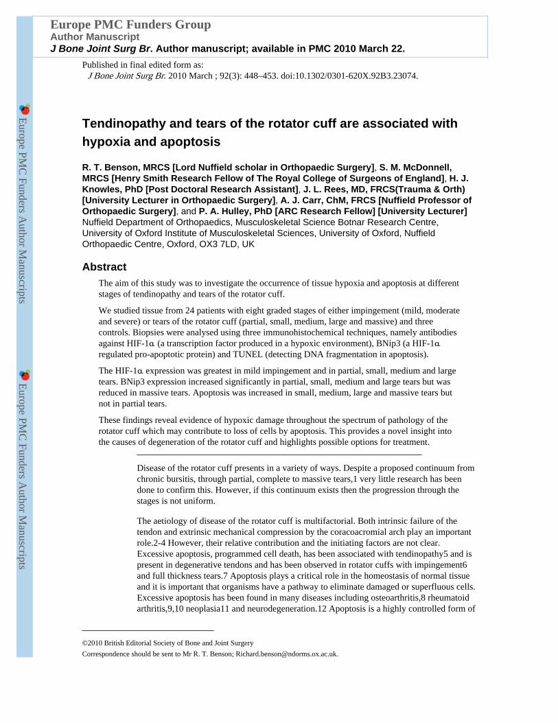
HIF-1α was present in all macroscopic groups, not only within the substance of the tendonbut also in blood vessels and bursa where these were present in the sections. HIF-1α wasabsent in the controls. The average apoptotic scores were as follows: mild impingement, 2.7;moderate impingement, 0.7; severe impingement, 0.3; partial tear, 3.0; small tear, 1.7;medium tear, 2.5; large tear, 2.0; massive tear, 1.3 and control 0. These results demonstratethe highest expression for HIF-1α being in the mild impingement group and the partial,medium and large tear groups. The highest levels of HIF-1α staining were seen in the bloodvessels and bursae. Examples of HIF-1α staining are shown in Figure 1.
The expression of BNip3 in the different macroscopic groups is seen in Figure 2. Thecontrol group had the lowest proportion of BNip3 positive cells. Mild, moderate and severeimpingement had similar proportions of BNip3 positive cells. There was a marked increasein such cells between severe impingement and partial tear. Partial, small, medium and largetears had a significantly higher proportion of BNip3 positive cells than controls and all theimpingement groups (p < 0.001). Between large and massive tears there was a fall in theproportion of positive cells. Massive tears still had a significantly higher proportion ofBNip3 positive cells than controls (p < 0.05).
There was a correlation between the proportion of BNip3 positive cells and the age of thepatient (r = 0.63, p < 0.001), but not with their length of symptoms.
Apoptosis was present in all the specimens. The apoptotic index averaged between 6.8 and21 throughout the spectrum of rotator cuff failure and the distribution of apoptotic indices isshown in Figure 3. Apoptosis was lowest in the control and mild impingement groups.Moderate and severe impingement and partial tears had similar indices. There was a markedincrease in the apoptotic index between partial and small full thickness tears. Massive tearshad the greatest index. Small, medium, large and massive tears had a significantly higherapoptotic index than controls (p < 0.001).
Benson et al. Page 4
J Bone Joint Surg Br. Author manuscript; available in PMC 2010 March 22.
Europe PM
C Funders A
uthor Manuscripts
Europe PM
C Funders A
uthor Manuscripts

There was a correlation between the apoptotic index and the age of patients (r = 0.65; p <0.001) but not with the duration of symptoms.
DiscussionAlthough apoptosis is a physiological process in normal healthy tissue, excessive apoptosishas been associated with tendinopathy.5,6,21 In our study, excessive apoptosis was foundwithin full thickness tears of the rotator cuff, with almost a threefold increase compared withthe control or mild groups. Yuan et al7 reported similar findings. In our study the controlpatients were younger (mean age 19.7 years (17 to 23)) than those with impingement and nofull thickness tear (mean age 47.7 years (39 to 67)) or those with full thickness tear (meanage 58.3 years (47 to 75)). We found a correlation between the proportion of apoptotic cellsand the age of the patients, whereas in Yuan et al’s7study, there was no correlation betweenthe proportion of apoptotic cells and age, duration of symptoms or size of tear.
Excessive apoptosis has been postulated as a primary cause of tendinopathy and tearingwithin the supraspinatus tendon, rather than a secondary effect of degeneration.7 Thereduced cell proliferation and increasing chondroid metaplasia as the rotator cuff teardeteriorates22 would support the view that the cellular characteristics of the tendon changewith worsening pathology. For example, abnormal tenocytes are more prevalent thancollagen disruption in early tendinosis in the patellar tendons of athletes.23
The apoptotic cells in the rotator cuff have been identified as fibroblast or fibroblast-like7which have an important role in the maintenance of the tendon extracellular matrix.24Reduction in their number and function would result in an impaired homeostasis of thecollagen matrix and reduced healing response to microtrauma. Hypoxic environments invitro have significantly reduced collagen synthesis.25
The regulation of apoptosis in tendinopathy is poorly understood. Several factors, includingmechanical overuse, hypoxia and oxidative stress are thought to contribute to itspathogenesis. In a rat model overuse has been shown to lead to supraspinatus tendinopathy.26 Pro-apoptotic genes are induced in overuse models and have been found to beupregulated in the torn human supraspinatus compared with controls.18 Stress-activatedprotein kinase, an upstream regulator of apoptosis, has been found in human and caninepatellar tendons undergoing cyclical strain.27,28 HIF-1α can be induced during cyclicalstrain of tenocytes and this can stimulate vascular endothelial growth factor, a potentangiogenic cytokine which has also been implicated in tendon degeneration.29
Degeneration of the tendon occurs mainly in areas of poor blood supply. In the rotator cuff,a critical zone of hypovascularity has been identified histologically30-32 and coincides withthe most common site for tears of the rotator cuff. Conversely, Doppler flow examinations33and histological biopsies34 at the site of tears of the rotator cuff support areas of hypervascularisation. This difference in findings is likely to reflect neovascularisation within thetendons.29 Levy et al,35 using laser Doppler flowmetry, found areas of hypoperfusionwithin the rotator cuff tendon in patients with impingement. Hypoxia is a strong regulator ofapoptosis and, although not exclusive to the hypoxic environment, expression of HIF-1αand BNip3 suggest it has a role in degeneration of the rotator cuff.
In our study there appeared to be an incremental increase in apoptosis with a worseningmacroscopic appearance. There appeared to be two major steps between mild and moderateappearances in the cuff, and between partial and small full thickness tears. After completerupture of the tendon, the increase in apoptosis could not be a result of excessive tensileloading and other mechanisms must play a role. For example, in vitro studies of stressdeprivation in tendon have demonstrated elevated levels of apoptosis which were even
Benson et al. Page 5
J Bone Joint Surg Br. Author manuscript; available in PMC 2010 March 22.
Europe PM
C Funders A
uthor Manuscripts
Europe PM
C Funders A
uthor Manuscripts

greater than when the tendon was cylically loaded.36 Our group have previously reported agenetic component to tears of the rotator cuff which may be expressed through the levels ofapoptosis within it.37
There was a large increase in the expression of BNip3 and HIF-1α once the supraspinatustendon was torn. The rise in BNip3 expression between severe impingement and partial tearappears to precede the rise in apoptosis between partial and small tears which would beexpected if BNip3 affects apoptosis. Its fall in massive tears may be a consequence of theloss of BNip3-positive cells due to apoptosis and the adaptation of surviving tenocytes intochondrocyte-like cells which cope better with reduced blood perfusion. There is no evidenceto show that oxygen supply to the tissues improves in massive tears. Indeed, there isevidence that these have the poorest blood supply.22
In the mild impingement group, although HIF-1α was highly expressed, BNip3 expressionwas low. This may represent an early stage of HIF expression prior to an upregulation toBNip3, or an effect of other inhibitory factors in the local micro-environment. Bothmoderate and severe impingement groups showed less evidence of HIF-1α, which mayindicate that the vascular remodelling induced earlier has restored oxygenation. This mayarise when the tendon reacts to a hypoxic insult by producing HIF-1α, which in turn is apotent stimulator of angiogenesis. The vascular response would then improve blood flow tothe tendon.38 There may also be other differences such as the severity of the bursitis and/ortissue swelling between these stages, which may affect perfusion and should be studiedfurther.
The pattern of worsening apoptosis and hypoxia-induced apoptosis supports a continuum offailure of the rotator cuff. However, the mechanisms by which they are acting may be verydifferent before and afterwards. By examining the rotator cuff at different stages of failure,we can try to identify features which may contribute to the aetiology of its disease, withinwhich we are yet to understand the significance of each factor and the size of its effect onfailure of the cuff. Our paper supports the presence of hypoxia and apoptosis in disease ofthe cuff but does not establish a relationship between cause and effect.
There are limitations to the study which must be highlighted. The controls were neither agenor tendon matched, which is a common problem with histological research in tendons.However, subscapularis has been used as a control for supraspinatus in other histologicalstudies.22 Secondly, despite both being in the critical zone, the site of the biopsy wasdifferent between the intact and the torn supraspinatus. Thirdly, in the study there was acorrelation between the age of the patient and the apoptotic index. Disease of the rotator cuffworsens with increasing age and the number of apoptotic cells will naturally increase. Asour controls were not age matched it is impossible to say whether disease progression or ageis the predominant cause for the rise in apoptosis.
The impact of excessive apoptosis on tendon function has not been studied. Although, wehave shown that its presence increases with deteriorating macroscopic appearance of thecuff, its effect on the integrity and function of the tissues can only be assumed. Reduction incell numbers would result in an impaired healing response following repair of the tendon.This may account for the high rates of re-rupture following repairs of the rotator cuff.
Apoptosis is a potentially reversible process. Pro- and anti-apoptotic proteins competewithin the cell to determine their fate. If we could manipulate the production of theseproteins we should theoretically be able to stimulate a greater healing response at the time ofrepair. This could also be achieved using stem cell therapy to replace lost tenocytes.
Benson et al. Page 6
J Bone Joint Surg Br. Author manuscript; available in PMC 2010 March 22.
Europe PM
C Funders A
uthor Manuscripts
Europe PM
C Funders A
uthor Manuscripts
References1. Neer CS 2nd. Anterior acromioplasty for the chronic impingement syndrome in the shoulder: a
preliminary report. J Bone Joint Surg [Am]. 1972; 54-A:41–50.
2. Budoff JE, Rodin D, Ochiai D, Nirschl RP. Arthroscopic rotator cuff debridement withoutdecompression for the treatment of tendinosis. Arthroscopy. 2005; 21:1081–9. [PubMed: 16171633]
3. Bunker T. Rotator cuff disease. Curr Orthop. 2002; 16:223–33.
4. Rees JL. The pathogenesis and surgical treatment of tears of the rotator cuff. J Bone Joint Surg [Br].2008; 90-B:827–32.
5. Lian Ø , Scott A, Engebretsen L, et al. Excessive apoptosis in patellar tendinopathy in athletes. Am JSports Med. 2007; 35:605–11. [PubMed: 17244903]
6. Tuoheti Y, Itoi E, Pradhan RL, et al. Apoptosis in the supraspinatus tendon with stage IIsubacromial impingement. J Shoulder Elbow Surg. 2005; 14:535–41. [PubMed: 16194748]
7. Yuan J, Murrell GA, Wei AQ, Wang MX. Apoptosis in rotator cuff tendonopathy. J Orthop Res.2002; 20:1372–9. [PubMed: 12472255]
8. Kim HA, Lee YJ, Seong SC, Choe KW, Song YW. Apoptotic chondrocyte death in humanosteoarthritis. J Rheumatol. 2000; 27:455–62. [PubMed: 10685814]
9. Dubikov AI, Belogolovykh LA, Medved EE. Apoptosis as a mechanism of autoimmuneinflammation in human knee joint. Bull Exp Biol Med. 2004; 138:568–70. [PubMed: 16134816]
10. Kim HA, Song YW. Apoptotic chondrocyte death in rheumatoid arthritis. Arthritis Rheum. 1999;42:1528–37. [PubMed: 10403282]
11. Schulze-Bergkamen H, Krammer PH. Apoptosis in cancer: implications for therapy. Semin Oncol.2004; 31:90–119. [PubMed: 14970941]
12. Jacobson MD. Anti-apoptosis therapy: a way of treating neural degeneration? Curr Biol. 1998;8:418–21.
13. Kothari S, Cizeau J, McMillan-Ward E, et al. BNIP3 plays a role in hypoxic cell death in humanepithelial cells that is inhibited by growth factors EGF and IGF. Oncogene. 2003; 22:4734–44.[PubMed: 12879018]
14. Chen G, Ray R, Dubik D, et al. The E1B 19K/Bcl-2-binding protein Nip3 is a dimericmitochondrial protein that activates apoptosis. J Exp Med. 1997; 186:1975–83. [PubMed:9396766]
15. Kammouni W, Wong K, Ma G, et al. Regulation of apoptosis in fibroblast-like synoviocytes by thehypoxia-induced Bcl-2 family member Bcl-2/adenovirus E1B 19-kd protein-interacting protein 3.Arthritis Rheum. 2007; 56:2854–63. [PubMed: 17763440]
16. Regula KM, Ens K, Kirshenbaum LA. Inducible expression of BNIP3 provokes mitochondrialdefects and hypoxia-mediated cell death of ventricular myocytes. Circ Res. 2002; 91:226–31.[PubMed: 12169648]
17. Scott A, Khan KM, Heer J, et al. High strain mechanical loading rapidly induces tendon apoptosis:an ex vivo rat tibialis anterior model. Br J Sports Med. 2005; 39:25.
18. Millar NL, Wei AQ, Molloy TJ, Bonar F, Murrell GA. Cytokines and apoptosis in supraspinatustendinopathy. J Bone Joint Surg [Br]. 2009; 91-B:417–24.
19. Petersen W, Varoga D, Zantop T, et al. Cyclic strain influences the expression of the vascularendothelial growth factor (VEGF) and the hypoxia inducible factor 1 alpha (HIF-1 alpha) intendon fibroblasts. J Orthop Res. 2004; 22:847–53. [PubMed: 15183444]
20. Post M, Silver R, Singh M. Rotator cuff tear: diagnosis and treatment. Clin Orthop. 1983; 173:78–91. [PubMed: 6825349]
21. Yuan J, Wang MX, Murrell GA. Cell death and tendinopathy. Clin Sports Med. 2003; 22:693–701.[PubMed: 14560541]
22. Matthews TJ, Hand GC, Rees JL, Athanasou NA, Carr AJ. Pathology of the torn rotator cufftendon: reduction in potential for repair as tear size increases. J Bone Joint Surg [Br]. 2006; 88-B:489–95.
Benson et al. Page 7
J Bone Joint Surg Br. Author manuscript; available in PMC 2010 March 22.
Europe PM
C Funders A
uthor Manuscripts
Europe PM
C Funders A
uthor Manuscripts
23. Cook JL, Feller JA, Bonar SF, Khan KM. Abnormal tenocyte morphology is more prevalent thancollagen disruption in asymptomatic athletes’ patellar tendons. J Orthop Res. 2004; 22:334–8.[PubMed: 15013093]
24. Blevins FT, Djurasovic M, Flatow EL, Vogel KG. Biology of the rotator cuff tendon. Orthop ClinNorth Am. 1997; 28:1–16. [PubMed: 9024427]
25. Rempel D, Abrahamsson SO. The effects of reduced oxygen tension on cell proliferation andmatrix synthesis in synovium and tendon explants from the rabbit carpal tunnel: an experimentalstudy in vitro. J Orthop Res. 2001; 19:143–8. [PubMed: 11332611]
26. Soslowsky LJ, Thomopoulos S, Tun S, et al. Overuse activity injures the supraspinatus tendon inan animal model: a histologic and biomechanical study. J Shoulder Elbow Surg. 2000; 9:79–84.[PubMed: 10810684]
27. Arnoczky SP, Tian T, Lavagnino M, et al. Activation of stress-activated protein kinases (SAPK) intendon cells following cyclic strain: the effects of strain frequency, strain magnitude, and cytosoliccalcium. J Orthop Res. 2002; 20:947–52. [PubMed: 12382958]
28. Skutek M, van Griensven M, Zeichen J, Brauer N, Bosch U. Cyclic mechanical stretching ofhuman patellar tendon fibroblasts: activation of JNK and modulation of apoptosis. Knee SurgSports Traumatol Arthrosc. 2003; 11:122–9. [PubMed: 12664206]
29. Pufe T, Petersen WJ, Mentlein R, Tillmann BN. The role of vasculature and angiogenesis for thepathogenesis of degenerative tendons disease. Scand J Med Sci Sports. 2005; 15:211–22.[PubMed: 15998338]
30. Rothman R, Parke W. The vascular anatomy of the rotator cuff. Clin Orthop. 1965; 41:176–86.[PubMed: 5832730]
31. Lohr JF, Uhthoff HK. The microvascular pattern of the supraspinatus tendon. Clin Orthop. 1990;254:35–8. [PubMed: 2323147]
32. Rathbun JB, Macnab I. The microvascular pattern of the rotator cuff. J Bone Joint Surg [Br]. 1970;52-B:540–53.
33. Swiontkowski, MF.; Iannotti, JP.; Boulas, HJ.; Esrerhai, JL. Intraoperative assessment of rotatorcuff vascularity using laser Doppler flowmetry. In: Post, M.; Morrey, BF.; Hawkins, RJ., editors.Surgery of the shoulder. Mosby-Year Book; St. Louis: 1990. p. 208-12.
34. Goodmurphy CW, Osborn J, Akesson EJ, et al. An immunocytochimical analysis of torn rotatorcuff tendon taken at the time of repair. J Shoulder Elbow Surg. 2003; 12:368–74. [PubMed:12934033]
35. Levy O, Relwani J, Zaman T, et al. Measurement of blood flow in the rotator cuff using laserDoppler flowmetry. J Bone Joint Surg [Br]. 2008; 90-B:893–8.
36. Egerbacher M, Arnoczky SP, Caballero O, Lavagnino M, Gardner KL. Loss of homeostatic tensioninduces apoptosis in tendon cells: an in vitro study. Clin Orthop. 2008; 466:1562–8. [PubMed:18459026]
37. Harvie P, Ostlere SJ, Teh J, et al. Genetic influences in the aetiology of tears of the rotator cuff:sibling risk of a full-thickness tear. J Bone Joint Surg [Br]. 2004; 86-B:696–700.
38. Benson, RT.; Rees, JL.; Hulley, PA., et al. Evidence for vascular remodelling and inflammatorycytokine response in rotator cuff failure; Procs British Shoulder and Elbow Society Meeting; 2008;
Benson et al. Page 8
J Bone Joint Surg Br. Author manuscript; available in PMC 2010 March 22.
Europe PM
C Funders A
uthor Manuscripts
Europe PM
C Funders A
uthor Manuscripts
Fig. 1.Photomicrograph giving examples of HIF-1α staining in a) bursa, b) blood vessels, c)moderate impingement, d) medium tear and e) massive tear.
Benson et al. Page 9
J Bone Joint Surg Br. Author manuscript; available in PMC 2010 March 22.
Europe PM
C Funders A
uthor Manuscripts
Europe PM
C Funders A
uthor Manuscripts

Fig. 2.A graph showing the percentage of cells positive for BNip3 in each of the differentmacroscopic groups of the rotator cuff in 10 HPF 400X.*** significant difference to controlof p < 0.001, * significant difference to control of p < 0.05.
Benson et al. Page 10
J Bone Joint Surg Br. Author manuscript; available in PMC 2010 March 22.
Europe PM
C Funders A
uthor Manuscripts
Europe PM
C Funders A
uthor Manuscripts

Fig. 3.Graph showing the average apoptotic index in each of the different macroscopic groups ofthe rotator cuff. *** Significant difference to control of p < 0.001.
Benson et al. Page 11
J Bone Joint Surg Br. Author manuscript; available in PMC 2010 March 22.
Europe PM
C Funders A
uthor Manuscripts
Europe PM
C Funders A
uthor Manuscripts

Europe PM
C Funders A
uthor Manuscripts
Europe PM
C Funders A
uthor Manuscripts
Benson et al. Page 12
Tabl
e I
Thi
s sh
ows
the
dist
ribu
tion
of d
emog
raph
ic a
nd p
re-o
pera
tive
clin
ical
fea
ture
s of
the
thre
e pa
tient
s in
eac
h m
acro
scop
ic g
roup
Impi
ngem
ent
Tea
r si
ze
Mild
Mod
erat
eSe
vere
Par
tial
art
icul
ar t
ear
Smal
lM
ediu
mL
arge
Mas
sive
Con
trol
Mea
n ag
e in
yrs
(ra
nge)
42.0
(39
to 4
5)48
.0(4
5 to
52)
48.0
41 to
53)
52.7
(40
to 6
7)61
.0(5
2 to
69)
54.3
(47
to 6
1)56
.7(4
9 to
65)
61.0
(52
to 7
5)19
.7(1
7 to
23)
Gen
der
M
:F1:
22:
12:
13
M2:
12:
12:
12:
13
M
Mea
n le
ngth
of
sym
ptom
s in
mth
s (r
ange
)22
(6
to 3
6)30
(12
to 4
8)14
(6
to 2
2)12
(9
to 1
7)22
(15
to 3
6)69
(11
to 1
74)
12 (
6 to
24)
41 (
2 to
84)
Mea
n no
. of
ster
oid
inje
ctio
ns(r
ange
)1.
7 (1
to 3
)3
(2 to
4)
1.6
(1 to
3)
1.3
(1 to
2)
2.7
(1 to
5)
2.3
(1 to
3)
2 (1
to 4
)2.
6 (2
to 4
)
Las
t ste
roid
inje
ctio
n (m
ths)
5 (2
to 6
)6
(4 to
9)
4 (2
to 7
)4
(2 to
5)
7 (5
to 8
)14
(6
to 2
4)12
(5
to 1
8)9
(2 to
24)
J Bone Joint Surg Br. Author manuscript; available in PMC 2010 March 22.


