Embed Size (px)
Citation preview

A Novel Serum Carbohydrate Marker onMucin 5ACValues for Diagnostic and Prognostic Indicators for Cholangiocarcinoma
Atit Silsirivanit, PhD1,2; Norie Araki, PhD3; Chaisiri Wongkham, MD, PhD1,2; Chawalit Pairojkul, MD2,4;
Yoshiki Narimatsu, PhD5; Kazuhiko Kuwahara, MD, PhD6; Hisashi Narimatsu, MD, PhD5; Sopit Wongkham, PhD1,2;
and Nobuo Sakaguchi, MD, PhD6
BACKGROUND: The incidence of cholangiocarcinoma (CCA) is increasing globally. Currently, there is no powerful marker
for the diagnosis of CCA, which has led to late diagnosis and poor patient outcome. This study was designed to establish
a new monoclonal antibody (MoAb) for detecting a serum marker associated with CCA. METHODS: Pooled CCA tissue
extracts were immunized to germinal center associated nuclear protein (GANP)-transgenic mice. The antibody-producing
hybridomas were prepared and initially screened by using an indirect enzyme-linked immunosorbent assay (ELISA). A
positive clone that reacted strongly with CCA serum or tumor tissue extract and failed to react with normal human serum
and liver extract was selected. RESULTS: An S121 immunoglobulin M MoAb that recognized a novel glycan epitope was
obtained. Immunohistochemistry of CCA tissues revealed that the MoAb reacted strongly with hyperplastic/dysplastic
and neoplastic bile ducts but not with normal bile ducts. In addition, experiments demonstrated that mucin 5AC
(MUC5AC) is a core glycoprotein for the S121 epitope. A sandwich ELISA using soybean agglutinin and an S121 MoAb was
developed for detecting S121 reactive antigen in patient sera. The level of serum S121 from patients with CCA was reduced
significantly after tumor removal, indicating the tumor origin of this antigen. The test was able to distinguish patients with
CCA from healthy individuals, active Opisthorchis viverrini-infected individuals and patients with various gastrointestinal
cancers, hepatoma, and benign hepatobiliary diseases with 87.63% sensitivity, 89.58% specificity, an 80.95% positive pre-
dictive value, and a 93.47% negative predictive value. Moreover, high serum S121 levels were related to a poor patient out-
come. CONCLUSIONS: The sugar antigen recognized by S121 MoAb is a new serum marker for the diagnosis and
prognosis of CCA. Cancer 2011;117:3393–403. VC 2011 American Cancer Society.
KEYWORDS: cholangiocarcinoma, tumor marker, enzyme-linked immunosorbent assay, carbohydrate marker, mucin,
mucin 5AC.
Cholangiocarcinoma (CCA) is a rare cancer in Western countries but is considered the major public healthproblem in the northeast of Thailand, where the incidence of CCA is highest in the world.1 With unknown factors as thecause, the incidence and mortality rate of CCA are increasing globally.1-3 CCA is a slow-growing but highly metastatic tu-mor, which leads to the high mortality rate. Most patients present late and have a median survival of months. The latedetection and poor survival after diagnosis has led a need for more powerful markers or techniques for the early diagnosisof CCA. Currently, complete resection is the therapy of choice; however, the difficulty in establishing the diagnosis ofCCA preoperatively limits the number of successful treatments. Therefore, the availability of a rapid and formal proof ofmalignancy by using less invasive procedures, such as a serummarker, remains a constant goal in the diagnosis of CCA.
DOI: 10.1002/cncr.25912, Received: October 23, 2010; Revised: December 7, 2010; Accepted: December 7, 2010, Published online February 1, 2011 in Wiley
Online Library (wileyonlinelibrary.com)
Corresponding author: Sopit Wongkham, PhD, Department of Biochemistry, Faculty of Medicine, Khon Kaen University, Khon Kaen 40002, Thailand; Fax: (011)
66-43-348-386; [email protected] and Nobuo Sakaguchi, MD, PhD, Department of Immunology, Graduate School of Medical Sciences, Kumamoto University,
Kumamoto, 860-8556, Japan; Fax: (011) 81-96-373-5138; [email protected]
1Department of Biochemistry, Faculty of Medicine, Khon Kaen University, Khon Kaen, Thailand; 2Liver Fluke and Cholangiocarcinoma Research Center, Faculty of
Medicine, Khon Kaen University, Khon Kaen, Thailand; 3Department of Tumor Genetics and Biology, Graduate School of Medical Sciences, Kumamoto University,
Kumamoto, Japan; 4Department of Pathology, Faculty of Medicine, Khon Kaen University, Khon Kaen, Thailand; 5Research Center for Medical Glycoscience,
National Institute of Advanced Industrial Science and Technology, Ibaraki, Japan; 6Department of Immunology, Graduate School of Medical Sciences, Kumamoto
University, Kumamoto, Japan
We thank Prof. James Will for assistance with the English-language presentation of this article.
N. Sakaguchi is a member of the Global Centers of Excellence Program of Acquired Immunodeficiency Syndrome Research in Japan.
Cancer August 1, 2011 3393
Original Article

There are several serum markers for demonstratingCCA, such as carcinoembryonic antigen (CEA),4,5 carbo-hydrate antigen 19-9 (CA 19-9), biliary alkaline phospha-tase,6 and serum mucin 5AC (MUC5AC).7-9 Thequestions about their overall accuracy, however, limit theuse of these markers for early detection. Moreover,patients without cancer may harbor low levels of thesemarkers in blood. The discovery of a new CCA-associatedmarker with high sensitivity and specificity remains animportant objective.
Protein-based markers potentially are powerful,because they are amenable to simple blood tests and canbe tested with routine assays. The monoclonal antibody(MoAb) approach has been used for investigating newmarkers in several cancers.10,11 This attractive approachnot only aids in the discovery of antigens or markers butalso provides a tool for generating several marker-detec-tion methods; moreover, it holds out the possibility ofidentifying a therapeutic bullet.12 In the current report,we describe a new MoAb, S121, which specifically detectscarbohydrate antigens in tumor tissues and sera frompatients with CCA. The epitope is observed as a sugarmoiety of mucin MUC5AC. A lectin-captured enzyme-linked immunosorbent assay (ELISA) was developed todetermine the level of S121-specific carbohydrate markersin serum. We also explored the potential for using thisassay as a diagnostic and prognostic marker for CCA.
MATERIALS AND METHODS
Tissues and Serum
Paraffin-embedded liver tissues and sera from patientswith CCA were obtained from the Specimen Bank of theLiver Fluke and Cholangiocarcinoma Research Center,Faculty of Medicine, Khon Kaen University, Thailand.Informed consent was obtained from each patient, andthe study protocol was approved by the Ethics Committeefor Human Research, Khon Kaen University (HE450525and HE471214). Preoperative sera were obtained from97 patients with CCA, 43 patients with benignhepatobiliary diseases and 47 patients with gastrointesti-nal cancers (12 stomach cancers, 9 pancreatic cancers, 13colon cancers, 3 carcinomas of the ampulla of Vater, and10 hepatomas). Preoperative and postoperative serumsamples (> 3 months postsurgery) were obtained from 17patients with CCA. Serum samples from 52 patients whohad active opisthorchiasis and from 51 healthy individualswere included as controls. All serum samples were storedat�20�C until analysis.
All cancer specimens were obtained from patientswith histologically proven disease. Tumor staging wasclassified according to the American Joint Committee onCancer classification and staging system.13 The diagnosisof benign hepatobiliary disease was based on clinical andhistologic records. Opisthorchiasis was defined for asymp-tomatic individuals who had normal serum liver functiontests and had positive detection of Opisthorchis viverrinieggs in their feces. Serum samples from healthy individu-als were obtained from visitors at the hospital whoattended an annual health checkup and were age-matched(based on their average age) with the patients with CCA.
Establishment of S121 MoAb
Two germinal center associated nuclear protein(GANP)-transgenic (GanpTg) mice14 were injected intra-peritoneally with 50 lg of pooled CCA tumor homoge-nates (n ¼ 5) in complete Freund adjuvant. Two weekslater, tumor homogenates (50 lg) in incomplete Freundadjuvant were injected subcutaneously. Two weeks afterthe second boost, the antigen (50 lg) in incompleteFreund adjuvant was prefusion boosted, and cell fusionwas performed 4 days later. The antibody-producinghybridomas initially were screened by using a standard,indirect ELISA in pooled sera from patients with CCA orhealthy individuals diluted 1:1000 or in crude extracts ofCCA tissue or normal liver tissue at a concentration of50 lg protein/mL as antigen.
A positive clone (S121 MoAb) was selected thatreacted strongly with CCA serum or tumor tissue extractbut failed to react with normal human serum and normalhuman liver tissue extract. Large amounts of S121 MoAbwere produced in ascetic fluids according to the standardprotocol. Briefly, after priming the mice with pristine(Sigma Chemical Company, St. Louis, Mo), the hybrid-oma was injected intraperitoneally into Balb/c-nude mice(Charles River Japan, Yokohama, Japan) to produce theascetic fluids. The S121 MoAb was obtained from theascetic fluids of the mice and further purified using aKAPTIV-M immunoglobulin M (IgM) purification col-umn (Technogen, Piana di Monte Verna, Italy) accordingto the manufacturer’s instructions.
Immunohistochemistry of CCA TissuesUsing S121 MoAb
Detection of S121-reactive antigen in CCA tissue sectionswith the indirect immunoperoxidase method was per-formed according to the standard protocol. After nonspe-cific binding was blocked, the sections were incubated
Original Article
3394 Cancer August 1, 2011
with 5 lg/mL of S121 MoAb at room temperature over-night and with 1:500 horseradish peroxidase (HRP)-con-jugated goat antimouse IgM (Southern Biotechnology,Birmingham, Ala) for 1 hour. The immunoreactivity wasdeveloped with diaminobenzidine tetrahydrochloride(Sigma Chemical Company) and 0.1% H2O2 in 50mmol/L Tris-HCl, pH 7.8. Sections that were incubatedwith phosphate-buffered saline (PBS) instead of S121MoAb were used as negative controls. Anti-MUC1 anti-body (Invitrogen, Carlsbad, Calif) was used as a protein-specific antibody control followed by EnVision-system-HRP (Dako, Glostrup, Denmark). The staining fre-quency of S121-specific antigen was scored semiquantita-tively on the basis of the percentage of positive cells asnegative (0% positive cells), 1þ (1%-25% positive cells),2þ (26%-50% positive cells), or 3þ (>50% positivecells).
Characterization of S121-Reactive Epitope
To determine whether the immunoepitope of S121MoAb was a protein or glycan moiety, protein antigenwas digested by trypsin (Invitrogen) or proteinase K(Sigma Chemical Company), whereas the sugar moietieswere destroyed by treatment with sodium periodate(NaIO4) (Sigma Chemical Company).15-17 After deparaf-finization and rehydration, the CCA tissue sections weretreated either with 10 lg/mL trypsin or 10 lg/mL pro-teinase K at 37�C, for 1 hour or with 20 mmole/L NaIO4
at 37�C for 2 hours. After washing with PBS, the sectionswere processed further for immunohistochemistry accord-ing to the standard protocol.
For characterization of the antigen epitope in serum,pooled sera (1 mg/mL) from 10 patients with CCA whohad different histologic types was serially diluted 2-fold indistilled water, and 1 lL of each diluted sample was dot-ted onto a nitrocellulose membrane. After drying at roomtemperature, the membrane was treated with 10 lg/mLtrypsin or proteinase K at 37�C for 1 hour. For deglycosy-lation, the membrane was incubated with 20 mmole/LNaIO4 in 50 mM sodium acetate buffer, pH 4.5, at 37�Cfor 2 hours. Then, the membrane was washed 3 times for10 minutes each with 0.3% Tween-20 in PBS (TPBS)and incubated with 5% skim milk in PBS for nonspecificblocking at 37�C for 1 hour. After 3 washings for 10minutes each in TPBS, the membrane was incubated for 1hour with 0.5 lg/mL S121 MoAb in TPBS and with1:10,000 HRP-conjugated goat antimouse IgM at roomtemperature for 1 hour. Then, the membrane was devel-oped with the Enhanced Chemiluminescence (ECL) Plus
Western Blotting Detection System (GE Healthcare,Buckinghamshire, United Kingdom). The images of ECLsignals were taken with an ImageQuant 400 image ana-lyzer and were analyzed using ImageQuant TL analysissoftware (GEHealthcare).
Gel-Filtration Chromatography
To determine the apparent molecular weight of S121antigen, pooled sera from patients with CCA (100 lL)were fractionated on a 0.5 � 10 cm sepharose-6B gel-fil-tration chromatography column (Pharmacia Biotech,Uppsala, Sweden) using 25 mM sodium phosphatebuffer, pH 7.4, in 150 mM NaCl with a constant flowrate of 0.3 mL per minute. The 0.5-mL eluted fractionswere collected, and the absorbance at 280 nm was deter-mined. The level of S121-specific antigen in each fractionwas determined with a soybean agglutinin (SBA)-cap-tured ELISA using S121MoAb.
Sodium Dodecyl Sulfate-PolyacrylamideGel Electrophoresis and Immunoblot
Pooled sera (30 lg) from patients with CCA were placedon 4% sodium dodecyl sulfate (SDS)-polyacrylamide gelsand electrophoresed at 20 mA for 2 hours in SDS-poly-acrylamide gel electrophoresis (SDS-PAGE) runningbuffer according to the method published by Laemmli.18
Then, the proteins were transferred onto a polyvinylidenefluoride membrane and probed by S121 MoAb asdescribed above (see Characterization of S121-ReactiveEpitope).
Glycoconjugate Microarray
The S121-specific sugar was analyzed by using a glycocon-jugate microarray that consisted of 98 known sugar com-pounds as described previously.19,20 Indocarbocyanine-labeled antimouse IgM (Jackson ImmunoResearch Labo-ratories, West Grove, Pa) was preincubated with 1 lg/mLS121 MoAb in a probing buffer (25 mM Tris–HCl, pH7.4; 0.8% NaCl; 1% Triton-X; 1 mMMnCl2; and 1 mMCaCl2) to yield a final dilution of 1:2000. The mixture(100 lL) was applied to the glycoconjugate microarrayand incubated at 20�C for 3 hours. The microarray wasanalyzed by using an evanescent-field fluorescence-assistedscanner (SC-profiler; GP Biosciences, Yokohama, Japan).
Identification of S121 Antigen
Pooled serum samples from patients with CCA were usedas a source of S121 antigen. First, albumin and immuno-globulin were depleted from the pooled sera using the
New Serum Marker for Cholangiocarcinoma/Silsirivanit et al
Cancer August 1, 2011 3395
Proteo Extract Albumin/IgG Removal Kit (Calbiochem,Darmstadt, Germany) according to the manufacturer’sinstructions; then, they were passed through a 0.2-mLS121-immunobilized agarose bead column. Theunbound proteins were washed with a �10 column vol-ume of PBS. The bound protein was eluted with 1 �SDS-PAGE sample buffer and further separated by 4%SDS-PAGE.18 The pooled serum samples from healthyindividuals were processed in the same manner and wereused as controls (the S121-negative sample). Gel from theS121-reactive band in patient sera and the correspondinggel from healthy individuals were excised for massspectroscopy.
Mass Spectrometry
Samples were in-gel digested with trypsin. The digestedpeptides were desalted by using Zip Tips C18 (Millipore;Bedford, Mass) and were analyzed with nanoelectrosprayionization liquid chromatography (LC)/tandem massspectrometry (MS/MS) using the LC Packings Ultimateinstrument on a QSTAR Pulsar i mass spectrometer(Applied Biosystems/MDS SCIEX, Foster City, Calif).The identified peptide was searched by using the Mascotsearch engine (Matrix Science, Tokyo, Japan). The pro-teins that were identified in CCA samples were subtractedfrom the proteins that were identified in controls.
Lectin-Captured ELISA for S121-SpecificAntigens in Sera From Patients With CCA
Fifty microliters of 40 lg/mL SBA (Sigma-Aldrich, Inc.,St. Louis, Mo) in 50 mM carbonate buffer, pH 9.6, werecoated into an individual well of a 96-well microtiter plate(Corning Incorporated, Corning, NY). After overnightincubation at 4�C in a moisture chamber, the plate waswashed 5 times with 0.05% Tween-20 in normal saline.Unbinding sites were blocked with 200 lL of 2% bovineserum albumin (BSA) in 0.05% Tween-20 in PBS(PBST) at 37�C for 1 hour. After washing, serum samples(1:10 dilution; 50 lL) in 1% BSA-PBST were added,incubated at 37�C for 1 hour, then incubated with 50 lLof 1 lg/mL S121 MoAb for 1 additional hour. Afterwashing, 50 lL of 1:4000 HRP-conjugated goat anti-mouse-IgM were added and incubated at 37�C for 1hour. After washing, freshly prepared 3,30,5,50-tetra-methyl benzidine (Sigma–Aldrich Inc.) substrate solution(100 lL) was added, and the plate was incubated in thedark for 15 minutes at room temperature; then, 50 lL of2N sulfuric acid were added to stop the reaction. The
optical density was read at 450 nm. All samples were proc-essed in duplicate.
MU5AC-S121 Sandwich ELISA
To determine whether S121 antigen has a glycan moietyon MUC5AC in serum, a sandwich ELISA using anti-MUC5AC MoAb (clone 22C5)7 and S121 MoAb wasperformed. Anti-MUC5AC MoAb (10 lg/mL) wascoated onto a 96-well microtiter plate overnight. The sub-sequent processes were similar to those described for thelectin-captured ELISA.
Statistical Analysis
Statistical analysis was performed using the SPSS softwarepackage (version 16.0; SPSS, Chicago, Ill) and SigmaStatsoftware (version 3.1; Systat Software, San Jose, Calif).The S121-specific antigens in sera from patients withCCA were compared with those from the control groupsusing the Mann-Whitney U test. The chi-square test wasused to compare the differences in clinicopathologic find-ings from patients with CCA. A receiver operating charac-teristic (ROC) curve was constructed to compare theability of serum S121 antigen to distinguish between thepatients with CCA and the control groups.21 The Youdenindex was used to select a cutoff value for the opticaldensity (OD) that would indicate the diagnostic values ofthe test. Patient survival was calculated from the time ofsurgical resection to death. Survival analyses were per-formed using the Kaplan-Meier method, and survival wascompared using the log-rank test. All P values < .05 wereconsidered statistically significant.
RESULTSMore than 400 antibody-producing clones were screened.Of these, a MoAb designated S121 was identified that hadhigh reactivity to pooled sera from patients with CCA butnot to sera from healthy individuals. The subclass of thisMoAb was named IgM/k.
S121-Specific Antigen Is Highly Expressed inNeoplastic Bile Ducts of CCA Tissue
Immunohistochemistry for S121-specific antigens wasperformed in 45 histologically proven CCA tumor tissues.Hepatocytes and almost all normal bile duct epithelialcells had negative immunoreactivity for the S121-specificantigen (Fig. 1A), whereas premalignant (Fig. 1B) andmalignant (Fig. 1C,D) bile ducts exhibited strong positivestaining. Forty-two of 45 CCA tissues (93%) had highreactivity that was both intense and frequent. Almost all
Original Article
3396 Cancer August 1, 2011

S121-reactive staining was distributed diffusely in thecytoplasm and densely at the apical surface. Some Kupffercells and inflammatory cells exhibited positive staining.There was no statistical correlation between S121-positivetissues and tumor staging or histologic type amongpatients with CCA (data not shown).
S121 MoAb Recognizes High-Molecular-Weight Antigen in Sera
To identify the antigen of S121 MoAb, pooled serumsamples from patients with CCA were fractionated basedon their molecular weight using Sepharose 6B column
chromatography. The S121-reactive fractions wereobserved mainly in the void volume (excluded fraction),suggesting a high molecular weight of S121-reactive anti-gen, as indicated in Figure 2A. An immunoblot analysis ofserum proteins after 4% SDS-PAGE using S121 MoAbrevealed an intense band at the top of the gel with an appa-rent molecular weight> 500 kDa (Fig. 2B).
S121 MoAb Recognizes Carbohydrate Moietiesof the Antigen
Immunoblotting of serum samples from patients withCCA using S121 MoAb revealed positive reactivity inspecimens that were treated with trypsin or proteinaseK. In contrast, immunoreactivity was diminished insamples that were treated with sodium periodate (Fig.3A). Similar observations were obtained with theimmunohistochemistry of S121 when sections of tissuefrom CCA tumors were treated with trypsin, proteinaseK, or sodium periodate (Fig. 3B). Positive immunohis-tochemical staining of S121 was retained after the pro-tein antigens were digested with trypsin or proteinaseK, whereas the signal was reduced when the sugar moi-eties were destroyed with sodium periodate. Theseresults indicate the significance of the carbohydratemoieties as immunoepitopes of S121 MoAb. To dem-onstrate the specificity of trypsin, proteinase K, and so-dium periodate treatments, the anti-MUC1 antibody,which recognizes protein fractions, was used instead ofS121 MoAb. Immunoreactivity for anti-MUC1 wasthe reverse of what we observed for S121 MoAb. Posi-tive immunostaining for anti-MUC1 antibody wasobserved in samples that were treated with sodium
Figure 2. The S121 epitope was identified in high-molecular-weight protein fractions. (A) Pooled serum samples from patientswith cholangiocarcinoma (CCA) were fractionated on a Sepharose 6B column, and the absorbance of protein at 280 nm (dottedline) and S121-specific antigen levels (solid line) were determined. S121 antigen was observed mainly in the void fractions. (B) Thisimmunoblot of pooled sera from healthy individuals (HE) was compared with serum samples from patients with CCA. The S121antigen was identified only in sera from patients with CCA at an apparent molecular weight >500 kDa. OD indicates outer diam-eter; ELISA, enzyme-linked immunosorbent assay.
Figure 1. These photomicrographs illustrate the immunoper-oxidase staining of S121 monoclonal antibody (mAb) in tissuesamples of cholangiocarcinoma (CCA). (A) Normal bile ductepithelium did not stain with the S121 mAb (arrow), whereasS121-positive staining was observed in (B) hyperplasia/dys-plasia and (C,D) CCA bile duct epithelium (original magnifica-tion, � 400 in A-C, � 100 in D).
New Serum Marker for Cholangiocarcinoma/Silsirivanit et al
Cancer August 1, 2011 3397

periodate, but immunostaining was reduced markedlyin samples that were treated with trypsin or proteinaseK (Fig. 3B). Taken together, these findings stronglysuggest that the S121 MoAb recognizes an epitope ofcarbohydrate itself or an epitope that is associated withcarbohydrate.
The potential structure of the glycan unit recognizedby S121 MoAb was investigated further using a glycocon-jugate microarray.19,20 Of 100 glycan structures that wereimmobilized in the array, no known glycan moiety orcommon tumor markers reported as CCAmarker, such assialyl-Lewis A (sLea), Ley, Lex, sLex, sialyl-Tn antigen, etc,reacted with S121 MoAb. Therefore, it is probable thatS121 MoAb recognizes a new carbohydrate-associatedantigen that has not yet been identified.
MUC5AC Mucin Was Identified asthe Core Glycoprotein of CarbohydrateMoieties Recognized by S121 MoAb
To identify the core protein of the S121 sugar epitope,S121 antigen in sera from patients with CCA was purifiedby S121 MoAb-affinity chromatography and separatedfurther by SDS-PAGE. The LC/MS/MS analysis revealedthat 14 peptide were sequences generated from trypticdigested CCA serum coinciding with those of MUC5AC(Fig. 4A). To confirm that the S121-recognizing glycanepitope was a component of MUC5AC mucin, a sand-wich ELISA using anti-MUC5AC (22C5-MoAb) andS121 MoAb was performed. The reactivity of S121MoAb obtained from the sandwich ELISA system usingMUC5AC antibody (22C5 MoAb) and S121 MoAb wassimilar to that of obtained with the lectin-captured ELISA
with S121 MoAb, as illustrated in Figure 4B. In addition,NaIO4-treated serum samples exhibited lower reactivityfor S121 MoAb obtained from the anti-MUC5AC cap-tured sandwich ELISA system. This result indicated thatthe sugar moieties of MUC5AC mucin are the epitopesrecognized by S121MoAb.
Value of Serum S121 Antigen as aDiagnostic Indicator of CCA
A lectin-capture ELISA was developed to determine thelevels S121-specific antigen in serum. Checkerboard stud-ies were performed to determine the optimal concentra-tions of S121 MoAb and to test sera with a fixed dilution(1:4000) of HRP-conjugated goat antimouse-IgM. Plateswere coated with various concentrations of SBA (10-50lg/mL) and reacted with different dilutions of test sera.The ELISA system using 50 lL of 40 lg/mL SBA at 1:10dilution of sera and using 50 lL of 1 lg/mL S121 MoAbyielded the highest absorbance for CCA sera and the low-est absorbance for normal, healthy sera. Therefore, thissystem was used for subsequent studies.
The levels of S121 antigen determined by SBA-cap-tured ELISA in serum samples from patients with CCAand from the control groups are illustrated in Figure 5A.The median serum S121 value was elevated significantlyin samples from patients with CCA compared with themedian value in samples from the control groups (patientswith gastrointestinal cancer, patients with benign hepato-biliary diseases, patients with opisthorchiasis, and healthyindividuals; P< .001). An analysis of the ROC curve wasperformed to determine the best cutoff S121 antigen valuethat distinguished patients with CCA from individuals inthe control groups. On the basis of ROC curve analysis,an area under the curve of 0.956 (95% confidence interval[CI], 0.934-0.977) is illustrated in Figure 5B. Using a cut-off OD of 450 nm at an area under the curve of 0.11 pro-duced sensitivity of 87.63% (85 of 97 patients) andspecificity of 89.58% (172 of 192 patients) with a positivepredictive value of 80.95% (85 of 105 patients) and a neg-ative predictive value of 93.47% (172 of 192 patients). Se-rum S121 levels were not associated with age, sex,histopathology, or tumor staging among the patients asdetermined by univariate analysis (data not shown).
To demonstrate the tumor origin of the S121 anti-gen identified in serum, the association of S121 antigendetected in serum and tumor tissues was investigated fur-ther. Serum levels of S121 antigen from patients withCCA were determined before and after they underwenttumor resection. Seventeen patients with CCA who did
Figure 3. These images characterize S121-reactive antigen in(A) pooled serum samples from patients with cholangiocarci-noma and (B) tumor tissues that were treated either withprotease (trypsin and proteinase K) or with periodate oxidi-zation as described in the text (see Materials and Methods).After treatment, S121 antigen levels in each sample weredetermined using (A) immunoblotting and (B) immunohisto-chemistry. A monoclonal antibody against the protein part ofmucin 1 (MUC1) was used as a control for the specific reac-tions of enzyme and sodium periodate treatment.
Original Article
3398 Cancer August 1, 2011

not receive any treatment after tumor resection wereincluded in this study. The elevated S121 level observedas OD in preoperative serum samples from patients withCCA was 0.324 � 0.134 and decreased significantly to0.239� 0.065 after tumor removal (P< .001) (Fig. 5C).
High Serum S121 Antigen Indicates a WorsePrognosis for Patients With CCA
In total, 97 patients with CCA who had different demo-graphic characteristics were included in this study (Table1). Patients with CCA were categorized according to their
Figure 4. (A) The identified peptides from S121-purified antigen mapped on mucin 5AC (MUC5AC) are shown. Most peptideswere hit at the N-terminus of MUC5AC, which is not a highly glycosylated part. Pro indicates proline; Thr, threonine; Ser, serine.(B) The presence of MUC5AC was determined in sera from patients with cholangiocarcinoma (CCA) using a sandwich enzyme-linked immunosorbent assay (ELISA) (lane 4, anti-MUC5AC and horseradish peroxidase [HRP]-conjugated soybean agglutinin[SBA]) and yielded results similar to those obtained with a lectin-captured ELISA of S121 antigen (lane 5, SBA and S121 MoAb)and from MUC5AC captured with an S121 MoAb ELISA (lane 6, anti-MUC5AC and S121 MoAb). Lanes 1, 2, and 3 were negativecontrols for each ELISA system. Treatment of serum with NaIO4 at 37�C for 2 hours (lane 7) reduced the reactivity of S121 MoAbobtained from the anti-MUC5AC-captured sandwich ELISA system. IgM indicates immunoglobulin M; Ab, antibody.
New Serum Marker for Cholangiocarcinoma/Silsirivanit et al
Cancer August 1, 2011 3399
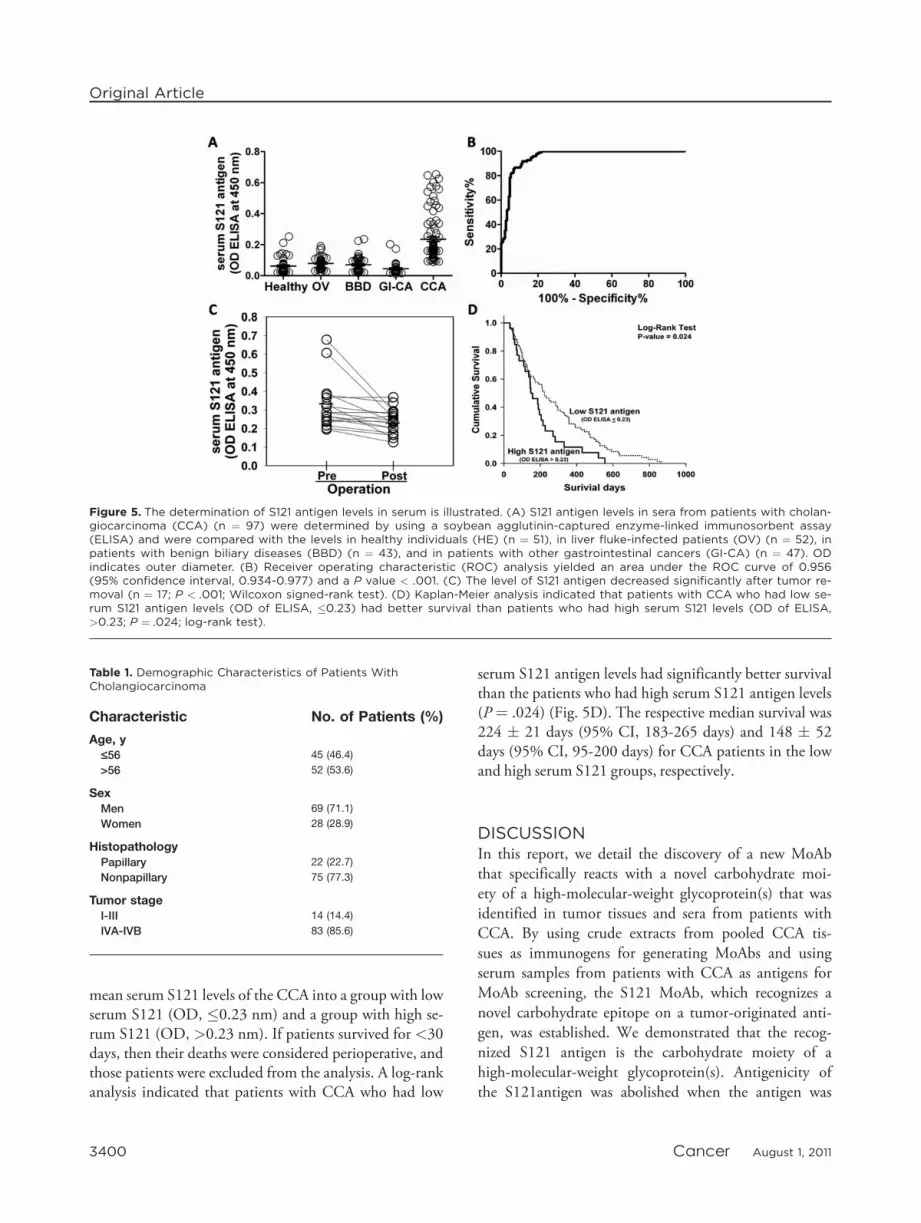
mean serum S121 levels of the CCA into a group with lowserum S121 (OD, �0.23 nm) and a group with high se-rum S121 (OD, >0.23 nm). If patients survived for <30days, then their deaths were considered perioperative, andthose patients were excluded from the analysis. A log-rankanalysis indicated that patients with CCA who had low
serum S121 antigen levels had significantly better survivalthan the patients who had high serum S121 antigen levels(P ¼ .024) (Fig. 5D). The respective median survival was224 � 21 days (95% CI, 183-265 days) and 148 � 52days (95% CI, 95-200 days) for CCA patients in the lowand high serum S121 groups, respectively.
DISCUSSIONIn this report, we detail the discovery of a new MoAbthat specifically reacts with a novel carbohydrate moi-ety of a high-molecular-weight glycoprotein(s) that wasidentified in tumor tissues and sera from patients withCCA. By using crude extracts from pooled CCA tis-sues as immunogens for generating MoAbs and usingserum samples from patients with CCA as antigens forMoAb screening, the S121 MoAb, which recognizes anovel carbohydrate epitope on a tumor-originated anti-gen, was established. We demonstrated that the recog-nized S121 antigen is the carbohydrate moiety of ahigh-molecular-weight glycoprotein(s). Antigenicity ofthe S121antigen was abolished when the antigen was
Table 1. Demographic Characteristics of Patients WithCholangiocarcinoma
Characteristic No. of Patients (%)
Age, y£56 45 (46.4)
>56 52 (53.6)
SexMen 69 (71.1)
Women 28 (28.9)
HistopathologyPapillary 22 (22.7)
Nonpapillary 75 (77.3)
Tumor stageI-III 14 (14.4)
IVA-IVB 83 (85.6)
Figure 5. The determination of S121 antigen levels in serum is illustrated. (A) S121 antigen levels in sera from patients with cholan-giocarcinoma (CCA) (n ¼ 97) were determined by using a soybean agglutinin-captured enzyme-linked immunosorbent assay(ELISA) and were compared with the levels in healthy individuals (HE) (n ¼ 51), in liver fluke-infected patients (OV) (n ¼ 52), inpatients with benign biliary diseases (BBD) (n ¼ 43), and in patients with other gastrointestinal cancers (GI-CA) (n ¼ 47). ODindicates outer diameter. (B) Receiver operating characteristic (ROC) analysis yielded an area under the ROC curve of 0.956(95% confidence interval, 0.934-0.977) and a P value < .001. (C) The level of S121 antigen decreased significantly after tumor re-moval (n ¼ 17; P < .001; Wilcoxon signed-rank test). (D) Kaplan-Meier analysis indicated that patients with CCA who had low se-rum S121 antigen levels (OD of ELISA, �0.23) had better survival than patients who had high serum S121 levels (OD of ELISA,>0.23; P ¼ .024; log-rank test).
Original Article
3400 Cancer August 1, 2011

treated with NaIO4, a treatment known to oxidize anddisrupt sugar conformation; whereas treatment with ahe proteolytic enzyme (trypsin or proteinase K) had noeffect on antigenicity of the S121 antigen. This resultstrongly suggests that the S121 epitope has a carbohy-drate structure. According to the glycoconjugatedmicroarray results, the S121 MoAb did not recognizeany of the known sugar moieties on the array, includ-ing sLea or CA 19-9, which are common tumormarkers of many cancers, including CCA. Hence, it ispossible that the S121 antigen is a novel carbohydratemoiety that is highly expressed in CCA.
In general, identifying the sugar structure onmucins, such as MUC family proteins, is very difficult,and the technology for such analysis has not beenestablished, because those carbohydrates are composedof densely branched sugar chains that are synthesizedwith high diversity on the mucin core protein. Thesugar array technology developed by Tateno et al19
may be the most powerful tool for identifying sugarepitopes; however, although nearly 100 distinguishedsugar chain structures were displayed on our arraychip, no reactivity against S121 MoAb was revealed;therefore, the epitope structure for S121 may be novel.Further study using other approaches will be needed toidentify the sugar chain structure of the S121 epitope.
The S121-reactive antigen was excluded in the voidvolume of a Sepharose 6B column in gel-filtration chro-matography and appeared at the top of a 4% SDS-PAGEgel; therefore, it may be a glycan epitope of the high-mo-lecular-weight glycoprotein that we identified asMUC5AC mucin using S121 MoAb affinity purificationand LC/MS/MS analysis. In the current study, almost allCCA tissues (93%) expressed S121 antigen in immuno-histochemistry analysis. It was demonstrated previouslythat MUC5AC is expressed aberrantly in CCA tissues andis associated with the type, histologic grade, and advancedstage of intrahepatic CCA.8 To our knowledge, there havebeen no reports on the upstream signal of MUC5ACexpression in CCA. However, recently, the Kruppel-likezinc-finger GLI1 was identified as the regulator of theMUC5AC mucin in pancreatic ductal adenocarcinomacells.22 GLI1 up-regulated MUC5AC, attenuated E-cad-herin-mediated cell-cell adhesion, and promoted cellmigration and invasion. Our current results also suggestthat CCA cells may have specific O-glycoenzyme up-regu-lation that causes the novel sugar chain modification onMUC5AC. This may be 1 of the most importantmechanisms to be clarified in future studies. Currently,
our analysis of the specific glycoenzyme genes in CCA isunderway.
The value of S121-reactive antigen is emphasized byour finding that it could be detected in patients’ sera withhigh sensitivity and specificity. In addition, this antigenoriginated from CCA tissue, because it was not observedin the normal bile duct or hepatocytes but was detectedstrongly in premalignant bile duct epithelium and CCAtissues. Moreover, serum levels of S121 were reduced aftertumor removal. Serum S121 levels were high in samplesfrom patients with CCA compared with the levels in sam-ples from the control groups, which included healthyindividuals, liver fluke-infected patients, patients with be-nign biliary diseases, and patients with gastrointestinaltract cancers. Our ROC analysis indicated that the serumS121 antigen could be used to diagnose CCA with87.63% sensitivity and 89.58% specificity. These datasuggest that serum S121 antigen can be used as a tumormarker for CCA. Moreover, the serum S121 level can beused as a prognostic marker, because who high serumS121 levels (OD, >0.23 nm) were associated with poorsurvival in patients with CCA.
In the past decade, there have been severalattempts to identify a better tumor marker for CCA.In addition to CEA and CA 19-9, which are the com-monly used tumor markers for CCA,4,23-26 biliary alka-line phosphatase,6 and MUC5AC7-9 reportedly havebeen used as candidate markers for CCA with variedsensitivity and specificity.6,9,27,28 Sensitivity from 50%to 80% and specificity from 80% to 90% have beenreported for CA 19-9,4,5,25 whereas serum MUC5ACreportedly had 60% to 70% sensitivity and 90% to97% specificity for diagnosing CCA.7-9 In the currentstudy, the S121epitope was at least as sensitive as a car-bohydrate moiety as the core protein of MUC5ACmucin. Detection of the sugar moiety provided bettersensitivity, because the detection of serum S121 yielded87.63% sensitivity and 89.58% specificity with80.95% positive predictive value and 93.47% negativepredictive value for the diagnosis of CCA. The advant-age of serum S121 compared with CEA and CA 19-9is that high levels of the S121 antigen were observedonly in serum from patients with CCA but not in se-rum from patients who had gastric cancer, pancreaticcancer, colon cancer, carcinoma of the ampulla ofVater, and hepatoma; whereas high levels of CEA andCA 19-9 have been reported not only in patients withCCA but also in patients with many different cancersand chronic inflammatory conditions (eg,pancreatitis).24,29,30
New Serum Marker for Cholangiocarcinoma/Silsirivanit et al
Cancer August 1, 2011 3401
Although the molecular function of the sugar-associ-ated epitope recognized by S121 MoAb has not beendetermined, the S121 antigenic moiety may play a signifi-cant role in the pathogenesis of CCA. The associationbetween the S121 antigen and the pathogenesis of CCA issupported by many aspects of the current study. First, theantigen was detected only in pathogenic bile duct epithe-lium. Immunohistochemistry using the S121 MoAbrevealed that the S121 antigen was not present in normalbile ducts or and hepatocytes but was expressed progres-sively in hyperplastic/dysplastic bile duct epithelium andCCA. Second, the S121 antigen was detected at signifi-cantly higher levels in serum from patients with CCAcompared with the levels detected in serum from individ-uals in the non-CCA control groups. Moreover, tumorresection significantly reduced the level of S121 in serum.Third, higher serum levels of S121 antigen were associatedwith a worse prognosis in patients with CCA.
In summary, this study established a MoAb that wedesignated as S121, which recognizes a not-yet-identified,carbohydrate-associated epitope that appeared specificallyin CCA tumor cells. We have established that the MoAbis applicable for studies using immunohistochemistry ofparaffin-embedded sections, immunoblotting, andELISA. The MoAb is useful as a tool for detecting S121antigen, which is elevated in sera from patients with CCA.The ability of S121 MoAb to differentiate between neo-plastic-bile duct and normal bile duct suggests the poten-tial application of S121MoAb in a therapeutic approach.
CONFLICT OF INTEREST DISCLOSURESThis work was cosupported by the Office of the Higher Educa-tion Commission, the National Research Council of Thailand,the National Research University Program, and Khon Kaen Uni-versity and by a Research Team Strengthening Grant from theNational Genetic Engineering and Biotechnology Center,National Science and Technology Development Agency, Thai-land. We are grateful for support from the Japan Student Serv-ices Organization (to A. Silsirivanit), the Ministry of HealthLabor and Welfare of Japan (to N. Araki), and the KurozumiMedical Foundation (to K. Kuwahara). N. Sakaguchi received agrant from the Founding Research Center for Emerging andReemerging Infectious Diseases and received support as a mem-ber of the Global Centers of Excellence Program of AcquiredImmunodeficiency Syndrome Research in Japan.
REFERENCES
1. Shaib Y, El-Serag HB. The epidemiology of cholangiocarci-noma. Semin Liver Dis. 2004;24:115-125.
2. Shaib YH, Davila JA, McGlynn K, El-Serag HB. Risingincidence of intrahepatic cholangiocarcinoma in the UnitedStates: a true increase? J Hepatol. 2004;40:472-477.
3. Patel T. Increasing incidence and mortality of primary intra-hepatic cholangiocarcinoma in the United States. HEPATO-
LOGY. 2001;33:1353-1357.4. Qin XL, Wang ZR, Shi JS, Lu M, Wang L, He QR. Utility
of serum CA19-9 in diagnosis of cholangiocarcinoma: incomparison with CEA. World J Gastroenterol. 2004;10:427-432.
5. Ramage JK, Donaghy A, Farrant JM, Iorns R, Williams R.Serum tumor markers for the diagnosis of cholangiocarci-noma in primary sclerosing cholangitis. Gastroenterology.1995;108:865-869.
6. Bhudhisawasdi V, Muisuk K, Areejitranusorn P, et al. Clinicalvalue of biliary alkaline phosphatase in nonjaundiced cholan-giocarcinoma. J Cancer Res Clin Oncol. 2004;130:87-92.
7. Bamrungphon W, Prempracha N, Bunchu N, et al. A newmucin antibody/enzyme-linked lectin-sandwich assay of se-rum MUC5AC mucin for the diagnosis of cholangiocarci-noma. Cancer Lett. 2007;247:301-308.
8. Boonla C, Wongkham S, Sheehan JK, et al. Prognosticvalue of serum MUC5AC mucin in patients with cholangio-carcinoma. Cancer. 1. 2003;98:1438-1443.
9. Wongkham S, Sheehan JK, Boonla C, et al. SerumMUC5AC mucin as a potential marker for cholangiocarci-noma. Cancer Lett. 2003;195:93-99.
10. Krueger P, Nitz C, Foster R, et al. A new small cell lungcancer (SCLC)-specific marker discovered through antigenicsubtraction of neuroblastoma cells. Cancer Immunol Immun-other. 2003;52:367-377.
11. Krueger P, Nitz C, Moore J, Foster R, Gelber O, Gelber C.Monoclonal antibody identifies a distinctive epitopeexpressed by human multiple myeloma cells. J Immunother.2001;24:334-344.
12. Van Aarsen LA, Leone DR, Ho S, et al. Antibody-mediatedblockade of integrin alpha v beta 6 inhibits tumor progres-sion in vivo by a transforming growth factor-beta-regulatedmechanism. Cancer Res. 2008;68:561-570.
13. Greene FL, Page DL, Flaming ID, et al. eds. AJCC CancerStaging Manual. 6th ed. New York: Springer-Verlag; 2002.
14. Sakaguchi N, Kimura T, Matsushita S, et al. Generation ofhigh-affinity antibody against T cell-dependent antigen in theGanp gene-transgenic mouse. J Immunol. 2005;174:4485-4494.
15. Bara J, Decaens C, Loridon-Rosa B, Oriol R. Immunohisto-logical characterization of mucin epitopes by pretreatmentof gastro-intestinal sections with periodic acid. J ImmunolMethods. 1992;149:105-113.
16. Cao Y, Blohm D, Ghadimi BM, Stosiek P, Xing PX, Kars-ten U. Mucins (MUC1 and MUC3) of gastrointestinal andbreast epithelia reveal different and heterogeneous tumor-associated aberrations in glycosylation. J Histochem Cyto-chem. 1997;45:1547-1557.
17. Gil J, Alvarez R, Vinuela JE, et al. Inhibition of in vivotumor growth by a monoclonal IgM antibody recognizingtumor cell surface carbohydrates. Cancer Res. 1990;50:7301-7306.
18. Laemmli UK. Cleavage of structural proteins during the as-sembly of the head of bacteriophage T4. Nature. 1970;227:680-685.
19. Tateno H, Mori A, Uchiyama N, et al. Glycoconjugatemicroarray based on an evanescent-field fluorescence-assisteddetection principle for investigation of glycan-binding pro-teins. Glycobiology. 2008;18:789-798.
20. Tateno H, Ohnishi K, Yabe R, et al. Dual specificity ofLangerin to sulfated and mannosylated glycans via a single
Original Article
3402 Cancer August 1, 2011
C-type carbohydrate recognition domain. J Biol Chem.2010;285:6390-6400.
21. Zweig MH, Campbell G. Receiver-operating characteristic(ROC) plots: a fundamental evaluation tool in clinical med-icine. Clin Chem. 1993;39:561-577.
22. Inaguma S, Kasai K, Ikeda H. GLI1 facilitates the migrationand invasion of pancreatic cancer cells through MUC5AC-mediated attenuation of E-cadherin [published online aheadof print October 25, 2010]. Oncogene. 2010.
23. Nakeeb A, Lipsett PA, Lillemoe KD, et al. Biliary carcino-embryonic antigen levels are a marker for cholangiocarci-noma. Am J Surg 171:147-152, 1996; discussion 152-143.
24. Ni XG, Bai XF, Mao YL, et al. The clinical value of serumCEA, CA19-9, and CA242 in the diagnosis and prognosisof pancreatic cancer. Eur J Surg Oncol. 2005;31:164-169.
25. Patel AH, Harnois DM, Klee GG, LaRusso NF, Gores GJ.The utility of CA 19-9 in the diagnoses of cholangiocarci-noma in patients without primary sclerosing cholangitis. AmJ Gastroenterol. 2000;95:204-207.
26. Saito K, Fujii Y, Kawakami S, et al. Increased expression ofsialyl-Lewis A correlates with poor survival in upper urinarytract urothelial cancer patients. Anticancer Res. 2003;23:3441-3446.
27. Uenishi T, Yamazaki O, Tanaka H, et al. Serum cytokeratin19 fragment (CYFRA21-1) as a prognostic factor in intrahe-patic cholangiocarcinoma. Ann Surg Oncol. 2008;15:583-589.
28. Watanabe H, Enjoji M, Nakashima M, et al. Clinical signif-icance of serum RCAS1 levels detected by monoclonal anti-body 22-1-1 in patients with cholangiocellular carcinoma.J Hepatol. 2003;39:559-563.
29. Del Favero G, Fabris C, Panucci A, et al. Carbohydrateantigen 19-9 (CA 19-9) and carcinoembryonic antigen(CEA) in pancreatic cancer. Role of age and liver dysfunc-tion. Bull Cancer. 1986;73:251-255.
30. Del Favero G, Fabris C, Plebani M, et al. CA 19-9 and car-cinoembryonic antigen in pancreatic cancer diagnosis. Can-cer. 1986;57:1576-1579.
New Serum Marker for Cholangiocarcinoma/Silsirivanit et al
Cancer August 1, 2011 3403


