Embed Size (px)
Citation preview

ORIGINAL RESEARCH ARTICLEpublished: 03 June 2014
doi: 10.3389/fphys.2014.00210
A multistep procedure to prepare pre-vascularized cardiactissue constructs using adult stem sells, dynamic cellcultures, and porous scaffoldsStefania Pagliari 1,2, Annalisa Tirella3,4, Arti Ahluwalia3,4, Sjoerd Duim5, Marie-Josè Goumans5,
Takao Aoyagi1* and Giancarlo Forte1,2*
1 Biomaterials Unit, International Center for Materials Nanoarchitectonics, National Institute for Materials Science, Tsukuba, Japan2 International Clinical Research Center, Integrated Center of Cellular Therapy and Regenerative Medicine, St. Anne’s University Hospital, Brno, Czech Republic3 Interdepartmental Research Center “E. Piaggio”, University of Pisa, Italy4 Institute of Clinical Physiology, National Research Council (CNR), Pisa, Italy5 Department of Molecular Cell Biology, Leiden University Medical Center, Leiden, Netherlands
Edited by:
Valentina Di Felice, University ofPalermo, Italy
Reviewed by:
Teun P. De Boer, University MedicalCenter Utrecht, NetherlandsMarcin Wysoczynski, University ofLouisville, USA
*Correspondence:
Takao Aoyagi, Smart BiomaterialsGroup, International Center forMaterials Nanoarchitectonics,National Institute for MaterialsScience, 1-1 Namiki, Tsukuba,Ibaraki 305-0051, Japane-mail: [email protected];Giancarlo Forte, International ClinicalResearch Center, Integrated Centerof Cellular Therapy andRegenerative Medicine, St. Anne’sUniversity Hospital, Pekarska 53,65691 Brno, Czech Republice-mail: [email protected]
The vascularization of tissue engineered products represents a key issue in regenerativemedicine which needs to be addressed before the translation of these protocols tothe bedside can be foreseen. Here we propose a multistep procedure to preparepre-vascularized three-dimensional (3D) cardiac bio-substitutes using dynamic cell culturesand highly porous biocompatible gelatin scaffolds. The strategy adopted exploits thepeculiar differentiation potential of two distinct subsets of adult stem cells to obtain humanvascularized 3D cardiac tissues. In the first step of the procedure, human mesenchymalstem cells (hMSCs) are seeded onto gelatin scaffolds to provide interconnected vessel-likestructures, while human cardiomyocyte progenitor cells (hCMPCs) are stimulated in vitroto obtain their commitment toward the cardiac phenotype. The use of a modular bioreactorallows the perfusion of the whole scaffold, providing superior performance in termsof cardiac tissue maturation and cell survival. Both the cell culture on natural-derivedpolymers and the continuous medium perfusion of the scaffold led to the formation ofa densely packaged proto-tissue composed of vascular-like and cardiac-like cells, whichmight complete maturation process and interconnect with native tissue upon in vivoimplantation. In conclusion, the data obtained through the approach here proposedhighlight the importance to provide stem cells with complementary signals in vitro able toresemble the complexity of cardiac microenvironment.
Keywords: cardiac tissue engineering, adult stem cells, vascularized three-dimensional (3D) scaffolds, dynamic
culture, patient-derived stem cells
INTRODUCTIONThe successful regeneration of injured areas of the myocardiumby tissue-engineered constructs relies on the long time viabil-ity and persistence of the bio-substitute in vivo, given the harshconditions cells in the infarcted milieu are exposed to. Previousinvestigations revealed the sudden disappearance of cells admin-istered by injection—systemically or locally—independent of celltype. This negative outcome has been ascribed to the low reten-tion and high mortality of cells in the hypoxic environmentcharacterized by an inflammatory response and the lack of localblood supply (Gnecchi et al., 2008; Menasche, 2011). The issue ofpromoting ischemic area vascularization has been lately addressedby cardiac tissue engineers through different approaches: (i) theadministration of pro-angiogenic factors supplied by direct injec-tion or through drug-releasing carriers (Sato et al., 2001; Chiuand Radisic, 2010; Singh et al., 2012); (ii) the infusion of endothe-lial progenitors (EPCs) or mature endothelial cells (ECs; Lianet al., 2008); and (iii) the pre-vascularization of the tissue con-structs produced in vitro before implantation (Caspi et al., 2007;Dvir et al., 2009). Although the first two strategies are potentially
interesting in a therapeutic perspective, they rely on the in situgeneration and organization of vascular structures which dependeither on the bioavailability of beneficial molecules or on thegrowth and differentiation capacity of vascular cells or their pro-genitors (Lovett et al., 2009). The early clinical trials in whichgrowth factors or cells were delivered to the injured heart yieldeddisappointing results in terms of improvement of cardiac func-tion (Urbich et al., 2005; Dubois et al., 2010; Simón-Yarza et al.,2012). The pre-vascularization of cardiac patches is also appropri-ate for providing a capillary network to support cells in the innercore of the implant, while biocompatible substrates are deemedto contribute to the improvement of retention and engraftmentof the transplanted cardiac tissue (Terrovitis et al., 2010; Segersand Lee, 2011). The advantage of the pre-vascularization of thickmuscle constructs was underlined by the demonstration that co-cultures including skeletal myoblasts, endothelial cells (or theirprogenitors) and embryonic fibroblasts on biocompatible porousscaffolds can enhance the overall survival and functionality ofthe constructs in vivo (Levenberg et al., 2005). Moreover, theadoption of scaffolds displaying an interconnected porosity itself
www.frontiersin.org June 2014 | Volume 5 | Article 210 | 1

Pagliari et al. Vascularized cardiac constructs
could foster host vascular cell recruitment, with the possibil-ity of vessels branching throughout the core of the construct.Alternatively, scaffoldless thick cardiac constructs were providedwith a vascular bed (Sekine et al., 2013), or with microchan-nels (Sakaguchi et al., 2013) to favor vessel ingrowth, althoughbiocompatible supports improve the handling of the grafts andcan provide cells with appropriate bio-mechanical signals to bet-ter induce tissue regeneration and repair. In this context, theuse of porous gelatin scaffolds represents a suitable tool for car-diac tissue engineering application (Sakai et al., 2001; Akhyariet al., 2002). In fact, gelatin is a cheap polymer derived fromcollagen denaturation and hydrolysis, and, due to its naturalorigin, it displays excellent cell adhesion property (Wu et al.,2011). It also features high biocompatibility, low immunogenic-ity, and biodegradability (Xing et al., 2014). In addition, gelatinsponges have been proven effective in inducing angiogenesis(Dreesmann et al., 2007) and their porous structure can favorthe vascularization of the construct by supporting the diffusionof cells and nutrients within its core area. Its mechanical proper-ties can be easily adjusted to match those encountered in livingtissues.
The use of autologous stem cells has been proposed for var-ious cell therapy applications as a mean to avoid the immunerejection issues raised by allogeneic or xenogeneic derivativesand the ethical concerns due to the use of embryonic material.Human bone marrow-derived mesenchymal stem cells (hMSCs)are an excellent candidate for regenerative medicine applicationsdue to their autologous origin, their immunomodulatory prop-erties and relative safety in clinical practice (Lalu et al., 2012).The in vitro multilineage differentiation potential of mesodermalprogenitors has been proven in a number of studies (Pittengeret al., 1999; Muraglia et al., 2000) and their ability to expressendothelial markers upon growth factor stimulation (Oswaldet al., 2004; Jazayeri et al., 2008; Portalska et al., 2012) andresponse to bio-mechanical stimulation (stretching, shear stress,substrate mechanical properties tuning; Lozito et al., 2009; Baiet al., 2010) has been shown. More importantly, the benefits ofMSC-based therapy have mainly been ascribed to their ability togenerate endothelial cells and exert pro-angiogenic and cardio-protective effects by paracrine mechanisms rather than to directthe generation of new contractile cells (Gnecchi et al., 2008; Meyeret al., 2009; Wöhrle et al., 2010; Loffredo et al., 2011).
Among the adult stem cell subsets so far proposed for cardiacmuscle repair, resident cardiac stem/progenitor cells (CSCs orCPCs) were shown to retain the ability to differentiate into all thecardiac tissue cell types (Beltrami et al., 2003; Forte et al., 2011)and favor cardiac healing by direct production of contractile cellsin vivo (Smits et al., 2009a,b).
By taking advantage of the peculiar differentiation potential ofhMSCs and human cardiomyocyte progenitor cells (hCMPCs),in the present investigation we propose a multistep procedure toobtain human pre-vascularized three-dimensional (3D) cardiacbio-substitutes based on highly porous gelatin scaffolds display-ing the stiffness of cardiac tissue. Given the thickness and thedimensions of the bio-construct, a modular dynamic culture sys-tem has been used to guarantee scaffold perfusion and promotecell colonization of the inner layers.
Although being here tested exclusively in vitro, the presentmethod allows for the formation of a 3D cardiac construct basedon a physiological environment given by the gelatin scaffold andfeaturing the use of autologous stem cells with unique potential.
MATERIALS AND METHODSPREPARATION OF POROUS GELATIN SCAFFOLDSAll materials used (unless specified) were purchased from Sigma-Aldrich (Italy). A 5% w/v gelatin solution was prepared by dis-solving gelatin (Type A, G1890, 300 bloom strength) in deionizedwater. Porous gelatin scaffolds were prepared with a multi-stepprocedure. The solution was stirred for 1 h at 50◦C, allowingcomplete dissolution, then casted in cylindrical shaped moldand physically gelled at room temperature. Samples were keptat 4◦C for 1 h, and then at −20◦C overnight. Gelatin sampleswere then freeze-dried (−50◦C, 150 mBar) to obtain a porousstructure as described elsewhere (Lien et al., 2009). Samples wereswollen in deionized water and then cross-linked by immer-sion in a 10 mM glutaraldehyde (GTA) solution in 40% v/vethanol/deionized water. The crosslinking reaction was controlledby keeping the glutaraldehyde/gelatin ratio (defined as molar con-centration of GTA versus gelatin weight) constant. The scaffoldswere immersed in GTA solution at 4◦C for 48 h, until the cross-linking reaction occurred. Therefore, the samples were immersedin 0.1 M glycine solution in deionized water for 2 h at roomtemperature in order to stop any further cross-linking reaction(and remove any excess of GTA). Phosphate buffered solution(PBS) was sequentially used to rinse samples. Samples were keptat −20◦C overnight, and finally freeze-dried (−50◦C, 150 mBar)until all water content was removed. Samples were then stored atroom temperature and sterilized with gas plasma before using.
SCAFFOLD MECHANO-ARCHITECTURAL PROPERTIES: SWELLING,POROSITY, AND STIFFNESSWater absorption capability, porosity and stiffness of the porousgelatin scaffolds were evaluated using the procedure describedelsewhere (Spinelli et al., 2012). Swelling ratio (Q) was cal-culated from the ratio of the weight of a dry (W0) and acompletely swollen (Weq) sample (Brannon-Peppas and Peppas,1990) returning the amount of adsorbed water. For the mea-surements, cryogel were swollen in deionized water at roomtemperature and weighted (Wi) at different time points untila swelling equilibrium was reached. A precision microbalance(AE240, Mettler, Italy) was used: in case of wet samples, blottingpaper was used to remove the water in excess. Porosity was indi-rectly evaluated by the imbibition method (Mwangi and Ofner,2004; Martucci et al., 2006), while pore size was directly measuredby processing both Scanning Electron Microscopy (SEM) andoptical microscopy acquisition of sample sections with ImageJ(Abramoff et al., 2004). Sample stiffness was measured by com-pressive mechanical tests. Prior to the tests, samples were com-pletely swollen in deionized water, compressive tests were thenperformed using a Zwick-Roell Z005 Instron twin column-testingmachine (Zwick Testing Machines Ltd., UK). Samples were com-pressed up to 5% of their initial length using a 0.01 mm·s−1 strainrate; tests were performed with the samples partially immersed inwater to preserve their hydration. Data were then post-processed
Frontiers in Physiology | Striated Muscle Physiology June 2014 | Volume 5 | Article 210 | 2
Pagliari et al. Vascularized cardiac constructs
and stress-strain curves were obtained. Samples stiffness was eval-uated within 1% strain (first linear zone) of the stress-straincurve.
BIOREACTOR WORKING CONDITIONSA computational fluid-dynamic (CFD) analysis of the modularchamber bioreactor with a porous scaffold was performed, assess-ing the perfusion and oxygenation of the millimeter sized scaffold.The analysis was performed using Brinkman and IncompressibleNavier-Stokes equations were combined with reaction and dif-fusion equations using Comsol Multiphysics (COMSOL AB,Stockholm, Sweden). The system was then modeled with a porousdomain representing the scaffold (porous section of 2 × 12 mmin size, with permeability of 1.68 × 10⁻¹⁰ m2 and 90% porosity)placed on the bottom of the bioreactor perfusion chamber, whichis represented by a fluid domain. A preliminary analysis was per-formed in order to verify scaffold oxygenation as function ofbioreactor flow rate (Supplementary Figure 1A). Chosen an innerflow rate to a value of 200 μL/min, fluid flow inside the perfusionchamber and scaffold’s perfusion was analyzed (SupplementaryFigure 1B).
CELL CULTUREhMSCs were purchased from Lonza and cultured in MSCGM™BulletKit™ (hMSC basal medium, Lonza Japan Ltd, Tokyo,Japan). The cells were expanded to the desired number andused between passages 3 and 7. For the endothelial differ-entiation in 2D conditions, hMSCs were seeded onto tissueculture polystyrene plates (TCPS) and cultured for 7 daysin endothelial differentiation medium [(EDM: hMSC basalmedium supplemented with 50 ng/ml VEGF165 (R&D sys-tems)]. hCMPCs were isolated from healthy donor atrial biop-sies, cultured in basal medium (M199/EGM-2 (Lonza) (3:1)10% FBS (Equitech-Bio Inc., Kerrville, Texas, USA), 1% MEMnonessential amino acids (from 50× stock; Life TechnologiesGaithersburg, Maryland, USA) and 2% penicillin/streptomycin(from 50× stock; Invitrogen, Carlsbad, California, USA)and differentiated in cardiac differentiation medium (CDM:IMDM/Hams F12 (1:1, Life Technologies) with L-glutamine (LifeTechnologies), 2% horse serum (Life Tchnologies), nonessen-tial amino acids, insulin–transferrin–selenium (Invitrogen),penicillin/streptomycin) as previously described (Smits et al.,2009a,b). Briefly, minced auricle were digested in collagenase A(1 mg/ml) for 2 h at 37◦C while stirring. Afterwards, the solu-tion was filtered through 40-μm cell strainer and centrifuged at300 g for 5 min at RT. Then, the pellet was resuspended in coldbuffer containing EDTA (2 mM) and 2% FBS and filtered through40-μm cell strainer. The filtered cell population was subjectedto magnetic cell sorting (Miltenyi Biotec, Sunnyvale, CA, USA)using Sca-1-coupled beads, following the manufacturer’s proto-col, in order to isolate Sca-1-like + progenitor cells. The cells werecultured on 0.1% gelatin- (Sigma-Aldrich, St Louis, Missouri,USA) coated plates in basal medium. For cardiac differentiation,cells were grown in differentiation medium supplemented with5 μM 5-azacytidine (Wako Pure Chemical Industries, Ltd, Osaka,Japan) for 72 h. After induction, the medium was replaced withdifferentiation medium supplemented with 10−4 M ascorbic acid
(Wako) and TGF-β1 (1 ng/ml, PeproTech, Rocky Hill, NJ, USA)and changed every 3 days for up to 3 weeks. Human umbilical veinendothelial cells (HUVEC) were purchased from Lonza, grown inEGM-2 bullet kit media and used between passage 2 and 4. Themedia were replenished every other day.
GENERATION OF TNT-GFP hCMPCsThe pGreenZeo lentiviral expression vector carrying the full-length copGFP gene under the control of cardiac Troponin Ttype 2 (TNNT2) promoter was purchased from SBI (SystemBiosciences Inc. Mountain View, CA, USA) and delivered intomammalian cells according to the manufacturer’s instructions.Briefly, hCMPCs were transduced at an approximate MOI of 20and incubated at 37◦C overnight. The following day, half of theculture medium was replaced with fresh medium and 48 h afterthe transduction, the medium was discarded and fresh mediumadded. The cells (hereafter referred as cTNT-GFP hCMPCs) werecultured on TCPS or gelatin scaffolds in the presence of basalmedium or CDM.
CELL SEEDING AND 3D DISTRIBUTION IN GELATIN SCAFFOLDSPrior to cell seeding, gelatin scaffolds were swollen in cell cul-ture medium in Ultra-low Attachment 24-well plates (Corning®Incorporated) for 24 h. The cells (2.0 × 10⁵) were directly seededon top of each scaffold in a small volume (100 μl) of appropriatemedium and allowed to adhere for 60 min before adding the restof medium. Then, the scaffolds were placed in the incubator for12 h and transferred in another low-adhesion well. Cardiac TNT-GFP hCMPCs were cultured into scaffolds for 7 days in completehCMPCs basal medium or CDM, while hMSCs-seeded scaffoldswere cultured for 4 days in a mixture of Matrigel™/hMSCs basalmedium (1:10) with or without VEGF (50 ng/mL) before beingprocessed or co-cultured with pre-conditioned cardiac progeni-tors. The formation of vascular-like structures on gelatin scaffoldswas followed by labeling hMSCs with the membrane-permeablelive-cell labeling dye Calcein AM (Invitrogen) after 4 days of cul-ture in EDM. Briefly, Calcein AM was added to hMSC-seededscaffolds at a final concentration of 2 μM for 30 min at 37◦Cand 5% CO2 in the dark. Five scaffolds have been used forany single experiment. The experiments have been performedindependently three times.
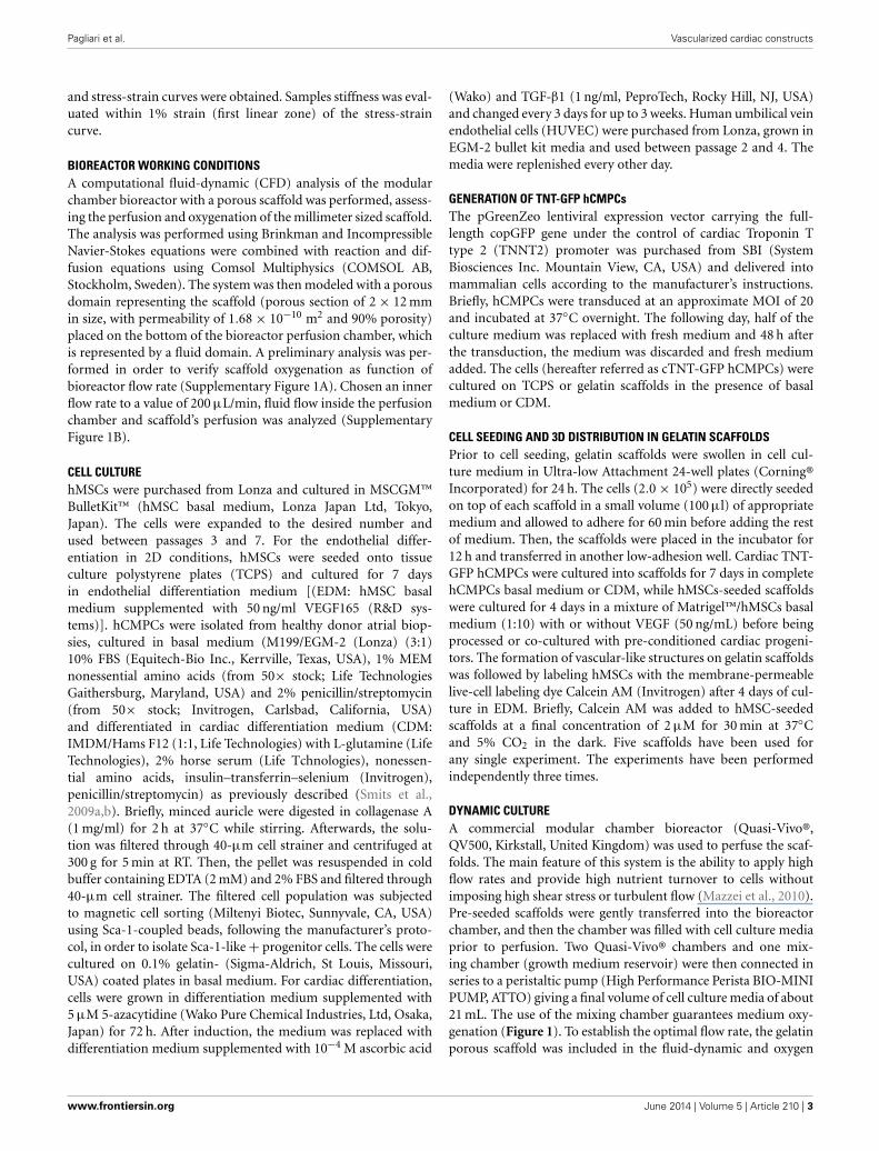
DYNAMIC CULTUREA commercial modular chamber bioreactor (Quasi-Vivo®,QV500, Kirkstall, United Kingdom) was used to perfuse the scaf-folds. The main feature of this system is the ability to apply highflow rates and provide high nutrient turnover to cells withoutimposing high shear stress or turbulent flow (Mazzei et al., 2010).Pre-seeded scaffolds were gently transferred into the bioreactorchamber, and then the chamber was filled with cell culture mediaprior to perfusion. Two Quasi-Vivo® chambers and one mix-ing chamber (growth medium reservoir) were then connected inseries to a peristaltic pump (High Performance Perista BIO-MINIPUMP, ATTO) giving a final volume of cell culture media of about21 mL. The use of the mixing chamber guarantees medium oxy-genation (Figure 1). To establish the optimal flow rate, the gelatinporous scaffold was included in the fluid-dynamic and oxygen
www.frontiersin.org June 2014 | Volume 5 | Article 210 | 3
Pagliari et al. Vascularized cardiac constructs
FIGURE 1 | QuasiVivo Bioreactor (QV500). The bioreactor is composed ofa peristaltic pump keeping a constant medium flow rate (200 μl/min) fromthe growth medium reservoir to two culture chambers connected in series,thus allowing scaffold inner layer perfusion.
transport model previously described (Mazzei et al., 2010), givingconsistent results (Supplementary Figure 1). Cardiac TNT-GFPhCMPCs grown for 7 days onto gelatin scaffolds in the bioreac-tor were used to study the effect of dynamic culture conditionson cell growth, differentiation potential and gene expressionprofile. The medium was replenished every third day. For thegeneration of vascularized cardiac grafts, scaffolds were soakedfor 12 h in diluted Matrigel™ (1:10 in hMSC basal medium)and switched to a new low-adhesion plate in EDM. The pre-conditioned scaffold was loaded with 2.0 × 10⁵ hMSCs and keptfor 4 days in the incubator at 37◦C, 5% CO2. After a 2 weekpre-conditioning passage in CDM on TCPS, 2.0 × 10⁵ cTNT-GFP hCMPCs were seeded onto the vascularized scaffold andallowed to attach for 24 h. Thence, the cell-loaded scaffolds werecarefully moved to Quasi-vivo bioreactor chambers and perfusedwith CDM at 200 μl/min (the rate suggested by the computa-tional model) for 7 days. Cells grown on TCPS and 3D scaffoldsunder static conditions were used as controls. Five scaffolds havebeen used for any single experiment. The experiments have beenperformed independently three times.
REAL-TIME QUANTITATIVE PCR ANALYSISTotal RNA was extracted by TRIZOL® Reagent according tothe manufacturer’s instructions (Invitrogen). One microgram ofRNA for each sample, measured by NanoDrop 2000 (ThermoScientific) and assessed on ReadyAgarose Precast Gel (Bio-Rad),was retro-transcribed using RT2 First Strand Kit including DNAsetreatment to remove genomic DNA (SA Biosciences Corp.,USA). The resulting cDNA was diluted 1:10 in DNase/RNase-free water and the expression profile of genes involved indifferent pathways analyzed by the following RT2 Profiler™PCR Arrays (SA Biosciences Corp.): (i) RT2 Profiler™ PCRArray Human Angiogenesis (PAHS-024Z); (ii) RT2 Profiler™PCR Array Human Extracellular Matrix & Adhesion Molecules(PAHS-013); (iii) qBiomarker iPSC PCR Array Cardiomyocytesdifferentiation (iPHS102); (for a comprehensive list of genesincluded in these arrays please refer to Supplementary Table1). Real-time PCR was performed on ABI 7500 Real-Time PCRSystem (AB Applied Biosystems, Foster City, CA) using RT2 SYBRGreen/ROX qPCR Master Mix (SA Biosciences Corp.) and the fol-lowing cycling parameters: 1 cycle at 95◦C for 10 min; 40 cycles
at 95◦C for 15 s, and 60◦C for 1 min. Data was analyzed by the��Ct method with the PCR Array Data Analysis Web Portal(http://www.SABiosciences.com/pcrarraydataanalysis.php). Thegraphs show the mean values of obtained fold changes byanalyzing independently two samples per each experimentalcondition.
STATISTICAL ANALYSISData are represented as mean ± SD. Student’s T-test was used toanalyze the data. Comparisons between different conditions wereconsidered statistically significant for P < 0.05.
For a more extensive description of the Materials and Methodsused, please refer to Supplementary Information.
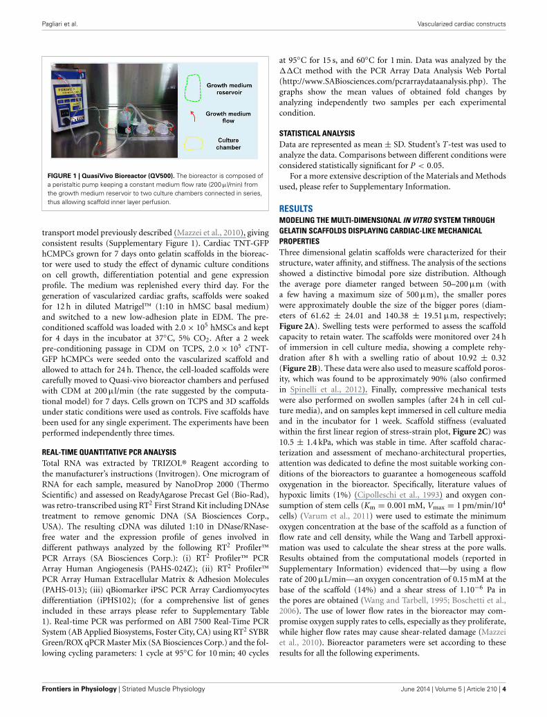
RESULTSMODELING THE MULTI-DIMENSIONAL IN VITRO SYSTEM THROUGHGELATIN SCAFFOLDS DISPLAYING CARDIAC-LIKE MECHANICALPROPERTIESThree dimensional gelatin scaffolds were characterized for theirstructure, water affinity, and stiffness. The analysis of the sectionsshowed a distinctive bimodal pore size distribution. Althoughthe average pore diameter ranged between 50–200 μm (witha few having a maximum size of 500 μm), the smaller poreswere approximately double the size of the bigger pores (diam-eters of 61.62 ± 24.01 and 140.38 ± 19.51 μm, respectively;Figure 2A). Swelling tests were performed to assess the scaffoldcapacity to retain water. The scaffolds were monitored over 24 hof immersion in cell culture media, showing a complete rehy-dration after 8 h with a swelling ratio of about 10.92 ± 0.32(Figure 2B). These data were also used to measure scaffold poros-ity, which was found to be approximately 90% (also confirmedin Spinelli et al., 2012). Finally, compressive mechanical testswere also performed on swollen samples (after 24 h in cell cul-ture media), and on samples kept immersed in cell culture mediaand in the incubator for 1 week. Scaffold stiffness (evaluatedwithin the first linear region of stress-strain plot, Figure 2C) was10.5 ± 1.4 kPa, which was stable in time. After scaffold charac-terization and assessment of mechano-architectural properties,attention was dedicated to define the most suitable working con-ditions of the bioreactors to guarantee a homogeneous scaffoldoxygenation in the bioreactor. Specifically, literature values ofhypoxic limits (1%) (Cipolleschi et al., 1993) and oxygen con-sumption of stem cells (Km = 0.001 mM, Vmax = 1 pm/min/104
cells) (Varum et al., 2011) were used to estimate the minimumoxygen concentration at the base of the scaffold as a function offlow rate and cell density, while the Wang and Tarbell approxi-mation was used to calculate the shear stress at the pore walls.Results obtained from the computational models (reported inSupplementary Information) evidenced that—by using a flowrate of 200 μL/min—an oxygen concentration of 0.15 mM at thebase of the scaffold (14%) and a shear stress of 1.10−6 Pa inthe pores are obtained (Wang and Tarbell, 1995; Boschetti et al.,2006). The use of lower flow rates in the bioreactor may com-promise oxygen supply rates to cells, especially as they proliferate,while higher flow rates may cause shear-related damage (Mazzeiet al., 2010). Bioreactor parameters were set according to theseresults for all the following experiments.
Frontiers in Physiology | Striated Muscle Physiology June 2014 | Volume 5 | Article 210 | 4
Pagliari et al. Vascularized cardiac constructs
FIGURE 2 | The porous gelatin scaffold. Three-dimensional renderingof scaffold, brightfield images of scaffold section and pore dimensionanalysis (A). Modifications of swelling ratio of scaffold over 24 h in
cell culture media (B). Stress-strain plot of gelatin samples (C)
obtained after 24 h and 1 week in culture medium (37◦C, 5%CO2). ∗P < 0.05.
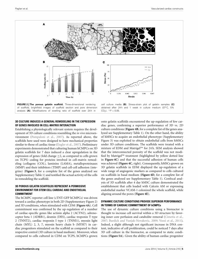
3D CULTURE INDUCES A GENERAL REMODELING IN THE EXPRESSIONOF GENES INVOLVED IN CELL-MATRIX INTERACTIONEstablishing a physiologically relevant system requires the devel-opment of 3D culture conditions resembling the in vivo microen-vironment (Pampaloni et al., 2007). As reported above, thescaffolds here used were designed to have mechanical propertiessimilar to those of cardiac tissue (Engler et al., 2007). Preliminaryexperiments demonstrated that culturing human hCMPCs on 3Dgelatin scaffolds for 7 days induced a clear upregulation in theexpression of genes (fold change ≥2, as compared to cells grownon TCPS) coding for proteins involved in cell-matrix remod-eling (collagens (COL), laminins (LAMA), metalloproteinases(MMP) and their inhibitors (TIMP) and cell-cell adhesion (inte-grins) (Figure 3, for a complete list of the genes analyzed seeSupplementary Table 1) and testified the actual activity of the cellsin remodeling the scaffold.
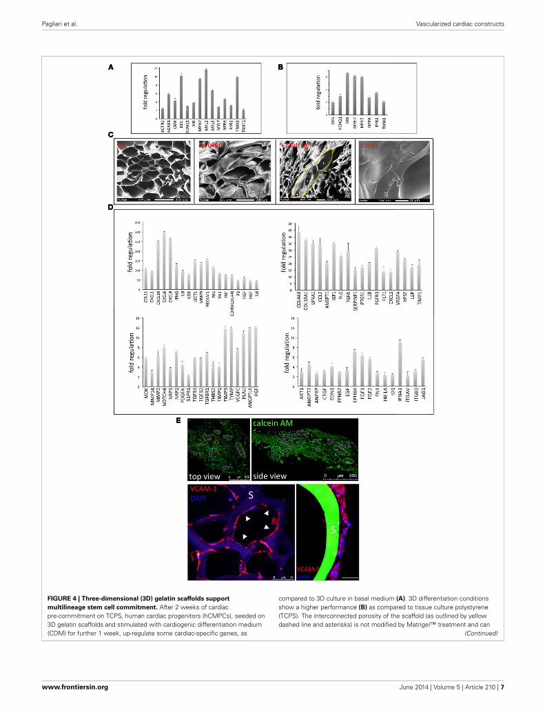
3D POROUS GELATIN SCAFFOLDS REPRESENT A PERMISSIVEENVIRONMENT FOR STEM CELL CARDIAC AND ENDOTHELIALCOMMITMENTThe hCMPC reporter cell line (cTNT-GFP hCMPCs) was driventoward a cardiac phenotype in both 2D (Supplementary Figure 2)and 3D conditions, when stimulated with CDM (Figure 4A). Cellcommitment was confirmed by the up-regulation of a numberof cardiac-specific genes like actinin alpha 2 (ACTN2), adreno-ceptor beta 1 (ADRB1), desmin (DES), cardiac troponin T type2 (TNNT2), cardiac troponin I type 1 (TNNI1), myosin lightchain (MYL) 2, 3, 7, myosin heavy chain b (MYH7) in car-diac progenitors stimulated on the scaffold as compared to theirrespective control (3D culture in basal medium). Moreover, whencompared to cells cultured in CDM on TCPS, hCMPCs grown
onto gelatin scaffolds encountered the up-regulation of few car-diac genes, confirming a superior performance of 3D vs. 2Dculture conditions (Figure 4B, for a complete list of the genes ana-lyzed see Supplementary Table 1). On the other hand, the abilityof hMSCs to acquire an endothelial phenotype (SupplementaryFigure 3) was exploited to obtain endothelial cells from hMSCsunder 3D culture conditions. The scaffolds were treated with amixture of EDM and Matrigel™ for 24 h. SEM analysis showedthat the interconnected porosity of the scaffold was not modi-fied by Matrigel™ treatment (highlighted by yellow dotted linein Figure 4C) and that the successful adhesion of human cellswas achieved (Figure 4C, right). Consequently, hMSCs grown on3D gelatin scaffolds in EDM displayed the up-regulation of awide range of angiogenic markers as compared to cells culturedon scaffolds in basal medium (Figure 4D, for a complete list ofthe genes analyzed see Supplementary Table 1). Confocal anal-ysis of 3D scaffolds after 4 day hMSC culture demonstrated theestablishment that cells loaded with Calcein AM or expressingendothelial marker VCAM-1 colonized the whole scaffold, whilealigning around the pores (Figure 4E).
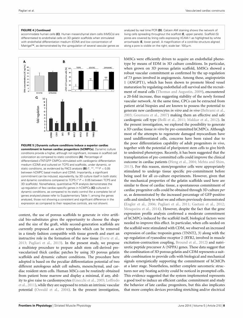
DYNAMIC CULTURE CONDITIONS PROVIDE SUPERIOR PERFORMANCEIN TERMS OF CARDIAC COMMITTMENT OF hCMPCsThe use of dynamic culture conditions using a bioreactor isthought to increase cell survival within a 3D structure by favor-ing inner core perfusion and catabolite removal (Cimetta et al.,2007; Burdick and Vunjak-Novakovic, 2009; Vozzi et al., 2011).Indeed, a slight although not significant increase in DNA con-tent, indicative of cell proliferation, could be noticed 7 days after3D cell culture in the bioreactor, as compared to static condi-tions (Figure 5A). Given the ability of human cardiac progenitor
www.frontiersin.org June 2014 | Volume 5 | Article 210 | 5
Pagliari et al. Vascularized cardiac constructs
FIGURE 3 | Three-dimensional (3D) stem cell culture induces a significant
remodeling in the expression of genes involved in cell adhesion and
ECM deposition. Human cardiac progenitor cells (hCMPCs) were grown for
1 week on 3D gelatin scaffolds and the expression of 73 genes involved incell-matrix interaction resulted upregulated, as evidenced by real time PCR(for a complete list of genes analyzed please refer to Supplementary Table1).
cells to proceed to cardiac maturation on 3D gelatin scaffolds,their capacity to activate the cardiac program when exposed todynamic conditions in a modular bioreactor was assessed. For thisexperiment, cTNT-GFP hCMPCs were pre-committed in CDMfor 2 weeks on TCPS. Afterwards, an aliquot of this cell popula-tion was seeded onto scaffolds and further stimulated for 1 weekunder static or dynamic conditions. The data confirmed that thecommitment of cTNT-GFP hCMPCs could be achieved by CDMin dynamic conditions (Figure 5B) and that a minor althoughsignificant spontaneous commitment of hCMPCs was inducedby 3D culture conditions per se, as also described in static cul-ture (white columns). Although the dynamic culture conditionsdid not significantly modify the percentage of GFP-positive cellswith respect to the static protocol, real time PCR array analysisdemonstrated that dynamic stimulation—when combined withdifferentiation medium—could lead to the up-regulation of fewcardiac-specific genes (Figure 5C, for a complete list of the genesanalyzed see Supplementary Table 1).
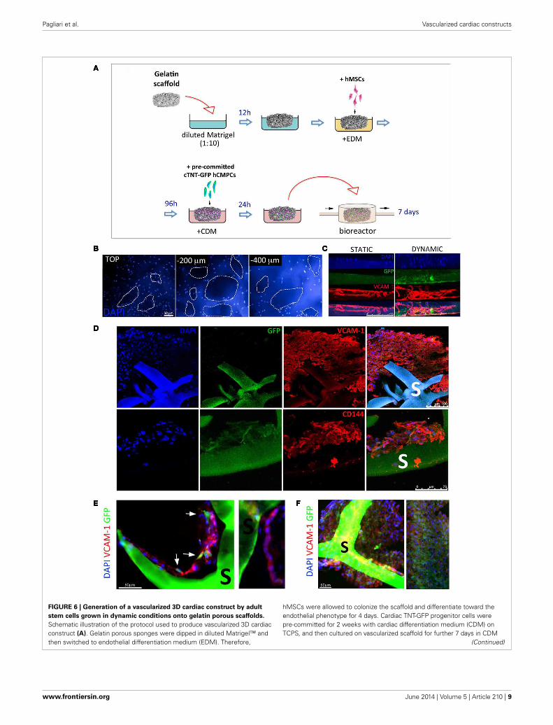
GENERATION OF 3D PRE-VASCULARIZED CARDIAC CONSTRUCTS BYDYNAMIC CULTURE AND POROUS SCAFFOLDSGiven the positive effect of dynamic culture conditions on car-diac gene activation, we sought to obtain vascularized cardiac3D tissues by co-culturing hMSCs-derived vascular cells andpre-differentiated hCMPCs in the bioreactor system for 7 days,as described in Figure 6A. Gelatin sponges were first seededwith hMSCs under endothelial differentiation conditions. In themeantime, cTNT-GFP hCMPCs seeded on TCPS were challengedwith CDM for 2 weeks, seeded onto hMSCs-loaded scaffoldsand allowed to adhere for 24 h before being exposed to contin-uous medium flow. The analysis of the co-cultured constructsafter 1 week testified massive scaffold colonization by the cells(Figure 6B). The scaffold thickness was not significantly modified
by medium perfusion (data not shown) that affected, instead, cellmigration inside the scaffold. In fact, the side of the colonizedscaffolds directly exposed to flow showed a more uniform andextensive distribution of vascular-like cells (in red) and hCMPC-derived cardiomyocytes (green) inside the scaffold, while thecells were mostly distributed on the scaffold surface in the staticcontrol (Figure 6C). The expression of VCAM-1 and CD144endothelial markers confirmed the presence of endothelial-likecells, while GFP expression testified the persistence of a number ofcardiac-like cells within the bio-construct (Figure 6D). Sectionsfrom the perfusion group showed VCAM-1-positive cells alignedforming tube-like structures around the pores and contactingGFP-positive cells (Figure 6E). The extensive cell distributionwithin the scaffold accounted for the formation of a denselypacked multicellular tissue derived from the two different stemcell types used (Figure 6F).
DISCUSSIONRebuilding functional portions of the myocardium requires thegeneration of bio-substitutes that best recapitulate the struc-ture and function of the healthy myocardium, thus providingnew cardiomyocytes with a functional vascular network, whichmay prevent or reduce pathological decline and improve car-diac function after injury (Simons and Ware, 2003; Kang et al.,2013). Exploiting cell sheet technology, our group and others suc-cessfully achieved the preparation of 3D cardiac tissues in theabsence of scaffolds by different cell types having a cardiac signif-icance (Haraguchi et al., 2006; Forte et al., 2011; Matsuura et al.,2012; Sekine et al., 2013). Nonetheless, the scaling up of solidengineered tissues to obtain a critical size substitute with thera-peutical relevance is limited by the diffusion of oxygen, nutrientsand waste products to and from the inner portion of the con-struct, impairing the survival of the newly formed tissue. In this
Frontiers in Physiology | Striated Muscle Physiology June 2014 | Volume 5 | Article 210 | 6
Pagliari et al. Vascularized cardiac constructs
FIGURE 4 | Three-dimensional (3D) gelatin scaffolds support
multilineage stem cell commitment. After 2 weeks of cardiacpre-commitment on TCPS, human cardiac progenitors (hCMPCs), seeded on3D gelatin scaffolds and stimulated with cardiogenic differentiation medium(CDM) for further 1 week, up-regulate some cardiac-specific genes, as
compared to 3D culture in basal medium (A). 3D differentiation conditionsshow a higher performance (B) as compared to tissue culture polystyrene(TCPS). The interconnected porosity of the scaffold (as outlined by yellowdashed line and asterisks) is not modified by Matrigel™ treatment and can
(Continued)
www.frontiersin.org June 2014 | Volume 5 | Article 210 | 7
Pagliari et al. Vascularized cardiac constructs
FIGURE 4 | Continued
accommodate human cells (C). Human mesenchymal stem cells (hMSCs) aredifferentiated to endothelial cells on 3D gelatin scaffolds when stimulatedwith endothelial differentiation medium (EDM) and low concentration ofMatrigel™, as demonstrated by the upregulation of several vascular genes as
analyzed by real time PCR (D). Calcein AM staining shows the network ofliving cells spreading throughout the scaffold (E, upper panels). Scaffold (S)pores are covered by lining cells expressing VCAM-1 as highlighted by whitearrowheads (E, lower panel). A magnification of a cord-like structure alignedalong a pore is visible on the right; scale bar: 100 μm.
FIGURE 5 | Dynamic culture conditions induce a superior cardiac
commitment in human cardiac progenitors (hCMPCs). Dynamic cultureconditions provide a higher, although not significant, increase in scaffold cellcolonization as compared to static conditions (A). Percentage ofdifferentiated cTNT-GFP CMPCs stimulated with cardiogenic differentiationmedium (CDM) and cultured on TCPS and scaffolds, under dynamic, orstatic conditions, as evidenced by FACS analysis (B) (∗, ∗∗, ∗∗∗P < 0.05between hCMPC basal medium and CDM). Importantly, a significantcommitment can be induced, equivalently, by 3D culture itself in both staticand dynamic conditions compared to TCPS (+P < 0.05 between TCPS and3D scaffolds). Nonetheless, quantitative PCR analysis demonstrates theup-regulation of few cardiac-specific genes in hCMPCs (C) cultured indynamic conditions, as compared to its static control (for a complete list ofgenes analyzed please refer to Supplementary Table 1; among the genesanalyzed, those not showing a consistent and significant difference in theexpression as compared to their respective controls, are not shown).
context, the use of porous scaffolds to generate in vitro artifi-cial bio-substitutes gives the opportunity to choose the shapeand the size of the graft, while second-generation scaffolds arecurrently proposed as active templates which can be removedin a timely fashion compatible with tissue growth and exert aninstructive role in the formation of the new tissue (Forte et al.,2013; Pagliari et al., 2013). In the present study, we proposea multistep procedure to prepare adult stem cell-derived pre-vascularized thick cardiac patches by using 3D porous gelatinscaffolds and dynamic culture conditions. The procedure hereadopted is based on the peculiar differentiation potential of twodifferent autologous adult cell subsets, mesenchymal, and car-diac resident stem cells. Human MSCs can be routinely obtainedfrom patient bone marrow and display a minimal, if any, abil-ity to give raise to cardiomyocytes (Gnecchi et al., 2005; Loffredoet al., 2011), while they are supposed to retain an intrinsic vascularpotential (Oswald et al., 2004). In the present investigation,
hMSCs were efficiently driven to acquire an endothelial pheno-type by means of EDM in 3D culture conditions. In particular,when grown on 3D porous gelatin scaffold, hMSCs showed arobust vascular commitment as confirmed by the up-regulationof 73 genes involved in angiogenesis. Among these, angiopoietin1 (ANGPT1), which has been shown to promote blood vesselmaturation by regulating endothelial cell survival and the recruit-ment of mural cells (Thomas and Augustin, 2009), encountereda 20-fold increase, thus suggesting stability of the newly formedvascular network. At the same time, CPCs can be extracted frompatient atrial biopsies and are known to possess the potential togenerate new cardiomyocytes in vitro and in vivo (Beltrami et al.,2003; Goumans et al., 2007) making them an effective and safecardiogenic cell type (Bolli et al., 2011; Makkar et al., 2012). Inthe present investigation, we explored the possibility to generatea 3D cardiac tissue in vitro by pre-committed hCMPCs. Althoughmost of the attempts to regenerate damaged myocardium haveused undifferentiated cells, concerns have been raised due tothe poor differentiation capability of adult progenitors in vivo,together with the potential of pluripotent stem cells to give birthto undesired phenotypes. Recently, it has been suggested that thetransplantation of pre-committed cells could improve the clinicaloutcome in cardiac patients (Heng et al., 2004; Mehta and Shim,2013). For this reason, stem/progenitor cells were appropriatelystimulated to undergo tissue specific pre-commitment beforebeing used for all co-culture experiments. However, given thatthe mechanical properties of the gelatin scaffolds used here aresimilar to those of cardiac tissue, a spontaneous commitment ofcardiac progenitor cells could be obtained through 3D culture perse, as demonstrated by the increased percentage of GFP-positivecells and similarly to what we and others previously demonstrated(Engler et al., 2006; Pagliari et al., 2011; Gaetani et al., 2012;Mosqueira et al., 2014). However, despite the fact that the geneexpression profile analysis confirmed a moderate commitmentof hCMPCs induced by the scaffold itself, biological factors wereneeded to improve this effect. In particular, when cells seeded onthe scaffold were stimulated with CDM, we observed an increasedexpression of cardiac troponin genes (TNNT2, 3) along with theup-regulation of ryanodine receptor 2 (RYR2, involved in muscleexcitation-contraction coupling, Bround et al., 2012) and natri-uretic peptide precursor A (NPPA) genes. These data suggest thatthe combination of 3D porous gelatin and CDM represents a suit-able combination to provide cells with biological and mechanicalsignals synergistically supporting the commitment of hCMCPsat a later stage. Nonetheless, neither complete sarcomeric struc-tures nor any beating activity could be noticed in prompted cells.This evidence suggested that the system implemented representsa good tool to induce an efficient cardiac commitment and studythe behavior of late cardiac progenitors, but this also implicatesthat more complex devices providing stretching and/or electrical
Frontiers in Physiology | Striated Muscle Physiology June 2014 | Volume 5 | Article 210 | 8
Pagliari et al. Vascularized cardiac constructs
FIGURE 6 | Generation of a vascularized 3D cardiac construct by adult
stem cells grown in dynamic conditions onto gelatin porous scaffolds.
Schematic illustration of the protocol used to produce vascularized 3D cardiacconstruct (A). Gelatin porous sponges were dipped in diluted Matrigel™ andthen switched to endothelial differentiation medium (EDM). Therefore,
hMSCs were allowed to colonize the scaffold and differentiate toward theendothelial phenotype for 4 days. Cardiac TNT-GFP progenitor cells werepre-committed for 2 weeks with cardiac differentiation medium (CDM) onTCPS, and then cultured on vascularized scaffold for further 7 days in CDM
(Continued)
www.frontiersin.org June 2014 | Volume 5 | Article 210 | 9
Pagliari et al. Vascularized cardiac constructs
FIGURE 6 | Continued
and in a perfusive modular bioreactor. Human cells colonize the scaffold innerlayers as shown by nuclei staining at different depths (B). Side view ofcellularized scaffolds cultured under static or dynamic conditions (C); infiltrationof GFP- (cardiomyocyte-like cells, green) and VCAM-1-positive cells
(endothelial-like cells, red) into scaffold is improved by dynamic culture.Immunohistochemistry analysis of the colonized scaffolds shows the massiveinfiltration of VCAM-1, CD144 cells in the core of the construct. Highermagnification images (D) shows the VCAM-1-positive cells aligned in tube-likestructures around the pores and contacting GFP-positive cells (E,F). S, scaffold.
stimulation are probably necessary to deliver more specific stim-uli and improve the outcome in terms of cardiac differentiation.The activation of genes involved in matrix remodeling observedin 3D testified the ability of cells to actively interact with thenatural support they grow on. In fact, while the up-regulationof Matrix metalloproteinase (MMP) genes and their regulators,tissue inhibitors of metalloproteinase (TIMP), is predictive ofgelatin remodeling and breakdown, the activation of genes encod-ing for different collagens and laminins suggest the concomitantsubstitution of the artificial matrix with neo-formed tissue.
Bioreactors have been widely proposed to intensify oxygenand nutrient transport and increase the viability and function ofthick cardiovascular tissue-engineered substitutes (Zimmermannet al., 2006; Cimetta et al., 2007; Burdick and Vunjak-Novakovic,2009). In our experimental setting, dynamic culture conditionspositively affected cardiac commitment, leading to a significantincrease in GFP-expressing cells as compared to TCPS and inde-pendently of biological stimulation. Since the increase in GFP wasnot significantly different from that observed under 3D static cul-ture conditions, it might be concluded that providing cells withcontinuous medium flow does not positively affect on cardiacprogenitor differentiation potential on the scaffold. Nevertheless,the expression of genes recognized to be essential for the for-mation or regulation of the contractile apparatus was positivelyinfluenced when dynamic flow was applied to cells grown in a 3Denvironment. Hence, the use of a modular bioreactor providingcontinuous media turnover to the scaffold resulted in a superiorperformance in terms of cardiac gene activation. In the light ofthese promising results, we also expected that direct perfusionof cells could promote the formation of a vascularized cardiacpatch. In line with previous descriptions (Maidhof et al., 2012),we observed more uniform and dense spatial cell distributionin the scaffold, with cells migrating toward the substrate innercore under dynamic conditions. Thus we established a methodfor sequential cell seeding: gelatin scaffolds were colonized withhMSCs and maintained in static conditions for 4 days in order tofavor the endothelialization of the substrate, then scaffolds wereloaded with pre-committed GFP-positive cardiac progenitors andcultured in perfusion bioreactor for 1 week in the presence ofcardiogenic medium. As a result, a 3D cardiac proto-tissue com-posed of densely packed cardiomyocyte-like cells intertwinedwith vessel-like structures was obtained. Although being promis-ing in terms of cell colonization and survival, these co-cultureconditions did not yield the formation of functional contractileand vascular structures. Thus additional experiments will be nec-essary to provide cells with other cues in vitro to complete theorganization of a proper vascularized cardiac tissue.
Nonetheless, irrespective of the stem cell subsets used, theprocedure here described provides a novel platform for thepreparation of complex 3D vascularized bio-constructs to be used
for in vitro studies aiming at the understanding of stem cell behav-ior in a more physiological context. Pre-clinical animal studiesaddressing the long-term in vivo survival and the relevance of theconstructs will be necessary before this technique may represent asuitable platform for future clinical applications.
ACKNOWLEDGMENTSThe present work was supported by the Japan Society for thePromotion of Science (JSPS) through the “Funding Programfor World-Leading Innovative R&D on Science and Technology(FIRST Program)” and the World Premier International (WPI)Research Center Initiative. Stefania Pagliari was supportedby JSPS fellowship; Marie-Josè Goumans was supported byNetherlands Institute for Regenerative Medicine (NIRM) andGiancarlo Forte by the European Regional Development Fund—Project FNUSA-ICRC (No. CZ.1.05/1.1.00/02.0123).
SUPPLEMENTARY MATERIALThe Supplementary Material for this article can be found onlineat: http://www.frontiersin.org/journal/10.3389/fphys.2014.
00210/abstract
REFERENCESAbramoff, M. D., Magalhaes, P. J., and Ram, S. J. (2004). Image processing with
ImageJ. Biophoton. Int. 11, 36–42.Akhyari, P., Fedak, P. W., Weisel, R. D., Lee, T. Y., Verma, S., Mickle, D. A.,
et al. (2002). Mechanical stretch regimen enhances the formation of bio-engineered autologous cardiac muscle grafts. Circulation 106, I137–I142. doi:10.1161/01.cir.0000032893.55215.fc
Bai, K., Huang, Y., Jia, X., Fan, Y., and Wang, W. (2010). Endotheliumoriented differentiation of bone marrow mesenchymal stem cells underchemical and mechanical stimulations. J. Biomech. 43, 1176–1181. doi:10.1016/j.jbiomech.2009.11.030
Beltrami, A. P., Barlucchi, L., Torella, D., Baker, M., Limana, F., Chimenti,S., et al. (2003). Adult cardiac stem cells are multipotent and supportmyocardial regeneration. Cell 114, 763–776. doi: 10.1016/S0092-8674(03)00687-1
Bolli, R., Chugh, A. R., D’Amario, D., Loughran, J. H., Stoddard, M. F., Ikram,S., et al. (2011). Cardiac stem cells in patients with ischaemic cardiomyopathy(SCIPIO): initial results of a randomised phase 1 trial. Lancet 378, 1847–1857.doi: 10.1016/S0140-6736(11)61590-0
Boschetti, F., Raimondi, M. T., Migliavacca, F., and Dubini, G. (2006).Prediction of the micro-fluid dynamic environment imposed to three-dimensional engineered cell systems in bioreactors. J. Biomech. 39, 418–425. doi:10.1016/j.jbiomech.2004.12.022
Brannon-Peppas, L., and Peppas, N. A. (1990). Dynamic and equilibrium swellingbehaviour of pH-sensitive hydrogels containing 2-hydroxyethyl methacrylate.Biomaterials 11, 635–644.
Bround, M. J., Asghari, P., Wambolt, R. B., Bohunek, L., Smits, C., Philit, M., et al.(2012). Cardiac ryanodine receptors control heart rate and rhythmicity in adultmice. Cardiovasc. Res. 96, 372–380. doi: 10.1093/cvr/cvs260
Burdick, J. A., and Vunjak-Novakovic, G. (2009). Engineered microenvironmentsfor controlled stem cell differentiation. Tissue Eng. Part A 15, 205–219. doi:10.1089/ten.tea.2008.0131
Caspi, O., Lesman, A., Basevitch, Y., Gepstein, A., Arbel, G., Habib, I. H.,et al. (2007). Tissue engineering of vascularized cardiac muscle from human
Frontiers in Physiology | Striated Muscle Physiology June 2014 | Volume 5 | Article 210 | 10
Pagliari et al. Vascularized cardiac constructs
embryonic stem cells. Circ. Res. 100, 263–272. doi: 10.1161/01.RES.0000257776.05673.ff
Chiu, L. L., and Radisic, M. (2010). Scaffolds with covalently immobilized VEGFand angiopoietin-1 for vascularization of engineered tissues. Biomaterials 31,226–241. doi: 10.1016/j.biomaterials.2009.09.039
Cimetta, E., Flaibani, M., Mella, M., Serena, E., Boldrin, L., De Coppi, P., et al.(2007). Enhancement of viability of muscle precursor cells on 3D scaffold in aperfusion bioreactor. Int. J. Artif. Organs 30, 415–428.
Cipolleschi, M. G., Dello Sbarba, P., and Olivotto, M. (1993). The role of hypoxiain the maintenance of hematopoietic stem cells. Blood 82, 2031–2037.
Dreesmann, L., Ahlers, M., and Schlosshauera, B. (2007). The pro-angiogenic char-acteristics of a cross-linked gelatin matrix. Biomaterials 28, 5536–5543. doi:10.1016/j.biomaterials.2007.08.040
Dubois, C., Liu, X., Claus, P., Marsboom, G., Pokreisz, P., Vandenwijngaert, S.,et al. (2010). Differential effects of progenitor cell populations on left ventricu-lar remodeling and myocardial neovascularization after myocardial infarction.J. Am. Coll. Cardiol. 55, 2232–2243. doi: 10.1016/j.jacc.2009.10.081
Dvir, T., Kedem, A., Ruvinov, E., Levy, O., Freeman, I., Landa, N., et al.(2009). Prevascularization of cardiac patch on the omentum improves itstherapeutic outcome. Proc. Natl. Acad. Sci. U.S.A. 106, 14990–14995. doi:10.1073/pnas.0812242106
Engler, A. J., Rehfeldt, F., Sen, S., and Discher, D. E. (2007). Microtissue elasticity:measurements by atomic force microscopy and its influence on cell differentia-tion. Methods Cell Biol. 83, 521–545. doi: 10.1016/S0091-679X(07)83022-6
Engler, A. J., Sen, S., Sweeney, H. L., and Discher, D. E. (2006). Matrixelasticity directs stem cell lineage specification. Cell 126, 677–689. doi:10.1016/j.cell.2006.06.044
Forte, G., Pagliari, S., Pagliari, F., Ebara, M., Di Nardo, P., and Aoyagi, T. (2013).Towards the generation of patient-specific patches for cardiac repair. Stem CellRev. 9, 313–325. doi: 10.1007/s12015-011-9325-8
Forte, G., Pietronave, S., Nardone, G., Zamperone, A., Magnani, E., Pagliari,S., et al. (2011). Human cardiac progenitor cell grafts as unrestricted sourceof super-numerary cardiac cells in healthy murine hearts. Stem Cells 29,2051–2061. doi: 10.1002/stem.763
Gaetani, R., Doevendans, P. A., Metz, C. H., Alblas, J., Messina, E., Giacomello,A., et al. (2012). Cardiac tissue engineering using tissue printing technol-ogy and human cardiac progenitor cells. Biomaterials 33, 1782–1790. doi:10.1016/j.biomaterials.2011.11.003
Gnecchi, M., He, H., Liang, O. D., Melo, L. G., Morello, F., Mu, H., et al.(2005). Paracrine action accounts for marked protection of ischemic heartby Akt-modified mesenchymal stem cells. Nat. Med. 11, 367–368. doi:10.1038/nm0405-367
Gnecchi, M., Zhang, Z., Ni, A., and Dzau, V. J. (2008). Paracrine mechanismsin adult stem cell signaling and therapy. Circ. Res. 103, 1204–1219. doi:10.1161/CIRCRESAHA.108.176826
Goumans, M. J., de Boer, T. P., Smits, A. M., van Laake, L. W., van Vliet, P., Metz,C. H., et al. (2007). TGF-beta1 induces efficient differentiation of human car-diomyocyte progenitor cells into functional cardiomyocytes in vitro. Stem CellRes. 1, 138–149. doi: 10.1016/j.scr.2008.02.003
Haraguchi, Y., Shimizu, T., Yamato, M., Kikuchi, A., and Okano, T.(2006). Electrical coupling of cardiomyocyte sheets occurs rapidly viafunctional gap junction formation. Biomaterials 27, 4765–4774. doi:10.1016/j.biomaterials.2006.04.034
Heng, B. C., Haider, H. Kh., Sim, E. K., Cao, T., and Ng, S. C. (2004).Strategies for directing the differentiation of stem cells into the cardiomyo-genic lineage in vitro. Cardiovasc. Res. 62, 34–42. doi: 10.1016/j.cardiores.2003.12.022
Jazayeri, M., Allameh, A., Soleimani, M., Jazayeri, S. H., Piryaei, A., andKazemnejad, S. (2008). Molecular and ultrastructural characterization ofendothelial cells differentiated from human bone marrow mesenchymal stemcells. Cell Biol. Int. 32, 1183–1192. doi: 10.1016/j.cellbi.2008.07.020
Kang, K. T., Coggins, M., Xiao, C., Rosenzweig, A., and Bischoff, J. (2013). Humanvasculogenic cells form functional blood vessels and mitigate adverse remod-eling after ischemia reperfusion injury in rats. Angiogenesis 16, 773–784. doi:10.1007/s10456-013-9354-9
Lalu, M. M., McIntyre, L., Pugliese, C., Fergusson, D., Winston, B. W., Marshall,J. C., et al. (2012). Safety of cell therapy with mesenchymal stromal cells(SafeCell): a systematic review and meta-analysis of clinical trials. PLoS ONE7:e47559. doi: 10.1371/journal.pone.0047559
Levenberg, S., Rouwkema, J., Macdonald, M., Garfein, E. S., Kohane, D. S.,Darland, D. C., et al. (2005). Engineering vascularized skeletal muscle tissue.Nat. Biotechnol. 23, 879–884. doi: 10.1038/nbt1109
Lian, F., Xue, S., Gu, P., and Zhu, H. S. (2008). The long-term effect ofautologous endothelial progenitor cells from peripheral blood implantationon infarcted myocardial contractile force. J. Int. Med. Res. 36, 40–46. doi:10.1177/147323000803600106
Lien, S. M., Ko, L. Y., and Huang, T. J. (2009). Effect of pore size and ECM secretionand cell growth in gelatin scaffold for articular cartilage tissue engineering. ActaBiomater. 5, 670–679. doi: 10.1016/j.actbio.2008.09.020
Loffredo, F. S., Steinhauser, M. L., Gannon, J., and Lee, R. T. (2011). Bone marrow-derived cell therapy stimulates endogenous cardiomyocyte progenitors andpromotes cardiac repair. Cell Stem Cell 8, 389–398. doi: 10.1016/j.stem.2011.02.002
Lovett, M., Lee, K., Edwards, A., and Kaplan, D. L. (2009). Vascularizationstrategies for tissue engineering. Tissue Eng. Part B Rev. 15, 353–370. doi:10.1089/ten.TEB.2009.0085
Lozito, T. P., Kuo, C. K., Taboas, J. M., and Tuan, R. S. (2009). Human mesenchymalstem cells express vascular cell phenotypes upon interaction with endothelialcell matrix. J. Cell. Biochem. 107, 714–722. doi: 10.1002/jcb.22167
Maidhof, R., Tandon, N., Lee, E. J., Luo, J., Duan, Y., Yeager, K., et al. (2012).Biomimetic perfusion and electrical stimulation applied in concert improvedthe assembly of engineered cardiac tissue. J. Tissue Eng. Regen. Med. 6, e12–e23.doi: 10.1002/term.525
Makkar, R. R., Smith, R. R., Cheng, K., Malliaras, K., Thomson, L. E., Berman, D.,et al. (2012). Intracoronary cardiosphere-derived cells for heart regenerationafter myocardial infarction (CADUCEUS): a prospective, randomised phase 1trial. Lancet 379, 895–904. doi: 10.1016/S0140-6736(12)60195-0
Martucci, J. F., Ruseckaite, R. A., and Vàzquez, A. (2006). Creep of glutaraldehyde-crosslinked gelatin films. Mater. Sci. Eng. A 435–436, 681–686. doi:10.1016/j.msea.2006.07.097
Matsuura, K., Wada, M., Konishi, K., Sato, M., Iwamoto, U., Sato, Y., et al. (2012).Fabrication of mouse embryonic stem cell-derived layered cardiac cell sheetsusing a bioreactor culture system. PLoS ONE 7:e52176. doi: 10.1371/jour-nal.pone.0052176
Mazzei, D., Guzzardi, M. A., Giusti, S., and Ahluwalia, A. (2010). A low shear stressmodular bioreactor for connected cell culture under high flow rates. Biotechnol.Bioeng. 106, 127–137. doi: 10.1002/bit.22671
Mehta, A., and Shim, W. (2013). Cardiac stem cell therapy: stemness or commit-ment? Cell Transplant. 22, 1–14. doi: 10.3727/096368912X653282
Menasche, P. (2011). Cardiac cell therapy: lessons from clinical trials. J. Mol. Cell.Cardiol. 50, 258–265. doi: 10.1016/j.yjmcc.2010.06.010
Meyer, G. P., Wollert, K. C., Lotz, J., Pirr, J., Rager, U., Lippolt, P., et al. (2009).Intracoronary bone marrow cell transfer after myocardial infarction: 5-yearfollow-up from the randomized-controlled BOOST trial. Eur. Heart J. 30,2978–2984. doi: 10.1093/eurheartj/ehp374
Mosqueira, D., Pagliari, S., Uto, K., Ebara, M., Romanazzo, S., Escobedo-Lucea, C.,et al. (2014). Hippo pathway effectors control cardiac progenitor cell fate byacting as dynamic sensors of substrate mechanics and nanostructure. ACS Nano8, 2033–2047. doi: 10.1021/nn4058984
Muraglia, A., Cancedda, R., and Quarto, R. (2000). Clonal mesenchymal progeni-tors from human bone marrow differentiate in vitro according to a hierarchicalmodel. J. Cell Sci. 113, 1161–1166.
Mwangi, J. W., and Ofner, C. M. 3rd (2004). Crosslinked gelatin matrices: releaseof a random coil macromolecular solute. Int. J. Pharm. 278, 319–327. doi:10.1016/j.ijpharm.2004.03.024
Oswald, J., Boxberger, S., Jorgensen, B., Feldmann, S., Ehninger, G., Bornhauser,M., et al. (2004). Mesenchymal stem cells can be differentiated into endothelialcells in vitro. Stem Cells 22, 377–384. doi: 10.1634/stemcells.22-3-377
Pagliari, S., Romanazzo, S., Mosqueira, D., Pinto-do-Ó, P., Aoyagi, T., andForte, G. (2013). Adult stem cells and biocompatible scaffolds as powerfuldrug delivery tools for cardiac repair. Curr. Med. Chem. 20, 3429–3447. doi:10.2174/09298673113209990032
Pagliari, S., Vilela-Silva, A. C., Forte, G., Pagliari, F., Mandoli, C., Vozzi, G., et al.(2011). Cooperation of biological and mechanical signals in cardiac progenitorcell differentiation. Adv. Mater. 23, 514–518. doi: 10.1002/adma.201003479
Pampaloni, F., Reynaud, E. G., and, Stelzer, E. H. (2007). The third dimensionbridges between cell culture and live tissue. Nat. Rev. Mol. Cell Biol. 8, 839–845.doi: 10.1038/nrm2236
www.frontiersin.org June 2014 | Volume 5 | Article 210 | 11
Pagliari et al. Vascularized cardiac constructs
Pittenger, M. F., Mackay, A. M., Beck, S. C., Jaiswal, R. K., Douglas, R., Mosca, J. D.,et al. (1999). Multilineage potential of adult human mesenchymal stem cells.Science 284,143–147. doi: 10.1126/science.284.5411.143
Portalska, K. J., Leferink, A., Groen, N., Fernandes, H., Moroni, L., van Blitterswijk,C., et al. (2012). Endothelial differentiation of mesenchymal stromal cells. PLoSONE 7:e46842. doi: 10.1371/journal.pone.0046842
Sakaguchi, K., Shimizu, T., Haraguchi, S., Sekine, H., Yamato, M., Umezu, M., et al.(2013). In vitro engineering of vascularized tissue surrogates. Sci. Rep. 3:1316.doi: 10.1038/srep01316
Sakai, T., Li, R. K., Weisel, R. D., Mickle, D. A., Kim, E. T., Jia, Z. Q., et al.(2001). The fate of a tissue-engineered cardiac graft in the right ventricu-lar outflow tract of the rat. J. Thorac. Cardiovasc. Surg. 121, 932–942. doi:10.1067/mtc.2001.113600
Sato, K., Wu, T., Laham, R. J., Johnson, R. B., Douglas, P., Li, J., et al. (2001). Efficacyof intracoronary or intravenous VEGF165 in a pig model of chronic myocar-dial ischemia. J. Am. Coll. Cardiol. 37, 616–623. doi: 10.1016/S0735-1097(00)01144-X
Segers, V. F. M., and Lee, R. T. (2011). Biomaterials to enhance stem cell functionin the heart. Circ. Res. 109, 910–922. doi: 10.1161/CIRCRESAHA.111.249052
Sekine, H., Shimizu, T., Sakaguchi, K., Dobashi, I., Wada, M., Yamato, M.,et al. (2013). In vitro fabrication of functional three-dimensional tis-sues with perfusable blood vessels. Nat. Commun. 4:1399. doi: 10.1038/ncomms2406
Simons, M., and Ware, J. A. (2003). Therapeutic angiogenesis in cardiovasculardisease. Nat. Rev. Drug Discov. 2, 863–871. doi: 10.1038/nrd1226
Simón-Yarza, T., Formiga, F. R., Tamayo, E., Pelacho, B., Prosper, F., and Blanco-Prieto, M. J. (2012). Vascular endothelial growth factor-delivery systems forcardiac repair: an overview. Theranostics 2, 541–552. doi: 10.7150/thno.3682
Singh, S., Wu, B. M., and Dunn, J. C. (2012). Delivery of VEGF using collagen-coated polycaprolactone scaffolds stimulates angiogenesis. J. Biomed. Mater. Res.100, 720–727. doi: 10.1002/jbm.a.34010
Smits, A. M., van Laake, L. W., den Ouden, K., Schreurs, C., Szuhai, K., van Echteld,C. J., et al. (2009a). Human cardiomyocyte progenitor cell transplantation pre-serves long-term function of the infarcted mouse myocardium. Cardiovasc. Res.83, 527–535. doi: 10.1093/cvr/cvp146
Smits, A. M., van Vliet, P., Metz, C. H., Korfage, T., Sluijter, J. P., Doevendans,P. A., et al. (2009b). Human cardiomyocyte progenitor cells differenti-ate into functional mature cardiomyocytes: an in vitro model for studyinghuman cardiac physiology and pathophysiology. Nat. Protoc. 4, 232–243. doi:10.1038/nprot.2008.229
Spinelli, A., Vinci, B., Tirella, A., Matteucci, M., Gargani, L., Ahluwalia, A., et al.(2012). Realization of a poro-elastic ultrasound replica of pulmonary tissue.Biomatter 2, 37–42. doi: 10.4161/biom.19835
Terrovitis, J. V., Smith, R. R., and Marbán, E. (2010). Assessment and optimizationof cell engraftment after transplantation into the heart Circ. Res. 106, 479–494.doi: 10.1161/CIRCRESAHA.109.208991
Thomas, M., and Augustin, H. G. (2009). The role of the Angiopoietins in vascularmorphogenesis. Angiogenesis 12, 125–137. doi: 10.1007/s10456-009-9147-3
Urbich, C., Heeschen, C., Aicher, A., Sasaki, K., Bruhl, T., Farhadi, M. R., et al.(2005). Cathepsin L is required for endothelial progenitor cell-induced neovas-cularization. Nat. Med. 11, 206–213. doi: 10.1038/nm1182
Varum, S., Rodrigues, A. S., Moura, M. B., Momcilovic, O., Easley, C. A. 4th,Ramalho-Santos, J., et al. (2011). Energy metabolism in human pluripotentstem cells and their differentiated counterparts. PLoS ONE 6:e20914. doi:10.1371/journal.pone.0020914
Vozzi, F., Mazzei, D., Vinci, B., Vozzi, G., Sbrana, T., Ricotti, L., et al. (2011). Aflexible bioreactor system for constructing in vitro tissue and organ models.Biotechnol. Bioeng. 108, 2129–2140. doi: 10.1002/bit.23164
Wang, D. M., and Tarbell, J. M. (1995). Modeling interstitial flow in an artery wallallows estimation of wall shear stress on smooth muscle cells. J. Biomech. Eng.117, 358–366.
Wöhrle, J., Merkle, N., Mailänder, V., Nusser, T., Schauwecker, P., von Scheidt, F.,et al. (2010). Results of intracoronary stem cell therapy after acute myocardialinfarction. Am. J. Cardiol. 105, 804–812. doi: 10.1016/j.amjcard.2009.10.060
Wu, S. C., Chang, W. H., Dong, C. G., Chen, K. Y., Chen, Y. S., and Yao, C. H.(2011). Cell adhesion and proliferation enhancement by gelatin nanofiber scaf-folds. J. Bioact. Compat. Polym. 26, 565–577. doi: 10.1177/0883911511423563
Xing, Q., Yates, K., Vogt, C., Qian, Z., Frost, M. C., and Zhao, F. (2014). Increasingmechanical strength of gelatin hydrogels by divalent metal ion removal. Sci. Rep.4:4706. doi: 10.1038/srep04706
Zimmermann, W. H., Melnychenko, I., Wasmeier, G., Didié, M., Naito, H.,Nixdorff, U., et al. (2006). Engineered heart tissue grafts improve systolicand diastolic function in infarcted rat hearts. Nat. Med. 12, 452–458. doi:10.1038/nm1394
Conflict of Interest Statement: The authors declare that the research was con-ducted in the absence of any commercial or financial relationships that could beconstrued as a potential conflict of interest.
Received: 21 March 2014; accepted: 15 May 2014; published online: 03 June 2014.Citation: Pagliari S, Tirella A, Ahluwalia A, Duim S, Goumans M-J, Aoyagi T andForte G (2014) A multistep procedure to prepare pre-vascularized cardiac tissue con-structs using adult stem sells, dynamic cell cultures, and porous scaffolds. Front.Physiol. 5:210. doi: 10.3389/fphys.2014.00210This article was submitted to Striated Muscle Physiology, a section of the journalFrontiers in Physiology.Copyright © 2014 Pagliari, Tirella, Ahluwalia, Duim, Goumans, Aoyagi and Forte.This is an open-access article distributed under the terms of the Creative CommonsAttribution License (CC BY). The use, distribution or reproduction in other forums ispermitted, provided the original author(s) or licensor are credited and that the originalpublication in this journal is cited, in accordance with accepted academic practice. Nouse, distribution or reproduction is permitted which does not comply with these terms.
Frontiers in Physiology | Striated Muscle Physiology June 2014 | Volume 5 | Article 210 | 12


