Embed Size (px)
Citation preview
R
A
SF
a
ARAA
KFOTAR
1
crIoapmahhariirAggrswms
f
0h
Immunology Letters 149 (2013) 101– 109
Contents lists available at SciVerse ScienceDirect
Immunology Letters
jou rn al hom epage: www.elsev ier .com/ locate / immlet
eview
molecular insight of CTLA-4 in food allergy
andeep Kumar, Alok Kumar Verma, Mukul Das, Premendra D. Dwivedi ∗
ood, Drug and Chemical Toxicology Group, CSIR-Indian Institute of Toxicology Research (CSIR-IITR), MG Marg Post Box No. 80, Lucknow 226001, India
r t i c l e i n f o
rticle history:eceived 13 October 2012ccepted 6 December 2012
a b s t r a c t
Food allergy is an immune provocation induced by certain food in susceptible individuals. Most of the foodallergic manifestations are evident in the individual having impaired oral tolerance. In spite of worldwideprevalence, there is no permanent cure of food allergy. Food allergic reactions are complex immunological
vailable online xxx
eywords:ood allergyral toleranceh2 reactionsnaphylaxis
events that comprises of several immune molecules like IgE, IL-4, IL-13 and T-cells, therefore, researchersare trying to pick the correct molecule to find out pivotal therapeutic solutions. Being a key regulatorymolecule in suppressing T-cells functional activities, cytotoxic T-cell lymphocyte antigen-4 (CTLA-4)or cluster of differentiation-152 (CD-152) has contributed a novel and revolutionary dimension towardtherapeutic research of several diseases. This review focuses on different immunological and mechanisticperspectives of CTLA-4 in correlation with food allergy.
egulatory T cells
. Introduction
Food allergic reactions are immunological disorders induced byertain food(s) in susceptible individuals. The prevalence of sucheactions is encompassing several countries throughout the world.n western countries, the prevalence of food allergy is reported in 5%f children and 3–4% of adult’s populations [1]. The susceptibility ofny individual is determined by several factors, including geneticredisposition, dietary habits and environmental set up. But, theost appropriate answer behind the cause of food allergy is still
waited. In the susceptible individuals the impaired oral toleranceas been reported to play very important role [2]. These factorsave been found to involve in induction of impaired oral tolerancend allergic sensitization to foods. The oral tolerance is a natu-al weapon against dietary antigens which suppress the antigennduced allergic reactions. Most of the food allergic reactions arenduced by IgE mediated immediate type hypersensitivity or type Ieaction, which occur within minutes to hour after food intake [3].long with type I reaction, late phase and non IgE mediated aller-ic reactions have also been reported in food allergy [4]. Due toenetic link up food allergic reactions are also referred as atopiceaction e.g., atopic dermatitis caused by egg, wheat, milk, and
oy [5]. The health complications induced by allergic foods have aide range including allergic rhinitis, bronchial asthma, atopic der-atitis and gastrointestinal disorders induced by foods like peanut,oybeans, kidney beans, red grams, green grams, eggs, and fishes,
∗ Corresponding author. Tel.: +91 522 2620107/2620106/2616191;ax: +91 522 2628227.
E-mail addresses: [email protected], [email protected] (P.D. Dwivedi).
165-2478/$ – see front matter © 2012 Elsevier B.V. All rights reserved.ttp://dx.doi.org/10.1016/j.imlet.2012.12.003
© 2012 Elsevier B.V. All rights reserved.
oral allergy syndrome (OAS) induced by cherry and other foods,eosinophilic esophagitis induced corn, soy, and wheat, edema ofthe uvula induced by seafood, by hazelnuts, and walnuts. Recurrentaphthous stomatitis (RAS) is one of the most common oral lesionsinduced by milk, gluten, and other allergens, food protein-inducedenterocolitis syndrome (FPIES) induced by rice and neonatal diar-rhea induced by milk, soy, and grains [6]. Some time the allergicreactions occur with a high severity which may lead to a fatal out-come. These reactions are known as anaphylaxis reactions. In USA,approximately 30,000 patients come to emergency ward of the hos-pitals for treatment of food induced anaphylaxis. Further, about 200food allergic patients have been reported to die due to aforesaidsymptoms [7].
Food allergy is a major and increasing medical problem; how-ever, our understanding of the immunological mechanisms of thedisease is limited. Given the significant prevalence of food allergiesin the recent years, in western and developing countries, ther-apeutic alternative to exclusion diets are strongly required. Theresearchers in this regimen are trying hard to find an effective andsafer therapeutic option. Recent years have been very productive infoods allergic researches including immunopathogenesis of manyallergic disorders, epitope mapping and its therapeutic exploita-tions, anti-IgE therapy and allergen-specific immunotherapy. But,still the several aspects are lacking of any pivotal therapeutic toolto combat food allergy completely. To date, avoidance of the foodcausing allergic manifestation in susceptible individuals is most
common suggestion by clinicians. Some molecules like anti IgE, IL-4, and regulatory cells (Treg) have been considered as a potentialtool for allergy treatment induced by food, but still such studies arenot in their fruitful shape. In this review, an attempt has been madeto highlight the functional aspects of cytotoxic T-cell lymphocyte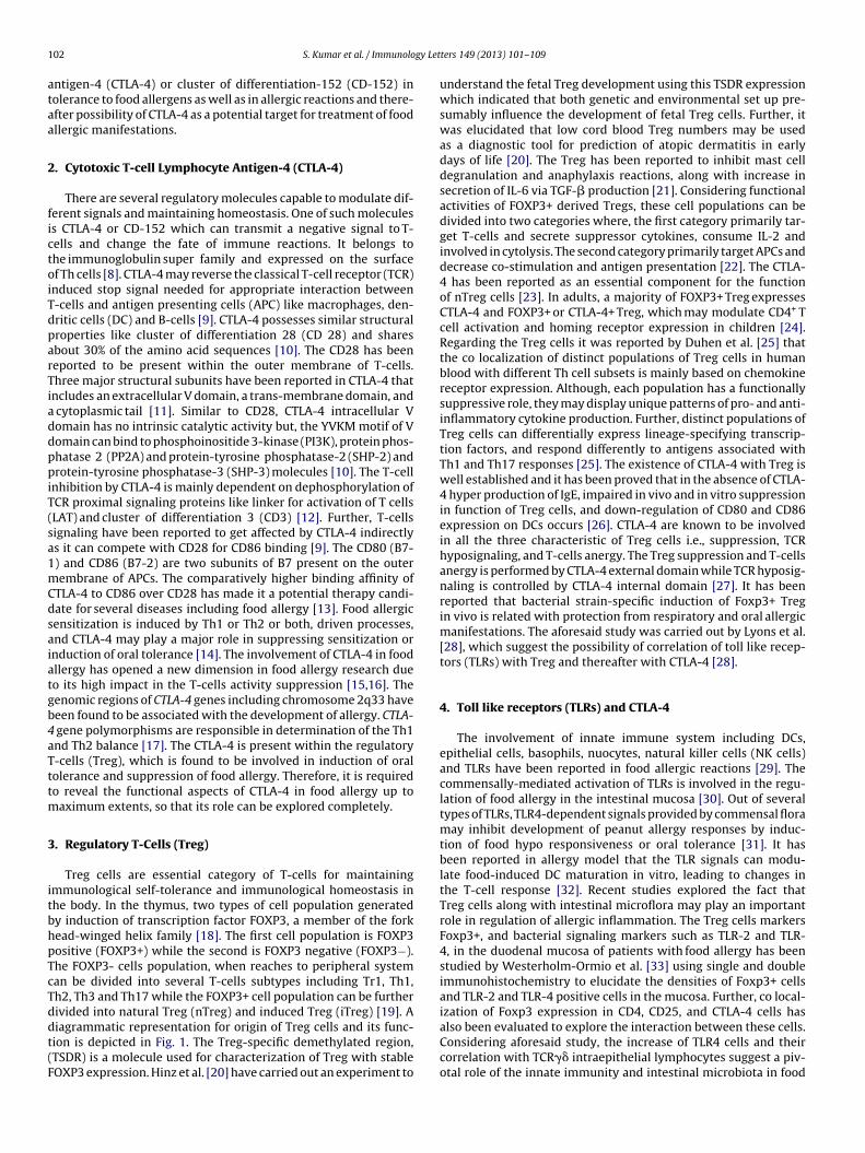
1 gy Let
ataa
2
fictoiTdparTiaddppiT(sa1mCdsaiatgb4aTttm
3
itbhpTcTddt(F
02 S. Kumar et al. / Immunolo
ntigen-4 (CTLA-4) or cluster of differentiation-152 (CD-152) inolerance to food allergens as well as in allergic reactions and there-fter possibility of CTLA-4 as a potential target for treatment of foodllergic manifestations.
. Cytotoxic T-cell Lymphocyte Antigen-4 (CTLA-4)
There are several regulatory molecules capable to modulate dif-erent signals and maintaining homeostasis. One of such moleculess CTLA-4 or CD-152 which can transmit a negative signal to T-ells and change the fate of immune reactions. It belongs tohe immunoglobulin super family and expressed on the surfacef Th cells [8]. CTLA-4 may reverse the classical T-cell receptor (TCR)nduced stop signal needed for appropriate interaction between-cells and antigen presenting cells (APC) like macrophages, den-ritic cells (DC) and B-cells [9]. CTLA-4 possesses similar structuralroperties like cluster of differentiation 28 (CD 28) and sharesbout 30% of the amino acid sequences [10]. The CD28 has beeneported to be present within the outer membrane of T-cells.hree major structural subunits have been reported in CTLA-4 thatncludes an extracellular V domain, a trans-membrane domain, and
cytoplasmic tail [11]. Similar to CD28, CTLA-4 intracellular Vomain has no intrinsic catalytic activity but, the YVKM motif of Vomain can bind to phosphoinositide 3-kinase (PI3K), protein phos-hatase 2 (PP2A) and protein-tyrosine phosphatase-2 (SHP-2) androtein-tyrosine phosphatase-3 (SHP-3) molecules [10]. The T-cell
nhibition by CTLA-4 is mainly dependent on dephosphorylation ofCR proximal signaling proteins like linker for activation of T cellsLAT) and cluster of differentiation 3 (CD3) [12]. Further, T-cellsignaling have been reported to get affected by CTLA-4 indirectlys it can compete with CD28 for CD86 binding [9]. The CD80 (B7-) and CD86 (B7-2) are two subunits of B7 present on the outerembrane of APCs. The comparatively higher binding affinity of
TLA-4 to CD86 over CD28 has made it a potential therapy candi-ate for several diseases including food allergy [13]. Food allergicensitization is induced by Th1 or Th2 or both, driven processes,nd CTLA-4 may play a major role in suppressing sensitization ornduction of oral tolerance [14]. The involvement of CTLA-4 in foodllergy has opened a new dimension in food allergy research dueo its high impact in the T-cells activity suppression [15,16]. Theenomic regions of CTLA-4 genes including chromosome 2q33 haveeen found to be associated with the development of allergy. CTLA-
gene polymorphisms are responsible in determination of the Th1nd Th2 balance [17]. The CTLA-4 is present within the regulatory-cells (Treg), which is found to be involved in induction of oralolerance and suppression of food allergy. Therefore, it is requiredo reveal the functional aspects of CTLA-4 in food allergy up to
aximum extents, so that its role can be explored completely.
. Regulatory T-Cells (Treg)
Treg cells are essential category of T-cells for maintainingmmunological self-tolerance and immunological homeostasis inhe body. In the thymus, two types of cell population generatedy induction of transcription factor FOXP3, a member of the forkead-winged helix family [18]. The first cell population is FOXP3ositive (FOXP3+) while the second is FOXP3 negative (FOXP3−).he FOXP3- cells population, when reaches to peripheral systeman be divided into several T-cells subtypes including Tr1, Th1,h2, Th3 and Th17 while the FOXP3+ cell population can be further
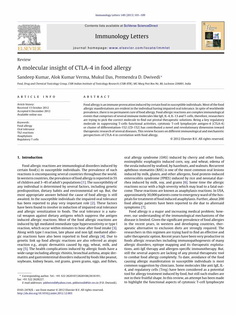
ivided into natural Treg (nTreg) and induced Treg (iTreg) [19]. Aiagrammatic representation for origin of Treg cells and its func-ion is depicted in Fig. 1. The Treg-specific demethylated region,TSDR) is a molecule used for characterization of Treg with stableOXP3 expression. Hinz et al. [20] have carried out an experiment toters 149 (2013) 101– 109
understand the fetal Treg development using this TSDR expressionwhich indicated that both genetic and environmental set up pre-sumably influence the development of fetal Treg cells. Further, itwas elucidated that low cord blood Treg numbers may be usedas a diagnostic tool for prediction of atopic dermatitis in earlydays of life [20]. The Treg has been reported to inhibit mast celldegranulation and anaphylaxis reactions, along with increase insecretion of IL-6 via TGF-� production [21]. Considering functionalactivities of FOXP3+ derived Tregs, these cell populations can bedivided into two categories where, the first category primarily tar-get T-cells and secrete suppressor cytokines, consume IL-2 andinvolved in cytolysis. The second category primarily target APCs anddecrease co-stimulation and antigen presentation [22]. The CTLA-4 has been reported as an essential component for the functionof nTreg cells [23]. In adults, a majority of FOXP3+ Treg expressesCTLA-4 and FOXP3+ or CTLA-4+ Treg, which may modulate CD4+ Tcell activation and homing receptor expression in children [24].Regarding the Treg cells it was reported by Duhen et al. [25] thatthe co localization of distinct populations of Treg cells in humanblood with different Th cell subsets is mainly based on chemokinereceptor expression. Although, each population has a functionallysuppressive role, they may display unique patterns of pro- and anti-inflammatory cytokine production. Further, distinct populations ofTreg cells can differentially express lineage-specifying transcrip-tion factors, and respond differently to antigens associated withTh1 and Th17 responses [25]. The existence of CTLA-4 with Treg iswell established and it has been proved that in the absence of CTLA-4 hyper production of IgE, impaired in vivo and in vitro suppressionin function of Treg cells, and down-regulation of CD80 and CD86expression on DCs occurs [26]. CTLA-4 are known to be involvedin all the three characteristic of Treg cells i.e., suppression, TCRhyposignaling, and T-cells anergy. The Treg suppression and T-cellsanergy is performed by CTLA-4 external domain while TCR hyposig-naling is controlled by CTLA-4 internal domain [27]. It has beenreported that bacterial strain-specific induction of Foxp3+ Tregin vivo is related with protection from respiratory and oral allergicmanifestations. The aforesaid study was carried out by Lyons et al.[28], which suggest the possibility of correlation of toll like recep-tors (TLRs) with Treg and thereafter with CTLA-4 [28].
4. Toll like receptors (TLRs) and CTLA-4
The involvement of innate immune system including DCs,epithelial cells, basophils, nuocytes, natural killer cells (NK cells)and TLRs have been reported in food allergic reactions [29]. Thecommensally-mediated activation of TLRs is involved in the regu-lation of food allergy in the intestinal mucosa [30]. Out of severaltypes of TLRs, TLR4-dependent signals provided by commensal floramay inhibit development of peanut allergy responses by induc-tion of food hypo responsiveness or oral tolerance [31]. It hasbeen reported in allergy model that the TLR signals can modu-late food-induced DC maturation in vitro, leading to changes inthe T-cell response [32]. Recent studies explored the fact thatTreg cells along with intestinal microflora may play an importantrole in regulation of allergic inflammation. The Treg cells markersFoxp3+, and bacterial signaling markers such as TLR-2 and TLR-4, in the duodenal mucosa of patients with food allergy has beenstudied by Westerholm-Ormio et al. [33] using single and doubleimmunohistochemistry to elucidate the densities of Foxp3+ cellsand TLR-2 and TLR-4 positive cells in the mucosa. Further, co local-
ization of Foxp3 expression in CD4, CD25, and CTLA-4 cells hasalso been evaluated to explore the interaction between these cells.Considering aforesaid study, the increase of TLR4 cells and theircorrelation with TCR�� intraepithelial lymphocytes suggest a piv-otal role of the innate immunity and intestinal microbiota in food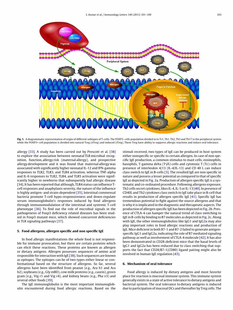
S. Kumar et al. / Immunology Letters 149 (2013) 101– 109 103
F OXP3w iTreg)
atnaarai[cibstppii
5
bcorafahgs
u
ig. 1. A diagrammatic representation of origin of different subtypes of T-cells. The Fhile the FOXP3+ cell population is divided into natural Treg (nTreg) and induced (
llergy [33]. A study has been carried out by Prescott et al. [38]o explore the association between neonatal TLR microbial recog-ition, function, allergy risk (maternal allergy), and prospectivellergy development and it was found that maternal allergy wasssociated with significantly higher neonatal IL-12 and IFN-gammaesponses to TLR2, TLR3, and TLR4 activation, whereas TNF-alphand IL-6 responses to TLR2, TLR4, and TLR5 activation were signif-cantly higher in newborns that subsequently had allergic disease34]. It has been reported that although, TLR4 status can influence T-ell responses and anaphylaxis severity, the nature of the influences highly antigen- and strain-dependent [35]. Intestinal commensalacteria promote T-cell hypo-responsiveness and down-regulateerum immunoglobulin’s responses induced by food allergenshrough immunomodulation of the intestinal and systemic T-cellhenotype [36]. To find out the role of microbial signals in theathogenesis of Foxp3 deficiency related diseases has been stud-
ed in Foxp3 mutant mice, which showed concurrent deficienciesn TLR signaling pathways [37].
. Food allergens, allergen specific and non specific IgE
In food allergic manifestations the whole food is not responsi-le for immune provocation, but there are certain proteins whichan elicit these reactions. These proteins are known as allergenr dietary antigens. Allergen possesses sequences of amino acidesponsible for interaction with IgE [38]. Such sequences are knowns epitopes. The epitopes can be of two types either linear or con-ormational based on the structure of allergens. So far, severalllergens have been identified from peanut (e.g., Ara h1 and Ara
2), soybeans (e.g., Gly mBD), cow milk proteins (e.g., casein), greenram (e.g., Vig r1 and Vig r4), and kidney beans (e.g., Pha v3) andeveral other foods [39].The IgE immunoglobulin is the most important immunoglob-lin encountered during food allergic reactions. Based on the
- cells population divided in to Tr1, Th1, Th2, Th3 and Th17 in the peripheral system. These Treg have ability to suppress allergic reactions and induce oral tolerance.
stimuli received, two types of IgE can be produced in host systemeither nonspecific or specific to certain allergen. In case of non spe-cific IgE production, a common stimulus to mast cells, eosinophils,basophils, T-gamma delta (T��)-cells and cytotoxic T (Tc)-cells inpresence of interleukin 4/13 (IL-4/IL-13) and CD 40 L can induceclass switch to IgE in B-cells [3]. The resulted IgE are non specific innature and possess a lesser potential as compared to that of specificIgE as depicted in Fig. 2a. Production of allergen specific IgE is a sys-tematic and co-ordinated procedure. Following allergens exposure,Th2 cells secret cytokines, likes IL-4, IL-5 or IL-13 [40]. In presence ofCD40L and Th2 cytokines class switch to IgE take place in B-cell thatresults in production of allergen specific IgE [41]. Specific IgE hastremendous potential to fight against the source allergens and thatis why it is implicated in the diagnostic and therapeutic aspects. Theproduction of allergen specific IgE has been depicted in Fig. 2b. Pres-ence of CTLA-4 can hamper the natural trend of class switching toIgE in B-cells by binding to B7 molecules as depicted in Fig. 2c. Alongwith IgE, the other immunoglobulins like IgG1 and IgG2a may alsoplay important roles in food allergic reactions and production ofIgE. Mice deficient in both B7-1 and B7-2 failed to generate antigen-specific IgG1 and IgG2a, indicating the role of B7 mediated signalingpathway as well as involvement of CTLA-4 molecule [42]. It has alsobeen demonstrated in CD28-deficient mice that the basal levels ofIgG1 and IgG2a has been reduced due to class switching that sup-ports the fact that CD28/B7-1(CD80) ligand pairing might also beinvolved in human IgE regulation [43].
6. Mechanism of oral tolerance
Food allergy is induced by dietary antigens and most favoriteplace for reaction is mucosal immune system. This immune systemgenerally exists in a state of active tolerance to dietary antigens andbacterial system. The oral tolerance to dietary antigens is induceddue to participation of mucosal DCs and thereafter by Treg cells. The
104 S. Kumar et al. / Immunology Letters 149 (2013) 101– 109
F can
c ace afI
ptssiptiadfeaaTBiat
ig. 2. A schematic diagram of formation of IgE. (A) Formation of non-specific IgEytotoxic (Tc) and T gamma delta (T��) cells. (B) Formation of specific IgE takes plL-13. (C) CTLA-4 may lead to modulation in class switching to IgE.
revalence of food allergy is related to the condition when immuneolerance is disrupted and a sensitizing immune response by food-pecific IgE production occurs instead [44]. Oral tolerance is thetate of local and systemic immune hypo- or unresponsiveness thats induced by oral administration of innocuous antigen such as foodroteins. These preventive immune mechanisms are very similaro a local process that regulates responses to commensal bacterian the large intestine and, together, mucosally induced toleranceppears to prevent intestinal disorders such as food allergy, celiacisease, and inflammatory bowel diseases [45]. Tolerance inductionor food allergy (TIFA), a preventive method based on the oral tol-rance property has been used as positive and radical treatmentpproach of food allergy. The process of oral tolerance is medi-ted by several immune components including cytokines like IL-10,
GF-� and IFN-� and regulatory cells such as Treg and regulatory-cells (Breg). Now a day, studies are going on to use IFN-�, as anmmunomodulatory biologics for tolerance induction against foodllergy [46]. This tolerance response must be inhibited or bypassedo generate allergic sensitization in experimental food allergy and
be induced by a common stimulus in mast cells, B-cells, eosinophils, basophils, Tter allergen exposure that leads to induction of Th2 cells and secretion of IL-4 and
this has been achieved by three main approaches first, geneticmodifications, second, experimental adjuvants, and third, bypass-ing oral tolerance by administering the antigen through alternativeroutes [47]. The DCs play an essential role in regulating the bal-ance between sensitization and oral tolerance responses to foodallergens in gut mucosa. The DCs can perform these functions bycontrolling T-cells differentiation and activation mediated by CD28and CTLA-4 molecules. The CD28/CTLA-4-B7 signaling pathwayplay influential role during the regulation of T-cell responses [48].
In non susceptible individuals, normal dietary proteins induceoral tolerance in gut mucosa as part of an active suppression ofimmune response induced by dietary antigens or allergens. In sus-ceptible individuals to food allergy, impairment in oral tolerancehas been well studied [49]. After having foods, it moves to stom-
ach, where its protein contents get completely or partially digestedby pepsin. Digested and partially digested proteins move to intes-tine where further degradation of these proteins takes place byintestinal proteolytic enzymes. Some proteins can get digested andabsorbed completely, while some partially degraded proteins can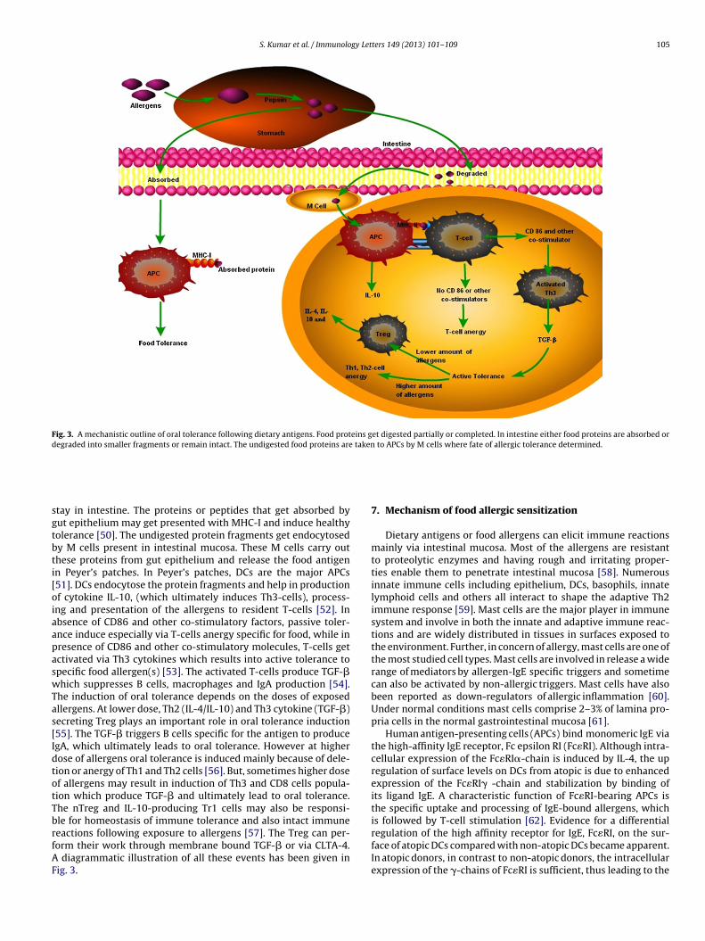
S. Kumar et al. / Immunology Letters 149 (2013) 101– 109 105
Fig. 3. A mechanistic outline of oral tolerance following dietary antigens. Food proteins get digested partially or completed. In intestine either food proteins are absorbed ordegraded into smaller fragments or remain intact. The undigested food proteins are taken to APCs by M cells where fate of allergic tolerance determined.
sgtbti[oiaapaswTas[IdtotTbrfAF
tay in intestine. The proteins or peptides that get absorbed byut epithelium may get presented with MHC-I and induce healthyolerance [50]. The undigested protein fragments get endocytosedy M cells present in intestinal mucosa. These M cells carry outhese proteins from gut epithelium and release the food antigenn Peyer’s patches. In Peyer’s patches, DCs are the major APCs51]. DCs endocytose the protein fragments and help in productionf cytokine IL-10, (which ultimately induces Th3-cells), process-ng and presentation of the allergens to resident T-cells [52]. Inbsence of CD86 and other co-stimulatory factors, passive toler-nce induce especially via T-cells anergy specific for food, while inresence of CD86 and other co-stimulatory molecules, T-cells getctivated via Th3 cytokines which results into active tolerance topecific food allergen(s) [53]. The activated T-cells produce TGF-�hich suppresses B cells, macrophages and IgA production [54].
he induction of oral tolerance depends on the doses of exposedllergens. At lower dose, Th2 (IL-4/IL-10) and Th3 cytokine (TGF-�)ecreting Treg plays an important role in oral tolerance induction55]. The TGF-� triggers B cells specific for the antigen to producegA, which ultimately leads to oral tolerance. However at higherose of allergens oral tolerance is induced mainly because of dele-ion or anergy of Th1 and Th2 cells [56]. But, sometimes higher dosef allergens may result in induction of Th3 and CD8 cells popula-ion which produce TGF-� and ultimately lead to oral tolerance.he nTreg and IL-10-producing Tr1 cells may also be responsi-
le for homeostasis of immune tolerance and also intact immuneeactions following exposure to allergens [57]. The Treg can per-orm their work through membrane bound TGF-� or via CLTA-4.diagrammatic illustration of all these events has been given inig. 3.
7. Mechanism of food allergic sensitization
Dietary antigens or food allergens can elicit immune reactionsmainly via intestinal mucosa. Most of the allergens are resistantto proteolytic enzymes and having rough and irritating proper-ties enable them to penetrate intestinal mucosa [58]. Numerousinnate immune cells including epithelium, DCs, basophils, innatelymphoid cells and others all interact to shape the adaptive Th2immune response [59]. Mast cells are the major player in immunesystem and involve in both the innate and adaptive immune reac-tions and are widely distributed in tissues in surfaces exposed tothe environment. Further, in concern of allergy, mast cells are one ofthe most studied cell types. Mast cells are involved in release a widerange of mediators by allergen-IgE specific triggers and sometimecan also be activated by non-allergic triggers. Mast cells have alsobeen reported as down-regulators of allergic inflammation [60].Under normal conditions mast cells comprise 2–3% of lamina pro-pria cells in the normal gastrointestinal mucosa [61].
Human antigen-presenting cells (APCs) bind monomeric IgE viathe high-affinity IgE receptor, Fc epsilon RI (Fc�RI). Although intra-cellular expression of the Fc�RI�-chain is induced by IL-4, the upregulation of surface levels on DCs from atopic is due to enhancedexpression of the Fc�RI� -chain and stabilization by binding ofits ligand IgE. A characteristic function of Fc�RI-bearing APCs isthe specific uptake and processing of IgE-bound allergens, which
is followed by T-cell stimulation [62]. Evidence for a differentialregulation of the high affinity receptor for IgE, Fc�RI, on the sur-face of atopic DCs compared with non-atopic DCs became apparent.In atopic donors, in contrast to non-atopic donors, the intracellularexpression of the �-chains of Fc�RI is sufficient, thus leading to the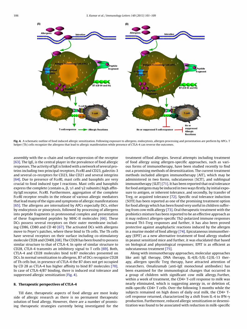
106 S. Kumar et al. / Immunology Letters 149 (2013) 101– 109
F re to ah esenc
a[rta[ceiFt[bioDimhmsCCDobIs
8
ssi
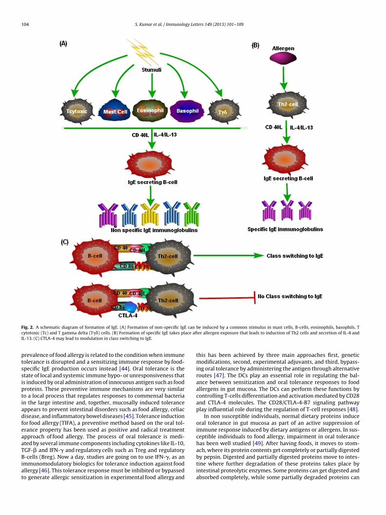
ig. 4. A schematic outline of food induced allergic sensitization. Following exposuelper (Th) cells recognize the allergens that lead to allergic manifestation while pr
ssembly with the �-chain and surface expression of the receptor63]. The IgE, is the central player in the prevalence of food allergicesponses. The activity of IgE is linked with a network of several pro-eins including two principal receptors, Fc�RI and CD23, galectin-3nd several co-receptors for CD23, like CD21 and several integrins64]. Due to presence of Fc�RI, mast cells and basophils are veryrucial to food induced type I reactions. Mast cells and basophilsxpress the complete (contain �, �, �1 and �2 subunits) high affin-ty IgE receptor, Fc�RI. Furthermore, aggregation of the completec�RI receptor results in the release of various allergic mediatorshat lead many of the signs and symptoms of allergic manifestations65]. The allergens are internalized by APCs especially DCs, eithery endocytosis or pinocytosis, followed by processing of allergens
nto peptide fragments in proteosomal complex and presentationf these fragmented peptides by MHC-II molecules [66]. TheseCs possess several receptors on their outer membrane includ-
ng CD86, CD80 and CD 40 [67]. The activated DCs with allergensove to Peyer’s patches, where these bind to Th cells. The Th cells
ave several receptors on their surface including co-stimulatoryolecule CD28 and CD40L [68]. The CD28 has been found to possess
imilar structure to that of CTLA-4. In spite of similar structure toD28, CTLA-4 transmits an inhibitory signal to T cells [69]. Both,TLA-4 and CD28 molecules bind to B7 molecules presented onCs. In normal sensitization to allergens, B7 of DCs recognize CD28f Th cells but, in presence of CTLA-4 the B7 does not get occupiedy CD 28 as CTLA-4 has higher affinity to bind B7 molecules [70].n case of CTLA-4/B7 binding, there is induced oral tolerance anduppressed allergic sensitization (Fig. 4).
. Therapeutic perspectives of CTLA-4
Till date, therapeutic aspects of food allergy are most leakyide of allergic research as there is no permanent therapeuticolution of food allergy. However, there are a number of promis-ng therapeutic strategies currently being investigated for the
llergens, endocytosis, allergen processing and presentation are perform by APCs. Te of CTLA-4 can reverse the outcomes.
treatment of food allergies. Several attempts including treatmentof food allergy using allergen-specific approaches, such as vari-ous forms of immunotherapy, have been studied recently to findout a promising methods of desensitization. The current treatmentmethods included allergen immunotherapy (AIT), which may beadministered in two forms, subcutaneous (SCIT), and sublingualimmunotherapy (SLIT) [71]. It has been reported that oral tolerancefor food antigens may be induced in two ways firstly, by initial expo-sure to antigen, or inherent tolerance, and secondly, by transfer ofTreg, or acquired tolerance [72]. Specific oral tolerance induction(SOTI) has been reported as one of the promising treatment optionfor food allergy which has been found very useful in children suffer-ing from cow milk allergy [73]. Oral therapeutic treatment with theprobiotics mixture has been reported to be an effective approach asit may redirect allergen-specific Th2-polarized immune responsestoward Th1-Treg responses and further, it has also been provedprotective against anaphylactic reactions induced by the allergenin a murine model of food allergy [74]. Epicutaneous immunother-apy (EPIT) as a new alternative treatment of food allergy studiedin peanut sensitized mice and further, it was elucidated that basedon biological and physiological responses; EPIT is as efficient assubcutaneous treatment [75].
Along with immunotherapy approaches, molecular approacheslike anti IgE therapy, DNA therapy, IL-4/IL-5/IL-12/IL-13 ther-apy, allergen specific Treg therapy, have attracted attention ofresearchers. Omalizumab (anti-IgE monoclonal antibodies) hasbeen examined for the immunological changes that occurred ina group of children with significant cow milk allergy. Further,within a week of treatment, the CD4+ T-cell response to milk wasnearly eliminated, which is suggesting anergy in, or deletion of,
milk-specific CD4+ T cells. Over the following 3 months while thesubjects remained on high doses of daily oral milk, the CD4+ T-cell response returned, characterized by a shift from IL-4 to IFN-�production. Furthermore, reduced allergic sensitization or desensi-tization was found to be associated with reduction in milk-specific
gy Let
I[aaittsimcie
bIeehaibfbiT
rdrmmtsintc
iibaabeopCAuhcctltfldrmarsg
[
[
[
[
[
S. Kumar et al. / Immunolo
gE and an enhancement in milk-specific IgG4 up to some extents76]. Thus, the use of anti-IgE monoclonal antibodies to treat foodllergic reactions have raised a positive ray in molecular medicines the anti-IgE monoclonal antibodies have been found to inducemmune tolerance [77]. Further, the anti IgE therapy is used to con-rol food allergy in which specific antibodies are used to bind IgEhat make them unavailable for allergens. Anti-IgE therapy has beenuccessfully tested in patients with food allergy, showing signif-cant efficacy in reducing symptom scores and the use of rescue
edications [78]. Although, anti IgE therapy was found effective toombat food allergy up to some extent but, some drawbacks likets high costs and long term treatment make it unapproachable forvery strata of society.
The SIT has potential to alter the allergy prevalence, which haseen demonstrated in allergic patients and the combination of anti-
gE and SIT have been also recommended as it has shown improvedfficacy, lesser adverse effects with potential immunomodulatoryffects [79]. But, still short term therapy and cost effectiveness isighly desirable. The key cytokines involved in food allergy havelso attracted researchers as one of the therapeutic tools target-ng IL-4 or IL-13 signaling to suppress food induced diarrhea haseen well described, but still requires additional work to make itrom bench work to bedside [80]. Thymus-derived Treg cells haseen reported to maintain oral-tolerance and therefore evidence
s growing regarding the use of adaptive Tregs in the regulation ofh1/Th2 immune responses induced due to food allergens [81].
Recent studies have explored the pivotal role of CD4+CD25+egulatory T cells (nTreg) in the maintenance of oral tolerance toietary antigens and further, in the regulation of the food allergicesponse. It has been reported that TGF-� secreting Treg (Th3)ay play an important role in induction of oral tolerance in aurine model sensitized with peanut [82]. The mechanisms behind
he allergic sensitization involved, mast cell and basophil suppres-ion, enhanced level of food-specific IgG4 antibodies, suppressionn the food-specific IgE/IgG4 ratio, up-regulated and expandedTreg and iTreg cells, a shift from a Th2 to a Th1 reactions, andhe development of T-cell anergy or deletion in antigen-specificells [83].
After exploration of underneath mechanisms involving CTLA-4n food allergy, a ray of its exploitation in therapeutics has comen light but, its use in food allergy is yet to be a reality. CTLA-4 haseen targeted in several diseases as therapeutic tools. It is gener-lly targetted by CTLA-4-Ig, a fusion protein working as antibodygainst CTLA-4. The fusion proteins CTLA4-Ig can be of two typesased on its generation abatacept or belatacept. After thoroughxaminations no toxicity has been observed due to consumptionf abatacept in animal models and well tolerated in non-humanrimates and rodents. Belatacept is a second generation form ofTLA4-Ig which has been recently approved by the Food and Drugdministration, USA [84]. Blocking CD28 interaction with B7 by these of CTLA-4Ig might contribute to control of unbalanced immuneomeostasis. It has been also reported that the CTLA-4Ig treatmentan hamper the development of peanut allergy by suppressingytokine responses, specific IgG1, IgG2a, and IgE and clinical symp-oms [16]. The CTLA-4Ig therapy can be either of short term orong term, while in other allergy therapy like anti IgE therapy longerm treatment is mandatory. High-dose CTLA-4-Ig has not beenound to induce permanent tolerance indicating the possibility ofow dose of CTLA-4Ig in treatment of food allergy [85]. It has beeniscussed in the earlier section that CTLA-4 signaling negativelyegulates immune activation, and due to this milestone nature it
ay play a critical role in preventing induction or progression ofllergic sensitization. To elucidate the role of CTLA-4 signaling inesponses to food allergens, a murine model of peanut allergy wastudied by van Wijk [16]. The outcome of this study sug-ests that CTLA-4 signaling is not the crucial factor in preventing
[
[
ters 149 (2013) 101– 109 107
sensitization to food allergens, but plays a pivotal role in regulatingthe intensity of a food allergic sensitization response [16].
9. Conclusions
At present, there is no effective therapy for IgE-mediated foodallergy, and patients have to rely upon food-allergen avoidance.Definitive therapies for food allergy are desperately needed as itsprevalence is increasing consistently throughout the world. Consid-ering, no treatment against food allergy, CTLA-4 can be used as oneof the most relevant therapeutic options. The involvement of CTLA-4 in CD28/CTLA-4-B7 signaling pathway and recognition of B7molecules by CTLA-4 have opened a positive conception regardingits involvement in food allergy. The CTLA-4 can be targeted atdifferent points including class switching to IgE and induction oforal tolerance. Taken together, CTLA-4 molecule has added a newepisode in food allergic research along with tremendous possibil-ity to consider it as one of the pivotal candidates for food allergytreatment.
Conflict of interest
The authors have declared that there is no conflict of interest.
Acknowledgments
This work was financially supported by InDepth Network Projectof CSIR-IITR. SK and AKV are thankful to CSIR, New Delhi for theaward of their Senior Research Fellowships. Thanks are also due topathway builder tool version 2.0. This is CSIR-IITR manuscript no3093.
References
[1] Rachid R, Umetsu DT. Immunological mechanisms for desensitization and tol-erance in food allergy. Semin Immunopathol 2012;34:689–702.
[2] Strid J, Thomson M, Hourihane J, Kimber I, Strobel S. A novel model of sensi-tization and oral tolerance to peanut protein. Immunology 2004;113:293–303.
[3] Kumar S, Verma AK, Das M, Dwivedi PD. Molecular mechanism of IgE mediatedfood allergy. Int Immunopharmacol 2012;13:432–9.
[4] Brandtzaeg P. Food allergy: separating the science from the mythology. Nat RevGastroenterol Hepatol 2010;7:380–400.
[5] Werfel T, Breuer K. Role of food allergy in atopic dermatitis. Curr Opin AllergyClin Immunol 2004;5:379–85.
[6] Kumar S, Verma AK, Das M, Dwivedi PD. Allergenic diversity among plantand animal foods and their allergenicity. Food Rev Int 2012;28:277–98,http://dx.doi.org/10.1080/87559129.2011.635391.
[7] Sampson HA. Peanut allergy. N Engl J Med 2002;346:1294–9.[8] Brunet JF, Denziot F, Luciani MF, Roux-Dosseto M, Suzan M, Mattei MG,
et al. A new member of the Immunoglobulin superfamily-CTLA-4. Nature1987;328:267–70.
[9] Rudd CE. The reverse stop-signal model for CTLA4 function. Nat Rev Immunol2008;8:153–60.
10] Sansom DM. CD28, CTLA-4 and their ligands: who does what and to whom?Immunology 2000;101:169–77.
11] Lindsten T, Lee KP, Harris ES, Petryniak B, Craighead N, Reynolds PJ, et al. Char-acterization of CTLA-4 structure and expression on human T cells. J Immunol1993;151:3489–99.
12] Yokosuka T, Takamatsu M, Kobayashi-Imanishi W, Hashimoto-Tane A, AzumaM, Saito T. Programmed cell death 1 forms negative costimulatory microclus-ters that directly inhibit T cell receptor signaling by recruiting phosphataseSHP2. J Exp Med 2012;209:1201–17.
13] Ahmed A, Mukherjee S, Nandi D. Intracellular concentrations of Ca2+ modu-late the strength of signal and alter the outcomes of cytotoxic T-lymphocyteantigen-4 (CD152)–CD80/CD 86 interactions in CD4+ T lymphocytes. Immunol-ogy 2009;126:363–77.
14] Pabst O, Mowat AM. Oral tolerance to food protein. Mucosal Immunol2012;5:232–9.
15] Akdis M, Verhagen J, Taylor A, Karamloo F, Karagiannidis C, Crameri R, et al.Immune responses in healthy and allergic individuals are characterized by afine balance between allergen-specific T regulatory 1 and T helper 2 cells. J ExpMed 2004;199:1567–75.
16] van Wijk F, Nierkens S, de Jong W, Wehrens EJ, Boon L, van Kooten P,et al. The CD28/CTLA-4-B7 signaling pathway is involved in both allergic
1 gy Let
[
[
[
[
[
[
[
[
[
[
[
[
[
[
[
[
[
[
[
[
[
[
[
[
[
[
[
[
[
[
[
[
[
[
[[
[
[
[
[
[
[
[
[
[
[
[
[
[
[
[
[
[
[
[
[
[
[
[
[
[
08 S. Kumar et al. / Immunolo
sensitization and tolerance induction to orally administered peanut proteins. JImmunol 2007;178:6894–900.
17] Munthe-Kaas MC, Carlsen KH, Helms PJ, Gerritsen J, Whyte M, Feijen M, et al.CTLA-4 polymorphisms in allergy and asthma and the TH1/TH2 paradigm. JAllergy Clin Immunol 2004;114:280–7.
18] Regateiro FS, Chen Y, Kendal AR, Hilbrands R, Adams E, Cobbold SP, et al.Foxp3 expression is required for the induction of therapeutic tissue tolerance.J Immunol 2012 [epub ahead of print].
19] Lan Q, Fan H, Quesniaux V, Ryffel B, Liu Z, Zheng SG. Induced Foxp3(+) regu-latory T cells: a potential new weapon to treat autoimmuneand inflammatorydiseases? J Mol Cell Biol 2012;4:22–8.
20] Hinz D, Bauer M, Röder S, Olek S, Huehn J, Sack U, et al. Cord blood Tregswith stable FOXP3 expression are influenced by prenatal environment andassociated with atopic dermatitis at the age of one year. Allergy 2012;67:380–9.
21] Ganeshan K, Bryce PJ. Regulatory T cells enhance mast cell production of IL-6via surface-bound TGF-�. J Immunol 2012;188:594–603.
22] Shevach EM. Mechanisms of foxp3+ T regulatory cell-mediated suppression.Immunity 2009;30:636–45.
23] Taylor MD, Harris A, Babayan SA, Bain O, Culshaw A, Allen JE, et al. CTLA-4 andCD4+ CD25+ regulatory T cells inhibit protective immunity to filarial parasitesin vivo. J Immunol 2007;179:4626–34.
24] Rabe H, Lundell AC, Andersson K, Adlerberth I, Wold AE, Rudin A. Higherproportions of circulating FOXP3+ and CTLA-4+ regulatory T cells are asso-ciated with lower fractions of memory CD4+ T cells in infants. J Leukoc Biol2011;90:1133–40.
25] Duhen T, Duhen R, Lanzavecchia A, Sallusto F, Campbell DJ. Functionally distinctsubsets of human FOXP3+ Treg cells that phenotypically mirror effector Th cells.Blood 2012;119:4430–40.
26] Wing K, Onishi Y, Prieto-Martin P, Yamaguchi T, Miyara M, FehervariZ, et al. CTLA-4 control over Foxp3+ regulatory T cell function. Science2008;322:271–5.
27] Tai X, Van Laethem F, Pobezinsky L, Guinter T, Sharrow SO, Adams A, et al.Basis of CTLA-4 function in regulatory and conventional CD4+ T cells. Blood2008;119:5155–63.
28] Lyons A, O’Mahony D, O’Brien F, MacSharry J, Sheil B, Ceddia M, et al. Bacterialstrain-specific induction of Foxp3+ T regulatory cells is protective in murineallergymodels. Clin Exp Allergy 2010;40:811–9.
29] Cario E. Bacterial interactions with cells of the intestinal mucosa, toll-like recep-tors and nod2. Gut 2005;54:1182–93.
30] Bashir ME, Louie S, Shi HN, Nagler-Anderson C. Toll-Like receptor 4 signalingby intestinal microbes influences susceptibility to food allergy. J Immunol2004;172:6978–87.
31] Pochard P, Vickery B, Berin MC, Grishin A, Sampson HA, Caplan M, et al. Target-ing Toll-like receptors on dendritic cells modifies the TH2 response to peanutallergens in vitro. J Allergy Clin Immunol 2010;126:92–700000.
32] Kato H, Fujihashi K, Kato R, Yuki Y, McGhee JR. Oral tolerance revisited: priororal tolerization abrogates cholera toxin-induced mucosal IgA responses. JImmunol 2001;166:3114–21.
33] Westerholm-Ormio M, Vaarala O, Tiittanen M, Savilahti E. Infiltration of Foxp3-and Toll-like receptor-4-positive cells in the intestines of children with foodallergy. J Pediatr Gastroenterol Nutr 2010;50:367–76.
34] Prescott SL, Noakes P, Chow BW, Breckler L, Thornton CA, Hollams EM, et al.Presymptomatic differences in Toll-like receptor function in infants who haveallergy. J Allergy Clin Immunol 2008;122:391–9, e1-5, 399.
35] Berin MC, Zheng Y, Domaradzki M, Li XM, Sampson HA. Role of TLR4 in allergicsensitization to food proteins in mice. Allergy 2006;61:64–71.
36] Tsuda M, Hosono A, Yanagibashi T, Kihara-Fujioka M, Hachimura S, Itoh K, et al.Intestinal commensal bacteria promote T cell hyporesponsiveness and down-regulate the serum antibody responses induced by dietary antigen. ImmunolLett 2010;132:45–52.
37] Rivas MN, Koh YT, Chen A, Nguyen A, Lee YH, Lawson G, et al. MyD88 is criti-cally involved in immune tolerance breakdown at environmental interfaces ofFoxp3-deficient mice. J Clin Invest 2012;122:1933–47.
38] Koppelman SJ, Knol EF, Vlooswijk RA, Wensing M, Knulst AC, Hefle SL, et al.Peanut allergen Ara h 3: isolation from peanuts and biochemical characteriza-tion. Allergy 2003;58:1144–51.
39] Verma AK, Kumar S, Das M, Dwivedi PD. A comprehensivereview of legume allergy. Clin Rev Allergy Immunol 2012,http://dx.doi.org/10.1007/s12016-012-8310-6.
40] Jarrett E, Mackenzie S, Bennich H. Parasite-induced ‘nonspecific’ IgE does notprotect against allergic reactions. Nature 1980;283:302–4.
41] Bloemen K, Verstraelen S, Van Den Heuvel R, Witters H, Nelissen I, Schoeters G.The allergic cascade, review of the most important molecules in the asthmaticlung. Immunol Lett 2007;113:6–18.
42] Zhang T, Franklin A, Boboila C, McQuay A, Gallagher MP, Manis JP, et al. Down-stream class switching leads to IgE antibody production by B lymphocyteslacking IgM switch regions. Proc Natl Acad Sci U S A 2010;107:3040–5.
43] Borriello F, Sethna MP, Boyd SD, Schweitzer AN, Tivol EA, Jacoby D, et al. B7-1and B7-2 have overlapping, critical roles in immunoglobulin class switching
and germinal center formation. Immunity 1997;6:303–13.44] Life P, Aubry JP, Estoppey S, Schnuriger V, Bonnefoy JY. CD28 functions as anadhesion molecule and is involved in the regulation of human IgE synthesis.Eur J Immunol 1995;25:333–9.
45] Steele L, Mayer L, Cecilia Berin M. Mucosal immunology of tolerance and allergyin the gastrointestinal tract. Immunol Res 2012 [epub ahead of print].
[
[
ters 149 (2013) 101– 109
46] Noh G, Lee JH. Oral tolerance induction for human food allergy. Inflamm AllergyDrug Targets 2012;11:131–42.
47] Berin MC. Mechanisms of allergic sensitization to foods: bypassing immunetolerance pathways. Immunol Allergy Clin North Am 2012;32:1–10.
48] Janine LC, Fiona P. Dendritic cells in intestinal immune regulation. Nat RevImmunol 2008;8:435–46.
49] Burks AW, Laubach S, Jones SM. Oral tolerance, food allergy, andimmunotherapy: implications for future treatment. J Allergy Clin Immunol2008;121:1344–50.
50] Sabra A, Bellanti JA, Rais JM, Castro HJ, de Inocencio JM, Sabra S. IgE and non-IgEfood allergy. Ann Allergy Asthma Immunol 2003;90:71–6.
51] Iwasaki A. Mucosal dendritic cells. Annu Rev Immunol 2007;25:381–418.52] Gourbeyre P, Denery S, Bodinier M. Probiotics, prebiotics, and synbiotics:
impact on the gut immune system and allergic reactions. J Leukoc Biol2011;89:685–95.
53] Kweon MN, Kiyono H. CD40L in autoimmunity and mucosally induced toler-ance. J Clin Invest 2002;109:171–3.
54] Wahl SM, Swisher J, McCartney-Francis N, Chen W. TGF-� the perpetrator ofimmune suppression by regulatory T cells and suicidal T cells. J Leukoc Biol2004;76:15–24.
55] Rubtsov YP, Rudensky AY. TGF-� signalling in control of T-cell-mediated self-reactivity. Nat Rev Immunol 2007;7:443–53.
56] Cerutti A. The regulation of IgA class switching. Nat Rev Immunol2008;8:421–34.
57] Smith DW, Anderson CN. Preventing intolerance, the induction of non respon-siveness to dietary and microbial antigens in the intestinal mucosa. J Immunol2005;174:3851–7.
58] Ruiter B, Shreffler WG. Innate immunostimulatory properties of allergens andtheir relevance to food allergy. Semin Immunopathol 2012;34:617–32.
59] von Bubnoff D, Koch S, Bieber T. Dendritic cells and atopic eczema/dermatitissyndrome. Curr Opin Allergy Clin Immunol 2003;3:353–8.
60] Mac Glashan Jr D. IgE receptor and signal transduction in mast cells andbasophils. Curr Opin Immunol 2008;20:717–23.
61] Sellge G, Bischoff SC. Isolation, culture, and characterization of intestinal mastcells. Methods Mol Biol 2006;315:123–38.
62] Kraneveld AD, Sagar S, Garssen J, Folkerts G. The two faces of mast cells in foodallergy and allergic asthma: the possible concept of Yin Yang. Biochim BiophysActa 2012;1822:93–9.
63] Bischoff SC. Physiological and pathophysiological functions of intestinal mastcells. Semin Immunopathol 2009;31:185–205.
64] Conrad DH, Ford JW, Sturgill JL, Gibb DR. CD23: an overlooked regulator ofallergic disease. Curr Allergy Asthma Rep 2007;7:331–7.
65] MacGlashan Jr D. IgE receptor and signal transduction in mast cells andbasophils. Curr Opin Immunol 2008;20:717–23.
66] Askew D, Harding CV. Antigen processing and CD24 expression determineantigen presentation by splenic CD4+ and CD8+ dendritic cells. Immunology2008;123:447–55.
67] Sun ZW, Qiu YH, Shi YJ, Tao R, Chen J, Ge Y, et al. Time courses of B7 familymolecules expressed on activated T-cells and their biological significance. CellImmunol 2005;236:146–53.
68] Kelsall BL, Leon F. Involvement of intestinal dendritic cells in oral toler-ance, immunity to pathogens, and inflammatory bowel disease. Immunol Rev2005;206:132–48.
69] Kriegel MA, Rathinam C, Flavell RA. E3 ubiquitin ligase GRAIL controls primaryT cell activation and oral tolerance. Proc Natl Acad Sci U S A 2009;106:16770–5.
70] Linsley JPS, Greene JL, Tan P, Bradshaw J, Ledbetter JA, Anasetti C, et al.Coexpression and functional cooperation of CTLA-4 and CD28 on activated Tlymphocytes. J Exp Med 1992;176:1595–604.
71] Frati F, Incorvaia C, Lombardi C, Senna G. Allergen immunotherapy: 100 years,but it does not look like. Eur Ann Allergy Clin Immunol 2012;44:99–106.
72] Yamashita H, Takahashi K, Tanaka H, Nagai H, Inagaki N. Overcoming foodallergy through acquired tolerance conferred by transfer of Tregs in a murinemodel. Allergy 2012;67:201–9.
73] Longo G, Barbi E, Berti I, Meneghetti R, Pittalis A, Ronfani L, et al. Specific oraltolerance induction in children with very severe cow’s milk-induced reactions.J Allergy Clin Immunol 2008;121:343–7.
74] Schiavi E, Barletta B, Butteroni C, Corinti S, Boirivant M, Di Felice G. Oraltherapeutic administration of a probiotic mixture suppresses established Th2responses and systemic anaphylaxis in a murine model of food allergy. Allergy2011;66:499–508.
75] Mondoulet L, Dioszeghy V, Vanoirbeek JA, Nemery B, Dupont C, Benhamou PH.Epicutaneous immunotherapy using a new epicutaneous delivery system inmice sensitized to peanuts. Int Arch Allergy Immunol 2011;154:299–309.
76] Bedoret D, Singh AK, Shaw V, Hoyte EG, Hamilton R, DeKruyff RH, et al.Changes in antigen-specific T-cell number and function during oral desensi-tization in cow’s milk allergy enabled with omalizumab. Mucosal Immunol2012;5:267–76.
77] Leung DY, Sampson HA, Yunginger JW, Burks Jr AW, Schneider LC, Wortel CH,et al. Effect of anti-IgE therapy in patients with peanut allergy. N Engl J Med2003;348:986–93.
78] Bouchez-Mahiout I, Pecquet C, Kerre S, Snégaroff J, Raison-Peyron N, LaurièreM. High molecular weight entities in industrial wheat protein hydrolysatesare immunoreactive with IgE from allergic patients. J Agric Food Chem2010;14:4207–15.
79] Crittenden RG, Bennett LE. Cow’s milk allergy: a complex disorder. J Am CollNutr 2005;24:582S–91S.

gy Let
[
[
[
[
[
S. Kumar et al. / Immunolo
80] Hogan SP, Rothenberg ME. The eosinophil as a therapeutic target in gastroin-testinal disease. Aliment Pharmacol Ther 2004;20:1231–40.
81] Akdis M, Blaser K, Akdis CA. T regulatory cells in allergy: novel concepts in
the pathogenesis, prevention, and treatment of allergic diseases. J Allergy ClinImmunol 2005;116:961–8.82] Stock P, Rolinck-Werninghaus C, Wahn U, Hamelmann E. The role of anti-IgEtherapy in combination with allergen specific immunotherapy for seasonalallergic rhinitis. BioDrugs 2007;21:403–10.
[
ters 149 (2013) 101– 109 109
83] Wu K, Bi Y, Sun K, Wang C. IL-10-producing type 1 regulatory T cells and allergy.Cell Mol Immunol 2007;4:269–75.
84] McGrath MM, Najafian N. The role of coinhibitory signaling path-
ways in transplantation and tolerance. Front Immunol 2012;3:47.85] Mihara M, Tan I, Chuzhin Y, Reddy B, Budhai L, Holzer A, et al. CTLA4 Ig inhibitsT cell-dependent B-cell maturation in murine systemic lupus erythematosus. JClin Invest 2000;106:91–101.