Embed Size (px)
Citation preview
BioMed CentralBiology Direct
ss
Open AcceResearchA computational analysis of the three isoforms of glutamate dehydrogenase reveals structural features of the isoform EC 1.4.1.4 supporting a key role in ammonium assimilation by plantsEmmanuel Jaspard*Address: UMR 1191 Physiologie Moléculaire des Semences, Université d'Angers – INRA – INH, Angers, France
Email: Emmanuel Jaspard* - [email protected]
* Corresponding author
AbstractBackground: There are three isoforms of glutamate dehydrogenase. The isoform EC 1.4.1.4(GDH4) catalyses glutamate synthesis from 2-oxoglutarate and ammonium, using NAD(P)H.Ammonium assimilation is critical for plant growth. Although GDH4 from animals and prokaryotesare well characterized, there are few data concerning plant GDH4, even from those whosegenomes are well annotated.
Results: A large set of the three GDH isoforms was built resulting in 116 non-redundant fullpolypeptide sequences. A computational analysis was made to gain more information concerningthe structure – function relationship of GDH4 from plants (Eukaryota, Viridiplantae). The testedplant GDH4 sequences were the two ones known to date, those of Chlorella sorokiniana. Thisanalysis revealed several structural features specific of plant GDH4: (i) the lack of a structure called"antenna"; (ii) the NAD(P)-binding motif GAGNVA; and (iii) a second putative coenzyme-bindingmotif GVLTGKG together with four residues involved in the binding of the reduced form of NADP.
Conclusion: A number of structural features specific of plant GDH4 have been found. The resultsreinforce the probable key role of GDH4 in ammonium assimilation by plants.
Reviewers: This article was reviewed by Tina Bakolitsa (nominated by Eugene Koonin), MartinJambon (nominated by Laura Landweber), Sandor Pangor and Franck Eisenhaber.
Open peer reviewReviewed by Tina Bakolitsa (nominated by EugeneKoonin), Martin Jambon (nominated by Laura Landwe-ber), Sandor Pangor and Franck Eisenhaber. For the fullreviews, please go to the Reviewers' comments section.
BackgroundThere are three isoforms of GDH. According to the follow-ing reaction:
2-oxoglutarate + NH4 + + NAD(P)H + H+ ⇔ glutamate +
H2O + NAD(P)+ GDH EC 1.4.1.2 (GDH2) catalyses essen-tially the formation of 2-oxoglutarate using NAD(P)+ [1];GDH EC 1.4.1.3 (GDH3) catalyses both the formation of2-oxoglutarate and the reverse reaction, thus exhibiting adual coenzyme specificity [NAD(P)+/NAD(P)H] [2];GDH EC 1.4.1.4 (GDH4) catalyses the formation of gluta-mate using NAD(P)H [3,4]. For example, GDH4 is knownto play an anabolic role in ammonium assimilation in theyeast Candida utilis [5]. Nevertheless, it is considered that
Published: 15 December 2006
Biology Direct 2006, 1:38 doi:10.1186/1745-6150-1-38
Received: 27 November 2006Accepted: 15 December 2006
This article is available from: http://www.biology-direct.com/content/1/1/38
© 2006 Jaspard; licensee BioMed Central Ltd. This is an Open Access article distributed under the terms of the Creative Commons Attribution License (http://creativecommons.org/licenses/by/2.0), which permits unrestricted use, distribution, and reproduction in any medium, provided the original work is properly cited.
Page 1 of 12(page number not for citation purposes)
Biology Direct 2006, 1:38 http://www.biology-direct.com/content/1/1/38
the major route of ammonium assimilation in plantsinvolves the glutamine synthetase – glutamate synthasecouple [6]. However, high ammonium concentrationdeactivates glutamine synthetase and induces GDH [7,8].In fact, data on the actual role of GDH4 from plants eitherin ammonium assimilation or in the formation of 2-oxoglutarate are controversial.
Several three-dimensional structures of GDH fromprokaryotic and eukaryotic organisms have been resolved[9-11]. All GDHs described to date are homo-oligomericproteins and the most striking differences between thethree isoforms arise from the primary and the quaternarystructures. GDHs were classified into four families on thebasis of the length of the polypeptide chain and thenumber of subunits [12]. GDH2 are dimeric (unique casefor this enzyme) [13], tetrameric [14] or hexameric [15];GDH3 are essentially hexameric [16]; GDH4 are tetra-meric [17] or hexameric [18].
A bioinformatics analysis of a large set of the three iso-forms of GDH was made to gain more information con-cerning the structure – function relationship of GDH4from plants (Eukaryota, Viridiplantae). The tested plantGDH4 sequences were the two ones known to date, thoseof Chlorella sorokiniana [19].
The following characteristics were found specific to GDH4from Chlorella sorokiniana: (i) a small N-terminal regionand no C-terminal extension; (ii) a central domain withthe substrates and the nucleotide-binding sites but with-out a structure called antenna ; (iii) a second putativecoenzyme-binding motif whose fingerprint sequence isGVLTGKG ; (iv) four residues (Lys, Ser, Arg and Thr)involved in the binding of the reduced form of the coen-zyme, NADPH. A model of the structure of the active siteof GDH4 from Chlorella sorokiniana, with NADPH andglutamate, is proposed. The role of these two coenzyme-binding motifs and of these four residues in the stabiliza-tion of the reduced form of NADP is discussed to explainthe functional specificity of plant GDH4 in the formationof glutamate.
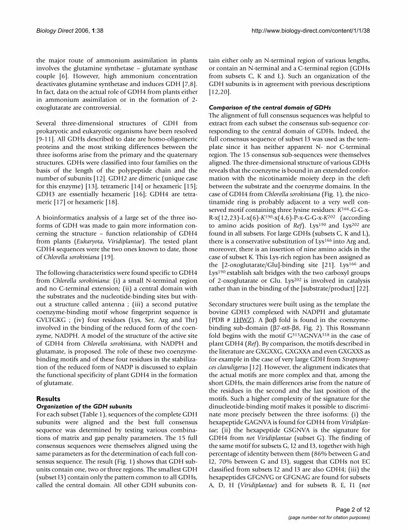
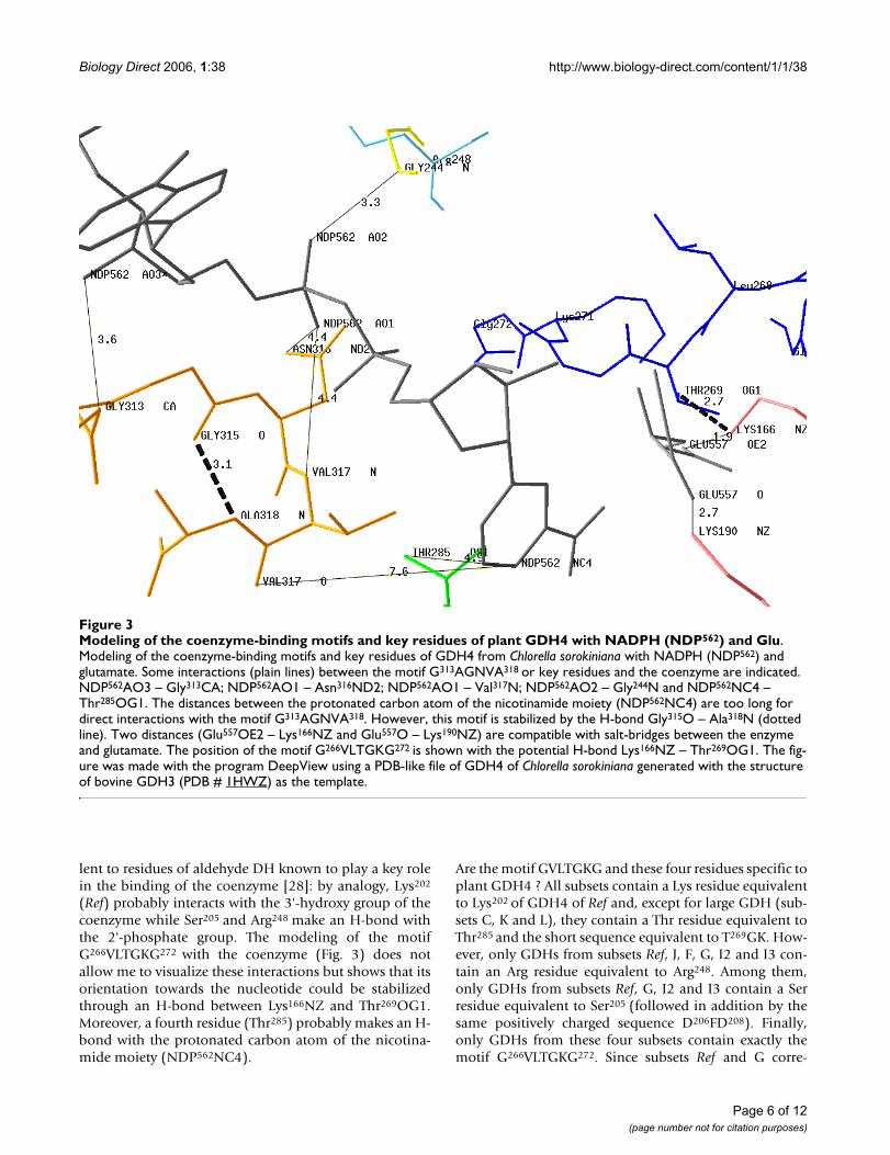
ResultsOrganization of the GDH subunitsFor each subset (Table 1), sequences of the complete GDHsubunits were aligned and the best full consensussequence was determined by testing various combina-tions of matrix and gap penalty parameters. The 15 fullconsensus sequences were themselves aligned using thesame parameters as for the determination of each full con-sensus sequence. The result (Fig. 1) shows that GDH sub-units contain one, two or three regions. The smallest GDH(subset I3) contain only the pattern common to all GDHs,called the central domain. All other GDH subunits con-
tain either only an N-terminal region of various lengths,or contain an N-terminal and a C-terminal region (GDHsfrom subsets C, K and L). Such an organization of theGDH subunits is in agreement with previous descriptions[12,20].
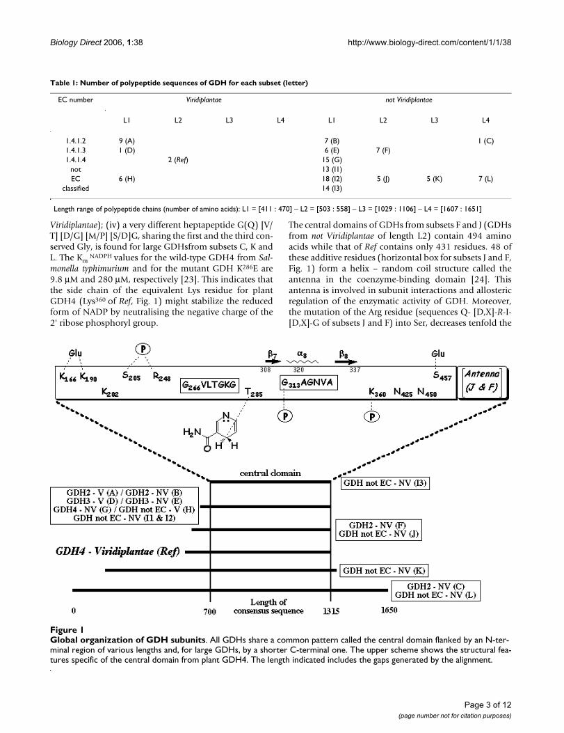
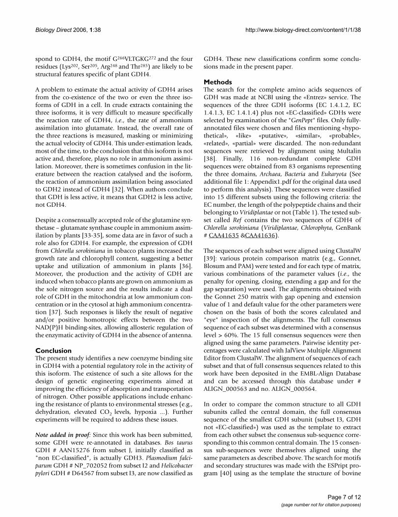
Comparison of the central domain of GDHsThe alignment of full consensus sequences was helpful toextract from each subset the consensus sub-sequence cor-responding to the central domain of GDHs. Indeed, thefull consensus sequence of subset I3 was used as the tem-plate since it has neither apparent N- nor C-terminalregion. The 15 consensus sub-sequences were themselvesaligned. The three-dimensional structure of various GDHsreveals that the coenzyme is bound in an extended confor-mation with the nicotinamide moiety deep in the cleftbetween the substrate and the coenzyme domains. In thecase of GDH4 from Chlorella sorokiniana (Fig. 1), the nico-tinamide ring is probably adjacent to a very well con-served motif containing three lysine residues: K166-G-G-x-R-x(12,23)-L-x(6)-K190-x(4,6)-P-x-G-G-x-K202 (accordingto amino acids position of Ref). Lys190 and Lys202 arefound in all subsets. For large GDHs (subsets C, K and L),there is a conservative substitution of Lys166 into Arg and,moreover, there is an insertion of nine amino acids in thecase of subset K. This Lys-rich region has been assigned asthe [2-oxoglutarate/Glu]-binding site [21]. Lys166 andLys190 establish salt bridges with the two carboxyl groupsof 2-oxoglutarate or Glu. Lys202 is involved in catalysisrather than in the binding of the [substrate/product] [22].
Secondary structures were built using as the template thebovine GDH3 complexed with NADPH and glutamate(PDB # 1HWZ). A βαβ fold is found in the coenzyme-binding sub-domain (β7-α8-β8, Fig. 2). This Rossmannfold begins with the motif G313AGNVA318 in the case ofplant GDH4 (Ref). By comparison, the motifs described inthe literature are GXGXXG, GXGXXA and even GXGXXS asfor example in the case of very large GDH from Streptomy-ces clavuligerus [12]. However, the alignment indicates thatthe actual motifs are more complex and that, among theshort GDHs, the main differences arise from the nature ofthe residues in the second and the last position of themotifs. Such a higher complexity of the signature for thedinucleotide-binding motif makes it possible to discrimi-nate more precisely between the three isoforms: (i) thehexapeptide GAGNVA is found for GDH4 from Viridiplan-tae; (ii) the hexapeptide GSGNVA is the signature forGDH4 from not Viridiplantae (subset G). The finding ofthe same motif for subsets G, I2 and I3, together with highpercentage of identity between them (86% between G andI2, 70% between G and I3), suggest that GDHs not ECclassified from subsets I2 and I3 are also GDH4; (iii) thehexapeptides GFGNVG or GFGNAG are found for subsetsA, D, H (Viridiplantae) and for subsets B, E, I1 (not
Page 2 of 12(page number not for citation purposes)
Biology Direct 2006, 1:38 http://www.biology-direct.com/content/1/1/38
Viridiplantae); (iv) a very different heptapeptide G(Q) [V/T] [D/G] [M/P] [S/D]G, sharing the first and the third con-served Gly, is found for large GDHsfrom subsets C, K andL. The Km
NADPH values for the wild-type GDH4 from Sal-monella typhimurium and for the mutant GDH K286E are9.8 μM and 280 μM, respectively [23]. This indicates thatthe side chain of the equivalent Lys residue for plantGDH4 (Lys360 of Ref, Fig. 1) might stabilize the reducedform of NADP by neutralising the negative charge of the2' ribose phosphoryl group.
The central domains of GDHs from subsets F and J (GDHsfrom not Viridiplantae of length L2) contain 494 aminoacids while that of Ref contains only 431 residues. 48 ofthese additive residues (horizontal box for subsets J and F,Fig. 1) form a helix – random coil structure called theantenna in the coenzyme-binding domain [24]. Thisantenna is involved in subunit interactions and allostericregulation of the enzymatic activity of GDH. Moreover,the mutation of the Arg residue (sequences Q- [D,X]-R-I-[D,X]-G of subsets J and F) into Ser, decreases tenfold the
Table 1: Number of polypeptide sequences of GDH for each subset (letter)
EC number Viridiplantae not Viridiplantae
L1 L2 L3 L4 L1 L2 L3 L4
1.4.1.2 9 (A) 7 (B) 1 (C)1.4.1.3 1 (D) 6 (E) 7 (F)1.4.1.4 2 (Ref) 15 (G)
not 13 (I1)EC 6 (H) 18 (I2) 5 (J) 5 (K) 7 (L)
classified 14 (I3)
Length range of polypeptide chains (number of amino acids): L1 = [411 : 470] – L2 = [503 : 558] – L3 = [1029 : 1106] – L4 = [1607 : 1651]
Global organization of GDH subunitsFigure 1Global organization of GDH subunits. All GDHs share a common pattern called the central domain flanked by an N-ter-minal region of various lengths and, for large GDHs, by a shorter C-terminal one. The upper scheme shows the structural fea-tures specific of the central domain from plant GDH4. The length indicated includes the gaps generated by the alignment.
Page 3 of 12(page number not for citation purposes)
Biology Direct 2006, 1:38 http://www.biology-direct.com/content/1/1/38
Page 4 of 12(page number not for citation purposes)
The two dinucleotide-binding motifs of plant GDH4Figure 2The two dinucleotide-binding motifs of plant GDH4. The part of each consensus sequence corresponding to the central domain of GDHs was extracted using the full consensus sequence of subset I3 as the template. The 15 subsets (J to L) are pre-sented in five groups (1 to 5) according to the percentages of identity. Secondary structures indicated above the alignment were generated using the bovine GDH3 complexed with NADPH and glutamate (PDB # 1HWZ) as the template. Amino acid position indicated above the alignments is that of plant GDH4 (Ref). Plain vertical boxes. amino acids identical for all consensus sub-sequences. Open vertical boxes. amino acids whose homology between all consensus sub-sequences was greater than 60%. The letter "X" accounts for an amino acid whose identity level was less than 60% after the first alignment of full consensus sequences. The found dinucleotide-binding motif G266VLTGKG272 (Ref) and the dinucleotide-binding motif G313AGNVA318 (Ref) included in the N-terminal part of the Rossmann fold (β7-α8-β8) are indicated at the bottom of the frame with asterisks and circles, respectively.
Biology Direct 2006, 1:38 http://www.biology-direct.com/content/1/1/38
activity of human GDH [25]. It has been shown thatplants GDH do not possess this antenna [26] and indeedthe GDH4 sequences of Ref do not contain this aminoacid sequence. Therefore, the lack of the antenna motifand of this Arg residue is specific to plant GDH4. Finally,since subset F corresponds to GDH3, one can make theassumption that GDHs from subset J are also GDH3.
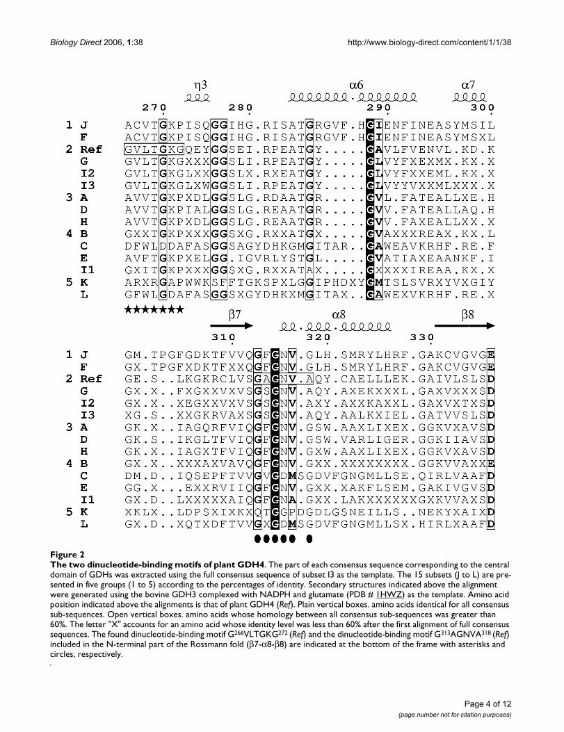
Modeling of the active center of plant GDH4A theoretical 3D structure of GDH4 from Chlorella soroki-niana was calculated with the homology-modeling pro-gram ESyPred3D that creates a PDB-like file, using as thetemplate the structure of bovine GDH3 (PDB # 1HWZ).This structure was chosen as the template for three rea-sons: (i) its length (501 amino acids) is similar to that ofthe GDH4 sequences of Chlorella sorokiniana (523 aminoacids); (ii) considering the dual coenzyme specificity[NAD(P)+/NAD(P)H] of GDH3, it is assumed that itsstructure is closer to that of plant GDH4 than to anyGDH2 structure; and (iii) the modeled data were obtainedfor the enzyme complexed to the reduced form of thecoenzyme, NADPH. Using these two PDB files (the cre-ated PDB-like file for GDH4 from Chlorella sorokinianaand PDB # 1HWZ), a putative structure of the active centerof GDH4 from Chlorella sorokiniana was modeled (Fig. 3)with the protein structure homology-modeling programDeepView.
The model reveals three interactions (NDP562AO3 –Gly313CA, NDP562AO1 – Asn316ND2 and NDP562AO1 –Val317N) between the coenzyme-binding motifG313AGNVA318 and NADPH (NDP562). Moreover, thismotif is stabilized by both the internal H-bond (Gly315O– Ala318N) and the interaction NDP562AO2 – Gly244N.This latter interaction was found by comparison with theH-bond network of the template bovine GDH3 (theequivalent of Gly244 being Ser170 in the case of bovineGDH3). As previously mentioned, Lys166 and Lys190 areknown to be the [2-oxoglutarate/Glu]-binding sites [21].The model shows two interactions (Glu557OE2 –Lys166NZ and Glu557O – Lys190NZ) whose distances arecompatible with salt bridges with the two carboxyl groupsof 2-oxoglutarate, i.e., the substrate of the reaction cata-lysed by plant GDH4.
Evidence for a second reduced coenzyme-binding site in plant GDH4Among the dehydrogenase family, aldehyde DH from thebacterium Vibrio harveyi is one of the most NADP-specific(Km
NADP is 150-fold lower than Km NAD) [27]. The
sequence of aldehyde DH from Vibrio harveyi (GenBank #Q56694) and those of GDH4 from Chlorella sorokinianaare roughly of the same length (510 and 523 amino acids,respectively) with 28% identity and 52% similarity. More-over, aldehyde DH from Vibrio harveyi is an oligomer of
50–55 kDa subunits such as GDH4 of Ref. Finally thenucleotide-binding motif for aldehyde DH of Vibrio har-veyi is G229SVGGG234 and is included in a Rossmann fold[28]. Such close functional and structural characteristicsled me to compare these two enzymes using aldehyde DHfrom Vibrio harveyi (PDB # 1EZ0) as the template. Theresults are presented in the Figure 1: (i) three putative keyresidues for the binding of NADPH are localized in GDH4from Chlorella sorokiniana: Lys202, Ser205 and Arg248; (ii)the motif G229SVGGG234 (aldehyde DH) is aligned withthe motif G266VLTGKG272 of GDH4 of Ref indicating thatthe latter is likely to be a second reduced coenzyme-bind-ing motif characterizing plant GDH4. A model of thismotif with NADPH and Glu is proposed (Fig. 3).
DiscussionThe functional specificity of GDH4 is the formation ofglutamate using NADPH. The results suggest that the fin-gerprint sequence G313AGNVA318 is the signature of one ofthe two coenzyme-binding motifs of plant GDH4. Noneof the distances calculated between the residues of thismotif and the protonated carbon atom of the nicotina-mide moiety (NDP562NC4) are compatible with interac-tions susceptible to stabilize the reduced form of thisatom (Fig. 3). Nevertheless, this result is not so surprising.First, Gly315 seems involved in maintaining the conforma-tion of the motif (through an H-bond with Ala318N)rather than in the coenzyme specificity, since it is con-served in the three isoforms of GDH (Fig. 2). Second,three residues of this motif interact with other parts ofNADPH: NDP562AO3 – Gly313CA, NDP562AO1 –Asn316ND2 and NDP562AO1 – Val317N. The second inter-action underlines the difference in orientation of thecoenzyme in the active site between GDH3 and GDH4because the equivalent Asn residue of bovine GDH3(Asn254) is H-bonded to the carboxyamide group of thenicotinamide ring [21]. Third, the mechanism of interac-tion between the adenine ribose and the fingerprintsequence GXGXXG/A depends (at least for NAD-dehydro-genases) on the nature of the residue occupying the lastposition of this motif but is independent of the coenzymespecificity [29].
Smith and coll. [30] have resolved the structure of threeabortive complexes of bovine GDH3 (GDH-NADH-Glu-GTP, GDH-NADPH-Glu-GTP and GDH-NAD-2-oxogluta-rate) and they have shown that NADH and NADPH bindto a second coenzyme site. The dissociation constantsfrom these two sites are 57 and 700 μM, respectively [31].
There is evidence for the existence of a second coenzyme-binding site in plant GDH4 whose sequence isG266VLTGKG272 (Ref; Fig. 1): (i) the perfect alignment ofthis motif with the motif GSVGGG of aldehyde DH; and(ii) the existence of three residues of GDH4 of Ref equiva-
Page 5 of 12(page number not for citation purposes)
Biology Direct 2006, 1:38 http://www.biology-direct.com/content/1/1/38
lent to residues of aldehyde DH known to play a key rolein the binding of the coenzyme [28]: by analogy, Lys202
(Ref) probably interacts with the 3'-hydroxy group of thecoenzyme while Ser205 and Arg248 make an H-bond withthe 2'-phosphate group. The modeling of the motifG266VLTGKG272 with the coenzyme (Fig. 3) does notallow me to visualize these interactions but shows that itsorientation towards the nucleotide could be stabilizedthrough an H-bond between Lys166NZ and Thr269OG1.Moreover, a fourth residue (Thr285) probably makes an H-bond with the protonated carbon atom of the nicotina-mide moiety (NDP562NC4).
Are the motif GVLTGKG and these four residues specific toplant GDH4 ? All subsets contain a Lys residue equivalentto Lys202 of GDH4 of Ref and, except for large GDH (sub-sets C, K and L), they contain a Thr residue equivalent toThr285 and the short sequence equivalent to T269GK. How-ever, only GDHs from subsets Ref, J, F, G, I2 and I3 con-tain an Arg residue equivalent to Arg248. Among them,only GDHs from subsets Ref, G, I2 and I3 contain a Serresidue equivalent to Ser205 (followed in addition by thesame positively charged sequence D206FD208). Finally,only GDHs from these four subsets contain exactly themotif G266VLTGKG272. Since subsets Ref and G corre-
Modeling of the coenzyme-binding motifs and key residues of plant GDH4 with NADPH (NDP562) and GluFigure 3Modeling of the coenzyme-binding motifs and key residues of plant GDH4 with NADPH (NDP562) and Glu. Modeling of the coenzyme-binding motifs and key residues of GDH4 from Chlorella sorokiniana with NADPH (NDP562) and glutamate. Some interactions (plain lines) between the motif G313AGNVA318 or key residues and the coenzyme are indicated. NDP562AO3 – Gly313CA; NDP562AO1 – Asn316ND2; NDP562AO1 – Val317N; NDP562AO2 – Gly244N and NDP562NC4 – Thr285OG1. The distances between the protonated carbon atom of the nicotinamide moiety (NDP562NC4) are too long for direct interactions with the motif G313AGNVA318. However, this motif is stabilized by the H-bond Gly315O – Ala318N (dotted line). Two distances (Glu557OE2 – Lys166NZ and Glu557O – Lys190NZ) are compatible with salt-bridges between the enzyme and glutamate. The position of the motif G266VLTGKG272 is shown with the potential H-bond Lys166NZ – Thr269OG1. The fig-ure was made with the program DeepView using a PDB-like file of GDH4 of Chlorella sorokiniana generated with the structure of bovine GDH3 (PDB # 1HWZ) as the template.
Page 6 of 12(page number not for citation purposes)
Biology Direct 2006, 1:38 http://www.biology-direct.com/content/1/1/38
spond to GDH4, the motif G266VLTGKG272 and the fourresidues (Lys202, Ser205, Arg248 and Thr285) are likely to bestructural features specific of plant GDH4.
A problem to estimate the actual activity of GDH4 arisesfrom the co-existence of the two or even the three iso-forms of GDH in a cell. In crude extracts containing thethree isoforms, it is very difficult to measure specificallythe reaction rate of GDH4, i.e., the rate of ammoniumassimilation into glutamate. Instead, the overall rate ofthe three reactions is measured, masking or minimizingthe actual velocity of GDH4. This under-estimation leads,most of the time, to the conclusion that this isoform is notactive and, therefore, plays no role in ammonium assimi-lation. Moreover, there is sometimes confusion in the lit-erature between the reaction catalysed and the isoform,the reaction of ammonium assimilation being associatedto GDH2 instead of GDH4 [32]. When authors concludethat GDH is less active, it means that GDH2 is less active,not GDH4.
Despite a consensually accepted role of the glutamine syn-thetase – glutamate synthase couple in ammonium assim-ilation by plants [33-35], some data are in favor of such arole also for GDH4. For example, the expression of GDHfrom Chlorella sorokiniana in tobacco plants increased thegrowth rate and chlorophyll content, suggesting a betteruptake and utilization of ammonium in plants [36].Moreover, the production and the activity of GDH areinduced when tobacco plants are grown on ammonium asthe sole nitrogen source and the results indicate a dualrole of GDH in the mitochondria at low ammonium con-centration or in the cytosol at high ammonium concentra-tion [37]. Such responses is likely the result of negativeand/or positive homotropic effects between the twoNAD(P)H binding-sites, allowing allosteric regulation ofthe enzymatic activity of GDH4 in the absence of antenna.
ConclusionThe present study identifies a new coenzyme binding sitein GDH4 with a potential regulatory role in the activity ofthis isoform. The existence of such a site allows for thedesign of genetic engineering experiments aimed atimproving the efficiency of absorption and transportationof nitrogen. Other possible applications include enhanc-ing the resistance of plants to environmental stresses (e.g.,dehydration, elevated CO2 levels, hypoxia ...). Furtherexperiments will be required to address these issues.
Note added in proof: Since this work has been submitted,some GDH were re-annotated in databases. Bos taurusGDH # AAN15276 from subset J, initially classified as"non EC-classified", is actually GDH3. Plasmodium falci-parum GDH # NP_702052 from subset I2 and Helicobacterpylori GDH # D64567 from subset I3, are now classified as
GDH4. These new classifications confirm some conclu-sions made in the present paper.
MethodsThe search for the complete amino acids sequences ofGDH was made at NCBI using the «Entrez» service. Thesequences of the three GDH isoforms (EC 1.4.1.2, EC1.4.1.3, EC 1.4.1.4) plus not «EC-classified» GDHs wereselected by examination of the "GenPept" files. Only fully-annotated files were chosen and files mentioning «hypo-thetical», «like» «putative», «similar», «probable»,«related», «partial» were discarded. The non-redundantsequences were retrieved by alignment using Multalin[38]. Finally, 116 non-redundant complete GDHsequences were obtained from 83 organisms representingthe three domains, Archaea, Bacteria and Eukaryota (Seeadditional file 1: Appendix1.pdf for the original data usedto perform this analysis). These sequences were classifiedinto 15 different subsets using the following criteria: theEC number, the length of the polypeptide chains and theirbelonging to Viridiplantae or not (Table 1). The tested sub-set called Ref contains the two sequences of GDH4 ofChlorella sorokiniana (Viridiplantae, Chlorophyta, GenBank# CAA41635 &CAA41636).
The sequences of each subset were aligned using ClustalW[39]: various protein comparison matrix (e.g., Gonnet,Blosum and PAM) were tested and for each type of matrix,various combinations of the parameter values (i.e., thepenalty for opening, closing, extending a gap and for thegap separation) were used. The alignments obtained withthe Gonnet 250 matrix with gap opening and extensionvalue of 1 and default value for the other parameters werechosen on the basis of both the scores calculated and"eye" inspection of the alignments. The full consensussequence of each subset was determined with a consensuslevel > 60%. The 15 full consensus sequences were thenaligned using the same parameters. Pairwise identity per-centages were calculated with JalView Multiple AlignmentEditor from ClustalW. The alignment of sequences of eachsubset and that of full consensus sequences related to thiswork have been deposited in the EMBL-Align Databaseand can be accessed through this database under #ALIGN_000563 and no. ALIGN_000564.
In order to compare the common structure to all GDHsubunits called the central domain, the full consensussequence of the smallest GDH subunit (subset I3, GDHnot «EC-classified») was used as the template to extractfrom each other subset the consensus sub-sequence corre-sponding to this common central domain. The 15 consen-sus sub-sequences were themselves aligned using thesame parameters as described above. The search for motifsand secondary structures was made with the ESPript pro-gram [40] using as the template the structure of bovine
Page 7 of 12(page number not for citation purposes)
Biology Direct 2006, 1:38 http://www.biology-direct.com/content/1/1/38
GDH3 complexed with NADPH and glutamate (ProteinData Bank # 1HWZ).
A theoretical 3D structure of GDH4 from Chlorella soroki-niana (subset Ref) was generated with the homology-modeling program ESyPred3D [41] using the structure ofbovine GDH3 (PDB # 1HWZ) as the template. The mod-eling and the drawing of the putative structure of theactive center of GDH4 from Chlorella sorokiniana were per-formed with the protein structure homology-modelingprogram DeepView (SwissPdb-Viewer v. 3.7) [42].
Competing interestsThe author(s) declare that they have no competing inter-ests.
Reviewers' commentsReviewer's report 1Constantina Bakolitsa, Burnham Institute for MedicalResearch, CA 92121, USA
I find your revised version much improved. You haveaddressed my remarks adequately, with the exception ofFigure 3 which I still think could benefit from a clearerrepresentation.
Author's Response
I do not agree. This figure is complex because it describes the co-existence of two NAD(P)H – binding sites and their interac-tions with various residues involved in the stabilization of thecoenzyme.
A couple of other points that might help further improveyour manuscript.
1. You still need to check for spelling/grammatical typos.
Author's Response
Language errors have been corrected.
2. Your conclusion could benefit from having a few moresentences added summarizing your work prior to lookingat future implications. Something perhaps like this: «Thepresent study identifies a new coenzyme binding site inGDH4 with a potential regulatory role in GDH4 activity.The existence of such a site allows for the design of geneticengineering experiments that could potentially improvethe efficiency of absorption and transportation of nitro-gen. Other possible applications include enhancing theresistance of plants to environmental stresses such asdehydration, elevated CO2 levels and hypoxia. Furtherexperiments will be required to address these issues.»
Author's Response
The conclusion has been modified in order to take into accountthis important remark.
Reviewer's report 2Martin Jambon, The Burnham Institute for Medical Research,CA 92037, USA
Subject: This article presents a computational analysis ofthe glutamate dehydrogenase (GDH). This enzyme comesin 3 forms, classified according to its coenzyme specificity(NAD mostly: EC 1.4.1.2 or NAD-GDH, here denotedGDH2; NAD or NADP: EC 1.4.1.3, denoted GDH3;NADP mostly: EC 1.4.1.4 or NADP-GDH, denotedGDH4).
The analysis is concerned by the role of GDH4 in plants,as it plays a role in ammonium assimilation and itsimportance with respect to the glutamine synthetase/glutamate synthase pathway is unclear. To date, there isno crystal structure of GDH4, and the only known gene inplants comes from Chlorella sorokiniana, and leads to twoisoforms.
Findings: The author conducted a sequence analysis of theGDH family and classified them into several groupsaccording to their size and coenzyme specificity. A repre-sentative from the GDH3 subset was carefully chosen toserve as a template for building a theoretical 3D model ofGDH4 from C. sorokiniana.
Besides analyzing functional motifs that are know fromother GDHs, the author proposes and discusses the pres-ence of a putative second NADPH binding site, based onthe similarity with an aldehyde dehydrogenase.
Criticism: This study appears to have been conducted care-fully, and brings an interesting perspective toward under-standing the role and the regulation of the NADP-GDH inplants. This certainly should be published. This studydoes not generate new experimental results but proposesmodels that would be useful for future experimentations.This is why it would be interesting to see diagrams for pro-posed models that would explain structure-function rela-tionships. In particular, is the role of the putative secondNADPH binding site to activate the enzyme ? It would beinteresting to draw rough scenarios of which cellular con-texts could cause the activity or inactivity of the enzyme,and how it is possible that the enzyme is used more forammonium assimilation than the opposite reaction.
Page 8 of 12(page number not for citation purposes)
Biology Direct 2006, 1:38 http://www.biology-direct.com/content/1/1/38
Author's Response
The role of the putative second NADPH binding site is likely toactivate the enzyme. The end of the discussion section has beenre-written in that sense. It seems difficult, through a computa-tional analysis, to draw such scenarios. I hope that this studycan initiate further experimental approaches that will allow it.In particular, the first purification of GDH4 from plant, fol-lowed by its biochemical, enzymatic and cristallographic chara-terization.
English language and typos.
Author's Response
Language errors have been corrected.
Reviewer's report 3Sandor Pongor, International Centre for Genetic Engineeringand Biotechnology, Italy
1. Generally speaking, computational analysis of proteinfamilies is always informative, in this respect the ms canbe considered for publication, especially as part of a gen-eral review on the given protein family. I am not sure if areview supported with computational details is within thescope of Biology Direct. This work falls somewhat short ofthat aim, there is no systematic description of the perti-nent literature, e.g.:
[a] Fontaine JX, Saladino F, Agrimonti C, Bedu M, Terce-Laforgue T, Tetu T, Hirel B, Restivo FM, Dubois F. Controlof the synthesis and subcellular targeting of the two GDHgenes products in leaves and stems of Nicotiana plumbag-inifolia and Arabidopsis thaliana. Plant Cell Physiol.2006 Mar;47(3):410–8. Epub 2006 Jan 17
[b] Masclaux-Daubresse C, Reisdorf-Cren M, Pageau K,Lelandais M, Grandjean O, Kronenberger J, Valadier MH,Feraud M, Jouglet T, Suzuki A. Glutamine synthetase-glutamate synthase pathway and glutamate dehydroge-nase play distinct roles in the sink-source nitrogen cycle intobacco. Plant Physiol. 2006 Feb;140(2):444–56. Epub2006 Jan 11.
[c] Cruz C, Bio AF, Dominguez-Valdivia MD, Aparicio-Tejo PM, Lamsfus C, Martins-Loucao MA. How doesglutamine synthetase activity determine plant tolerance toammonium? Planta. 2006 Apr;223(5):1068–80. Epub2005 Nov 16.
[d] Miflin BJ, Habash DZ. The role of glutamine syn-thetase and glutamate dehydrogenase in nitrogen assimi-lation and possibilities for improvement in the nitrogen
utilization of crops.J Exp Bot. 2002 Apr;53(370):979–87.Review.
[e] Stitt M, Muller C, Matt P, Gibon Y, Carillo P, Mor-cuende R, Scheible WR, Krapp A. Steps towards an inte-grated view of nitrogen metabolism. J Exp Bot. 2002Apr;53(370):959–70. Review.
[f] Suzuki A, Knaff DB. Glutamate synthase: structural,mechanistic and regulatory properties, and role in theamino acid metabolism. Photosynth Res.2005;83(2):191–217. Review
Author's Response
Four papers ([b], [c], [d] and [f]) suggested by Dr. Pongorwere already added in the revised version (Ref. N°[32,35,6]and [34], respectively). Concerning the two otherones (not added), some papers, maybe older but more original,were prefered.
2. The work is about the structural features of GDH thatcan be predicted from computational analysis. It is notentirely clear to me what the main reason and the maingoal of this analysis is. The author mentions, first in thetitle itself that a key role of GDH is suggested in this work.It is not apparent for me what this key role is, and how itcan be related to the findings of this paper. One of thefindings, the lack of the antenna region is not unique:non-mammalian GDH-s are generally knwon to lack theantenna regions.
Author's Response
First, as mentioned above by Dr Pangor himself, a computa-tional analysis of protein families is always informative. Sec-ondly, the goal of this study was to link some structural featuresof GDH4 to the reaction catalysed by this isoform (the assimi-lation of ammonium into glutamate). Third, the main findingis the putative second NAD(P)H – binding site together withfour residues involved in the stabilization of the coenzyme. Theincrease of both the expression and the activity of GDHobserved in some ammonium conditions is likely the result ofinteractions between the two sites, allowing allosteric regulationof the enzymatic activity of GDH4 in the absence of antenna.
3. The presentation of work is concise, the methodologi-cal details are put into the Appendix, which facilitates thereading of the work. The organization of the paper is notentirely clear. For instance, in the background there is afull summary of the conclusions. I do not see why this partbelongs there. Some of the references are incomplete(publication year missing).
Author's Response
Page 9 of 12(page number not for citation purposes)
Biology Direct 2006, 1:38 http://www.biology-direct.com/content/1/1/38
The text has been largely reduced and the message has beenconcentrated (most of the redundancy was removed). A globalorganization chart summarizing the key structural features ofthe central domain has been added in Figure 1. The alignmentfocalizes on the two coenzymes binding-sites. Four referenceshave been added, mainly for argumentation of the controversy.I did not noticed any missing publication year in the revised ver-sion.
Reviewer's report 4Frank Eisenhaber, Research Institute of Molecular Pathology(IMP), Vienna, Austria
The focus in this MS as described by the author is set onthe role of GDHs in plants in the process of nitrogenassimilation. Targeting this goal with a sequence-analyticstudies of various GDHs is problematic since it is knownthat these enzyme catalyze the reaction described on page3 of this MS and the relative share of GDHs in the N-assimilation process is unlikely to be determined withinthe protein sequence of the GDHs themselves. Thus, thiswork will not contribute to this point. In this context, thereviewer wonders that the paper Glevarec et al. Planta(2004) 286–297 is not referred to.
Author's Response
I agree with the remark concerning the paper of Glevarec et al.When it was published, it seemed that GDH4 has no role inammonium assimilation. Then, a lot of work has been pub-lished suggesting that it may have a key role. This paper is nowmentioned.
The analysis of protein sequences of various subgroups ofGDHs and the relationship of sequence patterns withfunction is another aspect of this MS; this question ismore likely to be solved with the methods used in thiswork.
The collection of the sequence set that is the object ofstudy is a critical point. It is the state of the art to collectthe family by statistically rigorous similarity criteriaapplied on homologous sequence segments (in this case,apparently the central domain). For example, the BLAST/PSI-BLAST suite can be used:
Schaffer AA, Aravind L, Madden TL, Shavirin S, Spouge JL,Wolf YI, Koonin EV, Altschul SF. Improving the accuracyof PSI-BLAST protein database searches with composi-tion-based statistics and other refinements. Nucleic AcidsRes. 2001 Jul 15;29(14):2994–3005.
Altschul SF, Koonin EV. Iterated profile searches with PSI-BLAST – a tool for discovery in protein databases. TrendsBiochem Sci. 1998 Nov;23(11):444–7.
Altschul SF, Madden TL, Schaffer AA, Zhang J, Zhang Z,Miller W, Lipman DJ. Gapped BLAST and PSI-BLAST: anew generation of protein database search programs.Nucleic Acids Res. 1997 Sep 1;25(17):3389–402.
It is unclear what kind of evidence supports the state-ments in the description lines, the similarity of the hydro-phobic pattern and the conservation of critical functionalresidues are stronger arguments for structural and func-tional similarity. By ignoring non-annotated sequences,the author removes possibly important informationabout sequence variability and sequence knowledgeabout isoforms in some organisms.
As a next step, the family is subgrouped into clusters bysequence similarity criteria applied on the homologoussegment. This is possible with programs such as CDhit,MCL or JACOP. Obvious cases can also be clustered man-ually. As distance criterion, the similarity determined withBLAST can be used.
Li W, Godzik A. cd-hit: a fast program for clustering andcomparing large sets of protein or nucleotide sequences.Bioinformatics. 2006 May 26
Li W, Jaroszewski L, Godzik A. Sequence clustering strate-gies improve remote homology recognitions while reduc-ing search times. Protein Eng. 2002 Aug;15(8):643–9
Sperisen P, Pagni M. JACOP: a simple and robust methodfor the automated classification of protein sequences withmodular architecture. BMC Bioinformatics. 2005 Aug31;6:216
Enright AJ, Van Dongen S, Ouzounis CA. An efficientalgorithm for large-scale detection of protein families.Nucleic Acids Res. 2002 Apr 1;30(7):1575–84
The reviewer suggest that the subfamilies should resemblethe GDH2-4 classification to some extent. Conservationof functional residues and, possibly, similarities in thesequence architectures within a subfamily (the sequencepieces outside the homologous domain) might supportthis clustering independently. Functional properties canpossibly transferred within these subfamilies, e.g. the ECnumbers.
Author's Response
I have carefully read Dr Eisenhaber's remarks, as well as itsbook chapter "Prediction of Protein function", and I agree withthese accute observations. However, I feel that they do not applyto my work. Dr Eisenhaber describes a very elegant strategy fora completely unknown amino acids (or nucleotide) sequence forwhich one wants to discover its function trough its structure.
Page 10 of 12(page number not for citation purposes)
Biology Direct 2006, 1:38 http://www.biology-direct.com/content/1/1/38
In this work, the dataset is clearly defined : all sequences corre-spond to the same enzyme (GDH) and moreover for most ofthem, the structure is known and even the enzymatic specificityat the EC number level. The goal of my work was to identifystructural features specific of the isoform GDH EC 1.4.1.4from plants in order to demonstrate its putative key role inammonium assimilation.
Concerning the removing of non-annotated sequences, I do notagree. Keeping in the data set the sequences annotated as«putative», «unknown protein », etc..., would have almostdecreased the precision of the information.
The conclusion about a second co-enzyme binding site is,at present, of speculative nature since a sequence patternconservation detail and a 3D modeling study provide justplausibility.
Author's Response
It seems true for all computational analysis.
Additional material
AcknowledgementsI would thank Pr. David Macherel for helpful reading of the manuscript.
References1. Duncan PA, White BA, Mackie RI: Purification and properties of
NADP-dependent glutamate dehydrogenase from Ruminoc-occus flavefaciens FD-1. Appl Environ Microbiol 1992, 58:4032-4037.
2. Maulik P, Ghosh S: NADPH/NADH-dependent cold-labileglutamate dehydrogenase in Azospirillum brasilense. Purifica-tion and properties. Eur J Biochem 1986, 155:595-602.
3. Coulton JW, Kapoor M: Studies on the kinetics and regulationof glutamate dehydrogenase of Salmonella typhimurium. CanJ Microbiol 1973, 19:439-450.
4. Botton B, Msatef Y: Purification and properties of NADP-dependent glutamate dehydrogenase from Sphaerostilberepens. Physiol Plant 1983, 59:438-444.
5. Sims AP, Folkes BF: A kinetic study of the assimilation of (15N)-ammonium and the synthesis of amino acids in an exponen-tially growing culture of candida utilis. Proc Roy Soc Lond B BiolSci 1964, 159:479-502.
6. Miflin BJ, Habash DZ: The role of glutamine synthetase andglutamate dehydrogenase in nitrogen assimilation and possi-bilities for improvement in the nitrogen utilization of crops.J Exp Bot 2002, 53:979-987.
7. Tempest DW, Meers JL, Brown CM: Synthesis of glutamate inAerobacter aerogenes by a hitherto unknown route. Biochem J1970, 117:405-407.
8. Robinson SA, Slade AP, Fox GG, Phillips R, Ratcliffe RG, Stewart GR:The role of glutamate dehydrogenase in plant nitrogenmetabolism. Plant Physiol 1991, 95:509-516.
9. Korber FC, Rizkallah PJ, Attwood TK, Wootton JC, McPherson MJ,North AC, Geddes AJ, Abeysinghe IS, Baker PJ, Dean JL, Engel PC,Stillman TJ, Rice DW: Crystallization of the NADP(+)-depend-ent glutamate dehydrogenase from Escherichia coli. J Mol Biol1993, 234:1270-1273.
10. Peterson PE, Pierce J, Smith TJ: Crystallization and characteriza-tion of bovine liver glutamate dehydrogenase. J Struct Biol1997, 120:73-77.
11. Smith TJ, Schmidt T, Fang J, Wu J, Siuzdak G, Stanley CA: The struc-ture of apo human glutamate dehydrogenase details subunitcommunication and allostery. J Mol Biol 2002, 318:765-777.
12. Minambres B, Olivera ER, Jensen RA, Luengo JM: A new class ofglutamate dehydrogenases (GDH). Biochemical and geneticcharacterization of the first member, the AMP-requiringNAD-specific GDH of Streptomyces clavuligerus. J Biol Chem2000, 275:39529-39542.
13. van Laere AJ: Purification and properties of NAD-dependentglutamate dehydrogenase from Phycomyces spores. J GenMicrobiol 1988, 134:1597-1601.
14. Meredith MJ, Gronostajski RM, Schmidt RR: Physical and kineticproperties of the nicotinamide adenine dinucleotide-specificglutamate dehydrogenase purified from Chlorella sorokini-ana. Plant Physiol 1978, 61:967-974.
15. Chavez S, Cau P: An NAD-specific glutamate dehydrogenasefrom cyanobacteria. Identification and properties. FEBS Lett1991, 285:35-38.
16. Moyano E, Cardenas J, Munoz-Blanco J: Purification and proper-ties of three NAD(P)+ isozymes of L-glutamate dehydroge-nase of Chlamydomonas reinhardtii. Biochim Biophys Acta 1992,1119:63-68.
17. Hudson RC, Ruttersmith LD, Daniel RM: Glutamate dehydroge-nase from the extremely thermophilic archaebacterial iso-late AN1. Biochim Biophys Acta 1993, 1202:244-250.
18. Park JH, Schofield PJ, Edwards MR: Giardia intestinalis : character-ization of a NADP-dependent glutamate dehydrogenase.Exp Parasitol 1998, 88:131-138.
19. Cock JM, Kim KD, Miller PW, Hutson RG, Schmidt RR: A nucleargene with many introns encoding ammonium-inducible chlo-roplastic NADP-specific glutamate dehydrogenase(s) inChlorella sorokiniana. Plant Mol Biol 1991, 17:1023-1044.
20. Lu CD, Abdelal AT: The gdhB gene of Pseudomonas aeruginosaencodes an arginine-inducible NAD(+)-dependent gluta-mate dehydrogenase which is subject to allosteric regula-tion. J Bacteriol 2001, 183:490-499.
21. Baker PJ, Britton KL, Engel PC, Farrants GW, Lilley KS, Rice DW,Stillman TJ: Subunit assembly and active site location in thestructure of glutamate dehydrogenase. Proteins 1992, 12:75-86.
22. Cho SW, Yoon HY, Ahn JY, Lee EY, Lee J: Cassette mutagenesisof lysine 130 of human glutamate dehydrogenase. An essen-tial residue in catalysis. Eur J Biochem 2001, 268:3205-3213.
23. Haeffner-Gormley L, Chen Z, Zalkin H, Colman RF: Importance oflysine-286 at the NADP site of glutamate dehydrogenasefrom Salmonella typhimurium. Biochemistry 1992, 31:7807-7814.
24. Peterson PE, Smith TJ: The structure of bovine glutamate dehy-drogenase provides insights into the mechanism of allostery.Structure 1999, 7:769-782.
25. Zaganas I, Spanaki C, Karpusas M, Plaitakis A: Substitution of Serfor Arg-443 in the regulatory domain of human housekeep-ing (GLUD1) glutamate dehydrogenase virtually abolishesbasal activity and markedly alters the activation of theenzyme by ADP and L-leucine. J Biol Chem 2002,277:46552-46558.
26. Banerjee S, Schmidt T, Fang J, Stanley CA, Smith TJ: Structural stud-ies on ADP activation of mammalian glutamate dehydroge-nase and the evolution of regulation. Biochemistry 2003,42:3446-3456.
27. Byers D, Meighen E: Vibrio harveyi aldehyde dehydrogenase.Partial reversal of aldehyde oxidation and its possible role inthe reduction of fatty acids for the bioluminescence reac-tion. J Biol Chem 1984, 259:7109-7114.
28. Ahvazi B, Coulombe R, Delarge M, Vedadi M, Zhang L, Meighen E, Vri-elink A: Crystal structure of the NADP+-dependent aldehydedehydrogenase from Vibrio harveyi : structural implications
Additional File 1Appendix 1. Table containing the name of the organism, the EC number, the length in amino acids and the GenBank accession number, for each of the 116 non-redundant complete GDH sequences used in this study.Click here for file[http://www.biomedcentral.com/content/supplementary/1745-6150-1-38-S1.pdf]
Page 11 of 12(page number not for citation purposes)
Biology Direct 2006, 1:38 http://www.biology-direct.com/content/1/1/38
Publish with BioMed Central and every scientist can read your work free of charge
"BioMed Central will be the most significant development for disseminating the results of biomedical research in our lifetime."
Sir Paul Nurse, Cancer Research UK
Your research papers will be:
available free of charge to the entire biomedical community
peer reviewed and published immediately upon acceptance
cited in PubMed and archived on PubMed Central
yours — you keep the copyright
Submit your manuscript here:http://www.biomedcentral.com/info/publishing_adv.asp
BioMedcentral
for cofactor specificity and affinity. Biochem J 2000,349:853-861.
29. Baker PJ, Britton KL, Rice DW, Rob A, Stillman TJ: Structural con-sequences of sequence patterns in the fingerprint region ofthe nucleotide binding fold. Implications for nucleotide spe-cificity. J Mol Biol 1992, 228:662-671.
30. Smith TJ, Peterson PE, Schmidt T, Fang J, Stanley CA: Structures ofbovine glutamate dehydrogenase complexes elucidate themechanism of purine regulation. J Mol Biol 2001, 307:707-720.
31. Koberstein R, Sund H: Studies of glutamate dehydrogenase.The influence of ADP, GTP, and L-glutamate on the bindingof the reduced coenzyme to beef-liver glutamate dehydro-genase. Eur J Biochem 1973, 36:545-552.
32. Masclaux-Daubresse C, Reisdorf-Cren M, Pageau K, Lelandais M,Grandjean O, Kronenberger J, Valadier MH, Feraud M, Jouglet T,Suzuki A: Glutamine synthetase-glutamate synthase pathwayand glutamate dehydrogenase play distinct roles in the sink-source nitrogen cycle in tobacco. Plant Physiol 2006,140:444-456.
33. Glevarec G, Bouton S, Jaspard E, Riou MT, Cliquet JB, Suzuki A,Limami AM: Respective roles of the glutamine synthetase/glutamate synthase cycle and glutamate dehydrogenase inammonium and amino acid metabolism during germinationand post-germinative growth in the model legume Medicagotruncatula. Planta 2004, 219:286-297.
34. Suzuki A, Knaff DB: Glutamate synthase: structural, mechanis-tic and regulatory properties, and role in the amino acidmetabolism. Photosynth Res 2005, 83:191-217.
35. Cruz C, Bio AF, Dominguez-Valdivia MD, Aparicio-Tejo PM, LamsfusC, Martins-Loucao MA: How does glutamine synthetase activitydetermine plant tolerance to ammonium? Planta 2006,223:1068-1080.
36. Huang GC, Meng SD, Wang R, Tian B: Cloning of glutamate dehy-drogenase cDNA from Chlorella sorokiniana and analysis oftransgenic tobacco plants. Acta Botanica Sinica 2002, 44:318-324.
37. Terce-Laforgue T, Dubois F, Ferrario-Mery S, Pou de Crecenzo MA,Sangwan R, Hirel B: Glutamate dehydrogenase of tobacco ismainly induced in the cytosol of phloem companion cellswhen ammonia is provided either externally or released dur-ing photorespiration. Plant Physiol 2004, 136:4308-4317.
38. Corpet F: Multiple sequence alignment with hierarchical clus-tering. Nucl Acids Res 1988, 16:10881-10890.
39. Higgins D, Thompson J, Gibson T, Thompson JD, Higgins DG, GibsonTJ: CLUSTAL W: improving the sensitivity of progressivemultiple sequence alignment through sequence weighting,position-specific gap penalties and weight matrix choice.Nucleic Acids Res 1994, 22:4673-4680.
40. Gouet P, Courcelle E, Stuart DI, Metoz F: ESPript: analysis of mul-tiple sequence alignments in PostScript. Bioinformatics 1999,15:305-308.
41. Lambert C, Leonard N, De Bolle X, Depiereux E: ESyPred3D: Pre-diction of proteins 3D structures. Bioinformatics 2002,18:1250-1256.
42. Guex N, Peitsch MC: SWISS-MODEL and the Swiss-Pdb-Viewer: an environment for comparative protein modeling.Electrophoresis 1997, 18:2714-2723.
Page 12 of 12(page number not for citation purposes)


