Embed Size (px)
Citation preview

The Impact of Tumour Characteristics on Hereditary Breast Cancer Screening
De invloed van tumorkenmerken op screening bij erfelijk risico voor borstkanker
Proefschrift
ter verkrijging van de graad van doctor aan de
Erasmus Universiteit Rotterdam
op gezag van de rector magnificus
Prof.dr. S.W.J. Lamberts
en volgens het besluit van het College voor Promoties
De openbare verdediging zal plaatsvinden op
Donderdag 22 juni 2006 om 16.00 uur,
door
Madeleine Marie Antoinette Tilanus-Linthorst
Geboren te Heerlen

Promotiecommissie
Promotor: Prof.dr. A.M.M. Eggermont
Kleine commissie: Prof.dr. J.W. Coebergh Prof. dr. H. Obertop Prof. dr. ir. C.M. van Duijn
Overige leden: Dr. R.M.L. Warren (Cambridge) Prof. dr. J.G.M. Klijn Dr. C.T.M. Brekelmans Prof. dr. M.J. Trappenburg Prof. dr. C.W. Burger
To “my” patients, for their trust and patience
Ontwerp omslag Niels Tilanus
ISBN 90-8559-185-6
Contents Chapter 1General Introduction 1.1 Breast cancer risk1.2 Mortality risk reduction for hereditary breast cancer1.3 Scope and outline of this thesis
56
1219
Chapter 22.A Prognostic factors for survival of familial non-BRCA1/BRCA2-associated breast cancer and ipsi-/contralateral recurrenceAccepted Br J Surg Jan 2006Madeleine MA Tilanus-Linthorst, Celina Alves, Caroline Seynaeve, Bonnie Bakri, Marian BE Menke-Pluymers, Cecile TM Brekelmans
21
2.B Tumour characteristics, survival and prognostic factors of hereditary breast cancer from BRCA2-, BRCA1-, and non BRCA1/2 families as compared to sporadic breast cancer cases.CTM Brekelmans, MMA Tilanus-Linthorst, C Seynaeve, A vd Ouweland, M Menke-Pluymers, CCM Bartels, M Kriege, CMG Crepin, JC Blom, H Meijers-Heijboer, AMM Eggermont, JGM Klijn.
37
Chapter 33.A Hereditary breast cancer growth rates and its impact on screening policy.European Journal of Cancer 2005;41(11):1610-17.Madeleine MA Tilanus-Linthorst, Mieke Kriege, Carla Boetes, Wim CJ Hop, Inge-Marie Obdeijn, Jan C Oosterwijk, Hans L Peterse, Harmine M Zonderland, Sybren Meijer, Alexander MM Eggermont, Harry J de Koning, Jan GM Klijn, Cecile TM Brekelmans
55
3.B Age and a BRCA1 or -2 mutation predict breast cancer growth rates in the UK, Dutch and Canadian MRI-screening studies. MMA Tilanus, Linthorst, AIM Obdeijn, WCJ Hop, P Causer, MO Leach, E Warner, L Pointon, J Wong, K Hill JGM Klijn, RML Warren, FJ Gilbert
71
Chapter 4Breast self-examination and screening women at high-risk: Comment on the MARIBS study.Lancet (Letter) 2005;366(9482): 291-2 and 1434.Madeleine M Tilanus-Linthorst, Inge-Marie Obdeijn, Karina CM Bartels
87
Chapter 5A BRCA1/2 mutation, high breast density and prominent pushing margins of a tumor independently contribute to a frequent false-negative mammography.International Journal of Cancer 2002;102(1):91-5 and (6):665Tilanus-Linthorst M, Verhoog L, Obdeijn IM, Bartels K, Menke-Pluymers M, Eggermont A, Klijn J, Meijers-Heijboer H, van der Kwast Th, Brekelmans C
93
Chapter 6MRI in patients with axillary metastases of occult breast carcinoma.Breast Cancer Res Treat. 1997;44(2):179-82Tilanus-Linthorst MM, Obdeijn AI, Bontenbal M, Oudkerk M
107
Chapter 7Earlier detection of breast cancer by surveillance of women at familial risk.European Journal of Cancer 2000;36(4):514-9.Tilanus-Linthorst MM, Bartels CC, Obdeijn AI, Oudkerk M
115
Chapter 8First experiences in screening women at high risk for breast cancer with MR-imaging.Breast Cancer Res Treat. 2000;63(1):53-60Tilanus-Linthorst MM, Obdeijn IM, Bartels KC, de Koning HJ, Oudkerk M
127
Chapter 9Selection bias influences reported contralateral breast cancer incidence and survival in high risk non-BRCA1/BRCA2 patients.Breast Cancer Res Treatment 2006;95(2):117-23.Madeleine MA Tilanus-Linthorst MD, Karina Bartels, Celina Alves, Bonnie Bakri, Ellen Crepuin, Ans van den Ouweland, Jan GM Klijn, Alexander M Eggermont, Hanne Meijers-Heijboer, Cecile TM Brekelmans
139
Chapter 10 Summary and ConclusionsSamenvatting
155161
List of Publications 165
Dankwoord 167
Curriculum Vitae 169

Chapter 1
General Introduction
1.1 Breast cancer risk1.1.2 Breast development and the sensitive age for ionizing radiation1.1.3 Carcinogenesis 1.1.4 Influence of the microenvironment on cancer development1.1.5 Genetic predisposition for breast cancer 1.1.6 Familial breast cancer risk without a major breast cancer gene
mutation1.1.7 Inherited risk and environmental factors1.1.8 Inflammation and cancer
1.2 Breast cancer mortality risk reduction1.2.1 Chemoprevention1.2.2 Surgical prevention.1.2.3 Secondary prevention by surveillance/screening
1.3 Scope and outline of this thesis
6
Introduction
1.1. Breast cancer riskIn the Western world breast cancer is a fairly common disease in women, nearly one in ten is diagnosed with breast cancer during her life. Worldwide 1.200.000 women are diag-nosed with breast cancer annually, in the Netherlands about 12.000, 25% of them before age 50 years 1. Worldwide the incidence doubled between 1975 and 2000, with the steepest increase in developing countries. Survival has clearly improved the last decade, mainly as a result of earlier detection by women’s awareness and mammography screening, and also by increased use of adjuvant hormonal and chemotherapy 2,3. The diagnosis is still frightening as approximately 3.500 women die annually of breast cancer metastases in the Netherlands, but an increasing number of women survives after the disease.
The main risk factors for breast cancer are associated with; increasing age, a family history for the disease and previous breast cancer. Only a small fraction, about 20%, of all breast cancer deaths in the western world and worldwide are estimated to be caused by preventable behavioural risk-factors like physical inactivity, obesity, alcohol consumption and use of hormonal replacement therapy (HRT) 4. These factors influence the hormonal balance, leading for instance to early menarche and late menopause, hormonal factors that are known to increase breast cancer risk. Like in postmenopausal hormonal replacement therapy, and nulliparity, the harmful effect seems to be the cumulative exposure to ovarian hormones/ ovulatory cycles. While also the preventive effect of prolonged breast-feeding may be caused by reduced ovulatory cycles, the protective effect of a first full-term preg-nancy at a relatively young age seems associated with early terminal differentiation of the breast epithelium. The increase in breast cancer risk with increasing ovulatory cycles and the decrease associated with terminal differentiation are explained by their influence on the number of cell divisions of the breast epithelial cells and accumulation of molecular and DNA damage 5,6.
1.1.2 Breast development and the sensitive age for ionizing radiation During childhood a few ducts, lined by epithelium, surround together with collagenous connective tissue the nipple. During puberty anterior-pituitary follicular- stimulating hor-mones cause follicular ripening in the ovaries, resulting in increased estrogenic hormone output. In response the mammary ducts elongate and their lining epithelium proliferates at the end of the mammary tubules, forming the sprouts of future lobules. The periductal fibrous tissue increases also. When ovulation starts and the corpus luteum secretes proges-terone, this stimulates the formation of lobules and acinar structures in the breast. In this period the breast appears to be extra sensitive to harmful effects e.g. by ionizing radiation, inducing cancers that are detectable decades later. Most breast cancers originate from the epithelial cells that line the ducts.
Age at exposure is crucial for the risk of ionizing radiation. Women who received mantle radiation for Hodgkin’s lymphoma before age 25 years have a nearly 30% cumula-
7
Ch
ap
te
r 1
tive risk for developing breast cancer at age 55, but this risk is considerably lower when treated above age 30 years 7,8.
1.1.3 Carcinogenesis Cancer cells are distinct from normal cells by uninhibited replication and by invasion in surrounding tissue.
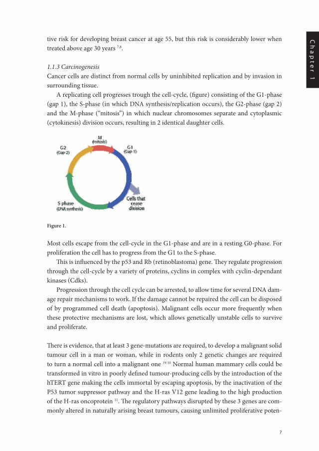
A replicating cell progresses trough the cell-cycle, (figure) consisting of the G1-phase (gap 1), the S-phase (in which DNA synthesis/replication occurs), the G2-phase (gap 2) and the M-phase (“mitosis”) in which nuclear chromosomes separate and cytoplasmic (cytokinesis) division occurs, resulting in 2 identical daughter cells.
Most cells escape from the cell-cycle in the G1-phase and are in a resting G0-phase. For proliferation the cell has to progress from the G1 to the S-phase.
This is influenced by the p53 and Rb (retinoblastoma) gene. They regulate progression through the cell-cycle by a variety of proteins, cyclins in complex with cyclin-dependant kinases (Cdks).
Progression through the cell cycle can be arrested, to allow time for several DNA dam-age repair mechanisms to work. If the damage cannot be repaired the cell can be disposed of by programmed cell death (apoptosis). Malignant cells occur more frequently when these protective mechanisms are lost, which allows genetically unstable cells to survive and proliferate.
There is evidence, that at least 3 gene-mutations are required, to develop a malignant solid tumour cell in a man or woman, while in rodents only 2 genetic changes are required to turn a normal cell into a malignant one 19.10 Normal human mammary cells could be transformed in vitro in poorly defined tumour-producing cells by the introduction of the hTERT gene making the cells immortal by escaping apoptosis, by the inactivation of the P53 tumor suppressor pathway and the H-ras V12 gene leading to the high production of the H-ras oncoprotein 11. The regulatory pathways disrupted by these 3 genes are com-monly altered in naturally arising breast tumours, causing unlimited proliferative poten-
Figure 1.
8
tial, anti-apoptosis strategies and invasive capabilities. However other genetic changes, working in the same pathways, may replace the above mentioned.
1.1.4 Influence of the microenvironment on cancer developmentInvasiveness of breast tumours occurred in vitro when fibroblasts, preferably immortalized fibroblasts were present. In their absence the process often stopped at the carcinoma in situ stage. Thus for the further development of a tumour, interaction of the mutated epithelial cells and the surrounding stroma, containing collagen and blood vessels, is important 12,13.
Especially with the endothelial cells, necessary for angiogenesis which is needed for the growth of a tumour above 0.4-2 mm. Tumour cells are supposed to have a pre-angiogenic, dormant state14. Vascular endothelial growth factor (VEGF), can induce angiogenesis and synthetic inhibitors of this angiogenic pathway can stop tumour progression in vitro 15. VEGF expression in tumour cells is also associated with more metastases by opening, as shown in a mouse model, the vascular endothelium sufficiently for tumour cells to pass through 16. All oncogenes and tumour suppressor genes influence directly or indirectly angiogenesis, 17,18 but they are not known to influence the occurrence of metastases 19.
1.1.5 Genetic predisposition for breast cancer The neoplastic process can be started by a somatic mutation (i.e. a mutation acquired dur-ing life and present in a limited number of cells of the body) in an oncogene or tumour suppressor gene that initiates clonal expansion. A germline mutation (inherited) in that gene, predisposes the owner to cancer, as this contributing mutation is present in every cell of the body, but it does not on its own cause the cancer. Carriers of such a germline mutation however, may develop multiple tumours, or tumours occurring at an earlier age19. According to the Knudson two-hit model, the first somatic mutation occurs in tu-mour-suppressor-gene-mutation-carriers in the normal copy of the gene, inherited from the unaffected parent 20,21. In BRCA1-associated breast tumours for instance, frequent loss of heterozygosity of the wild-type allele is seen indeed, suggesting that malignancy occurs when both functional alleles of BRCA1 are lost.
The genes that (when mutated) can increase the risk for breast cancer, e.g. BRCA1, BRCA2, ATM, Chk2, are nearly all involved in the normal DNA damage repair process, while p53 influences the progress from cells from the G1 into the S-(DNA-replication) -phase and thereby proliferation.
Deleterious mutations in the autosomal dominant transmitted genes BRCA1 and BRCA2 predispose for both breast and ovarian cancer.
BRCA1, located on chromosome 17q was first cloned in 1994 by Miki et al 22. It con-sists of 22 exons coding for a protein, the largest is exon 11.Many mutations that alter the function of the gene, are known today. The 2804del AA and IVS12-1643del3835 muta-tions are frequent in the Netherlands, originating each from one single origin/founder 23.
9
Ch
ap
te
r 1
The 185delAG and 5382insC mutations occur at a 10-fold higher frequency in the Ashke-nazi Jewish population.
BRCA2 on chromosome 13q was first cloned by Wooster’s group and has 27 coding ex-ons 24. 5579insA is a Dutch BRCA2 founder mutation. Deleterious BRCA2 mutations are less frequent than BRCA1 mutations in the Netherlands but not in the UK or Canada.
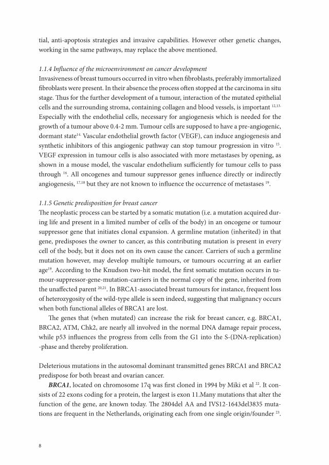
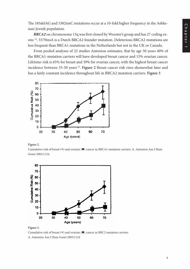
From pooled analyses of 22 studies Antoniou estimates, that by age 50 years 40% of the BRCA1-mutation carriers will have developed breast cancer and 15% ovarian cancer. Lifetime-risk is 65% for breast and 39% for ovarian cancer, with the highest breast cancer incidence between 35-50 years 25. Figure 2 Breast cancer risk rises shomewhat later and has a fairly constant incidence throughout life in BRCA2 mutation carriers. Figure 3
Figure 2.Cumulative risk of breast () and ovarian () cancer in BRCA1-mutation carriers. A. Antoniou Am J Hum Genet 2003;1124.
Figure 3.Cumulative risk of breast () and ovarian () cancer in BRC2 mutation carriersA. Antoniou Am J Hum Genet 2003;1124
Chap 1/ fig 3
10
BRCA1 functions as a sensor of DNA damage, and plays a role in cell cycle checkpoints. In response to DNA damage it may stop the cell progressing in the normal replicative growth cycle, triggering cell cycle arrest to allow more time for repair. If the damage cannot be repaired the cell can be disposed of by apoptotic cell death. Malignant cells occur more frequently when these protective mechanisms are lost, which allows genetically unstable cells to survive and proliferate.
BRCA1 and BRCA2 proteins are both involved in the pathway of the repair of double-strand breaks in DNA by homologous recombination 26. Double-strand breaks in DNA can be caused by ionizing radiation or for instance agents like mitomycin C and cisplatin. BRCA1 and 2 protein-deficient cells may therefore be more radiosensitive. In these BRCA1 or 2 defective cells double-strand breaks are repaired by an error prone mechanism-such as non homologous end-joining- and errors can lead to chromosomal rearrangements. It is thought that the resulting chromosomal instability is crucial for carcinogenesis 27. BRCA2 transports RAD51 from the cell cytoplasma to the nuclear site, where its action is requested for this DNA repair process 26. BRCA1 plays a part in a third DNA repair mechanism, nucleotide excision repair.
In a small population based study only 30% of the BRCA1 mutation carriers had a history of a first or second degree relative with breast cancer, vs 56% of the BRCA2 muta-tion carriers. Two or more affected relatives were seen in 20% of the BRCA1 and BRCA2 mutation carriers vs. 14% of controls of age 40 years or less 28.
Breast cancers arising in BRCA1 carriers tend to have distinctive histopathologic fea-tures; they are frequently high grade, with abundant lympocytic infiltration, and most are HER-2, estrogen-, and progesterone receptor negative. They more frequently show promi-nent pushing margins around the tumour, and not the extensive stromal reaction, termed desmoplasia in which excess collagen is deposited, causing the star- like spiculae 29-31. The pathologic characteristics of BRCA2 tumours are less different from sporadic ones 29-31
Other breast cancer susceptibility genes, transfer a lower life-time risk for breast can-cer than BRCA1 and BRCA2: P53 on chromosome 17p13, ATM on chromosome 11q23, PTEN on chromosome 10p and possibly a CHEK 2,1100delC mutation.
The P53 gene at chromosome 17p13, codes for a protein that functions to block the cell cycle if the DNA is damaged. This allows time to repair DNA If the damage is severe this protein can cause apoptosis (programmed cell death). A p53 mutation is the most frequent mutation seen in malignant tumour cells.
A P53 germline mutation may lead to the Li-Fraumeni syndrome, an autosomal domi-nant disorder, leading to an excess of breast cancers at a relatively young age, soft tissue and osteo-sarcoma, brain tumours, leukaemia, or adrenocortical carcinoma. P53 muta-tions are detected in only 1% of unselected women with breast cancer. Women with a P53 germline mutation who survive childhood cancer will develop breast cancer ≤ 50 years in about 50%. The increase in risk is however greatest before age 25 years and decreases, to a relative risk of 1.8 after the age of 45 32.
11
Ch
ap
te
r 1
Somatic mutations in P53 are found in up to 60% of human breast cancers. One inac-tivated allele may be sufficient for the development of breast cancer. Surprisingly breast cancer risk was decreased in homozygous carriers of 3 P53 polymorphisms (in intron 3, exon 4 and intron 6) 33
Ataxia Teleangiectasia is a recessive hereditary disorder. Homozygote carriers of the AT-mutated gene at chromosome 11q22-23, develop severe neurological problems eg cerebellar ataxia. ATM functions upstream of BRCA1 in the double-strand break repair mechanism. Homozygote carriers have an increased radiation sensitivity and risk for lym-phoma, breast and many other cancers.
A PTEN mutation on chromosome 10q23 leads to the autosomal dominant Cowden syndrome and predisposes for both benign and malignant tumours e.g., breast, thyroid, intestinal polyps and skin cancer.
1.1.6 Familial breast cancer risk without a major breast cancer gene mutationApproximately 10% of breast cancers are detected in patients with a clear family history, but high-penetrance germ-line mutations in BRCA1 or BRCA2 account for less than 20% of the familial aggregation of breast cancer 34.
The sensitivity of the DNA tests for deleterious BRCA1 and BRCA2 mutations in the Netherlands is estimated to be 80%. The risk of chance clustering of ≥ 3 breast cancers un-der the age of 60 in a family has been estimated as less than 10% 35. So some of these fami-lies will contain non-recognized BRCA1/2 mutations and some will be caused by chance-clustering, but many of these family-histories suggest an unidentified heritable risk.
Further, specific histopathologic characteristics have been described in non-BRCA1/2 breast cancers from families with at least 3 breast cancer cases, such as more frequent low-grade tumours, low mitotic count, a lower proliferation rate and more lobular carcinoma 29-31. These features seem to discriminate these non-BRCA1/2 from both BRCA1/ BRCA2 and sporadic breast cancers. At the moment however it is impossible to indicate which women in these families run the increased breast cancer risk.
1.1.7 Inherited risk and environmental factorsWe do not know by which influence about 60% of the BRCA1 mutation carriers does not have manifest breast cancer at age 50 yrs. and what determines which 40% of mutation carriers will not have signs of this disease at age 70 years. Nor why life-time risk is some-what lower for BRCA2 mutation carriers (45%) than for BRCA1, and why most cancers in BRCA2 carriers develop above age 50 yrs. The incidence of breast cancer halves in BRCA1 mutation carriers after menopause, when estrogen and progestreron levels fall sharply. In BRCA2 carriers however no decrease is seen.
Neither is it clear why ovarian cancer incidence is increased in both BRCA1 and 2 mu-tation carriers, and not for instance endometrial cancer, which is much more associated with estrogen/progesterone exposure. Nor why ovarian cancers develop less and later than breast cancers in both BRCA1 and 2 mutation carriers. In the general population no cor-
12
relation or inverse association is known between breast and ovarian cancer. Benign ovar-ian cysts however are by an unknown mechanism associated with reduced breast cancer risk (OR =0.70% 95%CI 0.59-0.82) 36.
Why do men with BRCA2 but not with BRCA1 mutations have a higher risk for pros-tate cancer and most likely also breast cancer37.
It has been shown in mice recently, that food substances like folic acid, can influ-ence the methylation of DNA and thereby the expression of genes38. Silencing genes like BRCA1 by methylation, seems to be a frequent event in sporadic breast cancer39. Which environmental and epigenetic factors, co-genes and gene-polymorphisms influence the expression of the main breast cancer genes needs further investigation.
1.1.8 Inflammation and cancerMany examples exist of chronic inflammations that predispose to cancer; like colitis ul-cerosa for colon cancer, gastric Helicobacter Pylori infection for stomach cancer, hepati-tis B and C for hepatocellular carcinoma, papilloma-virus infection for cervical cancer, schistosomiasis for bladder cancer. No single infectious agent is known today, to cause subclinical chronic inflammation, preceding human breast cancer.
While aspirin and non-steroidal anti-inflammatory drugs may reduce colon cancer risk, no such effect has been described yet for breast cancer.
1.2 Risk reductionFor a woman with increased breast cancer risk from a relatively early age on, because of a BRCA1 or BRCA2 gene mutation or a clear family history there are at the moment only a few options to reduce the mortality risk of the disease.
1.2.1. ChemopreventionTamoxifen has been shown to reduce by more than 50% the 30% risk of contralateral breast cancer in BRCA1 and 2 mutation carriers. In pre and postmenopausal carriers similar risk reduction was seen, suggesting that the anti-estrogen tamoxifen is effective in preventing ER-negative as wel as ER-positive second primary breast cancers40. These results suggest, that tamoxifen might be effective in preventing premenopausal primary breast cancers in both BRCA1 and BRCA2 mutation carriers also.
Stem cells are self-renewing. When they divide, one of the daughter cells differentiates and eventually stops dividing. The other retains its stem cell properties with the ability to divide in the same way. Cancer stem cells have been identified. The findings in immuno-deficient NOD/SCID mice with human breast cancer cells injected in their mammary glands, showed that only a small proportion of the tumour cells, that can be recognised by the surface markers CD44+/CD24-, are self-renewing and drive tumour growth and metastasis, the so called stem cells41. Preliminary evidence suggests that the proportion of stem cells of a tumour may determine how deadly it is and these cells should specifically be targeted. Clarke found 25% stem cells in an extremely aggressive tumour.

13
Ch
ap
te
r 1
Poly (ADP-ribose) polymerase (PARP) is an enzyme involved in base excision repair of DNA single-strand breaks. Inhibition of PARP-enzyme leads to chromosomal insta-bility, cell cycle arrest and apoptosis in BRCA1 or 2 lacking cells in mice42. This seems caused by the persistence of DNA lesions normally repaired by homologous recombina-tion. Whether this mechanism works the same in humans and without major side-effects needs further research.
There are no chemoprevention studies ongoing in unaffected BRCA carriers and women with familial breast cancer risk in the Netherlands. Such studies are necessary to weigh effectiveness and side-effects in different groups.
1.2.2 Surgical prevention.I. Preventive oophorectomy,Preventive oophorectomy has been shown to reduce not only the risk for ovarian cancer, but to also halve breast cancer risk if performed in premenopausal BRCA1 and 2 muta-tion carriers 43. In the Netherlands bilateral preventive salpingo-oophorectomy is often recommended to BRCA1 and 2 mutation carriers with a completed family from 40 years onwards, as ovarian screening has not been shown effective in detecting the disease at an early stage.II Bilateral risk-reducing mastectomy. By the total (simple) mastectomy 95-99% of breast tissue is removed including the areola-nipple complex. The nipple-areola complex is preserved with vascularisation and some ducts in the subcutaneous mastectomy. Both techniques do not allow the complete re-moval of all breast parenchyma, but a risk reduction of 90% for primary breast cancer may be reached44-46. Immediate reconstruction can be performed. After 5.2 yrs follow-up of a cohort of 76 healthy BRCA1/2 mutation carriers who choose risk-reducing bilateral mastectomy (mean age 37.7 yr.) and 63 under surveillance (mean age 39.5 yr.) 9 women in the surveillance group developed breast cancer and 2 metastases (age 23 and 26 yrs.) vs. one women with breast cancer metastases in the mastectomy group 3 yrs after both bi-lateral mastectomy and bilateral salpingo-oophorectomy at age 36 yrs47. As women make the choice for preventive surgery in order to prevent disease mortality, longer follow-up is needed to determine the effectiveness48.
1.2.3 Secondary prevention by surveillance/screeningScreening cannot prevent cancer but aims to reduce the mortality and part of the mor-bidity, by detecting the cancer at an early stage. This is based on several studies, showing that increasing size of breast cancer and increasing number of axillary nodal metastasis independently predict decreasing survival chances49-51. And increasing size of the pri-mary tumour is associated with more axillary metastases. Seemingly conflicting evidence however suggests that the proclivity to metastasize is acquired early in tumour genesis52. The percentage of patients with metastases increases faster with the size of the tumour in high grade breast cancers than in low grade53. So both the inherent aggressiveness/type
14
of breast cancer as indicated by grade, hormonal receptors or gene-expression profiles and the size of the cancer at detection seem to influence and predict survival. Tabar et al. found good cumulative 12 yr. disease specific survival rates of over 90% for all high grade tumours ≤ 1 cm54.
Several randomised studies and population studies have shown, that screening women above age 50 years with mammography may reduce mortality if a large part of the popula-tion participates55,56. In the Netherlands a 2-yearly mammography is therefore provided for every woman from age 50-75 years.
Four large prospective studies have recently shown that screening with MRI and mam-mography can detect hereditary breast cancers early57-60. Two of the studies in this thesis served as pilot-study for the Dutch multicentre MRI-screening for women at high risk study (MRISC). Cost-effectiveness of screening healthy BRCA1/2 mutation carriers with MRI has recently been shown61. The optimum screening procedure and interval is not yet clear for every risk-group, nor are all cancers detected in a 100% curable stage.
The different prevention strategies have also when free of charge different acceptability in different countries and hospitals. Screening with mammography was slightly less accept-able for high-risk British (76.9%), than French (90.8%) and Canadian (91.7%) women. Preventive oophorectomy > 40 yrs. was acceptable for 35% of the French, 45% of the Ca-nadian and 58% of the British women, bilateral risk-reducing mastectomy> age 35 yrs. for 7%, 22% and 23% respectively, chemoprevention for respectively 49%, 46% and 80%62.
None of the preventive measures we can offer today give 100% prevention of breast cancer mortality and all have clear side-effects. Until the cause of the disease can be di-rected effectively, improving the current options is needed.

15
Ch
ap
te
r 1
References
1. Visser o, Coebergh JWW, Schouten IJ et al. Incidence of cancer in the Netherlands. Utrecht, Comprehen-sive Cancer Centers, 2001.
2. Vervoort MM, Draisma G, Fracheboud J, van de Poll-Franse LV, de Koning HJ. Trends in the usage of adjuvant systemic therapy for breast cancer in the Netherlands and its effect on mortality. Brit J Ca 2004;91:242-247.
3. Visser O, van Leeuwen FE. Stage-specific survival of epithelial cancers in North-Holland/Flevoland, The Netherlands. Eur J Ca 2005;41:2321-2330.
4. Danaei G, Vander Hoorn S, Lopez AD et al Causes of cancer in the world; comparative risk-assessment of nine behavioural and environmental risk factors. Lancet 2005;366:1784-93.
5. Colditz GA. Epidemiology and prevention of breast cancer. Cancer Epidemiol Biomarkers Prev 2005;14(4);768-72.
6. Pike MC, Krailo MD, Henderson Be, Casagral JT, Hoel DG. Hormonal risk factors, breast tissue age and the age incidence of breast cancer. Nature 1983;303 (5920):767.
7. Travis LB, Hill D, Dores GM, Gospodarowicz M, van Leeuwen FE, Holowaty E et al. Cumulative absolute breast cancer risk for young women treated for Hodgkin lymphoma. JNatl Cancer Inst 2005;97:1428-37.
8. Van Leeuwen FE, Klokman WJ, van ’t Veer M, Hagenbeek A, Krol ADG, Vetter UAO et al. Long term risk of second malignancy in survivord of Hodgkin’s disease treated during adolescence or young adulthood. J Clin Oncol 2000;18:487-97.
9. Komarova NL, Sengupta A & Nowak MA. Mutation-selection networksof cancer initiation: tumor sup-pressor genes and chromosomal instability. J. Theor. Biol. 2003;223:433-50.
10. Hahn WC, Counter CM, Lundberg AS, Beijersbergen RL, Brooks MW, Weinberg RA. Creation of human tumour cells with defined genetic elements. Nature 1999;4000:464-468.
11. Elenbaas B, Spirio L, Koerner F, Fleming MD, Zimonjic DB, Donaher JL, Popescu NC, Hahn WC, Weinberg RA. Human breast cancer cells generated by oncogenic transformation of primary mammary epithelial cells. Genes Dev 2001;15(1): 50-65.
12. Shekhar MPV, Werdell J, Santer SJ, Pauley RJ, Tait l. Breast stroma plays a dominant regulatory role in breast epithelial growth and differentiation: Implications for tumor development and progression. Cancer research 2001;61:1320-26.
13. Dong-Le bourhis X, Bertthois Y, Millot G, Degeorges A, Sylvi M, Martin P-M, Calvo F. Effects of stromal and epithelial cells derived from normal and tumorous breast tissue on the proliferation of human breast cancer cell lines in co-culture. Int J Cancer 1997;71:42-48.
14. Gimbrone MA jr, Leapman SB, Cotran RS, Folkman J, Tumor dormancy in vivo by prevention of neovas-cularisation. J Exp Med.19772;136:261-276.
15. Ferrara N, Hillan KJ, Gerber HP & Novotny W. Discovery and development of bevacizumab an anti-VEGF antibody for treating cancer. Nat. Rev. Drug Discov. 2004;3:391-400.
16. Weis SM, Cheresh DA. Pathophysiological consequences of VEGF-induced vascular permeability. Nature 2005;437(7058):497-508.
17. Verheul HMH, Voest EE, Schlingemann RO. Are tumours angiogenesis-dependent? Review. J Pathol. 2004;202:5-13.
18. Folkman J. Angiogenesis in cancer, vascular, rheumatoid and other diseases. Nat. Med.1995;1(2):120-2.
19. Vogelstein B, Kenzler KW. Cancer genes and the pathways they control. Nature Medicine 2004;10(8):789-799.
16
20. Knudson AGJ. Mutation and cancer: statistical study of retinoblastoma1971 Proc Natl Acad of Sci USA 68:820-23.
21. Knudson AG. Cancer genetics. Am J Med Genet. 2002;111:96-102.
22. Miki Y, Swensen J, Shattuck-Eidens D, Futreal P, Harshman K, Tavtigian S, Liu Q, et al. A strong candidate for the breast and ovarian cancer susceptibility gene BRCA1. Science 1994;266:66-71.
23. Verhoog LC, vanden Ouweland AMW, Berns E, van Veghel-Plandsoen MM, van Staveren IL, Wagner A, Bartels CCM, Tilanus-Linthorst MMA, Devilee P, Seynaeve C, Halley DJJ et al. Large regional differ-ences in the frequency of distinct BRCA1/BRCA2 mutations in 517 Dutch breast and/or ovarian cancer families. EurJCa 2001;37:2082-2090.
24. Wooster R, Bignell G, Lancaster J, Swift S, Seal S, MangionJ, Collins N, et al. Identification of the breast cancer susceptibility gene BRCA2. Nature 1995;378:789-792.
25. Antoniou A et al. Average risks of breast and ovarian cancer associated with BRCA1 or BRCA2 mutations detected in case series unselected for family history: a combined analyses of 22 studies. AmJHum Genet 2003;72:1117-1130.
26. Venkitaraman AR. Cancer susceptibility and the functions of BRCA1 and BRCA2. cell 2002;108:171-182.
27. Narod SA, Foulkes WD. BRCA1 and BRCA2:1994 and beyond. Nature Rev Cancer 2004;4:665-76.
28. Armes JE, Egan AJM, Southey MC, Dite GS, McCredie ME, Giles GG, Hopper JL, Venter DJ. The histo-logic phenotypes of breast carcinoma occurring before age 40 years in women with and without BRCA1 or BRCA2 germline mutations. A population-based study. Cancer 1998;83:2335-45.
29. Lakhani SR, Gusterson BA, Jacquemier J, Sloane JP, Anderson TJ, van de Vijver MJ, Venter D, Freeman A, Antoniou A, McGuffog L, Smyth E, Steel CM et al. The pathology of familial breast cancer: Histological features of cancers in families not attributable to mutations in BRCA1 or BRCA2. Clin Cancer Res 6:782-9, 2000.
30. Adem C, Reynolds C, Soderberg CL, Cunningham JM, Reynolds C, Sebo TJ, Thibodeau SN, Hartmann LC, Jenkins RB. Pathologic characteristics of breast parenchyma in patients with hereditary breast carci-noma, including BRCA1 and BRCA2 mutation carriers. Cancer 97:1-11, 2003
31. Eerola H, Heikkilä P, Tamminen A et al. Histopathological features of breast tumours in BRCA1, BRCA2 and mutation negative breast cancer families. Breast Cancer Research. 2005 (7):93-100. DOI 10.1186/bcr953
32. Garber JE, Goldstein AM, Kantor AF, Deyfus MG, Fraumeni JF jr., Li FP. Follow-up study of twenty-four families with Li-Fraumeni syndrome. Cancer Res 1991;51:6094-7.
33. Sjalander A, Birgander R, Hallmans G, Cajander S, Lenner P, Athlin L, Beckman G, Beckman L. p53 polymorphisms and haplotypes in breast cancer. Carcinogenesis 1996;17;1313-16.
34. Dite GS, Jenkins MA, Southey MC, Hocking JS, Giles GG, McCredie MRE Venter DJ, Hopper JL. Familial risks, early-onset breast cancer, and BRCA1 and BRCA2 germline mutations JNatlCancerInst 2003;95:448-57.
35. Peto J, Collins N, Barfoot R, Seal S, Warren W, Rahman N, Easton DF, Evans C, Deacon J, Stratton MR. Prevalence of BRCA1 and BRCA2 gene mutations in patients with early-onset breast cancer. J Natl Can-cer Inst. 91:943-49, 1999.
36. Knight JA, John EM, Milne RL, Dite GS, Balbuena R, Shi EJQ, Giles GG, Ziogas A, Andrulis IL, Whitte-more AS, Hopper JL. An inverse association between ovarian cysts and breast cancer in the Breast Cancer Family Registry. IntJCa 2006;118:197-202.
37. van Asperen CJ, Brohet RM, Meijers-Heijboer EJ, Hoogerbrugge N, Verhoef S, Vasen HF, Ausems MG, Menko FH, Gomez Garcia EB, Klijn JG, Hogervorst FB, van Houwelingen JC, van’t Veer LJ, Rookus MA, van Leeuwen FE; Netherlands Collaborative Group on Hereditary Breast Cancer (HEBON). Cancer risks in BRCA2 families: estimates for sites other than breast and ovary. J Med Genet. 2005 Sep;42(9):711-9.
38. Pennisi E. Supplements restore gene function via methylation. Science2005;310:1761.

17
Ch
ap
te
r 1
39. Catteau A, Harris WH, Xu CF, Solorion E. Methylation of the BRCA1 prootor region in sporadic breast and ovarian cancer: correlation with disease characteristics. Oncogene1999;18:1957-65.
40. Gronwald J, Tung N, Foulkes WD, Offit K, Gershoni R, Daly M, Kim-Sing C, Olsson H, Ainswoth P, Eisen A, Saal H, Friedman E, Olopade O, Osborne M, Weitzel J, Lynch H, Ghadirian P, Lubinski J, Sun P, Narod SA. Tamoxifen and contralateral breast cancer in BRCA1 and BRCA2 carriers: An update. IntJCa dec 2005DOI 10.1002/ijc.21536.
41. Al-Hajj M, Wicha MS, Benito-Hernandez A, Morrison SJ, Clarke MF. Prospective identification of tu-morigenic breast cancer cells. Proc Natl Acad Sci U S A. 2003;100(7):3983-88.
42. Farmer H, McCabe N, Lord CJ, Tutt AN, Johnson DA, Richardson TB, Santarosa M, Dillon KJ, Hickson I, Martin NM, Jackson SP, Smith GC, Ashworth A. Targeting the DNA repair defect in BRCA mutant cells as a therapeutic strategy. Nature. 2005 Apr 14;434(7035):917-21.
43. Rebbeck TR, Lynch HT, Neuhausen SL, Narod SA, van’t Veer L, Garber JE, Evans G, Isaacs C, Daly MB, Matloff E, Olopade O, Weber BL et al. Prophylactic oophorectomy in carriers of BRCA1 or BRCA2 muta-tions. N Eng J Med 2002;346:1616-22.
44. Hartmann LC, Sellers TA, Woods JE et al. Efficacy of bilateral prophylactic mastectomy in women with a family history of breast cancer. N Engl J Med 1999;340:77-84.
45. Meijers-Heijboer H, van Geel B, van Putten WL, et. al. Breast cancer after prophylactic bilateral mastec-tomy in women with a BRCA1 or BRCA2 mutation. N Engl J Med 2001;345:159-64.
46. Rebbeck TR, Friebel T, Lynch HT et al. Bilateral prophylactic mastectomy reduces breast cancer risk in BRCA1 and BRCA2 mutation carriers: the PROSE Study Group. J Clin Oncol 2004;22:1055-62.
47. Klijn JGM, van Geel AN, Meijers-Heiboer H, Tilanus-Linthorst MMA, Bartels CCM, Crepin CMG, Menke-Pluymers MB, Follow-up of the Rotterdam study on prophylactic mastectomy versus surveillance in BRCA1/2 mutation carriers. J Clin Oncol 2004;22 (14S): 835S (9502).
48. Schrag D, Kuntz KM, Garber JE, Weeks JC. Decision analysis-effects of prophylactic mastectomy and oophorectomy on life expectancy among women with BRCA1 or BRCA2 mutations. N Engl J Med 1997;336:1465-71.
49. Sant M, Allemani C, Capocaccia R, et al. Stage at diagnosis is a key explanation of differences in breast cancer survival across Europe. Int. J Cancer 2003;106(3):416-22.
50. Michaelson JS, Silverstein M, Sgroi D, et al. The effect of tumor size and lymph node status on breast carcinoma lethality. Cancer 2003; 98; 2133-43.
51. Carter C, Allen C, Henson D. Relation of tumor size, lymph node status and survival in 24,740 breast cancer cases. Cancer 1989; 63:181-7.
52. Schmidt-Kittler O, Ragg Th, Daskalakis A, et al. From latent disseminated cells to overt metastasis: Ge-netic analysis of systemic breast cancer progression. Proc. Natl.Acad.Sci.USA 2003;100, 13:7737-42.
53. Tubiana M, Koscielny S. Natural history of human breast cancer: recent data and clinical implications. Breast Cancer Res and Treatm. 1991;18:125-40.
54. Tabar L, Fagerberg G, Day NE, et al. Breast cancer treatment and natural history: new insights from results of screening. Lancet 1992; 339:412-4
55. Fracheboud J, Otto SJ, van Dijk JA, Broedres MJ, Verbeek AL, de Koning HJ; National Evaluation Team for Breast cancer screening (NETB). Decreased rates of advanced breast cancer due to mammography screening in The Netherlands. Br J Cancer 2004;91(5):861-7.
56. Tabar L, Yen MF, Vitak B, Chen HH, Smith RA, Duffy SW. mammography service screening and mortality in breast cancer patients: 20-year follow-up before and after introduction of screening. Lancet 2003;361:1405-10.
57. Kriege M, Brekelmans CTM, Boetes C, et al. Efficacy of MRI and mammography for breast-cancer screening in women with a familial or genetic predisposition. N Eng JMed 2004;351:427-37.
18
58. Leach MO, MARIBS study group. Screening with magnetic resonance imaging and mammography of a UK population at high familial rik of breast cancer: a prospective multicentre cohort study (MARIBS). Lancet 2005;365:1769-78.
59. Warner E, Plewes DB, Hill KA, et al. Surveillance of BRCA1 and BRCA2 mutation carriers with magnetic resonance imaging, ultrasound, mammography and clinical breast examination. JAMA 2004;292(11):1317-25.
60. Kuhl CK, Schrading S, Leutner CC, Morakkabati-Spitz N, Wardelmann E, Fimmers R, Kuhn W, Schild HH. Mammography, Breast ultrasound, and Magnetic Resonance Imaging for surveillance of women at high familial risk for breast cancer. JCO 2005;23:8469-76.
61. Reijnsburger AJ, Draaisma G, der Kinderen AJ, Tilanu-Linthorst MMA, Oosterwijk JC, Zonderland HM, Manoliu RA, Boetes C, Brekelmans CTM, Habbema JDF, Rutgers EJT, Klijn JGM, de Koning HJ. Screening women with a familial or genetic predisposition to breast cancer: costs and effects of alternative screening policies. Submitted
62. Julian-Reynier CM, Bouchard LJ, Evans DG, Eisinger FA, Foulkes WD, Kerr b, Blanquaert IR, Moatti J-P, Sobol HH. Women’s attitudes toward preventive strategies for hereditary breast or ovarian carcinoma differ from one country to another. Differences among English, French and Canadian women. Cancer 2001;92:959-68.
19
Ch
ap
te
r 1
1.3 Scope and outline of this thesisIn this thesis we investigated which features of hereditary and familial breast cancer influ-ence the effectiveness of screening women in reducing the mortality of the disease. We examined how screening may be best adapted in this specific group.
The last 2 decades have seen a lively debate, whether breast tumours have either from their origin a more or less pronounced capacity to behave aggressively and metastasize, or cause metastases increasingly with increasing lifetime and size.
We therefore investigated in chapter 2.A the influence of tumour stage on breast can-cer specific survival in patients at high familial breast cancer risk without a BRCA1 or BRCA2 mutation. As the histopathologic characteristics described in this group suggested a possibly better survival we compared their survival with patients not selected for fam-ily history (“sporadic”) of the same age. Furthermore we assessed which other factors influenced survival, e.g. the occurrence of contralateral breast cancer, as in some studies 12-60% of familial patients get a contralateral preventive mastectomy.
We analyzed in chapter 2.B the influence of tumour stage and other factors on breast cancer survival in BRCA1 and BRCA2 mutation carriers, non-BRCA1/2 patients with fa-milial risk and sporadic patients. We also assessed ipsilateral recurrence and the incidence of contralateral breast cancer in these 4 groups, and its impact on survival.
The frequency of a screening test should be adapted to the expected tumour growth rate to prevent interval cancers. BRCA1 tumours have often a high mitotic count and BRCA1 and -2 tumours are more often grade 3 or 2 than sporadic cancers, suggesting fast-er growth. We therefore investigated in chapter 3.A and 3.B the growth rates of BRCA1, -2 and familial breast cancers detected respectively in the Dutch multicentre MRI-screening study MRISC or during screening at the Daniel den Hoed Cancer Centre (3.A). We per-formed an extended international study on factors influencing the growth rate of heredi-tary breast cancer in the British 22-centre MRI-screening study MARIBS, the Canadian uni-centre study and the extended MRISC study (3.B).
In chapter 4 we investigated the rate of interval cancers in the 3 above mentioned MRI-screening studies in the different risk-groups and discuss on the role of breast self-examination in a MRI-screening setting.
As mammography is the most used and best documented screening tool for breast cancer, we investigated in chapter 5 the factors that contribute to a decreased sensitivity of mammography in screening BRCA1 and 2 mutation carriers in comparison to as young sporadic patients.
In chapter 6 we investigate the effectiveness of breast-MRI for breast cancers, occult at clinical examination and mammography.
As tumour size and nodal status were proven to be a reliable proxy for survival in hereditary breast cancer also (in chapter 2), we investigated in chapter 7 tumour stages of familial high-risk patients detected during surveillance, partly with MRI. These results are

20
compared to the tumour stages in symptomatic patients visiting the outpatient clinic in the same period and in patients referred by the national breast screening program.
In chapter 8 a preliminary investigation is performed to indicate the extra cost caused by the addition of MRI to the other screening methods for women at high hereditary risk.
In chapter 9 we investigated the influence of DNA-testing selection bias on the contra-lateral breast cancer incidence and survival in women with a high familial risk for breast cancer, but a negative test for BRCA1&2.
Finally a general discussion and summary of the results reported in this thesis is given in chapter 10.

Chapter 2
2A. Contralateral recurrence and prognostic factors in familial non-BRCA1/2-associated breast cancer
Madeleine MA Tilanus-Linthorst, Celina Alves, Caroline Seynaeve, Marian BE Menke-Pluymers, Alexander MM Eggermont, Cecile TM Brekelmans
Accepted British Journal of Surgery 2006
22
Abstract
Background A higher incidence of contralateral breast cancer (CBC) and ipsilateral recurrence (ILR) has been reported in familial breast cancer (BC) than in sporadic cancer.This study in-vestigated the influence of contralateral cancer and tumour stage on survival in familial non-BRCA1/BRCA2-associated breast cancer.
MethodsThe incidences of contralateral breast cancer, ipsilateral recurrence, distant disease-free and overall survival (OS) were assessed in 327 patients from families with ≥ 3 breast and/or ovarian cancers, but no BRCA1 or BRCA2 gene mutation (familial-non-BRCA1/2), and in 327 control cases with sporadic breast cancer, matched for year and age at detec-tion.
ResultsMean follow-up was 7.3 yrs for patients with familial-non-BRCA1/2 cancers and 6.5 yrs. for sporadic patients. Tumours were stage T1 or lower in 62.1% of familial-non-BRCA1/2 cancers vs. 49.9% in sporadic breast cancers (p= 0.003), and node-negative in 55.8% ver-sus 52.1% respectively (p=0.477). After 10 years the incidence of metachronous contralat-eral breasrt cancer was 6.4% for familial-non-BRCA1/2 tumours versus 5.4% for sporadic cancers. The rate of ipsilateral recurrence was not significantly increased (17.0 versus 14.2 per cent respectively at 10 yrs; P=0.132). Tumour size (hazard ratio (HR) 1.02 per mm. increase; p=0.016) and node status (HR 2.6 for three or more involved nodes versus node negative, P=0.017) were independent predictors of overall survival in the familial-non-BRCA1/2 group and in the whole group, whereas contralateral breast cancer (HR 0.7; p=0.503) and risk-reducing contralateral mastectomy (HR 0.4; p=0.163) were not.
ConclusionStage at detection was a key determinant of prognosis in familial-non-BRCA1/2 breast cancer, whereas contralateral cancer was not. Risk-reducing contralateral mastectomy did not significantly improve survival, but early detection can. Decisions on breast-conserving treatment can be made on the same grounds in patients with familial and sporadic breast cancer.
23
Ch
ap
te
r 2
Introduction
A positive family history is a risk factor for breast cancer1,2 and possibly for contralateral breast cancer (CBC) 3-9. About 10 per cent of breast cancers are detected in women with a clear family history. High-penetrance germ-line mutations in BRCA1 or BRCA2, how-ever, can be demonstrated in fewer than 20 per cent of these familial patients10 A recent review of familial non-BRCA1/BRCA2-associated breast cancer concluded, that data on ipsilateral recurrence and contralateral tumours in this group are scarce and that survival analyses are hampered by small numbers or incomplete testing 11.
The likelihood of chance clustering of 3 or more breast cancers in female relatives under the age of 60 years has been estimated as less than 10% 10. Consistently, more low-grade tumours have been described in patients from families with at least 3 breast cancer cases, but a negative test for BRCA1/BRCA2. These tumours also have low mitotic count, a lower proliferation rate and more lobular carcinoma 12-14. These features discriminate fa-milial non-BRCA1/BRCA2-associated cancers from both BRCA1 or BRCA2 cancers and sporadic breast cancer, and suggest possible improved survival for these patients.
The authors recently compared 327 women with breast cancer and at least two other relatives with breast or ovarian cancer and negative testing for BRCA1 and BRCA2 (fa-milial non-BRCA1/2 cancer) and 327 age-matched influenced by DNA testing selection bias15, that is, women were more likely to have DNA testing after the development of a contralateral cancer and when they lived longer after diagnosis.
In studies not selected by family history, some have shown the same survival rate for bilateral breast carcinoma as for unilateral breast cancer others have shown a worse sur-vival.16-18. The impact of contralateral cancer and primary tumour stage on survival in fa-milial non-BRCA1/2 cancer has to our knowledge not previously been analyzed. Data on ipsilateral and contralateral recurrence in familial non-BRCA1/2 breast cancer are needed for evidence-based decisions on breast conserving treatment and risk-reducing contralat-eral mastectomy.
This study assessed the incidence of ipsilateral recurrence, contralateral breast cancer, distant disease free (DDFS) and overall survival (OS) in the two populations studied pre-viously 15. To estimate the importance of early detection, the impact of tumour stage on these endpoints was also assessed.
Patients and Methods
PatientsThe study population has been described previously15. In brief, from 265 consecutive families, registerd at ErasmusMC with at least 3 confirmed relatives with breast cancer or breast and ovarian cancer, including the index case, but with negative testing for BRCA1 or BRCA2 mutations before May 1 2004, all 327 women with primary breast cancer (in-
24
cluding ductal carcinoma in situ; DCIS), diagnosed between 1January1980 and 31 De-cember 2002 were selected. All had a pathology report of the tumour, follow-up data for at least 6 months and no previous cancer other than basal skin carcinoma Of these 262 women tested negative for BRCA1 and BRCA2, whereas in 65 patients one or more family members with breast or ovarian cancer tested negative. One of 117 familial patients tested positive test for CHEK 2*1100delC. DNA testing was performed at the Clinical Genetics Department of the Erasmus MC Rotterdam. BRCA1/BRCA2 and CHEK2*1100delC mu-tation analyses were reported19,20 The sensitivity for deleterious BRCA1 or BRCA2 muta-tions was estimated as 80%.
Control patients with breast cancer had no history of more than one family member with breast cancer > age 50 yrs (sporadic) and were matched for age and year of diagnosis to each patient with non-BRCA1/2 cancer.
Study protocolDetailed information was examined on family history, age at diagnosis, hormonal factors such as menopausal status, tumour characteristics (size, type, grade) node status, local and systemic treatment and local and distant from the medical files and from information at the Department of Clinical Genetics
For the purpose of the analyses follow-up was assumed to commence on the date of detection of the first breast cancer and to cease on the date of the last notes in the medi-cal files, death, or otherwise at loss to follow-up. Cancer in the contralateral breast was considered metachronous if detected more than 3 month’s after the first tumour, also after primary DCIS. The synchronous occurrence of metastases (within 3 months) with a con-tralateral cancer was counted as a failure in the group with unilateral BC. The endpoints of interest were date of first local and/or distant recurrence, the occurrence of a second primary breast tumour and date of death due to breast cancer or other cause. The census date for follow-up was 1 May, 2004.
The study was approved by the Erasmus MC Institutional Review Board. All DNA-tested patients gave informed consent for DNA analyses and their use in research.
Statistical methodsUsing chi-square tests (for categorical variables) or t-tests (for continuous variables) we compared patient and tumour characteristics between the familial non-BRCA1/2 patients and sporadic breast cancer patients. Kaplan-Meier survival curves were calculated and differences compared with the logrank test. Endpoints were the incidence of contralateral breast cancer, local recurrence, distant disease-free survival and overall survival. The si-multaneous effect of several prognostic variables on these four endpoints was investigated by Cox proportional hazard regression models.
The impact of contralateral breast cancer on distant disease-free and overall survival was investigated twice. In the first analyses survival was defined as the time from date of diagnosis of the first breast cancer. In the second, survival of patients with a contralateral
25
Ch
ap
te
r 2
breast cancer was counted from the date of diagnosis of the contralateral tumour, and the time before the occurrence of the contralateral cancer was counted as follow-up time in the unilateral group. This method was modeled by including a time-dependent variable for contralateral breast cancer. The difference between the two methods has been well explained by Heron et al.16
P-value < 0.050 (two-tailed) was considered statistically significant. All analyses were performed by STATA/SE TM for Windows version 8.1.
Results
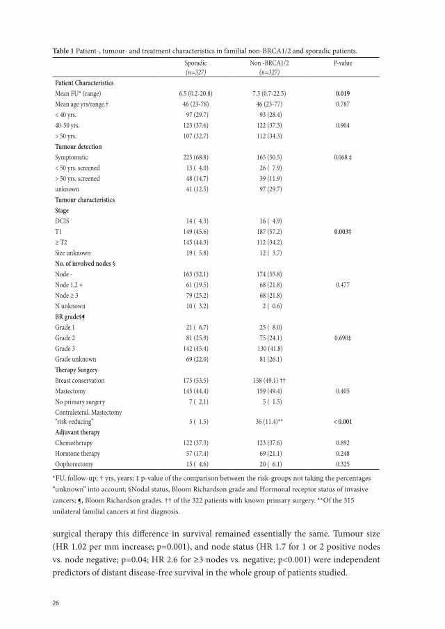
Patient and tumour characteristicsA hereditary breast cancer syndrome (HBC) was seen in 214 of the 265 families, and hereditary breast and ovarian cancer (HBOC) in 51. Patient, tumour and treatment char-acteristics have been described15 and are summarized in Table 1.
Some 65.7% of the familial non-BRCA1/2 patients were diagnosed in women at or under the age of 50 years. Tumours were with 62.1% vs. 49.9% ≤T1 smaller in famil-ial non-BRCA1/2 patients than in sporadic patients (p=0.003), whereas nodal status was comparable (p=0.477). Tumours were similar in women with non-BRCA1/2 cancer and those with sporadic tumours with regard to hormonal receptor status and grade; ER-nega-tive in 27.5% and 33.6% respectively (p=0.308) 15. There were no significant differences in surgical or adjuvant therapies; except that risk-reducing contralateral mastectomy was performed in 11.4% of familial non-BRCA1/2 patients compared with 1.5% of sporadic patients (p < 0.001).
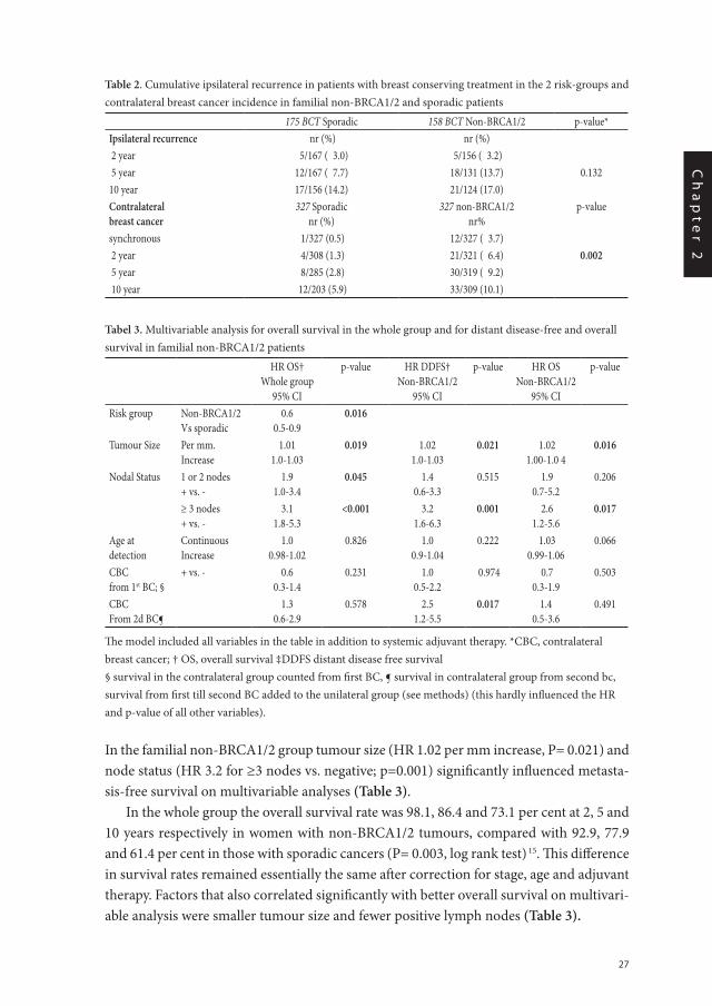
Incidence of ipsilateral recurrence and contralateral breast cancer At 10 yrs, the ipsilateral recurrence rate in patients who had breast conserving treat-ment was 14.2% vs. 17.0%. in sporadic and familial non-BRCA1/2 patients respectively (p=0.132) (Table 2). On multivariate analysis age at detection (HR 0.9; p=0.009) and node status (HR 3.5 for ≥3 nodes vs. node negative; p=0.007) correlated significantly with ip-silateral recurrence, but not risk group (HR 1.3 for familial non-BRCA1/2-associated vs. sporadic patients, p=0.44).
The 5-year rate of metachronous contralateral breast cancer was 5.5% for familial non-BRCA1/2 patients and 2.3% for sporadic patients. At 10 years the rate was 6.4% and 5.4% respectively. The rate for synchronous and metachronous contralateral tumours together at 10 years was 10.1 and 5.9% respectively (p=0.002) (Table 2).
Distant Disease Free and Overall Survival and the influence of tumour stage The distant disease-free survival rate was 90.6%, 77.0% and 65.1% at 2, 5 and 10years re-spectively for familial non-BRCA1/2 cancer, compared with (85.8%, 69.9% and 50.2%) for sporadic cancer (P= 0.005, log rank test) 15. After correction for stage, age, adjuvant and
26
surgical therapy this difference in survival remained essentially the same. Tumour size (HR 1.02 per mm increase; p=0.001), and node status (HR 1.7 for 1 or 2 positive nodes vs. node negative; p=0.04; HR 2.6 for ≥3 nodes vs. negative; p<0.001) were independent predictors of distant disease-free survival in the whole group of patients studied.
Table 1 Patient-, tumour- and treatment characteristics in familial non-BRCA1/2 and sporadic patients.
Sporadic(n=327)
Non -BRCA1/2(n=327)
P-value
Patient CharacteristicsMean FU* (range) 6.5 (0.2-20.8) 7.3 (0.7-22.5) 0.019Mean age yrs/range.† 46 (23-78) 46 (23-77) 0.787< 40 yrs. 97 (29.7) 93 (28.4)40-50 yrs. 123 (37.6) 122 (37.3) 0.904> 50 yrs. 107 (32.7) 112 (34.3)Tumour detectionSymptomatic 225 (68.8) 165 (50.5) 0.068 ‡< 50 yrs. screened 13 ( 4.0) 26 ( 7.9)> 50 yrs. screened 48 (14.7) 39 (11.9)unknown 41 (12.5) 97 (29.7)Tumour characteristicsStageDCIS 14 ( 4.3) 16 ( 4.9)T1 149 (45.6) 187 (57.2) 0.003‡≥ T2 145 (44.3) 112 (34.2)Size unknown 19 ( 5.8) 12 ( 3.7)No. of involved nodes §Node - 163 (52.1) 174 (55.8)Node 1,2 + 61 (19.5) 68 (21.8) 0.477Node ≥ 3 79 (25.2) 68 (21.8) N unknown 10 ( 3.2) 2 ( 0.6)BR grade§¶ Grade 1 21 ( 6.7) 25 ( 8.0)Grade 2 81 (25.9) 75 (24.1) 0.690‡Grade 3 142 (45.4) 130 (41.8)Grade unknown 69 (22.0) 81 (26.1)Therapy SurgeryBreast conservation 175 (53.5) 158 (49.1) ††Mastectomy 145 (44.4) 159 (49.4) 0.405No primary surgery 7 ( 2.1) 5 ( 1.5)Contraleteral. Mastectomy“risk-reducing”
5 ( 1.5) 36 (11.4)** < 0.001
Adjuvant therapyChemotherapy 122 (37.3) 123 (37.6) 0.892Hormone therapy 57 (17.4) 69 (21.1) 0.248Oophorectomy 15 ( 4.6) 20 ( 6.1) 0.325
*FU, follow-up; † yrs, years; ‡ p-value of the comparison between the risk-groups not taking the percentages “unknown” into account; §Nodal status, Bloom Richardson grade and Hormonal receptor status of invasive cancers; ¶, Bloom Richardson grades. †† of the 322 patients with known primary surgery. **Of the 315 unilateral familial cancers at first diagnosis.
27
Ch
ap
te
r 2
In the familial non-BRCA1/2 group tumour size (HR 1.02 per mm increase, P= 0.021) and node status (HR 3.2 for ≥3 nodes vs. negative; p=0.001) significantly influenced metasta-sis-free survival on multivariable analyses (Table 3).
In the whole group the overall survival rate was 98.1, 86.4 and 73.1 per cent at 2, 5 and 10 years respectively in women with non-BRCA1/2 tumours, compared with 92.9, 77.9 and 61.4 per cent in those with sporadic cancers (P= 0.003, log rank test) 15. This difference in survival rates remained essentially the same after correction for stage, age and adjuvant therapy. Factors that also correlated significantly with better overall survival on multivari-able analysis were smaller tumour size and fewer positive lymph nodes (Table 3).
Table 2. Cumulative ipsilateral recurrence in patients with breast conserving treatment in the 2 risk-groups and contralateral breast cancer incidence in familial non-BRCA1/2 and sporadic patients
175 BCT Sporadic 158 BCT Non-BRCA1/2 p-value*Ipsilateral recurrence nr (%) nr (%) 2 year 5/167 ( 3.0) 5/156 ( 3.2) 5 year 12/167 ( 7.7) 18/131 (13.7) 0.13210 year 17/156 (14.2) 21/124 (17.0) Contralateral breast cancer
327 Sporadicnr (%)
327 non-BRCA1/2nr%
p-value
synchronous 1/327 (0.5) 12/327 ( 3.7) 2 year 4/308 (1.3) 21/321 ( 6.4) 0.002 5 year 8/285 (2.8) 30/319 ( 9.2) 10 year 12/203 (5.9) 33/309 (10.1)
Tabel 3. Multivariable analysis for overall survival in the whole group and for distant disease-free and overall survival in familial non-BRCA1/2 patients
HR OS†Whole group
95% CI
p-value HR DDFS†Non-BRCA1/2
95% CI
p-value HR OSNon-BRCA1/2
95% CI
p-value
Risk group Non-BRCA1/2Vs sporadic
0.60.5-0.9
0.016
Tumour Size Per mm.Increase
1.011.0-1.03
0.019 1.021.0-1.03
0.021 1.021.00-1.0 4
0.016
Nodal Status 1 or 2 nodes+ vs. -
1.91.0-3.4
0.045 1.40.6-3.3
0.515 1.90.7-5.2
0.206
≥ 3 nodes + vs. -
3.11.8-5.3
<0.001 3.21.6-6.3
0.001 2.61.2-5.6
0.017
Age at detection
ContinuousIncrease
1.00.98-1.02
0.826 1.00.9-1.04
0.222 1.030.99-1.06
0.066
CBC from 1st BC; §
+ vs. - 0.60.3-1.4
0.231 1.00.5-2.2
0.974 0.70.3-1.9
0.503
CBCFrom 2d BC¶
1.30.6-2.9
0.578 2.51.2-5.5
0.017 1.40.5-3.6
0.491
The model included all variables in the table in addition to systemic adjuvant therapy. *CBC, contralateral breast cancer; † OS, overall survival ‡DDFS distant disease free survival§ survival in the contralateral group counted from first BC, ¶ survival in contralateral group from second bc, survival from first till second BC added to the unilateral group (see methods) (this hardly influenced the HR and p-value of all other variables).
28
In the familial non-BRCA1/2 group, tumour size (HR 1.02 per mm increase; p=0.016) and node status (HR 2.6 for ≥3 nodes vs. negative; p=0.017) also significantly influenced overall survival (Table 3).
Exclusion of the 103 probands from the non-BRCA1/2 group did not affect the results, neither did exclusion of women with non-BRCA1/2 cancers from the HBOC families.
Influence of contralateral breast cancer on survivalThe contralateral tumour was > 2 cm, whereas the primary had been ≤ 2 cm in 9/34 (27%) of the metachronous cancers in the whole group (when both sizes were known). The con-tralateral cancer was node positive whereas the primary tumour had been node negative in 7/33 (21%).
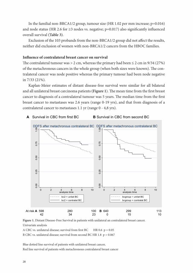
Kaplan-Meier estimates of distant disease-free survival were similar for all bilateral and all unilateral breast carcinoma patients (Figure 1). The mean time from the first breast cancer to diagnosis of a contralateral tumour was 5 years. The median time from the first breast cancer to metastases was 2.6 years (range 0-19 yrs), and that from diagnosis of a contralateral cancer to metastases 1.1 yr (range 0 - 4,8 yrs).
A Survival in CBC from first BC B Survival in CBC from second BC
0.00
0.25
0.50
0.75
1.00
0 2 4 6 8 10analysis time
bc2 = unilat BCbc2 = contralat BC
DDFS after metachronous contralateral BC
0.00
0.25
0.50
0.75
1.00
0 2 4 6 8 10analysis time
bcgroup = unilat BCbcgroup = contralat BC
DDFS after metachronous contralateral BC
At risk A 598 280 100 B 640 299 113 42 34 23 0 15 10
Figure 1. Distant Disease-Free Survival in patients with unilateral an contralateral breast cancer.Univariate analysisA CBC vs. unilateral disease; survival from first BC HR 0.6 p = 0.05B CBC vs. unilateral disease; survival from second BC HR 1.8 p = 0.067
Blue dotted line survival of patients with unilateral breast cancer,Red line survival of patients with metachronous contralateral breast cancer
29
Ch
ap
te
r 2
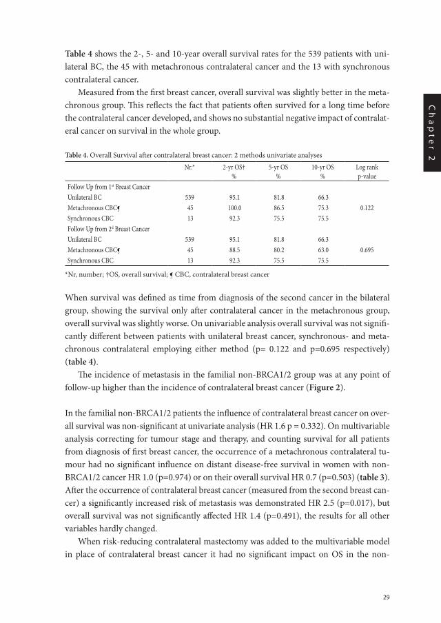
Table 4 shows the 2-, 5- and 10-year overall survival rates for the 539 patients with uni-lateral BC, the 45 with metachronous contralateral cancer and the 13 with synchronous contralateral cancer.
Measured from the first breast cancer, overall survival was slightly better in the meta-chronous group. This reflects the fact that patients often survived for a long time before the contralateral cancer developed, and shows no substantial negative impact of contralat-eral cancer on survival in the whole group.
Table 4. Overall Survival after contralateral breast cancer: 2 methods univariate analyses
Nr.* 2-yr OS†%
5-yr OS%
10-yr OS%
Log rankp-value
Follow Up from 1st Breast CancerUnilateral BC 539 95.1 81.8 66.3Metachronous CBC¶ 45 100.0 86.5 75.3 0.122Synchronous CBC 13 92.3 75.5 75.5Follow Up from 2d Breast Cancer Unilateral BC 539 95.1 81.8 66.3 Metachronous CBC¶ 45 88.5 80.2 63.0 0.695Synchronous CBC 13 92.3 75.5 75.5
*Nr, number; †OS, overall survival; ¶ CBC, contralateral breast cancer
When survival was defined as time from diagnosis of the second cancer in the bilateral group, showing the survival only after contralateral cancer in the metachronous group, overall survival was slightly worse. On univariable analysis overall survival was not signifi-cantly different between patients with unilateral breast cancer, synchronous- and meta-chronous contralateral employing either method (p= 0.122 and p=0.695 respectively) (table 4).
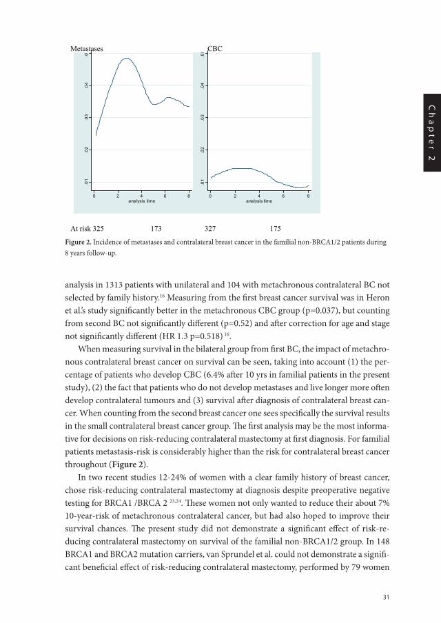
The incidence of metastasis in the familial non-BRCA1/2 group was at any point of follow-up higher than the incidence of contralateral breast cancer (Figure 2).
In the familial non-BRCA1/2 patients the influence of contralateral breast cancer on over-all survival was non-significant at univariate analysis (HR 1.6 p = 0.332). On multivariable analysis correcting for tumour stage and therapy, and counting survival for all patients from diagnosis of first breast cancer, the occurrence of a metachronous contralateral tu-mour had no significant influence on distant disease-free survival in women with non-BRCA1/2 cancer HR 1.0 (p=0.974) or on their overall survival HR 0.7 (p=0.503) (table 3). After the occurrence of contralateral breast cancer (measured from the second breast can-cer) a significantly increased risk of metastasis was demonstrated HR 2.5 (p=0.017), but overall survival was not significantly affected HR 1.4 (p=0.491), the results for all other variables hardly changed.
When risk-reducing contralateral mastectomy was added to the multivariable model in place of contralateral breast cancer it had no significant impact on OS in the non-
30
BRCA1/2 group (HR 0.4; p=0.163). Five of the 36 familial patients developed metastases after risk-reducing contralateral mastectomy.
Discussion
Ipsilateral and contralateral recurrenceIpsilateral breast cancer recurrence in this study was similar for familial non-BRCA1/2 and sporadic cancers. Therefore decisions on breast conserving treatment can be made on the same grounds in familial and sporadic patients. This is in line with the literature on breast conserving treatment in familial and hereditary cancer21,22.
There was a slightly higher rate of metachronous contralateral breast cancer in familial non-BRCA1/2 patients than in the sporadic group (6.4% vs. 5.4% at 10 years). In studies of familial breast cancer performed before DNA testing was available, the incidence of con-tralateral tumours was increased in some studies 6,7, but not in others.3This inconsistency may be explained by a different rate of BRCA1, BRCA2 and CHEK2*1100delC mutation carriers included in the various studies. 8,19,20 Although in the present study, the familial patients were extensively tested for deleterious BRCA1 and BRCA2 mutations the sensi-tivity of the DNA screening is not 100% and some occult mutation carriers may still have been in the study group. Furthermore the studies that assess contralateral cancer risk in patients with familial non-BRCA1/2 cancer have, to some extent, like the present one of-fered DNA testing preferentially to patients with contralateral breast cancer.3,6-8 When this selection bias on the reported incidence of contralateral cancers in the familial group was corrected, no significant difference in contralateral breast cancer incidence was shown anymore between familial non-BRCA1/2 and sporadic cancers 15.
The 3.7 per cent rate synchronous contralateral breast cancer (Table 2) in the familial group in our study highlights the importance of good preclinical investigation to detect contralateral cancer early.
Survival and the impact of contralateral cancer Both distant disease-free and overall survival were significantly better in familial non-BRCA1/2 patients than in sporadic patients. These results, however, were also clearly in-fluenced by selection bias and the survival difference disappeared after correcting for the fact that patients diagnosed before 1995 had to live longer to get DNA testing and thus were selected for longevity15.
On both univariable- and multivariable analyses, and measuring survival as the time from first or from second breast cancer, contralateral breast cancer had no significant negative impact on the overall survival rate in either the whole group or the familial non-BRCA1/2 group. Because of the relatively small numbers (21) of metachronous familial cancers however, the 95 per cent confidence interval was rather wide. Heron et al. how-ever, had comparable results for the influence of CBC on survival, using both method’s of
31
Ch
ap
te
r 2
analysis in 1313 patients with unilateral and 104 with metachronous contralateral BC not selected by family history.16 Measuring from the first breast cancer survival was in Heron et al.’s study significantly better in the metachronous CBC group (p=0.037), but counting from second BC not significantly different (p=0.52) and after correction for age and stage not significantly different (HR 1.3 p=0.518) 16.
When measuring survival in the bilateral group from first BC, the impact of metachro-nous contralateral breast cancer on survival can be seen, taking into account (1) the per-centage of patients who develop CBC (6.4% after 10 yrs in familial patients in the present study), (2) the fact that patients who do not develop metastases and live longer more often develop contralateral tumours and (3) survival after diagnosis of contralateral breast can-cer. When counting from the second breast cancer one sees specifically the survival results in the small contralateral breast cancer group. The first analysis may be the most informa-tive for decisions on risk-reducing contralateral mastectomy at first diagnosis. For familial patients metastasis-risk is considerably higher than the risk for contralateral breast cancer throughout (Figure 2).
In two recent studies 12-24% of women with a clear family history of breast cancer, chose risk-reducing contralateral mastectomy at diagnosis despite preoperative negative testing for BRCA1 /BRCA 2 23,24. These women not only wanted to reduce their about 7% 10-year-risk of metachronous contralateral cancer, but had also hoped to improve their survival chances. The present study did not demonstrate a significant effect of risk-re-ducing contralateral mastectomy on survival of the familial non-BRCA1/2 group. In 148 BRCA1 and BRCA2 mutation carriers, van Sprundel et al. could not demonstrate a signifi-cant beneficial effect of risk-reducing contralateral mastectomy, performed by 79 women
Figure 2 Incidence of metastases and contralateral breast cancer in the familial non-BRCA1/2 patients during 8 years follow-up. Metastases CBC
.01
.02
.03
.04
.0
0 2 4 6 8analysis time
.01
.02
.03
.04
.05
0 2 4 6 8analysis time
At risk 325 173 327 175 Chapter 2A / Figure 2 Figure 2. Incidence of metastases and contralateral breast cancer in the familial non-BRCA1/2 patients during
8 years follow-up.
32
on breast cancer specific survival (p=0.11) 25, although they showed a high CBC incidence, as expected in BRCA1-patients diagnosed before age 50 years19.
Although contralateral cancer had no significant influence on survival in the whole familial non-BRCA1/2 group, especially in the first year after contralateral breast cancer an increased rate of metastases was seen (analyses from second BC). It is not possible to differentiate the extent to which contralateral cancer is part of general metastatic disease and functions as a marker for metastasis or whether it is also the source of subsequent metastases. With a median time of 1.1 years to metastasis, considerably shorter than from primary breast cancer to metastasis, and parallel survival curves after 3 years (fig1), the former explanation seems more plausible.
In the present study, the stage in 20-25 per cent of the 45 metachronous contralateral cancers was more advanced, than in the primary breast cancer. Usually patients are under surveillance for 10 years after their diagnosis, with mammography and clinical examina-tion performed yearly in order to detect CBC early.
Impact of age and tumour stage on survivalAlthough familial cancers do grow faster at younger age26, age at detection did not
effect survival negatively in the present study on multivariable analyses. The main predic-tors of survival were, also in familial non-BRCA1/2 patients, lymph node involvement and tumour size. This is fully in accordance with literature on the influence of tumour stage in sporadic breast cancer. Unlike the findings in Michaelson’s study however, survival was in the total group already significantly lower with only one or 2 positive nodes27. The pres-ent findings, that tumour size and nodal stage have also a key influence on prognosis in familial cancer, is promising for the chances of improving survival by surveillance. In this study only a small percentage of the familial patients were under surveillance before age 50 years. This may have contributed to the smaller tumour size in this group, although the difference in node-negative patients was not significant. Favourable tumour stages have been reported in BRCA1/2 mutation carriers screened with MRI. 28-30 Cost benefit analyses of screening various high risk groups are due.
Conclusion Ipsilateral recurrence occurred with comparable frequency in familial-non-BRCA1/2 pa-tients and sporadic patients. Familial non-BRCA1/2 breast cancer patients can receive breast-conserving treatment on the same grounds as sporadic patients. Metachronous CBC incidence was only slightly increased in the familial group.
Stage at detection is also in familial BC a key indicator of prognosis, and early detection therefore important for survival. We did not demonstrate a significant influence of con-tralateral breast cancer on overall survival of familial non-BRCA1/2 patients, nor of risk-reducing controlateral mastectomy. CBC may however indicate imminent metastases.

33
Ch
ap
te
r 2
Acknowledgments
The authors thank Professor Terry Hyslop for methodological advice, Hanne Meijers-Heijboer and Ans van den Ouweland for DNA-test results, Karina Bartels and professor Jan Klijn for their critical reading of the manuscript, Ada van Eekelen and Elle Crepin for logistic support and Marijke Westerhout-Kersten and Kerstin van der Veen for assisting literature search. The work of Celina Alves and Bonnie Bakri was supported by a grant of Erasmus University MC.
34
Literature
1. Peto J, Easton DF, Matthews FE, Ford D, Swerdlow AJ. Cancer mortality in relatives of women with breast cancer: The OPCS study. Int.J. Cancer 1996;65:275-83.
2. Slattery ML, Kerber RA. A comprehensive evaluation of family history and breast cancer risk. The Utah Population Database. JAMA 1993;270:1563-8.
3. Eerola H, Vahteristo P, Sarantaus L, et al. Survival of breast cancer patients in BRCA1, BRCA2 and non-BRCA1/2 breast cancer families: a relative survival analysis from Finland. Int J Cancer 2001;93:368-72.
4. Malone KE, Daling JR, Weiss NS, McKnight B, White E, Voigt LF. Family history and survival of young women with invasive breast carcinoma. Cancer 1996;78:1417-25.
5. Cook LS, White E, Schwartz SM, McKnight B, Daling JR, Weiss NS. A population-based study of contra-lateral breast cancer following a first primary breast cancer. Cancer causes control. 1996;7:382-90.
6. Eccles D, Simmons P, Goddard J, et al. Familial breast cancer: an investigation into the outcome of treat-ment for early stages. Familial Cancer 2001;1:65-72.
7. Möller P, Borg A, Evans DG, et al. Survival in prospectively ascertained familial breast cancer: analy-sis of a series stratified by tumour characteristics, BRCA mutations and oophorectomy. Int. J. Cancer 2002;101:555-59.
8. Broeks A, de Witte L, Nooijen A, et al. Excess risk for contralateral breast cancer in CHEK2*1100delC germline mutation carriers. Breast Ca Res Treatm 2004;83:91-3.
9. Chen Y, Thompson W, Semenciw R, Mao Y. Epidemiology of contralateral breast cancer. Ca Epidem. Biom and Prev 1999;8:855-61.
10. Peto J, Collins N, Barfoot R, et al. Prevalence of BRCA1 and BRCA2 gene mutations in patients with early-onset breast cancer. J Natl Cancer Inst.1999;91:943-49.
11. Eccles DM, Pichert G. Familial non-BRCA1/BRCA2-associated breast cancer. Lancet Oncology 2006;6:705-11.
12. Lakhani SR, Gusterson BA, Jacquemier J, et al. The pathology of familial breast cancer: Histologi-cal features of cancers in families not attributable to mutations in BRCA1 or BRCA2. Clin Cancer Res 2000;6:782-9.
13. Adem C, Reynolds C, Soderberg CL, Cunningham JM, et al. Pathologic characteristics of breast paren-chyma in patients with hereditary breast carcinoma, including BRCA1 and BRCA2 mutation carriers. Cancer 2003;97:1-11.
14. Eerola H, Heikkilä P, Tamminen A et al. Histopathological features of breast tumours in BRCA1, BRCA2 and mutation negative breast cancer families. Breast Cancer Research. 2005 (7):93-100. DOI 10.1186/bcr953
15. Tilanus-Linthorst MMA, K Bartels, C Alves, et al. Selection bias influences reported contralateral breast cancer incidence and survival in high-risk non-BRCA1/2 patients. Br.Ca Res Treat. 2006;95(2):117-23.
16. Heron DE, Komarnicky LT, Hyslop T, Schwartz TF, Mansfield CM. Bilateral breast carcinoma. Cancer 2000;88:2739-50.
17. Newman LA, Sahin AA, Bondy ML, et al. A case-control study of unilateral and bilateral breast carci-noma patients. Cancer 2001;91:1845-53.
18. Herrington LJ, Barlow WE, Yu O et al. Efficacy of prophylactic mastectomy in women with unilateral breast cancer: a cancer research network project. JCO 2005;23:4275-86.
19. Verhoog LC, Brekelmans CTM, Seynaeve C, Meijers-heijboer EJ, Klijn JG. Contralateral breast cancer risk is influenced by the age at onset in BRCA1-associated breast cancer. BrJ Cancer 2000;83;384-6.

35
Ch
ap
te
r 2
20. de Bock GH, Schutte M, Krol-Warmerdam EMM, et al. Tumour characteristics and prognosis of breast cancer patients carrying the germline CHEK2*1100delC variant. J Med Genet 2004;41:731-5.
21. Robson M, Svahn T, McCormick B et al. Appropriateness of breast-conserving treatment of breast carci-noma in women with germline mutations in BRCA1 or BRCA2. Cancer 2005;103:44-51.
22. Brekelmans CT, Voogd AC, Botke BN et al. Family history of breast cancer and local recurrence after breast-conserving therapy. Eur J Cancer 1999;35:620-626.
23. Schwartz MD, Lerman C, Brogan B, et al. Impact of BRCA1/BRCA2 counseling and testing in newly diagnosed breast cancer patients.JClinOncol 2004;22:1823-9.
24. Weitzel JN, McCaffrey SM, Nedelcu R, et al. Effect of genetic cancer risk assessment on surgical decisions at breast cancer diagnosis. Arch Surg. 2003;138:1323—28.
25. van Sprundel TC, Schmidt MK, Rookus MA et al. Risk reduction of contralateral breast cancer and survival after contralateral prophylactic mastectomy in BRCA1 or BRCA2 mutation carriers. Brit JCa 2005;93:287-92.
26. Tilanus-Linthorst MMA, Kriege M, Boetes C, et al. Hereditary breast cancer growth rates and its impact on screening policy. Eur j Ca 2005;41 (11):160-17.
27. Michaelson JS, Silverstein M, Sgroi D, et al. The effect of tumor size and lymph node status on breast carcinoma lethality. Cancer 2003;98:2133-43.
28. Kriege M, Brekelmans CTM, Boetes C, et al. Efficacy of MRI and mammography for breast-cancer screening in women with a familial or genetic predisposition. N Eng JMed 2004;351:427-37.
29. Warner E, Plewes DB, Hill KA, et al. Surveillance of BRCA1 and BRCA2 mutation carriers with magnetic resonance imaging, ultrasound, mammography and clinical breast examination. JAMA 2004;292(11):1317-25.
30. Leach MO, MARIBS study group. Screening with magnetic resonance imaging and mammography of a UK population at high familial rik of breast cancer: a prospective multicentre cohort study (MARIBS). Lancet 2005;365:1769-78.


Chapter 2B
Tumour characteristics, survival and prognostic factors of hereditary breast cancer from BRCA2-, BRCA1- and non-BRCA1/2 families as compared to sporadic breast cancer cases
C.T.M. Brekelmans, M.M.A. Tilanus-Linthorst, C. Seynaeve, A. vd Ouweland, M.B.E. Menke-Pluymers, C.C.M. Bartels, M. Kriege, A.N. van Geel, C. W. Burger, A.M.M. Eggermont, H. Meijers-Heijboer, J.G.M. Klijn
38
Abstract
BackgroundAs yet, many studies reported on the tumour characteristics and survival of hereditary breast cancer (BC), including BRCA2-BC. However, due to small sample sizes, it is in-sufficiently known whether BRCA2-BC comprises of a specific tumour type and clinical course. Further, the prognostic impact of the classical tumour and treatment factors in hereditary BC is unclear.
Patients and methodsWe selected 103 BRCA2-, 223 BRCA1-associated and 311 non-BRCA1/2 BC patients di-agnosed between 1980 and 2004, ascertained at the Rotterdam Family Cancer Clinic. To correct for longevity bias, analyses were also performed while excluding index patients undergoing DNA testing more than 2 years after BC diagnosis. As a comparison group, 759 sporadic BC patients of comparable age at and year of diagnosis were selected. We compared tumour characteristics, the occurrence of ipsilateral recurrence (LRR) and con-tralateral BC (CBC) as well as distant disease-free (DDFS), BC-specific (BCSS) and overall survival (OS) between these groups. By multivariate modeling, the prognostic impact of tumour and treatment factors was investigated separately in hereditary BC.
ResultsWe confirmed the presence of the particular BRCA1-phenotype. In contrast, tumour characteristics of BRCA2-associated BC appeared to be quite similar to those of non-BRCA1/2 and sporadic BC, with the exception of a high risk of metachronous contralat-eral BC (3.1% per year) and a frequent occurrence of estrogen-receptor (ER)-positivity (83%). No significant differences between BRCA2-associated BC and BRCA1-associated, non-BRCA1/2 and sporadic BC were found with respect to LRR, DDFS, BCSS and OS.
Independent prognostic factors for BC-specific survival in hereditary BC (combining the 3 subgroups) were tumour stage, adjuvant chemotherapy, histologic grade, ER status and a prophylactic (salpingo-)oophorectomy. In this analysis, no prognostic impact was found for the occurrence of a contralateral BC or the performance of a (contralateral) prophylactic mastectomy.
ConclusionsApart from the frequent occurrence of contralateral BC and a positve ER-status, BRCA2-associated BC did not markedly differ from other hereditary or sporadic BC. Our observa-tion that tumour size and nodal status are prognostic factors also in hereditary BC, implies that the strategy to use these factors as a proxy for ultimate mortality, for instance in BC screening programmes, appears to be valid in this specific group of patients.
39
Ch
ap
te
r 2
Introduction
Hereditary BC is characterized by a young age of onset, and a high incidence of contra-lateral BC.
Five to ten percent of all breast cancer cases are hereditary, with germline mutations in the BRCA1, BRCA2, and CHEK-2 1100delC gene accounting for about 30% of these cases. Thus, the majority of hereditary BC is due to other germline mutations in as yet unknown genes. 1 2
Many studies report about the typical tumour characteristics of BRCA1-associated breast cancer, such as the basal-like phenotype and the high histologic grade 3 4 5. Despite these unfavourable characteristics, most reports describe a similar or worse survival as compared to sporadic BC 6 7.
Although less data are available on BRCA2-associated BC; the phenotype appears to be partly similar to that of BRCA1, with respect to the young age at diagnosis, the increased risk of contralateral BC, and the presence of continuous pushing margins, while a high histological grade has also been found in several studies. 4 8 9 Cyclin D is overexpressed in BRCA2 10, whereas an immunohistochemical RAD51/CHEK2 staining pattern can dif-ferentiate between BRCA2-associated and other breast cancers. 11 The clinical outcome appears to be not markedly different from sporadic BC. 12 13 14 15
Hereditary BC not due to a BRCA1 or BRCA2 mutation, hereafter called non-BRCA1/2, appears to be a heterogeneous group, although specific characteristics such as a low histologic grade and mitotic count have been reported. 4 16 In some non-BRCA1/2 families a CHEK2*1100delC-germline mutation can be found17, with one small study sug-gesting an unfavourable impact on disease-free, but not overall, survival. 18
In 1991, the Rotterdam Family Cancer Clinic was set up, an outpatient department for the counseling, surveillance and (preventive) treatment of members from families with a frequent occurrence of breast and/or ovarian cancer. In several reports we described the characteristics and clinical course of BC patients from these families. Recently, we pub-lished the tumour characteristics and survival of 223 BRCA1-associated breast cancers 7, and of 327 cases from families in which a BRCA1- and BRCA2-mutation was excluded.19 Both series of patients were matched for age at and year of diagnosis with sporadic control patients. In the current manuscript, we combine the data from both publications and add data from 103 BRCA2-associated BC cases in order to compare tumour characteristics and survival of these three cohorts of hereditary breast cancers to sporadic BC. Further, we investigated the impact of the classical prognostic factors in hereditary BC.
MethodsIncluded were all female patients with primary, invasive BC and available data on histo-pathology and follow-up that were diagnosed after 1-1-1980 in hereditary breast/ovarian cancer (HB(O)C) families undergoing DNA-analysis at the Family Cancer Clinic (Clinical
40
Genetics Department) of the Erasmus MC (See Verhoog EJC 2001 for minimal criteria for DNA-testing).Three cohorts of hereditary BC patients were defined:
☐ BRCA2 (n=103): BC cases from families with an identified deleterious BRCA2-mutation
☐ BRCA1 (n=223): BC cases from families with an identified deleterious BRCA1-mutation. 7
☐ Non-BRCA1/2 (n=311): BC cases within a family tested negative for a deleterious BRCA1- and BRCA2-mutation. 19 The number of patients is smaller than in the previous paper, as for the current analysis only the invasive cancer cases were used.
In addition, we selected a cohort of sporadic BC cases by combining two previously used cohorts from the Erasmus MC – Daniel den Hoed cancer registry: one cohort was matched for age at and year of diagnosis (within 5 years) to BRCA1-associated cases and one to non-BRCA1/2-associated BC cases. For this analysis, both cohorts were combined to form one reference group of sporadic BC cases. All medical files of potential control patients were checked to exclude a family history suggestive of hereditary breast cancer. Excluded were control patients with at least 2 additional family members with breast can-cer, or 1 additional family member with breast cancer under the age of 55 years or ovarian cancer (any age).
To correct for longevity bias, all cases of hereditary BC occurring within the family were included, regardless of mutation carrier status, except for BC occurring in proven non-mutation carriers in BRCA1/2 families. In addition, all three hereditary BC cohorts were divided into two groups: 1. index patients, undergoing DNA testing more than 2 years after their BC diagnosis (hereafter called the ‘late-tested index group’) and 2. all re-maining cases (hereafter called ’unselected cases’). This was done as we previously showed that the survival of the first group was extremely favourable, due to the selection of the longer living patients for DNA testing.7 19
All analyses concerning comparisons with the sporadic cohort were performed (table 1-3 and figure 1) in unselected hereditary cases only. The prognostic factors for breast can-cer specific survival were investigated by multivariate analyses in the 3 hereditary groups combined (table 4) and in the 4 risk-groups combined.
DNA analysisFor all families, DNA testing was performed at the Clinical Genetics Department of the Erasmus MC, Rotterdam. The coding parts and exon-flanking intronic regions of the BRCA1 gene (exon 3, 5-10, part of exon 11, and exon 12-23) were screened for the pres-ence of mutations using denaturing gradient gel electrophoresis (DGGE).20 All aberrant fragments were sequenced; exons 2 and 24 were directly sequenced. Presence of mutations in exon 11 was detected with the protein truncation test (PTT). Additionally, multiplex ligation-dependent probe amplification (MLPA) was performed for detection of large ge-nomic deletions and duplications. 21,22
41
Ch
ap
te
r 2
Most of the coding regions and exon-flanking intronic sequences of the BRCA2 gene were also screened by DGGE (exon 2-9, part of exon 10, the 5’ and 3’ parts of exon 11, exon 12-18 and exon 20-27). 20 Again all aberrant fragments were sequenced; the 3’ part of exon 10 and exon 19 was directly sequenced. The PTT test was used to screen for the presence of mutations in exon 11. 21,22
117 families from the non-BRCA1/2 cases were investigated for the presence of a CHEK-2 1100delC germline mutation. As only 1 family was found positive, no further separation was made into CHEK2-positive and -negative families.
Data registration and statistical methodsFor all four cohorts of BC patients, the following patient and tumour characteristics were extracted from the medical files: age at diagnosis, axillary lymph node status (negative, positive (1-3 or ≥ 4 positive nodes) and unknown), tumour diameter, presence of distant metastases at diagnosis, morphology of the tumour, histological grade (Bloom-Richard-son), ER- and PR-status (positive, negative or unknown), surgical and adjuvant systemic treatment (hormonal and/or chemotherapy). Further, registration if and when women underwent prophylactic bi- or contralateral mastectomy ((C)PM) and a bilateral (sal-pingo-)oophorectomy (B(S)O), with the reason of the B(S)O (prophylactic, for benign reasons or as treatment for breast or ovarian cancer), was undertaken.
Differences between these characteristics were tested by chi-square tests (categorical variables) or t-tests (continuous variables).
For the hereditary BC cases, information on the complete family pedigree, dates of DNA testing/diagnosis and the type of germline mutation were gathered from the depart-ment of Clinical Genetics.
Endpoints of interest were the occurrence of a local (ipsilateral) recurrence (LRR) after breast-conserving therapy (BCT), metachronous contralateral breast cancer (CBC), dis-tant metastases (DM), and death (overall or breast-cancer related), whichever occurred first. For all five endpoints, Kaplan-Meier survival curves were constructed for the three hereditary groups and the sporadic group. Differences between the curves were tested by the logrank test. Multivariately, differences in the abovementioned five endpoints between the three groups of hereditary and sporadic BC were examined by the Cox proportional hazard method, correcting for tumour and treatment factors.
As we had a special interest in the investigation of the impact of the traditional prog-nostic factors on BC-specific survival in hereditary breast cancer, we also performed a multivariate analysis excluding the sporadic cohort.
All statistical analyses were performed with STATA SE version 9.The study was supported by grant DDHK 2004-3124 from the Dutch Cancer Society
and approved by the local Ethics Committee on july 15, 2004.
42
Results
The complete cohorts of hereditary BC consisted of 103 BRCA2-associated cases, 223 BRCA1-associated cases, and 311 non-BRCA1/2 cases. After exclusion of the late-tested index cases, 90 BRCA2-associated, 170 BRCA1-associated and 238 non-BRCA1/2 cases remained (unselected cases). As there were no important differences with respect to tu-mour characteristics between the total and unselected hereditary cohorts, the data in table 1 are shown for the unselected hereditary cases only.
The mean age at diagnosis of BRCA2-associated cases was 44 years, like in the sporad-ic controls. This was slightly older, however not signficantly, than the BRCA1-associated cases (mean age 42 years), but significantly younger than non-BRCA1/2 associated cases (mean age 47 years).
As compared to non-BRCA1/2 BC, both BRCA2- and BRCA1-associated BC was less likely to be detected during the course of a screening programme.
Tumour size did not significantly differ between the groups, while nodal status was significantly more often node-positive in BRCA2-BC as compared to BRCA1-associated BC (p < 0.001). No signficant difference between BRCA2-BC and non-BRCA1/2 or spo-radic BC was noticed (p=0.13 and 0.39, respectively).
The typical tumour morphology of BRCA1-BC, with a high frequency of the medullary type (7%), was not found in BRCA2-BC: with 89% of the cases of the ductal tumour type, 9% of the lobular tumour type and 2% of the medullary type the morphology was not signifi-cantly different from non-BRCA1/2 (p=0.55) nor from sporadic BC (p=0.76). A similar pat-tern was found for the histologic grade: 88% of BRCA1-associated BC was grade III, which was significantly more (p=0.001) than in BRCA2-BC (65%). The latter percentage was not significantly different from that in non-BRCA1/2 BC (p=0.19), or sporadic BC (p=0.48).
Also with respect to the steroid receptor status, the well-known high incidence of estrogen (ER)- and progesterone (PgR)-negative cases in BRCA1-BC was not found in BRCA2-BC. On the contrary, BRCA2-BC was more often ER-positive as compared to non-BRCA1/2 BC, however non-significantly (p=0.13), and sporadic BC (p=0.005).
No clear differences in the type and administration of adjuvant treatment were found between the four groups, with the exception of a lower frequency of hormonal treatment in BRCA1-BC, most likely reflecting the high frequency of ER-negative BC in that subgroup.
The incidence of metachronous contralateral BC in BRCA2-associated BC was, with a yearly rate of 3.1%, identical to that in the BRCA1-BC group (p =0.98) and significantly higher than in the non-BRCA1/2 (1% per year; p=0.008) and sporadic BC cohort, respec-tively (0.7.% per year; p < 0.001).
Also the incidence of ovarian cancer after BC in the BRCA2-cohort was, with a yearly risk of 0.7%, equal to that in the BRCA1-cohort (p=0.88), and higher than in the non-BRCA1/2 cohort (0.1% per year; p=0.05). Three other cancers occurred after BC in the BRCA2-co-hort: a squamous carcinoma of the skin, brain tumor and invasive cervical cancer.
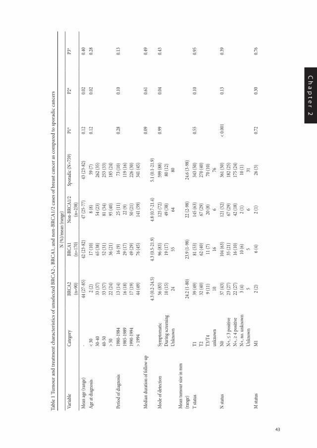
43
Ch
ap
te
r 2
Tabl
e 1
Tum
our a
nd tr
eatm
ent c
hara
cter
istic
s of u
nsel
ecte
d BR
CA
2-, B
RCA
1, a
nd n
on-B
RCA
1/2
case
s of b
reas
t can
cer a
s com
pare
d to
spor
adic
can
cers
N (%
)/mea
n (ra
nge)
Varia
ble
Categ
ory
BRCA
2 (n
=90)
BRCA
1 (n
=170
)No
n-BR
CA1/
2(n
=238
)Sp
orad
ic (N
=759
)P1
*P2
*P3
*
Mea
n ag
e (ra
nge)
-44
(27-
85)
42 (2
3-82
)47
(25-
77)
43 (2
3-82
)0.1
20.0
20.4
0Ag
e at d
iagno
sis<
3030
-40
40-5
0>
50
2 (2
)33
(37)
33 (3
7)22
(24)
17 (1
0)65
(38)
52 (3
1)36
(21)
8 (8
)54
(23)
81 (3
4)95
(40)
59 (7
)26
2 (35
)25
3 (33
)18
5 (24
)
0.12
0.02
0.28
Perio
d of
diag
nosis
1980
-198
419
85-1
989
1990
-199
4>
1994
13 (1
4)16
(18)
17 (1
9)44
(49)
16 (9
)29
(17)
49 (2
9)76
(45)
25 (1
1)22
(9)
50 (2
1)14
1 (59
)
73 (1
0)11
9 (16
)22
6 (30
)34
1 (45
)
0.28
0.10
0.13
Med
ian du
ratio
n of
follo
w-up
4.3 (0
.2-24
.5)4.3
(0.5-
21.9)
4.8 (0
.7-21
.4)5.1
(0.1-
21.9)
0.09
0.61
0.49
Mod
e of d
etecti
onSy
mpt
omati
cDu
ring s
cree
ning
Unkn
own
56 (8
5)10
(15)
24
96 (8
3)19
(17)
55
125 (
72)
49 (3
8)64
599 (
88)
80 (1
2)80
0.99
0.04
0.43
Mea
n tu
mou
r size
in m
m
(rang
e)24
.2 (1
-80)
23.9
(1-9
8)22
(2-9
8)24
.6 (3
-98)
T sta
tus
T1 T2 T3/T
4un
know
n
39 (4
9)32
(40)
9 (11
)10
81 (5
3)62
(40)
11 (7
)16
145 (
63)
67 (2
9)20
(8)
6
343 (
50)
270 (
40)
70 (1
0)76
0.55
0.10
0.95
N sta
tus
N0 N+, ≤
3 po
sitive
N+, ≥
4 po
sitive
N+, n
o. un
know
nUn
know
n
37 (4
3)23
(27)
22 (2
7)3 (
4) 5
104 (
63)
35 (2
1)16
(10)
10 (6
)5
121 (
52)
67 (2
9)42
(18)
2 (1) 6
361 (
50)
182 (
25)
175 (
24)
10 (1
)31
< 0.0
010.1
30.3
9
M st
atus
M1
2 (2)
6 (4
)2 (
1)26
(3)
0.72
0.30
0.76
44
Tabl
e 1
- con
tinue
d
Varia
ble
Categ
ory
BRCA
2 (n
=90)
BRCA
1 (n
=170
)No
n-BR
CA1/
2(n
=238
)Sp
orad
ic (N
=759
)P1
*P2
*P3
*
Mor
phol
ogy
Ducta
l/NOS
Lobu
larM
edul
lary
Tubu
larO
ther
* *Un
know
n
80 (8
9)8 (
9)2 (
2) 0 0 0
146 (
88)
6 (4)
12 (7
)0 1 (1) 5
206 (
87)
25 (1
1)3 (
1)4 (
2) 0 0
660 (
87)
79 (1
0)13
(2)
7 (1) 0 0
0.10
0.55
0.76
Histo
logic
grad
eI II III un
know
n
2 (3)
19 (3
2)38
(65)
31
1 (1
)12
(11)
93 (8
8)63
20 (8
)56
(31)
101 (
61)
61
40 (8
)15
5 (29
)33
7 (63
)22
7
0.002
0.19
0.48
Estro
gen
rece
ptor
statu
sNe
gativ
ePo
sitive
Unkn
own
11 (1
6)56
(84)
23
77 (7
3)29
(27)
64
47 (2
7)12
9 (73
)62
183 (
33)
367 (
67)
209
< 0.0
010.1
30.0
05
Prog
ester
one r
ecep
tor s
tatus
Nega
tive
Posit
iveUn
know
n
20 (3
6)35
(64)
36
64 (6
7)31
(33)
75
37 (2
6)10
5 (74
)96
148 (
46)
274 (
54)
337
< 0.0
010.1
60.8
8
Prim
ary s
urge
ryNo
ne/b
iops
y onl
yBC
TM
astec
tom
y
4 (4
)35
(39)
50 (5
6)
5 (3
)76
(46)
85 (5
1)
011
1 (47
)12
0 (53
)
15 (2
)41
0 (55
)32
6 (43
)
0.55
0.31
0.01
Adju
vant
trea
tmen
tHo
rmon
alCh
emot
hera
py18
(20)
43 (4
8)12
(8)
84 (5
1) 64
(27)
98 (4
1)11
9 (16
)32
9 (43
)0.0
040.9
00.8
90.3
20.2
90.4
3Pr
ophy
lactic
surg
ery
(C)P
MB(
S)O
14 (1
6)22
(24)
37 (2
2)55
(36)
25 (1
0)12
(5)
10 (1
)29
(4)
0.25
0.20
0.32
<0.00
1<
0.001
< 0.0
012nd
/3rd
canc
ers
Meta
chro
nous
CBC
O
varia
n ca
ncer
Oth
er
15 (3
.1%/y
r)5 (
0.7%
/yr)
3 (0.5
%/y
r)
25 (3
.1%/y
r)7 (
0.7%
/yr)
4 (0.4
%/y
r)
13 (1
%/y
r)2 (
0.1%
/yr)
3 (0.2
%/y
r)
33 (0
.7%/y
r)1
11 (0
.2%/y
r)
0.98
0.88
0.74
0.008
0.05
0.29
< 0.0
01 - 0.34
* diff
eren
ce b
etw
een
BRC
A2-
asso
ciat
ed tu
mou
rs a
nd B
RCA
1 (p
1), n
on-B
RCA
1/2
(p2)
and
spor
adic
bre
ast c
ance
rs (p
3), r
espe
ctiv
ely
** 1
pap
illar
y ca
rcin
oma
Abb
revi
atio
ns u
sed:
BC
T =
brea
st-c
onse
rvin
g th
erap
y; C
)PM
= (c
ontr
alat
eral
) pro
phyl
actic
mas
teco
my;
B(S
)O =
bila
tera
l (sa
lpin
go-)
ooph
orec
tom
y; C
BC =
con
tral
ater
al b
reas
t ca
ncer
45
Ch
ap
te
r 2
Tabl
e 2
Act
uari
al e
vent
and
surv
ival
rate
s of 5
end
poin
ts, s
epar
atel
y fo
r the
thre
e ge
netic
risk
gro
ups (
unse
lect
ed c
ases
onl
y) a
nd sp
orad
ic B
C c
ases
5-ye
ar10
-yea
rLo
g ran
k tes
ts1
Endp
oint
BRCA
2BR
CA1
Non-
BRCA
1/2
spor
adic
BRCA
2BR
CA1
Non-
BRCA
1/2
spor
adic
P1P2
P3Lo
cal r
ecur
renc
e rate
after
BCT
0.17
0.12
0.12
0.12
0.17
0.16
0.15
0.21
0.93
0.58
0.60
Meta
chro
nous
cont
ralat
eral
BC
rate
0.17
0.13
0.05
0.03
0.20
0.25
0.06
0.05
0.72
0.001
< 0.0
01
DDFS
0.73
0.68
0.73
0.64
0.61
0.60
0.61
0.50
0.90
0.49
0.14
BCSS
0.80
0.73
0.87
0.78
0.68
0.62
0.70
0.59
0.38
0.92
0.17
OS0.7
50.6
90.8
30.7
50.6
10.5
00.6
60.5
50.2
90.6
20.3
21 d
iffer
ence
bet
wee
n BR
CA
2-as
soci
ated
tum
ours
and
BRC
A1
(p1)
, non
-BRC
A1/
2 (p
2) a
nd sp
orad
ic b
reas
t can
cers
(p3)
, res
pect
ivel
y
Tabl
e 3
Haz
ard
ratio
s (H
R; i
n br
acke
ts: 9
5% C
.I.) o
f the
thre
e he
redi
tary
subg
roup
s (un
sele
cted
cas
es o
nly)
ver
sus s
pora
dic
BC, f
or fi
ve e
ndpo
ints
BRCA
1BR
CA2
Non-
BRCA
1/2
Endp
oint
HR
p-va
lue
HR
p-va
lue
HR
p-va
lue
LRR
after
BCT
10.8
4 (0.4
1-1.7
5)0.6
40.8
5 (0.2
6-2.7
7)0.7
91.4
3 (0.8
1-2.5
5)0.2
1M
etac
hron
ous C
BC1
5.83 (
3.32-
10.26
)<
0.001
6.09 (
3.14-
1.67)
< 0.0
011.6
7 (0.8
5-3.2
7)0.1
3Di
stant
DFS
21.2
5 (0.8
7-1.9
2)0.2
30.7
5 (0.4
4-1.2
9)0.3
00.8
2 (0.6
1-1.0
9)0.1
8BC
-spec
ific s
urvi
val2
1.21 (
0.83-
1.76)
0.33
0.84 (
0.48-
1.47)
0.78
0.99 (
0.73-
1.38)
0.99
Ove
rall
surv
ival2
1.30 (
0.91-
1.85)
0.15
1.07 (
0.66-
1.74)
0.78
0.99 (
0.73-
1.34)
0.97
1 Adj
uste
d fo
r age
at d
iagn
osis,
tum
our s
tage
, adj
uvan
t tre
atm
ent,
estr
ogen
rece
ptor
stat
us, m
orph
olog
y, hi
stol
ogic
gra
de a
nd a
B(S
)O
2 See
1 , + th
e oc
curr
ence
of a
met
achr
onou
s con
tral
ater
al B
C
46
In table 2 and figure 1, actuarial 5- and 10-year event and survival rates, as well as log rank tests, are presented for five different endpoints, for the three groups of unselected hereditary cases and sporadic BC, respectively. Table 3 presents multivariate hazard ratio’s for these five endpoints.
No significant differences were seen between the groups regarding the local recurrence rate (LRR) after breast conserving therapy. However, it has to be notified that the number of events for this endpoint was small, especially in the hereditary BC cohorts (3 events in the BRCA2-group, 8 in the BRCA1-group and 16 in the non-BRCA1/2 group), and thus, it was only possible to detect large differences with respect to this endpoint. Results did not change after correction for tumour and treatment factors (table 3).
In contrast, the uni- and multivariate incidence of contralateral BC in BRCA2-BC was comparable to BRCA1-associated BC (p=0.72), and significantly increased as compared to non-BRCA1/2 BC (p=0.001) as well as to sporadic BC (p<0.001). No signficant difference in contralateral BC incidence was seen between non-BRCA1/2 and sporadic BC (table 3: HR=1.67; p=0.13). Univariately, the breast cancer-specific survival (BCSS) was slightly better, however non-significantly, in BRCA2- as compared to sporadic BC (p= 0.16). The difference disappeared after correction for tumour and treatment factors (table 3).
Multivariate hazard ratios for potential prognostic factors for BC-specific survival in he-reditary BC only (BRCA1-, BRCA2 and non-BRCA1/2 BC combined) and the hereditary and sporadic patients combined are presented in table 4.
Table 4 Prognostic factors for breast-cancer specific survival, for the total group of BC cases, and hereditary breast cancer only (BRCA1-, BRCA2-associated and non-BRCA1/2 BC combined)
All BC cases Hereditary BC cases onlyVariable HR (95% CI) p-value HR (95% CI) p-valueAge at diagnosis (per year)1 0.97 (0.97-0.99) 0.001 0.99 (0.98-1.02) 0.94Tumour size T1 T2 T3/T4
1.002.17 (1.67-2.81)3.54 (2.39-5.26)
-< 0.001< 0.001
1.001.85 (1.23-2.80)4.87 (2.65-8.95)
-0.003
< 0.001Nodal status Negative 1-3 positive ≥ 4 positive
1.002.82 (1.95-4.07)4.54 (3.07-6.72)
-< 0.001< 0.001
1.002.85 (1.55-5.23)4.49 (2.33-8.64)
- 0.001
< 0.001Chemotherapy (yes vs. no) 0.44 (0.32-0.62) < 0.001 0.41 (0.22-0.75) 0.004Hormonal therapy (yes vs. no) 0.51 (0.34-0.77) 0.001 0.65 (0.34-1.25) 0.20Histologic grade (grade III vs. I/II) 1.47 (1.06-2.05) 0.02 2.01 (1.10-3.68) 0.02Estrogen receptor status (positive vs. negative) 0.58 (0.44-0.76) < 0.001 0.60 (0.38-0.95) 0.03B(S)O (yes vs. no) - - 0.40 (0.16-0.99) 0.05Metachronous CBC (yes vs. no) 0.90 (0.54-1.48) 0.68 0.93 (0.47-1.89) 0.86
1 Continuous variable HR= hazard ratio, B(S)O = bilateral (salpingo-)oophorectomy; CBC = contralateral breast cancer
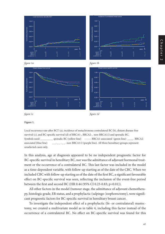
47
Ch
ap
te
r 2
In this analysis, age at diagnosis appeared to be no independent prognostic factor for BC-specific survival in hereditary BC, nor was the admittance of adjuvant hormonal treat-ment or the occurrence of a contralateral BC. This last factor was included in the model as a time-dependent variable, with follow-up starting as of the date of the CBC. When we included CBC with follow-up starting as of the date of the first BC, a significant favourable effect on BC-specific survival was seen, reflecting the inclusion of the event-free period between the first and second BC (HR 0.44 (95% CI 0.23-0.83; p=0.01)).
All other factors in the model (tumour stage, the admittance of adjuvant chemothera-py, histologic grade, ER status, and a prophylactic (salpingo-)oophorectomy), were signifi-cant prognostic factors for BC-specific survival in hereditary breast cancer.
To investigate the independent effect of a prophylactic (bi- or contralateral) mastec-tomy, we created a multivariate model as in table 4, including this factor instead of the occurrence of a contralateral BC. No effect on BC-specific survival was found for this
figure 1a figure 1b
figure 1c figure 1d
Figure 1.
Local recurrence rate after BCT (a), incidence of metachronous contralateral BC (b), distant disease-free survival (c), and BC-specific survival (d) of BRCA1-, BRCA2-, non-BRCA1/2 and sporadic BC.Symbols used: _________ sporadic BC (yellow line) ------- BRCA1-associated (green line) _._._. BRCA2-associated (blue line) _ _-_ _ -_ _- non-BRCA1/2 (purple line). All three hereditary groups represent unselected cases only.
Local recurrence rate after BCT
Cum
ulat
ive
prop
ortio
n
0 15
0.00
0.20
0.40
0.60
0.80
1.00
....
years At risk:
sporadic 410 289 142 53 17 BRCA1 76 42 27 12 4 BRCA2 33 16 9 4 2
non-BRCA1/2 111 66 38 18 4
Chapter 2B/Fure 1a Incidence of contralateral breast cancer
Cum
ulat
ive
prop
ortio
n
0 15
0.00
0.20
0.40
0.60
0.80
1.00
years At risk:
sporadic 756 486 236 92 31 BRCA1 161 82 36 15 7 BRCA2 87 39 22 14 5
non-BRCA1/2 228 134 66 33 14
Chapter2B/ figure 1b Distant disease-free survival
Cum
ulat
ive
prop
ortio
n
0 15
0.00
0.20
0.40
0.60
0.80
1.00
years At risk:
sporadic 758 423 201 82 30 BRCA1 167 83 41 21 9 BRCA2 89 47 26 14 9
non-BRCA1/2 237 126 65 31 16
Chapter 2B/ fig 1c Breast cancer-specific survival
Cum
ulat
ive
prop
ortio
n
0 15
0.00
0.20
0.40
0.60
0.80
1.00
years At risk:
sporadic 759 507 245 98 36 BRCA1 170 97 48 22 9 BRCA2 89 52 31 17 10
non-BRCA1/2 238 143 71 37 18
Chapter 2B/ Fig 1d
Local recurrence rate after BCT
Cum
ulat
ive
prop
ortio
n
0 15
0.00
0.20
0.40
0.60
0.80
1.00
....
years At risk:
sporadic 410 289 142 53 17 BRCA1 76 42 27 12 4 BRCA2 33 16 9 4 2
non-BRCA1/2 111 66 38 18 4
Chapter 2B/Fure 1a Incidence of contralateral breast cancer
Cum
ulat
ive
prop
ortio
n
0 15
0.00
0.20
0.40
0.60
0.80
1.00
years At risk:
sporadic 756 486 236 92 31 BRCA1 161 82 36 15 7 BRCA2 87 39 22 14 5
non-BRCA1/2 228 134 66 33 14
Chapter2B/ figure 1b Distant disease-free survival
Cum
ulat
ive
prop
ortio
n
0 15
0.00
0.20
0.40
0.60
0.80
1.00
years At risk:
sporadic 758 423 201 82 30 BRCA1 167 83 41 21 9 BRCA2 89 47 26 14 9
non-BRCA1/2 237 126 65 31 16
48
variable, not for the total hereditary group (HR 0.98 (95% CI 0.50-1.91), p=0.96) nor for the unselected cases only (HR 0.88 (0.40-1.95); p=0.75).
When we included sporadic breast cancer cases with the 3 hereditary groups in the model (except a BSO), all factors included in the model had a significant impact on bc-survival, except for the occurrence of contralateral cancer.
Discussion
In 1999 we published our initial report about the first 28 BRCA2-associated cases from the Rotterdam Family Cancer Clinic. 8 For the current analysis, we extended this series to 103 cases, and compared the tumour and treatment characteristics and survival of this group to other groups of hereditary and sporadic BC. We found that the clinical course in BRCA2-associated BC, with the exception of the high contralateral BC risk, was identical to that in non-BRCA1/2 and sporadic BC, with respect to LRR, DDFS, BC-specific and overall survival. Correction for tumour characteristics and treatment factors, including a B(S)O, did not essentially change these findings. We further found that the traditional prognostic factors, such as tumour stage and the admittance of adjuvant chemotherapy, were also independent prognostic factors in hereditary breast cancer. In addition, a pro-phylactic (salpingo-)oophorectomy nearly significantly (p=0.05) improved BC-specific survival. No prognostic impact was found for age at diagnosis, the admittance of adjuvant hormonal treatment, the occurrence of a contralateral BC or the performance of a (con-tralateral) prophylactic mastectomy.
While the special tumour features of BRCA1 are well-known, the phenotype of BRCA2-associated BC appears to be less specific. Our results were mostly in line with these observations. While we confirmed the frequently reported high incidence of grade III tumours in BRCA1-BC, no differences with respect to this variable were found be-tween BRCA2-BC as compared to non-BRCA1/2 and sporadic BC. This is in contrast with most other studies reporting a high prevalence of grade III tumours in BRCA2-BC. 9 23 24 In addition, another study4 reported a higher score for tubule formation but a lower mitotic count, both components of histologic grade. Two other studies did not find dif-ferences in histologic grade in BRCA2-BC cases as compared to control patients.25,26 As the assessment of histologic grade is hampered by a high interobserver-variation27, a valid comparison between studies of this variable is only possible after revision of all the histo-pathological material.
We also showed that the ER-positivity rate in BRCA2-BC (83%) was higher than in all other groups, and significantly higher than in sporadic tumours. This is in line with our previous publication 8, in which we found an even higher ER-positivity rate (93%), however not significantly different from sporadic BC, most likely because of the very small dataset (28 BRCA2-cases) at that time. To our knowledge, only Agnarsson also reported a significantly higher ER-positivity rate in BRCA2- as compared to sporadic cases (94
49
Ch
ap
te
r 2
versus 62%; p=0.002).9 Interestingly, Eerola et al also found a high ER-positivity rate in BRCA2-associated cases (79%), but only in the age group below age 50.28 We did not find this age-dependency in our dataset (83% and 86% ER-positivity in BRCA2-associated cases below and over the age of 50, respectively (p for difference between the age groups 0.77)). This is in line with findings from Foulkes et al, who found no change in ER status with age for BRCA2-carriers, in contrast to other subgroups. 29 As 65% of the tumours in our BRCA2- series was PgR-positive, this means that a substantial subgroup (21%) of our BRCA2-tumours was of the ER-positive/PgR-negative type. This is a higher frequency than expected in sporadic BC of comparable age (< 10%).30 A possible reason might be the inability of the ER to bind DNA, as was suggested previously by Osin et al.31 The incidence of ovarian cancers after BC in unselected BRCA2-BC was, with a yearly incidence of 0.7%, equal to that in BRCA1-BC and almost identical to the estimates in 152 BRCA2- cases, reported by Metcalfe et al. 19,32
Our results were based on a large dataset with detailed information about tumour and treatment characteristics and follow-up. All sporadic cases were selected from the cancer registry of the Rotterdam Erasmus MC, and all hereditary cases were ascertained at the Clinical Genetics Department. However, the analysis of the clinical course of he-reditary BC in family-based studies, such as ours, is hampered by various types of bias. For instance, the occurrence of a bilateral BC may prompt members from HBC/HBOC families to present themselves at the family cancer clinic. This was previously investigated by Tilanus et al, who noted that the higher incidence of a 2nd BC in non-BRCA1/2 BC as compared to sporadic cases was most outspoken before DNA testing and hardly increased thereafter, making it indeed likely that selection of patients with bilateral BC took place.19 For BRCA1/2 BC in the current series, the high incidence of a metachronous 2nd BC was maintained throughout the follow-up period (figure 1). However, this is no guarantee that oversampling of bilateral BC cases did not take place. Apart from a population-based study, a more optimal design in the family-based setting would be to perform a study while excluding all bilateral BC patients that occurred in the family before the date of DNA testing of the proband. In the current series, this would leave too small numbers to draw any meaningful conclusions about the incidence of contralateral BC in the various cohorts of hereditary BC. The unbiased incidence of bilateral hereditary BC is an interest-ing subject for further study, in unselected populations or large prospective databases in the family-based setting.
A further drawback of our study is that we included cases with a BC incidence date long before the possibility of germline-mutation testing, leading to a preferential selec-tion of long-living cases: the so-called longevity bias. Therefore, in our current and previ-ous comparisons with sporadic BC, we excluded cases with two years or more between BC diagnosis and DNA testing date. However, by excluding these so-called late-tested hereditary cases, which are characterized by an extremely favourable survival and a typi-cal tumour profile 7,19, we introduced a bias towards a unfavourable prognosis. Ideally, one would include all BC patients from each family. In practice, however, this is difficult

50
as data of especially deceased patients are frequently missing. An interesting alternative strategy is to match for the time between breast cancer and DNA diagnosis, as was done by Kirova et al. 33 In the near future, we will perform this type of matching and compare the results with those of the current analysis.
In the meantime we can conclude that, even with the selection bias towards an unfour-able clinical course, survival of neither of the three cohorts of hereditary BC appears to be significantly different from that in sporadic BC.
Further, our observation that tumour size and nodal status are prognostic factors also in hereditary BC, implies that the strategy to use these factors as a proxy for ultimate mor-tality, for instance in BC screening programmes or the consideration of (contralateral) prophylactic mastectomy, appears to be valid in this specific group of patients. A prophy-lactic contralateral mastectomy did not improve survival in the hereditary group.
51
Ch
ap
te
r 2
References
1. Eccles, D. M. Hereditary cancer: guidelines in clinical practice. Breast and ovarian cancer genetics. Ann.Oncol., 15 Suppl 4: iv133-iv138, 2004.
2. Nagy, R., Sweet, K., and Eng, C. Highly penetrant hereditary cancer syndromes. Oncogene, 23: 6445-6470, 2004.
3. Phillips, K. A. Immunophenotypic and pathologic differences between BRCA1 and BRCA2 hereditary breast cancers. J Clin.Oncol., 18: 107S-112S, 2000.
4. Lakhani, S. R., Jacquemier, J., Sloan, J. P., Gusterson, B. A., Andersen, T. J., and Klijn, J. G. M. Multifac-toral analysis of differences between sporadic breast cancers and cancers involving BRCA1 and BRCA2 mutations. J Natl Cancer Inst, 90(15): 1138-1145, 1998.
5. Narod, S. A. and Foulkes, W. D. BRCA1 and BRCA2: 1994 and beyond. Nat.Rev.Cancer, 4: 665-676, 2004.
6. Phillips, K. A., Andrulis, I. L., and Goodwin, P. J. Breast carcinomas arising in carriers of mutations in BRCA1 or BRCA2: are they prognostically different? J.Clin.Oncol., 17: 3653-3663, 1999.
7. Brekelmans, C. T., Seynaeve, C., Menke-Pluymers, M., Bruggenwirth, H. T., Tilanus-Linthorst, M. M., Bartels, C. C., Kriege, M., van Geel, A. N., Crepin, C. M., Blom, J. C., Meijers-Heijboer, H., and Klijn, J. G. Survival and prognostic factors in BRCA1-associated breast cancer. Ann.Oncol, 2005.
8. Verhoog, L. C., Brekelmans, C. T. M., Seynaeve, C., Dahmen, G., van Geel, A. N., Bartels, C. C. M., Tilanus-Linthorst, M. M. A., Wagner, A., Devilee, P., Halley, D., van den Ouweland, A. M. W., Meijers-Heijboer, E. J., and Klijn, J. G. M. Survival in hereditary breast cancer associated with germline mutations of BRCA2. J Clin Oncol, 17: 3396-3402, 1999.
9. Agnarsson, B. A., Jonasson, J. G., Bjornsdottir, I. B., Barkardóttir, R. B., Egilsson, V., and Sigurdsson, H. Inherited BRCA2 mutation associated with high grade breast cancer. Breast Cancer Res Treat, 47: 121-127, 1998.
10. Hedenfalk, I., Duggan, D., Chen, Y., Radmacher, M., Bittner, M., Simon, R., Meltzer, P., Gusterson, B., Esteller, M., Kallioniemi, O. P., Wilfond, B., Borg, A., and Trent, J. Gene-expression profiles in hereditary breast cancer. N.Engl.J.Med., 344: 539-548, 2001.
11. Honrado, E., Osorio, A., Palacios, J., Milne, R. L., Sanchez, L., Diez, O., Cazorla, A., Syrjakoski, K., Hunts-man, D., Heikkila, P., Lerma, E., Kallioniemi, A., Rivas, C., Foulkes, W. D., Nevanlinna, H., and Benitez, J. Immunohistochemical expression of DNA repair proteins in familial breast cancer differentiate BRCA2-associated tumors. J Clin.Oncol., 23: 7503-7511, 2005.
12. Verhoog, L. C., Berns, E. M. J. J., Brekelmans, C. T. M., Seynaeve, C., Meijers-Heijboer, E. J., and Klijn, J. G. M. Prognostic significance of germline BRCA2 mutations in hereditary breast cancer patients. J Clin Oncol, 18: 119s-124s, 2000.
13. Loman, N., Johannsson, O., Bendahl, P., Dahl, N., Einbeigi, Z., Gerdes, A., Borg, A., and Olsson, H. Prog-nosis and clinical presentation of BRCA2-associated breast cancer. Eur.J Cancer, 36: 1365-1373, 2000.
14. Eerola, H., Vahteristo, P., Sarantaus, L., Kyyronen, P., Pyrhonen, S., Blomqvist, C., Pukkala, E., Nevan-linna, H., and Sankila, R. Survival of breast cancer patients in BRCA1, BRCA2, and non-BRCA1/2 breast cancer families: a relative survival analysis from Finland. IJC, 93: 368-372, 2001.
15. El-Tamer, M. B., Russo, D., Mansukani, M., Troxel, A., Huiling, L., Ditkoff, B., and Schnabel, F. Survival and recurrence following breast cancer in BRCA1&2 mutation carriers. annals of surgical oncology, 10: S33, 2003.
16. Hedenfalk, I., Duggan, D., Chen, Y., Radmacher, M., Bittner, M., Simon, R., Meltzer, P., Gusterson, B., Esteller, M., Kallioniemi, O. P., Wilfond, B., Borg, A., and Trent, J. Gene-expression profiles in hereditary breast cancer. N.Engl.J.Med., 344: 539-548, 2001.
52
17. The CHEK2-breast cancer consortium Low-penetrance susceptibility to breast cancer due to CHEK2*1100delC in noncarriers of BRCA1 or BRCA2 mutations. Nature Genet, 31: 55-59, 2002.
18. de Bock, G. H., Schutte, M., Krol-Warmerdam, E. M., Seynaeve, C., Blom, J., Brekelmans, C. T., Meijers-Heijboer, H., van Asperen, C. J., Cornelisse, C. J., Devilee, P., Tollenaar, R. A., and Klijn, J. G. Tumour characteristics and prognosis of breast cancer patients carrying the germline CHEK2*1100delC variant. J.Med.Genet., 41: 731-735, 2004.
19. Tilanus-Linthorst, M. M., Bartels, K. C., Alves, C., Bakri, B., Crepin, E., van den, O. A., Klijn, J. G., Meijers-Heijboer, H., and Brekelmans, C. T. Selection bias influences reported contralateral breast cancer incidence and survival in high risk non-BRCA1/2 patients. Breast Cancer Res.Treat., 1-7, 2005.
20. van der Hout, A. H., van den, Ouweland A., van der Luijt, R. B., Gille, J. J. P., Bodmer D, Bruggenwirth, H. T., Mulder I.M., van der Vlies, P., Elfferich, P, Huisman, M. T., ten Berge, A. M., Kromosoeto, J., Jansen, R. P. M., van Zon, P. H. A., Vriesman, T., Arts, N., Boutmy-de Lange, M., Oosterwijk, J. C., Meijers-Heijboer, E. J., Ausems, M. G. E. M., Hoogerbrugge, N., Verhoef, S., Halley, D., Vos, Y. J., Hogervorst, F., Ligtenberg, M. J., and Hofstra, R. M. W. A DGGE system for comprehensieve mutation screening of BRCA1 and BRCA2. Application in a Dutch cancer clinic setting. Human Mutation. 2006. Ref Type: In Press
21. Hogervorst, F., Cornelis, R. S., Bout, M., van Vliet, M., Oosterwijk, J. C., Olmer, R., Bakker, B., Klijn, J. G. M., Vasen, H. F. A., Meijers-Heijboer, E. J., Menko, F. H., Cornelisse, C. J., den Dunnen, J. T., Devilee, P., and van Ommen, G. J. B. Rapid detection of BRCA1 mutations by the protein truncation test. Nature Genet, 10: 208-212, 1995.
22. Petrij-Bosch, A., Peelen, T., van Vliet, M., van Eijk, R., Olmer, R., Drussedau, M., Hogervorst, F., Hage-man, S., Meijers-Heijboer, E. J., Klijn, J. G. M., Vasen, H. F. A., Cornelisse, C. J., van ‘t Veer, L. J., Bakker, B., van Ommen, G. B., and Devilee, P. Large genomic deletions in BRCA1 as major founder mutations among dutch breast cancer families. Nature Genet, 17: 341-345, 1997.
23. Lynch, B. J., Holden, J. A., Buys, S. S., Neuhausen, S. L., and Gaffney, D. K. Pathobiologic Characteristics of Hereditary Breast Cancer. Hum Pathol, 29: 1140-1144, 1998.
24. Sensi, E., Tancredi, M., Aretini, P., Cipollini, G., Naccarato, A. G., Viacava, P., Bevilacqua, G., and Caligo, M. A. p53 inactivation is a rare event in familial breast tumors negative for BRCA1 and BRCA2 muta-tions. Breast Cancer Res.Treat., 82: 1-9, 2003.
25. Noguchi, S., Kasugai, T., Miki, Y., Fukutomi, T., Emi, M., and Nomizu, T. Clinicopathological analysis of BRCA1- or BRCA2- associated hereditary breast carcinoma in Japanese women. Ca, 85(10): 2200-2205, 1999.
26. Armes, J. E., Egan, A. J., Southey, M. C., Dite, G. S., McCredie, M. R., Giles, G. G., Hopper, J. L., and Ven-ter, D. J. The histologic phenotypes of breast carcinoma occurring before age 40 years in women with and without BRCA1 or BRCA2 germline mutations: a population-based study. Ca, 83: 2335-2345, 1998.
27. Subramaniam, D. S. and Isaacs, C. Utilizing prognostic and predictive factors in breast cancer. Curr.Treat.Options.Oncol., 6: 147-159, 2005.
28. Eerola, H., Heikkila, P., Tamminen, A., Aittomaki, K., Blomqvist, C., and Nevanlinna, H. Relationship of patients’ age to histopathological features of breast tumours in BRCA1 and BRCA2 and mutation-nega-tive breast cancer families. Breast Cancer Res., 7: R465-R469, 2005.
29. Foulkes, W. D., Metcalfe, K., Sun, P., Hanna, W. M., Lynch, H. T., Ghadirian, P., Tung, N., Olopade, O. I., Weber, B. L., McLennan, J., Olivotto, I. A., Begin, L. R., and Narod, S. A. Estrogen receptor status in BRCA1 and BRCA2-related breast cancer: the influence of age, grade, and histological type. Clin.Cancer Res., 10: 2029-2034, 2004.
30. Colditz, G. A., Rosner, B. A., Chen, W. Y., Holmes, M. D., and Hankinson, S. E. Risk factors for breast cancer according to estrogen and progesterone receptor status. J Natl Cancer Inst, 96: 218-228, 2004.
31. Osin, P., Gusterson, B. A., Philp, E., Waller, J., Bartek, J., Peto, J., and Crook, T. Predicted anti-oestrogen resistance in BRCA-associated familial breast cancers. EJC, 34: 1683-1686, 1998.

53
Ch
ap
te
r 2
32. Metcalfe, K. A., Lynch, H. T., Ghadirian, P., Tung, N., Olivotto, I. A., Foulkes, W. D., Warner, E., Olopade, O., Eisen, A., Weber, B., McLennan, J., Sun, P., and Narod, S. A. The risk of ovarian cancer after breast cancer in BRCA1 and BRCA2 carriers. Gynecol Oncol., 96: 222-226, 2005.
33. Kirova, Y. M., Stoppa-Lyonnet, D., Savignoni, A., Sigal-Zafrani, B., Fabre, N., and Fourquet, A. Risk of breast cancer recurrence and contralateral breast cancer in relation to BRCA1 and BRCA2 mutation status following breast-conserving surgery and radiotherapy. Eur.J.Cancer, 41: 2304-2311, 2005.


Chapter 3
3 A. Hereditary breast Cancer growth Rates and its Impact on Screening Policy
Madeleine MA Tilanus-Linthorst, Mieke Kriege, Carla Boetes, Wim CJ Hop, Inge-Marie Obdeijn, Jan C Oosterwijk, Hans L Peterse, Harmine M Zonderland, Sybren Meijer, Alexander MM Eggermont, Harry J de Koning, Jan GM Klijn, Cecile TM Brekelmans
European Journal of Cancer 2005;41(11):1610-17
56
Abstract
Imaging is often performed yearly for the surveillance of BRCA1/2 mutation carriers and women at high familial breast cancer risk. Growth of cancers in carriers may be faster as these tumours are predominantly high grade. Quantitative data on tumor growth rates in these two groups are lacking.
Here, we have examined 80 high risk women under surveillance for tumour size at diagnosis and preceding examinations at mammography and/or MRI. Tumour volume doubling time (DT) could be assessed in 30 cancers in BRCA1/2 mutation carriers and 25 non-carriers. Impact of age and menopausal status were also evaluated.
Mean DT of all invasive cancers was shorter in carriers (45 days CI: 26-73) than non-carriers (84 days CI: 58-131) (P = 0.048). Mean age at diagnosis was lower in carriers (40 yr.) than non-carriers (45 yr.) (P= 0.007). At multivariable analysis only age (P=0.03), not risk-group (P=0.26) nor menopause (P=0.58) correlated significantly with DT. The mean growth rate slowed down to half in each successive 10 years-older group.
In conclusion: Age at detection indicates the growth rates of hereditary and familial breast cancers. If recommended, the screening frequency should be adjusted according to a woman’s age. A high-sensitive biannual test may be appropriate before the age of 40 years.
Keywords: Breast cancer, surveillance, interval cancer, growth rate, BRCA1, familial breast cancer, screening, sojourn time, MRI, mammography.

57
Ch
ap
te
r 3
Introduction
Early detection is one of the limited options to possibly reduce the risk of mortality from breast cancer for women with a gene mutation (e.g. BRCA1, BRCA2, p53) or with a fam-ily history, indicative of an increased risk for breast cancer at a relatively young age. For BRCA1 mutation carriers the risk to develop breast cancer before 50 years of age is as high as 50%, while for BRCA2 the risk is slightly less [1,2]. Although breast cancer cells may disseminate early during tumour development [3], tumour size and lymph node status remain strong prognostic factors for survival in breast cancer [4-7]. Screening women at hereditary risk with Magnetic resonance imaging (MRI) can detect tumours at an early stage [8-9]. In the Dutch MRISC study 78% of the detected tumours were ductal carci-noma in situ (DCIS) or smaller than 2 cm, 79% node-negative [8].
However, a higher percentage of interval cancers have been observed in BRCA1/2 mu-tation carriers compared with women with high familial risk without a proven mutation (non-carriers) under the same surveillance scheme [8, 10]. One of the likely causes is dif-ferent growth rates of tumours, as in cancers of BRCA1 mutation carriers a high mitotic count and high grade tumours (63% and 69% respectively) were more frequently found than in sporadic cancers (32% and 38% respectively) and BRCA1/2-negative hereditary breast cancers (17% and 23% respectively) [11,12].
To our knowledge no quantitative data have been published on tumour growth rates in these hereditary risk groups based on measurements from imaging. Finding the optimal frequency at which a screening method should be applied can be as important to improve its effectiveness as the ability to detect cancers at an early stage [13].
Screening too frequently, increases the medicalisation of healthy women, the risk of false-positive results, cost and radiation risk [14], but too low a frequency may result in a delay in diagnosing breast cancer, missing the chance to improve prognosis.
In this study, we have investigated the influence of a BRCA1/2 mutation, age and meno-pausal status / bilateral preventive salpingo-oophorectomy (BPSO) on tumour growth rate in women at high familial risk.
Based on our results we have tried to define the optimal screening frequency for wom-en in different risk categories.
Material and methods
We could evaluate the size of 55 tumours at diagnosis and with the same radiologique technique, mammography (Mx) or MRI, at previous screening(s) for 80 breast cancer pa-tients examined. All tumours were detected in women under surveillance, because of: (a) a proven BRCA1 or BRCA2 mutation (carrier group), or (b) an estimated hereditary risk of 20-50% according to modified tables of Claus [8,15], while no BRCA1 or 2 mutation
58
could be demonstrated or no DNA investigation had been performed (non-carriers). The methods for BRCA1/2 mutation analyses are described elsewhere [16, 17].
From November 1, 1999 to July 1, 2003, 47 breast cancers were detected in women participating in the Dutch surveillance study MRISC in 2 cancer centers and 4 university hospitals. Screening consisted of clinical breast examination every 6 months and annual Mx and MRI. Imaging technique and protocol have been previously described [8]. Tu-mour growth rate was evaluable in 32 cases. Thirty-three consecutive cancers were de-tected in the women under surveillance for the same indication outside this study after Jan 1 1995, at the ErasmusMC. Surveillance for them was performed with biannual clinical examination and annual mammography. Additional MRI was performed with the same Tesla strength, intravascular contrast and subtractions as in the MRISC in 13 patients. Tumour growth rate could be assessed in 23 cases. In total growth rates were assessed in 55 patients. In 25 patients tumour growth rates could not be calculated as the tumour was neither measurable at diagnostic Mx or at MRI.
The diameter at pathology and mitotic count and Bloom-Richardson grading of the tumours, menopausal status and BPSO were taken from medical files.
Measurements and calculation of tumour growth rateTo estimate the growth rate of tumours, all diagnostic mammograms and MRI, were re-evaluated by a radiologist (CB or IO). For all the cancers visible at the diagnostic Mx /MRI, the previous examination(s) were reassessed. If the tumour could be clearly identi-fied from the diagnostic MRI, 3D measurements at right angles, including the single larg-est dimension (SLD), were taken from the diagnostic and previous MRI. For all cancers positively identified at the diagnostic Mx, tumour size was measured at both oblique and craniocaudal views at diagnostic and previous Mx. The tumour diameter was measured using the longest axis (a = SLD) and a second maximum diameter was measured perpen-dicular to the first (b). For tumours measurable at both views the largest and smallest size and the mean of the other two were used to calculate tumour volume. In the case of a stel-late mass, the centre was measured.
For cancers with a measurable tumour at 2 or more subsequent mammograms or MRI and where a previous mammogram/MRI showed no visible tumour (9 Mx, 2 MRI), only the measurable tumour sizes were used for the calculation of individual tumour volume doubling time (DT).
To calculate the DT of each cancer, either Mx or MRI measurements were used. The method with most measurement points was used. In case of equal number of measure-ments, the method with the single largest tumor diameter at diagnosis closest to the size at pathology was used.
The volume of the tumour was estimated using the formula for obloid spheroids V = 4/3 π.1/2a.1/2b.1/2c.
Tumour volumes were assumed to have exponential growth (i.e. growth with a con-stant volume doubling time). For patients with 2 real volume measurements, the slope of
59
Ch
ap
te
r 3
the straight line connecting the two log-transformed data points was calculated. In case of 3 or more real volume measurements, this slope was calculated using least-squares regres-sion. For patients with one last real measurement and one previous undetected tumour, the latter tumour size was set at 0.004 cm3 corresponding to a diameter of 2 mm (assumed lower detection limit). The resulting slopes for these patients therefore may under-esti-mate the true slope. However not including these for the estimation of growth rates would probably exclude many of the fast growing tumours [18].
Subsequently, tumour volume doubling times were calculated using the formula DT = log 2/β, where β was the slope of the regression line of the logarithm of the tumour volume vs.time. This outcome may over-estimate the true doubling time for the patients with an undetectable tumour at the previous visit and is treated as a left-censored observation in the statistical analysis [18].
Statistical methodsDifferences in patient and tumour characteristics between the 2 risk-groups were tested with the use of the t-test in case of continuous variables and of the chi-square test or Fisher’s exact test in case of categorical variables. To determine the correlation between tumour size at mammography /MRI and at histo-pathologic examination we calculated Pearson’s correla-tion coefficient separately for invasive cancers and ductal carcinoma in situ (DCIS). To get an approximate normal distribution of volume doubling times, these times were logarithmi-cally transformed for analysis. Comparison of the transformed DT between risk-groups was done using the t-test. Multiple regression was used to evaluate simultaneously the effects of age, risk-group and menopausal status. STATA-software (procedure CNREG) was used in these calculations to allow for the presence of left-censored volume doubling times. A two-sided P-value of less than 0.05 was considered to indicate statistical significance.
Results
Patients and tumour characteristics Of the total group of 55 tumours, in which growth rate could be assessed, 30 (5 DCIS, 4 of the DCIS in BRCA1) were detected in mutation carriers (25 BRCA1 and 5 BRCA2) (carriers) and 25 (3 DCIS) in women with an estimated life-time risk of 20-50% (non-car-rier group). Eighteen patients in the non-carrier group had tested negative for BRCA1 /2, while for 7 patients no DNA test result was available. Only 1/7 tumours in the non-tested group had characteristics suggestive of a BRCA1-associated tumour (both high grade and ER and PR negative), but with a mitotic count of 3. Patient and tumour characteristics of the carrier and non-carrier groups are shown in Table 1. Mean age at diagnosis was sig-nificantly lower in carriers than in non-carriers (40 years vs. 45 yrs, respectively, P=0.007); (39 yrs. for BRCA2 and 47 yrs. for the non-tested). Seven of the carriers were post meno-pausal at diagnosis, 6 after BPSO (no BRCA2), while 6 non-carriers were naturally post-
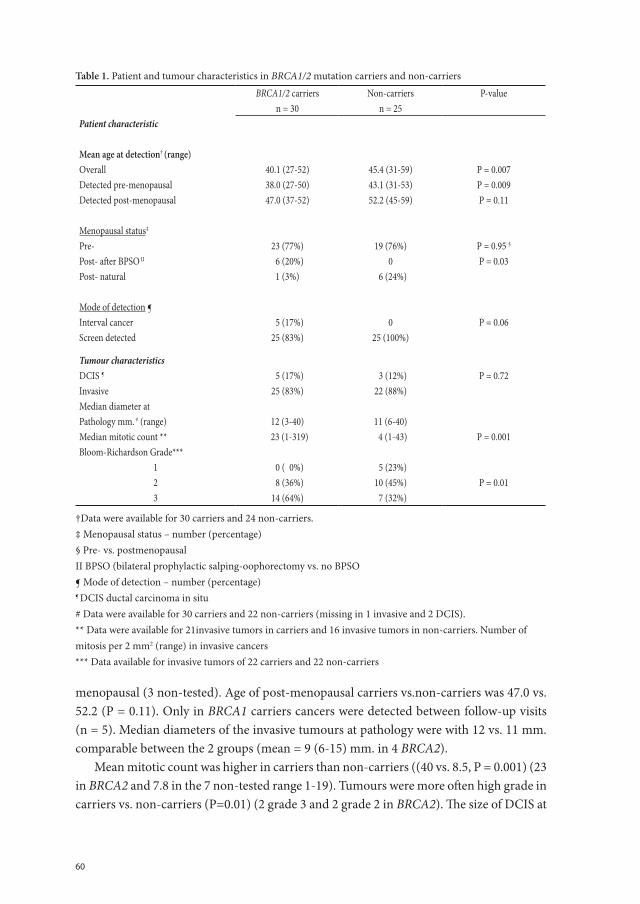
60
menopausal (3 non-tested). Age of post-menopausal carriers vs.non-carriers was 47.0 vs. 52.2 (P = 0.11). Only in BRCA1 carriers cancers were detected between follow-up visits (n = 5). Median diameters of the invasive tumours at pathology were with 12 vs. 11 mm. comparable between the 2 groups (mean = 9 (6-15) mm. in 4 BRCA2).
Mean mitotic count was higher in carriers than non-carriers ((40 vs. 8.5, P = 0.001) (23 in BRCA2 and 7.8 in the 7 non-tested range 1-19). Tumours were more often high grade in carriers vs. non-carriers (P=0.01) (2 grade 3 and 2 grade 2 in BRCA2). The size of DCIS at
Table 1. Patient and tumour characteristics in BRCA1/2 mutation carriers and non-carriers
BRCA1/2 carriers Non-carriers P-valuen = 30 n = 25
Patient characteristic
Mean age at detection† (range) Overall 40.1 (27-52) 45.4 (31-59) P = 0.007Detected pre-menopausal 38.0 (27-50) 43.1 (31-53) P = 0.009Detected post-menopausal 47.0 (37-52) 52.2 (45-59) P = 0.11
Menopausal status‡
Pre- 23 (77%) 19 (76%) P = 0.95 §
Post- after BPSO II 6 (20%) 0 P = 0.03 Post- natural 1 (3%) 6 (24%)
Mode of detection ¶Interval cancer 5 (17%) 0 P = 0.06 Screen detected 25 (83%) 25 (100%)
Tumour characteristicsDCIS ¶ 5 (17%) 3 (12%) P = 0.72Invasive 25 (83%) 22 (88%)Median diameter atPathology mm. # (range) 12 (3-40) 11 (6-40) Median mitotic count ** 23 (1-319) 4 (1-43) P = 0.001Bloom-Richardson Grade***
1 0 ( 0%) 5 (23%) 2 8 (36%) 10 (45%) P = 0.01 3 14 (64%) 7 (32%)
†Data were available for 30 carriers and 24 non-carriers.‡ Menopausal status – number (percentage)§ Pre- vs. postmenopausalII BPSO (bilateral prophylactic salping-oophorectomy vs. no BPSO¶ Mode of detection – number (percentage)¶ DCIS ductal carcinoma in situ# Data were available for 30 carriers and 22 non-carriers (missing in 1 invasive and 2 DCIS). ** Data were available for 21invasive tumors in carriers and 16 invasive tumors in non-carriers. Number of mitosis per 2 mm2 (range) in invasive cancers*** Data available for invasive tumors of 22 carriers and 22 non-carriers
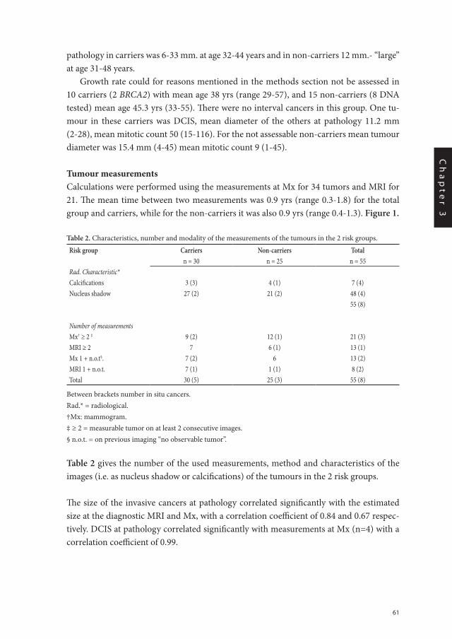
61
Ch
ap
te
r 3
pathology in carriers was 6-33 mm. at age 32-44 years and in non-carriers 12 mm.- “large” at age 31-48 years.
Growth rate could for reasons mentioned in the methods section not be assessed in 10 carriers (2 BRCA2) with mean age 38 yrs (range 29-57), and 15 non-carriers (8 DNA tested) mean age 45.3 yrs (33-55). There were no interval cancers in this group. One tu-mour in these carriers was DCIS, mean diameter of the others at pathology 11.2 mm (2-28), mean mitotic count 50 (15-116). For the not assessable non-carriers mean tumour diameter was 15.4 mm (4-45) mean mitotic count 9 (1-45).
Tumour measurements Calculations were performed using the measurements at Mx for 34 tumors and MRI for 21. The mean time between two measurements was 0.9 yrs (range 0.3-1.8) for the total group and carriers, while for the non-carriers it was also 0.9 yrs (range 0.4-1.3). Figure 1.
Table 2. Characteristics, number and modality of the measurements of the tumours in the 2 risk groups.
Risk group Carriers Non-carriers Totaln = 30 n = 25 n = 55
Rad. Characteristic* Calcifications 3 (3) 4 (1) 7 (4)Nucleus shadow 27 (2) 21 (2) 48 (4)
55 (8)
Number of measurementsMx† ≥ 2 ‡ 9 (2) 12 (1) 21 (3)MRI ≥ 2 7 6 (1) 13 (1) Mx 1 + n.o.t§. 7 (2) 6 13 (2) MRI 1 + n.o.t. 7 (1) 1 (1) 8 (2) Total 30 (5) 25 (3) 55 (8)
Between brackets number in situ cancers.Rad.* = radiological.†Mx: mammogram.‡ ≥ 2 = measurable tumor on at least 2 consecutive images.§ n.o.t. = on previous imaging “no observable tumor”.
Table 2 gives the number of the used measurements, method and characteristics of the images (i.e. as nucleus shadow or calcifications) of the tumours in the 2 risk groups.
The size of the invasive cancers at pathology correlated significantly with the estimated size at the diagnostic MRI and Mx, with a correlation coefficient of 0.84 and 0.67 respec-tively. DCIS at pathology correlated significantly with measurements at Mx (n=4) with a correlation coefficient of 0.99.
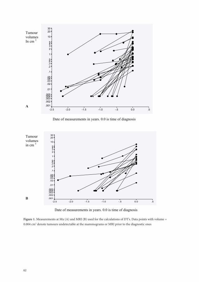
62
Chapter 3A figure 1 Figure 1. Measurements at Mx (A) and MRI (B) used for the calculations of DT’s. Data points with volume = 0.004 cm 3 denote tumours undetectable at the mammograms or MRI prior to the diagnostic ones
Tumour volumes In cm 3
A
Date of measurements in years. 0.0 is time of diagnosis
Tumour volumes in cm 3
Date of measurements in years. 0.0 is time of diagnosis
.50.0-.5-1.0-1.5-2.0-2.5
3020
10
5432
1
.5.4
.3
.2
.1
.05.04
.03
.02
.01
.005
.004
.003
.002
.001
.50.0-.5-1.0-1.5-2.0-2.5
3020
10
5432
1
.5.4
.3
.2
.1
.05.04
.03
.02
.01
.005
.004
.003
.002
.001
Figure 1. Measurements at Mx (A) and MRI (B) used for the calculations of DT’s. Data points with volume = 0.004 cm3 denote tumours undetectable at the mammograms or MRI prior to the diagnostic ones
B
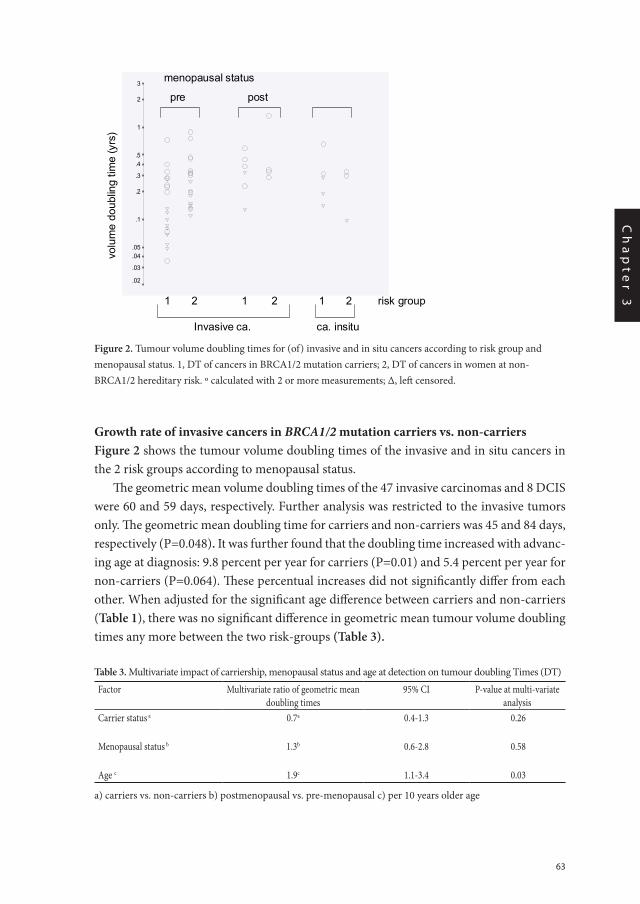
63
Ch
ap
te
r 3
Growth rate of invasive cancers in BRCA1/2 mutation carriers vs. non-carriersFigure 2 shows the tumour volume doubling times of the invasive and in situ cancers in the 2 risk groups according to menopausal status.
The geometric mean volume doubling times of the 47 invasive carcinomas and 8 DCIS were 60 and 59 days, respectively. Further analysis was restricted to the invasive tumors only. The geometric mean doubling time for carriers and non-carriers was 45 and 84 days, respectively (P=0.048). It was further found that the doubling time increased with advanc-ing age at diagnosis: 9.8 percent per year for carriers (P=0.01) and 5.4 percent per year for non-carriers (P=0.064). These percentual increases did not significantly differ from each other. When adjusted for the significant age difference between carriers and non-carriers (Table 1), there was no significant difference in geometric mean tumour volume doubling times any more between the two risk-groups (Table 3).
Table 3. Multivariate impact of carriership, menopausal status and age at detection on tumour doubling Times (DT)
Factor Multivariate ratio of geometric mean doubling times
95% CI P-value at multi-variate analysis
Carrier status a 0.7a 0.4-1.3 0.26
Menopausal status b 1.3b 0.6-2.8 0.58
Age c 1.9c 1.1-3.4 0.03
a) carriers vs. non-carriers b) postmenopausal vs. pre-menopausal c) per 10 years older age
____Chapter 3A/fig 1
_________________________________________________________Figure 2.Tumour volume doubling times for (of) invasive and in situ cancers according to risk group and menopausal status _________________________________________________________________________
volu
me
doub
ling
time
(yrs
)
risk group1 2 1 2 1 2
Invasive ca. ca. insitu
3
2
1
.5
.4
.3
.2
.1
.05
.04
.03
.02
menopausal status
pre post
Tumour volume doubling times of invasive and in situ cancers according to risk group and menopausal status ___________________________________________________________________________
Chapter 3A/ fig 2
_______________________________________________________________________
Figure 3. Tumour DT (months) according to age at diagnosis
Figure 2. Tumour volume doubling times for (of) invasive and in situ cancers according to risk group and menopausal status. 1, DT of cancers in BRCA1/2 mutation carriers; 2, DT of cancers in women at non-BRCA1/2 hereditary risk. º calculated with 2 or more measurements; Δ, left censored.
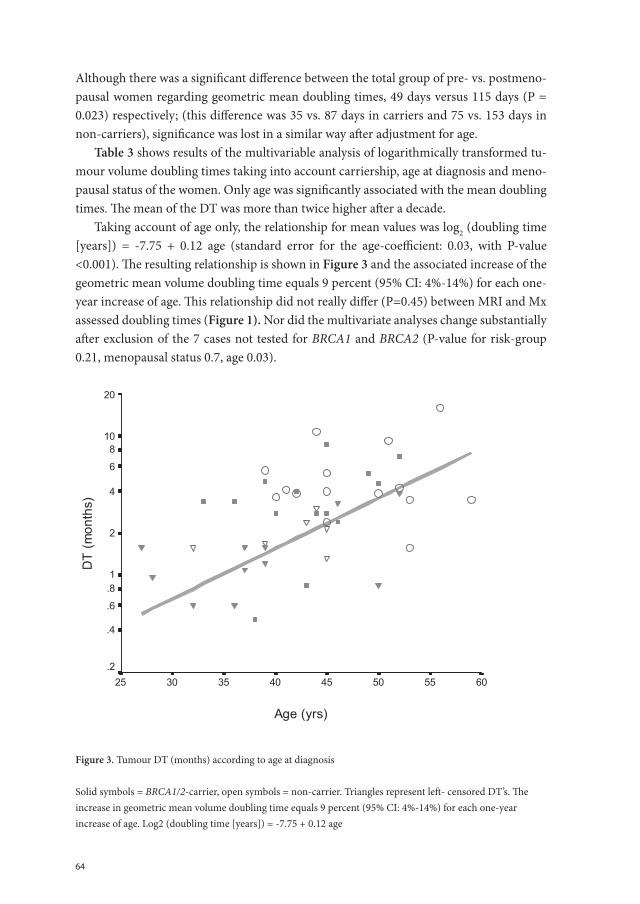
64
Although there was a significant difference between the total group of pre- vs. postmeno-pausal women regarding geometric mean doubling times, 49 days versus 115 days (P = 0.023) respectively; (this difference was 35 vs. 87 days in carriers and 75 vs. 153 days in non-carriers), significance was lost in a similar way after adjustment for age.
Table 3 shows results of the multivariable analysis of logarithmically transformed tu-mour volume doubling times taking into account carriership, age at diagnosis and meno-pausal status of the women. Only age was significantly associated with the mean doubling times. The mean of the DT was more than twice higher after a decade.
Taking account of age only, the relationship for mean values was log2 (doubling time [years]) = -7.75 + 0.12 age (standard error for the age-coefficient: 0.03, with P-value <0.001). The resulting relationship is shown in Figure 3 and the associated increase of the geometric mean volume doubling time equals 9 percent (95% CI: 4%-14%) for each one-year increase of age. This relationship did not really differ (P=0.45) between MRI and Mx assessed doubling times (Figure 1). Nor did the multivariate analyses change substantially after exclusion of the 7 cases not tested for BRCA1 and BRCA2 (P-value for risk-group 0.21, menopausal status 0.7, age 0.03).
Figure 3. Tumour DT (months) according to age at diagnosis
Solid symbols = BRCA1/2-carrier, open symbols = non-carrier. Triangles represent left- censored DT’s. The increase in geometric mean volume doubling time equals 9 percent (95% CI: 4%-14%) for each one-year increase of age. Log2 (doubling time [years]) = -7.75 + 0.12 age
Age (yrs)
6055504540353025
DT
(mon
ths)
20
108
6
4
2
1.8
.6
.4
.2
___________________________________________________________________________
Chapter 3A/ fig 3
65
Ch
ap
te
r 3
The tumour characteristics grade and mitotic count differed between the 2 risk-groups. At univariate analysis mitotic count correlated with DT (P=0.03), while grade did not (sig-nificantly so) (P= 0.3). When mitotic count and grade were entered into the multivariable model the results remained essentially unchanged with P value for age, grade and mitotic count P = 0.015; P= 0.8 and P= 0.4 respectively.
DCISFour in situ cancers were only visible at diagnosis not on previous imaging: 3 in carriers (6, 7 and 33 mm.) and 1 non - carrier (>40 mm) (Figure 2).
Discussion
The growth rates of hereditary breast cancer are important to estimate the optimal test frequency for screening, be it by breast imaging (Mx or MRI) or new emerging screening tools, e.g. serum-proteomic-pattern markers [9,13,19].
Tumour volume doubling time was with 45 vs. 84 days twice as short in invasive can-cers of BRCA1/2 mutation carriers compared to non-carriers and twice as short in pre- vs. postmenopausal women. However, mean age at detection differed significantly between the 2 risk groups and carriers were more often postmenopausal at a relatively young age after BPSO. Age at diagnosis and not risk-group nor menopausal status, was the only sig-nificant indicator of tumour growth rate at multivariate analysis. The on average higher tumour growth rates in carriers vs. non-carriers and pre- vs. postmenopausal women con-tributed apparently to earlier ages at detection. Tumours were more often high grade and the average mitotic count was higher in our younger carrier group as expected. When these indicators of growth rate were entered into the multivariate model still only age correlated independently with the estimated tumour doubling times (P=0.015). Tumour growth rates gradually slowed down (9% yearly) with increasing age at diagnosis, without a clear cut-off between the risk-groups or at menopause.
Our study was performed in women with a well-defined hereditary risk, within sur-veillance schemes with complete follow-up. The relatively low number of interval cancers (in 5 BRCA1 carriers only) may be due to the rather short screening intervals. We assessed the growth rates in only 4 invasive breast cancers in BRCA2 mutation carriers, who did not differ significantly with regard to age, tumour size, grade (and) or mitotic count from the BRCA1 mutation carriers. The DT pattern for BRCA1 mutation carriers and non-car-riers of the same age were similar.
In 7 patients no test for deleterious BRCA1 or 2 mutations was performed or com-pleted. But after exclusion of the 7 results of the analyses were essentially the same. In the two risk groups, patient and tumour characteristics did not differ between those with and without DT assessment. Therefore DT measurements in both risk groups may be repre-sentative for that group.
66
Either measurements at Mx or at MRI were used for DT calculations, but both corre-lated well with size at pathology. Neither the mean doubling time nor the results at multi-variable analyses differed significantly between assessments with either method.
The radiologist knew from the diagnostic imaging, where and how the cancer was depicted, therefore we estimated tumour size at the previous image with “no observable tumour” on retrospect, to have a max size of 2mm. This seems realistic as we could mea-sure 5 tumours at MRI and 8 Mx cases were < 4mm. despite high breast density in several. By extrapolating growth-curves of tumours measurable at ≥ 2 Mx/MRI but “no tumour” at the previous image (9Mx, 2MRI), occult- tumour-size was at Mx twice < 2mm. and 7 times < 4mm, at MRI twice < 2mm. Importantly, also when we assumed for the calcula-tion of our DT’s occult -tumour-size at Mx to be < 4 mm (instead of < 2 mm), results of the multivariable analysis did not essentially change.
Growth may not be continuous and possibly speed up or slow down under influence of host factors or size. However we performed calculations on the assumption of exponential growth of the tumours, as this is usually assumed to be the best approximation for the range of tumour sizes in our study (3-40 mm)[20,21]. Our findings in 12 of the 15 cases with more than 2 measurements were consistent with exponential growth, while in 1 there seemed to be a period without growth (figure 1).
Although the tumours in BRCA1 mutation carriers are more frequently oestrogen- and progesteron receptor negative, a clear influence on the occurrence of (contralateral) can-cers has been described for hormonal factors like menopause and BPSO, but less consis-tently also for breastfeeding, use of oral contraceptives, pregnancy, parity and tamoxifen [22]. All these hormonal influences may, like other host factors, have an impact on tumour growth rate and possibly to a different degree in carriers of BRCA1 or BRCA2 mutations and non-carriers. Within the size and scope of our study we could only account for the strongest proven hormonal influence of menopause/BPSO. Extended and different studies are needed to clarify these complex issues.
Spratt and colleagues calculated in sporadic breast cancer patients DT with a wide range from (of) 10-7051 days and age range 18-88 years. With age sorted in categories they did not find a clear relationship between growth rates and age [23]. They assessed however less fast growing tumours, by not including cancers that were only visible at diagnosis, not at the previous mammograms. Kusama, Spratt and colleagues on the other hand found significantly less tumours with short doubling times in patients age 60 years and over than in younger patients [24]. Peer and colleagues, calculated a median DT of 80 days (95% C.I. 44-147) for breast cancers in women less than 50 years of age not selected for risk, twice as fast as in women aged 50-70 years [18]. These results are quite similar to the pre- and postmenopausal growth rates we calculated from our non-carriers (mean 75 and 153 days respectively), reflecting most likely the comparable ages at detection. The data available from sporadic breast cancers in the literature substantially support our analyses.
Breast screening women aims to detect cancers at an early stage at which the future development of metastases is less likely, in order to possibly improve survival. Tumour size
67
Ch
ap
te
r 3
at diagnosis and the number of positive axillary nodes are strong prognostic factors for survival in sporadic and hereditary breast cancers [4-7, 25], even though other evidence suggests that the proclivity to metastasize is acquired early in tumour genesis [3]. The per-centage of patients with metastases seems to increase faster with size in high grade breast cancers than in low grade [26]. Tabar et al,.however, found good cumulative 12 yr. disease specific survival rates of over 90% for all high grade tumors ≤ 1 cm [27].
If we try to assess the optimal screening interval, taking the impact of tumour stage into account, we should consider, that a tumour with a diameter of 2 mm, missed at imag-ing, needs 4 doubling times to reach size 5 mm, where it becomes easier to detect but is most likely still node-negative. In that period, a tumour with the same growth rate missed at 4 mm. may reach 1 cm. With regard to stage at detection a 4 times DT screening interval seems acceptable. In our study this would result in screening intervals of 3-7 months from age 30 till 40 years; of 7-16 months from 40 till 50 yrs. and 16-32 months from 50 till 60 yrs (Figure 3), reflecting the gradual decrease in growth rate for tumours detected at increas-ing age. In practice and because of the range of DT’s at a given age this might translate into a biannual screening-test before age 40 yrs, annual between 40-50 yrs and once every 2 years at age 50-60 yrs. It has been suggested by different models that in selected groups of women, biannual imaging might be necessary to improve survival [13, 19, 28, 29]
At such frequency, a test with high sensitivity for invasive cancer seems the method of choice. In BRCA1/2 mutation carriers MRI seems preferable over mammography because the tumor characteristics cause frequent false-negative mammography results [30] In MRI screening studies sensitivity for invasive cancers proved better for MRI than mammogra-phy, but separate estimates for BRCA1/2 carriers are not yet available [8,9]. The number of BRCA1/2 mutation carriers under surveillance is relatively small and their expected tu-mor incidence high (2% yearly between age 25-50 years) [1, 2]. Cost-effectiveness analyses are now performed, impact on survival however has still to be shown.
In the large group of women at hereditary risk without a known BRCA1/2 mutation in the family, screening is usually started at an older age than in BRCA1/2 mutation carriers. Imaging annually between ages 40-50 yrs. and once every 2 years between 50-60 yrs. may be appropriate. This is in agreement with studies that have estimated the sojourn time (i.e. the length of time the disease is in the preclinical detectable phase) in women aged 40-49 to be one year [31, 32]
With 4 DCIS out of 30 cancers detected in BRCA1 carriers (and 1 in BRCA2) we can-not confirm that the in situ stage is skipped in BRCA1 cancers. With screening it can be detected. We could recognize DCIS 4 times only at diagnosis, not the previous year. We do not know for how long DCIS may grow before invasion starts - the event we aim to prevent. But DCIS could reach a considerable size (33 mm and > 40 mm respectively) in carriers and non-carriers.
In conclusion: Age at detection is the main indicator for growth rates of hereditary and fa-milial breast cancers. If screening may prove indicated from a certain age on, the woman’s

68
age, not the risk group should determine the screening interval. A high-sensitive biannual test may be appropriate before age 40 years.
Conflict of Interest StatementNone declared.
Acknowledgments
The authors like to thank Marijke Westerhout-Kersten and Kerstin van der Veen for assist-ing in literature search, Ada van Eekelen for logistic support. Petra Bos, Titia van Echten, Angelique Schlief, Marijke Hogenkamp, Irene Groot and Arjan Nieborg for their contri-bution to data management.
69
Ch
ap
te
r 3
References
1. Scott CL, Jenkins MA, Southey MC et al. Average age-specific cumulative risk of breast cancer according to type and site of germline mutations in BRCA1 and BRCA2 estimated from multiple-case breast cancer families attending Australian family cancer clinics. Hum Genet. 2003;112 (5-6):542-51.
2. Antoniou A, pharaoh P, Narod S et al. Average risk of Breast and ovarian cancer associated with BRCA1 or BRCA2 mutations detected in case series unselected for family history: a combined analysis of 22 stud-ies. Am.J HumGenet 2003;72:1117-30.
3. Schmidt-Kittler O, Ragg Th, Daskalakis A, et al. From latent disseminated cells to overt metastasis: Ge-netic analysis of systemic breast cancer progression. Proc. Natl.Acad.Sci.USA 2003;100, 13:7737-42.
4. Sant M, Allemani C, Capocaccia R, et al. Stage at diagnosis is a key explanation of differences in breast cancer survival across Europe. Int. J Cancer 2003;106(3):416-22.
5. Michaelson JS, Silverstein M, Sgroi D, et al. The effect of tumor size and lymph node status on breast carcinoma lethality. Cancer 2003; 98; 2133-43.
6. Carter C, Allen C, Henson D. Relation of tumor size, lymph node status and survival in 24,740 breast cancer cases. Cancer 1989; 63:181-7.
7. Eerola H, Vahteristo P, Sarantaus L, et al. Survival of breast cancer patients in BRCA1, BRCA2 and non-BRCA1/2 breast cancer families: a relative survival analysis from Finland. Int. J Cancer. 2001; 93:368-372.
8. Kriege M., Brekelmans CTM, Boetes C, et al. Efficacy of breast cancer screening with MRI and mammog-raphy in women with a familial or genetic predisposition. N Eng J Med 2004; July 29;351(5):427-37.
9. Robson ME, Offit K. Breast MRI for women with hereditary cancer risk. JAMA 2004;292(11):1368-70.
10. Brekelmans CT, Seynaeve C, Bartels CC, et al. Effectiveness of breast cancer surveillance in BRCA 1/2 gene mutation carriers and women with high family risk. J Clin Oncol 2001; 19:924-30.
11. Lakhani SR, Gusterson BA, Jacquemier J, et al. The pathology of familial breast cancer: Histological features of cancers in families not attributablre to mutations in BRCA1 or BRCA2. Clin Cancer Res 2000; 6:782-9.
12. Vaziri SAJ, Krumroy LM, Elson P, et al. Breast tumor immunophenotype of BRCA1-mutation carriers is influenced by age at diagnosis. Clin Cancer Res 2001; 7:1937-45.
13. Michaelson JS, Halpern E, Kopans D. Breast cancer: Computer simulation method for estimating optimal intervals for screening. Radiology 1999; 212:551-60.
14. Fletcher SW, Elmore JG. Clinical practice. Mammographic screening for breast cancer. N Engl J Med 2003; 348:1672-80.
15. Claus EB, Risch N, Thompson WD. Autosomal dominant inheritance of early-onset breast cancer. Impli-cations for risk predictions. Cancer 1994; 73:643-51.
16. Verhoog LC, Brekelmans CT, Seynaeve C, et al. Survival and tumour characteristics of breast-cancer patients with germline mutations of BRCA1. Lancet 1998; 351 (9099):316-21.
17. Verhoog LC, Brekelmans CT, Seynaeve C, et al. Survival in hereditary breast cancer associated with germ-line mutations of BRCA2. J Clin Oncol. 1999; 17: 3396-402.
18. Peer PGM, vanDijck JAAM, Hendriks JHCL, et al. Age-dependent growth rate of primary breast cancer. Cancer 1993; 71:3547-51.
19. Kopans DB, Rafferty E, Georgian-Smith D, et al. A simple model of breast carcinoma growth may provide explanations for observations of apparently complex phenomena. Cancer 2003; 97:2951-9.
20. Norton L. A Gompertzian model of human breast cancer growth. Cancer Res. 1988; 48:7067—71.
70
21. Speer JF, Petrosky VE, Retsky MW, Wardwell RH. Et al. Stochastic numerical model of breast cancer growth that simulates clinical data. Cancer Res 1984; 44:4124-30.
22. Rebbeck TR Prophylactic oophorectomy in BRCA1 and BRCA2 mutation carriers. Eur J Cancer 2002;6:S15-S17.
23. Spratt JA, von Fournier D, Spratt JS, et al. Mammographic assessment of human breast cancer growth and duration. Cancer 1993; 71:2020-6.
24. Kusama S, Spratt JS, Donegan WL et al. The gross rates of growth of human mammary carcinoma. Cancer 1972;30:594-99.
25. Seynaeve C, Tilanus-Linthorst MMA, Meijers Heijboer H, et al. BRCA1 versus non-BRCA1/2-associated breast cancer: tumor characteristics and impact of prognostic and treatment factors.Proc.Am.Soc.Clin.Oncol.2004;23:nr.9557.
26. Tubiana M, Koscielny S. Natural history of human breast cancer: recent data and clinical implications. Breast Cancer Res and Treatm. 1991;18:125-40.
27. Tabar L, Fagerberg G, Day NE, et al. Breast cancer treatment and natural history: new insights from results of screening. Lancet 1992; 339:412-4.
28. Ren JJ, Peer PG,. A study on the effectiveness of screening mammograms. Int. J Epidemiol.2000;29(5):803-6.
29. Duffy SW, Day NE, Tabar L et al. Markov models of breast tumor progression: some age-specific results. Monogr Natl Cancer Inst 1997;22:93-7.
30. Tilanus-Linthorst MMA, Verhoog L, Obdeijn AIM, et al. A BRCA 1/2 mutation, high breast density and prominent pushing margins of a tumor independently contribute to a frequent false-negative mammog-raphy. Int J Cancer.2002; 102:91-5.
31. Brekelmans CTM, Westers P, Faber JAJ, Peeters PHM, Collette H. Age-specific sensitivity and sojourn time in the DOM screening programme: a comparison of different methods. J Epidemiol. Community Health 1996; 50:68-71.
32. Feig SA. Increased benefit from shorter screening mammography interval for women aged 40-49 years. Cancer 1997; 80:2035-9.

Chapter 3.B
Young age and a BRCA1&2 mutation predict fast breast cancer growth: results from UK, Dutch, Canadian MRI-screening studies.
Madeleine MA Tilanus-Linthorst, Inge-Marie Obdeijn, Wim CJ Hop, Petrina A Causer, Martin O Leach, Ellen Warner, Linda Pointon, John W Wong, Kimberley Hill, Jan GM Klijn, Ruth ML Warren, Fiona J Gilbert
72
Abstract
Breast cancer in young women with high hereditary risk can be detected early by screen-ing with MRI. Interval cancers occur mostly in BRCA1 mutation carriers. To find the op-timal screening frequency for different groups, we investigated the independent influence of age, a BRCA1or BRCA2 mutation, menopause and mammographic breast density on the growth rate of breast cancers.
Material and methodsTo assess their tumour volume doubling time (DT) 125 cancers were reviewed from 3 MRI screening trials for women at hereditary risk (UK 22-centre, Canadian and Dutch 6-centre) for tumour size at diagnosis and preceding mammography and/or MRI.
ResultsDT of invasive cancers was assessed in 43 BRCA1- and 16 BRCA2 mutation carriers, and 41 women with 20-40% lifetime risk (non-BRCA1/2). Forty tumours, and 46% of BRCA1 cancers were of patients age 40 years or less. Invasive tumour pathology size was significantly greater in patients age 40 years or less than in older ones (median 15 vs. 9 mm) (p=0.003) and correlated continuously inversely with increasing age (p=0.001) at univariate and multivariate analyses (p<0.001) corrected for density. Tumour growth rate correlated at multivariate analysis with age (p= 0.004) and decreased 1,6 times in each 10 year older group. Growth was twice as fast in BRCA1 (p=0.003) and BRCA2 (p=0.03) than non-BRCA1/2 patients of the same age. Growth rate did not correlate independently with menopause, while a trend was shown for slower growth at higher breast density (p=0.07). In situ cancers were significantly more detected in dense breasts (75% in density>50%)) than invasive cancers (45%) (p=0.02).
Conclusion Annual MRI-screening detects smaller breast cancers in older age groups. Tumours de-tected young and in BRCA1 & -2 mutation carriers grow faster. Increased screening fre-quency is required for these groups to detect cancers early.
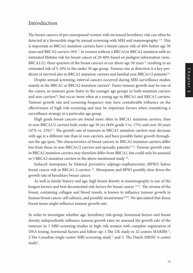
73
Ch
ap
te
r 3
Introduction
The breast cancers of pre-menopausal women with increased hereditary risk can often be detected at a favourable stage by annual screening with MRI and mammography 1-4. This is important as BRCA1 mutation carriers have a breast cancer risk of 40% before age 50 years and BRCA2 carriers 16% 5. In women without a BRCA1or BRCA2 mutation with an estimated lifetime risk for breast cancer of 20-40% based on pedigree information (non-BRCA1/2), three quarters of the breast cancers occur above age 50 years 6, resulting in an estimated risk of 5-10% in the under 50 age group. Tumour size at detection is a key-pre-dictor of survival also in BRCA1 mutation carriers and familial non-BRCA1/2 patients7,8.
Despite annual screening, interval cancers occurred during MRI surveillance studies mainly in the BRCA1 or BRCA2 mutation carriers9. Faster tumour growth may be one of the causes, as tumours grow faster in the younger age groups in both mutation carriers and non-carriers10, but occur more often at a young age in BRCA1 and BRCA2 carriers. Tumour growth rate and screening frequency may have considerable influence on the effectiveness of high risk-screening and may be important factors when considering a surveillance strategy in a particular age group.
High grade breast cancers are found more often in BRCA1 mutation carriers, than in non-BRCA1/2 carriers both under age 50 yrs (84% grade 3 vs. 17%) and over 50 years (47% vs. 23%)11. The growth rate of tumours in BRCA1 mutation carriers may decrease with age at a different rate than in non-carriers, and have possibly faster growth through-out the age span. The characteristics of breast cancers in BRCA2 mutation carriers differ less from those in non-BRCA1/2 carriers and sporadic patients11,12. Tumour growth rates in BRCA2 mutation carriers may therefore differ from BRCA1, but could only be assessed in 5 BRCA2 mutation carriers in the above mentioned study 10.
Induced menopause by bilateral preventive salpingo-oophorectomy (BPSO) halves breast cancer risk in BRCA1-2 carriers 13. Menopause and BPSO possibly slow down the growth rate of hereditary breast cancer.
As well as family history and age, high breast density at mammography is one of the longest known and best documented risk factors for breast cancer 14-17. The stroma of the breast, containing collagen and blood vessels, is known to influence tumour growth in human breast cancer cell cultures, and possibly invasiveness 18,19. We speculated that dense breast tissue might influence tumour growth rate.
In order to investigate whether age, hereditary risk-group, hormonal factors and breast density independently influence tumour growth rates we assessed the growth rate of the tumours in 3 MRI-screening studies in high risk women with complete registration of DNA testing, hormonal factors and follow-up: 1.The UK study in 22 centres MARIBS 2, 2.The Canadian single-centre MRI-screening study 3 and 3. The Dutch MRISC 6 centre study1.
74
Further we assess in these 3 large national studies with a yearly imaging frequency in all age groups the influence of age on tumour size.
Material and methods
Tumours found during screening in patients taking part in the Dutch MRISC study, The UK MARIBS study and the Canadian high risk screening study were included in this anal-ysis. All studies have been given institutional ethical approval and all women have given informed consent. The eligibility criteria for each study has been previously published1-3. and included BRCA1&2 gene mutation carriers and women at 20-40% lifetime risk of de-veloping breast cancer (non-BRCA1/2 carriers). Patients were included if the MRI and/or mammogram from the diagnostic screen was available for review together with the previ-ous screening examinations. The Dutch images were reviewed by I-M O, The UK images by RMLW and FJG and the Canadian images by PAC.
PatientsIn the Dutch high-risk screening study MRISC we could evaluate the size of 28 tumours at diagnosis, and with the same radiological technique, either Magnetic Resonance Imaging (MRI) or mammography (Mx) at previous examination(s), detected between July 1, 2003 and January 1, 2006,. These results were added to the previously described in 55 tumours detected at high-risk MRI-screening before July 200310.
Tumour size could be evaluated in 18 cancers detected within the22 centre- UK MAR-IBS study between August 1997 and May 2004 2. All non-BRCA1/2 patients were anony-mously DNA-tested.
The size of 24 cancers detected in the Canadian high-risk MRI screening study could be evaluated 3. The study included from November 1997-September 2005 unaffected women between age 25-60 yrs. and affected (past history of breast or ovarian cancer; until June 2003) who were a). BRCA1/2 mutation carrier, b). First-degree relatives of mutation carriers (until June 2003) and c). Women with a family history of ≥3 family members with breast cancer < 50 years or ovarian cancer (until July 2002) (non-BRCA1/2).
In total tumour volume doubling time (DT) could be assessed in 125 tumours.Breast density was measured visually at diagnostic Mx in a semi-quantitative 4 scale
system (< 25% of dense breast tissue = 1, 25-50%=2, 50-75%=3, and > 75%=4) in the Dutch and Canadian patients. The MARIBS study used a 3 point scale-fatty, mixed or dense.These were reclassified as 1, 2.5 and 4.
Measurements and calculation of tumour growth ratesThe way measurements were performed and growth rates calculated have been described extensively 10. In short, If the tumour could be clearly identified at the diagnostic MRI and previous imaging was available, 3D measurements at right angles, including the single

75
Ch
ap
te
r 3
largest diameter (SLD) of the tumour were performed. For all cancers positively identified at the diagnostic Mx with a previous available, the SLD was measured and the diameter perpendicular at both oblique and craniocaudal view.
For cancers with a measurable tumour at 2 or more subsequent MRI/Mx, and where a previous image showed no visible tumour (n.v.t.), only the measurable sizes were used for the calculation of individual tumour volume doubling time.
For patients with measurements at diagnosis and no visible tumour (n.v.t.) at the pre-vious examination the estimated tumour size at n.v.t., previously set at 0.004 cm3 corre-sponding to a diameter of 2 mm., was re-evaluated.
The volume of the tumour was estimated using the formula for obloid spheroids: V=4/3π.1/2a.1/2b.1/2c. When 4 sizes were assessed at Mx the SLD (a), the smallest (b) and the mean of the other 2 (c) were used.
We assessed whether tumour volume changes with time confirmed exponential growth (i.e. growth with a constant volume doubling time) or Gompertzian growth (exponential growth, but accelerated at small tumour size and slowing down at large tumour size) 20.
The slope of the straight line connecting the 2log-transformed volume measurements was calculated, using least-squares regression for 3 or more real volume measurements. Tumour volume doubling times were calculated using the formula: DT=log2/β, where β is the slope of the regression line of the logarithm of the tumour volume vs. time.
Statistical methodsDifferences in patient and tumour characteristics between the 3 risk-groups were tested with the use of the t-test in case of continuous variables and of the chi-square test or Fisher’s exact test in case of categorical variables. To determine the correlation between tumour size at mammography /MRI and at histo-pathologic examination we calculated Pearson’s cor-relation coefficient 21 separately for invasive cancers and ductal carcinoma in situ (DCIS). To get an approximate normal distribution of volume doubling times, these times were logarithmically transformed for analysis. Comparison of the transformed DT between risk-groups was done using the t-test. Multiple regression was used to evaluate simultane-ously the effects of age, risk-group and breast density. STATA-software (CNREG) was used in these calculations to allow for the presence of left-censored volume doubling times. A two-sided P-value of less than 0.05 was considered to indicate statistical significance.
Results
Patients and tumour characteristicsTumour-size could be assessed at diagnosis and previous imaging in 100 patients with invasive tumours and 25 ductal carcinoma in situ (dcis); 50 in BRCA1-, 23 in BRCA2 mutation carriers, and 52 in non-BRCA1/2 patients. The characteristics of the patients in the 3 studies are given in Table 1. Patients were on average significantly younger in the
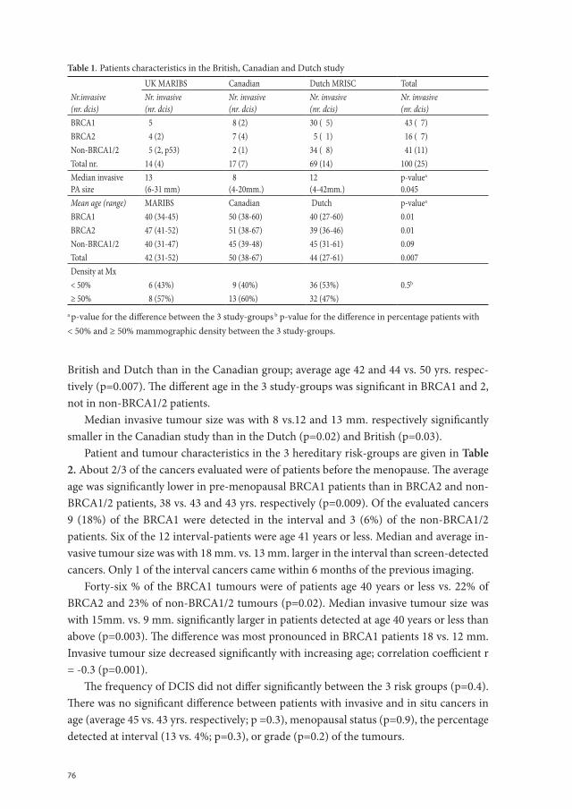
76
British and Dutch than in the Canadian group; average age 42 and 44 vs. 50 yrs. respec-tively (p=0.007). The different age in the 3 study-groups was significant in BRCA1 and 2, not in non-BRCA1/2 patients.
Median invasive tumour size was with 8 vs.12 and 13 mm. respectively significantly smaller in the Canadian study than in the Dutch (p=0.02) and British (p=0.03).
Patient and tumour characteristics in the 3 hereditary risk-groups are given in Table 2. About 2/3 of the cancers evaluated were of patients before the menopause. The average age was significantly lower in pre-menopausal BRCA1 patients than in BRCA2 and non-BRCA1/2 patients, 38 vs. 43 and 43 yrs. respectively (p=0.009). Of the evaluated cancers 9 (18%) of the BRCA1 were detected in the interval and 3 (6%) of the non-BRCA1/2 patients. Six of the 12 interval-patients were age 41 years or less. Median and average in-vasive tumour size was with 18 mm. vs. 13 mm. larger in the interval than screen-detected cancers. Only 1 of the interval cancers came within 6 months of the previous imaging.
Forty-six % of the BRCA1 tumours were of patients age 40 years or less vs. 22% of BRCA2 and 23% of non-BRCA1/2 tumours (p=0.02). Median invasive tumour size was with 15mm. vs. 9 mm. significantly larger in patients detected at age 40 years or less than above (p=0.003). The difference was most pronounced in BRCA1 patients 18 vs. 12 mm. Invasive tumour size decreased significantly with increasing age; correlation coefficient r = -0.3 (p=0.001).
The frequency of DCIS did not differ significantly between the 3 risk groups (p=0.4). There was no significant difference between patients with invasive and in situ cancers in age (average 45 vs. 43 yrs. respectively; p =0.3), menopausal status (p=0.9), the percentage detected at interval (13 vs. 4%; p=0.3), or grade (p=0.2) of the tumours.
Table 1. Patients characteristics in the British, Canadian and Dutch study
UK MARIBS Canadian Dutch MRISC TotalNr.invasive(nr. dcis)
Nr. invasive (nr. dcis)
Nr. invasive(nr. dcis)
Nr. invasive(nr. dcis)
Nr. invasive (nr. dcis)
BRCA1 5 8 (2) 30 ( 5) 43 ( 7)BRCA2 4 (2) 7 (4) 5 ( 1) 16 ( 7)Non-BRCA1/2 5 (2, p53) 2 (1) 34 ( 8) 41 (11)Total nr. 14 (4) 17 (7) 69 (14) 100 (25)Median invasive PA size
13(6-31 mm)
8(4-20mm.)
12(4-42mm.)
p-valuea
0.045Mean age (range) MARIBS Canadian Dutch p-valuea
BRCA1 40 (34-45) 50 (38-60) 40 (27-60) 0.01BRCA2 47 (41-52) 51 (38-67) 39 (36-46) 0.01Non-BRCA1/2 40 (31-47) 45 (39-48) 45 (31-61) 0.09Total 42 (31-52) 50 (38-67) 44 (27-61) 0.007Density at Mx < 50% 6 (43%) 9 (40%) 36 (53%) 0.5b
≥ 50% 8 (57%) 13 (60%) 32 (47%)a p-value for the difference between the 3 study-groups b p-value for the difference in percentage patients with < 50% and ≥ 50% mammographic density between the 3 study-groups.
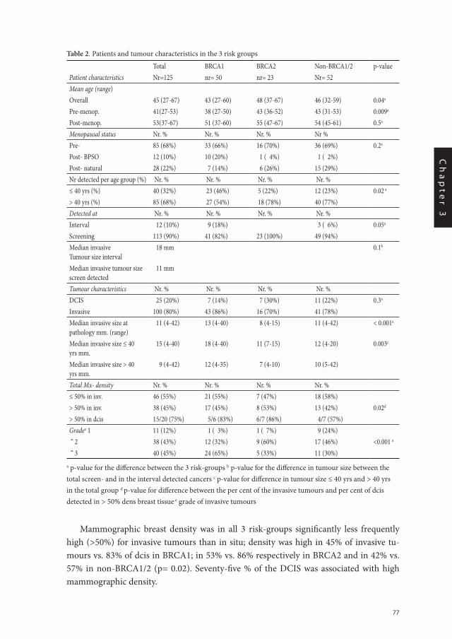
77
Ch
ap
te
r 3
Mammographic breast density was in all 3 risk-groups significantly less frequently high (>50%) for invasive tumours than in situ; density was high in 45% of invasive tu-mours vs. 83% of dcis in BRCA1; in 53% vs. 86% respectively in BRCA2 and in 42% vs. 57% in non-BRCA1/2 (p= 0.02). Seventy-five % of the DCIS was associated with high mammographic density.
Table 2. Patients and tumour characteristics in the 3 risk groups
Total BRCA1 BRCA2 Non-BRCA1/2 p-valuePatient characteristics Nr=125 nr= 50 nr= 23 Nr= 52Mean age (range)Overall 45 (27-67) 43 (27-60) 48 (37-67) 46 (32-59) 0.04a
Pre-menop. 41(27-53) 38 (27-50) 43 (36-52) 43 (31-53) 0.009a
Post-menop. 53(37-67) 51 (37-60) 55 (47-67) 54 (45-61) 0.5a
Menopausal status Nr. % Nr. % Nr. % Nr %Pre- 85 (68%) 33 (66%) 16 (70%) 36 (69%) 0.2a
Post- BPSO 12 (10%) 10 (20%) 1 ( 4%) 1 ( 2%)Post- natural 28 (22%) 7 (14%) 6 (26%) 15 (29%)Nr detected per age group (%) Nr. % Nr. % Nr. % Nr. %≤ 40 yrs (%) 40 (32%) 23 (46%) 5 (22%) 12 (23%) 0.02 a
> 40 yrs (%) 85 (68%) 27 (54%) 18 (78%) 40 (77%)Detected at Nr. % Nr. % Nr. % Nr. %Interval 12 (10%) 9 (18%) 3 ( 6%) 0.05a
Screening 113 (90%) 41 (82%) 23 (100%) 49 (94%)Median invasive Tumour size interval
18 mm 0.1b
Median invasive tumour size screen detected
11 mm
Tumour characteristics Nr. % Nr. % Nr. % Nr. %DCIS 25 (20%) 7 (14%) 7 (30%) 11 (22%) 0.3a
Invasive 100 (80%) 43 (86%) 16 (70%) 41 (78%)Median invasive size at pathology mm. (range)
11 (4-42) 13 (4-40) 8 (4-15) 11 (4-42) < 0.001a
Median invasive size ≤ 40 yrs mm.
15 (4-40) 18 (4-40) 11 (7-15) 12 (4-20) 0.003c
Median invasive size > 40 yrs mm.
9 (4-42) 12 (4-35) 7 (4-10) 10 (5-42)
Total Mx- density Nr. % Nr. % Nr. % Nr. %≤ 50% in inv. 46 (55%) 21 (55%) 7 (47%) 18 (58%)> 50% in inv. 38 (45%) 17 (45%) 8 (53%) 13 (42%) 0.02d
> 50% in dcis 15/20 (75%) 5/6 (83%) 6/7 (86%) 4/7 (57%)Gradee 1 11 (12%) 1 ( 3%) 1 ( 7%) 9 (24%) “ 2 38 (43%) 12 (32%) 9 (60%) 17 (46%) <0.001 a
“ 3 40 (45%) 24 (65%) 5 (33%) 11 (30%)
a p-value for the difference between the 3 risk-groups b p-value for the difference in tumour size between the total screen- and in the interval detected cancers c p-value for difference in tumour size ≤ 40 yrs and > 40 yrs in the total group d p-value for difference between the per cent of the invasive tumours and per cent of dcis detected in > 50% dens breast tissue e grade of invasive tumours
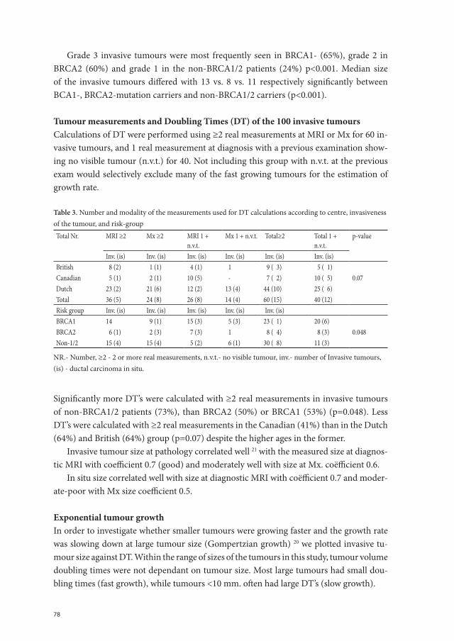
78
Grade 3 invasive tumours were most frequently seen in BRCA1- (65%), grade 2 in BRCA2 (60%) and grade 1 in the non-BRCA1/2 patients (24%) p<0.001. Median size of the invasive tumours differed with 13 vs. 8 vs. 11 respectively significantly between BCA1-, BRCA2-mutation carriers and non-BRCA1/2 carriers (p<0.001).
Tumour measurements and Doubling Times (DT) of the 100 invasive tumoursCalculations of DT were performed using ≥2 real measurements at MRI or Mx for 60 in-vasive tumours, and 1 real measurement at diagnosis with a previous examination show-ing no visible tumour (n.v.t.) for 40. Not including this group with n.v.t. at the previous exam would selectively exclude many of the fast growing tumours for the estimation of growth rate.
Table 3. Number and modality of the measurements used for DT calculations according to centre, invasiveness of the tumour, and risk-group
Total Nr. MRI ≥2 Mx ≥2 MRI 1 + n.v.t.
Mx 1 + n.v.t. Total≥2 Total 1 + n.v.t.
p-value
Inv. (is) Inv. (is) Inv. (is) Inv. (is) Inv. (is) Inv. (is)British 8 (2) 1 (1) 4 (1) 1 9 ( 3) 5 ( 1)Canadian 5 (1) 2 (1) 10 (5) - 7 ( 2) 10 ( 5) 0.07Dutch 23 (2) 21 (6) 12 (2) 13 (4) 44 (10) 25 ( 6)Total 36 (5) 24 (8) 26 (8) 14 (4) 60 (15) 40 (12)Risk group Inv. (is) Inv. (is) Inv. (is) Inv. (is) Inv. (is)BRCA1 14 9 (1) 15 (3) 5 (3) 23 ( 1) 20 (6)BRCA2 6 (1) 2 (3) 7 (3) 1 8 ( 4) 8 (3) 0.048Non-1/2 15 (4) 15 (4) 5 (2) 6 (1) 30 ( 8) 11 (3)
NR.- Number, ≥2 - 2 or more real measurements, n.v.t.- no visible tumour, inv.- number of Invasive tumours, (is) - ductal carcinoma in situ.
Significantly more DT’s were calculated with ≥2 real measurements in invasive tumours of non-BRCA1/2 patients (73%), than BRCA2 (50%) or BRCA1 (53%) (p=0.048). Less DT’s were calculated with ≥2 real measurements in the Canadian (41%) than in the Dutch (64%) and British (64%) group (p=0.07) despite the higher ages in the former.
Invasive tumour size at pathology correlated well 21 with the measured size at diagnos-tic MRI with coefficient 0.7 (good) and moderately well with size at Mx. coëfficient 0.6.
In situ size correlated well with size at diagnostic MRI with coëfficient 0.7 and moder-ate-poor with Mx size coefficient 0.5.
Exponential tumour growth In order to investigate whether smaller tumours were growing faster and the growth rate was slowing down at large tumour size (Gompertzian growth) 20 we plotted invasive tu-mour size against DT. Within the range of sizes of the tumours in this study, tumour volume doubling times were not dependant on tumour size. Most large tumours had small dou-bling times (fast growth), while tumours <10 mm. often had large DT’s (slow growth).
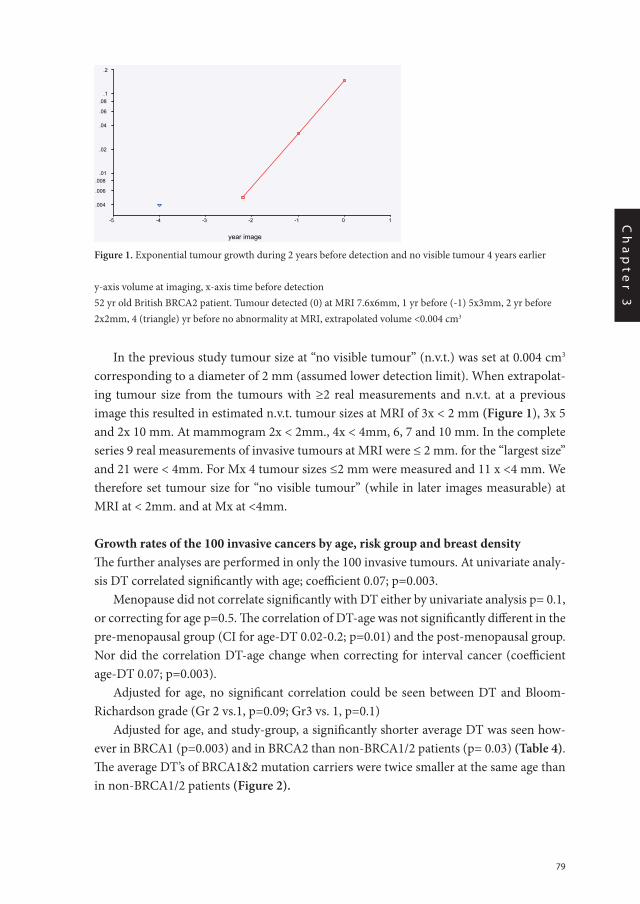
79
Ch
ap
te
r 3
In the previous study tumour size at “no visible tumour” (n.v.t.) was set at 0.004 cm3 corresponding to a diameter of 2 mm (assumed lower detection limit). When extrapolat-ing tumour size from the tumours with ≥2 real measurements and n.v.t. at a previous image this resulted in estimated n.v.t. tumour sizes at MRI of 3x < 2 mm (Figure 1), 3x 5 and 2x 10 mm. At mammogram 2x < 2mm., 4x < 4mm, 6, 7 and 10 mm. In the complete series 9 real measurements of invasive tumours at MRI were ≤ 2 mm. for the “largest size” and 21 were < 4mm. For Mx 4 tumour sizes ≤2 mm were measured and 11 x <4 mm. We therefore set tumour size for “no visible tumour” (while in later images measurable) at MRI at < 2mm. and at Mx at <4mm.
Growth rates of the 100 invasive cancers by age, risk group and breast density The further analyses are performed in only the 100 invasive tumours. At univariate analy-sis DT correlated significantly with age; coefficient 0.07; p=0.003.
Menopause did not correlate significantly with DT either by univariate analysis p= 0.1, or correcting for age p=0.5. The correlation of DT-age was not significantly different in the pre-menopausal group (CI for age-DT 0.02-0.2; p=0.01) and the post-menopausal group. Nor did the correlation DT-age change when correcting for interval cancer (coefficient age-DT 0.07; p=0.003).
Adjusted for age, no significant correlation could be seen between DT and Bloom-Richardson grade (Gr 2 vs.1, p=0.09; Gr3 vs. 1, p=0.1)
Adjusted for age, and study-group, a significantly shorter average DT was seen how-ever in BRCA1 (p=0.003) and in BRCA2 than non-BRCA1/2 patients (p= 0.03) (Table 4). The average DT’s of BRCA1&2 mutation carriers were twice smaller at the same age than in non-BRCA1/2 patients (Figure 2).
Chapter 3B fig 1 Fig 1 Exponential tumour growth during 2 years before detection and no visible .tumour 4 years earlier
year image
10-1-2-3-4-5
.2
.1.08
.06
.04
.02
.01.008
.006
.004
y-axis volume at imaging, x-axis time before detection 52 yr old British BRCA2 patient. �Tumour detected (0)at MRI 7.6x6mm, � 1 yr before(-1) 5x3mm, �2 yr before 2x2mm , �4 yr before no abnormality at MRI, extrapolated volume <0.004 cm3
Chapter 3B fig 1
Figure 2 Doubling times of the 100 invasive tumours according to age and risk-group
Age (yrs)
706050403020
Dou
blin
g tim
e (y
rs)
543
2
1
.5
.4
.3
.2
.1
.05
.04
.03
.02
.01
non carrier
brca2
brca1
Non carrier
BRCA2
BRCA1
Chapter 3B / fig 2
Figure 1. Exponential tumour growth during 2 years before detection and no visible tumour 4 years earlier
y-axis volume at imaging, x-axis time before detection52 yr old British BRCA2 patient. Tumour detected (0) at MRI 7.6x6mm, 1 yr before (-1) 5x3mm, 2 yr before 2x2mm, 4 (triangle) yr before no abnormality at MRI, extrapolated volume <0.004 cm3
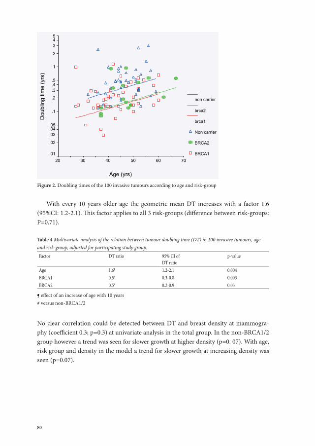
80
With every 10 years older age the geometric mean DT increases with a factor 1.6 (95%CI: 1.2-2.1). This factor applies to all 3 risk-groups (difference between risk-groups: P=0.71).
Table 4 Multivariate analysis of the relation between tumour doubling time (DT) in 100 invasive tumours, age and risk-group, adjusted for participating study group.
Factor DT ratio 95% CI of DT ratio
p-value
Age 1.6¶ 1.2-2.1 0.004BRCA1 0.5# 0.3-0.8 0.003BRCA2 0.5# 0.2-0.9 0.03
¶ effect of an increase of age with 10 years# versus non-BRCA1/2
No clear correlation could be detected between DT and breast density at mammogra-phy (coefficient 0.3; p=0.3) at univariate analysis in the total group. In the non-BRCA1/2 group however a trend was seen for slower growth at higher density (p=0. 07). With age, risk group and density in the model a trend for slower growth at increasing density was seen (p=0.07).
Chapter 3B fig 1 Fig 1 Exponential tumour growth during 2 years before detection and no visible .tumour 4 years earlier
year image
10-1-2-3-4-5
.2
.1.08
.06
.04
.02
.01.008
.006
.004
y-axis volume at imaging, x-axis time before detection 52 yr old British BRCA2 patient. �Tumour detected (0)at MRI 7.6x6mm, � 1 yr before(-1) 5x3mm, �2 yr before 2x2mm , �4 yr before no abnormality at MRI, extrapolated volume <0.004 cm3
Chapter 3B fig 1
Figure 2 Doubling times of the 100 invasive tumours according to age and risk-group
Age (yrs)
706050403020
Dou
blin
g tim
e (y
rs)
543
2
1
.5
.4
.3
.2
.1
.05
.04
.03
.02
.01
non carrier
brca2
brca1
Non carrier
BRCA2
BRCA1
Chapter 3B / fig 2 Figure 2. Doubling times of the 100 invasive tumours according to age and risk-group
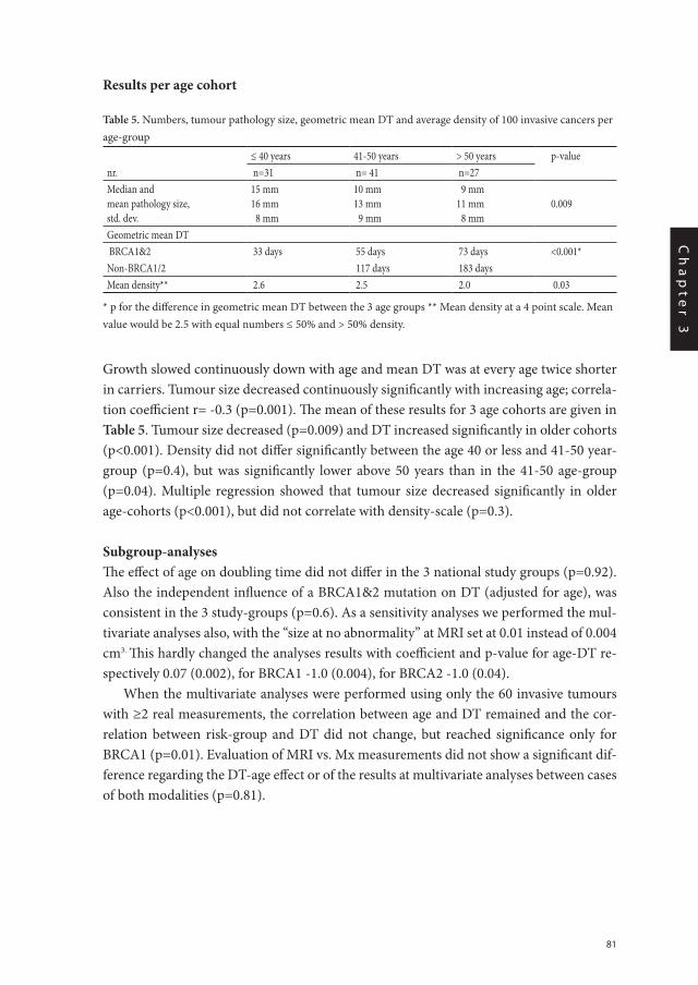
81
Ch
ap
te
r 3
Results per age cohort
Table 5. Numbers, tumour pathology size, geometric mean DT and average density of 100 invasive cancers per age-group
≤ 40 years 41-50 years > 50 years p-valuenr. n=31 n= 41 n=27Median andmean pathology size,std. dev.
15 mm16 mm 8 mm
10 mm13 mm 9 mm
9 mm11 mm 8 mm
0.009
Geometric mean DT BRCA1&2 33 days 55 days 73 days <0.001*Non-BRCA1/2 117 days 183 daysMean density** 2.6 2.5 2.0 0.03
* p for the difference in geometric mean DT between the 3 age groups ** Mean density at a 4 point scale. Mean value would be 2.5 with equal numbers ≤ 50% and > 50% density.
Growth slowed continuously down with age and mean DT was at every age twice shorter in carriers. Tumour size decreased continuously significantly with increasing age; correla-tion coefficient r= -0.3 (p=0.001). The mean of these results for 3 age cohorts are given in Table 5. Tumour size decreased (p=0.009) and DT increased significantly in older cohorts (p<0.001). Density did not differ significantly between the age 40 or less and 41-50 year-group (p=0.4), but was significantly lower above 50 years than in the 41-50 age-group (p=0.04). Multiple regression showed that tumour size decreased significantly in older age-cohorts (p<0.001), but did not correlate with density-scale (p=0.3).
Subgroup-analysesThe effect of age on doubling time did not differ in the 3 national study groups (p=0.92). Also the independent influence of a BRCA1&2 mutation on DT (adjusted for age), was consistent in the 3 study-groups (p=0.6). As a sensitivity analyses we performed the mul-tivariate analyses also, with the “size at no abnormality” at MRI set at 0.01 instead of 0.004 cm3. This hardly changed the analyses results with coefficient and p-value for age-DT re-spectively 0.07 (0.002), for BRCA1 -1.0 (0.004), for BRCA2 -1.0 (0.04).
When the multivariate analyses were performed using only the 60 invasive tumours with ≥2 real measurements, the correlation between age and DT remained and the cor-relation between risk-group and DT did not change, but reached significance only for BRCA1 (p=0.01). Evaluation of MRI vs. Mx measurements did not show a significant dif-ference regarding the DT-age effect or of the results at multivariate analyses between cases of both modalities (p=0.81).
82
Discussion
In these 3 large national MRI-screening studies, the decreasing growth rate of breast can-cers at increasing age was confirmed in BRCA1&2 mutation carriers and non-BRCA1/2 patients. Invasive tumour growth rate decreased on average 1,6 times in each 10 years older cohort. Although the average growth rate decreased slightly slower with increas-ing age compared to the previous study (DT ratio1.9)10, the correlation is robust. Slower growth of tumours detected at older age has been shown in mammography screening studies in patients above 40 years, not selected for family history 22,23.
A finding unique to our study is that at the same age, the average growth rate of the tu-mours of BRCA1&2 mutation carriers was twice as fast as in non-BRCA1/2 patients (DT ratio 0.5). This mutation-effect on tumour growth was also consistent in the 3 national study-groups. The average growth rate at a certain age did not differ however between carriers of a BRCA1 or a BRCA2 mutation. A comparable ratio for the growth rates of BRCA1&2 vs. non-BRC1-2 tumours was seen in the previous study (0.7) however not reaching significance adjusted for age. As we lack an age matched group of patients from the general population (“sporadic”) detected in a MRI-screening setting, we cannot say whether the growth rate of our non-BRCA1/2 patients possibly differs from “sporadic” patients of the same age. In that case tumours in BRCA1&2 mutation carriers might grow faster than in “sporadic” patients, but not twice as fast. This cannot be tested for the “sporadic” group since women aged < 40 years and at population risk do not undergo MRI-screening. Peer et al22 calculated median DT of 80 days in sporadic patients less than 50 years and twice slower growth above age 50 years. One might expect slower tumour growth in non-BRCA1/2 than sporadic patients, as they are more frequently low grade in some studies, although not in others8,11,12. We could not demonstrate a correlation of tumour grade with faster or slower growth independent of age however.
Eighteen percent of the tumours in BRCA1 vs.6% in the non-BRCA1/2 patients were detected during the screening interval in our study, and half of the interval-patients were ≤ 41 years. As the invasive tumour size was with 18 mm. vs. 13 mm. larger in the interval than screen-detected cancers, screening results might improve if interval cancers could be prevented. Only 1 of the interval cancers came within 6 months of the previous imaging.
Tumours were on average significantly larger (15 vs. 9mm.) in the 40 patients detected at age 40 years or below than above (p=0.003). In BRCA1 mutation carriers in this study 46% were detected under age 40 years vs. 22% and 23% in BRCA2 and non-BRCA1/2 patients (p=0.02). This reflects partly the age of the participating women in the 3 national studies. The frequent occurrence of tumours under age 40 in BRCA1 mutation carriers, the significantly larger tumour size under age 40, the relatively frequent interval cancers in BRCA1 patients, the larger size of the interval cancers and the twice faster tumour growth in BRCA1&2 mutation carriers than non-BRCA1/2 patients suggest, that especially the small group of BRCA1&2 mutation carriers < age 40 years will benefit from twice yearly high sensitive screening. In carriers this means screening with MRI 1-4.
83
Ch
ap
te
r 3
Tumour size decreased also continuously significantly with increasing age. In all 3 age-cohorts imaging was performed yearly. Breast density was not significantly lower in the 41-50 year group than the aged 40 year or less, and did not explain at multivariate analysis the on average smaller size of the tumours in the older group (median 10 and 15 mm respectively). The earlier detection is therefore most likely caused by the slower growth of the tumours in the on average 10 year older group. The BRCA1&2 mutation carriers above 40 years of age were in this joint study detected at a favourable stage by yearly imaging with MRI. Even in grade 3 tumours a 12 year survival of over 90% may be expected in node negative tumours of 1cm or less23. Our results suggest, that above age 50 the screening frequency could be decreased gradually, while below age 40 years the frequency should be higher for equal results.
In the large group of non-BRCA1/2 high-risk women, growth rate was twice slower than in carriers, and yearly imaging may be sufficient till age 45 years. Gradual decrease to once every 1,5 year between 45-55 and biannual imaging between 55-65 years would be most in accordance with our findings.
We could not demonstrate an influence of menopausal status independent of age, while the DT according to age did not differ in the small group of patients with an early menopause by bilateral preventive salpingo-oophorectomy (10%).
Dense breast tissue did not enhance growth rate. A trend was even seen for slower growth with increasing breast density especially in the non-BRCA1/2 familial patients. Therefore dense breast tissue does not seem a valid reason to increase screening frequen-cy. Nor did dense breast tissue enhance invasiveness of the tumours. On the contrary, breast tissue was significantly more often highly dense (> 50%) in DCIS than in invasive tumours and this was not caused by younger age in the DCIS group. The well-documented increased breast cancer risk in denser breast tissue does not seem to be caused by faster tumour growth or enhanced invasiveness therefore. Breast density was not measured pre-cisely however, but estimated visually on a 4 point categorical scale.
Half of the DCIS (13) were measured for growth at MRI and it is promising that they are increasingly detected at MRI, suggesting that addition of mammography to MRI screening may be not necessary in the future.
Tumour growthMany early studies of breast cancer growth rate have been performed in vitro in tumour cell-lines at the earliest stages of growth at a tumour size that would not be detectable in the living human 24-25. Recent studies in mice have suggested, that only a small proportion of the tumour cells, are self-renewing and drive tumour growth and metastasis, the so-called cancer stem cells 26,27. An increasing proportion of stem cells would suggest more aggressive breast cancer, 25% stem cells were found in an extremely aggressive breast tu-mour. We could not demonstrate however a correlation between tumour grade, the usual indicator of tumour aggressiveness and growth rate. Large scale mammography screen-ing-studies have given useful data for in human’s detectable tumour sizes 22,23,28-32. In our
84
study most tumours seemed to have exponential growth. Contrarily to the Gompertzian model most slowly growing tumours were small in our study, probably because slowly growing tumours have a better chance of being detected early.
We can recognise some limitations to the present study, that are consequent on the combination of material from three different national trials. The background inclusion criteria were different which results in the older age profile of the Canadian participants. The review processes have been undertaken separately by the three national groups, and although methods were precisely described and discussed, one cannot be sure that these were exactly comparable. The gain from combining the material to give greater statistical power for the subset analysis however exceeds these limitations, allowing the analyses for separate gene mutations, interval cases and of the correlation between age- DT and age-tumour size with sufficient power to give statistically significant findings.
A tumour can sometimes acquire the mutation(s) that enables it to metastasize early in its development. The chance to acquire this mutation(s) increases however most likely with every cell division and therefore with increasing size of the tumour. Also in BRCA1 mu-tation carriers and non-BRCA1/2 familial patients tumour size was a strong indicator of survival 7,8. Screening hereditary high-risk women may therefore effectively reduce mor-tality by detecting the tumour early. It depends on the risk group from what age on screen-ing may be indicated and cost-effective, but our study shows, that both young age and a BRCA1 or BRCA2 mutation, but not dense breast tissue, are good reasons to increase the frequency of the screening test. Especially the small group of BRCA1&2 mutation carriers under age 40 years need MRI twice yearly to prevent large tumours and interval cancers.
85
Ch
ap
te
r 3
References
1. Kriege M, Brekelmans CTM, Boetes C, et al. Efficacy of MRI and mammography for breast-cancer screening in women with a familial or genetic predisposition. N Eng JMed 2004;351:427-37.
2. Leach MO, MARIBS study group. Screening with magnetic resonance imaging and mammography of a UK population at high familial rik of breast cancer: a prospective multicentre cohort study (MARIBS). Lancet 2005;365:1769-78.
3. Warner E, Plewes DB, Hill KA, et al. Surveillance of BRCA1 and BRCA2 mutation carriers with magnetic resonance imaging, ultrasound, mammography and clinical breast examination. JAMA 2004;292(11):1317-25.
4. Kuhl CK, Schrading S, Leutner CC, Morakkabati-Spitz N, Wardelmann E, Fimmers R, Kuhn W, Schild HH. Mammography, Breast ultrasound, and Magnetic Resonance Imaging for surveillance of women at high familial risk for breast cancer. JCO 2005;23:8469-76.
5. Antoniou A, Pharaoh P, Narod S et al. Average risk of Breast and ovarian cancer associated with BRCA1 or BRCA2 mutations detected in case series unselected for family history: a combined analysis of 22 stud-ies. Am.J HumGenet 2003;72:1117-30
6. Eerola H, Vahteristo P, Sarantaus L, et al. Survival of breast cancer patients in BRCA1, BRCA2 and non-BRCA1/2 breast cancer families: a relative survival analysis from Finland. Int J Cancer 2001;93:368-72.
7. Brekelmans CTM, Seynaeve C, Menke-Pluymers M, Brüggenwirth HT, Tilanus-Linthorst MMA, Bartels CCM et al. Survival and prognostic factors in BRCA1-associated breast cancer. Ann Oncol 2006;17: 391-400.
8. Tilanus-Linthorst MMA, Alves C, Seynaeve C, Menke-Pluymers MBE, Eggermont AMM, Brekelmans CTM. Contralateral recurrence and prognostic factors in familial non-BRCA1/2-associated breast cancer. Accepted Brit J Surg 2006.
9. Tilanus-Linthorst MM, Obdeijn I-M, Bartels KCM. MARIBS study. Lancet 2005;366;291-291 and 1434.
10. Tilanus-Linthorst MM, Kriege M, Boetes C, Hop WCJ, Obdeijn I-M, Oosterwijk JC, Peterse HL, Zonder-land HM, Meijer S, Eggermont AMM, de Koning HJ, Klijn JGM, Brekelmans CTM. Hereditary breast cancer growth rates and its impact on screening policy. European Journal of Cancer 2005;41(11):1610-17.
11. Eerola H, Heikkilä P, Tamminen A, Aittomäki K, Blomqvist C, Nevanlinna H. Relationship of patients age to histopathologic features of breast tumours in BRCA1 and BRCA2 and mutation-negative breast cancer families. Breast Cancer Res 2005:7: R465-69.
12. Lakhani SR, Jacqumier J, Sloane JP, Gusterson BA, Anderson TJ, van de Vijver MJ, Farid LM, Venter D, Antoniou A, Storfer-Isser A, Smyth E, Steel CM, et al. Multifactorial analysis of differences between sporadic breast cancers and cancers involving BRCA1 and BRCA2 mutations. J Natl. Cancer Inst. 1998; 90:1138-1145
13. Rebbeck TR Prophylactic oophorectomy in BRCA1 and BRCA2 mutation carriers. Eur J Cancer 2002;6:S15-S17.
14. Wolfe JN, Risk for breast cancer development determined by mammographic parenchymal pattern. Can-cer 1976;37:2486-92.
15. Boyd NF, Rommers JM, Vogt K, Lee V, Hopper JL, Yaffe MJ, Paterson AD. Mammographic breast density as an intermediate phenotype for breast cancer. Lancet Oncology 2005;6:798-808.
16. Boyd NF, Dite GS, Stone J et al. Heritability of mammographic density, a risk factor for breast cancer. N Engl J Med 2002;347:886-94.
17. Warwick J, Pinney E, Warren RML, Duffy SW, Howell A, Wilson M, Cuzick J Breast density and breast cancer risk factors in a high-risk population. The breast. 2003;12:10-16.
86
18. Shekhar MPV, Werdell J, Santer SJ, Pauley RJ, Tait l. Breast stroma plays a dominant regulatory role in breast epithelial growth and differentiation: Implications for tumor development and progression. Cancer research 2001;61:1320-26.
19. Dong-Le Bourhis X, Bertthois Y, Millot G, Degeorges A, Sylvi M, Martin P-M, Calvo F. Effects of stromal and epithelial cells derived from normal and tumorous breast tissue on the proliferation of human breast cancer cell lines in co-culture. Int J Cancer 1997;71:42-48.
20. Norton L. A Gompertzian model of human breast cancer growth. Cancer Res. 1988; 48:7067—71.
21. Landis JR, Koch GG. The measurement of interobserver agreement for categorical data. Biometrics 1977;33:159-174.
22. Peer PGM, vanDijck JAAM, Hendriks JHCL, et al. Age-dependent growth rate of primary breast cancer. Cancer 1993; 71:3547-51.
23. Tabar L, Fagerberg G, Day NE, et al. Breast cancer treatment and natural history: new insights from results of screening. Lancet 1992; 339:412-4.
24. Duffy SW, Day NE, Tabar L et al. Markov models of breast tumor progression: some age-specific results. Monogr Natl Cancer Inst 1997;22:93-7.
25. Speer JF, Petrosky VE, Retsky MW, Wardwell RH. Et al. Stochastic numerical model of breast cancer growth that simulates clinical data. Cancer Res 1984; 44:4124-30.
26. Al-Hajj M, Wicha MS, Benito-Hernandez A, Morrison SJ, Clarke MF. Prospective identification of tu-morigenic breast cancer cells. Proc Natl Acad Sci U S A. 2003;100(7):3983-88.
27. Tözeren A, Coward CW, Petushi S. Origins of cell phenotypes in breast tumours. J.Theor.Biol. 2005;233:43-54.
28. Spratt JA, von Fournier D, Spratt JS, et al. Mammographic assessment of human breast cancer growth and duration. Cancer 1993; 71:2020-6.
29. Kusama S, Spratt JS, Donegan WL et al. The gross rates of growth of human mammary carcinoma. Cancer 1972;30:594-99.
30. Hart D, Shochat E, Agur Z.The growth law of primary breast cancer as inferred from mammography screening trials data. Brit J Cancer 1998;78(3):38207.
31. Kopans DB, Rafferty E, Georgian-Smith D, et al. A simple model of breast carcinoma growth may provide explanations for observations of apparently complex phenomena. Cancer 2003; 97:2951-9.
32. Michaelson JS, Halpern E, Kopans D. Breast cancer: Computer simulation method for estimating optimal intervals for screening. Radiology 1999; 212:551-60

Chapter 4
Breast self-examination and screening women at high risk
Madeleine M Tilanus-Linthorst, Inge-Marie Obdeijn, Karina CM Bartels
Lancet (Letter) 2005;366(9482):291-2 and 1434

89
Ch
ap
te
r 4
MARIBS study
The UK MARIBS study (May 21, p769)1 provides important information on the screening of women at high risk of breast cancer with MRI and mammography. The study will have had the additional benefit of spreading the MRI breast screening expertise of a few centres quickly over the country, as did the Dutch study.
Two points regarding this study and the other two large prospective MRI-screening studies done so far are worth noting.
First, the occurrence of only a few interval cancers during screening in the three studies seems a good result as is the promising state at detecton (table).
However, although it can result in more interval cancers, breast self-examination can decrease the number of large or node-positive cancers detected. In the three studies, the interval cancers were all node-negative and five of seven were less than 2 cm (table) (stage of 1 not presented in the MRISC). Selfexamination caused few extra investigations in the Dutch study. Therefore, the sensitivity and specificity of breast self-examination should be presented in all three studies, not only those of MRI, mammography, and clinical exami-nation. Self-examination can be more cost-effective than clinical examination and should not be neglected in our investigations. Monthly self-examination after good instruction does not necessarily increase anxiety, although it underlines that MRI-screening has limi-tations. The negative results of a large randomised study on breast self-examination are not applicable to the surveillance situation, because some of the Shanghai women in the study were not seen by a doctor nor underwent mammography after they felt a lump. Pre-senting the results of breast self-examination in the studies would illustrate that surveil-lance relies on cooperation between healthy women and well equipped doctors.
Second, the interval cancers in the three studies were nearly all in BRCA1/2 mutation car-riers (table). In our study on the growth rate of hereditary breast cancer, young age at de-tection predicted fast tumour growth both in BRCA1/2 mutation carriers and in patients at high familial risk. Breast cancers can occur at a very young age in BRCA1/2 mutation carriers, because the incidence increases rapidly from age 30 years onwards. Therefore, we should investigate whether imaging more frequently than yearly might be necessary in this small group of young mutation carriers. Twice yearly MRI in women younger than 40 years, or maybe MRI and mammography at different time points, should not be dismissed as too costly. All doctors now offering surveillance to young BRCA1/2 mutation carriers should be aware that there is a clear risk of interval cancer when imaging is done only once a year.
We declare that we have no conflict of interest.
*Madeleine M Tilanus-Linthorst, Inge-Marie Obdeijn, Karina C M Bartels
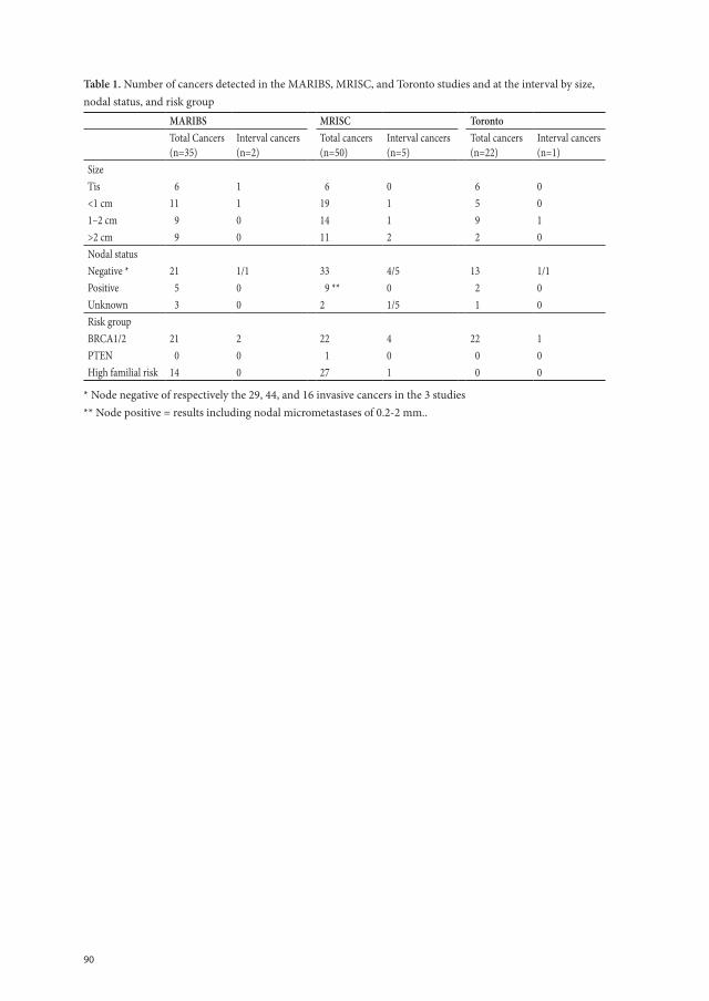
90
Table 1. Number of cancers detected in the MARIBS, MRISC, and Toronto studies and at the interval by size, nodal status, and risk group
MARIBS MRISC TorontoTotal Cancers(n=35)
Interval cancers(n=2)
Total cancers(n=50)
Interval cancers(n=5)
Total cancers(n=22)
Interval cancers(n=1)
SizeTis 6 1 6 0 6 0<1 cm 11 1 19 1 5 01–2 cm 9 0 14 1 9 1>2 cm 9 0 11 2 2 0Nodal statusNegative * 21 1/1 33 4/5 13 1/1Positive 5 0 9 ** 0 2 0Unknown 3 0 2 1/5 1 0Risk groupBRCA1/2 21 2 22 4 22 1PTEN 0 0 1 0 0 0High familial risk 14 0 27 1 0 0
* Node negative of respectively the 29, 44, and 16 invasive cancers in the 3 studies** Node positive = results including nodal micrometastases of 0.2-2 mm..

91
Ch
ap
te
r 4
References
1. MARIBS study group. Screening with magnetic resonance imaging and mammography of a UK popula-tion at high familial risk of breast cancer: a prospective multicentre cohort study (MARIBS). Lancet 2005; 365:1769–78.
2. Kriege M, Brekelmans CTM, Boetes C, et al. Efficacy of MRI and mammography for breastcancer screen-ing in women with a familial or genetic predisposition. N Engl J Med 2004;351:427–37.
3. Warner E, Plewes DB, Hill KA, et al. Surveillance of BRCA1 and BRCA2 mutation carriers with magnetic resonance imaging, ultrasound, mammography, and clinical breast examination. JAMA 2004; 292:1317–25.
4. Thomas DB, Gao DL, Ray RM, et al. Randomized trial of breast self-examination in Shanghai: final results. J Natl Cancer Inst 2002; 94:1445–57.
5. Tilanus-Linthorst MMA, Kriege M, Boetes C, etal. Hereditary breast cancer growth rates and its impact on screening policy. Eur J Cancer 2005;41 (11):1610–17.


Chapter 5
A BRCA1/2 mutation, high breast density and prominent pushing margins of a tumor independently contribute to a frequent false-negative mammography.
Madeleine Tilanus-Linthorst, Leon Verhoog, Inge-Marie Obdeijn, Karina Bartels, Marian Menke-Pluymers, Alexander Eggermont, Jan Klijn, Hanne Meijers-Heijboer, Theo vd Kwast, Cecile Brekelmans
Int.J.Cancer 2002;102:91-95
94
Summary
Aim Female BRCA1/2 mutation carriers develop in up to 50% breast cancer (BC) before age 50 years. We investigated whether the specific histologic features of BRCA1/2-associ-ated breast cancer influence imaging.
Methods We correlated the mammographic results with the histology of 34 BC in BRCA1/2 mutation carriers and 34 sporadic cancers in patients, matched for age and year of diagnosis.
Results Mammography was significantly more frequently false-negative in carriers than controls (62% vs. 29% p=0.01), despite comparable tumor size (mean Ø 1,51 vs 1,75) and breast density (high 41% vs 53%). The image in carriers was significantly less as spicu-lated mass (6 vs 18 p=0.01)
Cancers of BRCA1/2 mutation carriers had as expected frequently high mitotic counts (p<0.0001) and prominent pushing margins around the tumor (p= 0.08) (p= 0.05 for 32 BRCA1). A new observation is also, that prominent “pushing margins” significantly cor-related with a false-negative mammography (p=0.005) and with a mammographic image of a smooth / not a spiculated mass (p=0.01). False-negative mammography correlated independently with: BRCA1/2 mutation (p=0.02), prominent pushing margins (p=0.03) and high breast density (p=0.01). MRI was carried out in 12 carriers, had 100% sensitivity and detected 5 cancers, still occult at physical examination and mammography.
Conclusion A BRCA1/2 mutation and high breast density at mammography inde-pendently contribute to false-negative mammography results. In mutation carriers any mammographic mass must be regarded with suspicion. Pushing margins of the tumor partly explain these results. For early BC detection in mutation carriers additional meth-ods like MRI may be needed, but not necessarily in most other young women with breast symptoms.
95
Ch
ap
te
r 5
Introduction
Female carriers of a BRCA1 or BRCA2 mutation are at an increased risk of developing breast cancer (BC) at a young age. Risks of 3% at 30 years of age and up to 50% at 50 years have been described.1,2 Therefore carriers of these mutations often choose already at a young age surveillance, generally consisting of regular clinical breast examination and mammography, with the addition of breast magnetic resonance imaging (MRI) in some centers. However preliminary reports on the performance of mammography in the detection of breast cancer in mutation carriers show false-negative results of up to 62%.3-5 A breast tumor is best recognized at mammography as a malignancy, when it is depicted as a spiculated or ill-defined mass, malignant calcifications, an asymetric opacity or an architectural distortion. A well-defined mass at mammography indicates a malignancy in less than 1%. 6-7
One of the causes for the disappointing mammography results might be the specific histologic phenotype of BRCA1/ 2-associated breast cancers. This histologic phenotype is distinguishable from non hereditary tumors with more frequently high grade features like a high mitotic count, and less tubule formation. They show more often features of medul-lary carcinoma like continuous pushing margins (well defined pushing edge, caused by a continuous front of tumor cells not separated by connective tissue) around > 75% of the tumor.8 BRCA1 -associated tumors show more prominent lymphocytic infiltrate and a less extensive intraductal component has been described.8-13 Possibly this influences the imaging in BRCA1/2 carriers.
In this study we investigated whether the specific histologic features of breast cancer in BRCA1/2 gene mutation carriers influence their mammographic appearance and cause a false negative mammographic result.
We correlated the histopathologic characteristics of the breast cancers of BRCA1 and 2 mutation carriers with the mammographic result and presentation. To correct for the improvement of the mammography over two decades and for the influence of young age, we compared the BRCA1/2 -associated cases with sporadic cancers in patients matched for year of diagnosis and age at onset. Further we checked whether the histologic features in the 2 groups showed the differences described in the literature.
Patients and methods
BRCA1 and BRCA2 -associated cases.
In this cross-sectional study we included all consecutive cases of breast cancer from Jan 1980 till September 2001, in patients with a disease-causing BRCA1/2 germline mutation, whose mammography at time of diagnosis and histopathologic report could be obtained in the Daniel den Hoed Cancer Center. Carriers were identified through the registry of the Family Cancer Clinic of the Erasmus University Medical Center Rotterdam. We included
96
invasive and in situ cancers and both primary and contralateral breast cancer if the first treatment was surgery. We excluded all ipsilateral recurrences. In this way 43 cancers were identified. No imaging was available of the detection of 9 cancers, so the analyses were performed with 34 cancers.
Selection of control cases. The group of 34 BRCA1/2 associated BC cases was matched for age at onset and year of di-agnosis (the same year or as close as possible within 5 years) with 34 BC cases in sporadic patients without a family history of breast cancer, detected in the outpatient breast clinic of our hospital. The controls were not tested for mutations in BRCA1 or BRCA2. Controls were further excluded if the mammography performed at detection of BC or histologic slides were not available in our clinic.
Imaging review.All mammograms were reviewed by an experienced radiologist who was informed of the clinical data provided at the initial reading but blinded for carriership and the initial mammographic result. Appearance of the tumor was described and classification was per-formed according to the protocol of the American College of Radiology.14
True positive were the mammograms with classification: suspicious for malignancy or malignant. False-negative the mammograms with classification: no abnormality, benign or probably benign.
Pathology.Breast cancer staging was performed according to the TNM classification.15 The histo-
logic slides were reviewed by 2 pathologists (LCV and ThvdK) together and in consensus, who were uninformed of both the BRCA1/2 mutation status and radiological appearance of the tumors. Cases and controls were eligible for all the characteristics when invasive BC was present, (in in-situ carcinoma only presence of DCIS could be scored).
The slides with the largest diameter of the tumor were used for evaluation. The pres-ence of so-called “pushing margins”(smooth well-defined edge), lymphoplasmacytic in-filtrate and ductal carcinoma in situ (DCIS) were scored semi-quantitatively 9,10. A tumor with less than 25% of pushing margins was given 1 point, 25-75% 2 points and >75% 3 points. If DCIS was absent it was given 1 point, if present 2 points and extensive 3 points. The amount of stroma was scored as the estimated percentage of the area within the cir-cumference of the tumor that was not occupied by tumor cells. Less than 25% of stroma was 1 point, 25-75% 2 points and >75% 3 points. Since the quality of the stroma could account for a range of distinct appearances, for instance densely fibrotic, highly cellular or myxoid stroma, the degree of fibrosis was similarly scored.The percentage of tubule for-mation was scored according to the Nottingham modification of the Bloom-Richardson system; >75% (high) 1, 10-75% (medium) 2 and <10% (low) 3.16
97
Ch
ap
te
r 5
Statistical analysis.Differences in patient and tumor characteristics between carriers and sporadic cases were tested by t-tests for continuous variables and chi-square tests for categorical variables. Fisher’s exact test was used if the count in at least one cell was <5. Odds ratios (that can be interpreted as relative risks) to investigate the simultaneous effect of several variables on mammographic presentation were computed by conditional logistic regression.
A p-value of less than 0.05 was considered significant. All analyses were performed with SPSS version 9.0.
Results
Imaging at detection was available of 34 breast cancers, of which 2 in-situ, detected be-tween Jan 1, 1983 and Sept 1, 2001 in 26 BRCA1 and 2 BRCA2 (2 cancers) mutation carri-ers. These cases were matched for age at onset and year of detection to 34 sporadic cancers in 33 patients.
Review of mammography and histopathology.Mammography was significantly less often suspicious for malignancy in carriers as com-pared to controls, 38% vs. 71% respectively at review (p=0.01) (Table 1).
Table 1. Characteristics of 68 breast cancer cases in 2 8 BRCA 1/2 carriers and 33 controls.
Characteristics Carriers Sporadic group p-valueBreast cancers 34 34Mean age (range) 39.4 (23-52) 39.8 (24-53) 0.85MammographyInitial result *suspicious/malignant 13/34 (38%) 23/34 (68%) 0.03
At review*suspicious/malignant 13/34 (38%) 24/34 (71%) 0.01High breast density 14/34 (41%) 18/34 (53%) 0.47Appearance at reviewNo abnormality 11 7Slight distortion 1 0(partly) smooth mass 13 4 0.01Spic/ill-defined mass 6 18Calcifications only 3 5MRI malignant 12/12 (100%) 6/7 (86%) 0.37
PathologyDiameter all tumors 1.51 cm 1.75 0.30
* mammography classification suspicious or malignant, forming together the true positive results.
98
Review did not considerably change the percentage false-negative reports in both groups. The difference in true positive result of mammography was also significant in subgroup analyses with:
A) Only the 56 cases with all histologic variables complete, with mammography sus-pect in carriers in (10/28 (36%) vs controls 20/28 (71%) p = 0.02)
B) Only BRCA1 and their matched controls (12/32 (41%) vs 24/32 (75%) p = 0.01)C) Exclusion of 3 carriers with no histology available and their matched controls
(12/31 (39%) vs 22/31 (71%) p = 0.02) D) Only palpable tumors (9/24 (38%) vs 23/32 (72%) p = 0.01).
Not different between carriers vs. controls were the frequency of high breast density at mammography 14/34 (41%) vs. 18/34 (53%) (p= 0.47) and tumor size (mean ø 1,5 cm. vs. 1,75 cm. respectively (p= 0.30)).
Table 2. Histopathologic characteristics in the 2 groupsGroup BRCA1/2 carrier
n=34Sporadic can=34
p-value for trend
Tis 2/34 ( 6%) 0/34 ( 0%)T1 27/34 (79%) 27/34 (79%) 1.00≥ T2 5/34 (15%) 7/34 (21%)N0* 23/32 (72%) 22/34 (65%) 0.38# Pathol. Review 31 31**Mitotic count Low 1/28 ( 4%) 17/28 (61%)Medium 6 /28 (21%) 5 /28 (18%) 0.0001High 21/28 (75%) 6 /28 (21%)**Tubuli scoreHigh
1/28 ( 4%) 8 /28 (29%)
Medium 4 /28 (14%) 6 /28 (21%) 0.007Low 23/28 (82%) 14/28 (50%)**Pushing margins<25% 18/28 (64%) 23/28 (82%)25-75% 6 /28 (21%) 5 /28 (18%) 0.08>75% 4 /28 (14%) 0/28**Lymf.inf Grade 1
8 /28 (29%) 19/28 (68%)
Grade 2 11/28 (39%) 4 /28 (14%) 0.02Grade 3 9 /28 (32%) 5 /28 (18%)***DCIS Absent
13/31 (42%)
9 /31 (29%)
Present 11/31 (35%) 17/31 (55%) 0.60Extensive 7 /31 (23%) 5 /31 (16%)**Fibrosis Grade 1
12/28 (43%)
6 /28 (21%)
Grade 2 7 /28 (25%) 14/28 (50%) 0.40Grade 3 9 /28 (32%) 8 /28 (29%)
*node negative in invasive cancer. # In 3 carriers no histology was available; their 3 matched controls are excluded. **This characteristic could not be scored in 1 DCIS and 2 of the 4 T1a tumors; their 3 matched controls are excluded. ***DCIS + DCIS around invasive carcinoma
99
Ch
ap
te
r 5
The mammographic images differed significantly between carriers and sporadic cases: a “spiculated /ill-defined mass” was reported significantly less in carriers vs. controls (6 vs. 18) a (partially) smooth mass more often (13 vs. 4) (p = 0.01) (Table 1). MRI detected 5 cancers in carriers that were still occult at physical examination and mammography.
Histopathologic differences between carriers and controls The histologic slides of 3 carriers were not available (1 a DCIS) and we excluded their 3 matched controls. Only absence or presence of DCIS could be scored in 1 in situ carci-noma and in 2 of the 4 stage T1a tumors in carriers. Of their 3 matched controls also only DCIS was scored. Breast cancers of BRCA1/2 mutation carriers showed significantly more frequently than controls a high mitotic count (p<0.0001), a low score for tubule formation (p=0.007) and more lymphocytic infiltrate (p=0.02) (Table 2). A greater proportion of the tumor with continuous pushing margins was seen more frequently though non signifi-cantly (p= 0.08), but reached just significance when comparing only 32 cancers of BRCA1 carriers with their matched controls (p=0.05).
Correlation between pathology and imagingProminent versus absent or < 25% of continuous pushing margins at histopathologic ex-amination correlated significantly with a false-negative versus true positive (suspicious/malignant) result at mammography (p=0.005) (Table 3).
Table 3. Correlation between histology and mammography result
Mammography *true positive *false-negative p-value for trendNumber at mammography 37 (13 carriers + 24 controls) 31 (21 carriers + 10 controls)Histology available** 34 (12 carriers + 22 controls) 28 (19 carriers + 9 controls)Pushing margins# <25% 27/30 14/26 25-75% 3/30 8/26 0.005 >75% 0/30 4/26Lymf.inf# Grade 1 18/30 9/26 Grade 2 6/30 9/26 0.10 Grade 3 6/30 8/26DCIS*** Absent 11/34 11/28 Present 14/34 14/28 0.22 Extensive 9/34 3/28Fibrosis# Grade 1 6/30 12/26 Grade 2 14/30 7/26 0.13 Grade 3 10/30 7/26
*True positive: mammographic report was suspicious for malignancy or malignant. False negative: mammographic report was no abnormality, benign or probably benign.**In 3 carriers no histology was available, the histology of their matched controls was excluded #Characteristics could not be scored in 2 T1a tumors and 1 DCIS in carriers, their matched controls are excluded ***DCIS + DCIS around invasive carcinoma
100
The correlation with negative result of mammography was non significant for lymphocyt-ic infiltrate (p= 0.10), stroma (p=0.194), fibrosis (p=0.13) and absence of DCIS (p=0.22) (Table 3).
Absent or < 25% of continuous pushing margins at histology correlated also signifi-cantly with an image of a spiculated /ill-defined mass at mammography (p = 0.01) (Table 4). With pushing margins > 75% of the tumor (3 carriers) no spiculated mass was seen at mammography, while pushing margins scored mostly absent or low (score 1) if mam-mography showed a spiculated mass (3 carriers and 18 controls).
Table 4. Correlation between histology and mammographic image
Mammographic image Smooth mass Spic*/ill-defined mass P for trendNumber at mammography 17 (13 carriers + 4 controls) 24 (6 carrie + 18controls)Histology available** 15 24Pushing margins# <25% 8/14 21/24 25-75% 3/14 3/24 0.01 >75% 3/14 0/24Fibrosis# Grade 1 9/14
4/24
Grade 2 4/14 12/24 0.004 Grade 3 1/14 8/24Stroma 1 5/14
3/24
2 8/14 12/24 0.02 3 1/14 9/24DCIS*** Absent 3/15 10/24 Present 9/15 11/24 0.20 Extensive 3/15 3/24Lymfoc.inf Grade 1 4/14 16/24 Grade 2 7/14 4/24 0.10 Grade 3 3/14 4/24
* image of a spiculated mass at mammography ** histology was not available of 2 carriers #Characteristics could not be scored in 1 T1a tumor ***DCIS + DCIS around invasive carcinoma
A spiculated versus a smooth mass correlated also significantly with a high score for fibro-sis (p=0.004) and stroma (p=0.02) but not with lymphocytic infiltrate (p=0.10) or pres-ence of DCIS (p=0.20) (Table 4).
Multivariate analysis of influences on false negative mammography resultsIn table 5 multivariate analysis is performed in the 56 carriers + controls of whom all mammographic and histologic characteristics could be scored of the factors that poten-tially influence the false negative results at mammography; size of the tumor, breast den-sity, BRCA1/2 mutation carriership, prominent pushing tumor margins at histology and lymphocytic infiltrate.
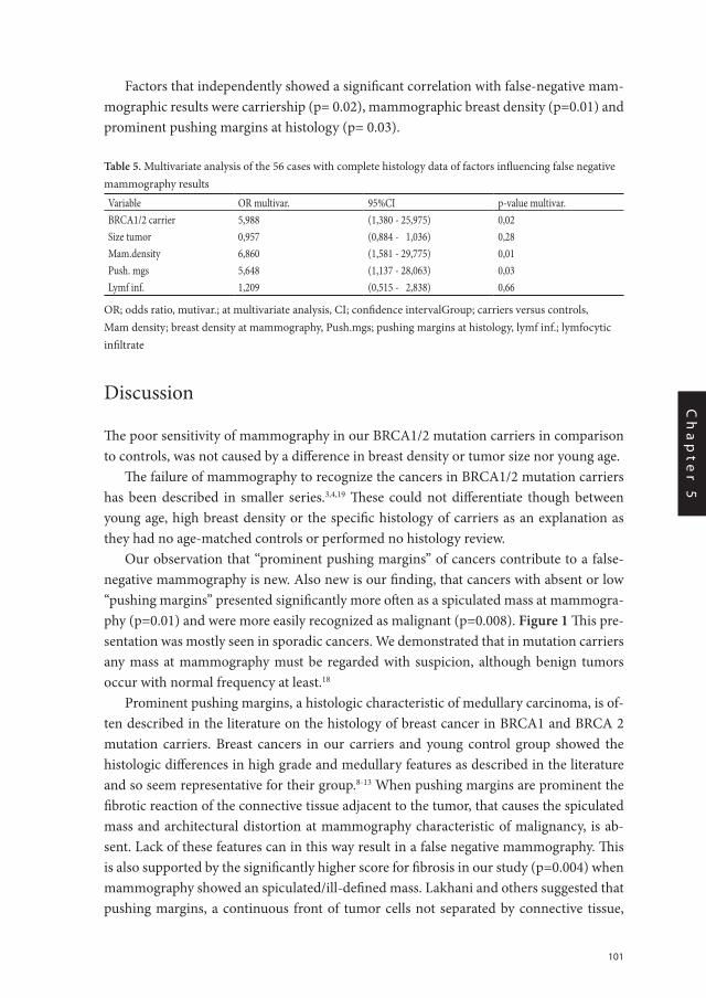
101
Ch
ap
te
r 5
Factors that independently showed a significant correlation with false-negative mam-mographic results were carriership (p= 0.02), mammographic breast density (p=0.01) and prominent pushing margins at histology (p= 0.03).
Table 5. Multivariate analysis of the 56 cases with complete histology data of factors influencing false negative mammography results
Variable OR multivar. 95%CI p-value multivar.BRCA1/2 carrier 5,988 (1,380 - 25,975) 0,02Size tumor 0,957 (0,884 - 1,036) 0,28Mam.density 6,860 (1,581 - 29,775) 0,01Push. mgs 5,648 (1,137 - 28,063) 0,03Lymf inf. 1,209 (0,515 - 2,838) 0,66
OR; odds ratio, mutivar.; at multivariate analysis, CI; confidence intervalGroup; carriers versus controls, Mam density; breast density at mammography, Push.mgs; pushing margins at histology, lymf inf.; lymfocytic infiltrate
Discussion
The poor sensitivity of mammography in our BRCA1/2 mutation carriers in comparison to controls, was not caused by a difference in breast density or tumor size nor young age.
The failure of mammography to recognize the cancers in BRCA1/2 mutation carriers has been described in smaller series.3,4,19 These could not differentiate though between young age, high breast density or the specific histology of carriers as an explanation as they had no age-matched controls or performed no histology review.
Our observation that “prominent pushing margins” of cancers contribute to a false-negative mammography is new. Also new is our finding, that cancers with absent or low “pushing margins” presented significantly more often as a spiculated mass at mammogra-phy (p=0.01) and were more easily recognized as malignant (p=0.008). Figure 1 This pre-sentation was mostly seen in sporadic cancers. We demonstrated that in mutation carriers any mass at mammography must be regarded with suspicion, although benign tumors occur with normal frequency at least.18
Prominent pushing margins, a histologic characteristic of medullary carcinoma, is of-ten described in the literature on the histology of breast cancer in BRCA1 and BRCA 2 mutation carriers. Breast cancers in our carriers and young control group showed the histologic differences in high grade and medullary features as described in the literature and so seem representative for their group.8-13 When pushing margins are prominent the fibrotic reaction of the connective tissue adjacent to the tumor, that causes the spiculated mass and architectural distortion at mammography characteristic of malignancy, is ab-sent. Lack of these features can in this way result in a false negative mammography. This is also supported by the significantly higher score for fibrosis in our study (p=0.004) when mammography showed an spiculated/ill-defined mass. Lakhani and others suggested that pushing margins, a continuous front of tumor cells not separated by connective tissue,
102
could result from a reduced potential for stromal infiltration by individual or small groups of tumor cells.8,9 However not all the tumors in BRCA1/2 mutation carriers have medul-lary features like prominent pushing margins and our multivariate analysis showed that Chapter 5 Fig 1 Figure 1
Chapter 5 Figure 1. Figure 1.A. Spiculated mass the arrow indicating the malignant tumor in the inner upper quadrant of the right breast of a 46 yr control patient.B. Absence of pushing margin in the 1,6 cm tumor A. at histologyC. Benign image despite low density at mammography of the cancer in the upper quadrant of the right breast of a 43 year BRCA1 mutation carrierD. Prominent pushing margin of 1,2 cm. cancer C. at histology

103
Ch
ap
te
r 5
carriership has also negative influence on imaging results independent of pushing mar-gins and breast density.
Several pathologic features have been described for mammographic occult breast cancers apart from tumor size.17 The difference in density between the fibrous stroma produced by the tumor mass and the surrounding fibroglandular tissue can be invisible in dense breasts. This was in our series in carriers and controls of equal influence. Breast density (high 41% in carriers vs 53% in controls) was for their age conform the literature.
The desmoplastic reaction produced by the tumor can be poor like in lobular carcino-ma, while intraductal carcinoma is virtually only recognized when it produces malignant microcalcifications.7,17 Consistent with the literature, DCIS around the invasive tumors was seen less often in our carriers compared to controls. However the result was not sig-nificant nor was the difference in presentation with microcalcifications at mammography. So the presence of DCIS did not strongly influence our imaging results.
Our pathologic review could not further elucidate the mechanism by which BRCA1/2 mutation carriership leads to false-negative mammography results.
Our finding that in BRCA1/2 mutation carriers breast cancer is frequently missed at mammography, must be taken into account when defining the optimal imaging strategy for carriers with symptoms or under surveillance. Additional detection methods such as breast-MRI may often be needed in these mutation carriers 3,4,19-20, but not necessarily in other young women with breast complaints.
Acknowledgements
We thank very much Martine Voskuil, Ada van Eekelen and Ellen Crepin for logistic sup-port and Marijke Westerhout-Kersten for assisting literature search.
104
References:
1. Ford D, Easton DF, Bishop DT, Narod SA, Goldgar DE. Risk of cancer in BRCA1-mutation carriers. Lancet 1994; 343:692-695.
2. Krainer M, Silva-Arrieta S, FitzGerald MG, Shimada A, Ishioka C, Kanamaru R, MacDonald DJ, Unsal H, Finkelstein DM, Bowcock A et al. Differential contributions of BRCA1 and BRCA2 to early-onset breast cancer. N Eng J Med 1997; 336:1416-1421.
3. Brekelmans CTM, Seynaeve C, Bartels CCM, Tilanus-Linthorst MMA, Meiers-Heijboer EJ, Crepin CMG, van Geel AN, Menke M, Verhoog LC, vdOuweland A, Obdeijn AIM, Klijn JGM. Effectiveness of breast cancer surveillance in BRCA1/2 gene mutation carriers and women with high familial risk. J Clin Oncol. 2001;19:924-930.
4. Kuhl CK, Schmutzler RK, Leutner CC, Kempe A, Wardelmann E, Hocke A, Maringa M, Pfeifer U, Krebs D, Schild HH.Breast MR imaging screening in 192 women proved or suspected to be carriers of a breast cancer susceptibility gene: preliminary results. Radiology 2001;215:267-79.
5. Goffin J, Chappuis PO, Wong N, Foulkes WD.Letter. JNCI 2001;93:1754
6. Knutzen AM, Gisvold JJ. Likelihood of malignant disease for various categories of mammographically detected, nonpalpable breast lesions. Mayo clin Proc.1993(68): 454-460.
7. Newstead GM, Baute PB, Toth HK. Invasive lobular and ductal carcinoma: Mammographic findings and stage at diagnosis. Radiology 1992;184:623-627.
8. Lakhani SR, Jacqumier J, Sloane JP, Gusterson BA, Anderson TJ, van de Vijver MJ, Farid LM, Venter D, Antoniou A, Storfer-Isser A, Smyth E, Steel CM, et al. Multifactorial analysis of differences between sporadic breast cancers and cancers involving BRCA1 and BRCA2 mutations. J Natl Cancer Inst. 1998; 90:1138-1145.
9. Breast Cancer Linkage Consortium. Pathology of familial breast cancer: differences between cancers in carriers of BRCA1 or BRCA2 mutations and sporadic cases. Lancet 1997;349:1488-1505.
10. Armes JE, Egan AJM, Southey ME, Dite GS, McCredie PhD, Giles GG, Hopper JL, Venter DJ. The histo-logic phenotypes of breast carcinoma occurring before age 40 years in women with and without BRCA1 and BRCA2 germline mutations. Cancer 1998;83:2335-45.
11. Marcus JN, Page DL, Watson P, Narod SA, Lenoir GM, Lynch HT. BRCA1 and BRCA2 herediteary breast carcinoma phenotypes.Cancer1997;80:543 56.
12. Eisinger F, Jacquemier J, Charpin C, Stoppa-Lyonnet D, Bressac-de Paillerets, Peyrat J-P, Longy M, Guine-bretièreJ-M, Sauvan R, Noguchi T, Birnbaum D, Sobol H. Mutations at BRCA1: The medullary breast carcinoma revisited. Cancer Res. 1998;58:1588-92.
13. Johansson OT, Idvall I, Anderson C, Borg Å, Barkardóttir RB, Egilsson V, Olsson H. Tumor biological features of BRCA1 induced- breast and ovarian cancer. Eur J Cancer 1997;33 362-371.
14. Kopans DB, Dorsi CJ, Adler DD. Breast Imaging Reporting and Data System. Reston VA. American Col-lege of Radiology. 1993
15. Sobin LH, Wittekind CH, editors UICC. TNM Classification of malignant tumours. 5th ed. New York: John Wiley & Sons Inc., 1997.
16. Elston CW. Histopathology 1991; 19:403-410
17. Holland R, Hendriks JHCL, Mravunac M. Mammographically occult breast cancer. Cancer1983; 52:1810-1819.
18. Webb PM, Byrne C, Scnitt SJ, Connolly JL, Jacobs T, Peiro G, Willet W, Colditz GA. Family history of breast cancer, age and benign breast disease. Int J Cancer 2002; 100:375-378.

105
Ch
ap
te
r 5
19. Heywang-Köbrunner SH, Bick U, Bradley jr.WG, Casselman J, Coulthard A, Fischer U, Müller-Schimpfle M, Oellinger H, Patt R, Teubner J, Friedrich M et al. International investigation of breast MRI: results of a multicentre study (11 sites) concerning diagnostic parameters for contrast-enhanced MRI based on 519 histopathologically correlated lesions. Eur. Radiol 2001;11:531-546.
20. Warner E, Plewes DB, Shumak RS, Catzavelos GC, Di Prospero LS, Yaffe MJ, Goel V, Ramsay E, Chart PL, Cole DEC, Taylor GA, Cutrara et al. Comparison of breast magnetic resonance imaging, mammog-raphy and ultrasound for surveillance of women at high risk for hereditary breast cancer.J Clin Oncol 2001;19:3524-31.


Chapter 6
MRI in patients with axillary metastases of occult breast carcinoma
M.M.A. Tilanus-Linthorst, A.I.M. Obdeijn, M. Bontenbal, and M. Oudkerk
Breast Cancer Research and Treatment 1997;44(2):179-182

108
Summary
In 4 women with adenocarcinoma metastasis in an axillary lymph node and no primary tumor found, we investigated whether Magnetic Resonance Imaging (MRI) of the breast could detect a clinically and mammographically occult breast tumor. MRI detected an en-hancing lesion in 3 women and an enhancing double lesion in one patient. MRI directed ultrasound guided fine needle aspiration cytology confirmed the 5 breast carcinomas in the 4 women.
In women with metastasis in an axillary lymph node consistent with breast cancer and without a primary tumor, MRI of the breast should be added to clinical examination and mammography before defining it as an occult primary and planning therapy.
109
Ch
ap
te
r 6
Introduction
Metastatic adenocarcinoma in an axillary lymph node, without an evident primary tu-mor, is most likely the first sign of breast cancer in women. In less than 0.4% of all breast cancer patients axillary metastases are the only clinical manifestation of the disease [1]. If after clinical examination and mammography no primary breast carcinoma is found, it is defined as occult [2–5]. Before 1980 axillary dissection and mastectomy were standard treatment in these patients, and breast cancer was detected in the dissected breast in 80–100% [1, 3, 4]. In the last decade therapy in these patients changed from mastectomy to irradiation or no treatment of the breast, axillary dissection, and systemic therapy. In the follow-up thereafter the primary breast cancer is detected in 13–50% [3, 5]. This reflects the fact that mammographic quality has markedly been improved in the last de-cade. Nevertheless mammography does not detect all malignancies. Even with optimal technique mammography shows no suspicious lesion in 6–20% of patients with a pal-pable malignancy [6–8]. Furthermore, mammographic sensitivity decreases strongly in dense breast tissue [7, 8].
In recent years MR Imaging of the breast with paramagnetic contrast has shown a high sensitivity for breast cancer of 89–97% [9–15]. MRI is less hampered in the detection of breast cancer by dense breast tissue and can depict cancers that were missed at mammog-raphy in dense parenchyma. Therefore, we investigated whether MRI of the breast should be added to clinical examination and mammography in patients with metastatic adeno-carcinoma in axillary nodes consistent with breast cancer without a primary tumor.
Patients and methods
Between March 1993 and February 1996, 4 patients with the diagnosis of metastatic ad-enocarcinoma in an axillary lymph node from an unknown primary were investigated at the radiology department. One patient had had a contralateral mastectomy for a T2N1M0 breast cancer 17 years before. Three patients had no history of previous malignancy. The diagnosis of adenocarcinoma metastasis in the axillary node(s) was made at histopatho-logic examination after axillary dissection in 3 patients and at cytologic examination after fine needle aspiration cytology (FNAC) in 1 patient.
TechniqueMammography was performed on a General Electric Senographe 600T unit (Milwaukee USA), focus 0.3 mm and Kodak screens (min RE). Standard oblique and craniocaudal projections were obtained. For ultrasound of the breast an Acuson 128XP/10 (ART) sys-tem with a 7.5 Hz linear array transducer was used.
The MR examination was performed with a 1.5 Tesla magnetic resonance imaging system (Magnetom- Helicon SP 4000 63/84, Siemens Erlangen Germany).
110
The women lay prone with the breast suspended in a double breast surface coil. The examination consisted of axial T1-weighted and fat suppressed scans before and after in-travenous administration of Gadolinium (Magnevist, Schering). After a localizer scan, the inversion recovery fat suppressed scan was performed with the following scan parameters: FOV 350 mm, contiguous slices of 5 mm thickness, scan matrix 224 ´ 256, scan time 7 minutes, 1 acquisition, TR = 1900 ms, TE = 20 ms, TI = 150 ms. Next, the gradient echo T1-weighted scans were performed before and 1, 3, and 5 minutes after contrast adminis-tration. The scan parameters were: flip angle 90°, FOV 320 mm, 23 contiguous slices of 4 mm, TR = 290 ms, TE = 5 ms, scan matrix 224 ´ 256, scan time 1 minute, 1 acquisition.
After these series a fat suppressed scan was performed for better delineation of en-hancing lesions in the otherwise high intensity of the fat of the breast. At a later stage sub-traction images were obtained from both the T1 weighted and the fat suppressed images with the use of a software subtraction function.
Any focal contrast enhancement in the breast parenchyma was considered as abnor-mal and possibly malignant.
Results
The age of the patients was: 34, 40, 55, and 79 (Table 1). In 3 patients the left axilla was involved, in 1 patient the right.
In the 4 women examined neither a breast mass was palpable on clinical examination nor a malignancy was suspected at mammography. Ultrasound screening of the ipsilateral breast did not show any suspicious lesion before MRI. MRI however showed 1 enhancing lesion in 3 patients and 2 lesions in 1 patient (Table 1, Figure 1).
The location of the lesions on MRI was: the right upper outer quadrant (UOQ), ret-roareolar, the lower outer quadrant (LOQ), and a double lesion directly lateral to the
Table 1. Summary of treatments and findingsPatient Age Diagnosis
Ax diss/FNAC Mamm.Density
Location at MRI Pathological examination and ø
Treatment
A 79 FNAC left side adeno carcinoma metastases
N Retroareolar Breast ductal adeno carcinoma 1.5 cm 3 lymph nodes +
Mastectomy Ax diss Tamoxifen
B 34 Ax diss left side 1 lymph node with adeno carcinoma metastases
N LOQ Breast ductal adeno carcinomaø 2 cm
Irradiation left breast adjuvant chemotherapy
C 55 Ax diss right side 8 lymph nodes with adeno carcinoma metastases
N UOQ Undifferentiated Carcinoma ø 1 cm + lymphatic invasion
Irradiation right breastTamoxifen
D 40 Ax diss left side 1 lymph node with adeno carcinoma metastases
D Lateral to nipple double lesion 2 ´ 1 cm ø
No lumpectomy of the breast performed
Chemotherapy
Ax diss – axillary dissection; FNAC – fine needle aspiration cytology; N – normal breast parenchyma; D – dysplastic breast pattern; Irradiation – 50 Gy+20 Gy boost irradiation of the breast.
111
Ch
ap
te
r 6
nipple. At MRI directed ultrasound examination, FNAC of a correlating structure could be performed in all patients. Cytologic examination showed carcinoma cells in all patients (5 breast lesions). Pathological examination after lumpectomy showed infiltrating ductal carcinoma of 1.5 and 2 cm in 2 patients and an undifferentiated carcinoma of 1 cm with lymphatic invasion in 1 patient. One patient had no lumpectomy but systemic chemo-therapy with MRI control for regression of the double breast lesion.
Discussion
The standard treatment of a small breast carcinoma is excision of the primary tumor and axillary dissection followed by postoperative radiotherapy of the breast. In our patients the tumors varied between 1–2 cm in diameter, illustrating that minimal invasive breast carcinoma can be clinically and radiologically occult.
For patients with metastatic adenocarcinoma in an axillary lymph node and no pri-mary tumor found, the best treatment is still not clear [5, 16, 17]. In case the primary tumor in the breast can be detected it is possible to choose optimal treatment, adding local therapy. In our patients this resulted in lumpectomy followed by radiotherapy to the breast in 2 patients and mastectomy in an elderly patient. All 4 patients were treated with systemic therapy.
Breast cancer is mammographically occult in 6–20% of patients with a clinically mani-fest tumor [6–8]. In our patients neither dimension nor location of the lesion could ex-plain why it did not show at mammography. The density of the breast parenchyma was an explanation for a mammographically occult tumor in only 1 of our patients. In retrospect, however, one lesion was visible as a benign shadow.
The high sensitivity of MRI in the detection of breast cancer can be of considerable value in patients in whom the clinical and mammographical the low specificity for breast cancer (30–67%) is a limitation to its use and requires confirmation that the detected
Figure 1. MRI subtraction image 5 minutes after intravenous contrast administration showing the enhancing double lesion in the left breast of patient D.
112
lesion is malignant [12–15]. In our group of patients malignancy was already established in the axillary nodes, but to prove that the primary tumor was identified, FNAC was per-formed. The procedure depends on the experience of the radiologist with ultrasound guided FNAC [18].
Davis described a similar patient in whom the breast lesion seen on MRI could not be found at ultrasonography, but MRI guided wire localization and excision was succesfully performed [19]. Equipment for MRI guided wire localization of breast lesions, however, is clinically scarcely available. If at MRI of the breast in women with axillary lymph node metastasis no lesion is found, only follow-up will determine whether the nodal metastasis is from a different origin, or from a breast cancer occult even at MRI. In conclusion, we think that in women with metastasis in an axillary lymph node consistent with breast can-cer without a primary tumor, MRI of the breast should be added to clinical examination and mammography, before defining it as an occult primary and planning therapy.
113
Ch
ap
te
r 6
References
1. Baron PL, Moore PM, Kinne DW, Candela FC, Osborne MP, Petrek JA: Occult breast cancer presenting with axillary metastasis. Arch Surg 125:210–214, 1990
2. Rosen PP, Kimmel M: Occult breast carcinoma presenting with axillary lymph node metastases: A follow-up study of 48 patients. Hum Pathol 21:518–523, 1990
3. Merson M, Andreola S, Galimberti V, Bufalino R, Marchini S, Veronesi U: Breast carcinoma presenting as axillary metastases without evidence of a primary tumor. Cancer 70:504–508, 1992
4. Iglehart JD, Ferguson BJ, Shingleton WW et al.: An ultrastructural analysis of breast carcinoma present-ing as isolated axillary adenopathy. Ann Surg 196:8–13, 1982
5. van Ooijen B, Bontenbal M, Henzen-Logmans SC, Koper PCM: Axillary nodal metastases from an occult primary consistent with breast carcinoma. Br J Surg 80:1299–1300, 1993
6. Wallis MGA: A review of breast carcinomas missed by mammography. Br J Radiol 6 (Suppl): S11, 1989
7. Edeiken S: Mammography in the symptomatic woman. Cancer 63:1412–1414, 1989
8. Anderson EDC, Muir BB, Walsch JS, Kirkpatrick AE: The efficacy of double reading mammograms in breast screening. Clin Radiol 49:248–251, 1994
9. Heywang SH, Wolf A, Pruss E et al.: MR imaging of the breast with Gd-DTPA: Use and limitations. Radi-ology 171:95–103, 1989
10. Pierce WP, Harms SE, Flamig DP et al.: Three-dimensional Gadolinium-enhanced MR imaging of the breast: Pulse sequence with fat suppression and magnetization transfer contrast. Radiology 181:757–763, 1991
11. Kaiser WA: MRM promises earlier breast cancer diagnosis. Diagn Imaging Int Nov/Dec: 44–50, 1992
12. Obdeijn AIM, Kuijpers TJA, van Dijk P, Oudkerk M: Limited indications for MR mammography. In: Advances in Magnetic Resonance Imaging. Blackwell Science, Berlin / Boston / Oxford / London / Edin-burgh / Melbourne /Paris/Vienna/Yokohama 1996, in press
13. Harms SE, Flamig DP, Hesley KL et al.: MR imaging of the breast with rotating delivery of excitation of resonance: Clinical experience with pathologic correlation. Radiology187:493–501, 1993
14. Allgayer B, Lukas P, Loos W, Kersting-Sommerhof B: MRT der Mamma mit 2D-Spin-Echo and Gradient-Echo Sequenzen in diagnostischen Problemfa¨llen. Fortschr Ro¨ ntgenstr 158:423–427, 1993
15. Harms SC, Flamig DP, Hesley KL et al.: Fat-suppressed three-dimensional MR imaging of the breast. Radiographics 13:247–267, 1993
16. Ellebroek N, Holmes F, Singletary T et al.: Treatment of patients with isolated axillary nodal metastases from an occult primary carcinoma consistent with breast origin. Cancer 66:1461–1467, 1990
17. Campanc F, Fourquet A, Ashby MA et al.: Presentation of axillary lymphadenopathy without detectable breast primary (T0N1b breast cancer): Experience at Institute Curie. Radiother Oncol 15:321–325, 1989
18. Gordon PB, Goldenberg SL, Chan NHL: Solid breast lesions: Diagnosis with US-guided fine needle aspi-ration biopsy. Radiology 189:573–580, 1993
19. Davis PL, Julian ThB, Staiger M et al.: Magnetic resonance imaging detection and wire localization of an occult breast cancer. Breast Cancer Research and Treatment 32:327–330,1994

Chapter 7
Earlier detection of breast cancer by surveillance of women at familial risk
M.M.A. Tilanus-Linthorst, C.C.M.Bartels, A.I.M. Obdeijn, M. Oudkerk
European Journal of Cancer 2000;369(4):514-519
116
Abstract
A positive family history increases the risk for breast cancer which often occurs at a much younger age than in the general population. We studied whether surveillance of these women resulted in the detection of breast cancer in an earlier stage than in symptomatic patients with a family history. Between January 1994 and April 1998, 294 women with 15-25% risk (moderate), mean age: 43.3 (22-75) years, were screened with a yearly physical examination and mammography form 5 years before the youngest age of onset in the fam-ily and 384 women with >25% risk (high) for breast cancer, mean age: 42.9 (20-74) years were screened with a physical examination every 6 months and yearly mammography. From September 1995 breast magnetic resonance imaging (MRI) was also carried out for 109 high risk women where mammography showed over 50% density. 26 breast cancers detected under surveillance were significantly more often found in an early T1N0 stage than the 24 breast cancers in patients with a family history referred in that period because of symptoms: 81 versus 46% (P = 0.018). Patients under surveillance were also less fre-quently node-positive than the symptomatic group: 19 versus 42% (P = 0.12). 20 patients with a family history referred by our national screening programme in that period had 21 breast cancers detected, 81% in stage T1N0 and 5% node-positive, which was comparable to the results in our national screening programme T1N0 66%, N + 24% resulting in a 30% reduction in mortality. The incidence in women under surveillance was 10.1 per 1000 in the ‘high’ risk group and 13.3 per 1000 in the ‘moderate’ risk group. Expected incidence in an average risk population aged 40-50 years is 1.5, expected in the group consisted of only gene carriers 15 per 1000. 23% of the breast cancers in the surveillance group were de-tected at physical examination, but occult at mammography. 38% were detected at mam-mography and clinically occult. Breast MRI (in the subgroup) detected 3 occult breast cancers. The results of this study show that women with a family history benefit from surveillance as breast cancer was detected significantly more often in a favourable T1N0 stage and a mortality reduction comparable to that obtained in our national screening programme may be expected also in women < 50 years of age. Both physical examination and mammography contribute tot this result, but the former in this study only contributed in women before menopause. Starting surveillance some years before the youngest age of onset in the family may result in higher detection rates. Screening with MRI can detect breast cancers, still occult at physical examination and mammography.
117
Ch
ap
te
r 7
Introduction
Women with a strong family history of breast cancer not only run a high risk to develop this malignancy, but their risk also increases at a much younger age than in the general population (1). Highly penetrant mutations in genes like BRCA1 and BRCA2 can be de-tected in women with these family histories and cause approximately 5% of all breast can-cers (2). In carriers of a BRCA1 mutation, the risk for breast cancer rises sharply from 3% at 30 years of age to 50% at 50 years (2-3). In BRCA2 mutation carriers the risk profile develops later (4).
40-80% of healthy women, who after a presymptomatic DNA test appeared to be carri-ers of the BRCA1 or 2 mutation causing breast cancer in their family, chose primarily sur-veillance and not (at least for the time being) preventive mastectomy (5-6). The increased risk of women with a family history if often not attributable to BRCA1 and BRCA2 muta-tions and these women also chose to continue surveillance (7).
In the Netherlands, women with an estimated risk of breast cancer over 25% based on their family history are recommended surveillance with monthly breast self examination (BSE), semi-annual examination by a physician (PE) and yearly mammography from 25 years onwards (8). However, there are not yet sufficient data to prove the effectiveness of surveillance from this age.
In this study, we investigated the value of surveillance in patients with a family history. Our central question was whether breast cancer was detected in an earlier stage in patients under surveillance than in patients with a family history, referred because of symptoms of this disease.
We also compared the stage of breast cancer of the patients under surveillance with the stage of breast cancers detected during national screening for 50-75 year old women. In this last group, the survival gain and cost-benefit analysis is known.
Patients and methods
In this study, we included the group of consecutive women who visited the outpatient breast clinic of the Rotterdam Cancer Centre between January 1994 and April 1998 with a family history of breast cancer. Some of these women were registered together with the patients of the Rotterdam Family Cancer Clinic in the Rotterdam/Leiden genetic working group. We included in this study only the group of women under surveillance at the breast clinic, as this group can be compared with the consecutive group of women referred to this clinic during the same time period because of symptoms or because of an abnormality detected at the national screening programme. Women were considered under surveil-lance, if they consented with the proposed surveillance scheme and no breast cancer was detected at clinical examination nor at two view mammography during the first visit. Risk estimation was performed by two breast clinic doctors or by the geneticist using the tables
118
of Houlston and Claus (9-10). Risk estimates were higher, with an increasing rate of af-fected relatives, decreasing age of onset in the relatives and/or one or more cases of ovarian cancer. A woman was considered at moderate risk, with for instance, 1 affected first degree relative or 2 second degree relatives < 50 years of age. High risk estimates were made for instance for patients with 2 first degree relatives < 50 years of age with breast cancer; 1 or more first degree relatives with breast cancer plus 1 or more with ovarian cancer. Accord-ing to the estimated risk two surveillance schemes were proposed.
294 women under surveillance with moderate risk (15-25%) got instructions on car-rying out a monthly BSE and were scheduled for a yearly physical examination and mam-mography, starting 5 years before the age at which the youngest family member had got breast cancer (age of onset).
384 women under surveillance with high risk (>25%) were scheduled for surveillance according to the national guidelines. From September 1995 onwards breast MRI in addi-tion to normal surveillance was performed in women with high risk and over 50% density at mammography (n=109).
We investigated and compared the stage in which breast cancers were detected in three groups of patients all with a positive family history:
Group 1: Patients who developed breast cancer whilst under surveillance because of their family history (n=26). The surveillance group.
Group 2: Patients with breast cancer, referred by the general practitioner in the same period because of symptoms of their disease who appeared to have a positive family his-tory (n=24). The symptomatic group. Patients could be referred because of a palpable mass, skin of nipple retraction, nipple discharge, inflammatory breast disease or pain.
Group 3: Patients detected at the national screening programme and referred to our clinic in that period who appeared to have positive family history (n=20). The screening group
We also evaluated the contribution of BSE, physical examination, mammography and fine needle aspiration cytology (FNAC). Statistical differences in stage between groups were analysed using the two-sided Fisher’s exact tests.
TechniqueMammography was performed on a General Electric Senographe 600T unit (Milwaukee, USA), focus 0.3 mm and Kodak screens (min RE). Standard oblique and craniocaudal projections were obtained during the first visit and alternated thereafter with mediolateral oblique projections only. There was dual reading of all mammograms by experienced ra-diologists. For ultrasound an Acuson 128XP/10 (ART) system with a 7.5 mHz linear array transducer was used. Breast MRI examination was performed with a 1.5 Tesla magnetic resonance imaging system (Vision, Siemens, Erlangen, Germany).
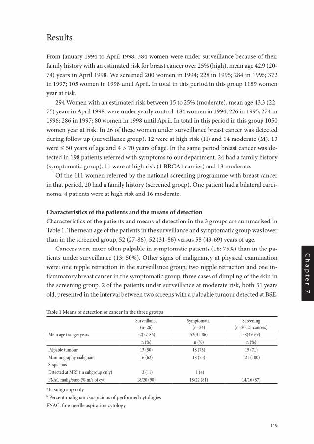
119
Ch
ap
te
r 7
Results
From January 1994 to April 1998, 384 women were under surveillance because of their family history with an estimated risk for breast cancer over 25% (high), mean age 42.9 (20-74) years in April 1998. We screened 200 women in 1994; 228 in 1995; 284 in 1996; 372 in 1997; 105 women in 1998 until April. In total in this period in this group 1189 women year at risk.
294 Women with an estimated risk between 15 to 25% (moderate), mean age 43.3 (22-75) years in April 1998, were under yearly control. 184 women in 1994; 226 in 1995; 274 in 1996; 286 in 1997; 80 women in 1998 until April. In total in this period in this group 1050 women year at risk. In 26 of these women under surveillance breast cancer was detected during follow up (surveillance group). 12 were at high risk (H) and 14 moderate (M). 13 were ≤ 50 years of age and 4 > 70 years of age. In the same period breast cancer was de-tected in 198 patients referred with symptoms to our department. 24 had a family history (symptomatic group). 11 were at high risk (1 BRCA1 carrier) and 13 moderate.
Of the 111 women referred by the national screening programme with breast cancer in that period, 20 had a family history (screened group). One patient had a bilateral carci-noma. 4 patients were at high risk and 16 moderate.
Characteristics of the patients and the means of detectionCharacteristics of the patients and means of detection in the 3 groups are summarised in Table 1. The mean age of the patients in the surveillance and symptomatic group was lower than in the screened group, 52 (27-86), 52 (31-86) versus 58 (49-69) years of age.
Cancers were more often palpable in symptomatic patients (18; 75%) than in the pa-tients under surveillance (13; 50%). Other signs of malignancy at physical examination were: one nipple retraction in the surveillance group; two nipple retraction and one in-flammatory breast cancer in the symptomatic group; three cases of dimpling of the skin in the screening group. 2 of the patients under surveillance at moderate risk, both 51 years old, presented in the interval between two screens with a palpable tumour detected at BSE,
Table 1 Means of detection of cancer in the three groups
Surveillance(n=26)
Symptomatic(n=24)
Screening(n=20; 21 cancers)
Mean age (range) years 52(27-86) 52(31-86) 58(49-69)n (%) n (%) n (%)
Palpable tumour 13 (50) 18 (75) 15 (71)Mammography malignant 16 (62) 18 (75) 21 (100)SuspiciousDetected at MRIa (in subgroup only) 3 (11) 1 (4)FNAC malig/susp (% m/s of cyt) 18/20 (90) 18/22 (81) 14/16 (87)
a In subgroup only b Percent malignant/suspicious of performed cytologiesFNAC, fine needle aspiration cytology
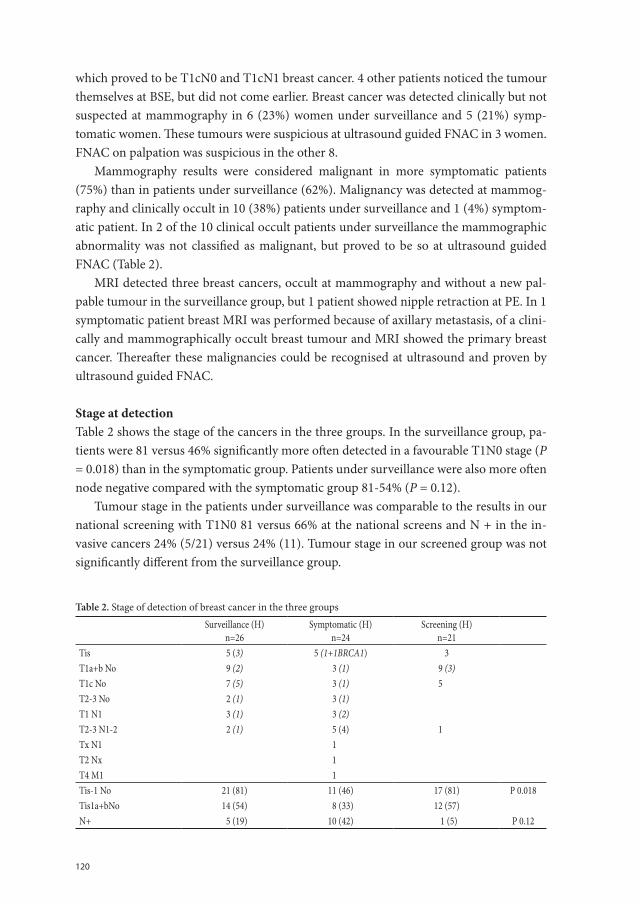
120
which proved to be T1cN0 and T1cN1 breast cancer. 4 other patients noticed the tumour themselves at BSE, but did not come earlier. Breast cancer was detected clinically but not suspected at mammography in 6 (23%) women under surveillance and 5 (21%) symp-tomatic women. These tumours were suspicious at ultrasound guided FNAC in 3 women. FNAC on palpation was suspicious in the other 8.
Mammography results were considered malignant in more symptomatic patients (75%) than in patients under surveillance (62%). Malignancy was detected at mammog-raphy and clinically occult in 10 (38%) patients under surveillance and 1 (4%) symptom-atic patient. In 2 of the 10 clinical occult patients under surveillance the mammographic abnormality was not classified as malignant, but proved to be so at ultrasound guided FNAC (Table 2).
MRI detected three breast cancers, occult at mammography and without a new pal-pable tumour in the surveillance group, but 1 patient showed nipple retraction at PE. In 1 symptomatic patient breast MRI was performed because of axillary metastasis, of a clini-cally and mammographically occult breast tumour and MRI showed the primary breast cancer. Thereafter these malignancies could be recognised at ultrasound and proven by ultrasound guided FNAC.
Stage at detectionTable 2 shows the stage of the cancers in the three groups. In the surveillance group, pa-tients were 81 versus 46% significantly more often detected in a favourable T1N0 stage (P = 0.018) than in the symptomatic group. Patients under surveillance were also more often node negative compared with the symptomatic group 81-54% (P = 0.12).
Tumour stage in the patients under surveillance was comparable to the results in our national screening with T1N0 81 versus 66% at the national screens and N + in the in-vasive cancers 24% (5/21) versus 24% (11). Tumour stage in our screened group was not significantly different from the surveillance group.
Table 2. Stage of detection of breast cancer in the three groups
Surveillance (H)n=26
Symptomatic (H)n=24
Screening (H)n=21
Tis 5 (3) 5 (1+1BRCA1) 3T1a+b No 9 (2) 3 (1) 9 (3)T1c No 7 (5) 3 (1) 5T2-3 No 2 (1) 3 (1)T1 N1 3 (1) 3 (2)T2-3 N1-2 2 (1) 5 (4) 1Tx N1 1T2 Nx 1T4 M1 1Tis-1 No 21 (81) 11 (46) 17 (81) P 0.018Tis1a+bNo 14 (54) 8 (33) 12 (57)N+ 5 (19) 10 (42) 1 (5) P 0.12
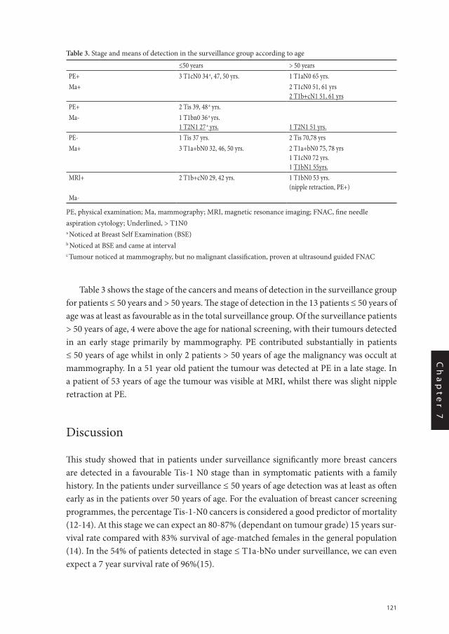
121
Ch
ap
te
r 7
Table 3 shows the stage of the cancers and means of detection in the surveillance group for patients ≤ 50 years and > 50 years. The stage of detection in the 13 patients ≤ 50 years of age was at least as favourable as in the total surveillance group. Of the surveillance patients > 50 years of age, 4 were above the age for national screening, with their tumours detected in an early stage primarily by mammography. PE contributed substantially in patients ≤ 50 years of age whilst in only 2 patients > 50 years of age the malignancy was occult at mammography. In a 51 year old patient the tumour was detected at PE in a late stage. In a patient of 53 years of age the tumour was visible at MRI, whilst there was slight nipple retraction at PE.
Discussion
This study showed that in patients under surveillance significantly more breast cancers are detected in a favourable Tis-1 N0 stage than in symptomatic patients with a family history. In the patients under surveillance ≤ 50 years of age detection was at least as often early as in the patients over 50 years of age. For the evaluation of breast cancer screening programmes, the percentage Tis-1-N0 cancers is considered a good predictor of mortality (12-14). At this stage we can expect an 80-87% (dependant on tumour grade) 15 years sur-vival rate compared with 83% survival of age-matched females in the general population (14). In the 54% of patients detected in stage ≤ T1a-bNo under surveillance, we can even expect a 7 year survival rate of 96%(15).
Table 3. Stage and means of detection in the surveillance group according to age
≤50 years > 50 years PE+ 3 T1cN0 34 a, 47, 50 yrs. 1 T1aN0 65 yrs.Ma+ 2 T1cN0 51, 61 yrs
2 T1b+cN1 51, 61 yrsPE+ 2 Tis 39, 48 a yrs.Ma- 1 T1bn0 36 a yrs.
1 T2N1 27 a yrs. 1 T2N1 51 yrs.PE- 1 Tis 37 yrs. 2 Tis 70,78 yrs Ma+ 3 T1a+bN0 32, 46, 50 yrs. 2 T1a+bN0 75, 78 yrs
1 T1cN0 72 yrs.1 T1bN1 55yrs.
MRI+ 2 T1b+cN0 29, 42 yrs. 1 T1bN0 53 yrs.(nipple retraction, PE+)
Ma-
PE, physical examination; Ma, mammography; MRI, magnetic resonance imaging; FNAC, fine needle aspiration cytology; Underlined, > T1N0a Noticed at Breast Self Examination (BSE)b Noticed at BSE and came at intervalc Tumour noticed at mammography, but no malignant classification, proven at ultrasound guided FNAC
122
Disease free and overall survival in patients with BRCA1 or 2 mutations or hereditary cancer does not differ significantly from survival in sporadic patients in most studies (16-19). This makes the effort to detect breast cancer at an early stage worthwhile in these patients.
Our study showed no differences apart from MRI in means and stage of detection between women at high or at moderate risk in the three groups. In the surveillance group, 6 of the 13 palpable tumours were noticed at BSE, but only 2 of these women came dur-ing the interval despite strong encouragement for women to do so at any possible suspect clinical sign. We could not demonstrate earlier detection by BSE in patients under sur-veillance in this study. Coebergh and colleagues demonstrated an earlier stage of breast cancer detection in the general population in the last decades due to better awareness of women of suspect clinical signs or less hesitation to visit a physician (20). Because of this we give instructions to all women pre- and postmenopausal on BSE and indeed often see symptomatic patients who detect very small tumours.
The 23% clinically detected mammographically occult tumours in the surveillance group were suspicious at ultrasound guided FNAC or at FNAC on palpation. This under-lines the necessity of FNAC in all palpable tumours. Physical examination, if followed by the right consequences, seems a useful addition to mammography certainly in the surveil-lance of premenopausal high risk women. However, PE hardly contributed to early detec-tion in patients > 55 years under surveillance. Menopause seems a better indicator for the effectiveness of PE than a fixed age (for instance ≤ 50 years of age). In our mostly post-menopausal patients detected at the national screening programme 71% had a clinically manifest tumour. Their tumour stage was as good as in the surveillance group. Screen-ing with only BSE and mammography 2-yearly may also be sufficient after menopause in women with a family history, although the percentage of women at high risk was too small to draw such a conclusion in this study.
Malignancies were detected at mammography exclusively in 38% during surveillance and in 4% of symptomatic patients. In young women >25 years of age mammography is of great value in our experience. This has also been described by Liberman in the screen-ing of women of 35-39 years old (21). Even in young women mammography is the most sensitive examination for the detection of in situ carcinoma. In only 28% (109/384) of our screened women with high risk did mammography results show >50% dense breast tissue which is likely limiting its sensitivity. Both Feig and Brekelmans showed that in premeno-pausal women mammography should be performed yearly, not 2-yearly, to prevent too high (50%) a rate of carcinomas developing during the intervening time period (22, 23). They explained this by faster growth rate of tumours occurring in patients at a younger age.
MRI detected three breast cancers in T1N0 stage during surveillance which were in two cases clinically occult and all mammographically occult. In the 109 women screened with MRI, no carcinomas were detected by palpation or mammography even 1 year after closing this study.
123
Ch
ap
te
r 7
Results of surveillanceIn our high risk group the detection rate was 10.1 per 1000 person-years; expected rate in an average risk population aged 40-50 years is 1.5 per 1000. If this group had consisted only of gene carriers a breast cancer incidence of 15 per 1000 would have been expected (2% per year between 25-50 years of age and 1% per year between 50-75 years of age). The risk estimation in our group seems realistic. Screening women at familial risk with physi-cal examination and yearly mammography was shown to be effective and seems worth-while in women before menopause, when this incidence can be expected.
In our moderate risk group the detection rate was 13.3 per 1000 person-years. Our risk estimation may have been somewhat low. The explanation of the higher incidence could also be explained by the fact that we screened this group only from 5 years younger than the youngest age of onset in the family. Maybe we should start surveillance closer to the youngest age of onset in women at high risk.
The stage of the tumours detected in women with a family history during the national screening programme, was as good as those amongst the younger patients of the surveil-lance group. For postmenopausal women at moderate risk the national screening scheme with only mammography 2-yearly together with BSE may be sufficient.
Between October 1988 and December 1995 Kollias and colleagues (1998) screened 1371 women under 50 years of age with a family history with an annual clinical examina-tion and 2-yearly mammography (24). Their incidence rate was 3.3 per 1000 visits. They detected a higher proportion of DCIS in the family history surveillance group compared with an age-matched symptomatic group; 21 versus 4%, but no differences for invasive tumour size or lymph node stage. This difference compared our study could be due to the interval of the screening mammography in premenopausal women; 2-yearly versus yearly in our study.
Between September 1992 and May 1997 Lalloo and colleagues screened 1259 women under 50 years who had a 4-fold increased risk with annual breast examination and mam-mography. They detected 9 incident + interval cancers, with an incident rate of 4.8 per 1000. The stage of detection was 1 LCIS, T1 7 out of 9, node-positive 4 out of 9 and 2 where the nodal status was unknown (25).
Multicentre trials have started in Great Britain, Germany and The Netherlands to de-termine the cost effectiveness of screening woman at high risk with different surveillance schemes.
Conclusion
In this study we demonstrated a significant earlier detection of breast cancer in women at increased risk under surveillance compared with symptomatic patients. The stage of detection was as favourable in the patients ≤ 50 years of age under surveillance as in the > 50 years of age patient group. The incidence in the high risk group was seven times the

124
incidence in an average risk population (40-50 years of age). Both physical examination and mammography made an important contribution to this result in patients < 55 years of age. Physical examination made no contribution in postmenopausal women with a family history. Screening with MRI can detect tumours occult at PE and mammography.
125
Ch
ap
te
r 7
References
1. Claus EB, Risch N, Thompson WD,. Genetic analysis of breast cancer in the Cancer and Steroid Hormone Study. Am J Hum Genet 1991;48:232-42.
2. Easton DF, Bishop DT, Crockford GP. Genetic linkage analysis in familial breast and ovarian cancer: Results from 214 families. Am J Hum Genet 19993;52:678-701.
3. Ford D, Easton DF, Bishop DT, Narod SA, Goldgar DE. Risk of cancer in BRCA1-mutation carriers. Lancet 1994;343:692-5.
4. Krainer M, Silva Arieta S, FitzGerald MG et al. Differential contributions of BRCA1 and BRCA2 to early-onset breast cancer. N Eng J Med. 1997; 336:1416-21.
5. Lerman C, Narod S, Schulman K, et al. BRCA1 testing in families with hereditary Breast-Ovarian cancer. A prospective study of patient decision making and outcomes. JAMA 1996;275:1885-1892.
6. Tilanus-Linthorst MMA, Bartels CCM, Obdeijn AIM, et al. Gunstige resultaten van periodieke controle bij vrouwen met verhoogd risico van borstkanker; retrospectief onderzoek. Ned Tijdschr Geneeskd 1995;4:139(9):445-50.
7. Burke W, Press N, Pinsky L BRCA1 and BRCA2: a small part of the puzzle. J Natl Cancer Inst.1999;91:943-949.
8. Wobbes Th. Borstkanker in de familie. Ned Tijdschr Geneeskd 1985;129:2193-2195.
9. Houlston RS, McCarter S, Parbhoo, Scurr JH, Slack J. Family history and risk of breast cancer. J Med Genet. 1992;29:134-137.
10. Claus EB, Risch N, Thompson WD. Autosomal dominant inheritance of early onset breast cancer. Cancer 1994;73:643-651.
11. Fracheboud J, de Koning HJ, Beemsterboer PMM, Boer R, Hendriks JHCL, Verbeek ALM et al. Nation-wide breast cancer screening in the Netherlands results of initial and subsequent screens 1990-1995. Int J Cancer 1998;75:694-98.
12. Day N,. Breast cancer screening programmes: the development of a monitoring and evaluation system. Br J Cancer;1989;59:954-958.
13. Tabar L, Int J Cancer 1996;68:693-699.
14. Galea MH, Blamey RW, Elston CW, Ellis IO. The Nottingham Prognostic Index in primary breast cancer. Breast Cancer Res Treat. 1992;22:207-219.
15. Rosner D, Lane WW. Node negative minimal invasive breast cancer patients are not candidates for rou-tine systemic adjuvant therapy. Cancer 1990;66:199-205.
16. Robson M, Gilewski T, Haas B. et al. BRCA-associated breast cancer in young women. J Clin Oncol 1998; 16:1642-1649.
17. Lee JS, Wacholder S, Struewin JP. Et al. Survival after breast cancer in Ashkenazi Jewish BRCA1 and BRCA2 mutation carriers. J Natl Cancer Inst 1995;91:259-263.
18. Marcus JN, Watson P, Page DL et al. Hereditary breast cancer: pathobiology, prognosis and BRCA1 and BRCA2 linkage. Cancer 1996;77:697-709.
19. Verhoog LC, Brekelmans CTM, Seynaeve C et al. Survival and tumour characteristics of breast cancerpa-tients with germline mutations of BRCA1. Lancet 1998;351:316-321.
20. Coebergh JW, Crommelin MA, Kluck HM. Et a. Borstkanker in Zui-Noord-Brabant en in Noord-Lim-burg; beloop van incidentie en vervroeging van de diagnose in een niet gescreende vrouwelijke bevolking. Ned Tijdschr.geneeskd. 1990;134:760-65.

126
21. Liberman L, Dershaw DD, Deutsch BM, Thaler HT, Lippin BS. Screening mammography: value in women 35-39 years old. AJR 1993;161:53-6.
22. Brekelmans CTM, Collette HJA, Collette C, Fracheboud J, Waard F de. Breast cancer after a negative screen, follow up of women participating in the DOM screening programme. Eur J Cancer 1992;28A: 893-5.
23. Feig SA. Increased benefit from shorter screening mammography interval for women aged 40-49 years. Cancer 1997; 80:2035-39.
24. Kollias J, Sibbering DM, Blamey RW, Holland PAM, Ozbuzko Z, Wilson ARM et al. Screening women aged less than 50 years with a family history of breast cancer. Eur J Cancer 1998;34(6):878-83.
25. Lalloo F, Boggis CRM, Evans DGR, Shenton A, Threlfall AG, Howell A. Screening by mammography women with a family history of breast cancer. Eur J Cancer 1998;34(6): 937-940.

Chapter 8
First experiences in screening women at high risk for breast cancer with MR imaging
Madeleine M.A. Tilanus-Linthorst, Inge Marie M. Obdeijn, Karina C.M. Bartels, Harry J. de Koning, and Matthijs Oudkerk
Breast Cancer Research and Treatment 63: 53–60, 2000
128
Summary
Women with a genetic predisposition for breast cancer are often advised surveillance with physical examination twice a year and mammography once a year from 25 years onwards. However, the sensitivity of the mammography decreases when breast tissue is dense and this is seen in 40–50% of women under 50 years.
We therefore investigated whether magnetic resonance imaging (MRI) in addition to the normal surveillance could detect cancers otherwise missed. In 109 women with over 25% risk of breast cancer, MRI was performed because over 50% dense breast tissue was seen at mammography and no suspect lesion was seen at the previous screening. MRI detected breast cancers in three patients (2.8%) occult at mammography and with no new palpable tumor, twice at stage T1bN0 and T1cN0 once. Two cancers were expected. MRI was false positive in six women, resulting in two benign local excisions because ul-trasound or fine needle examination confirmed suspicion. We had no false negativeMRI results. MRI proved true benign in four BRCA 1/2 gene mutation carriers at histologic examination.
Preoperative wire localization of the malignancies detected at MRI proved necessary as the tumor was not palpable in the lumpectomy specimen nor visible at specimen radi-ology.
The extra cost of breast MRI in addition to mammography and physical examination was €13.930 per detected cancer. The cost of the detection of one breast cancer patient in our national screening program is €9000. During follow-up of patients with a familial risk in whom the first breast cancer was detected at MRI, MRI detected two recurrent cancers in stage T1bN0 and T1cN0 and one contralateral cancer T1aNo. Breast MRI is promising in screening young women at high risk for breast cancer, as it can advance the detection of cancers still occult at mammography and physical examination; but the cost may be considerable.
129
Ch
ap
te
r 8
Introduction
An increasing number of women, of 25 years and older, at high risk for developing breast cancer because of a strong family history, are recommended to undergo surveillance with clinical examination and mammography [1]. Although breast cancer can be detected ear-lier by this policy there may be drawbacks to the regular use of mammography at such a young age.
Both patients and physicians are concerned about mammography at a young age be-cause of radiation exposure and false positive results [2]. There is no direct evidence of risk of cancers induced by doses within the mammographic range, but extrapolations from high to low doses are made [3, 4]. In women treated with mantle-field irradiation before the age of 30, a twelve-fold increased risk of breast cancer has been demonstrated [5, 6]. In women exposed to atomic bomb radiation at Hiroshima and Nagasaki the excess relative risk of breast cancer increased in proportion with the dose and inversely with age at the time of exposure [3, 7]. By extrapolations from these examples Beemsterboer et al., calculated, that the number of deaths induced versus prevented might rise from 1:242 to 1:66 by extending screening women aged 50–69 years with mammography with 2mGy per view, at two year interval, to women aged 40–49 with yearly mammography [4].
A second problem is, that in dense breast tissue the sensitivity of mammography for breast cancer may decrease and vary from 25% to 85% [8–10]. In women under 50 years breast tissue is dense in 40–50% of the cases [11–14]. This may have a negative influence on surveillance results in youngerwomen. Kerlikowske et al., reported a low sensitivity of 68.8% of first screening mammography in women <50 years of age with a family history, but demonstrated a decreasing sensitivity of mammography with increasing breast den-sity only in women older than 50 years [15].
Magnetic resonance imaging (MRI) has no limitations in mammographically dense breasts. MRI yields a high sensitivity in the detection of invasive breast cancer from 91% to 98% [16–19]. We showed in previous studies of women with axillary metastasis from clinical and radiological occult breast cancer, that MRI can detect these tumors [20, 21]. In this study we wanted to investigate, whether breast MRI, in addition to the normal surveillance scheme of clinical examination every 6 months and yearly mammography could detect breast cancers that were otherwise missed. We studied high-risk women with over 25% estimated lifetime risk for breast cancer in whom mammography showed breast tissue with more than 50% density.
Patients and methods
From September 1995 till April 1998 breast MRI was performed in addition to the normal surveillance, in women attending because of a family history the outpatient breast clinic of the Daniel den Hoed Cancer Center. MRI was only offered to women with over 25%
130
risk of breast cancer and more than 50% density at their mammography. Risk estimation was performed by the geneticist or the breast clinic doctors (M.T-L, C. B.) with the tables of Claus [22].
Breast density was estimated visually with a quantitative measure, that is, the percent-age of the area of the breast encompassed by fibroglandular tissue dense enough to ob-scure a cancer was estimated. Only patients with dense fibroglandular tissue > 50% of the mammogram were offered additional MRI. This corresponds mostly with grade 3 ‘hetere-geneously dense breast tissue’, which lowers the sensitivity of mammography, and grade 4 ‘extremely dense breast’, which lowers the sensitivity of mammography, of the American College of Radiology Breast Imaging and Data System protocol [23]. Breast density was normally described in the mammography report. There was dual reading of all mammog-raphies by two experienced radiologists. If breast density was not reported the two breast clinic doctors made the estimation.
Patients were considered under surveillance if they accepted the proposed scheme of clinical examination every 6 months and yearly mammography and no suspect lesion was detected at the first screening with both modalities. The additional breast MRI was combined with clinical examination 6 months after the screening with physical examina-tion and mammography. Focal enhancement on MRI was considered possibly malignant.If focal enhancement was seen, MRI guided ultrasound (US) examination followed. If this confirmed the lesion, US guided fine needle aspiration cytology (FNAC) was performed. A device for MRI directed stereocore biopsy was not available at our institution nor in the Netherlands at that time. If FNAC demonstrated a malignancy, mammography was performed, to see whether it was a radiological occult carcinoma. Then histology was obtained, followed by adequate treatment. If ultrasound and ultrasoundguided FNAC did not show a questionable lesion, normal follow-up was continued.
To calculate the expected incidence of breast cancer, we supposed, both in the 109 women screened with additional MRI and in the total high risk group of 384 women, that the average risk of being a BRCA1 or 2 gene mutation carrier was 50%. In a BRCA1 or 2 gene mutation carrier the expected breast cancer incidence is: 15 per 1000 (2% per year between 25 and 50 years of age and 1% per year between 50 and 75 years of age) in this group with mean age 41.5. We multiplied half this percentage with the women year at risk.
True negative MRI result was defined: No suspicion raised at MRI and no breast can-cer detected at follow-up with physical examination and mammography till one year after the last MRI in that patient in the study or at histopathological examination if performed within a year of the last breast MRI. False positive MRI result was defined: Enhancing le-sion called suspicious at MRI while no cancer could be demonstrated either at histologic examination or during follow-up with physical examination and mammography till one year after the MRI.
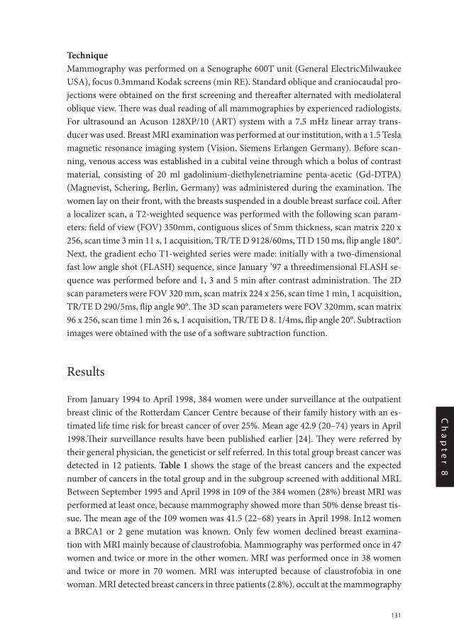
131
Ch
ap
te
r 8
TechniqueMammography was performed on a Senographe 600T unit (General ElectricMilwaukee USA), focus 0.3mmand Kodak screens (min RE). Standard oblique and craniocaudal pro-jections were obtained on the first screening and thereafter alternated with mediolateral oblique view. There was dual reading of all mammographies by experienced radiologists. For ultrasound an Acuson 128XP/10 (ART) system with a 7.5 mHz linear array trans-ducer was used. Breast MRI examination was performed at our institution, with a 1.5 Tesla magnetic resonance imaging system (Vision, Siemens Erlangen Germany). Before scan-ning, venous access was established in a cubital veine through which a bolus of contrast material, consisting of 20 ml gadolinium-diethylenetriamine penta-acetic (Gd-DTPA) (Magnevist, Schering, Berlin, Germany) was administered during the examination. The women lay on their front, with the breasts suspended in a double breast surface coil. After a localizer scan, a T2-weighted sequence was performed with the following scan param-eters: field of view (FOV) 350mm, contiguous slices of 5mm thickness, scan matrix 220 x 256, scan time 3 min 11 s, 1 acquisition, TR/TE D 9128/60ms, TI D 150 ms, flip angle 180°. Next, the gradient echo T1-weighted series were made: initially with a two-dimensional fast low angle shot (FLASH) sequence, since January ’97 a threedimensional FLASH se-quence was performed before and 1, 3 and 5 min after contrast administration. The 2D scan parameters were FOV 320 mm, scan matrix 224 x 256, scan time 1 min, 1 acquisition, TR/TE D 290/5ms, flip angle 90°. The 3D scan parameters were FOV 320mm, scan matrix 96 x 256, scan time 1 min 26 s, 1 acquisition, TR/TE D 8. 1/4ms, flip angle 20°. Subtraction images were obtained with the use of a software subtraction function.
Results
From January 1994 to April 1998, 384 women were under surveillance at the outpatient breast clinic of the Rotterdam Cancer Centre because of their family history with an es-timated life time risk for breast cancer of over 25%. Mean age 42.9 (20–74) years in April 1998.Their surveillance results have been published earlier [24]. They were referred by their general physician, the geneticist or self referred. In this total group breast cancer was detected in 12 patients. Table 1 shows the stage of the breast cancers and the expected number of cancers in the total group and in the subgroup screened with additional MRI. Between September 1995 and April 1998 in 109 of the 384 women (28%) breast MRI was performed at least once, because mammography showed more than 50% dense breast tis-sue. The mean age of the 109 women was 41.5 (22–68) years in April 1998. In12 women a BRCA1 or 2 gene mutation was known. Only few women declined breast examina-tion with MRI mainly because of claustrofobia. Mammography was performed once in 47 women and twice or more in the other women. MRI was performed once in 38 women and twice or more in 70 women. MRI was interupted because of claustrofobia in one woman. MRI detected breast cancers in three patients (2.8%), occult at the mammography
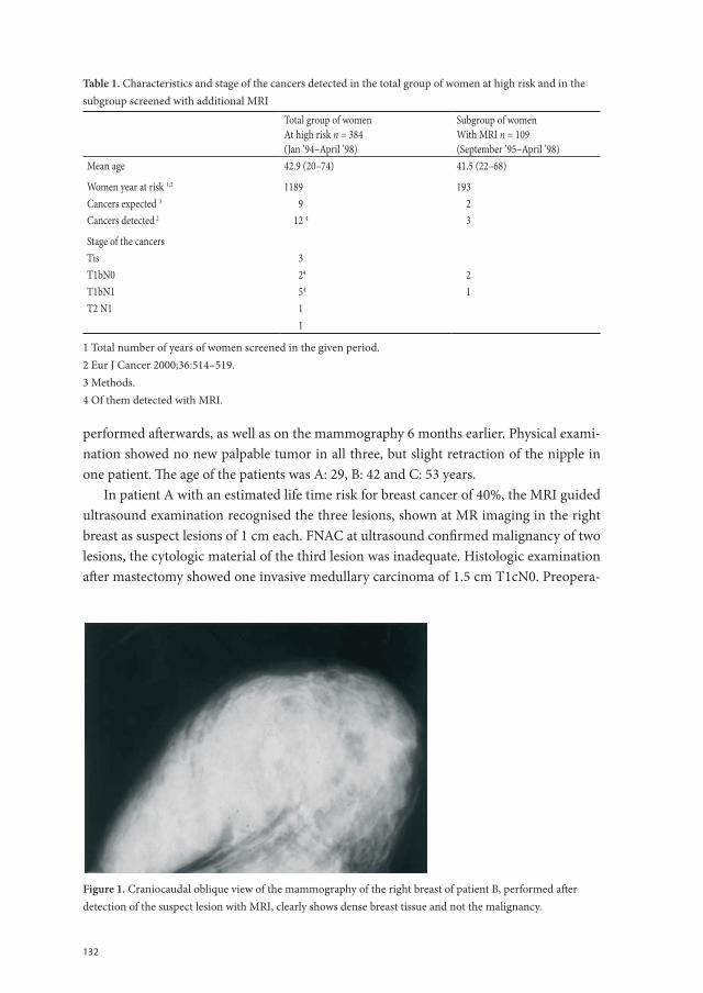
132
performed afterwards, as well as on the mammography 6 months earlier. Physical exami-nation showed no new palpable tumor in all three, but slight retraction of the nipple in one patient. The age of the patients was A: 29, B: 42 and C: 53 years.
In patient A with an estimated life time risk for breast cancer of 40%, the MRI guided ultrasound examination recognised the three lesions, shown at MR imaging in the right breast as suspect lesions of 1 cm each. FNAC at ultrasound confirmed malignancy of two lesions, the cytologic material of the third lesion was inadequate. Histologic examination after mastectomy showed one invasive medullary carcinoma of 1.5 cm T1cN0. Preopera-
Table 1. Characteristics and stage of the cancers detected in the total group of women at high risk and in the subgroup screened with additional MRI
Total group of womenAt high risk n = 384(Jan ’94–April ’98)
Subgroup of womenWith MRI n = 109(September ’95–April ’98)
Mean age 42.9 (20–74) 41.5 (22–68)
Women year at risk 1;2 1189 193Cancers expected 3 9 2Cancers detected 2 12 4 3
Stage of the cancersTis 3T1bN0 24 2T1bN1 54 1T2 N1 1
1
1 Total number of years of women screened in the given period.2 Eur J Cancer 2000;36:514–519.3 Methods.4 Of them detected with MRI.
Chapter 8 fig 1 Figure 1. Craniocaudal oblique view of the mammography of the right breast of patient B, performed after detection of the suspect lesion with MRI, clearly shows dense breast tissue and not the malignancy
Chapter 8 fig 1
Chapter 8 fig 2 Figure 2. The MRI image after substraction shows strong irregular enhancement dorsolateral in the right breast in patient B. Medial in the left breast a typical fibroadenoma.
Chapter 8 fig 2
Figure 1. Craniocaudal oblique view of the mammography of the right breast of patient B, performed after detection of the suspect lesion with MRI, clearly shows dense breast tissue and not the malignancy.
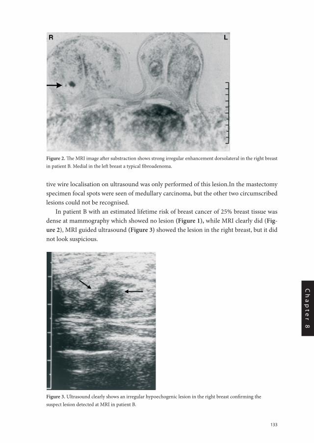
133
Ch
ap
te
r 8
tive wire localisation on ultrasound was only performed of this lesion.In the mastectomy specimen focal spots were seen of medullary carcinoma, but the other two circumscribed lesions could not be recognised.
In patient B with an estimated lifetime risk of breast cancer of 25% breast tissue was dense at mammography which showed no lesion (Figure 1), while MRI clearly did (Fig-ure 2), MRI guided ultrasound (Figure 3) showed the lesion in the right breast, but it did not look suspicious.
Chapter 8 fig 1 Figure 1. Craniocaudal oblique view of the mammography of the right breast of patient B, performed after detection of the suspect lesion with MRI, clearly shows dense breast tissue and not the malignancy
Chapter 8 fig 1
Chapter 8 fig 2 Figure 2. The MRI image after substraction shows strong irregular enhancement dorsolateral in the right breast in patient B. Medial in the left breast a typical fibroadenoma.
Chapter 8 fig 2 Figure 2. The MRI image after substraction shows strong irregular enhancement dorsolateral in the right breast in patient B. Medial in the left breast a typical fibroadenoma.
Chapt 8 fig 3
Figure 3. Ultrasound clearly shows an irregular hypoechogenic lesion in the right breast confirming the suspect lesion detected at MRI in patient B.Chapter 8 fig 3
Figure 3. Ultrasound clearly shows an irregular hypoechogenic lesion in the right breast confirming the suspect lesion detected at MRI in patient B.
134
FNAC, however, showed malignant cells. Histologic examination after ultrasound guided lumpectomy showed an adenocarcinoma of 1 cm T1bN0. The patient received breast conserving therapy. MRI performed during follow-up, showed a new lesion in the right breast 2 years later. At ultrasound guided FNAC it appeared to be recurrent breast cancer. In the mastectomy specimen it took great effort to find the tumor, which was not clearly palpable nor recognised at mammography of the slices. Histologic examination demonstrated an adenocarcinoma of 1.2x1.5 cm T1cN0.
In patient C with an estimated lifetime risk for breast cancer of 25% physical examination showed flattening of the nipple. MRI guided ultrasound recognised the lesion behind the left areola as suspicious. FNAC showed suspicious but not malignant cells. At histologic examination an adenocarcinoma of 1 cm with DCIS and LCIS was shown T1bN0.
We expected the incidence of two breast cancers in the 109 women screened with addi-tionalMRI, with 193 women year at risk. Breast MRI was false positive in six women. In four, physical examination, ultrasound and cytology showed no suspicion and follow-up continued. Local excision was performed in two patients – in one patient because the ul-trasound examination raised suspicion of the lesion indicated at MRI, in the other because clinical and cytological findings confirmed suspicion of the lesion, enhancing on MRI. Histologic examination showed atypia in one and no abnormality in the other patient. After 2.5 year of follow-up no malignancy was detected in either of them. After one year follow-up of the other 106 women screened with MRI, no breast cancers were detected at the regular screens with physical examination and mammography.
We had no false negative breast MRI results in the study period in this group. In four women with a proven BRCA1/2 gene-mutation, preventive mastectomy was performed after a period of follow-up. Only a small focus of lobular carcinoma in situ was detected in one. In a patient in whom excision was performed because of papillary cells at cytologic examination of nipple discharge, no abnormality was shown, confirming the negative MRI result. After closing this study all women who did not receive a negative BRCA1/BRCA2 test result continued follow-up according to the normal scheme, often with the yearly addition of MRI. No breast cancers were detected, missed on a breast MRI, made <1 year before.
Cost of added breast MRITo detect three breast cancers in the 109 women we performed 193 MRI examinations at €170; leading to 51 ultrasound examinations at €61; 29 FNAC at €127 and two benign excision biopsies at €1026. Bringing the additional detection cost at €13.930 per detect-ed patient. The total cost of MRI in the 109 women was €32.842 versus mammography €10.557.
135
Ch
ap
te
r 8
Discussion
It was only MRI, which detected three breast cancers in T1No stage, without a clinical manifest new tumor and occult at mammography during surveillance in the group of women screened with additional MRI. In the other 106 women screened with MRI no breast cancer was detected by palpation or mammography one year after closing this study. So breast MRI is likely to have advanced the detection of these three cancers. MRI is the most sensitive examination in the detection of invasive breast cancer. In women with axillary metastasis from a clinical and radiological occult breast cancer, breast MRI can detect these tumors and they can be occult also when mammography shows no dense breast tissue [21, 25, 26]. It may be useful to examine the value of MRI in high risk women with normal breast density also.
For DCIS, however, reported breast MRI sensitivity has been as low as 45% [27–29]. In young breast gene mutation carriers one would preferably detect breast cancer in its in situ stage, although this may mean overtreatment in an much older population.
For this reason it is not likely that MRI can replace mammography fully in a premeno-pausal screening population yet. Because of the reported low specifity of breast MRI (67–79%), it is necessary to confirm that a lesion is malignant, before deciding on operation. Reliable MRI guided needle biopsy was clinically not available in the Netherlands at the time of this study however. We could recognise the lesions detected with breast MRI at ul-trasound examination and confirm malignancy with FNAC. Follow-up showed no breast cancers, missed by not performing excisional biopsy if ultrasound and FNAC showed no suspicion of an onMRI enhancing lesion. The numbers are small though. Comparison with a previous MRI improves its specificity. It proved also useful if focal enhancement was seen on a MRI examination that was performed in the second half of the menstrual cycle to repeat the examination on the 5th–15th day. Focal enhancement, if caused by hormonal activity, then often disappears completely.
It is necessary to perform wire localisation of a breast lesion, detected by MRI, before operation, as it may be not palpable in the lumpectomy specimen nor recognisable at speci-men radiology. This is most likely the reason why in patient A one of the two lesions con-firmed at ultrasound guided FNAC could not be confirmed at histologic examination of the mastectomy specimen. The same problem occurred with the recurrent cancer in patient B.
At young age breast tissue is more sensitive to radiation than at older age. So especially for young breast genemutation carriers it may be an advantage if screening with breast MRI could partly replace mammography, despite the high cost. The addition of breast MRI to the surveillance scheme caused an extra cost of €13.930 per detected patient in our study, but because of the small numbers the estimation is not precise. This amount is considerable, compared to the €9000 that is spent for the early detection of one breast cancer patient in our national breast screening programme for women 50–70 years [30]. This makes investigations necessary for tailoring screening with MRI to whom would most benefit from it.
136
Recently in the Netherlands like in Great Britain and Germany, a multicentre trial with the two national cancer clinics and four university hospitals has started, to assess the cost effectiveness of screening with added breast MRI in women with different risk levels for breast cancer [31, 32].
During the follow-up of patients, with a positive family history in whom the first breast cancer was detected with MRI, MRI detected three carcinomas in this period; one contra-lateral T1aN0, two recurrent; one T1bN0 and one T1cN0 (patient B). It seems worthwhile to study the costeffectiveness of follow-up with breast MRI in these patients also.
Conclusion
In conclusion we think that breast MRI is promising in screening young women at high risk for breast cancer, as it can advance the detection of cancers while they are still occult at mammography and not yet clinically manifest. MRI may be useful also in the follow-up of breast cancer patients detected primarily at MRI. Further study is needed to determine whether breast MRI can (partly) replace mammography in screening these women and at what cost.
137
Ch
ap
te
r 8
References
1. Vasen HFA, Haites NE, Evans DGR, Steel CM, Moeller P, Hodgson S: Cuurrent policies for surveillance and management in women at risk of breast and ovarian cancer: a survey among 16 European family cancer clinics. Eur J Cancer 34(12): 1922–1926, 1998
2. SA Feig: Assessment of radiation risk from screening mammography. Cancer 77(5): 818–822, 1996
3. Mettler FA, Upton AC, Kelsey CA, Ashby RN, Rosenberg RD, Linver MN: Benefits versus risks from mammography: a critical reassesment. Cancer 77:903–909, 1996
4. Beemsterboer PMM, Warmerdam PG, Boer R, Koning HJ: Radiation risk of mammography related to benefit in screening programmes: a favourable balance? J Med Screen 5:81–87, 1998
5. Leeuwen FEvan, Klokman WJ, Hagenbeek A, Noyon R, van den Belt-Dusebout AW, Kerkhoff EH, Heerde P van, Somers R: Second cancer risk following Hodgkins disease: a 20-year follow-up study. J Clin Oncol 12(2): 312–325, 1994
6. O’Brien PC, Barton MB, Fisher R: Breast cancer following treatment for Hodgkins disease: the need for screening in a young population. Australas Radiol 39(3): 271–276, 1995
7. Boice JDJ, Land CE, Shore RE, Norman JE, Tokunaja M: Risk of breast cancer following low-dose radia-tion exposure. Radiology 131:589–597, 1979
8. Peer PGM, Verbeek ALM, Straatman H, Hendriks JHCL, Holland R: Age-specific sensitivities of mam-mographic screening for breast cancer. Breast Cancer Res Treat 38:153–160, 1996
9. Show de Paredes E, Marstellen LP, Eden BV: Breast cancers in women 35 years of age and younger mam-mographic findings. Radiology 177(1): 117–119, 1990
10. Morrow M: Identification and management of the women at increased risk for breastcancer development. Breast Cancer Res Treat 31:53–60, 1994
11. Stomper PC, D’Souza DJ, DiNitto PA, Arredondo MA: Analysis of parenchymal density on mammograms in 1353 women 25–79-year-old. Am J Roentgenol 167:1261–1265, 1996
12. Lehman CD, White E, Peacock S, Drucker MJ, Urban N: Effect of age and breast density on screening mammograms with false- positive findings. AJR 173:1651–1655, 1999
13. Kolb TM, Lichy J, Newhouse JH: Occult cancer in women with dense breasts: detection with screening US diagnostic yield and tumor chracteristics. Radiology 207; 191–199, 1998
14. Zonderland HM, Coerkamp EG, Hermans J, van de Vijver M, van Voorthuisen AE: Diagnosis of breast cancer: contribution of US as an adjunct to mammography. Radiology 213:413–422, 1999
15. Kerlikowske K, Grady D, Barclay J, Sickles EA, Enser V: Effect of age, breast density, and family history on the sensitivity of first screening mammography. JAMA 276:33–38, 1996
16. Bone B, Pentek Z, Perbeck L, Veress B: Diagnostic accuracy of mammography and contrast-enhanced MR Imaging in 238 histologically verified breast lesions. Acta Radiol 38:489–496, 1997
17. Heywang SH, Wolf A, Pruss E, Hilbertz ET, Eiermann W, Permanetter W: MR imaging of the breast with Gd DTPA: Use and limitations. Radiology 171:95–103, 1989
18. Obdeijn AIM, Kuijpers TJA, Dijk van P, Wiggers T, Oudkerk M: MR lesion detection in a breast cancer population. J Magn Reson Imaging 6:849–854, 1996
19. Heywang-Koebrunner SH, Viehweg P, Heinig A, Kuechler Ch: Contrast-enhanced MRI of the breast: ac-curacy, value, controversies, solutions. Eur J Radiol 24:94–108, 1997
20. Obdeijn AIM, Brouwers-Kuyper EMJ, Tilanus-Linthorst MMA, Wiggers Th, Oudkerk M: MR imaging guided sonography followed by fine-needle aspiration cytology in occult carcinoma of the breast. AJR 174:1079–1084, 2000

138
21. Tilanus-Linthorst MMA, Obdeijn AIM, Bontenbal M, Oudkerk M: MRI in patients with axillary metasta-ses of occult breast carcinoma. Breast Cancer Res Treat 44:179–182, 1997
22. Claus EB, Risch N, Thompson WD. Autosomal dominant inheritance of autosomal breast cancer. Cancer 73:643–651, 1994
23. Kopans DB, Dórsi CJ, Adler DD et al.: Breast Imaging Reporting and Data System. Reston VA, American College of Radiology, 1993
24. Tilanus-Linthorst MMA, Bartels CCM, Obdeijn AIM, Oudkerk M: Earlier detection of breast cancer by surveillance of women at familial risk. Eur J Cancer 36:514–519, 2000
25. Morris EA, Schwartz LH, Dershaw DD, Van Zee KJ, Abramson AF, Liberman L: MR imaging of the breast in patients with occult primary breast carcinoma Radiology 205:437–440, 1997
26. Porter BA, Seattle WA, Smith JP, Borrow JP: MR depiction of occult breast cancer in patients with malig-nant axillary adenopathy. Radiology 197(P): 130, 1995
27. Westerhof JP, FischerU, Moritz JD, Oestmann JW: MR imaging of mammographically detected clustered microcalcifications: Is there any value? Radiology 207:675–681, 1998
28. Gilles R, Zafrani B, Guinebretiere JM, Meunier M, Lucidarme O, Tardivon AA, Rochard F, Vanel D, Neuenschwander S, Arriagada R: Ductal carcinoma in situ: MR Imaging histopathologic correlation. Radiology 196:415–419, 1995
29. Picolli CW, Matteucci T, Outwater EK, Siegelman ES, Mitchell DG: Breast cancer diagnosis with MRim-aging: effect of clinical and mammographic findings on recommendations for biopsy. Radiology 197:52, 1995
30. de Koning HJ, van Ineveld BM, van Ootmarssen GJ, de Haes JC, Collette HJ, Hendriks JH, van der Maas PJ:. Breast cancer screening and cost-effectiveness; policy alternatives, quality of live considerations and the possible impact of uncertain factors. Int J Cancer 49:531–537, 1991
31. Bick U: An integrated early detection concept in women with a genetic predisposition for breast cancer. Radiology 37(8):591–596, 1997
32. Brown J, Coulthard A, Dixon K: Protocol for a national multicentre study of magnetic resonance imaging screening in women at genetic risk of breast cancer. Breast 9:78–82, 2000

Chapter 9
Selection bias influences reported contralateral breast cancer incidence and survival in high risk non-BRCA1/2 patients
Madeleine MA Tilanus-Linthorst, Karina CM Bartels, Celina Alves, Bonnie Bakri, Ellen Crepin, Ans van den Ouweland, Jan GM Klijn, Alexander M Eggermont, Hanne Meijers-Heijboer, Cecile TM Brekelmans
Breast Cancer Research and Treatment 2006;95 (2):117-123.
140
Abstract
PurposeThe results of studies comparing survival in familial and sporadic breast cancer (BC) can-cer are inconsistent. A higher incidence of contralateral breast cancer (CBC) has been reported in familial BC. Ascertainment bias may influence both the reported familial CBC and survival
DesignWe assessed CBC incidence, distant disease free (DDFS) and overall survival (OS) in 327 BC patients who had ≥ 3 breast and/or ovarian cancers in the family but no BRCA1/2 gene mutation (non-BRCA1/2). They were matched to 327 sporadic controls for year and age at detection. To correct for ascertainment bias, we analyzed also separately the results 1) of the 250 non-BRCA1/2 patients with DNA testing performed before diagnosis or within two years (“unselected”) and 2) of the 77 with testing ≥ 2 years after diagnosis (late-tested).
ResultsMedian follow-up of non-BRCA1/2 patients was 6.1 yrs. Ten years CBC incidence was 11% in non-BRCA1/2 vs. 6% in sporadic patients (p=0.002). At multivariate analysis CBC incidence was increased in late-tested non-BRCA1/2 (HR 4.6 p=0.001) not in “unselected” (HR 1.8 p=0.1). Increased CBC occurred in non-BRCA1/2 patients mainly before genetic testing, suggesting ascertainment bias. Tumors were ≤ T1 in 62% of non-BRCA1/2 vs. 50% of sporadic patients (p= 0.003), node-negative in 55% vs. 52% respectively (p=0.5). After correction for stage and therapy, OS did not differ between “unselected” non-BRCA1/2 and sporadic patients (HR 0.8; p=0.3), but was improved in late-tested non-BRCA1/2.
Conclusion Overall survival and contralateral BC were similar in “unselected” non-BRCA1/2 - and sporadic patients. Reports of higher CBC incidence and better survival in non-BRCA1/2 patients may substantially be caused by DNA-testing selection-bias.
141
Ch
ap
te
r 9
Introduction
Family history and age are the strongest risk factors for both primary and contralateral breast cancer in women [1-3].
Approximately 10% of breast cancers (BC) are detected in patients with a clear family history, but high-penetrance germ-line mutations in BRCA1 or BRCA2 account for less than 20% of the familial aggregation of breast cancer.
The risk of chance clustering of ≥ 3 breast cancers under the age of 60 in a family has been estimated as less than 10% [4]. Further, specific histopathologic characteristics have been described in non-BRCA1/2 breast cancers from families with at least 3 breast cancer cases, such as more frequent low-grade tumors, low mitotic count, a lower proliferation rate and more lobular carcinoma [5,6]. These features discriminate non-BRCA1/2 from both BRCA1 BRCA2 and sporadic breast cancers and suggest a possibly more favorable prognosis.
The data on survival of familial cancer, reviewed by Chappuis and summarized by Haffty however, are inconsistent [7,8], while Eerola did not find a difference in survival between 284 proven non-BRCA1/2 breast cancer patients from families with ≥ 3 breast cancer cases and 59.517 sporadic BC patients not matched for age [9].
A higher frequency of contralateral breast cancer (CBC) has been reported in patients from high risk families, also when proven BRCA1/2 negative, than in sporadic patients
and also in patients with a CHEK 2*1100delC mutation [9-19]. However DNA-testing may have been offered with preference to patients with bilateral BC.
In order to compare CBC incidence and survival in familial and sporadic breast cancer we assessed the incidence of CBC, distant disease free survival (DDFS) and overall sur-vival (OS) in two groups:
1. BC patients with ≥ 3 breast and/or ovarian cancers in the family but a negative test for deleterious BRCA1/2 gene mutations (non-BRCA1/2) and
2. control patients with sporadic BC matched for year and age at detection.To estimate the influence of ascertainment bias (preferential DNA-testing in longer living patients or patients with CBC) on the results, we performed all the analyses both for the total group of non-BRCA1/2 patients and also separately for patients whit DNA testing ≥ 2yr after diagnosis (late-tested) and the other non-BRCA1/2 patients (“unselected”),
Patients and Methods
Study subjectsFamilies were identified through the Rotterdam Family Cancer Clinic registration at Erasmus Medical Centre. The series of 292 families eligible for this study consisted of consecutive families with at least 3 confirmed breast cancer cases (“hereditary breast can-cer “/HBC) or breast and ovarian cancers (HBOC) including the index case, but with a
142
negative test for a BRCA1 or BRCA2 mutation before May 1 2004. We selected from these families all 350 women with primary breast cancer (including DCIS), who had been diag-nosed between 1-1-1980 and 31-12-2002 and available data on histopathology, follow-up data for at least 6 months and no previous cancer other than basal skin carcinoma. Nine patients were not eligible because a pathology report was lacking, 6 because follow-up data was lacking, 7 because the family member, who tested negative for BRCA1/2 did not have breast or ovarian cancer and 1 had lobular carcinoma in situ. This left 327 non-BRCA1/2 patients from 265 families for analyses. Of these 262 tested negative for BRCA1 and BRCA2, while in 65 patients one or more family members with breast or ovarian cancer tested negative.
Test results for CHEK 2*1100delC were available in 117 familial patients and positive in one. BRCA1/2 and CHEK2*1100delC mutation analyses were reported [10].
To each non-BRCA1/2 patient a control BC patient (sporadic) was matched for age and year of diagnosis (within 5 years). Excluded were control patients with a family his-tory of more than one family member with BC > age 50 yrs.. Other eligibility criteria were in conformity with the non-BRCA1/2 patients.
Study protocolWe extracted detailed information on family history, age at diagnosis, hormonal factors (e.g. menopausal status), tumor characteristics (diameter, morphology, Bloom-Richardson grade) axillary lymph node status, surgical and adjuvant systemic treatment (hormonal and / or chemotherapy) of non-BRCA1/2 and sporadic BC patients from the medical files. Whenever possible (93%) each breast cancer was reclassified according to TNM classifica-tion version 6 (2002), for the others we used the TNM classification at diagnoses. Further it was registered if and when women underwent prophylactic contralateral mastectomy or bilateral (salpingo-)oophorectomy (BSO) as wel as local and distant recurrences.
For the purpose of the analyses follow-up was assumed to commence on the date of detection of the first breast cancer and to cease on the date of the last notes in the medical files, death, or otherwise at loss to follow-up, whichever came first.
Cancer in the contralateral breast was considered metachronous if detected more than 3 month’s after the first, also after primary DCIS.
Endpoints of interest were the occurrence and date of contralateral breast cancer (CBC), distant metastases and date of death due to breast cancer or other cause. Census for the follow-up period was 1 May, 2004.
Ascertainment bias in non-BRCA1/2 patientsTo investigate the preferential selection of non-BRCA1/2 patients with long survival or CBC and investigate its impact, we created two separate subgroups 1. index patients in whom DNA-testing was performed more than 2 years after breast cancer diagnosis, here-after called late-tested (n= 77) and 2. all other non-BRCA1/2 breast cancer patients here-after called the “unselected” non-BRCA1/2 (n=250). The latter group consisted of 185
143
Ch
ap
te
r 9
patients tested for DNA either before diagnosis or within 2 years (103 of them probands) and 65 patients with DNA tested in family members with breast -ovarian cancers only.
To assess the magnitude of the remaining bias in the “unselected” non-BRCA1/2 group by including the probands we repeated the analyses, combining all probands with the late-tested non-BRCA1/2 sub-group. Further separate analyses were performed for HBC and HBOC families.
Statistical methodsUsing chi-square tests (categorical variables) or t-tests (continuous variables) we com-pared patient and tumor characteristics between the non-BRCA1/2 patients (both in total and separated in the subgroups defined above) and sporadic breast cancer patients.
Kaplan-Meier survival curves were calculated and differences tested by the logrank test. Endpoints were the incidence of contralateral breast cancer, local recurrence, distant disease-free survival and overall survival.
The simultaneous effect of several prognostic variables on these four endpoints was investigated by Cox proportional hazard regression models.
A p-value of less than 0.05 was considered statistically significant. All analyses were performed by STATA/SE for Windows version 8.1. A two-sided P-value of less than 0.05 was considered to indicate statistical significance.
The study was approved by the Erasmus MC IRB. All DNA-tested patients gave their in-formed consent on all DNA analyses and their use for research.
Results
Patient and tumor characteristicsA hereditary breast cancer syndrome (HBC) was seen in 214 of the 265 families, and a hereditary breast and ovarian cancer pattern (HBOC) in 51. In 43% of the families there was one or more BC patient < 40 yrs., in 66% there was ≥ 1 BC patient between 40-50 yrs, and in 94% ≥ 1 BC patient > 50 yrs. Patient, tumor and treatment characteristics are described in Table 1.
Median follow-up was 6.1 years in the total non-BRCA1/2 group, but significantly longer in late-tested than “unselected” non-BRCA1/2 patients (9.9 vs. 4.9 respectively; p<0.001). Non-BRCA1/2 and sporadic patients were matched for age, but late-tested non-BRCA1/2 patients were significantly younger than ”unselected”, mean age 43 vs. 47 yrs. respectively (p=0.002) and 83% vs. 60% respectively younger than ≤ 50 yrs (p=0.001). Seventy-nine % of the non-BRCA1/2 group were diagnosed after 1990, but only 31% late-tested vs. 60% “unselected” after 1995 (p< 0.001). Tumors were smaller in non-BRCA1/2 patients than in sporadic patients (p 0.003), nodal status was comparable and there was no difference in stage between late-tested and “unselected”non-BRCA1/2 patients. Tumors of
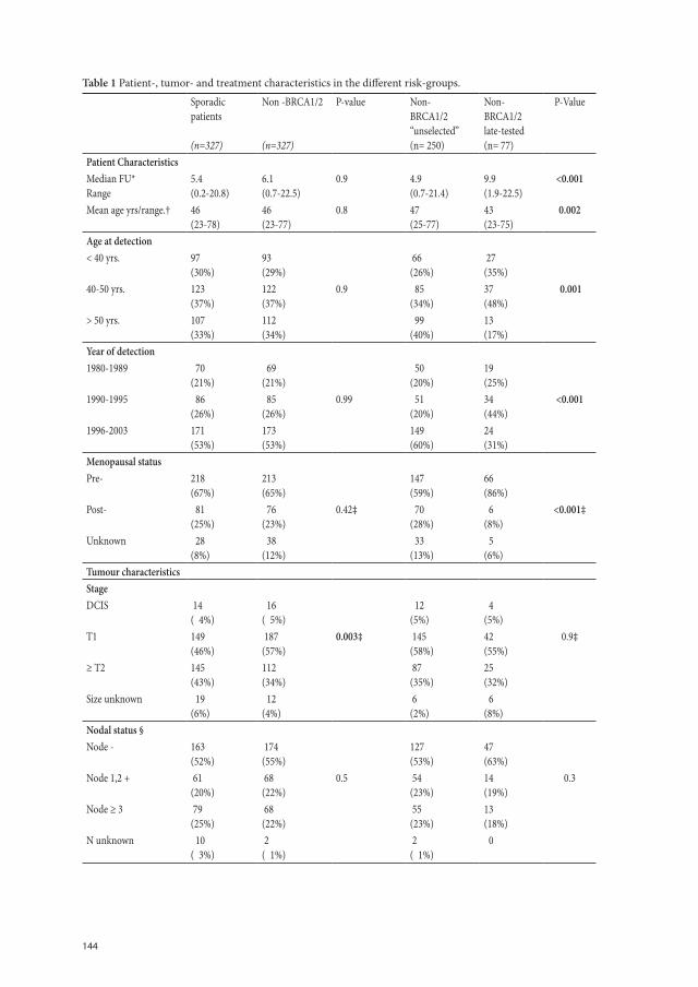
144
Table 1 Patient-, tumor- and treatment characteristics in the different risk-groups.
Sporadicpatients
(n=327)
Non -BRCA1/2
(n=327)
P-value Non-BRCA1/2“unselected”(n= 250)
Non-BRCA1/2late-tested(n= 77)
P-Value
Patient CharacteristicsMedian FU*Range
5.4(0.2-20.8)
6.1 (0.7-22.5)
0.9 4.9(0.7-21.4)
9.9(1.9-22.5)
<0.001
Mean age yrs/range.† 46(23-78)
46 (23-77)
0.8 47(25-77)
43(23-75)
0.002
Age at detection< 40 yrs. 97
(30%)93(29%)
66(26%)
27(35%)
40-50 yrs. 123 (37%)
122 (37%)
0.9 85(34%)
37(48%)
0.001
> 50 yrs. 107 (33%)
112 (34%)
99(40%)
13(17%)
Year of detection1980-1989 70
(21%) 69 (21%)
50(20%)
19(25%)
1990-1995 86(26%)
85(26%)
0.99 51(20%)
34(44%)
<0.001
1996-2003 171(53%)
173(53%)
149(60%)
24(31%)
Menopausal statusPre- 218
(67%)213(65%)
147(59%)
66(86%)
Post- 81(25%)
76 (23%)
0.42‡ 70(28%)
6(8%)
<0.001‡
Unknown 28 (8%)
38 (12%)
33(13%)
5(6%)
Tumour characteristicsStageDCIS 14
( 4%) 16 ( 5%)
12(5%)
4(5%)
T1 149(46%)
187 (57%)
0.003‡ 145(58%)
42(55%)
0.9‡
≥ T2 145 (43%)
112 (34%)
87(35%)
25(32%)
Size unknown 19 (6%)
12 (4%)
6(2%)
6(8%)
Nodal status §Node - 163
(52%) 174 (55%)
127(53%)
47(63%)
Node 1,2 + 61 (20%)
68 (22%)
0.5 54(23%)
14(19%)
0.3
Node ≥ 3 79 (25%)
68 (22%)
55(23%)
13(18%)
N unknown 10 ( 3%)
2( 1%)
2( 1%)
0
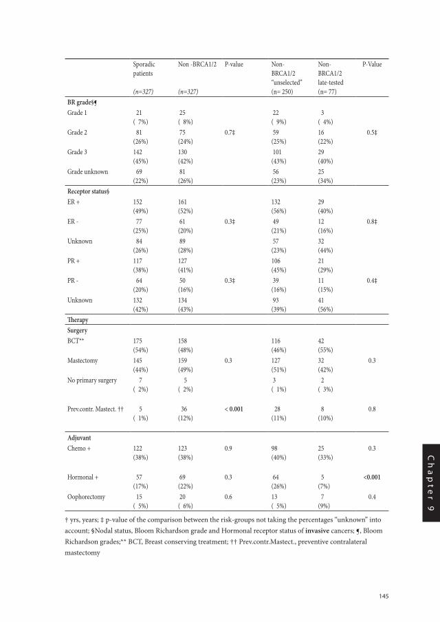
145
Ch
ap
te
r 9
Sporadicpatients
(n=327)
Non -BRCA1/2
(n=327)
P-value Non-BRCA1/2“unselected”(n= 250)
Non-BRCA1/2late-tested(n= 77)
P-Value
BR grade§¶ Grade 1 21
( 7%) 25 ( 8%)
22( 9%)
3( 4%)
Grade 2 81 (26%)
75 (24%)
0.7‡ 59(25%)
16(22%)
0.5‡
Grade 3 142 (45%)
130 (42%)
101(43%)
29(40%)
Grade unknown 69 (22%)
81 (26%)
56(23%)
25(34%)
Receptor status§ ER + 152
(49%)161(52%)
132(56%)
29(40%)
ER - 77 (25%)
61 (20%)
0.3‡ 49(21%)
12(16%)
0.8‡
Unknown 84 (26%)
89 (28%)
57(23%)
32(44%)
PR + 117 (38%)
127 (41%)
106(45%)
21(29%)
PR - 64(20%)
50 (16%)
0.3‡ 39(16%)
11(15%)
0.4‡
Unknown 132 (42%)
134 (43%)
93(39%)
41(56%)
TherapySurgeryBCT** 175
(54%)158 (48%)
116(46%)
42(55%)
Mastectomy 145 (44%)
159 (49%)
0.3 127(51%)
32(42%)
0.3
No primary surgery 7 ( 2%)
5 ( 2%)
3( 1%)
2( 3%)
Prev.contr. Mastect. †† 5 ( 1%)
36 (12%)
< 0.001 28(11%)
8(10%)
0.8
Adjuvant Chemo + 122
(38%)123 (38%)
0.9 98(40%)
25(33%)
0.3
Hormonal + 57(17%)
69 (22%)
0.3 64(26%)
5(7%)
<0.001
Oophorectomy 15 ( 5%)
20 ( 6%)
0.6 13( 5%)
7(9%)
0.4
† yrs, years; ‡ p-value of the comparison between the risk-groups not taking the percentages “unknown” into account; §Nodal status, Bloom Richardson grade and Hormonal receptor status of invasive cancers; ¶, Bloom Richardson grades;** BCT, Breast conserving treatment; †† Prev.contr.Mastect., preventive contralateral mastectomy
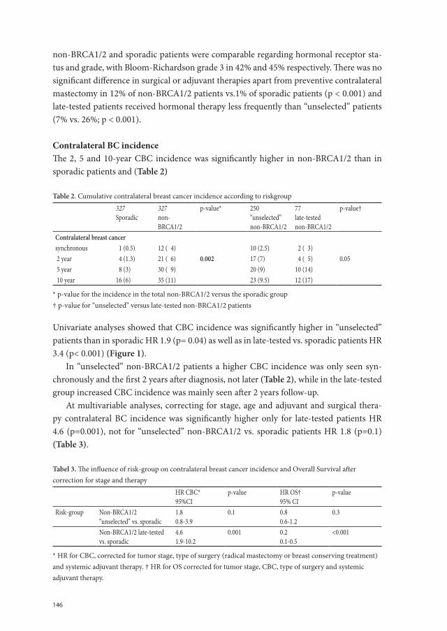
146
non-BRCA1/2 and sporadic patients were comparable regarding hormonal receptor sta-tus and grade, with Bloom-Richardson grade 3 in 42% and 45% respectively. There was no significant difference in surgical or adjuvant therapies apart from preventive contralateral mastectomy in 12% of non-BRCA1/2 patients vs.1% of sporadic patients (p < 0.001) and late-tested patients received hormonal therapy less frequently than “unselected” patients (7% vs. 26%; p < 0.001).
Contralateral BC incidence The 2, 5 and 10-year CBC incidence was significantly higher in non-BRCA1/2 than in sporadic patients and (Table 2)
Table 2. Cumulative contralateral breast cancer incidence according to riskgroup
327Sporadic
327non-BRCA1/2
p-value* 250“unselected”non-BRCA1/2
77late-testednon-BRCA1/2
p-value†
Contralateral breast cancer synchronous 1 (0.5) 12 ( 4) 10 (2.5) 2 ( 3) 2 year 4 (1.3) 21 ( 6) 0.002 17 (7) 4 ( 5) 0.05 5 year 8 (3) 30 ( 9) 20 (9) 10 (14) 10 year 16 (6) 35 (11) 23 (9.5) 12 (17)
* p-value for the incidence in the total non-BRCA1/2 versus the sporadic group † p-value for “unselected” versus late-tested non-BRCA1/2 patients
Univariate analyses showed that CBC incidence was significantly higher in “unselected” patients than in sporadic HR 1.9 (p= 0.04) as well as in late-tested vs. sporadic patients HR 3.4 (p< 0.001) (Figure 1).
In “unselected” non-BRCA1/2 patients a higher CBC incidence was only seen syn-chronously and the first 2 years after diagnosis, not later (Table 2), while in the late-tested group increased CBC incidence was mainly seen after 2 years follow-up.
At multivariable analyses, correcting for stage, age and adjuvant and surgical thera-py contralateral BC incidence was significantly higher only for late-tested patients HR 4.6 (p=0.001), not for “unselected” non-BRCA1/2 vs. sporadic patients HR 1.8 (p=0.1) (Table 3).
Tabel 3. The influence of risk-group on contralateral breast cancer incidence and Overall Survival after correction for stage and therapy
HR CBC*95%CI
p-value HR OS†95% CI
p-value
Risk-group Non-BRCA1/2 “unselected” vs. sporadic
1.80.8-3.9
0.1 0.80.6-1.2
0.3
Non-BRCA1/2 late-tested vs. sporadic
4.61.9-10.2
0.001 0.20.1-0.5
<0.001
* HR for CBC, corrected for tumor stage, type of surgery (radical mastectomy or breast conserving treatment) and systemic adjuvant therapy. † HR for OS corrected for tumor stage, CBC, type of surgery and systemic adjuvant therapy.
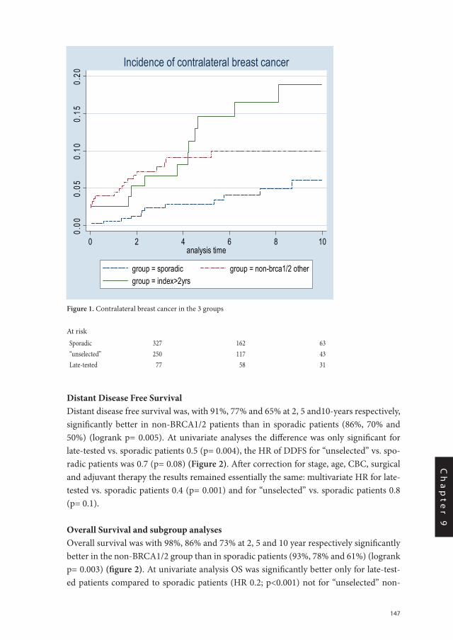
147
Ch
ap
te
r 9
Distant Disease Free SurvivalDistant disease free survival was, with 91%, 77% and 65% at 2, 5 and10-years respectively, significantly better in non-BRCA1/2 patients than in sporadic patients (86%, 70% and 50%) (logrank p= 0.005). At univariate analyses the difference was only significant for late-tested vs. sporadic patients 0.5 (p= 0.004), the HR of DDFS for “unselected” vs. spo-radic patients was 0.7 (p= 0.08) (Figure 2). After correction for stage, age, CBC, surgical and adjuvant therapy the results remained essentially the same: multivariate HR for late-tested vs. sporadic patients 0.4 (p= 0.001) and for “unselected” vs. sporadic patients 0.8 (p= 0.1).
Overall Survival and subgroup analysesOverall survival was with 98%, 86% and 73% at 2, 5 and 10 year respectively significantly better in the non-BRCA1/2 group than in sporadic patients (93%, 78% and 61%) (logrank p= 0.003) (figure 2). At univariate analysis OS was significantly better only for late-test-ed patients compared to sporadic patients (HR 0.2; p<0.001) not for “unselected” non-
Chapter 9 Fig 1Figure 1 Contralateral breast cancer in the 3 groups
0.00
0.05
0.10
0.15
0.20
0 2 4 6 8 10analysis time
group = sporadic group = non-brca1/2 othergroup = index>2yrs
Incidence of contralateral breast cancer
At risk Sporadic 327 162 63 “unselected” 250 117 43 Late-tested 77 58 31 Chapt 9/ fig 1
Figure 1. Contralateral breast cancer in the 3 groups
At riskSporadic 327 162 63“unselected” 250 117 43Late-tested 77 58 31
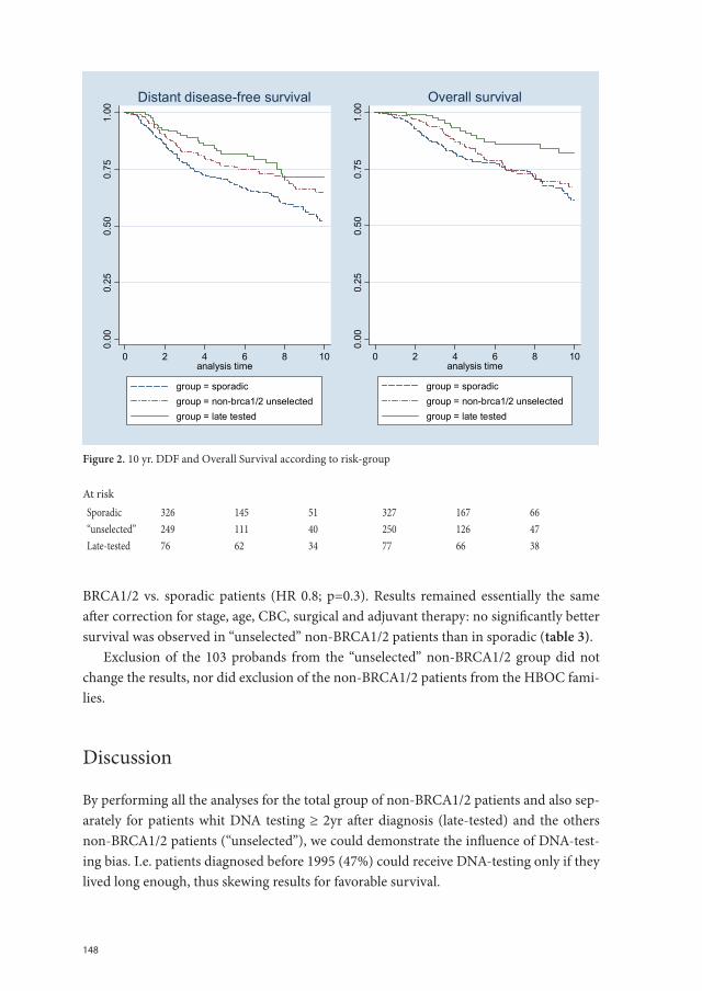
148
BRCA1/2 vs. sporadic patients (HR 0.8; p=0.3). Results remained essentially the same after correction for stage, age, CBC, surgical and adjuvant therapy: no significantly better survival was observed in “unselected” non-BRCA1/2 patients than in sporadic (table 3).
Exclusion of the 103 probands from the “unselected” non-BRCA1/2 group did not change the results, nor did exclusion of the non-BRCA1/2 patients from the HBOC fami-lies.
Discussion
By performing all the analyses for the total group of non-BRCA1/2 patients and also sep-arately for patients whit DNA testing ≥ 2yr after diagnosis (late-tested) and the others non-BRCA1/2 patients (“unselected”), we could demonstrate the influence of DNA-test-ing bias. I.e. patients diagnosed before 1995 (47%) could receive DNA-testing only if they lived long enough, thus skewing results for favorable survival.
Chapt 9/fig 2 Fig 2. 10 yr. DDF and Overall Survival according to risk-group
0.00
0.25
0.50
0.75
1.00
0 2 4 6 8 10analysis time
group = sporadicgroup = non-brca1/2 unselectedgroup = late tested
Distant disease-free survival
0.00
0.25
0.50
0.75
1.00
0 2 4 6 8 10analysis time
group = sporadicgroup = non-brca1/2 unselectedgroup = late tested
Overall survival
At risk Sporadic 326 145 51 327 167 66 “unselected” 249 111 40 250 126 47 Late-tested 76 62 34 77 66 38
Figure 2. 10 yr. DDF and Overall Survival according to risk-group
At riskSporadic 326 145 51 327 167 66“unselected” 249 111 40 250 126 47Late-tested 76 62 34 77 66 38
149
Ch
ap
te
r 9
Both distant disease free survival and overall survival were significantly better in non-BRCA1/2 patients than in sporadic patients (DDFS p= 0.005 and OS p= 0.003 at the lo-grank test). However this improved DDFS and OS reached significance at univariate anal-yses only in the late-tested non-BRCA1/2 patients, who had been selected at least partly for longevity, but not in the “unselected” non-BRCA1/2 (HR DDFS 0.8; p=0.1 and HR OS 0.8; p= 0.3). These results remained essentially the same after correction for stage, CBC and therapy (Table 3). This underlines the importance of adequate correction for longev-ity bias in family based studies like ours. The favorable survival in non-BRCA1/2 patients who received DNA-testing after 2 years of diagnosis, confirms also the prognostic im-portance of surviving the first 2 years. Our survival results in “unselected”non-BRCA1/2 patients are in line with Eerola et al., Eccles et al., while Möller et al. and Hamann et al. had no sporadic control group (Table 4) [9,16,17,20].
The 2 groups of non-BRCA1/2 patients also gave better insight in the influence of se-lection for DNA-testing on reported CBC incidence. In both non-BRCA1/2 subgroups the higher CBC incidence occurred mainly before the date of genetic testing. In “unselected” non-BRCA1/2 patients CBC incidence was increased before genetic testing only. In our younger late-tested group one expects the growth rate of the tumors to be faster and CBC to appear earlier [21]. However CBC was seen mainly after 2 years (table 2). The cause may be, that patients and family members may be more likely to seek and get genetic test-ing after bilateral breast cancer. In our study this may largely explain not only the high synchronous CBC incidence in all non-BRCA1/2 patients but also the early increase in metachronous CBC in the “unselected” group and later increase in the late-tested group.
In studies of familial breast cancer performed before DNA testing was available, CBC incidence is increased in some, but not in others [12-14]. This inconsistency may be explained by a different percentage of BRCA1/2 mutation carriers in the various stud-ies, as BRCA1/2 and CHEK2*1100delC mutation carriers with breast cancer have a RR of CBC of 2-6 [18,22]. To some extent the studies that assess CBC risk in patients with BRCA1/2-negative familial cancer have, like our own, selected the DNA tested cases for CBC incidence [9.16-18,22] table 4. Therefore, the higher rates of CBC may be substan-tially due to selection bias in these studies also. We need population-based studies with complete BRCA1/2 testing and pedigree information to assess the real CBC incidence in non-BRCA1/2 familial breast cancer.
With a follow-up of 6.1 yrs in non-BRCA1/2 and 5.4 yrs in sporadic patients our study provided extensive DNA testing in the familial group and fairly complete information on the pedigree, tumor stage, tumor characteristics and on therapy in both familial and spo-radic patients. The relatively high percentage (70%) of non-BRCA1/2 patients diagnosed before age 50 in our study, may reflect that DNA testing is offered to young patients more than to older patients. In Eerola’s study for instance, 70% of the cancers in BRCA1/2 nega-tive families with 3 or more BC cases were diagnosed above age 50 yrs [9]. The relatively young age at detection in our non-BRCA1/2 cases might be responsible for the higher
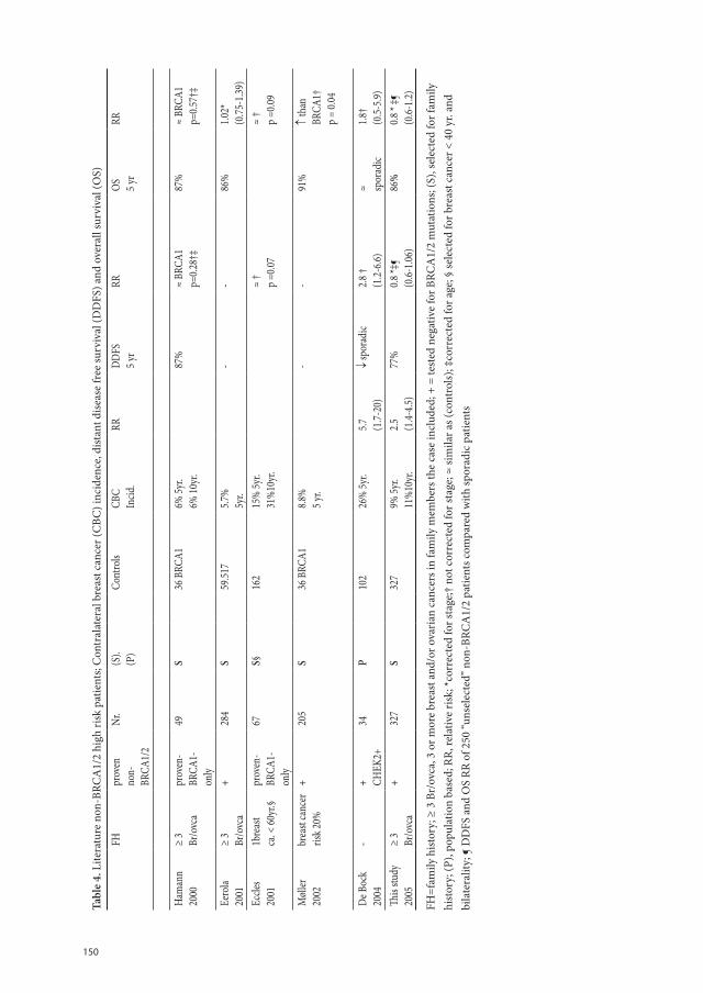
150
Tabl
e 4.
Lite
ratu
re n
on-B
RCA
1/2
high
risk
pat
ient
s; C
ontr
alat
eral
bre
ast c
ance
r (C
BC) i
ncid
ence
, dist
ant d
isea
se fr
ee su
rviv
al (D
DFS
) and
ove
rall
surv
ival
(OS)
FHpr
oven
non-
BRCA
1/2
Nr.
(S).
(P)
Cont
rols
CBC
Incid
.RR
DDFS
5 yr
RROS 5 y
rRR
Ham
ann
2000
≥ 3
Br/o
vca
prov
en-
BRCA
1-on
ly
49S
36 B
RCA1
6% 5y
r.6%
10yr
.87
%
≈ BR
CA1
p=0.2
8†‡
87%
≈
BRCA
1p=
0.57†
‡
Eero
la20
01≥
3Br
/ovc
a+
284
S59
.517
5.7%
5yr.
--
86%
1.02*
(0.75
-1.39
)Ec
cles
2001
1bre
ast
ca. <
60yr
.§pr
oven
-BR
CA1-
only
67S§
162
15%
5yr.
31%
10yr
.≈
†p =
0.07
≈ †
p =0.0
9
Møl
ler20
02br
east
canc
er
risk 2
0%+
205
S36
BRC
A18.8
%5 y
r.-
-91
%
↑ th
an
BRCA
1†p =
0.04
De B
ock
2004
-+ CH
EK2+
34P
102
26%
5yr.
5.7 (1.7-
20)
↓ sp
orad
ic2.8
†(1
.2-6.6
)≈ sp
orad
ic1.8
†(0
.5-5.9
)Th
is stu
dy20
05≥
3Br
/ovc
a+
327
S32
79%
5yr.
11%
10yr
.2.5 (1
.4-4.5
)77
%
0.8 *‡
¶(0
.6-1.0
6)86
%
0.8 *
‡¶(0
.6-1.2
)
FH=f
amily
hist
ory;
≥ 3
Br/
ovca
, 3 o
r mor
e br
east
and
/or o
vari
an c
ance
rs in
fam
ily m
embe
rs th
e ca
se in
clud
ed; +
= te
sted
neg
ativ
e fo
r BRC
A1/
2 m
utat
ions
; (S)
, sel
ecte
d fo
r fam
ily
hist
ory;
(P),
popu
latio
n ba
sed;
RR,
rela
tive
risk
; *co
rrec
ted
for s
tage
;† n
ot c
orre
cted
for s
tage
; ≈ si
mila
r as (
cont
rols)
; ‡co
rrec
ted
for a
ge; §
sele
cted
for b
reas
t can
cer <
40
yr. a
nd
bila
tera
lity;
¶ D
DFS
and
OS
RR o
f 250
“uns
elec
ted”
non
-BRC
A1/
2 pa
tient
s com
pare
d w
ith sp
orad
ic p
atie
nts
151
Ch
ap
te
r 9
percentage of grade-3 tumors (42%) than the percentage reported by Lakhani et al. (23% grade 3) in familial non-BRCA1/2 patients [5].
Conclusion
Overall survival and contralateral recurrence were similar in “unselected” non-BRCA1/2 - and sporadic patients. Both the higher CBC incidence and better survival in late-tested non-BRCA1/2 were mainly explained by DNA-testing selection-bias. Reports in other studies of higher CBC incidence and better survival in non-BRCA1/2 patients may also substantially be caused by this bias.
Acknowledgments
The authors like to thank Ada van Eekelen for logistic support and Marijke Westerhout-Kersten and Kerstin van der Veen for assisting literature search.
The work of Celina Alves and Bonnie Bakri was supported by a grant of Erasmus Univer-sity MC.
152
References
1. Harvey EB, Brinton LA. Second cancer following cancer of the breast. Connecticut Natl Cancer Inst. Monogr.68:99-112, 1985.
2. de la Rochefordière A, Asselain B, Campana F, Scholl SM, Fenton J, Vilcoq JR, Durand JC, Pouillart P, Magdelenat H, Fourquet A. Age as a prognostic factor in premenopausal breast cancer. Lancet 341:1039-43, 1993.
3. Peto J, Easton DF, Matthews FE, Ford D, Swerdlow AJ. Cancer mortality in relatives of women with breast cancer: The OPCS study. Int.J. Cancer 65:275-83, 1996.
4. Peto J, Collins N, Barfoot R, Seal S, Warren W, Rahman N, Easton DF, Evans C, Deacon J, Stratton MR. Prevalence of BRCA1 and BRCA2 gene mutations in patients with early-onset breast cancer. J Natl Can-cer Inst. 91:943-49, 1999.
5. Lakhani SR, Gusterson BA, Jacquemier J, Sloane JP, Anderson TJ, van de Vijver MJ, Venter D, Freeman A, Antoniou A, McGuffog L, Smyth E, Steel CM et al.The pathology of familial breast cancer: Histological features of cancers in families not attributable to mutations in BRCA1 or BRCA2. Clin Cancer Res 6:782-9, 2000.
6. Adem C, Reynolds C, Soderberg CL, Cunningham JM, Reynolds C, Sebo TJ, Thibodeau SN, Hartmann LC, Jenkins RB. Pathologic characteristics of breast parenchyma in patients with hereditary breast carci-noma, including BRCA1 and BRCA2 mutation carriers. Cancer 97:1-11, 2003.
7. .Chappuis PO, Rosenblatt J, Foulkes WD. The influence of familial hereditary factors on the prognosis of breast cancer. Ann Oncol 10:1163-70, 1999.
8. Haffty BG, Lannin D. Is breast-conserving therapy in the genetically predisposed breast cancer patient a reasonable and appropriate option. EurJCancer 40:1105-8, 2004.
9. Eerola H, Vahteristo P, Sarantaus L, Kyyrönen P, Pyrhönen S, Blomqvist C, Pukkala E, Nevanlinna H, Sankila R. Survival of breast cancer patients in BRCA1, BRCA2 and non-BRCA1/2 breast cancer families: a relative survival analysis from Finland. Int J Cancer 93:368-72, 2001.
10. Meijers-Heijboer H, van den Ouweland A, Klijn J, Wasielewski M, de Snoo A, Oldenburg R, Hollestelle, Houben m, Crepin E, van Veghel-Plandsoen M, van Duijn C, Bartels C et al. Low-penetrance susceptibili-tyto breast cancer due to CHEK2(*)1100delC in noncarriers of BRCA1 or BRCA2 mutations. Nat Genet 31:55-59, 2002.
11. Vahteristo P, Bartkova J, Eerola H, Syrjakoski K, Ojala S, Kilpivaara O, Tamminen A, Kononen J, Ait-tomäki K, Heikkilä P, Holli K, Blomqvist C et al.. A CHECK 2 genetic variant contributing to a substantial fraction of familial breast cancer. Am J Hum Genet. 71:432-38, 2002.
12. Anderson DE, Badzioch MD, Bilaterality in familial breast cancer patients. Cancer 56:2092-8, 1995.
13. Slattery ML, Kerber RA, A comprehensive evaluation of family history and breast cancer risk. The Utah Population Database. JAMA 270:1563-8, 1993.
14. Malone KE, Daling JR, Weiss NS, McKnight B, White E, Voigt LF. Family history and survival of young women with invasive breast carcinoma. Cancer 78:1417-25, 1996.
15. Cook LS, White E, Schwartz SM, McKnight B, Daling JR, Weiss NS. A population-based study of contra-lateral breast cancer following a first primary breast cancer. Cancer causes control. 7:382-90, 1996.
16. Eccles D, Simmons P, Goddard J, Coultas M, Hodgson S, Lalloo F, Evans G, Haites N. Familial breast cancer: an investigation into the outcome of treatment for early stages. Familial Cancer 1:65-72, 2001.
17. Möller P, Borg A, Evans DG, Haites N, Reis MM, Vasen H, Anderson E, Steel CM, Apold J, Goudie D, Howell A, Lalloo F et al. Survival in prospectively ascertained familial breast cancer: analysis of a series stratified by tumour characteristics, BRCA mutations and oophorectomy. Int. J. Cancer 101:555-59, 2002.

153
Ch
ap
te
r 9
18. Broeks A, de Witte L, Nooijen A, Huseinovic A, Klijn JGM, van Leeuwen FE, Russell NS, van ’t Veer LJ. Excess risk for contralateral breast cancer in CHEK2*1100delC germline mutation carriers. Breast Ca Res Treatm 83:91-3, 2004.
19. Chen Y, Thompson W, Semenciw R, Mao Y. Epidemiology of contralateral breast cancer. Ca Epidem. Biom and Prev 8:855-61, 1999.
20. Hamann U, Sinn H-P. Survival and tumor characteristics of German hereditary breast cancer patients. Breast Ca Res Treat. 59;185-92, 2000.
21. Tilanus-Linthorst MMA, Kriege M, Boetes C, Hop WCJ, Obdeijn I-M, Oosterwijk J, Peterse HL, Zonder-land HM, Meijer S, Eggermont AMM, de Koning HJ, Klijn JGM, Brekelmans CTM. Hereditary breast cancer growth rates and its impact on screening policy. Eur JCancer 41(11);1610-17,2005.
22. de Bock GH, Schutte M, Krol-Warmerdam EMM, Seynaeve C, Blom J, Brekelmans CTM, Meijers-Heij-boer H, van Asperen CJ, Cornelisse CJ, Devilee P, Tollenaat RAEM, Klijn JGM. Tumour characteristics and prognosis of breast cancer patients carrying the germline CHEK2*1100delC variant. J Med Genet 41:731-5, 2004.


Chapter 10
General Discussion and Summary

157
Ch
ap
te
r 1
0
General Discussion and Summary
IntroductionThe French surgeon Broca described the pedigree of his wife’s family in 1866, showing in 4 generations that 40% of the adult women got breast cancer. A century after family history was recognized to be a risk factor, the first breast cancer causing gene mutations in BRCA1 and BRCA2 were detected in 1993 and 1994. How BRCA1 and-2 mutations, by inhibit-ing DNA-damage repair predispose to cancer is fast unraveled. A decade later however in over 70% of the breast cancer patients with a clear family history no major breast cancer gene mutation can be demonstrated. In the Netherlands about a 1000 of the yearly newly diagnosed breast cancer patients have a clear family history or a BRCA1 or 2 mutation. Their first degree relatives have still limited options to prevent the disease or it’s mortality. In the absence of chemoprevention, screening is one of the most accepted options. Un-derstandably as the majority of the high risk non-BRCA1/2 carriers and even about 40% of the BRCA2 mutation carriers will not get the disease. At the moment there is no way to predict who in these families runs the highest risk or who runs the risk already before age 50 years. Although screening does not guarantee that the disease, if occurring, will be curable, cancers are with high frequency detected at an early stage especially by MRI.
Prognostic factors for hereditary breast cancerA tumour can sometimes acquire the mutation(s) that enable it to metastasize already early in its development. The chance that a tumour acquires this mutation(s) increases with every cell division and therefore with increasing tumour size. We showed in chapter 2, that the prognosis of a BRCA1, BRCA2 or familial non-BRCA1/2 patient decreases with increasing size of the tumour at diagnosis and independently with the number of positive axillary metastases, like in “sporadic” breast cancer patients. Survival was comparable in the 4 risk-groups, adjusted for stage, age, and therapy. This makes the screening approach, aiming at early detection to prevent early mortality promising. For nearly 100% cure how-ever all tumours should be detected in the in situ or T1a/bN0 stage.
Ipsilateral recurrence was in none of the hereditary risk-groups increased in compari-son with sporadic patients. Breast-conserving treatment is therefore an acceptable treat-ment option. Contralateral breast cancer incidence was significantly higher in BRCA1 and BRCA2 mutation carriers than in sporadic patients, but hardly increased in non-BRCA1/2 familial patients. Contralateral cancer did not worsen breast cancer specific sur-vival however after adjustment for stage, while adjuvant treatment significantly improved the outcome. Patients considering contralateral preventive mastectomy, should receive this information.
Screening women with high hereditary breast cancer riskWe show in chapter 3, that breast tumours in an 10 year older group grow on average 1,6 times slower in both BRCA1 and -2 mutation carriers and non-1/2 familial patients. We
158
assessed this first in a national study and confirmed it in 3 large international MRI-screen-ing studies, the British MARIBS, the Canadian study and the extended Dutch MRISC. In this larger combined study it was also shown, that tumours of BRCA1 and BRCA2 muta-tion carriers grow on average twice faster than tumours of non-carriers of the same age. Nearly 20% of the tumours in BRCA1 carriers in this joint study were detected during the interval. Interval cancers were larger than screen-detected ones (mean 17 vs 11mm). 46% of BRCA1 tumours occurred before age 40 years. Tumours detected with yearly screening in patients of age 40 or below were significantly larger than above that age (mean 15 mm vs 9mm. p=0.003). Tumour size decreased continuously with increasing age at detection during yearly MRI-screening.
The faster tumour growth in young BRCA1&2 carriers, higher rate of interval cancers in BRCA1-patients, larger size of the interval cancers, high cancer rate in BRCA1 patients under age 40 years and larger tumours in patients below age 40 years all suggest the neces-sity to perform a screening MRI twice yearly in BRCA1 &2 mutation carriers below age 40 years.
High breast density, a known risk factor for breast cancer was not associated with faster tumour growth. Dense breast tissue is therefore no reason for more frequent screen-ing examinations.
We demonstrate in chapter 4 that most of the interval cancers in the above mentioned British, Canadian and Dutch MRI-screening studies occurred in BRCA1 mutation carri-ers. We discuss that breast self examination/ breast awareness will have added to the rela-tively early stage at detection of these interval cancers and is most likely more cost-effec-tive than clinical examination beside MRI-screening. Although in a Shanghai randomized trial no effectiveness of breast self-examination was shown, the conclusion of the Shanghai trial can only be, that self-examination is not effective when a self detected lump is not assessed by a doctor and with additional mammography.
The sensitivity of mammography for breast cancers in BRCA1, and -2 mutation carriers was with 40% quite low in our study of chapter 5 and was significantly poorer than in as young “sporadic”patients (about 70%). High breast density decreased mammographic sensitivity equally in both risk-groups. The tumours in carriers had in accordance with the literature more often pushing margins at pathology. This specific pathologic feature influ-enced imaging. The tumours with these specific BRCA1/2 characterisics were depicted less often as a spiculated mass at mammography, the classical malignant sign, but more as a smooth mass, mimicking benign tumours. Furthermore a BRCA1 or-2 mutation was also independently associated with lowered sensitivity, possibly caused by faster tumour growth. These findings support that other imaging modalities than mammography are needed for the screening of BRCA1 /2 mutation carriers. MRI performed in 12 BRCA1/2 carriers had 100% sensitivity and detected 5 otherwise occult cancers.
159
Ch
ap
te
r 1
0
The capacity of MRI to detect the breast cancer that was not detectable at clinical examina-tion and mammography is demonstrated also in chapter 6 in 4 patients with axillary me-tastases compatible with breast cancer. Therefore breast MRI is mandatory in this group, as local treatment is important for survival and breast-conserving treatment can often been offered.
We demonstrated in the pilot-study of chapter 7, performed between January 1994 and April 1998, that breast cancers were detected significantly more in an early stage in famil-ial patients under surveillance, than in symptomatic familial patients. Tumours in patients under surveillance were in 54% ≤ 1cm and node negative, in 81% stage T1N0 (≤ 2cm and node negative), comparable to the stages of tumours in patients detected > age 50 years in the Dutch national screening program. In a sub-group of women with high breast density, yearly MRI was performed and detected 3 breast cancers at T1N0 stage, while still occult both clinical and at mammography. In familial patients ≤ age 50 years tumours were as often detected early as > age 50 years. These results were promising for high-risk women who choose surveillance and encouraging large MRI-screening studies in women at high hereditary risk. From MRI literature however it was known that MRI gives more false-positive results than mammography. This may lead to anxiety in the women and extra examinations and cost. We calculated in chapter 8 an extra cost of €13.000 per detected cancer by adding yearly MRI to the screening program of high-risk women with > 50% density at mammography in the study described in the previous chapter.
A “probably malignant” lesion at MRI, that cannot be detected or confirmed at mam-mography, ultrasound and clinical examination will lead to close MRI follow-up, because of the recognized lower specificity. However if the lesion grows, but remains occult at the other examinations, MRI-guided wire-localization and excision is mandatory. MRI-guided wire localization equipment is only recently commercially obtainable and at the moment only available in 3 of the 6 centres that participated in the Dutch MRISC study. The lower specificity and higher cost of MRI may reduce its cost-benefit ratio in women with only moderately increased risk. We also describe in chapter 8 the early detection of recurrent and contralateral cancer by MRI in patients with occult primary breast cancers, which may be worthwhile.
DNA-testing biasWe showed in chapter 9 that both familial breast cancer patients with bilateral cancer and longer living patients are offered DNA-testing for a BRCA1 or BRCA2 mutation with pref-erence. This results in too high an estimation of both the risk for contralateral cancer and of survival for familial patients with a negative BRCA1 and BRCA2 test-result.
160
Conclusions and future perspectives
Four large MRI-screening have shown now, that screening BRCA1 and BRCA2 mutation carriers with MRI can detect breast cancers early and is cost-effective. Our study supports that early detection reduces mortality also in hereditary breast cancer. However below age 40 years a higher than yearly MRI-frequency is needed to prevent large- and interval cancers. The screening frequency can decrease with increasing age. Breast self-examina-tion may be a more cost-effective addition to MRI-screening than clinical examination. In women with familial risk without a BRCA1 or BRCA2 mutation tumours grow on average slower than in carriers of the same age. Below age 50 yearly mammography may be a good option in this group. High breast density may obscure cancers, but does not increase their growth rate. Whether additional MRI is cost-effective in the familial non-BRCA1/2 group when density is high is not yet clear. Breast-MRI is indicated in patients with axillary me-tastases from an occult primary. BRCA mutation carriers and familial patients discussing therapy, should be informed, that local and adjuvant treatment, not contralateral mastec-tomy are key for optimal survival.
Untill effective chemoprevention is available for women at hereditary risk, or untill the disease can be cured in all, we should aim to improve MRI’s specificity, for instance by computer-aided software and try to lower its cost.
The development of a diagnostic breast cancer blood-test may not be an illusion.Research is needed to predict not only which women have an increased risk for breast
cancer, but also when this risk rises really, to avoid years of unnecessary screening and anxiety.
161
Ch
ap
te
r 1
0
Samenvatting en Algehele Discussie
IntroductieDe Franse chirurg Broca beschreef in 1866 de stamboom van zijn vrouw’s familie, waarin 40% van de volwassen vrouwen borstkanker kreeg. Een eeuw nadat een positieve familie-anamnese werd herkend als risico factor, werden de eerste borstkanker veroorzakende genmutaties aangetoond in het BRCA1-gen (1993) en het BRCA2 gen (1994). Hoe een BRCA1 of -2 genmutatie door verstoring van de reparatie van DNA-schade de kans op kanker vergroot wordt nu snel duidelijker, maar in 70% van de families met een duidelijk positieve familie-geschiedenis kan zo’n genmutatie niet worden aangetoond. Bij ongeveer 1000 van de vrouwen bij wie in Nederland jaarlijks borstkanker wordt vastgesteld is sprake van een duidelijk familiaire belasting of een BRCA1 of BRCA2 gen-mutatie. Hun vrouwe-lijke eerste-graads familieleden hebben beperkte mogelijkheden om het sterfte-risico van deze ziekte te verminderen. Bij gebrek aan bewezen werkzame chemopreventie is scree-ning een van de meest gekozen opties. Dit mede omdat de meeste van de vrouwen met familiair risico zonder BRCA1- of 2 gen-mutatie en ongeveer 40% van de BRCA 2 mutatie draagsters geen borstkanker zullen krijgen. Wie in deze families degene is met werkelijk hoog risico kan nu niet worden aangetoond en evenmin wie waarschijnlijk al voor zijn 50e de ziekte krijgt. Hoewel niet alle tijdens screening ontdekte borstkankers genezen zul-len worden, wordt borstkanker wel hoog frequent in een vroeg stadium, met zeer goede genezingskans, ontdekt.
Prognostische factoren voor erfelijk mammacarcinoom Soms heeft een zeer kleine tumor al de gen-mutatie(s) die uitzaaing mogelijk maakt. De kans dat een tumor deze metastase-veroorzakende mutatie verwerft neemt met iedere celdeling toe en daardoor met toenemende tumor-groootte. In hoofdstuk 2 tonen we aan, dat de kans aan borstkanker te overlijden toeneemt naarmate de tumor bij diagnose groter is en onafhankelijk daarvan met het aantal okselklieren met uitzaaiing, zoals dat bekend is van niet-erfelijk borstkanker. De overleving was in het zelfde tumorstadium en bij dezelf-de therapie gelijk in de BRCA1&2 mutaiedraagsters, vrouwen met familiair rsico en niet erfelik belaste borstkankerpatienten Screening lijkt daarom een kansrijke mogelijkheid om te vroege sterfte te voorkomen door het vroeg ontdekken van de tumor. Voor bijna 100% genezing zouden alle borstkankers < 1cm en zonder okselklieruitzaaiing moeten worden ontdekt. De tumor recidiveerde niet vaker in de borstsparend behandelde borst in de erfelijke groepen dan in de niet-erfelijke. Borst-sparende behandeling is dus goed mogelijk. Kanker in de andere borst kwam significant vaker voor bij BRCA1&2 mutatie-draagsters dan bij niet erfelijk-belaste patienten, maar nauwelijks vaker bij familiaire pa-tienten zonder BRCA1/2 mutatie. Het onstaan van deze contralaterale kanker had echter in de totale erfelijke groep geen aantoonbare invloed op de overleving, terwijl adjuvante therapie gepaard ging met duidelijke verbetering. Borstkankerpatienten die overwegen de andere borst uit voorzorg te laten verwijderen moeten deze informatie krijgen.
162
Screening van vrouwen met een erfelijk risico voor borstkankerIn hoofdstuk 3 tonen we aan, dat borst kankers van BRCA1&2 mutatie draagsters en vrouwen met familiair risico zonder genmutatie gemiddeld 1,6 keer langzamer groeien in een groep vrouwen die 10 jaar ouder is. Dit hebben we eerst in een landelijke studie vast-gesteld en daarna bevestigd in de gezamenlijke resultaten van 3 grote internationale MRI-screening onderzoeken, De Britse MARIBS studie, de Canadese studie en de Nederlandse MRISC studie. In deze gezamelijke studie toonden we ook dat tumoren van BRCA1&2 mutatie draagsters gemiddeld 2 keer zo snel groeien dan van familiaire niet-gen draagsters van gelijke leeftijd. 20% van de tumoren van BRCA1 draagsters werden ontdekt tijdens het screeningsinterval en deze interval carcinomen waren groter dan door screening ontdekte tumoren (17 versus 11 mm gemiddeld). 46% van de BRCA1 kankers werd ontdekt ≤ 40 jaar. De tumoren die met jaarlijks screenen bij vrouwen ≤ 40 jaar werden ontdekt waren gemiddeld significant groter dan boven de 40 jaar (15mm versus 9 mm; p=0.003). De door jaarlijks screenen ontdekte tumoren werden continue kleiner bij toenemende leeftijd.
De grotere tumorgroeisnelheid bij jonge BRCA1&2 draagsters, het hoger percentsage intervalcarcinomen bij BRCA1-draagsters onder de 40 jaar, de grotere tumorafmeting van interval carcinomen, het hoge percentage carcinomen dat wordt gedetecteerd < 40 jaar bij BRCA1-draagsters en de grotere tumorafmeting bij patienten onder de 40 suggereren alle de noodzaak BRCA1&2 draagsters < 40 jaar 2x per jaar met MRI te screenen. Hoge dicht-heid van het borstklierweefsel ging niet gepaard met hogere tumorgroeisnelheid en is dus geen reden frequenter te screenen.
In hoofdstuk 4 tonen we dat de meeste intervalcarcinomen in bovengenoemde Britse, Canadese en Nederlandse MRI-screening studies bij BRCA1 mutatie draagsters voor kwamen. We bediscussiëren, dat borstzelfonderzoek/oplettendheid ongetwijfeld heeft bij-gedragen aan het redelijk tijdig ontdekken van deze intervalcarcinomen. Tevens dat bij vrouwen met hoog erfelijk risico borstzelfonderzoek waarschijnlijk meer kosten-effectief is naast MRI dan palpatie door een arts.
Hoewel in gerandomizeerd onderzoek in Shanghai en Rusland de effectibviteit van borstzelfonderzoek niet werd aangetoond, mogen we uit de Shanghai studie slechts con-cluderen, dat borstzelfonderzoek niet effectief is als een gevonden afwijking niet daarna door een arts en met aanvullende mammografie wordt onderzocht.
De gevoeligheid van mammografie om borstkanker op te sporen was in onze in hoofdtuk 5 beschreven studie met 40% vrij laag bij BRCA1&2 draagsters en significant lager dan bij vrouwen met niet erfelijk borstkanker op gelijke leeftijd (70%). Hoge dichtheid van het borstklierweefsel verminderde de sensitiviteit in beide groepen. De tumoren van de gendraagsters hadden bij pathologisch onderzoek vaker een tumorcel-dichte gladde rand, zoals in de literatuur beschreven. Dit bleek het mammografie beeld te beinvloeden. De BRCA1&2 tumoren veroorzaakten minder vaak een stralige schaduw op de mammografie, het klassiek maligne kenmerk, maar vaker een gladde schaduw, passend bij goedaardige
163
Ch
ap
te
r 1
0
afwijkingen. Ook onafhankelijk van deze eigenschap werden BRCA1&2 tumoren minder vaak herkend op mammografie, mogelijk door de hogere groeisnelheid. Deze bevindin-gen tonen dat BRCA1/2 draagsters op een andere manier moeten worden gescreend dan met mammografie. MRI onderzoek werd verricht bij 12 BRCA1/2 carriers en had 100% sensitiviteit, MRI ontdekte 5 carcinomen die op mammografie en bij lichamelijk onder-zoek occult waren.
Het vermogen van de MRI om het carcinoom aan te tonen, dat niet werd ontdekt met lichamelijk onderzoek en mammografie, tonen we in hoofdstuk 6, bij 4 patienten met lymfkliermetastasen passend bij mammacarcinoom. MRI-onderzoek is daarom noodza-kelijk bij deze patienten, aangezien behandeling van de borst belangrijk is voor goede overleving en borstsparende behandeling vaak mogelijk is.
In de studie beschreven in hoofdstuk 7, die werd verricht tussen januari 1994 en april 1998 tonen we dat borstkanker door screening significant vaker vroeg werd ontdekt in pa-tienten met erfelijk risico dan wanneer zij met klachten kwamen. In de gescreende groep was 54% van de tumoren ≤ 1cm en zonder okselkliermetastase en 81% ≤ 2cm en zonder okselkliermetastase, vergelijkbar met de resultaten van de landelijke screening bij vrouwen 50 jaar. In een sub-groep vrouwen met dicht klierweefsel werd jaarlijks MRI-onderzoek verricht waardoor 3 carcinomen werden ontdekt in T1N0 stadium terwijl ze met klinisch onderzoek en mammografie niet ontdekt (occult) waren. Deze resultaten waren veelbe-lovend voor vrouwen met hoog erfelijk risico die voor screening kozen en stimulerend voor grote MRI-screening studies bij deze vrouwen. Uit literatuur was tijdens onze studie al bekend dat MRI vaker vals alarm slaat dan de mammographie. Dit veroorzaakt onrust bij de vrouwen en extra onderzoeken en kosten. We berekenden in het boven beschreven onderzoek in hoofdstuk 8 dat toevoeging van jaarlijks MRI aan de screening van vrouwen met hoog borstkanker risico en dens klierweefsel op de mammografie leidde tot €13.000 extra kosten per ontdekt carcinoom. Bij een “onzeker maligne” afwijking op MRI, die niet herkenbaar is bij lichamelijk onderzoek, echografie en mammografie wordt op korte ter-mijn vervolg-MRI verricht wegens dit frequent vals alarm. Neemt de afwijking toe, maar blijft niet herkenbaar bij de andere onderzoeken, dan wordt MRI-geleide draadlokalisatie en excisie noodzakelijk. De daarvoor benodigde apparatuur is pas recent te koop en in 2006 slechts in 3 van de 6 centra die deelnamen aan de Nederlandse MRI-screenings stu-die MRISC aanwezig. Het frequentere vals alarm zal MRI minder kosteneffectief maken bij vrouwen met een matig verhoogd borstkanker risico. In hoofdstuk 8 beschrijven we tevens, dat bij patienten bij wie de eerste borstkanker niet mammografisch zichtbaar was terugkerende kanker en kanker in de andere borst door MRI aangetoond werd en follow-up met MRI mogelijk geindiceerd is.
164
Selectie bias voor een DNA-testIn hoofdstuk 9 tonen we dat een DNA-test naar BRCA1 &2 mutaties vaker wordt aange-boden aan patienten met beiderzijds borstkanker en aan patienten die niet snel overlijden. Dit heeft geleid tot een te hoge inschatting van het risico op contralateraal borstkanker en op goede overleving bij vrouwen met familiaire belasing en een negatieve BRCA1&2 test.
Conclusie en verwachting voor de toekomst
Dat BRCA1&2 gendraagsters screenen met MRI borstkanker vroeg kan ontdekkken en kosten effectief is, is inmiddels door 4 grote internationale studies aangetoond. Onze stu-die maakt aannemelijk, dat hierdoor sterfte kan verminderen. Tevens dat een gen mu-tatie draagster voor haar 40e jaar vaker dan jaarlijks met MRI moet worden gescreend om grote- en interval carcinomen te voorkomen. Met toenemende leeftijd kan de scree-ningsfrquentie afnemen. Naast MRI-screening is borstzelfonderzoek mogelijk kostenef-fectiever dan lichamelijk onderzoek door een arts. Bij vrouwen met familiaire belasting voor borstkanker maar geen BRCA1- of 2 mutatie groeien tumoren langzamer. Jaarliks screenen met mammografie lijkt een goede optie in deze groep. Dicht borstklierweefsel kan kanker maskeren, maar verhoogt niet de groeisnelheid. Het is nog niet duidelijk of MRI in de familiaire risico groep bij hoge borstklierdichtheid kosten-effectief is.MRI is geindiceerd bij patienten met een anders niet ontdekt mammacarcinoom met lymklier-metastase. BRCA1&2 gendraagsters die hun behandeling bespreken moet worden verteld, dat primaire behandeling en eventueel adjuvante therapie belangrijk zijn voor de overle-ving, niet een preventieve contralaterale mastectomie.
Totdat effectieve chemopreventie beschikbaar is of borstkanker altijd genezen wordt moeten we de specificiteit van de MRI zien te verbeteren bevoorbeeld met behulp van computer software en de kosten ervan terugbrengen.
De ontwikkeling van een bloed-onderzoek naar borstkanker is mogelijk niet illusoir.Verder onderzoek is noodzakelijk om te specificeren welke vrouwen werkelijk een
hoog risico op borstkanker hebben en wanneer om jarenlang nutteloos screenen en angst te voorkomen.
165
List of Publications
1. MMA Tilanus-Linthorst, C Alves, C Seynaeve, B Bakri, MBE Menke Pluymers, CTM Brekelmans2 Con-tralateral recurrence and prognostic factors in familial non-BRCA1/2-associated breast cancer. Accepted 2006 Brit J Surg.
2. CTM Brekelmans, MMA Tilanus-Linthorst, C Seynaeve, M Menke-Pluymers, CCM Bartels, M Kriege, AN van Geel, CM Crepin, JC Blom, H Meijers-heijboer, AMM Eggermont, JGM Klijn. Tumour char-acteristics, survival and prognostic factors of hereditary breast cancer from BRCA2-, BRCA1-, and non BRCA1/2 families as compared to sporadic breast cancer cases. Submitted.
3. Reijnsburger AJ, Draaisma G, der Kinderen AJ, Tilanu-Linthorst MMA, Oosterwijk JC, Zonderland HM, Manoliu RA, Boetes C, Brekelmans CTM, Habbema JDF, Rutgers EJT, Klijn JGM, de Koning HJ. Screening women with a familial or genetic predisposition to breast cancer: costs and effects of alternative screening policies. Submitted.
4. Tilanus-Linthorst MM, Bartels KC, Alves C, Bakri B, Crepin E, van den Ouweland A, Klijn JG, Meijers-Heijboer H, Brekelmans CT Selection bias influences reported contralateral breast cancer incidence and survival in high risk non-BRCA1/2 patients. Breast Cancer Res Treat. 2006;95(2):117-23.
5. Brekelmans CT, Seynaeve C, Menke-Pluymers M, Bruggenwirth HT, Tilanus-Linthorst MM, Bartels CC, Kriege M, van Geel AN, Crepin CM, Blom JC, Meijers-Heijboer H, Klijn JG. Survival and prognostic fac-tors in BRCA1-associated breast cancer. Ann Oncol. 2006;17:391-400
6.. Tilanus-Linthorst MM, Obdeijn IM, Bartels KC MARIBS study. Lancet.Letter 2005 Jul 23-29;366(9482):291-2. Erratum in: Lancet. 2005 Oct 22-28; 366(9495):1434.
7. Tilanus-Linthorst MM, Kriege M, Boetes C, Hop WC, Obdeijn IM, Oosterwijk JC, Peterse HL, Zonder-land HM, Meijer S, Eggermont AM, de Koning HJ, Klijn JG, Brekelmans CT Hereditary breast cancer growth rates and its impact on screening policy. Eur J Cancer. 2005;41(11):1610-7
8. van Dooren S, Seynaeve C, Rijnsburger AJ, Duivenvoorden HJ, Essink-Bot ML, Tilanus-Linthorst MM, Klijn JG, de Koning HJ, Tibben A Exploring the course of psychological distress around two successive control visits in women at hereditary risk of breast cancer. Eur J Cancer. 2005 Jul;41(10):1416-25.
9. Kriege M, Brekelmans CT, Boetes C, Besnard PE, Zonderland HM, Obdeijn IM, Manoliu RA, Kok T, Peterse H, Tilanus-Linthorst MM, Muller SH, Meijer S, Oosterwijk JC, Beex LV, Tollenaar RA, de Koning HJ, Rutgers EJ, Klijn JG; Magnetic Resonance Imaging Screening Study Group. Efficacy of MRI and mam-mography for breast-cancer screening in women with a familial or genetic predisposition. N Engl J Med. 2004 Jul 29;351(5):427-37.
10. M van Dooren S, Rijnsburger AJ, Seynaeve C, Duivenvoorden HJ, Essink-BotML, Tilanus-Linthorst MM, de Koning HJ, Tibben A Psychological distress in women at increased risk for breast cancer: the role of risk perception. Eur J Cancer. 2004 Sep;40(14):2056-63.
11. Tilanus-Linthorst MM, Bartels CC, Kuenen-Boumeester V Summary of the Dutch College of Gen-eral Practitioners’ practice guideline ‘Diagnosis of breast cancer’ Ned Tijdschr Geneeskd. 2003 May 24;147(21):1037; author reply 1037-8.
12. Lodder L, Frets PG, Trijsburg RW, Klijn JG, Seynaeve C, Tilanus MM, Bartels CC, Meijers-Heijboer EJ, Verhoog LC, Niermeijer MF.Attitudes and distress levels in women at risk to carry a BRCA1/BRCA2 gene mutation who decline genetic testing. Am J Med Genet. 2003 Jun 15;119(3):266-72.
13. Tilanus-Linthorst M, Verhoog L, Obdeijn IM, Bartels K, Menke-Pluymers M, Eggermont A, Klijn J, Meijers-Heijboer H, van der Kwast T, Brekelmans C. A BRCA1/2 mutation, high breast density and prominent pushing margins of a tumor independently contribute to a frequent false-negative mammog-raphy. Int J Cancer. 2002 Nov 1;102(1):91-5. Erratum in: Int J Cancer 2002 Dec 20;102(6):665.
14. Lodder LN, Frets PG, Trijsburg RW, Meijers-Heijboer EJ, Klijn JG, Seynaev C, van Geel AN, Tilanus MM, Bartels CC, Verhoog LC, Brekelmans CT, Burger CW, Niermeijer MFOne year follow-up of women opting for presymptomatic testing for BRCA1 and BRCA2: emotional impact of the test outcome and
166
decisions on risk management (surveillance or prophylactic surgery). Breast Cancer Res Treat. 2002 May;73(2):97-112.
15. Brekelmans CT, Seynaeve C, Bartels CC, Tilanus-Linthorst MM, Meijers Heijboer EJ, Crepin CM, van Geel AA, Menke M, Verhoog LC, van den Ouweland A, Obdeijn IM, Klijn JG; Rotterdam Committee for Medical and Genetic Counseling Effectiveness of breast cancer surveillance in BRCA1/2 gene mutation carriers and women with high familial risk. J Clin Oncol. 2001 Feb 15;19(4):924-30.
16. Verhoog LC, van den Ouweland AM, Berns E, van Veghel-Plandsoen MM, van Staveren IL, Wagner A, Bartels CC, Tilanus-Linthorst MM, Devilee P, Seynaeve C, Halley DJ, Niermeijer MF, Klijn JG, Meijers-Heijboer H Large regional differences in the frequency of distinct BRCA1/BRCA2 mutations in 517 Dutch breast and/or ovarian cancer families. Eur J Cancer. 2001 Nov;37(16):2082-90.
17. Tilanus-Linthorst MM, Obdeijn IM, Bartels KC, de Koning HJ, Oudkerk M. First experiences in scree ing women at high risk for breast cancer with MR imaging. Breast Cancer Res Treat. 2000 Sep;63(1):53-60.
18. Tilanus-Linthorst MM, Bartels CC, Obdeijn AI, Oudkerk M. Earlier detection of breast cancer by sur-veillance of women at familial risk. Eur J Cancer. 2000 Mar;36(4):514-9.
19. Meijers-Heijboer EJ, Verhoog LC, Brekelmans CT, Seynaeve C, Tilanus Linthorst MM, Wagner A, Dukel L, Devilee P, van den Ouweland AM, van Geel AN, Klijn JG Presymptomatic DNA testing and prophylac-tic surgery in families with a BRCA1 or BRCA2 mutation. Lancet. 2000(10);355(9220):2015-20.
20. Obdeijn IM, Brouwers-Kuyper EM, Tilanus-Linthorst MM, Wiggers T, Oudkerk M. MR imaging-guided sonography followed by fine-needle aspiration cytology in occult carcinoma of the breast. Am J Roent-genol. 2000 Apr;174(4):1079-84.
21. Brekelmans CT, Bartels CC, Crepin E, van Geel AN, Meijers-Heijboer H, Seynaeve C, Tilanus-Linthorst MM, Verhoog LC, Wagner A, Klijn JG Breast cancer screening in high-risk women. Rotterdam Commit-tee of Medical and Genetic Counseling.Dis Markers. 1999 Oct;15(1-3):34-6.
22. Verhoog LC, Brekelmans CT, Seynaeve C, Dahmen G, van Geel AN, Bartels CC, Tilanus-Linthorst MM, Wagner A, Devilee P, Halley DJ, van den Ouweland AM, Meijers-Heijboer EJ, Klijn JG Survival in heredi-tary breast cancer associated with germline mutations of BRCA2. J Clin Oncol. 1999 Nov;17(11):3396-402.
23. Tilanus-Linthorst MM, Obdeijn AI, Bontenbal M, Oudkerk M. MRI in patients with axillary metastases of occult breast carcinoma. Breast Cancer Res Treat. 1997 Jun;44(2):179-82.
24. Verhoog LC, Brekelmans CT, Seynaeve C, van den Bosch LM, Dahmen G, van Geel AN, Tilanus-Linthorst MM, Bartels CC, Wagner A, van den Ouweland A, Devilee P, Meijers-Heijboer EJ, Klijn JG Survival and tumour characteristics of breast-cancer patients with germline mutations of BRCA1. Lancet. 1998 Jan 31;351(9099):316-21.
25. Tilanus-Linthorst MM, Obdeijn AI, Kuenen-Boumeester V, Oudkerk M. Preoperative diagnostic ste-reotaxic biopsy a good predictor of breast cancer in patients with mammographic findings indicating a possible malignancy. Ned Tijdschr Geneeskd. 1998 May 16;142(20):1167-8.
26. Obdeijn AIM, Tilanus-Linthorst MMA, Bontenbal m, Oudkerk M. MRI aan te bevelen bij patiënten met axillaire lymfkliermetastasen van een occult mammacarcinoom. Ned Tijdschr Geneeskd 1997;141:1819-22.
27. Tilanus-Linthorst MM, Obdeijn AI, Oudkerk M Expectant management concerning the breast in 5 patients with occult breast carcinoma]. Ned Tijdschr Geneeskd. 1996 Feb 3;140(5):283.
28. Tilanus-Linthorst MM, Bartels CC, Obdeijn AI, Kuenen-Boumeester V, Klijn JG, Oudkerk M. Good results in periodic screening in women with increased risk of breast cancer; retrospective study. Ned Ti-jdschr Geneeskd. 1995 Mar 4;139(9):445-9. Erratum in: Ned Tijdschr Geneeskd 1995 Dec 30;139(52):2768. Ned Tijdschr Geneeskd 1995 Mar 25;139(12):648.
29. Klijn JG, Devilee P, van Geel AN, Tilanus-Linthorst MM, Dudok-de Wit C, Meijers-Heijboer EJ. Initial Dutch results with a presymptomatic DNA tests in familial breast/ovarian carcinoma. Rotterdamse Werk-groep voor Erfelijke Tumoren. Ned Tijdschr Geneeskd. 1995 Mar 4;139(9):439-45.
30. Borstkankerscreening met MRI bij een verhoogde familiaire belasting AIM Obdeijn, MMA Tilanus-Linthorst. IKR Bulletin 2005 juni 27-30.

167
Dankwoord
De artikelen in dit proefschrift zijn ontstaan naar aanleiding van vragen van patiënten op de polikliniek mammadiagnostiek, waarop geen bevredigend antwoord in de bestaande literatuur leek te zijn. Mijn dank gaat dus allereerst naar alle patiënten, die me hun pro-bleem voorlegden en onduidelijkheden of nadelen van de huidige praktijk toonden.
Daarop volgt meteen mijn dank aan de collegae met wie ik samen dagelijks en in on-derzoek naar de antwoorden op deze vragen zocht. Allereerst Inge-Marie (Obdeijn), je wezenlijke patiënten betrokkenheid, intense eerlijkheid en deskundigheid is dagelijks sti-mulerend en is vaak cruciaal geweest om het onderzoek te continueren. We gaan geluk-kig zo door. Karina (Bartels), een meer loyale en goede collega is niet denkbaar in het mammadiagnostiek-bedrijf. Dagelijks zou van alles mislopen zonder Ada (van Eekelen)’s enorme inzet en vooruitziende blik.
Een promotie, welk een promotor, Lex (Eggermont), al 2 jaar geleden vond je dat ik dit boekje kon afronden. Je buitengewoon snelle inzicht als je een vraag of probleem wordt voorgelegd en je vermogen dit direct in een breder kader te plaatsen was blikverruimend en vaak ook zeer humoristisch. Je zult mammaonderzoek vast als prioriteit blijven zien.Jan-Willem (Coebergh), ik ben je zeer dankbaar dat je secretaris van de commissie wilde zijn, ook bij meer specifieke onderzoekvragen gaf je waardevolle raad. Cock (van Duijn), hartelijk dank dat je deel van de kleine commissie wilde zijn en maar net terug van je sabatical ook opponeert. Je gaf direct zeer heldere methodologische advie-zen en ondersteuning aan een je volledig onbekend onderzoekster. Huug (Obertop), je gaf de laatste zet aan dit boekje, mijn dank ook voor het zorgvuldig lezen met een editors-eye. Je legt het zwaartepunt bij wezenlijke zaken, als daadwerkelijk samenwerken. Dat verbetert kwaliteit en brengt het plezier terug in het werken in de aca-demie. Ruth Warren, it is such joy and inspiring to cooperate with you and your MARIBS col-leagues, as you ask the essential questions and work hard for the answers. Thank you very much for taking part in the committee.Jan (Klijn), eenzelfde interesse beweegt ons met Hegeliaanse dynamiek. Je kritische be-oordeling van mijn onderzoek heeft ongetwijfeld ook de kwaliteit verbeterd, je grootse aanpak van onderzoek werpt veel vrucht af.Cecile (Brekelmans), met grote werkkracht, integriteit en bescheidenheid stond jij de afge-lopen 11 jaar aan de basis van de beste onderzoeken van de werkgroep erfelijke tumoren van ErasmusMC.
Daarnaast dank ik graag de andere co-auteurs voor hun constructieve bijdrage aan het on-derzoek en speciaal Wim (Hop) en Harry (de Koning), die onafhankelijke deskundigheid paren aan een goed gevoel voor humor bij al die getallen. Dank voor het gezamenlijk on-

168
derzoek aan alle leden van de MRISC-werkgroep en speciaal, Miret (Emanuel), Claudette (Loo), Jan (Oosterwijk) en Carla (Boetes).Ik dank voor de plezierige dagelijkse samenwerking en overleg alle artsen, van de afde-ling chirurgie, radiologie, pathologie en klinische genetica, de leden van de mammawerk-groep, mammakernwerkgroep, de leden van de werkgroep erfelijke tumoren en speciaal Curt (Burger) en Caroline (Seynaeve), voor het zorgvudig registreren van alle gegevens Celien, Bonnie, Ellen, Petra, Annette, Jeanette, Mieke en Rian, voor hun zorgzaamheid op de poli Mieke, Irene, Corine, Dianara, Ria, Ginette en Fanny, voor hun attente behulp-zaamheid in de bibliotheek Marijke, Kerstin en Petrine.
Not least, lieve Huug, Niels, Diet, Mauk en Dintje, - de warmte, de muziek, het licht in mijn leven -, mijn moeder, die met mijn vader me levensvreugde en interesse in studie gaven, famille, vrinden en vriendinnen, met Prediker meen ik, er is een tijd van schrijven en een tijd van lezen, een tijd van computer en tijd voor huis-tuin, tijd voor fietsen naar Erasmus en tijd om te roeien, tijd voor symposia en tijd van reizen, tijd voor discussie en tijd om te zingen, tijd voor “prestatie”-indicatoren, maar nu meer tijd voor jullie.
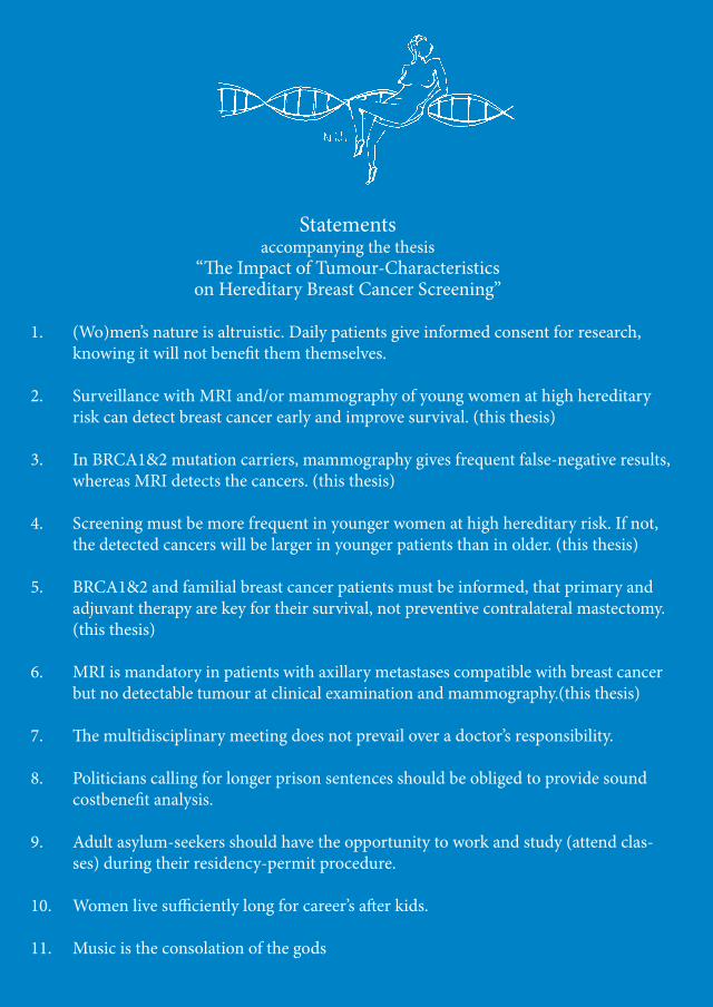
The Im
pact of Tumour C
haracteristics on Hereditary Breast C
ancer ScreeningM
adeleine Tilanus-Linthorst
Curriculum Vitae
Madeleine’s life started in Klimmen (L). She passed
gymnasium-β exam at Sacré Coeur Vaals and obtained
her medical degree at the Rijksuniversiteit Leiden. Af-
ter a first working experience in Lyallpur and Quetta
(Pakistan) she worked at the Department of Medicine of
the Oude en Nieuwe Gasthuis Delft. After 2 years train-
ing as a radiotherapist at the Daniel den Hoed Cancer
Clinic, she continued in 1980 working part-time at the
outpatient Breast Clinic, which is since 2001 part of the
Department of Surgery of ErasmusMC Rotterdam.
She is involved in research on hereditary breast cancer
since 1993.
She is joyfully married with Huug. Niels, Diet, Mauk
and Dintje brighten her days.
Statementsaccompanying the thesis
“The Impact of Tumour-Characteristicson Hereditary Breast Cancer Screening”
1. (Wo)men’s nature is altruistic. Daily patients give informed consent for research, knowing it will not benefit them themselves.
2. Surveillance with MRI and/or mammography of young women at high hereditary risk can detect breast cancer early and improve survival. (this thesis)
3. In BRCA1&2 mutation carriers, mammography gives frequent false-negative results, whereas MRI detects the cancers. (this thesis)
4. Screening must be more frequent in younger women at high hereditary risk. If not, the detected cancers will be larger in younger patients than in older. (this thesis)
5. BRCA1&2 and familial breast cancer patients must be informed, that primary and adjuvant therapy are key for their survival, not preventive contralateral mastectomy. (this thesis)
6. MRI is mandatory in patients with axillary metastases compatible with breast cancer but no detectable tumour at clinical examination and mammography.(this thesis)
7. The multidisciplinary meeting does not prevail over a doctor’s responsibility.
8. Politicians calling for longer prison sentences should be obliged to provide sound costbenefit analysis.
9. Adult asylum-seekers should have the opportunity to work and study (attend clas-ses) during their residency-permit procedure.
10. Women live sufficiently long for career’s after kids.
11. Music is the consolation of the gods


