Embed Size (px)
Citation preview
�����������������
Citation: Sousa, A.M.; Amaro, A.M.;
Piedade, A.P. 3D Printing of
Polymeric Bioresorbable Stents: A
Strategy to Improve Both Cellular
Compatibility and Mechanical
Properties. Polymers 2022, 14, 1099.
https://doi.org/10.3390/polym
14061099
Academic Editor: Carlos A.
García-González
Received: 25 January 2022
Accepted: 8 March 2022
Published: 9 March 2022
Publisher’s Note: MDPI stays neutral
with regard to jurisdictional claims in
published maps and institutional affil-
iations.
Copyright: © 2022 by the authors.
Licensee MDPI, Basel, Switzerland.
This article is an open access article
distributed under the terms and
conditions of the Creative Commons
Attribution (CC BY) license (https://
creativecommons.org/licenses/by/
4.0/).
polymers
Review
3D Printing of Polymeric Bioresorbable Stents: A Strategy toImprove Both Cellular Compatibility and Mechanical PropertiesAna M. Sousa , Ana M. Amaro and Ana P. Piedade *
Department of Mechanical Engineering, CEMMPRE, University of Coimbra, 3030-788 Coimbra, Portugal;[email protected] (A.M.S.); [email protected] (A.M.A.)* Correspondence: [email protected]; Tel.: +351-239-790-700
Abstract: One of the leading causes of death is cardiovascular disease, and the most common car-diovascular disease is coronary artery disease. Percutaneous coronary intervention and vascularstents have emerged as a solution to treat coronary artery disease. Nowadays, several types ofvascular stents share the same purpose: to reduce the percentage of restenosis, thrombosis, andneointimal hyperplasia and supply mechanical support to the blood vessels. Despite the numerousefforts to create an ideal stent, there is no coronary stent that simultaneously presents the appropriatecellular compatibility and mechanical properties to avoid stent collapse and failure. One of theemerging approaches to solve these problems is improving the mechanical performance of polymericbioresorbable stents produced through additive manufacturing. Although there have been numerousstudies in this field, normalized control parameters for 3D-printed polymeric vascular stents fabrica-tion are absent. The present paper aims to present an overview of the current types of stents and themain polymeric materials used to fabricate the bioresorbable vascular stents. Furthermore, a detaileddescription of the printing parameters’ influence on the mechanical performance and degradationprofile of polymeric bioresorbable stents is presented.
Keywords: vascular stents; polymers; degradation; mechanical properties; 3D printing
1. Introduction
According to the World Health Organization (WHO), cardiovascular diseases areamong the most prevalent and leading causes of death worldwide [1]. The WHO estimatesthat about 17 million deaths annually are related to cardiovascular diseases, and thisnumber will increase up to around 23.6 million by 2030 [2]. Usually, cardiovascular-relatedillness is associated with disorders in the heart, blood vessels, or both. Several risks arementioned in literature as a cause of cardiovascular diseases: unhealthy diet, physicalinactivity, obesity, hypertension, diabetes, tobacco, and harmful alcohol use [3,4].
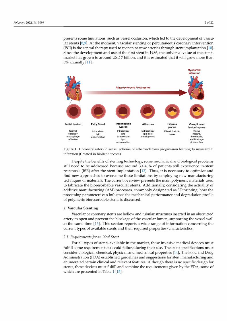
Among all cardiovascular problems, the most common is coronary artery disease,also called ischemic heart disease or coronary heart disease. Generally, these problems arerelated to disorders caused by narrowed heart arteries that supply blood to the cardiacmuscle [5]. Coronary artery disease results from the accumulation of plaques in the innersurface of the arteries, a condition medically known as atherosclerosis (Figure 1). Theseplaques begin to be constituted by cholesterol, fat, and, later, calcifications due to theaccumulation of calcium. After some time, atherosclerotic plaques harden, and the arteriesbecome more narrowed (stenosis), limiting the oxygen-rich blood flow in the arterial system.When the arteries that oxygenate the heart are completely blocked, ischemia of the heartwill occur, and myocardial infarction or heart attack will be experienced by the patient [5,6].
The first positive effort to treat atherosclerosis emerged with balloon angioplasty, aminimally invasive procedure with minimum costs [6]. This medical procedure involvesintroducing a guiding catheter, with a balloon, inside the artery that is narrowed, inflatingthe balloon to reopen the artery and restore the blood flow [7]. However, this procedure also
Polymers 2022, 14, 1099. https://doi.org/10.3390/polym14061099 https://www.mdpi.com/journal/polymers
Polymers 2022, 14, 1099 2 of 22
presents some limitations, such as vessel occlusion, which led to the development of vascu-lar stents [8,9]. At the moment, vascular stenting or percutaneous coronary intervention(PCI) is the central therapy used to reopen narrow arteries through stent implantation [10].Since the development and use of the first stent in 1986, the universal value of the stentsmarket has grown to around USD 7 billion, and it is estimated that it will grow more than5% annually [11].
Figure 1. Coronary artery disease: scheme of atherosclerosis progression leading to myocardialinfarction (Created in BioRender.com).
Despite the benefits of stenting technology, some mechanical and biological problemsstill need to be addressed because around 30–40% of patients still experience in-stentrestenosis (ISR) after the stent implantation [12]. Thus, it is necessary to optimize andfind new approaches to overcome these limitations by employing new manufacturingtechniques or materials. The current overview presents the main polymeric materials usedto fabricate the bioresorbable vascular stents. Additionally, considering the actuality ofadditive manufacturing (AM) processes, commonly designated as 3D printing, how theprocessing parameters can influence the mechanical performance and degradation profileof polymeric bioresorbable stents is discussed.
2. Vascular Stenting
Vascular or coronary stents are hollow and tubular structures inserted in an obstructedartery to open and prevent the blockage of the vascular lumen, supporting the vessel wallat the same time [13]. This section reports a wide range of information concerning thecurrent types of available stents and their required properties/characteristics.
2.1. Requirements for an Ideal Stent
For all types of stents available in the market, these invasive medical devices mustfulfill some requirements to avoid failure during their use. The stent specifications mustconsider biological, chemical, physical, and mechanical properties [14]. The Food and DrugAdministration (FDA) established guidelines and suggestions for stent manufacturing andenumerated certain clinical and relevant features. Although there is no specific design forstents, these devices must fulfill and combine the requirements given by the FDA, some ofwhich are presented in Table 1 [15].
Polymers 2022, 14, 1099 3 of 22
Table 1. Requirements and properties that devices (stents) and materials must satisfy to avoid failure(adapted from [16]).
Requirement Description
High radial strength Radial strength plays a crucial role in preventing the recoil of the stent by providing radial orstructural support to the vessel (ASTM F3067-14).
Low elastic radial recoil In order to attain a fixed final diameter of the stent appropriate for the host artery diameter, theproperty of low elastic radial recoil is of importance (ASTM F2079-09).
Good flexibility For the proper placement of the stent in the tortuous geometry of blood vessels, good flexibility ofthe designed stent is essential to place it with the help of a catheter (ASTM F2606).
Minimal stent profile During implantation, to avoid the unnecessary disturbance of blood flow, it is desirable to have aminimal stent profile.
Minimal foreshortening During the expansion of the vessel, the precise placing of the stent is important; hence, it shouldpossess minimum foreshortening.
Cellular compatibility The stent material must not cause any adverse reaction or injury in the human body, so cellularcompatibility is crucial.
Radiopacity For delivering the stent at the appropriate position, the radiopacity of the material mustbe considered.
Excellent fatigue propertiesThe blood flow induces cyclic stresses, and hence, due to the application of this cyclic load, fatigue
failure in the material drastically increases. The selection of the stent material is such that it canwithstand a minimum of 380 million cyclic load means up to 10 years (ASTM F2477-07).
One of the most crucial characteristics of stents is their cellular compatibility aimingto avoid adverse biological responses. Therefore, the device must have a nontoxic andcompatible base material to prevent events such as ISR, in-stent thrombosis (IST), and/orneointima hyperplasia in a stented blood vessel [17]. Although many base materials forstents were a revolution in the surgery field, the devices trigger adverse biological eventsdue to their permanent structure. In order to avoid these events, some devices have beenmade with bioresorbable materials that can support the artery wall during the healing and,after a specific time, be reabsorbed by the organism.
Regarding the mechanical properties, radial strength is the most important to considerduring stent fabrication [18–20]. Radial strength is defined as “the radial force that the stentcan withstand before collapsing”, which means that the device must have enough radialstrength to support the forces exerted by the artery wall (radial pressure) [21]. Anotherimportant property to consider is the elastic recoil, defined as the reduction in the stentdiameter after implantation. Recoil must be diminished to allow the tissue to heal andprevent the narrowing of the blood vessel. If there is a high percentage of elastic recoil,several adverse consequences can occur: stent restenosis, blow disruption, and, in the worstsituation, the device moving to another location [6]. In addition, radial strength stronglyinfluences the elastic recoil as the higher the radial strength, the lower the probability ofstent recoil. The FDA does not recommend any specific standard to evaluate the radialstrength, but several studies have followed the guideline of ASTM F3067-14 [22,23]. Otherstudies use the tensile test to assess the radial strength and stiffness by relating themdirectly with other material properties such as ultimate tensile strength and Young modulus,respectively [24]. A study from Al-Mangour et al. [25] states that a high tensile strengthhelps accomplish a sufficient radial strength to support the artery wall and maintain thelumen area.
The flexibility of vascular stents also should be considered before and after stentimplantation. Before implantation, the vascular stent must have enough flexibility to passinside the blood vessels during delivery, maintaining the shape and the design. Onceplaced in the artery, the flexibility of the device must be taken into consideration sincethe vascular stent must have the capacity to bend to be well fitted to the blood vessel [26].Additionally, it should resist compression from the arteries during the systolic and diastolicmovements [23].
Polymers 2022, 14, 1099 4 of 22
It is necessary to evaluate the fatigue properties to assess the long-term durabilityof cardiovascular stents and their clinical success. Fatigue can lead to the failure of astent, resulting in the loss of radial support and triggering biological events [27]. Usually,the fatigue of this type of device is influenced mainly by the cyclic loading generated bythe heart beating (systolic and diastolic movements). The inner diameter of the arterychanges due to the pressure caused by the pulsatile blood flow. Consequently, the stent isexposed simultaneously to bending, torsion, and compression [28]. These events can leadto crack initiation due to the oscillating stress condition in the stent and, therefore, stentfracture [29].
An invasive coronary device can be either a balloon-expandable stent (BE) or a self-expandable stent (SE) considering the deployment method. The most common is the BEprocedure, which expands the stent with a balloon catheter, deforming the device plastically.In the SE procedure, the stents have a greater diameter than the artery, and the device isconstrained before being positioned. Then, the SE device is released to expand if the basematerial responds to an external stimulus, for instance, temperature [27,28].
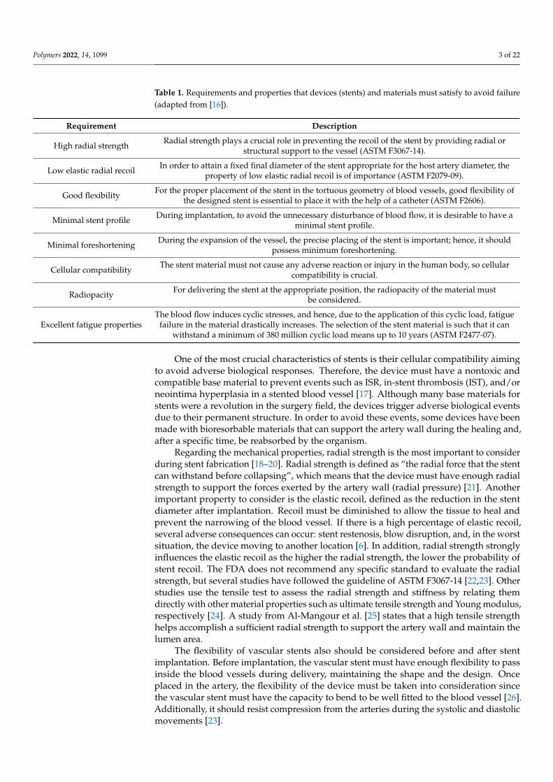
When bioresorbable materials are used to manufacture vascular stents, it is necessaryto know their degradation profile. It is essential to evaluate the degradation kinetic since itdetermines the time range during which the mechanical properties are still adequate [30].The degradation process must consider the type of material, molecular weight, crystallinitydegree, and the pH of the surrounding environment [31]. Moreover, the degradation rateis also influenced by the processing technique used to fabricate the vascular stent [32].Regarding the degradation of bioresorbable stents, several authors have described somein vitro procedures to assess the degradation profile, namely immersion in simulated bodyfluid (SBF) and immersion in phosphate-buffered solution (PBS) (Figure 2) [33,34].
Figure 2. Schematic representation of the in vitro degradation of vascular stents.
2.2. Types of Vascular Stents
In this review, the description of the vascular stents will be carried out under themost common terms presented by the authors and selling companies to facilitate theirunderstanding. According to the literature, vascular stents can be classified into threedistinct groups that have differences in the base material and surface of the stent: bare-metal stents (BMSs), drug-eluting stents (DESs), and bioresorbable stents (BRSs). Despitethe differences, all types of stents must share some of the characteristics and propertiesmentioned in the previous section.
Bare-metal stents were the first devices implanted to treat atherosclerosis; whencompared to balloon angioplasty, they reduced the rate of restenosis. Usually, these deviceshave a permanent metallic structure without drugs loaded on their surface [35]. Dueto the mechanical properties of metals, BMS stents have an increased radial strengththat allows robust mechanical support to the vessel wall with a thin strut. Devices with
Polymers 2022, 14, 1099 5 of 22
reduced struts are known for having a reduced crossing profile, which induces less tissueinjury and less disruption in the blood flow [36]. Nevertheless, introducing a permanentmetallic framework triggers inflammatory responses, such as neointimal hyperplasia, thatlead to the artery’s reblockage and the activation of the coagulation cascade [13]. Theseevents can be associated with the metallic ions released from the stainless steel (316LSS)and cobalt–chromium (CoCr) alloys through the presence of crevice, pitting, and stresscorrosion cracking. For instance, the release of nickel, molybdenum, and chromium ionscan trigger genotoxic and mutagenic events and activate allergic responses [36]. As a result,drug-eluting stents emerged to overcome the undesirable effects of BMS implantation [8].
Drug-eluting stents are characterized as having a permanent metallic structure with acoating that acts as a drug reservoir. An ideal DES includes the elution of antiproliferativeand anti-inflammatory drugs that are released over time, delaying the biological response.The selected drugs must be capable of helping appropriate healing and endothelializationand being efficient in inhibiting platelet aggregation, inflammation, vascular smooth musclecell (VSMC) proliferation, and migration [37]. Due to the presence of pharmaceuticals,DESs has shown lower ISR rates than BMSs [38].
DESs can be constituted by different materials in the core structure and the surfacecoating. Usually, the base structure of this type of stent is fabricated with metallic alloys. Thecoatings can be nondegradable (first generation of DESs) or degradable (second generationof DESs), and each one of them has different mechanisms and times to release the elutingdrugs [39]. The first generation of DESs consisted of a permanent metallic framework, anondegradable polymeric coating, and a pharmaceutical loaded on the coating layer. Thedegradable polymeric coating is characteristic of the second generation and diminishes theadverse clinical occurrences compared to the first generation of DESs [37]. Concerning thematerials used as coatings, the most used are bioresorbable polymers, but a few studiesalso describe inorganic coatings to be applied in DESs [39]. The arrival of new generationsof DESs reduced the biological events, but late IST and ISR events are still active andcompromise the long-term efficiency and safety of DESs [40]. Consequently, the concept ofBRSs arose.
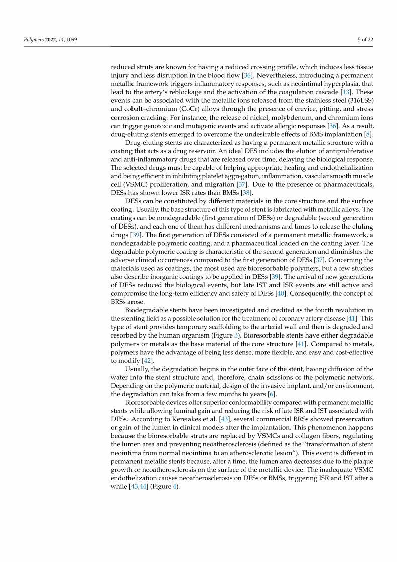
Biodegradable stents have been investigated and credited as the fourth revolution inthe stenting field as a possible solution for the treatment of coronary artery disease [41]. Thistype of stent provides temporary scaffolding to the arterial wall and then is degraded andresorbed by the human organism (Figure 3). Bioresorbable stents have either degradablepolymers or metals as the base material of the core structure [41]. Compared to metals,polymers have the advantage of being less dense, more flexible, and easy and cost-effectiveto modify [42].
Usually, the degradation begins in the outer face of the stent, having diffusion of thewater into the stent structure and, therefore, chain scissions of the polymeric network.Depending on the polymeric material, design of the invasive implant, and/or environment,the degradation can take from a few months to years [6].
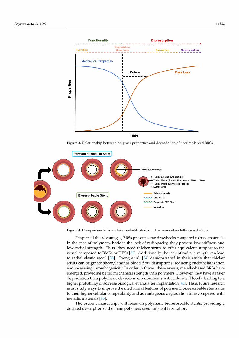
Bioresorbable devices offer superior conformability compared with permanent metallicstents while allowing luminal gain and reducing the risk of late ISR and IST associated withDESs. According to Kereiakes et al. [43], several commercial BRSs showed preservationor gain of the lumen in clinical models after the implantation. This phenomenon happensbecause the bioresorbable struts are replaced by VSMCs and collagen fibers, regulatingthe lumen area and preventing neoatherosclerosis (defined as the “transformation of stentneointima from normal neointima to an atherosclerotic lesion”). This event is different inpermanent metallic stents because, after a time, the lumen area decreases due to the plaquegrowth or neoatherosclerosis on the surface of the metallic device. The inadequate VSMCendothelization causes neoatherosclerosis on DESs or BMSs, triggering ISR and IST after awhile [43,44] (Figure 4).
Polymers 2022, 14, 1099 6 of 22
Figure 3. Relationship between polymer properties and degradation of postimplanted BRSs.
Figure 4. Comparison between bioresorbable stents and permanent metallic-based stents.
Despite all the advantages, BRSs present some drawbacks compared to base materials.In the case of polymers, besides the lack of radiopacity, they present low stiffness andlow radial strength. Thus, they need thicker struts to offer equivalent support to thevessel compared to BMSs or DESs [37]. Additionally, the lack of radial strength can leadto radial elastic recoil [38]. Toong et al. [24] demonstrated in their study that thickerstruts can originate shear/laminar blood flow disruptions, reducing endothelializationand increasing thrombogenicity. In order to thwart these events, metallic-based BRSs haveemerged, providing better mechanical strength than polymers. However, they have a fasterdegradation than polymeric devices in environments with chloride (blood), leading to ahigher probability of adverse biological events after implantation [41]. Thus, future researchmust study ways to improve the mechanical features of polymeric bioresorbable stents dueto their higher cellular compatibility and advantageous degradation time compared withmetallic materials [45].
The present manuscript will focus on polymeric bioresorbable stents, providing adetailed description of the main polymers used for stent fabrication.
Polymers 2022, 14, 1099 7 of 22
3. Polymeric Materials for Bioresorbable Vascular Stents
Bioresorbable stents are made with materials that can be degraded and absorbed bythe human organism, entering the main metabolic pathways, such as the Krebs cycle. Themain objective of bioresorbable materials is to provide temporary support to the vesselduring the healing time and then be eliminated by the body, leaving the artery with ahealthy endothelium and normal blood flow. As mentioned previously, the absence offoreign material will decrease the risk of late ISR and IST [41]. In addition, significantgroups of patients, such as children, the elderly, or diabetics, who suffer from problemsinvolving several repeated surgeries will benefit from this technology since removing theimplant is unnecessary [39].
Currently, polyesters are used to manufacture BRSs because of their tailorable biodegrad-ability. Polymers such as poly(lactic acid) (PLA) and its enantiomers, poly(ε-caprolactone)(PCL), poly(glycolic acid) (PGA), and poly(lactic-co-glycolic acid) (PLGA), are used inpolymeric-based cardiovascular stents [41]. This section will address some aspects of thesepolymers, including their degradation and/or reabsorption processes.
3.1. Poly(lactic acid)
PLA is accepted by several regulatory agencies as a safe, biodegradable material tobe applied in numerous medical applications [46]. Amongst all polymers suggested andapplied for fabrication of vascular stents, by far the most common is PLA due to its optimalcombination of cellular compatibility, degrading pattern, and mechanical strength [23,47].
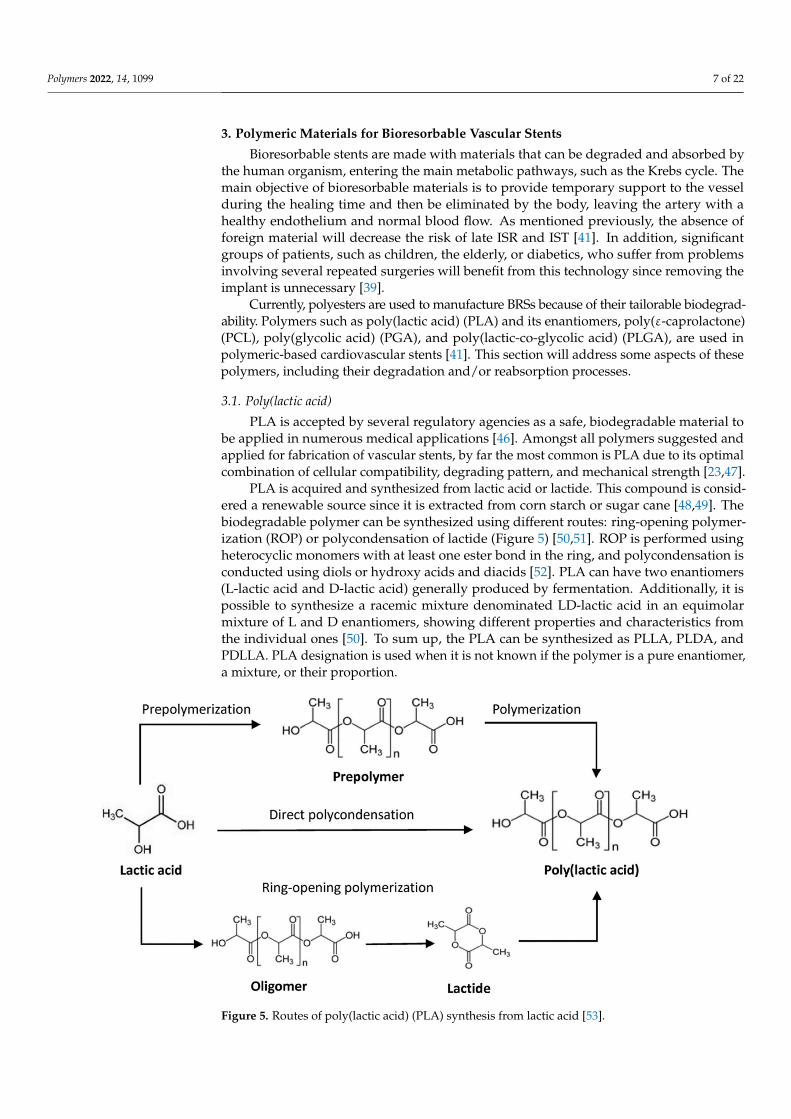
PLA is acquired and synthesized from lactic acid or lactide. This compound is consid-ered a renewable source since it is extracted from corn starch or sugar cane [48,49]. Thebiodegradable polymer can be synthesized using different routes: ring-opening polymer-ization (ROP) or polycondensation of lactide (Figure 5) [50,51]. ROP is performed usingheterocyclic monomers with at least one ester bond in the ring, and polycondensation isconducted using diols or hydroxy acids and diacids [52]. PLA can have two enantiomers(L-lactic acid and D-lactic acid) generally produced by fermentation. Additionally, it ispossible to synthesize a racemic mixture denominated LD-lactic acid in an equimolarmixture of L and D enantiomers, showing different properties and characteristics fromthe individual ones [50]. To sum up, the PLA can be synthesized as PLLA, PLDA, andPDLLA. PLA designation is used when it is not known if the polymer is a pure enantiomer,a mixture, or their proportion.
Figure 5. Routes of poly(lactic acid) (PLA) synthesis from lactic acid [53].
Polymers 2022, 14, 1099 8 of 22
The literature shows that the different enantiomers of PLA produce different charac-teristics and properties in vascular stents. The percentage of these compounds determinesproperties such as glass transition and melting temperatures or percentage of crystallinitywhich will affect the mechanical properties and the biodegradation rate of the stent [6].For instance, PDLLA has lower tensile strength and higher degradation time than PLLAand PDLA [54]. In what concerns the application in vascular stents, the PLA can also becopolymerized with other polymers such as PGA or PCL to improve the performance ofthe stents. Compared to other polymeric materials, PLLA has better mechanical propertiesthan PCL and a lower degradation time than PLGA (Table 2).
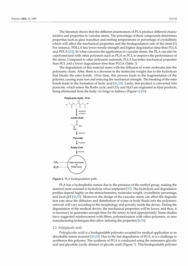
The degradation of this material starts with the diffusion of water molecules into thepolymeric chain. After, there is a decrease in the molecular weight due to the hydrolysisthat breaks the ester bonds. Over time, this process leads to the fragmentation of thepolymer, causing mass loss and reducing the mechanical strength. The breaking of the esterbonds leads to the formation of lactic acid [46,55]. Lastly, this product is converted intopyruvate, which enters the Krebs cycle, and CO2 and H2O are originated as final products,being eliminated from the body via lungs or kidneys (Figure 6) [56].
Figure 6. PLA biodegradation path.
PLA has a hydrophobic nature due to the presence of the methyl group, making thematerial more resistant to hydrolysis when implanted [57]. The hydrolysis and degradationprofiles depend highly on the stereochemistry, molecular weight, crystallinity percentage,and local pH [46,58]. Moreover, the design of the vascular stents can affect the degrada-tion rate since the diffusion and distribution of water or body fluids into the polymericnetwork will vary according to the morphology and porosity inside the device. During thedegradation of the medical device, the mechanical properties will be lower, and thus, itis necessary to guarantee enough time for the artery to heal appropriately. Some studieshave suggested reinforcement with fibers, polymerization with other polymers, or newmanufacturing techniques that allow tailoring the properties [6,59].
3.2. Poly(glycolic acid)
Poly(glycolic acid) is a biodegradable polyester accepted for medical application as anabsorbable suture material [49,60]. Due to the fast degradation of PGA, it is a challenge tosynthesize this polymer. The synthesis of PGA is conducted using the monomers glycolicacid and glycolide (cyclic dimmer of glycolic acid) (Figure 7). This biodegradable polymer
Polymers 2022, 14, 1099 9 of 22
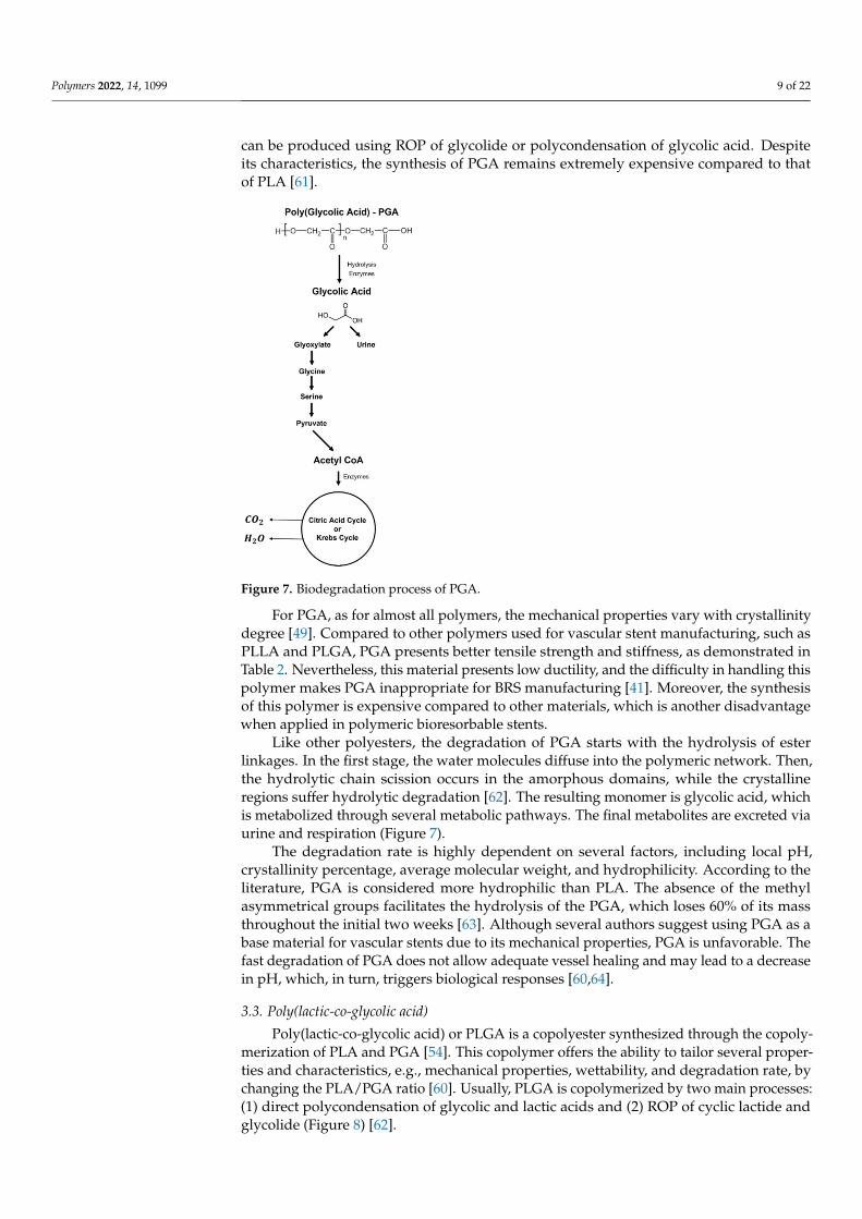
can be produced using ROP of glycolide or polycondensation of glycolic acid. Despiteits characteristics, the synthesis of PGA remains extremely expensive compared to thatof PLA [61].
Figure 7. Biodegradation process of PGA.
For PGA, as for almost all polymers, the mechanical properties vary with crystallinitydegree [49]. Compared to other polymers used for vascular stent manufacturing, such asPLLA and PLGA, PGA presents better tensile strength and stiffness, as demonstrated inTable 2. Nevertheless, this material presents low ductility, and the difficulty in handling thispolymer makes PGA inappropriate for BRS manufacturing [41]. Moreover, the synthesisof this polymer is expensive compared to other materials, which is another disadvantagewhen applied in polymeric bioresorbable stents.
Like other polyesters, the degradation of PGA starts with the hydrolysis of esterlinkages. In the first stage, the water molecules diffuse into the polymeric network. Then,the hydrolytic chain scission occurs in the amorphous domains, while the crystallineregions suffer hydrolytic degradation [62]. The resulting monomer is glycolic acid, whichis metabolized through several metabolic pathways. The final metabolites are excreted viaurine and respiration (Figure 7).
The degradation rate is highly dependent on several factors, including local pH,crystallinity percentage, average molecular weight, and hydrophilicity. According to theliterature, PGA is considered more hydrophilic than PLA. The absence of the methylasymmetrical groups facilitates the hydrolysis of the PGA, which loses 60% of its massthroughout the initial two weeks [63]. Although several authors suggest using PGA as abase material for vascular stents due to its mechanical properties, PGA is unfavorable. Thefast degradation of PGA does not allow adequate vessel healing and may lead to a decreasein pH, which, in turn, triggers biological responses [60,64].
3.3. Poly(lactic-co-glycolic acid)
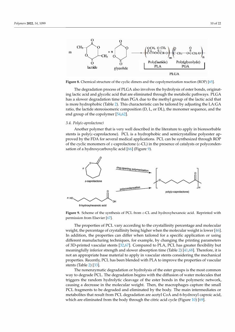
Poly(lactic-co-glycolic acid) or PLGA is a copolyester synthesized through the copoly-merization of PLA and PGA [54]. This copolymer offers the ability to tailor several proper-ties and characteristics, e.g., mechanical properties, wettability, and degradation rate, bychanging the PLA/PGA ratio [60]. Usually, PLGA is copolymerized by two main processes:(1) direct polycondensation of glycolic and lactic acids and (2) ROP of cyclic lactide andglycolide (Figure 8) [62].
Polymers 2022, 14, 1099 10 of 22
Figure 8. Chemical structure of the cyclic dimers and the copolymerization reaction (ROP) [65].
The degradation process of PLGA also involves the hydrolysis of ester bonds, originat-ing lactic acid and glycolic acid that are eliminated through the metabolic pathways. PLGAhas a slower degradation time than PGA due to the methyl group of the lactic acid thatis more hydrophobic (Table 2). This characteristic can be tailored by adjusting the LA:GAratio, the lactide stereoisomeric composition (D, L, or DL), the monomer sequence, and theend group of the copolymer [54,62].
3.4. Poly(ε-aprolactone)
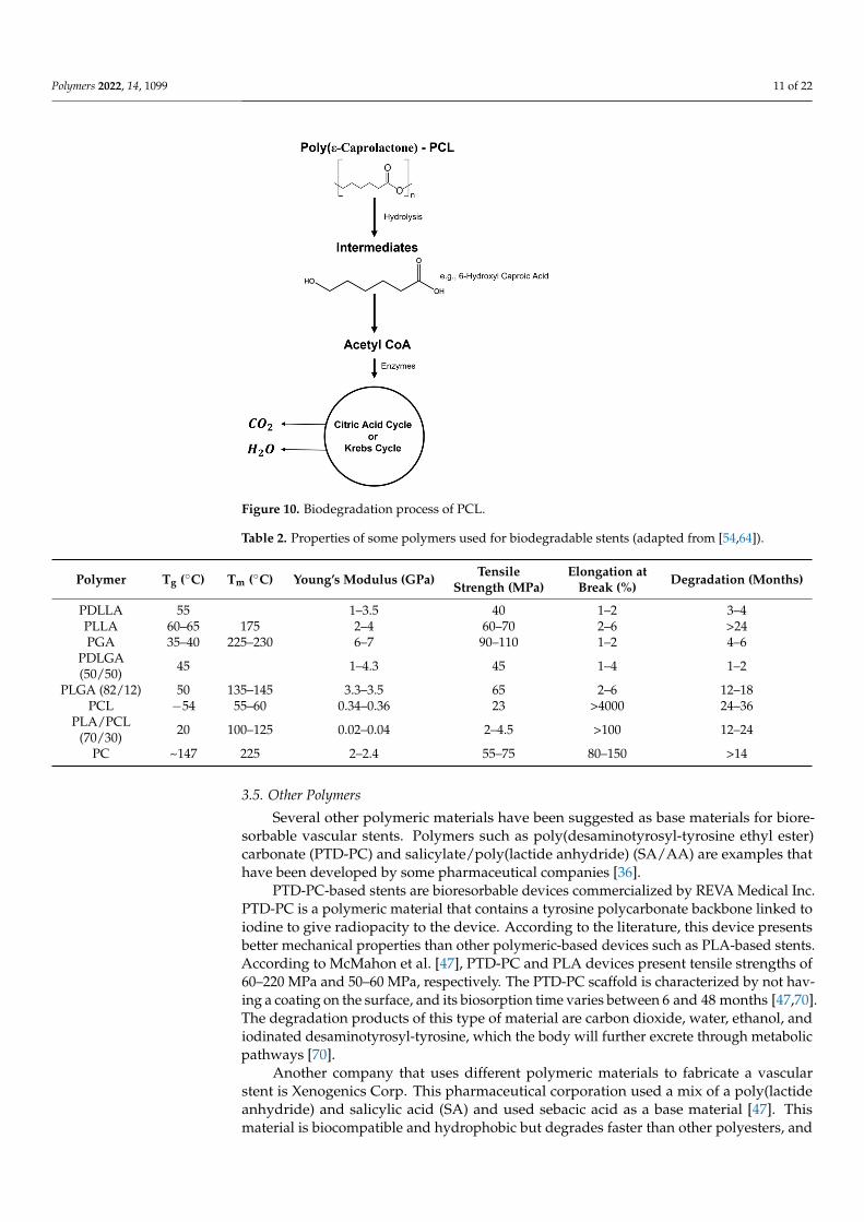
Another polymer that is very well described in the literature to apply in bioresorbablestents is poly(ε-caprolactone). PCL is a hydrophobic and semicrystalline polyester ap-proved by the FDA for several medical applications. PCL can be synthesized through ROPof the cyclic monomers of ε-caprolactone (ε-CL) in the presence of catalysts or polyconden-sation of a hydroxycarboxylic acid [66] (Figure 9).
Figure 9. Scheme of the synthesis of PCL from ε-CL and hydroxyhexanoic acid. Reprinted withpermission from Elsevier [67].
The properties of PCL vary according to the crystallinity percentage and molecularweight, the percentage of crystallinity being higher when the molecular weight is lower [46].In addition, the properties can differ when tailored for a specific application or usingdifferent manufacturing techniques, for example, by changing the printing parametersof 3D-printed vascular stents [32,67]. Compared to PLA, PCL has greater flexibility butmeaningfully inferior strength and slower absorption time (Table 2) [41,68]. Therefore, it isnot an appropriate base material to apply in vascular stents considering the mechanicalproperties. Recently, PCL has been blended with PLA to improve the properties of vascularstents (Table 2) [33].
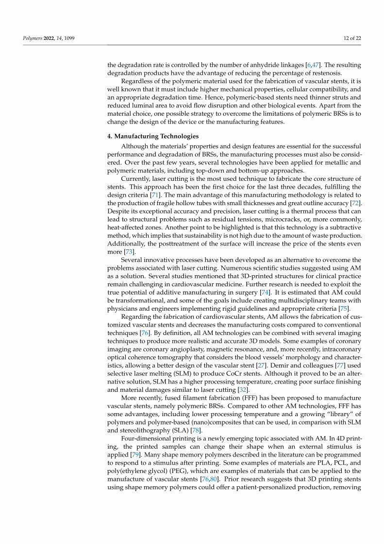
The nonenzymatic degradation or hydrolysis of the ester groups is the most commonway to degrade PCL. The degradation begins with the diffusion of water molecules thattriggers the random hydrolytic cleavage of the ester bonds in the polymeric network,causing a decrease in the molecular weight. Then, the macrophages capture the smallPCL fragments to be degraded and eliminated by the body. The main intermediates ormetabolites that result from PCL degradation are acetyl CoA and 6-hydroxyl caproic acid,which are eliminated from the body through the citric acid cycle (Figure 10) [69].
Polymers 2022, 14, 1099 11 of 22
Figure 10. Biodegradation process of PCL.
Table 2. Properties of some polymers used for biodegradable stents (adapted from [54,64]).
Polymer Tg (◦C) Tm (◦C) Young’s Modulus (GPa) TensileStrength (MPa)
Elongation atBreak (%) Degradation (Months)
PDLLA 55 1–3.5 40 1–2 3–4PLLA 60–65 175 2–4 60–70 2–6 >24PGA 35–40 225–230 6–7 90–110 1–2 4–6
PDLGA(50/50) 45 1–4.3 45 1–4 1–2
PLGA (82/12) 50 135–145 3.3–3.5 65 2–6 12–18PCL −54 55–60 0.34–0.36 23 >4000 24–36
PLA/PCL(70/30) 20 100–125 0.02–0.04 2–4.5 >100 12–24
PC ~147 225 2–2.4 55–75 80–150 >14
3.5. Other Polymers
Several other polymeric materials have been suggested as base materials for biore-sorbable vascular stents. Polymers such as poly(desaminotyrosyl-tyrosine ethyl ester)carbonate (PTD-PC) and salicylate/poly(lactide anhydride) (SA/AA) are examples thathave been developed by some pharmaceutical companies [36].
PTD-PC-based stents are bioresorbable devices commercialized by REVA Medical Inc.PTD-PC is a polymeric material that contains a tyrosine polycarbonate backbone linked toiodine to give radiopacity to the device. According to the literature, this device presentsbetter mechanical properties than other polymeric-based devices such as PLA-based stents.According to McMahon et al. [47], PTD-PC and PLA devices present tensile strengths of60–220 MPa and 50–60 MPa, respectively. The PTD-PC scaffold is characterized by not hav-ing a coating on the surface, and its biosorption time varies between 6 and 48 months [47,70].The degradation products of this type of material are carbon dioxide, water, ethanol, andiodinated desaminotyrosyl-tyrosine, which the body will further excrete through metabolicpathways [70].
Another company that uses different polymeric materials to fabricate a vascularstent is Xenogenics Corp. This pharmaceutical corporation used a mix of a poly(lactideanhydride) and salicylic acid (SA) and used sebacic acid as a base material [47]. Thismaterial is biocompatible and hydrophobic but degrades faster than other polyesters, and
Polymers 2022, 14, 1099 12 of 22
the degradation rate is controlled by the number of anhydride linkages [6,47]. The resultingdegradation products have the advantage of reducing the percentage of restenosis.
Regardless of the polymeric material used for the fabrication of vascular stents, it iswell known that it must include higher mechanical properties, cellular compatibility, andan appropriate degradation time. Hence, polymeric-based stents need thinner struts andreduced luminal area to avoid flow disruption and other biological events. Apart from thematerial choice, one possible strategy to overcome the limitations of polymeric BRSs is tochange the design of the device or the manufacturing features.
4. Manufacturing Technologies
Although the materials’ properties and design features are essential for the successfulperformance and degradation of BRSs, the manufacturing processes must also be consid-ered. Over the past few years, several technologies have been applied for metallic andpolymeric materials, including top-down and bottom-up approaches.
Currently, laser cutting is the most used technique to fabricate the core structure ofstents. This approach has been the first choice for the last three decades, fulfilling thedesign criteria [71]. The main advantage of this manufacturing methodology is related tothe production of fragile hollow tubes with small thicknesses and great outline accuracy [72].Despite its exceptional accuracy and precision, laser cutting is a thermal process that canlead to structural problems such as residual tensions, microcracks, or, more commonly,heat-affected zones. Another point to be highlighted is that this technology is a subtractivemethod, which implies that sustainability is not high due to the amount of waste production.Additionally, the posttreatment of the surface will increase the price of the stents evenmore [73].
Several innovative processes have been developed as an alternative to overcome theproblems associated with laser cutting. Numerous scientific studies suggested using AMas a solution. Several studies mentioned that 3D-printed structures for clinical practiceremain challenging in cardiovascular medicine. Further research is needed to exploit thetrue potential of additive manufacturing in surgery [74]. It is estimated that AM couldbe transformational, and some of the goals include creating multidisciplinary teams withphysicians and engineers implementing rigid guidelines and appropriate criteria [75].
Regarding the fabrication of cardiovascular stents, AM allows the fabrication of cus-tomized vascular stents and decreases the manufacturing costs compared to conventionaltechniques [76]. By definition, all AM technologies can be combined with several imagingtechniques to produce more realistic and accurate 3D models. Some examples of coronaryimaging are coronary angioplasty, magnetic resonance, and, more recently, intracoronaryoptical coherence tomography that considers the blood vessels’ morphology and character-istics, allowing a better design of the vascular stent [27]. Demir and colleagues [77] usedselective laser melting (SLM) to produce CoCr stents. Although it proved to be an alter-native solution, SLM has a higher processing temperature, creating poor surface finishingand material damages similar to laser cutting [32].
More recently, fused filament fabrication (FFF) has been proposed to manufacturevascular stents, namely polymeric BRSs. Compared to other AM technologies, FFF hassome advantages, including lower processing temperature and a growing “library” ofpolymers and polymer-based (nano)composites that can be used, in comparison with SLMand stereolithography (SLA) [78].
Four-dimensional printing is a newly emerging topic associated with AM. In 4D print-ing, the printed samples can change their shape when an external stimulus isapplied [79]. Many shape memory polymers described in the literature can be programmedto respond to a stimulus after printing. Some examples of materials are PLA, PCL, andpoly(ethylene glycol) (PEG), which are examples of materials that can be applied to themanufacture of vascular stents [76,80]. Prior research suggests that 3D printing stentsusing shape memory polymers could offer a patient-personalized production, removing
Polymers 2022, 14, 1099 13 of 22
the requirement for balloon expansion, reducing the possibility of stent displacement, andleading to a new age for stent technology [76,81,82].
Jia et al. [83] proposed using FFF to create a polymeric BRS using a shape memorypolymer, PLA. After the production of the vascular stent, the device can keep the compactedshape at ambient temperature and then self-expand through temperature stimulus [83].Another study mentions the use of FFF as a technique to fabricate BRSs based on PCL,also a shape memory polymer. This approach can create a safer, custom-made, and moreappropriate device [32,33]. Nevertheless, it is necessary to improve the mechanical strengthof the devices made with these polymers.
5. Processing Parameters in AM
It is known from previous studies that the characteristics and properties of the biore-sorbable thermoplastic polymers can suffer changes during the manufacturing process. FFFis a technique with numerous parameters that influence the quality of the 3D-printed parts,such as the mechanical properties and the degradation rate [84]. Many questions remainunanswered about the effect of the printing parameters on vascular stents. To addressthese questions, we address the influence of the main printing parameters, such as layerthickness, raster angle, raster width, build orientation, infill pattern, and infill density.
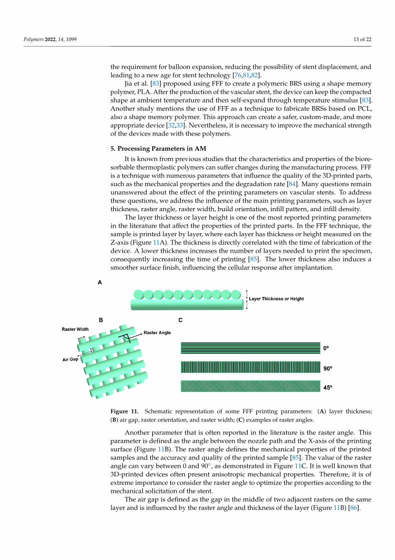
The layer thickness or layer height is one of the most reported printing parametersin the literature that affect the properties of the printed parts. In the FFF technique, thesample is printed layer by layer, where each layer has thickness or height measured on theZ-axis (Figure 11A). The thickness is directly correlated with the time of fabrication of thedevice. A lower thickness increases the number of layers needed to print the specimen,consequently increasing the time of printing [85]. The lower thickness also induces asmoother surface finish, influencing the cellular response after implantation.
Figure 11. Schematic representation of some FFF printing parameters: (A) layer thickness;(B) air gap, raster orientation, and raster width; (C) examples of raster angles.
Another parameter that is often reported in the literature is the raster angle. Thisparameter is defined as the angle between the nozzle path and the X-axis of the printingsurface (Figure 11B). The raster angle defines the mechanical properties of the printedsamples and the accuracy and quality of the printed sample [85]. The value of the rasterangle can vary between 0 and 90◦, as demonstrated in Figure 11C. It is well known that3D-printed devices often present anisotropic mechanical properties. Therefore, it is ofextreme importance to consider the raster angle to optimize the properties according to themechanical solicitation of the stent.
The air gap is defined as the gap in the middle of two adjacent rasters on the samelayer and is influenced by the raster angle and thickness of the layer (Figure 11B) [86].
Polymers 2022, 14, 1099 14 of 22
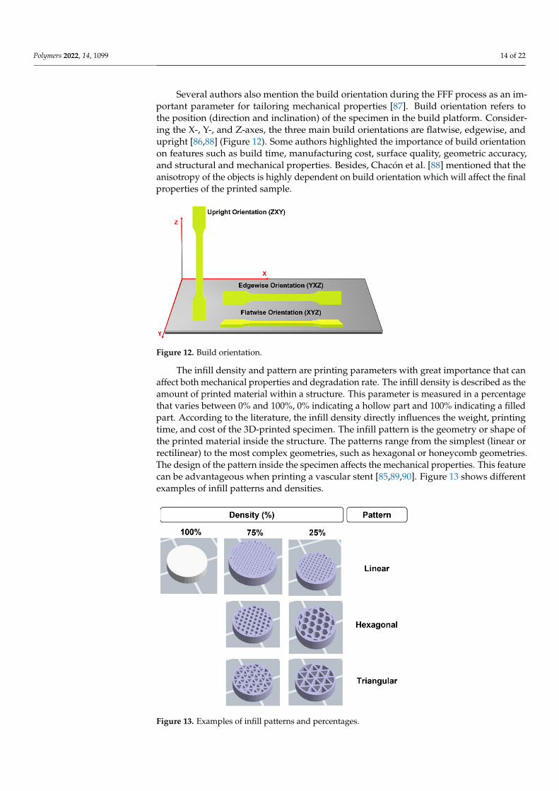
Several authors also mention the build orientation during the FFF process as an im-portant parameter for tailoring mechanical properties [87]. Build orientation refers tothe position (direction and inclination) of the specimen in the build platform. Consider-ing the X-, Y-, and Z-axes, the three main build orientations are flatwise, edgewise, andupright [86,88] (Figure 12). Some authors highlighted the importance of build orientationon features such as build time, manufacturing cost, surface quality, geometric accuracy,and structural and mechanical properties. Besides, Chacón et al. [88] mentioned that theanisotropy of the objects is highly dependent on build orientation which will affect the finalproperties of the printed sample.
Figure 12. Build orientation.
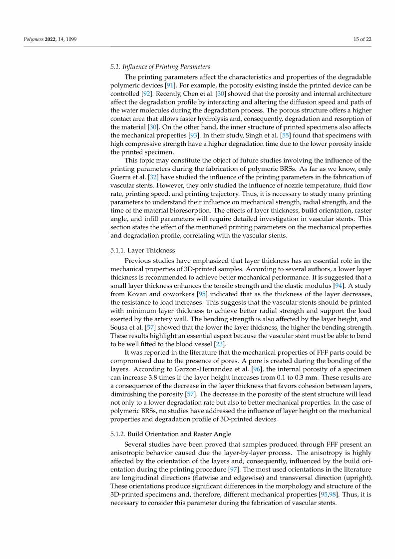
The infill density and pattern are printing parameters with great importance that canaffect both mechanical properties and degradation rate. The infill density is described as theamount of printed material within a structure. This parameter is measured in a percentagethat varies between 0% and 100%, 0% indicating a hollow part and 100% indicating a filledpart. According to the literature, the infill density directly influences the weight, printingtime, and cost of the 3D-printed specimen. The infill pattern is the geometry or shape ofthe printed material inside the structure. The patterns range from the simplest (linear orrectilinear) to the most complex geometries, such as hexagonal or honeycomb geometries.The design of the pattern inside the specimen affects the mechanical properties. This featurecan be advantageous when printing a vascular stent [85,89,90]. Figure 13 shows differentexamples of infill patterns and densities.
Figure 13. Examples of infill patterns and percentages.
Polymers 2022, 14, 1099 15 of 22
5.1. Influence of Printing Parameters
The printing parameters affect the characteristics and properties of the degradablepolymeric devices [91]. For example, the porosity existing inside the printed device can becontrolled [92]. Recently, Chen et al. [30] showed that the porosity and internal architectureaffect the degradation profile by interacting and altering the diffusion speed and path ofthe water molecules during the degradation process. The porous structure offers a highercontact area that allows faster hydrolysis and, consequently, degradation and resorption ofthe material [30]. On the other hand, the inner structure of printed specimens also affectsthe mechanical properties [93]. In their study, Singh et al. [55] found that specimens withhigh compressive strength have a higher degradation time due to the lower porosity insidethe printed specimen.
This topic may constitute the object of future studies involving the influence of theprinting parameters during the fabrication of polymeric BRSs. As far as we know, onlyGuerra et al. [32] have studied the influence of the printing parameters in the fabrication ofvascular stents. However, they only studied the influence of nozzle temperature, fluid flowrate, printing speed, and printing trajectory. Thus, it is necessary to study many printingparameters to understand their influence on mechanical strength, radial strength, and thetime of the material bioresorption. The effects of layer thickness, build orientation, rasterangle, and infill parameters will require detailed investigation in vascular stents. Thissection states the effect of the mentioned printing parameters on the mechanical propertiesand degradation profile, correlating with the vascular stents.
5.1.1. Layer Thickness
Previous studies have emphasized that layer thickness has an essential role in themechanical properties of 3D-printed samples. According to several authors, a lower layerthickness is recommended to achieve better mechanical performance. It is suggested that asmall layer thickness enhances the tensile strength and the elastic modulus [94]. A studyfrom Kovan and coworkers [95] indicated that as the thickness of the layer decreases,the resistance to load increases. This suggests that the vascular stents should be printedwith minimum layer thickness to achieve better radial strength and support the loadexerted by the artery wall. The bending strength is also affected by the layer height, andSousa et al. [57] showed that the lower the layer thickness, the higher the bending strength.These results highlight an essential aspect because the vascular stent must be able to bendto be well fitted to the blood vessel [23].
It was reported in the literature that the mechanical properties of FFF parts could becompromised due to the presence of pores. A pore is created during the bonding of thelayers. According to Garzon-Hernandez et al. [96], the internal porosity of a specimencan increase 3.8 times if the layer height increases from 0.1 to 0.3 mm. These results area consequence of the decrease in the layer thickness that favors cohesion between layers,diminishing the porosity [57]. The decrease in the porosity of the stent structure will leadnot only to a lower degradation rate but also to better mechanical properties. In the case ofpolymeric BRSs, no studies have addressed the influence of layer height on the mechanicalproperties and degradation profile of 3D-printed devices.
5.1.2. Build Orientation and Raster Angle
Several studies have been proved that samples produced through FFF present ananisotropic behavior caused due the layer-by-layer process. The anisotropy is highlyaffected by the orientation of the layers and, consequently, influenced by the build ori-entation during the printing procedure [97]. The most used orientations in the literatureare longitudinal directions (flatwise and edgewise) and transversal direction (upright).These orientations produce significant differences in the morphology and structure of the3D-printed specimens and, therefore, different mechanical properties [95,98]. Thus, it isnecessary to consider this parameter during the fabrication of vascular stents.
Polymers 2022, 14, 1099 16 of 22
Regarding the mechanical properties, it is demonstrated in several studies that thevertical direction presents the poorest mechanical properties compared to longitudinalbuilding orientations [87,99]. Chacón and colleagues [88] stated in their manuscript thatthe vertical direction showed that lowest value of tensile strength and stiffness comparedto the other directions. Likewise, Ashtankar et al. [100] observed that when the buildorientation is flatwise, the tensile and compressive stresses are 23.68% and 16.65% greaterthan the values for the vertical direction, respectively. Furthermore, it is described that thelongitudinal samples present a translayer failure and a ductile fracture while the transversalsample presents an interlayer failure and a more brittle fracture [88]. Another characteristicto take into account during stent manufacturing is flexibility. According to the literature, theflexural properties of samples printed in upright build orientation are worse than those ofspecimens printed with longitudinal orientations. The authors stated that vertical printedspecimens suffered failure at lower deflection values [97].
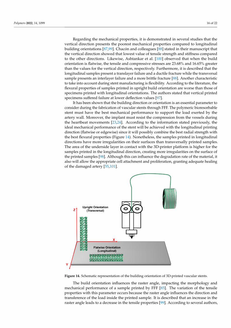
It has been shown that the building direction or orientation is an essential parameter toconsider during the fabrication of vascular stents through FFF. The polymeric bioresorbablestent must have the best mechanical performance to support the load exerted by theartery wall. Moreover, the implant must resist the compression from the vessels duringthe heartbeat movements [23,24]. According to the information stated previously, theideal mechanical performance of the stent will be achieved with the longitudinal printingdirection (flatwise or edgewise) since it will possibly combine the best radial strength withthe best flexural properties (Figure 14). Nonetheless, the samples printed in longitudinaldirections have more irregularities on their surfaces than transversally printed samples.The area of the underside layer in contact with the 3D-printer platform is higher for thesamples printed in the longitudinal direction, creating more irregularities on the surface ofthe printed samples [98]. Although this can influence the degradation rate of the material, italso will allow the appropriate cell attachment and proliferation, granting adequate healingof the damaged artery [55,101].
Figure 14. Schematic representation of the building orientation of 3D-printed vascular stents.
The build orientation influences the raster angle, impacting the morphology andmechanical performance of a sample printed by FFF [85]. The variation of the tensileproperties with this parameter occurs because the raster angle influences the direction andtransference of the load inside the printed sample. It is described that an increase in theraster angle leads to a decrease in the tensile properties [99]. According to several authors,
Polymers 2022, 14, 1099 17 of 22
to improve the mechanical properties and reduce the anisotropy of the printed part, it issuggested to print with a raster angle of 45◦/−45◦ [92,99]. Harynska and coworkers [102]demonstrated that the tensile strength is higher for an angle of 45◦/−45◦ compared to theraster angles of 0 and 90◦. In addition, the authors explained that this happens because ofthe greater tension of the layers positioned perpendicularly to the stretching direction ofthe printed specimen [102].
The samples with a raster angle of 90◦ present the best results regarding the compres-sive strength. Nevertheless, the 45◦/−45◦ specimens revealed an increased capacity to bearcyclic tensile loading [99]. Although compressive strength is an important factor in thefabrication of stents, the dynamic mechanical behavior presents higher importance whenthe device is implanted. As mentioned previously in the requirements for vascular stents,the bloodstream induces cyclic stresses (cyclic loads), which lead to fatigue failure andcompromise the long-term durability of the device [16,27]. Thus, the best raster angle tobe applied when printing vascular stents is 45◦/−45◦ in order to ensure the stability anddurability of vascular stents after implantation.
5.1.3. Infill Parameters
The infill parameters have great importance in tailoring the mechanical propertiesand the degradation profile of printed devices. The infill density or percentage is theparameter with the most important role in controlling the factors mentioned before. Somestudies showed that mechanical properties are more influenced by the infill percentagethan by the build orientation or layer thickness [103]. For all infill patterns, it is consensualthat the tensile properties increase as the infill density increase [104]. Research fromCulbreath et al. [84] stated that a rise in the infill percentage leads to a higher tensilestrength value. In addition, the flexural proprieties are dependent on the infill percentage,increasing as the infill density increases [105]. This phenomenon happens due to the higherquantity of material inside the structure to support a higher load [85].
In the case of polymeric BRSs, a high tensile strength helps accomplish a sufficientradial strength to resist the forces exerted by the vessel wall. In addition, the stent musthave enough flexural strength to support the bending forces [24,25]. Thus, when usingthe FFF technology, it is recommended to print a vascular stent with the maximum infillpercentage to achieve the best combination of mechanical properties.
When considering bioresorbable stents, it is necessary to evaluate the degradationof the material. In the case of a lower infill percentage, the number of pores inside theprinted structure will be higher (Figure 13). Consequently, it will facilitate the degradationprocess due to the greater contact area that easily allows the diffusion of water moleculesand, therefore, the hydrolysis of the material and its resorption.
Nam et al. [106] tested the recovery of a device printed with PLA with different infillpercentages. It was proved that a higher infill density allowed a better shape recovery [106].Once again, the maximum infill percentage seems to be the appropriate choice to producepolymeric BRSs that respond to an external stimulus to self-expand inside the artery. Furtherinvestigation using polymeric BRSs is needed, representing an essential opportunity foradvancement in the research field of 4D printing in the medical industry.
The infill pattern determines the shape inside the structure, controlling the rasterand bonding between layers [85]. A few studies have explored the influence of infillpatterns on mechanical properties. For instance, Dezaki et al. [105] mentioned in their studythat devices printed with a honeycomb pattern presented the best mechanical properties,namely tensile strength. On the other hand, Akhoundi and Behravesh [13] concluded intheir research that a triangular infill pattern had better tensile strength when comparedwith a honeycomb. In addition, Algarni et al. [103] compared different infill patterns andconcluded that this parameter significantly influenced fatigue life. There is still a lack ofconsensus regarding the adequate pattern that provides the best mechanical performance.Further work is essential to disentangle these complexities related to the infill pattern, and
Polymers 2022, 14, 1099 18 of 22
questions remain regarding the effect of the infill pattern on the stability and mechanicalperformance of 3D-printed polymeric BRSs.
In conclusion, there are some potentially open questions about optimizing severalprinting parameters to produce polymeric bioresorbable stents through FFF with the bestmechanical performance during the healing time and artery recovery.
6. Conclusions and Future Perspectives
Polymeric BRSs are a promising approach for vascular stenting in specific situations.For these devices, the conjugation of the appropriate mechanical properties with the ade-quate cellular response is the golden rule for optimal performance. Besides the propertiesor characteristics of the polymeric material and the design of the devices, the manufac-turing technique may also influence the device’s performance. Additive manufacturingtechniques arise because they conjugate complex geometry, personalization, and reducedraw material use and waste production. Among AM technologies, FFF is arising as one ofthe best choices to attain these objectives.
Despite the exciting advances in FFF in healthcare, plenty of challenges remain. Thenumber of 3D-printable materials is growing, but 3D-printable and implantable devicessuited for the fabrication of stents lack FDA approval. Besides, the absence of well-definedFDA guidance for printing parameters and testing procedures is problematic for researchers.As far as we know, there is still no 3D-printed cardiovascular stent with the approvalof the FDA for the treatment of atherosclerosis. Future research should consider thepotential effects of printing parameters in vascular stent fabrication, such as printingpatterns’ influence on the mechanical properties and degradation profile. Therefore, theprinting parameters for the main base materials used in polymeric BRSs should be studiedby studying either single-material or multimaterial samples. Many recommendationsfor future research are given throughout this manuscript. This area is one of the toughchallenges for all academics making 4D-printed vascular stents an open field to explore.
Author Contributions: A.M.S.: investigation, writing—original draft. A.M.A.: conceptualization,supervision. A.P.P.: conceptualization, supervision, project administration, funding acquisition, writing—review and editing. All authors have read and agreed to the published version of the manuscript.
Funding: This work was funded by Fundação para a Ciência e Tecnologia (FCT), through the PhDgrant (UI/BD/150913/2021) and within the financial support of the Research Center CEMMPRE(UIDB/00285/2020).
Informed Consent Statement: Not applicable.
Data Availability Statement: Not applicable.
Conflicts of Interest: The authors declare no conflict of interest.
References1. Roth, G.A.; Mensah, G.A.; Johnson, C.O.; Addolorato, G.; Ammirati, E.; Baddour, L.M.; Barengo, N.C.; Beaton, A.Z.; Benjamin, E.J.;
Benziger, C.P.; et al. Global Burden of Cardiovascular Diseases and Risk Factors, 1990–2019: Update From the GBD 2019 Study. J.Am. Coll. Cardiol. 2020, 76, 2982–3021. [CrossRef] [PubMed]
2. Amani, S.; Faraji, G.; Kazemi Mehrabadi, H.; Abrinia, K.; Ghanbari, H. A combined method for producing high strength andductility magnesium microtubes for biodegradable vascular stents application. J. Alloys Compd. 2017, 723, 467–476. [CrossRef]
3. Behera, S.S.; Pramanik, K.; Nayak, M.K. Recent Advancement in the Treatment of Cardiovascular Diseases: Conventional Therapyto Nanotechnology. Curr. Pharm. Des. 2015, 21, 4479–4497. [CrossRef] [PubMed]
4. Dahlöf, B. Cardiovascular Disease Risk Factors: Epidemiology and Risk Assessment. Am. J. Cardiol. 2010, 105, 3A–9A. [CrossRef][PubMed]
5. Institute of Medicine (US) Committee on Social Security Cardiovascular Disability Criteria. Cardiovascular Disability: Updating theSocial Security Listings; National Academies Press (US): Washington, DC, USA, 2010; ISBN 978-0-309-15698-1.
6. Bink, N.; Mohan, V.B.; Fakirov, S. Recent advances in plastic stents: A comprehensive review. Int. J. Polym. Mater. Polym. Biomater.2021, 70, 54–74. [CrossRef]
7. Ringer, A.J.; Hopkins, L.N. Endovascular Therapy. In Encyclopedia of the Neurological Sciences; Aminoff, M.J., Daroff, R., Eds.;Academic Press: New York, NY, USA, 2003; pp. 148–151, ISBN 978-0-12-226870-0.
Polymers 2022, 14, 1099 19 of 22
8. Blair, R.W.; Dunne, N.J.; Lennon, A.B.; Menary, G.H. Multi-objective optimisation of material properties and strut geometry forpoly(L-lactic acid) coronary stents using response surface methodology. PLoS ONE 2019, 14, e0218768. [CrossRef] [PubMed]
9. Wache, H.M.; Tartakowska, D.J.; Hentrich, A.; Wagner, M.H. Development of a polymer stent with shape memory effect as a drugdelivery system. J. Mater. Sci. Mater. Med. 2003, 14, 109–112. [CrossRef] [PubMed]
10. Pan, C.; Han, Y.; Lu, J. Structural Design of Vascular Stents: A Review. Micromachines 2021, 12, 770. [CrossRef]11. Qiu, T.; Zhao, L. Research into biodegradable polymeric stents: A review of experimental and modelling work. Vessel Plus 2018, 2, 12.
[CrossRef]12. Park, J.; Kim, J.K.; Park, S.A.; Lee, D.W. Biodegradable polymer material based smart stent: Wireless pressure sensor and 3D
printed stent. Microelectron. Eng. 2019, 206, 1–5. [CrossRef]13. Chen, W.; Habraken, T.C.J.; Hennink, W.E.; Kok, R.J. Polymer-Free Drug-Eluting Stents: An Overview of Coating Strategies and
Comparison with Polymer-Coated Drug-Eluting Stents. Bioconjug. Chem. 2015, 26, 1277–1288. [CrossRef] [PubMed]14. Liu, S.J.; Chiang, F.J.; Hsiao, C.Y.; Kau, Y.C.; Liu, K.S. Fabrication of balloon-expandable self-lock drug-eluting polycaprolactone
stents using micro-injection molding and spray coating techniques. Ann. Biomed. Eng. 2010, 38, 3185–3194. [CrossRef] [PubMed]15. Choubey, R.K.; Pradhan, S.K. Prediction of strength and radial recoil of various stents using FE analysis. Mater. Today Proc. 2020,
27, 2254–2259. [CrossRef]16. Saraf, A.R.; Yadav, S.P. Fundamentals of bare-metal stents. In Functionalised Cardiovascular Stents; Wall, J.G., Podbielska, H.,
Wawrzynska, M., Eds.; Elsevier: Amsterdam, The Netherlands, 2018; pp. 27–44, ISBN 978-0-08-100496-8.17. Pant, S.; Bressloff, N.W.; Limbert, G. Geometry parameterization and multidisciplinary constrained optimization of coronary
stents. Biomech. Model. Mechanobiol. 2012, 11, 61–82. [CrossRef] [PubMed]18. McCormick, C. Overview of cardiovascular stent designs. In Functionalised Cardiovascular Stents; Wall, J.G., Podbielska, H.,
Wawrzynska, M., Eds.; Elsevier: Amsterdam, The Netherlands, 2018; pp. 3–26, ISBN 978-0-08-100496-8.19. Shen, X.; Yi, H.; Ni, Z. Effects of Stent Design Parameters on Radial Force of Stent. In Proceedings of the 2nd International
Conference on Bioinformatics and Biomedical Engineering, Shanghai, China, 16–18 May 2008.20. Liu, R.; Xu, S.; Luo, X.; Liu, Z. Theoretical and Numerical Analysis of Mechanical Behaviors of a Metamaterial-Based Shape
Memory Polymer Stent. Polymers 2020, 12, 1784. [CrossRef]21. Kumar, A.; Bhatnagar, N. Finite element simulation and testing of cobalt-chromium stent: A parametric study on radial strength,
recoil, foreshortening, and dogboning. Comput. Methods Biomech. Biomed. Eng. 2020, 24, 245–259. [CrossRef]22. ASTM International. F3067-14-Guide for Radial Loading of Balloon Expandable and Self Expanding Vascular Stents.
Available online: https://www.astm.org/f3067-14.html (accessed on 24 January 2022).23. Song, K.; Bi, Y.; Zhao, H.; Wu, T.; Xu, F.; Zhao, G. Structural optimization and finite element analysis of poly-l-lactide acid coronary
stent with improved radial strength and acute recoil rate. J. Biomed. Mater. Res. Part B Appl. Biomater. 2020, 108, 2754–2764.[CrossRef] [PubMed]
24. Toong, D.W.Y.; Ng, J.C.K.; Huang, Y.; Wong, P.E.H.; Leo, H.L.; Venkatraman, S.S.; Ang, H.Y. Bioresorbable metals in cardiovascularstents: Material insights and progress. Materialia 2020, 12, 100727. [CrossRef]
25. Al-Mangour, B.; Mongrain, R.; Yue, S. Coronary Stents Fracture: An Engineering Approach (Review). Mater. Sci. Appl. 2013, 4,606–621. [CrossRef]
26. Chen, C.; Xiong, Y.; Li, Z.; Chen, Y. Flexibility of biodegradable polymer stents with different strut geometries. Materials 2020, 13, 3332.[CrossRef]
27. Karanasiou, G.S.; Papafaklis, M.I.; Conway, C.; Michalis, L.K.; Tzafriri, R.; Edelman, E.R.; Fotiadis, D.I. Stents: Biomechanics,Biomaterials, and Insights from Computational Modeling. Ann. Biomed. Eng. 2017, 45, 853–872. [CrossRef] [PubMed]
28. Xu, J.; Yang, J.; Huang, N.; Uhl, C.; Zhou, Y.; Liu, Y. Mechanical response of cardiovascular stents under vascular dynamicbending. Biomed. Eng. Online 2016, 15, 21. [CrossRef] [PubMed]
29. Marrey, R.V.; Burgermeister, R.; Grishaber, R.B.; Ritchie, R.O. Fatigue and life prediction for cobalt-chromium stents: A fracturemechanics analysis. Biomaterials 2006, 27, 1988–2000. [CrossRef]
30. Chen, F.; Ekinci, A.; Li, L.; Cheng, M.; Johnson, A.A.; Gleadall, A.; Han, X. How do the printing parameters of fused filamentfabrication and structural voids influence the degradation of biodegradable devices? Acta Biomater. 2021, 136, 254–265. [CrossRef]
31. Luo, Q.; Liu, X.; Li, Z.; Huang, C.; Zhang, W.; Meng, J.; Chang, Z.; Hua, Z. Degradation Model of Bioabsorbable CardiovascularStents. PLoS ONE 2014, 9, e110278. [CrossRef]
32. Guerra, A.J.; Ciurana, J. 3D-printed bioabsordable polycaprolactone stent: The effect of process parameters on its physical features.Mater. Des. 2018, 137, 430–437. [CrossRef]
33. Guerra, A.J.; Cano, P.; Rabionet, M.; Puig, T.; Ciurana, J. 3D-Printed PCL/PLA Composite Stents: Towards a New Solution toCardiovascular Problems. Materials 2018, 11, 1679. [CrossRef]
34. Zhang, Y.; Forsyth, M.; Hinton, B.; Wallace, G.G. Control of biodegradation of a Mg alloy in simulated body fluid. Aust. Inst.Innov. Mater. Pap. 2011, 349, 1813–1820.
35. Bagheri, M.; Mohammadi, M.; Steele, T.W.; Ramezani, M. Nanomaterial coatings applied on stent surfaces. Nanomedicine 2016, 11,1309–1326. [CrossRef]
36. Cockerill, I.; See, C.W.; Young, M.L.; Wang, Y.; Zhu, D. Designing Better Cardiovascular Stent Materials: A Learning Curve. Adv.Funct. Mater. 2021, 31, 2005361. [CrossRef] [PubMed]
Polymers 2022, 14, 1099 20 of 22
37. Borhani, S.; Hassanajili, S.; Ahmadi Tafti, S.H.; Rabbani, S. Cardiovascular stents: Overview, evolution, and next generation. Prog.Biomater. 2018, 7, 175–205. [CrossRef] [PubMed]
38. Hou, R.; Wu, L.; Wang, J.; Yang, Z.; Tu, Q.; Zhang, X.; Huang, N. Surface-Degradable Drug-Eluting Stent with Anticoagulation,Antiproliferation, and Endothelialization Functions. Biomolecules 2019, 9, 69. [CrossRef] [PubMed]
39. Beshchasna, N.; Saqib, M.; Kraskiewicz, H.; Wasyluk, Ł.; Kuzmin, O.; Duta, O.C.; Ficai, D.; Ghizdavet, Z.; Marin, A.; Ficai, A.;et al. Recent Advances in Manufacturing Innovative Stents. Pharmaceutics 2020, 12, 349. [CrossRef] [PubMed]
40. Saleh, Y.E.; Gepreel, M.A.; Allam, N.K. Functional Nanoarchitectures For Enhanced Drug Eluting Stents. Sci. Rep. 2017, 7, 40291.[CrossRef]
41. Ang, H.Y.; Huang, Y.Y.; Lim, S.T.; Wong, P.; Joner, M.; Foin, N. Mechanical behavior of polymer-based vs. metallic-basedbioresorbable stents. J. Thorac. Dis. 2017, 9, S923–S934. [CrossRef]
42. Govindarajan, T.; Shandas, R. A Survey of Surface Modification Techniques for Next-Generation Shape Memory Polymer StentDevices. Polymers 2014, 6, 2309–2331. [CrossRef]
43. Kereiakes, D.J.; Onuma, Y.; Serruys, P.W.; Stone, G.W. Bioresorbable Vascular Scaffolds for Coronary Revascularization. Circulation2016, 134, 168–182. [CrossRef]
44. Komiyama, H.; Takano, M.; Hata, N.; Seino, Y.; Shimizu, W.; Mizuno, K. Neoatherosclerosis: Coronary stents seal atheroscleroticlesions but result in making a new problem of atherosclerosis. World J. Cardiol. 2015, 7, 776–783. [CrossRef]
45. Li, H.; Wang, X.; Wei, Y.; Liu, T.; Gu, J.; Li, Z.; Wang, M.; Zhao, D.; Qiao, A.; Liu, Y. Multi-Objective Optimizations of BiodegradablePolymer Stent Structure and Stent Microinjection Molding Process. Polymers 2017, 9, 20. [CrossRef] [PubMed]
46. Konta, A.A.; García-Piña, M.; Serrano, D.R. Personalised 3D Printed Medicines: Which Techniques and Polymers Are MoreSuccessful? Bioengineering 2017, 4, 79. [CrossRef]
47. McMahon, S.; Bertollo, N.; Cearbhaill, E.D.O.; Salber, J.; Pierucci, L.; Duffy, P.; Dürig, T.; Bi, V.; Wang, W. Bio-resorbable polymerstents: A review of material progress and prospects. Prog. Polym. Sci. 2018, 83, 79–96. [CrossRef]
48. Zia, K.M.; Noreen, A.; Zuber, M.; Tabasum, S.; Mujahid, M. Recent developments and future prospects on bio-based polyestersderived from renewable resources: A review. Int. J. Biol. Macromol. 2016, 82, 1028–1040. [CrossRef] [PubMed]
49. Benatti, A.C.B.; Pattaro, A.F.; Rodrigues, A.A.; Xavier, M.V.; Kaasi, A.; Barbosa, M.I.R.; Jardini, A.L.; Filho, R.M.; Kharmandayan, P.Bioreabsorbable polymers for tissue engineering: PLA, PGA, and their copolymers. In Materials for Biomedical Engineering;Holban, A.M., Grumezescu, A.M., Eds.; Elsevier: Amsterdam, The Netherlands, 2019; pp. 83–116, ISBN 978-0-12-816901-8.
50. Masutani, K.; Kimura, Y. PLA synthesis. From the monomer to the polymer. In Poly(lactic acid) Science and Technology: Processing,Properties, Additives and Applications; Jiménez, A., Peltzer, M., Ruseckaite, R., Eds.; Royal Society of Chemistry: London, UK, 2014;pp. 1–36, ISBN 978-1-84973-879-8.
51. Botvin, V.; Karaseva, S.; Salikova, D.; Dusselier, M. Syntheses and chemical transformations of glycolide and lactide as monomersfor biodegradable polymers. Polym. Degrad. Stab. 2021, 183, 109427. [CrossRef]
52. Li, S.; Vert, M. Biodegradation of Aliphatic Polyesters. In Degradable Polymers; Scott, G., Ed.; Springer: Dordrecht, The Netherlands,2002; pp. 71–131, ISBN 978-94-017-1217-0.
53. Hu, Y.; Daoud, W.A.; Cheuk, K.K.L.; Lin, C.S.K. Newly Developed Techniques on Polycondensation, Ring-Opening Polymeriza-tion and Polymer Modification: Focus on Poly(Lactic Acid). Materials 2016, 9, 133. [CrossRef] [PubMed]
54. Ang, H.Y.; Bulluck, H.; Wong, P.; Venkatraman, S.S.; Huang, Y.; Foin, N. Bioresorbable stents: Current and upcoming bioresorbabletechnologies. Int. J. Cardiol. 2017, 228, 931–939. [CrossRef] [PubMed]
55. Singh, D.; Babbar, A.; Jain, V.; Gupta, D.; Saxena, S.; Dwibedi, V. Synthesis, characterization, and bioactivity investigation ofbiomimetic biodegradable PLA scaffold fabricated by fused filament fabrication process. J. Braz. Soc. Mech. Sci. Eng. 2019, 41, 121.[CrossRef]
56. Nelson, D.L.; Cox, M.M. Lehninger Principles of Biochemistry, 6th ed.; Learning, M., Ed.; Freeman & Company, W&H: New York,NY, USA, 2012; ISBN 1464109621.
57. Sousa, A.M.; Pinho, A.C.; Piedade, A.P. Mechanical properties of 3D printed mouthguards: Influence of layer height and devicethickness. Mater. Des. 2021, 203, 109624. [CrossRef]
58. Im, S.H.; Jung, Y.; Kim, S.H. Current status and future direction of biodegradable metallic and polymeric vascular scaffolds fornext-generation stents. Acta Biomater. 2017, 60, 3–22. [CrossRef]
59. Ang, H.Y.; Toong, D.; Chow, W.S.; Seisilya, W.; Wu, W.; Wong, P.; Venkatraman, S.S.; Foin, N.; Huang, Y. Radiopaque FullyDegradable Nanocomposites for Coronary Stents. Sci. Rep. 2018, 8, 17409. [CrossRef]
60. Pinho, A.C.; Fonseca, A.C.; Serra, A.C.; Santos, J.D.; Coelho, J.F.J. Peripheral Nerve Regeneration: Current Status and NewStrategies Using Polymeric Materials. Adv. Healthc. Mater. 2016, 5, 2732–2744. [CrossRef]
61. Budak, K.; Sogut, O.; Aydemir Sezer, U. A review on synthesis and biomedical applications of polyglycolic acid. J. Polym. Res.2020, 27, 208. [CrossRef]
62. Samantaray, P.; Little, A.; Haddleton, D.; McNally, T.; Tan, B.; Sun, Z.; Huang, W.; Ji, Y. Poly(glycolic acid) (PGA): A versatilebuilding block expanding high performance and sustainable bioplastic applications. Green Chem. 2020, 22, 4055–4081. [CrossRef]
63. Gorth, D.; Webster, T.J. Matrices for tissue engineering and regenerative medicine. In Biomaterials for Artificial Organs; Lysaght, M.,Webster, T.J., Eds.; Woodhead Publishing Series in Biomaterials; Woodhead Publishing: Sawston, UK, 2011; pp. 270–286,ISBN 978-1-84569-653-5.
Polymers 2022, 14, 1099 21 of 22
64. Garcia-Garcia, H.M.; Serruys, P.W.; Campos, C.M.; Muramatsu, T.; Nakatani, S.; Zhang, Y.-J.; Onuma, Y.; Stone, G.W. AssessingBioresorbable Coronary Devices: Methods and Parameters. JACC Cardiovasc. Imaging 2014, 7, 1130–1148. [CrossRef] [PubMed]
65. Erbetta, C.; Alves, R.; Resende, J.; Freitas, R.; Sousa, R. Synthesis and Characterization of Poly(D,L-Lactide-co-Glycolide)Copolymer. J. Biomater. Nanobiotechnol. 2012, 3, 18.
66. Labet, M.; Thielemans, W. Synthesis of polycaprolactone: A review. Chem. Soc. Rev. 2009, 38, 3484–3504. [CrossRef] [PubMed]67. Bartnikowski, M.; Dargaville, T.R.; Ivanovski, S.; Hutmacher, D.W. Degradation mechanisms of polycaprolactone in the context of
chemistry, geometry and environment. Prog. Polym. Sci. 2019, 96, 1–20. [CrossRef]68. Wang, L.; Jiao, L.; Pang, S.; Yan, P.; Wang, X.; Qiu, T. The Development of Design and Manufacture Techniques for Bioresorbable
Coronary Artery Stents. Micromachines 2021, 12, 990. [CrossRef] [PubMed]69. Woodruff, M.A.; Hutmacher, D.W. The return of a forgotten polymer—Polycaprolactone in the 21st century. Prog. Polym. Sci.
2010, 35, 1217–1256. [CrossRef]70. Tenekecioglu, E.; Farooq, V.; Bourantas, C.V.; Silva, R.C.; Onuma, Y.; Yılmaz, M.; Serruys, P.W. Bioresorbable scaffolds: A new
paradigm in percutaneous coronary intervention. BMC Cardiovasc. Disord. 2016, 16, 38. [CrossRef] [PubMed]71. Saraf, A.R.; Sadaiah, M. Photochemical machining of a novel cardiovascular stent. Mater. Manuf. Process. 2017, 32, 1740–1746.
[CrossRef]72. Raval, A.; Choubey, A.; Engineer, C.; Kothwala, D. Development and assessment of 316LVM cardiovascular stents. Mater. Sci.
Eng. A 2004, 386, 331–343. [CrossRef]73. Wang, C.; Zhang, L.; Fang, Y.; Sun, W. Design, Characterization, and 3D Printing of Cardiovascular Stents with Zero Poisson’s
Ratio in Longitudinal Deformation. Engineering 2021, 7, 979–990. [CrossRef]74. Jovic, T.H.; Combellack, E.J.; Jessop, Z.M.; Whitaker, I.S. 3D Bioprinting and the Future of Surgery. Front. Surg. 2020, 7, 609836.
[CrossRef] [PubMed]75. Giannopoulos, A.A.; Mitsouras, D.; Yoo, S.-J.; Liu, P.P.; Chatzizisis, Y.S.; Rybicki, F.J. Applications of 3D printing in cardiovascular
diseases. Nat. Rev. Cardiol. 2016, 13, 701–718. [CrossRef] [PubMed]76. Yeazel, T.R.; Becker, M.L. Advancing Toward 3D Printing of Bioresorbable Shape Memory Polymer Stents. Biomacromolecules 2020,
21, 3957–3965. [CrossRef]77. Demir, A.G.; Previtali, B. Additive manufacturing of cardiovascular CoCr stents by selective laser melting. Mater. Des. 2017, 119,
338–350. [CrossRef]78. Zhao, D.; Zhou, R.; Sun, J.; Li, H.; Jin, Y. Experimental study of polymeric stent fabrication using homemade 3D printing system.
Polym. Eng. Sci. 2019, 59, 1122–1131. [CrossRef]79. Piedade, A.P. 4D Printing: The Shape-Morphing in Additive Manufacturing. J. Funct. Biomater. 2019, 10, 9. [CrossRef]80. Pinho, A.C.; Buga, C.S.; Piedade, A.P. The chemistry behind 4D printing. Appl. Mater. Today 2020, 19, 100611. [CrossRef]81. Omid, S.O.; Zahra, G.; Leila, M.K.; Ali, M.; Fateme, B. Self-expanding stents based on shape memory alloys and shape memory
polymers. J. Compos. Compd. 2020, 2, 92–98.82. Lin, C.; Zhang, L.; Liu, Y.; Liu, L.; Leng, J. 4D printing of personalized shape memory polymer vascular stents with negative
Poisson’s ratio structure: A preliminary study. Sci. China Technol. Sci. 2020, 63, 578–588. [CrossRef]83. Jia, H.; Gu, S.-Y.; Chang, K. 3D printed self-expandable vascular stents from biodegradable shape memory polymer. Adv. Polym.
Technol. 2018, 37, 3222–3228. [CrossRef]84. Culbreath, C.J.; Gaerke, B.; Taylor, M.S.; McCullen, S.D.; Mefford, O.T. Effect of infill on resulting mechanical properties of
additive manufactured bioresorbable polymers for medical devices. Materialia 2020, 12, 100732. [CrossRef]85. Doshi, M.; Mahale, A.; Kumar Singh, S.; Deshmukh, S. Printing parameters and materials affecting mechanical properties of
FDM-3D printed Parts: Perspective and prospects. Mater. Today Proc. 2021, in press. [CrossRef]86. Khan, S.; Joshi, K.; Deshmukh, S. A comprehensive review on effect of printing parameters on mechanical properties of FDM
printed parts. Mater. Today Proc. 2021, in press. [CrossRef]87. Pinho, A.C.; Piedade, A.P. Influence of Build Orientation, Geometry and Artificial Saliva Aging on the Mechanical Properties of
3D Printed Poly(ε-caprolactone). Materials 2021, 14, 3335. [CrossRef] [PubMed]88. Chacón, J.M.; Caminero, M.A.; García-Plaza, E.; Núñez, P.J. Additive manufacturing of PLA structures using fused deposition
modelling: Effect of process parameters on mechanical properties and their optimal selection. Mater. Des. 2017, 124, 143–157.[CrossRef]
89. Akhoundi, B.; Behravesh, A.H. Effect of Filling Pattern on the Tensile and Flexural Mechanical Properties of FDM 3D PrintedProducts. Exp. Mech. 2019, 59, 883–897. [CrossRef]
90. Dudescu, C.; Racz, L. Effects of Raster Orientation, Infill Rate and Infill Pattern on the Mechanical Properties of 3D PrintedMaterials. ACTA Univ. Cibiniensis 2017, 69, 23–30. [CrossRef]
91. Jain, S.; Fuoco, T.; Yassin, M.A.; Mustafa, K.; Finne-Wistrand, A. Printability and Critical Insight into Polymer Propertiesduring Direct-Extrusion Based 3D Printing of Medical Grade Polylactide and Copolyesters. Biomacromolecules 2020, 21, 388–396.[CrossRef]
92. Chiulan, I.; Frone, A.N.; Brandabur, C.; Panaitescu, D.M. Recent Advances in 3D Printing of Aliphatic Polyesters. Bioengineering2018, 5, 2. [CrossRef] [PubMed]
93. Baptista, R.; Guedes, M.; Pereira, M.F.C.; Maurício, A.; Carrelo, H.; Cidade, T. On the effect of design and fabrication parameterson mechanical performance of 3D printed PLA scaffolds. Bioprinting 2020, 20, e00096. [CrossRef]
Polymers 2022, 14, 1099 22 of 22
94. Jo, W.; Kwon, O.C.; Moon, M.W. Investigation of influence of heat treatment on mechanical strength of FDM printed 3D objects.Rapid Prototyp. J. 2018, 24, 637–644. [CrossRef]
95. Kovan, V.; Altan, G.; Topal, E.S. Effect of layer thickness and print orientation on strength of 3D printed and adhesively bondedsingle lap joints. J. Mech. Sci. Technol. 2017, 31, 2197–2201. [CrossRef]
96. Garzon-Hernandez, S.; Garcia-Gonzalez, D.; Jérusalem, A.; Arias, A. Design of FDM 3D printed polymers: An experimental-modelling methodology for the prediction of mechanical properties. Mater. Des. 2020, 188, 108414. [CrossRef]
97. Somireddy, M.; Czekanski, A. Anisotropic material behavior of 3D printed composite structures–Material extrusion additivemanufacturing. Mater. Des. 2020, 195, 108953. [CrossRef]
98. Gonzalez Ausejo, J.; Rydz, J.; Musioł, M.; Sikorska, W.; Sobota, M.; Włodarczyk, J.; Adamus, G.; Janeczek, H.; Kwiecien, I.;Hercog, A.; et al. A comparative study of three-dimensional printing directions: The degradation and toxicological profile of aPLA/PHA blend. Polym. Degrad. Stab. 2018, 152, 191–207. [CrossRef]
99. Gao, X.; Qi, S.; Kuang, X.; Su, Y.; Li, J.; Wang, D. Fused filament fabrication of polymer materials: A review of interlayer bond.Addit. Manuf. 2021, 37, 101658. [CrossRef]
100. Ashtankar, K.M.; Kuthe, A.M.; Rathour, B.S. Effect of Build Orientation on Mechanical Properties of Rapid Prototyping (FusedDeposition Modelling) Made Acrylonitrile Butadiene Styrene (ABS) Parts. In Proceedings of the ASME 2013 InternationalMechanical Engineering Congress and Exposition, San Diego, CA, USA, 15–21 November 2013.
101. Cherian, A.M.; Joseph, J.; Nair, M.B.; Nair, S.V.; Maniyal, V.; Menon, D. Successful Reduction of Neointimal Hyperplasia onStainless Steel Coronary Stents by Titania Nanotexturing. ACS Omega 2020, 5, 17582–17591. [CrossRef]
102. Harynska, A.; Carayon, I.; Kosmela, P.; Szeliski, K.; Łapinski, M.; Pokrywczynska, M.; Kucinska-Lipka, J.; Janik, H. A compre-hensive evaluation of flexible FDM/FFF 3D printing filament as a potential material in medical application. Eur. Polym. J. 2020,138, 109958. [CrossRef]
103. Algarni, M.; Ghazali, S. Comparative Study of the Sensitivity of PLA, ABS, PEEK, and PETG’s Mechanical Properties to FDMPrinting Process Parameters. Crystals 2021, 11, 995. [CrossRef]
104. Abeykoon, C.; Sri-Amphorn, P.; Fernando, A. Optimization of fused deposition modeling parameters for improved PLA and ABS3D printed structures. Int. J. Lightweight Mater. Manuf. 2020, 3, 284–297. [CrossRef]
105. Lalegani Dezaki, M.; Ariffin, M.K.A.M.; Serjouei, A.; Zolfagharian, A.; Hatami, S.; Bodaghi, M. Influence of Infill PatternsGenerated by CAD and FDM 3D Printer on Surface Roughness and Tensile Strength Properties. Appl. Sci. 2021, 11, 7272.[CrossRef]
106. Nam, S.; Pei, E. The Influence of Shape Changing Behaviors from 4D Printing through Material Extrusion Print Patterns and InfillDensities. Materials 2020, 13, 3754. [CrossRef] [PubMed]


