Embed Size (px)
Citation preview
2. Cell cycle
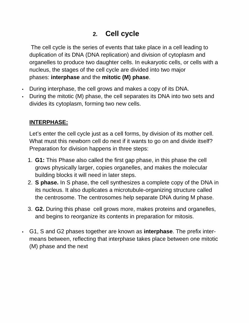
The cell cycle is the series of events that take place in a cell leading to duplication of its DNA (DNA replication) and division of cytoplasm and organelles to produce two daughter cells. In eukaryotic cells, or cells with a nucleus, the stages of the cell cycle are divided into two major phases: interphase and the mitotic (M) phase.
• During interphase, the cell grows and makes a copy of its DNA. • During the mitotic (M) phase, the cell separates its DNA into two sets and
divides its cytoplasm, forming two new cells.
INTERPHASE:
Let’s enter the cell cycle just as a cell forms, by division of its mother cell. What must this newborn cell do next if it wants to go on and divide itself? Preparation for division happens in three steps:
1. G1: This Phase also called the first gap phase, in this phase the cell grows physically larger, copies organelles, and makes the molecular building blocks it will need in later steps.
2. S phase. In S phase, the cell synthesizes a complete copy of the DNA in its nucleus. It also duplicates a microtubule-organizing structure called the centrosome. The centrosomes help separate DNA during M phase.
3. G2. During this phase cell grows more, makes proteins and organelles, and begins to reorganize its contents in preparation for mitosis.
• G1, S and G2 phases together are known as interphase. The prefix inter-
means between, reflecting that interphase takes place between one mitotic (M) phase and the next
Cell cycle figure
3 Study of different stages of Mitosis
All the multicellular organism are descedents of one of originl cell (zygote) through a division process called mitosis. Te function of mitosis is to construct an exact copy of each chromosome, an identical set of chromosomes and then to distribute through division of the original cell to each of the two daughter cells. Mitosis is divided into following stages.
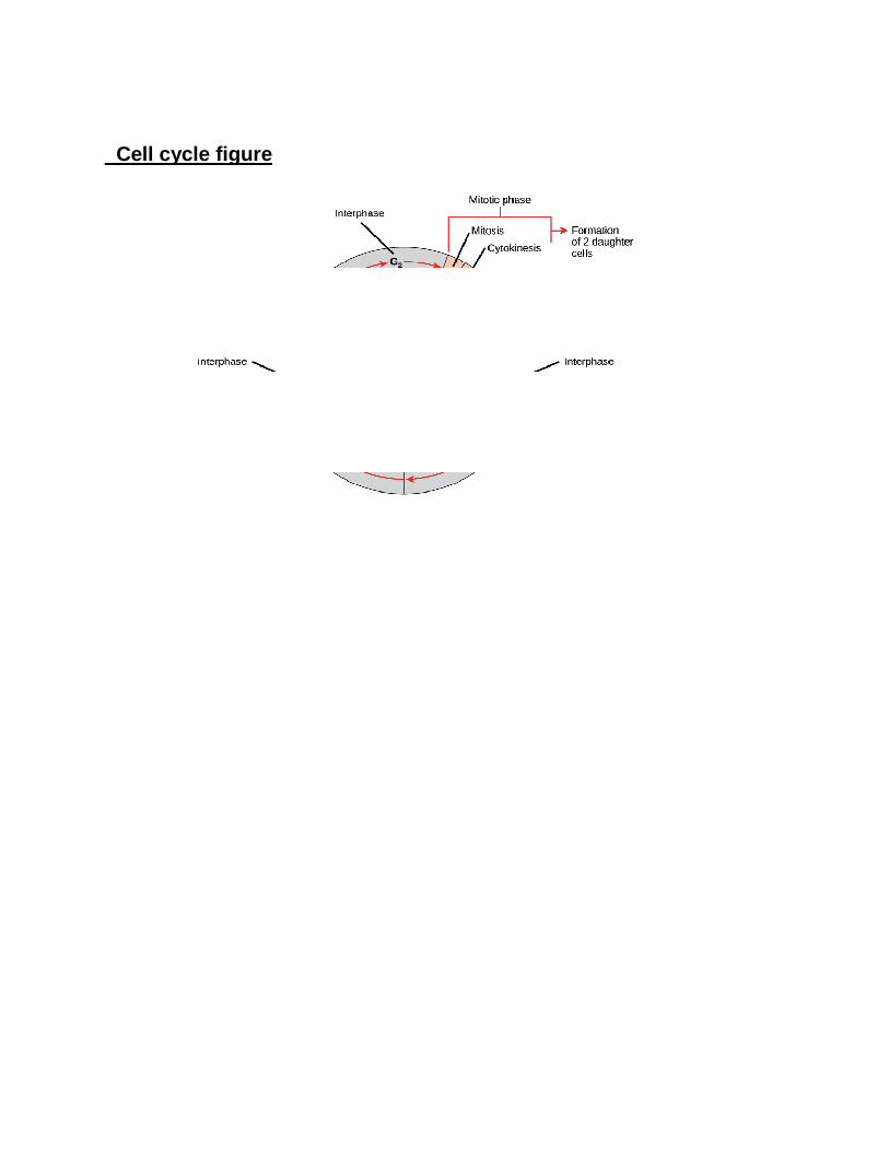
1. Interphase: It is period between two division cycles. When the cell is ready to begin mitosis DNA molecules replicates. This process produce chromosome with two identical chromatids both attached to a centromere.
2. Prophase: The chromosome become visible due to coiling, shortening and thickening. During late prophase each of the chromosomes splits up longitudinally given rise to two identical chromatids. At the end ofprophase nucleolus and nuclear membrane disappears.
3. Metaphase: Chromosomes get arranged themselves on equatorial plate or centre of the cell. The spindle fibre and spindle apparatus become fuly formed. The spindle fibres get attached o the centromere of chromosomes. All centromeres lie on the equatorial plate in one plane.
4. Anaphase : It begins when centromere splits into two, allowing sister chromatids to separate and move towards opposite poles led by their centromeres. Separated chromatids can be designated as daughter chromosomes (new chromosomes).
5. Telophase: An identical set of chromosomes is assembled at each pole of the cell. The chromosomes begin to coil to an interphase condition. The spindle degenerates and the nuclear membrane reforms.
Cytokinesis: The cytoplasm divides by the process called cytokinesis. The cytokinesis may takes place either by cell plate formation (plant cell) or by
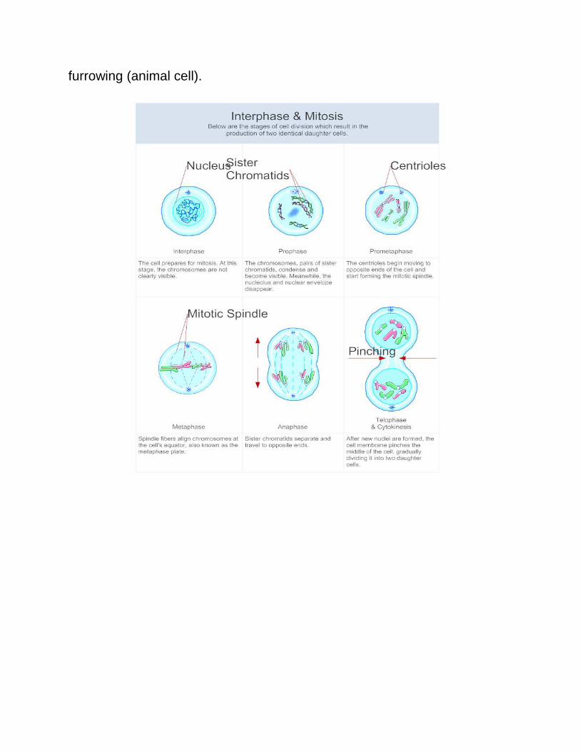
furrowing (animal cell).
4.Study of different stages of Meiosis
Sexual reproduction involves the production of gamates (gamatogenesis) and their union (fertilization) occurs in specialized cells (germ cells) of the reproductive organs. Gamates contain haploid (n) number of chromosomes, but are originated from diploid (2n)cells of germ line. Obviously during the process the number of chromosome must be reduced t half. meiosis actually involves two divisions. The first meiotic division (Meiosis 1) is reductional division producing two haploid cells from single diploid cell.The second division is (Meiosis 2) a equational division which separates the sister chromatids of the haploid cells and results in production of four daughter cells.
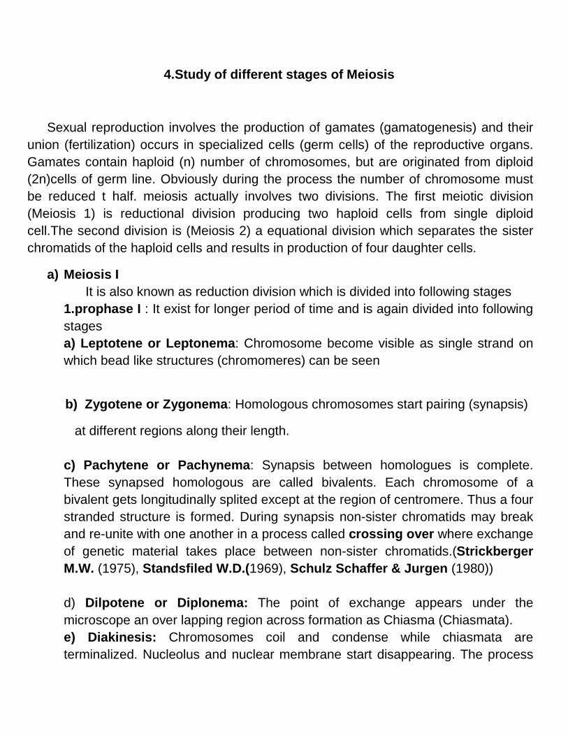
a) Meiosis I It is also known as reduction division which is divided into following stages 1.prophase I : It exist for longer period of time and is again divided into following stages a) Leptotene or Leptonema: Chromosome become visible as single strand on which bead like structures (chromomeres) can be seen
b) Zygotene or Zygonema: Homologous chromosomes start pairing (synapsis)
at different regions along their length. c) Pachytene or Pachynema: Synapsis between homologues is complete. These synapsed homologous are called bivalents. Each chromosome of a bivalent gets longitudinally splited except at the region of centromere. Thus a four stranded structure is formed. During synapsis non-sister chromatids may break and re-unite with one another in a process called crossing over where exchange of genetic material takes place between non-sister chromatids.(Strickberger M.W. (1975), Standsfiled W.D.(1969), Schulz Schaffer & Jurgen (1980)) d) Dilpotene or Diplonema: The point of exchange appears under the microscope an over lapping region across formation as Chiasma (Chiasmata). e) Diakinesis: Chromosomes coil and condense while chiasmata are terminalized. Nucleolus and nuclear membrane start disappearing. The process
of condensation and realational coiling is progressively advanced from leptotene to diakinesis. Nuclear membrane and nucleolus exist in all stages of Prophase I 2. Metaphase I: Bivalents orient themselves at random on the equatorial plate in such a way that all chaismata are in one plateand one chromosome of bivalent on either side of equatorial plate 3. Anaphase I: At first anaphase the centromeres do not divide but continue to hold sister chromatids. The homologous separates and move to opposite poles. Thus reduction in the chromosome number from diploid to haploid condition is achived. 4. Telophase I: Two groups are formed at two poles ,coiled chromosome get recoilied. The nuclear membrane may not formed. The daughter nuclei thus receives one chromosome from each pair . the centromere are still undivided. Cytokinesis may or may not begin. Interphase may or may not takes place after meiosis I
Meiosis II Second stage of meiotic division is same as equational division of normal mitiosis and all the phases designated here are Prophase II, Metaphase II, Anaphase II,Telophase II and Cytokinesis. Distinguishing features of second meiotic diviosion are given below.
1. Prophase II: Two chromatids from each f the chromosome of the preceeding telophase I are still associated at their centromere.Matrix becomes prominent and chromosomes stain heavily
2. Metaphase II: Nuclear membrane and nucleoli disappear. The chromosomes with their chromatids lie at equatorial plate again with their centromeres at equator but at righ angle to the first division. Spindle structure is formed and attached to the centromere.
3. Anaphase II: At this stage, centromere are divided and two daughter chromosomes are separated. With the division of centromere and separation of chromatids by their centromere each chromatid become the
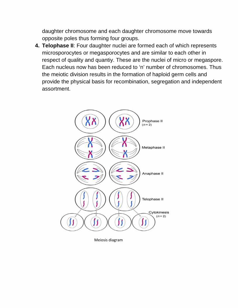
daughter chromosome and each daughter chromosome move towards opposite poles thus forming four groups.
4. Telophase II: Four daughter nuclei are formed each of which represents microsporocytes or megasporocytes and are similar to each other in respect of quality and quantiy. These are the nuclei of micro or megaspore. Each nucleus now has been reduced to ‘n’ number of chromosomes. Thus the meiotic division results in the formation of haploid germ cells and provide the physical basis for recombination, segregation and independent assortment.
Meiosis diagram
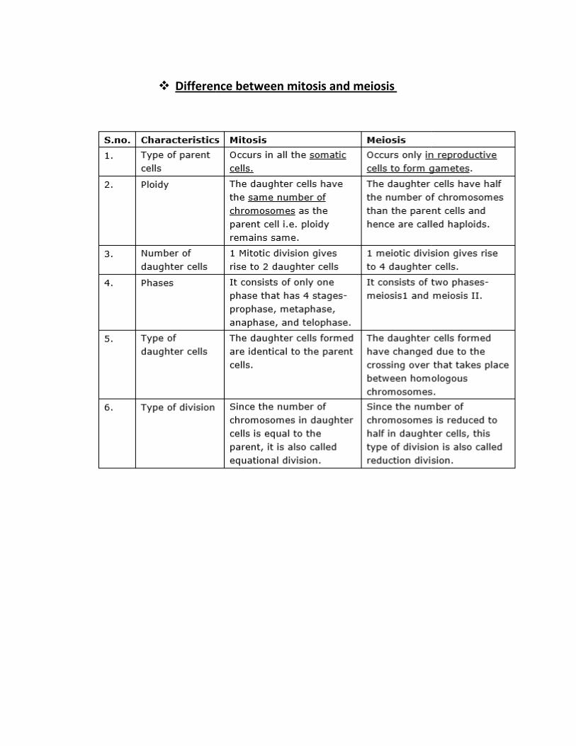
� Difference between mitosis and meiosis
Difference between mitosis and meiosis
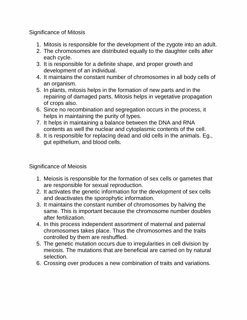
Significance of Mitosis
1. Mitosis is responsible for the development of the zygote into an adult. 2. The chromosomes are distributed equally to the daughter cells after
each cycle. 3. It is responsible for a definite shape, and proper growth and
development of an individual. 4. It maintains the constant number of chromosomes in all body cells of
an organism. 5. In plants, mitosis helps in the formation of new parts and in the
repairing of damaged parts. Mitosis helps in vegetative propagation of crops also.
6. Since no recombination and segregation occurs in the process, it helps in maintaining the purity of types.
7. It helps in maintaining a balance between the DNA and RNA contents as well the nuclear and cytoplasmic contents of the cell.
8. It is responsible for replacing dead and old cells in the animals. Eg., gut epithelium, and blood cells.
Significance of Meiosis
1. Meiosis is responsible for the formation of sex cells or gametes that are responsible for sexual reproduction.
2. It activates the genetic information for the development of sex cells and deactivates the sporophytic information.
3. It maintains the constant number of chromosomes by halving the same. This is important because the chromosome number doubles after fertilization.
4. In this process independent assortment of maternal and paternal chromosomes takes place. Thus the chromosomes and the traits controlled by them are reshuffled.
5. The genetic mutation occurs due to irregularities in cell division by meiosis. The mutations that are beneficial are carried on by natural selection.
6. Crossing over produces a new combination of traits and variations.
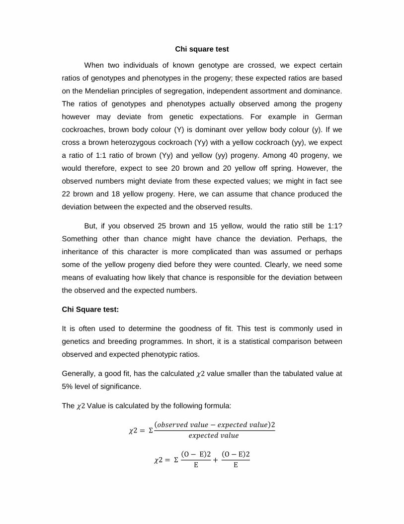
Chi square test
When two individuals of known genotype are crossed, we expect certain
ratios of genotypes and phenotypes in the progeny; these expected ratios are based
on the Mendelian principles of segregation, independent assortment and dominance.
The ratios of genotypes and phenotypes actually observed among the progeny
however may deviate from genetic expectations. For example in German
cockroaches, brown body colour (Y) is dominant over yellow body colour (y). If we
cross a brown heterozygous cockroach (Yy) with a yellow cockroach (yy), we expect
a ratio of 1:1 ratio of brown (Yy) and yellow (yy) progeny. Among 40 progeny, we
would therefore, expect to see 20 brown and 20 yellow off spring. However, the
observed numbers might deviate from these expected values; we might in fact see
22 brown and 18 yellow progeny. Here, we can assume that chance produced the
deviation between the expected and the observed results.
But, if you observed 25 brown and 15 yellow, would the ratio still be 1:1?
Something other than chance might have chance the deviation. Perhaps, the
inheritance of this character is more complicated than was assumed or perhaps
some of the yellow progeny died before they were counted. Clearly, we need some
means of evaluating how likely that chance is responsible for the deviation between
the observed and the expected numbers.
Chi Square test:
It is often used to determine the goodness of fit. This test is commonly used in
genetics and breeding programmes. In short, it is a statistical comparison between
observed and expected phenotypic ratios.
Generally, a good fit, has the calculated �2 value smaller than the tabulated value at
5% level of significance.
The �2 Value is calculated by the following formula:
�2 = Σ����� ���� − ���� �����2
���� ����
�2 = Σ �O − E�2
E+
�O − E�2
E
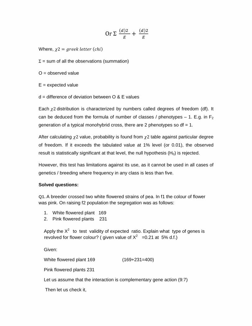
Or Σ ����
�+
����
�
Where, �2 = ��� ���� ��ℎ �
Σ = sum of all the observations (summation)
O = observed value
E = expected value
d = difference of deviation between O & E values
Each �2 distribution is characterized by numbers called degrees of freedom (df). It
can be deduced from the formula of number of classes / phenotypes – 1. E.g. in F2
generation of a typical monohybrid cross, there are 2 phenotypes so df = 1.
After calculating �2 value, probability is found from �2 table against particular degree
of freedom. If it exceeds the tabulated value at 1% level (or 0.01), the observed
result is statistically significant at that level, the null hypothesis (H0) is rejected.
However, this test has limitations against its use, as it cannot be used in all cases of
genetics / breeding where frequency in any class is less than five.
Solved questions:
Q1. A breeder crossed two white flowered strains of pea. In f1 the colour of flower was pink. On raising f2 population the segregation was as follows:
1. White flowered plant 169 2. Pink flowered plants 231 Apply the X2 to test validity of expected ratio. Explain what type of genes is revolved for flower colour? ( given value of X2 =0.21 at 5% d.f.) Given:
White flowered plant 169 (169+231=400)
Pink flowered plants 231
Let us assume that the interaction is complementary gene action (9:7)
Then let us check it,
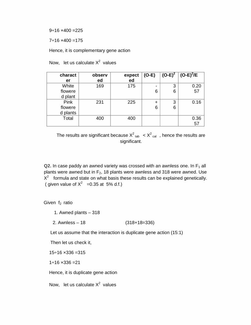
9÷16 ×400 =225
7÷16 ×400 =175
Hence, it is complementary gene action Now, let us calculate X2 values
character
observed
expected
(O-E) (O-E)2 (O-E)2/E
White flowered plant
169 175 -6
36
0.2057
Pink flowered plants
231 225 +6
36
0.16
Total 400 400 0.3657
The results are significant because X2
tab < X2 cal , hence the results are
significant.
Q2. In case paddy an awned variety was crossed with an awnless one. In F1 all plants were awned but in F2, 18 plants were awnless and 318 were awned. Use X2 formula and state on what basis these results can be explained genetically. ( given value of X2 =0.35 at 5% d.f.) Given f2 ratio
1. Awned plants – 318
2. Awnless – 18 (318+18=336)
Let us assume that the interaction is duplicate gene action (15:1)
Then let us check it,
15÷16 ×336 =315
1÷16 ×336 =21
Hence, it is duplicate gene action Now, let us calculate X2 values
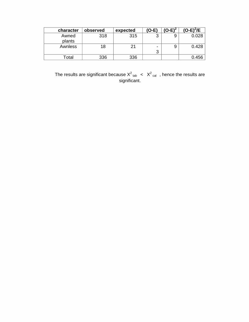
character observed expected (O-E) (O-E)2 (O-E)2/E Awned plants
318 315 3 9 0.028
Awnless 18 21 -3
9 0.428
Total 336 336 0.456
The results are significant because X2 tab < X2
cal , hence the results are significant.
Study of chromosome structure, morphology, number and types - Karyotype and Idiogram
A chromosome is a structure that occurs within cells and that contains the cell's genetic
material. That genetic material, which determines how an organism develops, is a molecule of
deoxyribonucleic acid (DNA). A molecule of DNA is a very long, coiled structure that contains
many identifiable subunits known as genes. In prokaryotes, or cells without a nucleus, the
chromosome is merely a circle of DNA. In eukaryotes, or cells with a distinct nucleus,
chromosomes are much more complex in structure.
Historical background
The terms chromosome and gene were used long before biologists really understood
what these structures were. When the Austrian monk and biologist Gregor Mendel (1822–1884)
developed the basic ideas of heredity, he assumed that genetic traits were somehow
transmitted from parents to offspring in some kind of tiny "package." That package was later
given the name "gene." When the term was first suggested, no one had any idea as to what a
gene might look like. The term was used simply to convey the idea that traits are transmitted
from one generation to the next in certain discrete units.
The term "chromosome" was first suggested in 1888 by the German anatomist Heinrich
Wilhelm Gottfried von Waldeyer-Hartz (1836–1921). Waldeyer-Hartz used the term to describe
certain structures that form during the process of cell division (reproduction).
One of the greatest breakthroughs in the history of biology occurred in 1953 when
American biologist James Watson and English chemist Francis Crick discovered the chemical
structure of a class of compounds known as deoxyribonucleic acids (DNA). The Watson and
Crick discovery made it possible to express biological concepts (such as the gene) and
structures (such as the chromosome) in concrete chemical terms.
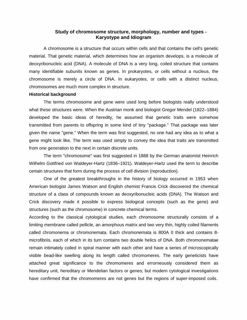
According to the classical cytological studies, each chromosome structurally consists of a
limiting membrane called pellicle, an amorphous matrix and two very thin, highly coiled filaments
called chromonema or chromonemata. Each chromonemata is 800A 0 thick and contains 8-
microfibriis, each of which in its turn contains two double helics of DNA. Both chromonematae
remain intimately coiled in spiral manner with each other and have a series of microscopically
visible bead-like swelling along its length called chromomeres. The early geneticists have
attached great significance to the chromomeres and errorneously considered them as
hereditary unit, hereditary or Mendelian factors or genes; but modern cytological investigations
have confirmed that the chromomeres are not genes but the regions of super-imposed coils.
The recent cytological findings have also condemned the view that chromosomes have pellicle,
matrix and chromonemata.
A. Nucleolus organizer B. Chromosome C. Nucleolus
The structure of chromosomes and genes
A chromosome is an organized structure of DNA and protein that is found in cells. A
chromosome is a single piece of coiled DNA containing many genes, regulatory elements and
other nucleotide sequences. Chromosomes also contain DNA-bound proteins, which serve to
package the DNA and control its functions. The word chromosome comes from the Greek
chroma - color and soma - body due to their property of being very strongly stained by particular
dyes. Chromosomes vary widely between different organisms. The DNA molecule may be
circular or linear, and can be composed of 10,000 to 1,000,000,000 nucleotides in a long chain.
Typically eukaryotic cells (cells with nuclei) have large linear chromosomes and prokaryotic cells
(cells without defined nuclei) have smaller circular chromosomes, although there are many
exceptions to this rule.
Today we know that a chromosome contains a single molecule of DNA along with
several kinds of proteins. A molecule of DNA, in turn, consists of thousands and thousands of
subunits, known as nucleotides, joined to each other in very long chains. A single molecule of
DNA within a chromosome may be as long as 8.5 centimeters (3.3 inches). To fit within a
chromosome, the DNA molecule has to be twisted and folded into a very complex shape.
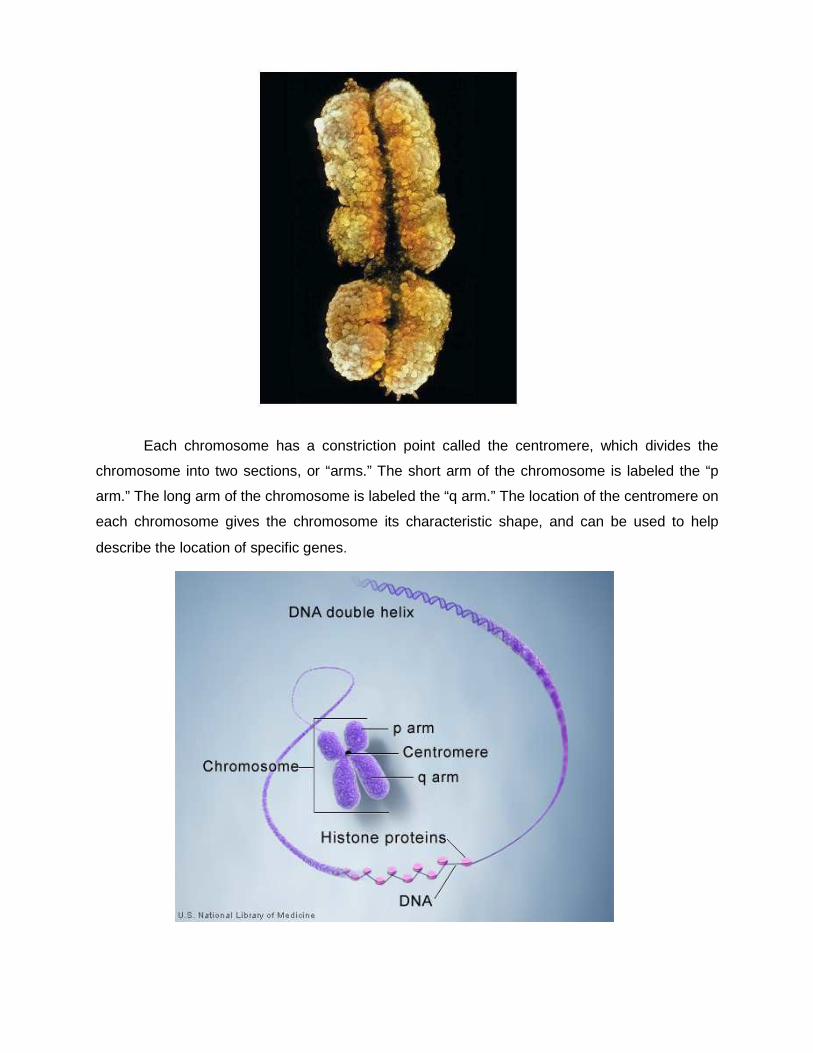
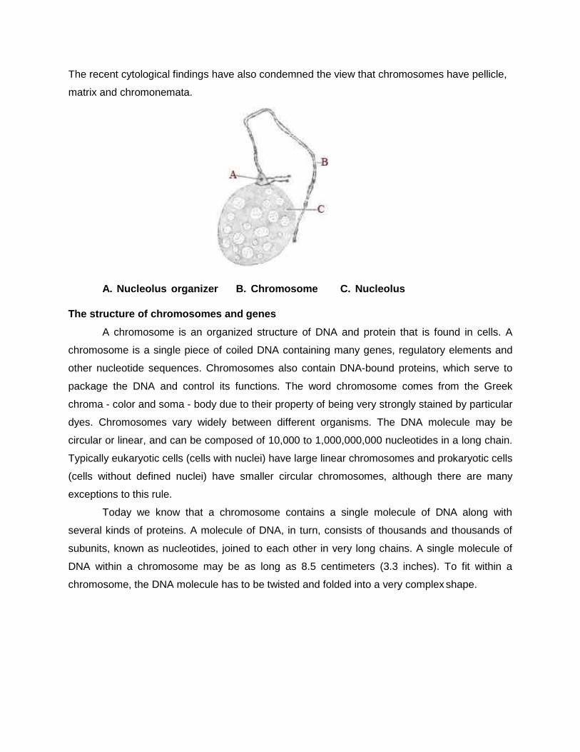
Each chromosome has a constriction point called the centromere, which divides the
chromosome into two sections, or “arms.” The short arm of the chromosome is labeled the “p
arm.” The long arm of the chromosome is labeled the “q arm.” The location of the centromere on
each chromosome gives the chromosome its characteristic shape, and can be used to help
describe the location of specific genes.
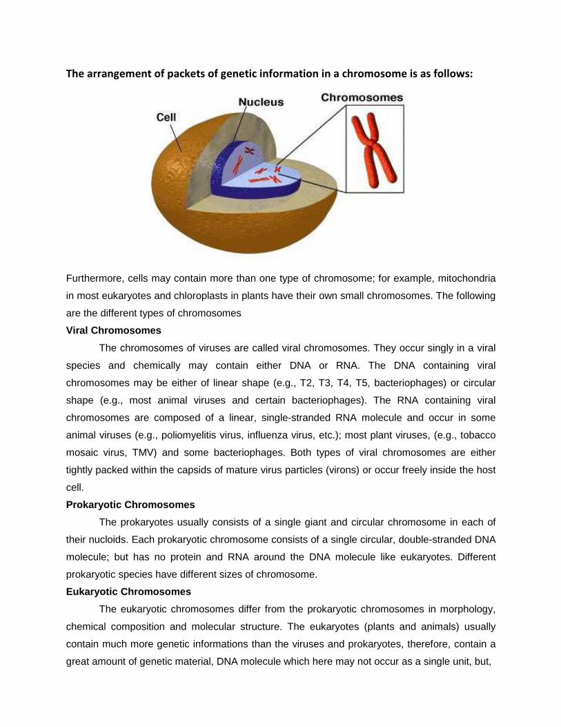
The arrangement of packets of genetic information in a chromosome is as follows:
Furthermore, cells may contain more than one type of chromosome; for example, mitochondria
in most eukaryotes and chloroplasts in plants have their own small chromosomes. The following
are the different types of chromosomes
Viral Chromosomes
The chromosomes of viruses are called viral chromosomes. They occur singly in a viral
species and chemically may contain either DNA or RNA. The DNA containing viral
chromosomes may be either of linear shape (e.g., T2, T3, T4, T5, bacteriophages) or circular
shape (e.g., most animal viruses and certain bacteriophages). The RNA containing viral
chromosomes are composed of a linear, single-stranded RNA molecule and occur in some
animal viruses (e.g., poliomyelitis virus, influenza virus, etc.); most plant viruses, (e.g., tobacco
mosaic virus, TMV) and some bacteriophages. Both types of viral chromosomes are either
tightly packed within the capsids of mature virus particles (virons) or occur freely inside the host
cell.
Prokaryotic Chromosomes
The prokaryotes usually consists of a single giant and circular chromosome in each of
their nucloids. Each prokaryotic chromosome consists of a single circular, double-stranded DNA
molecule; but has no protein and RNA around the DNA molecule like eukaryotes. Different
prokaryotic species have different sizes of chromosome.
Eukaryotic Chromosomes
The eukaryotic chromosomes differ from the prokaryotic chromosomes in morphology,
chemical composition and molecular structure. The eukaryotes (plants and animals) usually
contain much more genetic informations than the viruses and prokaryotes, therefore, contain a
great amount of genetic material, DNA molecule which here may not occur as a single unit, but,
as many units called chromosomes. Different species of eukaryotes have different but always
constant and characteristic number of chromosomes. In eukaryotes, nuclear chromosomes are
packaged by proteins into a condensed structure called chromatin. This allows the very long
DNA molecules to fit into the cell nucleus. The shape of the eukaryotic chromosomes is
changeable from phase to phase in the continuous process of the cell growth and cell division.
Chromosomes are the essential unit for cellular division and must be replicated, divided, and
passed successfully to their daughter cells so as to ensure the genetic diversity and survival of
their progeny. They are thin, coiled, elastic, contractile thread-like structures during the
interphase (when no division of cell occurs) and are called chromatin threads which under low
magnification look like a compact stainable mass, often called as chromatin substance or
material. During metaphase stage of mitosis and prophase of meiosis, these chromatin threads
become highly coiled and folded to form compact and individually distinct ribbon-shaped
chromosomes. These chromosomes contain a clear zone called kinetochore or centromere
along their length.
Eukaryotes (cells with nuclei such as plants, yeast, and animals) possess multiple large
linear chromosomes contained in the cell's nucleus. Each chromosome has one centromere,
with one or two arms projecting from the centromere, although, under most circumstances,
these arms are not visible as such. In addition, most eukaryotes have a small circular
mitochondrial genome, and some eukaryotes may have additional small circular or linear
cytoplasmic chromosomes.
The number and position of centromeres is variable, but is definite in a specific
chromosome of all the cells and in all the individuals of the same species. Thus, according to
the number of the centromere the eukaryotic chromosomes may be acentric (without any
centromere), mono centric (with one centromere), dicentric (with two centromeres) or
polycentric (with more than two centromeres). The centromere has small granules or spherules
and divides the chromosomes into two or more equal or unequal chromosomal arms.
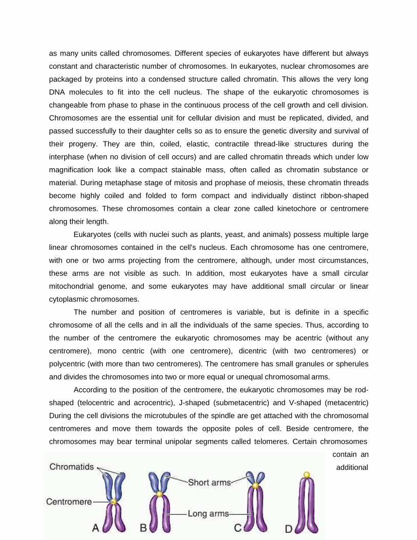
According to the position of the centromere, the eukaryotic chromosomes may be rod-
shaped (telocentric and acrocentric), J-shaped (submetacentric) and V-shaped (metacentric)
During the cell divisions the microtubules of the spindle are get attached with the chromosomal
centromeres and move them towards the opposite poles of cell. Beside centromere, the
chromosomes may bear terminal unipolar segments called telomeres. Certain chromosomes
contain an
additional
specialized segment, the nucleolus organizer, which is associated with the nucleolus.
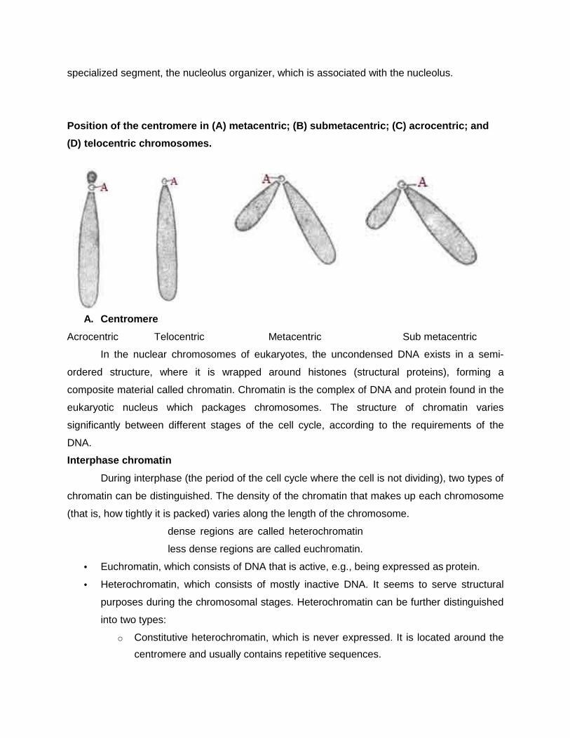
Position of the centromere in (A) metacentric; (B) submetacentric; (C) acrocentric; and
(D) telocentric chromosomes.
A. Centromere
Acrocentric Telocentric Metacentric Sub metacentric
In the nuclear chromosomes of eukaryotes, the uncondensed DNA exists in a semi-
ordered structure, where it is wrapped around histones (structural proteins), forming a
composite material called chromatin. Chromatin is the complex of DNA and protein found in the
eukaryotic nucleus which packages chromosomes. The structure of chromatin varies
significantly between different stages of the cell cycle, according to the requirements of the
DNA.
Interphase chromatin
During interphase (the period of the cell cycle where the cell is not dividing), two types of
chromatin can be distinguished. The density of the chromatin that makes up each chromosome
(that is, how tightly it is packed) varies along the length of the chromosome.
dense regions are called heterochromatin
less dense regions are called euchromatin.
• Euchromatin, which consists of DNA that is active, e.g., being expressed as protein.
• Heterochromatin, which consists of mostly inactive DNA. It seems to serve structural
purposes during the chromosomal stages. Heterochromatin can be further distinguished
into two types:
o Constitutive heterochromatin, which is never expressed. It is located around the
centromere and usually contains repetitive sequences.
o Facultative heterochromatin, which is sometimes expressed.
Individual chromosomes cannot be distinguished at this stage - they appear in the nucleus as a
homogeneous tangled mix of DNA and protein.
Diploids and Haploids
In contrast to prokaryotes, most eukaryote are diploids, i.e., each somatic cell of them
contains one set of chromosomes inherited from the maternal (female) parent and a comparable
set of chromosomes (called homologous chromosomes) from the paternal (male) parent. The
number of chromosomes in a dual set of a diploid somatic cell is called the diploid number (2n).
The sex cells (sperms and ova) of a diploid eukaryote cell contain half the number of
chromosomal sets found in the somatic cells and are known as haploid (n) cells. A haploid set of
chromosome is also called genome. The fertilization process restores the diploid number of a
diploid species.
Chemical Structure of Chromosomes
Chemically, the eukaryotic chromosomes are composed of deoxyribonucleic acid (DNA),
ribonucleic acid (RNA), histone and non-histone proteins and certain metallic ions. The histone
proteins have basic properties and have significant role in controlling or regulating the functions
of chromosomal DNA. The non-histone proteins are mostly acidic and have been considered
more important than histones as regulatory molecules. Some non-histone proteins also have
enzymatic activities. The most important enzymatic proteins of chromosomes are
phosphoproteins, DNA polymerase, RNA-polymerase, DPN-pyropbosphorylase, and nucleoside
triphosphatase. The metal ions as Ca+ and Mg+ are supposed to maintain the oragnization of
chromosomes intact.
Molecular Structure of Chromosomes
According to the recent and widely accepted theory of Dupraw (1965, 1970) and Hans
Ris (1967) called unistranded theory, each eukaryotic chromosome is composed of a single,
greatly elongated and highly folded nucleoprotein fibre of 100A 0 thick. This nucleo- protein fibre
in its turn is composed of a single, linear, double stranded DNA molecule which remains
wrapped in equal amounts of histone and non-histone proteins and variable amounts of different
kinds of RNA. Dupraw produced a “folded-fibre Model" to show the ultrastructure of
chromosome.
FIBRE FOLDED MODEL
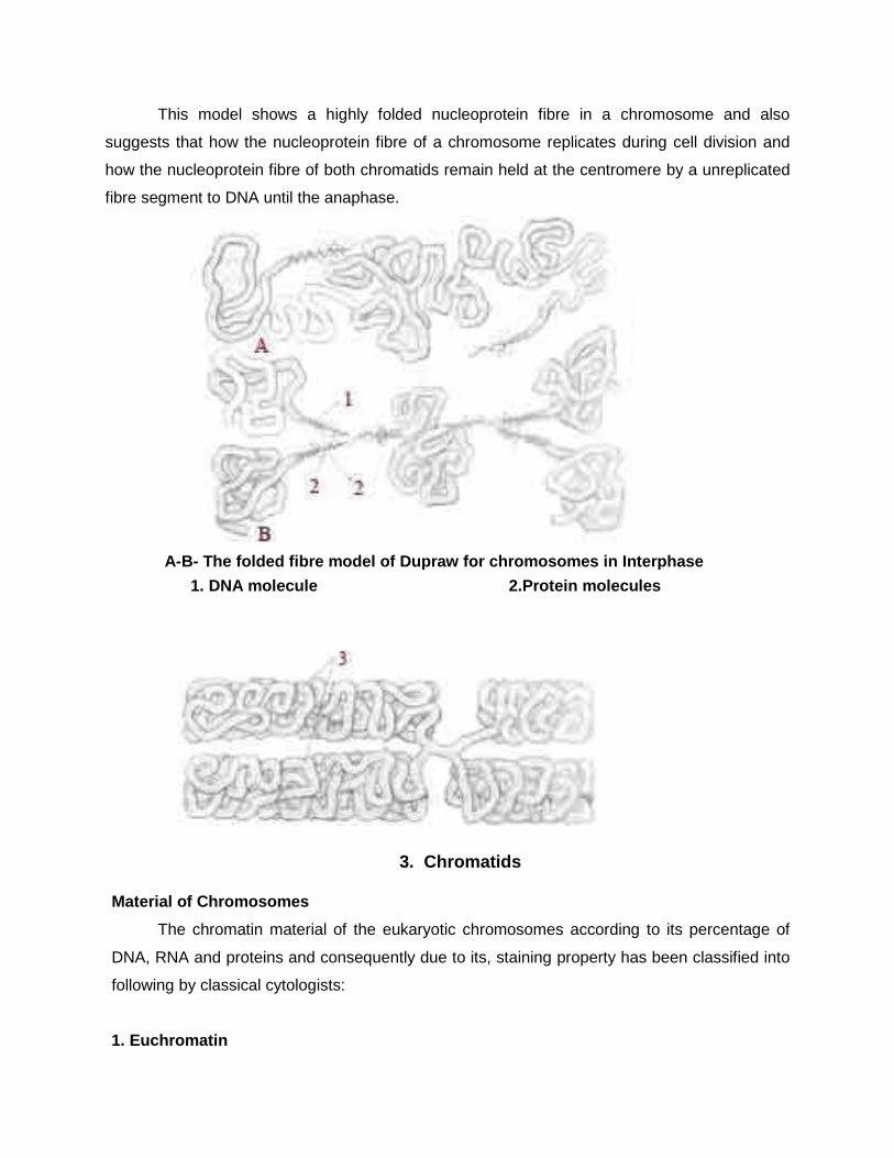
This model shows a highly folded nucleoprotein fibre in a chromosome and also
suggests that how the nucleoprotein fibre of a chromosome replicates during cell division and
how the nucleoprotein fibre of both chromatids remain held at the centromere by a unreplicated
fibre segment to DNA until the anaphase.
A-B- The folded fibre model of Dupraw for chromosomes in Interphase
1. DNA molecule 2.Protein molecules
3. Chromatids
Material of Chromosomes
The chromatin material of the eukaryotic chromosomes according to its percentage of
DNA, RNA and proteins and consequently due to its, staining property has been classified into
following by classical cytologists:
1. Euchromatin
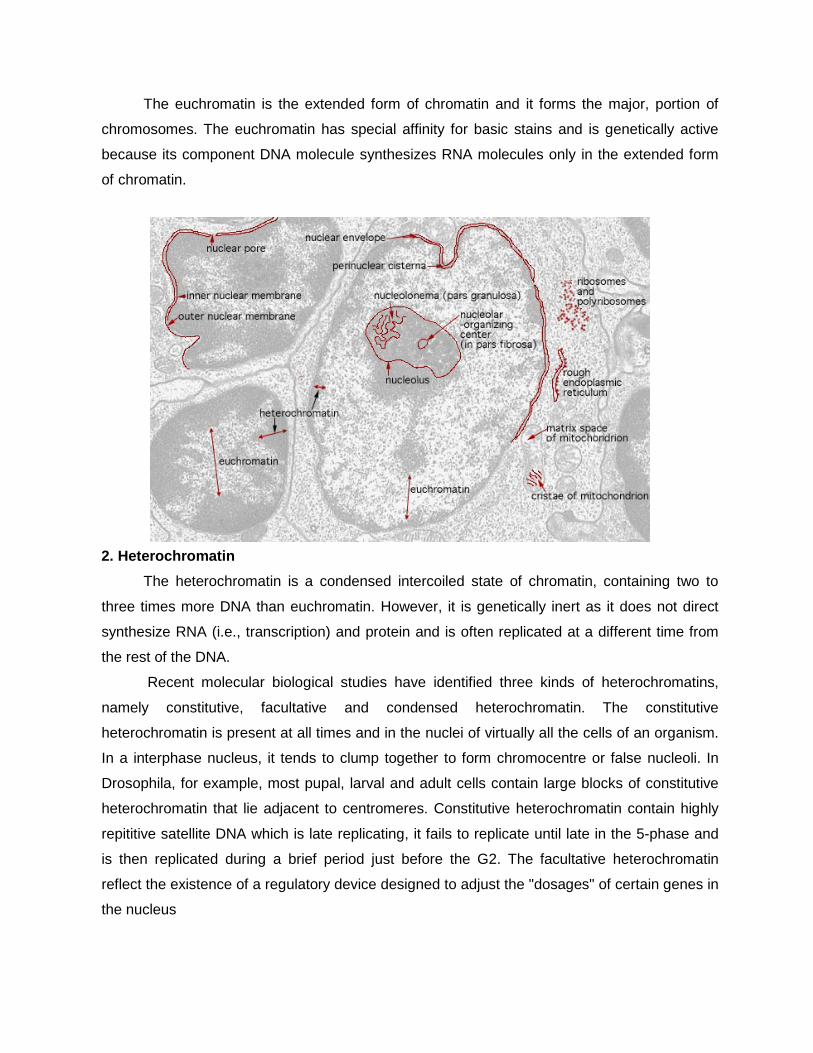
The euchromatin is the extended form of chromatin and it forms the major, portion of
chromosomes. The euchromatin has special affinity for basic stains and is genetically active
because its component DNA molecule synthesizes RNA molecules only in the extended form
of chromatin.
2. Heterochromatin
The heterochromatin is a condensed intercoiled state of chromatin, containing two to
three times more DNA than euchromatin. However, it is genetically inert as it does not direct
synthesize RNA (i.e., transcription) and protein and is often replicated at a different time from
the rest of the DNA.
Recent molecular biological studies have identified three kinds of heterochromatins,
namely constitutive, facultative and condensed heterochromatin. The constitutive
heterochromatin is present at all times and in the nuclei of virtually all the cells of an organism.
In a interphase nucleus, it tends to clump together to form chromocentre or false nucleoli. In
Drosophila, for example, most pupal, larval and adult cells contain large blocks of constitutive
heterochromatin that lie adjacent to centromeres. Constitutive heterochromatin contain highly
repititive satellite DNA which is late replicating, it fails to replicate until late in the 5-phase and
is then replicated during a brief period just before the G2. The facultative heterochromatin
reflect the existence of a regulatory device designed to adjust the "dosages" of certain genes in
the nucleus
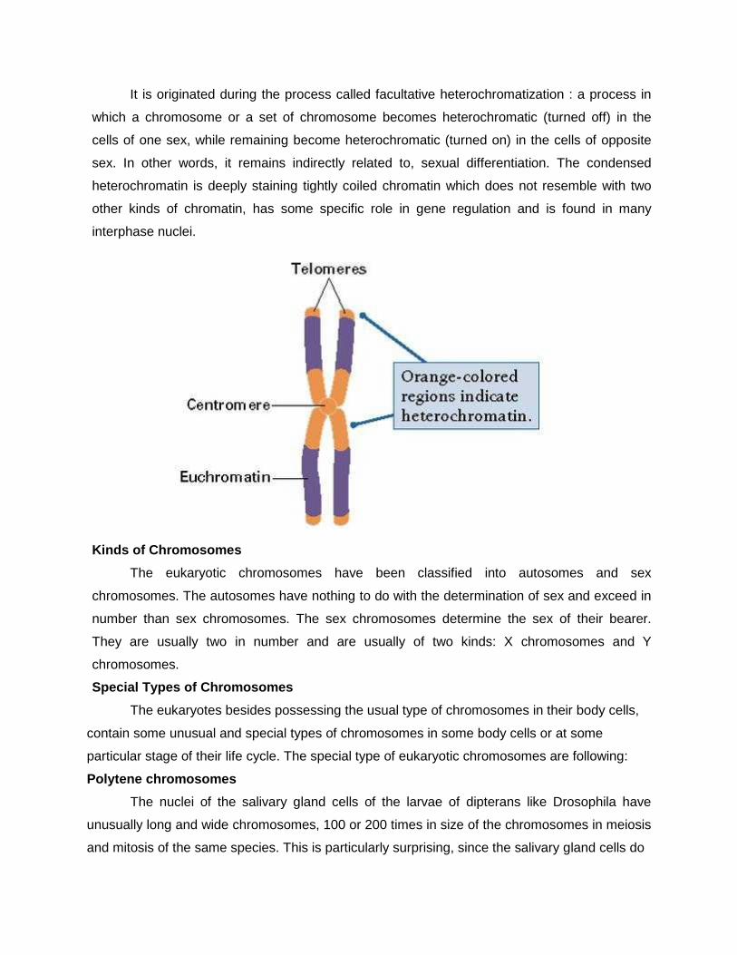
It is originated during the process called facultative heterochromatization : a process in
which a chromosome or a set of chromosome becomes heterochromatic (turned off) in the
cells of one sex, while remaining become heterochromatic (turned on) in the cells of opposite
sex. In other words, it remains indirectly related to, sexual differentiation. The condensed
heterochromatin is deeply staining tightly coiled chromatin which does not resemble with two
other kinds of chromatin, has some specific role in gene regulation and is found in many
interphase nuclei.
Kinds of Chromosomes
The eukaryotic chromosomes have been classified into autosomes and sex
chromosomes. The autosomes have nothing to do with the determination of sex and exceed in
number than sex chromosomes. The sex chromosomes determine the sex of their bearer.
They are usually two in number and are usually of two kinds: X chromosomes and Y
chromosomes.
Special Types of Chromosomes
The eukaryotes besides possessing the usual type of chromosomes in their body cells,
contain some unusual and special types of chromosomes in some body cells or at some
particular stage of their life cycle. The special type of eukaryotic chromosomes are following:
Polytene chromosomes
The nuclei of the salivary gland cells of the larvae of dipterans like Drosophila have
unusually long and wide chromosomes, 100 or 200 times in size of the chromosomes in meiosis
and mitosis of the same species. This is particularly surprising, since the salivary gland cells do
not divide after the glands are formed, yet their chromosomes replicate several times (a process
called endomitosis) and become exceptionally giant-sized to be called polytene or multistranded
chromosomes (discovered by Balbiani (l881) and named by Koller).The endomitosis process
result in the production of 2X chromosomes, where X gives the number of multiplication cycle.
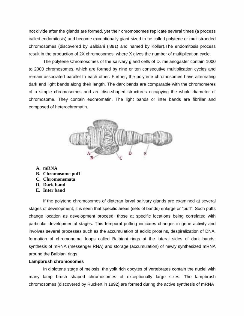
The polytene Chromosomes of the salivary gland cells of D. melanogaster contain 1000
to 2000 chromosomes, which are formed by nine or ten consecutive multiplication cycles and
remain associated parallel to each other. Further, the polytene chromosomes have alternating
dark and light bands along their length. The dark bands are comparable with the chromomeres
of a simple chromosomes and are disc-shaped structures occupying the whole diameter of
chromosome. They contain euchromatin. The light bands or inter bands are fibrillar and
composed of heterochromatin.
A. mRNA B. Chromosome puff C. Chromonemata D. Dark band E. Inter band
If the polytene chromosomes of dipteran larval salivary glands are examined at several
stages of development; it is seen that specific areas (sets of bands) enlarge or "puff". Such puffs
change location as development proceed, those at specific locations being correlated with
particular developmental stages. This temporal puffing indicates changes in gene activity and
involves several processes such as the accumulation of acidic proteins, despiralization of DNA,
formation of chromonemal loops called Balbiani rings at the lateral sides of dark bands,
synthesis of mRNA (messenger RNA) and storage (accumulation) of newly synthesized mRNA
around the Balbiani rings.
Lampbrush chromosomes
In diplotene stage of meiosis, the yolk rich oocytes of vertebrates contain the nuclei with
many lamp brush shaped chromosomes of exceptionally large sizes. The lampbrush
chromosomes (discovered by Ruckert in 1892) are formed during the active synthesis of mRNA
molecules for the future use by the egg during cleavage when no synthesis of mRNA molecules
is possible due to active involvement of chromosomes in the mitotic cell division.
.
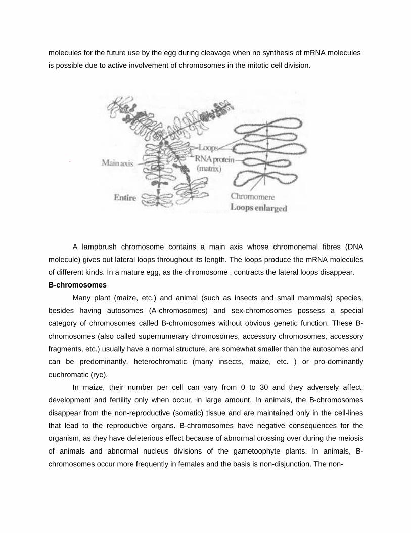
A lampbrush chromosome contains a main axis whose chromonemal fibres (DNA
molecule) gives out lateral loops throughout its length. The loops produce the mRNA molecules
of different kinds. In a mature egg, as the chromosome , contracts the lateral loops disappear.
B-chromosomes
Many plant (maize, etc.) and animal (such as insects and small mammals) species,
besides having autosomes (A-chromosomes) and sex-chromosomes possess a special
category of chromosomes called B-chromosomes without obvious genetic function. These B-
chromosomes (also called supernumerary chromosomes, accessory chromosomes, accessory
fragments, etc.) usually have a normal structure, are somewhat smaller than the autosomes and
can be predominantly, heterochromatic (many insects, maize, etc. ) or pro-dominantly
euchromatic (rye).
In maize, their number per cell can vary from 0 to 30 and they adversely affect,
development and fertility only when occur, in large amount. In animals, the B-chromosomes
disappear from the non-reproductive (somatic) tissue and are maintained only in the cell-lines
that lead to the reproductive organs. B-chromosomes have negative consequences for the
organism, as they have deleterious effect because of abnormal crossing over during the meiosis
of animals and abnormal nucleus divisions of the gametoophyte plants. In animals, B-
chromosomes occur more frequently in females and the basis is non-disjunction. The non-
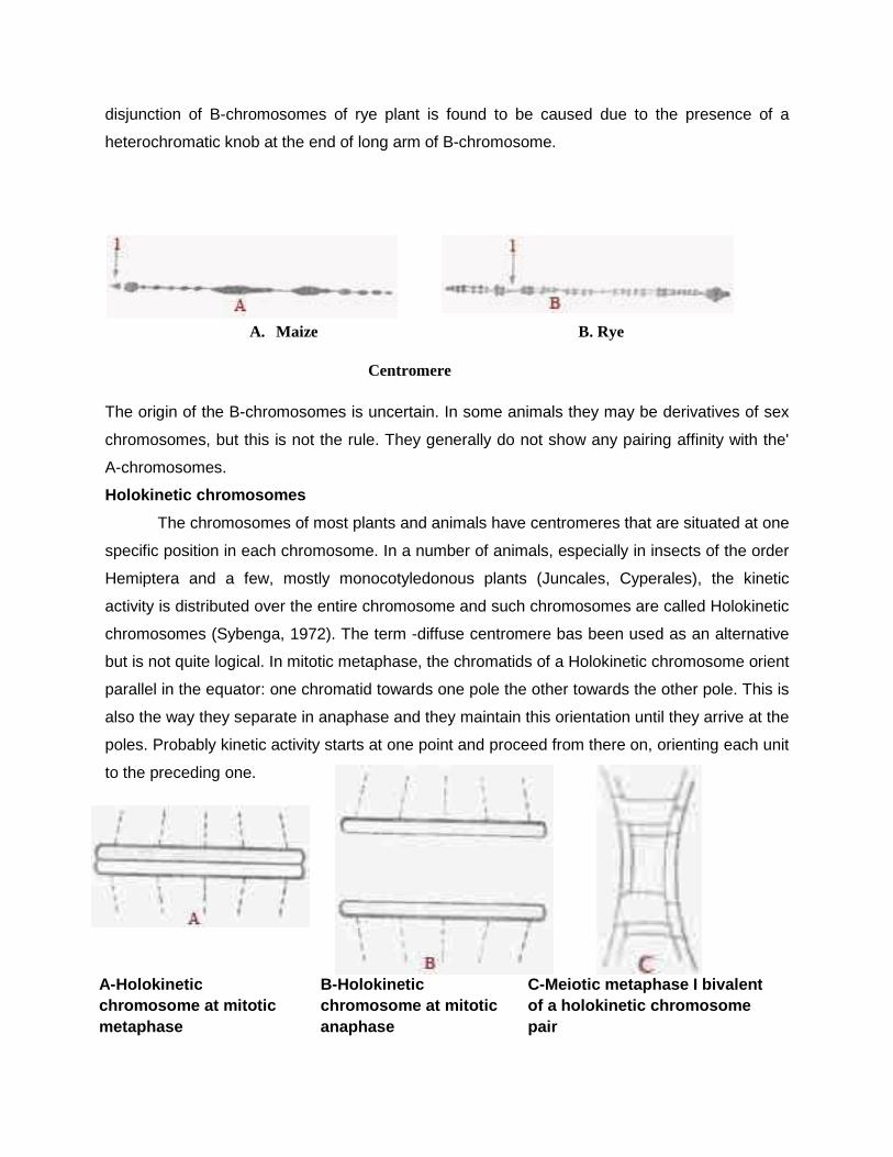
disjunction of B-chromosomes of rye plant is found to be caused due to the presence of a
heterochromatic knob at the end of long arm of B-chromosome.
A. Maize B. Rye
Centromere
The origin of the B-chromosomes is uncertain. In some animals they may be derivatives of sex
chromosomes, but this is not the rule. They generally do not show any pairing affinity with the'
A-chromosomes.
Holokinetic chromosomes
The chromosomes of most plants and animals have centromeres that are situated at one
specific position in each chromosome. In a number of animals, especially in insects of the order
Hemiptera and a few, mostly monocotyledonous plants (Juncales, Cyperales), the kinetic
activity is distributed over the entire chromosome and such chromosomes are called Holokinetic
chromosomes (Sybenga, 1972). The term -diffuse centromere bas been used as an alternative
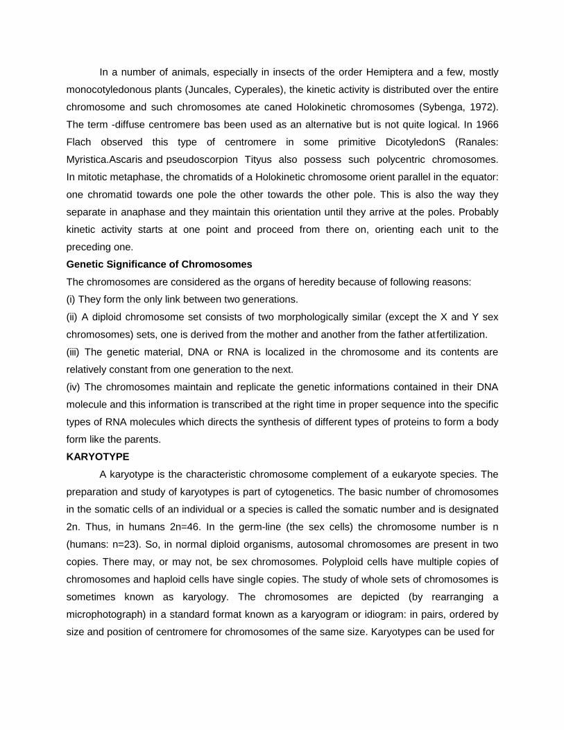
but is not quite logical. In mitotic metaphase, the chromatids of a Holokinetic chromosome orient
parallel in the equator: one chromatid towards one pole the other towards the other pole. This is
also the way they separate in anaphase and they maintain this orientation until they arrive at the
poles. Probably kinetic activity starts at one point and proceed from there on, orienting each unit
to the preceding one.
A-Holokinetic chromosome at mitotic metaphase
B-Holokinetic chromosome at mitotic anaphase
C-Meiotic metaphase I bivalent of a holokinetic chromosome pair
In a number of animals, especially in insects of the order Hemiptera and a few, mostly
monocotyledonous plants (Juncales, Cyperales), the kinetic activity is distributed over the entire
chromosome and such chromosomes ate caned Holokinetic chromosomes (Sybenga, 1972).
The term -diffuse centromere bas been used as an alternative but is not quite logical. In 1966
Flach observed this type of centromere in some primitive DicotyledonS (Ranales:
Myristica.Ascaris and pseudoscorpion Tityus also possess such polycentric chromosomes.
In mitotic metaphase, the chromatids of a Holokinetic chromosome orient parallel in the equator:
one chromatid towards one pole the other towards the other pole. This is also the way they
separate in anaphase and they maintain this orientation until they arrive at the poles. Probably
kinetic activity starts at one point and proceed from there on, orienting each unit to the
preceding one.
Genetic Significance of Chromosomes
The chromosomes are considered as the organs of heredity because of following reasons:
(i) They form the only link between two generations.
(ii) A diploid chromosome set consists of two morphologically similar (except the X and Y sex
chromosomes) sets, one is derived from the mother and another from the father at fertilization.
(iii) The genetic material, DNA or RNA is localized in the chromosome and its contents are
relatively constant from one generation to the next.
(iv) The chromosomes maintain and replicate the genetic informations contained in their DNA
molecule and this information is transcribed at the right time in proper sequence into the specific
types of RNA molecules which directs the synthesis of different types of proteins to form a body
form like the parents.
KARYOTYPE
A karyotype is the characteristic chromosome complement of a eukaryote species. The
preparation and study of karyotypes is part of cytogenetics. The basic number of chromosomes
in the somatic cells of an individual or a species is called the somatic number and is designated
2n. Thus, in humans 2n=46. In the germ-line (the sex cells) the chromosome number is n
(humans: n=23). So, in normal diploid organisms, autosomal chromosomes are present in two
copies. There may, or may not, be sex chromosomes. Polyploid cells have multiple copies of
chromosomes and haploid cells have single copies. The study of whole sets of chromosomes is
sometimes known as karyology. The chromosomes are depicted (by rearranging a
microphotograph) in a standard format known as a karyogram or idiogram: in pairs, ordered by
size and position of centromere for chromosomes of the same size. Karyotypes can be used for
many purposes; such as, to study chromosomal aberrations, cellular function, taxonomic
relationships, and to gather information about past evolutionary events.
Idiogram
Staining
The study of karyotypes is made possible by staining. Usually, a suitable dye is applied
after cells have been arrested during cell division by a solution of colchicine. For humans, white
blood cells are used most frequently because they are easily induced to divide and grow in
tissue culture. Sometimes observations may be made on non-dividing (interphase) cells. The
sex of an unborn fetus can be determined by observation of interphase cells (see amniotic
centesis and Barr body).
Most (but not all) species have a standard karyotype. The normal human karyotypes
contain 22 pairs of autosomal chromosomes and one pair of sex chromosomes. Normal
karyotypes for females contain two X chromosomes and are denoted 46, XX; males have both
an X and a Y chromosome denoted 46,XY. Any variation from the standard karyotype may lead
to developmental abnormalities.
Six different characteristics of karyotypes are usually observed and compared:
1. differences in absolute sizes of chromosomes. Chromosomes can vary in absolute size
by as much as twenty-fold between genera of the same family: Lotus tenuis and Vicia
faba (legumes), both have six pairs of chromosomes (n=6) yet V. faba chromosomes are
many times larger. This feature probably reflects different amounts of DNA duplication.
2. differences in the position of centromeres. This is brought about by translocations.
3. differences in relative size of chromosomes can only be caused by segmental
interchange of unequal lengths.
4. differences in basic number of chromosomes may occur due to successive unequal
translocations which finally remove all the essential genetic material from a
chromosome, permitting its loss without penalty to the organism (the dislocation
hypothesis). Humans have one pair fewer chromosomes than the great apes, but the
genes have been mostly translocated (added) to other chromosomes.
5. differences in number and position of satellites, which (when they occur) are small
bodies attached to a chromosome by a thin thread.
6. differences in degree and distribution of heterochromatic regions. Heterochromatin
stains darker than euchromatin, indicating tighter packing, and mainly consists of
genetically inactive repetitive DNA sequences.
A full account of a karyotype may therefore include the number, type, shape and banding of the
chromosomes, as well as other cytogenetic information.
Variation is often found:
1. between the sexes
2. between the germ-line and soma (between gametes and the rest of the body)
3. between members of a population (chromosome polymorphism)
4. geographical variation between races
5. mosaics or otherwise abnormal individuals
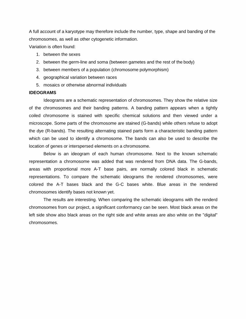
IDEOGRAMS
Ideograms are a schematic representation of chromosomes. They show the relative size
of the chromosomes and their banding patterns. A banding pattern appears when a tightly
coiled chromosome is stained with specific chemical solutions and then viewed under a
microscope. Some parts of the chromosome are stained (G-bands) while others refuse to adopt
the dye (R-bands). The resulting alternating stained parts form a characteristic banding pattern
which can be used to identify a chromosome. The bands can also be used to describe the
location of genes or interspersed elements on a chromosome.
Below is an ideogram of each human chromosome. Next to the known schematic
representation a chromosome was added that was rendered from DNA data. The G-bands,
areas with proportional more A-T base pairs, are normally colored black in schematic
representations. To compare the schematic ideograms the rendered chromosomes, were
colored the A-T bases black and the G-C bases white. Blue areas in the rendered
chromosomes identify bases not known yet.
The results are interesting. When comparing the schematic ideograms with the renderd
chromosomes from our project, a significant conformancy can be seen. Most black areas on the
left side show also black areas on the right side and white areas are also white on the "digital"
chromosomes.
CHROMOSOMES
E. Strasburger in 1875 first discovered thread-like structures which
appeared during cell division. These thread like structures were called
chromosomes due to their affinity for basic dyes. The term chromosome is derived
from two Greek words; chrom = colour, soma=body. This term was first used by
Waldeyer in 1888. Of all components of cell, the chromosomes have been studied
most extensively and perhaps more is known about them than any other cell
organelle. The chromosome has greater constancy than any other cell component
and it maintains it special qualities from one cell generation to another.
Chromosomes contributed to the division of cells and they are of prime
importance as they carry the genes which are the hereditary material.
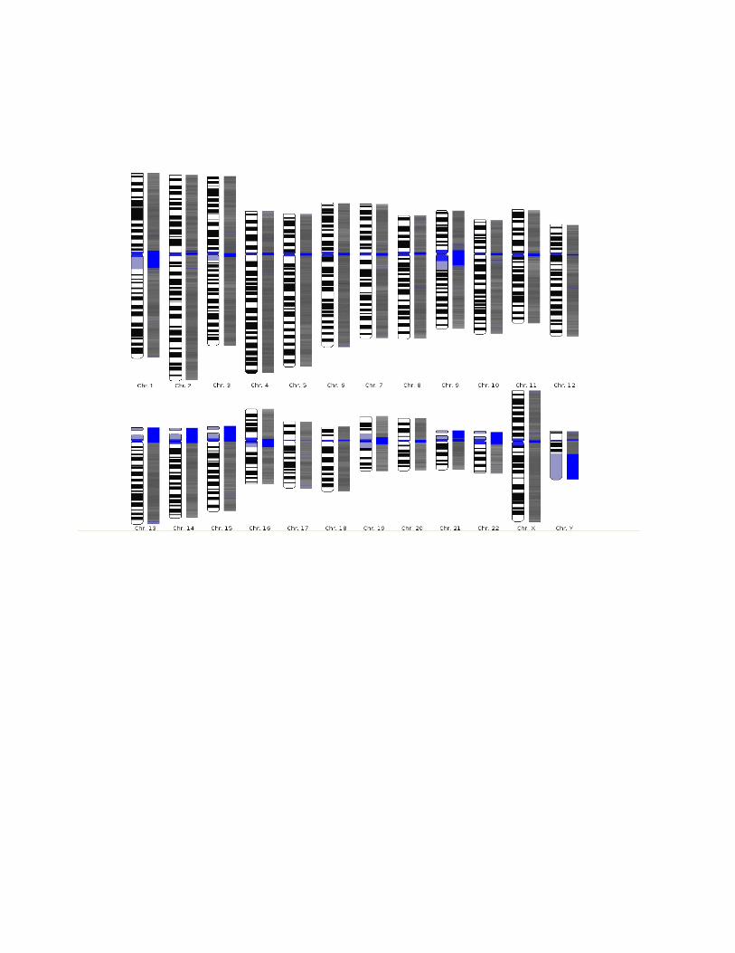
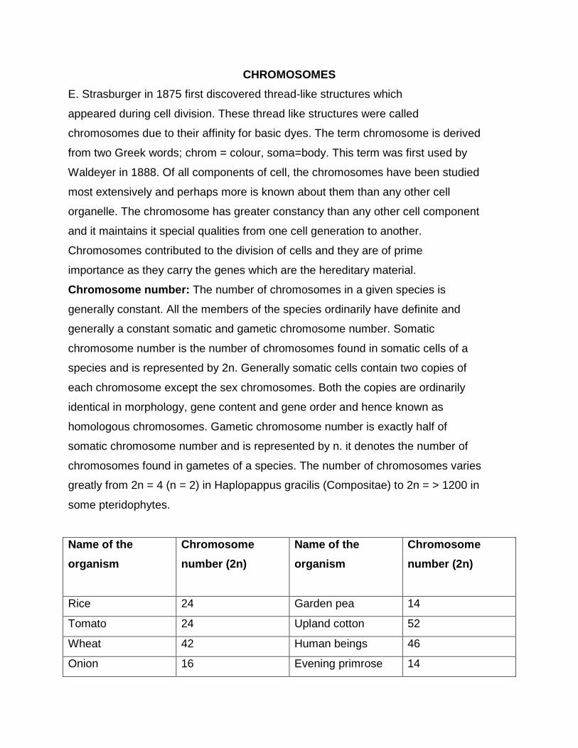
Chromosome number: The number of chromosomes in a given species is
generally constant. All the members of the species ordinarily have definite and
generally a constant somatic and gametic chromosome number. Somatic
chromosome number is the number of chromosomes found in somatic cells of a
species and is represented by 2n. Generally somatic cells contain two copies of
each chromosome except the sex chromosomes. Both the copies are ordinarily
identical in morphology, gene content and gene order and hence known as
homologous chromosomes. Gametic chromosome number is exactly half of
somatic chromosome number and is represented by n. it denotes the number of
chromosomes found in gametes of a species. The number of chromosomes varies
greatly from 2n = 4 (n = 2) in Haplopappus gracilis (Compositae) to 2n = > 1200 in
some pteridophytes.
Name of the
organism
Chromosome
number (2n)
Name of the
organism
Chromosome
number (2n)
Rice 24 Garden pea 14
Tomato 24 Upland cotton 52
Wheat 42 Human beings 46
Onion 16 Evening primrose 14
(Oenothera)
Maize 20 Drosophila 8
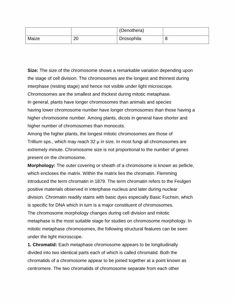
Size: The size of the chromosome shows a remarkable variation depending upon
the stage of cell division. The chromosomes are the longest and thinnest during
interphase (resting stage) and hence not visible under light microscope.
Chromosomes are the smallest and thickest during mitotic metaphase.
In general, plants have longer chromosomes than animals and species
having lower chromosome number have longer chromosomes than those having a
higher chromosome number. Among plants, dicots in general have shorter and
higher number of chromosomes than monocots.
Among the higher plants, the longest mitotic chromosomes are those of
Trillium sps., which may reach 32 µ in size. In most fungi all chromosomes are
extremely minute. Chromosome size is not proportional to the number of genes
present on the chromosome.
Morphology: The outer covering or sheath of a chromosome is known as pellicle,
which encloses the matrix. Within the matrix lies the chromatin. Flemming
introduced the term chromatin in 1879. The term chromatin refers to the Feulgen
positive materials observed in interphase nucleus and later during nuclear
division. Chromatin readily stains with basic dyes especially Basic Fuchsin, which
is specific for DNA which in turn is a major constituent of chromosomes.
The chromosome morphology changes during cell division and mitotic
metaphase is the most suitable stage for studies on chromosome morphology. In
mitotic metaphase chromosomes, the following structural features can be seen
under the light microscope.
1. Chromatid: Each metaphase chromosome appears to be longitudinally
divided into two identical parts each of which is called chromatid. Both the
chromatids of a chromosome appear to be joined together at a point known as
centromere. The two chromatids of chromosome separate from each other
during mitotic anaphase (and during anaphase II of meiosis) and move
towards opposite poles.
Since the two chromatids making up a chromosome are produced
through replication of a single chromatid during synthesis (S) phase of
interphase, they are referred to as sister chromatids. In contrast, the
chromatids of homologous chromosomes are known as non-sister chromatids.
2. Centromere: Centromere and telomere are the most stable parts of
chromosomes. The region where two sister chromatids appear to be joined
during mitotic metaphase is known as centromere. It generally appears as
constriction and hence called primary constriction. Centromere is a localized
and easily detectable morphological region of the chromosomes which helps in
the movement of the chromosomes to opposite poles during anaphase of cell
division. The centromere divides the chromosomes into two transverse parts
called arms. The centromere consists of two disk shaped bodies called
kinetochores. The kinetochores do not form part of the chromatid but lie one
on each side of the chromosome such that each chromatid is having its own
kinetochore. One kinetochore is attached to the spindle fibres towards one
pole and the other similarly towards the other pole.
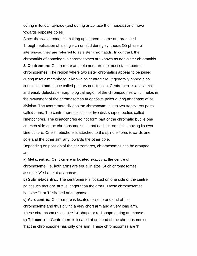
Depending on position of the centromeres, chromosomes can be grouped
as:
a) Metacentric: Centromere is located exactly at the centre of
chromosome, i.e. both arms are equal in size. Such chromosomes
assume ‘V’ shape at anaphase.
b) Submetacentric: The centromere is located on one side of the centre
point such that one arm is longer than the other. These chromosomes
become ‘J’ or ‘L’ shaped at anaphase.
c) Acrocentric: Centromere is located close to one end of the
chromosome and thus giving a very chort arm and a very long arm.
These chromosomes acquire ‘ J’ shape or rod shape during anaphase.
d) Telocentric: Centromere is located at one end of the chromosome so
that the chromosome has only one arm. These chromosomes are ‘I”
shaped or rod shaped.
Normally chromosomes are monocentric having one centromere each.
Acentric (without centromere) and dicentric (with two centromeres)
chromosomes, if produced due to chromosomal aberrations, cannot orient
properly on the equatorial plate and lag behind other chromosomes during
anaphase movements.
In certain organisms, centromere does not occupy a specific position, but is
diffused through out the body of chromosome. Such chromosomes, which
do not have a localized centromere, are found in Luzula sps. and insects
belonging to the order Hemiptera.
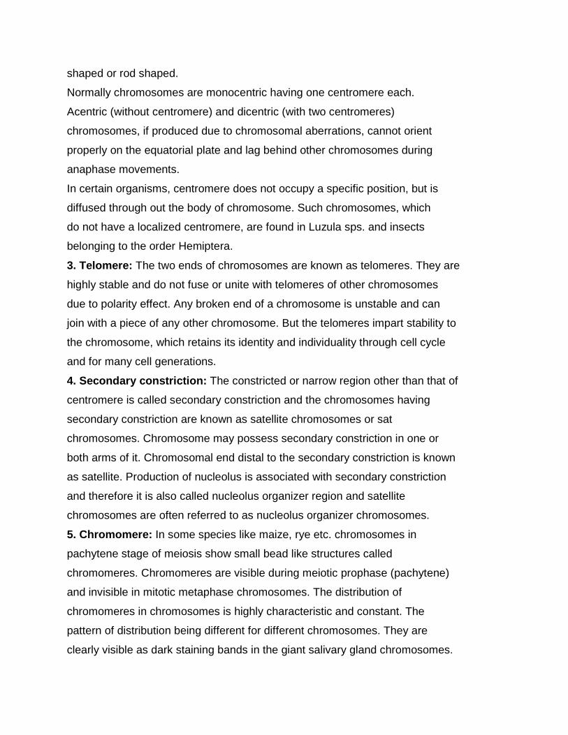
3. Telomere: The two ends of chromosomes are known as telomeres. They are
highly stable and do not fuse or unite with telomeres of other chromosomes
due to polarity effect. Any broken end of a chromosome is unstable and can
join with a piece of any other chromosome. But the telomeres impart stability to
the chromosome, which retains its identity and individuality through cell cycle
and for many cell generations.
4. Secondary constriction: The constricted or narrow region other than that of
centromere is called secondary constriction and the chromosomes having
secondary constriction are known as satellite chromosomes or sat
chromosomes. Chromosome may possess secondary constriction in one or
both arms of it. Chromosomal end distal to the secondary constriction is known
as satellite. Production of nucleolus is associated with secondary constriction
and therefore it is also called nucleolus organizer region and satellite
chromosomes are often referred to as nucleolus organizer chromosomes.
5. Chromomere: In some species like maize, rye etc. chromosomes in
pachytene stage of meiosis show small bead like structures called
chromomeres. Chromomeres are visible during meiotic prophase (pachytene)
and invisible in mitotic metaphase chromosomes. The distribution of
chromomeres in chromosomes is highly characteristic and constant. The
pattern of distribution being different for different chromosomes. They are
clearly visible as dark staining bands in the giant salivary gland chromosomes.
Chromomeres are regions of tightly folded DNA. Chromomeres of single
chromosome show considerable variation in size. They may differ in size as in
the case of maize or they may be of uniform size as in the case of rye.
6. Chromonema: A chromosome consists of two chromatids and each chromatid
consists of thread like coiled structures called chromonema (plural
chromonemata). The term chromonema was coined by Vejdovsky in 1912.
The chromonemata form the gene bearing portion of chromosomes.
7. Matrix: The mass of acromatic material which surrounds the chromonemata is
called matrix. The matrix is enclosed in a sheath which is known as pellicle.
Both matrix and pellicle are non genetic materials and appear only at
metaphase, when the nucleolus disappears.
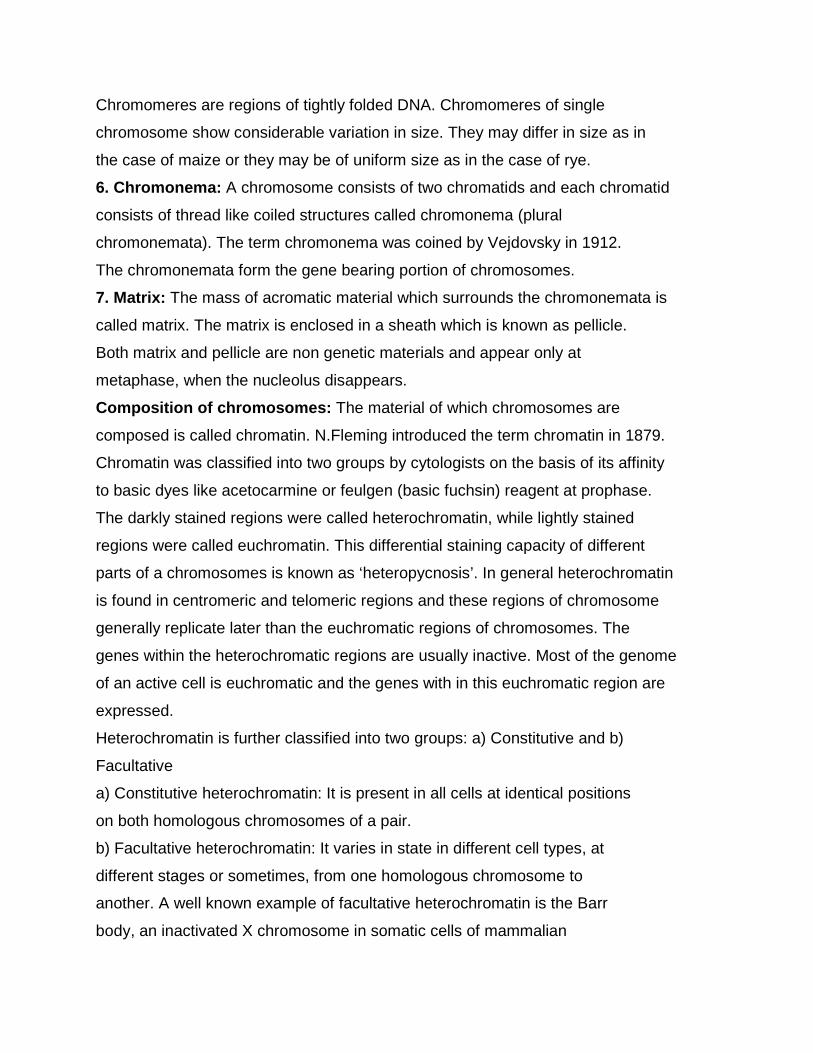
Composition of chromosomes: The material of which chromosomes are
composed is called chromatin. N.Fleming introduced the term chromatin in 1879.
Chromatin was classified into two groups by cytologists on the basis of its affinity
to basic dyes like acetocarmine or feulgen (basic fuchsin) reagent at prophase.
The darkly stained regions were called heterochromatin, while lightly stained
regions were called euchromatin. This differential staining capacity of different
parts of a chromosomes is known as ‘heteropycnosis’. In general heterochromatin
is found in centromeric and telomeric regions and these regions of chromosome
generally replicate later than the euchromatic regions of chromosomes. The
genes within the heterochromatic regions are usually inactive. Most of the genome
of an active cell is euchromatic and the genes with in this euchromatic region are
expressed.
Heterochromatin is further classified into two groups: a) Constitutive and b)
Facultative
a) Constitutive heterochromatin: It is present in all cells at identical positions
on both homologous chromosomes of a pair.
b) Facultative heterochromatin: It varies in state in different cell types, at
different stages or sometimes, from one homologous chromosome to
another. A well known example of facultative heterochromatin is the Barr
body, an inactivated X chromosome in somatic cells of mammalian
female(XX).
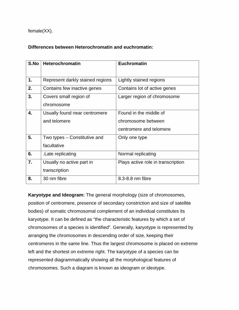
Differences between Heterochromatin and euchromatin:
S.No Heterochromatin Euchromatin
1. Represent darkly stained regions Lightly stained regions
2. Contains few inactive genes Contains lot of active genes
3. Covers small region of
chromosome
Larger region of chromosome
4. Usually found near centromere
and telomere
Found in the middle of
chromosome between
centromere and telomere
5. Two types – Constitutive and
facultative
Only one type
6. .Late replicating Normal replicating
7. Usually no active part in
transcription
Plays active role in transcription
8. 30 nm fibre 8.3-8.8 nm fibre
Karyotype and Ideogram: The general morphology (size of chromosomes,
position of centromere, presence of secondary constriction and size of satellite
bodies) of somatic chromosomal complement of an individual constitutes its
karyotype. It can be defined as “the characteristic features by which a set of
chromosomes of a species is identified”. Generally, karyotype is represented by
arranging the chromosomes in descending order of size, keeping their
centromeres in the same line. Thus the largest chromosome is placed on extreme
left and the shortest on extreme right. The karyotype of a species can be
represented diagrammatically showing all the morphological features of
chromosomes. Such a diagram is known as ideogram or ideotype.
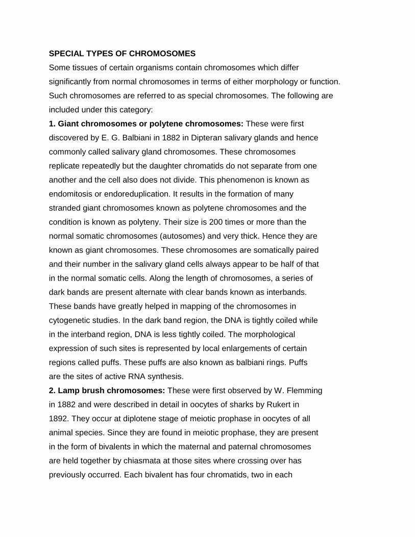
SPECIAL TYPES OF CHROMOSOMES
Some tissues of certain organisms contain chromosomes which differ
significantly from normal chromosomes in terms of either morphology or function.
Such chromosomes are referred to as special chromosomes. The following are
included under this category:
1. Giant chromosomes or polytene chromosomes: These were first
discovered by E. G. Balbiani in 1882 in Dipteran salivary glands and hence
commonly called salivary gland chromosomes. These chromosomes
replicate repeatedly but the daughter chromatids do not separate from one
another and the cell also does not divide. This phenomenon is known as
endomitosis or endoreduplication. It results in the formation of many
stranded giant chromosomes known as polytene chromosomes and the
condition is known as polyteny. Their size is 200 times or more than the
normal somatic chromosomes (autosomes) and very thick. Hence they are
known as giant chromosomes. These chromosomes are somatically paired
and their number in the salivary gland cells always appear to be half of that
in the normal somatic cells. Along the length of chromosomes, a series of
dark bands are present alternate with clear bands known as interbands.
These bands have greatly helped in mapping of the chromosomes in
cytogenetic studies. In the dark band region, the DNA is tightly coiled while
in the interband region, DNA is less tightly coiled. The morphological
expression of such sites is represented by local enlargements of certain
regions called puffs. These puffs are also known as balbiani rings. Puffs
are the sites of active RNA synthesis.
2. Lamp brush chromosomes: These were first observed by W. Flemming
in 1882 and were described in detail in oocytes of sharks by Rukert in
1892. They occur at diplotene stage of meiotic prophase in oocytes of all
animal species. Since they are found in meiotic prophase, they are present
in the form of bivalents in which the maternal and paternal chromosomes
are held together by chiasmata at those sites where crossing over has
previously occurred. Each bivalent has four chromatids, two in each
homologue. The axis of each homologue consists of a row of granules or
chromomeres, each of which have two loop like lateral extensions, one for
each chromatid. Thus each loop represents one chromatid of a
chromosome and is composed of one DNA double helix. One end of each
loop is thinner than other which is known as thickend. There is extensive
RNA synthesis at thin ends of the loop while there is little or no RNA
synthesis at the thick ends.
3. Accessory chromosomes: In many species some chromosomes are
found in addition to normal somatic chromosomes. These extra
chromosomes are called accessory chromosomes or B-chromosomes or
supernumerary chromos omes. These chromosomes are broadly similar to
normal somatic chromosomes in their morphology, but have some peculiar
functional aspects. For instance, presence of several such chromosomes
often leads to reduction in vigour and fertility in males. These
chromosomes are generally smaller in size than the normal somatic
complement. They are believed to be generally inactive genetically.
However they may not be completely devoid of genes. Origin of these
chromosomes in most species is unknown.
4. Isochromosomes: An isochromosome is the one in which two arms are
identical with each other in gene content and morphology. Such a
chromosome is in assense a reverse duplication with entromeres
separating the two arms. Every isochromosome is metacentric. The
attached ‘x’ chromosome of Drosophila is a classical example of an
isochromosome. However its origin is uncertain. There is no evidence that
isochromosomes had any evolutionary significane.
5. Allosomes / sex chromosomes: Chromosomes differing in morphology
and number in male and female are called allosomes. They are responsible
for determination of sex. Eg: X and Y chromosomes in human beings and
Drosophila. Chromosomes which have no relation with determination of sex
and contain genes which determine somatic characters of individuals are
called autosomes and are represented by letter ‘A’.

CYTOPLASMIC INHERITANCE
Inheritance due to genes located in cytoplasm (plasmagenes) is called
cytoplasmic inheritance. Since genes govering traits showing cytoplasmic
inheritance are loc ated outside the nucleus and in the cytoplasm, they are refered
to as plasmagenes. The sum total of genes present in the cytoplasm of a cell or
an individual is known as plasmon. The plasmagenes are located in DNA present
in mitochondria (mt DNA) and in chloroplasts (cp DNA). Together both the DNAs
are called organelle DNA. Therefore, this type of inheritance is often referred to as
organellar inheritance, plastid inheritance or mitochondrial inheritance. In this,
generally, the character of only one of the two parents (usually female) is
transmitted to the progeny. Hence such inheritance is usually referred to as extra -
nuclear or extra-chromosomal or maternal or uniparental inheritance.
The cytoplasmic inheritance is of two types: 1) Plastid inheritance and 2)
mitochondrial inheritance.
1. Plastidial or chloroplast inheritance: Plastids self duplicated and have
some amount of DNA and plays an important role in cytoplasmic
inheritance. Plastids have green pigments called chloroplasts. chloroplasts
contain a unique circular DNA (cp DNA) in the stroma that is completely
different from the nuclear genome. Some examples of plastid inheritance
are given below:
a) Leaf variegation in Mirabilis jalapa : The conclusive evidence for
cytoplasmic inheritance was first presented by C. Correns in Mirabilis
jalapa (Four ‘O’ clock plant) in 1909. He studied inheritance of leaf
variegation in M. jalapa. Variegation refers to the presence of white or
yellow spots of variable size on the green background of leaves. In M.
jalapa, leaves may be green, white or variegated. Some branches may
have only green, only white or only variegated leaves. Correns made
crosses in all possible combinations among the flowers produced on these
three types of branches. When flowers from green branch were used as
female parent, all the progeny were green irrespective of the phenotype
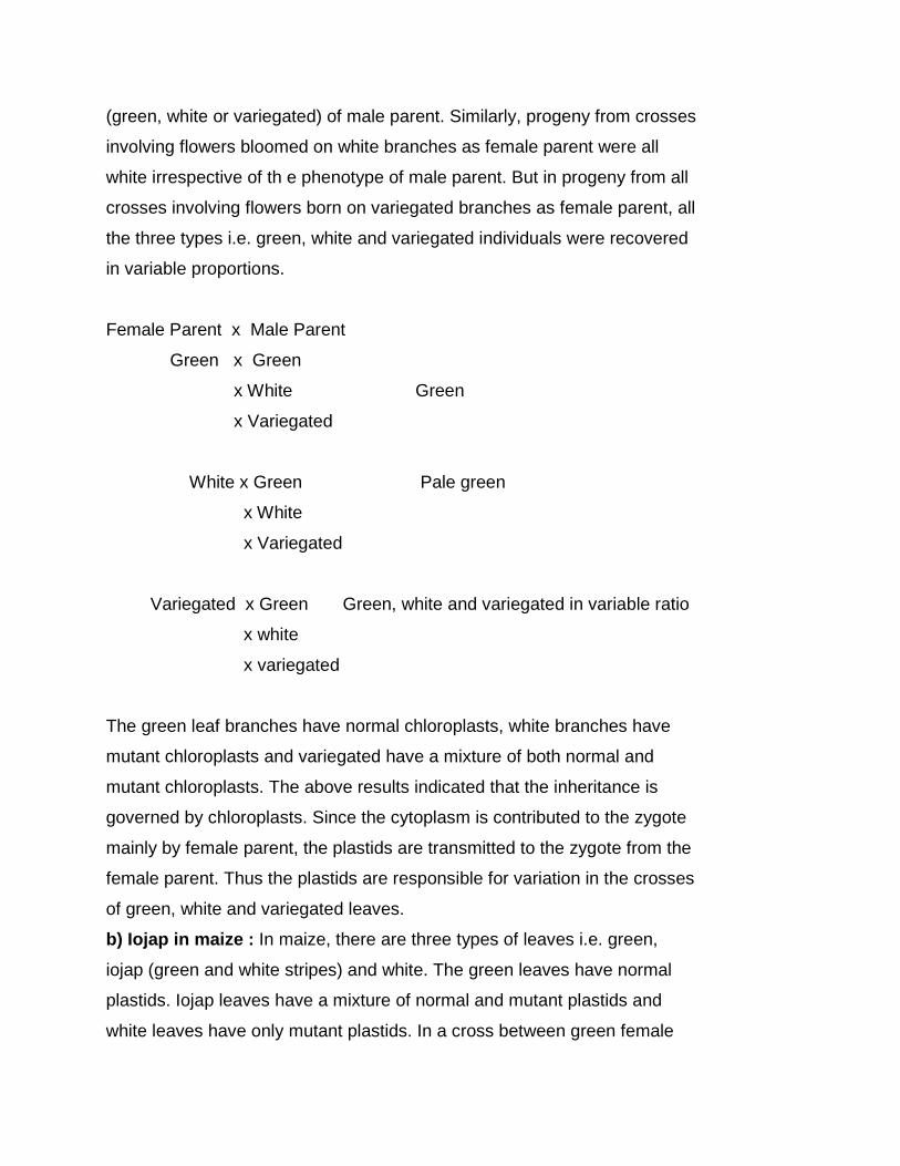
(green, white or variegated) of male parent. Similarly, progeny from crosses
involving flowers bloomed on white branches as female parent were all
white irrespective of th e phenotype of male parent. But in progeny from all
crosses involving flowers born on variegated branches as female parent, all
the three types i.e. green, white and variegated individuals were recovered
in variable proportions.
Female Parent x Male Parent
Green x Green
x White Green
x Variegated
White x Green Pale green
x White
x Variegated
Variegated x Green Green, white and variegated in variable ratio
x white
x variegated
The green leaf branches have normal chloroplasts, white branches have
mutant chloroplasts and variegated have a mixture of both normal and
mutant chloroplasts. The above results indicated that the inheritance is
governed by chloroplasts. Since the cytoplasm is contributed to the zygote
mainly by female parent, the plastids are transmitted to the zygote from the
female parent. Thus the plastids are responsible for variation in the crosses
of green, white and variegated leaves.
b) Iojap in maize : In maize, there are three types of leaves i.e. green,
iojap (green and white stripes) and white. The green leaves have normal
plastids. Iojap leaves have a mixture of normal and mutant plastids and
white leaves have only mutant plastids. In a cross between green female
and iojap male, only green individuals are produced in F1 generation. But in
the reciprocal cross (iojap female and green male) all the three kinds of
progeny are obtained in variable proportions in F1.
Parents Female x Male
Phenotype Green x Iojap
F1 Green
Reciprocal cross
Parents Female x Male
Phenotype Iojap x Green
F1 Green, Iojap, White in variable proportion
These reciprocal differences can be attributed to the type of plastids in the
egg cell since only female parent is contributing cytoplasm to the zygote.
2. Mitochondrial inheritance: The inheritance of some characters, such as
cytoplasmic male sterility in plants, pokyness in Neurospora etc., is governed by
mitochondrial DNA (mtDNA).
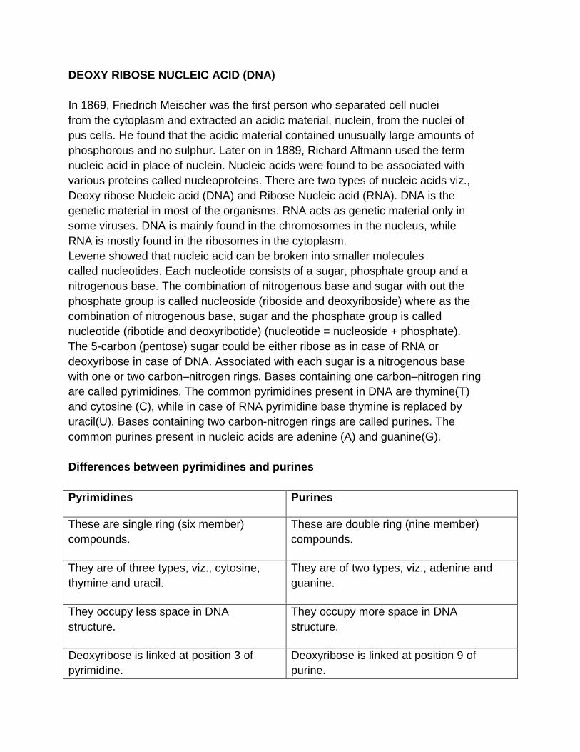
DEOXY RIBOSE NUCLEIC ACID (DNA) In 1869, Friedrich Meischer was the first person who separated cell nuclei from the cytoplasm and extracted an acidic material, nuclein, from the nuclei of pus cells. He found that the acidic material contained unusually large amounts of phosphorous and no sulphur. Later on in 1889, Richard Altmann used the term nucleic acid in place of nuclein. Nucleic acids were found to be associated with various proteins called nucleoproteins. There are two types of nucleic acids viz., Deoxy ribose Nucleic acid (DNA) and Ribose Nucleic acid (RNA). DNA is the genetic material in most of the organisms. RNA acts as genetic material only in some viruses. DNA is mainly found in the chromosomes in the nucleus, while RNA is mostly found in the ribosomes in the cytoplasm. Levene showed that nucleic acid can be broken into smaller molecules called nucleotides. Each nucleotide consists of a sugar, phosphate group and a nitrogenous base. The combination of nitrogenous base and sugar with out the phosphate group is called nucleoside (riboside and deoxyriboside) where as the combination of nitrogenous base, sugar and the phosphate group is called nucleotide (ribotide and deoxyribotide) (nucleotide = nucleoside + phosphate). The 5-carbon (pentose) sugar could be either ribose as in case of RNA or deoxyribose in case of DNA. Associated with each sugar is a nitrogenous base with one or two carbon–nitrogen rings. Bases containing one carbon–nitrogen ring are called pyrimidines. The common pyrimidines present in DNA are thymine(T) and cytosine (C), while in case of RNA pyrimidine base thymine is replaced by uracil(U). Bases containing two carbon-nitrogen rings are called purines. The common purines present in nucleic acids are adenine (A) and guanine(G). Differences between pyrimidines and purines Pyrimidines Purines
These are single ring (six member) compounds.
These are double ring (nine member) compounds.
They are of three types, viz., cytosine, thymine and uracil.
They are of two types, viz., adenine and guanine.
They occupy less space in DNA structure.
They occupy more space in DNA structure.
Deoxyribose is linked at position 3 of pyrimidine.
Deoxyribose is linked at position 9 of purine.
Levene proposed that each of the deoxy-ribonucleotides was present in equal amounts and connected together in chains in which each of the four different nucleotides was regularly repeated in a tetranucleotide sequence (AGCT, AGCT etc.). In 1940 Erwin Chargaff and other biochemists showed that all the nucleotide bases were not present in equal amounts and that the ratio of different bases changed between different species. It was also shown by Chargaff that the number of purine bases (A + G) is equal to the number of pyrimidine bases (C + T) i.e. A + G = C + T. It was also shown that the ratios of adenine to thymine and guanine to cytosine are constant and close to one in various eukaryotic species. By the early 1950’s X – ray studies of DNA by Wilkins, Franklin and others indicated a well organized multiple stranded fibre of about 220A in diameter that was also characterized by the presence of groups or bases spaced, 3.40A apart along the fibre and occurrence of a repeating unit at every 340A. Taking into account the facts known at that time Watson and Crick in 1953 proposed a “double helix” structure of DNA which quickly gained wide acceptance. The salient features of double helix structure of DNA are: · The DNA molecule consists of two polynucleotide chains wound around each other in a right-handed double helix. · The two strands of a DNA molecule are oriented anti-parallel to each other i.e. the 5’ end of one strand is located with the 3’ end of the other strand at the same end of a DNA molecule. · Each polydeoxyribonucleotide strand is composed of many deoxyribonucleotides joined together by phosphodiester linkage between their sugar and phosphate residues and the sugar phosphate backbones are on the outsides of the double helix with the nitrogen bases oriented toward the central axis. · The half steps of one strand extend to meet half steps of the other strand and the base pairs are called complementary base pairs. The adenine present in one stand of a DNA molecule is linked by two hydrogen bonds with the thymine located opposite to it in the second strand, and vice-versa. Similarly, guanine located in one strand forms three hydrogen bonds with the cytosine present opposite to it in the second strand, and vice-versa. The pairing of one purine and one pyrimidine maintains the constant width of the DNA double helix. · The bases are connected by hydrogen bonds. Although the hydrogen bonds are weaker, the fact that so many of them occur along the length of DNA double helix provides a high degree of stability and rigidity to the molecule. · The diameter of this helix is 200A, while its pitch (the length of helix required to complete one turn) is 340A. In each DNA strand, the bases
occur at a regular interval of 3.40A so that about 10 base pairs are present in one pitch of a DNA double helix. · The helix has two external grooves, a deep wide one, called major groove and a shallow narrow one, called minor groove. Both these groves are large enough to allow protein molecules to come in contact with the bases. · This DNA structure offers a ready explanation of how a molecule could form perfect copies of itself. During replication, the two strands of a DNA molecule unwind and the unpaired bases in the single-stranded regions of the two strands by hydrogen bonds with their complementary bases present in the cytoplasm as free nucleotides. These nucleotides become joined by phospho-diester linkages generating complementary strands of the old ones with the help of appropriate enzymes. The DNA molecule satisfies the requirement of genetic material in the following ways:- 1. It can replicate itself accurately during cell growth and division. 2. Its structure is sufficiently stable so that heritable charges i.e., mutations can occur only very rarely. 3. It has a potential to carry all kinds of necessary biological information. 4. It transmits all the biological information to the daughter cells. Thus the essential functions of DNA are the storage and transmission of genetic information and the expression of this information in the form of synthesis of cellular proteins. Types of DNA The double helix described by Watson and Crick has right handed helical coiling and is called B-DNA. It is a biologically important form of DNA that is commonly and naturally found in most living systems. This double helical structure of DNA exist in other alternate forms such as A-form, C-form etc. which differ in features such as the number of nucleotide base pairs per turn of the helix. The Bform contains ~ 10 (range 10.0 – 10.6) base pairs per turn. The B-DNA is the most stable form and it can change to another form depending upon the humidity and salt concentration of the sample. The A- form is also a right-handed helix, but it has 11 base pairs per turn. The C-form of DNA has 9.3 base pairs per turn, while the D-form of DNA, which is rare form, has 8 base pairs per turn. Another form of DNA, in which the helix is left-handed, called Z-DNA was discovered by Rich. In Z-DNA sugar and phosphate linkages follow a zigzag pattern. Z-DNA plays a role in the regulation of the gene activity. Comparison of B-DNA and Z-DNA Characteristic B-DNA Z-DNA
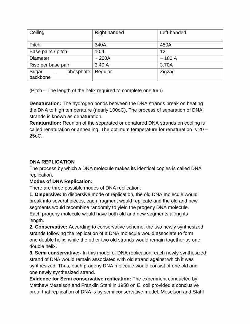
Coiling Right handed Left-handed
Pitch 340A 450A Base pairs / pitch 10.4 12 Diameter ~ 200A ~ 180 A Rise per base pair 3.40 A 3.70A Sugar – phosphate backbone
Regular Zigzag
(Pitch – The length of the helix required to complete one turn) Denaturation: The hydrogen bonds between the DNA strands break on heating the DNA to high temperature (nearly 100oC). The process of separation of DNA strands is known as denaturation. Renaturation: Reunion of the separated or denatured DNA strands on cooling is called renaturation or annealing. The optimum temperature for renaturation is 20 – 25oC. DNA REPLICATION The process by which a DNA molecule makes its identical copies is called DNA replication. Modes of DNA Replication: There are three possible modes of DNA replication. 1. Dispersive: In dispersive mode of replication, the old DNA molecule would break into several pieces, each fragment would replicate and the old and new segments would recombine randomly to yield the progeny DNA molecule. Each progeny molecule would have both old and new segments along its length. 2. Conservative: According to conservative scheme, the two newly synthesized strands following the replication of a DNA molecule would associate to form one double helix, while the other two old strands would remain together as one double helix. 3. Semi conservative:- In this model of DNA replication, each newly synthesized strand of DNA would remain associated with old strand against which it was synthesized. Thus, each progeny DNA molecule would consist of one old and one newly synthesized strand. Evidence for Semi conservative replication: The experiment conducted by Matthew Meselson and Franklin Stahl in 1958 on E. coli provided a conclusive proof that replication of DNA is by semi conservative model. Meselson and Stahl
labeled DNA of E. coli bacteria with heavy nitrogen i.e. 15N by growing them on a medium containing 15N for many generations to replace the normal nitrogen (14N). The density of normal and heavy nitrogen differs. The 14N is lighter (1.710 g/cm3) than 15N (1.724 g/cm3). It is possible to detect such minute differences in density through density gradient centrifugation. Distinct bands are formed in the centrifuge tube for different density DNA. DNA extracts of E. coli with 15N gave a characteristic heavy band at one end of a tube that had been centrifuged at a high speed in an ultra-centrifuge. These labeled cells were then grown on a normal unlabelled media containing 14N for one generation. DNA was again extracted and processed and it was found to consist of a hybrid DNA containing both 14N and 15N at the same time. This indicated that the DNA had not replicated in two separate labeled and unlabelled forms. The next generation of growth on unlabelled DNA was found to be in amounts equal to the partially labeled hybrid DNA. Additional generation of growth on unlabelled media gave a relative increase in the amount of unlabelled DNA. After two generations half the DNA was with intermediate density and half with light bands which further confirm semiconservative mode of DNA replication. After third generation, ¾ DNA was found with 14N and ¼ with hybrid nitrogen (14N+15N). When the hybrid DNA was denatured by heating upto 100oC it was found to produce two separate single strands and in the ultracentrifuge density gradient, it was observed to form two separate bands; one band containing 15N and the other 14N. Thus it was concluded that DNA replication was by semi-conservative mode. The major objection put forth for the semi-conservative replication was that the DNA molecule must unwind a number of times (1/10 of the total number of nucleotides) which cannot be accomplished without breaking with in a short span of time say two minutes. Cairns (1968) provided evidence for this in his experiments with radio-active labeled E. coli chromosomes. The E. coli chromosome is a double stranded circular chromosome. It was shown that the two circular component strands separate during replication with each strand duplicating individually producing a q shaped structures during replication. This indicates that unwinding and replication proceed simultaneously. Method of semi-conservative replication: DNA replication was found to begin at various initiation points , called origin of replication, and proceed bi-directionally. Two enzymes, DNA gyrase and DNA helicase induce unwinding of complementary strands of DNA. Single-strand DNA binding (SSB) proteins bind to the single-stranded DNA, stabilizing it and preventing it from reanneaing. An enzyme, primase, initiates replication by synthesizing the primer. DNA polymerases synthesize the complementary strand by progressively adding deoxyribonucleotides. The DNA replication always proceeds in 5’ à 3’ direction.
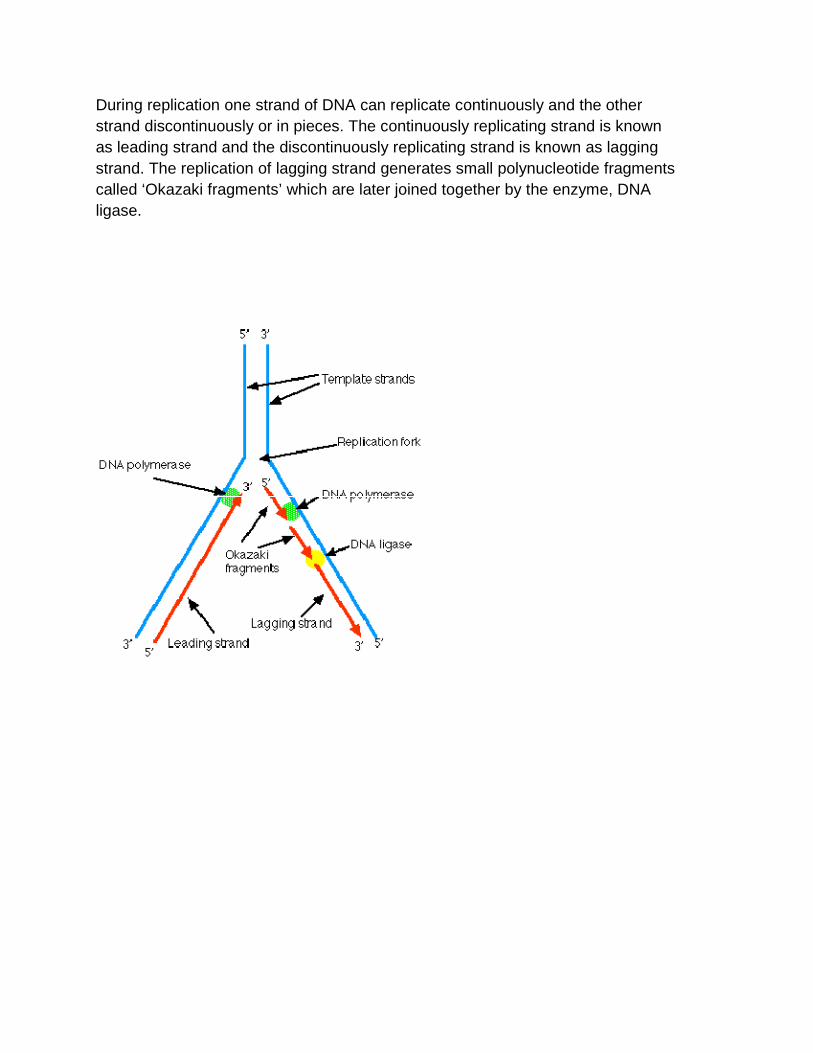
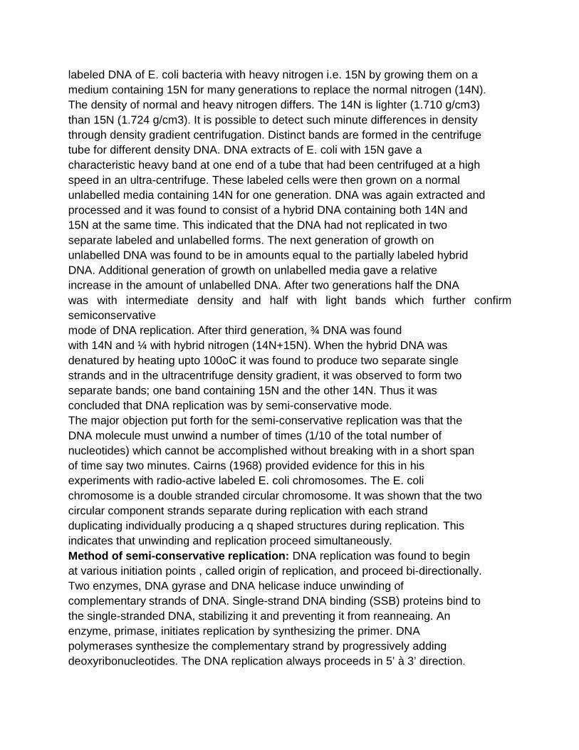
During replication one strand of DNA can replicate continuously and the other strand discontinuously or in pieces. The continuously replicating strand is known as leading strand and the discontinuously replicating strand is known as lagging strand. The replication of lagging strand generates small polynucleotide fragments called ‘Okazaki fragments’ which are later joined together by the enzyme, DNA ligase.
RNA (Ribose nucleic acid) Structure of RNA: RNA like DNA is a polynucleotide. RNA nucleotides have ribose sugar, which participate in the formation of sugar phosphate backbone of RNA. Thymine is absent and is replaced by Uracil. Usually RNA is a single stranded structure. Single stranded RNA is the genetic material in most plant viruses Eg. TMV. Double stranded RNA is also found to be the genetic material in some organisms. Eg. : Plant wound and tumour viruses. RNA performs nongenetic function. There are three main types or forms of RNA. 1. messenger RNA (m-RNA): It constitutes about 5-10% of the total cellular RNA. It is a single stranded base for base complementary copy of one of the DNA strands of a gene. It provides the information for the amino acid
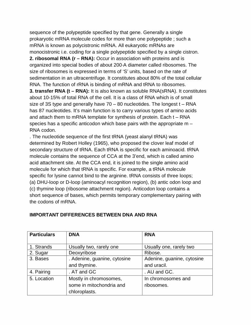
sequence of the polypeptide specified by that gene. Generally a single prokaryotic mRNA molecule codes for more than one polypeptide ; such a mRNA is known as polycistronic mRNA. All eukaryotic mRNAs are monocistronic i.e. coding for a single polypeptide specified by a single cistron. 2. ribosomal RNA (r – RNA): Occur in association with proteins and is organized into special bodies of about 200·A diameter called ribosomes. The size of ribosomes is expressed in terms of ‘S’ units, based on the rate of sedimentation in an ultracentrifuge. It constitutes about 80% of the total cellular RNA. The function of rRNA is binding of mRNA and tRNA to ribosomes. 3. transfer RNA (t – RNA): It is also known as soluble RNA(sRNA). It constitutes about 10-15% of total RNA of the cell. It is a class of RNA which is of small size of 3S type and generally have 70 – 80 nucleotides. The longest t – RNA has 87 nucleotides. It’s main function is to carry various types of amino acids and attach them to mRNA template for synthesis of protein. Each t – RNA species has a specific anticodon which base pairs with the appropriate m – RNA codon. . The nucleotide sequence of the first tRNA (yeast alanyl tRNA) was determined by Robert Holley (1965), who proposed the clover leaf model of secondary structure of tRNA. Each tRNA is specific for each aminoacid. tRNA molecule contains the sequence of CCA at the 3’end, which is called amino acid attachment site. At the CCA end, it is joined to the single amino acid molecule for which that tRNA is specific. For example, a tRNA molecule specific for lysine cannot bind to the arginine. tRNA consists of three loops; (a) DHU-loop or D-loop (aminoacyl recognition region), (b) antic odon loop and (c) thymine loop (ribosome attachment region). Anticodon loop contains a short sequence of bases, which permits temporary complementary pairing with the codons of mRNA. IMPORTANT DIFFERENCES BETWEEN DNA AND RNA Particulars DNA RNA
1. Strands Usually two, rarely one Usually one, rarely two 2. Sugar Deoxyribose Ribose. 3. Bases . Adenine, guanine, cytosine
and thymine. Adenine, guanine, cytosine and uracil.
4. Pairing . AT and GC . AU and GC. 5. Location Mostly in chromosomes,
some in mitochondria and chloroplasts.
In chromosomes and ribosomes.
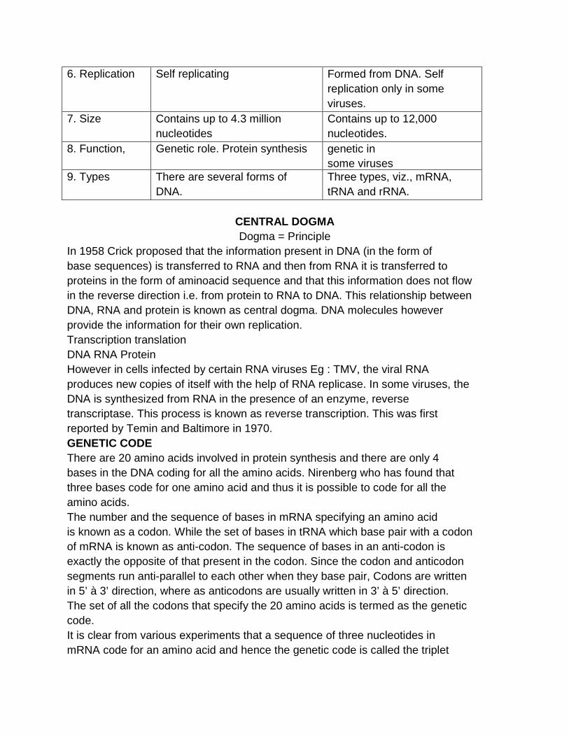
6. Replication Self replicating Formed from DNA. Self replication only in some viruses.
7. Size Contains up to 4.3 million nucleotides
Contains up to 12,000 nucleotides.
8. Function, Genetic role. Protein synthesis genetic in some viruses
9. Types There are several forms of DNA.
Three types, viz., mRNA, tRNA and rRNA.
CENTRAL DOGMA Dogma = Principle
In 1958 Crick proposed that the information present in DNA (in the form of base sequences) is transferred to RNA and then from RNA it is transferred to proteins in the form of aminoacid sequence and that this information does not flow in the reverse direction i.e. from protein to RNA to DNA. This relationship between DNA, RNA and protein is known as central dogma. DNA molecules however provide the information for their own replication. Transcription translation DNA RNA Protein However in cells infected by certain RNA viruses Eg : TMV, the viral RNA produces new copies of itself with the help of RNA replicase. In some viruses, the DNA is synthesized from RNA in the presence of an enzyme, reverse transcriptase. This process is known as reverse transcription. This was first reported by Temin and Baltimore in 1970. GENETIC CODE There are 20 amino acids involved in protein synthesis and there are only 4 bases in the DNA coding for all the amino acids. Nirenberg who has found that three bases code for one amino acid and thus it is possible to code for all the amino acids. The number and the sequence of bases in mRNA specifying an amino acid is known as a codon. While the set of bases in tRNA which base pair with a codon of mRNA is known as anti-codon. The sequence of bases in an anti-codon is exactly the opposite of that present in the codon. Since the codon and anticodon segments run anti-parallel to each other when they base pair, Codons are written in 5’ à 3’ direction, where as anticodons are usually written in 3’ à 5’ direction. The set of all the codons that specify the 20 amino acids is termed as the genetic code. It is clear from various experiments that a sequence of three nucleotides in mRNA code for an amino acid and hence the genetic code is called the triplet
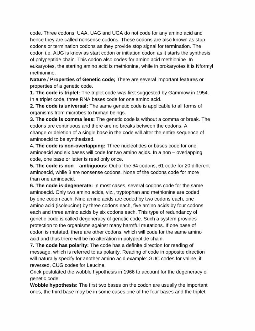
code. Three codons, UAA, UAG and UGA do not code for any amino acid and hence they are called nonsense codons. These codons are also known as stop codons or termination codons as they provide stop signal for termination. The codon i.e. AUG is know as start codon or initiation codon as it starts the synthesis of polypeptide chain. This codon also codes for amino acid methionine. In eukaryotes, the starting amino acid is methionine, while in prokaryotes it is Nformyl methionine. Nature / Properties of Genetic code; There are several important features or properties of a genetic code. 1. The code is triplet: The triplet code was first suggested by Gammow in 1954. In a triplet code, three RNA bases code for one amino acid. 2. The code is universal: The same genetic code is applicable to all forms of organisms from microbes to human beings. 3. The code is comma less: The genetic code is without a comma or break. The codons are continuous and there are no breaks between the codons. A change or deletion of a single base in the code will alter the entire sequence of aminoacid to be synthesized. 4. The code is non-overlapping: Three nucleotides or bases code for one aminoacid and six bases will code for two amino acids. In a non – overlapping code, one base or letter is read only once. 5. The code is non – ambiguous: Out of the 64 codons, 61 code for 20 different aminoacid, while 3 are nonsense codons. None of the codons code for more than one aminoacid. 6. The code is degenerate: In most cases, several codons code for the same aminoacid. Only two amino acids, viz., tryptophan and methionine are coded by one codon each. Nine amino acids are coded by two codons each, one amino acid (isoleucine) by three codons each, five amino acids by four codons each and three amino acids by six codons each. This type of redundancy of genetic code is called degeneracy of genetic code. Such a system provides protection to the organisms against many harmful mutations. If one base of codon is mutated, there are other codons, which will code for the same amino acid and thus there will be no alteration in polypeptide chain. 7. The code has polarity: The code has a definite direction for reading of message, which is referred to as polarity. Reading of code in opposite direction will naturally specify for another amino acid example: GUC codes for valine, if reversed, CUG codes for Leucine. Crick postulated the wobble hypothesis in 1966 to account for the degeneracy of genetic code. Wobble hypothesis: The first two bases on the codon are usually the important ones, the third base may be in some cases one of the four bases and the triplet
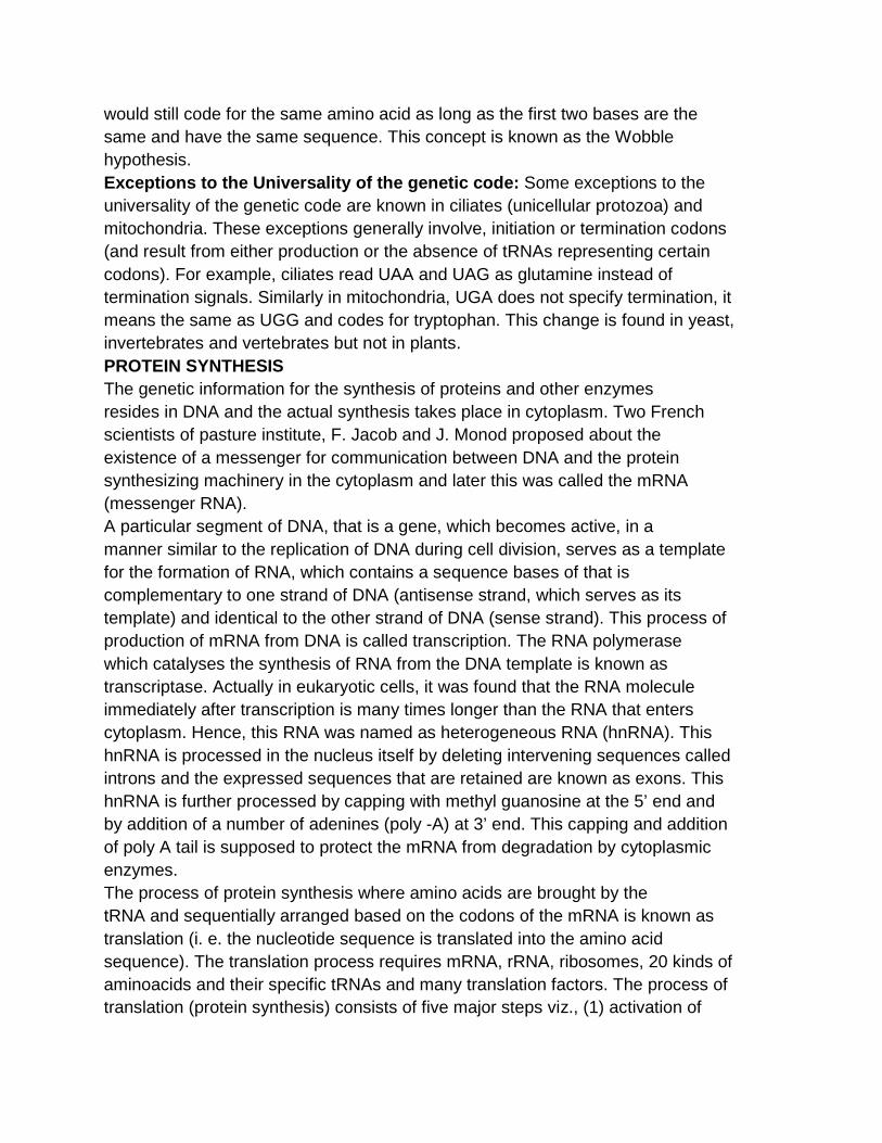
would still code for the same amino acid as long as the first two bases are the same and have the same sequence. This concept is known as the Wobble hypothesis. Exceptions to the Universality of the genetic code: Some exceptions to the universality of the genetic code are known in ciliates (unicellular protozoa) and mitochondria. These exceptions generally involve, initiation or termination codons (and result from either production or the absence of tRNAs representing certain codons). For example, ciliates read UAA and UAG as glutamine instead of termination signals. Similarly in mitochondria, UGA does not specify termination, it means the same as UGG and codes for tryptophan. This change is found in yeast, invertebrates and vertebrates but not in plants. PROTEIN SYNTHESIS The genetic information for the synthesis of proteins and other enzymes resides in DNA and the actual synthesis takes place in cytoplasm. Two French scientists of pasture institute, F. Jacob and J. Monod proposed about the existence of a messenger for communication between DNA and the protein synthesizing machinery in the cytoplasm and later this was called the mRNA (messenger RNA). A particular segment of DNA, that is a gene, which becomes active, in a manner similar to the replication of DNA during cell division, serves as a template for the formation of RNA, which contains a sequence bases of that is complementary to one strand of DNA (antisense strand, which serves as its template) and identical to the other strand of DNA (sense strand). This process of production of mRNA from DNA is called transcription. The RNA polymerase which catalyses the synthesis of RNA from the DNA template is known as transcriptase. Actually in eukaryotic cells, it was found that the RNA molecule immediately after transcription is many times longer than the RNA that enters cytoplasm. Hence, this RNA was named as heterogeneous RNA (hnRNA). This hnRNA is processed in the nucleus itself by deleting intervening sequences called introns and the expressed sequences that are retained are known as exons. This hnRNA is further processed by capping with methyl guanosine at the 5’ end and by addition of a number of adenines (poly -A) at 3’ end. This capping and addition of poly A tail is supposed to protect the mRNA from degradation by cytoplasmic enzymes. The process of protein synthesis where amino acids are brought by the tRNA and sequentially arranged based on the codons of the mRNA is known as translation (i. e. the nucleotide sequence is translated into the amino acid sequence). The translation process requires mRNA, rRNA, ribosomes, 20 kinds of aminoacids and their specific tRNAs and many translation factors. The process of translation (protein synthesis) consists of five major steps viz., (1) activation of
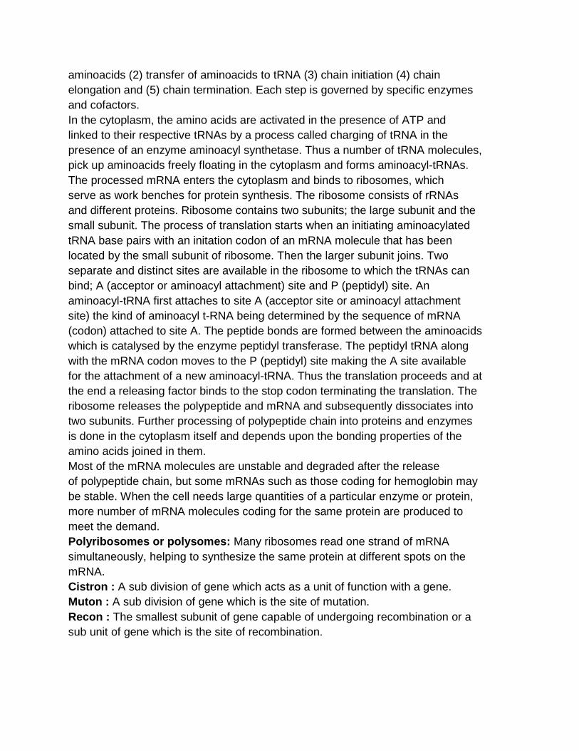
aminoacids (2) transfer of aminoacids to tRNA (3) chain initiation (4) chain elongation and (5) chain termination. Each step is governed by specific enzymes and cofactors. In the cytoplasm, the amino acids are activated in the presence of ATP and linked to their respective tRNAs by a process called charging of tRNA in the presence of an enzyme aminoacyl synthetase. Thus a number of tRNA molecules, pick up aminoacids freely floating in the cytoplasm and forms aminoacyl-tRNAs. The processed mRNA enters the cytoplasm and binds to ribosomes, which serve as work benches for protein synthesis. The ribosome consists of rRNAs and different proteins. Ribosome contains two subunits; the large subunit and the small subunit. The process of translation starts when an initiating aminoacylated tRNA base pairs with an initation codon of an mRNA molecule that has been located by the small subunit of ribosome. Then the larger subunit joins. Two separate and distinct sites are available in the ribosome to which the tRNAs can bind; A (acceptor or aminoacyl attachment) site and P (peptidyl) site. An aminoacyl-tRNA first attaches to site A (acceptor site or aminoacyl attachment site) the kind of aminoacyl t-RNA being determined by the sequence of mRNA (codon) attached to site A. The peptide bonds are formed between the aminoacids which is catalysed by the enzyme peptidyl transferase. The peptidyl tRNA along with the mRNA codon moves to the P (peptidyl) site making the A site available for the attachment of a new aminoacyl-tRNA. Thus the translation proceeds and at the end a releasing factor binds to the stop codon terminating the translation. The ribosome releases the polypeptide and mRNA and subsequently dissociates into two subunits. Further processing of polypeptide chain into proteins and enzymes is done in the cytoplasm itself and depends upon the bonding properties of the amino acids joined in them. Most of the mRNA molecules are unstable and degraded after the release of polypeptide chain, but some mRNAs such as those coding for hemoglobin may be stable. When the cell needs large quantities of a particular enzyme or protein, more number of mRNA molecules coding for the same protein are produced to meet the demand. Polyribosomes or polysomes: Many ribosomes read one strand of mRNA simultaneously, helping to synthesize the same protein at different spots on the mRNA. Cistron : A sub division of gene which acts as a unit of function with a gene. Muton : A sub division of gene which is the site of mutation. Recon : The smallest subunit of gene capable of undergoing recombination or a sub unit of gene which is the site of recombination.
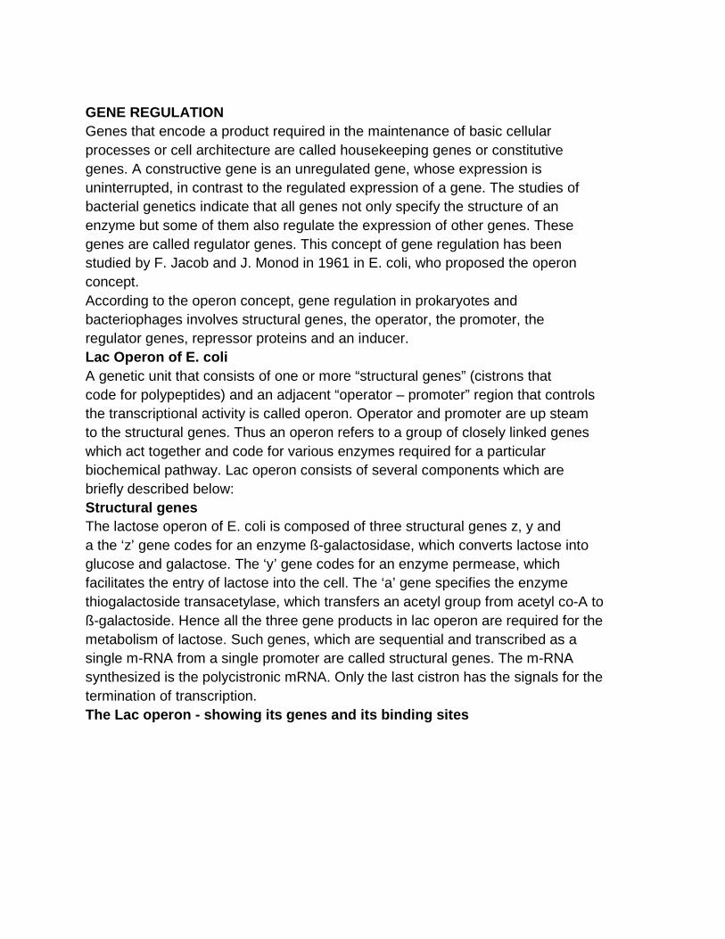
GENE REGULATION Genes that encode a product required in the maintenance of basic cellular processes or cell architecture are called housekeeping genes or constitutive genes. A constructive gene is an unregulated gene, whose expression is uninterrupted, in contrast to the regulated expression of a gene. The studies of bacterial genetics indicate that all genes not only specify the structure of an enzyme but some of them also regulate the expression of other genes. These genes are called regulator genes. This concept of gene regulation has been studied by F. Jacob and J. Monod in 1961 in E. coli, who proposed the operon concept. According to the operon concept, gene regulation in prokaryotes and bacteriophages involves structural genes, the operator, the promoter, the regulator genes, repressor proteins and an inducer. Lac Operon of E. coli A genetic unit that consists of one or more “structural genes” (cistrons that code for polypeptides) and an adjacent “operator – promoter” region that controls the transcriptional activity is called operon. Operator and promoter are up steam to the structural genes. Thus an operon refers to a group of closely linked genes which act together and code for various enzymes required for a particular biochemical pathway. Lac operon consists of several components which are briefly described below: Structural genes The lactose operon of E. coli is composed of three structural genes z, y and a the ‘z’ gene codes for an enzyme ß-galactosidase, which converts lactose into glucose and galactose. The ‘y’ gene codes for an enzyme permease, which facilitates the entry of lactose into the cell. The ‘a’ gene specifies the enzyme thiogalactoside transacetylase, which transfers an acetyl group from acetyl co-A to ß-galactoside. Hence all the three gene products in lac operon are required for the metabolism of lactose. Such genes, which are sequential and transcribed as a single m-RNA from a single promoter are called structural genes. The m-RNA synthesized is the polycistronic mRNA. Only the last cistron has the signals for the termination of transcription. The Lac operon - showing its genes and its binding sites
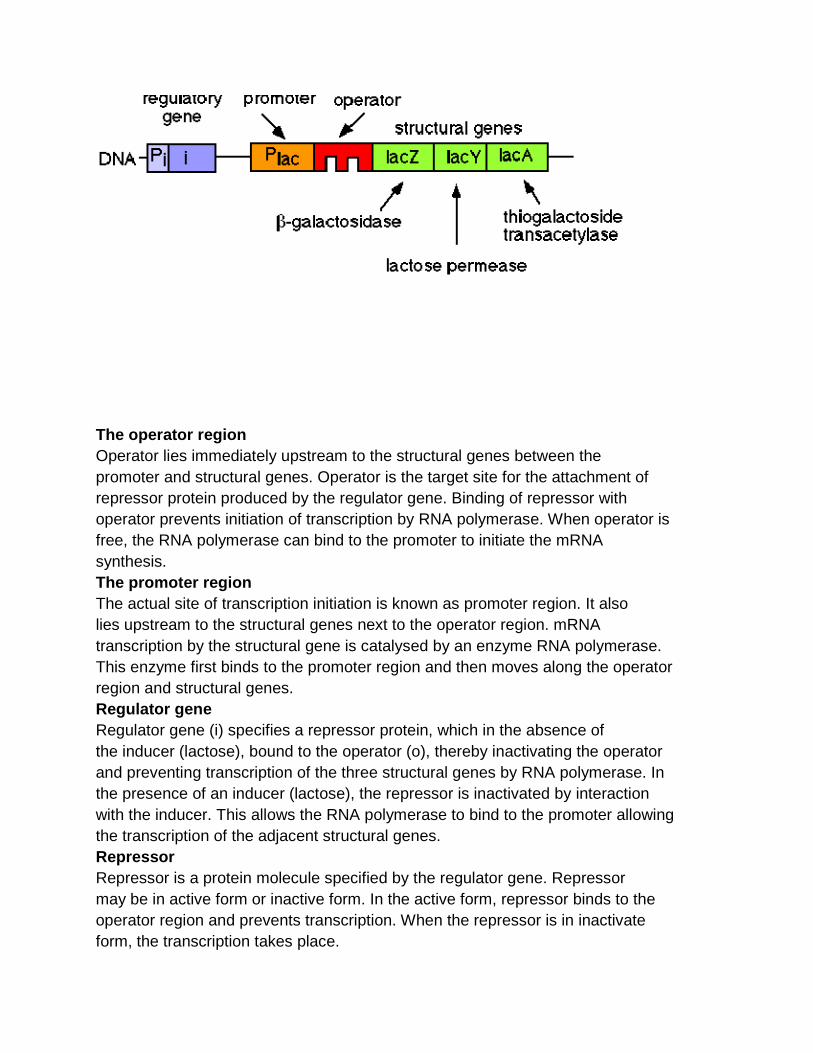
The operator region Operator lies immediately upstream to the structural genes between the promoter and structural genes. Operator is the target site for the attachment of repressor protein produced by the regulator gene. Binding of repressor with operator prevents initiation of transcription by RNA polymerase. When operator is free, the RNA polymerase can bind to the promoter to initiate the mRNA synthesis. The promoter region The actual site of transcription initiation is known as promoter region. It also lies upstream to the structural genes next to the operator region. mRNA transcription by the structural gene is catalysed by an enzyme RNA polymerase. This enzyme first binds to the promoter region and then moves along the operator region and structural genes. Regulator gene Regulator gene (i) specifies a repressor protein, which in the absence of the inducer (lactose), bound to the operator (o), thereby inactivating the operator and preventing transcription of the three structural genes by RNA polymerase. In the presence of an inducer (lactose), the repressor is inactivated by interaction with the inducer. This allows the RNA polymerase to bind to the promoter allowing the transcription of the adjacent structural genes. Repressor Repressor is a protein molecule specified by the regulator gene. Repressor may be in active form or inactive form. In the active form, repressor binds to the operator region and prevents transcription. When the repressor is in inactivate form, the transcription takes place.
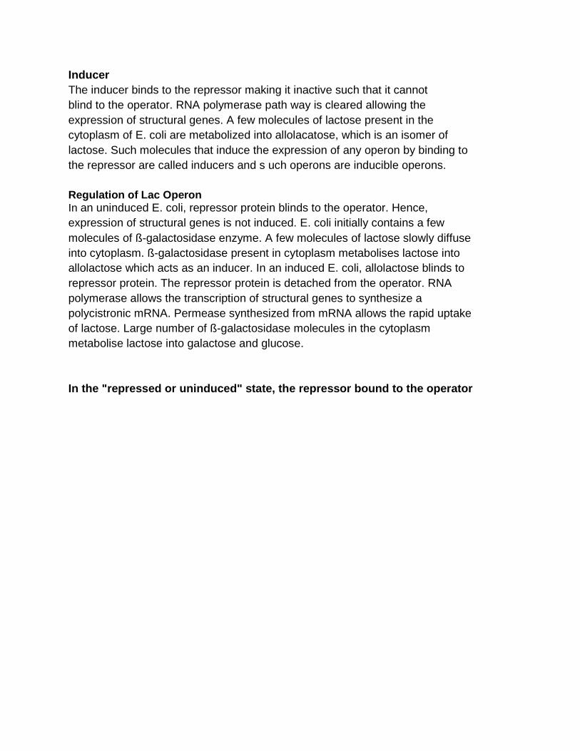
Inducer The inducer binds to the repressor making it inactive such that it cannot blind to the operator. RNA polymerase path way is cleared allowing the expression of structural genes. A few molecules of lactose present in the cytoplasm of E. coli are metabolized into allolacatose, which is an isomer of lactose. Such molecules that induce the expression of any operon by binding to the repressor are called inducers and s uch operons are inducible operons. Regulation of Lac Operon In an uninduced E. coli, repressor protein blinds to the operator. Hence, expression of structural genes is not induced. E. coli initially contains a few molecules of ß-galactosidase enzyme. A few molecules of lactose slowly diffuse into cytoplasm. ß-galactosidase present in cytoplasm metabolises lactose into allolactose which acts as an inducer. In an induced E. coli, allolactose blinds to repressor protein. The repressor protein is detached from the operator. RNA polymerase allows the transcription of structural genes to synthesize a polycistronic mRNA. Permease synthesized from mRNA allows the rapid uptake of lactose. Large number of ß-galactosidase molecules in the cytoplasm metabolise lactose into galactose and glucose. In the "repressed or uninduced" state, the represso r bound to the operator
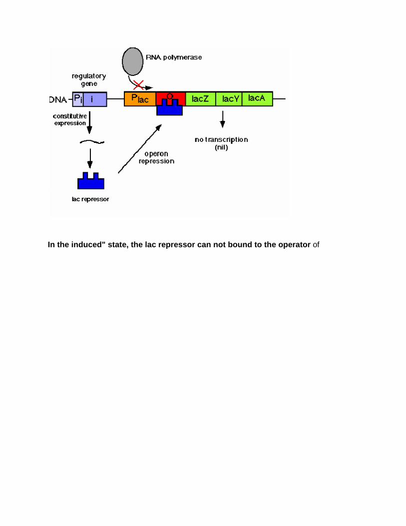
In the induced" state, the lac repressor can not bo und to the operator of
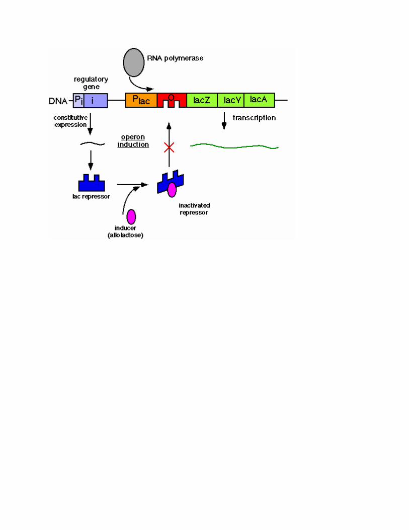
GENE ACTION
Gene action refers to the manner in which genes control the phenotypic
expression of various characters in an organism. Alleles of the gene may interact
with one another in a number of ways to produce variability in their phenotypic
expression. The dominant and recessive relationship is fundamental and is
essentially constant with each pair of alleles.
Gene action can be of the following types:
1. Based on the dominance effect:
a) Complete dominance
b) Incomplete dominance
c) Co-dominance
d) Over dominance
e) Pseudo-dominance
2. Based on lethal effects :
a) Dominant lethals
b) Recessive lethals
3. Based on epistatic action :
a) Epistatic factors
b) Supplementary factors
c) Duplicate factors
d) Complementary factors
e) Additive factors
f) Inhibitory factors
4. Based on number of genes involved:
a) Monogenic
b) Digenic
c) Oligogenic
d) Polygenic
5. Based on pleiotropism / pleiotropic gene action
1. Based on dominance effect: It was noted by many workers that many
characters of F1 hybrid were exactly the same as those of one or the other
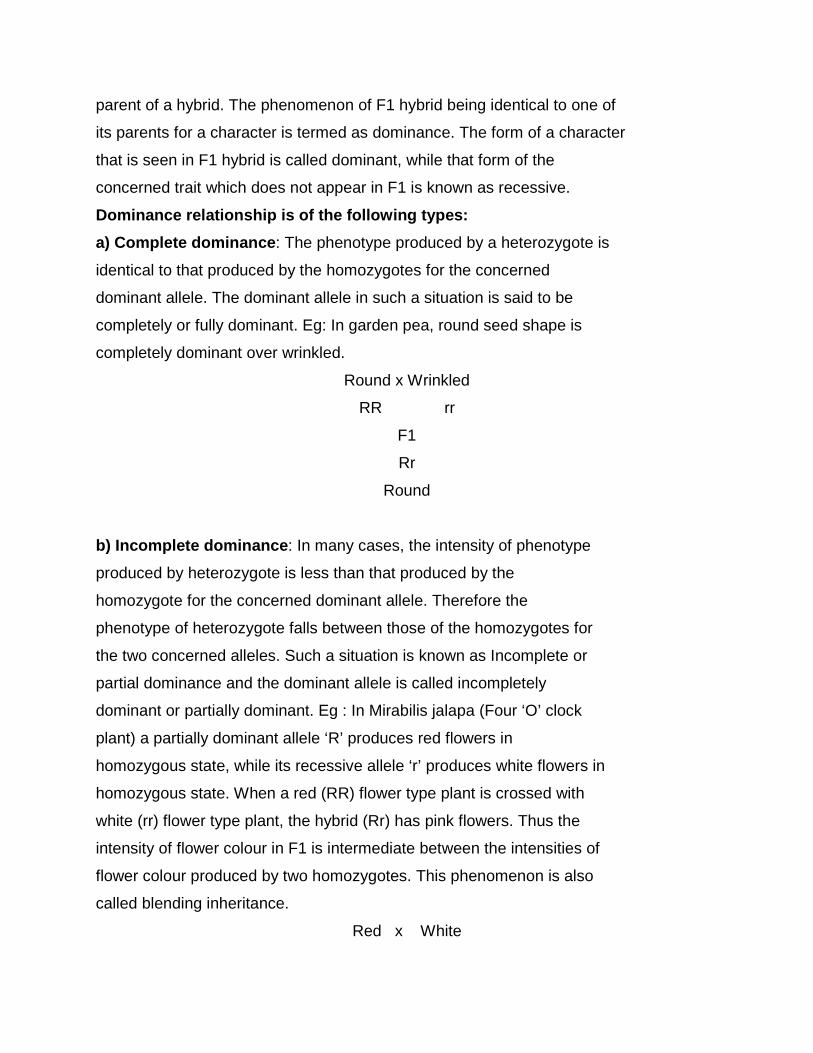
parent of a hybrid. The phenomenon of F1 hybrid being identical to one of
its parents for a character is termed as dominance. The form of a character
that is seen in F1 hybrid is called dominant, while that form of the
concerned trait which does not appear in F1 is known as recessive.
Dominance relationship is of the following types:
a) Complete dominance: The phenotype produced by a heterozygote is
identical to that produced by the homozygotes for the concerned
dominant allele. The dominant allele in such a situation is said to be
completely or fully dominant. Eg: In garden pea, round seed shape is
completely dominant over wrinkled.
Round x Wrinkled
RR rr
F1
Rr
Round
b) Incomplete dominance: In many cases, the intensity of phenotype
produced by heterozygote is less than that produced by the
homozygote for the concerned dominant allele. Therefore the
phenotype of heterozygote falls between those of the homozygotes for
the two concerned alleles. Such a situation is known as Incomplete or
partial dominance and the dominant allele is called incompletely
dominant or partially dominant. Eg : In Mirabilis jalapa (Four ‘O’ clock
plant) a partially dominant allele ‘R’ produces red flowers in
homozygous state, while its recessive allele ‘r’ produces white flowers in
homozygous state. When a red (RR) flower type plant is crossed with
white (rr) flower type plant, the hybrid (Rr) has pink flowers. Thus the
intensity of flower colour in F1 is intermediate between the intensities of
flower colour produced by two homozygotes. This phenomenon is also
called blending inheritance.
Red x White
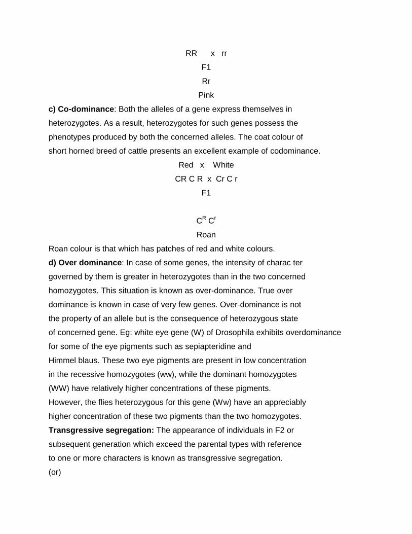
RR x rr
F1
Rr
Pink
c) Co-dominance: Both the alleles of a gene express themselves in
heterozygotes. As a result, heterozygotes for such genes possess the
phenotypes produced by both the concerned alleles. The coat colour of
short horned breed of cattle presents an excellent example of codominance.
Red x White
CR C R x Cr C r
F1
CR Cr
Roan
Roan colour is that which has patches of red and white colours.
d) Over dominance: In case of some genes, the intensity of charac ter
governed by them is greater in heterozygotes than in the two concerned
homozygotes. This situation is known as over-dominance. True over
dominance is known in case of very few genes. Over-dominance is not
the property of an allele but is the consequence of heterozygous state
of concerned gene. Eg: white eye gene (W) of Drosophila exhibits overdominance
for some of the eye pigments such as sepiapteridine and
Himmel blaus. These two eye pigments are present in low concentration
in the recessive homozygotes (ww), while the dominant homozygotes
(WW) have relatively higher concentrations of these pigments.
However, the flies heterozygous for this gene (Ww) have an appreciably
higher concentration of these two pigments than the two homozygotes.
Transgressive segregation: The appearance of individuals in F2 or
subsequent generation which exceed the parental types with reference
to one or more characters is known as transgressive segregation.
(or)
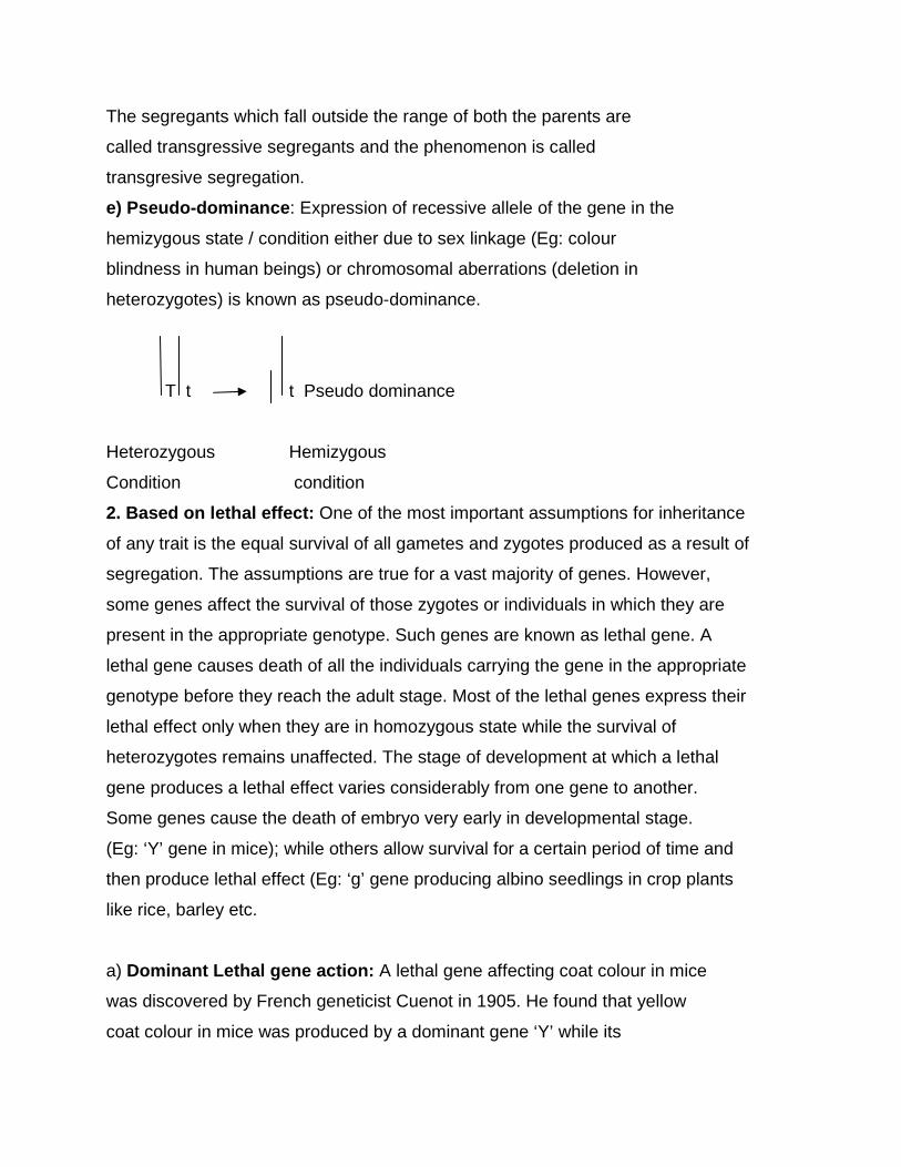
The segregants which fall outside the range of both the parents are
called transgressive segregants and the phenomenon is called
transgresive segregation.
e) Pseudo-dominance: Expression of recessive allele of the gene in the
hemizygous state / condition either due to sex linkage (Eg: colour
blindness in human beings) or chromosomal aberrations (deletion in
heterozygotes) is known as pseudo-dominance.
T t t Pseudo dominance
Heterozygous Hemizygous
Condition condition
2. Based on lethal effect: One of the most important assumptions for inheritance
of any trait is the equal survival of all gametes and zygotes produced as a result of
segregation. The assumptions are true for a vast majority of genes. However,
some genes affect the survival of those zygotes or individuals in which they are
present in the appropriate genotype. Such genes are known as lethal gene. A
lethal gene causes death of all the individuals carrying the gene in the appropriate
genotype before they reach the adult stage. Most of the lethal genes express their
lethal effect only when they are in homozygous state while the survival of
heterozygotes remains unaffected. The stage of development at which a lethal
gene produces a lethal effect varies considerably from one gene to another.
Some genes cause the death of embryo very early in developmental stage.
(Eg: ‘Y’ gene in mice); while others allow survival for a certain period of time and
then produce lethal effect (Eg: ‘g’ gene producing albino seedlings in crop plants
like rice, barley etc.
a) Dominant Lethal gene action: A lethal gene affecting coat colour in mice
was discovered by French geneticist Cuenot in 1905. He found that yellow
coat colour in mice was produced by a dominant gene ‘Y’ while its
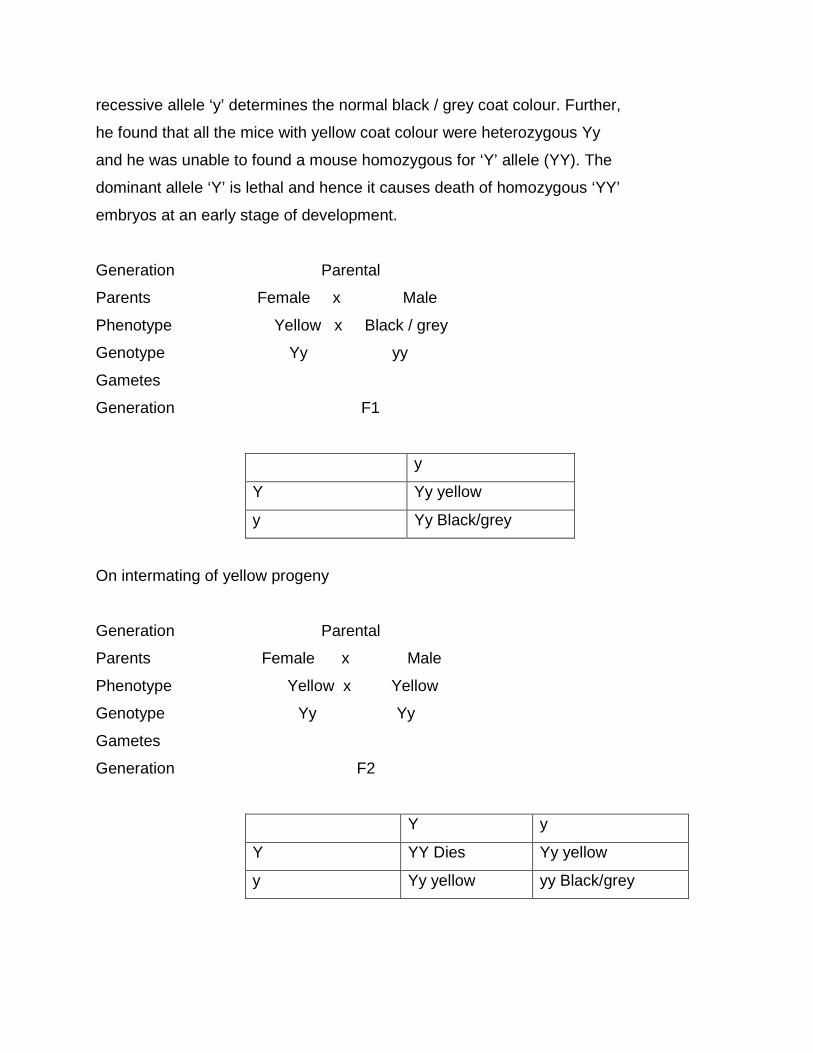
recessive allele ‘y’ determines the normal black / grey coat colour. Further,
he found that all the mice with yellow coat colour were heterozygous Yy
and he was unable to found a mouse homozygous for ‘Y’ allele (YY). The
dominant allele ‘Y’ is lethal and hence it causes death of homozygous ‘YY’
embryos at an early stage of development.
Generation Parental
Parents Female x Male
Phenotype Yellow x Black / grey
Genotype Yy yy
Gametes
Generation F1
y
Y Yy yellow
y Yy Black/grey
On intermating of yellow progeny
Generation Parental
Parents Female x Male
Phenotype Yellow x Yellow
Genotype Yy Yy
Gametes
Generation F2
Y y
Y YY Dies Yy yellow
y Yy yellow yy Black/grey
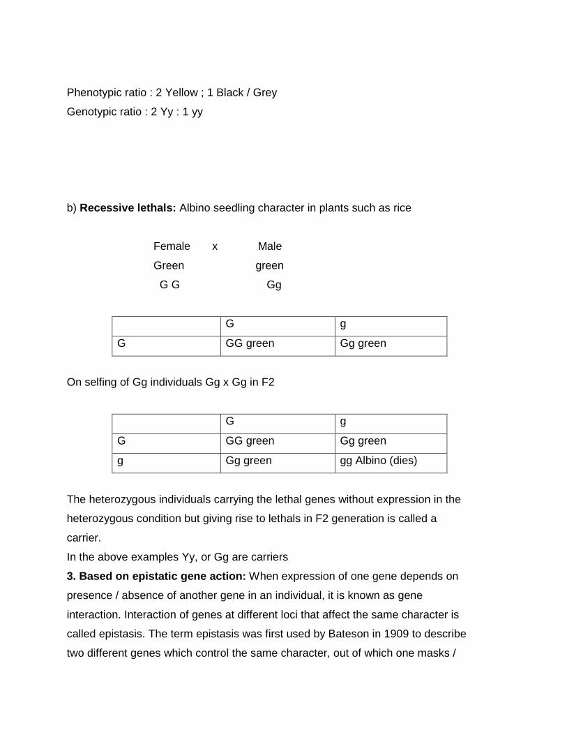
Phenotypic ratio : 2 Yellow ; 1 Black / Grey
Genotypic ratio : 2 Yy : 1 yy
b) Recessive lethals: Albino seedling character in plants such as rice
Female x Male
Green green
G G Gg
G g
G GG green Gg green
On selfing of Gg individuals Gg x Gg in F2
G g
G GG green Gg green
g Gg green gg Albino (dies)
The heterozygous individuals carrying the lethal genes without expression in the
heterozygous condition but giving rise to lethals in F2 generation is called a
carrier.
In the above examples Yy, or Gg are carriers
3. Based on epistatic gene action: When expression of one gene depends on
presence / absence of another gene in an individual, it is known as gene
interaction. Interaction of genes at different loci that affect the same character is
called epistasis. The term epistasis was first used by Bateson in 1909 to describe
two different genes which control the same character, out of which one masks /

suppresses the expression of another gene. Gene that masks the action of
another gene is called epistatic gene while the gene whose expression is being
masked is called hypostatic gene. Epistatic gene interaction can be of the
following types.
(i) Epistatic factors - 12 : 3 : 1 (ii) Supplementary factors - 9 : 3 : 4
(iii) Duplicate factors - 15 : 1 (iv) Complementary factors - 9 : 7
(v) Additive factors - 9 : 6 : 1 (vi) Inhibitory factors - 13 : 3
4. Based on number of genes involved:
a) Monogenic - Each character is controlled by one gene
b) Digenic - Each character is controlled by two genes
c) Oligogenic - Each character is controlled by few genes
d) Polygenic - Each character is controlled by many genes
Polygenic or polymerism: In general, one gene controls or affects a single
character. But some characters are known to be controlled by more number of
genes. Such genes are called poly genes and the phenomenon is called
polymerism.
Eg : Yield in plants.
5. Based on pleiotropism / pleiotropic gene action: In general, one gene
affects a single character. But some of the genes are known to affect or control
more than one character. Such genes are called pleiotropic genes and the
phenomenon is known as pleiotropism. Many fold phenotypic expressions of a
single gene is called pleiotropism or pleiotropic gene effects. Eg: White eye gene
effects the shape of sperm storage organs and other structures in Drosophila.
These genes are found in all crop plants. Good example of pleiotropism has been
reported in wheat. A gene governing awns in Ona’s variety of wheat also
increases the yield as well as seed weight.
Turner’s syndrome :- It was first described by H.H.Turner in 1938.It was
associated with monosomy for x chromosome . It has frequency of about 1 per
2500 live female birth. More than 90% of the XO zygote abort spontaneously.
Adult with Turner’s syndrome are female who has virtually no ovaries and whose
secondary sex characters are poorly developed. Such female have short stature
lowset ears, webbed neck and a shield like chest but genetically do not have
mental retardation.
Klinefelter syndrome:-Another trisomic condition for the sex chromosome (xxy)
produces Klinefelter syndrome .It was first described by H.F. Klinefelter in 1942.
Such individuals are phenotypically male but some tendency towards femaleness,
particularly in secondary sex characteristics They may have enlarged breasts, less
body hair, small tests and small prostrate glands. xxy,is the most common
karyotype. But klinefelter patients may be xxyy,xxxy,xxxyy,xxxxy,xxxxyy,and
xxxxxy. Patients having more than two x chromosomes show mental retardation
as well.
LAC OPERON AND FINE STRUCTURE OF GENE
The hereditary units which are transmitted from one generation to
the next generation arc called genes. A gene is the fundamental biologic
unit, like the atom which is the fundamental physical unit. Mendel while
explaining the result of his monohybrid and dihybrid crosses, first of all
conceived of the genes as particulate units and referred them by various
names such as hereditary factors or hereditary elements. But his concept
about the gene was entirely hypothetical and he remained ignorant about
the physical and chemical nature of gene.
Even before the rediscovery of Menders laws in 1900, it was
already established that chromosomes have a definite role in the
inheritance because it was found that chromosomes were the only link
between one generation and the next generation and a diploid
chromosome set consists of two morphologically similar sets, one is
derived from the mother and the other from the father at fertilization.
Later on, a parallel behavior among chromosomes and genes was
discovered.
Earlier workers proposed various hypotheses to explain the nature
of genes. For instance, De Vries postulated one gene one character
hypothesis according to which a particular trait of an individual is
controlled by a particular gene. Bateson and Punnett proposed the
presence or absence theory. According to them, in a cross the character
which dominates the other has a determiner, while, the recessive
character has no such determiner. But all the theories were discarded by
Morgan, who produced the particulate gene theory in 1926. He considered
genes as corpuscles, which are arranged in a linear order on the
chromosomes and appear like beads on a string. Each gene was supposed
to be different from ail others. The particulate theory of gene was widely
accepted and supported by cytological observations. But, the discovery of
DNA molecule as a sole carrier of genetic informations base altogether
discarded the Morgan's theory. Therefore, before defining the gene it will
be advisable to consider both the classical as well as modern definitions of
gene.
Changing Concept of Gene
The concept of gene has been the focal point of study from the
beginning of twentieth century to establish the basis of heredity. The gene
has been examined from two main angles, i.e., (1) genetic view, and (2)
biochemical and molecular view. These aspects are briefly described
below:
(1) A Genetic View
The genetic view or perspective of gene is based mainly on the
Mendelian inheritance, chromosomal theory of inheritance and linkage
studies. Mendel used the term factors for genes and reported that factors
were responsible for transmission of characters from parents to their
offspring. Sutton and Boveri (1903) based on the study of mitosis and
meiosis in higher plants established parallel behaviour of chromosomes
and genes. They reported that both chromosomes and genes segregate
and exhibit random assortment, which clearly demonstrated that genes
are located on chromosomes. The Sutton- Boveri hypothesis is known as
chromosome theory of inheritance.
Morgan based on linkage studies in Drosophila reported that genes
are located on the chromosome in a linear fashion. Some genes do not
assort independently because of linkage between them. He suggested that
recombinants are the result of crossing over. The crossing over increases
if the distance between two genes is more. The number of linkage group
is the same as the number of chromosomes. The chromosome theory and
linkage studies reveal that genes are located on the chromosomes. This
view is sometimes called as bead theory. The important points about the
bead theory are given below:
1. The gene is viewed as a fundamental unit of structure, indivisible
by crossing over. Crossing over occurs between genes but not
within a gene.
2. The gene is considered as a basic unit of change or mutation. It
changes from one allelic form to another, but there are no smaller
components within a gene that can change.
3. The gene is viewed as a basic unit of function. Parts of a gene, if
they exist, cannot function.
The chromosome has been viewed merely as a vector or transporter of
genes and exists simply to permit their orderly segregation and to shuffle
them in recombination. The bead theory is no more valid for any of the
above three points. Now evidences are available which indicate that: (1) a
gene is divisible (2) part of a gene can mutate, and (3) part of a gene can
function.
The Gene is Divisible
Earlier it was believed that gene is a basic unit of structure which is
indivisible by crossing over. In other words, crossing over occurs between
genes but not within a gene. Now intragenic recombination has been
observed in many organisms which indicates that a gene is divisible. The
intragenic recombination has following two main features.
1. It occurs with rare frequency so that a very large test cross
progeny is required for its detection. Benzer expected to detect
a recombination frequency as low as 10-6, the lowest he actually
found was 10-4 (0.01 x 2 = 0.02%).
2. The alleles in which intragenic recombination occurs are
separated by small distances within a gene and are functionally
related.
Examples of intragenic recombination include bar eye, star
asteroid eye and lozenge eye in Drosophila. The bar locus is briefly
described below. Lozenge eye and star asteroid have been discussed
under pseudoalleles.
Bar Eye in Drosophila
The first case of intragenic recombination was recorded in
Drosophila for bar locus which controls size of eye. The bar locus contains
more than one unit of function. The dominant bar gene in Drosophila
produces slit like eye instead of normal oval eye. Bar phenotype is caused
by tandem duplication of 16A region in X chromosome, which results due
to unequal crossing over. The flies with different dose of 16A region have
different types of eye as follows:
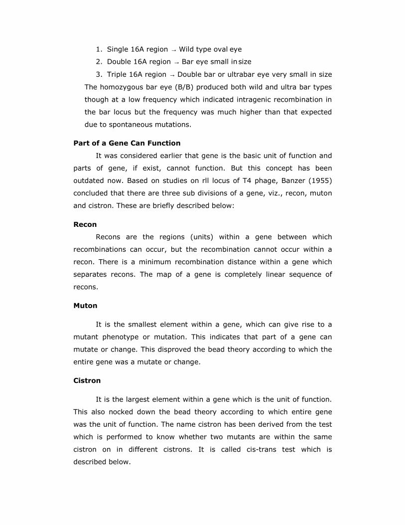
1. Single 16A region → Wild type oval eye
2. Double 16A region → Bar eye small in size
3. Triple 16A region → Double bar or ultrabar eye very small in size
The homozygous bar eye (B/B) produced both wild and ultra bar types
though at a low frequency which indicated intragenic recombination in
the bar locus but the frequency was much higher than that expected
due to spontaneous mutations.
Part of a Gene Can Function
It was considered earlier that gene is the basic unit of function and
parts of gene, if exist, cannot function. But this concept has been
outdated now. Based on studies on rll locus of T4 phage, Banzer (1955)
concluded that there are three sub divisions of a gene, viz., recon, muton
and cistron. These are briefly described below:
Recon
Recons are the regions (units) within a gene between which
recombinations can occur, but the recombination cannot occur within a
recon. There is a minimum recombination distance within a gene which
separates recons. The map of a gene is completely linear sequence of
recons.
Muton
It is the smallest element within a gene, which can give rise to a
mutant phenotype or mutation. This indicates that part of a gene can
mutate or change. This disproved the bead theory according to which the
entire gene was a mutate or change.
Cistron
It is the largest element within a gene which is the unit of function.
This also nocked down the bead theory according to which entire gene
was the unit of function. The name cistron has been derived from the test
which is performed to know whether two mutants are within the same
cistron on in different cistrons. It is called cis-trans test which is
described below.
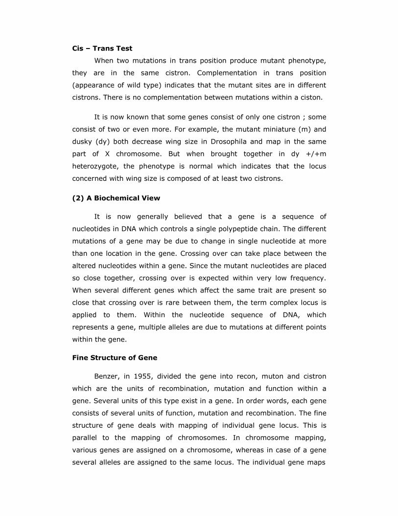
Cis – Trans Test
When two mutations in trans position produce mutant phenotype,
they are in the same cistron. Complementation in trans position
(appearance of wild type) indicates that the mutant sites are in different
cistrons. There is no complementation between mutations within a ciston.
It is now known that some genes consist of only one cistron ; some
consist of two or even more. For example, the mutant miniature (m) and
dusky (dy) both decrease wing size in Drosophila and map in the same
part of X chromosome. But when brought together in dy +/+m
heterozygote, the phenotype is normal which indicates that the locus
concerned with wing size is composed of at least two cistrons.
(2) A Biochemical View
It is now generally believed that a gene is a sequence of
nucleotides in DNA which controls a single polypeptide chain. The different
mutations of a gene may be due to change in single nucleotide at more
than one location in the gene. Crossing over can take place between the
altered nucleotides within a gene. Since the mutant nucleotides are placed
so close together, crossing over is expected within very low frequency.
When several different genes which affect the same trait are present so
close that crossing over is rare between them, the term complex locus is
applied to them. Within the nucleotide sequence of DNA, which
represents a gene, multiple alleles are due to mutations at different points
within the gene.
Fine Structure of Gene
Benzer, in 1955, divided the gene into recon, muton and cistron
which are the units of recombination, mutation and function within a
gene. Several units of this type exist in a gene. In order words, each gene
consists of several units of function, mutation and recombination. The fine
structure of gene deals with mapping of individual gene locus. This is
parallel to the mapping of chromosomes. In chromosome mapping,
various genes are assigned on a chromosome, whereas in case of a gene
several alleles are assigned to the same locus. The individual gene maps
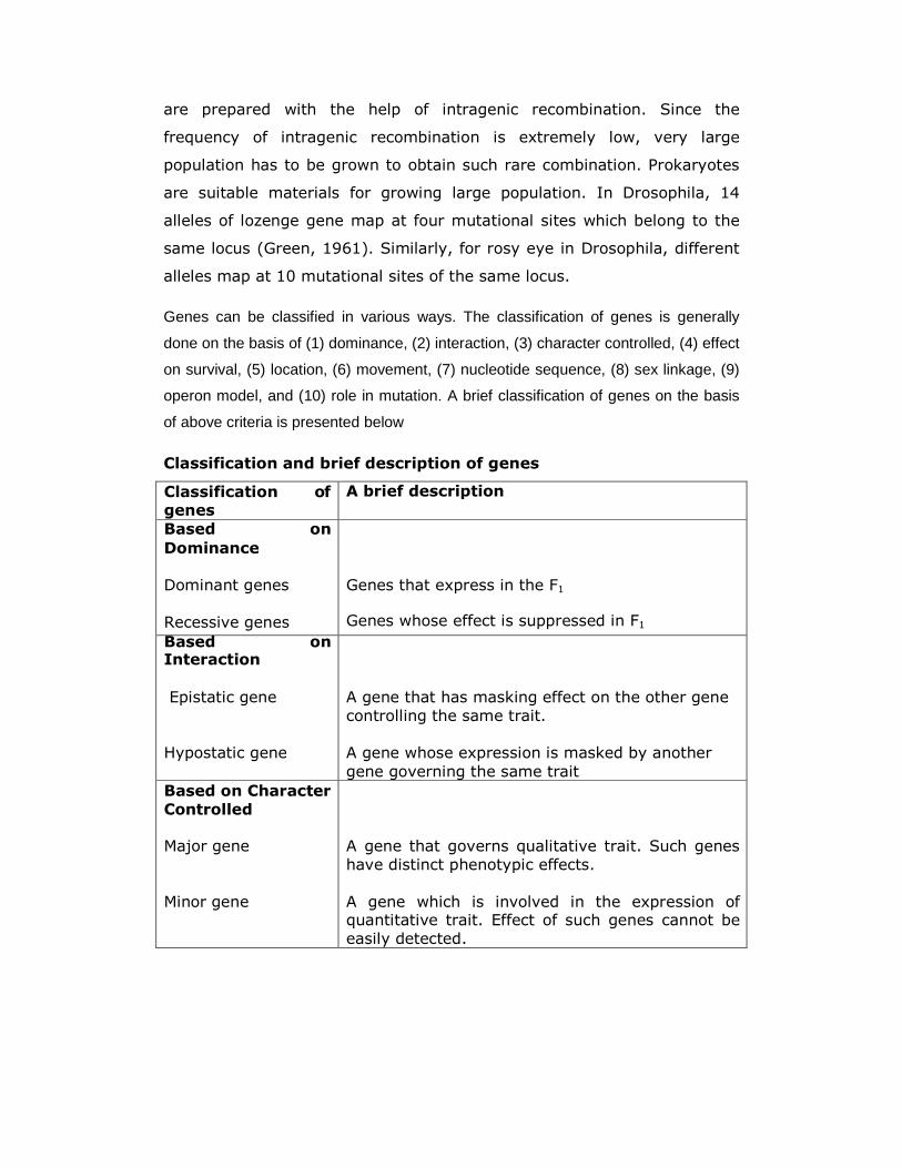
are prepared with the help of intragenic recombination. Since the
frequency of intragenic recombination is extremely low, very large
population has to be grown to obtain such rare combination. Prokaryotes
are suitable materials for growing large population. In Drosophila, 14
alleles of lozenge gene map at four mutational sites which belong to the
same locus (Green, 1961). Similarly, for rosy eye in Drosophila, different
alleles map at 10 mutational sites of the same locus.
Genes can be classified in various ways. The classification of genes is generally
done on the basis of (1) dominance, (2) interaction, (3) character controlled, (4) effect
on survival, (5) location, (6) movement, (7) nucleotide sequence, (8) sex linkage, (9)
operon model, and (10) role in mutation. A brief classification of genes on the basis
of above criteria is presented below
Classification and brief description of genes
Classification of genes
A brief description
Based on
Dominance
Dominant genes
Recessive genes
Genes that express in the F1
Genes whose effect is suppressed in F1
Based on Interaction
Epistatic gene
Hypostatic gene
A gene that has masking effect on the other gene
controlling the same trait.
A gene whose expression is masked by another
gene governing the same trait
Based on Character
Controlled
Major gene
Minor gene
A gene that governs qualitative trait. Such genes
have distinct phenotypic effects.
A gene which is involved in the expression of quantitative trait. Effect of such genes cannot be
easily detected.
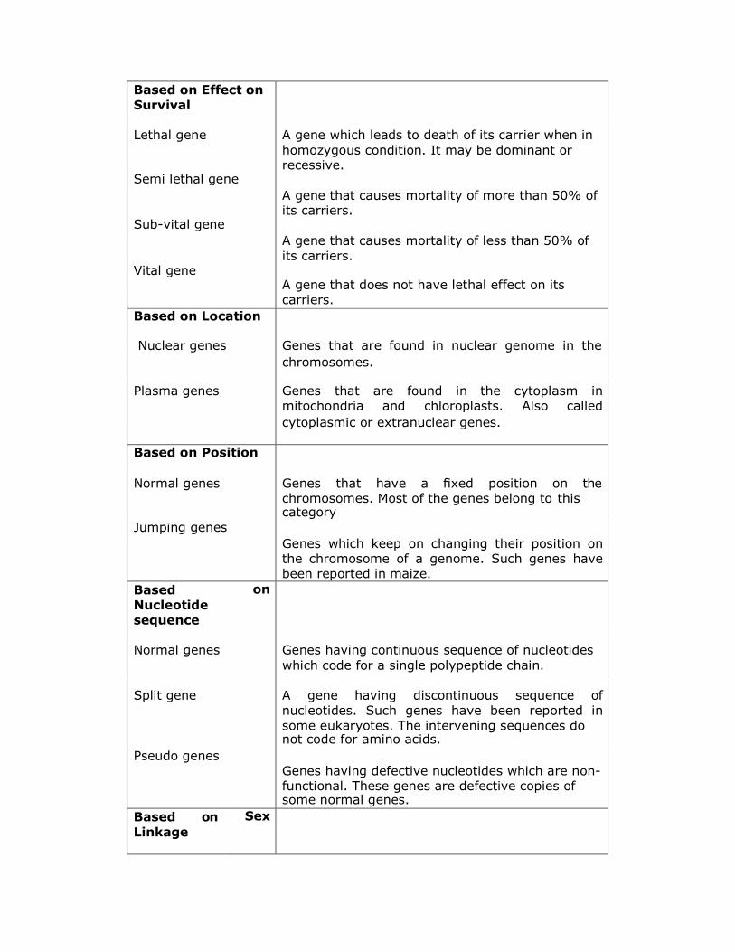
Based on Effect on
Survival
Lethal gene A gene which leads to death of its carrier when in
homozygous condition. It may be dominant or recessive.
Semi lethal gene
A gene that causes mortality of more than 50% of its carriers.
Sub-vital gene
A gene that causes mortality of less than 50% of
its carriers. Vital gene
A gene that does not have lethal effect on its carriers.
Based on Location
Nuclear genes Genes that are found in nuclear genome in the
chromosomes.
Plasma genes Genes that are found in the cytoplasm in mitochondria and chloroplasts. Also called
cytoplasmic or extranuclear genes.
Based on Position
Normal genes Genes that have a fixed position on the chromosomes. Most of the genes belong to this category
Jumping genes
Genes which keep on changing their position on the chromosome of a genome. Such genes have been reported in maize.
Based Nucleotide
sequence
on
Normal genes Genes having continuous sequence of nucleotides
which code for a single polypeptide chain.
Split gene A gene having discontinuous sequence of
nucleotides. Such genes have been reported in
some eukaryotes. The intervening sequences do not code for amino acids.
Pseudo genes
Genes having defective nucleotides which are non- functional. These genes are defective copies of some normal genes.
Based on
Linkage
Sex
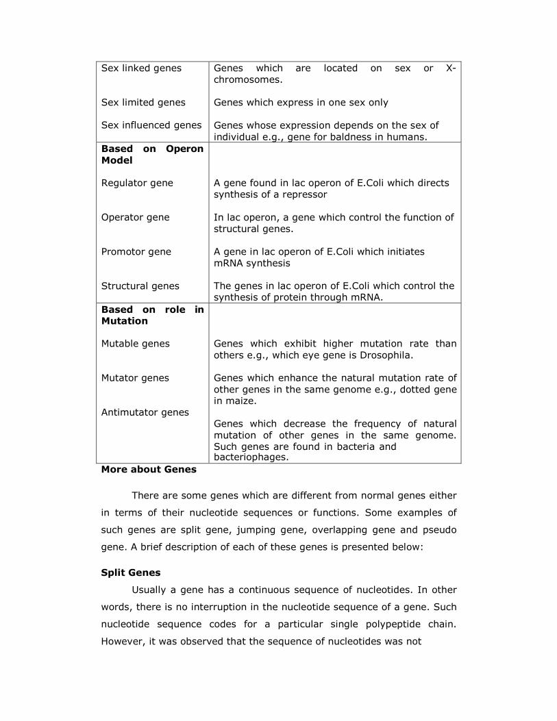
Sex linked genes
Sex limited genes
Sex influenced genes
Genes which are located on sex or X-
chromosomes.
Genes which express in one sex only
Genes whose expression depends on the sex of
individual e.g., gene for baldness in humans.
Based on Operon
Model
Regulator gene
Operator gene
Promotor gene
Structural genes
A gene found in lac operon of E.Coli which directs
synthesis of a repressor
In lac operon, a gene which control the function of
structural genes.
A gene in lac operon of E.Coli which initiates
mRNA synthesis
The genes in lac operon of E.Coli which control the synthesis of protein through mRNA.
Based on role in Mutation
Mutable genes
Mutator genes
Antimutator genes
Genes which exhibit higher mutation rate than
others e.g., which eye gene is Drosophila. Genes which enhance the natural mutation rate of
other genes in the same genome e.g., dotted gene in maize.
Genes which decrease the frequency of natural
mutation of other genes in the same genome. Such genes are found in bacteria and bacteriophages.
More about Genes
There are some genes which are different from normal genes either
in terms of their nucleotide sequences or functions. Some examples of
such genes are split gene, jumping gene, overlapping gene and pseudo
gene. A brief description of each of these genes is presented below:
Split Genes
Usually a gene has a continuous sequence of nucleotides. In other
words, there is no interruption in the nucleotide sequence of a gene. Such
nucleotide sequence codes for a particular single polypeptide chain.
However, it was observed that the sequence of nucleotides was not
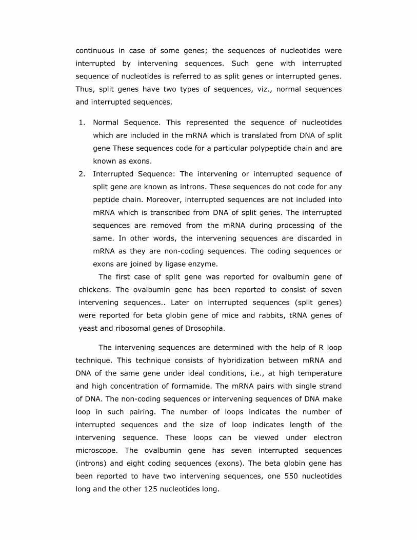
continuous in case of some genes; the sequences of nucleotides were
interrupted by intervening sequences. Such gene with interrupted
sequence of nucleotides is referred to as split genes or interrupted genes.
Thus, split genes have two types of sequences, viz., normal sequences
and interrupted sequences.
1. Normal Sequence. This represented the sequence of nucleotides
which are included in the mRNA which is translated from DNA of split
gene These sequences code for a particular polypeptide chain and are
known as exons.
2. Interrupted Sequence: The intervening or interrupted sequence of
split gene are known as introns. These sequences do not code for any
peptide chain. Moreover, interrupted sequences are not included into
mRNA which is transcribed from DNA of split genes. The interrupted
sequences are removed from the mRNA during processing of the
same. In other words, the intervening sequences are discarded in
mRNA as they are non-coding sequences. The coding sequences or
exons are joined by ligase enzyme.
The first case of split gene was reported for ovalbumin gene of
chickens. The ovalbumin gene has been reported to consist of seven
intervening sequences.. Later on interrupted sequences (split genes)
were reported for beta globin gene of mice and rabbits, tRNA genes of
yeast and ribosomal genes of Drosophila.
The intervening sequences are determined with the help of R loop
technique. This technique consists of hybridization between mRNA and
DNA of the same gene under ideal conditions, i.e., at high temperature
and high concentration of formamide. The mRNA pairs with single strand
of DNA. The non-coding sequences or intervening sequences of DNA make
loop in such pairing. The number of loops indicates the number of
interrupted sequences and the size of loop indicates length of the
intervening sequence. These loops can be viewed under electron
microscope. The ovalbumin gene has seven interrupted sequences
(introns) and eight coding sequences (exons). The beta globin gene has
been reported to have two intervening sequences, one 550 nucleotides
long and the other 125 nucleotides long.
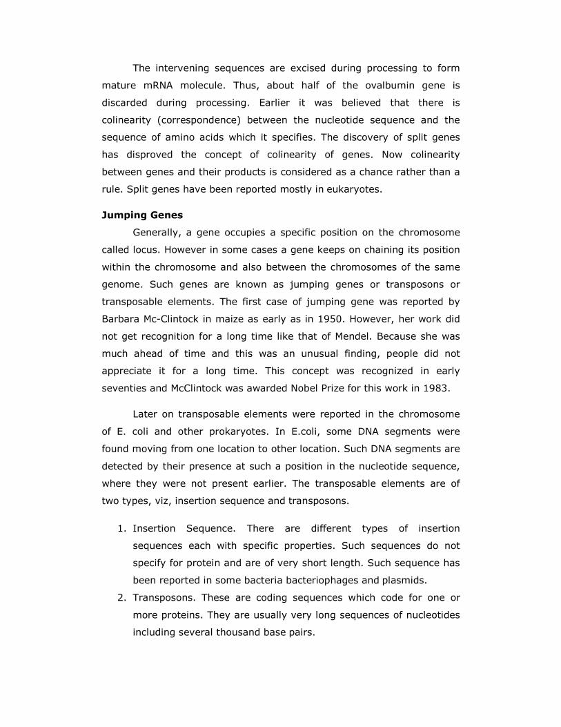
The intervening sequences are excised during processing to form
mature mRNA molecule. Thus, about half of the ovalbumin gene is
discarded during processing. Earlier it was believed that there is
colinearity (correspondence) between the nucleotide sequence and the
sequence of amino acids which it specifies. The discovery of split genes
has disproved the concept of colinearity of genes. Now colinearity
between genes and their products is considered as a chance rather than a
rule. Split genes have been reported mostly in eukaryotes.
Jumping Genes
Generally, a gene occupies a specific position on the chromosome
called locus. However in some cases a gene keeps on chaining its position
within the chromosome and also between the chromosomes of the same
genome. Such genes are known as jumping genes or transposons or
transposable elements. The first case of jumping gene was reported by
Barbara Mc-Clintock in maize as early as in 1950. However, her work did
not get recognition for a long time like that of Mendel. Because she was
much ahead of time and this was an unusual finding, people did not
appreciate it for a long time. This concept was recognized in early
seventies and McClintock was awarded Nobel Prize for this work in 1983.
Later on transposable elements were reported in the chromosome
of E. coli and other prokaryotes. In E.coli, some DNA segments were
found moving from one location to other location. Such DNA segments are
detected by their presence at such a position in the nucleotide sequence,
where they were not present earlier. The transposable elements are of
two types, viz, insertion sequence and transposons.
1. Insertion Sequence. There are different types of insertion
sequences each with specific properties. Such sequences do not
specify for protein and are of very short length. Such sequence has
been reported in some bacteria bacteriophages and plasmids.
2. Transposons. These are coding sequences which code for one or
more proteins. They are usually very long sequences of nucleotides
including several thousand base pairs.
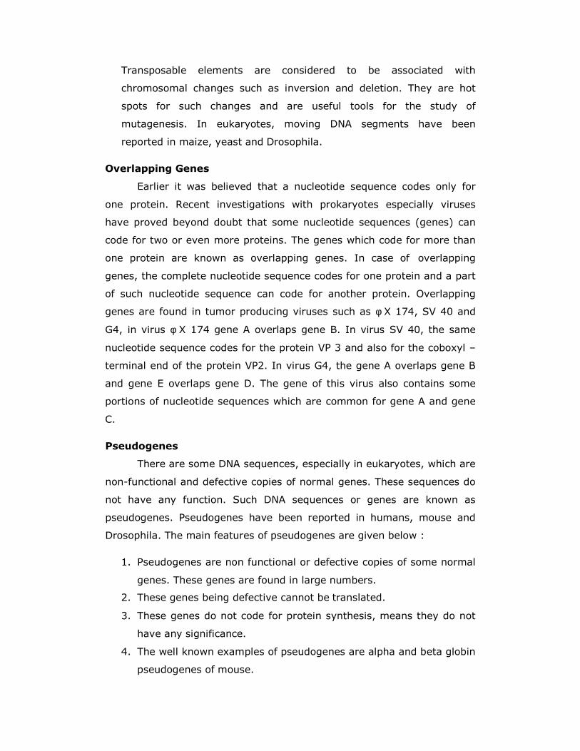
Transposable elements are considered to be associated with
chromosomal changes such as inversion and deletion. They are hot
spots for such changes and are useful tools for the study of
mutagenesis. In eukaryotes, moving DNA segments have been
reported in maize, yeast and Drosophila.
Overlapping Genes
Earlier it was believed that a nucleotide sequence codes only for
one protein. Recent investigations with prokaryotes especially viruses
have proved beyond doubt that some nucleotide sequences (genes) can
code for two or even more proteins. The genes which code for more than
one protein are known as overlapping genes. In case of overlapping
genes, the complete nucleotide sequence codes for one protein and a part
of such nucleotide sequence can code for another protein. Overlapping
genes are found in tumor producing viruses such as φ X 174, SV 40 and
G4, in virus φ X 174 gene A overlaps gene B. In virus SV 40, the same
nucleotide sequence codes for the protein VP 3 and also for the coboxyl –
terminal end of the protein VP2. In virus G4, the gene A overlaps gene B
and gene E overlaps gene D. The gene of this virus also contains some
portions of nucleotide sequences which are common for gene A and gene
C.
Pseudogenes
There are some DNA sequences, especially in eukaryotes, which are
non-functional and defective copies of normal genes. These sequences do
not have any function. Such DNA sequences or genes are known as
pseudogenes. Pseudogenes have been reported in humans, mouse and
Drosophila. The main features of pseudogenes are given below :
1. Pseudogenes are non functional or defective copies of some normal
genes. These genes are found in large numbers.
2. These genes being defective cannot be translated.
3. These genes do not code for protein synthesis, means they do not
have any significance.
4. The well known examples of pseudogenes are alpha and beta globin
pseudogenes of mouse.
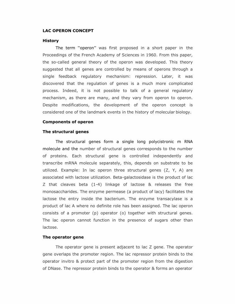
LAC OPERON CONCEPT
History
The term ‘‘operon’’ was first proposed in a short paper in the
Proceedings of the French Academy of Sciences in 1960. From this paper,
the so-called general theory of the operon was developed. This theory
suggested that all genes are controlled by means of operons through a
single feedback regulatory mechanism: repression. Later, it was
discovered that the regulation of genes is a much more complicated
process. Indeed, it is not possible to talk of a general regulatory
mechanism, as there are many, and they vary from operon to operon.
Despite modifications, the development of the operon concept is
considered one of the landmark events in the history of molecular biology.
Components of operon
The structural genes
The structural genes form a single long polycistronic m RNA
molecule and the number of structural genes corresponds to the number
of proteins. Each structural gene is controlled independently and
transcribe mRNA molecule separately, this, depends on substrate to be
utilized. Example: In lac operon three structural genes (Z, Y, A) are
associated with lactose utilization. Beta-galactosidase is the product of lac
Z that cleaves beta (1-4) linkage of lactose & releases the free
monosaccharides. The enzyme permease (a product of lacy) facilitates the
lactose the entry inside the bacterium. The enzyme transacylase is a
product of lac A where no definite role has been assigned. The lac operon
consists of a promoter (p) operator (o) together with structural genes.
The lac operon cannot function in the presence of sugars other than
lactose.
The operator gene
The operator gene is present adjacent to lac Z gene. The operator
gene overlaps the promoter region. The lac repressor protein binds to the
operator invitro & protect part of the promoter region from the digestion
of DNase. The repressor protein binds to the operator & forms an operator
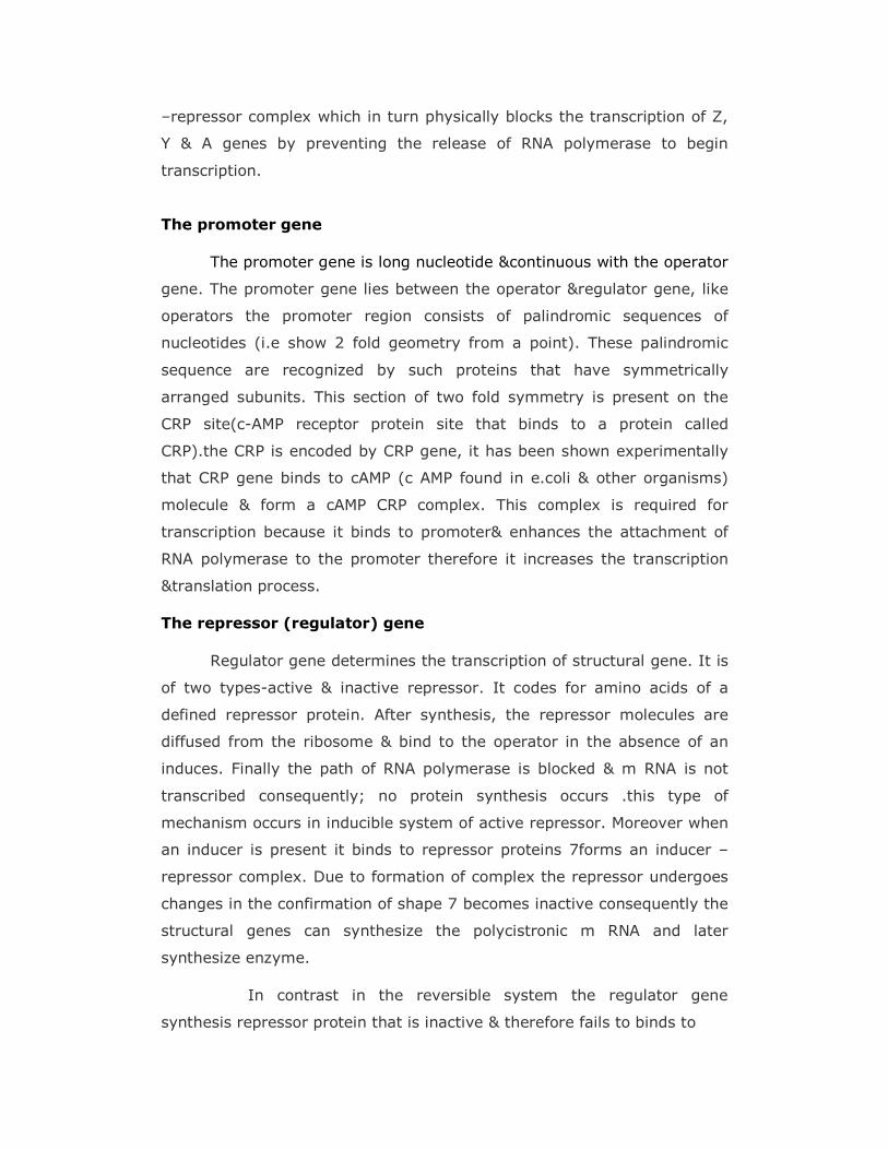
–repressor complex which in turn physically blocks the transcription of Z,
Y & A genes by preventing the release of RNA polymerase to begin
transcription.
The promoter gene
The promoter gene is long nucleotide &continuous with the operator
gene. The promoter gene lies between the operator ®ulator gene, like
operators the promoter region consists of palindromic sequences of
nucleotides (i.e show 2 fold geometry from a point). These palindromic
sequence are recognized by such proteins that have symmetrically
arranged subunits. This section of two fold symmetry is present on the
CRP site(c-AMP receptor protein site that binds to a protein called
CRP).the CRP is encoded by CRP gene, it has been shown experimentally
that CRP gene binds to cAMP (c AMP found in e.coli & other organisms)
molecule & form a cAMP CRP complex. This complex is required for
transcription because it binds to promoter& enhances the attachment of
RNA polymerase to the promoter therefore it increases the transcription
&translation process.
The repressor (regulator) gene
Regulator gene determines the transcription of structural gene. It is
of two types-active & inactive repressor. It codes for amino acids of a
defined repressor protein. After synthesis, the repressor molecules are
diffused from the ribosome & bind to the operator in the absence of an
induces. Finally the path of RNA polymerase is blocked & m RNA is not
transcribed consequently; no protein synthesis occurs .this type of
mechanism occurs in inducible system of active repressor. Moreover when
an inducer is present it binds to repressor proteins 7forms an inducer –
repressor complex. Due to formation of complex the repressor undergoes
changes in the confirmation of shape 7 becomes inactive consequently the
structural genes can synthesize the polycistronic m RNA and later
synthesize enzyme.
In contrast in the reversible system the regulator gene
synthesis repressor protein that is inactive & therefore fails to binds to
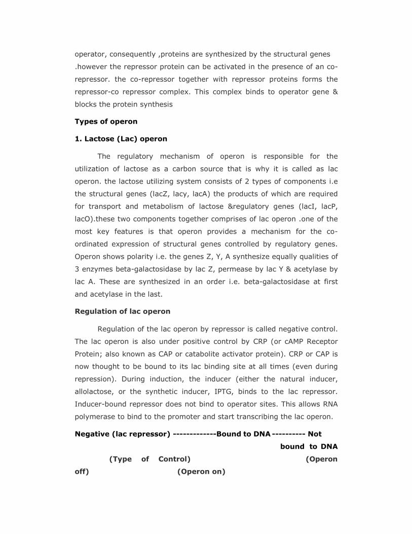
operator, consequently ,proteins are synthesized by the structural genes
.however the repressor protein can be activated in the presence of an co-
repressor. the co-repressor together with repressor proteins forms the
repressor-co repressor complex. This complex binds to operator gene &
blocks the protein synthesis
Types of operon
1. Lactose (Lac) operon
The regulatory mechanism of operon is responsible for the
utilization of lactose as a carbon source that is why it is called as lac
operon. the lactose utilizing system consists of 2 types of components i.e
the structural genes (lacZ, lacy, lacA) the products of which are required
for transport and metabolism of lactose ®ulatory genes (lacI, lacP,
lacO).these two components together comprises of lac operon .one of the
most key features is that operon provides a mechanism for the co-
ordinated expression of structural genes controlled by regulatory genes.
Operon shows polarity i.e. the genes Z, Y, A synthesize equally qualities of
3 enzymes beta-galactosidase by lac Z, permease by lac Y & acetylase by
lac A. These are synthesized in an order i.e. beta-galactosidase at first
and acetylase in the last.
Regulation of lac operon
Regulation of the lac operon by repressor is called negative control.
The lac operon is also under positive control by CRP (or cAMP Receptor
Protein; also known as CAP or catabolite activator protein). CRP or CAP is
now thought to be bound to its lac binding site at all times (even during
repression). During induction, the inducer (either the natural inducer,
allolactose, or the synthetic inducer, IPTG, binds to the lac repressor.
Inducer-bound repressor does not bind to operator sites. This allows RNA
polymerase to bind to the promoter and start transcribing the lac operon.
Negative (lac repressor) -------------Bound to DNA ---------- Not
bound to DNA
(Type of Control) (Operon
off) (Operon on)
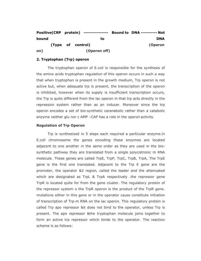
Positive(CRP protein) --------------- Bound to DNA ---------- Not
bound to DNA
(Type of control) (Operon
on) (Operon off)
2. Tryptophan (Trp) operon
The tryptophan operon of E.coli is responsible for the synthesis of
the amino acids tryptophan regulation of this operon occurs in such a way
that when tryptophan is present in the growth medium, Trp operon is not
active but, when adequate trp is present, the transcription of the operon
is inhibited, however when its supply is insufficient transcription occurs,
the Trp is quite different from the lac operon in that trp acts directly in the
repression system rather than as an inducer. Moreover since the trp
operon encodes a set of bio-synthetic caranabolic rather than a catabolic
enzyme neither glu nor c AMP –CAP has a role in the operon activity.
Regulation of Trp Operon
Trp is synthesized in 5 steps each required a particular enzyme.in
E.coli chromosome the genes encoding these enzymes are located
adjacent to one another in the same order as they are used in the bio-
synthetic pathway they are translated from a single polycistronic m RNA
molecule. These genes are called TrpE, TrpP, TrpC, TrpB, TrpA, The TrpE
gene is the first one translated. Adjacent to the Trp E gene are the
promoter, the operator &2 region, called the leader and the attenuated
which are designated as TrpL & TrpA respectively .the repressor gene
TrpR is located quite for from the gene cluster. The regulatory protein of
the repressor system o the TrpR operon is the product of the TrpR gene.
mutations either in this gene or in the operator cause constitute initiation
of transcription of Trp-m RNA on the lac operon. This regulatory protein is
called Trp apo repressor &it does not bind to the operator, unless Trp is
present. The apo repressor &the tryptophan molecule joins together to
form an active trp repressor which binds to the operator. The reaction
scheme is as follows:
Apo repressor (no trp) ---------------active repressor (transcription
occurs)
Apo repressor+trp------------------active repressor +operator-------
--------- inactive operator (transcription does not occur)
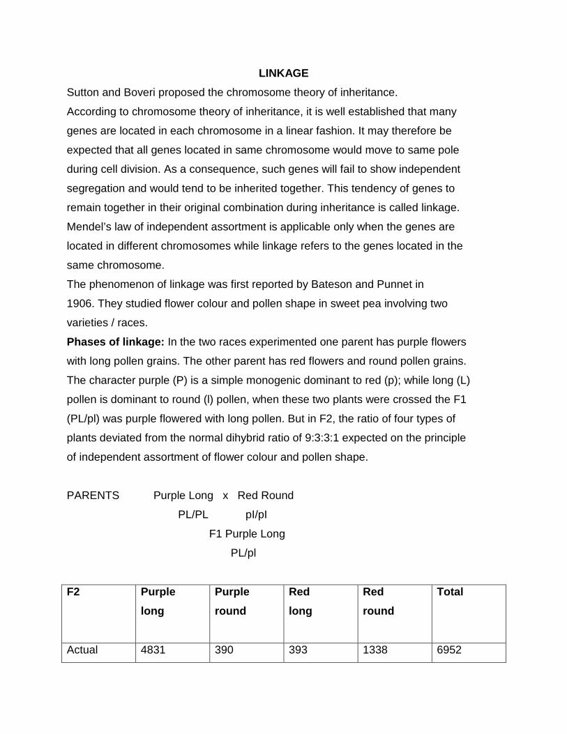
LINKAGE
Sutton and Boveri proposed the chromosome theory of inheritance.
According to chromosome theory of inheritance, it is well established that many
genes are located in each chromosome in a linear fashion. It may therefore be
expected that all genes located in same chromosome would move to same pole
during cell division. As a consequence, such genes will fail to show independent
segregation and would tend to be inherited together. This tendency of genes to
remain together in their original combination during inheritance is called linkage.
Mendel’s law of independent assortment is applicable only when the genes are
located in different chromosomes while linkage refers to the genes located in the
same chromosome.
The phenomenon of linkage was first reported by Bateson and Punnet in
1906. They studied flower colour and pollen shape in sweet pea involving two
varieties / races.
Phases of linkage: In the two races experimented one parent has purple flowers
with long pollen grains. The other parent has red flowers and round pollen grains.
The character purple (P) is a simple monogenic dominant to red (p); while long (L)
pollen is dominant to round (l) pollen, when these two plants were crossed the F1
(PL/pl) was purple flowered with long pollen. But in F2, the ratio of four types of
plants deviated from the normal dihybrid ratio of 9:3:3:1 expected on the principle
of independent assortment of flower colour and pollen shape.
PARENTS Purple Long x Red Round
PL/PL pI/pI
F1 Purple Long
PL/pl
F2 Purple
long
Purple
round
Red
long
Red
round
Total
Actual 4831 390 393 1338 6952
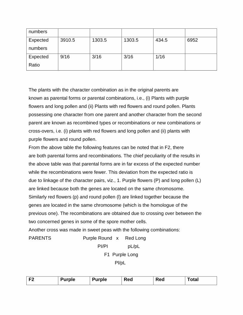
numbers
Expected
numbers
3910.5 1303.5 1303.5 434.5 6952
Expected
Ratio
9/16 3/16 3/16 1/16
The plants with the character combination as in the original parents are
known as parental forms or parental combinations, i.e., (i) Plants with purple
flowers and long pollen and (ii) Plants with red flowers and round pollen. Plants
possessing one character from one parent and another character from the second
parent are known as recombined types or recombinations or new combinations or
cross-overs, i.e. (i) plants with red flowers and long pollen and (ii) plants with
purple flowers and round pollen.
From the above table the following features can be noted that in F2, there
are both parental forms and recombinations. The chief peculiarity of the results in
the above table was that parental forms are in far excess of the expected number
while the recombinations were fewer. This deviation from the expected ratio is
due to linkage of the character pairs, viz., 1. Purple flowers (P) and long pollen (L)
are linked because both the genes are located on the same chromosome.
Similarly red flowers (p) and round pollen (l) are linked together because the
genes are located in the same chromosome (which is the homologue of the
previous one). The recombinations are obtained due to crossing over between the
two concerned genes in some of the spore mother cells.
Another cross was made in sweet peas with the following combinations:
PARENTS Purple Round x Red Long
PI/PI pL/pL
F1 Purple Long
Pl/pL
F2 Purple Purple Red Red Total
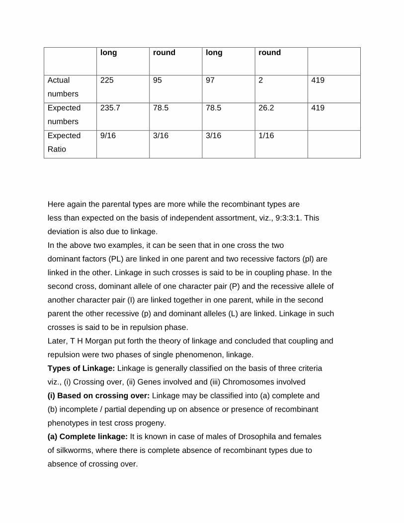
long round
long
round
Actual
numbers
225 95 97 2 419
Expected
numbers
235.7 78.5 78.5 26.2 419
Expected
Ratio
9/16 3/16 3/16 1/16
Here again the parental types are more while the recombinant types are
less than expected on the basis of independent assortment, viz., 9:3:3:1. This
deviation is also due to linkage.
In the above two examples, it can be seen that in one cross the two
dominant factors (PL) are linked in one parent and two recessive factors (pl) are
linked in the other. Linkage in such crosses is said to be in coupling phase. In the
second cross, dominant allele of one character pair (P) and the recessive allele of
another character pair (I) are linked together in one parent, while in the second
parent the other recessive (p) and dominant alleles (L) are linked. Linkage in such
crosses is said to be in repulsion phase.
Later, T H Morgan put forth the theory of linkage and concluded that coupling and
repulsion were two phases of single phenomenon, linkage.
Types of Linkage: Linkage is generally classified on the basis of three criteria
viz., (i) Crossing over, (ii) Genes involved and (iii) Chromosomes involved
(i) Based on crossing over: Linkage may be classified into (a) complete and
(b) incomplete / partial depending up on absence or presence of recombinant
phenotypes in test cross progeny.
(a) Complete linkage: It is known in case of males of Drosophila and females
of silkworms, where there is complete absence of recombinant types due to
absence of crossing over.
(b) Incomplete / partial linkage: If some frequency of crossing over also
occurs between the linked genes, it is known as incomplete / partial
linkage. Recombinant types are also observed besides parental
combinations in the test cross progeny. Incomplete linkage has been
observed in maize, p ea, Drosophila female and several other organisms.
(ii) Based on genes involved : Depending on whether all dominant or some
dominant and some recessive alleles are linked together, linkage can be
categorized into (a) Coupling phase and (b) Repulsion phase
(a) Coupling phase: All dominant alleles are present on the same
chromosome or all recessive alleles are present on same chromosome.
TR tr
----- --- Coupling phase
TR tr
(b) Repulsion phase: Dominant alleles of some genes are linked with
recessive alleles of other genes on same chromosome.
Tr tR
----- --- Repulsion phase
Tr tR
(iii) Based on chromosomes involved: Based on the location of genes on the
chromosomes, linkage can be categorized into (a) autosomal linkage and (b)
X-chromosomal linkage / allosomal linkage / sex linkage
(a) Autosomal linkage: It refers to linkage of those genes which are located
in autosomes (other than sex chromosomes).
(b) X-chromosomal linkage / allosomal linkage / sex linkage: It refers to
linkage of genes which are located in sex chromosomes i.e. either ‘X’ or ‘Y’
(generally ‘X’)
Characteristic features of Linkage:
1. Linkage involves two or more genes which are located in same
chromosome in a linear fashion.
2. Linkage reduces variability.
3. Linkage may involve either dominant or recessive alleles (coupling phase)
or some dominant and some recessive alleles (repulsion phase).
4. It may involve either all desirable traits or all undesirable traits or some
desirable and some undesirable traits.
5. It is observed for oligo-genic traits as well as polygenic traits.
6. Linkage usually involves those genes which are located close to each
other.
7. The strength of linkage depends on the distance between the linked genes.
Lesser the distance, higher the strength and vice versa.
8. Presence of linkage leads to higher frequency of parental types than
recombinants in test cross. When two genes are linked the segregation
ratio of dihybrid test cross progeny deviates significantly from 1:1:1:1 ratio.
9. Linkage can be determined from test cross progeny data.
10. If crossing over does not occur, all genes located on one chromosome are
expected to be inherited together. Thus the maximum number of linkage
groups possible in an organism is equal to the haploid chromosome
number.
Eg. Onion 2n = 16 n = 8 maximum linkage groups possible = 8
Maize 2n = 20 n = 10 maximum linkage groups possible = 10
11. Linkage can be broken by repeated intermating of randomly selected plants
in segregating population for several generations or by mutagenic
treatment.
12. Besides pleiotropy, linkage is an important cause of genetic correlation
between various plant characters.
Linkage and pleiotropy: A close association between two or more characters
may result either due to linkage or pleiotropy or both. Pleiotropy refers to the
control of two or more characters by a single gene. A tight linkage between two
loci can be often confused with pleiotropy. The only way to distinguish between
linkage and pleiotropy is to find out a crossover product between linked
characters. Intermating in segregating populations may break a tight linkage, but a
huge population has to be raised to find out the crossover product. If a cross over
product is not found in spite of repeated intermatings, there seems to be the case
of pleiotropy rather than linkage.
Linkage groups : Linkage group refers to a group of genes which are present in
one chromosome. In other words, all those genes which are located in one
chromosome constitute one linkage group. The number of linkage groups is
limited in each individual. The maximum number of linka ge groups is equal to the
haploid chromosome number of an organism. For example there are ten linkage
groups in corn (2n = 20), seven in garden pea (2n = 14), seven in barley (2n =
14), four in Drosophila melanogaster (2n = 8) and 23 in man (2n = 46).
Detection of linkage: Test cross is the most common method of detecting the
linkage. In this method, the F, heterozygous at two loci (AB/ab) is crossed to a
double recessive parent (ab/ab) and the phenotypic ratio of test cross progeny is
examined. If the phenotypic ratio of test cross progeny shows 1:1:1:1 ratio of
parental and recombinant genotypes, it indicates absence of linkage. If the
frequency of parental types and recombinant types deviate significantly from the
normal dihybrid test cross ratio of 1:1:1:1, it reveals presence of linkage between
two genes under study.
Another way to detect the presence or absence of linkage is to self
pollinate the individual heterozygous at two loci. If there is complete dominance at
each locus and no epistasis, the segregation ratio of the progeny will be 9:3:3:1.
Presence of linkage either in coupling or repulsion phase will lead to significant
deviation from 9:3:3:1 ratio. The deviation of observed values from the expected
ratio is tested with the help of x2 test.
Significance of Linkage in Plant Breeding :
1. Linkage limits the variability among the individuals.
2. Linkage between two or more loci controlling different desirable characters is
advantageous for a plant breeder. A linkage between genes controlling two
different desirable characters will help in simultaneous improvement of both
the characters.
3. Linkage is undesirable when desirable and undesirable genes are linked
together.
4. The estimates of genetic variances for quantitative characters are greatly
influenced by the presence of linkage
CROSSING OVER
The term crossing over was first used by Morgan and Cattell in 1912. The
exchange of precisely homologous segments between non-sister chromatids of
homologous chromosomes is called crossing over.
Mechanism of crossing over: It is responsible for recombination between linked
genes and takes place during pachytene stage of meiosis i.e. after the
homologous chromosomes have undergone pairing and before they begin to
separate. It occurs through the process of breakage and reunion of chromatids.
During pachytene, each chromosome of a bivalent (chromosome pair) has two
chromatids so that each bivalent has four chromatids or strands (four-strand
stage). Generally one chromatid from each of the two homologues of a bivalent is
involved in crossing over. In this process, a segment of one of the chromatids
becomes attached in place of the homologous segment of the nonsister chromatid
and vice-versa. It is assumed that breaks occur at precisely homologous points in
the two nonsister chromatids involved in crossing over; this is followed by reunion
of the acentric segments. This produces a cross (x) like figure at the point of
exchange of the chromatid segments. This figure is called chiasma (which is seen
in diplotene stage of meiosis) (plural-chiasmata).
Obviously, each event of crossing over produces two recombinant
chromatids (involved in the crossing over) called cross over chromatids and two
original chromatids (not involved in crossing over) referred to as noncrossover
chromatids. The crossover chromatids will have new combinations of the linked
genes, i.e. will be recombinant; gametes carrying them will produce the
recombinant phenotypes in test-crosses, which are called crossover types.
Similarly, the noncrossover chromatids will contain the parental gene
combinations and the gametes carrying them will give rise to the parental
phenotypes or noncrossover types. Therefore the frequency of crossing over
between two genes can be estimated as the frequency of recombinant progeny
from a test-cross for these genes. This frequency is usually expressed as percent.
Thus, the frequency of crossing over (%) can be calculated using the formula;
No. of recombinant progeny from a test cross
Frequency of crossing over(%) = ---------------------------------------------------------------- x 100
Total number of progeny
Types of crossing over: Depending upon the number of chiasmata involved,
crossing over is of three types.
1. Single crossing over: It refers to the formation of single chiasma between
non-sister chromatids of homologous chromosomes. It involves two linked
genes (Two point test cross).
2. Double crossing over: It refers to the formation of two chiasmata between
non-sister chromatids of homologous chromosomes. It involves three linked
genes (Three point test cross).
3. Multiple crossing over: Occurrence of more than two crossing overs between
non-sister chromatids of homologous chromosomes is known as multiple
crossing over. However, the frequency of such type of crossing over is
extremely low.
Factors affecting crossing over: The frequency of crossing over between the
linked genes is affected by several factors.
1. Distance between the genes: The frequency of crossing over between the
two genes is positively associated with the distance between their location in
the chromosome. Crossing over between the two genes would increase with
an increase in distance between them.
2. sex: The frequency of recombination is markedly influenced by the sex of
heterozygotes for linked genes. In general, the heterogametic sex shows
relatively lower recombination frequencies than the homogametic sex of the
same species. Eg: No crossing over occurs between linked genes in
Drosophila males and females of silkworm.
3. Age of female: The frequency of crossing over shows a progressive decline
with the advancing age of Drosophila females.
4. Temperature: In Drosophila, the lowest frequency of crossing over is
observed when females are cultured at 220C. The frequency of recombination
tends to increase both at the lower and higher temperatures than 220C.
5. Nutrition: The frequency of crossing over in Drosophila is affected by the
presence of metallic ions Eg: Ca+2 and Mg+2 in its food. Higher the amount,
lower will be the crossing over frequenc y and vice-versa.
6. Chemicals: Treatment of Drosophila females with certain antibiotics like
mitomycin D and actinomycin D and certain alkylating agents such as
ethylmethane sulphonate promotes crossing over.
7. Radiations: An increase in frequency of crossing over is observed when
Drosophila females are irradiated with x-rays and g-rays.
8. Plasmagenes: In some species, plasma genes reduce the frequency of
crossing over. Eg: The Tifton male sterile cytoplasm reduces the frequency of
crossing over in bajra.
9. Genotype: Many genes are known to affect the occurrence as well as the rate
of crossing over. For example C3G gene of Drosophila located in chromosome
3 prevents crossing over when present in homozygous state while it promotes
crossing over in the heterozygous state.
10.Chromosomal aberrations: In Drosophila, some chromosomal aberrations
Eg: paracentric inversions, reduce recombination between the genes located
with in the inverted segment.
11.Distance from centromere: Centromere tends to suppress recombination.
Therefore genes located in the vicinity of centromeres show a relatively lower
frequency of crossing over than those located away from them.
Significance of crossing over in Plant Breeding:
1. It increases variability
2. It helps to break linkages
3. It makes possible to construct chromosome maps
Cytological proof of crossing over: The first cytological evidence in support of
genetic crossing over was provided by Curt stern in 1931 on the basis of his
experiments with Drosophila by using cytological markers. He used a Drosophila
female fly in which one X-chromosome was broken into two segments and out of
these two segments, one behaved as X-chromosome. This chromosome had one
recessive mutant allele car (carnation) for eye colour and another dominant allele
B (Bar) for eye shape. The other X-chromosome had small portion of Ychromosome
attached to its one end. This chromosome had the dominant allele +
(wild type allele of car) producing dull red eye colour and a recessive allele + (wild
type allele of B) producing normal ovate eye shape. Thus the phenotype of
female is barred (since B is dominant to +) with normal eye colour (since car is
recessive to +) and both the X-chromosomes in the female had distinct
morphology and could be easily identified under microscope. Such females were
crossed with male flies having recessive alleles for both genes (car +). As a result
of crossing over female flies produce four types of gametes viz., two parental
types or non crossover types (car B and + +) and two recombinant types or cross
over types (car + and + B). The male flies produce only two types of gametes (car
+ and Y), because crossing over does not occur in Drosophila male. A random
union of two types of male gametes with four types of female gametes will
produce males and females in equal number of four each.
Stern cytologically examined the chromosomes of recombinant types i. e.
carnation with normal eye shape and barred with normal eye colour. It was found
that carnation flies did not have any fragmented X-chromosome but rather had
normal X-chromosome. On the other hand barred flies had a fragmented
X-chromosome with a segment of Y-chromosome attached to one of the two
fragments of X-chromosome. Such chromosome combination in barred is possible
only through exchange of segments between non-sister chromatids of
homologous chromosomes. This has proved that genetic crossing over was
accompanied with an actual exchange of chromosome segments.
Similar proof of cytological crossing over was provided by Creighton and
McClintock in maize.
Coincidence: It refers to the occurrence of two or more distinct crossing overs at
the same time in the same region of a pair of homologous chromosomes and as a
result, a double cross over product is obtained. Coefficient of coincidence is
estimated b y using the formula:
Observed frequency of double cross over
Coefficient of coincidence = -------------------------------------------------------
Expected frequency of double cross over
(The ratio between the observed and the expected frequencies of double
crossovers is called coefficient of coincidence)
Interference: The occurrence of crossing over in one region of a chromosome
interferes with its occurrence in the neighbouring segment. This is known as
interference. The term interference was coined by Muller. It may also be defined
as the tendency of one crossing over to prevent another crossing over from
occurring in its vicinity. This is called positive interference. Sometimes, one
crossing over enhances the chance of another crossing over in the adjacent
region. This is termed as negative interference. Eg: Aspergillus, bacteriophages.
The effect of interference reduces as the distance from the first crossing over
increases. The intensity of interference may be estimated as coefficient of
interference.
Coefficient of interference = 1 - coefficient of coincidence
Differences between crossing over and linkage
Crossing over Linkage
1. It leads to separation of linked genes It keeps the genes together
2. It involves exchange of segments
between non-sister chromatids of
homologous chromosomes
It involves individual chromosomes
3. The frequency of crossing over can .
never exceed 50 %
The number of linkage groups can
never be more than haploid
chromosome number
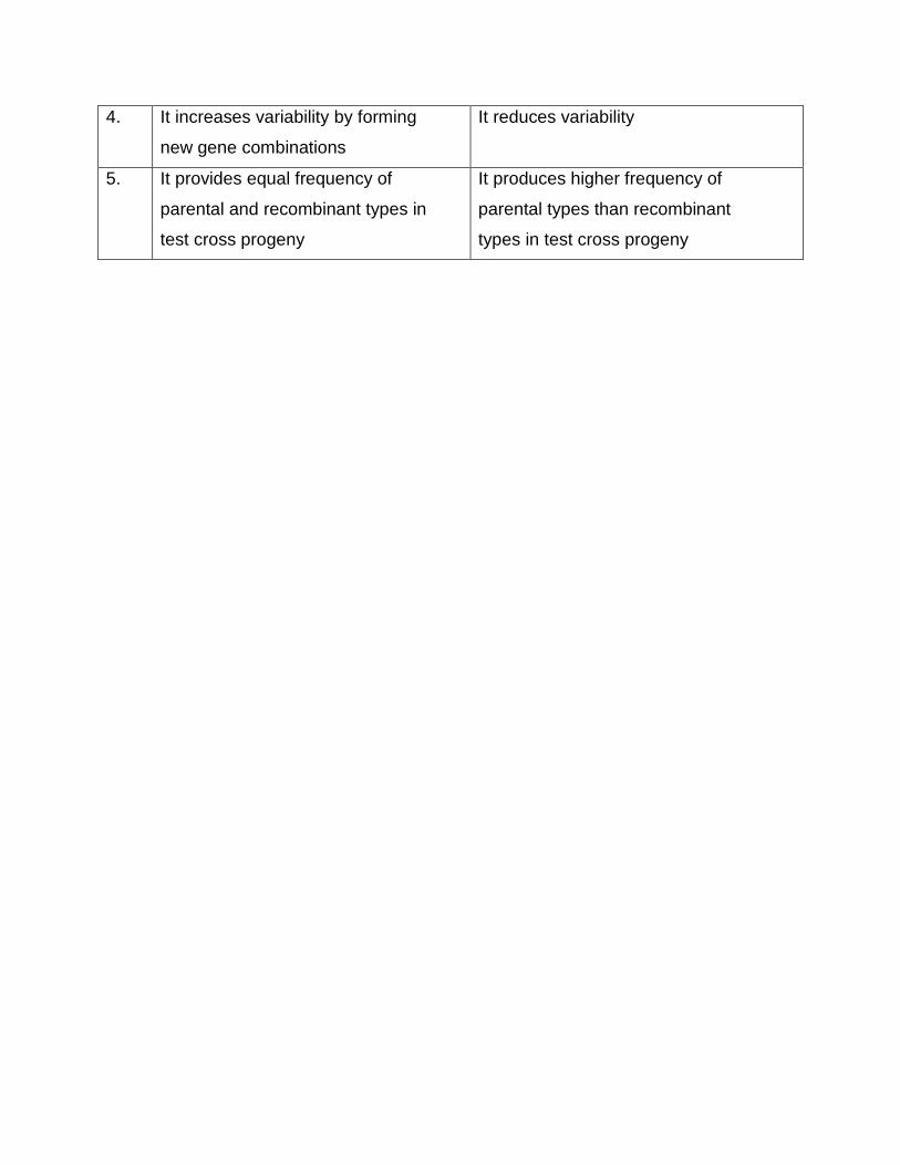
4. It increases variability by forming
new gene combinations
It reduces variability
5. It provides equal frequency of
parental and recombinant types in
test cross progeny
It produces higher frequency of
parental types than recombinant
types in test cross progeny
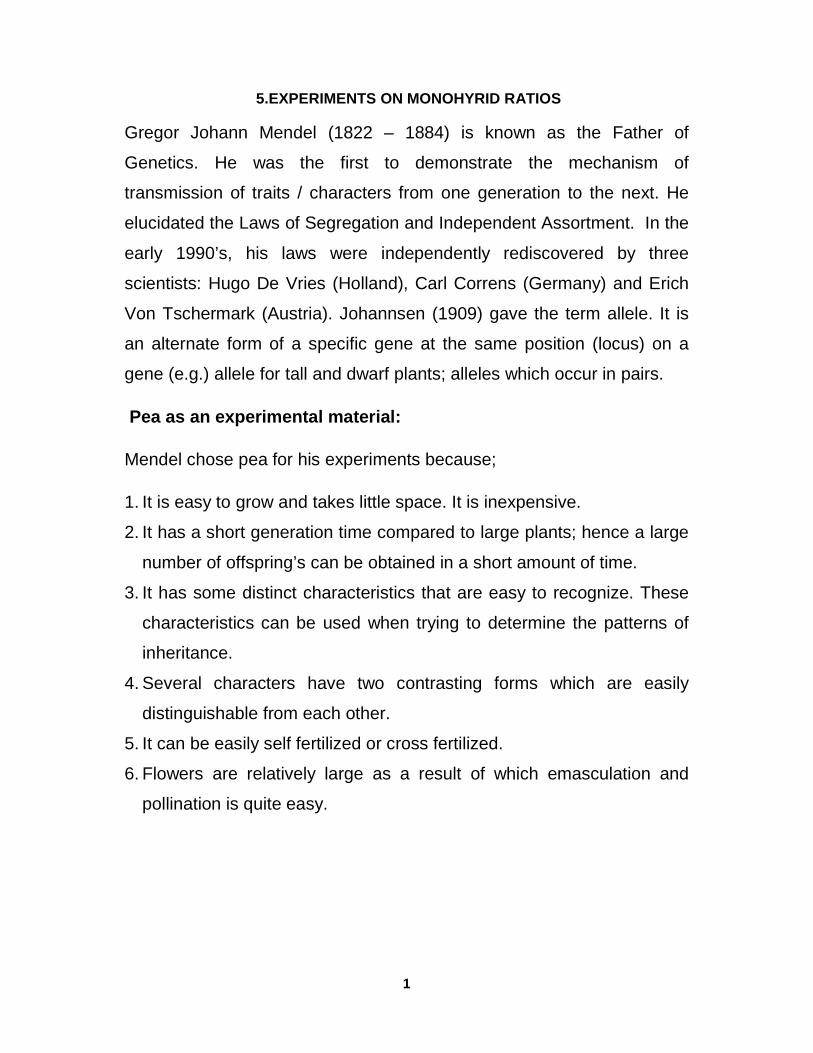
1
5.EXPERIMENTS ON MONOHYRID RATIOS
Gregor Johann Mendel (1822 – 1884) is known as the Father of
Genetics. He was the first to demonstrate the mechanism of
transmission of traits / characters from one generation to the next. He
elucidated the Laws of Segregation and Independent Assortment. In the
early 1990’s, his laws were independently rediscovered by three
scientists: Hugo De Vries (Holland), Carl Correns (Germany) and Erich
Von Tschermark (Austria). Johannsen (1909) gave the term allele. It is
an alternate form of a specific gene at the same position (locus) on a
gene (e.g.) allele for tall and dwarf plants; alleles which occur in pairs.
Pea as an experimental material:
Mendel chose pea for his experiments because;
1. It is easy to grow and takes little space. It is inexpensive.
2. It has a short generation time compared to large plants; hence a large
number of offspring’s can be obtained in a short amount of time.
3. It has some distinct characteristics that are easy to recognize. These
characteristics can be used when trying to determine the patterns of
inheritance.
4. Several characters have two contrasting forms which are easily
distinguishable from each other.
5. It can be easily self fertilized or cross fertilized.
6. Flowers are relatively large as a result of which emasculation and
pollination is quite easy.
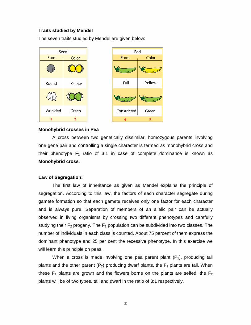
Traits studied by Mendel
The seven traits studied by Mendel are given below
Monohybrid crosses in Pea
A cross between two genetically
one gene pair and controlling
their phenotype F2 ratio of 3:1 in case of complete dominance is known as
Monohybrid cross .
Law of Segregation:
The first law of inheritance as given as
segregation. According to this law, the factors of each character segregate during
gamete formation so that each gamete receives only one factor for each character
and is always pure. Separation of members of an allelic pair can
observed in living organisms by crossing two different phenotypes and carefully
studying their F2 progeny. The F
number of individuals in each class is counted. About 75 percent of them expres
dominant phenotype and 25 per cent the recessive phenotype. In this exercise we
will learn this principle on peas.
When a cross is made involving one pea parent plant (P
plants and the other parent (P
these F1 plants are grown and the flowers borne on the plants are selfed, the F
plants will be of two types, tall and dwarf in the ratio of 3:1 respectively.
2
The seven traits studied by Mendel are given below:
A cross between two genetically dissimilar, homozygous parents involving
one gene pair and controlling a single character is termed as monohybrid cross and
ratio of 3:1 in case of complete dominance is known as
The first law of inheritance as given as Mendel explains the principle of
segregation. According to this law, the factors of each character segregate during
gamete formation so that each gamete receives only one factor for each character
and is always pure. Separation of members of an allelic pair can
observed in living organisms by crossing two different phenotypes and carefully
progeny. The F2 population can be subdivided into two classes. The
number of individuals in each class is counted. About 75 percent of them expres
dominant phenotype and 25 per cent the recessive phenotype. In this exercise we
will learn this principle on peas.
When a cross is made involving one pea parent plant (P1), producing tall
plants and the other parent (P2) producing dwarf plants, the F1 plants are tall. When
plants are grown and the flowers borne on the plants are selfed, the F
plants will be of two types, tall and dwarf in the ratio of 3:1 respectively.
, homozygous parents involving
monohybrid cross and
ratio of 3:1 in case of complete dominance is known as
explains the principle of
segregation. According to this law, the factors of each character segregate during
gamete formation so that each gamete receives only one factor for each character
and is always pure. Separation of members of an allelic pair can be actually
observed in living organisms by crossing two different phenotypes and carefully
population can be subdivided into two classes. The
number of individuals in each class is counted. About 75 percent of them express the
dominant phenotype and 25 per cent the recessive phenotype. In this exercise we
), producing tall
plants are tall. When
plants are grown and the flowers borne on the plants are selfed, the F2
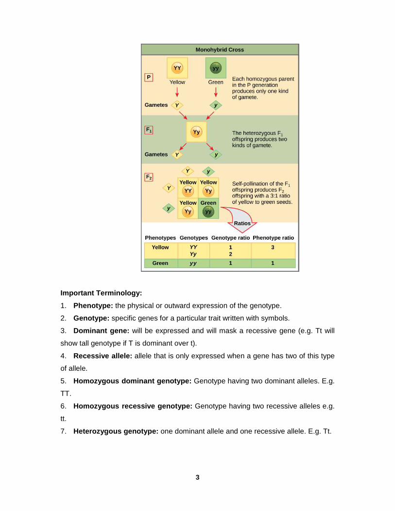
3
Important Terminology:
1. Phenotype: the physical or outward expression of the genotype.
2. Genotype: specific genes for a particular trait written with symbols.
3. Dominant gene: will be expressed and will mask a recessive gene (e.g. Tt will
show tall genotype if T is dominant over t).
4. Recessive allele: allele that is only expressed when a gene has two of this type
of allele.
5. Homozygous dominant genotype: Genotype having two dominant alleles. E.g.
TT.
6. Homozygous recessive genotype: Genotype having two recessive alleles e.g.
tt.
7. Heterozygous genotype: one dominant allele and one recessive allele. E.g. Tt.
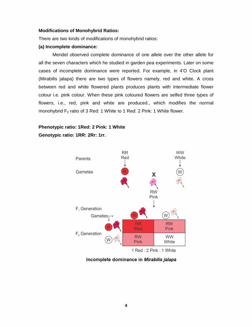
4
Modifications of Monohybrid Ratios:
There are two kinds of modifications of monohybrid ratios:
(a) Incomplete dominance:
Mendel observed complete dominance of one allele over the other allele for
all the seven characters which he studied in garden pea experiments. Later on some
cases of incomplete dominance were reported. For example, in 4’O Clock plant
(Mirabilis jalapa) there are two types of flowers namely, red and white. A cross
between red and white flowered plants produces plants with intermediate flower
colour i.e. pink colour. When these pink coloured flowers are selfed three types of
flowers, i.e., red, pink and white are produced., which modifies the normal
monohybrid F2 ratio of 3 Red: 1 White to 1 Red: 2 Pink: 1 White flower.
Phenotypic ratio: 1Red: 2 Pink: 1 White
Genotypic ratio: 1RR: 2Rr: 1rr.
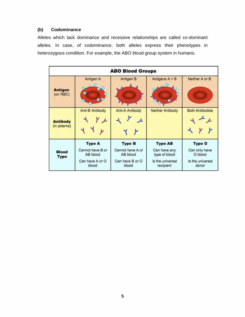
5
(b) Codominance
Alleles which lack dominance and recessive relationships are called co-dominant
alleles. In case, of codominance, both alleles express their phenotypes in
heterozygous condition. For example, the ABO blood group system in humans.
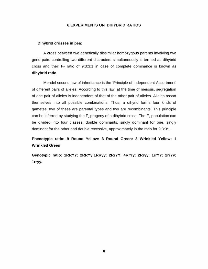
6
6.EXPERIMENTS ON DIHYBRID RATIOS
Dihybrid crosses in pea:
A cross between two genetically dissimilar homozygous parents involving two
gene pairs controlling two different characters simultaneously is termed as dihybrid
cross and their F2 ratio of 9:3:3:1 in case of complete dominance is known as
dihybrid ratio.
Mendel second law of inheritance is the ‘Principle of Independent Assortment’
of different pairs of alleles. According to this law, at the time of meiosis, segregation
of one pair of alleles is independent of that of the other pair of alleles. Alleles assort
themselves into all possible combinations. Thus, a dihyrid forms four kinds of
gametes, two of these are parental types and two are recombinants. This principle
can be inferred by studying the F2 progeny of a dihybrid cross. The F2 population can
be divided into four classes: double dominants, singly dominant for one, singly
dominant for the other and double recessive, approximately in the ratio for 9:3:3:1.
Phenotypic ratio: 9 Round Yellow: 3 Round Green: 3 Wrinkled Yellow: 1
Wrinkled Green
Genotypic ratio: 1RRYY: 2RRYy:1RRyy: 2RrYY: 4RrYy: 2Rryy: 1rrYY: 2rrYy:
1rryy.
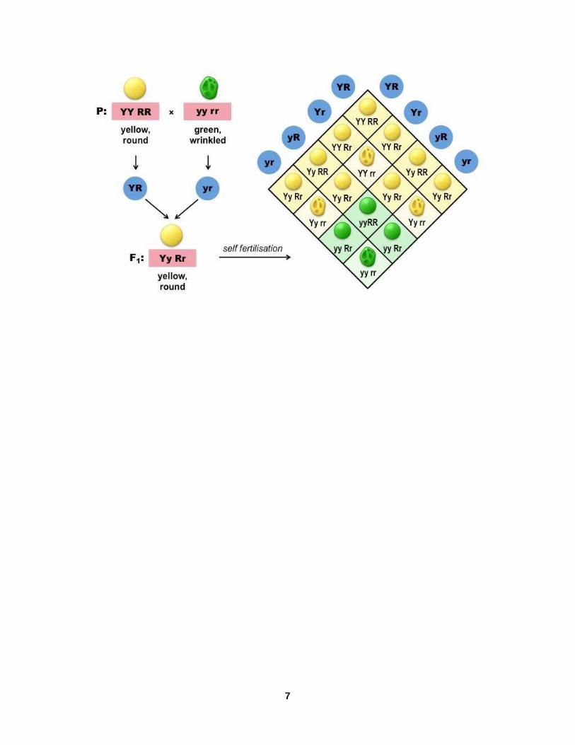
7
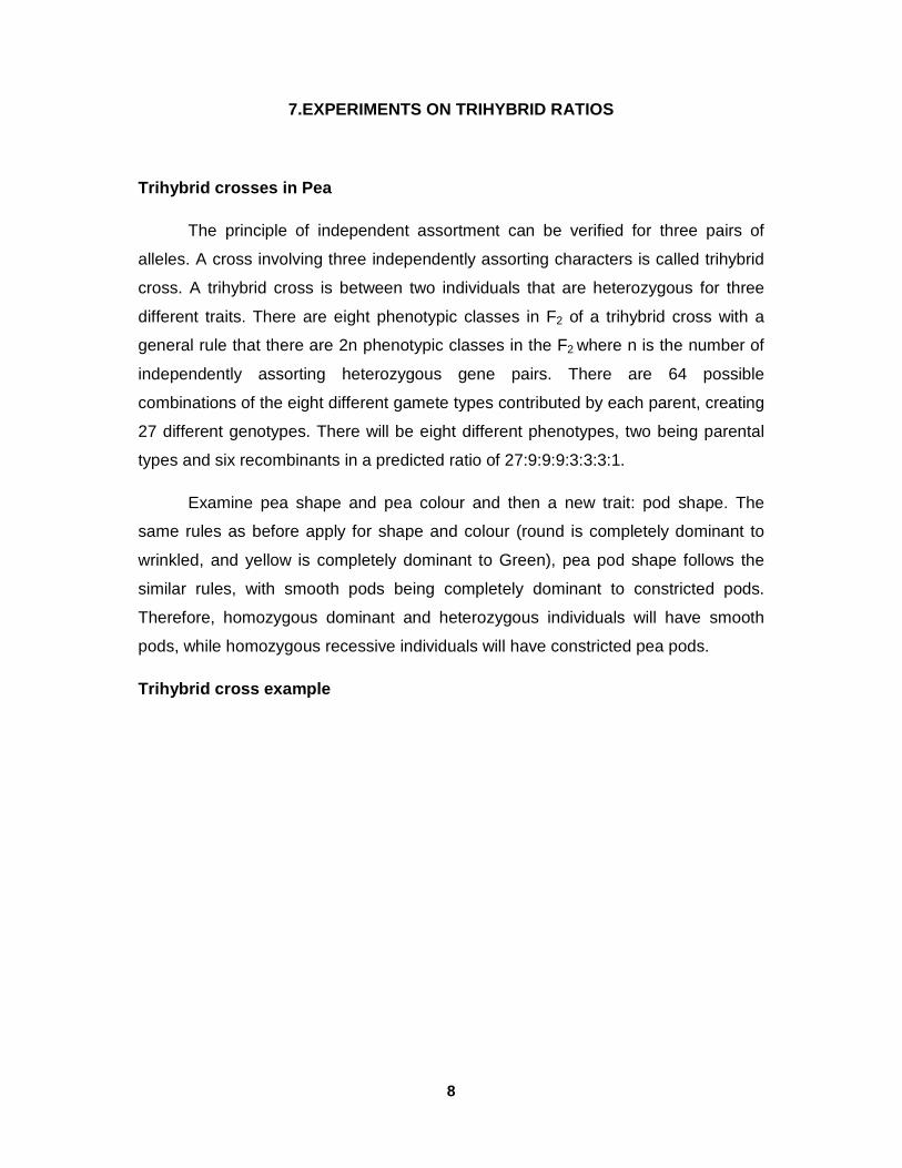
8
7.EXPERIMENTS ON TRIHYBRID RATIOS
Trihybrid crosses in Pea
The principle of independent assortment can be verified for three pairs of
alleles. A cross involving three independently assorting characters is called trihybrid
cross. A trihybrid cross is between two individuals that are heterozygous for three
different traits. There are eight phenotypic classes in F2 of a trihybrid cross with a
general rule that there are 2n phenotypic classes in the F2 where n is the number of
independently assorting heterozygous gene pairs. There are 64 possible
combinations of the eight different gamete types contributed by each parent, creating
27 different genotypes. There will be eight different phenotypes, two being parental
types and six recombinants in a predicted ratio of 27:9:9:9:3:3:3:1.
Examine pea shape and pea colour and then a new trait: pod shape. The
same rules as before apply for shape and colour (round is completely dominant to
wrinkled, and yellow is completely dominant to Green), pea pod shape follows the
similar rules, with smooth pods being completely dominant to constricted pods.
Therefore, homozygous dominant and heterozygous individuals will have smooth
pods, while homozygous recessive individuals will have constricted pea pods.
Trihybrid cross example
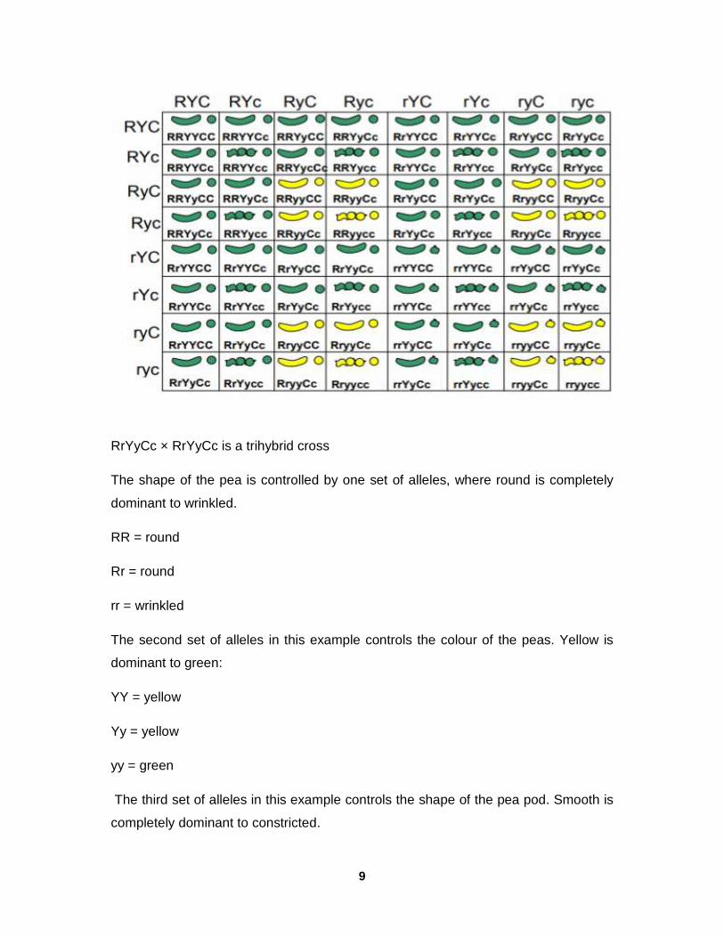
9
RrYyCc × RrYyCc is a trihybrid cross
The shape of the pea is controlled by one set of alleles, where round is completely
dominant to wrinkled.
RR = round
Rr = round
rr = wrinkled
The second set of alleles in this example controls the colour of the peas. Yellow is
dominant to green:
YY = yellow
Yy = yellow
yy = green
The third set of alleles in this example controls the shape of the pea pod. Smooth is
completely dominant to constricted.
10
CC = Smooth
Cc = smooth
cc = constricted
The gametes for each parent in a tribhybrid cross would be RYC, RYc, RyC, Ryc,
rYC, rYc, ryC, ryc, with one eight of a chance for any of them. The resulting formed
from 8 × 8 crossing will be formed in a checker board.
The phenotypic ratio observed is:
27: round, yellow, smooth pod
9: round, yellow, constricted pod
9: round, green, smooth pod
9: round, green, constricted pod
3: wrinkled, yellow, smooth pod
3: wrinkled, yellow, constricted pod
3: wrinkled green smooth pod
1: wrinkled, green, constricted pod.
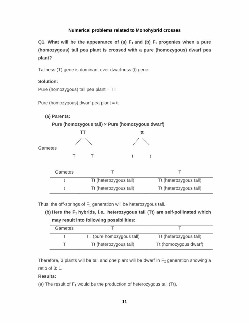
11
Numerical problems related to Monohybrid crosses
Q1. What will be the appearance of (a) F 1 and (b) F 2 progenies when a pure
(homozygous) tall pea plant is crossed with a pure (homozygous) dwarf pea
plant?
Tallness (T) gene is dominant over dwarfness (t) gene.
Solution:
Pure (homozygous) tall pea plant = TT
Pure (homozygous) dwarf pea plant = tt
(a) Parents:
Pure (homozygous tall) × Pure (homozygous dwarf)
TT tt
Gametes
T T t t
Gametes T T
t Tt (heterozygous tall) Tt (heterozygous tall)
t Tt (heterozygous tall) Tt (heterozygous tall)
Thus, the off-springs of F1 generation will be heterozygous tall.
(b) Here the F 1 hybrids, i.e., heterozygous tall (Tt) are self-pol linated which
may result into following possibilities:
Gametes T T
T TT (pure homozygous tall) Tt (heterozygous tall)
T Tt (heterozygous tall) Tt (homozygous dwarf)
Therefore, 3 plants will be tall and one plant will be dwarf in F2 generation showing a
ratio of 3: 1.
Results:
(a) The result of F1 would be the production of heterozygous tall (Tt).
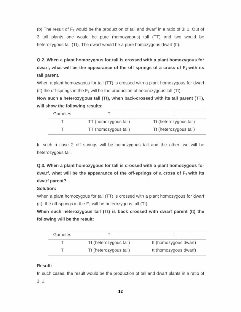
12
(b) The result of F2 would be the production of tall and dwarf in a ratio of 3: 1. Out of
3 tall plants one would be pure (homozygous) tall (TT) and two would be
heterozygous tall (Tt). The dwarf would be a pure homozygous dwarf (tt).
Q.2. When a plant homozygous for tall is crossed wi th a plant homozygous for
dwarf, what will be the appearance of the off sprin gs of a cross of F 1 with its
tall parent.
When a plant homozygous for tall (TT) is crossed with a plant homozygous for dwarf
(tt) the off-springs in the F1 will be the production of heterozygous tall (Tt).
Now such a heterozygous tall (Tt), when back-crosse d with its tall parent (TT),
will show the following results:
Gametes T t
T TT (homozygous tall) Tt (heterozygous tall)
T TT (homozygous tall) Tt (heterozygous tall)
In such a case 2 off springs will be homozygous tall and the other two will be
heterozygous tall.
Q.3. When a plant homozygous for tall is crossed wi th a plant homozygous for
dwarf, what will be the appearance of the off-sprin gs of a cross of F 1 with its
dwarf parent?
Solution:
When a plant homozygous for tall (TT) is crossed with a plant homozygous for dwarf
(tt), the off-springs in the F1 will be heterozygous tall (Tt).
When such heterozygous tall (Tt) is back crossed wi th dwarf parent (tt) the
following will be the result:
Gametes T t
T Tt (heterozygous tall) tt (homozygous dwarf)
T Tt (heterozygous tall) tt (homozygous dwarf)
Result:
In such cases, the result would be the production of tall and dwarf plants in a ratio of
1: 1.
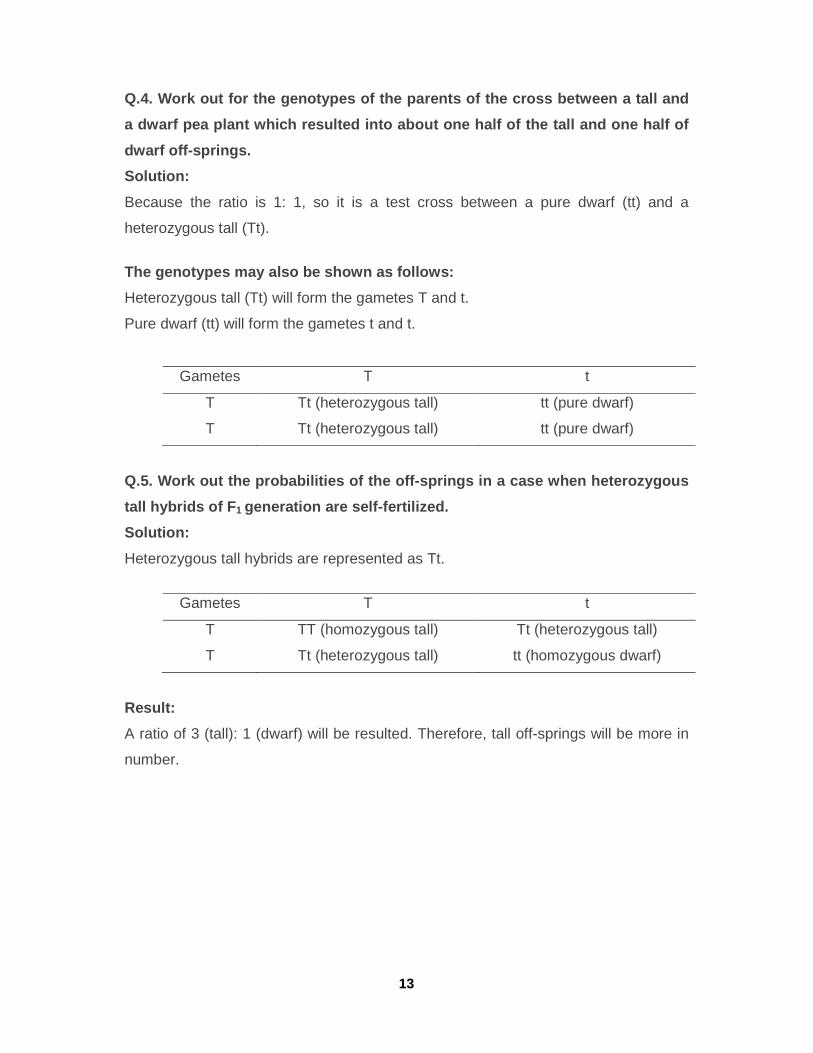
13
Q.4. Work out for the genotypes of the parents of t he cross between a tall and
a dwarf pea plant which resulted into about one hal f of the tall and one half of
dwarf off-springs.
Solution:
Because the ratio is 1: 1, so it is a test cross between a pure dwarf (tt) and a
heterozygous tall (Tt).
The genotypes may also be shown as follows:
Heterozygous tall (Tt) will form the gametes T and t.
Pure dwarf (tt) will form the gametes t and t.
Gametes T t
T Tt (heterozygous tall) tt (pure dwarf)
T Tt (heterozygous tall) tt (pure dwarf)
Q.5. Work out the probabilities of the off-springs in a case when heterozygous
tall hybrids of F 1 generation are self-fertilized.
Solution:
Heterozygous tall hybrids are represented as Tt.
Gametes T t
T TT (homozygous tall) Tt (heterozygous tall)
T Tt (heterozygous tall) tt (homozygous dwarf)
Result:
A ratio of 3 (tall): 1 (dwarf) will be resulted. Therefore, tall off-springs will be more in
number.
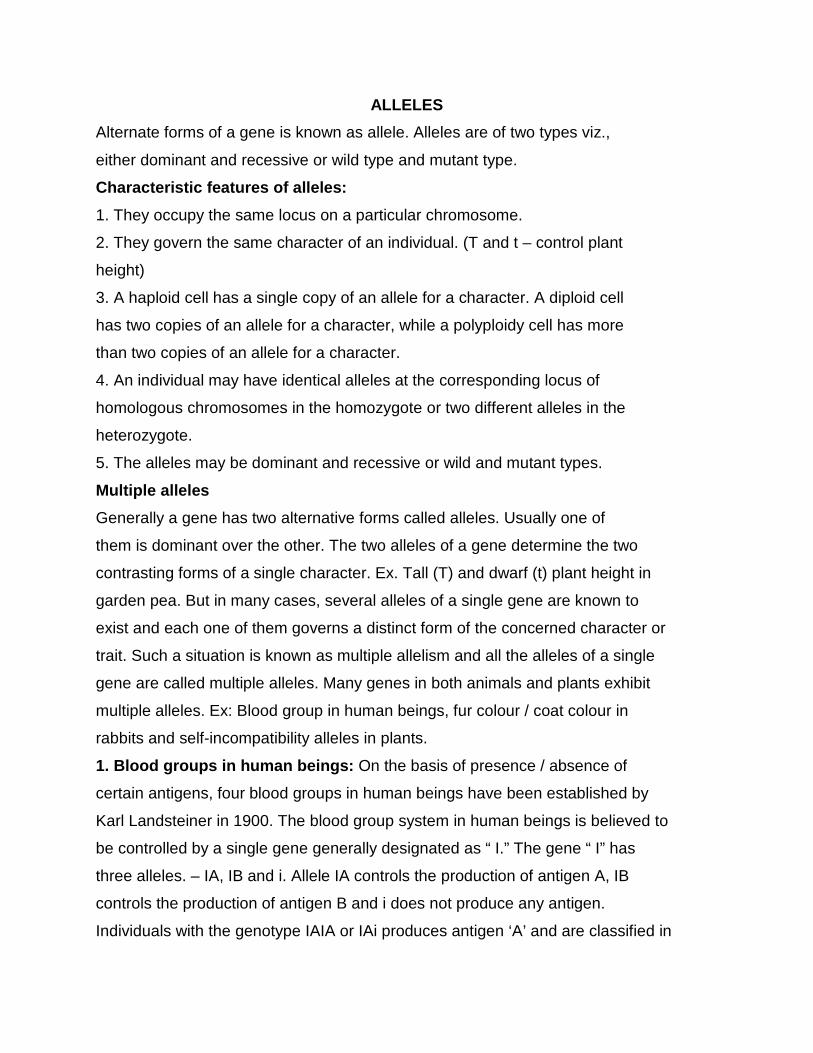
ALLELES
Alternate forms of a gene is known as allele. Alleles are of two types viz.,
either dominant and recessive or wild type and mutant type.
Characteristic features of alleles:
1. They occupy the same locus on a particular chromosome.
2. They govern the same character of an individual. (T and t – control plant
height)
3. A haploid cell has a single copy of an allele for a character. A diploid cell
has two copies of an allele for a character, while a polyploidy cell has more
than two copies of an allele for a character.
4. An individual may have identical alleles at the corresponding locus of
homologous chromosomes in the homozygote or two different alleles in the
heterozygote.
5. The alleles may be dominant and recessive or wild and mutant types.
Multiple alleles
Generally a gene has two alternative forms called alleles. Usually one of
them is dominant over the other. The two alleles of a gene determine the two
contrasting forms of a single character. Ex. Tall (T) and dwarf (t) plant height in
garden pea. But in many cases, several alleles of a single gene are known to
exist and each one of them governs a distinct form of the concerned character or
trait. Such a situation is known as multiple allelism and all the alleles of a single
gene are called multiple alleles. Many genes in both animals and plants exhibit
multiple alleles. Ex: Blood group in human beings, fur colour / coat colour in
rabbits and self-incompatibility alleles in plants.
1. Blood groups in human beings: On the basis of presence / absence of
certain antigens, four blood groups in human beings have been established by
Karl Landsteiner in 1900. The blood group system in human beings is believed to
be controlled by a single gene generally designated as “ I.” The gene “ I” has
three alleles. – IA, IB and i. Allele IA controls the production of antigen A, IB
controls the production of antigen B and i does not produce any antigen.
Individuals with the genotype IAIA or IAi produces antigen ‘A’ and are classified in
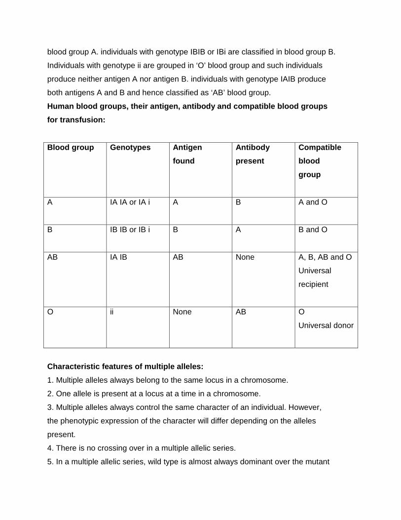
blood group A. individuals with genotype IBIB or IBi are classified in blood group B.
Individuals with genotype ii are grouped in ‘O’ blood group and such individuals
produce neither antigen A nor antigen B. individuals with genotype IAIB produce
both antigens A and B and hence classified as ‘AB’ blood group.
Human blood groups, their antigen, antibody and compatible blood groups
for transfusion:
Blood group Genotypes Antigen
found
Antibody
present
Compatible
blood
group
A IA IA or IA i A B A and O
B IB IB or IB i B A B and O
AB IA IB AB None A, B, AB and O
Universal
recipient
O ii None AB O
Universal donor
Characteristic features of multiple alleles:
1. Multiple alleles always belong to the same locus in a chromosome.
2. One allele is present at a locus at a time in a chromosome.
3. Multiple alleles always control the same character of an individual. However,
the phenotypic expression of the character will differ depending on the alleles
present.
4. There is no crossing over in a multiple allelic series.
5. In a multiple allelic series, wild type is almost always dominant over the mutant
type.
6. A cross between two strains homozygous for mutant alleles will always
produce a mutant phenotype and never a wild phenotype. In other words,
multiple alleles do not show complementation.
7. Further, F2 generations from such crosses show typical monohybrid ratio for
the concerned trait.
Pseudo alleles: Two alleles each of two or more tightly linked genes affecting the
same function as a result of which, they appear as multiple alleles. (or) Alleles,
which have two separate gene loci, but often inherit together due to close linkage
and have very rare chance of crossing over are called pseudo alleles. Eg:
Lozenge eye in Drosophila.
Iso allele: An allele which is similar in its phenotypic expression to that of other
independently occurring allele is called isoallele.
Penetrance; It is the ability of a gene to express itself in all the individuals, which
carry it in the appropriate genotype. (or) The frequency with which a gene
produces a phenotypic or visible effect in the individuals which carry it in the
appropriate genotype is known as penetrance. It refers to the proportion of
individuals which exhibit phenotypic effect of a specific gene carried by them.
1. Penetrance of some genes is limited to one sex only. For example : Milk
production in cattle or human beings.
2. It may be affected by environmental conditions
3. Penetrance is expressed in percentage
It is of two types
1. Complete penetrance: When all the individuals that carry a particular
gene exhibit its phenotypic effect, it is known as complete penetrance. In
this case, all homozygous dominant individuals will exhibit one phenotype,
while all homozygous recessives will exhibit another phenotype. Recessive
alleles have no or zero penetrance in heterozygous condition.
Tall x Dwarf
TT x tt
(100%) (100%)
F1 Tt Tall
[T exhibits Complete (100%) penetrance while t has no(0%) penetrance]
2. Incomplete penetrance: When specific gene does not express their effect
in all the individuals which carry them in appropriate genotype, it is known
as incomplete penetrance. For example : The recessive gene producing
partial chlorophyll deficiency in the cotyledonary leaves of lima bean shows
incomplete penetrance as it expresses itself only in 10% of the individuals.
Almost all the genes showing incomplete penetrance exhibit variable
expressivity as well
Expressivity: The degree of phenotypic expression of a gene in different
individuals is called expressivity. It is also influenced by environmental conditions
in some cases. Expressivity is a lso two types.
1. Uniform expressivity: When the phenotypic expression of a gene is
identical or similar in all the individuals, which carry such a gene, it is
known as uniform expressivity. Most of the qualitative characters exhibit
uniform expressivity. Eg: Seed shape in pea rr genotypes have wrinkled
seed shape, while RR or Rr genotypes exhibit round seed shape.
2. Variable expressivity: When the phenotypic expression differs in different
carriers of a gene, it is known as variable expressivity. Eg: Recessive gene
producing partial chlorophyll deficiency in cotyledonary leaves of lima
beans.
QUALITATIVE AND QUANTITATIVE CHARACTERS
The phenotype of any individual can be classified into two types:
1) Qualitative characters and 2) Quantitative c haracters
1. Qualitative characters : The characters that show discontinuous variation
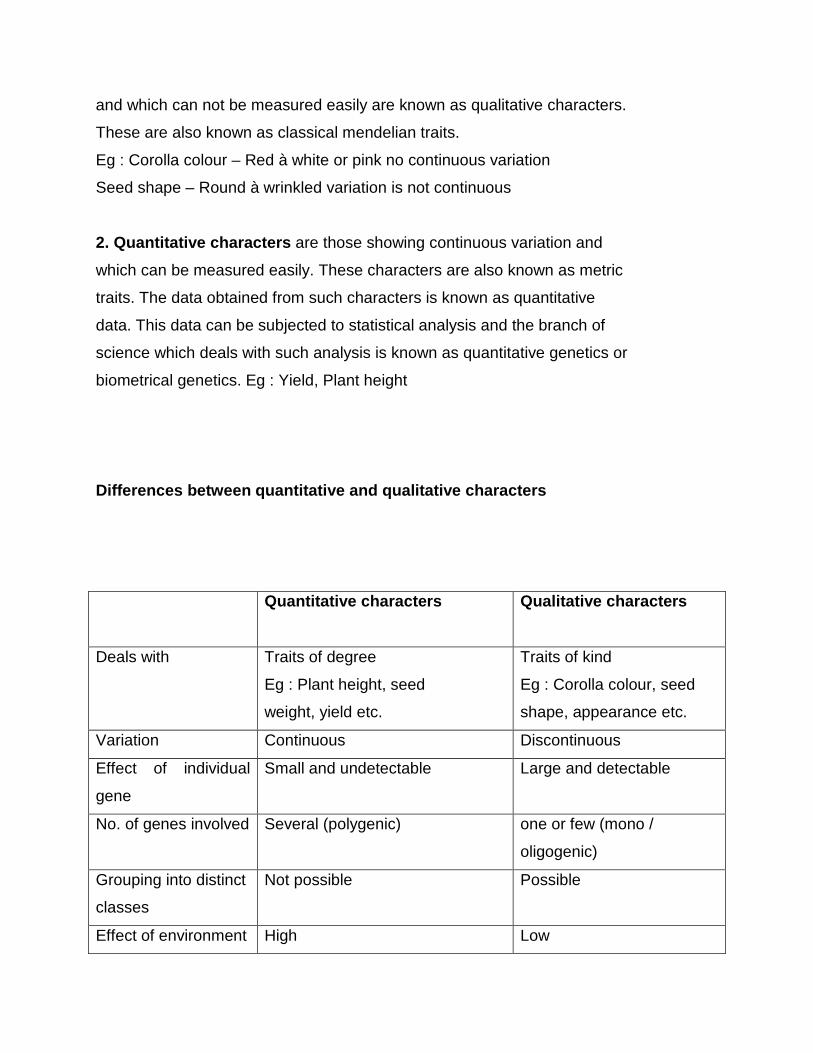
and which can not be measured easily are known as qualitative characters.
These are also known as classical mendelian traits.
Eg : Corolla colour – Red à white or pink no continuous variation
Seed shape – Round à wrinkled variation is not continuous
2. Quantitative characters are those showing continuous variation and
which can be measured easily. These characters are also known as metric
traits. The data obtained from such characters is known as quantitative
data. This data can be subjected to statistical analysis and the branch of
science which deals with such analysis is known as quantitative genetics or
biometrical genetics. Eg : Yield, Plant height
Differences between quantitative and qualitative characters
Quantitative characters Qualitative characters
Deals with Traits of degree
Eg : Plant height, seed
weight, yield etc.
Traits of kind
Eg : Corolla colour, seed
shape, appearance etc.
Variation Continuous Discontinuous
Effect of individual
gene
Small and undetectable Large and detectable
No. of genes involved Several (polygenic) one or few (mono /
oligogenic)
Grouping into distinct
classes
Not possible Possible
Effect of environment High Low
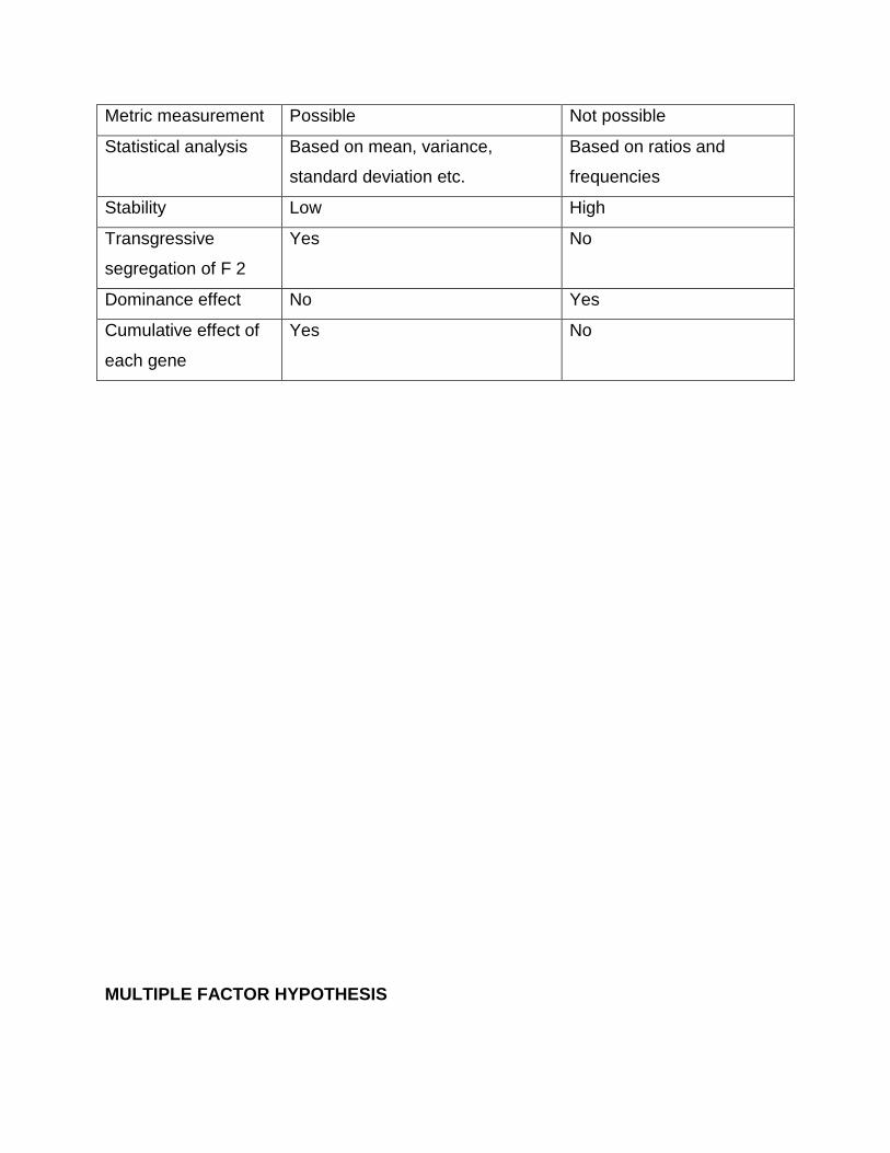
Metric measurement Possible Not possible
Statistical analysis Based on mean, variance,
standard deviation etc.
Based on ratios and
frequencies
Stability Low High
Transgressive
segregation of F 2
Yes No
Dominance effect No Yes
Cumulative effect of
each gene
Yes No
MULTIPLE FACTOR HYPOTHESIS
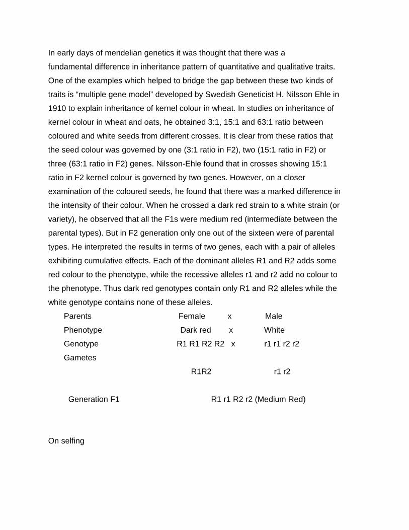
In early days of mendelian genetics it was thought that there was a
fundamental difference in inheritance pattern of quantitative and qualitative traits.
One of the examples which helped to bridge the gap between these two kinds of
traits is “multiple gene model” developed by Swedish Geneticist H. Nilsson Ehle in
1910 to explain inheritance of kernel colour in wheat. In studies on inheritance of
kernel colour in wheat and oats, he obtained 3:1, 15:1 and 63:1 ratio between
coloured and white seeds from different crosses. It is clear from these ratios that
the seed colour was governed by one (3:1 ratio in F2), two (15:1 ratio in F2) or
three (63:1 ratio in F2) genes. Nilsson-Ehle found that in crosses showing 15:1
ratio in F2 kernel colour is governed by two genes. However, on a closer
examination of the coloured seeds, he found that there was a marked difference in
the intensity of their colour. When he crossed a dark red strain to a white strain (or
variety), he observed that all the F1s were medium red (intermediate between the
parental types). But in F2 generation only one out of the sixteen were of parental
types. He interpreted the results in terms of two genes, each with a pair of alleles
exhibiting cumulative effects. Each of the dominant alleles R1 and R2 adds some
red colour to the phenotype, while the recessive alleles r1 and r2 add no colour to
the phenotype. Thus dark red genotypes contain only R1 and R2 alleles while the
white genotype contains none of these alleles.
Parents Female x Male
Phenotype Dark red x White
Genotype R1 R1 R2 R2 x r1 r1 r2 r2
Gametes
R1R2 r1 r2
Generation F1 R1 r1 R2 r2 (Medium Red)
On selfing
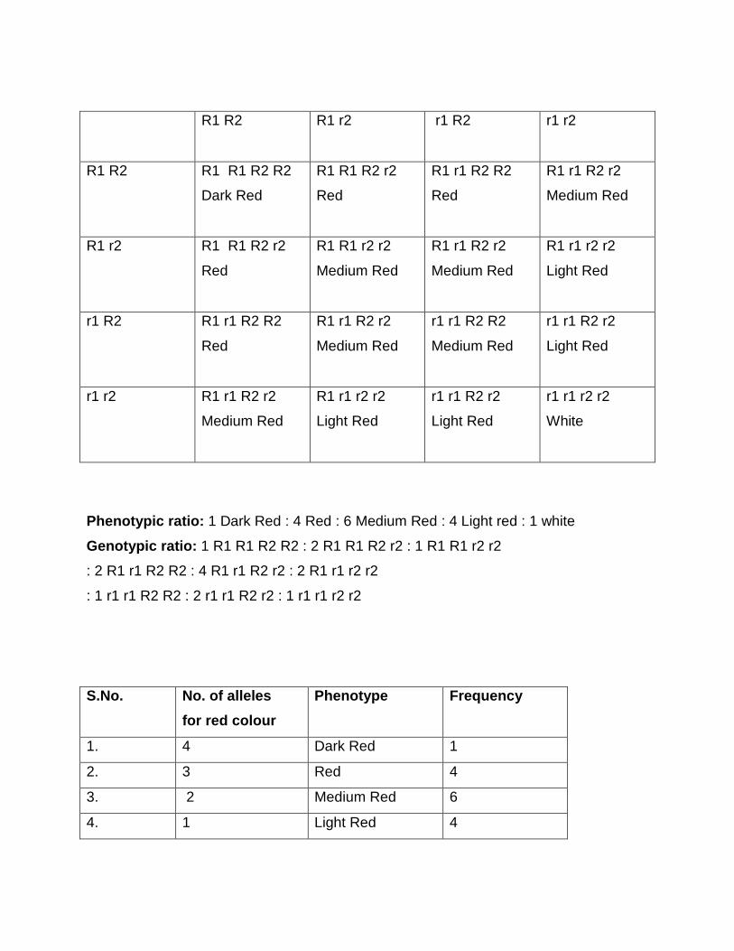
R1 R2 R1 r2 r1 R2 r1 r2
R1 R2 R1 R1 R2 R2
Dark Red
R1 R1 R2 r2
Red
R1 r1 R2 R2
Red
R1 r1 R2 r2
Medium Red
R1 r2 R1 R1 R2 r2
Red
R1 R1 r2 r2
Medium Red
R1 r1 R2 r2
Medium Red
R1 r1 r2 r2
Light Red
r1 R2 R1 r1 R2 R2
Red
R1 r1 R2 r2
Medium Red
r1 r1 R2 R2
Medium Red
r1 r1 R2 r2
Light Red
r1 r2 R1 r1 R2 r2
Medium Red
R1 r1 r2 r2
Light Red
r1 r1 R2 r2
Light Red
r1 r1 r2 r2
White
Phenotypic ratio: 1 Dark Red : 4 Red : 6 Medium Red : 4 Light red : 1 white
Genotypic ratio: 1 R1 R1 R2 R2 : 2 R1 R1 R2 r2 : 1 R1 R1 r2 r2
: 2 R1 r1 R2 R2 : 4 R1 r1 R2 r2 : 2 R1 r1 r2 r2
: 1 r1 r1 R2 R2 : 2 r1 r1 R2 r2 : 1 r1 r1 r2 r2
S.No.
No. of alleles
for red colour
Phenotype Frequency
1. 4 Dark Red 1
2. 3 Red 4
3. 2 Medium Red 6
4. 1 Light Red 4
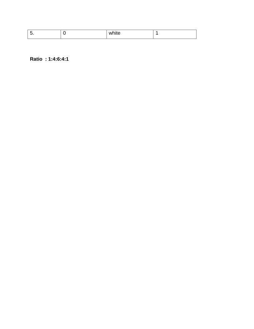
5. 0 white 1
Ratio : 1:4:6:4:1
MUTATIONS
Mutation in a broad sense include all those heritable changes which alter
the phenotype of an individual. Thus mutation can be defined as a sudden
heritable change in the character of an organism which is not due to either
segregation or recombination.
• The term mutation was first used by Hugo de Vries to describe the sudden
phenotypic changes which were heritable, while working with Oenothera
lamarckiana.
• But the earliest record of mutations dates back to 1791 when Seth Wright
noticed a male lamb with unusually short legs in his flock of sheep. This
lamb served as a source of short leg trait for the development of Ancon
breed of sheep.
• However the systematic studies on mutations were started in 1910 by T.H.
Morgan who used Drosophila melanogaster for his studies.
• In 1927, H.J. Muller demonstrated for the first time the artificial induction of
mutations by using x-rays in Drosophila.
• Similarly in 1928, L.J. Stadler demonstrated an increase in the rate of
mutations due to x-rays in barley and maize.
• Induction of mutations by chemicals in fungus Aspergillus was
demonstrated by R.A. Steinberg in 1939.
• C. Auerbach and J.N. Robson in 1946 used chemicals to induce mutations
in Drosophila.
• The first plant breeding programme using mutations (mutation breeding)
was initiated in 1929 in Sweden, Germany and Russia.
• In India it was initiated in early 1930s.
Terminology
Muton: The smallest unit of gene capable of undergoing mutation and it is
represented by a nucleotide.
Mutator gene: A gene which causes another gene or genes to undergo
spontaneous mutation.
Mutable genes: Genes which show very high rates of mutation as compared to
other genes.
Mutant: An organism or cell showing a mutant phenotype due to mutant allele of
a gene.
Mutagen: A physical or chemical agent which induces mutation.
Hot spots: Highly mutable sites with in a gene.
Gene mutations or point mutations: The changes which alter the chemical
structure of a gene at molecular level.
Classification of mutations: Mutations can be classified in several ways.
1. Based on direction of mutations :
a) Forward mutation : Any change from wild type allele to mutant allele
b) Backward mutation or reverse mutation: A change from mutant allele to
wild type
2. Based on source / cause of mutations :
a) Spontaneous mutation: Mutation that occur naturally
b) Induced mutation: Mutation that originates in response to mutagenic
treatment
3. Based on tissue of origin :
a) Somatic mutation: A mutation in somatic tissue
b) Germinal mutation: A mutation in germline cells or in reproductive tissues
4. Based on effect on survival :
a) Lethal mutation: Mutation which kills the individual that carries it. (survival
0%)
b) Sub-lethal mutation: When mortality is more than 50% of individuals that
carry mutation
c) Sub-vital mutation: When mortality is less than 50% of individual that
carry mutation.
d) Vital mutation: When all the mutant individuals survive (survival-100%)
5. Based on trait or character effected :
a) Morphological mutation: A mutation that alters the morphological
features of an individual
b) Biochemical mutation: A mutation that alters the biochemical function of
an individual.
6. Based on visibility or quantum of morphological effect produced :
a) Macro-mutations: Produce a distinct morphological change in phenotype
(which can be detected easily with out any confusion due to environmental
effects) Generally found in qualitative characters. Eg : colour of flowers,
height of plant etc.
b) Micro-mutations: Mutations with invisible phenotypic changes, (which can
be easily confused with effects produced due to environment). Generally
observed in quantitative characters.
7. Based on the site of mutation or on cytological basis :
a) Chromosomal mutations: Mutations associated with detectable changes
in either chromosome number or structure.
b) Gene or point mutations: Mutations produced by alterations in base
sequences of concerned genes.
c) Cytoplasmic mutations: Mutations associated with the changes in
chloroplast DNA (cpDNA) and mitochondrial DNA (mtDNA).
Characteristic features of mutations:
1. Mutations are mostly recessive and very rarely dominant.
2. Most mutations have harmful effects and very few (less than 0.1 %) are
beneficial.
3. Mutations may be due to a change in a gene, a group of genes or in entire
chromosome.
4. If gene mutations are not lethal, the mutant individuals may survive.
However, chromosomal mutations are generally lethal and such mutants do
not survive.
5. If mutation occur at both loci simultaneously, the mutants can be identified
in M1 generation. However, if it is restricted to one locus only, (dominant to
recessive) the effect can be seen only in M2 generation.
6. Macro-mutations are visible and can be easily identified, while micro -
mutations can not be seen with naked eye and need special statistical tests
(or statistical analysis).
7. Many of the mutants show sterility.
8. Most mutants are of negative selection value.
9. Mutation for altogether new character generally does not occur.
10. Mutations are random i.e. they can occur in any tissue or cell of an
organism. However some genes show higher mutation rate than others.
11. Mutations can be sectorial. The branches arising from mutated sector show
mutant characters.
12. Mutations are recurrent i.e. the same mutation may occur again and again.
13. Induced mutations commonly show pleiotropy often due mutation in closely
linked genes.
I. Spontaneous mutations: Spontaneous mutations occur naturally without any
apparent cause. There are two possible sources of origin of these mutations.
1. Due to error during DNA replication.
2. Due to mutagenic effect of natural environment Eg : UV rays from sunlight
The rate of spontaneous mutations is very low. 1 in 10 lakhs i.e. 10-6. But different
genes may show considerably different mutation rates. In crop plants some
varieties were developed through spontaneous mutations. They are
S.no Crop Variety
1. Rice GEB-24, Dee-Geo-Woo-Gen
2. Wheat . Norin
3. Groundnut TMV-10
4. Sorghum Co-4 (coimbatore 4)
II. Induced mutations: Mutations can be induced artificially through treatment
with either physical or chemical mutagens. The exploitation of induced
mutations for crop improvement is called mutation breeding. The rate of
induced mutations is very high. The induced mutations did not differ from
spontaneous mutations in expression.
Examples of popular induced mutants in crop plants are:
S.No. Crop Mutant variety Original variety Mutagen
1. Rice Jagannath T-141 X-rays
Mahsuri mutant Mahsuri, g-rays
2. Wheat Sharbati sonara Sonara 64 UV rays
NP-836 NP-799 x rays
3. French Beans Pusa Parvati Wax podded x-rays
Pusa Lal Meeruti Meeruti x-rays
4. Tomato S-12 Sioux g-rays
5. Castor Aruna HC-6 Thermal neutrons
6. Cotton . MCU 7 1143 EE x-rays
MCU 10 MCU 4 g-rays
7. Oat JO5 JO 1 g-rays
. Artificial induction of mutations: Mutations can be induced artificially using
1. Physical mutagens or radiations
2. Chemical agents
1. Physical mutagens: Include various types of radiations, viz., x-rays, g-rays,
a-rays, ß-rays, fast neutrons, thermal or slow neutrons, UV rays etc. The
physical mutagens are classified into
a) Ionizing radiations: They work through the release of ions. They have
deep penetrating capacity. Eg : x-rays, g-rays, a -particles etc.
b) Non-ionizing radiations : They function through excitation and have a
very low penetrating capacity. They are used for studies on bacteria and
viruses. Eg : UV rays.
Sources of physical mutagens:
Gamma garden
Gamma green house
Vertical gamma irradiation facility
Horizontal gamma irradiation facility
X-ray machine
Isotopes
Small portable irradiators, accelerators and cyclotrons
Nuclear reactors
2. Chemical mutagens : These can be divided into four groups.
a) Alkylating agents: This is the most powerful group of mutagens. These
are the chemicals which are mainly used to induce mutations in cultivated
plants. They induce mutations especially transitions and transversions by
adding an alkyl group (either ethyl or methyl) at various positions in DNA.
Alkylation produces mutation by changing hydrogen bonding in various
ways. Eg: Dimethyl sulphonate (DMS), Ethyl methane sulphonate (EMS),
Nitrosomethyl Urea (NMU), Nitrosoethyl Urea (NEU), Methyl methane
sulphonate (MMS).
b) Base analogues: These are chemicals which are very similar to DNA
bases, such chemicals are sometimes incorporated in DNA in place of
normal bases during replication. Thus they can cause mutation by wrong
base pairing. An incorrect base pairing results in transitions or
transversions after DNA replication. Eg: 5– bromouracil, 3-bromodeoxy
uridine, 2 -amino purine.
c) Antibiotics: A number of antibiotics like mitomycin and streptomycin have
been found to possess chromosome breaking properties. Their usefulness
for practical purposes is very limited.
d) Acridine dyes: Acridine dyes Eg: proflavin, acriflavin, acridine orange, etc.
are very effective mutagens. These are positively charged and they insert
themselves between two base pairs of DNA. This is known as intercalation.
Replication of intercalated DNA molecules results in addition or deletion of
one or few base pairs which produces frame shift mutations.
e) Miscellaneous: Hydoxyl amine produce chromosomal aberrations. Nitrous
acid (deaminating agent) has strong mutagenic activity in a variety of
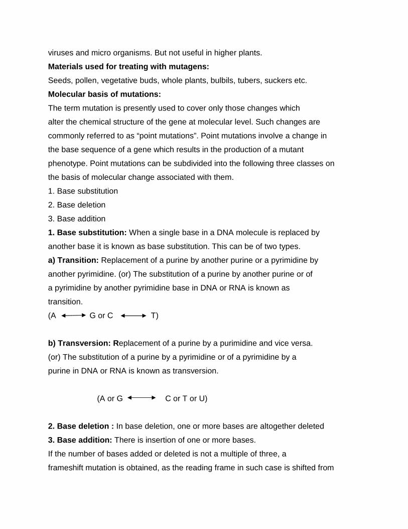
viruses and micro organisms. But not useful in higher plants.
Materials used for treating with mutagens:
Seeds, pollen, vegetative buds, whole plants, bulbils, tubers, suckers etc.
Molecular basis of mutations:
The term mutation is presently used to cover only those changes which
alter the chemical structure of the gene at molecular level. Such changes are
commonly referred to as “point mutations”. Point mutations involve a change in
the base sequence of a gene which results in the production of a mutant
phenotype. Point mutations can be subdivided into the following three classes on
the basis of molecular change associated with them.
1. Base substitution
2. Base deletion
3. Base addition
1. Base substitution: When a single base in a DNA molecule is replaced by
another base it is known as base substitution. This can be of two types.
a) Transition: Replacement of a purine by another purine or a pyrimidine by
another pyrimidine. (or) The substitution of a purine by another purine or of
a pyrimidine by another pyrimidine base in DNA or RNA is known as
transition.
(A G or C T)
b) Transversion: R eplacement of a purine by a purimidine and vice versa.
(or) The substitution of a purine by a pyrimidine or of a pyrimidine by a
purine in DNA or RNA is known as transversion.
(A or G C or T or U)
2. Base deletion : In base deletion, one or more bases are altogether deleted
3. Base addition: There is insertion of one or more bases.
If the number of bases added or deleted is not a multiple of three, a
frameshift mutation is obtained, as the reading frame in such case is shifted from
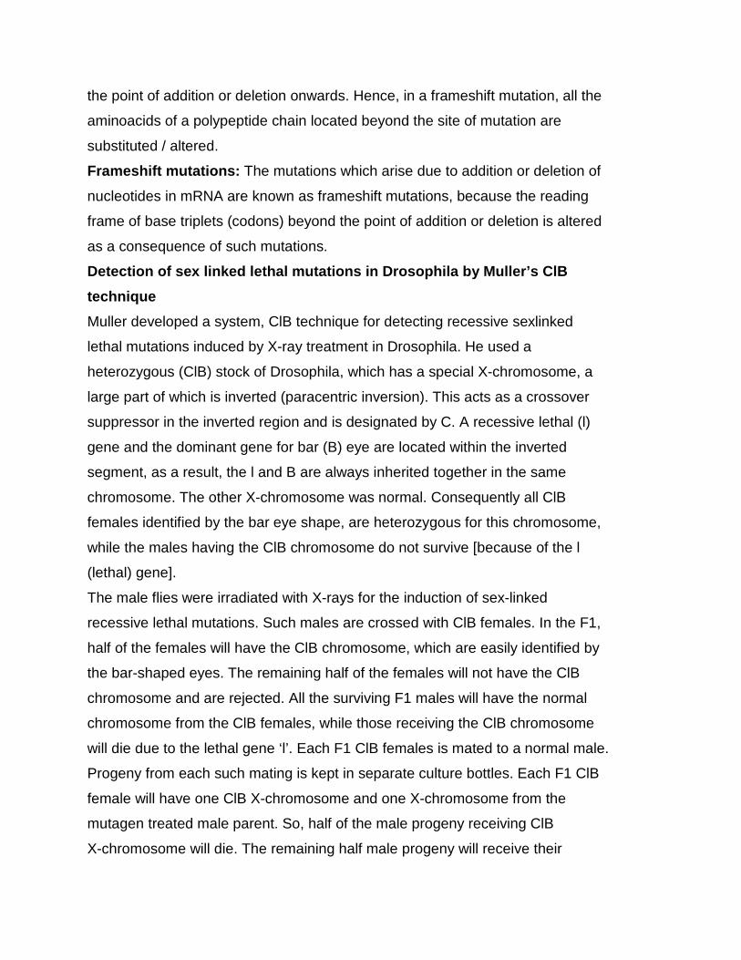
the point of addition or deletion onwards. Hence, in a frameshift mutation, all the
aminoacids of a polypeptide chain located beyond the site of mutation are
substituted / altered.
Frameshift mutations: The mutations which arise due to addition or deletion of
nucleotides in mRNA are known as frameshift mutations, because the reading
frame of base triplets (codons) beyond the point of addition or deletion is altered
as a consequence of such mutations.
Detection of sex linked lethal mutations in Drosophila by Muller’s ClB
technique
Muller developed a system, ClB technique for detecting recessive sexlinked
lethal mutations induced by X-ray treatment in Drosophila. He used a
heterozygous (ClB) stock of Drosophila, which has a special X-chromosome, a
large part of which is inverted (paracentric inversion). This acts as a crossover
suppressor in the inverted region and is designated by C. A recessive lethal (l)
gene and the dominant gene for bar (B) eye are located within the inverted
segment, as a result, the l and B are always inherited together in the same
chromosome. The other X-chromosome was normal. Consequently all ClB
females identified by the bar eye shape, are heterozygous for this chromosome,
while the males having the ClB chromosome do not survive [because of the l
(lethal) gene].
The male flies were irradiated with X-rays for the induction of sex-linked
recessive lethal mutations. Such males are crossed with ClB females. In the F1,
half of the females will have the ClB chromosome, which are easily identified by
the bar-shaped eyes. The remaining half of the females will not have the ClB
chromosome and are rejected. All the surviving F1 males will have the normal
chromosome from the ClB females, while those receiving the ClB chromosome
will die due to the lethal gene ‘l’. Each F1 ClB females is mated to a normal male.
Progeny from each such mating is kept in separate culture bottles. Each F1 ClB
female will have one ClB X-chromosome and one X-chromosome from the
mutagen treated male parent. So, half of the male progeny receiving ClB
X-chromosome will die. The remaining half male progeny will receive their
X-chromosome from their mutagen treated grand father which may or may not
carry the induced mutation. In case lethal mutation was induced, no males will be
observed. On the other hand, if no lethal mutation was induced, half of the males
will survive. Thus the ClB method was the simple, rapid and most efficient method
for detecting sex-linked lethal mutations.
Significance of mutations in Plant Breeding:
1. When a variety is exceptionally good except for one or few characters.
2. When a recessive character is desirable and transfer of that character from
wild species is difficult.
3. When a desirable character is linked with an undesirable character.
4. If there is no known source of resistance gene in the available germplasm
5. To create variability
6. To develop male sterile lines
7. To create variations in vegetatively propagated plants
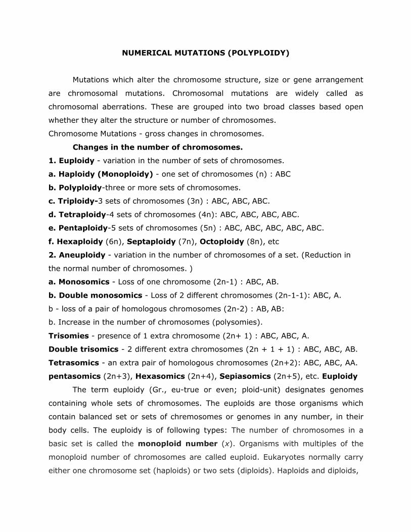
NUMERICAL MUTATIONS (POLYPLOIDY)
Mutations which alter the chromosome structure, size or gene arrangement
are chromosomal mutations. Chromosomal mutations are widely called as
chromosomal aberrations. These are grouped into two broad classes based open
whether they alter the structure or number of chromosomes.
Chromosome Mutations - gross changes in chromosomes.
Changes in the number of chromosomes.
1. Euploidy - variation in the number of sets of chromosomes.
a. Haploidy (Monoploidy) - one set of chromosomes (n) : ABC
b. Polyploidy-three or more sets of chromosomes.
c. Triploidy-3 sets of chromosomes (3n) : ABC, ABC, ABC.
d. Tetraploidy-4 sets of chromosomes (4n): ABC, ABC, ABC, ABC.
e. Pentaploidy-5 sets of chromosomes (5n) : ABC, ABC, ABC, ABC, ABC.
f. Hexaploidy (6n), Septaploidy (7n), Octoploidy (8n), etc
2. Aneuploidy - variation in the number of chromosomes of a set. (Reduction in
the normal number of chromosomes. )
a. Monosomics - Loss of one chromosome (2n-1) : ABC, AB.
b. Double monosomics - Loss of 2 different chromosomes (2n-1-1): ABC, A.
b - loss of a pair of homologous chromosomes (2n-2) : AB, AB:
b. Increase in the number of chromosomes (polysomies).
Trisomies - presence of 1 extra chromosome (2n+ 1) : ABC, ABC, A.
Double trisomics - 2 different extra chromosomes (2n + 1 + 1) : ABC, ABC, AB.
Tetrasomics - an extra pair of homologous chromosomes (2n+2): ABC, ABC, AA.
pentasomics (2n+3), Hexasomics (2n+4), Sepiasomics (2n+5), etc. Euploidy
The term euploidy (Gr., eu-true or even; ploid-unit) designates genomes
containing whole sets of chromosomes. The euploids are those organisms which
contain balanced set or sets of chremosomes or genomes in any number, in their
body cells. The euploidy is of following types: The number of chromosomes in a
basic set is called the monoploid number (x). Organisms with multiples of the
monoploid number of chromosomes are called euploid. Eukaryotes normally carry
either one chromosome set (haploids) or two sets (diploids). Haploids and diploids,
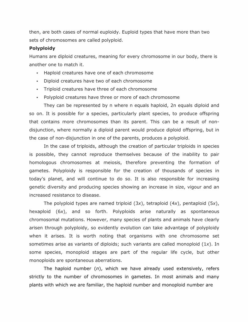
then, are both cases of normal euploidy. Euploid types that have more than two
sets of chromosomes are called polyploid.
Polyploidy
Humans are diploid creatures, meaning for every chromosome in our body, there is
another one to match it.
• Haploid creatures have one of each chromosome
• Diploid creatures have two of each chromosome
• Triploid creatures have three of each chromosome
• Polyploid creatures have three or more of each chromosome
They can be represented by n where n equals haploid, 2n equals diploid and
so on. It is possible for a species, particularly plant species, to produce offspring
that contains more chromosomes than its parent. This can be a result of non-
disjunction, where normally a diploid parent would produce diploid offspring, but in
the case of non-disjunction in one of the parents, produces a polyploid.
In the case of triploids, although the creation of particular triploids in species
is possible, they cannot reproduce themselves because of the inability to pair
homologous chromosomes at meiosis, therefore preventing the formation of
gametes. Polyploidy is responsible for the creation of thousands of species in
today's planet, and will continue to do so. It is also responsible for increasing
genetic diversity and producing species showing an increase in size, vigour and an
increased resistance to disease.
The polyploid types are named triploid (3x), tetraploid (4x), pentaploid (5x),
hexaploid (6x), and so forth. Polyploids arise naturally as spontaneous
chromosomal mutations. However, many species of plants and animals have clearly
arisen through polyploidy, so evidently evolution can take advantage of polyploidy
when it arises. It is worth noting that organisms with one chromosome set
sometimes arise as variants of diploids; such variants are called monoploid (1x). In
some species, monoploid stages are part of the regular life cycle, but other
monoploids are spontaneous aberrations.
The haploid number (n), which we have already used extensively, refers
strictly to the number of chromosomes in gametes. In most animals and many
plants with which we are familiar, the haploid number and monoploid number are
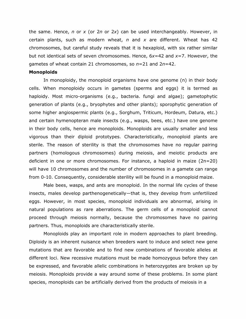
the same. Hence, n or x (or 2n or 2x) can be used interchangeably. However, in
certain plants, such as modern wheat, n and x are different. Wheat has 42
chromosomes, but careful study reveals that it is hexaploid, with six rather similar
but not identical sets of seven chromosomes. Hence, 6x=42 and x=7. However, the
gametes of wheat contain 21 chromosomes, so n=21 and 2n=42.
Monoploids
In monoploidy, the monoploid organisms have one genome (n) in their body
cells. When monoploidy occurs in gametes (sperms and eggs) it is termed as
haploidy. Most micro-organisms (e.g., bacteria. fungi and algae); gametophytic
generation of plants (e.g., bryophytes and other plants); sporophytic generation of
some higher angiospermic plants (e.g., Sorghum, Triticum, Hordeum, Datura, etc.)
and certain hymenopteran male insects (e.g., wasps, bees, etc.) have one genome
in their body cells, hence are monoploids. Monoploids are usually smaller and less
vigorous than their diploid prototypes. Characteristically, monoploid plants are
sterile. The reason of sterility is that the chromosomes have no regular pairing
partners (homologous chromosomes) during meiosis, and meiotic products are
deficient in one or more chromosomes. For instance, a haploid in maize (2n=20)
will have 10 chromosomes and the number of chromosomes in a gamete can range
from 0-10. Consequently, considerable sterility will be found in a monoploid maize.
Male bees, wasps, and ants are monoploid. In the normal life cycles of these
insects, males develop parthenogenetically—that is, they develop from unfertilized
eggs. However, in most species, monoploid individuals are abnormal, arising in
natural populations as rare aberrations. The germ cells of a monoploid cannot
proceed through meiosis normally, because the chromosomes have no pairing
partners. Thus, monoploids are characteristically sterile.
Monoploids play an important role in modern approaches to plant breeding.
Diploidy is an inherent nuisance when breeders want to induce and select new gene
mutations that are favorable and to find new combinations of favorable alleles at
different loci. New recessive mutations must be made homozygous before they can
be expressed, and favorable allelic combinations in heterozygotes are broken up by
meiosis. Monoploids provide a way around some of these problems. In some plant
species, monoploids can be artificially derived from the products of meiosis in a
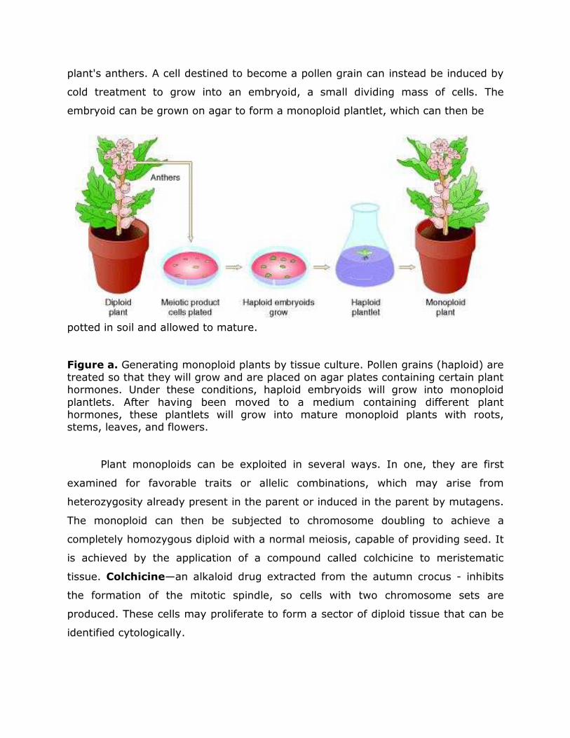
plant's anthers. A cell destined to become a pollen grain can instead be induced by
cold treatment to grow into an embryoid, a small dividing mass of cells. The
embryoid can be grown on agar to form a monoploid plantlet, which can then be
potted in soil and allowed to mature.
Figure a. Generating monoploid plants by tissue culture. Pollen grains (haploid) are
treated so that they will grow and are placed on agar plates containing certain plant hormones. Under these conditions, haploid embryoids will grow into monoploid
plantlets. After having been moved to a medium containing different plant hormones, these plantlets will grow into mature monoploid plants with roots, stems, leaves, and flowers.
Plant monoploids can be exploited in several ways. In one, they are first
examined for favorable traits or allelic combinations, which may arise from
heterozygosity already present in the parent or induced in the parent by mutagens.
The monoploid can then be subjected to chromosome doubling to achieve a
completely homozygous diploid with a normal meiosis, capable of providing seed. It
is achieved by the application of a compound called colchicine to meristematic
tissue. Colchicine—an alkaloid drug extracted from the autumn crocus - inhibits
the formation of the mitotic spindle, so cells with two chromosome sets are
produced. These cells may proliferate to form a sector of diploid tissue that can be
identified cytologically.
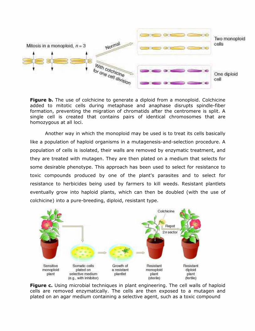
Figure b. The use of colchicine to generate a diploid from a monoploid. Colchicine
added to mitotic cells during metaphase and anaphase disrupts spindle-fiber formation, preventing the migration of chromatids after the centromere is split. A
single cell is created that contains pairs of identical chromosomes that are homozygous at all loci.
Another way in which the monoploid may be used is to treat its cells basically
like a population of haploid organisms in a mutagenesis-and-selection procedure. A
population of cells is isolated, their walls are removed by enzymatic treatment, and
they are treated with mutagen. They are then plated on a medium that selects for
some desirable phenotype. This approach has been used to select for resistance to
toxic compounds produced by one of the plant's parasites and to select for
resistance to herbicides being used by farmers to kill weeds. Resistant plantlets
eventually grow into haploid plants, which can then be doubled (with the use of
colchicine) into a pure-breeding, diploid, resistant type.
Figure c. Using microbial techniques in plant engineering. The cell walls of haploid cells are removed enzymatically. The cells are then exposed to a mutagen and
plated on an agar medium containing a selective agent, such as a toxic compound
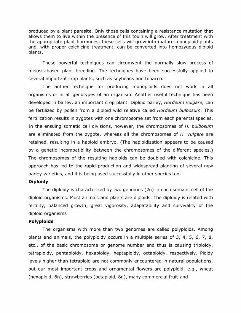
produced by a plant parasite. Only those cells containing a resistance mutation that allows them to live within the presence of this toxin will grow. After treatment with
the appropriate plant hormones, these cells will grow into mature monoploid plants and, with proper colchicine treatment, can be converted into homozygous diploid plants.
These powerful techniques can circumvent the normally slow process of
meiosis-based plant breeding. The techniques have been successfully applied to
several important crop plants, such as soybeans and tobacco.
The anther technique for producing monoploids does not work in all
organisms or in all genotypes of an organism. Another useful technique has been
developed in barley, an important crop plant. Diploid barley, Hordeum vulgare, can
be fertilized by pollen from a diploid wild relative called Hordeum bulbosum. This
fertilization results in zygotes with one chromosome set from each parental species.
In the ensuing somatic cell divisions, however, the chromosomes of H. bulbosum
are eliminated from the zygote, whereas all the chromosomes of H. vulgare are
retained, resulting in a haploid embryo. (The haploidization appears to be caused
by a genetic incompatibility between the chromosomes of the different species.)
The chromosomes of the resulting haploids can be doubled with colchicine. This
approach has led to the rapid production and widespread planting of several new
barley varieties, and it is being used successfully in other species too.
Diploidy
The diploidy is characterized by two genomes (2n) in each somatic cell of the
diploid organisms. Most animals and plants are diploids. The diploidy is related with
fertility, balanced growth, great vigorosity, adapatability and survivality of the
diploid organisms
Polyploids
The organisms with more than two genomes are called polyploids. Among
plants and animals, the polyploidy occurs in a multiple series of 3, 4, 5, 6, 7, 8,
etc., of the basic chromosome or genome number and thus is causing triploidy,
tetraploidy, pentaploidy, hexaploidy, heptaploidy, octaploidy, respectively. Ploidy
levels higher than tetraploid are not commonly encountered in natural populations,
but our most important crops and ornamental flowers are polyploid, e.g., wheat
(hexaploid, 6n), strawberries (octaploid, 8n), many commercial fruit and
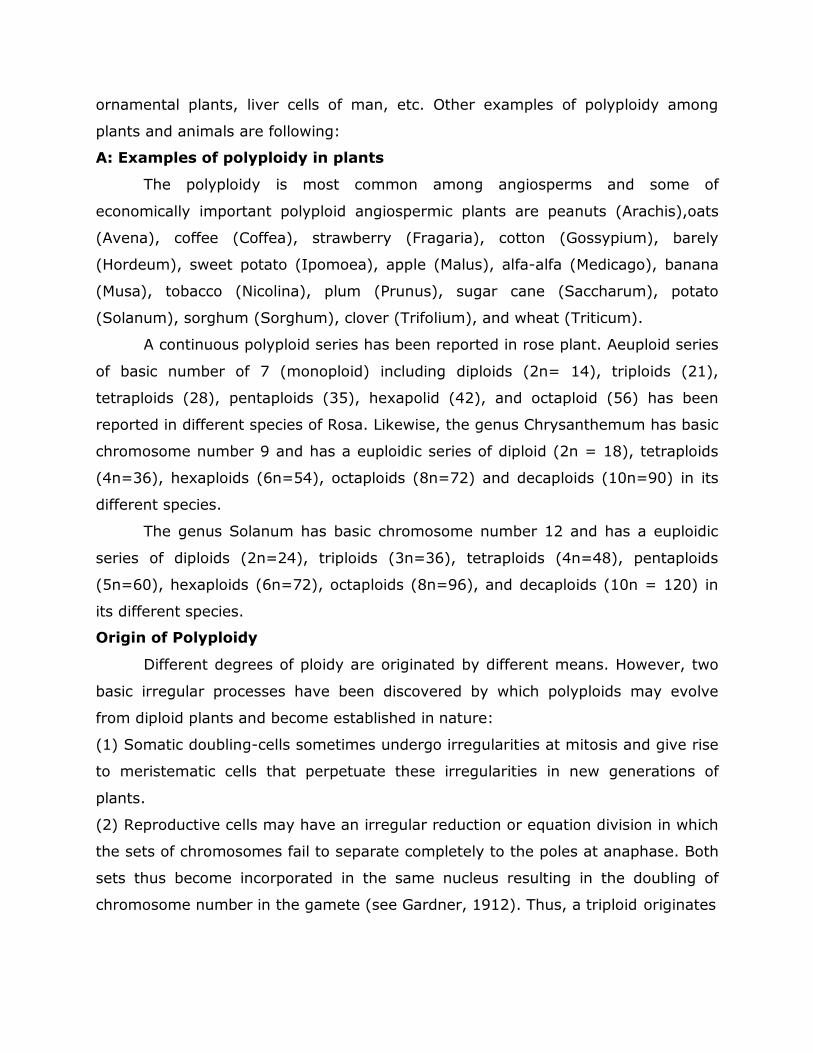
ornamental plants, liver cells of man, etc. Other examples of polyploidy among
plants and animals are following:
A: Examples of polyploidy in plants
The polyploidy is most common among angiosperms and some of
economically important polyploid angiospermic plants are peanuts (Arachis),oats
(Avena), coffee (Coffea), strawberry (Fragaria), cotton (Gossypium), barely
(Hordeum), sweet potato (Ipomoea), apple (Malus), alfa-alfa (Medicago), banana
(Musa), tobacco (Nicolina), plum (Prunus), sugar cane (Saccharum), potato
(Solanum), sorghum (Sorghum), clover (Trifolium), and wheat (Triticum).
A continuous polyploid series has been reported in rose plant. Aeuploid series
of basic number of 7 (monoploid) including diploids (2n= 14), triploids (21),
tetraploids (28), pentaploids (35), hexapolid (42), and octaploid (56) has been
reported in different species of Rosa. Likewise, the genus Chrysanthemum has basic
chromosome number 9 and has a euploidic series of diploid (2n = 18), tetraploids
(4n=36), hexaploids (6n=54), octaploids (8n=72) and decaploids (10n=90) in its
different species.
The genus Solanum has basic chromosome number 12 and has a euploidic
series of diploids (2n=24), triploids (3n=36), tetraploids (4n=48), pentaploids
(5n=60), hexaploids (6n=72), octaploids (8n=96), and decaploids (10n = 120) in
its different species.
Origin of Polyploidy
Different degrees of ploidy are originated by different means. However, two
basic irregular processes have been discovered by which polyploids may evolve
from diploid plants and become established in nature:
(1) Somatic doubling-cells sometimes undergo irregularities at mitosis and give rise
to meristematic cells that perpetuate these irregularities in new generations of
plants.
(2) Reproductive cells may have an irregular reduction or equation division in which
the sets of chromosomes fail to separate completely to the poles at anaphase. Both
sets thus become incorporated in the same nucleus resulting in the doubling of
chromosome number in the gamete (see Gardner, 1912). Thus, a triploid originates
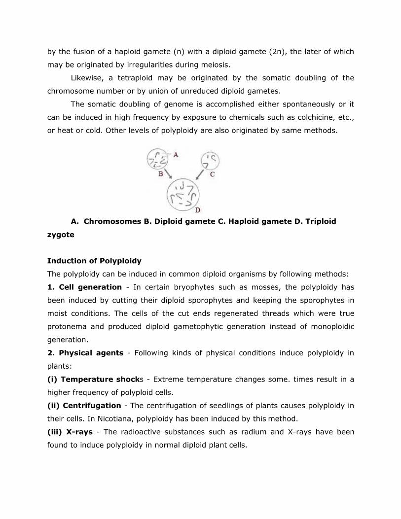
by the fusion of a haploid gamete (n) with a diploid gamete (2n), the later of which
may be originated by irregularities during meiosis.
Likewise, a tetraploid may be originated by the somatic doubling of the
chromosome number or by union of unreduced diploid gametes.
The somatic doubling of genome is accomplished either spontaneously or it
can be induced in high frequency by exposure to chemicals such as colchicine, etc.,
or heat or cold. Other levels of polyploidy are also originated by same methods.
A. Chromosomes B. Diploid gamete C. Haploid gamete D. Triploid
zygote
Induction of Polyploidy
The polyploidy can be induced in common diploid organisms by following methods:
1. Cell generation - In certain bryophytes such as mosses, the polyploidy has
been induced by cutting their diploid sporophytes and keeping the sporophytes in
moist conditions. The cells of the cut ends regenerated threads which were true
protonema and produced diploid gametophytic generation instead of monoploidic
generation.
2. Physical agents - Following kinds of physical conditions induce polyploidy in
plants:
(i) Temperature shocks - Extreme temperature changes some. times result in a
higher frequency of polyploid cells.
(ii) Centrifugation - The centrifugation of seedlings of plants causes polyploidy in
their cells. In Nicotiana, polyploidy has been induced by this method.
(iii) X-rays - The radioactive substances such as radium and X-rays have been
found to induce polyploidy in normal diploid plant cells.
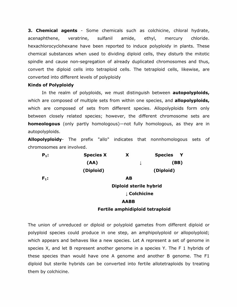
3. Chemical agents - Some chemicals such as colchicine, chloral hydrate,
acenaphthene, veratrine, sulfanil amide, ethyl, mercury chloride.
hexachlorocyclohexane have been reported to induce polyploidy in plants. These
chemical substances when used to dividing diploid cells, they disturb the mitotic
spindle and cause non-segregation of already duplicated chromosomes and thus,
convert the diploid cells into tetraploid cells. The tetraploid cells, likewise, are
converted into different levels of polyploidy
Kinds of Polyploidy
In the realm of polyploids, we must distinguish between autopolyploids,
which are composed of multiple sets from within one species, and allopolyploids,
which are composed of sets from different species. Allopolyploids form only
between closely related species; however, the different chromosome sets are
homeologous (only partly homologous)—not fully homologous, as they are in
autopolyploids.
Allopolyploidy- The prefix "allo" indicates that nonnhomologous sets of
chromosomes are involved.
P1: Species X X Species Y
(AA) ↓ (BB)
(Diploid) (Diploid)
F1: AB
Diploid sterile hybrid
↓ Colchicine
AABB
Fertile amphidiploid tetraploid
The union of unreduced or diploid or polyploid gametes from different diploid or
polyploid species could produce in one step, an amphipolyploid or allopolyploid;
which appears and behaves like a new species. Let A represent a set of genome in
species X, and let B represent another genome in a species Y. The F 1 hybrids of
these species than would have one A genome and another B genome. The F1
diploid but sterile hybrids can be converted into fertile allotetraploids by treating
them by colchicine.
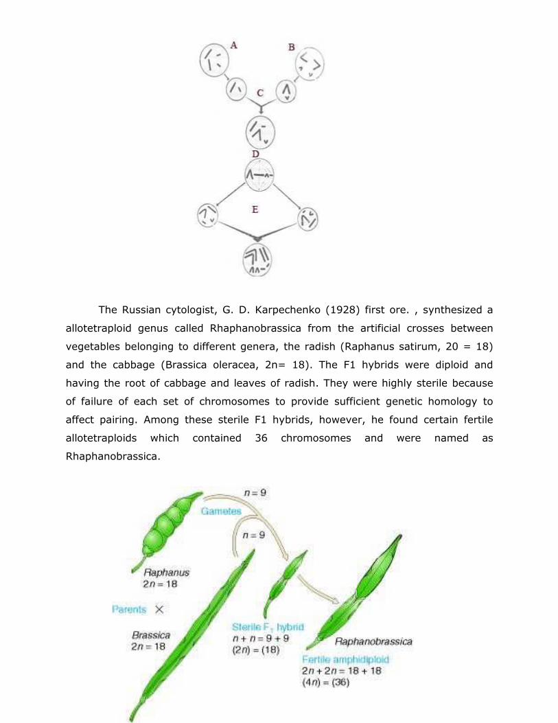
The Russian cytologist, G. D. Karpechenko (1928) first ore. , synthesized a
allotetraploid genus called Rhaphanobrassica from the artificial crosses between
vegetables belonging to different genera, the radish (Raphanus satirum, 20 = 18)
and the cabbage (Brassica oleracea, 2n= 18). The F1 hybrids were diploid and
having the root of cabbage and leaves of radish. They were highly sterile because
of failure of each set of chromosomes to provide sufficient genetic homology to
affect pairing. Among these sterile F1 hybrids, however, he found certain fertile
allotetraploids which contained 36 chromosomes and were named as
Rhaphanobrassica.
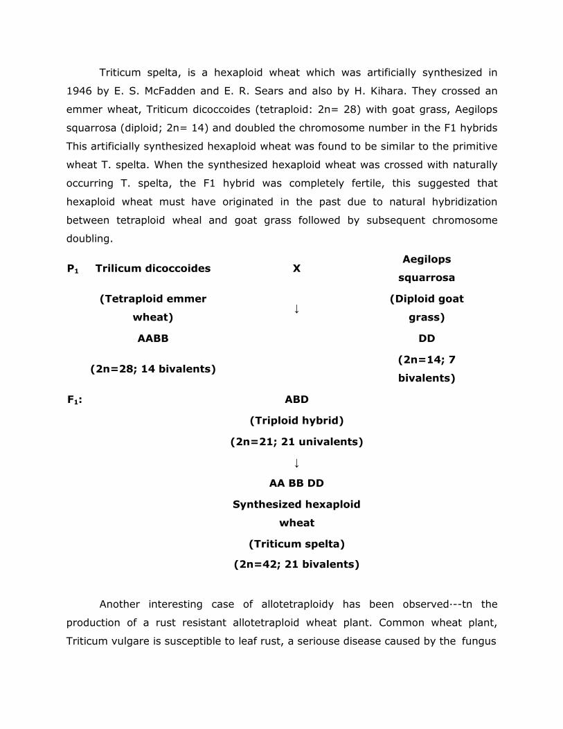
Triticum spelta, is a hexaploid wheat which was artificially synthesized in
1946 by E. S. McFadden and E. R. Sears and also by H. Kihara. They crossed an
emmer wheat, Triticum dicoccoides (tetraploid: 2n= 28) with goat grass, Aegilops
squarrosa (diploid; 2n= 14) and doubled the chromosome number in the F1 hybrids
This artificially synthesized hexaploid wheat was found to be similar to the primitive
wheat T. spelta. When the synthesized hexaploid wheat was crossed with naturally
occurring T. spelta, the F1 hybrid was completely fertile, this suggested that
hexaploid wheat must have originated in the past due to natural hybridization
between tetraploid wheal and goat grass followed by subsequent chromosome
doubling.
P1 Trilicum dicoccoides X Aegilops
squarrosa
(Tetraploid emmer
wheat)
↓
(Diploid goat
grass)
AABB
DD
(2n=28; 14 bivalents)
(2n=14; 7
bivalents)
F1:
ABD
(Triploid hybrid)
(2n=21; 21 univalents)
↓
AA BB DD
Synthesized hexaploid
wheat
(Triticum spelta)
(2n=42; 21 bivalents)
Another interesting case of allotetraploidy has been observed·--tn the
production of a rust resistant allotetraploid wheat plant. Common wheat plant,
Triticum vulgare is susceptible to leaf rust, a seriouse disease caused by the fungus
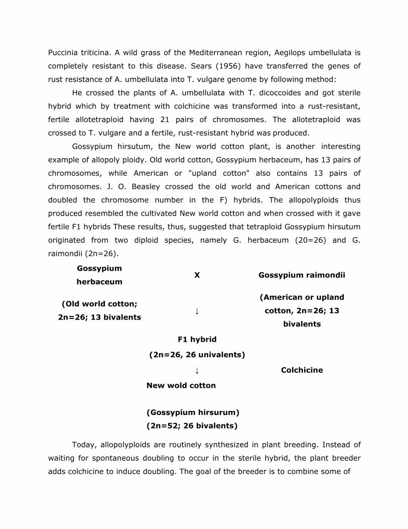
Puccinia triticina. A wild grass of the Mediterranean region, Aegilops umbellulata is
completely resistant to this disease. Sears (1956) have transferred the genes of
rust resistance of A. umbellulata into T. vulgare genome by following method:
He crossed the plants of A. umbellulata with T. dicoccoides and got sterile
hybrid which by treatment with colchicine was transformed into a rust-resistant,
fertile allotetraploid having 21 pairs of chromosomes. The allotetraploid was
crossed to T. vulgare and a fertile, rust-resistant hybrid was produced.
Gossypium hirsutum, the New world cotton plant, is another interesting
example of allopoly ploidy. Old world cotton, Gossypium herbaceum, has 13 pairs of
chromosomes, while American or "upland cotton" also contains 13 pairs of
chromosomes. J. O. Beasley crossed the old world and American cottons and
doubled the chromosome number in the F) hybrids. The allopolyploids thus
produced resembled the cultivated New world cotton and when crossed with it gave
fertile F1 hybrids These results, thus, suggested that tetraploid Gossypium hirsutum
originated from two diploid species, namely G. herbaceum (20=26) and G.
raimondii (2n=26).
Gossypium
herbaceum X Gossypium raimondii
(Old world cotton;
2n=26; 13 bivalents
↓
(American or upland
cotton, 2n=26; 13
bivalents
F1 hybrid
(2n=26, 26 univalents)
↓ Colchicine
New wold cotton
(Gossypium hirsurum)
(2n=52; 26 bivalents)
Today, allopolyploids are routinely synthesized in plant breeding. Instead of
waiting for spontaneous doubling to occur in the sterile hybrid, the plant breeder
adds colchicine to induce doubling. The goal of the breeder is to combine some of
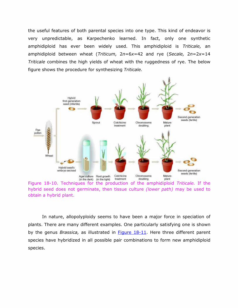
the useful features of both parental species into one type. This kind of endeavor is
very unpredictable, as Karpechenko learned. In fact, only one synthetic
amphidiploid has ever been widely used. This amphidiploid is Triticale, an
amphidiploid between wheat (Triticum, 2n=6x=42 and rye (Secale, 2n=2x=14
Triticale combines the high yields of wheat with the ruggedness of rye. The below
figure shows the procedure for synthesizing Triticale.
Figure 18-10. Techniques for the production of the amphidiploid Triticale. If the
hybrid seed does not germinate, then tissue culture (lower path) may be used to
obtain a hybrid plant.
In nature, allopolyploidy seems to have been a major force in speciation of
plants. There are many different examples. One particularly satisfying one is shown
by the genus Brassica, as illustrated in Figure 18-11. Here three different parent
species have hybridized in all possible pair combinations to form new amphidiploid
species.
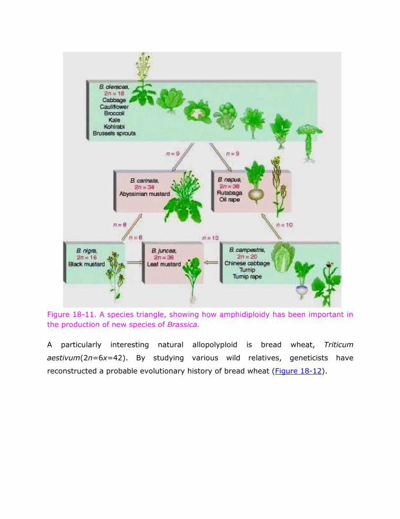
Figure 18-11. A species triangle, showing how amphidiploidy has been important in
the production of new species of Brassica.
A particularly interesting natural allopolyploid is bread wheat, Triticum
aestivum(2n=6x=42). By studying various wild relatives, geneticists have
reconstructed a probable evolutionary history of bread wheat (Figure 18-12).
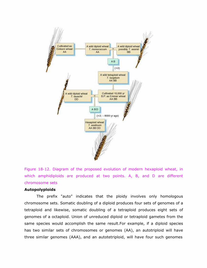
Figure 18-12. Diagram of the proposed evolution of modern hexaploid wheat, in
which amphidiploids are produced at two points. A, B, and D are different
chromosome sets
Autopolyploids
The prefix "auto" indicates that the ploidy involves only homologous
chromosome sets. Somatic doubling of a diploid produces four sets of genomes of a
tetraploid and likewise, somatic doubling of a tetraploid produces eight sets of
genomes of a octaploid. Union of unreduced diploid or tetraploid gametes from the
same species would accomplish the same result.For example, if a diploid species
has two similar sets of chromosomes or genomes (AA), an autotriploid will have
three similar genomes (AAA), and an autotetriploid, will have four such genomes
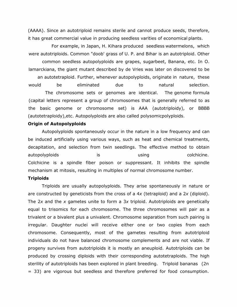
(AAAA). Since an autotriploid remains sterile and cannot produce seeds, therefore,
it has great commercial value in producing seedless varities of economical plants.
For example, in Japan, H. Kihara produced seedless watermelons, which
were autotriploids. Common "doob' grass of U. P. and Bihar is an autotriploid. Other
common seedless autopolyploids are grapes, sugarbeet, Banana, etc. In O.
lamarckiana, the giant mutant described by de Vries was later on discovered to be
an autotetraploid. Further, whenever autopolyploids, originate in nature, these
would be eliminated due to natural selection.
The chromosome sets or genomes are identical. The genome formula
(capital letters represent a group of chromosomes that is generally referred to as
the basic genome or chromosome set) is AAA (autotriploidy), or BBBB
(autotetraploidy),etc. Autopolyploids are also called polysomicpolyploids.
Origin of Autopolyploids
Autopolyploids spontaneously occur in the nature in a low frequency and can
be induced artificially using various ways, such as heat and chemical treatments,
decapitation, and selection from twin seedlings. The effective method to obtain
autopolyploids is using colchicine.
Colchicine is a spindle fiber poison or suppressant. It inhibits the spindle
mechanism at mitosis, resulting in multiples of normal chromosome number.
Triploids
Triploids are usually autopolyploids. They arise spontaneously in nature or
are constructed by geneticists from the cross of a 4x (tetraploid) and a 2x (diploid).
The 2x and the x gametes unite to form a 3x triploid. Autotriploids are genetically
equal to trisomics for each chromosome. The three chromosomes will pair as a
trivalent or a bivalent plus a univalent. Chromosome separation from such pairing is
irregular. Daughter nuclei will receive either one or two copies from each
chromosome. Consequently, most of the gametes resulting from autotriploid
individuals do not have balanced chromosome complements and are not viable. If
progeny survives from autotriploids it is mostly an aneuploid. Autotriploids can be
produced by crossing diploids with their corresponding autotetraploids. The high
sterility of autotriploids has been explored in plant breeding. Triploid bananas (2n
= 33) are vigorous but seedless and therefore preferred for food consumption.
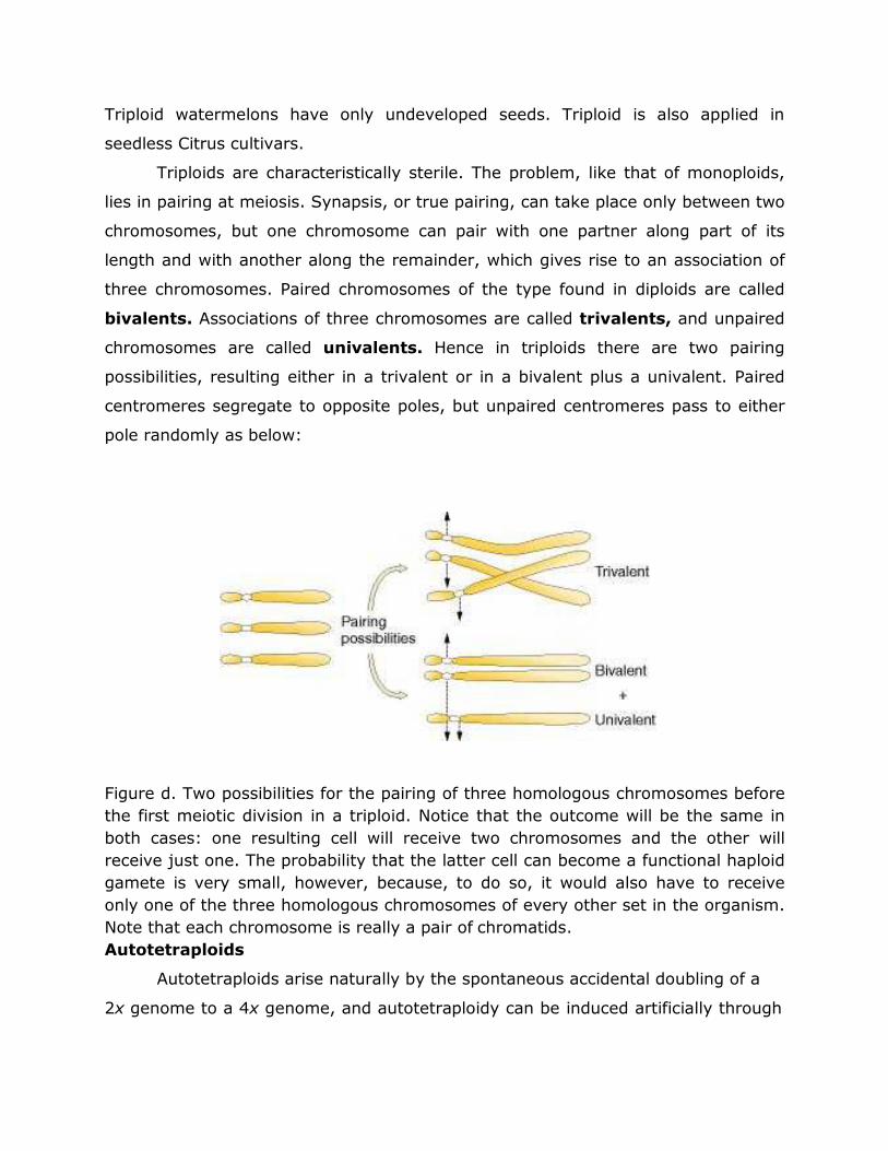
Triploid watermelons have only undeveloped seeds. Triploid is also applied in
seedless Citrus cultivars.
Triploids are characteristically sterile. The problem, like that of monoploids,
lies in pairing at meiosis. Synapsis, or true pairing, can take place only between two
chromosomes, but one chromosome can pair with one partner along part of its
length and with another along the remainder, which gives rise to an association of
three chromosomes. Paired chromosomes of the type found in diploids are called
bivalents. Associations of three chromosomes are called trivalents, and unpaired
chromosomes are called univalents. Hence in triploids there are two pairing
possibilities, resulting either in a trivalent or in a bivalent plus a univalent. Paired
centromeres segregate to opposite poles, but unpaired centromeres pass to either
pole randomly as below:
Figure d. Two possibilities for the pairing of three homologous chromosomes before
the first meiotic division in a triploid. Notice that the outcome will be the same in
both cases: one resulting cell will receive two chromosomes and the other will
receive just one. The probability that the latter cell can become a functional haploid
gamete is very small, however, because, to do so, it would also have to receive
only one of the three homologous chromosomes of every other set in the organism.
Note that each chromosome is really a pair of chromatids.
Autotetraploids
Autotetraploids arise naturally by the spontaneous accidental doubling of a
2x genome to a 4x genome, and autotetraploidy can be induced artificially through
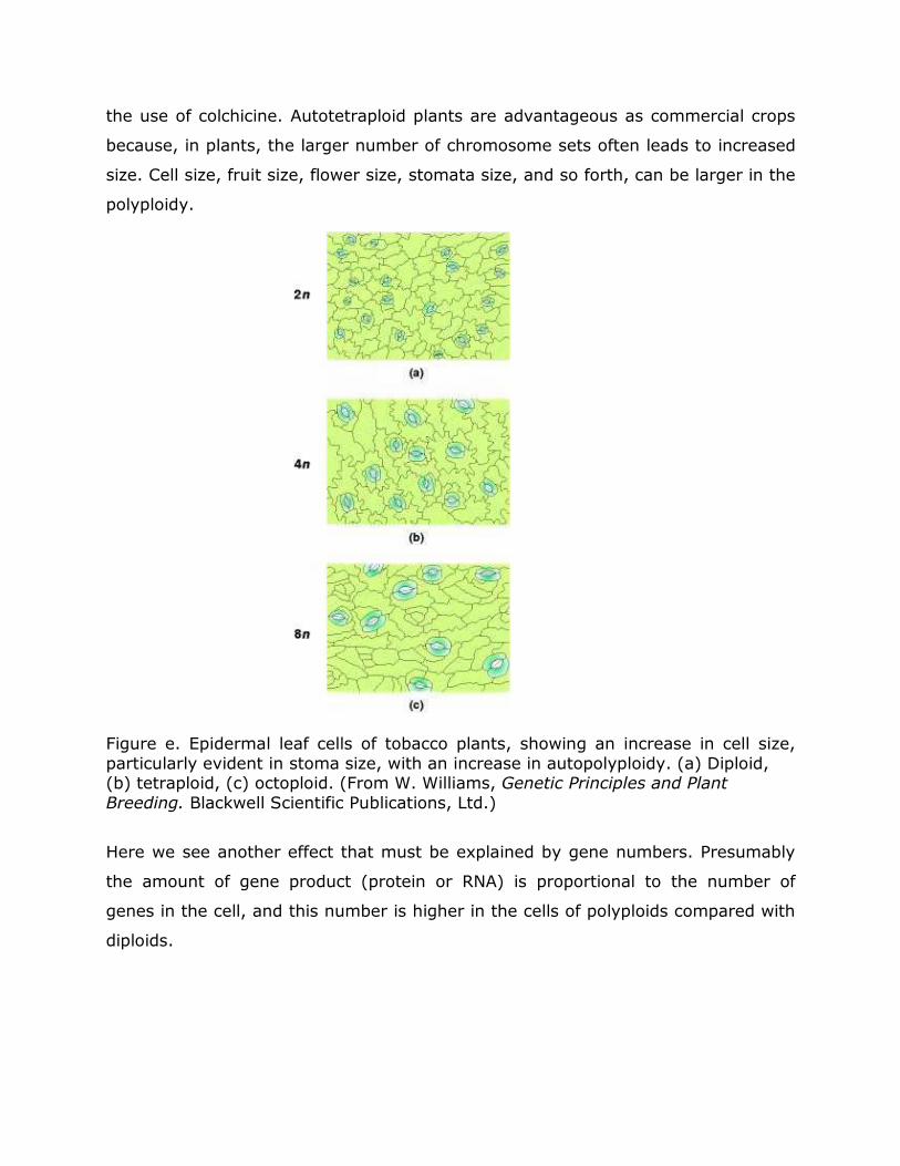
the use of colchicine. Autotetraploid plants are advantageous as commercial crops
because, in plants, the larger number of chromosome sets often leads to increased
size. Cell size, fruit size, flower size, stomata size, and so forth, can be larger in the
polyploidy.
Figure e. Epidermal leaf cells of tobacco plants, showing an increase in cell size,
particularly evident in stoma size, with an increase in autopolyploidy. (a) Diploid, (b) tetraploid, (c) octoploid. (From W. Williams, Genetic Principles and Plant
Breeding. Blackwell Scientific Publications, Ltd.)
Here we see another effect that must be explained by gene numbers. Presumably
the amount of gene product (protein or RNA) is proportional to the number of
genes in the cell, and this number is higher in the cells of polyploids compared with
diploids.
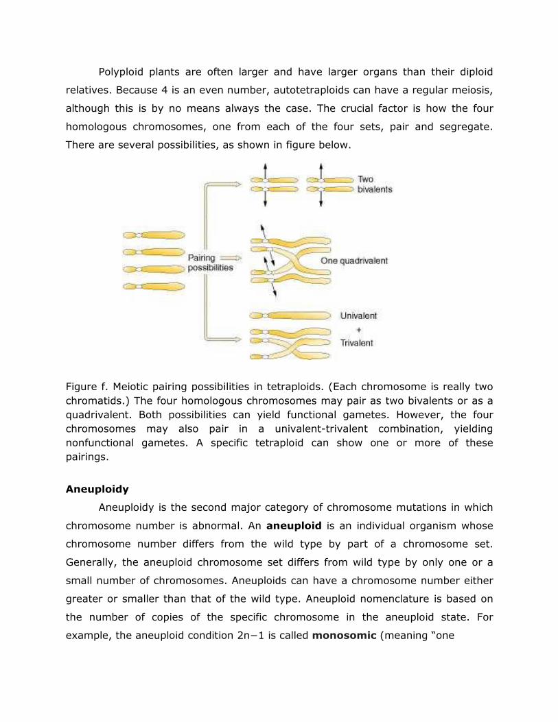
Polyploid plants are often larger and have larger organs than their diploid
relatives. Because 4 is an even number, autotetraploids can have a regular meiosis,
although this is by no means always the case. The crucial factor is how the four
homologous chromosomes, one from each of the four sets, pair and segregate.
There are several possibilities, as shown in figure below.
Figure f. Meiotic pairing possibilities in tetraploids. (Each chromosome is really two
chromatids.) The four homologous chromosomes may pair as two bivalents or as a
quadrivalent. Both possibilities can yield functional gametes. However, the four
chromosomes may also pair in a univalent-trivalent combination, yielding
nonfunctional gametes. A specific tetraploid can show one or more of these
pairings.
Aneuploidy
Aneuploidy is the second major category of chromosome mutations in which
chromosome number is abnormal. An aneuploid is an individual organism whose
chromosome number differs from the wild type by part of a chromosome set.
Generally, the aneuploid chromosome set differs from wild type by only one or a
small number of chromosomes. Aneuploids can have a chromosome number either
greater or smaller than that of the wild type. Aneuploid nomenclature is based on
the number of copies of the specific chromosome in the aneuploid state. For
example, the aneuploid condition 2n−1 is called monosomic (meaning “one
chromosome”) because only one copy of some specific chromosome is present
instead of the usual two found in its diploid progenitor. The aneuploid 2n+1 is
called trisomic, 2n−2 is nullisomic, and n+1 is disomic.
Nullisomics (2n−2)
Although nullisomy is a lethal condition in diploids, an organism such as
bread wheat, which behaves meiotically like a diploid although it is a hexaploid, can
tolerate nullisomy. The four homoeologous chromosomes apparently compensate
for a missing pair of homologs. In fact, all the possible 21 bread wheat nullisomics
have been produced. Their appearances differ from the normal hexaploids;
furthermore, most of the nullisomics grow less vigorously.
The diploid organisms which have lost a pair of homologous chromosomes are
called nullosomics with the genomic formula, 2n-2. The nullosomics exhibit reduced
vigor, fertility and survivality, but, polyploidic nullosomics such as nullosomic
hexaploid what (6n-2) survive to maturity because of the genetic redundancy in
polyploids.
Monosomics (2n−1)
Monosomic chromosome complements are generally deleterious for two main
reasons. First, the missing chromosome perturbs the overall gene balance in the
chromosome set. Second, having a chromosome missing allows any deleterious
recessive allele on the single chromosome to be hemizygous and thus to be directly
expressed phenotypically. Notice that these are the same effects produced by
deletions.
Nondisjunction in mitosis or meiosis is the cause of most aneuploids.
Disjunction is the normal separation of homologous chromosomes or chromatids to
opposite poles at nuclear division. Nondisjunction is a failure of this disjoining
process, and two chromosomes (or chromatids) go to one pole and none to the
other. Nondisjunction occurs spontaneously; it is another example of a chance
failure of a basic cellular process.
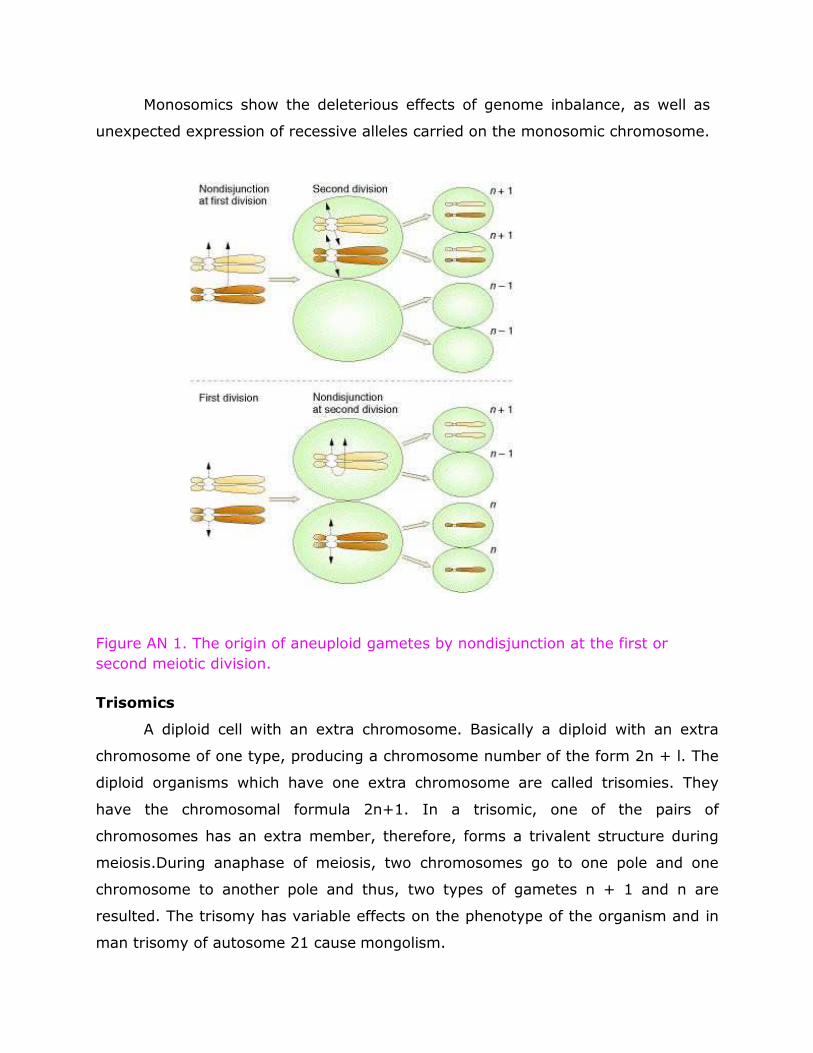
Monosomics show the deleterious effects of genome inbalance, as well as
unexpected expression of recessive alleles carried on the monosomic chromosome.
Figure AN 1. The origin of aneuploid gametes by nondisjunction at the first or
second meiotic division.
Trisomics
A diploid cell with an extra chromosome. Basically a diploid with an extra
chromosome of one type, producing a chromosome number of the form 2n + l. The
diploid organisms which have one extra chromosome are called trisomies. They
have the chromosomal formula 2n+1. In a trisomic, one of the pairs of
chromosomes has an extra member, therefore, forms a trivalent structure during
meiosis.During anaphase of meiosis, two chromosomes go to one pole and one
chromosome to another pole and thus, two types of gametes n + 1 and n are
resulted. The trisomy has variable effects on the phenotype of the organism and in
man trisomy of autosome 21 cause mongolism.
In plants, first case of trisomy was investigated in Datura stromonium
(Jimson weed) by
Blakeslee and Belting in 1924. D. stramonium normally has 12 pairs of
chromosomes in the somatic cells, but in a individual they discovered 25
chromosomes (2n + 1). The size, shape and spine characteristic of seed capsule of
this trisomic plant had difference with seed capsule of the wild type. species.
Theoretically, because the complement was composed of 12 chromosome pairs
differing in the genes they carried, 12 distinguishable trisomies were possible in
Jimson weed. Through experimental breeding, Blakeslee and his associates
succecceded in producing all 12 possible trisomies. These were grown in Blakeslee's
garden and each was found to have a distinguishable phenotype that was attributed
to an extra set of the genes contained in one of the 12 chromosomes.
Tetrasomy
When one chromosome of an otherwise diploid organism is present in
quadruplicate, the tetrasomy is resulted. The tetrasomics have the .chromosomal
formula 20+2. During meiosis a quadrivalent is formed by extra chromosomes and
segregation of chromosomes occurs like autotetraploids.
Double Trisomy
In a diploid organism when two different chromosomes are represented in
triplicate, the, double, trisomy is resulted. A double trisomic has the chromosomal
formula 2n+1+1.
The following indicates in details the types of chromosome mutation where whole
genes are moved:
Pre -Mendelian History of Genetics
Various views were prevailing about the process of heredity before the
rediscovery of Mendel’s laws of inheritance in 1900. Some of the important
theories or concept proposed by various scientists are as follows
Preformation Theory:
This theory was proposed by two Dutch biologists, Swammerdam and Bonnet
(1720-1793). This theory states that a miniature human, called homunculus, is
already present in the egg and sperm which grows into human being after
formation of zygote. This theory was soon rejected because this could not be
proved scientifically.
2. Theory of Epigenesis:
Proposed by a German biologist Wolff (1738-1794), the theory states that the egg
and sperms are undifferentiated cells. The differentiation into various
organs/parts takes place only after fertilization in the zygote resulting in the
development of adult tissues and organs. This concept is universally accepted.
3. Theory of Acquired Characters:
Proposed by the famous French biologist Lamarck (1744-1829) this theory states
that a new character once acquired by an individual shall pass on to its progeny. It
means if a man develops a strong muscle by exercise, all his children would have
strong muscles. On the other hand, if a person becomes weak all his children
would be weak. This concept was totally rejected by Weismann on the basis of
experiment on rats performed through 22 generations.
4. Theory of Pengenes:
This theory, proposed by the famous English naturalist Charles Darwin (1809-
1882), states that very small, exact but invisible copies of each body organ and
component (called gemmules) are transported by the blood stream to the sex
organs. These gemmules are assembled in the gametes. After fertilization these
gemmules move out to different parts of the body resulting in the development
of respective organ. A defective gemmule will lead to the development of
defective organ in an individual. This theory too was rejected because it lacked
scientific basis.
5. Germplasm Theory:
This theory, advocated by August Weismann (1889), a German biologist, states
that body tissues are of two types, viz., germplasm and somatoplasm. The
germplasm refers to the reproductive tissues or cells which produce gametes.
The somatoplasm includes all other body tissues which are not related to sexual
reproduction. Thus, transmission of characters from one generation to other
takes place only through germplasm. Any change in the germplasm will lead to
change in the next generation. This theory is accepted in a broad sense.
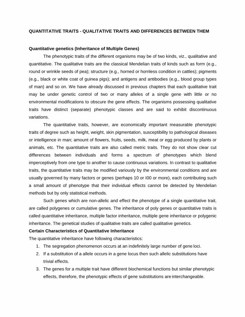
QUANTITATIVE TRAITS - QUALITATIVE TRAITS AND DIFFERENCES BETWEEN THEM
Quantitative genetics (Inheritance of Multiple Genes)
The phenotypic traits of the different organisms may be of two kinds, viz., qualitative and
quantitative. The qualitative traits are the classical Mendelian traits of kinds such as form (e.g.,
round or wrinkle seeds of pea); structure (e.g., horned or hornless condition in cattles); pigments
(e.g., black or white coat of guinea pigs); and antigens and antibodies (e.g., blood group types
of man) and so on. We have already discussed in previous chapters that each qualitative trait
may be under genetic control of two or many alleles of a single gene with little or no
environmental modifications to obscure the gene effects. The organisms possessing qualitative
traits have distinct (separate) phenotypic classes and are said to exhibit discontinuous
variations.
The quantitative traits, however, are economically important measurable phenotypic
traits of degree such as height, weight, skin pigmentation, susceptibility to pathological diseases
or intelligence in man; amount of flowers, fruits, seeds, milk, meat or egg produced by plants or
animals, etc. The quantitative traits are also called metric traits. They do not show clear cut
differences between individuals and forms a spectrum of phenotypes which blend
imperceptively from one type to another to cause continuous variations. In contrast to qualitative
traits, the quantitative traits may be modified variously by the environmental conditions and are
usually governed by many factors or genes (perhaps 10 or I00 or more), each contributing such
a small amount of phenotype that their individual effects cannot be detected by Mendelian
methods but by only statistical methods.
Such genes which are non-allelic and effect the phenotype of a single quantitative trait,
are called polygenes or cumulative genes. The inheritance of poly genes or quantitative traits is
called quantitative inheritance, multiple factor inheritance, multiple gene inheritance or polygenic
inheritance. The genetical studies of qualitative traits are called qualitative genetics.
Certain Characteristics of Quantitative Inheritance
The quantitative inheritance have following characteristics:
1. The segregation phenomenon occurs at an indefinitely large number of gene loci.
2. If a substitution of a allele occurs in a gene locus then such allelic substitutions have
trivial effects.
3. The genes for a multiple trait have different biochemical functions but similar phenotypic
effects, therefore, the phenotypic effects of gene substitutions are interchangeable.
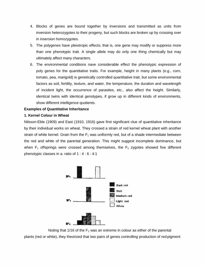
4. Blocks of genes are bound together by inversions and transmitted as units from
inversion heterozygotes to their progeny, but such blocks are broken up by crossing over
in insersion homozygotes.
5. The polygenes have pleiotropic effects; that is, one gene may modify or suppress more
than one phenotypic trait. A single allele may do only one thing chemically but may
ultimately affect many characters.
6. The environmental conditions nave considerable effect the phenotypic expression of
poly genes for the quantitative traits. For example, height in many plants (e.g., corn,
tomato, pea, marigold) is genetically controlled quantitative trait, but some environmental
factors as soil, fertility, texture, and water, the temperature, the duration and wavelength
of incident light, the occurrence of parasites, etc., also affect the height. Similarly,
identical twins with identical genotypes, if grow up in different kinds of environments,
show different intelligence quotients.
Examples of Quantitative Inheritance
1. Kernel Colour in Wheat
Nilsson-Eble (1909) and East (1910, 1916) gave first significant clue of quantitative inheritance
by their individual works on wheat. They crossed a strain of red kernel wheat plant with another
strain of white kernel. Grain from the F1 was uniformly red, but of a shade intermediate between
the red and white of the parental generation. This might suggest incomplete dominance, but
when F1 offsprings were crossed among themselves, the F2 zygotes showed five different
phenotypic classes in a. ratio of 1 : 4 : 6 : 4:1
Noting that 1/16 of the F2 was an extreme in colour as either of the parental
plants (red or white), they theorized that two pairs of genes controlling production of red pigment
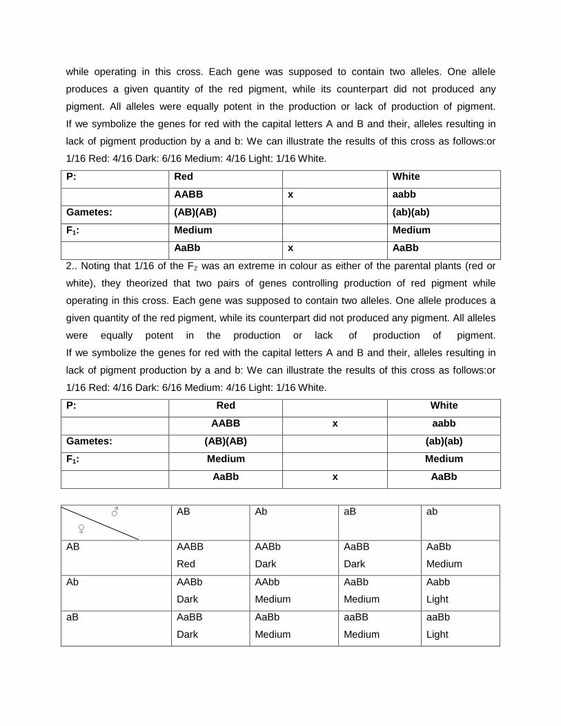
while operating in this cross. Each gene was supposed to contain two alleles. One allele
produces a given quantity of the red pigment, while its counterpart did not produced any
pigment. All alleles were equally potent in the production or lack of production of pigment.
If we symbolize the genes for red with the capital letters A and B and their, alleles resulting in
lack of pigment production by a and b: We can illustrate the results of this cross as follows:or
1/16 Red: 4/16 Dark: 6/16 Medium: 4/16 Light: 1/16 White.
P: Red White
AABB x aabb
Gametes: (AB)(AB) (ab)(ab)
F1: Medium Medium
AaBb x AaBb
2.. Noting that 1/16 of the F2 was an extreme in colour as either of the parental plants (red or
white), they theorized that two pairs of genes controlling production of red pigment while
operating in this cross. Each gene was supposed to contain two alleles. One allele produces a
given quantity of the red pigment, while its counterpart did not produced any pigment. All alleles
were equally potent in the production or lack of production of pigment.
If we symbolize the genes for red with the capital letters A and B and their, alleles resulting in
lack of pigment production by a and b: We can illustrate the results of this cross as follows:or
1/16 Red: 4/16 Dark: 6/16 Medium: 4/16 Light: 1/16 White.
P: Red White
AABB x aabb
Gametes: (AB)(AB) (ab)(ab)
F1: Medium Medium
AaBb x AaBb
♂
♀
AB Ab aB ab
AB AABB
Red
AABb
Dark
AaBB
Dark
AaBb
Medium
Ab AABb
Dark
AAbb
Medium
AaBb
Medium
Aabb
Light
aB AaBB
Dark
AaBb
Medium
aaBB
Medium
aaBb
Light
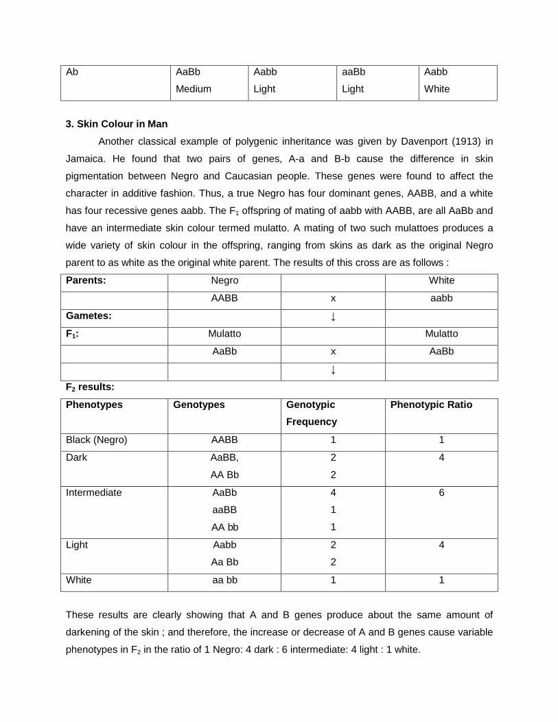
Ab AaBb
Medium
Aabb
Light
aaBb
Light
Aabb
White
3. Skin Colour in Man
Another classical example of polygenic inheritance was given by Davenport (1913) in
Jamaica. He found that two pairs of genes, A-a and B-b cause the difference in skin
pigmentation between Negro and Caucasian people. These genes were found to affect the
character in additive fashion. Thus, a true Negro has four dominant genes, AABB, and a white
has four recessive genes aabb. The F1 offspring of mating of aabb with AABB, are all AaBb and
have an intermediate skin colour termed mulatto. A mating of two such mulattoes produces a
wide variety of skin colour in the offspring, ranging from skins as dark as the original Negro
parent to as white as the original white parent. The results of this cross are as follows :
Parents: Negro White
AABB x aabb
Gametes: ↓
F1: Mulatto Mulatto
AaBb x AaBb
↓
F2 results:
Phenotypes Genotypes Genotypic
Frequency
Phenotypic Ratio
Black (Negro) AABB 1 1
Dark AaBB,
AA Bb
2
2
4
Intermediate AaBb
aaBB
AA bb
4
1
1
6
Light Aabb
Aa Bb
2
2
4
White aa bb 1 1
These results are clearly showing that A and B genes produce about the same amount of
darkening of the skin ; and therefore, the increase or decrease of A and B genes cause variable
phenotypes in F2 in the ratio of 1 Negro: 4 dark : 6 intermediate: 4 light : 1 white.
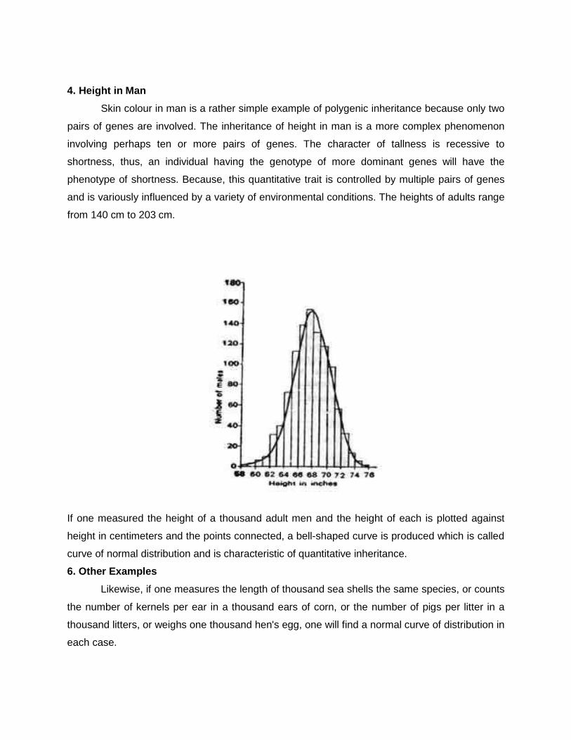
4. Height in Man
Skin colour in man is a rather simple example of polygenic inheritance because only two
pairs of genes are involved. The inheritance of height in man is a more complex phenomenon
involving perhaps ten or more pairs of genes. The character of tallness is recessive to
shortness, thus, an individual having the genotype of more dominant genes will have the
phenotype of shortness. Because, this quantitative trait is controlled by multiple pairs of genes
and is variously influenced by a variety of environmental conditions. The heights of adults range
from 140 cm to 203 cm.
If one measured the height of a thousand adult men and the height of each is plotted against
height in centimeters and the points connected, a bell-shaped curve is produced which is called
curve of normal distribution and is characteristic of quantitative inheritance.
6. Other Examples
Likewise, if one measures the length of thousand sea shells the same species, or counts
the number of kernels per ear in a thousand ears of corn, or the number of pigs per litter in a
thousand litters, or weighs one thousand hen's egg, one will find a normal curve of distribution in
each case.
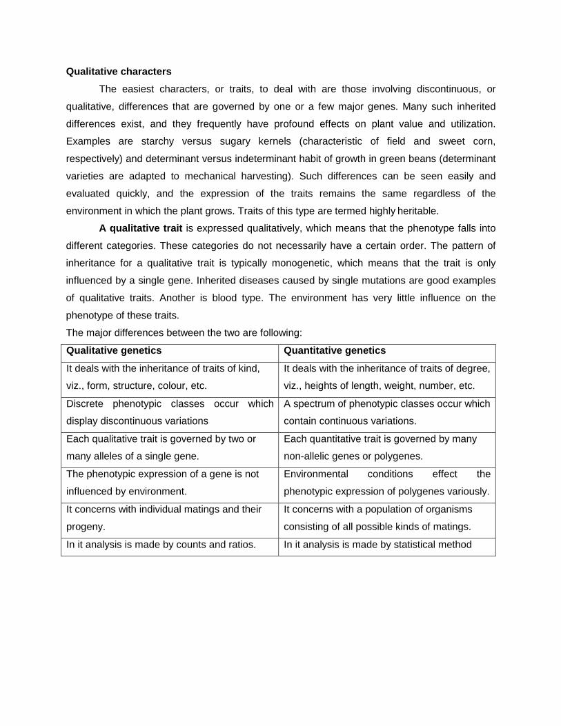
Qualitative characters
The easiest characters, or traits, to deal with are those involving discontinuous, or
qualitative, differences that are governed by one or a few major genes. Many such inherited
differences exist, and they frequently have profound effects on plant value and utilization.
Examples are starchy versus sugary kernels (characteristic of field and sweet corn,
respectively) and determinant versus indeterminant habit of growth in green beans (determinant
varieties are adapted to mechanical harvesting). Such differences can be seen easily and
evaluated quickly, and the expression of the traits remains the same regardless of the
environment in which the plant grows. Traits of this type are termed highly heritable.
A qualitative trait is expressed qualitatively, which means that the phenotype falls into
different categories. These categories do not necessarily have a certain order. The pattern of
inheritance for a qualitative trait is typically monogenetic, which means that the trait is only
influenced by a single gene. Inherited diseases caused by single mutations are good examples
of qualitative traits. Another is blood type. The environment has very little influence on the
phenotype of these traits.
The major differences between the two are following:
Qualitative genetics Quantitative genetics
It deals with the inheritance of traits of kind,
viz., form, structure, colour, etc.
It deals with the inheritance of traits of degree,
viz., heights of length, weight, number, etc.
Discrete phenotypic classes occur which
display discontinuous variations
A spectrum of phenotypic classes occur which
contain continuous variations.
Each qualitative trait is governed by two or
many alleles of a single gene.
Each quantitative trait is governed by many
non-allelic genes or polygenes.
The phenotypic expression of a gene is not
influenced by environment.
Environmental conditions effect the
phenotypic expression of polygenes variously.
It concerns with individual matings and their
progeny.
It concerns with a population of organisms
consisting of all possible kinds of matings.
In it analysis is made by counts and ratios. In it analysis is made by statistical method
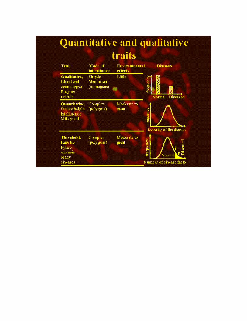
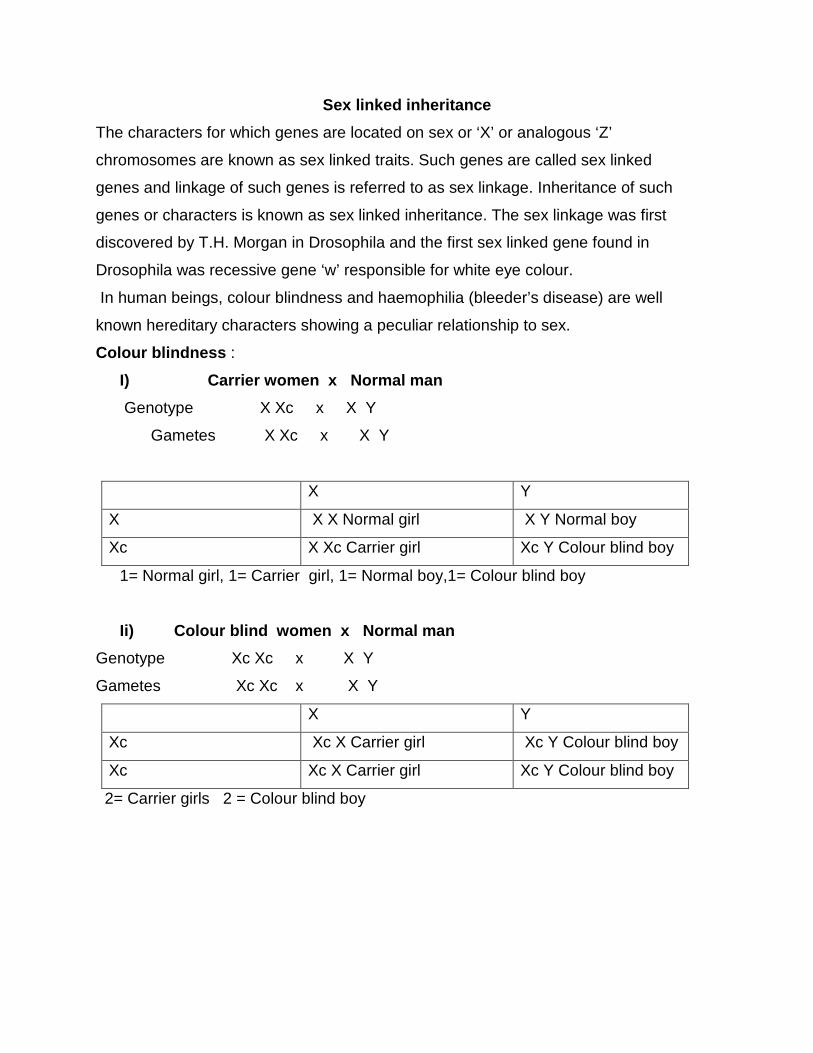
Sex linked inheritance
The characters for which genes are located on sex or ‘X’ or analogous ‘Z’
chromosomes are known as sex linked traits. Such genes are called sex linked
genes and linkage of such genes is referred to as sex linkage. Inheritance of such
genes or characters is known as sex linked inheritance. The sex linkage was first
discovered by T.H. Morgan in Drosophila and the first sex linked gene found in
Drosophila was recessive gene ‘w’ responsible for white eye colour.
In human beings, colour blindness and haemophilia (bleeder’s disease) are well
known hereditary characters showing a peculiar relationship to sex.
Colour blindness :
I) Carrier women x Normal man
Genotype X Xc x X Y
Gametes X Xc x X Y
X Y
X X X Normal girl X Y Normal boy
Xc X Xc Carrier girl Xc Y Colour blind boy
1= Normal girl, 1= Carrier girl, 1= Normal boy,1= Colour blind boy
Ii) Colour blind women x Normal man
Genotype Xc Xc x X Y
Gametes Xc Xc x X Y
X Y
Xc Xc X Carrier girl Xc Y Colour blind boy
Xc Xc X Carrier girl Xc Y Colour blind boy
2= Carrier girls 2 = Colour blind boy
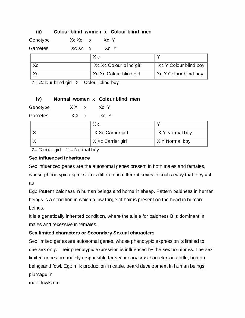
iii) Colour blind women x Colour blind men
Genotype Xc Xc x Xc Y
Gametes Xc Xc x Xc Y
X c Y
Xc Xc Xc Colour blind girl Xc Y Colour blind boy
Xc Xc Xc Colour blind girl Xc Y Colour blind boy
2= Colour blind girl 2 = Colour blind boy
iv) Normal women x Colour blind men
Genotype X X x Xc Y
Gametes X X x Xc Y
X c Y
X X Xc Carrier girl X Y Normal boy
X X Xc Carrier girl X Y Normal boy
2= Carrier girl 2 = Normal boy
Sex influenced inheritance
Sex influenced genes are the autosomal genes present in both males and females,
whose phenotypic expression is different in different sexes in such a way that they act
as
Eg.: Pattern baldness in human beings and horns in sheep. Pattern baldness in human
beings is a condition in which a low fringe of hair is present on the head in human
beings.
It is a genetically inherited condition, where the allele for baldness B is dominant in
males and recessive in females.
Sex limited characters or Secondary Sexual characters
Sex limited genes are autosomal genes, whose phenotypic expression is limited to
one sex only. Their phenotypic expression is influenced by the sex hormones. The sex
limited genes are mainly responsible for secondary sex characters in cattle, human
beingsand fowl. Eg.: milk production in cattle, beard development in human beings,
plumage in
male fowls etc.
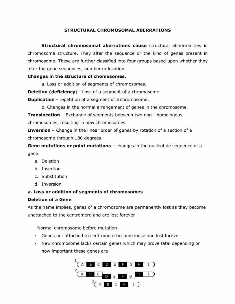
STRUCTURAL CHROMOSOMAL ABERRATIONS
Structural chromosomal aberrations cause structural abnormalities in
chromosome structure. They alter the sequence or the kind of genes present in
chromosome. These are further classified into four groups based upon whether they
alter the gene sequences, number or location.
Changes in the structure of chomosomes.
a. Loss or addition of segments of chromosomes.
Deletion (deficiency) - Loss of a segment of a chromosome
Duplication - repetition of a segment of a chromosome.
b. Changes in the normal arrangement of genes in the chromosome.
Translocation – Exchange of segments between two non - homologous
chromosomes, resulting in new chromosomes.
Inversion – Change in the linear order of genes by rotation of a section of a
chromosome through 180 degrees.
Gene mutations or point mutations – changes in the nucleotide sequence of a
gene.
a. Deletion
b. Insertion
c. Substitution
d. Inversion
a. Loss or addition of segments of chromosomes
Deletion of a Gene
As the name implies, genes of a chromosome are permanently lost as they become
unattached to the centromere and are lost forever
Normal chromosome before mutation
• Genes not attached to centromere become loose and lost forever
• New chromosome lacks certain genes which may prove fatal depending on
how important these genes are
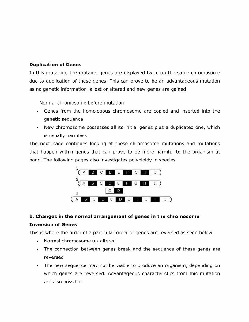
Duplication of Genes
In this mutation, the mutants genes are displayed twice on the same chromosome
due to duplication of these genes. This can prove to be an advantageous mutation
as no genetic information is lost or altered and new genes are gained
Normal chromosome before mutation
• Genes from the homologous chromosome are copied and inserted into the
genetic sequence
• New chromosome possesses all its initial genes plus a duplicated one, which
is usually harmless
The next page continues looking at these chromosome mutations and mutations
that happen within genes that can prove to be more harmful to the organism at
hand. The following pages also investigates polyploidy in species.
b. Changes in the normal arrangement of genes in the chromosome
Inversion of Genes
This is where the order of a particular order of genes are reversed as seen below
• Normal chromosome un-altered
• The connection between genes break and the sequence of these genes are
reversed
• The new sequence may not be viable to produce an organism, depending on
which genes are reversed. Advantageous characteristics from this mutation
are also possible
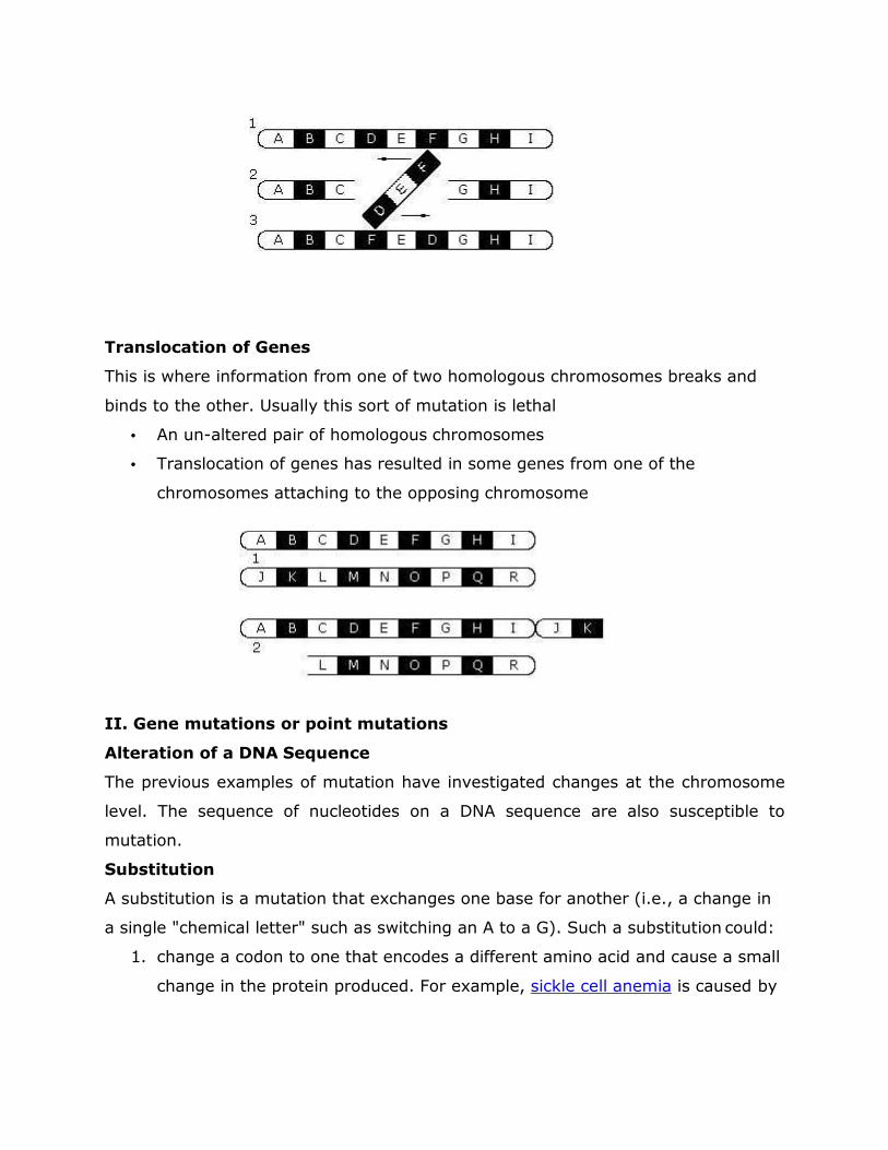
Translocation of Genes
This is where information from one of two homologous chromosomes breaks and
binds to the other. Usually this sort of mutation is lethal
• An un-altered pair of homologous chromosomes
• Translocation of genes has resulted in some genes from one of the
chromosomes attaching to the opposing chromosome
II. Gene mutations or point mutations
Alteration of a DNA Sequence
The previous examples of mutation have investigated changes at the chromosome
level. The sequence of nucleotides on a DNA sequence are also susceptible to
mutation.
Substitution
A substitution is a mutation that exchanges one base for another (i.e., a change in
a single "chemical letter" such as switching an A to a G). Such a substitution could:
1. change a codon to one that encodes a different amino acid and cause a small
change in the protein produced. For example, sickle cell anemia is caused by
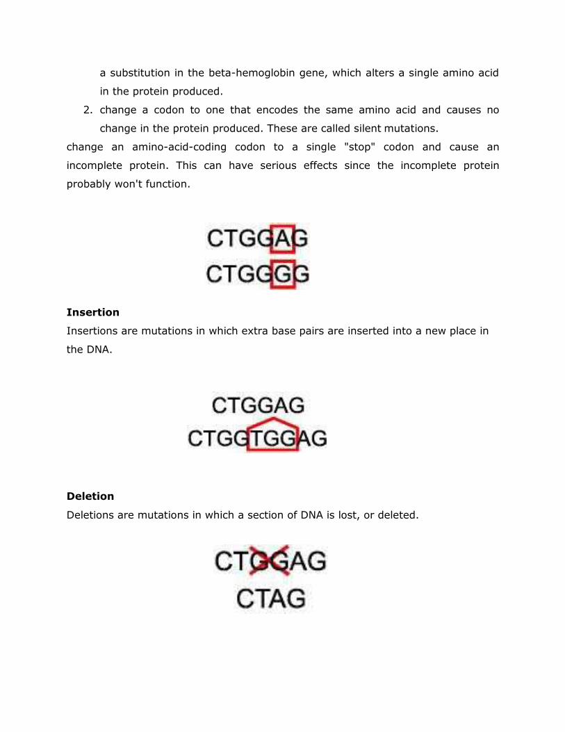
a substitution in the beta-hemoglobin gene, which alters a single amino acid
in the protein produced.
2. change a codon to one that encodes the same amino acid and causes no
change in the protein produced. These are called silent mutations.
change an amino-acid-coding codon to a single "stop" codon and cause an
incomplete protein. This can have serious effects since the incomplete protein
probably won't function.
Insertion
Insertions are mutations in which extra base pairs are inserted into a new place in
the DNA.
Deletion
Deletions are mutations in which a section of DNA is lost, or deleted.
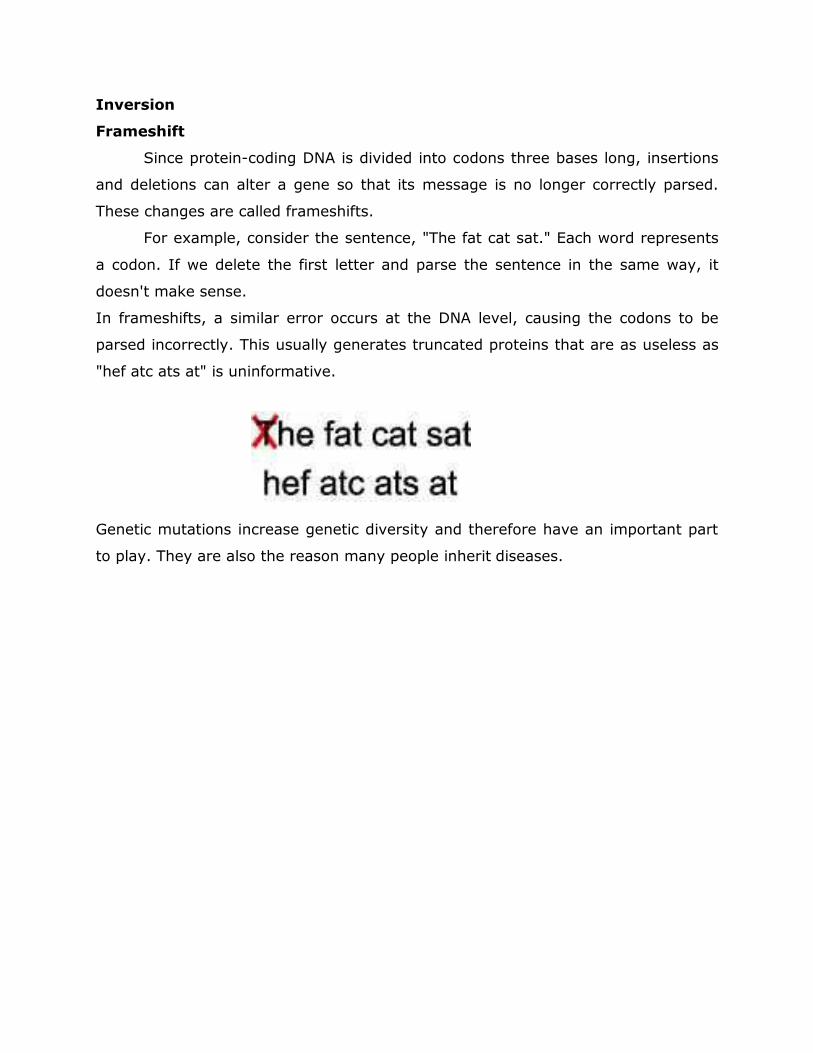
Inversion
Frameshift
Since protein-coding DNA is divided into codons three bases long, insertions
and deletions can alter a gene so that its message is no longer correctly parsed.
These changes are called frameshifts.
For example, consider the sentence, "The fat cat sat." Each word represents
a codon. If we delete the first letter and parse the sentence in the same way, it
doesn't make sense.
In frameshifts, a similar error occurs at the DNA level, causing the codons to be
parsed incorrectly. This usually generates truncated proteins that are as useless as
"hef atc ats at" is uninformative.
Genetic mutations increase genetic diversity and therefore have an important part
to play. They are also the reason many people inherit diseases.
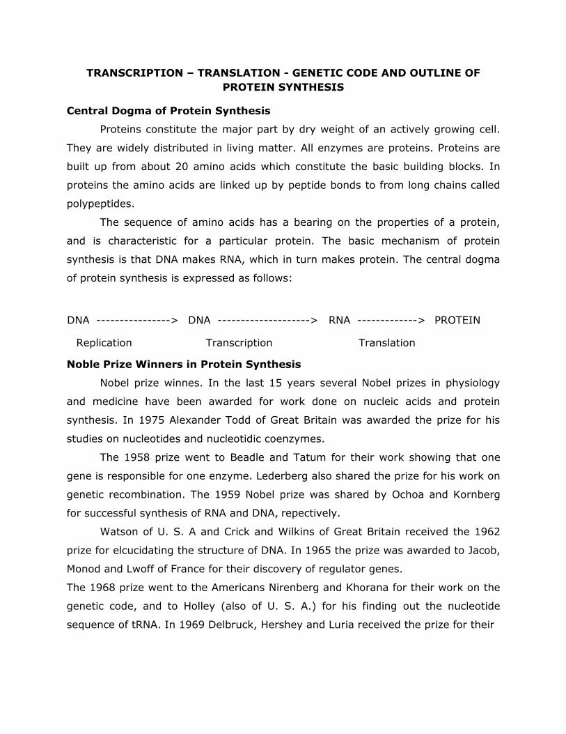
TRANSCRIPTION – TRANSLATION - GENETIC CODE AND OUTLINE OF
PROTEIN SYNTHESIS
Central Dogma of Protein Synthesis
Proteins constitute the major part by dry weight of an actively growing cell.
They are widely distributed in living matter. All enzymes are proteins. Proteins are
built up from about 20 amino acids which constitute the basic building blocks. In
proteins the amino acids are linked up by peptide bonds to from long chains called
polypeptides.
The sequence of amino acids has a bearing on the properties of a protein,
and is characteristic for a particular protein. The basic mechanism of protein
synthesis is that DNA makes RNA, which in turn makes protein. The central dogma
of protein synthesis is expressed as follows:
DNA ----------------> DNA --------------------> RNA -------------> PROTEIN
Replication Transcription Translation
Noble Prize Winners in Protein Synthesis
Nobel prize winnes. In the last 15 years several Nobel prizes in physiology
and medicine have been awarded for work done on nucleic acids and protein
synthesis. In 1975 Alexander Todd of Great Britain was awarded the prize for his
studies on nucleotides and nucleotidic coenzymes.
The 1958 prize went to Beadle and Tatum for their work showing that one
gene is responsible for one enzyme. Lederberg also shared the prize for his work on
genetic recombination. The 1959 Nobel prize was shared by Ochoa and Kornberg
for successful synthesis of RNA and DNA, repectively.
Watson of U. S. A and Crick and Wilkins of Great Britain received the 1962
prize for elcucidating the structure of DNA. In 1965 the prize was awarded to Jacob,
Monod and Lwoff of France for their discovery of regulator genes.
The 1968 prize went to the Americans Nirenberg and Khorana for their work on the
genetic code, and to Holley (also of U. S. A.) for his finding out the nucleotide
sequence of tRNA. In 1969 Delbruck, Hershey and Luria received the prize for their
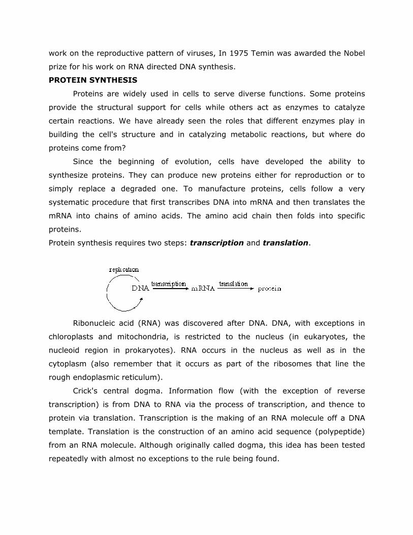
work on the reproductive pattern of viruses, In 1975 Temin was awarded the Nobel
prize for his work on RNA directed DNA synthesis.
PROTEIN SYNTHESIS
Proteins are widely used in cells to serve diverse functions. Some proteins
provide the structural support for cells while others act as enzymes to catalyze
certain reactions. We have already seen the roles that different enzymes play in
building the cell's structure and in catalyzing metabolic reactions, but where do
proteins come from?
Since the beginning of evolution, cells have developed the ability to
synthesize proteins. They can produce new proteins either for reproduction or to
simply replace a degraded one. To manufacture proteins, cells follow a very
systematic procedure that first transcribes DNA into mRNA and then translates the
mRNA into chains of amino acids. The amino acid chain then folds into specific
proteins.
Protein synthesis requires two
Ribonucleic acid (RNA)
chloroplasts and mitochondria, is restricted to the nucleus (in eukaryotes, the
nucleoid region in prokaryotes). RNA occurs in the nucleus as well as in
cytoplasm (also remember that it occurs as part of the
rough endoplasmic reticulum).
Crick's central dogma. Information flow (w
transcription) is from DNA to RNA via the process of
protein via translation. Transcription is the making of an RNA molecule off a DNA
template. Translation is the construction of an
from an RNA molecule. Although originally called dogma, this idea has been tested
repeatedly with almost no exceptions to the rule being found.
work on the reproductive pattern of viruses, In 1975 Temin was awarded the Nobel
prize for his work on RNA directed DNA synthesis.
Proteins are widely used in cells to serve diverse functions. Some proteins
provide the structural support for cells while others act as enzymes to catalyze
certain reactions. We have already seen the roles that different enzymes play in
s structure and in catalyzing metabolic reactions, but where do
Since the beginning of evolution, cells have developed the ability to
synthesize proteins. They can produce new proteins either for reproduction or to
aded one. To manufacture proteins, cells follow a very
systematic procedure that first transcribes DNA into mRNA and then translates the
mRNA into chains of amino acids. The amino acid chain then folds into specific
Protein synthesis requires two steps: transcription and translation.
Ribonucleic acid (RNA) was discovered after DNA. DNA, with exceptions in
ondria, is restricted to the nucleus (in eukaryotes, the
region in prokaryotes). RNA occurs in the nucleus as well as in
cytoplasm (also remember that it occurs as part of the ribosomes
reticulum).
Crick's central dogma. Information flow (with the exception of
) is from DNA to RNA via the process of transcription, and thence to
. Transcription is the making of an RNA molecule off a DNA
template. Translation is the construction of an amino acid sequence
from an RNA molecule. Although originally called dogma, this idea has been tested
repeatedly with almost no exceptions to the rule being found.
work on the reproductive pattern of viruses, In 1975 Temin was awarded the Nobel
Proteins are widely used in cells to serve diverse functions. Some proteins
provide the structural support for cells while others act as enzymes to catalyze
certain reactions. We have already seen the roles that different enzymes play in
s structure and in catalyzing metabolic reactions, but where do
Since the beginning of evolution, cells have developed the ability to
synthesize proteins. They can produce new proteins either for reproduction or to
aded one. To manufacture proteins, cells follow a very
systematic procedure that first transcribes DNA into mRNA and then translates the
mRNA into chains of amino acids. The amino acid chain then folds into specific
.
was discovered after DNA. DNA, with exceptions in
ondria, is restricted to the nucleus (in eukaryotes, the
region in prokaryotes). RNA occurs in the nucleus as well as in the
ribosomes that line the
ith the exception of reverse
, and thence to
. Transcription is the making of an RNA molecule off a DNA
amino acid sequence (polypeptide)
from an RNA molecule. Although originally called dogma, this idea has been tested
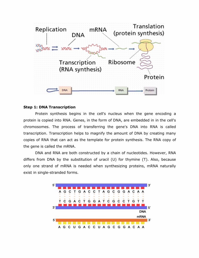
Step 1: DNA Transcription
Protein synthesis begins in the cell's nucleus when the gene encoding a
protein is copied into RNA. Genes, in the form of DNA, are embedded in in the cell's
chromosomes. The process of transferring the gene's DNA into RNA is called
transcription. Transcription helps to magnify the amount of DNA by creating many
copies of RNA that can act as the template for protein synthesis. The RNA copy of
the gene is called the mRNA.
DNA and RNA are both constructed by a chain of nucleotides. However, RNA
differs from DNA by the substitution of uracil (U) for thymine (T). Also, because
only one strand of mRNA is needed when synthesizing proteins, mRNA naturally
exist in single-stranded forms.
After transcription, the mRNA is transported out of the cell's nucleus through
nuclear pores to go to the site of translation, the rough endoplasmic reticulum.
Transcription is a process of making an RNA strand from a DNA template,
and the RNA molecule that is made is called transcript. In the synthesis of proteins,
there are actually three types of RNA that participate and play different roles:
a. Messenger RNA(mRNA) which carries the genetic information from DNA and is
used as a template for protein synthesis.
b. Ribosomal RNA(rRNA) which is a major constituent of the cellular particles
called ribosomes on which protein synthesis actually takes place.
c. A set of transfer RNA(tRNA) molecules, each of which incorporates a
particular amino acid subunit into the growing protein when it recognizes a specific
group of three adjacent bases in the mRNA.
DNA maintains genetic information in the nucleus. RNA takes that
information into the cytoplasm, where the cell uses it to construct specific proteins,
RNA synthesis is transcription; protein synthesis is translation.
RNA differs from DNA in that it is single stranded, contains Uracil instead of
Thymine and ribose instead of deoxyribose, and has different functions. The central
dogma depicts RNA as a messenger between gene and protein, but does not
adequately describe RNA's other function.
Transcription is highly controlled and complex. In Prokaryotes, genes are
expressed as required, and in multicellular organisms, specialized cell types express
subsets of gene. Transcription factors recognize sequences near a gene and bind
sequentially, creating a binding transcription. Transcription proceeds as RNAP
inserts complementary RNA bases opposite the coding strand of DNA. Antisense
RNA blocks gene expression.
Messenger RNA transmits information in a gene to cellular structures that
build proteins. Each three mRNA bases in a row forms a codon that specifies a
particular amino acid. Ribosomal RNA and proteins form ribosomes, which
physically support the other participants in protein synthesis and help catalyze
formation of bonds betweens amino acids.
In eukaryotes, RNA is often altered before it is active. Messenger RNA gains
a cap of modified nucleotides and a poly A tail. Introns are transcribed and cut out,
and exons are reattached by ribozymes. RNA editing introduced bases changes that
alter the protein product in different cell types.
The genetic code is triplet, non-overlapping, continuous, universal, and
degenerate. As translation begins, mRNA, tRNA with bound amino acids, ribosomes,
energy molecules and protein factos assemble. The mRNA leader sequence binds to
rRNA in the small subunit of a ribosome, and the first codon attracts a tRNA bearing
methionine. Next, as the chain elongates, the large ribosomal subunit attaches and
the appropriate anticodon parts of tRNA molecules form peptide bonds, a
polypeptide grows. At a stop codon, protein synthesis ceases. Protein folding begins
as translation proceeds, with enzymes and chaperone proteins assisting the amino
acid chain in assuming its final functional form. Translation is efficient and
economical, as RNA, ribosomes, enzymes, and key proteins are recycled.
RNA transcription requires the following components
The enzyme RNA polymerase
� A DNA template
� All four types of ribonucleoside triphosphates (ATP, GTP and UTP)
� Divalent metal ions Mg++ or Mn++ as a co-factor
� No primer is needed for RNA synthesis
� RNA transcription is a process that involves the following steps.
Binding of RNA Polymerase to DNA Double Helix
The histone coat protecting the DNA double helix of the gene to be
transcribed is removed, on a signal from the cytoplasm, exposing the
polynucleotide sequences in this region of DNA. The RNA polymerase enzyme binds
to a specific site, called promoter, in the DNA double helix. This site is located on
the 5 side of the gene to be transcribed. It signals the beginning of RNA synthesis.
The promoter also determines the DNA strand that is to be transcribed.
Exposure of RNA Bases
The histone coat protecting the DNA double helix of the gene to be
transcribed is removed, on a signal from the cytoplasm, exposing the
polynucleotide sequences in this region of DNA. The RNA polymerase enzyme binds
to a specific site, called promoter, in the DNA double helix. This site is located on
the 5 side of the gene to be transcribed. It signals the beginning of RNA synthesis.
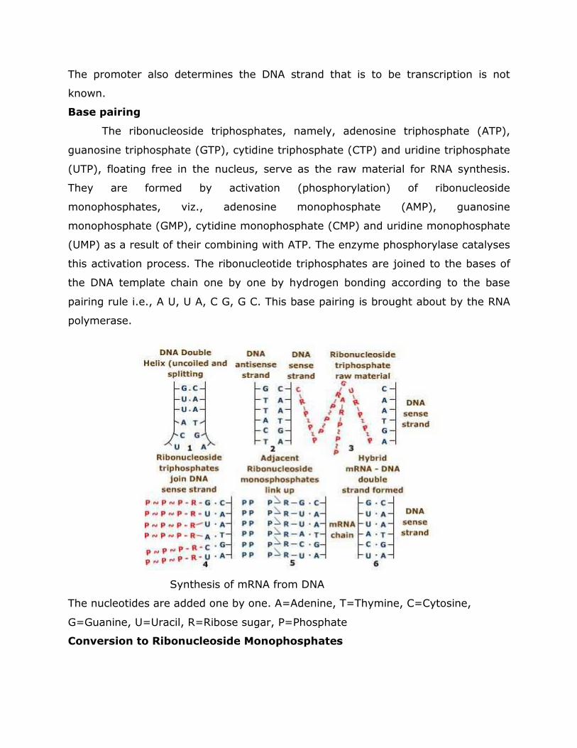
The promoter also determines the DNA strand that is to be transcription is not
known.
Base pairing
The ribonucleoside triphosphates, namely, adenosine triphosphate (ATP),
guanosine triphosphate (GTP), cytidine triphosphate (CTP) and uridine triphosphate
(UTP), floating free in the nucleus, serve as the raw material for RNA synthesis.
They are formed by activation (phosphorylation) of ribonucleoside
monophosphates, viz., adenosine monophosphate (AMP), guanosine
monophosphate (GMP), cytidine monophosphate (CMP) and uridine monophosphate
(UMP) as a result of their combining with ATP. The enzyme phosphorylase catalyses
this activation process. The ribonucleotide triphosphates are joined to the bases of
the DNA template chain one by one by hydrogen bonding according to the base
pairing rule i.e., A U, U A, C G, G C. This base pairing is brought about by the RNA
polymerase.
Synthesis of mRNA from DNA
The nucleotides are added one by one. A=Adenine, T=Thymine, C=Cytosine,
G=Guanine, U=Uracil, R=Ribose sugar, P=Phosphate
Conversion to Ribonucleoside Monophosphates
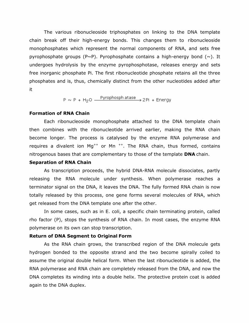
The various ribonucleoside triphosphates on linking to the DNA template
chain break off their high-energy bonds. This changes them to ribonucleoside
monophosphates which represent the normal components of RNA, and sets free
pyrophosphate groups (P~P). Pyrophosphate contains a high-energy bond (~). It
undergoes hydrolysis by the enzyme pyrophosphotase, releases energy and sets
free inorganic phosphate Pi. The first ribonucleotide phosphate retains all the three
phosphates and is, thus, chemically distinct from the other nucleotides added after
it
Formation of RNA Chain
Each ribonucleoside monophosphate attached to the DNA template chain
then combines with the ribonucleotide arrived earlier, making the RNA chain
become longer. The process is catalysed by the enzyme RNA polymerase and
requires a divalent ion Mg++ or Mn ++. The RNA chain, thus formed, contains
nitrogenous bases that are complementary to those of the template DNA chain.
Separation of RNA Chain
As transcription proceeds, the hybrid DNA-RNA molecule dissociates, partly
releasing the RNA molecule under synthesis. When polymerase reaches a
terminator signal on the DNA, it leaves the DNA. The fully formed RNA chain is now
totally released by this process, one gene forms several molecules of RNA, which
get released from the DNA template one after the other.
In some cases, such as in E. coli, a specific chain terminating protein, called
rho factor (P), stops the synthesis of RNA chain. In most cases, the enzyme RNA
polymerase on its own can stop transcription.
Return of DNA Segment to Original Form
As the RNA chain grows, the transcribed region of the DNA molecule gets
hydrogen bonded to the opposite strand and the two become spirally coiled to
assume the original double helical form. When the last ribonucleotide is added, the
RNA polymerase and RNA chain are completely released from the DNA, and now the
DNA completes its winding into a double helix. The protective protein coat is added
again to the DNA duplex.
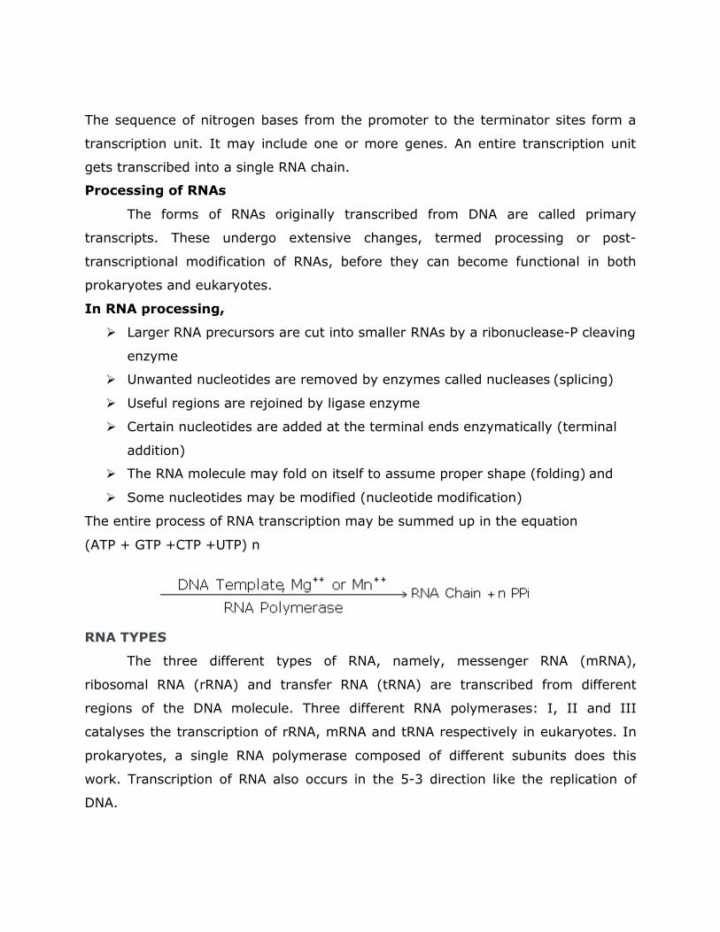
The sequence of nitrogen bases from the promoter to the terminator sites form a
transcription unit. It may include one or more genes. An entire transcription unit
gets transcribed into a single RNA chain.
Processing of RNAs
The forms of RNAs originally transcribed from DNA are called primary
transcripts. These undergo extensive changes, termed processing or post-
transcriptional modification of RNAs, before they can become functional in both
prokaryotes and eukaryotes.
In RNA processing,
� Larger RNA precursors are cut into smaller RNAs by a ribonuclease-P cleaving
enzyme
� Unwanted nucleotides are removed by enzymes called nucleases (splicing)
� Useful regions are rejoined by ligase enzyme
� Certain nucleotides are added at the terminal ends enzymatically (terminal
addition)
� The RNA molecule may fold on itself to assume proper shape (folding) and
� Some nucleotides may be modified (nucleotide modification)
The entire process of RNA transcription may be summed up in the equation
(ATP + GTP +CTP +UTP) n
RNA TYPES
The three different types of RNA, namely, messenger RNA (mRNA),
ribosomal RNA (rRNA) and transfer RNA (tRNA) are transcribed from different
regions of the DNA molecule. Three different RNA polymerases: I, II and III
catalyses the transcription of rRNA, mRNA and tRNA respectively in eukaryotes. In
prokaryotes, a single RNA polymerase composed of different subunits does this
work. Transcription of RNA also occurs in the 5-3 direction like the replication of
DNA.
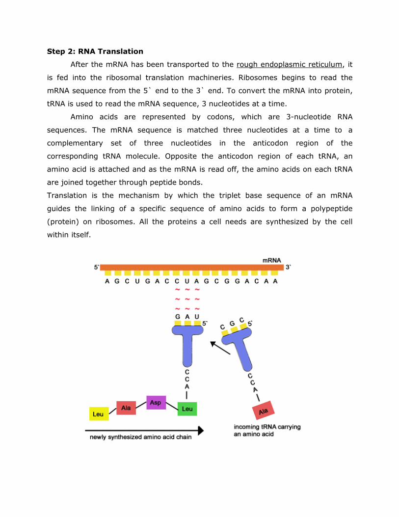
Step 2: RNA Translation
After the mRNA has been transported to the rough endoplasmic reticulum, it
is fed into the ribosomal translation machineries. Ribosomes begins to read the
mRNA sequence from the 5` end to the 3` end. To convert the mRNA into protein,
tRNA is used to read the mRNA sequence, 3 nucleotides at a time.
Amino acids are represented by codons, which are 3-nucleotide RNA
sequences. The mRNA sequence is matched three nucleotides at a time to a
complementary set of three nucleotides in the anticodon region of the
corresponding tRNA molecule. Opposite the anticodon region of each tRNA, an
amino acid is attached and as the mRNA is read off, the amino acids on each tRNA
are joined together through peptide bonds.
Translation is the mechanism by which the triplet base sequence of an mRNA
guides the linking of a specific sequence of amino acids to form a polypeptide
(protein) on ribosomes. All the proteins a cell needs are synthesized by the cell
within itself.
Machinery for Protein Synthesis
Protein synthesis requires amino acids, DNA, RNAs, ribosomes and enzymes,
enzyme activators and ATP molecules
Amino Acids
Proteins are the polymers of amino acids. Therefore, amino acids form the
raw material for protein synthesis. The proteins found in living organisms need
about 20 amino acids as building blocks or monomers. These are available in the
cytoplasmic matrix as an amino acid pool.
DNA as Specificity Control
In order to maintain its own special characteristics a cell must manufacture
proteins exactly similar to those already present in it. Thus, protein synthesis
requires specificity control to provide instructions about the exact sequence in
which the given numbers and kinds of amino acids should be linked to form the
desired polypeptides. The specificity control is exercised by DNA through mRNA.
Sequences of 3 consecutive nitrogenous bases in the DNA double helix form the
biochemical or genetic code. Each base triplet codes for a specific amino acid. Since
the DNA is more or less stable, the proteins formed in a cell are exactly like the
preexisting proteins
RNAs
RNA molecule is a long, unbranched, single-stranded polymer of
ribonucleotides. Each nucleotide unit is composed of three smaller molecules, a
phosphate group, a 5-carbon ribose sugar, and a nitrogen-containing base. The
bases in RNA are adenine, guanine, uracil and cytosine. The various components
are linked up as in DNA. There are three types of RNA in every cell: messenger RNA
or mRNA, ribosomal RNA or rRNA and transfer RNA or tRNA. The three types of
RNAs are transcribed from different regions of DNA template. RNA chain is
complementary to the DNA strand, which produces it. All the three kinds of RNAs
play a role in protein synthesis.
Differences Between RNA Types
Features
Ribosomal RNA
(rRNA)
Messenger RNA
(mRNA)
Transfer RNA
(tRNA)
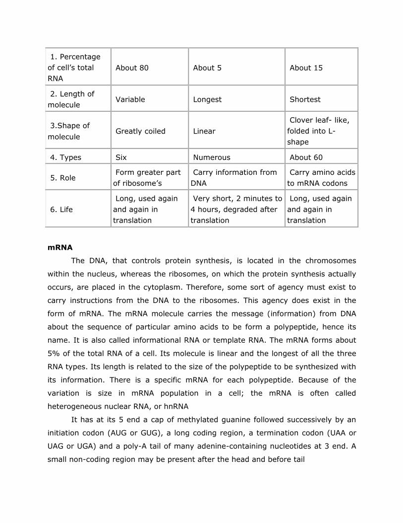
1. Percentage
of cell’s total
RNA
About 80
About 5
About 15
2. Length of
molecule
Variable
Longest
Shortest
3.Shape of
molecule
Greatly coiled
Linear
Clover leaf- like,
folded into L-
shape
4. Types Six Numerous About 60
5. Role
Form greater part
of ribosome’s
Carry information from
DNA
Carry amino acids
to mRNA codons
6. Life
Long, used again
and again in
translation
Very short, 2 minutes to
4 hours, degraded after
translation
Long, used again
and again in
translation
mRNA
The DNA, that controls protein synthesis, is located in the chromosomes
within the nucleus, whereas the ribosomes, on which the protein synthesis actually
occurs, are placed in the cytoplasm. Therefore, some sort of agency must exist to
carry instructions from the DNA to the ribosomes. This agency does exist in the
form of mRNA. The mRNA molecule carries the message (information) from DNA
about the sequence of particular amino acids to be form a polypeptide, hence its
name. It is also called informational RNA or template RNA. The mRNA forms about
5% of the total RNA of a cell. Its molecule is linear and the longest of all the three
RNA types. Its length is related to the size of the polypeptide to be synthesized with
its information. There is a specific mRNA for each polypeptide. Because of the
variation is size in mRNA population in a cell; the mRNA is often called
heterogeneous nuclear RNA, or hnRNA
It has at its 5 end a cap of methylated guanine followed successively by an
initiation codon (AUG or GUG), a long coding region, a termination codon (UAA or
UAG or UGA) and a poly-A tail of many adenine-containing nucleotides at 3 end. A
small non-coding region may be present after the head and before tail
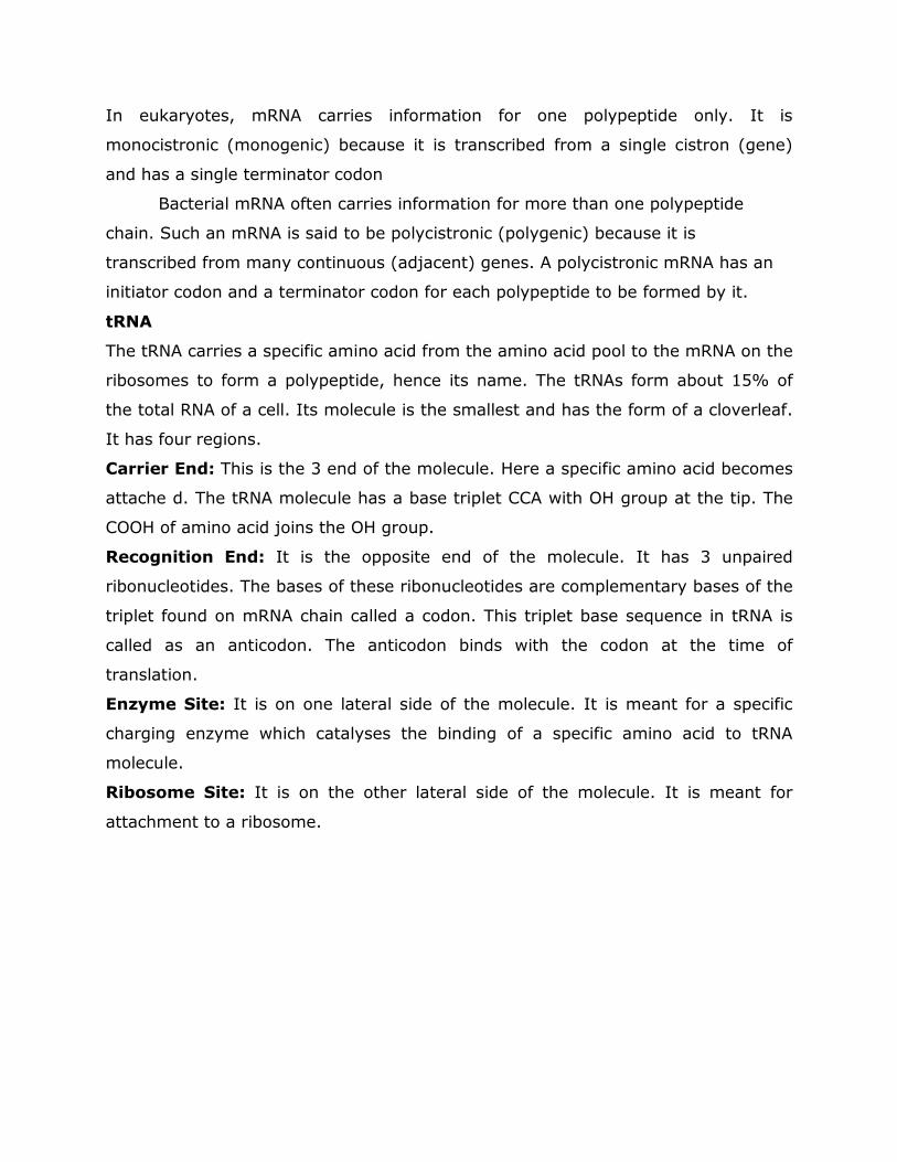
In eukaryotes, mRNA carries information for one polypeptide only. It is
monocistronic (monogenic) because it is transcribed from a single cistron (gene)
and has a single terminator codon
Bacterial mRNA often carries information for more than one polypeptide
chain. Such an mRNA is said to be polycistronic (polygenic) because it is
transcribed from many continuous (adjacent) genes. A polycistronic mRNA has an
initiator codon and a terminator codon for each polypeptide to be formed by it.
tRNA
The tRNA carries a specific amino acid from the amino acid pool to the mRNA on the
ribosomes to form a polypeptide, hence its name. The tRNAs form about 15% of
the total RNA of a cell. Its molecule is the smallest and has the form of a cloverleaf.
It has four regions.
Carrier End: This is the 3 end of the molecule. Here a specific amino acid becomes
attache d. The tRNA molecule has a base triplet CCA with OH group at the tip. The
COOH of amino acid joins the OH group.
Recognition End: It is the opposite end of the molecule. It has 3 unpaired
ribonucleotides. The bases of these ribonucleotides are complementary bases of the
triplet found on mRNA chain called a codon. This triplet base sequence in tRNA is
called as an anticodon. The anticodon binds with the codon at the time of
translation.
Enzyme Site: It is on one lateral side of the molecule. It is meant for a specific
charging enzyme which catalyses the binding of a specific amino acid to tRNA
molecule.
Ribosome Site: It is on the other lateral side of the molecule. It is meant for
attachment to a ribosome.
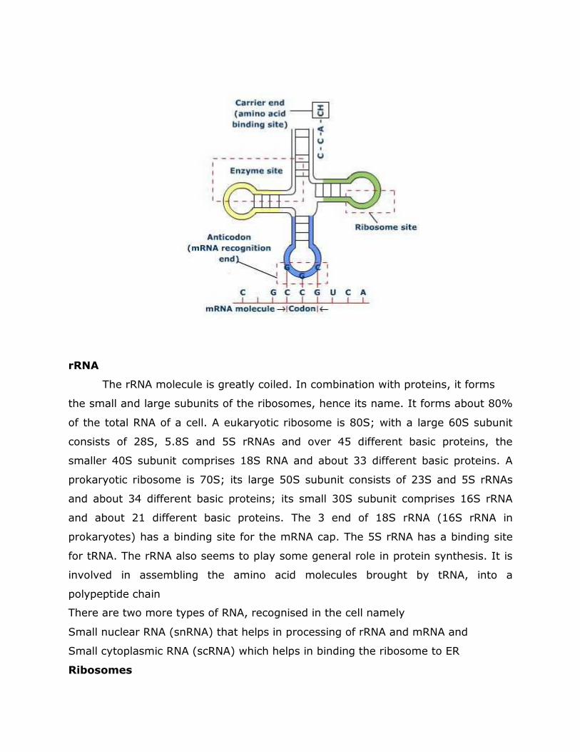
rRNA
The rRNA molecule is greatly coiled. In combination with proteins, it forms
the small and large subunits of the ribosomes, hence its name. It forms about 80%
of the total RNA of a cell. A eukaryotic ribosome is 80S; with a large 60S subunit
consists of 28S, 5.8S and 5S rRNAs and over 45 different basic proteins, the
smaller 40S subunit comprises 18S RNA and about 33 different basic proteins. A
prokaryotic ribosome is 70S; its large 50S subunit consists of 23S and 5S rRNAs
and about 34 different basic proteins; its small 30S subunit comprises 16S rRNA
and about 21 different basic proteins. The 3 end of 18S rRNA (16S rRNA in
prokaryotes) has a binding site for the mRNA cap. The 5S rRNA has a binding site
for tRNA. The rRNA also seems to play some general role in protein synthesis. It is
involved in assembling the amino acid molecules brought by tRNA, into a
polypeptide chain
There are two more types of RNA, recognised in the cell namely
Small nuclear RNA (snRNA) that helps in processing of rRNA and mRNA and
Small cytoplasmic RNA (scRNA) which helps in binding the ribosome to ER
Ribosomes
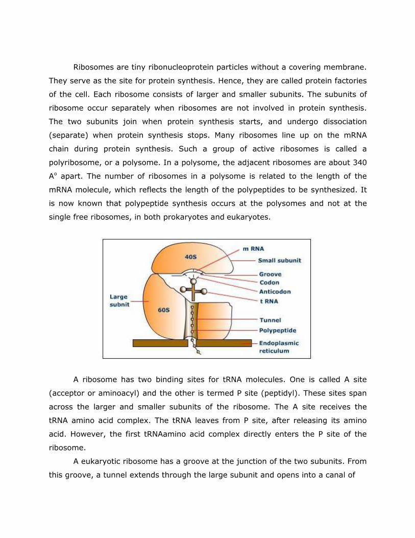
Ribosomes are tiny ribonucleoprotein particles without a covering membrane.
They serve as the site for protein synthesis. Hence, they are called protein factories
of the cell. Each ribosome consists of larger and smaller subunits. The subunits of
ribosome occur separately when ribosomes are not involved in protein synthesis.
The two subunits join when protein synthesis starts, and undergo dissociation
(separate) when protein synthesis stops. Many ribosomes line up on the mRNA
chain during protein synthesis. Such a group of active ribosomes is called a
polyribosome, or a polysome. In a polysome, the adjacent ribosomes are about 340
Ao apart. The number of ribosomes in a polysome is related to the length of the
mRNA molecule, which reflects the length of the polypeptides to be synthesized. It
is now known that polypeptide synthesis occurs at the polysomes and not at the
single free ribosomes, in both prokaryotes and eukaryotes.
A ribosome has two binding sites for tRNA molecules. One is called A site
(acceptor or aminoacyl) and the other is termed P site (peptidyl). These sites span
across the larger and smaller subunits of the ribosome. The A site receives the
tRNA amino acid complex. The tRNA leaves from P site, after releasing its amino
acid. However, the first tRNAamino acid complex directly enters the P site of the
ribosome.
A eukaryotic ribosome has a groove at the junction of the two subunits. From
this groove, a tunnel extends through the large subunit and opens into a canal of
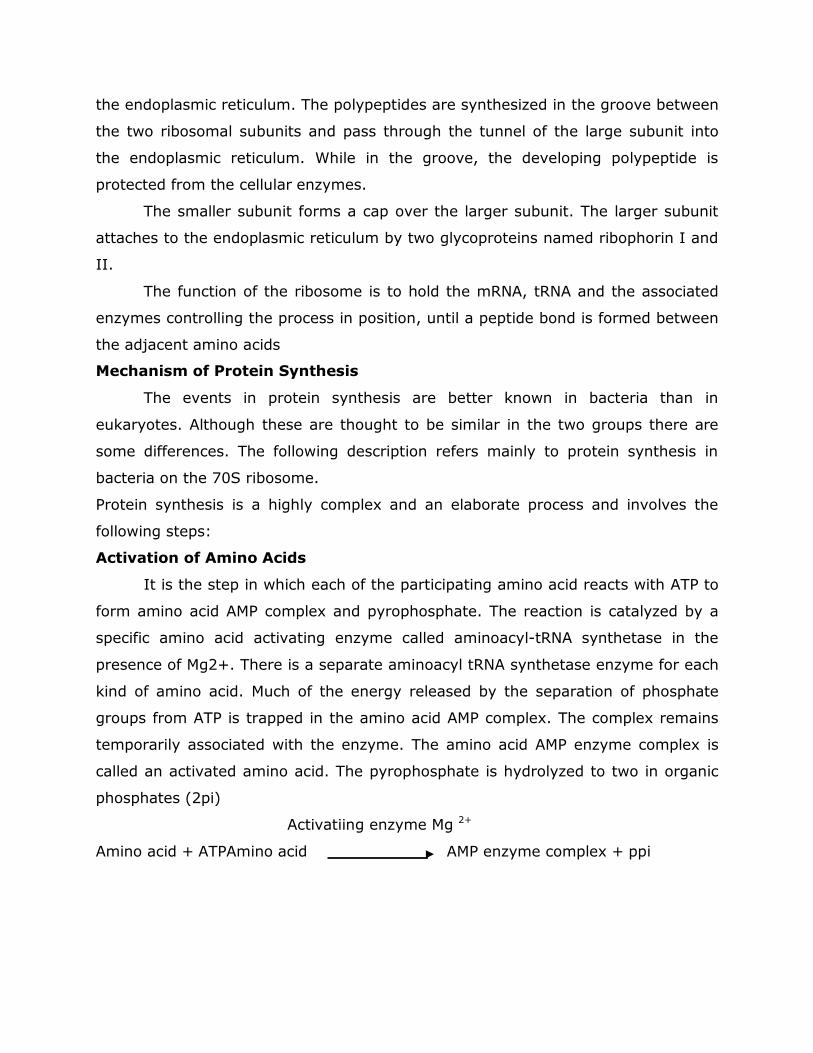
the endoplasmic reticulum. The polypeptides are synthesized in the groove between
the two ribosomal subunits and pass through the tunnel of the large subunit into
the endoplasmic reticulum. While in the groove, the developing polypeptide is
protected from the cellular enzymes.
The smaller subunit forms a cap over the larger subunit. The larger subunit
attaches to the endoplasmic reticulum by two glycoproteins named ribophorin I and
II.
The function of the ribosome is to hold the mRNA, tRNA and the associated
enzymes controlling the process in position, until a peptide bond is formed between
the adjacent amino acids
Mechanism of Protein Synthesis
The events in protein synthesis are better known in bacteria than in
eukaryotes. Although these are thought to be similar in the two groups there are
some differences. The following description refers mainly to protein synthesis in
bacteria on the 70S ribosome.
Protein synthesis is a highly complex and an elaborate process and involves the
following steps:
Activation of Amino Acids
It is the step in which each of the participating amino acid reacts with ATP to
form amino acid AMP complex and pyrophosphate. The reaction is catalyzed by a
specific amino acid activating enzyme called aminoacyl-tRNA synthetase in the
presence of Mg2+. There is a separate aminoacyl tRNA synthetase enzyme for each
kind of amino acid. Much of the energy released by the separation of phosphate
groups from ATP is trapped in the amino acid AMP complex. The complex remains
temporarily associated with the enzyme. The amino acid AMP enzyme complex is
called an activated amino acid. The pyrophosphate is hydrolyzed to two in organic
phosphates (2pi)
Activatiing enzyme Mg 2+
Amino acid + ATPAmino acid AMP enzyme complex + ppi
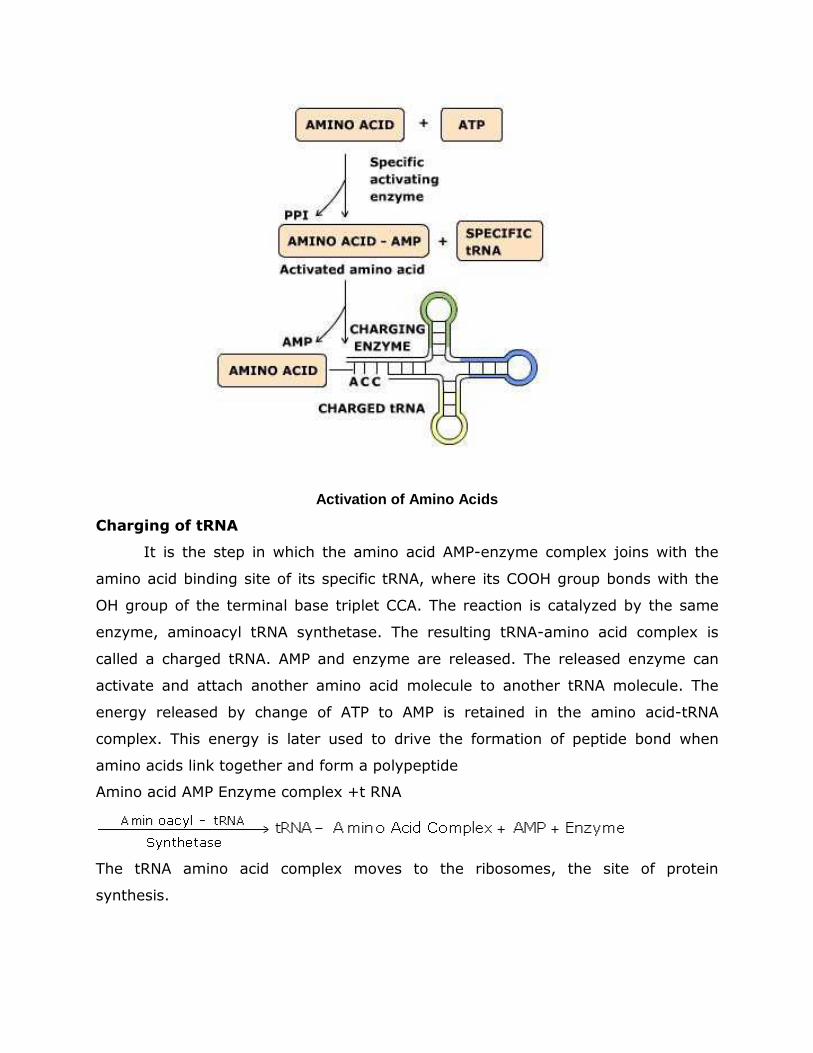
Activation of Amino Acids
Charging of tRNA
It is the step in which the amino acid AMP-enzyme complex joins with the
amino acid binding site of its specific tRNA, where its COOH group bonds with the
OH group of the terminal base triplet CCA. The reaction is catalyzed by the same
enzyme, aminoacyl tRNA synthetase. The resulting tRNA-amino acid complex is
called a charged tRNA. AMP and enzyme are released. The released enzyme can
activate and attach another amino acid molecule to another tRNA molecule. The
energy released by change of ATP to AMP is retained in the amino acid-tRNA
complex. This energy is later used to drive the formation of peptide bond when
amino acids link together and form a polypeptide
Amino acid AMP Enzyme complex +t RNA
The tRNA amino acid complex moves to the ribosomes, the site of protein
synthesis.
Activation of Ribosome
It is the step in which the smaller and the larger subunits of ribosome are
joined together. This is brought about by mRNA chain. The latter joins the smaller
ribosomal subunit with the help of the first codon by a base pairing with an
appropriate sequence on rRNA. The combination of the two is called initiation
complex. The larger subunit later joins the small subunit, forming active ribosome.
Activation of ribosome by mRNA requires proper concentration of Mg ++
Assembly of Amino Acids (Polypeptide Formation)
It is the step in which the amino acids are assembled into a polypeptide
chain. It involves 3 events: initiation, elongation and termination of polypeptide
chain
Initiation of Polypeptide Chain
The mRNA chain has at its 5 end an “initiator” or “start” codon (AUG or GUG)
that signals the beginning of polypeptide formation. This codon lies close to the P
site of the ribosome. The amino acid formylmethionine (methionine in eukaryotes)
initiates the process. It is carried by tRNA having an anticodon UAC which bonds
with the initiator codon AUG of mRNA. Initiation factors (IF1, IF2 and IF3) and GTP
promote the initiation process.
The large ribosomal subunit now joins the small subunit to complete the
ribosome. At this stage, GTP is hydrolysed to GDP. The ribosome has
formylmethionine bearing tRNA at the P site. Later, the formylmethionine is
changed to normal methionine by the enzyme deformylase in prokaryotes. If not
required, methionine is later separated from the polypeptide chain by a proteolytic
enzyme aminopeptidase.
Elongation of Polypeptide Chain
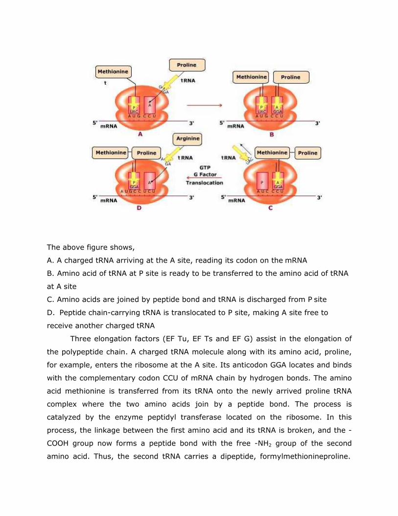
The above figure shows,
A. A charged tRNA arriving at the A site, reading its codon on the mRNA
B. Amino acid of tRNA at P site is ready to be transferred to the amino acid of tRNA
at A site
C. Amino acids are joined by peptide bond and tRNA is discharged from P site
D. Peptide chain-carrying tRNA is translocated to P site, making A site free to
receive another charged tRNA
Three elongation factors (EF Tu, EF Ts and EF G) assist in the elongation of
the polypeptide chain. A charged tRNA molecule along with its amino acid, proline,
for example, enters the ribosome at the A site. Its anticodon GGA locates and binds
with the complementary codon CCU of mRNA chain by hydrogen bonds. The amino
acid methionine is transferred from its tRNA onto the newly arrived proline tRNA
complex where the two amino acids join by a peptide bond. The process is
catalyzed by the enzyme peptidyl transferase located on the ribosome. In this
process, the linkage between the first amino acid and its tRNA is broken, and the -
COOH group now forms a peptide bond with the free -NH2 group of the second
amino acid. Thus, the second tRNA carries a dipeptide, formylmethionineproline.
The energy required for the formation of a peptide bond comes from the free
energy released by separation of amino acid (formylmethionine or methionine) from
its tRNA.
The first tRNA, now uncharged, separates from mRNA chain at the P site of
the ribosome and returns to the mixed pool of tRNAs in the cytoplasm. Here, it is
now available to transport another molecule of its specific amino acid.
Now the ribosome moves one codon along the mRNA in the 3 direction. With
this, tRNAdipeptide complex at the A site is pulled to the P site. This process is
called translocation. It requires GTP and a translocase protein called EF-G factor.
The GTP is hydrolysed to GDP and inorganic phosphate to release energy for the
process
At this stage, a third tRNA molecule with its own specific amino acid,
arginine, for example arrives at the A site of the ribosome and binds with the help
of anticodon AGA to the complementary codon UCU of the mRNA chain. The
dipeptide formylmethionineproline is shifted from the preceding tRNA on the third
tRNA where it joins the amino acid arginine again with the help of peptidyl
transferase enzyme. The dipeptide, thus, becomes a tripeptide, formyl-methionine-
proline-arginine. The second tRNA being now uncharged, leaves the mRNA chain,
vacating the P site. The tRNAtripeptide complex is translocated from A site to P site
The entire process involving arrival of tRNA-amino acid complex, peptide bond
formation and translocation is repeated. As the ribosome moves over the mRNA, all
the codons of mRNA arrive at the A site one after another, and the peptide chain
grows. Thus, the amino acids are linked up into a polypeptide in a sequence
communicated by the DNA through the mRNA. A polypeptide chain which is in the
process of synthesis is often called a nascent polypeptide
The growing polypeptide chain always remains attached to its original
ribosome, and is not transferred from one ribosome to another. Only one
polypeptide chain can be synthesized at a time on a given ribosome.
Termination and Release of Polypeptide Chain:
At the terminal end of mRNA chain there is a stop, or terminator codon (UAA,
UAG or UGA). It is not joined by the anticodon of any tRNA amino acid complex.
Hence, there can be no further addition of amino acids to the polypeptide chain.
The linkage between the last tRNA and the polypeptide chain is broken by three
release factors. (RF 1, RF 2 and RF 3) and GTP. The release is catalyzed by the
peptidyl transferase enzyme, the same enzyme that forms the peptide bonds. The
ribosome jumps off the mRNA chain at the stop codon and dissociates into its two
subunits. The completed polypeptide (amino acid chain) becomes free in the
cytoplasm.
The ribosomes and the tRNAs on release from the mRNA can function again
in the same manner and result in the formation of another polypeptide of the same
protein.
Modification of Released Polypeptide
The just released polypeptide is a straight, linear exhibiting a primary
molecule, structure. It may lose some amino acids from the end with the help of a
peptidase enzyme, and then coil and fold on itself to acquire secondary and tertiary
structure. It may even combine with other polypeptides, to have quaternary
structure.
The proteins synthesized on free polysomes are released into the cytoplasm
and function as structural and enzymatic proteins. The proteins formed on the
polysomes attached to ER pass into the ER channels and are exported as cell
secretions by exocytosis after packaging in the Golgi apparatus.
Polysome Formation
When the ribosome has moved sufficiently down the mRNA chain towards 3
end, another ribosome takes up position at the initiator codon of mRNA, and starts
synthesis of a second molecule of the same polypeptide chain. At any given time,
the mRNA chain will, therefore, carry many ribosomes over which are similar
polypeptide chains of varying length, shortest near the initiator codon and longest
near the terminator codon. A row of ribosomes joined to the mRNA molecule, is
called a polyribosome, or a polysome. Synthesis of many molecules of the same
polypeptide simultaneously from one mRNA molecule by a polysome is called
translational amplification.
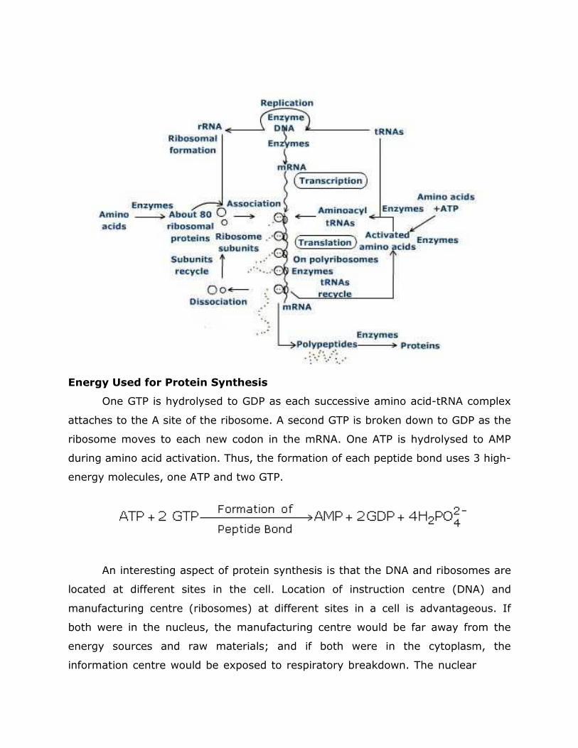
Energy Used for Protein Synthesis
One GTP is hydrolysed to GDP as each successive amino acid-tRNA complex
attaches to the A site of the ribosome. A second GTP is broken down to GDP as the
ribosome moves to each new codon in the mRNA. One ATP is hydrolysed to AMP
during amino acid activation. Thus, the formation of each peptide bond uses 3 high-
energy molecules, one ATP and two GTP.
An interesting aspect of protein synthesis is that the DNA and ribosomes are
located at different sites in the cell. Location of instruction centre (DNA) and
manufacturing centre (ribosomes) at different sites in a cell is advantageous. If
both were in the nucleus, the manufacturing centre would be far away from the
energy sources and raw materials; and if both were in the cytoplasm, the
information centre would be exposed to respiratory breakdown. The nuclear
envelope preserves stability of the DNA by protecting it from respiratory
destruction. The message in the DNA in the form of genes (codes) are, permanent,
authentic master documents from which working copies are prepared in the form of
mRNAs, as and when required by the cell.
The complex process by which the information in RNA is decoded into a
polypeptide is one of the exciting discoveries the genetic code
With only four biochemical letters (A, G, C, U) a one letter code could not
unambiguously encode 20 amino acids. A two letter code could encode only 16
amino acids. So, a triplet code based on 3 biochemical letters or nucleotide bases
could make 4 x 4 x 4 = 64 codons. This will be required to code for 20 or so
different amino acids.
The discovery of the genetic code became possible through the contribution
of many scientists like Francis Crick, Seveno Ochoa, Maxell Ninenberg, Hargobind
Khorana and J. H. Malther in the 1960s. Ninemberg and Khorana shared the Nobel
Prize in 1968.
Characteristics of Genetic Code
The genetic code is a triplet code: Three adjacent bases, termed as codon,
specify one amino acid
Non-overlapping: Adjacent codons do not overlap
No punctuation: The genetic code is comma less
The genetic code is universal i.e., a given codon specifies the same amino acid in all
protein synthesising organisms
The genetic code is degenerate: it lacks specificity and one amino acid often has
more than one code triplet
Each codon codes for only one amino acid, none for more than one
Three of the 64 codons, names UAA, UAG and UGA do not specify any amino acid
but signal the end of the message. They are called nonsense or terminator codons
The codons AUG and GUG are called the initiation or start codons as they begin the
synthesis of polypeptide.
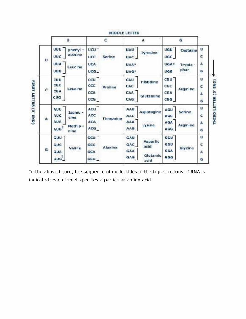
In the above figure, the sequence of nucleotides in the triplet codons of RNA is
indicated; each triplet specifies a particular amino acid.


