Embed Size (px)
Citation preview

UNIVERSIDAD NACIONAL UNIVERSIDAD NACIONAL PEDRO RUIZ GALLOPEDRO RUIZ GALLO
FACULTAD DE ENFERMERIAFACULTAD DE ENFERMERIASEGUNDA ESPECIALIDAD “SEGUNDA ESPECIALIDAD “CUIDADOS DE CUIDADOS DE
ENFERMERIA A PERSONAS CON ENFERMERIA A PERSONAS CON AFECCIONES NEFROLOGICASAFECCIONES NEFROLOGICAS “ “
LAMBAYEQUELAMBAYEQUECURSO : INGLES TECNICOCURSO : INGLES TECNICO
II UNIDAD II UNIDAD The Human BodyThe Human Body
““ANATOMY RENAL SYSTEM”.ANATOMY RENAL SYSTEM”.DOCENTE: Magister Lupe Rivera GonzalesDOCENTE: Magister Lupe Rivera Gonzales

THE HUMAN BODYTHE HUMAN BODY
To study the man like the whole alive To study the man like the whole alive organisms it is considered two aspects: organisms it is considered two aspects: first their form external and internal form first their form external and internal form their their MorphologyMorphology and second like work and second like work the different tissues organs and systems the different tissues organs and systems that form the man denominated to this that form the man denominated to this aspect the aspect the Physiology Physiology
We rememberd the following We rememberd the following concepts :concepts :

Human anatomyHuman anatomy: is the science that : is the science that studied the structure of the organs and studied the structure of the organs and systems that integrate the human systems that integrate the human organism.organism.
Morphology:Morphology: called Macroscopic called Macroscopic Anatomy, studied to the body dividing it in Anatomy, studied to the body dividing it in regions, sections or partsregions, sections or parts..
PhysiologyPhysiology: studied the functions that : studied the functions that carry out the cells, organs and systems. carry out the cells, organs and systems.

THE PARTS OF THE HUMAN BODYTHE PARTS OF THE HUMAN BODY

PRACTICAMOS LO
APRENDIDO !!

THE HUMAN BODY SYSTEMTHE HUMAN BODY SYSTEM

ANATOMY RENAL SYSTEMANATOMY RENAL SYSTEMRenalArtery
Aorta Artery
Kidney
Urinary Bladder Urethra
Urether
Veins digsinferior
RenalVeins

ANATOMY RENAL SYSTEMANATOMY RENAL SYSTEM
The kidneys are essentially regulatory organs The kidneys are essentially regulatory organs which maintain the volume and composition of which maintain the volume and composition of body fluid by filtration of the blood and selective body fluid by filtration of the blood and selective reabsorption or secretion of filtered solutes.reabsorption or secretion of filtered solutes.
the kidneys are retroperitoneal organs (ie the kidneys are retroperitoneal organs (ie located behind the peritoneum) situated on the located behind the peritoneum) situated on the posterior wall of the abdomen on each side of posterior wall of the abdomen on each side of the vertebral column, at about the level of the the vertebral column, at about the level of the twelfth rib. The left kidney is lightly higher in the twelfth rib. The left kidney is lightly higher in the abdomen than the right, due to the presence of abdomen than the right, due to the presence of the liver pushing the right kidney down.the liver pushing the right kidney down.

ANATOMY RENAL SYSTEMANATOMY RENAL SYSTEM The kidneys take their blood supply directly from The kidneys take their blood supply directly from
the aorta via the renal arteries; blood is returned to the aorta via the renal arteries; blood is returned to the inferior vena cava via the renal veins. Urine the inferior vena cava via the renal veins. Urine (the filtered product containing waste materials (the filtered product containing waste materials and water) excreted from the kidneys passes and water) excreted from the kidneys passes down the fibromusculardown the fibromuscular ureters ureters and collects in the and collects in the bladderbladder. The bladder muscle (the. The bladder muscle (the detrusor detrusor musclemuscle) is capable of distending to accept urine ) is capable of distending to accept urine without increasing the pressure inside; this means without increasing the pressure inside; this means that large volumes can be collected (700-1000ml) that large volumes can be collected (700-1000ml) without high-pressure damage to the renal system without high-pressure damage to the renal system occuring.occuring.When urine is passed, the When urine is passed, the urethral sphincterurethral sphincter at at the base of the bladder relaxes, the detrusor the base of the bladder relaxes, the detrusor contracts, and urine is voided via the contracts, and urine is voided via the urethraurethra. .

How do the kidneys and urinary How do the kidneys and urinary system work?system work?
The body takes nutrients from food and converts them The body takes nutrients from food and converts them to energy. After the body has taken the food that it to energy. After the body has taken the food that it needs, waste products are left behind in the bowel and needs, waste products are left behind in the bowel and in the blood.in the blood.
The kidney and urinary systems keep chemicals, such The kidney and urinary systems keep chemicals, such as potassium and sodium, and water in balance and as potassium and sodium, and water in balance and remove a type of waste, called urea, from the blood. remove a type of waste, called urea, from the blood. Urea is produced when foods containing protein, such Urea is produced when foods containing protein, such as meat, poultry, and certain vegetables, are broken as meat, poultry, and certain vegetables, are broken down in the body. Urea is carried in the bloodstream to down in the body. Urea is carried in the bloodstream to the kidneys.the kidneys.
Other important functions of the kidneys include blood Other important functions of the kidneys include blood pressure regulation and the production of pressure regulation and the production of erythropoietin, which controls red blood cell production erythropoietin, which controls red blood cell production in the bone marrow.in the bone marrow.

Structure of the kidney Structure of the kidney two kidneys - a pair of purplish-brown organs located two kidneys - a pair of purplish-brown organs located
below the ribs toward the middle of the back. Their below the ribs toward the middle of the back. Their function is to remove liquid waste from the blood in the function is to remove liquid waste from the blood in the form of urine; keep a stable balance of salts and other form of urine; keep a stable balance of salts and other substances in the blood; and produce erythropoietin, a substances in the blood; and produce erythropoietin, a hormone that aids the formation of red blood cells.hormone that aids the formation of red blood cells.
The kidneys remove urea from the blood through tiny The kidneys remove urea from the blood through tiny filtering units called nephrons. Each nephron consists filtering units called nephrons. Each nephron consists of a ball formed of small blood capillaries, called a of a ball formed of small blood capillaries, called a glomerulus, and a small tube called a renal tubule. glomerulus, and a small tube called a renal tubule. Urea, together with water and other waste substances, Urea, together with water and other waste substances, forms the urine as it passes through the nephrons and forms the urine as it passes through the nephrons and down the renal tubules of the kidney.down the renal tubules of the kidney.

URETERSURETERS
• two ureters - narrow tubes that carry urine two ureters - narrow tubes that carry urine from the kidneys to the bladder. Muscles from the kidneys to the bladder. Muscles in the ureter walls continually tighten and in the ureter walls continually tighten and relax forcing urine downward, away from relax forcing urine downward, away from the kidneys. If urine backs up, or is the kidneys. If urine backs up, or is allowed to stand still, a kidney infection allowed to stand still, a kidney infection can develop. About every 10 to 15 can develop. About every 10 to 15 seconds, small amounts of urine are seconds, small amounts of urine are emptied into the bladder from the ureters. emptied into the bladder from the ureters.

BLADDERBLADDER
• bladder - a triangle-shaped, hollow organ bladder - a triangle-shaped, hollow organ located in the lower abdomen. It is held in located in the lower abdomen. It is held in place by ligaments that are attached to place by ligaments that are attached to other organs and the pelvic bones. The other organs and the pelvic bones. The bladder's walls relax and expand to store bladder's walls relax and expand to store urine, and contract and flatten to empty urine, and contract and flatten to empty urine through the urethra. The typical urine through the urethra. The typical healthy adult bladder can store up to two healthy adult bladder can store up to two cups (tz.) of urine for two to five hours. cups (tz.) of urine for two to five hours.

two sphincter muscles - circular muscles that two sphincter muscles - circular muscles that help keep urine from leaking by closing tightly help keep urine from leaking by closing tightly like a rubber band around the opening of the like a rubber band around the opening of the bladder. bladder.
nerves in the bladder - alert a person when it is nerves in the bladder - alert a person when it is time to urinate, or empty the bladder. time to urinate, or empty the bladder.
urethra - the tube that allows urine to pass urethra - the tube that allows urine to pass outside the body. The brain signals the bladder outside the body. The brain signals the bladder muscles to tighten, which squeezes urine out of muscles to tighten, which squeezes urine out of the bladder. At the same time, the brainthe bladder. At the same time, the brain signals signals the sphincter muscles to relax to let urine exit the sphincter muscles to relax to let urine exit the bladder through the urethra. When all the the bladder through the urethra. When all the signals occur in the correct order, normal signals occur in the correct order, normal urination occurs. urination occurs.

Internal Anatomy of the kidney Internal Anatomy of the kidney

NEPHRONNEPHRON THE NEPRONSTHE NEPRONS
The functional unit of the kidney is the nephrons. Their The functional unit of the kidney is the nephrons. Their basic functions are: basic functions are:
1. Filtration: some substances are transferred from the 1. Filtration: some substances are transferred from the blood until the nephrons. blood until the nephrons.
2. Secretion: when the liquid filtrate moves through the 2. Secretion: when the liquid filtrate moves through the nephrons, it wins additional materials (waste and nephrons, it wins additional materials (waste and substances in excess). substances in excess).
3. R3. Reabsorptioneabsorption: some useful substances are returned : some useful substances are returned to the blood for their reutilisation. to the blood for their reutilisation.
As consequence of these activities is formed the urine. As consequence of these activities is formed the urine.

Anatomy of the NephronAnatomy of the Nephron Anatomy of the Nephron Anatomy of the Nephron
The nephron is composed of two parts: The nephron is composed of two parts:
1. The renal corpuscle or corpuscle of Malpighi, where 1. The renal corpuscle or corpuscle of Malpighi, where they filter the fluids. they filter the fluids.
2. The renal tubule where it passes the filtered water. 2. The renal tubule where it passes the filtered water.
The renal corpuscle has, in turn two components: The renal corpuscle has, in turn two components:
" The glomerulus's, I wind in a ball of tiny capillary " The glomerulus's, I wind in a ball of tiny capillary surrounded of a double epithelium. As in definitive surrounded of a double epithelium. As in definitive they are glasses, the glomerulus's is also part of the they are glasses, the glomerulus's is also part of the cardiovascular system. cardiovascular system.
" The capsule glomerular or capsule of Bowman that it " The capsule glomerular or capsule of Bowman that it surrounds the glomerulus's. surrounds the glomerulus's.

The blood enters in the renal corpuscle through the The blood enters in the renal corpuscle through the arteriole afferent and it leaves for the arteriole efferent. arteriole afferent and it leaves for the arteriole efferent. The filtration of the blood is verified in the capsule of The filtration of the blood is verified in the capsule of Bowman, leaving the produced urine, like it will be Bowman, leaving the produced urine, like it will be seen subsequently by a conduit or special tubule. seen subsequently by a conduit or special tubule.
The external wall or parietal layer of the capsule of The external wall or parietal layer of the capsule of Bowman is separated from the interior wall or visceral Bowman is separated from the interior wall or visceral layer for the call capsular space or space of Bowman. layer for the call capsular space or space of Bowman. As the blood flows through the capillary of the As the blood flows through the capillary of the glomerulus's, the water and some solutes they filter glomerulus's, the water and some solutes they filter passing to the space of Bowman passing to the space of Bowman

Structure of the kidneyStructure of the kidney On sectioning, the kidney has a pale outer region- the On sectioning, the kidney has a pale outer region- the
cortexcortex- and a darker inner region- the- and a darker inner region- the medulla medulla.The .The medulla is divided into 8-18 conical regions, called the medulla is divided into 8-18 conical regions, called the renal pyramidsrenal pyramids; the base of each pyramid starts at ; the base of each pyramid starts at the corticomedullary border, and the apex ends in the the corticomedullary border, and the apex ends in the renal papillarenal papilla which merges to form the which merges to form the renal pelvisrenal pelvis and then on to form the ureter. In humans, the renal and then on to form the ureter. In humans, the renal pelvis is divided into two or three spaces -the pelvis is divided into two or three spaces -the major major calycescalyces- which in turn divide into further - which in turn divide into further minor minor calycescalyces. The walls of the calyces, pelvis and ureters . The walls of the calyces, pelvis and ureters are lined with smooth muscle that can contract to force are lined with smooth muscle that can contract to force urine towards the bladder by peristalisis.urine towards the bladder by peristalisis.
The cortex and the medulla are made up of The cortex and the medulla are made up of nephronsnephrons; these are the functional units of the kidney, ; these are the functional units of the kidney, and each kidney contains about 1.3 million of themand each kidney contains about 1.3 million of them..

Structure of the Nephron Structure of the Nephron
The Nephron is the unit of the kidney responsible for ultrafiltration of the blood and reabsorption or excretion of products in the subsequent filtrate. Each nephron is made up of:

Structure of the NephronStructure of the Nephron A filtering unit- the A filtering unit- the glomerulusglomerulus. 125ml/min of filtrate is formed . 125ml/min of filtrate is formed
by the kidneys as blood is filtered through this sieve-like by the kidneys as blood is filtered through this sieve-like structure. structure. This filtration is uncontrolled. This filtration is uncontrolled.
The The proximal convoluted tubuleproximal convoluted tubule. Controlled absorption of . Controlled absorption of glucose, sodium, and other solutes goes on in this region. glucose, sodium, and other solutes goes on in this region.
The The loop of loop of HenleHenle. This region is responsible for concentration . This region is responsible for concentration and dilution of urine by utilising a and dilution of urine by utilising a counter-current multiplyingcounter-current multiplying mechanism- basically, it is water-impermeable but can pump mechanism- basically, it is water-impermeable but can pump sodium out, which in turn affects the osmolarity of the sodium out, which in turn affects the osmolarity of the surrounding tissues and will affect the subsequent movement of surrounding tissues and will affect the subsequent movement of water in or out of the water-permeable collecting duct. water in or out of the water-permeable collecting duct.
The The distal convoluted tubuledistal convoluted tubule. This region is responsible, . This region is responsible, along with the along with the collecting ductcollecting duct that it joins, for absorbing water that it joins, for absorbing water back into the body- simple maths will tell you that the kidney back into the body- simple maths will tell you that the kidney doesn't produce 125ml of urine every minute. 99% of the water doesn't produce 125ml of urine every minute. 99% of the water is normally reabsorbed, leaving highly concentrated urine to is normally reabsorbed, leaving highly concentrated urine to flow into the collecting duct and then into the renal pelvis.flow into the collecting duct and then into the renal pelvis.
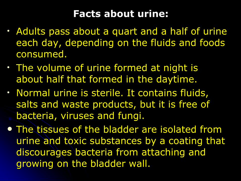
Facts about urine:Facts about urine:
• Adults pass about a quart and a half of urine Adults pass about a quart and a half of urine each day, depending on the fluids and foods each day, depending on the fluids and foods consumed. consumed.
• The volume of urine formed at night is The volume of urine formed at night is about half that formed in the daytime. about half that formed in the daytime.
• Normal urine is sterile. It contains fluids, Normal urine is sterile. It contains fluids, salts and waste products, but it is free of salts and waste products, but it is free of bacteria, viruses and fungi. bacteria, viruses and fungi.
The tissues of the bladder are isolated from The tissues of the bladder are isolated from urine and toxic substances by a coating that urine and toxic substances by a coating that discourages bacteria from attaching and discourages bacteria from attaching and growing on the bladder wall.growing on the bladder wall.

THANKS FOR YOUR ATTENTIONTHANKS FOR YOUR ATTENTION