Embed Size (px)
Citation preview
Thermo Fisher Scientific • 180 Oyster Point Blvd• South San Francisco, CA 94080 • www.lifetechnologies.com
ABSTRACT In recent years advanced sample preparation tools, molecular assays and high-throughput next generation sequencing (NGS) technology have changed the study of molecular events associated with pathological processes. However, molecular analysis is limited by sample processing, sample quality and quantity, and the number of tumor cells present within the sample. Tumor cell heterogeneity further confounds the molecular analysis of a sample. Laser capture micro-dissection (LCM) technology provides a simple and efficient approach for rapid and accurate selection and dissection of homogeneous populations of cells under direct microscopic visualization and pairs perfectly with NGS technologies such as AmpliSeqTM which enables transcriptome analysis from small amounts of starting material. In this study we have demonstrated an integrated and efficient workflow solution by combining the advantages of LCM sample preparation technology and Ion AmpliseqTM RNA library kit workflows. Our approach offers researchers a complete LCM sample to targeted RNA-Seq solution. By leveraging the highly reproducible Ion AmpliSeq TM technology to selectively amplify specific RNA targets by using either AmpliseqTM RNA Ready-to-use or custom panels, on Ion Torrent semiconductor sequencing platforms offer many advantages over other workflows such as faster turnaround time and data analysis, and sample multiplexing. This combined workflow has been used to characterize expression of cancer biomarkers in isolated normal and tumor cell populations in fresh frozen samples.
Acknowledgements Author would like to thank Junko Stevens, and Mark E Shannon, Kristin Schmidt, Joseph Pham and Vidya Venkatesh for their support and guidance.
© 2014 Thermo Fisher Scientific Inc. All rights reserved. All trademarks are the property of Thermo Fisher Scientific and its subsidiaries unless otherwise specified. TaqMan is a registered trademark of Roche Molecular Systems, Inc., used under permission and license.
Use of Laser Capture Micro dissection In Conjunction With Ampliseq-RNA Workflow To Study Tumor and Normal Cell Populations
22nd Molecular Med TRI-CON 2015
Mousumi Rath, Mark E Shannon, Junko Stevens, and Vidya Venkatesh Life Science Solutions R&D, Life Technologies, 180 Oyster Point Blvd, South San Francisco, CA, 94080, USA
MATERIALS AND METHODS Arcturus® HistoGene staining kit (Cat # KIT0401) was used to stain and prepare the 7um fresh frozen breast cancer tissue section mounted on PEN membrane glass slide. In addition, tissue scrape protocol was performed to evaluate the fresh frozen block RNA quality prior to LCM . The cells were captured using Cap Sure® HS LCM Caps, (Cat # LCM0214) that enable the highly sensitive extraction of biological molecules from small numbers of cells. Picopure® RNA isolation kit (Cat # KIT0204) was used to extract RNA from LCM captured samples. RNA quality was checked on Agilent Bionalayzer 6000 Pico kit . RNA samples were processed using the Ion AmpliSeqTM RNA Library Kit and the Ion AmpliSeqTM cancer Hotspot Panel . Libraries were quantified using the Agilent® Bioanalyzer® instrument on a High Sensitivity DNA chip and qPCR method using the Ion Library Quantitation kit. Templated libraries were sequenced Ion 318TM chip on the Ion PGMTM using the Ion PGMTM Sequencing Kit 200 v2.
WORKFLOW OVERVIEW
RESULTS
Figure 3: RNA Isolation Workflow and Sample RNA Bioanalyzer Traces
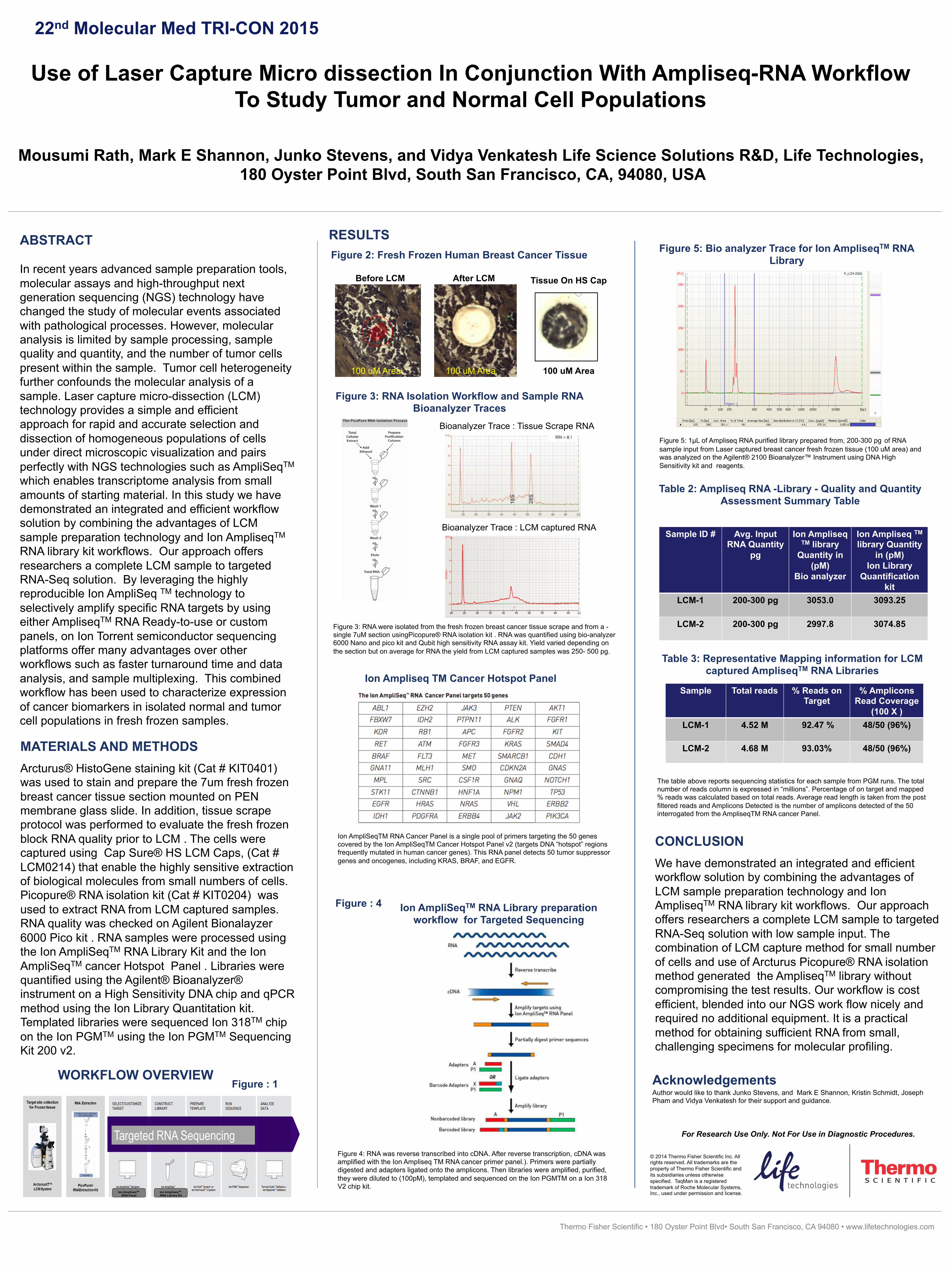
Figure 2: Fresh Frozen Human Breast Cancer Tissue
Bioanalyzer Trace : Tissue Scrape RNA
Figure 3: RNA were isolated from the fresh frozen breast cancer tissue scrape and from a -single 7uM section usingPicopure® RNA isolation kit . RNA was quantified using bio-analyzer 6000 Nano and pico kit and Qubit high sensitivity RNA assay kit. Yield varied depending on the section but on average for RNA the yield from LCM captured samples was 250- 500 pg.
Bioanalyzer Trace : LCM captured RNA
Figure 4: RNA was reverse transcribed into cDNA. After reverse transcription, cDNA was amplified with the Ion Ampliseq TM RNA cancer primer panel.). Primers were partially digested and adapters ligated onto the amplicons. Then libraries were amplified, purified, they were diluted to (100pM), templated and sequenced on the Ion PGMTM on a Ion 318 V2 chip kit.
Ion AmpliSeqTM RNA Library preparation workflow for Targeted Sequencing
Figure : 4
Figure : 1
Ion Ampliseq TM Cancer Hotspot Panel
Ion AmpliSeqTM RNA Cancer Panel is a single pool of primers targeting the 50 genes covered by the Ion AmpliSeqTM Cancer Hotspot Panel v2 (targets DNA “hotspot” regions frequently mutated in human cancer genes). This RNA panel detects 50 tumor suppressor genes and oncogenes, including KRAS, BRAF, and EGFR.
Figure 5: 1µL of Ampliseq RNA purified library prepared from, 200-300 pg of RNA sample input from Laser captured breast cancer fresh frozen tissue (100 uM area) and was analyzed on the Agilent® 2100 Bioanalyzer™ Instrument using DNA High Sensitivity kit and reagents.
Sample ID # Avg. Input RNA Quantity
pg
Ion Ampliseq TM library
Quantity in (pM)
Bio analyzer
Ion Ampliseq TM library Quantity
in (pM) Ion Library
Quantification kit
LCM-1
200-300 pg 3053.0 3093.25
LCM-2 200-300 pg 2997.8 3074.85
Table 2: Ampliseq RNA -Library - Quality and Quantity Assessment Summary Table
Figure 5: Bio analyzer Trace for Ion AmpliseqTM RNA Library
Before LCM After LCM Tissue On HS Cap
100 uM Area 100 uM Area 100 uM Area
Table 3: Representative Mapping information for LCM captured AmpliseqTM RNA Libraries
Sample Total reads % Reads on Target
% Amplicons Read Coverage
(100 X ) LCM-1 4.52 M 92.47 % 48/50 (96%)
LCM-2 4.68 M 93.03% 48/50 (96%)
The table above reports sequencing statistics for each sample from PGM runs. The total number of reads column is expressed in “millions”. Percentage of on target and mapped % reads was calculated based on total reads. Average read length is taken from the post filtered reads and Amplicons Detected is the number of amplicons detected of the 50 interrogated from the AmpliseqTM RNA cancer Panel.
CONCLUSION We have demonstrated an integrated and efficient workflow solution by combining the advantages of LCM sample preparation technology and Ion AmpliseqTM RNA library kit workflows. Our approach offers researchers a complete LCM sample to targeted RNA-Seq solution with low sample input. The combination of LCM capture method for small number of cells and use of Arcturus Picopure® RNA isolation method generated the AmpliseqTM library without compromising the test results. Our workflow is cost efficient, blended into our NGS work flow nicely and required no additional equipment. It is a practical method for obtaining sufficient RNA from small, challenging specimens for molecular profiling.
For Research Use Only. Not For Use in Diagnostic Procedures.
Ion AmpliseqTM RNA Panel
Ion AmpliseqTM RNA Library Kit


![Dissection-BKW · 2018. 6. 1. · Dissection. Wereplaceournaive c -sumalgorithmbymoreadvancedtime-memorytechniqueslike Schroeppel-Shamir[34]anditsgeneralization,Dissection[11],toreducetheclassicrunningtime.Wecall](https://img.dokumen.tips/doc/110x75/5ffc5cc4c887922f656f708b/dissection-bkw-2018-6-1-dissection-wereplaceournaive-c-sumalgorithmbymoreadvancedtime-memorytechniqueslike.jpg)