Embed Size (px)
Citation preview

R E S EARCH ART I C L E
DEAFNESS
Tmc gene therapy restores auditory function in deaf miceCharles Askew,1,2 Cylia Rochat,3 Bifeng Pan,1 Yukako Asai,1 Hena Ahmed,1 Erin Child,1
Bernard L. Schneider,3 Patrick Aebischer,3 Jeffrey R. Holt1*
Genetic hearing loss accounts for up to 50% of prelingual deafness worldwide, yet there are no biologic treatmentscurrently available. To investigate gene therapy as a potential biologic strategy for restoration of auditory function inpatients with genetic hearing loss, we tested a gene augmentation approach inmousemodels of genetic deafness. Wefocused on DFNB7/11 and DFNA36, which are autosomal recessive and dominant deafnesses, respectively, caused bymutations in transmembrane channel–like 1 (TMC1).Mice that carry targeteddeletion of Tmc1or adominant Tmc1pointmutation, known as Beethoven, are good models for human DFNB7/11 and DFNA36. We screened several adeno-associated viral (AAV) serotypes and promoters and identified AAV2/1 and the chicken b-actin (Cba) promoter as anefficient combination for driving the expression of exogenous Tmc1 in inner hair cells in vivo. Exogenous Tmc1 or itsclosely related ortholog, Tmc2, were capable of restoring sensory transduction, auditory brainstem responses, andacoustic startle reflexes in otherwise deafmice, suggesting that gene augmentationwith Tmc1 or Tmc2 is well suited forfurther development as a strategy for restoration of auditory function in deaf patients who carry TMC1mutations.
on J
uly
9, 2
015
Dow
nloa
ded
from
INTRODUCTION
Hearing loss is the most common sensory deficit in the world, withboth genetic and environmental factors causing dysfunction of the pri-mary sensory cells of the inner ear, known as hair cells (1). Hair cellsconvert mechanical stimuli into electrical signals and are essential fornormal auditory and balance functions. Unfortunately, hair cells lackthe ability to regenerate; thus, hair cell damage or death is cumulative,causing progressive hearing loss. The current standards of care forhearing loss are hearing aids or cochlear implants, which provide in-complete restoration of function in a limited patient population. Phar-macologic, stem cell, and gene therapies are being explored as alternativetherapies (1). Of these possible strategies, gene therapy may be bestsuited for restoration of hair cell function in genetic hearing loss(1–4). However, few studies have provided proof-of-principle evidencesupporting gene therapy as a viable strategy for restoration of auditoryfunction in mouse models of genetic hearing loss. One notable excep-tion is the restoration of auditory function in mice lacking vesicularglutamate transporter 3 (VGLUT3), a glutamate transport protein ex-pressed in auditory inner hair cells (IHCs), required for synaptic trans-mission from IHC to postsynaptic neurons of the 8th cranial nerve (5).The authors of that study used adeno-associated viral (AAV) vectorsto deliver the coding sequence for VGLUT3 into IHCs of early postnatalVglut3 knockout mice. Although an important advancement, VGLUT3mutations are not common in humans and, when present, are dom-inant, suggesting that the clinical utility of VGLUT3 augmentationmay be limited.
To explore gene therapy for a common form of genetic hearingloss that affects hair cells, we used mice that carry mutations in trans-membrane channel–like gene 1 (Tmc1). Mutations in human TMC1account for 4 to 8% of genetic deafness in some populations (6, 7). Todate, 40 TMC1 mutations have been identified that cause deafness inhumans (8, 9). Most are recessive and cause prelingual deafness, whereasat least five are dominant and cause progressive hearing loss with on-
1Department of Otolaryngology, F.M. Kirby Neurobiology Center, Boston Children’sHospital and Harvard Medical School, Boston, MA 02115, USA. 2Neuroscience GraduateProgram, University of Virginia, Charlottesville, VA 22908, USA. 3Brain Mind Institute, ÉcolePolytechnique Fédérale de Lausanne, Lausanne, CH-1015 Lausanne, Switzerland.*Corresponding author. E-mail: [email protected]
www.Sc
set during the mid-teen years (7), suggesting possible windows of op-portunity for clinical intervention.
Although the precise molecular function of TMC1 is unclear, thereis agreement that TMC1 and its closely related ortholog, TMC2, affectthe permeation properties of sensory transduction channels in auditoryhair cells (10–12) and are likely channel components (11). Mice defi-cient in Tmc1 and Tmc2 lack sensory transduction, are deaf, and suffersevere balance dysfunction despite the presence of normal hair cellmorphology (10) and hair cells that survive into mature stages (13).Mice that carry the Beethoven (Bth) (14) point mutation p.M412K inTMC1 retain sensory transduction but have reduced calcium perme-ability (11). Beethoven mice are an excellent model for dominant-progressive hearing loss (DFNA36) in humans who carry an identicalsubstitution in the orthologous position (p.M418K) of the humanTMC1 gene (15). Mice that carry Tmc1 deletions (10) are good modelsfor recessive hearing loss (DFNB7/11) in humans with loss-of-functionmutations in TMC1.
Previously, adenoviral vectors were used in vitro to introduce thecoding sequence for Tmc1 or Tmc2 into hair cells excised from micedeficient in Tmc1 and Tmc2 (10). These experiments demonstratedpartial restoration of sensory transduction in cultured hair cells in vitro.To extend these studies to an in vivo setting and to develop gene ther-apy strategies to treat genetic deafness in humans, we designed AAVvectors that carried the coding sequence for Tmc1 or Tmc2 and in-jected them in the ears of Tmc1 mutant mice. Here, we demonstratethat Tmc1 and Tmc2 are functionally redundant, and that either genecan restore sensory transduction and partial auditory function in vivoin mice that carry recessive Tmc1 mutations. In addition, we usedTmc2 gene therapy to preserve auditory function and hair cell survivalin mice that carried dominant Bth mutations in Tmc1. Our resultssupport continued development of gene therapy strategies for hearingrestoration in humans with genetic deafness.
RESULTS
AAV2/1 targets cochlear hair cells in vitroTo identify AAV serotypes with the highest viral transduction rate incochlear hair cells, we incubated AAV-Cmv-eGFP reporter vectors
ienceTranslationalMedicine.org 8 July 2015 Vol 7 Issue 295 295ra108 1

R E S EARCH ART I C L E
containing capsid serotypes 1, 2, 6, 8, or 9, at titers that ranged from3 × 1010 to 4 × 10¹³ genome copies (gc)/ml, with organotypic mousecochlear cultures. Green fluorescent protein (GFP)–positive hair cellswere evident in all cultures. Confocal images from the mid-basal regionof the organ of Corti demonstrated viral transduction of hair cells foreach serotype tested at an effective viral concentration of 3 × 10¹⁰ to
on J
uly
9, 2
015
Dow
nloa
ded
from
3 × 10¹¹ gc/ml (Fig. 1A). The total num-ber of hair cells per cochlea ranged from1575 to 3046, depending on the quality ofthe dissection, with an average of 2348 ±389 (±SD; n = 28) (Fig. 1B). Quantifica-tion of viral transduction rates for wholecochleas revealed that AAV serotype 2/1transduced the greatest number of haircells at equivalent viral titers for each se-rotype. AAV2/1 transduced an average of58% hair cells along the length of the co-chlea, compared with 14% for AAV2/6, theserotype with the next highest viral trans-duction rate (Fig. 1B).
We noted a tonotopic gradient for vi-ral transduction, apparent for AAV2/1 atall concentrations tested (Fig. 1C), withmore total hair cells expressing GFP inthe base of the cochlea (up to 95%) thanat the apex (up to 54%). The rate of viraltransduction of IHCs declined sharplyfrom base to apex (from 81 to 5%; n = 7),whereas viral transduction rates in outerhair cells (OHCs) persisted at higher ratesalong the base-to-apex axis (from 84 to57%; n = 7). The mechanism of the basal-apical gradient is not clear.
Exogenous promoters driveexpression in cochlear culturesNext, we examined the activity of differ-ent promoters in cochlear cultures in vitrousing the AAV2/1 vector for delivery andenhanced GFP (eGFP) expression as areadout of promoter activity. Promoterswere chosen from three different sourcesthat are known to have constitutive activ-ity in most cells types: cytomegalovirus(Cmv), chicken b-actin (Cba), and mousephosphoglycerate kinase 1 (Pgk1). Addi-tionally, we investigated the activity of thesynapsin 1 (Syn1) promoter, which is knownto be active in cells with synaptic machin-ery but has not been investigated in haircells. We found that both Cmv and Cbapromoters drove robust eGFP expressionin hair cells, as well as many types of sup-porting cells in the cochlea (Fig. 1D).Surprisingly, although phosphoglyceratekinase is an enzyme present in most cells,the Pgk1 promoter drove eGFP expressiononly in supporting cells of the inner sulcus
www.Sc
(Fig. 1D). We also observed Syn1-driven eGFP expression in spiral gan-glion neurons (Fig. 1D), consistent with a recent report in mice (16) andthe localization of synapsin protein (17). There was no detectable eGFPexpression in IHCs or OHCs despite the presence of ribbon synapsesin these cells. Because both Cmv and Cba drove robust exogenousgene expression in hair cells, these promoters were chosen for further
Fig. 1. Screen for AAV serotype and promoter in cochlear hair cells. (A) Representative confocalimages of the mid-base of cochlear cultures exposed to AAV-Cmv-eGFP with capsid serotypes indicated.
Wild-type (WT) cochleas were dissected at P0 and exposed to viral concentrations of 3.3 × 10¹⁰ gc/ml(AAV2/1, AAV2/2, and AAV2/6) or 3.3 × 10¹¹ gc/ml (AAV2/8 and AAV2/9) for 24 hours. The tissue wascultured for 7 days, fixed, stained with Alexa 546–phalloidin (red) and imaged for GFP (green) on a Zeiss700 confocal microscope. Projection images were generated from stacks of 20 to 40 optical sectionscollected at 1.2-mm intervals. Scale bar, 50 mm. (B) Viral transduction rates were determined from thenumber of eGFP-positive hair cells (green) in each cochlea divided by the total hair cells with Alexa546–phalloidin–positive hair bundles. Data are means ± SD (n, number of cochleas). Symbols show trans-duction rates for each cochlea. (C) Viral transduction rates for all hair cells subdivided into five equal re-gions and plotted for the entire length of the tonotopic axis. Data are means ± SD [n as shown in (B)]. (D)Representative images of cochleas dissected from P0 WT mice, exposed to AAV2/1-eGFP vectors withpromoters indicated (titers: 1 × 10¹¹ to 1 × 10¹² gc/ml). Scale bars, 50 mm.ienceTranslationalMedicine.org 8 July 2015 Vol 7 Issue 295 295ra108 2

R E S EARCH ART I C L E
on J
uly
9, 2
015
Dow
nloa
ded
from
characterization of AAV2/1 transduction in vivo using eGFP as areporter.
Round window injection of AAV2/1 targets hair cellsTo investigate expression of AAV vectors in the cochlea in vivo, wedeveloped a method for viral delivery to the perilymphatic spaces viaround window membrane (RWM) injection into early postnatal mice(P0 to P2). Our RWM injection protocol is similar to other methods(5), except that we left the bulla intact and inserted the micropipettedirectly through the overlying fascia until it penetrated both the bullaand RWM into the scala tympani. In initial experiments, successfultargeting of the perilymphatic spaces was confirmed, where dye-filledturns of the entire cochlea were visually discernible through the bulla.
In the next series of experiments, we injected either AAV2/1-Cba-eGFP or AAV2/1-Cmv-eGFP unilaterally into the left ear through theRWM of P0 to P2 wild-type mice. When injected ears were harvestedat P8 to P10, eGFP fluorescence revealed that both AAV2/1-Cba-eGFP (Fig. 2, A and B) and AAV2/1-Cmv-eGFP (fig. S1) vectors tar-geted hair cells and supporting cells and drove transgene expressionthroughout the cochlea. However, unlike the in vitro results, eGFP wasmainly expressed in IHCs in vivo. GFP-positive OHCs were seen spo-radically in the basal half of the cochlea, near the injection site, butvery few GFP-positive OHCs were found in the apical half of the co-chlea (Fig. 2, A and B). In cochleas injected with AAV2/1-Cba-eGFP,59 ± 2% (±SD; n = 2) of IHCs were eGFP-positive, and with AAV2/1-Cmv-eGFP, 70 ± 9% were eGFP-positive (n = 4). The utricles of in-jected mice also displayed eGFP expression in vestibular hair cells andsupporting cells (fig. S2), which confirmed that the injections distributedviral particles throughout the membranous labyrinth. We did not seeevidence of GFP expression upon gross inspection of the auditorybrainstem or other bodily tissues.
Both Cmv and Cba drove robust expression of eGFP in hair cells,but we opted to focus on the Cba promoter, which has recently beenshown effective for driving exogenous Vglut3 expression in mouseIHCs in vivo (5). To assay for possible deleterious consequences onhair cell function, we measured sensory transduction currents fromAAV2/1-Cba-eGFP–transduced wild-type hair cells after RWM injec-tions at P0 to P2. Whole-cell, tight-seal recording revealed sensorytransduction currents from eGFP-positive IHCs that were similar tothose of GFP-negative control hair cells from the same tissue (Fig. 2, Cto F) and similar to currents of uninjected control cells (11). The sen-sitivity of the cells, as indicated by the steepness of the stimulus-responserelationship, was unaltered in the eGFP-positive cochlear hair cells rela-tive to control cells (Fig. 2, D to F). Current amplitudes (Fig. 2E) andadaptation properties (Fig. 2C) were also unaffected, suggesting that viraltransduction and eGFP expression do not alter sensory transduction.Because sensory transduction has not been previously recorded afterthe in vivo injection of viral vectors, these data offer assurance thathair cell function is not compromised by AAV2/1 injection or eGFPexpression.
To assay for possible consequences of intracochlear injection ofAAV2/1-Cba-eGFP on auditory function, we recorded auditory brain-stem responses (ABRs) from ears of injected and uninjected wild-typemice at P25 (Fig. 2G). The ABR assay uses scalp electrodes to monitorthe summed electrical activity of the auditory brainstem, with the firstpeak representing activity in the 8th cranial nerve. Consistent withprevious studies that showed no detrimental effect on ABRs after inutero injection (18) or adult injection (19), auditory thresholds were
www.Sc
not significantly different between uninjected wild-type control mice,wild-type mice that received a sham RWM injection with phosphate-buffered saline (P = 0.27, t test), or wild-type mice that received RWMinjection that contained AAV2/1-Cmv-eGFP (P = 0.95, t test) (Fig. 2H).In summary, neither the injection technique, AAV transduction, noreGFP expression affected hair cell or auditory function in any of ourassays, suggesting that AAV2/1 vectors are safe for delivery of exogenousgenes into the inner ears of neonatal mice.
AAV2/1-Cba-Tmc vectors rescue hair cell function in vitroTo assess the potential for gene therapy restoration of hair cell andauditory function, we generated AAV2/1-Cba vectors that carriedthe coding sequence for wild-type Tmc1 or Tmc2 fused to 3xFLAGtags at their C termini. To evaluate the functionality of these vectors,we applied the AAV2/1-Cba-Tmc1 or AAV2/1-Cba-Tmc2 vectors di-rectly to organotypic cochlear cultures excised at P0 from Tmc1D/D;Tmc2D/D mice, which lack TMC1 and TMC2 protein expression, aredeaf, and lack sensory transduction in both OHCs (10) and IHCs (11).After 5 to 7 days, hair cells of the vector-exposed Tmc1D/D;Tmc2D/D
cultures recovered FM1-43 uptake (fig. S3A), a styryl dye that perme-ates transduction channels open at rest (20–22). Because uninfectedhair cells from Tmc1D/D;Tmc2D/D do not take up FM1-43 (10), dyeuptake in cells exposed to AAV2/1-Cba-Tmc vectors indicates recov-ery of sensory transduction.
Sensory transduction currents were recorded from both IHCs andOHCs. Figure S3B shows representative currents recorded from Tmc1D/D;Tmc2D/D hair cells and hair cells in the same tissue 5 to 7 days afterexposure to AAV2/1-Cba-Tmc1. Peak sensory transduction currentsfrom OHCs ranged in amplitude from 66 to 420 pA, and those fromIHCs ranged from 50 to 800 pA (fig. S3B). The average (±SD) peaktransduction current for IHCs rescued by AAV2/1-Cba-Tmc1 was306 ± 211 pA (n = 4), and that for OHCs, 289 ± 98 pA (n = 10).For IHCs rescued by AAV2/1-Cba-Tmc2, the mean peak transductioncurrent was 766 ± 142 pA (n = 2). These results demonstrate that eitherTmc1 or Tmc2 can restore sensory transduction at the cellular levelwhen delivered into nonfunctional hair cells in vitro.
AAV2/1-Cba-Tmc vectors restore sensory transduction in vivoTo evaluate the ability of AAV2/1-Cba-Tmc1 vectors to drive exoge-nous expression of Tmc1 in vivo, we used quantitative reverse transcrip-tion polymerase chain reaction (RT-PCR) with primers specific forTmc1 mRNA (10). Tmc1D/D mice were injected at P1 with AAV2/1-Cba-Tmc1 at a titer of 2 × 10¹³ gc/ml into one ear. RNA was har-vested from injected and uninjected cochleas at P14. Injected cochleashad Tmc1 mRNA expression levels that were 12-fold higher than thosein uninjected cochleas (Fig. 3A), consistent with AAV2/1-Cba-Tmc1–driven expression of exogenous Tmc1.
To test the ability of the AAV-Cba-Tmc vectors to drive TMC pro-tein expression in hair cells in vivo, we injected Tmc1D/D;Tmc2D/D miceat P0 to P2 and excised cochlear tissue 6 to 7 days later. We observedprominent Tmc1-3xFLAG staining in the cell bodies of most IHCs(Fig. 3, B and C) and at the tips of IHC stereocilia (Fig. 3D), the siteof hair cell sensory transduction, which confirmed that TMC1 andTMC2 were expressed and properly targeted. Consistent with our pre-vious in vivo observation (Fig. 2 and fig. S1), we saw little expressionof the exogenous protein in OHCs (Fig. 3, B to D).
To assay for rescue of FM1-43 uptake and hair cell sensory transduc-tion, live cochlea were excised at P6 to P7 and maintained in organotypic
ienceTranslationalMedicine.org 8 July 2015 Vol 7 Issue 295 295ra108 3

R E S EARCH ART I C L E
on J
uly
9, 2
015
Dow
nloa
ded
from
cultures until the equivalent of P8 to P9. FM1-43FX was pipetted overthe tissue. Figure 3E shows a representative image of a cochlear cultureharvested from a Tmc1D/D;Tmc2D/D mouse exposed to FM1-43FX, whichreveals no dye uptake in IHCs or OHCs. In contrast, Tmc1D/D;Tmc2D/D
mice injected with AAV2/1-Cba-Tmc1 vectors had robust FM1-43FX
www.Sc
uptake in most IHCs (71%, 57 of 80 cells) along the entire lengthof the cochlea in the injected ear (Fig. 3F). Very few OHCs took upthe dye, consistent with a lack of viral transduction in OHCs in vivo.
Because FLAG and FM1-43 labeling indicated high viral transduc-tion rates in IHCs in AAV2/1-Cba-Tmc1–injected cochleas, we assayed
Fig. 2. In vivo injection of AAV2/1-Cba-eGFP through the RWM. (A) Rep-resentative confocal images from the apex and base of a WT cochlea
for GFP-negative and GFP-positive cells revealed no difference in sensitivity.(E and F) Peak sensory transduction currents (E) and 10 to 90% operating
injected through the RWM with 1 ml of AAV2/1-Cba-eGFP (6 × 10¹² gc/ml)at P2, harvested at P9, and stained with Alexa 546–phalloidin (red) andimaged for GFP (green). Scale bar, 100 mm. (B) Apex and base from thesame cochlea in (A) at higher magnification. Scale bar, 50 mm. (C) Familiesof sensory transduction currents evoked by mechanical displacement ofIHC bundles from control (GFP-negative) cells and GFP-positive cells. Scalebars and displacement protocols are provided. (D) Stimulus-response curves
range (F) from control and GFP-positive cells. Data are means ± SD (n, num-ber of cells). (G) Families of ABR waveforms recorded at P25 from uninjectedWT and AAV2/1-Cba-eGFP injected ears. The stimulus was an 8-kHz toneburst between 25 and 70 dB in 5-dB increments. (H) Auditory thresholdsplotted as a function of stimulus frequency for uninjected WT mice, sham-injected WT mice, and AAV2/1-Cba-eGFP–injected mice. Data are means ±SD (n, number of mice).
ienceTranslationalMedicine.org 8 July 2015 Vol 7 Issue 295 295ra108 4

R E S EARCH ART I C L E
on J
uly
9, 2
015
Dow
nloa
ded
from
for rescue of sensory transduction current in cochlear IHCs at P7 toP9. Sensory transduction currents were recorded from AAV-Cba-Tmc–positive IHCs that expressed exogenous Tmc1 or Tmc2 (Fig. 3G),identified by the presence of FM1-43 uptake. Although the FM1-43uptake data revealed variable viral transduction throughout the co-chlea, cells that were FM1-43–positive had normal sensory transduc-tion current amplitudes (Fig. 3, G and H) and normal sensitivity (Fig. 3,I and J), relative to wild-type (11) and GFP-positive controls (Fig. 2, Cto F). FM1-43–negative cells from the same ear lacked sensory trans-duction currents entirely (Fig. 3, G and H). The differences in adapta-tion rate and extent between hair cells exposed to AAV2/1-Cba-Tmc1and AAV2/1-Cba-Tmc2 (Fig. 3G) were consistent with the differencesobserved in hair cells expressing endogenous Tmc1 or Tmc2 (11). In sum-mary, the single hair cell physiology data suggest that AAV2/1-Cba-Tmcvectors are capable of complete restoration of sensory transduction invivo with all the properties of native sensory transduction.
AAV2/1-Cba-Tmc1 rescues ABRs in Tmc1-deficient miceTo model AAV gene therapy for rescue of genetic deafness in pa-tients who carry recessive mutations in Tmc1, we injected Tmc1-deficientanimals (P0 to P2) in vivo with AAV2/1-Cba-Tmc1 and measured theauditory function at 25 to 30 days. Figure 4A shows families of ABRwaveforms recorded in response to 8-kHz tone bursts. The data wererecorded from an uninjected Tmc1D/D mouse and a Tmc1D/D mouseinjected with AAV2/1-Cba-Tmc1. Uninjected Tmc1D/D mice lacked re-sponses at all stimulus intensities and frequencies tested, which rangedbetween 0 and 115 dB and 5 and 32 kHz, respectively, indicating pro-found deafness, consistent with previous reports (9). However, prom-inent ABR waveforms, which represented substantial recovery of auditorytransmission from the cochlea to the brainstem via the 8th cranial
www.Sc
Fig. 3. Exogenous, AAV-delivered Tmc1/2 restores sensory transduc-tion in Tmc-deficient hair cells in vivo. P0 to P2 Tmc1D/D;Tmc2D/D mice
were injected via the RWM with AAV2/1-Cba-Tmc1 (2.4 × 10¹³ gc/ml) orAAV2/1-Cba-Tmc2 (1.8 × 10¹³ gc/ml). Cochleas were harvested 6 to 7 daysafter injection. (A) Quantitative RT-PCR expression analysis of Tmc1 mRNAfrom total RNA harvested from two uninjected Tmc1D/D cochleas and twoTmc1D/D cochleas injected with AAV2/1-Cba-Tmc1 (n = 3 technical replicates).(B) Percent TMC1-FLAG–positive hair cells in AAV2/1-Cba-Tmc1–injectedcochleas (n, number of FLAG-positive cells over total number of cells).(C) Confocal image of a cochlea injected with AAV2/1-Cba-Tmc1 and stainedwith Alexa 488 anti-FLAG antibody (green) and Alexa 546–phalloidin (red).Scale bar, 50 mm. (D) Projection from z-stack images of a WT cochlea in-jected with AAV6/1-Cba-Tmc2 showing FLAG staining at the tips of hair cellstereocilia. Scale bar, 5 mm. (E and F) FM1-43 uptake in Tmc1D/D;Tmc2D/Dtissue not exposed to AAV2/1-Cba-Tmc vectors (control; E) or injected (F)with AAV2/1-Cba-Tmc1. OC, organ of Corti. Scale bar, 50 mm. (G) Represent-ative families of sensory transduction currents recorded from IHCs of aTmc1D/D;Tmc2D/D mouse injected with AAV2/1-Cba-Tmc1 that were FM1-43–negative (left) or FM1-43–positive (middle). FM1-43–positive IHC currentsfrom a Tmc1D/D;Tmc2D/D mouse injected with AAV2/1-Cba-Tmc2 (right). (H)Peak sensory transduction current amplitudes from FM1-43–negative andFM1-43–positive IHCs of Tmc1D/D;Tmc2D/D mice injected with AAV2/1-Cba-Tmc1 or AAV2/1-Cba-Tmc2 as indicated. Bars are means ± SD. Circles are in-dividual measurements (n, number of cells). (I) Stimulus-response curves fromthe currents shown in (G). (J) Ten to 90% operating range measured fromstimulus-response curves in (I). Bars are means ± SD. Circles are individual mea-surements (n, number of cells).
ienceTranslationalMedicine.org 8 July 2015 Vol 7 Issue 295 295ra108 5

R E S EARCH ART I C L E
on J
uly
9, 2
015
Dow
nloa
ded
from
nerve, were present in 50% (8 of 16) of the AAV2/1-Cba-Tmc1–injected mice. In the eight mice with no recovery of ABR responses,we found little evidence of viral transduction in hair cells, suggestingthat the injections may have been unsuccessful, perhaps due toclogged or improperly targeted injection needles.
For the eight AAV2/1-Cba-Tmc1–injected mice with auditory re-sponses, we quantified peak 1 amplitudes as a function of stimulus in-tensity at 8 kHz and compared them with those observed in uninjectedTmc1D/D control mice (Fig. 4B). Peak 1 amplitudes increased monoton-ically in the eight Tmc1D/D mice injected with AAV-Cba-Tmc1, indicat-ing a stimulus-dependent increase in the auditory response. MinimumABR thresholds showed recovery of auditory function in these mice,particularly at frequencies between 5 and 16 kHz (Fig. 4C). The ABRthresholds at 85 to 100 dB (Fig. 4C) represent a substantial improve-ment relative to uninjected Tmc1D/D control mice, which are profoundlydeaf and have no detectable responses to sound stimuli even at 115 dB,the highest intensity tested. The data demonstrate partial recovery ofauditory function at the systems level in the otherwise deaf mice.
Although there was recovery of ABR responses in the AAV2/1-Cba-Tmc1–injected mice, the responses did not reach wild-typelevels (Fig. 2H). To investigate the reason for the incomplete recovery,including possible toxicity associated with overexpression of Tmc1, weinjected wild-type C57BL/6 mice with AAV2/1-Cba-Tmc1. ABR thresh-olds in the AAV2/1-Cba-Tmc1–injected wild-type mice were unalteredrelative to uninjected controls (fig. S4A). This finding suggests that thereis little toxicity associated with the injection procedure, exposure toAAV2/1-Cba-Tmc1 vectors, or overexpression of Tmc1 in hair cells,spiral ganglion neurons, or any other cell type necessary for normalauditory function. Furthermore, we did not observe FM1-43 uptakein non-hair cells (Fig. 3, E and F), suggesting that aberrant expres-sion of Tmc1 does not lead to the formation of functional channels inother cell types.
To investigate other possible causes of the incomplete recovery, wemeasured distortion product otoacoustic emissions (DPOAEs) in un-injected Tmc1D/D mice and Tmc1D/D mice injected with AAV2/1-Cba-Tmc1. The DPOAE assay is a specific test for OHC function. OHCs arerequired for cochlear amplification, enhanced sensitivity, and normal
www.Sc
auditory function (23, 24). DPOAE measurements revealed elevatedthresholds relative to wild-type mice and no difference between un-injected Tmc1D/D mice and those injected with AAV2/1-Cba-Tmc1vectors (Fig. 4D), which suggests little recovery of OHC function. Un-injected wild-type mice and wild-type mice injected with either AAV2/1-Cba-eGFP or AAV2/1-Cba-Tmc1 had normal DPOAE thresholds(fig. S4B), consistent with the suggestion that the injection itself, ex-posure to AAV2/1 vectors, and Tmc1 overexpression caused littletoxicity in OHCs or elsewhere in the cochlea.
After the ABR and DPOAE measurements, the mice were eutha-nized and their inner ear tissue was excised for histological examina-tion. There was no overt evidence of inflammation, tissue damage, or
Fig. 4. Exogenous Tmc1 rescues auditory function in Tmc1D/D mice. (A)Families of ABR waveforms recorded from an uninjected Tmc1D/D mouse
and from a Tmc1D/D mouse injected with AAV2/1-Cba-Tmc1. ABRs were re-corded at P25 to P30 using 8-kHz tone bursts at sound pressure levels be-tween 75 and 105 dB in 5-dB increments. Scale bar applies to both families.(B) Peak 1 amplitudes measured from 8-kHz ABR waveforms, as shown in(A), for eight Tmc1D/D mice injected with AAV2/1-Cba-Tmc1 vectors. Opencircles are mean responses (± SD) from uninjected Tmc1D/D mice (n = 8). (C)ABR thresholds plotted as a function of sound frequency for eight Tmc1D/Dmice injected with AAV2/1-Cba-Tmc1 vectors. Open circles are means ofuninjected Tmc1D/D mice at the highest sound intensity tested (arrows)(n = 8). (D) DPOAE thresholds as a function of stimulus frequency for WT,uninjected Tmc1D/D mice and Tmc1D/D mice injected with AAV2/1-Cba-Tmc1.Data are means ± SD (n, number of animals). (E) Percentage of survivingIHCs (relative to WT) in 5-mm mid-cochlea sections from Tmc1D/D mice andAAV2/1-Cba-Tmc1–injected mice (upper n, number of IHCs; lower n, num-ber of cochlea). (F) Confocal images of cochlear whole mounts harvested atP30 from an uninjected Tmc1D/D mouse and a Tmc1D/D mouse injected withAAV2/1-Cba-Tmc1. The tissue was stained for MYO7A (green) and phalloidin(red). Scale bar, 50 mm. Figure S5 shows low-magnification images of thesame cochleas.
ienceTranslationalMedicine.org 8 July 2015 Vol 7 Issue 295 295ra108 6
R E S EARCH ART I C L E
on J
uly
9, 2
015
Dow
nloa
ded
from
decay in the injected ears. Cochlear whole mounts were stained withan anti-MYO7A antibody to label hair cells and with Alexa546–phalloidin to label hair bundles (Fig. 4F). Counts of surviving IHCs re-vealed no significant difference (P = 0.6) between uninjected Tmc1D/D
cochleas and those injected with AAV2/1-Cba-Tmc1 (Fig. 4E), suggest-ing that AAV2/1-Cba-Tmc1 injection was neither detrimental nor ben-eficial for IHC survival at P30. Hair bundle morphology and tip linksremain normal in surviving Tmc1D/D hair cells (10), and we detected nochanges after AAV2/1-Cba-Tmc1 injection.
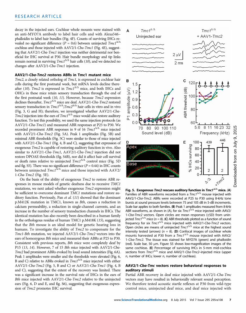
AAV2/1-Cba-Tmc2 restores ABRs in Tmc1 mutant miceTmc2, a closely related ortholog of Tmc1, is expressed in cochlear haircells during the first postnatal week, but mRNA levels decline there-after (10). Tmc2 is expressed in Tmc1D/D mice, and both IHCs andOHCs in these mice retain sensory transduction through the end ofthe first postnatal week (10, 11). However, because Tmc2 expressiondeclines thereafter, Tmc1D/D mice are deaf. AAV2/1-Cba-Tmc2 restoredsensory transduction in Tmc1D/D;Tmc2D/D hair cells in vitro and in vivo(Fig. 3, G and H); therefore, we investigated whether AAV2/1-Cba-Tmc2 injection into the ears of Tmc1D/D mice would also restore auditoryfunction. To test this possibility, we used the same injection protocols (asAAV2/1-Cba-Tmc1) and measured ABR responses at P25 to P30. Werecorded prominent ABR responses in 9 of 16 Tmc1D/D mice injectedwith AAV2/1-Cba-Tmc2 (Fig. 5A). Peak 1 amplitudes (Fig. 5B) andminimal ABR thresholds (Fig. 5C) were similar to those of mice injectedwith AAV2/1-Cba-Tmc1 (Fig. 4, B and C), suggesting that expression ofexogenous Tmc2 is capable of restoring auditory function in vivo. Alsosimilar to AAV2/1-Cba-Tmc1, AAV2/1-Cba-Tmc2 injection did notrestore DPOAE thresholds (fig. S4B), nor did it affect hair cell survivalor death rates relative to uninjected Tmc1D/D control mice (Fig. 5Dand fig. S5). There was no significant difference (P = 0.44) in IHC countsbetween uninjected Tmc1D/D mice and those injected with AAV2/1-Cba-Tmc2 (Fig. 5E).
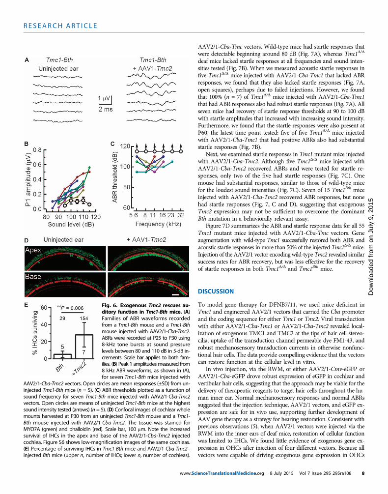
On the basis of the ability of exogenous Tmc2 to restore ABR re-sponses in mouse models of genetic deafness due to recessive TMC1mutations, we next asked whether exogenous Tmc2 expression mightbe sufficient to overcome dominant TMC1 mutations and restore au-ditory function. Previously, Pan et al. (11) showed that the dominantp.M412K mutation in TMC1, known as Bth, causes a reduction incalcium permeability, a reduction in single-channel currents, and anincrease in the number of sensory transduction channels in IHCs. Theidentical mutation has also recently been described in a human familyin the orthologous residue of human TMC1 p.M418K (15), suggestingthat the Bth mouse is an ideal model for genetic hearing loss inhumans. To investigate the ability of Tmc2 to compensate for theTmc1-Bth mutation, we injected AAV2/1-Cba-Tmc2 vectors into theears of homozygous Bthmice and measured their ABRs at P25 to P30.Consistent with previous reports, Bth mice were completely deaf byP15 (13, 14). However, 7 of 15 Bth mice injected with AAV2/1-Cba-Tmc2 had prominent ABRs evoked by loud sound intensities (Fig. 6A).Peak 1 amplitudes were smaller and the thresholds were elevated (Fig. 6,B and C) relative to ABRs evoked in Tmc1D/D mice injected with eitherAAV2/1-Cba-Tmc2 (Fig. 5, B and C) or AAV2/1-Cba-Tmc1 (Fig. 4, Band C), suggesting that the extent of the recovery was limited. Therewas a significant increase in the survival rate of IHCs in the ears ofBth mice injected with AAV2/1-Cba-Tmc2 relative to the uninjectedears (Fig. 6, D and E, and fig. S6), suggesting that exogenous expres-sion of Tmc2 promotes IHC survival.
www.Sc
AAV2/1-Cba-Tmc vectors restore behavioral responses toauditory stimuliPartial ABR recovery in deaf mice injected with AAV2/1-Cba-Tmcvectors may have resulted in behaviorally relevant sound perception.We therefore tested acoustic startle reflexes at P30 from wild-typecontrol mice, uninjected deaf mice, and deaf mice injected with
Fig. 5. Exogenous Tmc2 rescues auditory function in Tmc1D/D mice. (A)Families of ABR waveforms recorded from a Tmc1D/D mouse injected with
AAV2/1-Cba-Tmc2. ABRs were recorded at P25 to P30 using 8-kHz tonebursts at sound pressure levels between 75 and 105 dB in 5-dB increments.Scale bar applies to both families. (B) Peak 1 amplitudes measured from 8-kHzABR waveforms, as shown in (A), for six Tmc1D/D mice injected with AAV2/1-Cba-Tmc2 vectors. Open circles are mean responses (±SD) from unin-jected Tmc1D/D mice (n = 8). (C) ABR thresholds plotted as a function of soundfrequency for six Tmc1D/D mice injected with AAV2/1-Cba-Tmc2 vectors.Open circles are means of uninjected Tmc1D/D mice at the highest soundintensity tested (arrows) (n = 8). (D) Confocal images of cochlear wholemounts harvested at P30 from a Tmc1D/D mouse injected with AAV2/1-Cba-Tmc2. The tissue was stained for MYO7A (green) and phalloidin(red). Scale bar, 50 mm. Figure S5 shows low-magnification images of thesame cochleas. (E) Percentage of surviving IHCs in 5-mm mid-cochleasections from Tmc1D/D mice and AAV2/1-Cba-Tmc2–injected mice (uppern, number of IHCs; lower n, number of cochleas).ienceTranslationalMedicine.org 8 July 2015 Vol 7 Issue 295 295ra108 7
R E S EARCH ART I C L E
www.Sc
on J
uly
9, 2
015
aded
from
AAV2/1-Cba-Tmc vectors. Wild-type mice had startle responses thatwere detectable beginning around 80 dB (Fig. 7A), whereas Tmc1D/D
deaf mice lacked startle responses at all frequencies and sound inten-sities tested (Fig. 7B). When we measured acoustic startle responses infive Tmc1D/D mice injected with AAV2/1-Cba-Tmc1 that lacked ABRresponses, we found that they also lacked startle responses (Fig. 7A,open squares), perhaps due to failed injections. However, we foundthat 100% (n = 7) of Tmc1D/D mice injected with AAV2/1-Cba-Tmc1that had ABR responses also had robust startle responses (Fig. 7A). Allseven mice had recovery of startle response thresholds at 90 to 100 dBwith startle amplitudes that increased with increasing sound intensity.Furthermore, we found that the startle responses were also present atP60, the latest time point tested: five of five Tmc1D/D mice injectedwith AAV2/1-Cba-Tmc1 that had positive ABRs also had substantialstartle responses (Fig. 7B).
Next, we examined startle responses in Tmc1mutant mice injectedwith AAV2/1-Cba-Tmc2. Although five Tmc1D/D mice injected withAAV2/1-Cba-Tmc2 recovered ABRs and were tested for startle re-sponses, only two of the five had startle responses (Fig. 7C). Onemouse had substantial responses, similar to those of wild-type micefor the loudest sound intensities (Fig. 7C). Seven of 15 Tmc1Bth miceinjected with AAV2/1-Cba-Tmc2 recovered ABR responses, but nonehad startle responses (Fig. 7, C and D), suggesting that exogenousTmc2 expression may not be sufficient to overcome the dominantBth mutation in a behaviorally relevant assay.
Figure 7D summarizes the ABR and startle response data for all 55Tmc1 mutant mice injected with AAV2/1-Cba-Tmc vectors. Geneaugmentation with wild-type Tmc1 successfully restored both ABR andacoustic startle responses in more than 50% of the injected Tmc1D/D mice.Injection of the AAV2/1 vector encoding wild-type Tmc2 revealed similarsuccess rates for ABR recovery, but was less effective for the recoveryof startle responses in both Tmc1D/D and Tmc1Bth mice.
Dow
nlo
DISCUSSION
To model gene therapy for DFNB7/11, we used mice deficient inTmc1 and engineered AAV2/1 vectors that carried the Cba promoterand the coding sequence for either Tmc1 or Tmc2. Viral transductionwith either AAV2/1-Cba-Tmc1 or AAV2/1-Cba-Tmc2 revealed local-ization of exogenous TMC1 and TMC2 at the tips of hair cell stereo-cilia, uptake of the transduction channel permeable dye FM1-43, androbust mechanosensory transduction currents in otherwise nonfunc-tional hair cells. The data provide compelling evidence that the vectorscan restore function at the cellular level in vitro.
In vivo injection, via the RWM, of either AAV2/1-Cmv-eGFP orAAV2/1-Cba-eGFP drove robust expression of eGFP in cochlear andvestibular hair cells, suggesting that the approach may be viable for thedelivery of therapeutic reagents to target hair cells throughout the hu-man inner ear. Normal mechanosensory responses and normal ABRssuggested that the injection technique, AAV2/1 vectors, and eGFP ex-pression are safe for in vivo use, supporting further development ofAAV gene therapy as a strategy for hearing restoration. Consistent withprevious observations (5), when AAV2/1 vectors were injected via theRWM into the inner ears of deaf mice, restoration of cellular functionwas limited to IHCs. We found little evidence of exogenous gene ex-pression in OHCs after injection of four different vectors. Because allvectors were capable of driving exogenous gene expression in OHCs
Fig. 6. Exogenous Tmc2 rescues au-ditory function in Tmc1-Bth mice. (A)Families of ABR waveforms recordedfrom a Tmc1-Bth mouse and a Tmc1-Bthmouse injected with AAV2/1-Cba-Tmc2.ABRs were recorded at P25 to P30 using8-kHz tone bursts at sound pressurelevels between 80 and 110 dB in 5-dB in-crements. Scale bar applies to both fam-ilies. (B) Peak 1 amplitudesmeasured from8 kHz ABR waveforms, as shown in (A),for seven Tmc1-Bth mice injected with
AAV2/1-Cba-Tmc2 vectors. Open circles are mean responses (±SD) from un-injected Tmc1-Bth mice (n = 5). (C) ABR thresholds plotted as a function ofsound frequency for seven Tmc1-Bth mice injected with AAV2/1-Cba-Tmc2vectors. Open circles are means of uninjected Tmc1-Bth mice at the highestsound intensity tested (arrows) (n = 5). (D) Confocal images of cochlear wholemounts harvested at P30 from an uninjected Tmc1-Bth mouse and a Tmc1-Bth mouse injected with AAV2/1-Cba-Tmc2. The tissue was stained forMYO7A (green) and phalloidin (red). Scale bar, 100 mm. Note the increasedsurvival of IHCs in the apex and base of the AAV2/1-Cba-Tmc2 injectedcochlea. Figure S6 shows low-magnification images of the same cochleas.(E) Percentage of surviving IHCs in Tmc1-Bth mice and AAV2/1-Cba-Tmc2–injected Bth mice (upper n, number of IHCs; lower n, number of cochleas).
ienceTranslationalMedicine.org 8 July 2015 Vol 7 Issue 295 295ra108 8

R E S EARCH ART I C L E
on J
uly
9, 2
015
Dow
nloa
ded
from
in vitro, we suspect that the lack of viral transduction in OHCs in vivoresulted from limited viral access to the hair cell apical surface viaRWM injection into perilymphatic spaces. Kilpatrick et al. (19) re-ported that introduction of AAV vectors into the scala media, whichbathes hair cell apical membranes, yielded GFP expression in bothIHCs and OHCs. The challenge of scala media injection is that it re-quires a more invasive surgical approach and can cause mixing of highK+ (~140 mM) endolymph and perilymph leading to hair cell de-polarization and cell death. To target OHCs may require vectors thatcan enter via the basolateral membrane or delivery methods that targetendolymphatic spaces without disrupting endolymph/perilymphbarriers.ABRs were recovered in >50% of Tmc1D/D deaf mice injected withAAV2/1-Cba-Tmc1, indicating successful transmission of auditory
www.ScienceTranslationalMedicine.org
information from the cochlea to the au-ditory brainstem. The ABR thresholdswere elevated relative to those of wild-type mice, indicating incomplete recoveryof auditory function. DPOAE responsesdid not recover, suggesting that the elevatedABR thresholds were due to lack of re-covery of OHC function, in turn due tolow viral transduction rates in OHCs.Functional OHCs are required for cochlearamplification, a process that provides me-chanical feedback to the cochlea by increas-ing gain to soft sounds. OHC dysfunctionis known to yield elevated ABR thresh-olds, shifted up to 60 dB higher thanwild type. Thus, in Tmc1D/D mice, in whichall cochlear hair cells lack sensory trans-duction, rescue of IHC but not OHC func-tion yielded ABR thresholds ~60 dB higherthan wild type, similar to thresholds inmice with OHC dysfunction (24). InVglut3knockout mice, OHCs remain functionalbut the mice are deaf because of IHC dys-function (5). After Vglut3 gene augmen-tation, ABR thresholds recovered to nearwild-type levels because restoration offunction was only required in IHCs, whichaccount for ~25% of the cochlear hair cellpopulation. Our experiments revealedhigh viral transduction rates in IHCsand the transduced cells had mechanosen-sory currents equivalent to those of wildtype, but the OHC dysfunction remained.Although the recovery was incomplete, theresult was considered a success becausenormal mechanosensory function in IHCsis a prerequisite for auditory function. Hadthe outcome been the converse—restorationof OHC but not IHC function—the animalswould still be deaf.
We also found that AAV2/1-Cba-Tmc2vectors were capable of restoring sensorytransduction and partial ABR responses inTmc1D/D mice, which supports the hypoth-
esis that Tmc1 and Tmc2 perform somewhat redundant functions andcan substitute for each other, at least in IHCs. The AAV2/1-Cba-Tmc2transduction pattern was similar to AAV2/1-Cba-Tmc1 and was re-stricted primarily to IHCs, resulting in similar recovery at elevated ABRthresholds. That hair cell survival rates were not altered in Tmc1D/D miceinjected with either AAV2/1-Cba-Tmc1 or AAV2/1-Cba-Tmc2 was im-portant for two reasons: (i) neither vector caused loss or decay of hair cells,and (ii) hair cells remained in uninjected Tmc1D/D mice up to P60, suggest-ing that there may be a window of opportunity for therapeutic interven-tion. Whether a similar window exists in humans with recessive TMC1mutations is unknown. If patients with TMC1 mutations retain viablehair cells, they may present an opportunity for clinical intervention.
Restoration of auditory function was limited in Bth mice injectedwith AAV2/1-Cba-Tmc2. There was significant preservation of IHCs
Fig. 7. Exogenous Tmc expression rescues acoustic startle responses in Tmc1mutant mice. (A) Star-tle response amplitudes measured at P30 and plotted as a function of sound intensity and as mean ± SD
of four control C57BL/6 mice (open circles), seven individual Tmc1D/D mice injected with AAV2/1-Cba-Tmc1,and five AAV2/1-Cba-Tmc1–injected mice with no recovery (open squares). (B) Startle responses measured atP60 and plotted as mean ± SD of four Tmc1D/D mice (open circles) and five individual Tmc1D/D mice injectedwith AAV2/1-Cba-Tmc1. (C) Startle responses measured at P30, plotted for two individual Tmc1D/D miceinjected with AAV2/1-Cba-Tmc2 and as mean ± SD of seven Tmc1-Bth mice (open circles) injected withAAV2/1-Cba-Tmc2. (D) Summary bar graph showing the percentage of Tmc1 mutant mice with recoveryas assayed by ABRs and startle responses for mice injected with either AAV2/1-Cba-Tmc1 or AAV2/1-Cba-Tmc2.Numerator indicates n mice with recovery of function; denominator indicates n injected mice tested. Notall mice were tested with both assays.8 July 2015 Vol 7 Issue 295 295ra108 9

R E S EARCH ART I C L E
on J
uly
9, 2
015
Dow
nloa
ded
from
in AAV2/1-Cba-Tmc2–injected Bth mice. The mechanism that pro-moted IHC survival is unknown. On the basis of the measurementsof sensory transduction and calcium permeability in mice that ex-pressed wild-type Tmc2, Tmc1, or Tmc1-Bth, Pan et al. (11) found asignificant reduction in calcium entry in Tmc1-Bth IHCs, whereasTmc2 cells had high calcium entry. We hypothesize that appropriatelevels of calcium entry are required for maintenance and survival ofIHCs. Therefore, by introducing exogenous Tmc2, calcium homeostasiswas restored, which enhanced hair cell survival in the Bthmice injectedwith AAV2/1-Cba-Tmc2.
As a final test of auditory function, we measured acoustic startlereflexes in Tmc1 mutant mice. The otherwise unresponsive Tmc1D/D
mice recovered startle responses after injection of AAV2/1-Cba-Tmc1,and the responses persisted for up to 60 days, the latest time pointtested. It was unclear why Tmc1-Bth mice injected with AAV2/1-Cba-Tmc2 recovered partial ABR function but did not recover startle re-sponses. The extent of the ABR recovery in Tmc1-Bth mice injectedwith AAV2/1-Cba-Tmc2 was less than the ABR recovery in Tmc1D/D
mice injected with AAV2/1-Cba-Tmc2, suggesting that there may be aminimal threshold required to drive behavioral responses to loudsounds. Therapies aimed at restoration of auditory function for dom-inant DFNA36 deafness may require development of alternate strate-gies, perhaps by suppression of the dominant allele.
In conclusion, the data provide compelling proof-of-principle evi-dence demonstrating that gene augmentation in a mouse model ofDFNB7/11 is effective in restoring cellular function in vitro in bothIHCs and OHCs, restoring IHC function in vivo, partial recovery ofsystems level function in vivo, and recovery of acoustic startle reflexesat the behavioral level. Recovery of ABR and startle responses waslikely a direct result of recovery of IHC sensory transduction at thecellular level and suggests that Tmc1 reexpression can restore auditoryfunction at every level.
Thirty-five TMC1 mutations have been identified that cause re-cessive prelingual deafness in humans, which underscores the signifi-cance of TMC1 for normal auditory function and the need fortherapeutic reagents to remedy the disorder. Although our gene ther-apy strategy is not yet ready for clinical application, the challenges thatremain are not insurmountable. Continued development of Tmc genetherapy will need to provide characterization of the long-term expres-sion pattern of the exogenous constructs, including their ability tomaintain recovery; improved design of vectors, promoters, and deliverytechniques that drive exogenous gene expression in OHCs; and furtherevaluation of the therapeutic window of opportunity in humans withrecessive TMC1mutations. Finally, we suggest that AAV-mediated geneaugmentation in the inner ear may be a model that could be expandedto address some of the more than 70 forms of genetic deafness.
MATERIALS AND METHODS
Study designThe aim of this study was to identify AAV serotypes and promotersfor delivery and expression of exogenous Tmc1 and Tmc2 in hair cellsof the mouse cochlea and to evaluate the ability of these vectors torestore function in mouse models of genetic deafness in humans. AAVvectors were injected in vivo, and the outcomes were evaluated usingquantitative RT-PCR, immunolocalization and confocal microscopy,imaging FM1-43 uptake, single-cell recording, histology and imaging
www.Scie
of whole cochleas, measurement of ABRs, DPOAEs, and acoustic startlereflexes. Left ears were injected and right ears were used as uninjectedcontrols. Each experiment was replicated as indicated by n values in thefigure legends. All experiments with mice and viral vectors were ap-proved by the Institutional Animal Care and Use Committee (protocols#2146 and #2878) at Boston Children’s Hospital and the InstitutionalBiosafety Committee (protocol #IBC-P00000447).
In vivo injection of viral vectorsMouse pups (P0 to P2) were injected via the RWM using beveledglass microinjection pipettes, as described in SupplementaryMethods.
Hair cell electrophysiologyOrganotypic cochlear cultures were bathed in standard artificial peri-lymph containing 137 mM NaCl, 0.7 mM NaH2PO4, 5.8 mM KCl,1.3 mM CaCl2, 0.9 mM MgCl2, 10 mM Hepes, and 5.6 mM D-glucose.Vitamins (1:50) and amino acids (1:100) were added to the solutionfrom concentrates (Invitrogen), and NaOH was used to adjust the fi-nal pH to 7.40 (310 mosmol/kg). Recording pipettes (3 to 5 megohms)were pulled from R6 capillary glass (King Precision Glass) and filledwith intracellular solution containing 135 mM CsCl, 5 mM Hepes,5 mM EGTA, 2.5 mM MgCl2, 2.5 mM Na2–adenosine triphosphate,and 0.1 mM CaCl2, where CsOH was used to adjust the final pH to7.40 (285 mosmol/kg). Whole-cell, tight-seal voltage-clamp recordingswere done at −84 mV at room temperature (22° to 24°C) using anAxopatch 200B amplifier (Molecular Devices). Sensory transductioncurrents were filtered at 10 kHz with a low-pass Bessel filter and digit-ized at≥20 kHz with a 16-bit acquisition board (Digidata 1440A) andpCLAMP 10 software (Molecular Devices). Data were stored for off-line analysis using OriginPro 8 (OriginLab).
ABR and DPOAEABR recordings were conducted as described previously (25), at 32°Cin a soundproof chamber. To test hearing function, anesthetized micewere presented pure tone stimuli of 5.6, 8, 11.3, 16, 22.6, or 32 kHz atsound pressure levels between 10 and 115 dB in 5-dB steps until athreshold intensity that evoked a reproducible ABR waveform (peaks1 to 4) was detected. Responses were collected, and data were analyzedas described in Supplementary Methods.
DPOAE data were collected under the same conditions and duringthe same recording sessions as the ABR data. Primary tones wereproduced at a frequency ratio of 1.2 (f2/f1) for the generation of DPOAEsat 2f1–f2, where the f2 level was 10 dB sound pressure level below f1level for each f2/f1 pair. The f2 levels were swept in 5-dB steps from 20 to80 dB. Waveform and spectral analyses are described in Supplemen-tary Methods.
Acoustic startle reflexesMice were tested for startle reflexes in response to broadband auditorystimulation at varying intensities, as described in Supplementarymethods.
Statistical analysisAll mean values and error bars presented in the figures representmean ± SD. Comparisons for statistical significance between injectedears and uninjected ears were performed using a two-tailed paired t test.P < 0.05 was considered significant.
nceTranslationalMedicine.org 8 July 2015 Vol 7 Issue 295 295ra108 10

R E S EARCH ART I C L E
SUPPLEMENTARY MATERIALS
www.sciencetranslationalmedicine.org/cgi/content/full/7/295/295ra108/DC1Materials and MethodsFig. S1. In vivo injection of AAV2/1-Cmv-eGFP through the RWM.Fig. S2. In vivo injection also targets vestibular hair cells.Fig. S3. Exogenous Tmc1/2 restores sensory transduction in Tmc-deficient hair cells in vitro.Fig. S4. Auditory function in wild-type mice injected with AAV vectors.Fig. S5. Confocal images of Tmc1D/D cochleas injected with AAV2/1-Cba-Tmc vectors.Fig. S6. Confocal images of Tmc1-Bth cochleas injected with AAV2/1-Cba-Tmc2.References (26, 27)
on J
uly
9, 2
015
Dow
nloa
ded
from
REFERENCES AND NOTES
1. G. S. G. Géléoc, J. R. Holt, Sound strategies for hearing restoration. Science 344, 1241062 (2014).2. D. C. Kohrman, Y. Raphael, Gene therapy for deafness. Gene Ther. 20, 1119–1123 (2013).3. B. W. Kesser, G. T. Hashisaki, K. Fletcher, H. Eppard, J. R. Holt, An in vitro model system to
study gene therapy in the human inner ear. Gene Ther. 14, 1121–1131 (2007).4. B. W. Kesser, G. T. Hashisaki, J. R. Holt, Gene transfer in human vestibular epithelia and the
prospects for inner ear gene therapy. Laryngoscope 118, 821–831 (2008).5. O. Akil, R. P. Seal, K. Burke, C. Wang, A. Alemi, M. During, R. H. Edwards, L. R. Lustig, Restoration
of hearing in the VGLUT3 knockout mouse using virally mediated gene therapy. Neuron 75,283–293 (2012).
6. S.-I. Kitajiri, R. McNamara, T. Makishima, T. Husnain, A. U. Zafar, R. A. Kittles, Z. M. Ahmed,T. B. Friedman, S. Riazuddin, A. J. Griffith, Identities, frequencies and origins of TMC1mutations causing DFNB7/B11 deafness in Pakistan. Clin. Genet. 72, 546–550 (2007).
7. A. Sirmaci, D. Duman, H. Öztürkmen-Akay, S. Erbek, A. İncesulu, B. Öztürk-Hişmi, Z. S. Arici,E. B. Yüksel-Konuk, S. Taşir-Yilmaz, S. Tokgöz-Yilmaz, F. B. Cengiz, İ. Aslan, M. Yildirim,A. Hasanefendioğlu-Bayrak, A. Ayçiçek, İ. Yilmaz, S. Fitoz, F. Altin, H. Özdağ, M. Tekin,Mutations in TMC1 contribute significantly to nonsyndromic autosomal recessive sensori-neural hearing loss: A report of five novel mutations. Int. J. Pediatr. Otorhinolaryngol. 73,699–705 (2009).
8. Y. Kawashima, K. Kurima, B. Pan, A. J. Griffith, J. R. Holt, Transmembrane channel-like (TMC)genes are required for auditory and vestibular mechanosensation. Pflugers Arch. 467, 85–94(2015).
9. H. Nakanishi, K. Kurima, Y. Kawashima, A. J. Griffith, Mutations of TMC1 cause deafness bydisrupting mechanoelectrical transduction. Auris Nasus Larynx 41, 399–408 (2014).
10. Y. Kawashima, G. S. G. Géléoc, K. Kurima, V. Labay, A. Lelli, Y. Asai, T. Makishima, D. K. Wu,C. C. Della Santina, J. R. Holt, A. J. Griffith, Mechanotransduction in mouse inner ear haircells requires transmembrane channel-like genes. J. Clin. Invest. 121, 4796–4809 (2011).
11. B. Pan, G. S. G. Géléoc, Y. Asai, G. C. Horwitz, K. Kurima, K. Ishikawa, Y. Kawashima, A. J. Griffith,J. R. Holt, TMC1 and TMC2 are components of the mechanotransduction channel in hair cellsof the mammalian inner ear. Neuron 79, 504–515 (2013).
12. M. Beurg, W. Xiong, B. Zhao, U. Müller, R. Fettiplace, Subunit determination of the con-ductance of hair-cell mechanotransducer channels. Proc. Natl. Acad. Sci. U.S.A. 112, 1589–1594(2015).
13. W. Marcotti, A. Erven, S. L. Johnson, K. P. Steel, C. J. Kros, Tmc1 is necessary for normalfunctional maturation and survival of inner and outer hair cells in the mouse cochlea. J. Physiol.574, 677–698 (2006).
14. S. Vreugde, A. Erven, C. J. Kros, W. Marcotti, H. Fuchs, K. Kurima, E. R. Wilcox, T. B. Friedman,A. J. Griffith, R. Balling, M. Hrabé De Angelis, K. B. Avraham, K. P. Steel, Beethoven, a mousemodel for dominant, progressive hearing loss DFNA36. Nat. Genet. 30, 257–258 (2002).
15. Y. Zhao, D. Wang, L. Zong, F. Zhao, L. Guan, P. Zhang W. Shi, L. Lan, H. Wang, Q. Li, B. Han,L. Yang, X. Jin, J. Wang, J. Wang, Q. Wang, A novel DFNA36 mutation in TMC1 orthologousto the Beethoven (Bth) mouse associated with autosomal dominant hearing loss in a Chinesefamily. PLOS One 9, e97064 (2014).
www.Scie
16. V.H. Hernandez, A. Gehrt, K. Reuter, Z. Jing, M. Jeschke, A. Mendoza Schulz, G. Hoch, M. Bartels,G. Vogt, C. W. Garnham, H. Yawo, Y. Fukazawa, G. J. Augustine, E. Bamberg, S. Kügler, T. Salditt,L. de Hoz, N. Strenzke, T. Moser, Optogenetic stimulation of the auditory pathway. J. Clin.Invest. 124, 1114–1129 (2014).
17. S. Safieddine, R. J. Wenthold, SNARE complex at the ribbon synapses of cochlear hair cells:Analysis of synaptic vesicle- and synaptic membrane-associated proteins. Eur. J. Neurosci.11, 803–812 (1999).
18. J. C. Bedrosian, M. A. Gratton, J. V. Brigande, W. Tang, J. Landau, J. Bennett, In vivo deliveryof recombinant viruses to the fetal murine cochlea: Transduction characteristics and long-term effects on auditory function. Mol. Ther. 14, 328–335 (2006).
19. L. A. Kilpatrick, Q. Li, J. Yang, J. C. Goddard, D. M. Fekete, H. Lang, Adeno-associated virus-mediated gene delivery into the scala media of the normal and deafened adult mouse ear.Gene Ther. 18, 569–578 (2011).
20. J. E. Gale, W. Marcotti, H. J. Kennedy, C. J. Kros, G. P. Richardson, FM1-43 dye behaves as a per-meant blocker of the hair-cell mechanotransducer channel. J. Neurosci. 21, 7013–7025 (2001).
21. J. R. Meyers, R. B. MacDonald, A. Duggan, D. Lenzi, D. G. Standaert, J. T. Corwin, D. P. Corey,Lighting up the senses: FM1-43 loading of sensory cells through nonselective ion chan-nels. J. Neurosci. 23, 4054–4065 (2003).
22. G. S. Géléoc, J. R. Holt, Developmental acquisition of sensory transduction in hair cells ofthe mouse inner ear. Nat. Neurosci. 10, 1019–1020 (2003).
23. A. J. Hudspeth, Integrating the active process of hair cells with cochlear function. Nat. Rev.Neurosci. 15, 600–614 (2014).
24. M. C. Liberman, J. Gao, D. Z. Z. He, X. Wu, S. Jia, J. Zuo, Prestin is required for electromotilityof the outer hair cell and for the cochlear amplifier. Nature 419, 300–304 (2002).
25. S. F. Maison, X.-P. Liu, D. E. Vetter, R. A. Eatock, N. M. Nathanson, J. Wess, M. C. Liberman,Muscarinic signaling in the cochlea: Presynaptic and postsynaptic effects on efferentfeedback and afferent excitability. J. Neurosci. 30, 6751–6762 (2010).
26. D. Grimm, M. A. Kay, J. A. Kleinschmidt, Helper virus-free, optically controllable, and two-plasmid-based production of adeno-associated virus vectors of serotypes 1 to 6. Mol. Ther.7, 839–850 (2003).
27. A. E. Stauffer, J. R. Holt, Sensory transduction and adaptation in inner and outer hair cells ofthe mouse auditory system. J. Neurophysiol. 98, 3360–3369 (2007).
Acknowledgments: We thank M. C. Liberman for assistance with ABR recordings; Y. Shu andZ.-Y. Chen for technical assistance with RWM injections; V. Padrun, F. Pidoux, and A. Aebi atÉcole Polytechnique Fédérale de Lausanne (EPFL) Brain Mind Institute for technical assistancewith AAV production; Behavior and Viral Cores at Boston Children’s Hospital [supported byBoston Children’s Hospital Intellectual and Developmental Disabilities Research Center (BCHIDDRC), P30 HD18655]. Funding: This work was supported by the Bertarelli Foundation, Kidz bKidz Foundation (Jessica and David Freier), and Program in Translational Neuroscience andNeuroengineering. Author contributions: C.A. performed experiments, analyzed data, andhelped write the manuscript; C.R. designed and generated vectors; B.P. performed experimentsand analyzed data; Y.A. designed and generated vectors, performed experiments, and analyzeddata; H.A. performed experiments and analyzed data; E.C. analyzed data; B.L.S. designed and gen-erated vectors and helped design experiments; P.A. helped design vectors and experiments; J.R.H.conceived the study, designed experiments, analyzed data, and helped write the manuscript. Allauthors critically reviewed and approved the manuscript. Competing interests: The authors de-clare that they have no competing interests. Data and material availability: Plasmids for AAVvector production are available via a material transfer agreement.
Submitted 13 December 2014Accepted 17 June 2015Published 8 July 201510.1126/scitranslmed.aab1996
Citation: C. Askew, C. Rochat, B. Pan, Y. Asai, H. Ahmed, E. Child, B. L. Schneider, P. Aebischer,J. R. Holt, Tmc gene therapy restores auditory function in deaf mice. Sci. Transl. Med. 7,295ra108 (2015).
nceTranslationalMedicine.org 8 July 2015 Vol 7 Issue 295 295ra108 11

DOI: 10.1126/scitranslmed.aab1996, 295ra108 (2015);7 Sci Transl Med
et al.Charles Askew gene therapy restores auditory function in deaf miceTmc
Editor's Summary
implants and hearing aids.will work long-term to maintain hearing recovery, perhaps supplementary existing technologies such as cochlearmutations have been implicated in recessive prelingual deafness, so it is hoped that this gene therapeutic approach
TMC1and partially restore hearing, as determined by auditory brainstem responses and startle reflexes. More than 30 dominant human deafness, respectively. Both vectors were able to transduce inner hair cells of the mouse cochlea,
models representative of autosomal recessive and−−Beethoven or Tmc1delivered to mice with mutations in , was packaged in adeno-associated viral vectors andTmc2, or its ortholog, Tmc1like 1, −transmembrane channel
replace mutant genes associated with the mechanotransduction machinery of the inner ear. The gene encoding Because genetics is a major cause of deafness, Askew and colleagues developed a therapeutic approach to
Can you hear me now?
/content/7/295/295ra108.full.htmlcan be found at:
and other services, including high-resolution figures,A complete electronic version of this article
/content/suppl/2015/07/06/7.295.295ra108.DC1.html can be found in the online version of this article at: Supplementary Material
http://stm.sciencemag.org/content/scitransmed/2/21/21ra16.full.html http://stm.sciencemag.org/content/scitransmed/6/233/233ra54.full.html
can be found online at:Related Resources for this article
http://www.sciencemag.org/about/permissions.dtl in whole or in part can be found at: article
permission to reproduce this of this article or about obtaining reprintsInformation about obtaining
is a registered trademark of AAAS. Science Translational Medicinerights reserved. The title NW, Washington, DC 20005. Copyright 2015 by the American Association for the Advancement of Science; alllast week in December, by the American Association for the Advancement of Science, 1200 New York Avenue
(print ISSN 1946-6234; online ISSN 1946-6242) is published weekly, except theScience Translational Medicine
on J
uly
9, 2
015
Dow
nloa
ded
from