Embed Size (px)
Citation preview

THE CENTRAL NERVOUS SYSTEM
AND THE EPIDERMIS
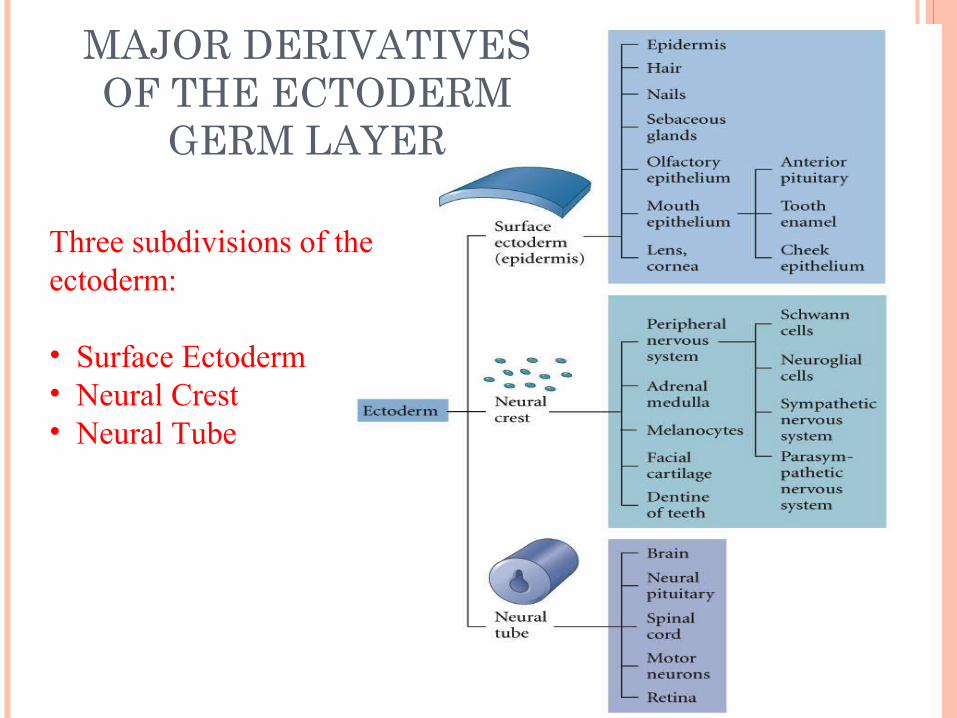
Three subdivisions of the ectoderm:
• Surface Ectoderm• Neural Crest• Neural Tube
MAJOR DERIVATIVES OF THE ECTODERM
GERM LAYER

Neural plate Neurulation Neural tube neurula

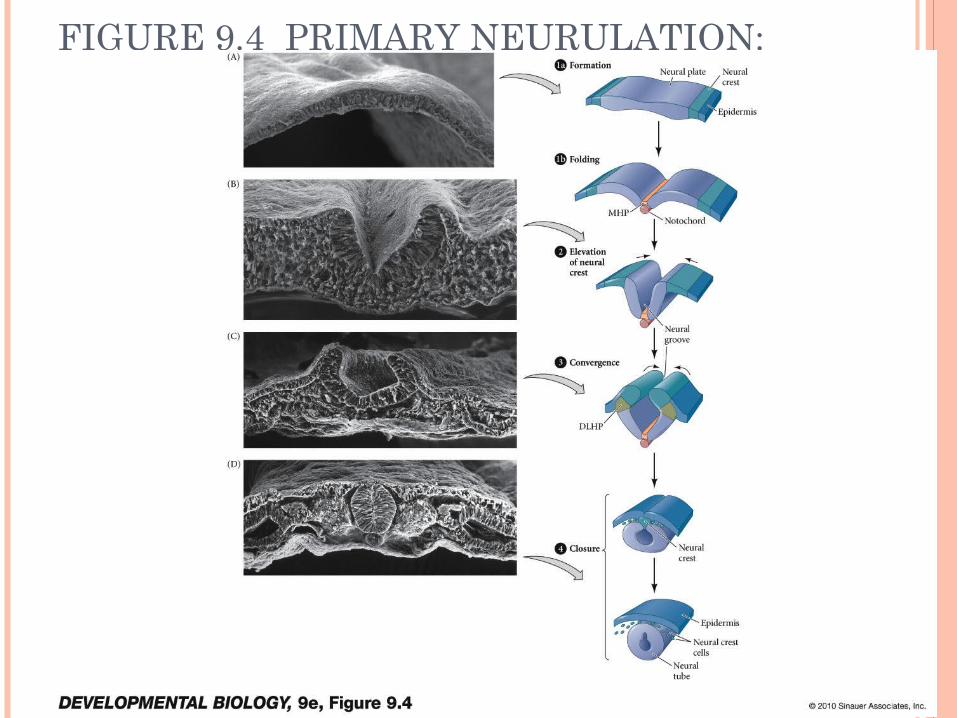
FIGURE 9.4 PRIMARY NEURULATION: NEURAL TUBE FORMATION IN THE CHICK EMBRYO

PRIMARY NEURULATION
MPH (medial hinge point) – the combined Hensen's node and epiblast region that is involved in the intial bending of the neural plate during neurulation.
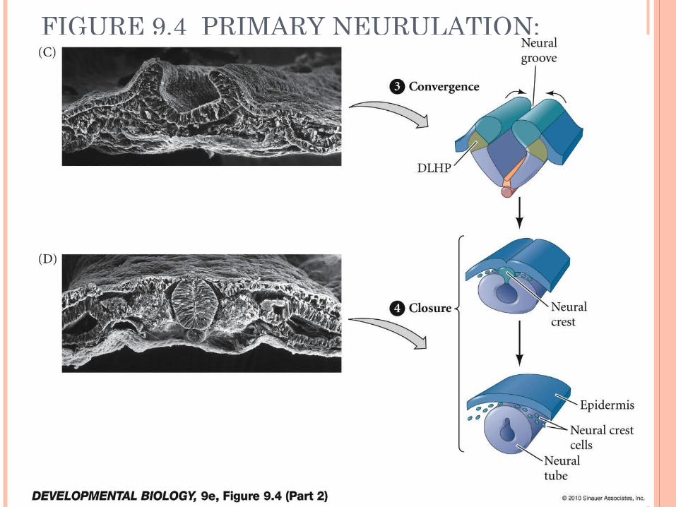
FIGURE 9.4 PRIMARY NEURULATION: NEURAL TUBE FORMATION IN THE CHICK EMBRYO (PART 2)

Anencephaly is the absence of a major portion of the brain that occurs during embryonic development. This is a cephalic disorder resulting from a neural tube defect occurring when the rostal end of the neural fails to close. This typically happens between the 23rd and 26th day of conception.

Spina Bifida is a similar defect this time occurring at the caudal end of the neural tube. Children with this condition often times have locomotor disorders.

EXPRESSION OF N- AND E-CADHERIN ADHESION PROTEINS
In the experimental protocol (B) neurulation has been altered by the injection of N-cadherin and E-cadherin. The alteration leads to the neural tube not showing the normal stages of separation from the presumptive epidermis.


SECONDARY NEURULATION


• Anterior-Posterior Axis (mammalian brain)
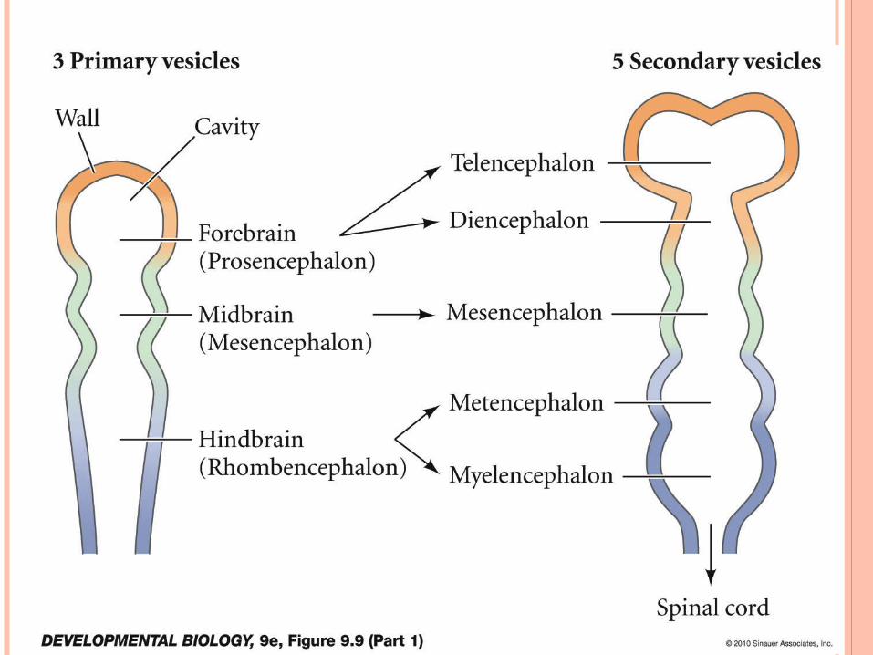
FIGURE 9.9 EARLY HUMAN BRAIN DEVELOPMENT (PART 1)
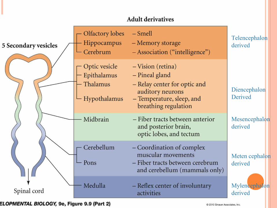
FIGURE 9.9 EARLY HUMAN BRAIN DEVELOPMENT (PART 2) Telencephalon
derived
DiencephalonDerived
Mesencephalon derived
Meten cephalon derived
Mylencephalon derived

FIGURE 9.10 RHOMBOMERES OF THE CHICK HINDBRAIN
The rhombencephalon is divided into smaller compartments through periodic swelling called rhombomeres. Each rhombomere forms ganglia clusters whose axons form a nerve.
Example: in chicks, the first neuron appear in even numbered rhombomeres: r2 ( 5th- trigeminal) , r4 (7th-face and 8th- vestibuloaccoustic), r6 (9th-glossopharengeal)
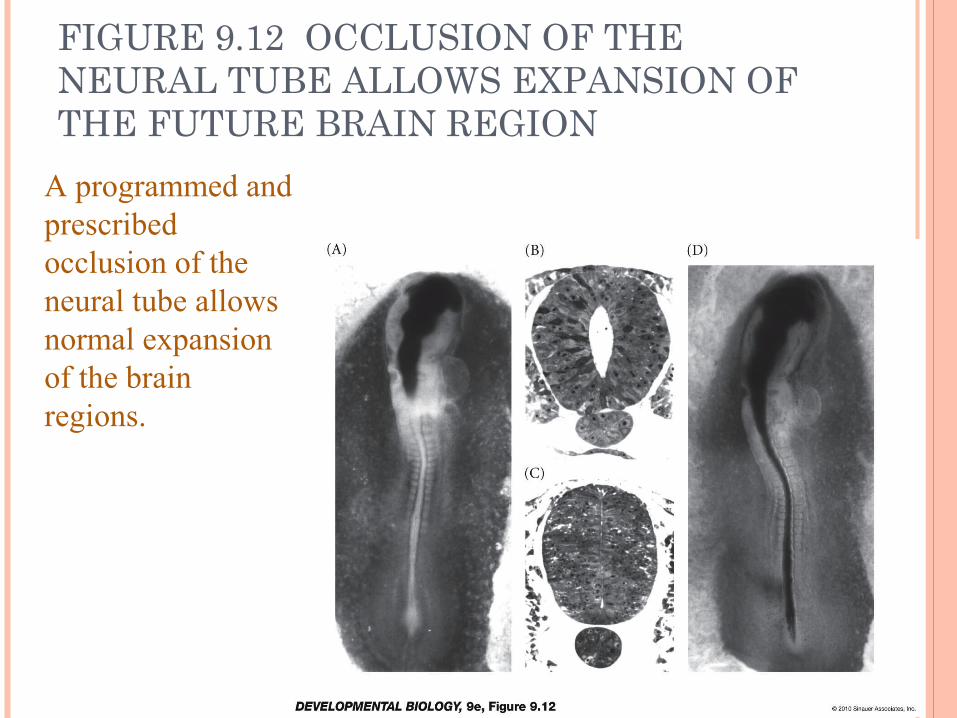
FIGURE 9.12 OCCLUSION OF THE NEURAL TUBE ALLOWS EXPANSION OF THE FUTURE BRAIN REGION
A programmed and prescribed occlusion of the neural tube allows normal expansion of the brain regions.

Long term, non prescribed occlusion may lead to hydrocephaly.
Note that the example here has occurred post parturition. In cases where hydrocephaly occurs earlier in development, viability of the fetus is doubtful.

DORSAL-VENTRAL AXIS (SPINAL CORD)


TISSUE ARCHITECTURE OF
THE CENTRAL NERVOUS SYSTEM

SPINAL CORD AND MEDULLA ORGANIZATION

FIGURE 9.19 DIFFERENTIATION OF THE WALLS OF THE NEURAL TUBE
The neural tube will further differentiate into a wide array of structures that are dependent upon location.

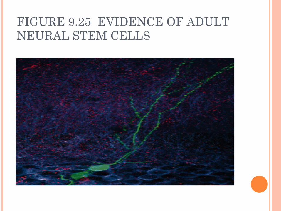
FIGURE 9.25 EVIDENCE OF ADULT NEURAL STEM CELLS

NEURON
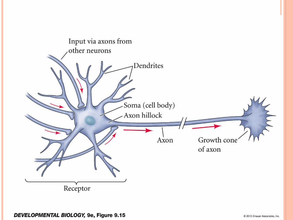
FIGURE 9.15 DIAGRAM OF A MOTOR NEURON

FIGURE 9.16 AXON GROWTH CONES
These projections form microspikes.
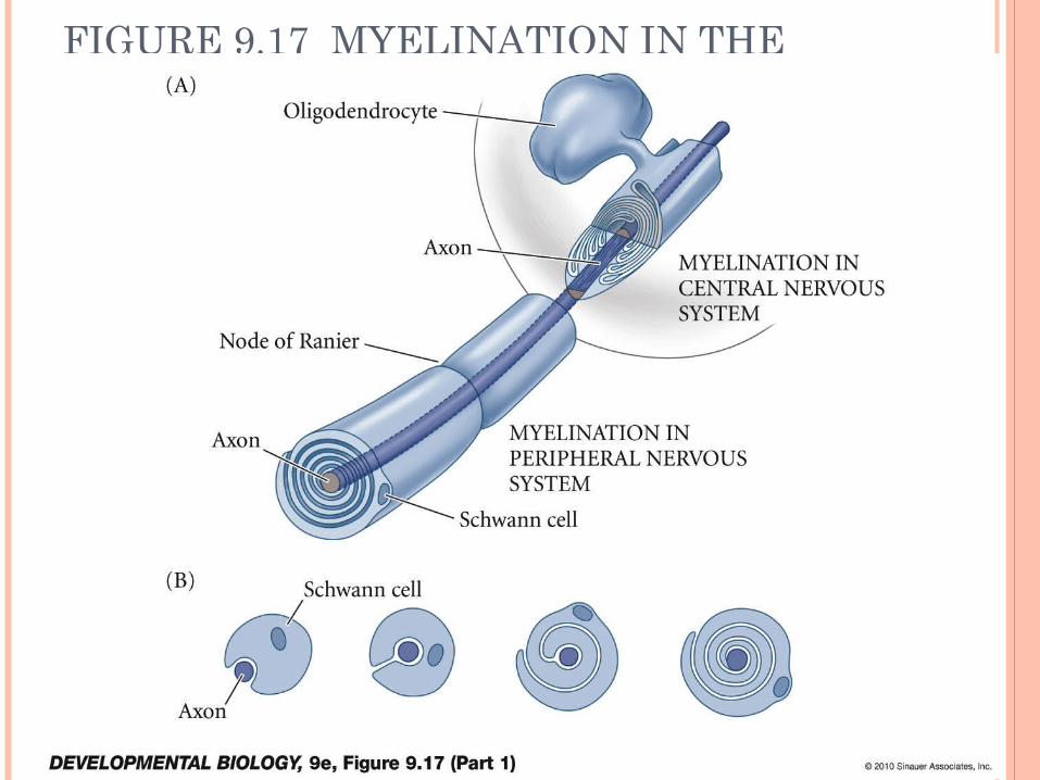
FIGURE 9.17 MYELINATION IN THE CENTRAL AND PERIPHERAL NERVOUS SYSTEMS (PART 1)

Some studies have suggested that over pruning of dendritic spines may occur in the prodromal and early stages of schizophrenia.

Marcello Malpighi (most work occurred in the 1660s)was an Italian physician, who identified (and named after himself) several anatomical structures. Examples include the Malpighian tubule system in the kidney and the Malpighi layer in the skin.

FIGURE 9.37 LAYERS OF THE HUMAN EPIDERMIS
The Malpighian layer of the skin is defined as the combination of both the stratum basale and stratum spinosum layers of the epidermis.
Epidermal Growth Factora.Transforming growth factorb.Keratinocyte growth factor
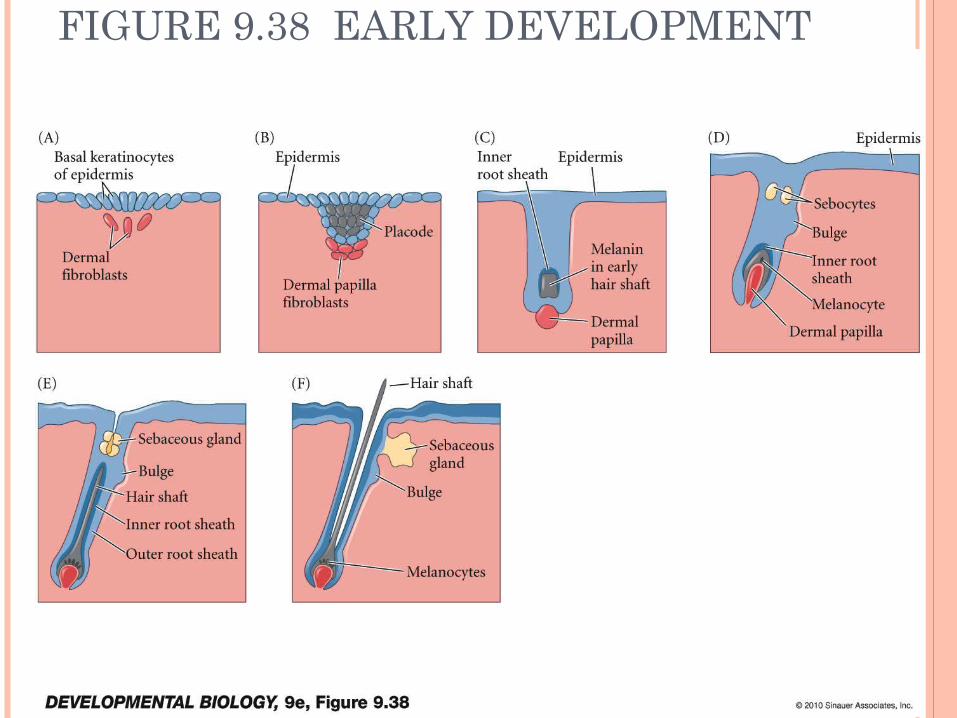
FIGURE 9.38 EARLY DEVELOPMENT OF THE HAIR FOLLICLE AND HAIR SHAFT

FIGURE 9.41 FACIAL ANOMALIES OF ANHIDROTIC ECOTODERMAL DYSPLASIA, CAUSED BY MUTATION OF AN EDA GENE• Anhidrotic Ectodermal Dysplasia (also called
"Christ-Siemens-Touraine Syndrome“) is a disorder resulting in the abnormal development of a variety of structures including the skin, hair, nails, teeth, and sweat glands.
• reduced ability to sweat (hypohidrosis) because they have fewer sweat glands than normal or their sweat glands do not function properly potentially leading to a dangerously high body temperature (hyperthermia) in certain circumstances
• Affected individuals tend to have sparse scalp and body hair (hypotrichosis). The hair is often light-coloured, brittle, and slow-growing.
• This condition is also characterized by absent teeth (hypodontia) or teeth that are malformed. The teeth that are present are frequently small and pointed.
SUMMARY The neural tube forms from the shaping and folding of
the neural plate. In primary neurulation, the surface ectoderm folds into a tube that separates from the surface. In secondary neurulation, the ectoderm forms a cord then forms a cavity within it.
The dorsal-ventral patterning of the neural tube is accomplished by proteins of the TGF-Beta family and from the sonic hedgehog protein. Both types of protein appear to work through gradients
The brain forms three primary vesicles: prosencephalon, mesencephalon, and rhombencephalon
The brain expands through fluid secretion putting positive pressure on the vesicles

The neurons of the brain are organized into cortices (layers) and nuclei (clusters)
New neurons are formed in the neural tube. The neural precursors can migrate away from the neural tube and form a new layer. Neurons forming later have to migrate through the existing layers. This forms the cortical layer. The germinal zone at the lumen of the neural tube is called the ventricular zone. The new layer is called the mantle zone.
In cerebellum, a second germinal zone, the external granule layer is formed.
The cerebral cortex in humans has six regions, and the mantle zone is called neocortex.
Dendrites receives signals from other neurons, while axons transmits them. The place where the signaling takes place is called a synapse

Axons grow from the soma. They are led by the growth cone
TGF and FGF7 are important in normal skin development

End.


