Embed Size (px)
Citation preview
RESPIRATORY SYSTEMGROUP 6
It is a biological system consisting of specific organs and structures used for the process of respiration in an organism
WHAT IS RESPIRATORY SYSTEM?
Gas exchange: Oxygen enters blood and carbon dioxide leaves
Regulation of blood pH: Altered by changing blood carbon dioxide levels
Voice production: Movement of air past vocal folds makes sound and speech.
Olfaction: Smell occurs when airborne molecules are drawn into nasal cavity.
Protection: Against microorganisms by preventing entry and removing them from respiratory surfaces.
RESPIRATORY SYSTEM FUNCTIONS
Nostrils
Trachea
Bronchi
Lungs
Alveolus
Diaphragm
MAIN PARTS:
Respiratory system does this through breathing. When we breathe, we inhale oxygen and exhale carbon dioxide. This exchange of gases is the respiratory system’s means of getting oxygen to the blood.
HOW DOES IT WORK?
Endoderm forms the respiratory system, having a sheet of approximately 500-1000 cells.
Phases of Lung Development- growth and transcription factors.
EMBRYONIC ORIGIN
Mechanism of respiration
Chordates have one of two basic structures for respiration:
Gills – for aquatic chordates
Example: tunicates, fish and amphibians
Lungs - for terrestrial chordates
Examples: adult amphibians, reptiles, birds, and mammals
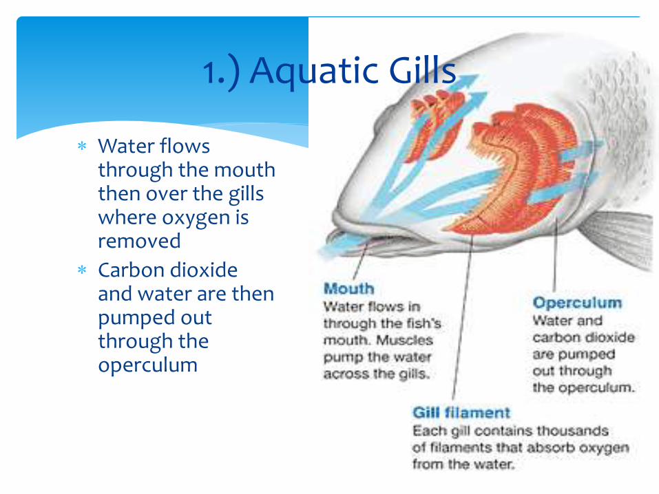
1.) Aquatic Gills
Water flows through the mouth then over the gills where oxygen is removed
Carbon dioxide and water are then pumped out through the operculum
Increase the surface area
Containing blood vessels covered by a thin epithelial layer
Organized into a series of plates
Countercurrent principle
Maybe: internal (as in crabs and fish) or
external to the body (as in some amphibians).
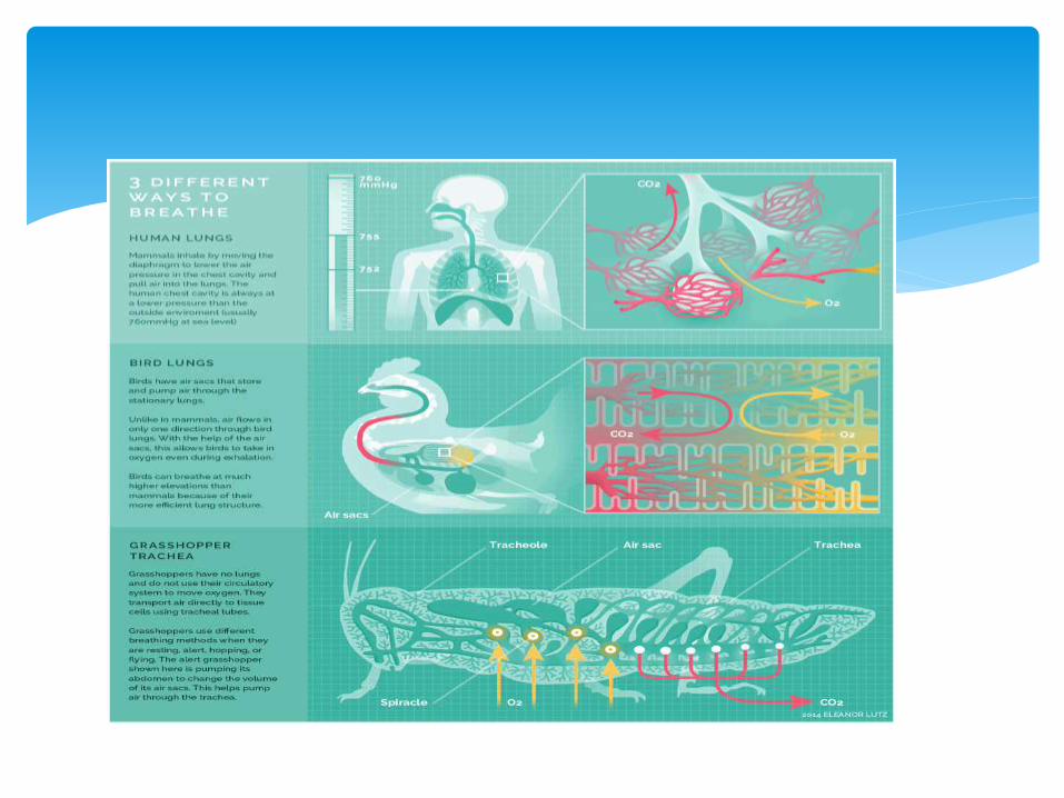
2. ) Vertebrate lungs
As you move from amphibians to mammals the surface area of the lungs increases Insures a greater amount of gas exchange (or a two way
flow of air).
Birds, by contrast have lungs and air sacs which have only a one-way flow of air. This allows for them to have constant contact with fresh
air. This adaptation enables them to fly at high altitudes
where there is less oxygen.
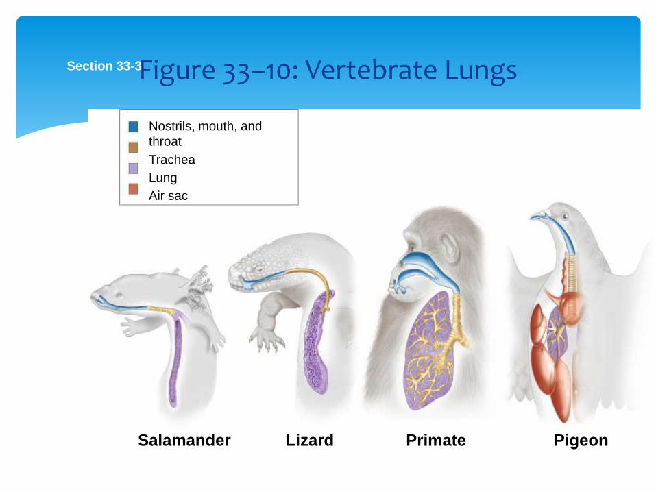
Section 33-3
Salamander Lizard PigeonPrimate
Nostrils, mouth, and
throat
Trachea
Lung
Air sac
Figure 33–10: Vertebrate Lungs
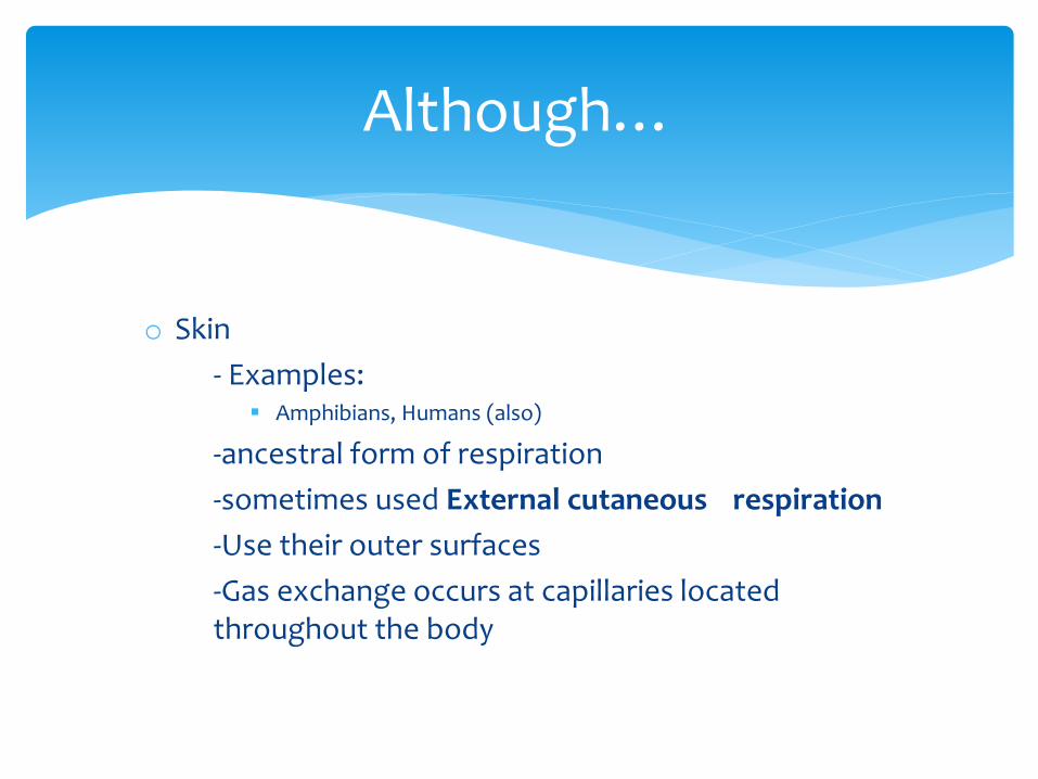
Although…
o Skin
- Examples: Amphibians, Humans (also)
-ancestral form of respiration
-sometimes used External cutaneous respiration
-Use their outer surfaces
-Gas exchange occurs at capillaries located throughout the body
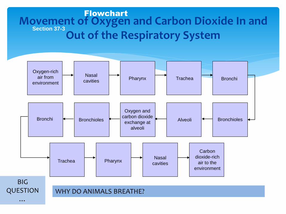
Section 37-3
Flowchart
Oxygen and
carbon dioxide
exchange at
alveoli
Oxygen-rich
air from
environment
Bronchioles
Nasal
cavitiesPharynx Trachea Bronchi
BronchiolesAlveoli
PharynxNasal
cavities
Carbon
dioxide-rich
air to the
environment
Bronchi
Trachea
Movement of Oxygen and Carbon Dioxide In and Out of the Respiratory System
BIG QUESTION
…WHY DO ANIMALS BREATHE?
OSTEICHTHYES
Ocat, Clint
Reyes, Frenzy Janiña
Gills
mediate gas exchange
located at the side of the head
made up of gill filaments , feather structures that provide a large surface for gas exchange
Adult fishes have a pair of gills. Each gills is covered by a bony lid. A fish draws in water by closing the lid over its gills and opening its mouth. When the fish closes its mouth and opens the gill lid, the water is forced out and over the respiratory surfaces of the gill filaments.
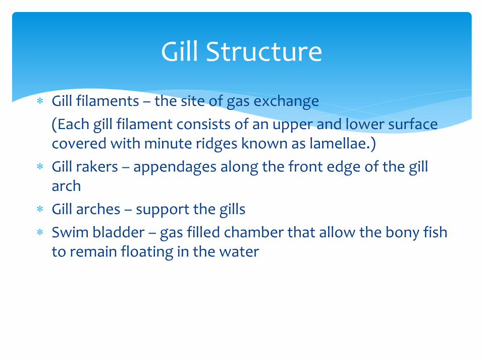
Gill Structure
Gill filaments – the site of gas exchange
(Each gill filament consists of an upper and lower surface covered with minute ridges known as lamellae.)
Gill rakers – appendages along the front edge of the gill arch
Gill arches – support the gills
Swim bladder – gas filled chamber that allow the bony fish to remain floating in the water
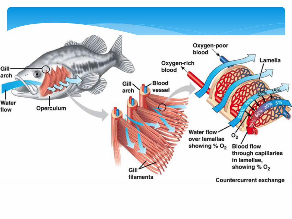
Bony fishes
Water enters the gill chamber through the fish’s mouth and exits through gill openings under the operculum. Blood flowing through the gill filaments absorbs oxygen from the water.
Some species of bony fishes can absorb considerable amounts of oxygen through their skin.
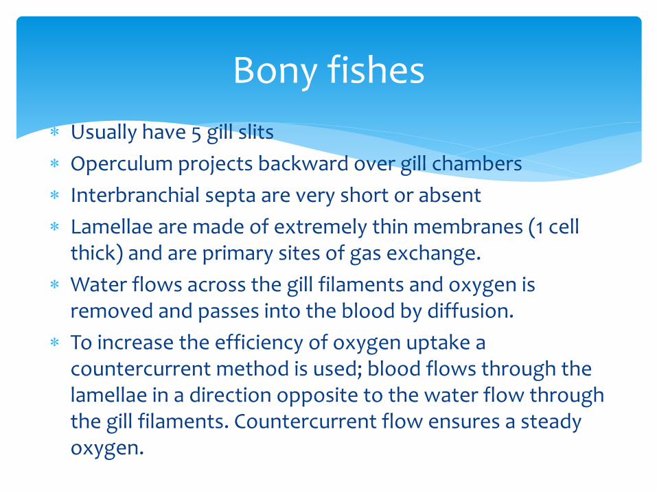
Bony fishes
Usually have 5 gill slits
Operculum projects backward over gill chambers
Interbranchial septa are very short or absent
Lamellae are made of extremely thin membranes (1 cell thick) and are primary sites of gas exchange.
Water flows across the gill filaments and oxygen is removed and passes into the blood by diffusion.
To increase the efficiency of oxygen uptake a countercurrent method is used; blood flows through the lamellae in a direction opposite to the water flow through the gill filaments. Countercurrent flow ensures a steady oxygen.

ReptilesNikki Atilano
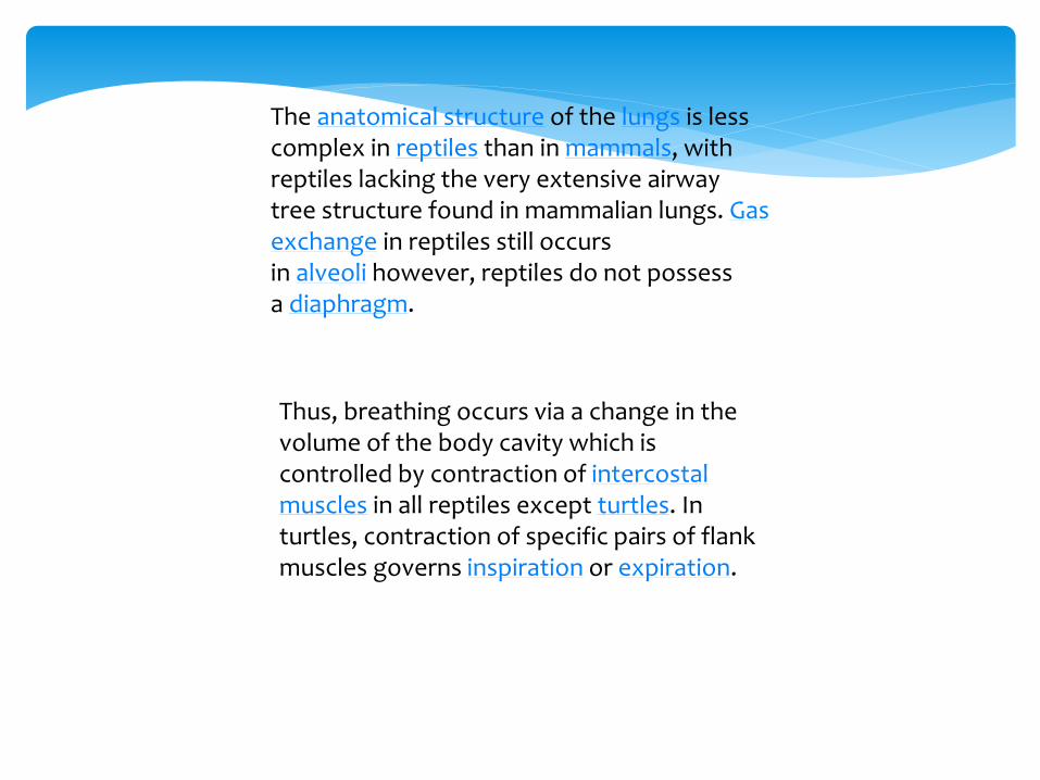
The anatomical structure of the lungs is less complex in reptiles than in mammals, with reptiles lacking the very extensive airway tree structure found in mammalian lungs. Gas exchange in reptiles still occurs in alveoli however, reptiles do not possess a diaphragm.
Thus, breathing occurs via a change in the volume of the body cavity which is controlled by contraction of intercostal muscles in all reptiles except turtles. In turtles, contraction of specific pairs of flank muscles governs inspiration or expiration.
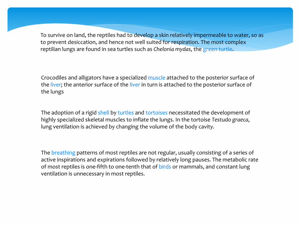
To survive on land, the reptiles had to develop a skin relatively impermeable to water, so as to prevent desiccation, and hence not well suited for respiration. The most complex reptilian lungs are found in sea turtles such as Chelonia mydas, the green turtle.
Crocodiles and alligators have a specialized muscle attached to the posterior surface of the liver; the anterior surface of the liver in turn is attached to the posterior surface of the lungs
The adoption of a rigid shell by turtles and tortoises necessitated the development of highly specialized skeletal muscles to inflate the lungs. In the tortoise Testudo graeca, lung ventilation is achieved by changing the volume of the body cavity.
The breathing patterns of most reptiles are not regular, usually consisting of a series of active inspirations and expirations followed by relatively long pauses. The metabolic rate of most reptiles is one-fifth to one-tenth that of birds or mammals, and constant lung ventilation is unnecessary in most reptiles.
AMPHIBIANS
PARTS and FUNCTIONS
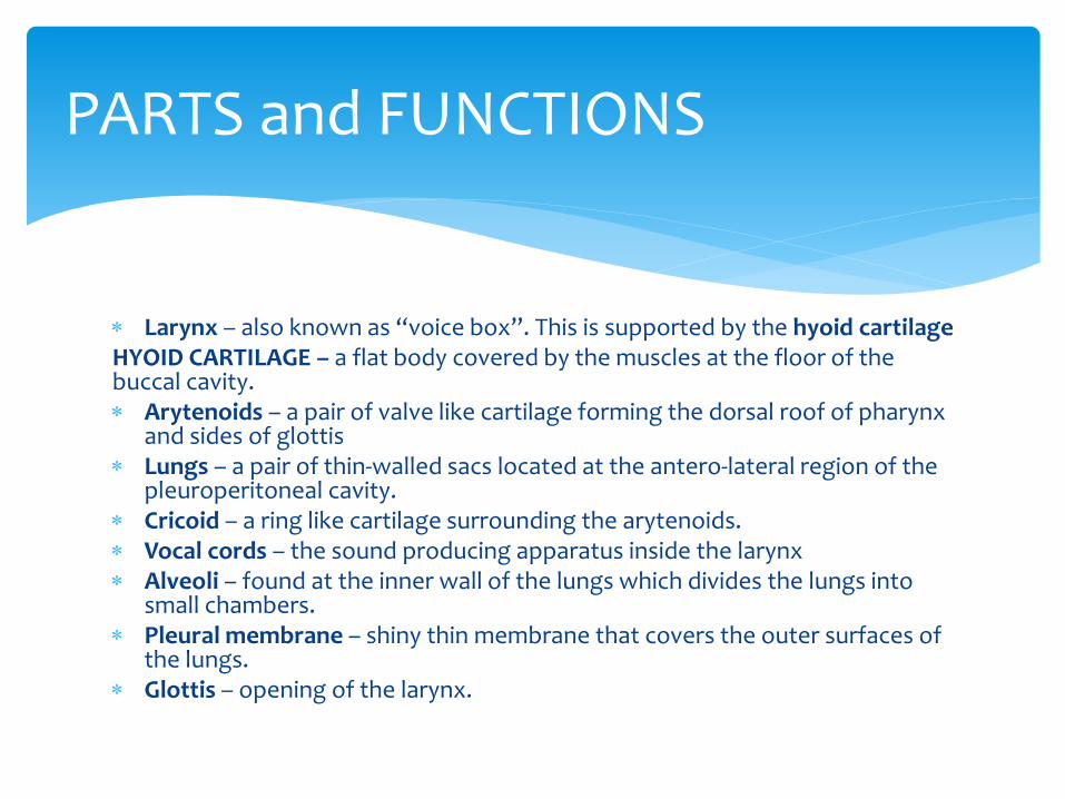
Larynx – also known as “voice box”. This is supported by the hyoid cartilageHYOID CARTILAGE – a flat body covered by the muscles at the floor of the buccal cavity. Arytenoids – a pair of valve like cartilage forming the dorsal roof of pharynx
and sides of glottis Lungs – a pair of thin-walled sacs located at the antero-lateral region of the
pleuroperitoneal cavity. Cricoid – a ring like cartilage surrounding the arytenoids. Vocal cords – the sound producing apparatus inside the larynx Alveoli – found at the inner wall of the lungs which divides the lungs into
small chambers. Pleural membrane – shiny thin membrane that covers the outer surfaces of
the lungs. Glottis – opening of the larynx.
Unlike birds and mammals, amphibians are cold blooded.
They do not use up any energy for keeping their bodies at a constant temperature.
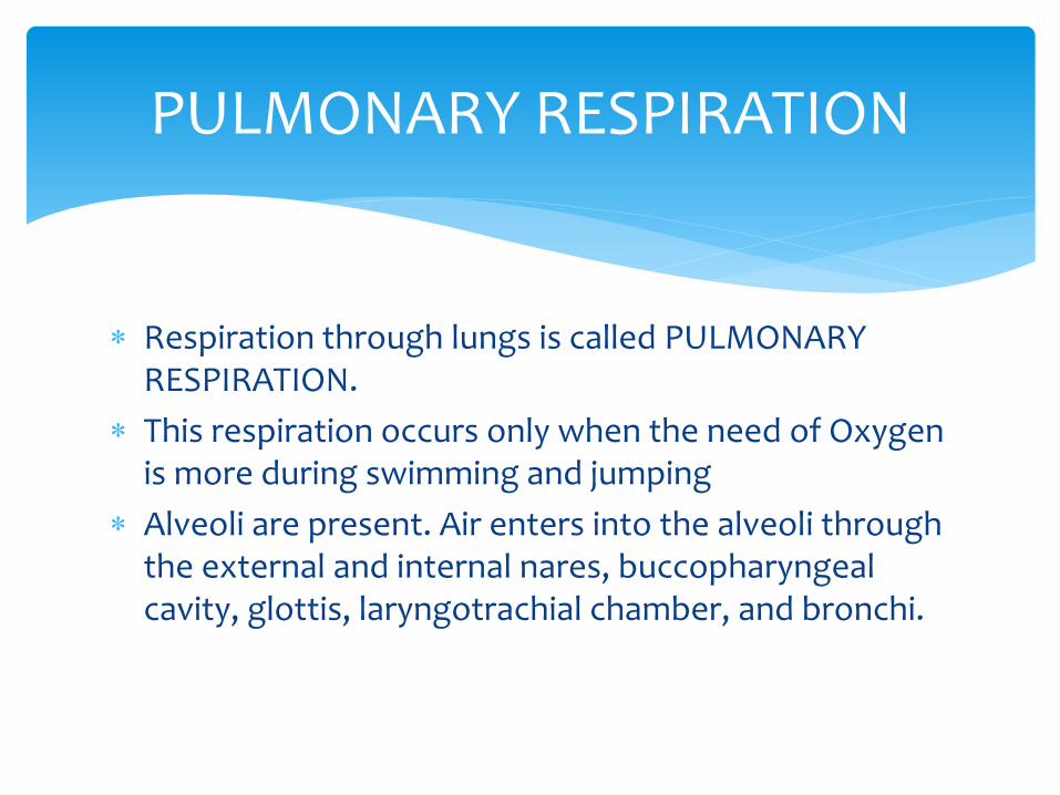
PULMONARY RESPIRATION
Respiration through lungs is called PULMONARY RESPIRATION.
This respiration occurs only when the need of Oxygen is more during swimming and jumping
Alveoli are present. Air enters into the alveoli through the external and internal nares, buccopharyngealcavity, glottis, laryngotrachial chamber, and bronchi.
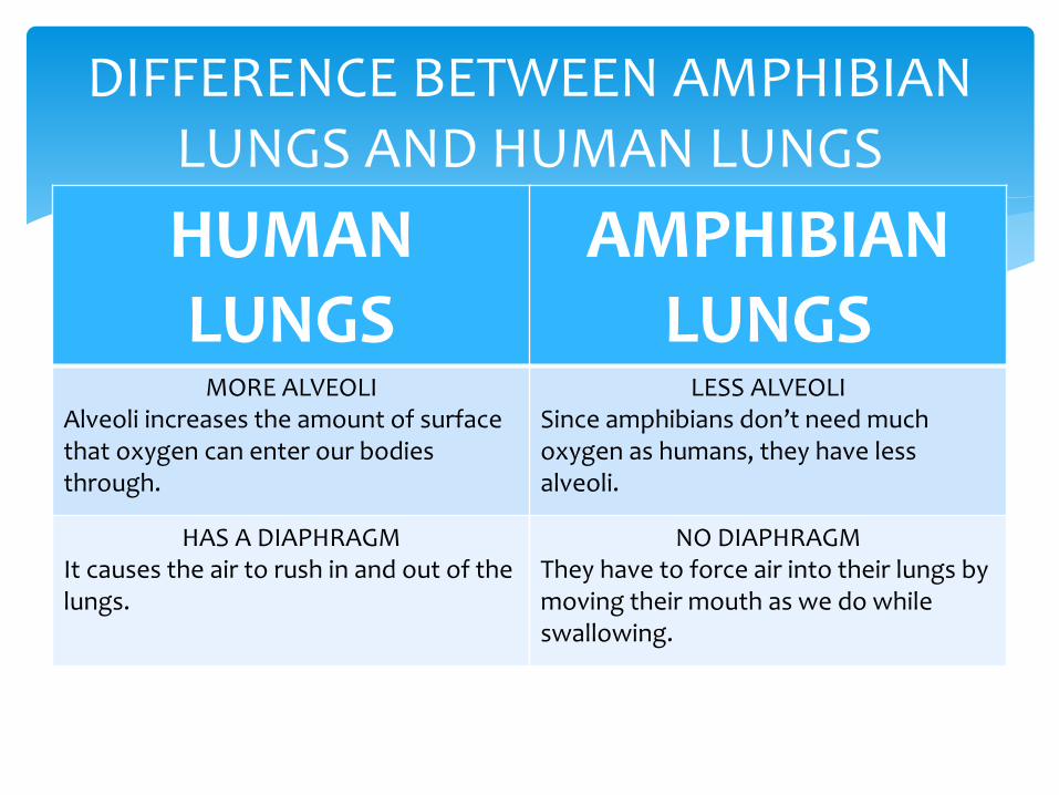
DIFFERENCE BETWEEN AMPHIBIAN LUNGS AND HUMAN LUNGS
HUMAN LUNGS
AMPHIBIAN LUNGS
MORE ALVEOLIAlveoli increases the amount of surface that oxygen can enter our bodies through.
LESS ALVEOLISince amphibians don’t need much oxygen as humans, they have less alveoli.
HAS A DIAPHRAGMIt causes the air to rush in and out of the lungs.
NO DIAPHRAGMThey have to force air into their lungs by moving their mouth as we do while swallowing.
CUTANEOUS RESPIRATION
Respiration through the skin is called CUTANEOUS RESPIRATION.
It occurs in hibernation and in water
The skin of amphibians are very thin and is rich with blood capillaries.
The water carries oxygen which diffuses into the capillaries and the carbon dioxide in the blood diffuses out.
AVES
Function:
• Respiration
• Thermoregulation (maintaining normal body temp.
• Communication
Parts:
• Larynx – is not used to make sound
• Syrinx – serve as voice box
• Lungs
• Air Sacs - Depending upon the species, the bird has seven or nine air sacs.
The air sacs include:
Two posterior thoracic
Two abdominal
Two anterior thoracic
Two cervical (these are not present in some species)
One interclavicular
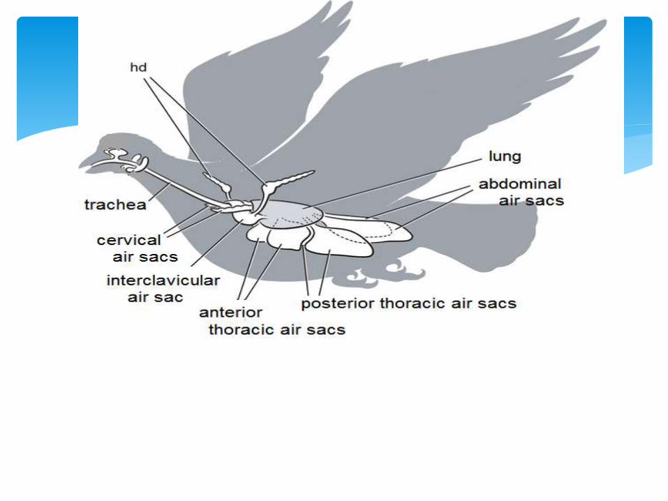
The air sacs of birds extend into the humerus (the bone between the shoulder and elbow), the femur (the thigh
bone), the vertebrae and even the skull.
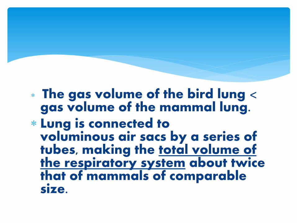
The gas volume of the bird lung < gas volume of the mammal lung.
Lung is connected to voluminous air sacs by a series of tubes, making the total volume of the respiratory system about twice that of mammals of comparable size.
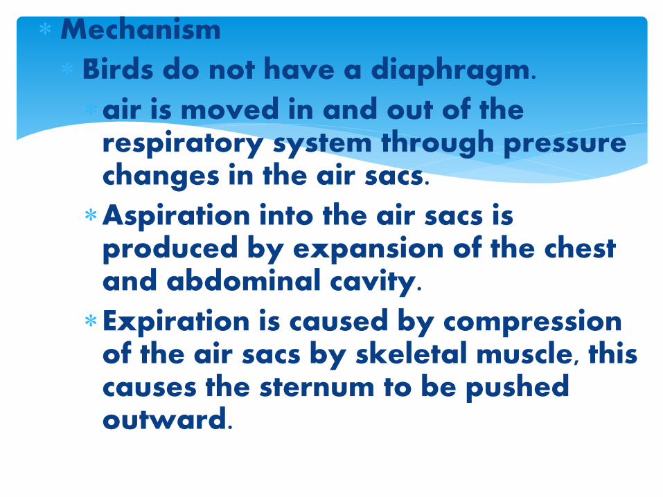
Mechanism
Birds do not have a diaphragm.
air is moved in and out of the respiratory system through pressure changes in the air sacs.
Aspiration into the air sacs is produced by expansion of the chest and abdominal cavity.
Expiration is caused by compression of the air sacs by skeletal muscle, this causes the sternum to be pushed outward.
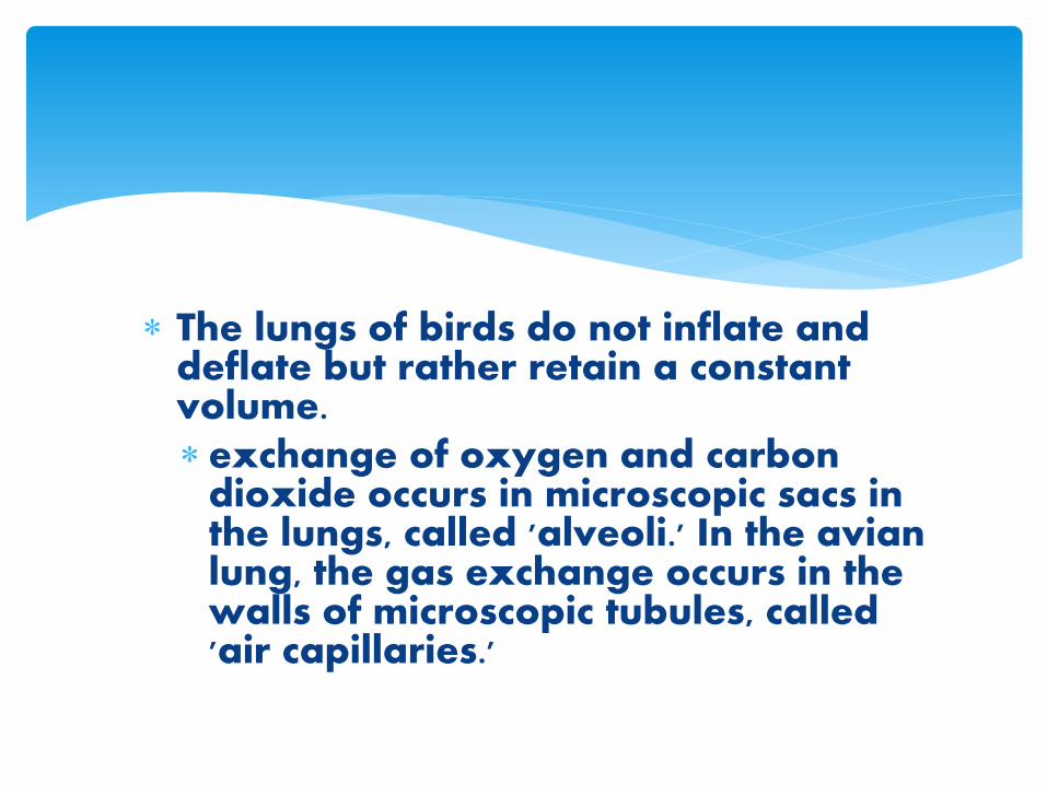
The lungs of birds do not inflate and deflate but rather retain a constant volume.exchange of oxygen and carbon
dioxide occurs in microscopic sacs in the lungs, called 'alveoli.' In the avian lung, the gas exchange occurs in the walls of microscopic tubules, called 'air capillaries.'
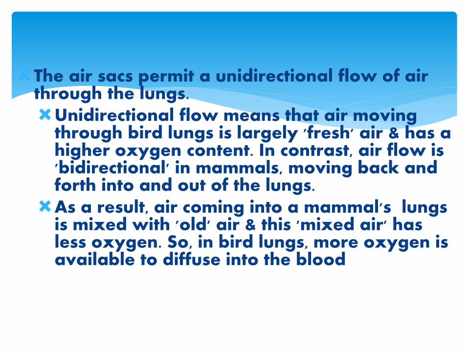
The air sacs permit a unidirectional flow of air through the lungs. Unidirectional flow means that air moving
through bird lungs is largely 'fresh' air & has a higher oxygen content. In contrast, air flow is 'bidirectional' in mammals, moving back and forth into and out of the lungs.
As a result, air coming into a mammal's lungs is mixed with 'old' air & this 'mixed air' has less oxygen. So, in bird lungs, more oxygen is available to diffuse into the blood
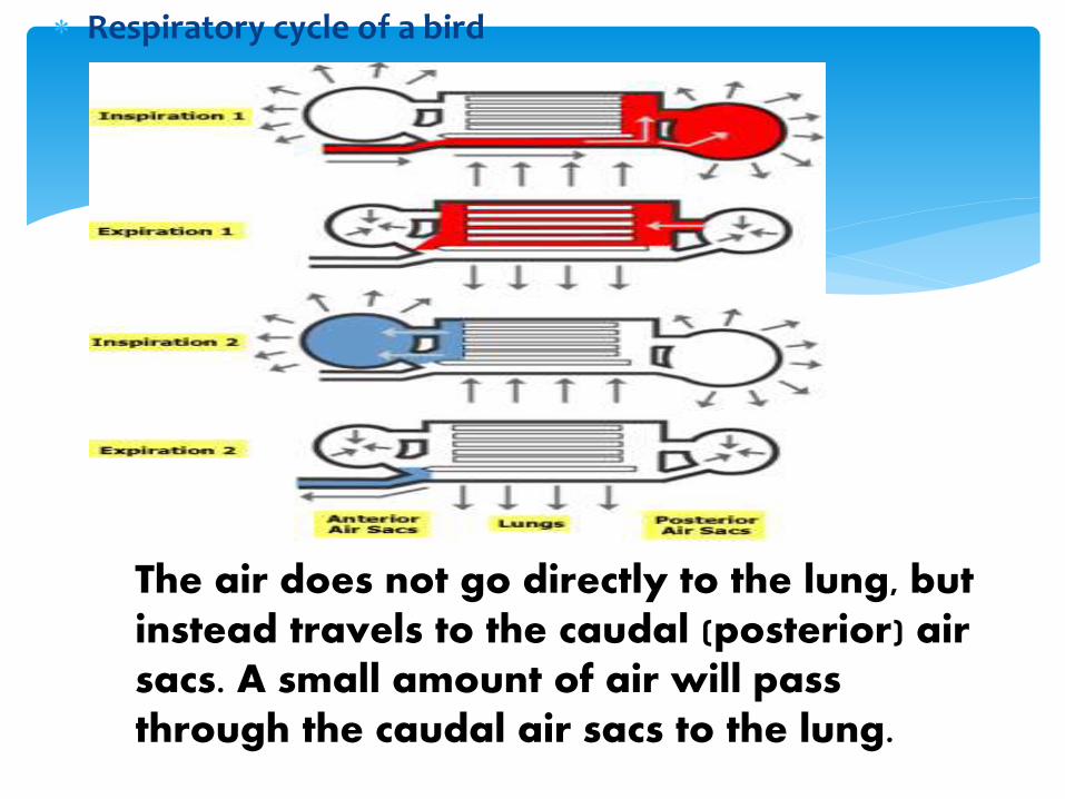
Respiratory cycle of a bird
The air does not go directly to the lung, but instead travels to the caudal (posterior) air sacs. A small amount of air will pass through the caudal air sacs to the lung.
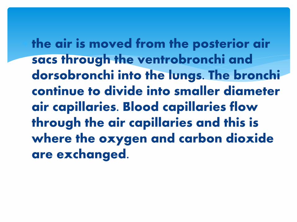
the air is moved from the posterior air sacs through the ventrobronchi and dorsobronchi into the lungs. The bronchi continue to divide into smaller diameter air capillaries. Blood capillaries flow through the air capillaries and this is where the oxygen and carbon dioxide are exchanged.
When the bird inspires the second time, the air moves to the cranial air sacs.
On the second expiration, the air moves out of the cranial air sacs, through the syrinx into the trachea, through the larynx, and finally through the nasal cavity and out of the nostrils.
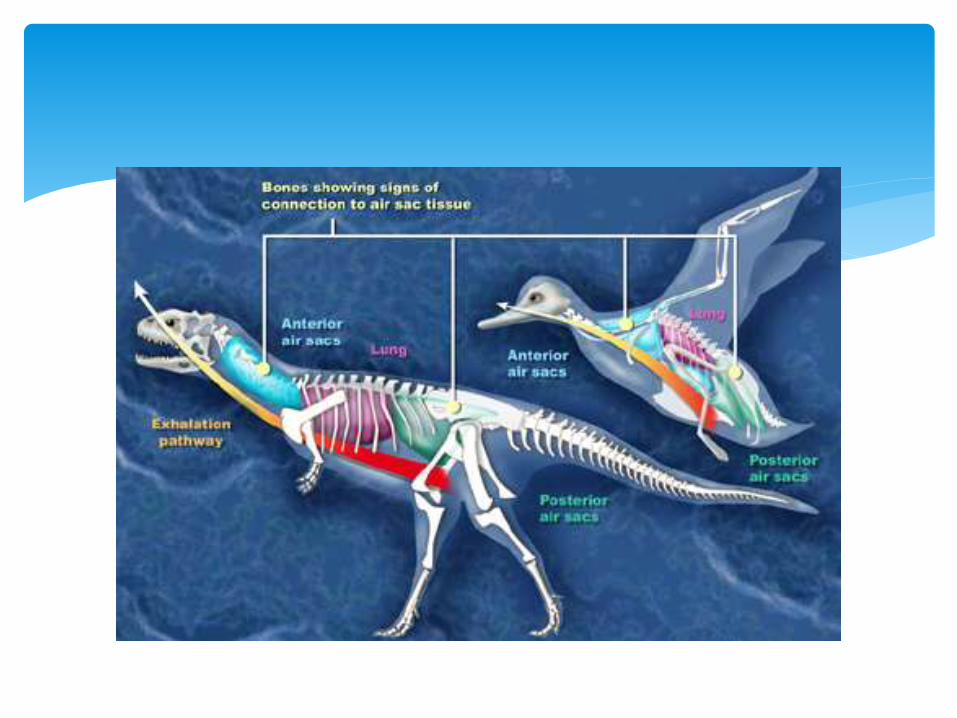
Bird-like respiratory systems in dinosaurs -- A recent analysis showing the presence of a very bird-like pulmonary, or lung, system in predatory dinosaurs provides more evidence of an evolutionary link between dinosaurs and birds.
Gabriel Tacan PRETTY
Mammals
The mammalian respiratory system equilibrates air to the body, protects against foreign materials, and allows for gas exchange.
In mammals, pulmonary ventilation occurs via inhalation when air enters the body through the nasal cavity.
The chief organ in mammalian respiration is the lungs.
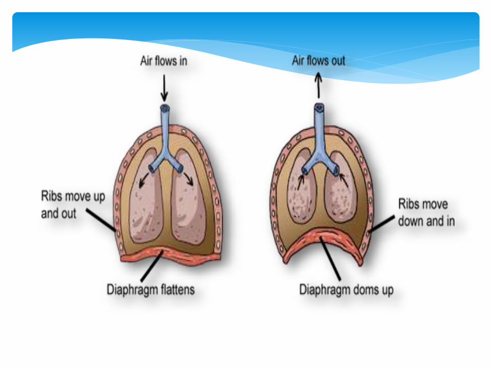
Inhalation happens when the rib cage opens up and the diaphragm flattens and moves downward. The lungs can then expand into the larger space that causes the air pressure inside them to decrease, and the drop in air pressure inside the lung makes the outside air rush inside.
Exhalation is the opposite process. The diaphragm and the rib muscles relax to their neutral state that causes the lungs to contract. The squashing of the lungs increases their air pressure and forces the air to flow out.
Horses are obligate nasal breathers, which means that they must breathe through their noses.
It is thought that this modification allows horses to graze with their heads down while separate nasal passages breath in air and sniff for potential predators.
Marine mammals breathe oxygen with lungs just like their terrestrial brethren, but with a few differences. First of all, to prevent water from getting into their airway they have adapted muscles or cartilaginous flaps to seal their tracheas when under the water. Additionally, they exchange up to 90% of their gases in a single breath, which helps them gather as much oxygen as possible.
Lastly, it can be dangerous for diving mammals to have air in their lungs when they dive to great depths.

![Respiratory System [โหมดความเข้ากันได้] · PATHOLOGY OF RESPIRATORY SYSTEM นพ. อรรณพ นาคะป ท Respiratory system U it](https://img.dokumen.tips/doc/110x75/5fa578efd4e80f055f6b3401/respiratory-system-aaaaaaaaaaaaaaaaaa-pathology.jpg)


![Respiratory system roadmap.pptx [Repaired] - Loginanatomical-sciences.health.wits.ac.za/roadmaps/Respiratory system... · DIVISION OF THE RESPIRATORY SYSTEM CONDUCTING PORTION Nasal](https://img.dokumen.tips/doc/110x75/5a78c3d87f8b9ae6228c9db0/respiratory-system-repaired-loginanatomical-scienceshealthwitsaczaroadmapsrespiratory.jpg)