Embed Size (px)
Citation preview
MIOT Hospitals : 4/112, Mount Poonamallee Road, Manapakkam Chennai - 600089, IndiaTel: +91 44 4200 2288, Fax: +91 44 2249 1313 Email: [email protected] www.miotinternational.com
w w w . m i o t i n t e r n a t i o n a l . c o m
Precision Onco-Imaging using state-of-the art
PET/CT
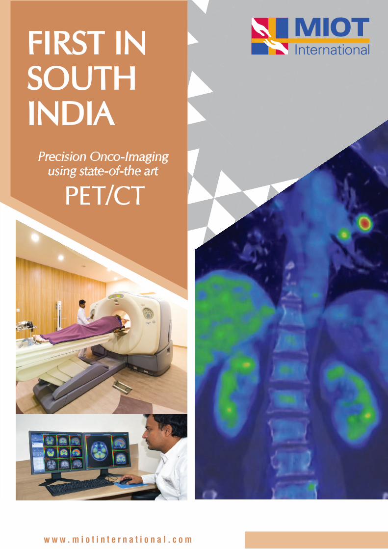
FIRST IN SOUTHINDIA
Targeted Radionuclide Therapy:MIOT opens the door to better and safer treatment beyond imaging:In addition to the wide range of diagnostic services (using PET/CT and SPECT) the Department of Nuclear Medicine at MIOT is equipped with the state-of-the-art radionuclide therapy ward for providing ‘Targeted Radionuclide therapy’ for certain cancers.
Targeted Radionuclide Therapy is rapidly evolving into an alternative mode of treatment in oncology with better tumor targeting and lesser treatment related toxicity than conventional chemotherapy / external beam radiotherapy. Several randomized and ongoing clinical trials in cancer patients have shown its potential to improve quality of life and also increase the overall and disease specific survival.
Following are the radionuclide therapies available in MIOT. Radio-iodine therapy for localized and metastatic thyroid cancer. Radio-iodinated MIBG therapy for pheochromocytomas/paragangliomas. ‘Peptide Receptor Radionuclide Therapy’ (PRRT) for neuroendocrine tumors. Radio-embolization therapy for primary and metastatic liver cancers.
For further clarification or for an appointment, please call at 044 4200 2288 (extn 4452) or email us at [email protected]
Diagnosis to rehabilitation - under one roofWe are one of the few centres in India offering complete care from diagnosis to treatment and rehabilitation in a single facility. We fight alongside our patients with the world’s latest treatments and a wealth of experience and expertise.
World-class imaging & laboratory facilities - for accurate diagnosisOur imaging and diagnostic facilities, which include a state-of-the-art laboratory that is ranked 8th in the world andadvanced imaging modalities, deliver speedy and accurate reports.
Full time dedicated team of Oncologists, customised treatment plansOur team of Oncologists works with other specialists to devise a customised treatment plan that is comfortable for every patient. The goal is to ensure that patients complete their course of treatment as planned. This is essential for their effective recovery and return to normal life.
New-wave treatment with focus on patient comfortWe offer the latest chemotherapeutic drugs and are hence able to anticipate and offset side effects with pre-medications which make treatment easier. In surgery, we work with other specialists, including reconstructive surgeons, to preserve as
much of the patient’s organ as we can while removing the cancerous tissue completely.
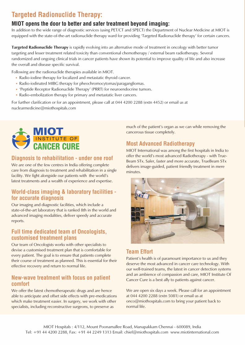
Most Advanced RadiotherapyMIOT International was among the first hospitals in India to offer the world’s most advanced Radiotherapy - with True-Beam STx. Safer, faster and more accurate, TrueBeam STx delivers image-guided, patient friendly treatment in mere minutes.
Team EffortPatient’s health is of paramount importance to us and they deserve the most advanced in cancer care technology. With our well-trained teams, the latest in cancer detection systems and an ambience of compassion and care, MIOT Institute Of Cancer Cure is a best ally to patients against cancer.
We are open six days a week. Please call for an appointment at 044 4200 2288 (extn 5081) or email us at [email protected] to bring your patient back to normal life.
How will PET/CT scan help in a patient’s treatment? Improved Diagnosis: See MORE, See BETTER, See EARLIER: The significantly improved precision (higher imaging / diagnostic accuracy) of co-registration of anatomy and function (down to the molecullar level) by the PET/CT results in improved reader (nuclear physician) confidence in earlier detection of disease precise localization and interpretation (characterization) of the hyper metabolic disease areas marked reduction in inconclusive scan findings.
Better Evaluation and devising treatment protocol: The PET/CT scan results not only help to have a major impact on physicians’ diagnosis of cancer but also helps the physician to predict the likely outcome of various therapeutic alternatives during primary staging / re-staging of cancer planning and individualizing treatment protocols and methods by efficiently monitoring the response of the disease to treatment .
Impacting disease management & lives: Lesser time & better patient compliance Both functional and detailed anatomical imaging done in a single setting. Avoiding un-necessary surgeries and invasive procedures. Reducing treatment related side effects. Reducing overall treatment costs.
Besides revolutionizing cancer diagnosis and treatment protocols, the PET/CT is a powerful tool for diagnostic assessment of modern day diseases such as neuro degenerative disorders (alzeihmer’s, parkinsons disease etc..) and coronary artery disease.
Sensitivity.Accuracy.Confidence.
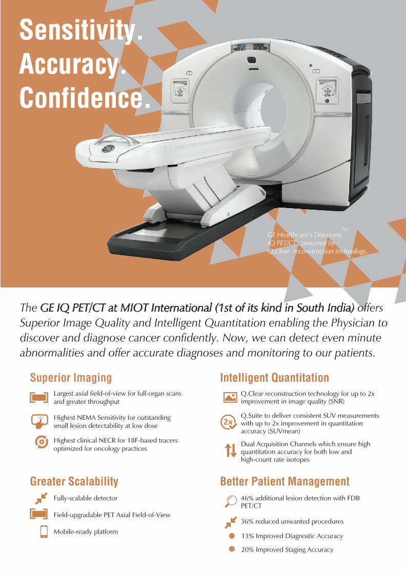
The GE IQ PET/CT at MIOT International (1st of its kind in South India) offers Superior Image Quality and Intelligent Quantitation enabling the Physician to discover and diagnose cancer confidently. Now, we can detect even minute abnormalities and offer accurate diagnoses and monitoring to our patients.
TMGE Healthcare’s Discovery IQ PET/CT , powered by Q.Clear reconstruction technology,
Superior Imaging Intelligent Quantitation
Greater Scalability Better Patient Management
Largest axial field-of-view for full-organ scansand greater throughput
Highest NEMA Sensitivity for outstandingsmall lesion detectability at low dose
Highest clinical NECR for 18F-based tracersoptimized for oncology practices
Q.Clear reconstruction technology for up to 2x improvement in image quality (SNR)
Q.Suite to deliver consistent SUV measurements with up to 2x improvement in quantitation accuracy (SUVmean)
Dual Acquisition Channels which ensure high quantitation accuracy for both low and high-count rate isotopes
Fully-scalable detector
Field-upgradable PET Axial Field-of-View
Mobile-ready platform
46% additional lesion detection with FDB PET/CT
36% reduced unwanted procedures
13% Improved Diagnostic Accuracy
20% Improved Staging Accuracy
What is a PET/CT? PET/CT is the work horse of modern onco imaging which can detect disease that is not identified on conventional CT scans alone. PET/CT scan consists of 2 components – the diagnostic CT (computed tomography) scan (anatomical) and the PET (positron emission tomography) scan (functional) – both in the same sitting.
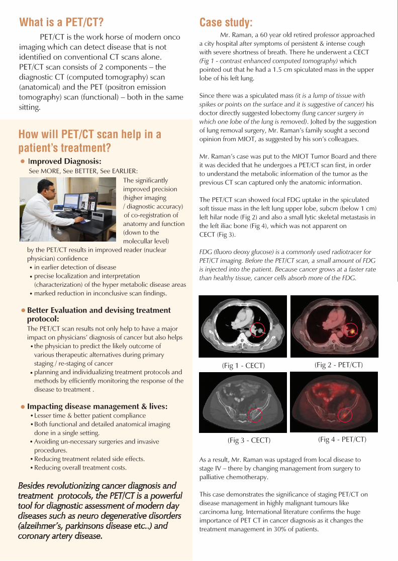
Case study: Mr. Raman, a 60 year old retired professor approached a city hospital after symptoms of persistent & intense cough with severe shortness of breath. There he underwent a CECT (Fig 1 - contrast enhanced computed tomography) which pointed out that he had a 1.5 cm spiculated mass in the upper lobe of his left lung.
Since there was a spiculated mass (it is a lump of tissue with spikes or points on the surface and it is suggestive of cancer) his doctor directly suggested lobectomy (lung cancer surgery in which one lobe of the lung is removed). Jolted by the suggestion of lung removal surgery, Mr. Raman’s family sought a second opinion from MIOT, as suggested by his son’s colleagues.
Mr. Raman’s case was put to the MIOT Tumor Board and there it was decided that he undergoes a PET/CT scan first, in order to understand the metabolic information of the tumor as the previous CT scan captured only the anatomic information.
The PET/CT scan showed focal FDG uptake in the spiculated soft tissue mass in the left lung upper lobe, subcm (below 1 cm) left hilar node (Fig 2) and also a small lytic skeletal metastasis in the left iliac bone (Fig 4), which was not apparent on CECT (Fig 3).
FDG (fluoro deoxy glucose) is a commonly used radiotracer for PET/CT imaging. Before the PET/CT scan, a small amount of FDG is injected into the patient. Because cancer grows at a faster rate than healthy tissue, cancer cells absorb more of the FDG.
As a result, Mr. Raman was upstaged from local disease to stage IV – there by changing management from surgery to palliative chemotherapy.
This case demonstrates the significance of staging PET/CT on disease management in highly malignant tumours like carcinoma lung. International literature confirms the huge importance of PET CT in cancer diagnosis as it changes the treatment management in 30% of patients.
(Fig 1 - CECT) (Fig 2 - PET/CT)
(Fig 3 - CECT) (Fig 4 - PET/CT)
How will PET/CT scan help in a patient’s treatment? Improved Diagnosis: See MORE, See BETTER, See EARLIER: The significantly improved precision (higher imaging / diagnostic accuracy) of co-registration of anatomy and function (down to the molecullar level) by the PET/CT results in improved reader (nuclear physician) confidence in earlier detection of disease precise localization and interpretation (characterization) of the hyper metabolic disease areas marked reduction in inconclusive scan findings.
Better Evaluation and devising treatment protocol: The PET/CT scan results not only help to have a major impact on physicians’ diagnosis of cancer but also helps the physician to predict the likely outcome of various therapeutic alternatives during primary staging / re-staging of cancer planning and individualizing treatment protocols and methods by efficiently monitoring the response of the disease to treatment .
Impacting disease management & lives: Lesser time & better patient compliance Both functional and detailed anatomical imaging done in a single setting. Avoiding un-necessary surgeries and invasive procedures. Reducing treatment related side effects. Reducing overall treatment costs.
Besides revolutionizing cancer diagnosis and treatment protocols, the PET/CT is a powerful tool for diagnostic assessment of modern day diseases such as neuro degenerative disorders (alzeihmer’s, parkinsons disease etc..) and coronary artery disease.
Sensitivity.Accuracy.Confidence.
The GE IQ PET/CT at MIOT International (1st of its kind in South India) offers Superior Image Quality and Intelligent Quantitation enabling the Physician to discover and diagnose cancer confidently. Now, we can detect even minute abnormalities and offer accurate diagnoses and monitoring to our patients.
TMGE Healthcare’s Discovery IQ PET/CT , powered by Q.Clear reconstruction technology,
Superior Imaging Intelligent Quantitation
Greater Scalability Better Patient Management
Largest axial field-of-view for full-organ scansand greater throughput
Highest NEMA Sensitivity for outstandingsmall lesion detectability at low dose
Highest clinical NECR for 18F-based tracersoptimized for oncology practices
Q.Clear reconstruction technology for up to 2x improvement in image quality (SNR)
Q.Suite to deliver consistent SUV measurements with up to 2x improvement in quantitation accuracy (SUVmean)
Dual Acquisition Channels which ensure high quantitation accuracy for both low and high-count rate isotopes
Fully-scalable detector
Field-upgradable PET Axial Field-of-View
Mobile-ready platform
46% additional lesion detection with FDB PET/CT
36% reduced unwanted procedures
13% Improved Diagnostic Accuracy
20% Improved Staging Accuracy
What is a PET/CT? PET/CT is the work horse of modern onco imaging which can detect disease that is not identified on conventional CT scans alone. PET/CT scan consists of 2 components – the diagnostic CT (computed tomography) scan (anatomical) and the PET (positron emission tomography) scan (functional) – both in the same sitting.
Case study: Mr. Raman, a 60 year old retired professor approached a city hospital after symptoms of persistent & intense cough with severe shortness of breath. There he underwent a CECT (Fig 1 - contrast enhanced computed tomography) which pointed out that he had a 1.5 cm spiculated mass in the upper lobe of his left lung.
Since there was a spiculated mass (it is a lump of tissue with spikes or points on the surface and it is suggestive of cancer) his doctor directly suggested lobectomy (lung cancer surgery in which one lobe of the lung is removed). Jolted by the suggestion of lung removal surgery, Mr. Raman’s family sought a second opinion from MIOT, as suggested by his son’s colleagues.
Mr. Raman’s case was put to the MIOT Tumor Board and there it was decided that he undergoes a PET/CT scan first, in order to understand the metabolic information of the tumor as the previous CT scan captured only the anatomic information.
The PET/CT scan showed focal FDG uptake in the spiculated soft tissue mass in the left lung upper lobe, subcm (below 1 cm) left hilar node (Fig 2) and also a small lytic skeletal metastasis in the left iliac bone (Fig 4), which was not apparent on CECT (Fig 3).
FDG (fluoro deoxy glucose) is a commonly used radiotracer for PET/CT imaging. Before the PET/CT scan, a small amount of FDG is injected into the patient. Because cancer grows at a faster rate than healthy tissue, cancer cells absorb more of the FDG.
As a result, Mr. Raman was upstaged from local disease to stage IV – there by changing management from surgery to palliative chemotherapy.
This case demonstrates the significance of staging PET/CT on disease management in highly malignant tumours like carcinoma lung. International literature confirms the huge importance of PET CT in cancer diagnosis as it changes the treatment management in 30% of patients.
(Fig 1 - CECT) (Fig 2 - PET/CT)
(Fig 3 - CECT) (Fig 4 - PET/CT)
MIOT Hospitals : 4/112, Mount Poonamallee Road, Manapakkam Chennai - 600089, IndiaTel: +91 44 4200 2288, Fax: +91 44 2249 1313 Email: [email protected] www.miotinternational.com
w w w . m i o t i n t e r n a t i o n a l . c o m
Precision Onco-Imaging using state-of-the art
PET/CT
FIRST IN SOUTHINDIA
Targeted Radionuclide Therapy:MIOT opens the door to better and safer treatment beyond imaging:In addition to the wide range of diagnostic services (using PET/CT and SPECT) the Department of Nuclear Medicine at MIOT is equipped with the state-of-the-art radionuclide therapy ward for providing ‘Targeted Radionuclide therapy’ for certain cancers.
Targeted Radionuclide Therapy is rapidly evolving into an alternative mode of treatment in oncology with better tumor targeting and lesser treatment related toxicity than conventional chemotherapy / external beam radiotherapy. Several randomized and ongoing clinical trials in cancer patients have shown its potential to improve quality of life and also increase the overall and disease specific survival.
Following are the radionuclide therapies available in MIOT. Radio-iodine therapy for localized and metastatic thyroid cancer. Radio-iodinated MIBG therapy for pheochromocytomas/paragangliomas. ‘Peptide Receptor Radionuclide Therapy’ (PRRT) for neuroendocrine tumors. Radio-embolization therapy for primary and metastatic liver cancers.
For further clarification or for an appointment, please call at 044 4200 2288 (extn 4452) or email us at [email protected]
Diagnosis to rehabilitation - under one roofWe are one of the few centres in India offering complete care from diagnosis to treatment and rehabilitation in a single facility. We fight alongside our patients with the world’s latest treatments and a wealth of experience and expertise.
World-class imaging & laboratory facilities - for accurate diagnosisOur imaging and diagnostic facilities, which include a state-of-the-art laboratory that is ranked 8th in the world andadvanced imaging modalities, deliver speedy and accurate reports.
Full time dedicated team of Oncologists, customised treatment plansOur team of Oncologists works with other specialists to devise a customised treatment plan that is comfortable for every patient. The goal is to ensure that patients complete their course of treatment as planned. This is essential for their effective recovery and return to normal life.
New-wave treatment with focus on patient comfortWe offer the latest chemotherapeutic drugs and are hence able to anticipate and offset side effects with pre-medications which make treatment easier. In surgery, we work with other specialists, including reconstructive surgeons, to preserve as
much of the patient’s organ as we can while removing the cancerous tissue completely.
Most Advanced RadiotherapyMIOT International was among the first hospitals in India to offer the world’s most advanced Radiotherapy - with True-Beam STx. Safer, faster and more accurate, TrueBeam STx delivers image-guided, patient friendly treatment in mere minutes.
Team EffortPatient’s health is of paramount importance to us and they deserve the most advanced in cancer care technology. With our well-trained teams, the latest in cancer detection systems and an ambience of compassion and care, MIOT Institute Of Cancer Cure is a best ally to patients against cancer.
We are open six days a week. Please call for an appointment at 044 4200 2288 (extn 5081) or email us at [email protected] to bring your patient back to normal life.