Embed Size (px)
Citation preview

BASE HOSPITAL GROUPONTARIO
Chapter 10 for 12 Lead Training
-12 Lead Interpretation – Part 2-
Ontario Base Hospital GroupEducation Subcommittee
2008
TIME IS MUSCLE

OBHG Education Subcommittee
12 Lead Interpretation – Part 2
REVIEWERS/CONTRIBUTORS
Neil Freckleton, AEMCA, ACPHamilton Base Hospital
Jim Scott, AEMCA, PCPSault Area Hospital
Ed Ouston, AEMCA, ACPOttawa Base Hospital
Laura McCleary, AEMCA, ACPSOCPC
Tim Dodd, AEMCA, ACPHamilton Base Hospital
Dr. Rick Verbeek, Medical DirectorSOCPC2008 Ontario Base Hospital Group
AUTHOR
Greg Soto, BEd, BA, ACPNiagara Base Hospital

OBHG Education Subcommittee
Chapter 10 - Objectives
Recognize ST-depression and relate to the ACS patient
Recognize Reciprocal Changes (RCs) and relate to the significance of STEMI
Recognized Q-waves and relate to the ACS patient
Discuss the evolution of an AMIExplain the reasons why a normal ECG
does not rule out AMI

OBHG Education Subcommittee
Epicardial Coronary Artery
Lateral Wall of LV
Positive Electrode
Septum
Left Ventricular
Cavity
Inferior Wall of LV
Ischemia
Thrombus forming

OBHG Education Subcommittee
Ischemia
Inadequate oxygen to tissue
Subendocardial
Represented by ST depression or T-wave inversion
May or may not result in infarct

OBHG Education Subcommittee
ST depression

OBHG Education Subcommittee
T-wave Inversion

OBHG Education Subcommittee
Evolution of AMI
• Hyperacute T WaveHyperacute T Wave

OBHG Education Subcommittee
Evolution of AMI
• AcuteAcute

OBHG Education Subcommittee
Evolution of AMI
• AcuteAcute

OBHG Education Subcommittee
Evolution of AMI
• Age undeterminedAge undetermined

OBHG Education Subcommittee
Hyper-acute T-waves
Earliest ECG sign of AMI
Tall and peaked w/in minutes of blood flow interruption
Differential Dx:hyperkalemia BERLVH

OBHG Education Subcommittee
Why hyper-acute T-wave are important to recognize

OBHG Education Subcommittee
AMI Recognition
A “normal” 12-lead ECG
DOES NOT rule out AMINot all AMI have STE (approx. 50%)Early AMI may have no STE but may
evolve over timeNon STEMI AMI have non specific but
abnormal ECGs

OBHG Education Subcommittee
Why can’t AMI be ruled out?
PHECG has high specificity for STEMI = 97%*
Meaning = when PHECG shows STEMI it almost always turns out to be an AMI.

OBHG Education Subcommittee
PHECG has only moderate sensitivity for AMI = 68%
Meaning - when PHECG does not show STEMI only 68% of time does it turn out to NOT be an AMI. (over 30% of AMI patients do not have STE on PHECG)
CAN’T RULE OUT AMI WITH NO STE on 12 LEAD ECG
Source: Ioannides JA et al. Accuracy & clinical effect of out-of-hospital ECG in the diagnosis of acute cardiac ischemia: a meta-analysis. Annals of Emergency Medicine 2001;37.
Why can’t AMI be ruled out?

OBHG Education Subcommittee
Practice

OBHG Education Subcommittee
Practice

OBHG Education Subcommittee
Practice

OBHG Education Subcommittee
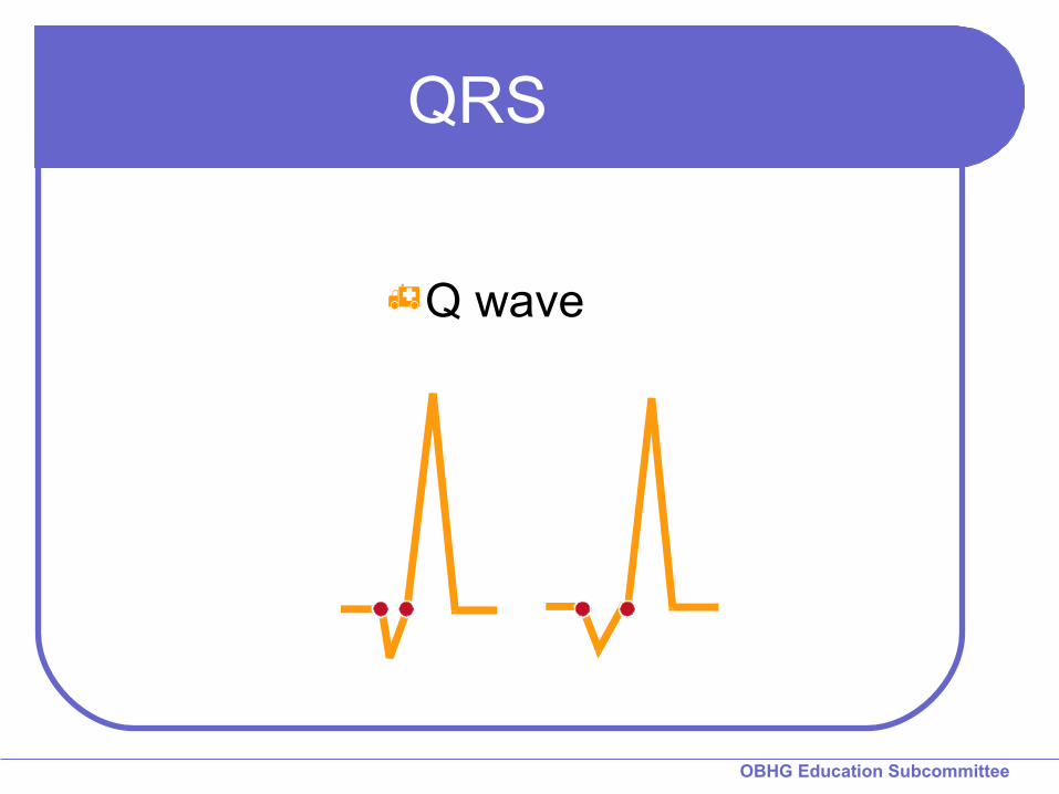
QRS
Q wavesPhysiologic Q waves
< .04 sec (40ms)
Pathologic Q waves>.04 sec (40 ms)

OBHG Education Subcommittee
Q-wave & Infarct
represent irreversible necrosis – death of tissue
may develop early (1st hour) but usually 8-12 hours post-AMI
may persist permanently but some resolve regardless of reperfusion
not all AMIs produce Q-waves
OBHG Education Subcommittee
QRS
Q wave

OBHG Education Subcommittee
QS Complex

OBHG Education Subcommittee
Common Q-waves
“age undetermined”Likely old septal MI ↑ index of suspicion not
a bad idea
Q-wave associated with an AMI = necrosis has likely begun
↑ ↑ severity/seriousnessseverity/seriousness

BASE HOSPITAL GROUPONTARIO
Sample ECGs with Q-waves
Find them……….

OBHG Education Subcommittee
Septal – V1 to V3
(? – II, III, aVF )

OBHG Education Subcommittee
Lateral - aVL

OBHG Education Subcommittee
aVL, V1 – V5

BASE HOSPITAL GROUPONTARIO
Reciprocal Changes

OBHG Education Subcommittee
Reciprocal Changes
Occur in larger MIAble to “see” the MI on the opposite side
because it is larger
RC’s make the STE more likely to be due to AMIDon’t have to have RC’s but they make
the diagnosis easier

OBHG Education Subcommittee
Reciprocal Changes

OBHG Education Subcommittee
Reciprocal Changes

OBHG Education Subcommittee
Reciprocal Changes
II, III, aVFII, III, aVF I, aVL, V leadsI, aVL, V leads

OBHG Education Subcommittee
Reciprocal Changes
Inferior
Anterior = Septal, Anterior and Lateral walls

OBHG Education Subcommittee
Practice

OBHG Education Subcommittee
Practice

OBHG Education Subcommittee
AMI Recognition

OBHG Education Subcommittee
AMI Recognition
Imitators of infarctBBB LVHVentricular beatsPericarditisEarly RepolarizationOthers

OBHG Education Subcommittee
Summary
AMI recognitionKnow what you are looking for
> 1mm of ST elevation in limb leads> 2mm of ST elevation in chest
leadsTwo contiguous leads
Know where you are lookingPositive electrode as an “eye”Memorize lead locations

OBHG Education Subcommittee
Summary
Reciprocal ChangesNot necessary to presume
infarctionStrong confirming evidence
when present

OBHG Education Subcommittee
Summary
ST segment elevation is presumptive evidence for AMI
Other conditions may also cause ST elevation

OBHG Education Subcommittee
Summary
NEVER FORGET:
A normal 12-Lead ECG DOES NOT rule out AMI

BASE HOSPITAL GROUPONTARIO
QUESTIONS?

BASE HOSPITAL GROUPONTARIO
Well Done!
Education Subcommittee
START QUIT