Embed Size (px)
Citation preview
VISUAL PATHWAYS AND THE OPTIC NERVE
DR.EJAZ-UL-HAQPGR DEPARTMENT OF OPHTHALMOLOGY UNIT-III

Contents Definition of visual field Normal vision What are the causes of visual field defect? Anatomy of Visual pathways
Optic nerve Optic chiasm Retrochiasm
Visual field deficits due to various lesions. How to approach a patient with specific visual field deficit ? Optic neuropathies Summary
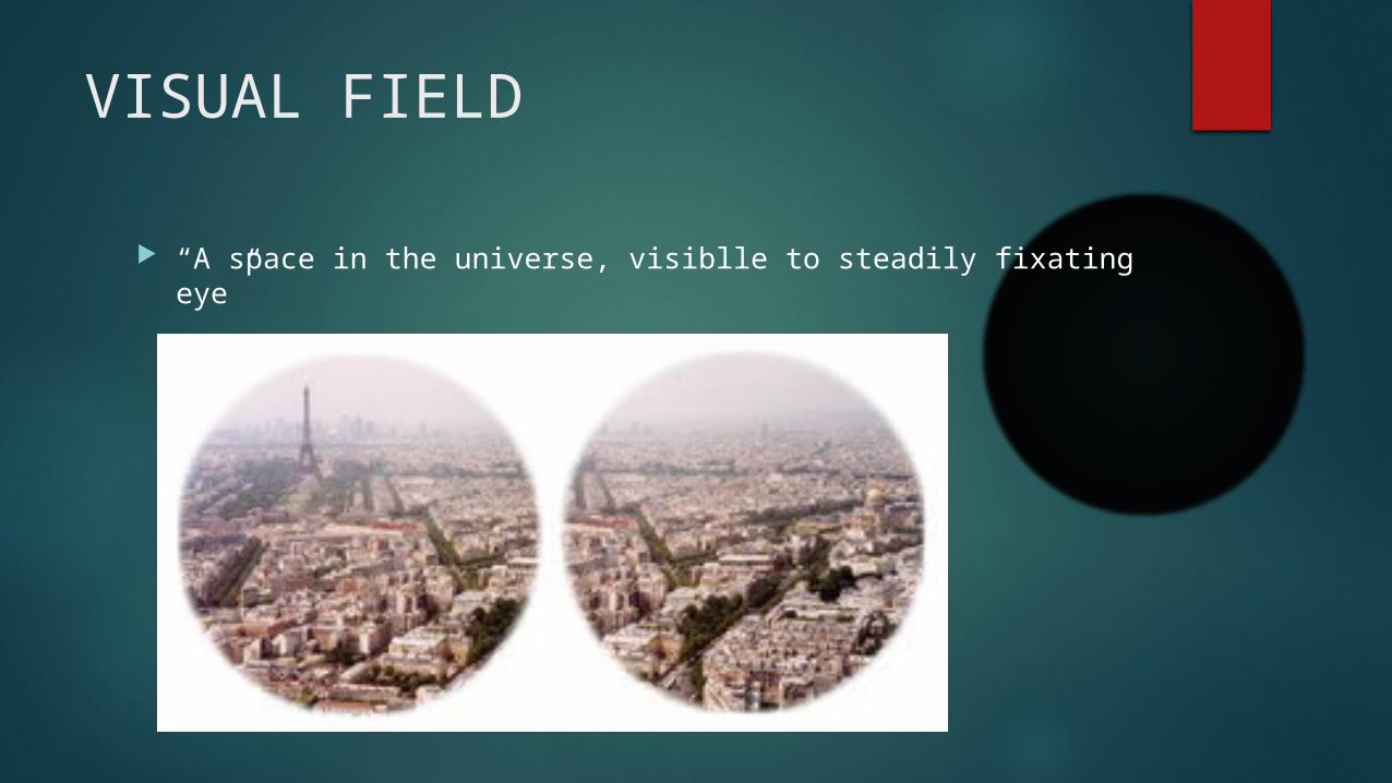
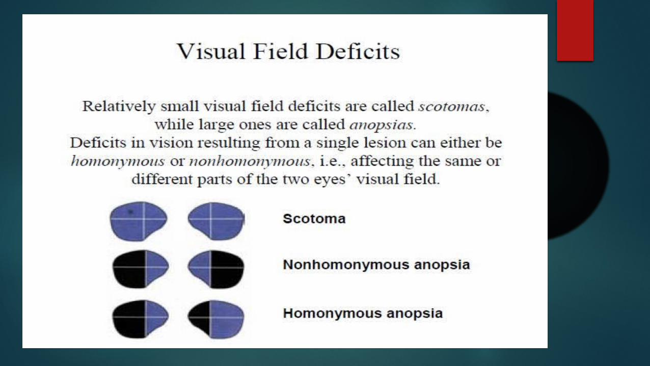
VISUAL FIELD
“A space in the universe, visiblle to steadily fixating eye”
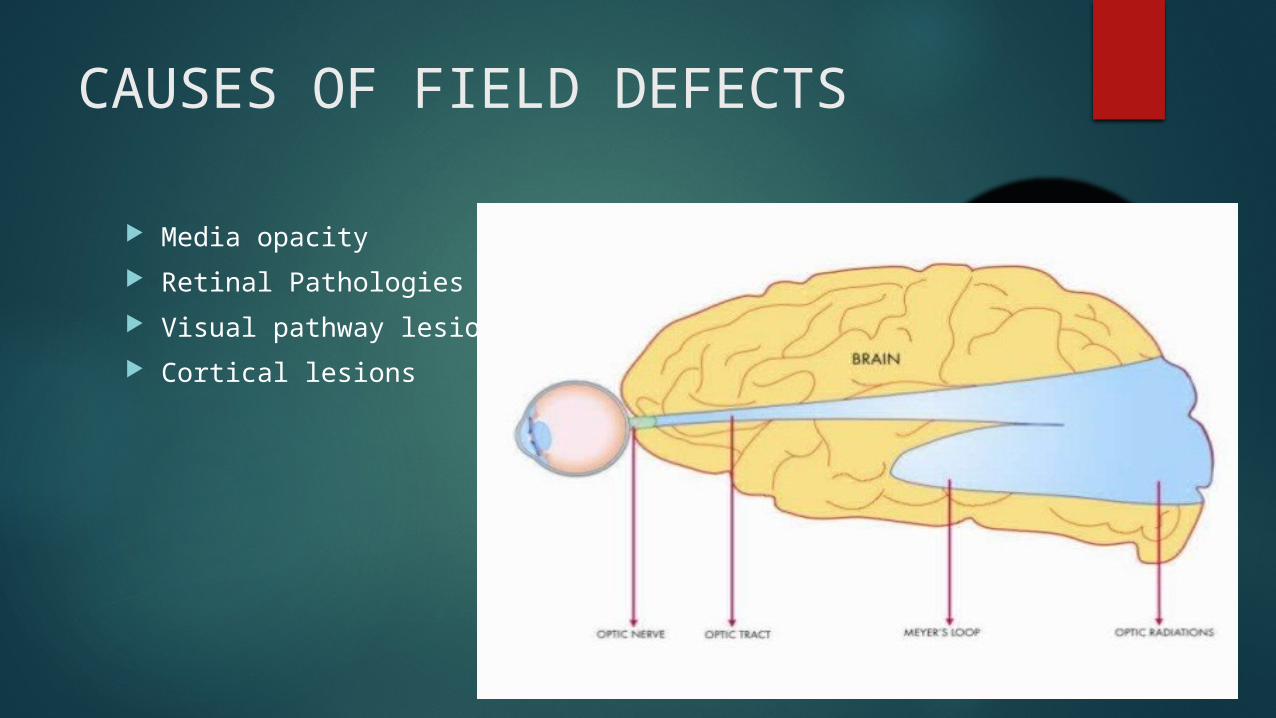
CAUSES OF FIELD DEFECTS
Media opacity Retinal Pathologies Visual pathway lesions Cortical lesions
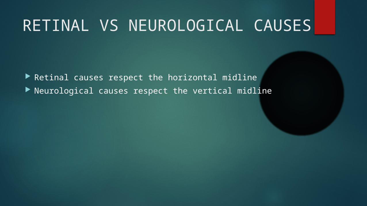
RETINAL VS NEUROLOGICAL CAUSES
Retinal causes respect the horizontal midline Neurological causes respect the vertical midline
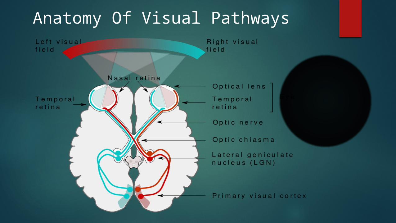
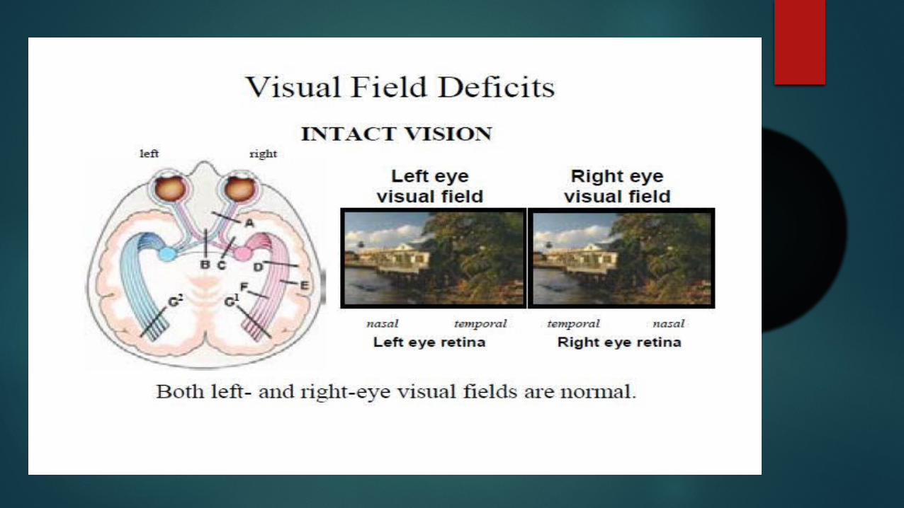
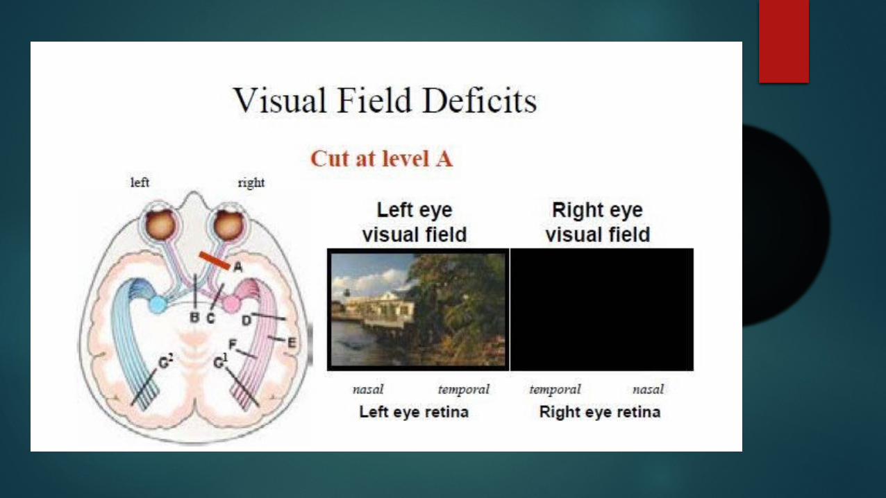
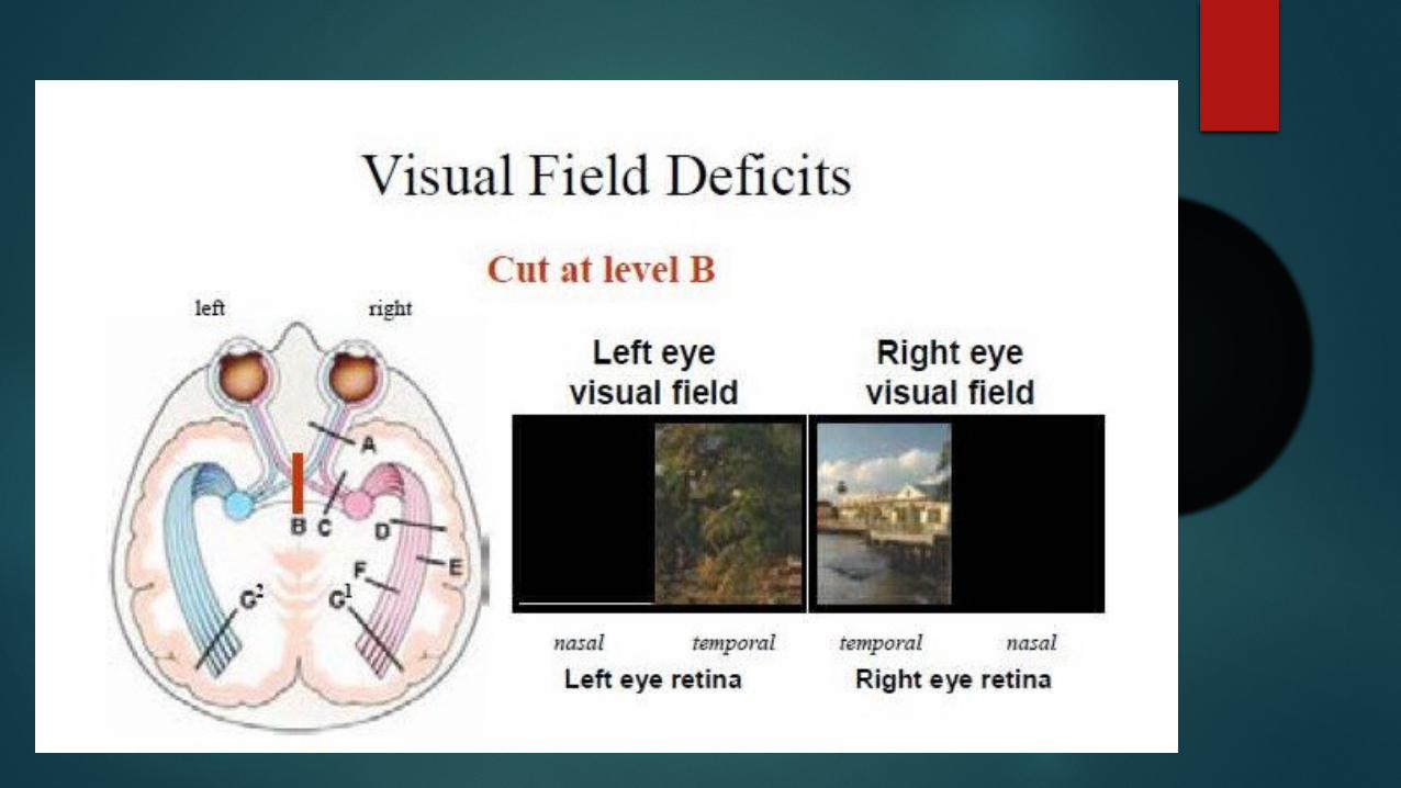
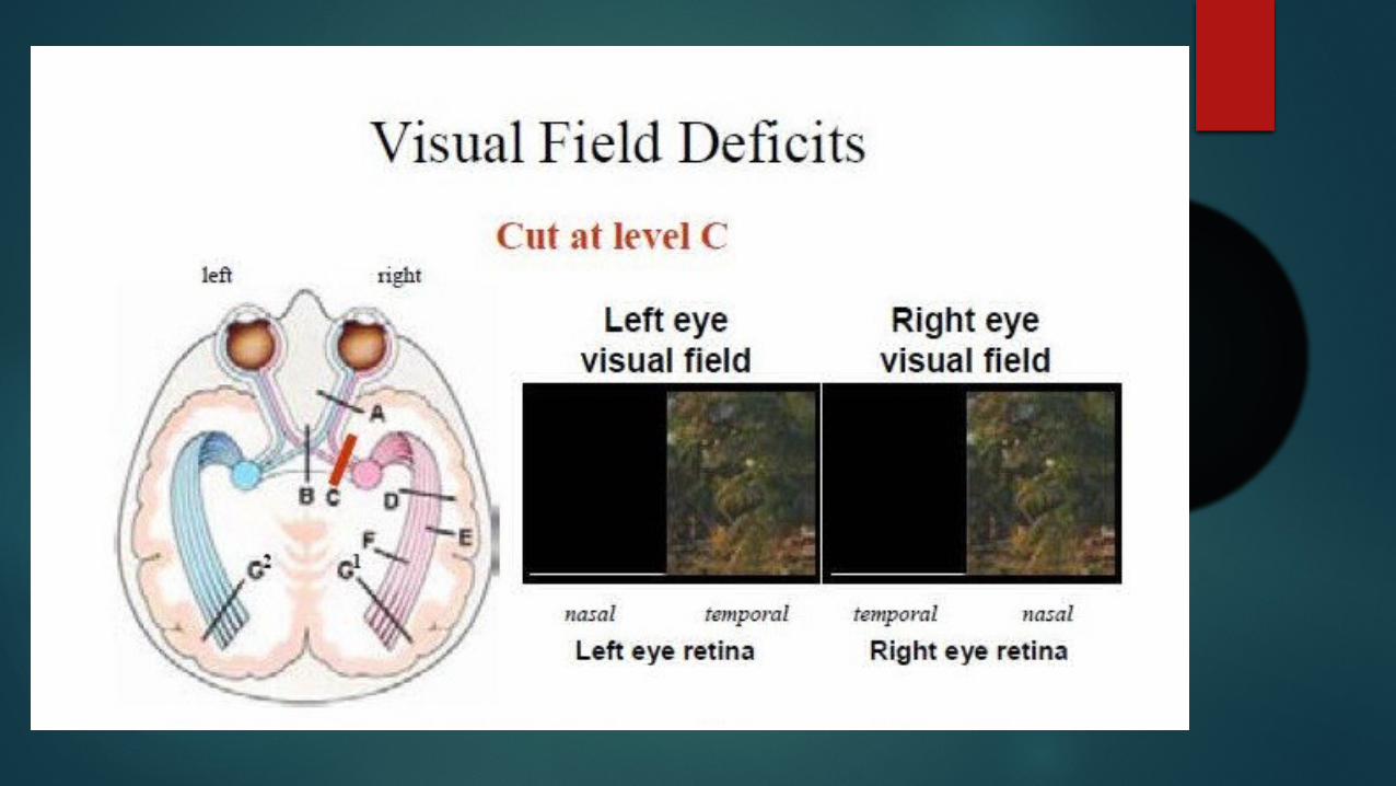
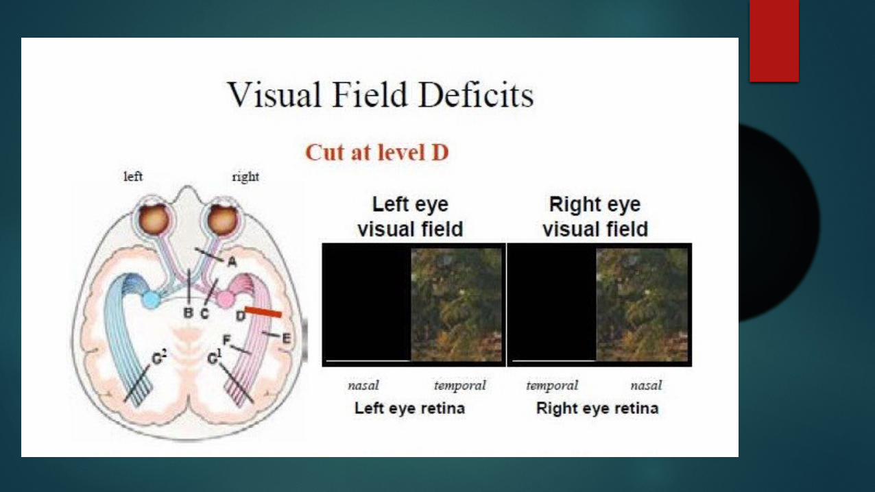
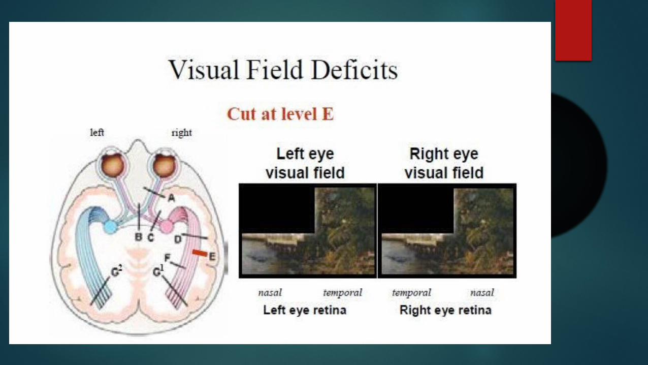
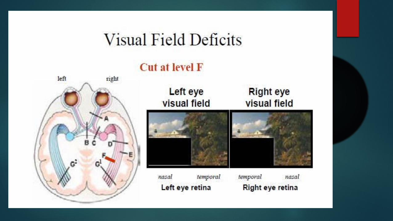
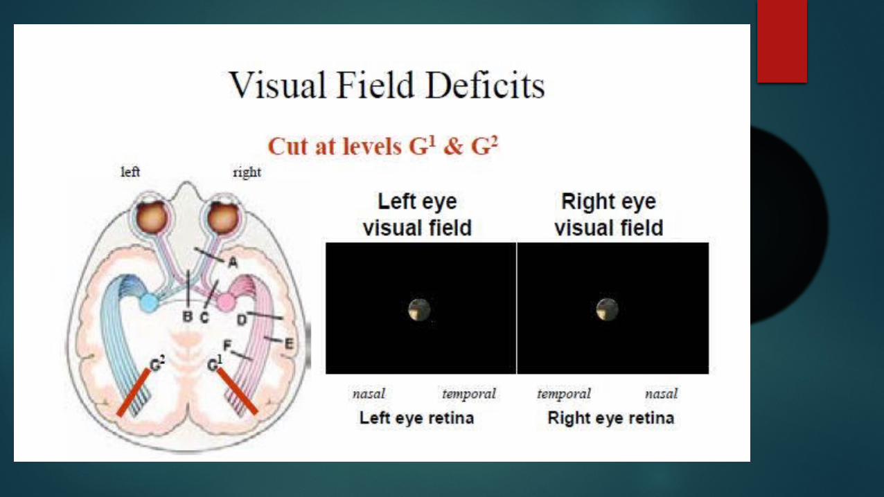
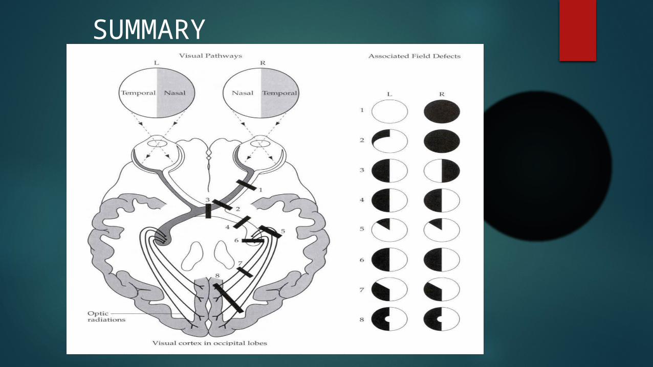
Anatomy Of Visual Pathways
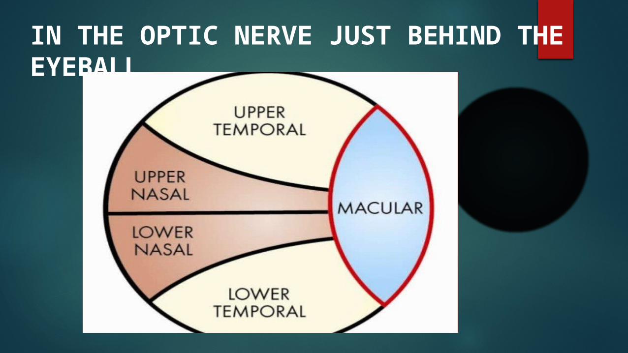
IN THE OPTIC NERVE JUST BEHIND THE EYEBALL
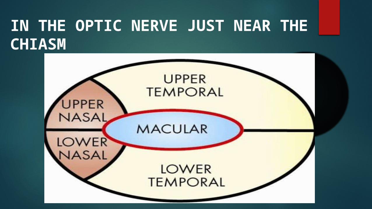
IN THE OPTIC NERVE JUST NEAR THE CHIASM
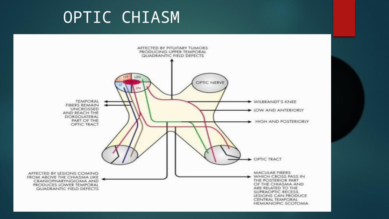
OPTIC CHIASM

OPTIC RADIATION
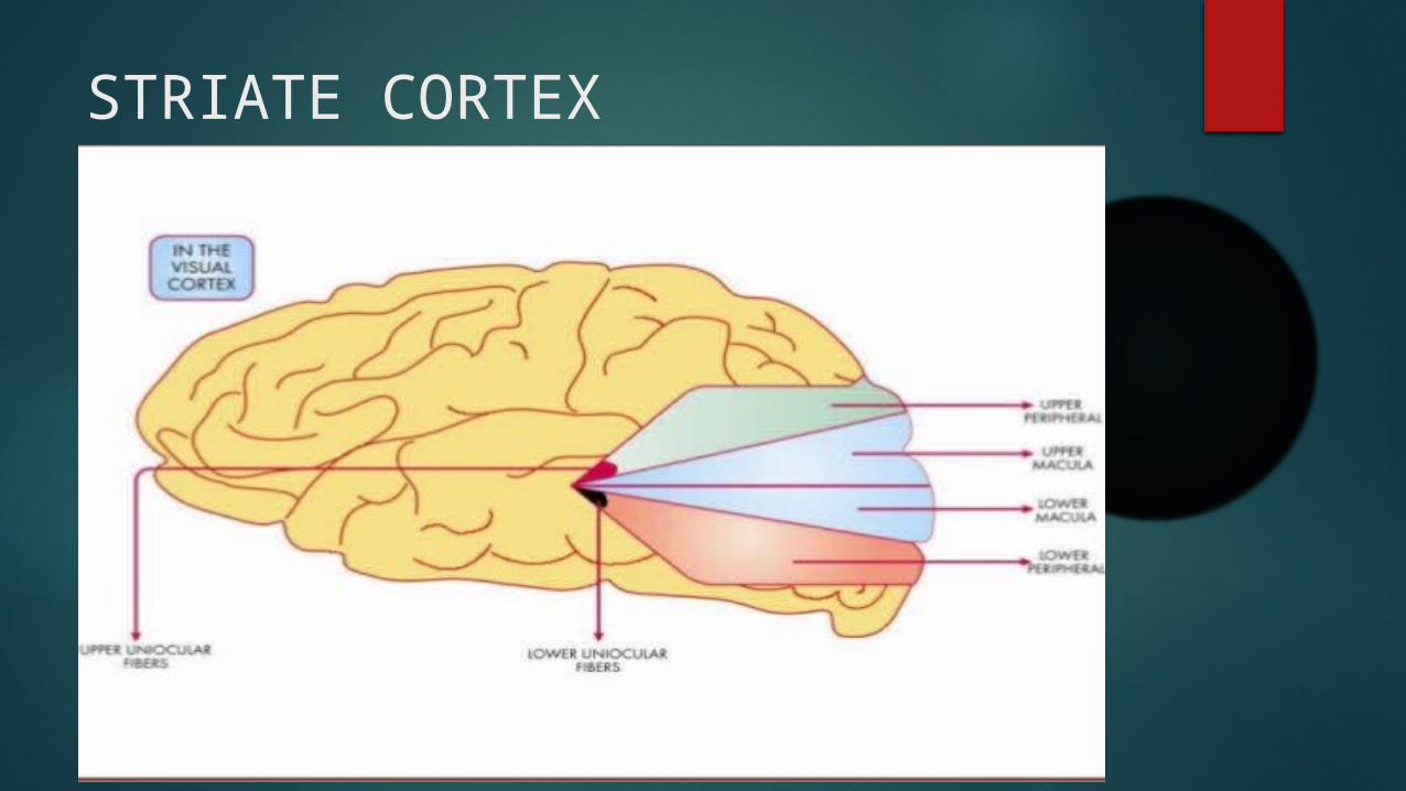
STRIATE CORTEX

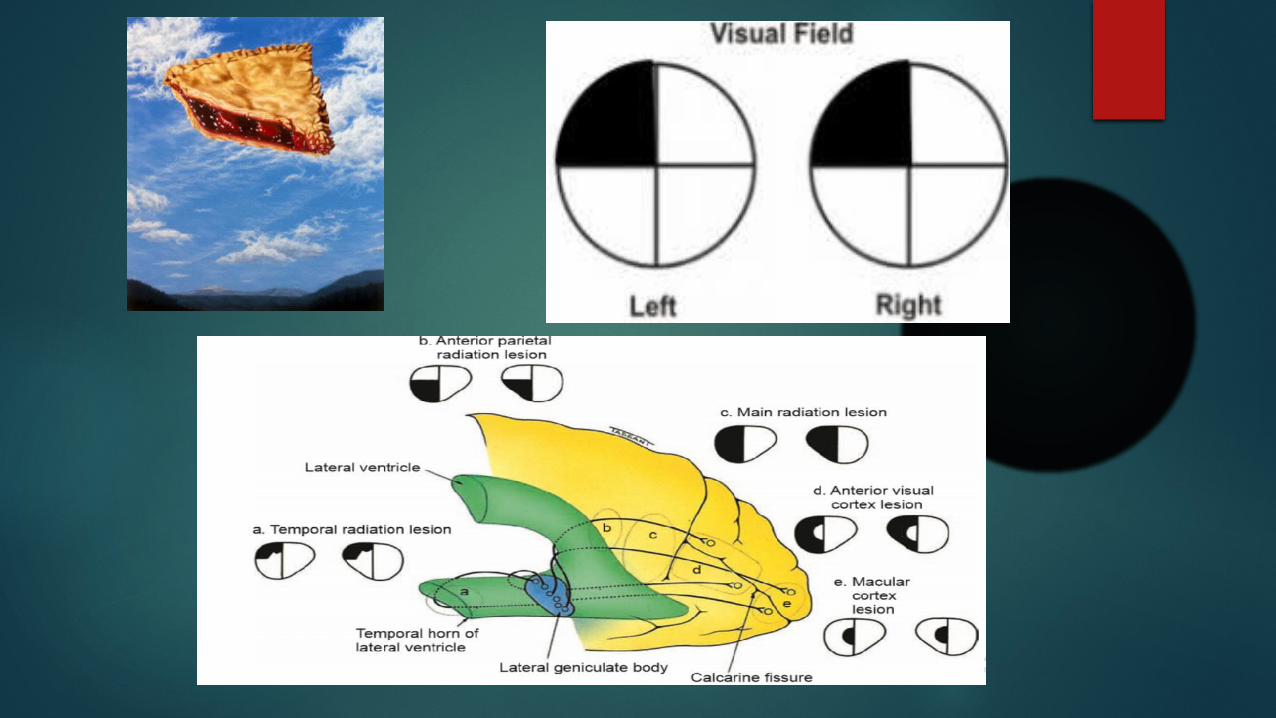
Visual pathway lesions

HOW TO APPROACH A PATIENT WITH SPECIFIC VISUAL FIELD DEFECT??

Patient Presention with Bitemporal Hemianopia LOOK FOR: Color desaturation Optic atrophy Headache EOM paresis To specify the type of adenoma Amenorrhea-galactorhea syndrome in females Hypogonadism : impotence, infertility, decreased libido, gynecomastia,
galactorhea Features of cushing syndrome(Central obesity, moon face, cutaneuous
striae, pigmentation,hypertenstion)Increased blood cortisol level Acromegaly in adults Gigantisn in children
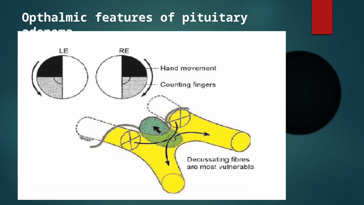
Opthalmic features of pituitary adenoma
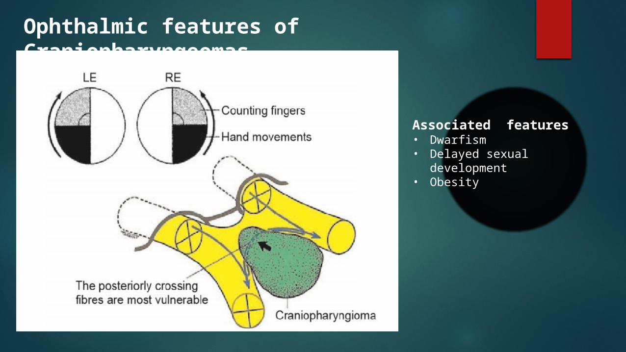
Ophthalmic features of Craniopharyngeomas
Associated features• Dwarfism• Delayed sexual development• Obesity
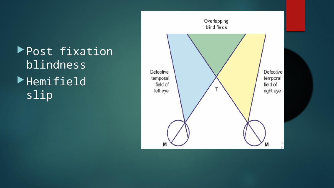
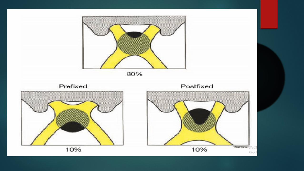
Post fixation blindness
Hemifield slip
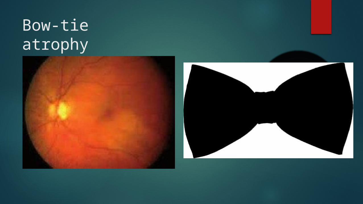
Bow-tie atrophy
Careful fundoscopic examination to rule out:-
Nasal Retinitis Pigmentosa Dermatochalasis Tilted discs Nasal retinoschisis
Investigations
MRI with gadolinium contrast CT scan Endocrinal evaluation
THESE PATIENTS NEEDS URGENT NEUROSURGICAL INTERVETION

PATIENT PRESENTIONG WITH HOMONYMOUS HEMIANOPIA Congruity Wernicke hemianopic pupil Optic atrophy (ipsilateral and
contralateral) Contralateral pyramidal signs
Patient with contralateral superior quadrantanopia
Associaed features C/L hemisensory loss and hemiparesis Paroxysmal olfactory and gustatory hallucinations Formed visual hallucinations Seizures Receptive dysphasia(Dominat hemisphere)
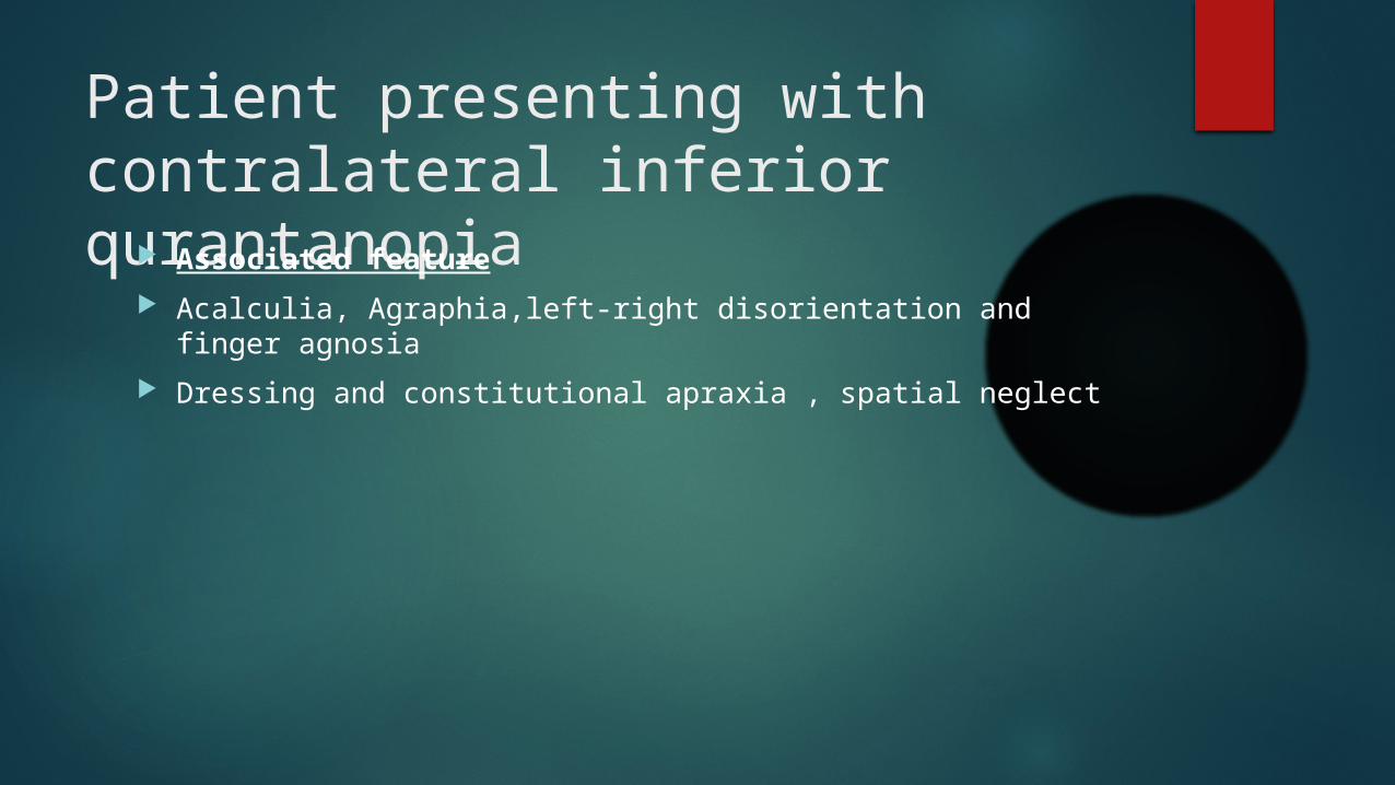
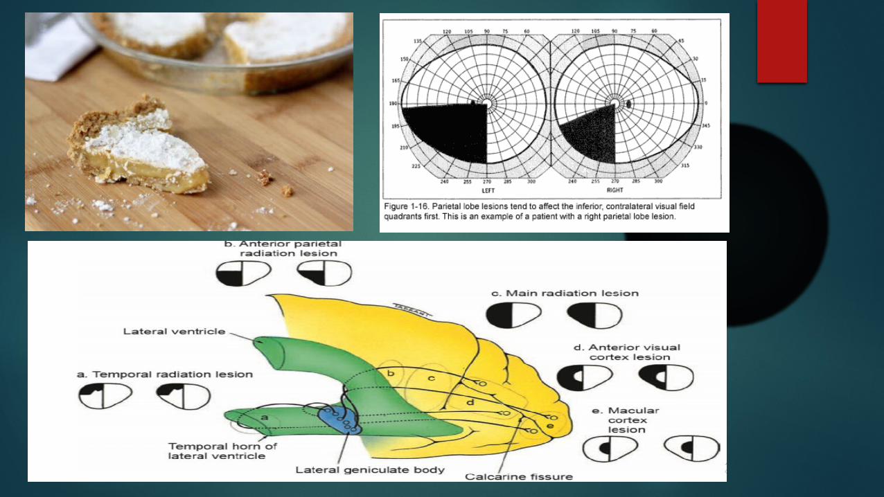
Patient presenting with contralateral inferior qurantanopia Associated feature
Acalculia, Agraphia,left-right disorientation and finger agnosia Dressing and constitutional apraxia , spatial neglect
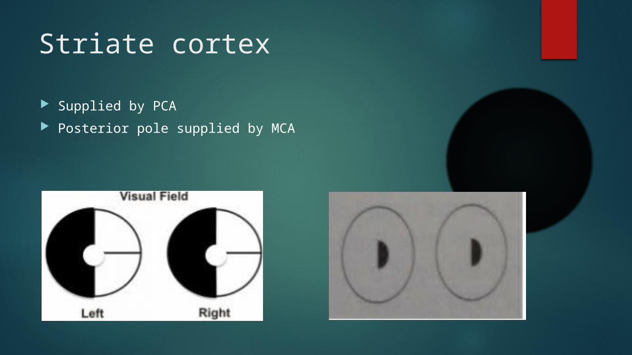
Striate cortex Supplied by PCA Posterior pole supplied by MCA
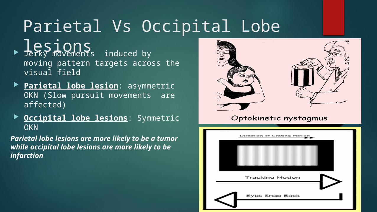
Parietal Vs Occipital Lobe lesions Jerky movements induced by moving
pattern targets across the visual field Parietal lobe lesion: asymmetric OKN
(Slow pursuit movements are affected) Occipital lobe lesions: Symmetric
OKN
Parietal lobe lesions are more likely to be a tumor while occipital lobe lesions are more likely to be infarction
Optic nerve neuropathies
Symptoms of optic nerve dysfunction Visual loss Dark adaptation is lowered Impaired color vision Transient obscuration of vision Depth perception is impaired Pain : mild dull eye ache
Signs Of Optic nerve dysfunction
Decreased VA RAPD Dyschromatopsia Diminished light brightness sensitivity Deminished contrast sensitivity Visual field defects
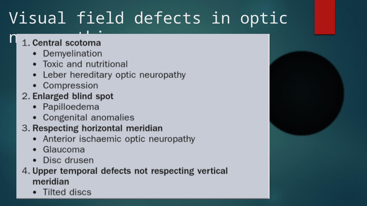
Visual field defects in optic neuropathies
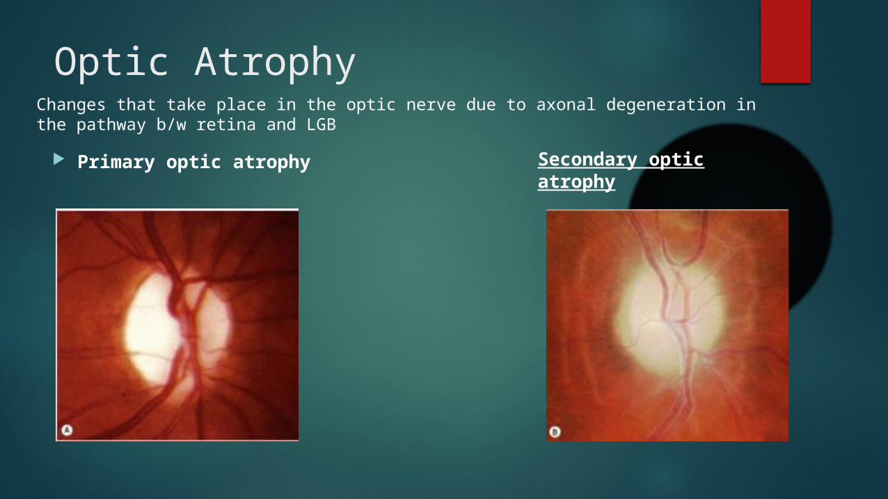
Optic Atrophy
Primary optic atrophy
Changes that take place in the optic nerve due to axonal degeneration in the pathway b/w retina and LGB
Secondary optic atrophy
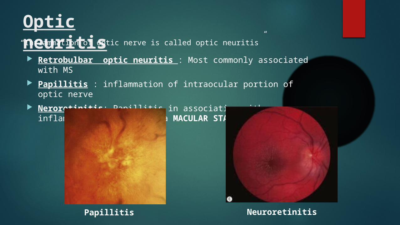
Optic neuritis Retrobulbar optic neuritis : Most commonly associated with
MS Papillitis : inflammation of intraocular portion of optic nerve Neroretinitis: Papillitis in association with inflammation of RNFL
and a MACULAR STAR
“Inflammation of optic nerve is called optic neuritis”
Papillitis Neuroretinitis
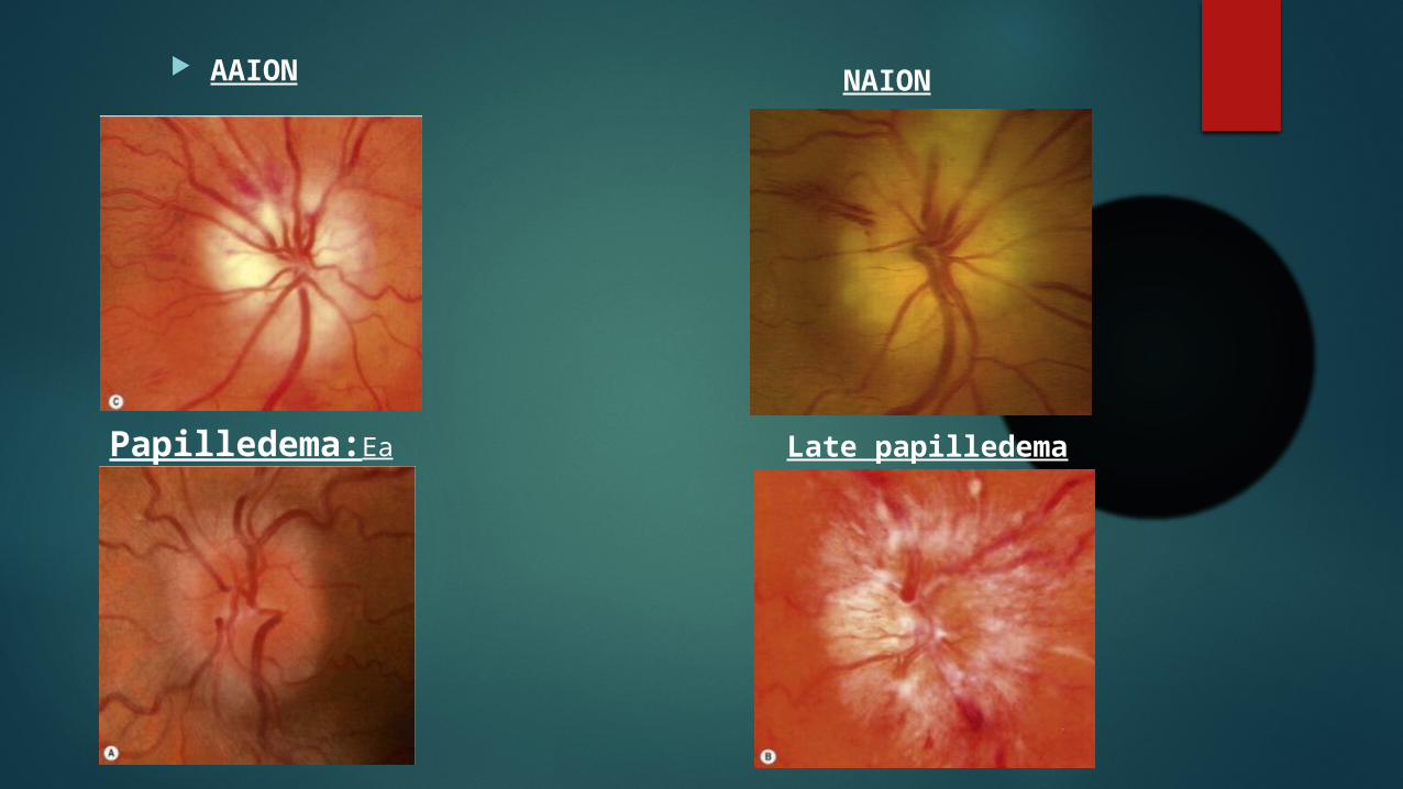
AAION NAION
Papilledema:Early
Late papilledema
SUMMARY