Embed Size (px)
Citation preview

Imaging in Thyroid disorders Molecular imaging
Radionuclide imaging Isotope scanning
SPECT/CT
Dr Ahmed Esawy

Dr. Ahmed Eisawy
MBBS M.Sc MD
Dr Ahmed Esawy
Thyroid Scintigraphy: Indications and contraindications
. Thyroid uptake is useful for: 1. Differentiating hyperthyroidism from other forms of thyrotoxicosis (e.g., thyroiditis and thyrotoxicosis factitia). 2. Calculating iodine-131 administered activity for patients to be treated for hyperthyroidism or ablative therapy. . Whole-body imaging for thyroid carcinoma is useful for: 1. Determining the presence and location of residual functioning thyroid tissue after surgery for thyroid cancer or after ablative therapy with radioactive iodine. 2. Determining the presence and location of metastases from iodine-avid forms of thyroid cancer.
Dr Ahmed Esawy
Contraindications to Thyroid Scintigraphy : Administration of iodine-131 sodium iodide to pregnant or lactating patients (whether currently nursing or not) is contraindicated.
Dr Ahmed Esawy

Evaluation of the Thyroid Disease (Radioisotope Scanning)
Prior to FNA, was the initial diagnostic procedure of choice
Performed with: technetium 99m pertechnetate or radioactive iodine
Technetium 99m pertechnetate cost-effective
readily available
short half-life
trapped but not organified by the thyroid - cannot determine functionality of a nodule
Dr Ahmed Esawy

Imaging in Pediatric Thyroid disorders: Outline
Imaging modalities • ACR-SNM-SPR guidelines for thyroid scintigraphy Imaging in: 1. Congenital hypothyroidism 2. Thyrotoxicosis 3. Thyroid nodules 4. Radioiodine whole body scan in differentiated thyroid cancers .
Dr Ahmed Esawy

Normal scan of thyroid gland
Dr Ahmed Esawy

Dr Ahmed Esawy

GIOTRE
DIFFUSE FOCAL/NODULAR
MULTINODULAR UNINODULAR
NON-TOXIC TOXIC
Structural / Anatomy
Functional /biochemical
Dr Ahmed Esawy

Dr Ahmed Esawy

palpable cold nodule in a patient with Graves disease has a high likelihood of malignancy (4%)
mnemonic: CATCH LAMP
Colloid cyst
Adenoma (most common)
Thyroiditis
Carcinoma
Hematoma
Lymphoma, Lymph node
Abscess
Metastasis (kidney, breast)
Parathyroid
Probability of a cold nodule to represent thyroid cancer:
Dr Ahmed Esawy
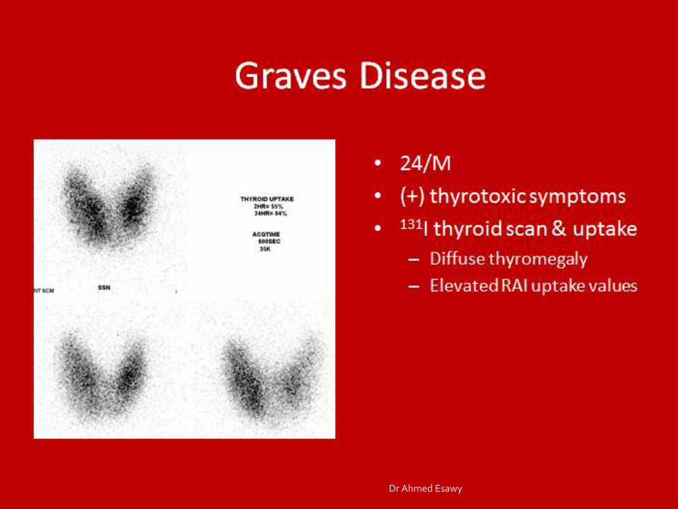
Graves Disease
24/M
(+) thyrotoxic symptoms
131I thyroid scan & uptake Diffuse thyromegaly
Elevated RAI uptake values
Dr Ahmed Esawy

Diffuse Toxic Goiter
30/F
Palpitations, excessive sweating, irritability, anterior neck enlargement
99mTcO4 thyroid scan
Diffuse thyromegaly
Scintigraphic evidence of increased gland uptake function
38 sec acquisition time
Reduced background tracer activity
Dr Ahmed Esawy

Graves’ disease
Dr Ahmed Esawy

Graves – Basedow disease
Dr Ahmed Esawy
Dr Ahmed Esawy

Congenital hypothyroidism: Scintigraphy
Dr Ahmed Esawy
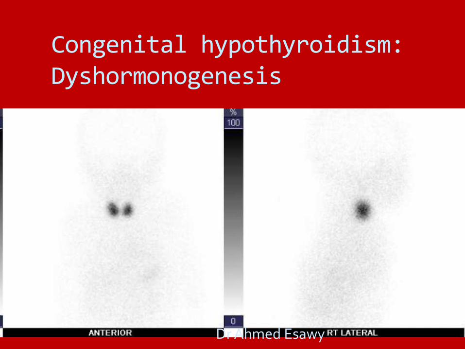
Congenital hypothyroidism: Dyshormonogenesis
Dr Ahmed Esawy

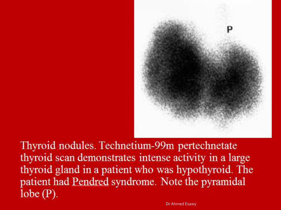
10-day-old girl with sublingual thyroid gland..
Dr Ahmed Esawy
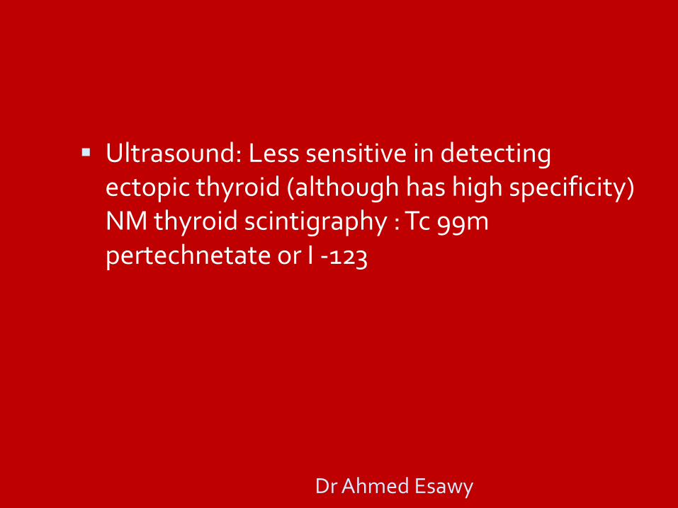
Ultrasound: Less sensitive in detecting ectopic thyroid (although has high specificity) NM thyroid scintigraphy : Tc 99m pertechnetate or I -123
Dr Ahmed Esawy
Preclinical stage: Scintigraphy may show increased uptake
• Difficult to distinguish Hashitoxicosis from
Graves disease by US or scintigraphy.
Dr Ahmed Esawy
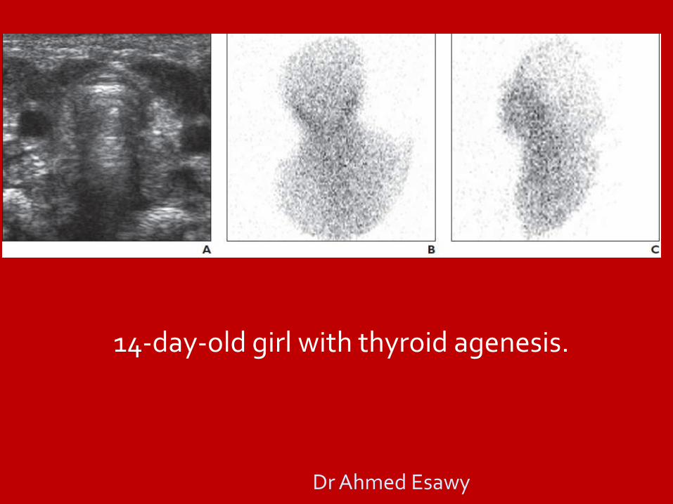
14-day-old girl with thyroid agenesis.
Dr Ahmed Esawy
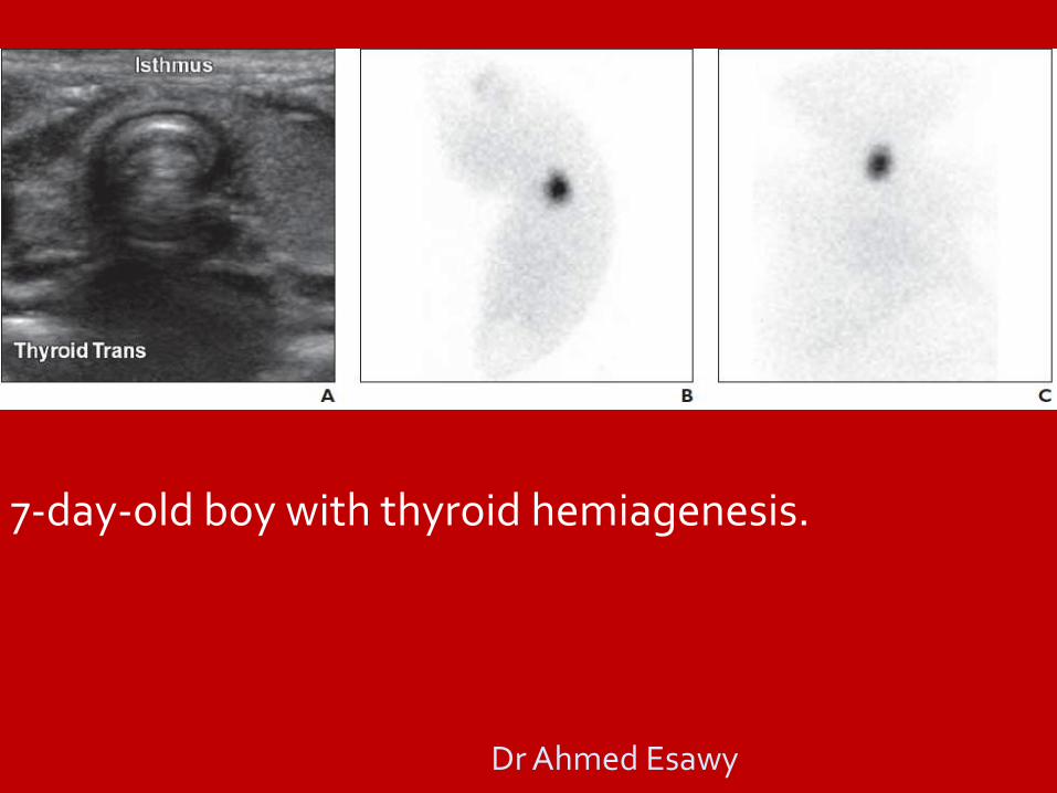
7-day-old boy with thyroid hemiagenesis.
Dr Ahmed Esawy

20-day-old girl with hemiagenesis and sublingual thyroid.
Dr Ahmed Esawy
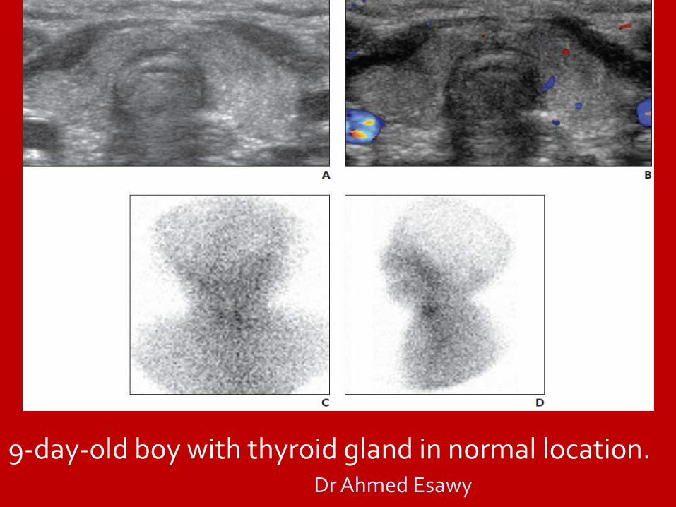
9-day-old boy with thyroid gland in normal location. Dr Ahmed Esawy

Hashitoxicosis
Dr Ahmed Esawy

Graves disease / Hashimotos thyroiditis
Thyroid inferno Graves disease: 4 hour uptake of 40% Dr Ahmed Esawy

Subacute Thyroiditis
30/M
Hyperthyroid symptoms
131I thyroid scan
Thyroid not visualized
Only background radioactivity
Dr Ahmed Esawy
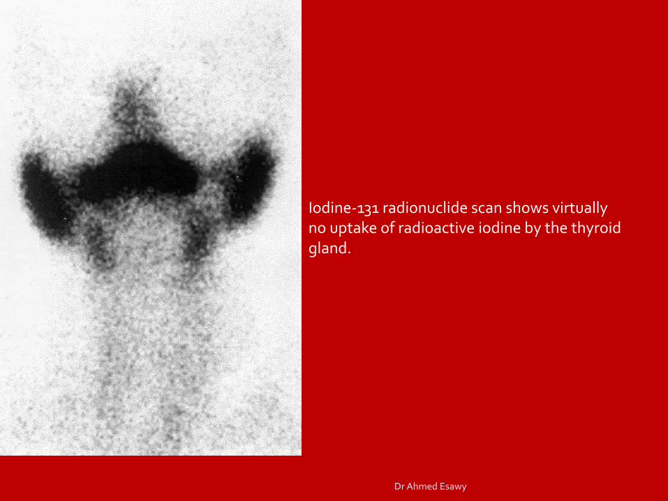
Iodine-131 radionuclide scan shows virtually no uptake of radioactive iodine by the thyroid gland.
Dr Ahmed Esawy

NODULAR GIOTRE
UNINODULAR
MULTINODULAR MNG
INACTIVE
COLD
TOXIC NODULE
TOXIC NODULE
TOXIC MULTINODULAR GIOTRE INACTIVE
COLD
MALIGNANT BENIGN Dr Ahmed Esawy
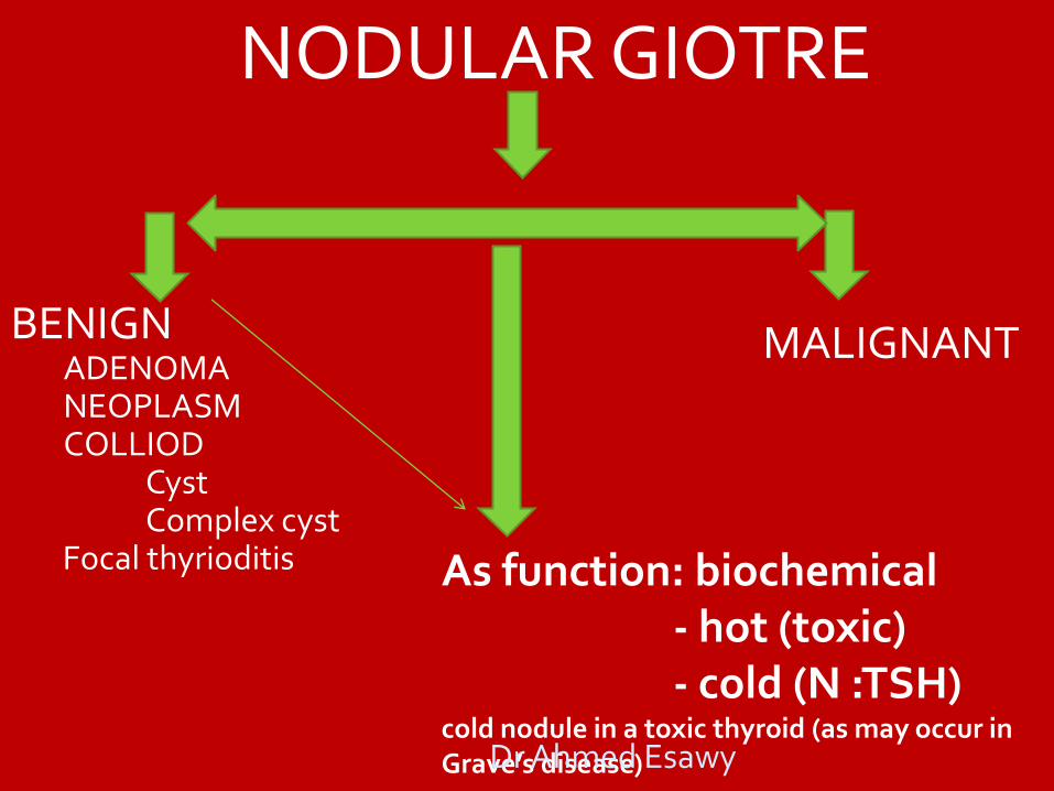
NODULAR GIOTRE
BENIGN ADENOMA NEOPLASM COLLIOD
Cyst Complex cyst
Focal thyrioditis
MALIGNANT
As function: biochemical - hot (toxic) - cold (N :TSH) cold nodule in a toxic thyroid (as may occur in Grave’s disease) Dr Ahmed Esawy
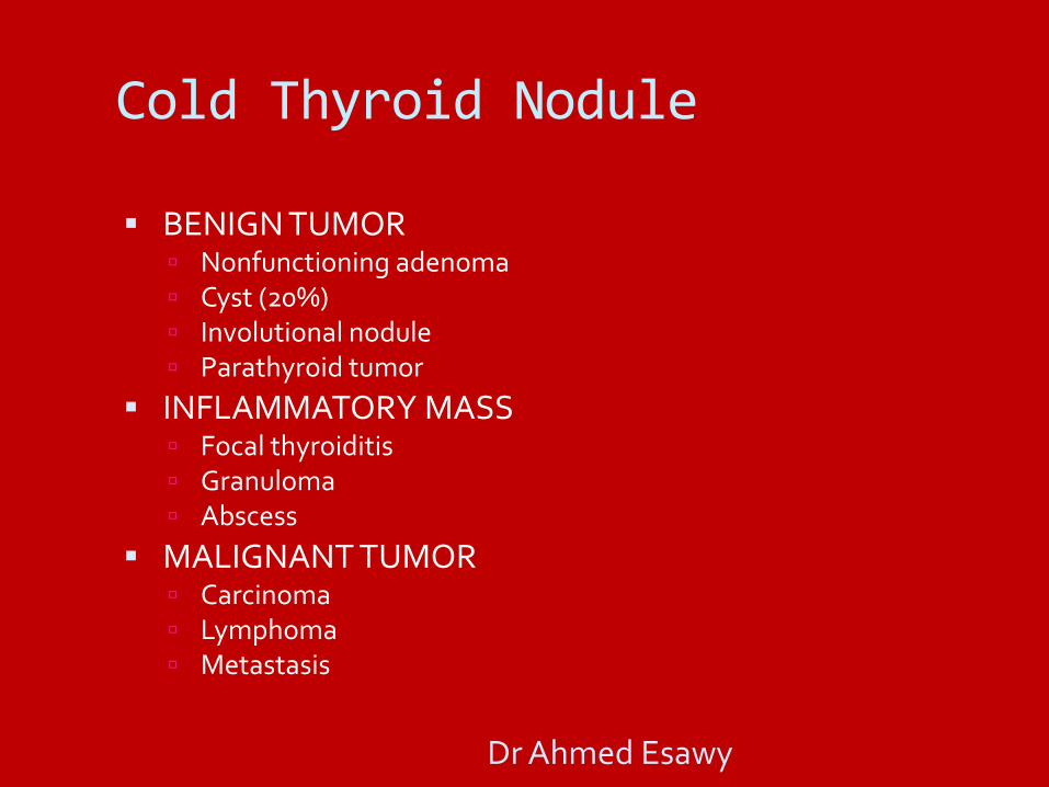
Cold Thyroid Nodule BENIGN TUMOR
Nonfunctioning adenoma Cyst (20%) Involutional nodule Parathyroid tumor
INFLAMMATORY MASS Focal thyroiditis Granuloma Abscess
MALIGNANT TUMOR Carcinoma Lymphoma Metastasis
Dr Ahmed Esawy

“Cold” nodule = focal defect
Dr Ahmed Esawy

Cold nodule, R lobe (99mTcO4)
Dr Ahmed Esawy

Dr Ahmed Esawy

Adenomatous nodule in a 66-year-old man with a low thyroid-stimulating hormone level of 0.1 mIU/mL. (a) Transverse US image shows a predominantly solid 2.4-cm nodule with well-circumscribed margins and a surrounding halo (benign US features). (b) Scintigraphic image obtained with 123I shows increased uptake in a hot nodule and relative photopenia of the adjacent normal thyroid tissue. The outline of the neck is not well visualized.
Dr Ahmed Esawy

Autonomous functioning thyroid adenoma
Dr Ahmed Esawy

Thyroid nodules. CT scan shows a mass in the posterior mediastinum (P), which displaces the air-filled esophagus to the right (arrow)
Thyroid nodules. Iodine-123 thyroid scan shows that a mass is a multinodular goiter (G). The posterior mediastinal mass is a hiatus hernia (H); the stomach (S) is shown. Further investigation revealed that thyrotoxicosis was the cause of the patient's symptoms
Dr Ahmed Esawy

Dr Ahmed Esawy

Autonomous adenoma
Initial scan - euthyreosis Repeat scan - hyperhyreosis Dr Ahmed Esawy

Cold nodule
Dr Ahmed Esawy
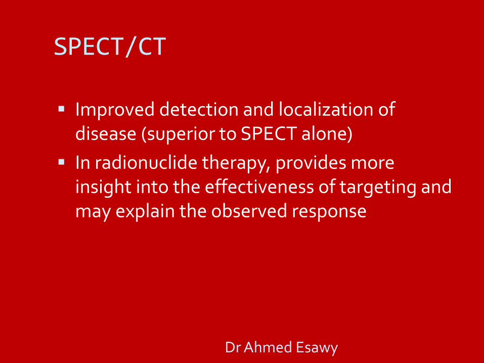
SPECT/CT
Improved detection and localization of disease (superior to SPECT alone)
In radionuclide therapy, provides more insight into the effectiveness of targeting and may explain the observed response
Dr Ahmed Esawy

Thyroid SPECT Agents
99mTcO4
99mTc sestamibi
99mTc tetrofosmi
201TlCl
123I
131I
Dr Ahmed Esawy
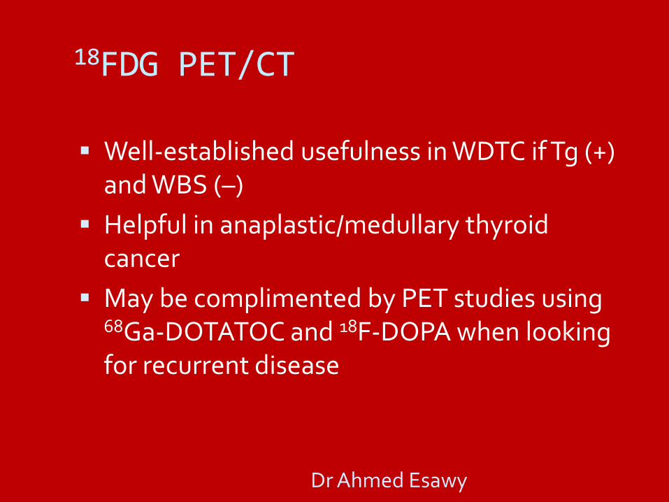
18FDG PET/CT
Well-established usefulness in WDTC if Tg (+) and WBS (–)
Helpful in anaplastic/medullary thyroid cancer
May be complimented by PET studies using 68Ga-DOTATOC and 18F-DOPA when looking for recurrent disease
Dr Ahmed Esawy

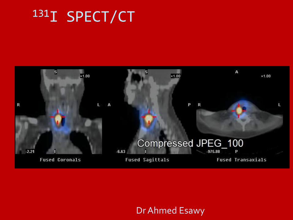
131I SPECT/CT
131I SPECT-CT is more accurate than 18FDG PET-CT in well-differentiated thyroid cancer
regional and distant metastasis
residual/recurrent disease
The most important advantage of fusion 18FDG PET-CT and 131I SPECT-CT is detection of metastasis in normal sized lymph nodes.
Dr Ahmed Esawy

Indications of PET/CT
residual or recurrent thyroid cancer WHEN elevated Tg + RAI scan (–)
When localized, may require surgery or radiotherapy
Extent of poorly differentiated TCAs & invasive Hurthle cell Cas
Treatment response following systemic or local therapy
Dr Ahmed Esawy
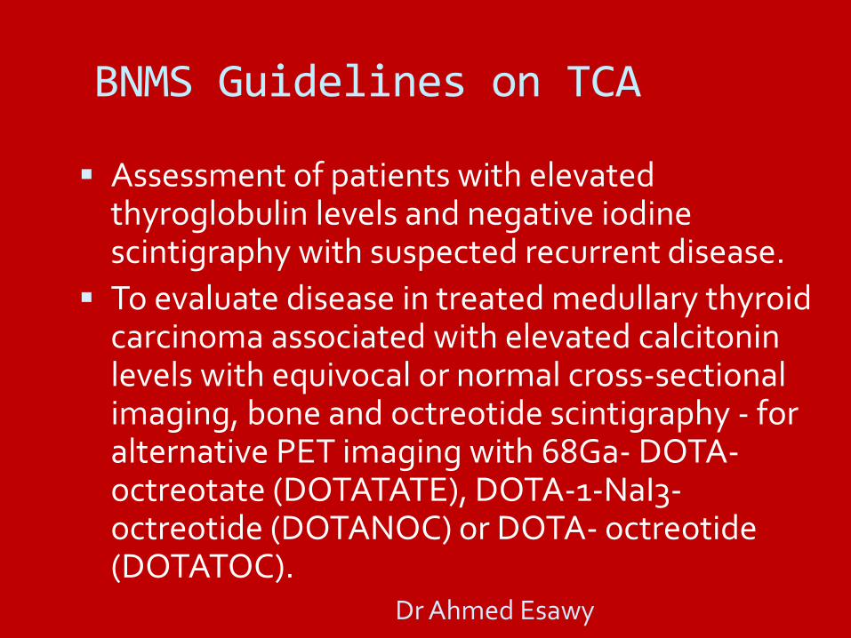
BNMS Guidelines on TCA
Assessment of patients with elevated thyroglobulin levels and negative iodine scintigraphy with suspected recurrent disease.
To evaluate disease in treated medullary thyroid carcinoma associated with elevated calcitonin levels with equivocal or normal cross-sectional imaging, bone and octreotide scintigraphy - for alternative PET imaging with 68Ga- DOTA-octreotate (DOTATATE), DOTA-1-NaI3-octreotide (DOTANOC) or DOTA- octreotide (DOTATOC).
Dr Ahmed Esawy
131I SPECT/CT
Dr Ahmed Esawy
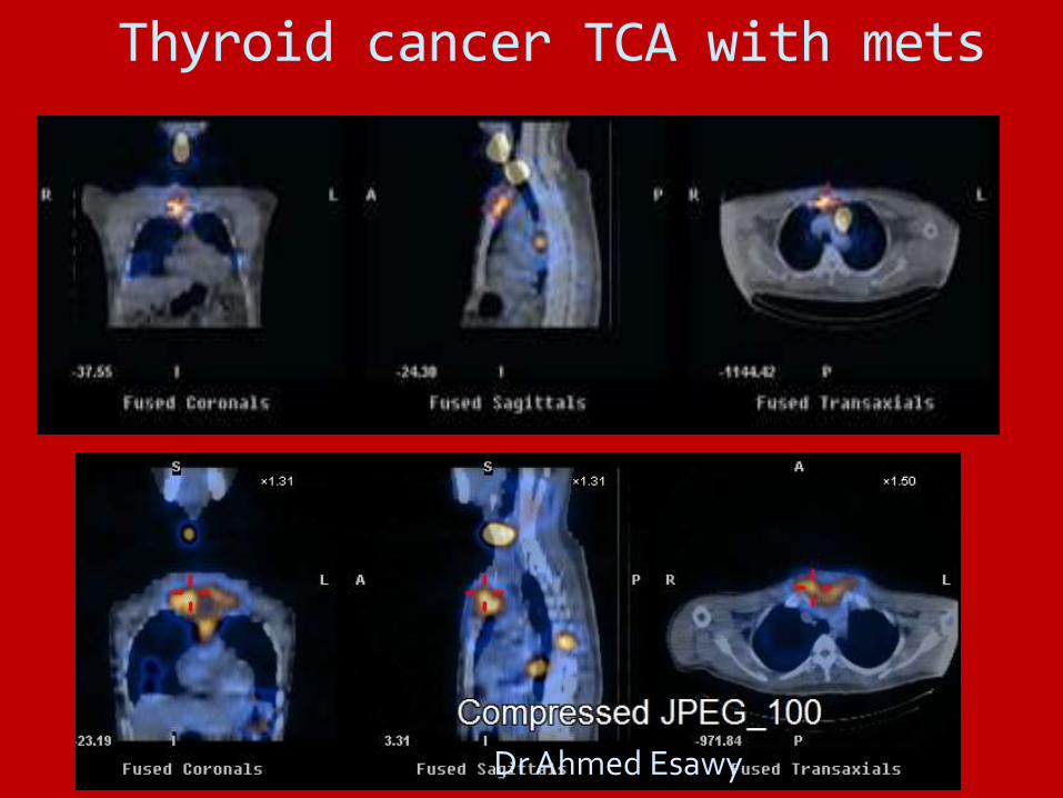
Thyroid cancer TCA with mets
Dr Ahmed Esawy

Dr Ahmed Esawy
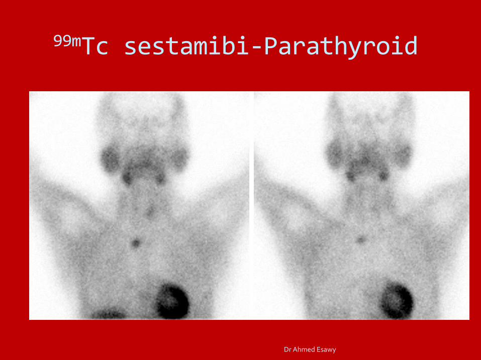
99mTc sestamibi-Parathyroid
Dr Ahmed Esawy

Medullary Thyroid Carcinoma
Dr Ahmed Esawy

Dr Ahmed Esawy

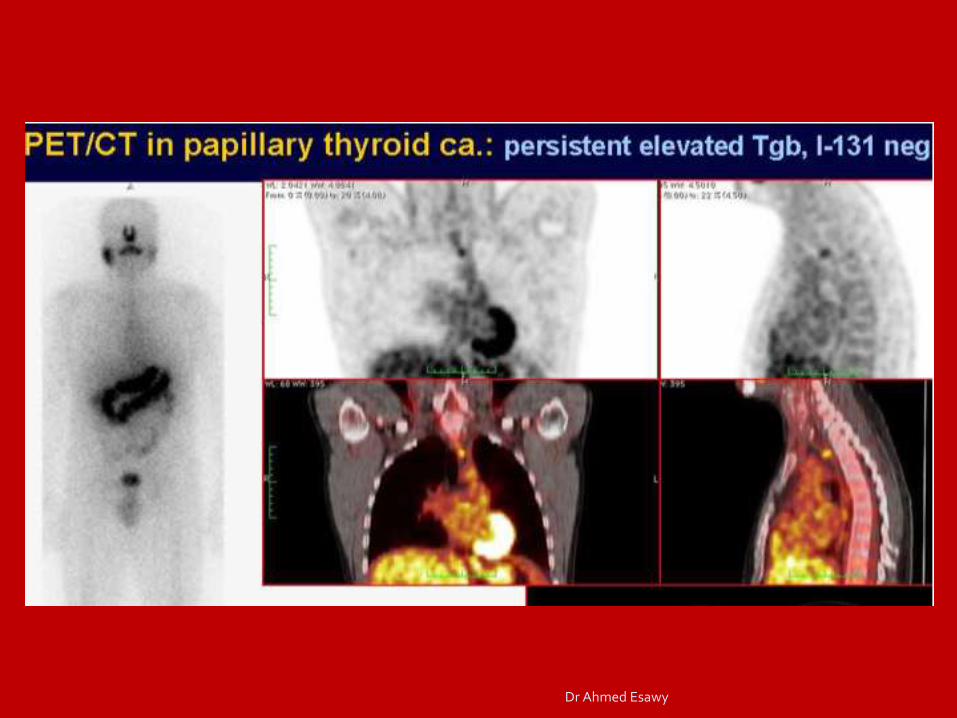
Whole body 131I Scintigraphy
78/M, (+) 13-year FU, (+) rising Tg up to 1447 µg/L Dr Ahmed Esawy
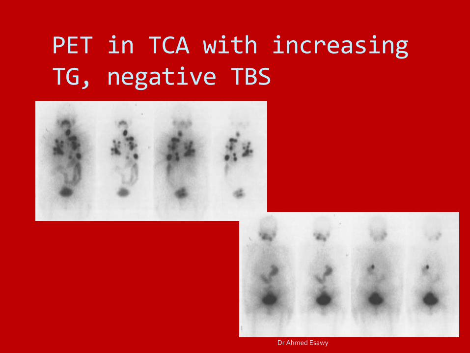
PET in TCA with increasing TG, negative TBS
Dr Ahmed Esawy
Dr Ahmed Esawy

Metastatic PTCA
Dr Ahmed Esawy

Metastatic PTCA
Dr Ahmed Esawy

131I WBS (–) 18FDG PET (–) ↑↑ Tg (56000 µg/L)
Dr Ahmed Esawy
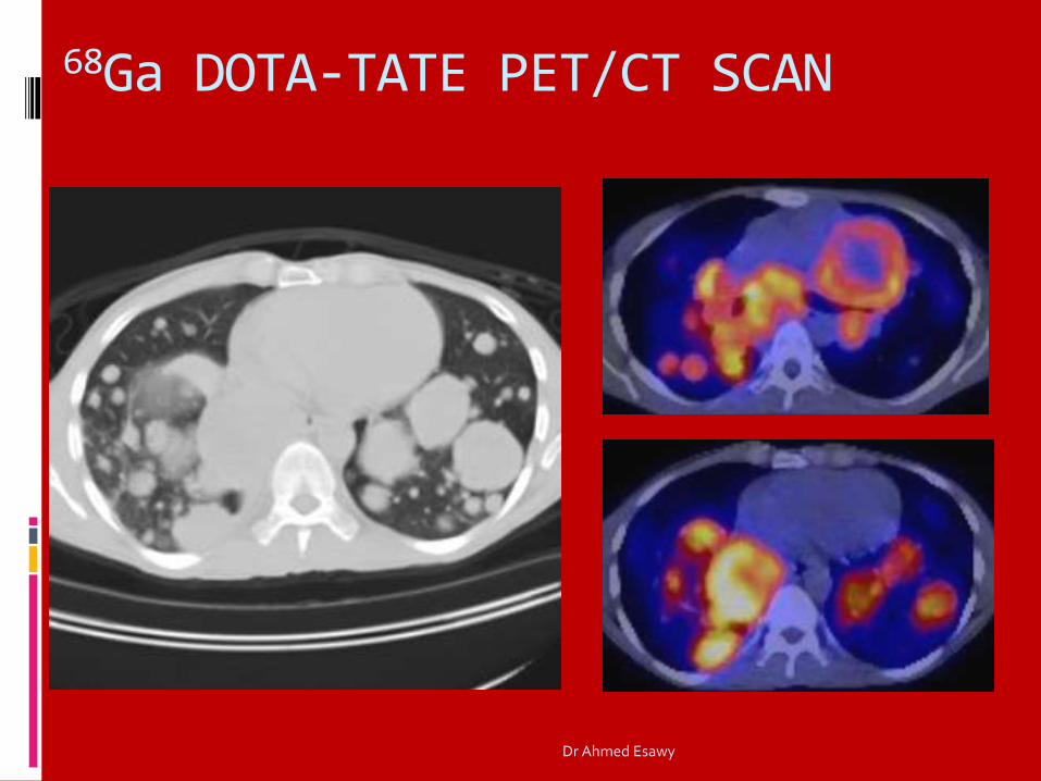
68Ga DOTA-TATE PET/CT SCAN
Dr Ahmed Esawy

68Ga DOTA-TATE PET/CT SCAN
Somatostatin receptor expression in thyroid CA
Patients with positive studies may be treated with Peptide Receptor Radionuclide Therapy (PRRT)
117Lu DOTA-TATE
90Y DOTA-TATE
Dr Ahmed Esawy

18FDG Scan in Medullary TCA
Intense FDG uptake in a hypodense nodule, L thyroid lobe
Serum Calcitonin: 800
Final Diagnosis: Medullary TCA
PET only CT Only
PET-CT Fusion
Dr Ahmed Esawy
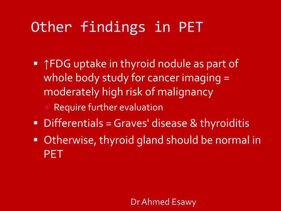
Other findings in PET
↑FDG uptake in thyroid nodule as part of whole body study for cancer imaging = moderately high risk of malignancy
Require further evaluation
Differentials = Graves' disease & thyroiditis
Otherwise, thyroid gland should be normal in PET
Dr Ahmed Esawy
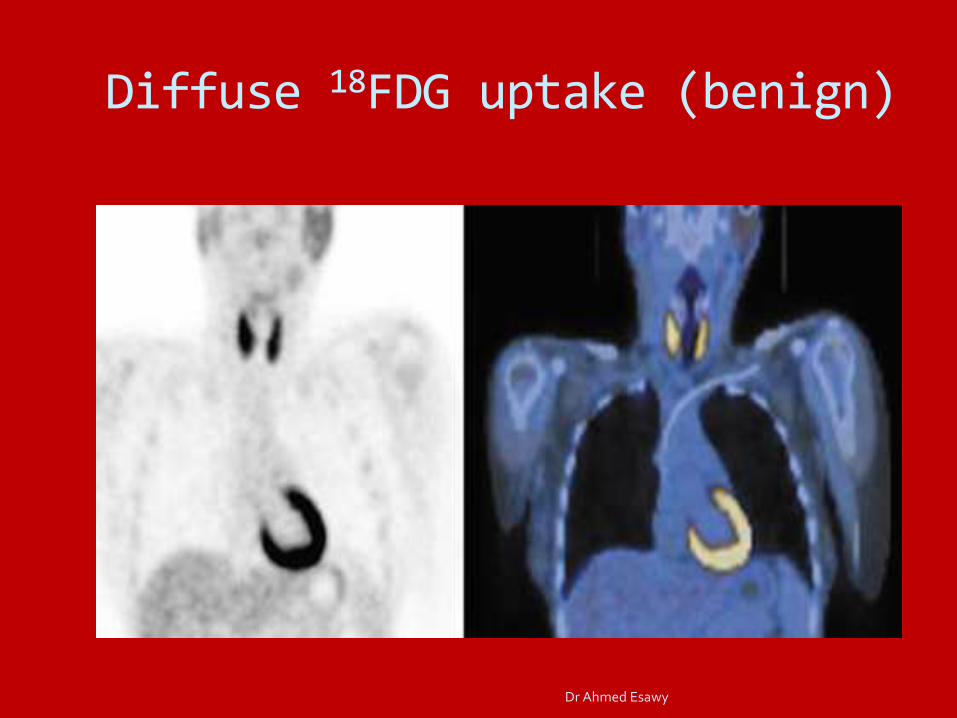
Diffuse 18FDG uptake (benign)
Dr Ahmed Esawy
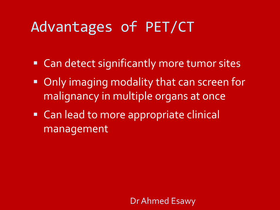
Advantages of PET/CT
Can detect significantly more tumor sites
Only imaging modality that can screen for malignancy in multiple organs at once
Can lead to more appropriate clinical management
Dr Ahmed Esawy
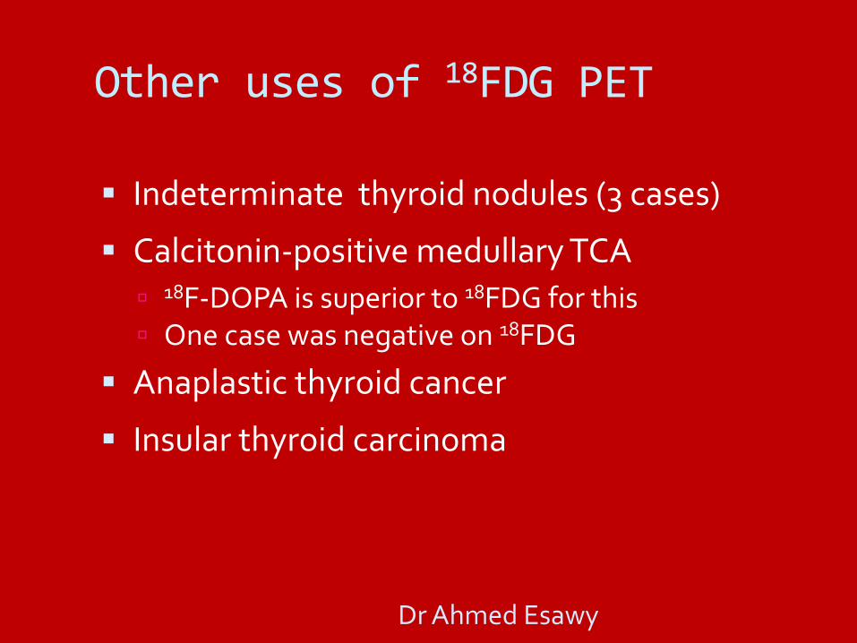
Other uses of 18FDG PET
Indeterminate thyroid nodules (3 cases)
Calcitonin-positive medullary TCA
18F-DOPA is superior to 18FDG for this
One case was negative on 18FDG
Anaplastic thyroid cancer
Insular thyroid carcinoma
Dr Ahmed Esawy
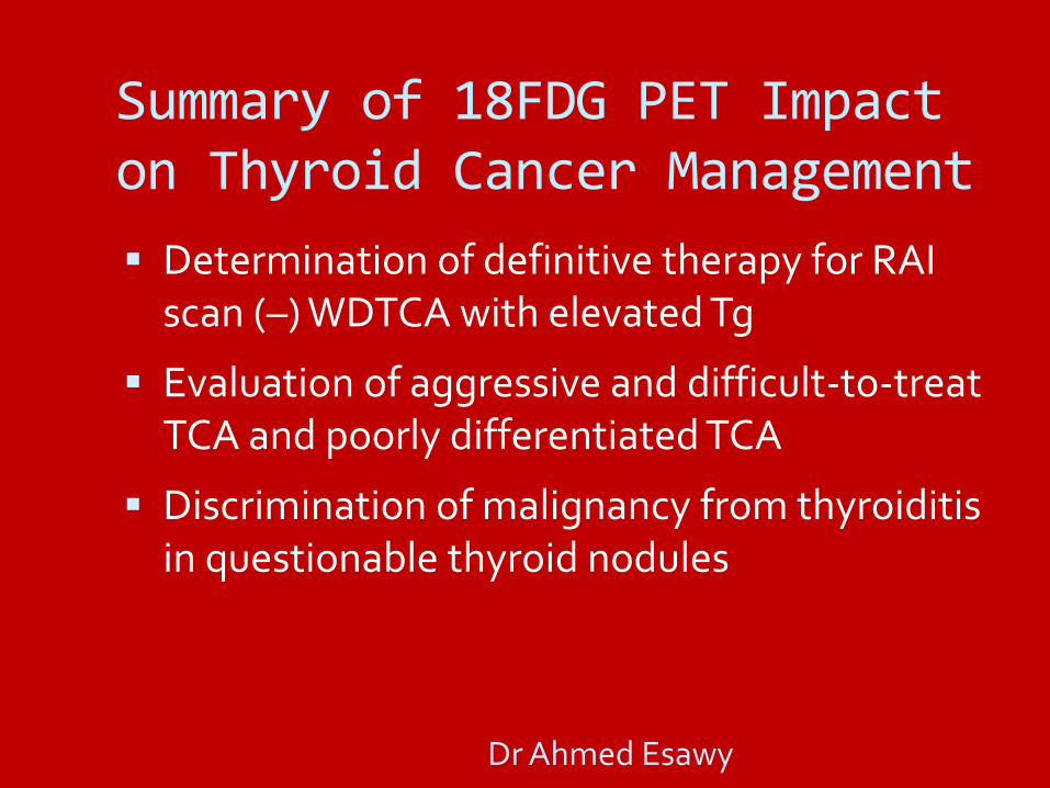
Summary of 18FDG PET Impact on Thyroid Cancer Management
Determination of definitive therapy for RAI scan (–) WDTCA with elevated Tg
Evaluation of aggressive and difficult-to-treat TCA and poorly differentiated TCA
Discrimination of malignancy from thyroiditis in questionable thyroid nodules
Dr Ahmed Esawy
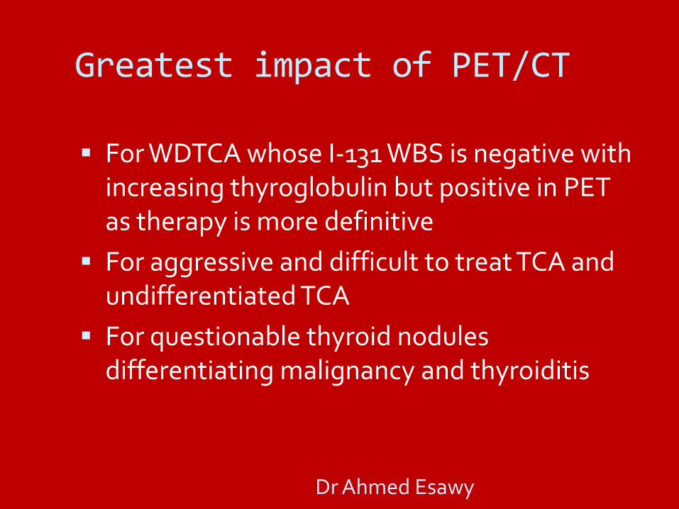
Greatest impact of PET/CT
For WDTCA whose I-131 WBS is negative with increasing thyroglobulin but positive in PET as therapy is more definitive
For aggressive and difficult to treat TCA and undifferentiated TCA
For questionable thyroid nodules differentiating malignancy and thyroiditis
Dr Ahmed Esawy

Interesting Case
CT (L) & fused PET/CT (R) in 56 y/o woman with lung cancer. Focal FDG uptake in R thyroid lobe with low CT attenuation (76 HU). PTCA on histopath.
Dr Ahmed Esawy

Interesting Case
CT (L) & fused PET/CT (R) in 65 y/o man with esophageal cancer. Focal FDG uptake in L thyroid lobe with very low CT attenuation (3.6 HU). Diffusely increased FDG uptake in surrounding gland tissue. Follicular adenoma with lymphocytic thyroiditis on histopath.
Dr Ahmed Esawy
Interesting Case
63/M with PTCA, s/p thyroidectomy, RAI therapy, thoracotomy, and radiotherapy
Neck MRI = L anterior neck nodule suspicious for recurrence
(+) pulmonary nodules on CT
Biopsy of thyroid & lung nodules = not malignant
(+) RAI-avid right cervical lesion with elevated Tg
Dr Ahmed Esawy

Interesting Case
Calcified hypermetabolic R paratracheal node, multiple bilateral hypermetabolic non-calcified pulmonary nodules, multiple cervical, hilar and substernal nodes, and hypermetabolic lesions in a left rib and sternum, suspicious for metastases.
Dr Ahmed Esawy

Interesting Case
65/F with PTCA, s/p thyroidectomy & multiple RAI therapies (cumulative dose = 1150 mCi)
elevated Tg at >800
(+) nodules in both lungs and left adrenal
(+) R lung base RAI-avid lesion on post-therapy whole body scan
Dr Ahmed Esawy

Interesting Case
Hypermetabolic right lung base mass corresponding to RAI-avid R lung base lesion seen on post-therapy scan, consistent with persistent metastatic thyroid cancer.
Dr Ahmed Esawy
Interesting Case
67/F with PTCA, s/p thyroidectomy, L radical neck dissection, multiple RAI therapies & gamma knife treatment
elevated Tg, (–) RAI whole body scan
(+) nodules in both lungs and left adrenal
(+) R lung base RAI-avid lesion on post-therapy whole body scan
CT showed possible recurrence in L thyroid bed
Dr Ahmed Esawy

Interesting Case
FDG-avid right cavernous sinus mass involving the petrous part of the temporal bone is most likely metastatic in nature.
Dr Ahmed Esawy

Interesting Case
Hypermetabolic lesions/masses in the left neck extending to the thoracic inlet specifically to the left thyroid bed with hypermetabolic bilateral cervical lymphadenopathies are consistent with recurrent metastatic disease.
Dr Ahmed Esawy

Interesting Case
Hypermetabolic osseous metastases in the cervico thoracic spine.
Dr Ahmed Esawy
Interesting Case
77/M with insular TCA, s/p thyroidectomy
L thyroid nodule and lung nodules on pre-op CT
Post-op PET was requested for evaluation of disease extent
Dr Ahmed Esawy
Interesting Case
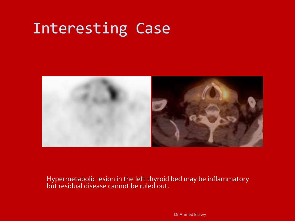
Hypermetabolic lesion in the left thyroid bed may be inflammatory but residual disease cannot be ruled out.
Dr Ahmed Esawy

Interesting Case
Hypermetabolic R hilar nodes. Differentials include inflammatory reaction vs. metastases.
Dr Ahmed Esawy

TCA Staging
Dr Ahmed Esawy

TCA Follow-up & Monitoring
Dr Ahmed Esawy
Conclusions Ultrasound and thyroid scans are still the
mainstay in imaging the thyroid gland
CT and MRI have limited values and can be utilized in identifying lymph nodes, local tumor extension, diff. thyroiditis and as FNA guide
PET/CT is best for WDTCA that have dedifferentiated hence negative on I-131-WBS but increasing thyroglobulin as well as in aggressive and difficult cases of TCA and certain suspicious nodules by FNAB
Dr Ahmed Esawy

Thanks you so much!
Dr Ahmed Esawy