Embed Size (px)
Citation preview
The Skeletal System
Yoga Anatomy TeacherTraining
Functions of skeletal system:1. Support2. Protection3. Movement4. Storage: maintains homeostasis of blood calcium which is
vital for nerve and muscle function.
5. Hemopoiesis= blood cell formation; occurs in the red bone
marrow.
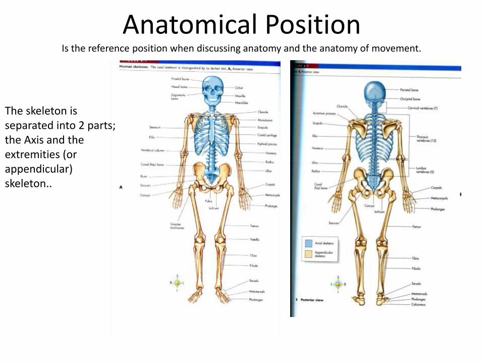
Anatomical PositionIs the reference position when discussing anatomy and the anatomy of movement.
The skeleton is separated into 2 parts; the Axis and the extremities (or appendicular) skeleton..
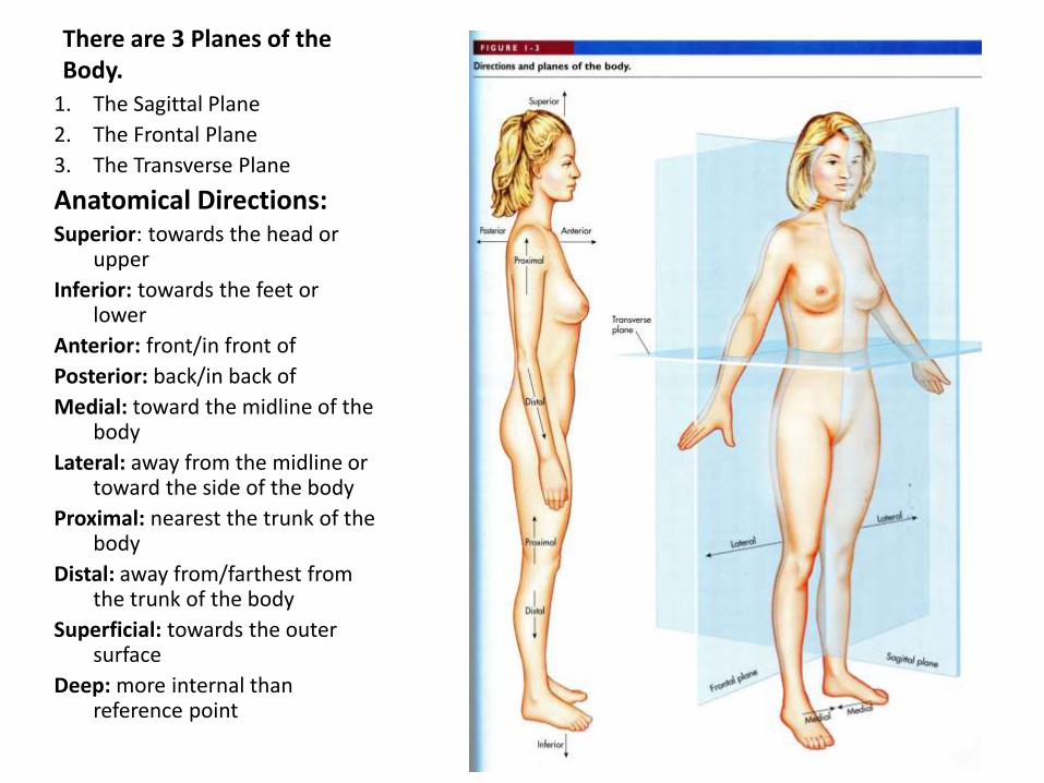
There are 3 Planes of the Body.
1. The Sagittal Plane
2. The Frontal Plane
3. The Transverse Plane
Anatomical Directions:Superior: towards the head or
upper
Inferior: towards the feet or lower
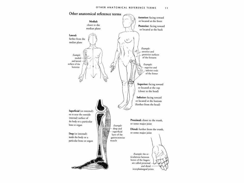
Anterior: front/in front of
Posterior: back/in back of
Medial: toward the midline of the body
Lateral: away from the midline or toward the side of the body
Proximal: nearest the trunk of the body
Distal: away from/farthest from the trunk of the body
Superficial: towards the outer surface
Deep: more internal than reference point
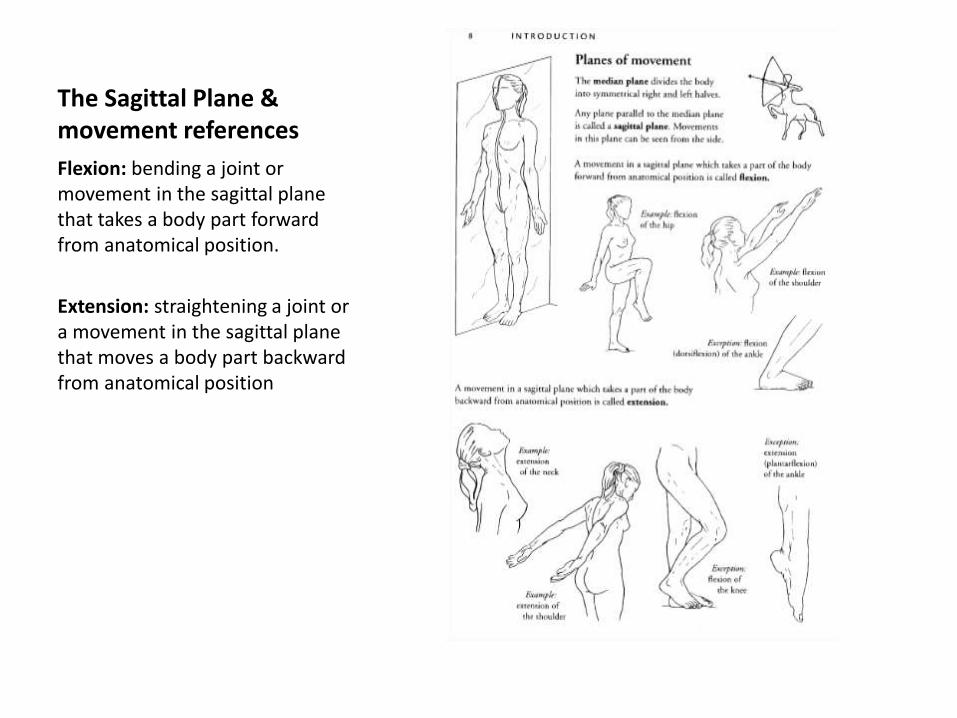
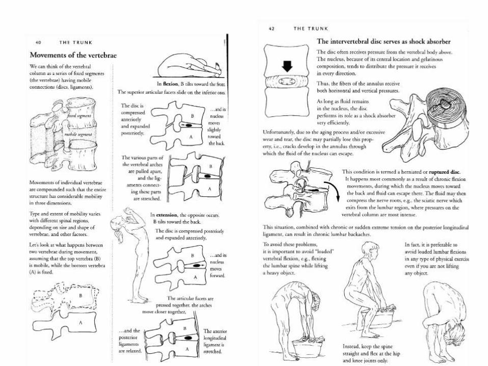
The Sagittal Plane & movement references
Flexion: bending a joint or movement in the sagittal plane that takes a body part forward from anatomical position.
Extension: straightening a joint or a movement in the sagittal plane that moves a body part backward from anatomical position
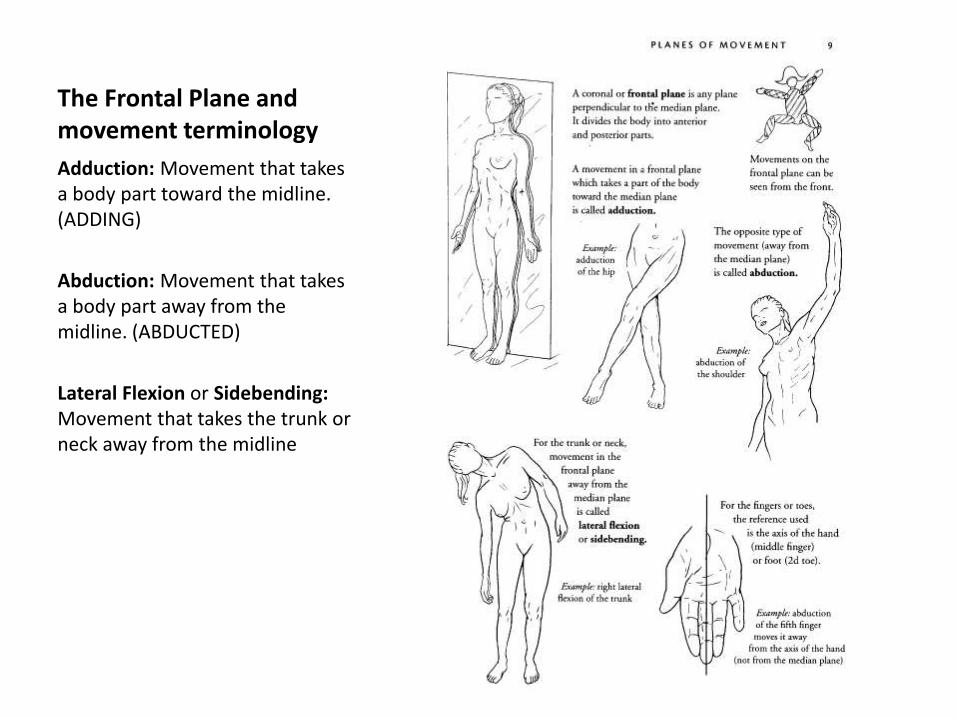
The Frontal Plane and movement terminology
Adduction: Movement that takes a body part toward the midline. (ADDING)
Abduction: Movement that takes a body part away from the midline. (ABDUCTED)
Lateral Flexion or Sidebending: Movement that takes the trunk or neck away from the midline

The Transverse Plane and Movement Terminology
Pronation is also used to describe fallen arches in the feet.
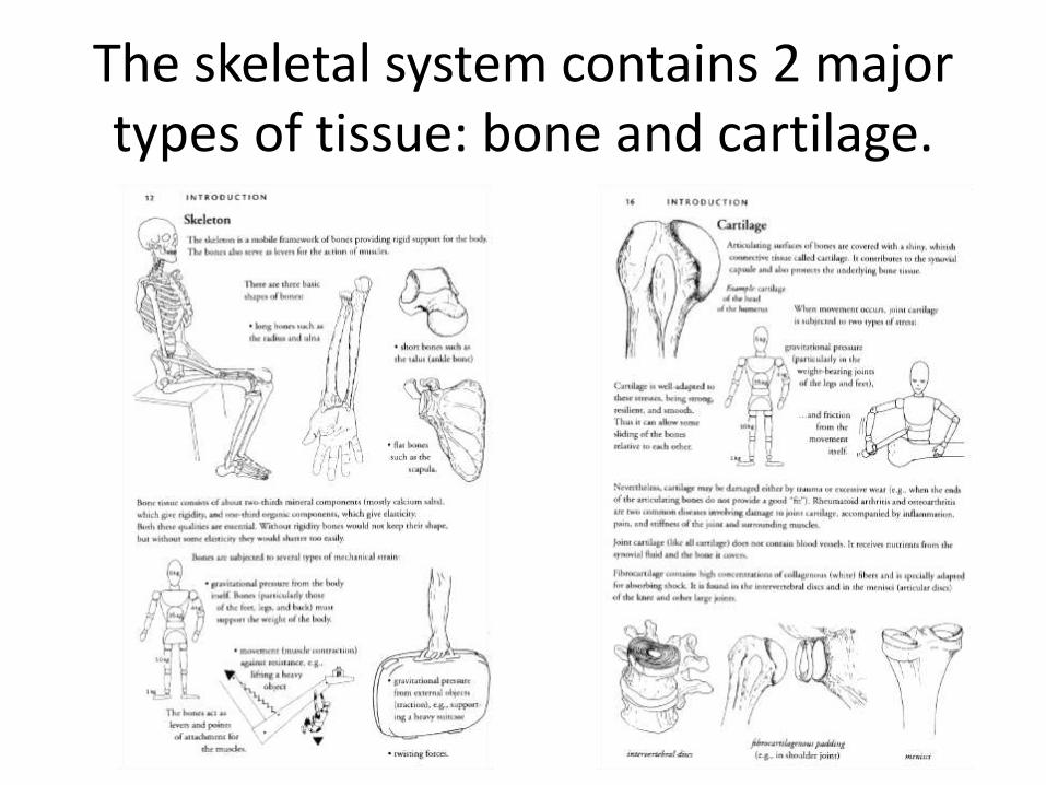
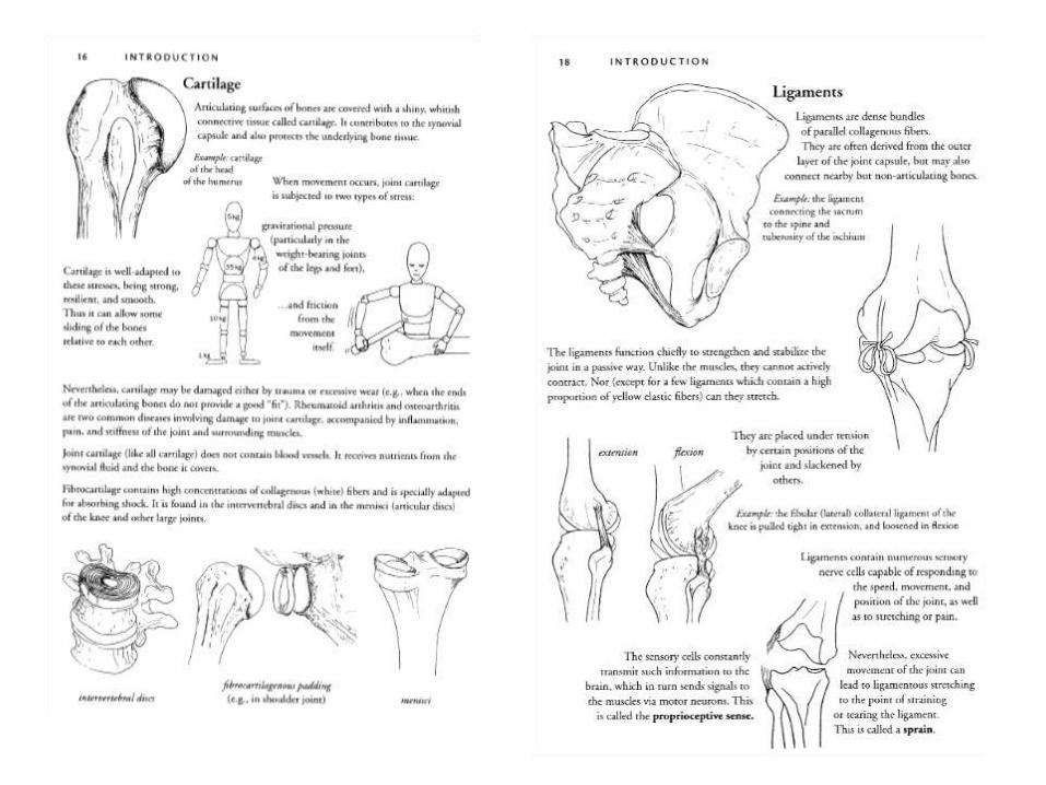
The skeletal system contains 2 major types of tissue: bone and cartilage.

Types of Joints
Types of joints also includes sutures (cranial bones called synarthroses) & public symphysis (amphiarthroses)
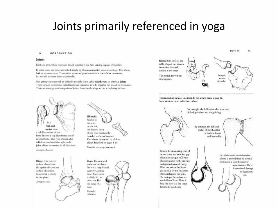
Joints primarily referenced in yoga

Structure of a Joint
-Joint capsule: made of fibrous connective tissue (the body's strongest & toughest material) and lined with a synovial membrane that secretes synovial fluid to provide lubrication. -Joint Cavity-Articular Cartilage covering the ends of the 2 joining bones
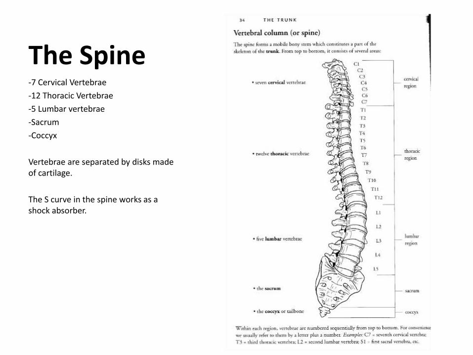
The Spine-7 Cervical Vertebrae
-12 Thoracic Vertebrae
-5 Lumbar vertebrae
-Sacrum
-Coccyx
Vertebrae are separated by disks made of cartilage.
The S curve in the spine works as a shock absorber.
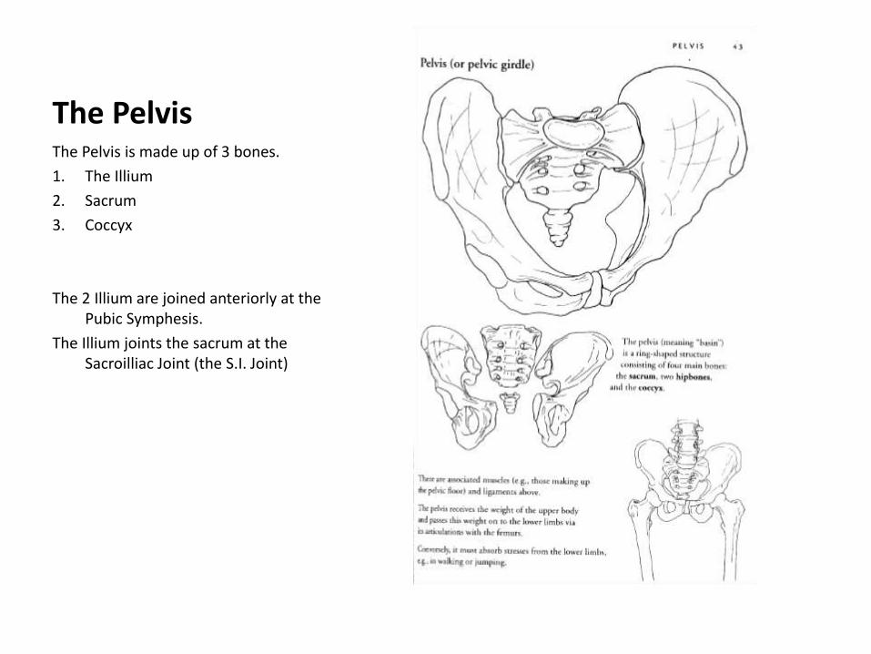
The PelvisThe Pelvis is made up of 3 bones.
1. The Illium
2. Sacrum
3. Coccyx
The 2 Illium are joined anteriorly at the Pubic Symphesis.
The Illium joints the sacrum at the Sacroilliac Joint (the S.I. Joint)
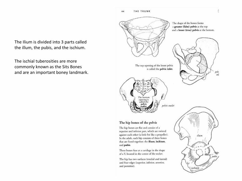
The Ilium is divided into 3 parts called the illum, the pubis, and the ischium.
The ischial tuberosities are more commonly known as the Sits Bones and are an important boney landmark.

Injuries are very common at he S.I. joint and L4 & L5 (the Lumbosacral joint) because they are crucial postural weight bearing junctures.
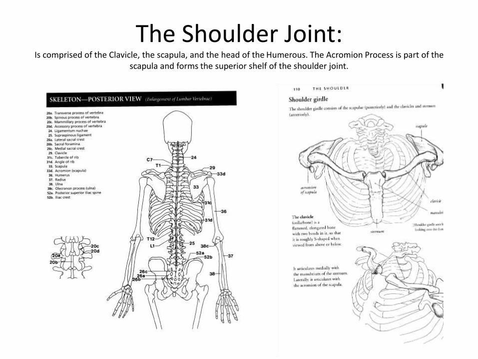
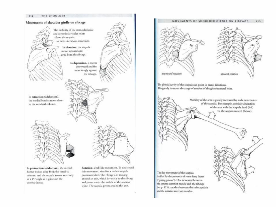
The Shoulder Joint:Is comprised of the Clavicle, the scapula, and the head of the Humerous. The Acromion Process is part of the
scapula and forms the superior shelf of the shoulder joint.
Compression is when 2 bones hit against each other and prevent movement.
Tension occurs when tissues are not flexible enough to allow 2 bones to move apart.

The Elbow Joint
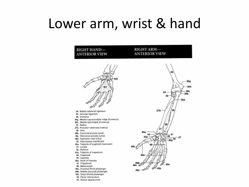
Lower arm, wrist & hand
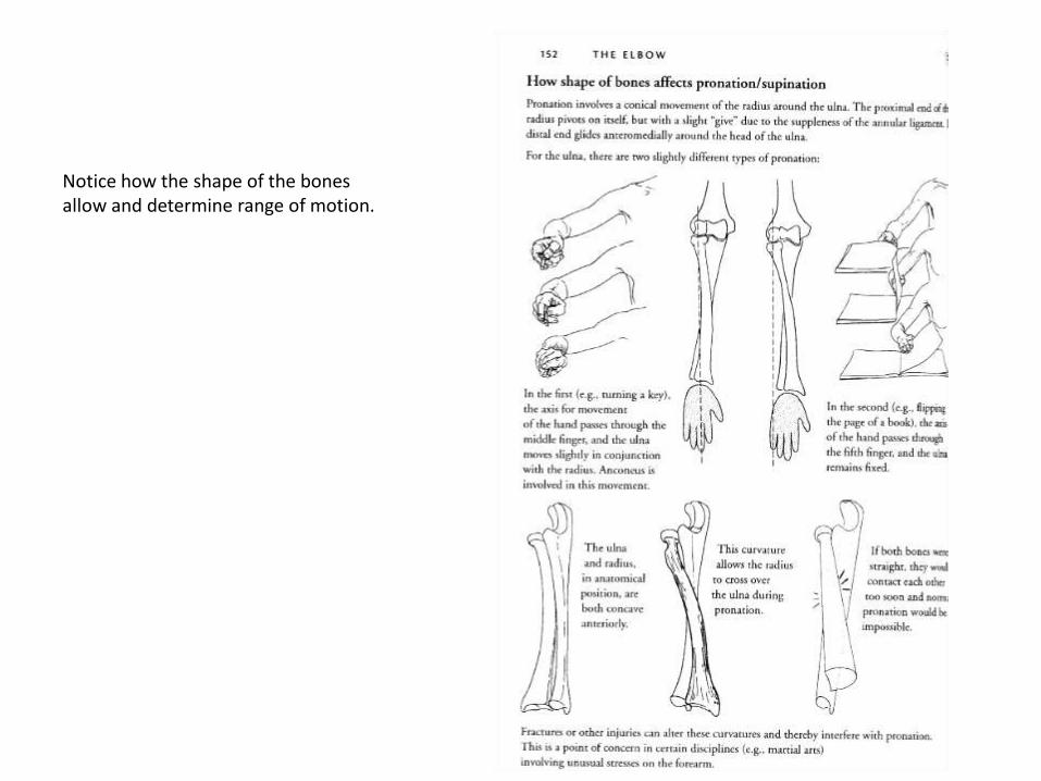
Notice how the shape of the bones allow and determine range of motion.

The Hip Joint
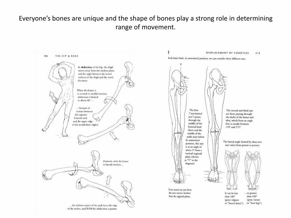
Everyone’s bones are unique and the shape of bones play a strong role in determining range of movement.

The Knee, Foot, & Ankle Joints
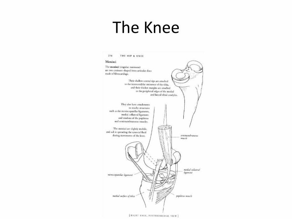
The Knee