Embed Size (px)
Citation preview

THE HIP IN CEREBRAL
PALSY
Topic presentation by
Dr. Libin Thomas Manathara
Amala Institute of Medical Sciences

Topics
• Introduction
• Flexion deformities
• Adduction deformities
• Adductor tenotomy
• Iliopsoas recession
• Iliopsoas release at the lesser trochanter
• Subluxation and Dislocation
• Varus derotational osteotomy
• San Diego procedure
• Proximal femoral resection
• Hip arthrodesis
• Total Hip Arthroplasty

Introduction
• In patients with cerebral palsy, all hips
should be considered abnormal until
proved otherwise
• Deformities of the hip in patients with
cerebral palsy range from mild painless
subluxation to complete dislocation with
joint destruction, pain, and impaired
mobility
Introduction
• When a hip begins to subluxate, it rarely
improves without treatment

Introduction
• Progressive hip instability occurs in
approximately 15% of patients with
cerebral palsy, the causes of which
includes
– muscle imbalance,
– retained primitive reflexes,
– abnormal positioning, and
– pelvic obliquity
Introduction
• Beals (1969) described a practical
radiographic method for quantifying the
amount of hip subluxation present
• It was described by Reimers (1980) as the
“migration percentage”

Introduction
• The migration percentage is determinedby drawing the Hilgenreiner lineconnecting the two triradiate cartilagesand then perpendicular lines at the lateralmargins of the bony acetabula

Introduction
• The width of the femoral head uncovered
(lateral to the perpendicular line) is divided
by the total width of the femoral head and
multiplied by 100 to give the migration
percentage
• This index typically is 0 until age 4 years
and less than 5% from 4 years until
skeletal maturity

Subluxated left hip joint. Migration index (MI) is calculated by dividing width of uncovered
femoral head A by total width of femoral head B.
Acetabulum is dysplastic, with lateral corner of acetabulum above weight bearing dome.
Normal hip (left side) with acetabular index (AI) indicated. Lateral corner is sharp and
below weight-bearing dome.
H, horizontal axis.

Introductionhttp://www.orthobullets.com/pediatrics/4118/developmental-dysplasia-of-the-hip
• Perkin's line is a line perpendicular
line to Hilgenreiner's through a
point at the lateral margin of
acetabulum
• The Acetabular index (AI) is an
angle formed by a line drawn from
point on the lateral triradiate
cartilage (X) to point on lateral
margin of acetabulum (O) and
Hilgenreiners line and it should be
less than 25° in patients older than
6 months

Introduction
• Reimers described a migration of greater
than 33% as subluxation and greater than
100% as dislocation
• More important than the absolute value is
the change observed within a given patient

Flexion Deformities
• Excessive hip flexion brings the center of
gravity anteriorly and is compensated for
by
– increased lumbar lordosis
– knee flexion
– ankle dorsiflexion

Typical crouch posture caused by flexion deformities of hips or fixed
flexion deformities of knees.

Flexion Deformities
• It is important to determine whether the
increased hip flexion is primary or
secondary to knee or ankle contractures
because if unrecognized and a hip flexor
release is done, it can weaken the hip
further and increase hip flexion

Flexion Deformities
• Children with flexion-internal rotation
deformity sit with a wide base of support in
the W position
– hips flexed 90 degrees
– maximally internally rotated
– knees maximally flexed
– feet externally rotated

W postition
Flexion Deformities
• Single-stage multilevel procedures arepreferable to staged single-levelprocedures because
– hospitalization is reduced
– immobilization is a one time event
– rehabilitation time is reduced
– number of anesthetic exposures aredecreased
– minimize the effects of surgery on socialinteraction and education

Flexion Deformities
• Hip flexion contractures from 15 to 30
degrees are usually treated with psoas
lengthening through an intramuscular
recession over the pelvic brim

Flexion Deformities
• Contractures of more than 30 degrees may
require more extensive releases of the following
– rectus femoris
– tensor fasciae latae
– anterior fibers of the gluteus minimus
– anterior fibres of the gluteus medius
– sartorius
– iliopsoas

Adduction Deformities
• Adduction is the most common deformity
of the hip in children with cerebral palsy
and cause difficulties like
– scissoring of the legs during gait
– hip subluxation
– in severely affected children, difficulty with
perineal hygiene

Adduction Deformities
• For mild contractures, an adductortenotomy usually is sufficient
• More severe contractures often requirerelease of the gracilis and the anterior halfof the adductor brevis
• Adductor tenotomies usually are donebilaterally to prevent a “windswept” pelvis

Windswept pelvis (Dr Henry Knipe and Dr Behnam Shayegi et al.) http://radiopaedia.org/articles/windswept-pelvis
Wind-swept pelvis fracture is a combination a unilateral AP compression (open book) injury with a contralateral lateral
compression injury
It occurs when the internal rotation of one iliac wing causes a unilateral sacral compression fracture, while the same
forces cause external rotation of the opposite hemipelvis, resulting in diastasis of the sacro-iliac joint
This causes classic “wind-swept pelvis” appearance
Case courtesy of Dr Matt A. Morgan, Radiopaedia.org, rID: 37825

Adduction Deformities-
Adductor tenotomy• Adductor tenotomy is indicated for a
patient with a mild adduction contracture,as indicated by a scissoring gait or earlyhip subluxation
• This procedure should be done earlybecause damage to the developingacetabulum from abnormal hip muscleforces is greatest before 4 years of age

Adduction Deformities-
Adductor tenotomy• The ideal candidate for soft tissue
lengthening is an ambulatory child
younger than 8 years, and preferably
younger than 4 years, who has
– hip abduction of less than 30 degrees and
– a migration index of less than 50%

Adduction Deformities-
Adductor tenotomy• Neurectomy of the anterior branch of the
obturator nerve should be avoided toprevent iatrogenic hip abductioncontracture

Obturator nerve
• The obturator nerve arises
from the ventral divisions of
L2, L3 and L4
• It passes through the
obutrator formane to enter
the thigh
• It divides into an anterior
and posterior branch in
thigh

Obturator nerve

Adduction Deformities-
Adductor tenotomy• Early soft tissue release alone may be
insufficient to prevent long-term hipsubluxation and dislocation
• It may delay major bony surgery, however,until the risk of recurrence is decreasedand the bone stock for reconstruction isimproved

Adduction Deformities-
Adductor tenotomy• Procedure
• Place the patient supine on the operating
table, and prepare the area from the toes
to the inferior costal margin, isolating the
perineum
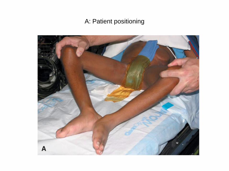
A: Patient positioning

Adduction Deformities-
Adductor tenotomy• Identify the adductor longus by palpation,
and make a 3-cm transverse incision over
the adductor longus tendon approximately
1 cm distal from its origin
• Dissect through the subcutaneous tissue,
and identify the adductor longus fascia

B: Skin incision and subcutaneous dissection to identify adductor
longus fascia

Adduction Deformities-
Adductor tenotomy• Make a longitudinal incision in the
adductor fascia; identify the tendinous
portion of the adductor longus, and resect
it with electrocautery

Adduction Deformities-
Adductor tenotomy• Release with electrocautery any remaining
muscle fibers of the adductor longus asnecessary.
• Avoid injury to the anterior branch of theobturator nerve, which is in the intervalbetween the adductor longus and brevis


C: Hemostat placed under anterior branch of obturator nerve

Adduction Deformities-
Adductor tenotomy• Gradually abduct the hip, and determine the
amount of correction obtained
• If further correction is required, slowly releasethe anterior half of the adductor brevis usingelectrocautery and avoiding injury to thebranches of the obturator nerve
• It is important not to release an excessiveamount of the adductor brevis and to protect theposterior branch of the obturator nerve toprevent an abduction contracture

Adduction Deformities-
Adductor tenotomy• If the gracilis muscle is found to be tight,
release it with electrocautery
• When the final correction is obtained,
close the wound in layers
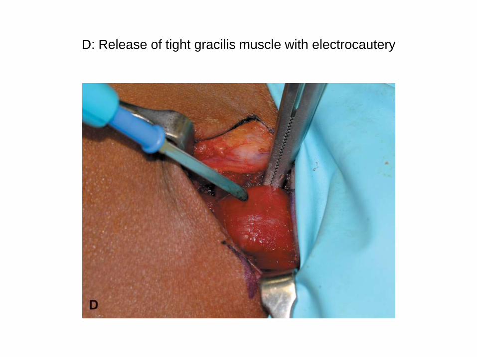
D: Release of tight gracilis muscle with electrocautery

Adduction Deformities-
Adductor tenotomy• Take care to close the adductor fascia to
help prevent skin dimpling postoperatively

E: Closure of adductor fascia

Iliopsoas Recession
• Bleck recommended iliopsoas recessionwhen the hip internally rotates duringwalking or when passive external rotationis absent with the hip in full extension andpresent when the joint is passively flexedto 90 degrees

Iliopsoas Recession
• This procedure usually is done inconjunction with other soft tissue releasesof the lower extremities
• Iliopsoas recession is used morecommonly than complete tenotomy at thelevel of the lesser trochanter to avoidcausing excessive hip flexion weakness

Iliopsoas Recession
• SKAGGS et al
• Place the patient supine with a roll under
the buttock of the operative side
• Palpate the course of the femoral artery,
and mark it on the skin, keeping in mind
that the femoral nerve is lateral to it

Iliopsoas Recession
• For an isolated iliopsoas recession, make a 5cm
“bikini” incision
• This incision can be modified as needed if other
procedures are going to be done at the same
time
• Center the incision medial to and 2 cm below the
anterior superior iliac spine

Bikini incision

Iliopsoas Recession
• Identify and develop the interval between
the tensor fasciae latae and sartorius to
expose the direct head of the rectus
femoris with its origin at the anterior
inferior iliac spine
• It is not necessary to identify the femoral
neurovascular structures

Iliopsoas Recession
• Palpate the pelvic brim just medial and
inferior to the rectus femoris origin to
locate the iliopsoas tendon in a shallow
groove
• Slightly flex the hip to relax the soft tissuestructures around the hip

Iliopsoas Recession
• Place a right-angle retractor on the lateral
aspect of the iliopsoas muscle, and pull
the retractor medially and anteriorly,
exposing the posteromedial aspect of the
muscle and the psoas tendon (see Fig)
• The retractor is protecting the femoral
nerve, which is medial to it

Skaggs et al. surgical approach for iliopsoas recession
When procedure is done alone, much smaller incision is adequate

Iliopsoas Recession
• Dissect the surrounding muscle fascia,
and isolate the tendon from the muscle
with a right-angle clamp
• Verify that there is enough muscle
remaining at that level so that continuity is
maintained after tendon release

Iliopsoas Recession
• Under direct vision, carefully internally and
externally rotate the hip to see the tendon
loosen and tighten
• If there is any doubt as to the identification
of the tendon, use an elevator to dissect
around the tendon proximally until its
muscle fibers are identified

Iliopsoas Recession
• An electrical nerve stimulator or careful
brief stimulation with electrocautery also
can be used to help confirm that the
tendon has been found and that the
femoral nerve has not been mistakenly
identified

Iliopsoas Recession
• Release the tendinous portion, leaving the
muscle fibers in continuity
• Extend and internally rotate the hip to
separate the tendon ends
• Close the wound in layers, and apply
sterile dressings

Iliopsoas Recession
• Postoperative Care
• Patients with an isolated iliopsoas release arestarted immediately in a physical therapyprogram emphasizing hip extension and externalrotation
• Patients, especially those who are unable tocooperate with physical therapy, are placedprone at bed rest to help improve hip extension

Iliopsoas Release at the Lesser
Trochanter
• Iliopsoas release at its insertion on thelesser trochanter is better fornonambulatory patients than forambulatory patients because of the risk ofcausing excessive hip flexion weakness,which can severely affect an ambulatorypatient
• It often is done at the same time asanother procedure, such as an adductorrelease or varus derotational osteotomy

Iliopsoas Release at the Lesser
Trochanter
• Additional release of the secondary hip
flexors including the sartorius and rectus
femoris also may be used for severe
deformities

Iliopsoas Release at the Lesser
Trochanter
• Procedure
• Make a transverse incision 1 to 3 cm distal to the
inguinal crease
• If an adductor release is to be done at the same
time, make a longitudinal incision in the adductor
longus fascia and transect the adductor longus
with electrocautery; perform a myotomy of the
gracilis if necessary

Iliopsoas Release at the Lesser
Trochanter• Resect as much of the adductor brevis as
necessary to obtain 45 degrees of
abduction
• Develop the interval between the residual
adductor brevis and the pectineus or
between the pectineus and the
neurovascular bundle until the femur is
identified

Iliopsoas Release at the Lesser
Trochanter
• Open the bursa over the iliopsoas and itssheath
• Place a retractor into the tendon sheath,and retract the tendon medially

Iliopsoas Release at the Lesser
Trochanter
• Pass a right-angle clamp under the tendon
of the iliopsoas, which can be completely
released with electrocautery in a
nonambulatory child
• Release the iliopsoas as far proximally as
possible in an ambulatory child to preserve
the iliacus muscle attachment to the
iliopsoas tendon

Iliopsoas Release at the Lesser
Trochanter• Postoperative Care
• Physical therapy is started 2 days after
surgery, emphasizing range-of-motion
exercises of the hips and knees

Iliopsoas Release at the Lesser
Trochanter• Leg-knee immobilizers are used 8 to 12
hours a day for 1 month
• Parents are encouraged to have the child
sleep prone as much as possible

THANK YOU