Embed Size (px)
Citation preview
THETHE BREAST CLINIC BREAST CLINIC GUIDEGUIDE
DR. MOHAMAD AL-GAILANI FRCS
Consultant Breast Surgeon
Riyadh, KSA
2018

Breast Embryology

BREAST ANATOMY

ABERRATION OF NORMAL DEVELOPMENT & INVOLUTION (ANDI)

TIME LINE OF COMMON BREAST CONDITIONS

MANAGEMENT OF BREAST DISEASES

THE TRIPLE ASSESSMENT

MAMMOGRAPHY (From age 35 years)

BREAST ULTRASOUND
• Any age• Cyst versus Solid• Complimentary to
Mammography

FINE NEEDLE ASPIRATION CYTOLOGY (FNAC)C1 Inadequate C2 Benign C3 Indeterminate C4 Suspicious C5 Malignant

NEEDLE CORE BIOPSY (NCB)
• The Gold Standard in breast biopsy
• Manual or Ultrasound Guided• More reliable than Fine Needle
Aspiration Cytology (FNAC)• Can request Receptor Status and
Immunohistochemistry for Cancer
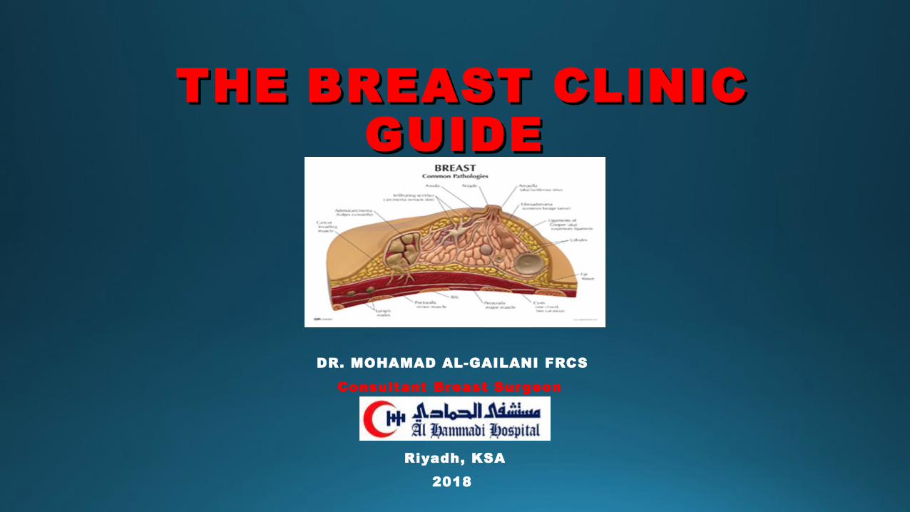
TOP 10 BREAST COMPLAINTS

1. MASTALGIA (PAIN)


MASTALGIA: TREATMENT

2. FIBROADENOSIS (PAINFUL LUMPINESS)

3. BREAST LUMP

• Breast Screening can spot a lump as small as few mm in diameter.
• Breast awareness and breast self exam (BSE) has a higher chance of recognising a lump earlier.
• The earlier diagnosis of a malignant breast lump, the better the chance of survival following treatment.

ANDI: FIBROADENOMA & BREAST CYST
YOUNG AGE AND TEENS
MIDDLE AGE WOMEN

FIBROADENOMA
• Part of ANDI• Women younger than 30 years. • Discreet lump and freely mobile. • If less than 10mm then 2/3 may disappear within 2 years• If larger, unlikely to disappear• Needle biopsy if not for excision• Offer excision if anxious, send for histology

BREAST CYST• Part of ANDI• Women in their 40’s • Overlaps with breast cancer age incidence • Ultrasound is diagnostic.• Aspirate in clinic if feasible • No fluid cytology necessary, only if bloody. • The lump should disappear after aspiration • Recurrent• 1% Malignant Cyst adenocarcinoma• Suspect if bloody fluid, re-accumulates rapidly or
if ultrasound is suggestive.

4. DUCT ECTASIA


5. NIPPLE DISCHARGE

• Physical examination• “Haemostix” can be used to test for the presence of blood • Cytology if bloody• Ultrasound or Mammography in > 35 years of age• Serum Prolactin• Cancer is unlikely if the discharge is coming from both
nipples and/or multiple ducts.

NIPPLE DISCHARGE:INDICATIONS FOR SURGERY

6. MASTITIS AND BREAST ABSCESS

7. FAT NECROSIS

8. ECZEMA OF SKIN V ECZEMA OF NIPPLE
Skin Condition Paget’s Disease of Nipple BREAST CANCER

9. Nipple Inversion

10. BREAST CANCER
PEAU D’ ORANGE
INFLAMMATORY CANCER

Breast Cancer: Workup

BREAST CLINIC GUIDETake Home Message

QuestionQuestions?s?